JP6469558B2 - 内視鏡システム及び内視鏡システムの作動方法 - Google Patents
内視鏡システム及び内視鏡システムの作動方法 Download PDFInfo
- Publication number
- JP6469558B2 JP6469558B2 JP2015199393A JP2015199393A JP6469558B2 JP 6469558 B2 JP6469558 B2 JP 6469558B2 JP 2015199393 A JP2015199393 A JP 2015199393A JP 2015199393 A JP2015199393 A JP 2015199393A JP 6469558 B2 JP6469558 B2 JP 6469558B2
- Authority
- JP
- Japan
- Prior art keywords
- blood vessel
- index value
- observation
- vessel index
- image
- Prior art date
- Legal status (The legal status is an assumption and is not a legal conclusion. Google has not performed a legal analysis and makes no representation as to the accuracy of the status listed.)
- Active
Links
Images





Classifications
-
- A—HUMAN NECESSITIES
- A61—MEDICAL OR VETERINARY SCIENCE; HYGIENE
- A61B—DIAGNOSIS; SURGERY; IDENTIFICATION
- A61B5/00—Measuring for diagnostic purposes; Identification of persons
- A61B5/02—Detecting, measuring or recording for evaluating the cardiovascular system, e.g. pulse, heart rate, blood pressure or blood flow
- A61B5/02007—Evaluating blood vessel condition, e.g. elasticity, compliance
-
- A—HUMAN NECESSITIES
- A61—MEDICAL OR VETERINARY SCIENCE; HYGIENE
- A61B—DIAGNOSIS; SURGERY; IDENTIFICATION
- A61B1/00—Instruments for performing medical examinations of the interior of cavities or tubes of the body by visual or photographical inspection, e.g. endoscopes; Illuminating arrangements therefor
- A61B1/00002—Operational features of endoscopes
- A61B1/00004—Operational features of endoscopes characterised by electronic signal processing
- A61B1/00009—Operational features of endoscopes characterised by electronic signal processing of image signals during a use of endoscope
- A61B1/000094—Operational features of endoscopes characterised by electronic signal processing of image signals during a use of endoscope extracting biological structures
-
- A—HUMAN NECESSITIES
- A61—MEDICAL OR VETERINARY SCIENCE; HYGIENE
- A61B—DIAGNOSIS; SURGERY; IDENTIFICATION
- A61B1/00—Instruments for performing medical examinations of the interior of cavities or tubes of the body by visual or photographical inspection, e.g. endoscopes; Illuminating arrangements therefor
- A61B1/00002—Operational features of endoscopes
- A61B1/00043—Operational features of endoscopes provided with output arrangements
- A61B1/00045—Display arrangement
- A61B1/0005—Display arrangement combining images e.g. side-by-side, superimposed or tiled
-
- A—HUMAN NECESSITIES
- A61—MEDICAL OR VETERINARY SCIENCE; HYGIENE
- A61B—DIAGNOSIS; SURGERY; IDENTIFICATION
- A61B1/00—Instruments for performing medical examinations of the interior of cavities or tubes of the body by visual or photographical inspection, e.g. endoscopes; Illuminating arrangements therefor
- A61B1/00163—Optical arrangements
- A61B1/00188—Optical arrangements with focusing or zooming features
-
- A—HUMAN NECESSITIES
- A61—MEDICAL OR VETERINARY SCIENCE; HYGIENE
- A61B—DIAGNOSIS; SURGERY; IDENTIFICATION
- A61B1/00—Instruments for performing medical examinations of the interior of cavities or tubes of the body by visual or photographical inspection, e.g. endoscopes; Illuminating arrangements therefor
- A61B1/04—Instruments for performing medical examinations of the interior of cavities or tubes of the body by visual or photographical inspection, e.g. endoscopes; Illuminating arrangements therefor combined with photographic or television appliances
- A61B1/041—Capsule endoscopes for imaging
-
- A—HUMAN NECESSITIES
- A61—MEDICAL OR VETERINARY SCIENCE; HYGIENE
- A61B—DIAGNOSIS; SURGERY; IDENTIFICATION
- A61B1/00—Instruments for performing medical examinations of the interior of cavities or tubes of the body by visual or photographical inspection, e.g. endoscopes; Illuminating arrangements therefor
- A61B1/06—Instruments for performing medical examinations of the interior of cavities or tubes of the body by visual or photographical inspection, e.g. endoscopes; Illuminating arrangements therefor with illuminating arrangements
- A61B1/0638—Instruments for performing medical examinations of the interior of cavities or tubes of the body by visual or photographical inspection, e.g. endoscopes; Illuminating arrangements therefor with illuminating arrangements providing two or more wavelengths
-
- A—HUMAN NECESSITIES
- A61—MEDICAL OR VETERINARY SCIENCE; HYGIENE
- A61B—DIAGNOSIS; SURGERY; IDENTIFICATION
- A61B1/00—Instruments for performing medical examinations of the interior of cavities or tubes of the body by visual or photographical inspection, e.g. endoscopes; Illuminating arrangements therefor
- A61B1/06—Instruments for performing medical examinations of the interior of cavities or tubes of the body by visual or photographical inspection, e.g. endoscopes; Illuminating arrangements therefor with illuminating arrangements
- A61B1/0646—Instruments for performing medical examinations of the interior of cavities or tubes of the body by visual or photographical inspection, e.g. endoscopes; Illuminating arrangements therefor with illuminating arrangements with illumination filters
-
- A—HUMAN NECESSITIES
- A61—MEDICAL OR VETERINARY SCIENCE; HYGIENE
- A61B—DIAGNOSIS; SURGERY; IDENTIFICATION
- A61B1/00—Instruments for performing medical examinations of the interior of cavities or tubes of the body by visual or photographical inspection, e.g. endoscopes; Illuminating arrangements therefor
- A61B1/06—Instruments for performing medical examinations of the interior of cavities or tubes of the body by visual or photographical inspection, e.g. endoscopes; Illuminating arrangements therefor with illuminating arrangements
- A61B1/0661—Endoscope light sources
- A61B1/0669—Endoscope light sources at proximal end of an endoscope
-
- A—HUMAN NECESSITIES
- A61—MEDICAL OR VETERINARY SCIENCE; HYGIENE
- A61B—DIAGNOSIS; SURGERY; IDENTIFICATION
- A61B1/00—Instruments for performing medical examinations of the interior of cavities or tubes of the body by visual or photographical inspection, e.g. endoscopes; Illuminating arrangements therefor
- A61B1/06—Instruments for performing medical examinations of the interior of cavities or tubes of the body by visual or photographical inspection, e.g. endoscopes; Illuminating arrangements therefor with illuminating arrangements
- A61B1/0661—Endoscope light sources
- A61B1/0684—Endoscope light sources using light emitting diodes [LED]
-
- A—HUMAN NECESSITIES
- A61—MEDICAL OR VETERINARY SCIENCE; HYGIENE
- A61B—DIAGNOSIS; SURGERY; IDENTIFICATION
- A61B5/00—Measuring for diagnostic purposes; Identification of persons
- A61B5/0059—Measuring for diagnostic purposes; Identification of persons using light, e.g. diagnosis by transillumination, diascopy, fluorescence
- A61B5/0082—Measuring for diagnostic purposes; Identification of persons using light, e.g. diagnosis by transillumination, diascopy, fluorescence adapted for particular medical purposes
- A61B5/0084—Measuring for diagnostic purposes; Identification of persons using light, e.g. diagnosis by transillumination, diascopy, fluorescence adapted for particular medical purposes for introduction into the body, e.g. by catheters
-
- A—HUMAN NECESSITIES
- A61—MEDICAL OR VETERINARY SCIENCE; HYGIENE
- A61B—DIAGNOSIS; SURGERY; IDENTIFICATION
- A61B5/00—Measuring for diagnostic purposes; Identification of persons
- A61B5/02—Detecting, measuring or recording for evaluating the cardiovascular system, e.g. pulse, heart rate, blood pressure or blood flow
- A61B5/021—Measuring pressure in heart or blood vessels
- A61B5/02133—Measuring pressure in heart or blood vessels by using induced vibration of the blood vessel
-
- A—HUMAN NECESSITIES
- A61—MEDICAL OR VETERINARY SCIENCE; HYGIENE
- A61B—DIAGNOSIS; SURGERY; IDENTIFICATION
- A61B5/00—Measuring for diagnostic purposes; Identification of persons
- A61B5/02—Detecting, measuring or recording for evaluating the cardiovascular system, e.g. pulse, heart rate, blood pressure or blood flow
- A61B5/026—Measuring blood flow
- A61B5/0261—Measuring blood flow using optical means, e.g. infrared light
-
- A—HUMAN NECESSITIES
- A61—MEDICAL OR VETERINARY SCIENCE; HYGIENE
- A61B—DIAGNOSIS; SURGERY; IDENTIFICATION
- A61B5/00—Measuring for diagnostic purposes; Identification of persons
- A61B5/103—Measuring devices for testing the shape, pattern, colour, size or movement of the body or parts thereof, for diagnostic purposes
- A61B5/1032—Determining colour of tissue for diagnostic purposes
-
- A—HUMAN NECESSITIES
- A61—MEDICAL OR VETERINARY SCIENCE; HYGIENE
- A61B—DIAGNOSIS; SURGERY; IDENTIFICATION
- A61B5/00—Measuring for diagnostic purposes; Identification of persons
- A61B5/103—Measuring devices for testing the shape, pattern, colour, size or movement of the body or parts thereof, for diagnostic purposes
- A61B5/107—Measuring physical dimensions, e.g. size of the entire body or parts thereof
-
- A—HUMAN NECESSITIES
- A61—MEDICAL OR VETERINARY SCIENCE; HYGIENE
- A61B—DIAGNOSIS; SURGERY; IDENTIFICATION
- A61B5/00—Measuring for diagnostic purposes; Identification of persons
- A61B5/103—Measuring devices for testing the shape, pattern, colour, size or movement of the body or parts thereof, for diagnostic purposes
- A61B5/107—Measuring physical dimensions, e.g. size of the entire body or parts thereof
- A61B5/1079—Measuring physical dimensions, e.g. size of the entire body or parts thereof using optical or photographic means
-
- A—HUMAN NECESSITIES
- A61—MEDICAL OR VETERINARY SCIENCE; HYGIENE
- A61B—DIAGNOSIS; SURGERY; IDENTIFICATION
- A61B5/00—Measuring for diagnostic purposes; Identification of persons
- A61B5/145—Measuring characteristics of blood in vivo, e.g. gas concentration or pH-value ; Measuring characteristics of body fluids or tissues, e.g. interstitial fluid or cerebral tissue
- A61B5/1455—Measuring characteristics of blood in vivo, e.g. gas concentration or pH-value ; Measuring characteristics of body fluids or tissues, e.g. interstitial fluid or cerebral tissue using optical sensors, e.g. spectral photometrical oximeters
- A61B5/1459—Measuring characteristics of blood in vivo, e.g. gas concentration or pH-value ; Measuring characteristics of body fluids or tissues, e.g. interstitial fluid or cerebral tissue using optical sensors, e.g. spectral photometrical oximeters invasive, e.g. introduced into the body by a catheter
-
- A—HUMAN NECESSITIES
- A61—MEDICAL OR VETERINARY SCIENCE; HYGIENE
- A61B—DIAGNOSIS; SURGERY; IDENTIFICATION
- A61B5/00—Measuring for diagnostic purposes; Identification of persons
- A61B5/48—Other medical applications
- A61B5/4842—Monitoring progression or stage of a disease
-
- A—HUMAN NECESSITIES
- A61—MEDICAL OR VETERINARY SCIENCE; HYGIENE
- A61B—DIAGNOSIS; SURGERY; IDENTIFICATION
- A61B5/00—Measuring for diagnostic purposes; Identification of persons
- A61B5/48—Other medical applications
- A61B5/4887—Locating particular structures in or on the body
- A61B5/489—Blood vessels
-
- A—HUMAN NECESSITIES
- A61—MEDICAL OR VETERINARY SCIENCE; HYGIENE
- A61B—DIAGNOSIS; SURGERY; IDENTIFICATION
- A61B5/00—Measuring for diagnostic purposes; Identification of persons
- A61B5/72—Signal processing specially adapted for physiological signals or for diagnostic purposes
- A61B5/7271—Specific aspects of physiological measurement analysis
- A61B5/7282—Event detection, e.g. detecting unique waveforms indicative of a medical condition
-
- G—PHYSICS
- G02—OPTICS
- G02B—OPTICAL ELEMENTS, SYSTEMS OR APPARATUS
- G02B23/00—Telescopes, e.g. binoculars; Periscopes; Instruments for viewing the inside of hollow bodies; Viewfinders; Optical aiming or sighting devices
- G02B23/24—Instruments or systems for viewing the inside of hollow bodies, e.g. fibrescopes
-
- G—PHYSICS
- G02—OPTICS
- G02B—OPTICAL ELEMENTS, SYSTEMS OR APPARATUS
- G02B23/00—Telescopes, e.g. binoculars; Periscopes; Instruments for viewing the inside of hollow bodies; Viewfinders; Optical aiming or sighting devices
- G02B23/24—Instruments or systems for viewing the inside of hollow bodies, e.g. fibrescopes
- G02B23/2407—Optical details
- G02B23/2423—Optical details of the distal end
- G02B23/243—Objectives for endoscopes
- G02B23/2438—Zoom objectives
-
- G—PHYSICS
- G02—OPTICS
- G02B—OPTICAL ELEMENTS, SYSTEMS OR APPARATUS
- G02B23/00—Telescopes, e.g. binoculars; Periscopes; Instruments for viewing the inside of hollow bodies; Viewfinders; Optical aiming or sighting devices
- G02B23/24—Instruments or systems for viewing the inside of hollow bodies, e.g. fibrescopes
- G02B23/2407—Optical details
- G02B23/2461—Illumination
-
- G—PHYSICS
- G06—COMPUTING OR CALCULATING; COUNTING
- G06T—IMAGE DATA PROCESSING OR GENERATION, IN GENERAL
- G06T7/00—Image analysis
- G06T7/0002—Inspection of images, e.g. flaw detection
- G06T7/0012—Biomedical image inspection
-
- A—HUMAN NECESSITIES
- A61—MEDICAL OR VETERINARY SCIENCE; HYGIENE
- A61B—DIAGNOSIS; SURGERY; IDENTIFICATION
- A61B5/00—Measuring for diagnostic purposes; Identification of persons
- A61B5/0059—Measuring for diagnostic purposes; Identification of persons using light, e.g. diagnosis by transillumination, diascopy, fluorescence
- A61B5/0082—Measuring for diagnostic purposes; Identification of persons using light, e.g. diagnosis by transillumination, diascopy, fluorescence adapted for particular medical purposes
- A61B5/0084—Measuring for diagnostic purposes; Identification of persons using light, e.g. diagnosis by transillumination, diascopy, fluorescence adapted for particular medical purposes for introduction into the body, e.g. by catheters
- A61B5/0086—Measuring for diagnostic purposes; Identification of persons using light, e.g. diagnosis by transillumination, diascopy, fluorescence adapted for particular medical purposes for introduction into the body, e.g. by catheters using infrared radiation
-
- G—PHYSICS
- G06—COMPUTING OR CALCULATING; COUNTING
- G06T—IMAGE DATA PROCESSING OR GENERATION, IN GENERAL
- G06T2207/00—Indexing scheme for image analysis or image enhancement
- G06T2207/10—Image acquisition modality
- G06T2207/10068—Endoscopic image
-
- G—PHYSICS
- G06—COMPUTING OR CALCULATING; COUNTING
- G06T—IMAGE DATA PROCESSING OR GENERATION, IN GENERAL
- G06T2207/00—Indexing scheme for image analysis or image enhancement
- G06T2207/10—Image acquisition modality
- G06T2207/10141—Special mode during image acquisition
-
- G—PHYSICS
- G06—COMPUTING OR CALCULATING; COUNTING
- G06T—IMAGE DATA PROCESSING OR GENERATION, IN GENERAL
- G06T2207/00—Indexing scheme for image analysis or image enhancement
- G06T2207/30—Subject of image; Context of image processing
- G06T2207/30004—Biomedical image processing
- G06T2207/30101—Blood vessel; Artery; Vein; Vascular
Landscapes
- Health & Medical Sciences (AREA)
- Life Sciences & Earth Sciences (AREA)
- Physics & Mathematics (AREA)
- Surgery (AREA)
- Engineering & Computer Science (AREA)
- General Health & Medical Sciences (AREA)
- Medical Informatics (AREA)
- Pathology (AREA)
- Public Health (AREA)
- Biophysics (AREA)
- Biomedical Technology (AREA)
- Heart & Thoracic Surgery (AREA)
- Veterinary Medicine (AREA)
- Molecular Biology (AREA)
- Animal Behavior & Ethology (AREA)
- Optics & Photonics (AREA)
- Radiology & Medical Imaging (AREA)
- Nuclear Medicine, Radiotherapy & Molecular Imaging (AREA)
- Physiology (AREA)
- Cardiology (AREA)
- Vascular Medicine (AREA)
- Dentistry (AREA)
- Oral & Maxillofacial Surgery (AREA)
- General Physics & Mathematics (AREA)
- Signal Processing (AREA)
- Computer Vision & Pattern Recognition (AREA)
- Astronomy & Astrophysics (AREA)
- Spectroscopy & Molecular Physics (AREA)
- Artificial Intelligence (AREA)
- Psychiatry (AREA)
- Hematology (AREA)
- Quality & Reliability (AREA)
- Microelectronics & Electronic Packaging (AREA)
- Theoretical Computer Science (AREA)
- Endoscopes (AREA)
- Instruments For Viewing The Inside Of Hollow Bodies (AREA)
Description
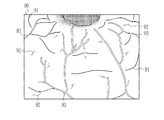
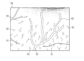
本発明の内視鏡システムは、内視鏡によって観察対象を撮像して得た画像を取得する画像取得部と、画像から、観察対象の血管の血管指標値として、第1観察条件での第1血管指標値を取得し、且つ第1観察条件とは異なる第2観察条件での第2血管指標値を取得することによって、第1血管指標値と第2血管指標値とを含む少なくとも2以上の血管指標値を取得する血管指標値取得部と、第1血管指標値と第2血管指標値とを用いて血管パラメータを算出する血管パラメータ算出部と、第1血管指標値を第1観察条件に基づいた第1補正値で補正し、第2血管指標値を第2観察条件に基づいた第2補正値で補正する血管指標値補正部と、画像から、観察対象との間の観察距離である第1観察距離と、第1観察距離とは異なる第2観察距離とを取得する距離取得部とを備え、血管パラメータ算出部は、第1補正値で補正した第1血管指標値及び第2補正値で補正した第2血管指標値に基づいた重み付けを行って演算し、第1補正値は、第1観察条件が第1観察距離である場合の補正値であり、第2補正値は、第2観察条件が第2観察距離である場合の補正値である。
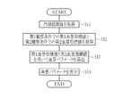
本発明の内視鏡システムの作動方法は、画像取得部が、内視鏡によって観察対象を撮像して得た画像を取得するステップと、血管指標値取得部が、画像に基づく血管指標値の算出処理によって、観察対象の血管の血管指標値として、第1観察条件での第1血管指標値を取得し、且つ第1観察条件とは異なる第2観察条件での第2血管指標値を取得することによって、第1血管指標値と第2血管指標値とを含む少なくとも2以上の血管指標値を取得するステップと、血管パラメータ算出部が、第1血管指標値と第2血管指標値とを用いて血管パラメータを算出するステップとを備える。
図1に示すように、内視鏡システム10は、内視鏡12と、光源装置14と、プロセッサ装置16と、モニタ18と、コンソール19とを有する。内視鏡12は、光源装置14と光学的に接続し、かつ、プロセッサ装置16と電気的に接続する。内視鏡12は、被検体内に挿入する挿入部12aと、挿入部12aの基端部分に設けた操作部12bと、挿入部12aの先端側に設けた湾曲部12c及び先端部12dを有している。操作部12bのアングルノブ13aを操作することにより、湾曲部12cは湾曲動作する。この湾曲動作によって、先端部12dを所望の方向に向ける。
上記第1実施形態では、血管パラメータ算出部78は、第1血管指標値と第2血管指標値とに対して、第1血管指標値と第2血管指標値とに基づいた重み付け係数を乗算しているが、第2実施形態では、第1血管指標値と第2血管指標値とを観察条件に基づいた補正値を用いてそれぞれ補正した後、上記重み付け係数の乗算を行う。この場合、図9に示すように、画像処理部100には、上記第1実施形態の画像処理部60の各構成に加え、血管指標値補正部102を設ける。以下では、上記第1実施形態と同じ構成については説明を省略する。
上記第2実施形態では、観察条件取得部72により第1観察条件及び第2観察条件を取得しているが、第3実施形態では、第1観察距離及び第2観察距離を取得する。この場合、図10に示すように、画像処理部110には、上記第2実施形態の画像処理部100の観察条件取得部72に代えて、距離取得部112を設けている。
上記第各実施形態では、血管パラメータをモニタ18に表示しているが、第4実施形態では、血管パラメータを用いて観察対象の粘膜の状態を判定し、この判定結果を血管パラメータとともにモニタ18に表示する。図12に示すように、第4実施形態の画像処理部120には、上記第1実施形態の画像処理部60の各部に加え、判定部122を設ける。
第5実施形態では、上記各実施形態で示した4色のLED20a〜20dの代わりに、キセノンランプ等の広帯域光源と回転フィルタを用いて観察対象の照明を行う。また、カラーの撮像センサ38に代えて、モノクロの撮像センサで観察対象の撮像を行う。それ以外については、第1実施形態と同様である。
上記各実施形態では、撮像センサ38が設けられた内視鏡12を被検体内に挿入して観察を行う内視鏡システム10、200によって本発明を実施しているが、第6実施形態では、カプセル内視鏡システムにおいて本発明を実施する。カプセル内視鏡システムは、例えば、図16に示すカプセル内視鏡300と、プロセッサ装置(図示しない)とを少なくとも有する。カプセル内視鏡300は、光源302と、光源制御部303と、撮像センサ304と、画像信号取得処理部306と、送受信アンテナ308とを備えている。光源302は、内視鏡システム10の光源20と同様に構成され、光源制御部303の制御によって、照明光を発光する。画像信号取得処理部306は、画像信号取得部50、DSP52、ノイズ低減部54、信号処理部58として機能する。カプセル内視鏡システムのプロセッサ装置は、内視鏡システム10のプロセッサ装置16と同様に構成され、画像処理部60としても機能する。画像信号取得処理部306が生成する内視鏡画像は、送受信アンテナ308を介してプロセッサ装置に送信される。そして、プロセッサ装置では、受信した内視鏡画像から、第1観察条件での第1血管指標値を取得し、且つ第2観察条件での第2血管指標値を取得し、これら第1血管指標値及び第2血管指標値を用いて血管パラメータを算出する。
12 内視鏡
14 光源装置
16 プロセッサ装置
70 画像取得部
74 血管抽出部
76 血管指標値取得部
78 血管パラメータ算出部
102 血管指標値補正部
112 距離取得部
122 判定部
Claims (15)
- 内視鏡によって観察対象を撮像して得た画像を取得する画像取得部と、
前記画像に基づく血管指標値の算出処理によって、前記観察対象の血管の血管指標値として、第1観察条件での第1血管指標値を取得し、且つ前記第1観察条件とは異なる第2観察条件での第2血管指標値を取得することによって、前記第1血管指標値と前記第2血管指標値とを含む少なくとも2以上の血管指標値を取得する血管指標値取得部と、
前記第1血管指標値と前記第2血管指標値とを用いて血管パラメータを算出する血管パラメータ算出部とを備える内視鏡システム。 - 前記血管パラメータ算出部は、前記第1血管指標値及び前記第2血管指標値に対して、前記第1血管指標値と前記第2血管指標値とに基づいた重み付けを行って演算する請求項1に記載の内視鏡システム。
- 前記第1血管指標値を前記第1観察条件に基づいた第1補正値で補正し、前記第2血管指標値を前記第2観察条件に基づいた第2補正値で補正する血管指標値補正部を備え、
前記血管パラメータ算出部は、前記第1補正値で補正した前記第1血管指標値及び前記第2補正値で補正した前記第2血管指標値に対して、前記重み付けを行う請求項2に記載の内視鏡システム。 - 前記血管パラメータ算出部は、四則演算により前記血管パラメータを算出する請求項1に記載の内視鏡システム。
- 前記血管パラメータを用いて、前記観察対象の粘膜の状態を判定する判定部を備える請求項1〜4のいずれか1項に記載の内視鏡システム。
- 前記判定部は、前記血管パラメータを用いて、前記観察対象の粘膜の状態を、正常、腺腫、及びがんを含む3種類以上の状態のいずれかに判定する請求項5に記載の内視鏡システム。
- 前記判定部は、前記血管パラメータを用いて、前記観察対象の粘膜の状態を、正常、過形成ポリープ、SSA/P、腺腫、側方発達型腫瘍、及びがんを含む状態のいずれかに判定する請求項6に記載の内視鏡システム。
- 前記判定部は、前記観察対象の粘膜の状態をがんの状態であると判定した場合、更に、がんのステージを判定する請求項6または7に記載の内視鏡システム。
- 前記血管指標値取得部は、血管の本数、血管の分岐数、血管の分岐角度、分岐点間距離、血管の交差数、血管の太さ、血管の太さの変化、血管の太さの変化の複雑度、血管の長さ、血管の間隔、血管の深さ、血管の高低差、血管の傾き、血管の面積、血管の密度、血管のコントラスト、血管の色、血管の色の変化、血管の蛇行度、血管の血液濃度、血管の酸素飽和度、動脈の割合、静脈の割合、投与した色素の濃度、血管の走行パターン、及び血管の血流量のうちから、少なくとも2以上の血管指標値を取得する請求項1〜8のいずれか1項に記載の内視鏡システム。
- 前記第1血管指標値は、前記第2血管指標値と種類が同じであり、且つ値が互いに異なっている請求項9に記載の内視鏡システム。
- 前記第1血管指標値は、前記第2血管指標値と種類が異なっている請求項9に記載の内視鏡システム。
- 前記第1観察条件及び前記第2観察条件は、前記観察対象との間の観察距離、前記内視鏡のズーム倍率、特殊光を用いる観察モード、前記観察対象に散布した色素のうちの少なくともいずれかである請求項1〜11のいずれか1項に記載の内視鏡システム。
- 内視鏡によって観察対象を撮像して得た画像を取得する画像取得部と、
前記画像から、前記観察対象の血管の血管指標値として、第1観察条件での第1血管指標値を取得し、且つ前記第1観察条件とは異なる第2観察条件での第2血管指標値を取得することによって、前記第1血管指標値と前記第2血管指標値とを含む少なくとも2以上の血管指標値を取得する血管指標値取得部と、
前記第1血管指標値と前記第2血管指標値とを用いて血管パラメータを算出する血管パラメータ算出部とを備え、
前記血管パラメータ算出部は、前記第1血管指標値及び前記第2血管指標値に対して、前記第1血管指標値と前記第2血管指標値とに基づいた重み付けを行って演算し、前記第1血管指標値と前記第2血管指標値とを含む少なくとも2以上の血管指標値のうち、特定の血管指標値以外の血管指標値に対してのみ前記重み付けを行う内視鏡システム。 - 内視鏡によって観察対象を撮像して得た画像を取得する画像取得部と、
前記画像から、前記観察対象の血管の血管指標値として、第1観察条件での第1血管指標値を取得し、且つ前記第1観察条件とは異なる第2観察条件での第2血管指標値を取得することによって、前記第1血管指標値と前記第2血管指標値とを含む少なくとも2以上の血管指標値を取得する血管指標値取得部と、
前記第1血管指標値と前記第2血管指標値とを用いて血管パラメータを算出する血管パラメータ算出部と、
前記第1血管指標値を前記第1観察条件に基づいた第1補正値で補正し、前記第2血管指標値を前記第2観察条件に基づいた第2補正値で補正する血管指標値補正部と、
前記画像から、前記観察対象との間の観察距離である第1観察距離と、前記第1観察距離とは異なる第2観察距離とを取得する距離取得部とを備え、
前記血管パラメータ算出部は、前記第1補正値で補正した前記第1血管指標値及び前記第2補正値で補正した前記第2血管指標値に基づいた重み付けを行って演算し、
前記第1補正値は、前記第1観察条件が前記第1観察距離である場合の補正値であり、
前記第2補正値は、前記第2観察条件が前記第2観察距離である場合の補正値である内視鏡システム。 - 画像取得部が、内視鏡によって観察対象を撮像して得た画像を取得するステップと、
血管指標値取得部が、前記画像に基づく血管指標値の算出処理によって、前記観察対象の血管の血管指標値として、第1観察条件での第1血管指標値を取得し、且つ前記第1観察条件とは異なる第2観察条件での第2血管指標値を取得することによって、前記第1血管指標値と前記第2血管指標値とを含む少なくとも2以上の血管指標値を取得するステップと、
血管パラメータ算出部が、前記第1血管指標値と前記第2血管指標値とを用いて血管パラメータを算出するステップとを備える内視鏡システムの作動方法。
Priority Applications (4)
| Application Number | Priority Date | Filing Date | Title |
|---|---|---|---|
| JP2015199393A JP6469558B2 (ja) | 2015-10-07 | 2015-10-07 | 内視鏡システム及び内視鏡システムの作動方法 |
| PCT/JP2016/077437 WO2017061256A1 (ja) | 2015-10-07 | 2016-09-16 | 内視鏡システム及び内視鏡システムの作動方法 |
| EP16853410.5A EP3360459A4 (en) | 2015-10-07 | 2016-09-16 | Endoscope system and operation method for endoscope system |
| US15/924,277 US20180206738A1 (en) | 2015-10-07 | 2018-03-19 | Endoscope system and method of operating endoscope system |
Applications Claiming Priority (1)
| Application Number | Priority Date | Filing Date | Title |
|---|---|---|---|
| JP2015199393A JP6469558B2 (ja) | 2015-10-07 | 2015-10-07 | 内視鏡システム及び内視鏡システムの作動方法 |
Publications (2)
| Publication Number | Publication Date |
|---|---|
| JP2017070504A JP2017070504A (ja) | 2017-04-13 |
| JP6469558B2 true JP6469558B2 (ja) | 2019-02-13 |
Family
ID=58487398
Family Applications (1)
| Application Number | Title | Priority Date | Filing Date |
|---|---|---|---|
| JP2015199393A Active JP6469558B2 (ja) | 2015-10-07 | 2015-10-07 | 内視鏡システム及び内視鏡システムの作動方法 |
Country Status (4)
| Country | Link |
|---|---|
| US (1) | US20180206738A1 (ja) |
| EP (1) | EP3360459A4 (ja) |
| JP (1) | JP6469558B2 (ja) |
| WO (1) | WO2017061256A1 (ja) |
Families Citing this family (7)
| Publication number | Priority date | Publication date | Assignee | Title |
|---|---|---|---|---|
| JP6640865B2 (ja) * | 2015-09-29 | 2020-02-05 | 富士フイルム株式会社 | 画像処理装置、内視鏡システム、及び画像処理方法 |
| JP6858672B2 (ja) | 2017-08-29 | 2021-04-14 | 富士フイルム株式会社 | 医療画像処理システム及び内視鏡システム |
| EP3698702B1 (en) * | 2017-10-17 | 2023-06-07 | FUJIFILM Corporation | Medical image processing device and endoscope device |
| WO2019078237A1 (ja) | 2017-10-18 | 2019-04-25 | 富士フイルム株式会社 | 医療画像処理装置、内視鏡システム、診断支援装置、並びに医療業務支援装置 |
| CN113679327B (zh) * | 2021-10-26 | 2022-02-18 | 青岛美迪康数字工程有限公司 | 内镜检查图像采集方法及装置 |
| WO2023085262A1 (ja) * | 2021-11-09 | 2023-05-19 | 学校法人順天堂 | 消化管表層微小血管の血流速度測定内視鏡システム |
| WO2024034253A1 (ja) * | 2022-08-12 | 2024-02-15 | 富士フイルム株式会社 | 内視鏡システム及びその作動方法 |
Family Cites Families (6)
| Publication number | Priority date | Publication date | Assignee | Title |
|---|---|---|---|---|
| JP5631764B2 (ja) * | 2011-01-25 | 2014-11-26 | 富士フイルム株式会社 | 内視鏡システム及びその作動方法 |
| US9636041B2 (en) * | 2011-01-28 | 2017-05-02 | Bar Ilan University | Method and system for non-invasively monitoring biological or biochemical parameters of individual |
| JP6092792B2 (ja) * | 2014-01-20 | 2017-03-08 | 富士フイルム株式会社 | 内視鏡システム用プロセッサ装置、内視鏡システム、内視鏡システム用プロセッサ装置の作動方法、内視鏡システムの作動方法 |
| JP5977772B2 (ja) * | 2014-02-06 | 2016-08-24 | 富士フイルム株式会社 | 内視鏡システム、内視鏡システムのプロセッサ装置、内視鏡システムの作動方法、プロセッサ装置の作動方法 |
| JP2015091351A (ja) * | 2014-12-24 | 2015-05-14 | 富士フイルム株式会社 | 内視鏡装置 |
| JP6336949B2 (ja) * | 2015-01-29 | 2018-06-06 | 富士フイルム株式会社 | 画像処理装置及び画像処理方法、並びに内視鏡システム |
-
2015
- 2015-10-07 JP JP2015199393A patent/JP6469558B2/ja active Active
-
2016
- 2016-09-16 WO PCT/JP2016/077437 patent/WO2017061256A1/ja not_active Ceased
- 2016-09-16 EP EP16853410.5A patent/EP3360459A4/en not_active Withdrawn
-
2018
- 2018-03-19 US US15/924,277 patent/US20180206738A1/en not_active Abandoned
Also Published As
| Publication number | Publication date |
|---|---|
| WO2017061256A1 (ja) | 2017-04-13 |
| JP2017070504A (ja) | 2017-04-13 |
| EP3360459A1 (en) | 2018-08-15 |
| US20180206738A1 (en) | 2018-07-26 |
| EP3360459A4 (en) | 2018-12-19 |
Similar Documents
| Publication | Publication Date | Title |
|---|---|---|
| JP6768871B2 (ja) | 内視鏡システム及び内視鏡システムの作動方法 | |
| JP6577088B2 (ja) | 内視鏡システム | |
| JP6633641B2 (ja) | 画像処理装置、内視鏡システム、及び画像処理方法 | |
| JP6420492B2 (ja) | 画像処理装置、内視鏡システム、及び画像処理装置の作動方法 | |
| JP6469558B2 (ja) | 内視鏡システム及び内視鏡システムの作動方法 | |
| JP6640866B2 (ja) | 画像処理装置、内視鏡システム、及び画像処理方法 | |
| US10426318B2 (en) | Image processing apparatus, endoscope system, and image processing method | |
| JP6823072B2 (ja) | プロセッサ装置及び内視鏡システム | |
| JP6538634B2 (ja) | プロセッサ装置及び内視鏡システム並びにプロセッサ装置の作動方法 | |
| JP6461760B2 (ja) | 画像処理装置、及び画像処理装置の作動方法、並びに内視鏡システム | |
| WO2016121811A1 (ja) | 画像処理装置及び画像処理方法、並びに内視鏡システム | |
| WO2018079116A1 (ja) | 内視鏡システム | |
| JPWO2018079217A1 (ja) | 内視鏡システム及びその作動方法 |
Legal Events
| Date | Code | Title | Description |
|---|---|---|---|
| A621 | Written request for application examination |
Free format text: JAPANESE INTERMEDIATE CODE: A621 Effective date: 20180206 |
|
| A131 | Notification of reasons for refusal |
Free format text: JAPANESE INTERMEDIATE CODE: A131 Effective date: 20180925 |
|
| A521 | Request for written amendment filed |
Free format text: JAPANESE INTERMEDIATE CODE: A523 Effective date: 20181113 |
|
| TRDD | Decision of grant or rejection written | ||
| A01 | Written decision to grant a patent or to grant a registration (utility model) |
Free format text: JAPANESE INTERMEDIATE CODE: A01 Effective date: 20181218 |
|
| A61 | First payment of annual fees (during grant procedure) |
Free format text: JAPANESE INTERMEDIATE CODE: A61 Effective date: 20190116 |
|
| R150 | Certificate of patent or registration of utility model |
Ref document number: 6469558 Country of ref document: JP Free format text: JAPANESE INTERMEDIATE CODE: R150 |
|
| R250 | Receipt of annual fees |
Free format text: JAPANESE INTERMEDIATE CODE: R250 |
|
| R250 | Receipt of annual fees |
Free format text: JAPANESE INTERMEDIATE CODE: R250 |
|
| R250 | Receipt of annual fees |
Free format text: JAPANESE INTERMEDIATE CODE: R250 |
|
| R250 | Receipt of annual fees |
Free format text: JAPANESE INTERMEDIATE CODE: R250 |
|
| R250 | Receipt of annual fees |
Free format text: JAPANESE INTERMEDIATE CODE: R250 |