JP6549612B2 - 改善された多相動的造影磁気共鳴撮像方法 - Google Patents
改善された多相動的造影磁気共鳴撮像方法 Download PDFInfo
- Publication number
- JP6549612B2 JP6549612B2 JP2016563051A JP2016563051A JP6549612B2 JP 6549612 B2 JP6549612 B2 JP 6549612B2 JP 2016563051 A JP2016563051 A JP 2016563051A JP 2016563051 A JP2016563051 A JP 2016563051A JP 6549612 B2 JP6549612 B2 JP 6549612B2
- Authority
- JP
- Japan
- Prior art keywords
- magnetic resonance
- resonance imaging
- image
- interest
- fat
- Prior art date
- Legal status (The legal status is an assumption and is not a legal conclusion. Google has not performed a legal analysis and makes no representation as to the accuracy of the status listed.)
- Active
Links
Images


Classifications
-
- A—HUMAN NECESSITIES
- A61—MEDICAL OR VETERINARY SCIENCE; HYGIENE
- A61B—DIAGNOSIS; SURGERY; IDENTIFICATION
- A61B5/00—Measuring for diagnostic purposes; Identification of persons
- A61B5/05—Detecting, measuring or recording for diagnosis by means of electric currents or magnetic fields; Measuring using microwaves or radio waves
- A61B5/055—Detecting, measuring or recording for diagnosis by means of electric currents or magnetic fields; Measuring using microwaves or radio waves involving electronic [EMR] or nuclear [NMR] magnetic resonance, e.g. magnetic resonance imaging
-
- A—HUMAN NECESSITIES
- A61—MEDICAL OR VETERINARY SCIENCE; HYGIENE
- A61B—DIAGNOSIS; SURGERY; IDENTIFICATION
- A61B5/00—Measuring for diagnostic purposes; Identification of persons
- A61B5/48—Other medical applications
- A61B5/4869—Determining body composition
-
- A—HUMAN NECESSITIES
- A61—MEDICAL OR VETERINARY SCIENCE; HYGIENE
- A61B—DIAGNOSIS; SURGERY; IDENTIFICATION
- A61B5/00—Measuring for diagnostic purposes; Identification of persons
- A61B5/72—Signal processing specially adapted for physiological signals or for diagnostic purposes
- A61B5/7203—Signal processing specially adapted for physiological signals or for diagnostic purposes for noise prevention, reduction or removal
- A61B5/7207—Signal processing specially adapted for physiological signals or for diagnostic purposes for noise prevention, reduction or removal of noise induced by motion artifacts
-
- G—PHYSICS
- G01—MEASURING; TESTING
- G01R—MEASURING ELECTRIC VARIABLES; MEASURING MAGNETIC VARIABLES
- G01R33/00—Arrangements or instruments for measuring magnetic variables
- G01R33/20—Arrangements or instruments for measuring magnetic variables involving magnetic resonance
- G01R33/44—Arrangements or instruments for measuring magnetic variables involving magnetic resonance using nuclear magnetic resonance [NMR]
- G01R33/48—NMR imaging systems
- G01R33/4828—Resolving the MR signals of different chemical species, e.g. water-fat imaging
-
- G—PHYSICS
- G01—MEASURING; TESTING
- G01R—MEASURING ELECTRIC VARIABLES; MEASURING MAGNETIC VARIABLES
- G01R33/00—Arrangements or instruments for measuring magnetic variables
- G01R33/20—Arrangements or instruments for measuring magnetic variables involving magnetic resonance
- G01R33/44—Arrangements or instruments for measuring magnetic variables involving magnetic resonance using nuclear magnetic resonance [NMR]
- G01R33/48—NMR imaging systems
- G01R33/54—Signal processing systems, e.g. using pulse sequences ; Generation or control of pulse sequences; Operator console
- G01R33/56—Image enhancement or correction, e.g. subtraction or averaging techniques, e.g. improvement of signal-to-noise ratio and resolution
- G01R33/565—Correction of image distortions, e.g. due to magnetic field inhomogeneities
- G01R33/56509—Correction of image distortions, e.g. due to magnetic field inhomogeneities due to motion, displacement or flow, e.g. gradient moment nulling
-
- G—PHYSICS
- G06—COMPUTING OR CALCULATING; COUNTING
- G06T—IMAGE DATA PROCESSING OR GENERATION, IN GENERAL
- G06T7/00—Image analysis
- G06T7/0002—Inspection of images, e.g. flaw detection
- G06T7/0012—Biomedical image inspection
-
- A—HUMAN NECESSITIES
- A61—MEDICAL OR VETERINARY SCIENCE; HYGIENE
- A61B—DIAGNOSIS; SURGERY; IDENTIFICATION
- A61B5/00—Measuring for diagnostic purposes; Identification of persons
- A61B5/48—Other medical applications
- A61B5/4869—Determining body composition
- A61B5/4872—Body fat
-
- G—PHYSICS
- G01—MEASURING; TESTING
- G01R—MEASURING ELECTRIC VARIABLES; MEASURING MAGNETIC VARIABLES
- G01R33/00—Arrangements or instruments for measuring magnetic variables
- G01R33/20—Arrangements or instruments for measuring magnetic variables involving magnetic resonance
- G01R33/44—Arrangements or instruments for measuring magnetic variables involving magnetic resonance using nuclear magnetic resonance [NMR]
- G01R33/48—NMR imaging systems
- G01R33/54—Signal processing systems, e.g. using pulse sequences ; Generation or control of pulse sequences; Operator console
- G01R33/56—Image enhancement or correction, e.g. subtraction or averaging techniques, e.g. improvement of signal-to-noise ratio and resolution
- G01R33/5601—Image enhancement or correction, e.g. subtraction or averaging techniques, e.g. improvement of signal-to-noise ratio and resolution involving use of a contrast agent for contrast manipulation, e.g. a paramagnetic, super-paramagnetic, ferromagnetic or hyperpolarised contrast agent
-
- G—PHYSICS
- G01—MEASURING; TESTING
- G01R—MEASURING ELECTRIC VARIABLES; MEASURING MAGNETIC VARIABLES
- G01R33/00—Arrangements or instruments for measuring magnetic variables
- G01R33/20—Arrangements or instruments for measuring magnetic variables involving magnetic resonance
- G01R33/44—Arrangements or instruments for measuring magnetic variables involving magnetic resonance using nuclear magnetic resonance [NMR]
- G01R33/48—NMR imaging systems
- G01R33/54—Signal processing systems, e.g. using pulse sequences ; Generation or control of pulse sequences; Operator console
- G01R33/56—Image enhancement or correction, e.g. subtraction or averaging techniques, e.g. improvement of signal-to-noise ratio and resolution
- G01R33/563—Image enhancement or correction, e.g. subtraction or averaging techniques, e.g. improvement of signal-to-noise ratio and resolution of moving material, e.g. flow contrast angiography
- G01R33/56366—Perfusion imaging
-
- G—PHYSICS
- G06—COMPUTING OR CALCULATING; COUNTING
- G06T—IMAGE DATA PROCESSING OR GENERATION, IN GENERAL
- G06T2207/00—Indexing scheme for image analysis or image enhancement
- G06T2207/10—Image acquisition modality
- G06T2207/10072—Tomographic images
- G06T2207/10088—Magnetic resonance imaging [MRI]
-
- G—PHYSICS
- G06—COMPUTING OR CALCULATING; COUNTING
- G06T—IMAGE DATA PROCESSING OR GENERATION, IN GENERAL
- G06T2207/00—Indexing scheme for image analysis or image enhancement
- G06T2207/30—Subject of image; Context of image processing
- G06T2207/30004—Biomedical image processing
- G06T2207/30096—Tumor; Lesion
Landscapes
- Health & Medical Sciences (AREA)
- Life Sciences & Earth Sciences (AREA)
- Physics & Mathematics (AREA)
- Engineering & Computer Science (AREA)
- General Health & Medical Sciences (AREA)
- Medical Informatics (AREA)
- Nuclear Medicine, Radiotherapy & Molecular Imaging (AREA)
- Veterinary Medicine (AREA)
- Biophysics (AREA)
- Molecular Biology (AREA)
- Surgery (AREA)
- Animal Behavior & Ethology (AREA)
- Biomedical Technology (AREA)
- Public Health (AREA)
- Pathology (AREA)
- Heart & Thoracic Surgery (AREA)
- Signal Processing (AREA)
- High Energy & Nuclear Physics (AREA)
- Radiology & Medical Imaging (AREA)
- General Physics & Mathematics (AREA)
- Condensed Matter Physics & Semiconductors (AREA)
- Computer Vision & Pattern Recognition (AREA)
- Artificial Intelligence (AREA)
- Physiology (AREA)
- Psychiatry (AREA)
- Quality & Reliability (AREA)
- Theoretical Computer Science (AREA)
- Magnetic Resonance Imaging Apparatus (AREA)
Description
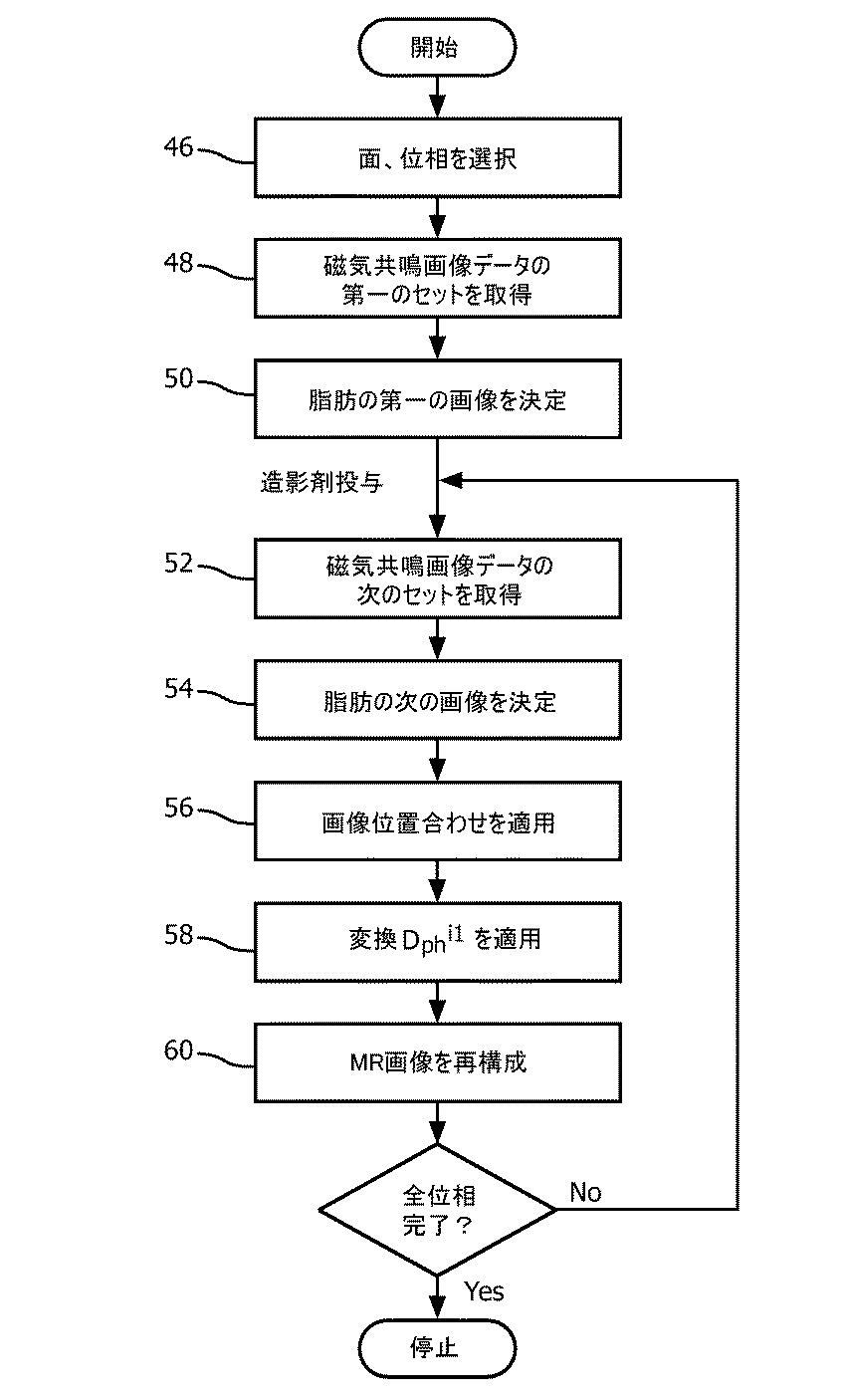
・関心対象に造影剤を投与する前に磁気共鳴画像データの少なくとも第一のセットを取得する段階と;
・水/脂肪磁気共鳴信号分離技法を用いることによって、磁気共鳴画像データの前記第一のセットから、関心対象の少なくとも前記一部の脂肪の空間分布の第一の画像を決定する段階と;
・関心対象に造影剤を投与した後に関心対象の少なくとも前記一部の磁気共鳴画像データの少なくとも第二のセットを取得する段階と;
・水/脂肪磁気共鳴信号分離技法を用いることによって、磁気共鳴画像データの前記第二のセットから、関心対象の少なくとも前記一部の脂肪の空間分布の少なくとも第二の画像を決定する段階と;
・磁気共鳴画像データの前記第一のセットの取得と磁気共鳴画像データの少なくとも前記第二のセットの取得との間の時間に生起した関心対象の潜在的な動きを補正するために、脂肪の空間分布の前記第一の画像を参照しての画像位置合わせ法を脂肪の空間分布の少なくとも前記第二の画像に適用する段階とを含む、
方法によって達成される。
・造影剤を投与した後に磁気共鳴画像データの複数のセットを取得する段階と;
・脂肪の空間分布の決定された第一の画像を、造影剤を投与した後に取得された磁気共鳴画像データの前記複数のセットの磁気共鳴画像データの各セットの画像再構成のための事前知識として使う段階とを含む。
・磁気共鳴画像データの前記第一のセットから関心対象の少なくとも前記一部の脂肪の空間分布の前記第一の画像を決定することから、局所的な静磁場強度B0の空間分布を得る段階と;
・局所的な静磁場強度の得られた空間分布を使うことによって、関心対象に造影剤を投与した後に取得された磁気共鳴画像データのいずれかのセットから磁気共鳴画像を再構成する段階とを含む。
・関心対象の少なくとも前記一部を中に配置するために設けられる検査空間と、
・前記検査空間において静磁場B0を生成するよう構成された主磁石と、
・静磁場B0に重畳される傾斜磁場を生成するよう構成された傾斜磁場コイル・システムと、
・磁気共鳴励起のために関心対象の前記一部のまたは該一部の中の核に高周波励起場B1を加えるために設けられた少なくとも一つの高周波アンテナ・デバイスと、
・高周波励起場B1を加えることによって励起された関心対象の前記一部のまたは該一部の中の核からの磁気共鳴信号を受信するよう構成された少なくとも一つの高周波アンテナ・デバイスと、
・当該磁気共鳴撮像システムの機能を制御するための制御ユニットと、
・受信された磁気共鳴信号から関心対象の少なくとも前記一部の磁気共鳴画像を決定するために磁気共鳴信号を処理するよう構成された画像処理ユニットとを有する。
・脂肪の空間分布の決定された第一の画像Ipreが再構成のための事前知識として用いられ、
・脂肪の空間分布の第二ないし第五の画像I2 phないしI5 phに位置合わせ法を適用するために、関心対象20のアフィン動きが関わる。
xpre 造影剤投与前に取得された磁気共鳴画像データのセット(第一のセット)
xi 造影剤投与後に取得された磁気共鳴画像データのi番目のセット
yi xiのk空間データ表現
Ψ 圧縮センシング法の、疎表現にする変換
U 不足サンプリング演算子
F フーリエ変換演算子
H 好ましくはk空間によって表わされる該当する領域に適応された高域通過フィルタ
| |p 数学的なpノルム
λ1〜λ3 正則化パラメータ(実数)。
12 スキャン・ユニット
14 主磁石
16 検査空間
18 中心軸
20 関心対象
22 傾斜磁場コイル・システム
24 人間インターフェース・デバイス
26 制御ユニット
28 メモリ・ユニット
30 プロセッサ・ユニット
32 画像処理ユニット
34 金属高周波シールド
36 高周波アンテナ・デバイス(送信)
38 複数の高周波アンテナ・デバイス(受信)
40 高周波送信器
42 呼吸モニタリング装置
44 ソフトウェア・モジュール
46 準備段階
48 磁気共鳴画像データの第一のセットを取得する段階
50 脂肪の空間分布の第一の画像を決定する段階
52 磁気共鳴画像データの第二のセットを取得する段階
54 脂肪の空間分布の第二の画像を決定する段階
56 画像位置合わせ法を適用する段階
58 決定された変換を適用する段階
60 画像再構成のために脂肪の空間分布の第一の画像を使う段階
B0 静磁場
B1 高周波励起場
Ipre 脂肪の空間分布の第一の画像
Ii ph 脂肪の空間分布の第iの画像
xpre 磁気共鳴画像データの第一のセット
xi 磁気共鳴画像データの第iのセット
Claims (8)
- 多相動的造影磁気共鳴画像を取得することに関する磁気共鳴撮像システムの動作方法であって、前記磁気共鳴撮像システムは関心対象の少なくとも一部の磁気共鳴画像を取得するよう構成されており、当該方法は、
・関心対象に造影剤を投与する前に磁気共鳴画像データの少なくとも第一のセットを取得する段階と;
・水/脂肪磁気共鳴信号分離技法を用いることによって、磁気共鳴画像データの前記第一のセットから、関心対象の少なくとも前記一部の脂肪の空間分布の第一の画像を決定する段階と;
・関心対象に造影剤を投与した後に関心対象の少なくとも前記一部の磁気共鳴画像データの少なくとも第二のセットを取得する段階と;
・水/脂肪磁気共鳴信号分離技法を用いることによって、磁気共鳴画像データの前記第二のセットから、関心対象の少なくとも前記一部の脂肪の空間分布の少なくとも第二の画像を決定する段階と;
・磁気共鳴画像データの前記第一のセットの取得と磁気共鳴画像データの少なくとも前記第二のセットの取得との間の時間に生起した関心対象の潜在的な動きを補正するために、脂肪の空間分布の前記第一の画像を参照して画像位置合わせ法を脂肪の空間分布の少なくとも前記第二の画像に適用する段階と;
・脂肪の空間分布の決定された前記第一の画像を、造影剤投与後に取得された磁気共鳴画像データの少なくとも前記第二のセットの画像再構成のための事前知識として使う段階とを含む、
方法。 - ・造影剤を投与した後に磁気共鳴画像データの複数のセットを取得する段階と;
・脂肪の空間分布の決定された前記第一の画像を、造影剤を投与した後に取得された磁気共鳴画像データの前記複数のセットの磁気共鳴画像データの各セットの画像再構成のための事前知識として使う段階とを含む、
請求項1記載の方法。 - 前記水/脂肪磁気共鳴信号分離技法は、化学シフトに起因する励起された核のラーモア周波数の差に基づく、請求項1または2記載の方法。
- ・磁気共鳴画像データの前記第一のセットから関心対象の少なくとも前記一部の脂肪の空間分布の前記第一の画像を決定することから、局所的な静磁場強度B0の空間分布を得る段階と;
・局所的な静磁場強度B0の得られた空間分布を使うことによって、関心対象に造影剤を投与した後の磁気共鳴画像データのいずれかのセットから磁気共鳴画像を再構成する段階とを含む、
請求項1ないし3のうちいずれか一項記載の方法。 - 造影剤を投与した後に取得された、磁気共鳴画像データの前記第二のセットは、圧縮センシング法を用いることによって得られる、請求項1ないし4のうちいずれか一項記載の方法。
- 関心対象に造影剤を投与する前に取得された磁気共鳴画像データにフィルタを適用する段階をさらに含み、前記フィルタは、k空間における高域通過フィルタと等価である、請求項1ないし5のうちいずれか一項記載の方法。
- 関心対象の少なくとも一部の磁気共鳴画像を取得するよう構成された磁気共鳴撮像システムであって、
・関心対象の少なくとも前記一部を中に配置するために設けられる検査空間と、
・前記検査空間において静磁場B0を生成するよう構成された主磁石と、
・静磁場B0に重畳される傾斜磁場を生成するよう構成された傾斜磁場コイル・システムと、
・磁気共鳴励起のために関心対象の前記一部のまたは該一部の中の核に高周波励起場B1を加えるよう構成された少なくとも一つの高周波アンテナ・デバイスと、
・高周波励起場B1を加えることによって励起された関心対象の前記一部のまたは該一部の中の核からの磁気共鳴信号を受信するよう構成された少なくとも一つの高周波アンテナ・デバイスと、
・当該磁気共鳴撮像システムの機能を制御するよう構成された制御ユニットと、
・受信された磁気共鳴信号から関心対象の少なくとも前記一部の磁気共鳴画像を決定するために磁気共鳴信号を処理するよう構成された画像処理ユニットとを有しており、
前記制御ユニットは、請求項1ないし6のうちいずれか一項記載の方法の段階を実行するよう構成されている、磁気共鳴撮像システム。 - 多相動的造影磁気共鳴画像を取得することに関する磁気共鳴撮像システムを動作させる請求項1ないし6のうちいずれか一項記載の方法を実行するためのソフトウェア・モジュールであって、実施される方法段階は、当該ソフトウェア・モジュールのプログラム・コードに変換されており、前記プログラム・コードは前記磁気共鳴撮像システムのメモリ・ユニットにおいて実装可能であり、前記磁気共鳴撮像システムのプロセッサ・ユニットによって実行可能である、ソフトウェア・モジュール。
Applications Claiming Priority (3)
| Application Number | Priority Date | Filing Date | Title |
|---|---|---|---|
| US201461980668P | 2014-04-17 | 2014-04-17 | |
| US61/980,668 | 2014-04-17 | ||
| PCT/IB2015/052354 WO2015159172A1 (en) | 2014-04-17 | 2015-03-31 | Method of improved multiple-phase dynamic contrast-enhanced magnetic resonance imaging |
Publications (2)
| Publication Number | Publication Date |
|---|---|
| JP2017511227A JP2017511227A (ja) | 2017-04-20 |
| JP6549612B2 true JP6549612B2 (ja) | 2019-07-24 |
Family
ID=52998020
Family Applications (1)
| Application Number | Title | Priority Date | Filing Date |
|---|---|---|---|
| JP2016563051A Active JP6549612B2 (ja) | 2014-04-17 | 2015-03-31 | 改善された多相動的造影磁気共鳴撮像方法 |
Country Status (5)
| Country | Link |
|---|---|
| US (1) | US11241162B2 (ja) |
| EP (1) | EP3131458B1 (ja) |
| JP (1) | JP6549612B2 (ja) |
| CN (1) | CN106456046A (ja) |
| WO (1) | WO2015159172A1 (ja) |
Families Citing this family (10)
| Publication number | Priority date | Publication date | Assignee | Title |
|---|---|---|---|---|
| KR101951000B1 (ko) * | 2017-02-01 | 2019-02-21 | 삼성전자주식회사 | 자기 공명 신호 획득 방법 및 장치 |
| CN107730567B (zh) * | 2017-10-30 | 2021-02-02 | 上海联影医疗科技股份有限公司 | 医学成像方法及系统 |
| CN108280862B (zh) * | 2018-01-31 | 2021-07-23 | 安徽福晴医疗科技有限公司 | 一种磁共振图像的重建方法及装置 |
| DE102018206950A1 (de) * | 2018-05-04 | 2019-11-07 | Siemens Healthcare Gmbh | B1 Feldkarte bei Kontrastmittelinjektion |
| EP3644086A1 (de) * | 2018-10-25 | 2020-04-29 | Bayer AG | Magnetresonanz-fingerprinting-verfahren für aufnahmen mit kontrastmittel |
| CN110599560B (zh) * | 2019-08-05 | 2023-07-25 | 上海联影医疗科技股份有限公司 | 磁共振成像方法、装置、存储介质及计算机设备 |
| DE102019220456A1 (de) * | 2019-12-20 | 2021-06-24 | Siemens Healthcare Gmbh | Medizinische Bilddaten für longitudinale Studien |
| US20220117583A1 (en) * | 2020-10-16 | 2022-04-21 | The Board Of Trustees Of The Leland Stanford Junior University | Quantification of Dynamic Contrast Enhanced Imaging using Second Order Statistics and Perfusion Modeling |
| US11982728B2 (en) | 2022-02-16 | 2024-05-14 | Shanghai United Imaging Healthcare Co., Ltd. | Systems and methods for magnetic resonance imaging |
| CN117148244A (zh) * | 2022-05-24 | 2023-12-01 | 上海联影医疗科技股份有限公司 | 图像水脂分离方法、装置、设备和计算机可读存储介质 |
Family Cites Families (11)
| Publication number | Priority date | Publication date | Assignee | Title |
|---|---|---|---|---|
| US6791323B2 (en) * | 2001-10-16 | 2004-09-14 | Cornell Research Foundation, Inc. | Method and apparatus for measuring and correcting motion effects using navigator echoes |
| US7440628B2 (en) * | 2004-08-31 | 2008-10-21 | Siemens Medical Solutions Usa, Inc. | Method and system for motion correction in a sequence of images |
| WO2007124450A2 (en) * | 2006-04-21 | 2007-11-01 | The Trustees Of The University Of Pennsylvania | Motion artifact compensation |
| CN102077108B (zh) * | 2008-04-28 | 2015-02-25 | 康奈尔大学 | 分子mri中的磁敏度精确量化 |
| JP5405293B2 (ja) * | 2009-12-28 | 2014-02-05 | 株式会社東芝 | 画像処理装置及び磁気共鳴イメージング装置 |
| WO2011161566A1 (en) * | 2010-06-24 | 2011-12-29 | Koninklijke Philips Electronics N.V. | Dynamic contrast enhanced mr imaging with compressed sensing reconstruction |
| US8649585B2 (en) * | 2010-11-15 | 2014-02-11 | Siemens Aktiengesellschaft | Method and system for retrospective image combination under minimal total deformation constrain for free-breathing cardiac magnetic resonance imaging with motion correction |
| EP2515136A1 (en) * | 2011-04-21 | 2012-10-24 | Koninklijke Philips Electronics N.V. | Contrast enhanced magnetic resonance angiography with chemical shift encoding for fat suppression |
| EP2626718A1 (en) * | 2012-02-09 | 2013-08-14 | Koninklijke Philips Electronics N.V. | MRI with motion correction using navigators acquired using a Dixon technique |
| WO2014154544A1 (en) * | 2013-03-25 | 2014-10-02 | Fatnav Ekonomisk Förening | Real-time motion correction for mri using fat navigators |
| GB2512876A (en) * | 2013-04-09 | 2014-10-15 | Image Analysis Ltd | Methods and apparatus for quantifying inflammation |
-
2015
- 2015-03-31 WO PCT/IB2015/052354 patent/WO2015159172A1/en not_active Ceased
- 2015-03-31 CN CN201580020191.0A patent/CN106456046A/zh active Pending
- 2015-03-31 EP EP15718101.7A patent/EP3131458B1/en active Active
- 2015-03-31 US US15/303,535 patent/US11241162B2/en active Active
- 2015-03-31 JP JP2016563051A patent/JP6549612B2/ja active Active
Also Published As
| Publication number | Publication date |
|---|---|
| US20170027472A1 (en) | 2017-02-02 |
| CN106456046A (zh) | 2017-02-22 |
| WO2015159172A1 (en) | 2015-10-22 |
| EP3131458A1 (en) | 2017-02-22 |
| JP2017511227A (ja) | 2017-04-20 |
| US11241162B2 (en) | 2022-02-08 |
| EP3131458B1 (en) | 2019-12-11 |
Similar Documents
| Publication | Publication Date | Title |
|---|---|---|
| JP6549612B2 (ja) | 改善された多相動的造影磁気共鳴撮像方法 | |
| US9712789B2 (en) | Method and apparatus to generate image data | |
| CN103654781B (zh) | 借助激励检查对象进行磁共振成像的方法和磁共振设备 | |
| US10677870B2 (en) | System and method for optimized diffusion-weighted imaging | |
| US11682145B2 (en) | Systems and methods for generating medical image data for longitudinal studies | |
| JP2009533161A (ja) | バックグラウンド磁気共鳴イメージング | |
| US10165960B2 (en) | Magnetic resonance 2D navigator technique | |
| US20190054194A1 (en) | System and Method For Evaluation of Subjects Using Magnetic Resonance Imaging and Oxygen-17 | |
| EP3606422A1 (en) | System and method for dynamic multiple contrast enhanced, magnetic resonance fingerprinting (dmce-mrf) | |
| US20180368721A1 (en) | Medical imaging device and magnetic resonance imaging device, and control method therefor | |
| CN102596027B (zh) | 磁共振成像装置以及磁共振摄像方法 | |
| US8995738B2 (en) | System and method for magnetic resonance imaging parametric mapping using confidence maps | |
| US10362961B2 (en) | System and method for neutral contrast magnetic resonance imaging of calcifications | |
| US10459055B2 (en) | System and method for reduced field of view MR fingerprinting for parametric mapping | |
| US12517205B2 (en) | Computer-implemented method, computer program and processing apparatus for reconstructing a dynamic series of magnetic resonance images | |
| CN112773353A (zh) | 拍摄辅助装置以及存储有拍摄辅助程序的存储介质 | |
| US11630176B2 (en) | System and method for controlling physiological noise in functional magnetic resonance imaging | |
| US20170097402A1 (en) | Systems and methods for imaging vascular calcifications with magnetic resonance imaging | |
| US9055882B2 (en) | Method and apparatus to generate magnetic resonance images | |
| JP4160468B2 (ja) | 磁気共鳴イメージング装置 | |
| WO2024081779A2 (en) | Imaging slow flow dynamics of cerebrospinal fluid using magnetic resonance imaging | |
| US11841411B2 (en) | Method and apparatus for the recording and reconstruction of a four-dimensional dynamic magnetic resonance image data record | |
| US20260072109A1 (en) | Method for Reconstructing a Time Series of Magnetic Resonance Datasets for Water Fat Separation Based on the Dixon Method | |
| WO2018195287A1 (en) | Magnetic resonance imaging methods for determining a contrast agent concentration based on a resonance frequency shift induced by the contrast agent | |
| US20150054508A1 (en) | Magnetic resonance imaging apparatus and image processing apparatus |
Legal Events
| Date | Code | Title | Description |
|---|---|---|---|
| A521 | Request for written amendment filed |
Free format text: JAPANESE INTERMEDIATE CODE: A523 Effective date: 20161018 |
|
| A621 | Written request for application examination |
Free format text: JAPANESE INTERMEDIATE CODE: A621 Effective date: 20180328 |
|
| A977 | Report on retrieval |
Free format text: JAPANESE INTERMEDIATE CODE: A971007 Effective date: 20181128 |
|
| A131 | Notification of reasons for refusal |
Free format text: JAPANESE INTERMEDIATE CODE: A131 Effective date: 20181211 |
|
| A521 | Request for written amendment filed |
Free format text: JAPANESE INTERMEDIATE CODE: A523 Effective date: 20190221 |
|
| TRDD | Decision of grant or rejection written | ||
| A01 | Written decision to grant a patent or to grant a registration (utility model) |
Free format text: JAPANESE INTERMEDIATE CODE: A01 Effective date: 20190604 |
|
| A61 | First payment of annual fees (during grant procedure) |
Free format text: JAPANESE INTERMEDIATE CODE: A61 Effective date: 20190627 |
|
| R150 | Certificate of patent or registration of utility model |
Ref document number: 6549612 Country of ref document: JP Free format text: JAPANESE INTERMEDIATE CODE: R150 |
|
| R250 | Receipt of annual fees |
Free format text: JAPANESE INTERMEDIATE CODE: R250 |
|
| R250 | Receipt of annual fees |
Free format text: JAPANESE INTERMEDIATE CODE: R250 |
|
| R250 | Receipt of annual fees |
Free format text: JAPANESE INTERMEDIATE CODE: R250 |
|
| R250 | Receipt of annual fees |
Free format text: JAPANESE INTERMEDIATE CODE: R250 |