JP7140630B2 - 画像処理装置、データ生成装置及びプログラム - Google Patents
画像処理装置、データ生成装置及びプログラム Download PDFInfo
- Publication number
- JP7140630B2 JP7140630B2 JP2018197801A JP2018197801A JP7140630B2 JP 7140630 B2 JP7140630 B2 JP 7140630B2 JP 2018197801 A JP2018197801 A JP 2018197801A JP 2018197801 A JP2018197801 A JP 2018197801A JP 7140630 B2 JP7140630 B2 JP 7140630B2
- Authority
- JP
- Japan
- Prior art keywords
- data
- region
- image
- lesion
- function
- Prior art date
- Legal status (The legal status is an assumption and is not a legal conclusion. Google has not performed a legal analysis and makes no representation as to the accuracy of the status listed.)
- Active
Links
Images




Landscapes
- Apparatus For Radiation Diagnosis (AREA)
- Magnetic Resonance Imaging Apparatus (AREA)
- Nuclear Medicine (AREA)
- Ultra Sonic Daignosis Equipment (AREA)
- Image Analysis (AREA)
Description
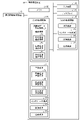
図1の実施形態に係る画像処理装置100を示すブロック図である。
2 学習機能
2a 異常領域学習機能
2b 境界領域学習機能
2c 正常領域学習機能
3 判定機能
4 抽出機能
4a 異常領域抽出機能
4b 境界領域抽出機能
4c 正常領域抽出機能
100 画像処理装置
120 第1の処理回路
130 第2の処理回路
Claims (11)
- 訓練用の医用データから、病変内部の領域である第1の領域の画像に係る第1のデータと、病変周囲の領域である第2の領域の画像に係る第2のデータと、病変外部の領域である第3の領域の画像に係る第3のデータとを抽出する抽出部と、
入力医用データについての判定を行う学習済モデルを、前記抽出部が抽出した前記第1のデータと、前記第2のデータと、前記第3のデータとを訓練データとして用いて生成する学習部と、
を備える、
画像処理装置。 - 前記学習済モデルに基づいて、前記入力医用データについての前記判定を行う判定部を更に備える、請求項1に記載の画像処理装置。
- 前記判定部は、前記第2の領域における特徴量に基づいて、前記判定を行う、請求項2に記載の画像処理装置。
- 前記判定部は、前記第1の領域と前記第2の領域との位置関係に基づいて、前記判定を行う、請求項2に記載の画像処理装置。
- 前記判定部は、前記第1の領域が前記第2の領域に囲まれるように位置しているかに基づいて、前記判定を行う、請求項4に記載の画像処理装置。
- 前記抽出部は、前記第3のデータの抽出の際、訓練を行う対象の組織とは異なる組織を除外して抽出を行う、請求項2に記載の画像処理装置。
- 前記異なる組織は、血管又は気管支である、請求項6に記載の画像処理装置。
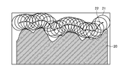
- 前記抽出部は、前記第1の領域から定められた距離の範囲にある領域を、前記第2の領域として抽出する、請求項1に記載の画像処理装置。
- 前記距離の範囲は、病変の種類に応じた値として設定された値である、請求項8に記載の画像処理装置。
- 訓練用の医用データから、病変内部の領域である第1の領域の画像に係る第1のデータと、病変周囲の領域である第2の領域の画像に係る第2のデータと、病変外部の領域である第3の領域の画像に係る第3のデータとを抽出する抽出部と、
前記抽出部が抽出した前記第1のデータと、前記第2のデータと、前記第3のデータとを、入力医用データについての判定を行う学習済モデルの訓練データとして使用するために出力する出力部とを備える、
データ生成装置。 - 訓練用の医用データから、病変内部の領域である第1の領域の画像に係る第1のデータと、病変周囲の領域である第2の領域の画像に係る第2のデータと、病変外部の領域である第3の領域の画像に係る第3のデータとを抽出し、
抽出した前記第1のデータと、前記第2のデータと、前記第3のデータとを、入力医用データについての判定を行う学習済モデルの訓練データとして使用するために出力する
各処理をコンピュータに実行させる、プログラム。
Priority Applications (1)
| Application Number | Priority Date | Filing Date | Title |
|---|---|---|---|
| JP2018197801A JP7140630B2 (ja) | 2018-10-19 | 2018-10-19 | 画像処理装置、データ生成装置及びプログラム |
Applications Claiming Priority (1)
| Application Number | Priority Date | Filing Date | Title |
|---|---|---|---|
| JP2018197801A JP7140630B2 (ja) | 2018-10-19 | 2018-10-19 | 画像処理装置、データ生成装置及びプログラム |
Publications (2)
| Publication Number | Publication Date |
|---|---|
| JP2020062355A JP2020062355A (ja) | 2020-04-23 |
| JP7140630B2 true JP7140630B2 (ja) | 2022-09-21 |
Family
ID=70387986
Family Applications (1)
| Application Number | Title | Priority Date | Filing Date |
|---|---|---|---|
| JP2018197801A Active JP7140630B2 (ja) | 2018-10-19 | 2018-10-19 | 画像処理装置、データ生成装置及びプログラム |
Country Status (1)
| Country | Link |
|---|---|
| JP (1) | JP7140630B2 (ja) |
Families Citing this family (5)
| Publication number | Priority date | Publication date | Assignee | Title |
|---|---|---|---|---|
| JP7483502B2 (ja) * | 2020-05-29 | 2024-05-15 | 富士フイルムヘルスケア株式会社 | 画像診断支援装置および画像診断支援方法 |
| JP7444382B2 (ja) | 2020-09-15 | 2024-03-06 | 国立研究開発法人国立がん研究センター | 画像符号化装置、方法およびプログラム、画像復号化装置、方法およびプログラム、画像処理装置、学習装置、方法およびプログラム、並びに類似画像検索装置、方法およびプログラム |
| JP7794451B2 (ja) * | 2020-09-18 | 2026-01-06 | 国立大学法人 筑波大学 | 情報処理装置及びプログラム |
| JP2024061696A (ja) * | 2021-03-24 | 2024-05-08 | ソニーグループ株式会社 | 医療用画像解析装置、医療用画像解析方法及び医療用画像解析システム |
| JP2023081573A (ja) * | 2021-12-01 | 2023-06-13 | キヤノンメディカルシステムズ株式会社 | 医用画像処理装置、医用画像処理方法及び医用画像処理プログラム |
Citations (3)
| Publication number | Priority date | Publication date | Assignee | Title |
|---|---|---|---|---|
| JP2006325640A (ja) | 2005-05-23 | 2006-12-07 | Konica Minolta Medical & Graphic Inc | 異常陰影候補の表示方法及び医用画像処理システム |
| JP2010504129A (ja) | 2006-09-22 | 2010-02-12 | コーニンクレッカ フィリップス エレクトロニクス エヌ ヴィ | 肺結節の高度コンピュータ支援診断 |
| US20160148376A1 (en) | 2014-11-26 | 2016-05-26 | Samsung Electronics Co., Ltd. | Computer aided diagnosis (cad) apparatus and method |
Family Cites Families (2)
| Publication number | Priority date | Publication date | Assignee | Title |
|---|---|---|---|---|
| JP6510189B2 (ja) * | 2014-06-23 | 2019-05-08 | キヤノンメディカルシステムズ株式会社 | 医用画像処理装置 |
| JP6670222B2 (ja) * | 2016-11-01 | 2020-03-18 | 株式会社日立ハイテク | 画像診断支援装置及びシステム、画像診断支援方法 |
-
2018
- 2018-10-19 JP JP2018197801A patent/JP7140630B2/ja active Active
Patent Citations (3)
| Publication number | Priority date | Publication date | Assignee | Title |
|---|---|---|---|---|
| JP2006325640A (ja) | 2005-05-23 | 2006-12-07 | Konica Minolta Medical & Graphic Inc | 異常陰影候補の表示方法及び医用画像処理システム |
| JP2010504129A (ja) | 2006-09-22 | 2010-02-12 | コーニンクレッカ フィリップス エレクトロニクス エヌ ヴィ | 肺結節の高度コンピュータ支援診断 |
| US20160148376A1 (en) | 2014-11-26 | 2016-05-26 | Samsung Electronics Co., Ltd. | Computer aided diagnosis (cad) apparatus and method |
Non-Patent Citations (1)
| Title |
|---|
| 理化学研究所, 国立がん研究センター,AIで早期胃がん領域の高精度検出に成功 -早期発見・領域検出で早期治療に大きく貢献-,国立がん研究センタープレスリリース,2018年07月20日,インターネット:<https://www.ncc.go.jp/jp/information/pr_release/2018/0721/index.html> |
Also Published As
| Publication number | Publication date |
|---|---|
| JP2020062355A (ja) | 2020-04-23 |
Similar Documents
| Publication | Publication Date | Title |
|---|---|---|
| Wang | Mammography with deep learning for breast cancer detection | |
| Weng et al. | INet: convolutional networks for biomedical image segmentation | |
| Wong et al. | Scribbleprompt: fast and flexible interactive segmentation for any biomedical image | |
| Zhang et al. | Review of breast cancer pathologigcal image processing | |
| Bhattacharya et al. | A review of artificial intelligence in prostate cancer detection on imaging | |
| JP7140630B2 (ja) | 画像処理装置、データ生成装置及びプログラム | |
| Midya et al. | Computerized diagnosis of liver tumors from CT scans using a deep neural network approach | |
| Streba et al. | Contrast-enhanced ultrasonography parameters in neural network diagnosis of liver tumors | |
| Islam et al. | ICHNet: Intracerebral hemorrhage (ICH) segmentation using deep learning | |
| Jung et al. | Deep learning for medical image analysis: Applications to computed tomography and magnetic resonance imaging | |
| Rani et al. | Superpixel with nanoscale imaging and boosted deep convolutional neural network concept for lung tumor classification | |
| Gayathri Devi et al. | Automatic segmentation of colon in 3D CT images and removal of opacified fluid using cascade feed forward neural network | |
| Ren et al. | Investigation of a novel deep learning-based computed tomography perfusion mapping framework for functional lung avoidance radiotherapy | |
| Mukku et al. | Deep learning-based cervical lesion segmentation in colposcopic images | |
| Bi et al. | Hybrid CNN-transformer network for interactive learning of challenging musculoskeletal images | |
| Raina et al. | Deep-learning model for quality assessment of urinary bladder ultrasound images using multiscale and higher-order processing | |
| Cau et al. | Applications of artificial intelligence-based models in vulnerable carotid plaque | |
| Luo et al. | Bi-DenseNet: Automatic recognition of ocular surface disease using smartphone imaging | |
| Celik et al. | Forecasting the “T” stage of esophageal cancer by deep learning methods: A pilot study | |
| Streba et al. | Artificial intelligence and automatic image interpretation in modern medicine | |
| Papandrianos et al. | A Convolutional Neural Network-based explainable classification method of SPECT myocardial perfusion images in nuclear cardiology | |
| Burrows et al. | Evaluation of a hybrid pipeline for automated segmentation of solid lesions based on mathematical algorithms and deep learning | |
| Rahmany et al. | A priori knowledge integration for the detection of cerebral aneurysm | |
| Sathya et al. | Development of CAD system based on enhanced clustering based segmentation algorithm for detection of masses in breast DCE-MRI | |
| Xue et al. | 3d petct tumor lesion segmentation via gcn refinement |
Legal Events
| Date | Code | Title | Description |
|---|---|---|---|
| A621 | Written request for application examination |
Free format text: JAPANESE INTERMEDIATE CODE: A621 Effective date: 20210812 |
|
| A977 | Report on retrieval |
Free format text: JAPANESE INTERMEDIATE CODE: A971007 Effective date: 20220715 |
|
| TRDD | Decision of grant or rejection written | ||
| A01 | Written decision to grant a patent or to grant a registration (utility model) |
Free format text: JAPANESE INTERMEDIATE CODE: A01 Effective date: 20220809 |
|
| A61 | First payment of annual fees (during grant procedure) |
Free format text: JAPANESE INTERMEDIATE CODE: A61 Effective date: 20220908 |
|
| R150 | Certificate of patent or registration of utility model |
Ref document number: 7140630 Country of ref document: JP Free format text: JAPANESE INTERMEDIATE CODE: R150 |