JP7576801B2 - 表面にパターン化されたエバネッセント場を形成する装置およびその方法 - Google Patents
表面にパターン化されたエバネッセント場を形成する装置およびその方法 Download PDFInfo
- Publication number
- JP7576801B2 JP7576801B2 JP2021568495A JP2021568495A JP7576801B2 JP 7576801 B2 JP7576801 B2 JP 7576801B2 JP 2021568495 A JP2021568495 A JP 2021568495A JP 2021568495 A JP2021568495 A JP 2021568495A JP 7576801 B2 JP7576801 B2 JP 7576801B2
- Authority
- JP
- Japan
- Prior art keywords
- focal plane
- light
- emitting element
- optical
- side focal
- Prior art date
- Legal status (The legal status is an assumption and is not a legal conclusion. Google has not performed a legal analysis and makes no representation as to the accuracy of the status listed.)
- Active
Links
- 238000000034 method Methods 0.000 title claims description 19
- 230000003287 optical effect Effects 0.000 claims description 71
- 239000000523 sample Substances 0.000 claims description 9
- 238000001914 filtration Methods 0.000 claims description 5
- 238000011144 upstream manufacturing Methods 0.000 claims description 5
- 239000012472 biological sample Substances 0.000 claims description 4
- 230000002186 photoactivation Effects 0.000 claims description 3
- 230000005284 excitation Effects 0.000 description 15
- 210000004027 cell Anatomy 0.000 description 11
- 230000004913 activation Effects 0.000 description 7
- 238000000386 microscopy Methods 0.000 description 6
- 210000000170 cell membrane Anatomy 0.000 description 5
- 238000011161 development Methods 0.000 description 5
- 230000018109 developmental process Effects 0.000 description 5
- 238000005286 illumination Methods 0.000 description 5
- 230000000638 stimulation Effects 0.000 description 5
- 230000001427 coherent effect Effects 0.000 description 4
- 238000002060 fluorescence correlation spectroscopy Methods 0.000 description 4
- 238000000059 patterning Methods 0.000 description 4
- 230000015572 biosynthetic process Effects 0.000 description 3
- 230000001413 cellular effect Effects 0.000 description 3
- 238000010586 diagram Methods 0.000 description 3
- 230000000694 effects Effects 0.000 description 3
- 239000011521 glass Substances 0.000 description 3
- 239000012528 membrane Substances 0.000 description 3
- 210000004379 membrane Anatomy 0.000 description 3
- 108091008695 photoreceptors Proteins 0.000 description 3
- 210000001747 pupil Anatomy 0.000 description 3
- 239000000758 substrate Substances 0.000 description 3
- 238000000492 total internal reflection fluorescence microscopy Methods 0.000 description 3
- NCGICGYLBXGBGN-UHFFFAOYSA-N 3-morpholin-4-yl-1-oxa-3-azonia-2-azanidacyclopent-3-en-5-imine;hydrochloride Chemical compound Cl.[N-]1OC(=N)C=[N+]1N1CCOCC1 NCGICGYLBXGBGN-UHFFFAOYSA-N 0.000 description 2
- 101001034652 Homo sapiens Insulin-like growth factor 1 receptor Proteins 0.000 description 2
- 102100039688 Insulin-like growth factor 1 receptor Human genes 0.000 description 2
- 101150099493 STAT3 gene Proteins 0.000 description 2
- XUIMIQQOPSSXEZ-UHFFFAOYSA-N Silicon Chemical compound [Si] XUIMIQQOPSSXEZ-UHFFFAOYSA-N 0.000 description 2
- 102000040945 Transcription factor Human genes 0.000 description 2
- 108091023040 Transcription factor Proteins 0.000 description 2
- 230000001464 adherent effect Effects 0.000 description 2
- 238000004458 analytical method Methods 0.000 description 2
- 239000002131 composite material Substances 0.000 description 2
- 230000003436 cytoskeletal effect Effects 0.000 description 2
- 230000001086 cytosolic effect Effects 0.000 description 2
- 230000007423 decrease Effects 0.000 description 2
- 238000001514 detection method Methods 0.000 description 2
- 102000052116 epidermal growth factor receptor activity proteins Human genes 0.000 description 2
- 108700015053 epidermal growth factor receptor activity proteins Proteins 0.000 description 2
- 238000005530 etching Methods 0.000 description 2
- 238000000799 fluorescence microscopy Methods 0.000 description 2
- 238000010348 incorporation Methods 0.000 description 2
- 230000003834 intracellular effect Effects 0.000 description 2
- 230000004068 intracellular signaling Effects 0.000 description 2
- 239000007791 liquid phase Substances 0.000 description 2
- 108020004084 membrane receptors Proteins 0.000 description 2
- 102000006240 membrane receptors Human genes 0.000 description 2
- YOHYSYJDKVYCJI-UHFFFAOYSA-N n-[3-[[6-[3-(trifluoromethyl)anilino]pyrimidin-4-yl]amino]phenyl]cyclopropanecarboxamide Chemical compound FC(F)(F)C1=CC=CC(NC=2N=CN=C(NC=3C=C(NC(=O)C4CC4)C=CC=3)C=2)=C1 YOHYSYJDKVYCJI-UHFFFAOYSA-N 0.000 description 2
- 239000002120 nanofilm Substances 0.000 description 2
- 239000002105 nanoparticle Substances 0.000 description 2
- 230000005693 optoelectronics Effects 0.000 description 2
- 230000000737 periodic effect Effects 0.000 description 2
- 238000000206 photolithography Methods 0.000 description 2
- 239000004038 photonic crystal Substances 0.000 description 2
- 102000004169 proteins and genes Human genes 0.000 description 2
- 108090000623 proteins and genes Proteins 0.000 description 2
- 230000007261 regionalization Effects 0.000 description 2
- 230000004044 response Effects 0.000 description 2
- 238000002198 surface plasmon resonance spectroscopy Methods 0.000 description 2
- 239000010409 thin film Substances 0.000 description 2
- 101150078024 CRY2 gene Proteins 0.000 description 1
- 102000001301 EGF receptor Human genes 0.000 description 1
- 108060006698 EGF receptor Proteins 0.000 description 1
- 108010047852 Integrin alphaVbeta3 Proteins 0.000 description 1
- 102000004310 Ion Channels Human genes 0.000 description 1
- 102000018697 Membrane Proteins Human genes 0.000 description 1
- 108010052285 Membrane Proteins Proteins 0.000 description 1
- 206010034972 Photosensitivity reaction Diseases 0.000 description 1
- 239000012736 aqueous medium Substances 0.000 description 1
- 210000000270 basal cell Anatomy 0.000 description 1
- 210000002469 basement membrane Anatomy 0.000 description 1
- 230000005540 biological transmission Effects 0.000 description 1
- 230000005754 cellular signaling Effects 0.000 description 1
- 230000008859 change Effects 0.000 description 1
- 238000007796 conventional method Methods 0.000 description 1
- 210000004748 cultured cell Anatomy 0.000 description 1
- 210000000805 cytoplasm Anatomy 0.000 description 1
- 238000004925 denaturation Methods 0.000 description 1
- 230000036425 denaturation Effects 0.000 description 1
- 230000001627 detrimental effect Effects 0.000 description 1
- 238000009792 diffusion process Methods 0.000 description 1
- 210000002889 endothelial cell Anatomy 0.000 description 1
- 239000007850 fluorescent dye Substances 0.000 description 1
- 210000001650 focal adhesion Anatomy 0.000 description 1
- 230000004927 fusion Effects 0.000 description 1
- 238000003384 imaging method Methods 0.000 description 1
- 230000006872 improvement Effects 0.000 description 1
- 230000002779 inactivation Effects 0.000 description 1
- 230000004807 localization Effects 0.000 description 1
- 230000033001 locomotion Effects 0.000 description 1
- 238000006384 oligomerization reaction Methods 0.000 description 1
- 239000013307 optical fiber Substances 0.000 description 1
- 230000035515 penetration Effects 0.000 description 1
- 230000000886 photobiology Effects 0.000 description 1
- 208000007578 phototoxic dermatitis Diseases 0.000 description 1
- 231100000018 phototoxicity Toxicity 0.000 description 1
- 230000010287 polarization Effects 0.000 description 1
- 230000008569 process Effects 0.000 description 1
- 230000001902 propagating effect Effects 0.000 description 1
- 108020003175 receptors Proteins 0.000 description 1
- 102000005962 receptors Human genes 0.000 description 1
- 230000009467 reduction Effects 0.000 description 1
- 238000011160 research Methods 0.000 description 1
- 230000035945 sensitivity Effects 0.000 description 1
- 238000010869 super-resolution microscopy Methods 0.000 description 1
- XLYOFNOQVPJJNP-UHFFFAOYSA-N water Substances O XLYOFNOQVPJJNP-UHFFFAOYSA-N 0.000 description 1
Images


Classifications
-
- G—PHYSICS
- G01—MEASURING; TESTING
- G01N—INVESTIGATING OR ANALYSING MATERIALS BY DETERMINING THEIR CHEMICAL OR PHYSICAL PROPERTIES
- G01N21/00—Investigating or analysing materials by the use of optical means, i.e. using sub-millimetre waves, infrared, visible or ultraviolet light
- G01N21/62—Systems in which the material investigated is excited whereby it emits light or causes a change in wavelength of the incident light
- G01N21/63—Systems in which the material investigated is excited whereby it emits light or causes a change in wavelength of the incident light optically excited
- G01N21/64—Fluorescence; Phosphorescence
- G01N21/645—Specially adapted constructive features of fluorimeters
- G01N21/6456—Spatial resolved fluorescence measurements; Imaging
- G01N21/6458—Fluorescence microscopy
-
- G—PHYSICS
- G01—MEASURING; TESTING
- G01N—INVESTIGATING OR ANALYSING MATERIALS BY DETERMINING THEIR CHEMICAL OR PHYSICAL PROPERTIES
- G01N21/00—Investigating or analysing materials by the use of optical means, i.e. using sub-millimetre waves, infrared, visible or ultraviolet light
- G01N21/62—Systems in which the material investigated is excited whereby it emits light or causes a change in wavelength of the incident light
- G01N21/63—Systems in which the material investigated is excited whereby it emits light or causes a change in wavelength of the incident light optically excited
- G01N21/64—Fluorescence; Phosphorescence
- G01N21/645—Specially adapted constructive features of fluorimeters
- G01N21/648—Specially adapted constructive features of fluorimeters using evanescent coupling or surface plasmon coupling for the excitation of fluorescence
-
- G—PHYSICS
- G02—OPTICS
- G02B—OPTICAL ELEMENTS, SYSTEMS OR APPARATUS
- G02B21/00—Microscopes
- G02B21/06—Means for illuminating specimens
- G02B21/08—Condensers
- G02B21/082—Condensers for incident illumination only
-
- G—PHYSICS
- G02—OPTICS
- G02B—OPTICAL ELEMENTS, SYSTEMS OR APPARATUS
- G02B21/00—Microscopes
- G02B21/16—Microscopes adapted for ultraviolet illumination ; Fluorescence microscopes
-
- G—PHYSICS
- G02—OPTICS
- G02B—OPTICAL ELEMENTS, SYSTEMS OR APPARATUS
- G02B27/00—Optical systems or apparatus not provided for by any of the groups G02B1/00 - G02B26/00, G02B30/00
- G02B27/56—Optics using evanescent waves, i.e. inhomogeneous waves
Landscapes
- Physics & Mathematics (AREA)
- General Physics & Mathematics (AREA)
- Chemical & Material Sciences (AREA)
- Analytical Chemistry (AREA)
- Health & Medical Sciences (AREA)
- Optics & Photonics (AREA)
- Biochemistry (AREA)
- Life Sciences & Earth Sciences (AREA)
- Nuclear Medicine, Radiotherapy & Molecular Imaging (AREA)
- General Health & Medical Sciences (AREA)
- Immunology (AREA)
- Pathology (AREA)
- Microscoopes, Condenser (AREA)
- Investigating, Analyzing Materials By Fluorescence Or Luminescence (AREA)
- Investigating Or Analysing Materials By Optical Means (AREA)
- Lenses (AREA)
Description
ディオプトリの表面が配置された像の側焦点面にある対物レンズと、
所定の径の平行光を放出する光放出素子と、
前記光放出素子と前記対物レンズとの間に設けられた光学アセンブリであって、前記対物レンズの物体面が前記光放出素子の像側焦点面と光学的に共役な光学アセンブリであって、前記光学アセンブリは、前記光入射素子からの平行光が前記対物レンズに向かって出射され、前記双眼鏡の臨界角θc以上の最小入射角θminで前記双眼鏡の表面に向かって反射され、前記双眼鏡の表面で全反射を起こして前記双眼鏡の表面にエバネッセント波を発生させるように構成されている、光学アセンブリと、
対物レンズの光軸に対して軸外に設置された、光放出素子の物体面にパターンを形成するための光学装置であって、光放出素子の物体面の光ビームに透過光でパターンを形成するための光学装置で形成されたパターンがディオプトリの表面に見られるようにするための光学装置と、
を備えることを特徴とする装置に関する。
レンズの像側焦点面上に面を配置するステップと、
前記パターン形成装置を用いて、前記光放出素子の物体面に透過光の所望の振幅パターンを形成するステップと、
コリメートされた光ビームを光入射素子に入射させるステップとを含む方法に関する。
Claims (10)
- それぞれの屈折率n1とn2の2つの媒体を分離するディオプトリの表面(S)にパターン化されたエバネッセント場を形成する装置(1、10、20)であって、
前記ディオプトリの表面(S)が配置された像側焦点面にある対物レンズ(O)と、
直径(d)のコリメートされた光ビーム(B)を放出する光放出素子(2;12;22)と、
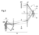
前記光放出素子(2;12;22)と前記対物レンズ(O)との間の光学アセンブリ(4;14;24)であって、前記対物レンズ(O)の物体側焦点面が前記光放出素子(2;12;22)の前記像側焦点面と光学的に共役であり、前記光学アセンブリ(4;14;24)が前記光放出素子(2;12;22)からの前記コリメートされた光ビーム(B)が対物レンズ(O)に向かって放出された後に臨界角θc以上の最小入射角θminで前記ディオプトリの表面(S)に向かって反射されるように構成され、前記光ビーム(B)が前記ディオプトリの表面(S)で全反射を起こして前記ディオプトリの表面(S)にエバネッセント波を生成するように構成される、前記光学アセンブリ(4;14;24)と、
前記光放出素子(2;12;22)の前記物体側焦点面にパターンを形成するための光学装置(FS1;BS)であって、前記対物レンズ(O)の光軸に対して軸外に取り付けられており、前記光放出素子(2;12;22)の前記物体側焦点面の前記光ビームに透過光で前記パターンを形成するための前記光学装置によって形成された前記パターンが、前記ディオプトリの表面で検出され、前記ディオプトリの表面(S)で反射された前記光ビーム(B)は、前記対物レンズ(O)に向かって反射されて、前記対物レンズ(O)の後側焦点面(BFP)に集束される、前記光学装置(FS1;BS)と、
を備えることを特徴とする装置。 - 前記光放出素子(2;12;22)の前記物体側焦点面と前記光学アセンブリとの間に少なくとも1つの光学素子(AS)を備え、当該少なくとも1つの光学素子(AS)は、形成された前記パターンの解像度の向上の調整と、前記ディオプトリにおける前記光ビーム(B)の入射角のフィルタリングの調整とのいずれかをすることができることを特徴とする、請求項1に記載の装置(1;10;20)。
- 前記少なくとも1つの光学素子が、空間ダイアフラム、フィルタのいずれかであることを特徴とする、請求項2に記載の装置(1;10;20)。
- 前記パターンを形成する前記光学装置が、ダイアフラム、振幅マスク、空間光変調器、マイクロミラーアレイのうちの少なくとも1つであることを特徴とする、請求項1から3のいずれか一項に記載の装置(1;10;20)。
- 前記光放出素子に入射される前記コリメートされた光ビームを生成するためのレーザー光源をさらに備えることを特徴とする、請求項1から4のいずれか一項に記載の装置(1;10;20)。
- 前記光学アセンブリは、第1の中間レンズによって構成されており、前記第1の中間レンズの物体側焦点面が前記光放出素子の前記像側焦点面と光学的に共役であり、前記第1の中間レンズの像側焦点面が前記対物レンズ(O)の前記物体側焦点面と光学的に共役であることを特徴とする、請求項1から5のいずれか一項に記載の装置(1;10;20)。
- 前記第1の中間レンズは、第2の中間レンズによって、前記光放出素子と光学的に共役し、前記第2の中間レンズの物体側焦点面が前記光放出素子の前記像側焦点面に対応し、前記第2の中間レンズの像側焦点面が前記第1の中間レンズの前記物体側焦点面に対応することを特徴とする、請求項6に記載の装置(1)。
- 請求項1から7のいずれか一項に記載の装置(1;10;20)であって、前記装置は全反射蛍光顕微鏡であり、試料は前記ディオプトリに対応すると共に当該全反射蛍光顕微鏡の前記対物レンズの前記像側焦点面に配置され、前記光学アセンブリは顕微鏡光学系に対応し、前記光放出素子は前記顕微鏡光学系の上流に配置されるか前記顕微鏡光学系に組み込まれ、パターンを形成するための前記光学装置は光源と前記光放出素子と間に配置されるか前記顕微鏡光学系に組み込まれることを特徴とする、装置。
- 請求項1から8のいずれか一項に記載の装置(1;10;20)を用いて、表面にパターン化されたエバネッセント場を形成する方法であって、
前記対物レンズ(O)の前記像側焦点面上に前記表面(S)を配置するステップと、
前記パターンを形成するための前記光学装置(FS1、BS)を用いて、前記光放出素子(2;12;22)の前記物体側焦点面に所望の前記透過光の振幅パターンを形成するステップと、
前記コリメートされた光ビームを前記光放出素子(2;12;22)に入射させるステップと、
を含むことを特徴とする、方法。 - 生物学的試料(E)の特定の表面領域を光活性化によって選択的に励起するために、表面エバネッセント場を前記生物学的試料(E)上に作成することを特徴とする、請求項9に記載の方法。
Applications Claiming Priority (3)
| Application Number | Priority Date | Filing Date | Title |
|---|---|---|---|
| FR1905159 | 2019-05-16 | ||
| FR1905159A FR3096146B1 (fr) | 2019-05-16 | 2019-05-16 | Dispositif de creation d’un champ evanescent a motifs sur une surface et procede associe |
| PCT/IB2020/054529 WO2020230051A1 (fr) | 2019-05-16 | 2020-05-13 | Dispositif de creation d'un champ evanescent a motifs sur une surface et procede associe |
Publications (2)
| Publication Number | Publication Date |
|---|---|
| JP2022532248A JP2022532248A (ja) | 2022-07-13 |
| JP7576801B2 true JP7576801B2 (ja) | 2024-11-01 |
Family
ID=67262769
Family Applications (1)
| Application Number | Title | Priority Date | Filing Date |
|---|---|---|---|
| JP2021568495A Active JP7576801B2 (ja) | 2019-05-16 | 2020-05-13 | 表面にパターン化されたエバネッセント場を形成する装置およびその方法 |
Country Status (5)
| Country | Link |
|---|---|
| US (1) | US12169286B2 (ja) |
| EP (1) | EP3969954A1 (ja) |
| JP (1) | JP7576801B2 (ja) |
| FR (1) | FR3096146B1 (ja) |
| WO (1) | WO2020230051A1 (ja) |
Families Citing this family (1)
| Publication number | Priority date | Publication date | Assignee | Title |
|---|---|---|---|---|
| FR3151401A1 (fr) | 2023-07-19 | 2025-01-24 | Universite Grenoble Alpes | Dispositif de création d’un champ évanescent |
Citations (7)
| Publication number | Priority date | Publication date | Assignee | Title |
|---|---|---|---|---|
| JP2004109348A (ja) | 2002-09-17 | 2004-04-08 | Institute Of Physical & Chemical Research | 顕微鏡装置 |
| JP2006162790A (ja) | 2004-12-03 | 2006-06-22 | Olympus Corp | 全反射蛍光照明装置 |
| JP2009042411A (ja) | 2007-08-08 | 2009-02-26 | Nikon Corp | 顕微鏡用照明装置 |
| US20120319007A1 (en) | 2009-12-22 | 2012-12-20 | Carl Zeiss Microimaging Gmbh | Luminescence microscopy |
| JP2014002373A (ja) | 2012-05-25 | 2014-01-09 | Olympus Corp | 顕微鏡装置 |
| JP2014092682A (ja) | 2012-11-02 | 2014-05-19 | Olympus Corp | 顕微鏡用照明装置及びそれを備えた顕微鏡 |
| WO2015008415A1 (ja) | 2013-07-17 | 2015-01-22 | 株式会社ニコン | 構造化照明装置及び構造化照明顕微鏡装置 |
Family Cites Families (7)
| Publication number | Priority date | Publication date | Assignee | Title |
|---|---|---|---|---|
| CN1588003A (zh) * | 2004-08-27 | 2005-03-02 | 中国科学院上海光学精密机械研究所 | 单分子荧光样品快速纵向超分辨的方法 |
| DE102009043747A1 (de) | 2009-09-30 | 2011-03-31 | Carl Zeiss Microlmaging Gmbh | Verfahren zur Erzeugung eines Mikroskopbildes und Mikroskop |
| WO2012153495A1 (ja) | 2011-05-06 | 2012-11-15 | 株式会社ニコン | 構造化照明顕微鏡及び構造化照明観察方法 |
| US9958784B2 (en) | 2013-09-24 | 2018-05-01 | The Institute Of Optics And Electronics, Chinese Academy Of Sciences | Super-resolution imaging photolithography |
| JP6351951B2 (ja) | 2013-10-22 | 2018-07-04 | 浜松ホトニクス株式会社 | 全反射型光照射装置 |
| US20150168300A1 (en) * | 2014-01-28 | 2015-06-18 | National Institute Of Standards And Technology | Article and process for modifying light |
| JP6396137B2 (ja) | 2014-09-18 | 2018-09-26 | オリンパス株式会社 | 顕微鏡照明装置、顕微鏡照明方法および顕微鏡 |
-
2019
- 2019-05-16 FR FR1905159A patent/FR3096146B1/fr active Active
-
2020
- 2020-05-13 WO PCT/IB2020/054529 patent/WO2020230051A1/fr not_active Ceased
- 2020-05-13 JP JP2021568495A patent/JP7576801B2/ja active Active
- 2020-05-13 EP EP20731212.5A patent/EP3969954A1/fr active Pending
- 2020-05-13 US US17/611,421 patent/US12169286B2/en active Active
Patent Citations (7)
| Publication number | Priority date | Publication date | Assignee | Title |
|---|---|---|---|---|
| JP2004109348A (ja) | 2002-09-17 | 2004-04-08 | Institute Of Physical & Chemical Research | 顕微鏡装置 |
| JP2006162790A (ja) | 2004-12-03 | 2006-06-22 | Olympus Corp | 全反射蛍光照明装置 |
| JP2009042411A (ja) | 2007-08-08 | 2009-02-26 | Nikon Corp | 顕微鏡用照明装置 |
| US20120319007A1 (en) | 2009-12-22 | 2012-12-20 | Carl Zeiss Microimaging Gmbh | Luminescence microscopy |
| JP2014002373A (ja) | 2012-05-25 | 2014-01-09 | Olympus Corp | 顕微鏡装置 |
| JP2014092682A (ja) | 2012-11-02 | 2014-05-19 | Olympus Corp | 顕微鏡用照明装置及びそれを備えた顕微鏡 |
| WO2015008415A1 (ja) | 2013-07-17 | 2015-01-22 | 株式会社ニコン | 構造化照明装置及び構造化照明顕微鏡装置 |
Also Published As
| Publication number | Publication date |
|---|---|
| WO2020230051A1 (fr) | 2020-11-19 |
| JP2022532248A (ja) | 2022-07-13 |
| EP3969954A1 (fr) | 2022-03-23 |
| FR3096146B1 (fr) | 2022-07-29 |
| US12169286B2 (en) | 2024-12-17 |
| US20220214556A1 (en) | 2022-07-07 |
| FR3096146A1 (fr) | 2020-11-20 |
Similar Documents
| Publication | Publication Date | Title |
|---|---|---|
| EP2888575B1 (en) | Stimulated emission depletion microscopy | |
| JP6234105B2 (ja) | 超解像顕微鏡 | |
| JP6996048B2 (ja) | 広視野高分解能顕微鏡 | |
| Yih et al. | Temporal focusing-based multiphoton excitation microscopy via digital micromirror device | |
| US12241837B2 (en) | Apparatus for structured illumination microscopy, method for illuminating a sample and method of structured illumination microscopy | |
| EP1941313B1 (en) | An optical system for illumination of an evanescent field | |
| CN106980174A (zh) | 一种综合性荧光超分辨显微成像装置 | |
| CN111103678B (zh) | 晶格光片显微镜和在晶格光片显微镜中平铺晶格光片的方法 | |
| CN108292034B (zh) | 用于利用结构化的光片照射检查试样的方法和设备 | |
| JP2007233370A (ja) | 試料を高い空間分解能で検査するための方法および顕微鏡 | |
| JP2018517180A (ja) | ビーム整形及び光シート顕微鏡検査のためのアセンブリ及び方法 | |
| CN102818794A (zh) | 生物荧光显微检测仪器 | |
| JP2022519567A (ja) | 波面解析装置、蛍光顕微鏡画像化システムおよび対象を顕微鏡画像化する方法 | |
| WO2020212769A1 (en) | Reconfigurable counterpropagating holographic optical tweezers with low-na lens | |
| JP7576801B2 (ja) | 表面にパターン化されたエバネッセント場を形成する装置およびその方法 | |
| US9729800B2 (en) | Image generation system | |
| JP4920918B2 (ja) | 位相フィルタ、光学装置及びラスタ顕微鏡 | |
| US20060214106A1 (en) | Point scanning laser scanning microscope and methods for adjustment of a microscope | |
| CN116953905A (zh) | 一种斜光片成像系统 | |
| JP5039307B2 (ja) | 対物レンズおよび顕微鏡 | |
| CN111879737A (zh) | 一种产生高通量超衍射极限焦斑的装置和方法 | |
| CN111886491A (zh) | 散射辅助超定位显微术方法及相关装置 | |
| CN119901716A (zh) | 一种共焦单分子三维探测和追踪方法及系统 | |
| CN112305741A (zh) | 一种多焦点光学系统 | |
| CN115516360A (zh) | 多重环遮蔽罩、光照射装置、光学显微镜和光声显微镜 |
Legal Events
| Date | Code | Title | Description |
|---|---|---|---|
| A621 | Written request for application examination |
Free format text: JAPANESE INTERMEDIATE CODE: A621 Effective date: 20230512 |
|
| A977 | Report on retrieval |
Free format text: JAPANESE INTERMEDIATE CODE: A971007 Effective date: 20240116 |
|
| A131 | Notification of reasons for refusal |
Free format text: JAPANESE INTERMEDIATE CODE: A131 Effective date: 20240214 |
|
| A521 | Request for written amendment filed |
Free format text: JAPANESE INTERMEDIATE CODE: A523 Effective date: 20240507 |
|
| A131 | Notification of reasons for refusal |
Free format text: JAPANESE INTERMEDIATE CODE: A131 Effective date: 20240827 |
|
| A521 | Request for written amendment filed |
Free format text: JAPANESE INTERMEDIATE CODE: A523 Effective date: 20240829 |
|
| TRDD | Decision of grant or rejection written | ||
| A01 | Written decision to grant a patent or to grant a registration (utility model) |
Free format text: JAPANESE INTERMEDIATE CODE: A01 Effective date: 20240917 |
|
| A61 | First payment of annual fees (during grant procedure) |
Free format text: JAPANESE INTERMEDIATE CODE: A61 Effective date: 20241011 |
|
| R150 | Certificate of patent or registration of utility model |
Ref document number: 7576801 Country of ref document: JP Free format text: JAPANESE INTERMEDIATE CODE: R150 |