JP7635401B2 - Anti-TNFR2 humanized antibodies and uses thereof - Google Patents
Anti-TNFR2 humanized antibodies and uses thereof Download PDFInfo
- Publication number
- JP7635401B2 JP7635401B2 JP2023546194A JP2023546194A JP7635401B2 JP 7635401 B2 JP7635401 B2 JP 7635401B2 JP 2023546194 A JP2023546194 A JP 2023546194A JP 2023546194 A JP2023546194 A JP 2023546194A JP 7635401 B2 JP7635401 B2 JP 7635401B2
- Authority
- JP
- Japan
- Prior art keywords
- antibody
- antigen
- tnfr2
- cells
- binding
- Prior art date
- Legal status (The legal status is an assumption and is not a legal conclusion. Google has not performed a legal analysis and makes no representation as to the accuracy of the status listed.)
- Active
Links
Classifications
-
- A—HUMAN NECESSITIES
- A61—MEDICAL OR VETERINARY SCIENCE; HYGIENE
- A61K—PREPARATIONS FOR MEDICAL, DENTAL OR TOILETRY PURPOSES
- A61K40/00—Cellular immunotherapy
- A61K40/30—Cellular immunotherapy characterised by the recombinant expression of specific molecules in the cells of the immune system
- A61K40/31—Chimeric antigen receptors [CAR]
-
- A—HUMAN NECESSITIES
- A61—MEDICAL OR VETERINARY SCIENCE; HYGIENE
- A61P—SPECIFIC THERAPEUTIC ACTIVITY OF CHEMICAL COMPOUNDS OR MEDICINAL PREPARATIONS
- A61P35/00—Antineoplastic agents
-
- A—HUMAN NECESSITIES
- A61—MEDICAL OR VETERINARY SCIENCE; HYGIENE
- A61P—SPECIFIC THERAPEUTIC ACTIVITY OF CHEMICAL COMPOUNDS OR MEDICINAL PREPARATIONS
- A61P37/00—Drugs for immunological or allergic disorders
-
- C—CHEMISTRY; METALLURGY
- C07—ORGANIC CHEMISTRY
- C07K—PEPTIDES
- C07K14/00—Peptides having more than 20 amino acids; Gastrins; Somatostatins; Melanotropins; Derivatives thereof
- C07K14/435—Peptides having more than 20 amino acids; Gastrins; Somatostatins; Melanotropins; Derivatives thereof from animals; from humans
- C07K14/705—Receptors; Cell surface antigens; Cell surface determinants
- C07K14/70503—Immunoglobulin superfamily
- C07K14/7051—T-cell receptor (TcR)-CD3 complex
-
- C—CHEMISTRY; METALLURGY
- C07—ORGANIC CHEMISTRY
- C07K—PEPTIDES
- C07K16/00—Immunoglobulins [IG], e.g. monoclonal or polyclonal antibodies
- C07K16/18—Immunoglobulins [IG], e.g. monoclonal or polyclonal antibodies against material from animals or humans
- C07K16/28—Immunoglobulins [IG], e.g. monoclonal or polyclonal antibodies against material from animals or humans against receptors, cell surface antigens or cell surface determinants
- C07K16/2803—Immunoglobulins [IG], e.g. monoclonal or polyclonal antibodies against material from animals or humans against receptors, cell surface antigens or cell surface determinants against the immunoglobulin superfamily
- C07K16/2827—Immunoglobulins [IG], e.g. monoclonal or polyclonal antibodies against material from animals or humans against receptors, cell surface antigens or cell surface determinants against the immunoglobulin superfamily against B7 molecules, e.g. CD80, CD86
-
- C—CHEMISTRY; METALLURGY
- C07—ORGANIC CHEMISTRY
- C07K—PEPTIDES
- C07K16/00—Immunoglobulins [IG], e.g. monoclonal or polyclonal antibodies
- C07K16/18—Immunoglobulins [IG], e.g. monoclonal or polyclonal antibodies against material from animals or humans
- C07K16/28—Immunoglobulins [IG], e.g. monoclonal or polyclonal antibodies against material from animals or humans against receptors, cell surface antigens or cell surface determinants
- C07K16/2878—Immunoglobulins [IG], e.g. monoclonal or polyclonal antibodies against material from animals or humans against receptors, cell surface antigens or cell surface determinants against the NGF-receptor/TNF-receptor superfamily, e.g. CD27, CD30, CD40, CD95
-
- C—CHEMISTRY; METALLURGY
- C12—BIOCHEMISTRY; BEER; SPIRITS; WINE; VINEGAR; MICROBIOLOGY; ENZYMOLOGY; MUTATION OR GENETIC ENGINEERING
- C12N—MICROORGANISMS OR ENZYMES; COMPOSITIONS THEREOF; PROPAGATING, PRESERVING, OR MAINTAINING MICROORGANISMS; MUTATION OR GENETIC ENGINEERING; CULTURE MEDIA
- C12N15/00—Mutation or genetic engineering; DNA or RNA concerning genetic engineering, vectors, e.g. plasmids, or their isolation, preparation or purification; Use of hosts therefor
- C12N15/09—Recombinant DNA-technology
- C12N15/63—Introduction of foreign genetic material using vectors; Vectors; Use of hosts therefor; Regulation of expression
-
- A—HUMAN NECESSITIES
- A61—MEDICAL OR VETERINARY SCIENCE; HYGIENE
- A61K—PREPARATIONS FOR MEDICAL, DENTAL OR TOILETRY PURPOSES
- A61K39/00—Medicinal preparations containing antigens or antibodies
- A61K2039/505—Medicinal preparations containing antigens or antibodies comprising antibodies
-
- A—HUMAN NECESSITIES
- A61—MEDICAL OR VETERINARY SCIENCE; HYGIENE
- A61K—PREPARATIONS FOR MEDICAL, DENTAL OR TOILETRY PURPOSES
- A61K39/00—Medicinal preparations containing antigens or antibodies
- A61K2039/505—Medicinal preparations containing antigens or antibodies comprising antibodies
- A61K2039/507—Comprising a combination of two or more separate antibodies
-
- A—HUMAN NECESSITIES
- A61—MEDICAL OR VETERINARY SCIENCE; HYGIENE
- A61K—PREPARATIONS FOR MEDICAL, DENTAL OR TOILETRY PURPOSES
- A61K2239/00—Indexing codes associated with cellular immunotherapy of group A61K40/00
- A61K2239/10—Indexing codes associated with cellular immunotherapy of group A61K40/00 characterized by the structure of the chimeric antigen receptor [CAR]
- A61K2239/11—Antigen recognition domain
- A61K2239/13—Antibody-based
-
- C—CHEMISTRY; METALLURGY
- C07—ORGANIC CHEMISTRY
- C07K—PEPTIDES
- C07K2317/00—Immunoglobulins specific features
- C07K2317/10—Immunoglobulins specific features characterized by their source of isolation or production
- C07K2317/14—Specific host cells or culture conditions, e.g. components, pH or temperature
-
- C—CHEMISTRY; METALLURGY
- C07—ORGANIC CHEMISTRY
- C07K—PEPTIDES
- C07K2317/00—Immunoglobulins specific features
- C07K2317/20—Immunoglobulins specific features characterized by taxonomic origin
- C07K2317/24—Immunoglobulins specific features characterized by taxonomic origin containing regions, domains or residues from different species, e.g. chimeric, humanized or veneered
-
- C—CHEMISTRY; METALLURGY
- C07—ORGANIC CHEMISTRY
- C07K—PEPTIDES
- C07K2317/00—Immunoglobulins specific features
- C07K2317/30—Immunoglobulins specific features characterized by aspects of specificity or valency
- C07K2317/31—Immunoglobulins specific features characterized by aspects of specificity or valency multispecific
-
- C—CHEMISTRY; METALLURGY
- C07—ORGANIC CHEMISTRY
- C07K—PEPTIDES
- C07K2317/00—Immunoglobulins specific features
- C07K2317/30—Immunoglobulins specific features characterized by aspects of specificity or valency
- C07K2317/33—Crossreactivity, e.g. for species or epitope, or lack of said crossreactivity
-
- C—CHEMISTRY; METALLURGY
- C07—ORGANIC CHEMISTRY
- C07K—PEPTIDES
- C07K2317/00—Immunoglobulins specific features
- C07K2317/50—Immunoglobulins specific features characterized by immunoglobulin fragments
- C07K2317/52—Constant or Fc region; Isotype
- C07K2317/522—CH1 domain
-
- C—CHEMISTRY; METALLURGY
- C07—ORGANIC CHEMISTRY
- C07K—PEPTIDES
- C07K2317/00—Immunoglobulins specific features
- C07K2317/50—Immunoglobulins specific features characterized by immunoglobulin fragments
- C07K2317/52—Constant or Fc region; Isotype
- C07K2317/526—CH3 domain
-
- C—CHEMISTRY; METALLURGY
- C07—ORGANIC CHEMISTRY
- C07K—PEPTIDES
- C07K2317/00—Immunoglobulins specific features
- C07K2317/50—Immunoglobulins specific features characterized by immunoglobulin fragments
- C07K2317/56—Immunoglobulins specific features characterized by immunoglobulin fragments variable (Fv) region, i.e. VH and/or VL
-
- C—CHEMISTRY; METALLURGY
- C07—ORGANIC CHEMISTRY
- C07K—PEPTIDES
- C07K2317/00—Immunoglobulins specific features
- C07K2317/50—Immunoglobulins specific features characterized by immunoglobulin fragments
- C07K2317/56—Immunoglobulins specific features characterized by immunoglobulin fragments variable (Fv) region, i.e. VH and/or VL
- C07K2317/565—Complementarity determining region [CDR]
-
- C—CHEMISTRY; METALLURGY
- C07—ORGANIC CHEMISTRY
- C07K—PEPTIDES
- C07K2317/00—Immunoglobulins specific features
- C07K2317/50—Immunoglobulins specific features characterized by immunoglobulin fragments
- C07K2317/56—Immunoglobulins specific features characterized by immunoglobulin fragments variable (Fv) region, i.e. VH and/or VL
- C07K2317/567—Framework region [FR]
-
- C—CHEMISTRY; METALLURGY
- C07—ORGANIC CHEMISTRY
- C07K—PEPTIDES
- C07K2317/00—Immunoglobulins specific features
- C07K2317/70—Immunoglobulins specific features characterized by effect upon binding to a cell or to an antigen
- C07K2317/73—Inducing cell death, e.g. apoptosis, necrosis or inhibition of cell proliferation
- C07K2317/732—Antibody-dependent cellular cytotoxicity [ADCC]
-
- C—CHEMISTRY; METALLURGY
- C07—ORGANIC CHEMISTRY
- C07K—PEPTIDES
- C07K2317/00—Immunoglobulins specific features
- C07K2317/70—Immunoglobulins specific features characterized by effect upon binding to a cell or to an antigen
- C07K2317/76—Antagonist effect on antigen, e.g. neutralization or inhibition of binding
-
- C—CHEMISTRY; METALLURGY
- C07—ORGANIC CHEMISTRY
- C07K—PEPTIDES
- C07K2317/00—Immunoglobulins specific features
- C07K2317/90—Immunoglobulins specific features characterized by (pharmaco)kinetic aspects or by stability of the immunoglobulin
- C07K2317/92—Affinity (KD), association rate (Ka), dissociation rate (Kd) or EC50 value
-
- C—CHEMISTRY; METALLURGY
- C07—ORGANIC CHEMISTRY
- C07K—PEPTIDES
- C07K2319/00—Fusion polypeptide
-
- C—CHEMISTRY; METALLURGY
- C07—ORGANIC CHEMISTRY
- C07K—PEPTIDES
- C07K2319/00—Fusion polypeptide
- C07K2319/01—Fusion polypeptide containing a localisation/targetting motif
- C07K2319/03—Fusion polypeptide containing a localisation/targetting motif containing a transmembrane segment
Landscapes
- Health & Medical Sciences (AREA)
- Chemical & Material Sciences (AREA)
- Life Sciences & Earth Sciences (AREA)
- Organic Chemistry (AREA)
- Immunology (AREA)
- General Health & Medical Sciences (AREA)
- Genetics & Genomics (AREA)
- Medicinal Chemistry (AREA)
- Biochemistry (AREA)
- Molecular Biology (AREA)
- Biophysics (AREA)
- Proteomics, Peptides & Aminoacids (AREA)
- Engineering & Computer Science (AREA)
- Zoology (AREA)
- Public Health (AREA)
- Veterinary Medicine (AREA)
- Animal Behavior & Ethology (AREA)
- Nuclear Medicine, Radiotherapy & Molecular Imaging (AREA)
- Bioinformatics & Cheminformatics (AREA)
- Pharmacology & Pharmacy (AREA)
- Chemical Kinetics & Catalysis (AREA)
- General Chemical & Material Sciences (AREA)
- Wood Science & Technology (AREA)
- General Engineering & Computer Science (AREA)
- Biomedical Technology (AREA)
- Biotechnology (AREA)
- Cell Biology (AREA)
- Toxicology (AREA)
- Gastroenterology & Hepatology (AREA)
- Microbiology (AREA)
- Plant Pathology (AREA)
- Physics & Mathematics (AREA)
- Epidemiology (AREA)
- Medicines Containing Antibodies Or Antigens For Use As Internal Diagnostic Agents (AREA)
- Peptides Or Proteins (AREA)
- Micro-Organisms Or Cultivation Processes Thereof (AREA)
- Medicinal Preparation (AREA)
- Medicines That Contain Protein Lipid Enzymes And Other Medicines (AREA)
- Medicines Containing Material From Animals Or Micro-Organisms (AREA)
Description
本発明は、バイオ医薬品分野及びバイオエンジニアリング分野に関し、具体的には、抗TNFR2ヒト化抗体又はその抗原結合断片、並びに前記抗TNFR2ヒト化抗体又はその抗原結合断片の医薬組成物及びその使用に関する。 The present invention relates to the fields of biopharmaceuticals and bioengineering, and more particularly to an anti-TNFR2 humanized antibody or an antigen-binding fragment thereof, as well as a pharmaceutical composition of the anti-TNFR2 humanized antibody or an antigen-binding fragment thereof and uses thereof.
免疫は体の防御反応であり、多くの遺伝子、タンパク質、細胞の影響を受ける。免疫異常は、腫瘍、免疫不全(エイズなど)、アレルギー、関節リウマチなどの疾患を含む多くの疾患を引き起こす可能性がある。ここ数年、腫瘍免疫療法は新しい治療法として登場し、腫瘍治療研究分野で大きなホットトピックとなっている。免疫チェックポイントタンパク質を標的とする抗PD-1及び抗CTLA-4モノクローナル抗体などのアンタゴニスト抗体は、複数種の種類の癌の治療に使用されており、且つ悪性腫瘍患者の生存期間を大幅に延長する革命的な結果をもたらしている。 Immunity is the body's defense response and is influenced by many genes, proteins, and cells. Immune abnormalities can cause many diseases, including tumors, immunodeficiencies (such as AIDS), allergies, rheumatoid arthritis, and other diseases. In recent years, tumor immunotherapy has emerged as a new treatment and has become a major hot topic in the field of tumor treatment research. Antagonist antibodies such as anti-PD-1 and anti-CTLA-4 monoclonal antibodies that target immune checkpoint proteins have been used to treat multiple types of cancer and have brought revolutionary results in significantly extending the survival time of patients with malignant tumors.
しかし、免疫チェックポイントタンパク質に対するアンタゴニスト抗体治療に未応答であるか、又は一時的な治療後に抵抗性や耐性を生じる癌患者はまだ多く存在している。そのため、有効性と安全性を更に向上させるために、単独で、又は免疫チェックポイントタンパク質に対するアンタゴニスト抗体を含む他の腫瘍治療法と組み合わせて使用できる、癌治療の新規薬物を開発する必要がある。 However, there are still many cancer patients who are unresponsive to antagonist antibody therapy against immune checkpoint proteins or develop resistance or tolerance after temporary treatment. Therefore, to further improve efficacy and safety, it is necessary to develop new drugs for cancer treatment that can be used alone or in combination with other tumor treatments, including antagonist antibodies against immune checkpoint proteins.
本発明者は、ヒトTNFR2がヒトTreg及び複数種のヒト腫瘍細胞の表面で過剰発現されることを発見し、ヒトTNFR2が患者の腫瘍発生を促進し、腫瘍微小環境において免疫抑制及び免疫逃避を媒介する可能性を示唆した。TNFR2を介してTreg細胞の機能を調節し、腫瘍の発生を抑制することは、非常に潜在力がある抗腫瘍戦略であると考えられる。本発明者は、1)TNFR2に特異的に結合し、TNFR2とそのリガンドTNFαの結合を遮断することによって、Treg細胞の増殖及びその媒介性の阻害機能を阻害し、エフェクターT細胞の増幅、及びエフェクターT細胞や他の免疫細胞媒介性抗腫瘍機能を促進することができる、2)TNFR2はヒト腫瘍細胞株で高度に発現されるため、この抗体もTNFR2の高発現腫瘍細胞に対する殺傷効果を直接的に媒介することができる、3)既存の抗PD-1/L1と併用するとより良い抗腫瘍効果を持ち、持続的な免疫記憶を形成することができる、TNFR2に対するアンタゴニスト抗体を調製した。前記抗TNFR2に対するアンタゴニスト抗体の説明については、PCT/CN2020/106057を参照されたい(その全体は参照により本願に組み込まれる)。 The inventors have discovered that human TNFR2 is overexpressed on the surface of human Tregs and several types of human tumor cells, suggesting that human TNFR2 may promote tumor development in patients and mediate immune suppression and immune escape in the tumor microenvironment. Regulating the function of Treg cells through TNFR2 to suppress tumor development is considered to be a very potential anti-tumor strategy. The inventors have prepared an antagonist antibody against TNFR2 that 1) specifically binds to TNFR2 and blocks the binding of TNFR2 to its ligand TNFα, thereby inhibiting the proliferation of Treg cells and their mediated inhibitory function, and promoting the expansion of effector T cells and the anti-tumor function mediated by effector T cells and other immune cells; 2) because TNFR2 is highly expressed in human tumor cell lines, this antibody can also directly mediate the killing effect on tumor cells with high expression of TNFR2; and 3) has a better anti-tumor effect and can form a sustained immune memory when combined with existing anti-PD-1/L1. For a description of the anti-TNFR2 antagonist antibodies, see PCT/CN2020/106057, the entire contents of which are incorporated herein by reference.
本発明は、抗TNFR2ヒト化抗体又はその抗原結合断片を提供し、並びに前記抗TNFR2ヒト化抗体又はその抗原結合断片の医薬組成物及びその使用を提供する。 The present invention provides an anti-TNFR2 humanized antibody or an antigen-binding fragment thereof, as well as a pharmaceutical composition of the anti-TNFR2 humanized antibody or an antigen-binding fragment thereof and uses thereof.
第1の態様において、本発明は、TNFR2に特異的に結合するヒト化抗体又は抗原結合断片を含み、前記抗体又は抗原結合断片は、重鎖可変領域と軽鎖可変領域を含み、
(1)前記重鎖可変領域は、
a.配列番号19~27から選ばれるいずれかに示されるVH配列、
b.配列番号19~27から選ばれるいずれかの配列と比較して、少なくとも80%、85%、90%、91%、92%、93%、94%、95%、96%、97%、98%、99%又は100%の配列同一性を有するVH配列、又は、
c.配列番号19~27から選ばれるいずれかの配列と比較して、1、2、3、4、5、6、7、8、9、10又はそれ以上のアミノ酸の挿入、欠失及び/又は置換を有するVH配列、
を含み、
(2)前記軽鎖可変領域は、
a.配列番号28~37から選ばれるいずれかに示されるVL配列、
b.配列番号28~37から選ばれるいずれかの配列と比較して、少なくとも80%、85%、90%、91%、92%、93%、94%、95%、96%、97%、98%、99%又は100%の配列同一性を有するVL配列、又は、
c.配列番号28~37から選ばれるいずれかの配列と比較して、1、2、3、4、5、6、7、8、9、10又はそれ以上のアミノ酸の挿入、欠失及び/又は置換を有するVL配列、
を含み、
任意に、前記アミノ酸の挿入、欠失及び/又は置換は、重鎖可変領域又は/及び軽鎖可変領域のFR領域に生じ、
任意に、前記置換は保存的アミノ酸置換である。
In a first aspect, the invention includes a humanized antibody or antigen-binding fragment that specifically binds to TNFR2, the antibody or antigen-binding fragment comprising a heavy chain variable region and a light chain variable region,
(1) The heavy chain variable region comprises:
a. A VH sequence shown in any one of SEQ ID NOs: 19 to 27;
b. A VH sequence having at least 80%, 85%, 90%, 91%, 92%, 93%, 94%, 95%, 96%, 97%, 98%, 99% or 100% sequence identity to any of the sequences selected from SEQ ID NOs: 19 to 27; or
c. A VH sequence having 1, 2, 3, 4, 5, 6, 7, 8, 9, 10 or more amino acid insertions, deletions and/or substitutions compared to any sequence selected from SEQ ID NOs: 19-27;
Including,
(2) The light chain variable region comprises:
a. A VL sequence selected from SEQ ID NOs: 28 to 37;
b. A VL sequence having at least 80%, 85%, 90%, 91%, 92%, 93%, 94%, 95%, 96%, 97%, 98%, 99% or 100% sequence identity to any of the sequences selected from SEQ ID NOs: 28 to 37; or
c. A VL sequence having 1, 2, 3, 4, 5, 6, 7, 8, 9, 10 or more amino acid insertions, deletions and/or substitutions compared to any sequence selected from SEQ ID NOs: 28-37;
Including,
Optionally, the amino acid insertions, deletions and/or substitutions occur in the FR regions of the heavy chain variable region or/and the light chain variable region;
Optionally, the substitutions are conservative amino acid substitutions.
幾つかの実施形態において、本発明に記載の抗体又は抗原結合断片であって、前記重鎖可変領域は、配列番号19~27のいずれかの配列と比較してCDRが完全に一致し、FRが少なくとも85%、90%、91%、92%、93%、94%、95%、96%、97%、98%、99%又は100%の配列同一性を有するVH配列を含み、前記軽鎖可変領域は、配列番号28~37のいずれかの配列と比較してCDRが完全に一致し、FRが少なくとも85%、90%、91%、92%、93%、94%、95%、96%、97%、98%、99%又は100%の配列同一性を有するVL配列を含む。
幾つかの実施形態において、本発明に記載の抗体又は抗原結合断片に含まれる重鎖可変領域及び軽鎖可変領域は、
(1)配列番号19に示されるVHと配列番号28に示されるVLを有する組み合わせ、
(2)配列番号20に示されるVHと配列番号29に示されるVLを有する組み合わせ、
(3)配列番号19に示されるVHと配列番号30に示されるVLを有する組み合わせ、
(4)配列番号20に示されるVHと配列番号31に示されるVLを有する組み合わせ、
(5)配列番号19に示されるVHと配列番号31に示されるVLを有する組み合わせ、
(6)配列番号21に示されるVHと配列番号32に示されるVLを有する組み合わせ、
(7)配列番号22に示されるVHと配列番号32に示されるVLを有する組み合わせ、
(8)配列番号23に示されるVHと配列番号32に示されるVLを有する組み合わせ、
(9)配列番号24に示されるVHと配列番号32に示されるVLを有する組み合わせ、
(10)配列番号25に示されるVHと配列番号32に示されるVLを有する組み合わせ、
(11)配列番号26に示されるVHと配列番号33に示されるVLを有する組み合わせ、
(12)配列番号27に示されるVHと配列番号34に示されるVLを有する組み合わせ、
(13)配列番号27に示されるVHと配列番号35に示されるVLを有する組み合わせ、
(14)配列番号27に示されるVHと配列番号33に示されるVLを有する組み合わせ、
(15)配列番号27に示されるVHと配列番号36に示されるVLを有する組み合わせ、
(16)配列番号26に示されるVHと配列番号34に示されるVLを有する組み合わせ、
(17)配列番号27に示されるVHと配列番号37に示されるVLを有する組み合わせ、
(18)前記(1)~(17)のいずれかの配列の組み合わせと比較して、少なくとも80%、85%、90%、91%、92%、93%、94%、95%、96%、97%、98%、99%又は100%の配列同一性を有するVHとVLの組み合わせ、
(19)前記(1)~(17)のいずれかの配列の組み合わせと比較して、1、2、3、4、5、6、7、8、9、10又はそれ以上のアミノ酸の挿入、欠失及び/又は置換を有するVHとVLの組み合わせ、又は、
(20)前記(1)~(17)のいずれかの配列の組み合わせと比較して、CDRが完全に一致し、FRが少なくとも85%、90%、91%、92%、93%、94%、95%、96%、97%、98%、99%又は100%の配列同一性を有するVHとVLの組み合わせ、
から選ばれ、
任意に、前記アミノ酸の挿入、欠失及び/又は置換は、重鎖可変領域又は/及び軽鎖可変領域のFR領域に生じ、
任意に、前記置換は保存的アミノ酸置換である。
In some embodiments, the antibody or antigen-binding fragment according to the invention, wherein the heavy chain variable region comprises a VH sequence in which the CDRs are completely identical and the FRs have at least 85%, 90%, 91%, 92%, 93%, 94%, 95%, 96%, 97%, 98%, 99% or 100% sequence identity to any of SEQ ID NOs: 19-27, and the light chain variable region comprises a VL sequence in which the CDRs are completely identical and the FRs have at least 85%, 90%, 91%, 92%, 93%, 94%, 95%, 96%, 97%, 98%, 99% or 100% sequence identity to any of SEQ ID NOs: 28-37.
In some embodiments, the heavy chain variable region and the light chain variable region comprised in the antibody or antigen-binding fragment of the invention are
(1) A combination having a VH represented by SEQ ID NO: 19 and a VL represented by SEQ ID NO: 28;
(2) A combination having a VH represented by SEQ ID NO: 20 and a VL represented by SEQ ID NO: 29;
(3) A combination having a VH represented by SEQ ID NO: 19 and a VL represented by SEQ ID NO: 30;
(4) A combination having VH represented by SEQ ID NO: 20 and VL represented by SEQ ID NO: 31;
(5) A combination having VH represented by SEQ ID NO: 19 and VL represented by SEQ ID NO: 31;
(6) A combination having VH represented by SEQ ID NO: 21 and VL represented by SEQ ID NO: 32;
(7) A combination having VH represented by SEQ ID NO: 22 and VL represented by SEQ ID NO: 32;
(8) A combination having VH represented by SEQ ID NO: 23 and VL represented by SEQ ID NO: 32;
(9) A combination having VH represented by SEQ ID NO: 24 and VL represented by SEQ ID NO: 32;
(10) A combination having VH represented by SEQ ID NO: 25 and VL represented by SEQ ID NO: 32;
(11) A combination having VH represented by SEQ ID NO: 26 and VL represented by SEQ ID NO: 33;
(12) A combination having VH represented by SEQ ID NO: 27 and VL represented by SEQ ID NO: 34;
(13) A combination having VH represented by SEQ ID NO: 27 and VL represented by SEQ ID NO: 35;
(14) A combination having VH represented by SEQ ID NO: 27 and VL represented by SEQ ID NO: 33;
(15) A combination having VH represented by SEQ ID NO: 27 and VL represented by SEQ ID NO: 36;
(16) A combination having VH represented by SEQ ID NO: 26 and VL represented by SEQ ID NO: 34;
(17) A combination having VH represented by SEQ ID NO: 27 and VL represented by SEQ ID NO: 37;
(18) A combination of VH and VL having at least 80%, 85%, 90%, 91%, 92%, 93%, 94%, 95%, 96%, 97%, 98%, 99% or 100% sequence identity compared to any combination of sequences in (1) to (17).
(19) A combination of VH and VL having 1, 2, 3, 4, 5, 6, 7, 8, 9, 10 or more amino acid insertions, deletions and/or substitutions compared to any of the combinations of sequences (1) to (17) above; or
(20) A combination of VH and VL in which the CDRs are completely identical and the FRs have at least 85%, 90%, 91%, 92%, 93%, 94%, 95%, 96%, 97%, 98%, 99% or 100% sequence identity compared to any of the combinations of sequences in (1) to (17).
Selected from
Optionally, the amino acid insertions, deletions and/or substitutions occur in the FR regions of the heavy chain variable region or/and the light chain variable region;
Optionally, the substitutions are conservative amino acid substitutions.
幾つかの実施形態において、本発明に記載の抗体又は抗原結合断片であって、前記抗体又は抗原結合断片は、配列番号1、7又は13から選ばれるCDR1-VHと、配列番号2、8又は14から選ばれるCDR2-VHと、配列番号3、9又は15から選ばれるCDR3-VHと、配列番号4、10又は16から選ばれるCDR1-VLと、配列番号5、11又は17から選ばれるCDR2-VLと、及び配列番号6、12又は18から選ばれるCDR3-VLと、を含む。 In some embodiments, the antibody or antigen-binding fragment described in the present invention comprises a CDR1-VH selected from SEQ ID NO: 1, 7, or 13, a CDR2-VH selected from SEQ ID NO: 2, 8, or 14, a CDR3-VH selected from SEQ ID NO: 3, 9, or 15, a CDR1-VL selected from SEQ ID NO: 4, 10, or 16, a CDR2-VL selected from SEQ ID NO: 5, 11, or 17, and a CDR3-VL selected from SEQ ID NO: 6, 12, or 18.
幾つかの実施形態において、本発明に記載の抗体又は抗原結合断片は、ヒトTNFR2に対する結合について7nM以下の解離定数(KD)を有し、カニクイザルTNFR2に対する結合について5nM以下の解離定数(KD)を有する。 In some embodiments, the antibodies or antigen-binding fragments described herein have a dissociation constant (KD) of 7 nM or less for binding to human TNFR2 and a dissociation constant (KD) of 5 nM or less for binding to cynomolgus monkey TNFR2.
幾つかの実施形態において、本発明に記載の抗体又は抗原結合断片は、重鎖定常領域及び/又は軽鎖定常領域を含むか又は含まなく、好ましくは、前記重鎖定常領域は、全長重鎖定常領域又はその断片を含み、前記断片はCH1ドメイン、Fcドメイン又はCH3ドメインから選ばれてもよく、好ましくは、前記重鎖定常領域及び/又は軽鎖定常領域は、ヒト重鎖定常領域及び/又はヒト軽鎖定常領域であり、好ましくは、前記重鎖定常領域は、IgG1重鎖定常領域、IgG2重鎖定常領域、IgG3重鎖定常領域又はIgG4重鎖定常領域などのIgG重鎖定常領域から選ばれてもよく、好ましくは、前記重鎖定常領域は、ヒトIgG1重鎖定常領域、ヒトIgG2重鎖定常領域、ヒトIgG3重鎖定常領域又はヒトIgG4重鎖定常領域である。 In some embodiments, the antibody or antigen-binding fragment according to the invention may or may not comprise a heavy chain constant region and/or a light chain constant region, preferably the heavy chain constant region comprises a full-length heavy chain constant region or a fragment thereof, the fragment may be selected from a CH1 domain, an Fc domain or a CH3 domain, preferably the heavy chain constant region and/or the light chain constant region is a human heavy chain constant region and/or a human light chain constant region, preferably the heavy chain constant region may be selected from an IgG heavy chain constant region, such as an IgG1 heavy chain constant region, an IgG2 heavy chain constant region, an IgG3 heavy chain constant region or an IgG4 heavy chain constant region, preferably the heavy chain constant region is a human IgG1 heavy chain constant region, a human IgG2 heavy chain constant region, a human IgG3 heavy chain constant region or a human IgG4 heavy chain constant region.
幾つかの実施形態において、本発明に記載の抗体又は抗原結合断片は、モノクローナル抗体、ポリクローナル抗体、天然抗体、工学操作された抗体、単一特異性抗体、多重特異性抗体(例えば、二重特異性抗体)、一価抗体、多価抗体、全長抗体、抗体断片、Fab、Fab’、F(ab’)2、Fd、Fv、scFv又は二重抗体(diabody)から選ばれる。 In some embodiments, the antibody or antigen-binding fragment described in the present invention is selected from a monoclonal antibody, a polyclonal antibody, a natural antibody, an engineered antibody, a monospecific antibody, a multispecific antibody (e.g., a bispecific antibody), a monovalent antibody, a multivalent antibody, a full-length antibody, an antibody fragment, Fab, Fab', F(ab')2, Fd, Fv, scFv, or a diabody.
幾つかの実施形態において、本発明に記載の抗体又は抗原結合断片は、別の分子をリンカー(linker)を介して又は介さずに結合させ、好ましくは、前記別の分子は治療剤又はトレーサーから選ばれてもよく、好ましくは、前記治療剤は放射性同位体、化学療法剤又は免疫調節剤から選ばれ、前記トレーサーは放射線造影剤、常磁性イオン、金属、蛍光標識、化学発光標識、超音波造影剤又は光増感剤から選ばれる。 In some embodiments, the antibody or antigen-binding fragment described herein is linked to another molecule with or without a linker, preferably the other molecule may be selected from a therapeutic agent or a tracer, preferably the therapeutic agent is selected from a radioisotope, a chemotherapeutic agent or an immunomodulatory agent, and the tracer is selected from a radiological contrast agent, a paramagnetic ion, a metal, a fluorescent label, a chemiluminescent label, an ultrasound contrast agent or a photosensitizer.
幾つかの実施形態において、本発明に記載の抗体又は抗原結合断片は、以下の特性を有する。
(1)細胞表面にTNFR2を発現する細胞に特異的に結合すること、(2)Treg細胞に特異的に結合すること、(3)TNFαとTNFR2タンパク質の結合を阻害すること、(4)TNFαと細胞表面に発現するTNFR2の結合を阻害すること、(5)TNFα媒介Treg増殖及び/又はTreg機能を阻害すること、(6)TNFα媒介IκBαの分解を阻害すること、(7)TregによるTcon細胞増殖の阻害を阻害すること、(8)TNFR2発現細胞に対するADCC機能を媒介すること、(9)腫瘍浸潤免疫細胞TILにおけるCD8+T/Treg細胞の比率を増加させること、又は/及び(10)腫瘍の成長を阻害することである。
In some embodiments, the antibody or antigen-binding fragment according to the invention has the following properties:
(1) specifically binding to cells expressing TNFR2 on the cell surface, (2) specifically binding to Treg cells, (3) inhibiting the binding of TNFα to TNFR2 protein, (4) inhibiting the binding of TNFα to TNFR2 expressed on the cell surface, (5) inhibiting TNFα-mediated Treg proliferation and/or Treg function, (6) inhibiting TNFα-mediated degradation of IκBα, (7) inhibiting the inhibition of Treg cell proliferation by Tcon cells, (8) mediating ADCC function against TNFR2-expressing cells, (9) increasing the ratio of CD8+ T/Treg cells in tumor-infiltrating immune cells (TILs), and/or (10) inhibiting tumor growth.
第2の態様において、本発明は、少なくとも第1の抗原結合モジュール及び第2の抗原結合モジュールを含む多重特異性抗原結合分子を含み、前記第1の抗原結合モジュールは本発明に記載の抗体又は抗原結合断片を含み、前記第2の抗原結合モジュールはTNFR2以外の他の抗原に特異的に結合するか、又は第1の抗原結合モジュールとは異なるTNFR2エピトープに結合し、好ましくは、前記他の抗原は、CD3、CD4、CD5、CD8、CD14、CD15、CD16、CD16A、CD19、CD20、CD21、CD22、CD23、CD25、CD33、CD37、CD38、CD40、CD40L、CD46、CD52、CD54、CD70、CD74、CD80、CD86、CD126、CD138、B7、PD-1、PD-L1、PD-2、CTLA4、PRVIG、TIGHT、HAS、CLDN18.2、MSLN、MUC、Ia、HLA-DR、テネイシン、EGFR、VEGF、P1GF、ED-Bフィブロネクチン、癌遺伝子産物、IL-2、IL-6、IL-15、IL-21、TRAIL-R1又はTRAIL-R2から選ばれ、好ましくは、前記多重特異性抗体は、「二重特異性」、「三重特異性」又は「四重特異性」である。
第3の態様において、本発明は、細胞外抗原結合ドメイン、膜貫通ドメイン、及び細胞内シグナル伝達ドメインを含むキメラ抗原受容体(CAR)を含み、前記細胞外抗原結合ドメインは、本発明の第1の態様に記載のTNFR2ヒト化抗体又は抗原結合断片を含むか、又は本発明の第2の態様に記載の多重特異性抗原結合分子を含む。
第4の態様において、本発明は、本発明の第3の態様に記載のキメラ抗原受容体を含むか又は本発明の第3の態様に記載のキメラ抗原受容体をコードする核酸断片を含む免疫エフェクター細胞を含み、好ましくは、前記免疫エフェクター細胞はT細胞、NK細胞(natural killer cell)、NKT細胞(natural killer cell)、単核細胞、マクロファージ、樹状細胞又はマスト細胞から選ばれ、前記T細胞は、炎症性T細胞、細胞毒性T細胞、制御性T細胞(Treg)又はヘルパーT細胞から選ばれてもよく、好ましくは、前記免疫エフェクター細胞は、同種異系免疫エフェクター細胞又は自己免疫細胞である。
第5の態様において、本発明は、本発明の第1の態様に記載のTNFR2ヒト化抗体又は抗原結合断片、第2の態様に記載の多重特異性抗原結合分子、又は第3の態様に記載のキメラ抗原受容体をコードする単離された核酸断片を含む。
第6の態様において、本発明は、第5の態様に記載の核酸断片を含むベクター(vector)を含む。
第7の態様において、本発明は、第6の態様に記載のベクターを含む宿主細胞を含み、好ましくは、前記細胞は、細菌(大腸菌)、真菌(酵母)、昆虫細胞又は哺乳動物細胞(CHO細胞株又は293T細胞株)等の原核細胞又は真核細胞であり、好ましくは、前記細胞はフコシルトランスフェラーゼを欠き、より好ましくは、前記フコシルトランスフェラーゼはFUT8である。
In a second aspect, the present invention provides a multispecific antigen-binding molecule comprising at least a first antigen-binding module and a second antigen-binding module, wherein the first antigen-binding module comprises an antibody or antigen-binding fragment according to the present invention, and the second antigen-binding module specifically binds to an antigen other than TNFR2 or binds to a different TNFR2 epitope than the first antigen-binding module, preferably the other antigen is selected from the group consisting of CD3, CD4, CD5, CD8, CD14, CD15, CD16, CD16A, CD19, CD20, CD21, CD22, CD23, CD25, CD33, CD37, and preferably said multispecific antibody is selected from CD38, CD40, CD40L, CD46, CD52, CD54, CD70, CD74, CD80, CD86, CD126, CD138, B7, PD-1, PD-L1, PD-2, CTLA4, PRVIG, TIGHT, HAS, CLDN18.2, MSLN, MUC, Ia, HLA-DR, tenascin, EGFR, VEGF, P1GF, ED-B fibronectin, oncogene product, IL-2, IL-6, IL-15, IL-21, TRAIL-R1 or TRAIL-R2, and preferably said multispecific antibody is "bispecific", "trispecific" or "tetraspecific".
In a third aspect, the present invention comprises a chimeric antigen receptor (CAR) comprising an extracellular antigen-binding domain, a transmembrane domain, and an intracellular signalling domain, wherein said extracellular antigen-binding domain comprises a TNFR2 humanised antibody or antigen-binding fragment according to the first aspect of the invention, or comprises a multispecific antigen-binding molecule according to the second aspect of the invention.
In a fourth aspect, the present invention comprises an immune effector cell comprising a chimeric antigen receptor according to the third aspect of the invention or comprising a nucleic acid fragment encoding a chimeric antigen receptor according to the third aspect of the invention, preferably said immune effector cell is selected from a T cell, a natural killer cell (NK cell), a natural killer cell (NKT cell), a monocyte, a macrophage, a dendritic cell or a mast cell, said T cell may be selected from an inflammatory T cell, a cytotoxic T cell, a regulatory T cell (Treg) or a helper T cell, preferably said immune effector cell is an allogeneic immune effector cell or an autoimmune cell.
In a fifth aspect, the present invention comprises an isolated nucleic acid fragment encoding a TNFR2 humanized antibody or antigen-binding fragment according to the first aspect of the invention, a multispecific antigen-binding molecule according to the second aspect, or a chimeric antigen receptor according to the third aspect.
In a sixth aspect, the present invention comprises a vector comprising the nucleic acid fragment according to the fifth aspect.
In a seventh aspect, the invention comprises a host cell comprising a vector according to the sixth aspect, preferably said cell is a prokaryotic or eukaryotic cell, such as a bacterial (E. coli), a fungal (yeast), an insect cell or a mammalian cell (CHO cell line or 293T cell line), preferably said cell lacks a fucosyltransferase, more preferably said fucosyltransferase is FUT8.
第8の態様において、本発明は、本発明のいずれか一項に記載の抗体若しくは抗原結合断片、又は本発明に記載の多重特異性抗原結合分子を調製するための方法を含み、前記方法は、第7の態様に記載の細胞を培養すること、及び前記細胞によって発現される抗体若しくは抗原結合断片又は多重特異性抗原結合分子を単離することを含む。
第9の態様において、本発明は、免疫エフェクター細胞を調製するための方法を含み、前記方法は、前記CARをコードする核酸断片を前記免疫エフェクター細胞に導入することを含み、任意に、前記方法は、前記免疫エフェクター細胞による前記CARの発現を開始することを更に含む。
In an eighth aspect, the present invention comprises a method for preparing an antibody or antigen-binding fragment according to any one of the aspects of the invention, or a multispecific antigen-binding molecule according to the invention, said method comprising culturing a cell according to the seventh aspect, and isolating the antibody or antigen-binding fragment or multispecific antigen-binding molecule expressed by the cell.
In a ninth aspect, the invention comprises a method for preparing an immune effector cell, said method comprising introducing a nucleic acid fragment encoding said CAR into said immune effector cell, optionally said method further comprising initiating expression of said CAR by said immune effector cell.
第10の態様において、本発明は、本発明に記載の抗体又は抗原結合断片、本発明に記載の多重特異性抗原結合分子、本発明に記載のキメラ抗原受容体、本発明に記載の免疫エフェクター細胞、本発明に記載の核酸断片、本発明に記載のベクター、本発明に記載の細胞、及び本発明に記載の方法によって調製された生成物を含む医薬組成物を含み、好ましくは、前記組成物は、薬学的に許容されるキャリア(carrier)、希釈剤又はアジュバントを更に含み、好ましくは、前記組成物は、追加の抗腫瘍剤、免疫治療剤又は免疫抑制剤を更に含み、好ましくは、前記追加の抗腫瘍剤は、PD-1抗体、PD-L1抗体又はCTLA-4抗体から選ばれる。
第11の態様において、本発明は、免疫異常に関連する疾患を治療及び/又は予防するための薬物の調製における前記抗体又は抗原結合断片、前記多重特異性抗原結合分子、前記キメラ抗原受容体、前記免疫エフェクター細胞、前記核酸断片、前記ベクター、前記細胞、前記方法によって調製された生成物、及び前記医薬組成物の使用を含み、好ましくは、前記免疫異常に関連する疾患は、Treg細胞及び/又はMDSC機能に関連する疾患であり、好ましくは、前記疾患は、癌又は自己免疫疾患であり、好ましくは、前記癌は、卵巣癌、進行性表皮T細胞リンパ腫、ステージIII/IV転移性結腸直腸癌、トリプルネガティブ乳癌、膵臓癌、非小細胞肺癌、及び/又は転移性黒色腫などのCTLA-4とPD-1療法に対して耐性を有する進行固形腫瘍から選ばれ、好ましくは、前記自己免疫疾患は、関節リウマチ、多発性硬化症、全身性硬化症、視神経脊髄炎スペクトラム障害、全身性エリテマトーデス、重症筋無力症、IgG4関連疾患から選ばれてもよい。
第12の態様において、本発明は、免疫異常に関連する疾患を治療及び/又は予防するための方法を含み、前記方法は、本発明に記載の抗体又は抗原結合断片、前記多重特異性抗原結合分子、前記キメラ抗原受容体、前記免疫エフェクター細胞、前記核酸断片、前記ベクター、前記細胞、前記方法によって調製された生成物、及び前記医薬組成物の有効量を受検者に投与することを含み、好ましくは、前記免疫異常に関連する疾患は、Treg細胞及び/又はMDSC機能に関連する疾患であり、好ましくは、前記疾患は、癌又は自己免疫疾患であり、好ましくは、前記癌は、卵巣癌、進行性表皮T細胞リンパ腫、ステージIII/IV転移性結腸直腸癌、トリプルネガティブ乳癌、膵臓癌、非小細胞肺癌、及び/又は転移性黒色腫などのCTLA-4とPD-1療法に対して耐性を有する進行固形腫瘍から選ばれ、好ましくは、前記自己免疫疾患は、関節リウマチ、多発性硬化症、全身性硬化症、視神経脊髄炎スペクトラム障害、全身性エリテマトーデス、重症筋無力症、IgG4関連疾患から選ばれてもよい。
第13の態様において、本発明はまた、追加の抗腫瘍治療を前記対象に投与することを含み、例えば、抗PD-1/PD-L1抗体などのPD-1/PD-L1治療、抗CTLA-4抗体などの抗CTLA-4治療剤を含む、化学療法剤、標的治療剤及び免疫治療剤であり、好ましくは、前記追加の抗腫瘍治療はPD-1/PD-L1治療から選ばれ、好ましくは、前記PD-1/PD-L1治療はPD-L1抗体から選ばれる。
第14の態様において、本発明は、TNFR2を含有する疑いのあるサンプルを本発明に記載の抗体又はその抗原結合断片と接触させることを含む、インビトロでTNFR2を検出する方法を含む。
In a tenth aspect, the present invention comprises a pharmaceutical composition comprising an antibody or antigen-binding fragment according to the invention, a multispecific antigen-binding molecule according to the invention, a chimeric antigen receptor according to the invention, an immune effector cell according to the invention, a nucleic acid fragment according to the invention, a vector according to the invention, a cell according to the invention, and a product prepared by a method according to the invention, preferably said composition further comprises a pharma- ceutically acceptable carrier, diluent or adjuvant, preferably said composition further comprises an additional anti-tumor agent, immunotherapeutic agent or immunosuppressant agent, preferably said additional anti-tumor agent is selected from a PD-1 antibody, a PD-L1 antibody or a CTLA-4 antibody.
In an eleventh aspect, the present invention comprises the use of the antibody or antigen-binding fragment, the multispecific antigen-binding molecule, the chimeric antigen receptor, the immune effector cell, the nucleic acid fragment, the vector, the cell, the product prepared by the method, and the pharmaceutical composition in the preparation of a medicament for treating and/or preventing a disease associated with an immune dysregulation, preferably the disease associated with an immune dysregulation is a disease associated with Treg cell and/or MDSC function, preferably the disease is a cancer or an autoimmune disease, preferably the cancer is selected from advanced solid tumors resistant to CTLA-4 and PD-1 therapy such as ovarian cancer, advanced epidermal T-cell lymphoma, stage III/IV metastatic colorectal cancer, triple negative breast cancer, pancreatic cancer, non-small cell lung cancer, and/or metastatic melanoma, preferably the autoimmune disease may be selected from rheumatoid arthritis, multiple sclerosis, systemic sclerosis, neuromyelitis optica spectrum disorder, systemic lupus erythematosus, myasthenia gravis, IgG4-associated disease.
In a twelfth aspect, the present invention comprises a method for treating and/or preventing a disease associated with an immune disorder, said method comprising administering to a subject an effective amount of an antibody or antigen-binding fragment, said multispecific antigen-binding molecule, said chimeric antigen receptor, said immune effector cell, said nucleic acid fragment, said vector, said cell, a product prepared by said method, and said pharmaceutical composition according to the present invention, preferably, said disease associated with an immune disorder is a disease associated with Treg cell and/or MDSC function, preferably , the disease is cancer or an autoimmune disease, preferably the cancer is selected from advanced solid tumors resistant to CTLA-4 and PD-1 therapy such as ovarian cancer, advanced epidermal T-cell lymphoma, stage III/IV metastatic colorectal cancer, triple negative breast cancer, pancreatic cancer, non-small cell lung cancer, and/or metastatic melanoma, and preferably the autoimmune disease may be selected from rheumatoid arthritis, multiple sclerosis, systemic sclerosis, neuromyelitis optica spectrum disorder, systemic lupus erythematosus, myasthenia gravis, IgG4-related disease.
In a thirteenth aspect, the invention also comprises administering to the subject an additional anti-tumor therapy, e.g., chemotherapeutic agents, targeted therapeutic agents and immunotherapeutic agents, including PD-1/PD-L1 therapies, such as anti-PD-1/PD-L1 antibodies, anti-CTLA-4 therapeutic agents, such as anti-CTLA-4 antibodies, preferably, the additional anti-tumor therapy is selected from a PD-1/PD-L1 therapy, preferably, the PD-1/PD-L1 therapy is selected from a PD-L1 antibody.
In a fourteenth aspect, the present invention comprises a method for detecting TNFR2 in vitro comprising contacting a sample suspected of containing TNFR2 with an antibody or antigen-binding fragment thereof according to the present invention.
(用語の定義及び説明)
特に明記しない限り、本明細書に使用される用語は、当業者に通常理解されている意味を有する。本明細書で明確に定義された用語について、その用語の意味はその定義が優先する。
本明細書に使用されるように、「TNFR2」という用語は、腫瘍壊死因子受容体スーパーファミリーメンバー1B(TNFRSF1B)又はCD120bとしても知られる腫瘍壊死因子受容体2を指し、これは腫瘍壊死因子-α(TNFα)に結合する膜受容体である。前記TNFR2は、好ましくはヒトTNFR2である。
(Definitions and explanations of terms)
Unless otherwise specified, terms used herein have the meanings commonly understood by those of ordinary skill in the art. For any term expressly defined herein, the meaning of that term shall prevail.
As used herein, the term "TNFR2" refers to tumor necrosis factor receptor 2, also known as tumor necrosis factor receptor superfamily member 1B (TNFRSF1B) or CD120b, which is a membrane receptor that binds tumor necrosis factor-α (TNFα). Said TNFR2 is preferably human TNFR2.
本明細書に使用されるように、「抗-腫瘍壊死因子受容体2抗体」、「腫瘍壊死因子受容体2抗体」、「抗TNFR2抗体」、「TNFR2抗体」、「抗-TNFR2抗体部分」及び/又は「抗-TNFR2抗体断片」という用語は、TNFR2に特異的に結合することができる免疫グロブリン分子の少なくとも一部(例えば重鎖若しくは軽鎖の少なくとも1つの相補性決定領域(CDR)又はそのリガンド結合部分、重鎖若しくは軽鎖可変領域、重鎖若しくは軽鎖定常領域、フレームワーク領域又はそれらの任意の部分であるが、これらに限定されない)を含むタンパク質又はペプチド含有分子を指す。TNFR2抗体には、抗体CDRと構造的及び溶媒露出度に類似するBC、DE及びFG構造ループを含有する抗体様タンパク質足場(第10フィブロネクチンIII型ドメイン(10Fn3)など)も含まれる。10Fn3ドメインの三次構造は、IgG重鎖可変領域の三次構造と類似しており、且つ10Fn3のBC、DE及びFGループの残基をTNFR2モノクローナル抗体からのCDR-H1、CDR-H2又はCDR-H3領域の残基で置換することにより、当業者は、例えば、TNFR2モノクローナル抗体のCDRをフィブロネクチン足場にグラフトすることができる。
本明細書に使用されるように、「抗体」(Ab)という用語は、抗体のポリクローナル、モノクローナル、遺伝子操作、及び他の修飾形態(キメラ抗体、ヒト化抗体、完全ヒト化抗体、異種結合抗体(例えば、二重特異性抗体、三重特異性抗体及び四重特異性抗体、ダイアボディ、トリアボディ及びテトラボディ)、抗体複合体を含むが、これらに限定されない)、並びに抗体の抗原結合断片(例えば、Fab’、F(ab’)2、Fab、Fv、rIgG、及びscFv断片を含む)を含む、標的抗原に特異的に結合するか又は免疫反応性を有する免疫グロブリン分子を指す。また、特に明記しない限り、「モノクローナル抗体」(mAb)という用語は、標的タンパク質に特異的に結合することができる完全な抗体分子、及び不完全な抗体断片(例えばFabやF(ab’)2断片で、完全抗体のFc断片を欠き(動物サイクルからより迅速に除去される)、従って、Fc媒介性エフェクター機能(effector function)を欠く(Wahl et al、J.Nucl.Med.24:316、1983を参照、その内容は参照により本明細書に組み込まれる))を含むことを意味する。
本明細書に使用されるように、「モノクローナル抗体」という用語は、抗体の産生方法に限定されることなく、単一のクローン(任意の真核生物、原核生物、又はファージクローンを含む)に由来する抗体を指す。
本明細書に使用されるように、「抗原結合断片」及び「抗体断片」という用語は交換可能であり、標的抗原に特異的に結合する能力を保持する1つ又は複数の抗体断片を指す。抗体の抗原結合機能は、全長抗体の断片によって実行することができる。抗体断片は、Fab、F(ab’)2、scFv、SMIP、ダイアボディ、トリアボディ、アフィボディ(affibody)、ナノボディ、アプタマー又はドメイン抗体であり得る。抗体の「抗原結合断片」という用語を包含する結合断片の実例は、(i)VL、VH、CL及びCH1ドメインからなる一価断片であるFab断片、(ii)ヒンジ領域でジスルフィド結合によって連結された2つのFab断片を含む二価断片であるF(ab)2断片、(iii)VH及びCH1ドメインからなるFd断片、(iv)抗体の単一アームのVL及びVHドメインからなるFv断片、(V)VH及びVLドメインを含むdAb、(vi)VHドメインからなるdAb断片(Wardら、Nature 341:544-546、1989)、(vii)VH又はVLドメインからなるdAb、(viii)単離された相補性決定領域(CDR)、及び(ix)任意選択で合成リンカーによって連結されていてもよい2つ以上の単離されたCDRの組み合わせを含むが、これらに限定されない。また、Fv断片の2つのドメインVL及びVHは別々の遺伝子でコードされるが、これら2つのドメインは、そのうちのVL領域とVH領域がペアになって一価分子を形成する単一のタンパク質鎖(単鎖Fv(scFv)と呼ばれ、例えば、Bird et al.Science 242:423-426、1988及びHuston et al、Proc.Natl.Acad.Sci USA 85:5879-5883、1988を参照)を形成することができるリンカーを介して、組換え法を使用して連結することができる。これらの抗体断片は、当業者に知られている通常の技術により得られ、且つ、これらの断片は完全な抗体と同じ方法で使用するためにスクリーニングされる。抗原結合断片は、組換えDNA技術、完全な免疫グロブリンの酵素的又は化学的切断、或いは幾つかの実施形態では当技術分野で知られている化学的ペプチド合成手順によって生成することができる。
As used herein, the terms "anti-tumor necrosis factor receptor 2 antibody,""tumor necrosis factor receptor 2 antibody,""anti-TNFR2antibody,""TNFR2antibody,""anti-TNFR2 antibody portion," and/or "anti-TNFR2 antibody fragment" refer to a protein or peptide-containing molecule that comprises at least a portion of an immunoglobulin molecule that can specifically bind to TNFR2, such as, but not limited to, at least one complementarity determining region (CDR) of a heavy or light chain or a ligand binding portion thereof, a heavy or light chain variable region, a heavy or light chain constant region, a framework region, or any portion thereof. TNFR2 antibodies also include antibody-like protein scaffolds that contain BC, DE, and FG structural loops that are structurally and solvent accessible similar to antibody CDRs, such as the tenth fibronectin type III domain (10Fn3). The tertiary structure of the Fn3 domain resembles that of an IgG heavy chain variable region, and by substituting residues of the BC, DE and FG loops of Fn3 with residues of the CDR-H1, CDR-H2 or CDR-H3 regions from a TNFR2 monoclonal antibody, one skilled in the art can, for example, graft the CDRs of a TNFR2 monoclonal antibody onto a fibronectin scaffold.
As used herein, the term "antibody" (Ab) refers to immunoglobulin molecules that specifically bind to or are immunoreactive with a target antigen, including polyclonal, monoclonal, genetically engineered, and other modified forms of antibodies (including, but not limited to, chimeric antibodies, humanized antibodies, fully human antibodies, heteroconjugate antibodies (e.g., bispecific, trispecific and tetraspecific antibodies, diabodies, triabodies and tetrabodies), antibody conjugates), and antigen-binding fragments of antibodies (including, for example, Fab', F(ab')2, Fab, Fv, rIgG, and scFv fragments). Additionally, unless otherwise specified, the term "monoclonal antibody" (mAb) is meant to include intact antibody molecules capable of specific binding to a target protein, as well as incomplete antibody fragments, such as Fab and F(ab')2 fragments, which lack the Fc fragment of intact antibodies (and are cleared more rapidly from the animal cycle) and therefore lack Fc-mediated effector functions (see Wahl et al., J. Nucl. Med. 24:316, 1983, the contents of which are incorporated herein by reference).
As used herein, the term "monoclonal antibody" refers to an antibody that is derived from a single clone (including any eukaryotic, prokaryotic, or phage clone) without limitation by the method of production of the antibody.
As used herein, the terms "antigen-binding fragment" and "antibody fragment" are interchangeable and refer to one or more antibody fragments that retain the ability to specifically bind to a target antigen. The antigen-binding function of an antibody can be performed by a fragment of a full-length antibody. The antibody fragment can be a Fab, F(ab')2, scFv, SMIP, diabody, triabody, affibody, nanobody, aptamer, or domain antibody. Examples of binding fragments encompassed by the term "antigen-binding fragment" of an antibody include, but are not limited to, (i) a Fab fragment, which is a monovalent fragment consisting of the VL, VH, CL, and CH1 domains; (ii) an F(ab)2 fragment, which is a bivalent fragment comprising two Fab fragments linked by a disulfide bond at the hinge region; (iii) an Fd fragment consisting of the VH and CH1 domains; (iv) an Fv fragment consisting of the VL and VH domains of a single arm of an antibody; (V) a dAb comprising the VH and VL domains; (vi) a dAb fragment consisting of the VH domain (Ward et al., Nature 341:544-546, 1989); (vii) a dAb consisting of a VH or VL domain; (viii) an isolated complementarity determining region (CDR); and (ix) a combination of two or more isolated CDRs, optionally linked by a synthetic linker. Also, although the two domains of the Fv fragment, VL and VH, are encoded by separate genes, these two domains can be linked using recombinant techniques via a linker that can form a single protein chain (called single-chain Fv (scFv), see, for example, Bird et al. Science 242:423-426, 1988 and Huston et al., Proc. Natl. Acad. Sci USA 85:5879-5883, 1988) in which the VL and VH regions pair to form a monovalent molecule. These antibody fragments are obtained by conventional techniques known to those of skill in the art, and the fragments are screened for use in the same manner as intact antibodies. Antigen-binding fragments can be produced by recombinant DNA techniques, enzymatic or chemical cleavage of intact immunoglobulins, or in some embodiments, chemical peptide synthesis procedures known in the art.
本明細書に使用されるように、「相補性決定領域」(CDR)という用語は、軽鎖及び重鎖可変ドメインの両方に見られる超可変領域を指す。可変ドメインにおけるより保守性の高い部分は、フレームワーク領域(FR)と呼ばれる。当技術分野で理解されているように、抗体の超可変領域を表すアミノ酸位置は、文脈や当技術分野で知られている様々な定義に従って変化し得る。可変ドメイン内の幾つかの位置は、これらの位置が基準のセット(IMGT又はKABATなど)の中の超可変領域内にあると考えられ、異なる基準のセット(KABAT又はIMGTなど)の中の超可変領域外にあると考えられるため、ハイブリッド超可変位置と見なすことができる。これらの位置の1つ又は複数は、拡張された超可変領域でも見つけることができる。本発明は、これらのハイブリッド超可変位置に修飾を含む抗体を含む。天然の重鎖及び軽鎖可変ドメインはそれぞれ、ラメラ構造を接続するループを形成し、場合によってはラメラ構造の一部を形成する3つのCDR(CDR1、CDR2、及びCDR3)によって連結された主にラメラ構造をとる4つのフレームワーク領域を含む。各鎖のCDRは、FR1-CDR1-FR2-CDR2-FR3-CDR3-FR4の配列でFR領域によってしっかりと保持され、且つ他の抗体鎖からのCDRとともに、抗体の抗原結合部位の形成に寄与する(Kabat et al、Sequences of Proteins of Immunological Interest、National Institute of Health、Bethesda、Md.1987を参照、その内容は参照により本明細書に組み込まれる)。例えば、本明細書では、CDR1-VH、CDR2-VH、及びCDR3-VHは、それぞれ重鎖可変領域(VH)の第1のCDR、第2のCDR、第3のCDRを指し、これらの3つのCDRは重鎖(又はその可変領域)のCDR組み合わせ(VHCDR組み合わせ)を構成し、CDR1-VL、CDR2-VL、及びCDR3-VLは、それぞれ軽鎖可変領域(VL)の第1のCDR、第2のCDR、第3のCDRを指し、これらの3つのCDRは軽鎖(又はその可変領域)のCDR組み合わせ(VLCDR組み合わせ)を構成する。 As used herein, the term "complementarity determining region" (CDR) refers to the hypervariable regions found in both light and heavy chain variable domains. The more conserved portions of the variable domains are called framework regions (FR). As understood in the art, the amino acid positions that represent the hypervariable regions of an antibody can vary according to the context and the various definitions known in the art. Some positions within the variable domain can be considered hybrid hypervariable positions because these positions are considered to be within the hypervariable region in a set of standards (such as IMGT or KABAT) and outside the hypervariable region in a different set of standards (such as KABAT or IMGT). One or more of these positions can also be found in extended hypervariable regions. The present invention includes antibodies that contain modifications at these hybrid hypervariable positions. Naturally occurring heavy and light chain variable domains each contain four framework regions that adopt a predominantly lamellar structure linked by three CDRs (CDR1, CDR2, and CDR3) that form loops connecting and, in some cases, form part of the lamellar structure. The CDRs in each chain are held together by the FR regions in the sequence FR1-CDR1-FR2-CDR2-FR3-CDR3-FR4 and, together with the CDRs from the other antibody chain, contribute to the formation of the antigen-binding site of antibodies (see Kabat et al., Sequences of Proteins of Immunological Interest, National Institute of Health, Bethesda, Md. 1987, the contents of which are incorporated herein by reference). For example, in this specification, CDR1-VH, CDR2-VH, and CDR3-VH refer to the first CDR, second CDR, and third CDR of the heavy chain variable region (VH), respectively, and these three CDRs constitute a CDR combination of the heavy chain (or its variable region) (VH CDR combination), and CDR1-VL, CDR2-VL, and CDR3-VL refer to the first CDR, second CDR, and third CDR of the light chain variable region (VL), respectively, and these three CDRs constitute a CDR combination of the light chain (or its variable region) (VL CDR combination).
本明細書に使用されるように、「Kabat番号付けシステム」という用語は、一般に、Elvin A.Kabatによって提案された免疫グロブリンアラインメント及び番号付けシステムを指す(例えば、Kabat et al、Sequences of Proteins of Immunological Interest、5th Ed.Public Health Service、National Institutes of Health、Bethesda,Md.,1991を参照)。 As used herein, the term "Kabat numbering system" generally refers to the immunoglobulin alignment and numbering system proposed by Elvin A. Kabat (see, e.g., Kabat et al., Sequences of Proteins of Immunological Interest, 5th Ed. Public Health Service, National Institutes of Health, Bethesda, Md., 1991).
本明細書に使用されるように、「VH」という用語は、抗体の免疫グロブリン重鎖(Fv、scFv又はFabの重鎖を含む)の可変領域を指す。「VL」という用語は免疫グロブリン軽鎖(Fv、scFv、dsFv又はFabの軽鎖を含む)の可変領域を指す。
本明細書における「重鎖定常領域」という用語は、抗体と抗原の結合に直接関与しないが、Fc受容体との相互作用などのエフェクター機能を示す、抗体重鎖のカルボキシ末端部分を指し、抗体の可変ドメインと比較して、より保存的アミノ酸配列を持っていることを意味する。「重鎖定常領域」は、少なくともCH1ドメイン、ヒンジ領域、CH2ドメイン、CH3ドメイン、又はそれらの変異体若しくは断片を含む。「重鎖定常領域」には「全長重鎖定常領域」と「重鎖定常領域断片」が含まれ、前者は天然抗体定常領域と基本的に類似した構造を有し、後者は「全長重鎖定常領域の一部」のみを含む。例示的に、典型的な「全長抗体重鎖定常領域」は、CH1ドメイン-ヒンジ領域-CH2ドメイン-CH3ドメインからなり、抗体がIgEである場合、またCH4ドメインも含み、抗体が重鎖抗体である場合、CH1ドメインは含まない。例示的に、典型的な「重鎖定常領域断片」は、CH1、Fc、又はCH3ドメインから選ばれてもよい。
本明細書における「軽鎖定常領域」という用語は、抗体と抗原の結合に直接関与しない、抗体軽鎖のカルボキシ末端部分を指し、前記軽鎖定常領域は、定常κドメイン又は定数λドメインから選ばれてもよい。
本明細書における「Fc」という用語は、完全な抗体のパパイン加水分解によって得られる抗体のカルボキシ末端部分を指し、典型的には抗体のCH3及びCH2ドメインを含む。Fc領域は、例えば天然配列Fc領域、組換えFc領域及び変異体Fc領域を含む。免疫グロブリン重鎖のFc領域の境界は、わずかに変化することができるが、ヒトIgG重鎖のFc領域は通常、位置Cys226でのアミノ酸残基から、又はPro230から、そのカルボキシ末端まで延びるように定義される。Fc領域のC末端リジン(EUKabat番号付けシステムの残基447に基づくもの)は、例えば抗体の生産又は精製期間、又は抗体重鎖をコードする核酸に対して組換え操作を行うことによって除去することができるから、Fc領域はLys447を含んでも含まなくてもよい。
As used herein, the term "VH" refers to the variable region of an antibody's immunoglobulin heavy chain (including the heavy chain of an Fv, scFv, or Fab). The term "VL" refers to the variable region of an immunoglobulin light chain (including the light chain of an Fv, scFv, dsFv, or Fab).
The term "heavy chain constant region" as used herein refers to the carboxy-terminal portion of an antibody heavy chain that is not directly involved in binding of an antibody to an antigen but exhibits effector functions such as interaction with an Fc receptor, and has a more conservative amino acid sequence compared to the variable domain of an antibody. The "heavy chain constant region" includes at least a CH1 domain, a hinge region, a CH2 domain, a CH3 domain, or a variant or fragment thereof. The "heavy chain constant region" includes a "full-length heavy chain constant region" and a "heavy chain constant region fragment", the former having a structure basically similar to that of a natural antibody constant region, and the latter including only a "part of a full-length heavy chain constant region". Exemplarily, a typical "full-length antibody heavy chain constant region" is composed of a CH1 domain-hinge region-CH2 domain-CH3 domain, and when the antibody is an IgE, it also includes a CH4 domain, and when the antibody is a heavy chain antibody, it does not include a CH1 domain. Exemplarily, a typical "heavy chain constant region fragment" may be selected from a CH1, Fc, or CH3 domain.
The term "light chain constant region" as used herein refers to the carboxy-terminal portion of an antibody light chain that is not directly involved in the binding of an antibody to an antigen, said light chain constant region may be selected from a constant kappa domain or a constant lambda domain.
The term "Fc" herein refers to the carboxy-terminal portion of an antibody obtained by papain hydrolysis of an intact antibody and typically includes the CH3 and CH2 domains of the antibody. Fc regions include, for example, native sequence Fc regions, recombinant Fc regions, and variant Fc regions. Although the boundaries of the Fc region of an immunoglobulin heavy chain can vary slightly, the Fc region of a human IgG heavy chain is usually defined to stretch from an amino acid residue at position Cys226, or from Pro230, to the carboxy-terminus thereof. Because the C-terminal lysine (based on residue 447 of the EUKabat numbering system) of the Fc region can be removed, for example, during production or purification of the antibody, or by recombinant engineering of a nucleic acid encoding an antibody heavy chain, the Fc region may or may not include Lys447.
本明細書に使用されるように、「パーセント(%)配列一致性」及び「パーセント(%)配列同一性」という用語は交換可能であり、最大パーセント配列一致性を達成するために配列をアラインし、(必要に応じて)ギャップを導入した(例えば、最適なアラインメントのために、候補配列と参照配列の一方又は両方にギャップを導入することができ、比較の目的のために、非相同配列を無視することができる)後、候補配列のアミノ酸(又はヌクレオチド)残基が、参照配列のアミノ酸(又はヌクレオチド)残基と同一であるパーセントを指す。パーセント配列一致性を決定するという目的のために、当業者に周知の種々の方法でアラインメントを実現させることができ、例えば、BLAST、ALIGN、又はMegalign(DNASTAIi)ソフトウェアなどの公衆に取得可能なコンピュータソフトウェアを用いてもよい。当業者は、比較される配列の全長範囲内に最大アラインメントを実現する必要がある任意のアルゴリズムを含み、測定アラインメントに用いられる適当なパラメータを決定することができる。例えば、候補配列との比較のためにアラインされた参照配列は、候補配列が候補配列の全長又は候補配列の連続するアミノ酸(又はヌクレオチド)残基の選択された部分にわたって50%~100%の配列同一性を呈することを示すことができる。比較目的でアラインされた候補配列の長さは、例えば、参照配列の長さの少なくとも30%(例えば、30%、40%、50%、60%、70%、80%、90%又は100%)であってもよい。候補配列中の位置が、参照配列中の対応する位置と同じアミノ酸(又はヌクレオチド)残基によって占められている場合、これらの分子は、その位置で同一である。 As used herein, the terms "percent sequence identity" and "percent sequence identity" are interchangeable and refer to the percentage of amino acid (or nucleotide) residues in a candidate sequence that are identical to the amino acid (or nucleotide) residues in a reference sequence after aligning the sequences and introducing gaps (if necessary) to achieve maximum percent sequence identity (e.g., for optimal alignment, gaps can be introduced in one or both of the candidate and reference sequences, and non-homologous sequences can be ignored for comparison purposes). For purposes of determining percent sequence identity, alignment can be achieved in a variety of ways well known to those of skill in the art, for example using publicly available computer software such as BLAST, ALIGN, or Megalign (DNASTAIi) software. Those of skill in the art can determine appropriate parameters to be used in measuring alignment, including any algorithm required to achieve maximum alignment within the full length of the sequences being compared. For example, a reference sequence aligned for comparison to a candidate sequence can show that the candidate sequence exhibits 50% to 100% sequence identity over the entire length of the candidate sequence or over a selected portion of contiguous amino acid (or nucleotide) residues of the candidate sequence. The length of the candidate sequence aligned for comparison purposes may be, for example, at least 30% (e.g., 30%, 40%, 50%, 60%, 70%, 80%, 90% or 100%) of the length of the reference sequence. When a position in the candidate sequence is occupied by the same amino acid (or nucleotide) residue as the corresponding position in the reference sequence, the molecules are identical at that position.
本明細書における「保存的アミノ酸」という用語は、一般に同じクラスに属するアミノ酸、又は類似する特徴(例えば、電荷、側鎖の大きさ、疎水性、親水性、主鎖の配座や剛性など)を有するアミノ酸を指す。例示的に、以下の各グループ内のアミノ酸は互いの保存的アミノ酸残基に属し、グループ内のアミノ酸残基の置換は保存的アミノ酸置換に属する。
(1)酸性アミノ酸:Asp(D)及びGlu(E)、
(2)塩基性アミノ酸:Lys(K)、Arg(R)及びHis(H)、
(3)親水性無電荷アミノ酸:Ser(S)、Thr(T)、Asn(N)及びGln(Q)、
(4)脂肪族無電荷アミノ酸:Gly(G)、Ala(A)、Val(V)、Leu(L)及びIle(I)、
(5)非極性無電荷アミノ酸:Cys(C)、Met(M)及びPro(P)、
(6)芳香族アミノ酸:Phe(F)、Tyr(Y)及びTrp(W)。
本明細書に使用されるように、「特異的結合」という用語は、例えば抗体又はその抗原結合断片によって特異的に認識されるタンパク質及び他の生体分子の不均一なグループにおける抗原の存在を決定する結合反応を指す。抗原に特異的に結合する抗体又はその抗原結合断片は、100nM未満のKDで抗原に結合する。例えば、抗原に特異的に結合する抗体又はその抗原結合断片は、100nM(例えば、1pM~100nMの間)までのKDで抗原に結合する。特定の抗原又はそのエピトープへの特異的結合を示さない抗体又はその抗原結合断片は、その特定の抗原又はそのエピトープに対して100nMより大きい(例えば、500nM、1μM、100μM、500μM又は1mMより大きい)KDを示す。特定のタンパク質又は炭水化物と特異的に免疫反応する抗体は、様々なイムノアッセイを使用して選択することができる。例えば、固相ELISAイムノアッセイは、タンパク質又は炭水化物と特異的に免疫反応する抗体を選択するために日常的に使用されている。Harlow&Lane、Antibodies、Alaboratory Manual、Cold Spring Harbor Press、NewYork(1988)及びHarlow&Lane、Using Antibodies、A Laboratory Manual、Cold Spring Harbor Press、NewYork(1999)を参照し、これには特異的免疫反応性を決定するために使用できるイムノアッセイと条件が記載されている。
The term "conservative amino acids" as used herein generally refers to amino acids that belong to the same class or have similar characteristics (e.g., charge, side chain size, hydrophobicity, hydrophilicity, main chain conformation and rigidity, etc.) By way of example, the amino acids in each of the following groups belong to the conserved amino acid residues of each other, and substitutions of amino acid residues within the groups belong to the conserved amino acid substitutions.
(1) acidic amino acids: Asp (D) and Glu (E);
(2) Basic amino acids: Lys (K), Arg (R) and His (H);
(3) hydrophilic uncharged amino acids: Ser (S), Thr (T), Asn (N) and Gln (Q);
(4) aliphatic uncharged amino acids: Gly (G), Ala (A), Val (V), Leu (L) and Ile (I);
(5) nonpolar uncharged amino acids: Cys (C), Met (M) and Pro (P);
(6) Aromatic amino acids: Phe (F), Tyr (Y) and Trp (W).
As used herein, the term "specific binding" refers to a binding reaction that determines the presence of an antigen in a heterogeneous group of proteins and other biomolecules that are specifically recognized, for example, by an antibody or antigen-binding fragment thereof. An antibody or antigen-binding fragment thereof that specifically binds to an antigen binds to the antigen with a KD of less than 100 nM. For example, an antibody or antigen-binding fragment thereof that specifically binds to an antigen binds to the antigen with a KD of up to 100 nM (e.g., between 1 pM and 100 nM). An antibody or antigen-binding fragment thereof that does not exhibit specific binding to a particular antigen or epitope thereof exhibits a KD of greater than 100 nM (e.g., greater than 500 nM, 1 μM, 100 μM, 500 μM, or 1 mM) for that particular antigen or epitope thereof. Antibodies that specifically immunoreact with a particular protein or carbohydrate can be selected using a variety of immunoassays. For example, solid-phase ELISA immunoassays are routinely used to select antibodies that specifically immunoreact with a protein or carbohydrate. See Harlow & Lane, Antibodies, A Laboratory Manual, Cold Spring Harbor Press, New York (1988) and Harlow & Lane, Using Antibodies, A Laboratory Manual, Cold Spring Harbor Press, New York (1999), which describe immunoassays and conditions that can be used to determine specific immune reactivity.
本明細書に使用されるように、「多重特異性抗体」という用語は、少なくとも2つの抗原結合部位を有することを指し、前記少なくとも2つの抗原結合部位の各抗原結合部位は、同じ抗原の異なるエピトープ又は異なる抗原の異なるエピトープと結合する。従って、「二重特異性」、「三重特異性」、「四重特異性」などの用語は、抗体/抗原結合分子が結合できる異なるエピトープの数を指す。「二重特異性抗体」という用語は、少なくとも2つの異なる抗原に対してモノクローナル結合特異性を有する抗体であり、通常はヒト又はヒト化抗体である。本発明では、結合特異性の一方をTNFR2の抗原エピトープに対して検出することができ、他方をTNFR2の別の抗原エピトープ又は任意の他の抗原に対して、例えば細胞表面タンパク質、受容体、受容体サブユニット、組織特異的抗原、ウイルス由来タンパク質、ウイルスコード化エンベロープタンパク質、細菌由来タンパク質又は細菌表面タンパク質などに対して検出することができる。
本明細書に使用されるように、「価」という用語は、抗体/抗原結合分子における所定数の結合部位の存在を示す。従って、「一価」、「二価」、「四価」及び「六価」という用語は、抗体/抗原結合分子中の1つの結合部位、2つの結合部位、4つの結合部位及び6つの結合部位の存在をそれぞれ示す。
本明細書に使用されるように、「ヒト化抗体」という用語は、ヒト抗体との配列相同性を高めるためにアミノ酸配列が修飾された、遺伝子操作された非ヒト抗体を指す。一般的に言えば、ヒト化抗体のCDR領域の全部又は一部は、非ヒト抗体(ドナー抗体)に由来し、非CDR領域の全部又は一部(例えば、可変領域FR及び/又は定常領域)は、ヒト免疫グロブリン(受容体抗体)に由来する。ヒト化抗体は通常、抗原特異性、親和性、反応性、免疫細胞活性を増強する能力、免疫応答を増強する能力などを含むが、これらに限定されないドナー抗体の予想される特性を保持するか、又は部分的に保持する。
本明細書に使用されるように、「キメラ抗体」という用語は、ある供給元の生物(例えば、ラット又はマウス)の免疫グロブリンに由来する可変配列及び異なる生物(例えば、ヒト)の免疫グロブリンに由来する定常領域を有する抗体を指す。キメラ抗体を生産する方法は、本分野で既知の内容である。例えば、Morrison,1985,Science 229(4719):1202-7、Oi et al,1986,BioTechniques 4:214-221、Gillies et al,1985 J Immunol Methods 125:191-202を参照し、これらは参照により本明細書に組み込まれる。
本明細書に使用されるように、「抗体複合体」という用語は、抗体分子が直接又は連結リンカーを介して別の分子に化学的に結合することによって形成されるコンジュゲート/複合体を指す。前記別の分子は、治療剤又はトレーサーであってもよく、好ましくは、前記治療剤は放射性同位体、化学療法剤又は免疫調節剤から選ばれ、前記トレーサーは放射線造影剤、常磁性イオン、金属、蛍光標識、化学発光標識、超音波造影剤又は光増感剤から選ばれる。「抗体複合体」は、例えば、薬物分子が前記別の分子である抗体-薬物複合体(ADC)である。
As used herein, the term "multispecific antibody" refers to having at least two antigen-binding sites, each of which binds to a different epitope of the same antigen or a different epitope of a different antigen. Thus, terms such as "bispecific", "trispecific", "tetraspecific" and the like refer to the number of different epitopes to which an antibody/antigen-binding molecule can bind. The term "bispecific antibody" refers to an antibody, usually a human or humanized antibody, that has monoclonal binding specificity for at least two different antigens. In the present invention, one of the binding specificities can be detected against an antigenic epitope of TNFR2, and the other can be detected against another antigenic epitope of TNFR2 or any other antigen, such as a cell surface protein, a receptor, a receptor subunit, a tissue-specific antigen, a virus-derived protein, a virus-encoded envelope protein, a bacterial-derived protein or a bacterial surface protein.
As used herein, the term "valency" refers to the presence of a given number of binding sites in an antibody/antigen-binding molecule. Thus, the terms "monovalent,""bivalent,""tetravalent," and "hexavalent" refer to the presence of one binding site, two binding sites, four binding sites, and six binding sites, respectively, in an antibody/antigen-binding molecule.
As used herein, the term "humanized antibody" refers to a genetically engineered non-human antibody whose amino acid sequence has been modified to increase sequence homology with human antibodies. Generally speaking, all or part of the CDR regions of a humanized antibody are derived from a non-human antibody (donor antibody) and all or part of the non-CDR regions (e.g., variable regions FR and/or constant regions) are derived from a human immunoglobulin (acceptor antibody). A humanized antibody usually retains or partially retains the expected properties of the donor antibody, including, but not limited to, antigen specificity, affinity, reactivity, ability to enhance immune cell activity, ability to enhance immune responses, etc.
As used herein, the term "chimeric antibody" refers to an antibody that has variable sequences derived from the immunoglobulin of one source organism (e.g., rat or mouse) and constant regions derived from the immunoglobulin of a different organism (e.g., human). Methods for producing chimeric antibodies are known in the art. See, e.g., Morrison, 1985, Science 229(4719):1202-7; Oi et al, 1986, BioTechniques 4:214-221; Gillies et al, 1985 J Immunol Methods 125:191-202, which are incorporated herein by reference.
As used herein, the term "antibody conjugate" refers to a conjugate/complex formed by chemically binding an antibody molecule, either directly or through a linker, to another molecule. The other molecule may be a therapeutic agent or a tracer, preferably, the therapeutic agent is selected from a radioisotope, a chemotherapeutic agent or an immunomodulatory agent, and the tracer is selected from a radiocontrast agent, a paramagnetic ion, a metal, a fluorescent label, a chemiluminescent label, an ultrasound contrast agent or a photosensitizer. An "antibody conjugate" is, for example, an antibody-drug conjugate (ADC) in which a drug molecule is the other molecule.
本明細書における「抗原キメラ受容体(CAR)」という用語は、免疫エフェクター細胞上で発現するように操作され、抗原に特異的に結合する人工免疫エフェクター細胞表面受容体を指し、少なくとも(1)抗体の可変重鎖又は軽鎖などの細胞外抗原結合ドメイン、(2)CARを免疫エフェクター細胞にアンカリングする膜貫通ドメイン、及び(3)細胞内シグナル伝達ドメインを含む。CARは、細胞外抗原結合ドメインを使用して、MHC非制限様式で、T細胞及び他の免疫エフェクター細胞を癌細胞などの選択された標的にリダイレクトすることができる。
本明細書に使用されるように、「制御性T細胞」又は「Treg」という用語は、サプレッサーT細胞(suppressor T cells)とも呼ばれ、自己抗原に対する耐性を維持し、過度の免疫反応を制御し、正常細胞に対する免疫損傷を回避し、自己免疫疾患の発生を防止するために、生体免疫反応を負に制御するリンパ球のグループである。Tregは、CD4、FOXP3、及びCD25というバイオマーカーを発現し、ナイーブCD47細胞と同じ系統に由来すると考えられている。Tregは腫瘍の発生に非常に重要な役割を果たしており、多くの研究によると、メラノーマ、卵巣癌、乳癌、結腸癌、肺癌、膵臓癌など、腫瘍微小環境におけるTreg細胞の数が著しく増加すること、Treg細胞の数が腫瘍患者の生存率と密接に関係していることが分かる。また、腫瘍細胞は腫瘍浸潤性Treg細胞の増殖を誘導し、増殖したTreg細胞はTGF-βなどの免疫抑制因子を大量に分泌し、CD8+T細胞などの免疫細胞の機能を阻害し、腫瘍に対する免疫細胞の殺傷効果を妨げ、多くの固形腫瘍及び血液腫瘍で免疫療法が失敗する重要な薬剤耐性機構となる。最近の研究では、PD-1/PD-L1などの免疫療法患者の免疫寛容もTregと密接に関係していることが分かる。
The term "antigen chimeric receptor (CAR)" herein refers to an artificial immune effector cell surface receptor that is engineered for expression on an immune effector cell and specifically binds to an antigen, and includes at least (1) an extracellular antigen-binding domain, such as a variable heavy or light chain of an antibody, (2) a transmembrane domain that anchors the CAR to the immune effector cell, and (3) an intracellular signaling domain. The CAR can use the extracellular antigen-binding domain to redirect T cells and other immune effector cells to a selected target, such as a cancer cell, in an MHC-unrestricted manner.
As used herein, the term "regulatory T cells" or "Treg", also known as suppressor T cells, is a group of lymphocytes that negatively regulate the body's immune response to maintain tolerance to self-antigens, control excessive immune responses, avoid immune damage to normal cells, and prevent the occurrence of autoimmune diseases. Treg express biomarkers CD4, FOXP3, and CD25, and are believed to be derived from the same lineage as naive CD47 cells. Treg plays a very important role in tumor development, and many studies have shown that the number of Treg cells in the tumor microenvironment is significantly increased in melanoma, ovarian cancer, breast cancer, colon cancer, lung cancer, pancreatic cancer, etc., and that the number of Treg cells is closely related to the survival rate of tumor patients. In addition, tumor cells induce the proliferation of tumor-infiltrating Treg cells, and the proliferated Treg cells secrete large amounts of immunosuppressive factors such as TGF-β, which inhibit the function of immune cells such as CD8+ T cells and hinder the killing effect of immune cells against tumors, becoming an important drug resistance mechanism that causes the failure of immunotherapy in many solid tumors and blood tumors. Recent studies have shown that immune tolerance of patients undergoing immunotherapy such as PD-1/PD-L1 is also closely related to Treg.
本明細書に使用されるように、「ベクター」という用語は、DNAベクター(プラスミドなど)、RNAベクター、ウイルス又は他の適切なレプリコン(例えば、ウイルスベクター)などの核酸ベクターを含む。外来タンパク質をコードするポリヌクレオチドを原核細胞又は真核細胞に送達するための様々なベクターが開発されている。本発明の発現ベクターは、ポリヌクレオチド配列と、例えばタンパク質の発現及び/又はこれらのポリヌクレオチド配列を哺乳動物細胞のゲノムに組み込むための追加の配列要素とを含む。本発明の抗体及び抗体断片を発現させるために使用できる特定のベクターには、遺伝子の転写を指示する調節配列(プロモーター及びエンハンサー領域など)を含むプラスミドが含まれる。抗体及び抗体断片を発現させるための他の有用なベクターは、これらの遺伝子の翻訳速度を高めるか、又は遺伝子の転写によって生産されたmRNAの安定性や核出力を改善するポリヌクレオチド配列を含む。これらの配列要素には、発現ベクターに保持される遺伝子の効率的な転写を指示するために、例えば5’及び3’非翻訳領域、内部リボソーム進入部位(IRES)、及びポリアデニル化シグナル部位が含まれる。本発明の発現ベクターはまた、当該ベクターを含む細胞を選択するための標識をコードするポリヌクレオチドを含むことができる。適切な標識の実例には、抗生物質(アンピシリン、クロラムフェニコール、カナマイシン又はノーセオトリシンなど)に対する耐性をコードする遺伝子が含まれる。 As used herein, the term "vector" includes nucleic acid vectors, such as DNA vectors (e.g., plasmids), RNA vectors, and viruses or other suitable replicons (e.g., viral vectors). A variety of vectors have been developed for delivering polynucleotides encoding foreign proteins to prokaryotic or eukaryotic cells. Expression vectors of the invention include polynucleotide sequences and additional sequence elements, e.g., for protein expression and/or for integrating these polynucleotide sequences into the genome of mammalian cells. Particular vectors that can be used to express the antibodies and antibody fragments of the invention include plasmids that include regulatory sequences (e.g., promoter and enhancer regions) that direct the transcription of genes. Other useful vectors for expressing antibodies and antibody fragments include polynucleotide sequences that increase the translation rate of these genes or improve the stability or nuclear output of the mRNA produced by transcription of the genes. These sequence elements include, for example, 5' and 3' untranslated regions, internal ribosome entry sites (IRES), and polyadenylation signal sites to direct efficient transcription of genes carried in the expression vector. Expression vectors of the invention can also include polynucleotides that encode markers for selecting cells containing the vector. Examples of suitable markers include genes encoding resistance to antibiotics (such as ampicillin, chloramphenicol, kanamycin or nourseothricin).
本明細書に使用されるように、「受検者」、「対象」及び「患者」という用語は、本明細書に記載される特定の疾患又は病症(癌又は感染症など)に対する治療を受けている生体を指す。対象及び患者の実例には、疾患又は病症(例えば、癌又は感染症などの細胞増殖性疾患)に対する治療を受けている、ヒト、霊長類、ブタ、ヤギ、ウサギ、ハムスター、ネコ、イヌ、モルモット、ウシ科のメンバー(家畜のウシ、バイソン、バッファロー、シフゾウ、ヤクなど)、ウシ、ヒツジ、馬、バイソンなどの哺乳動物が含まれる。
本明細書に使用されるように、「治療」という用語は、対象における望ましくない生理的変化又は病変、例えば細胞増殖性疾患(癌又は感染症など)の進行を予防、減速(低減)することを目的とする、外科手術又は薬物処置(surgical or therapeutic treatment)を指す。有益又は望ましい臨床結果には、症状の軽減、疾患程度の低下、安定した疾患状態(即ち、悪化しないこと)、疾患進行の遅延又は減速、疾患状態の改善又は緩和、及び寛解(部分的寛解でも全体的寛解でも)を含むが、これらに限定されず、検出可能か検出不能かを問わない。治療を必要とする対象には、すでに病症又は疾患を患っている対象、及び病症又は疾患を患いやすい対象、或いは病症又は疾患を予防すべき対象が含まれる。減速、軽減、低下、緩和、寛解などの用語に言及する場合、解消、消失、非発生などの状況も意味として含まれる。
本明細書に使用されるように、「有効量」という用語は、細胞、組織、又は対象に単独で、又は別の治療剤と組み合わせて投与された場合に、疾患の病症又はこの疾患の進行を予防又は寛解するのに有効な治療剤の量を指す。「有効量」はまた、症状を寛解する、例えば、関連する医学的状態を治療、治癒、防止若しくは寛解する、又はそのような状態の速度増加を治療、治癒、防止若しくは寛解するのに十分な化合物の量を指す。活性成分を単独で個体に投与する場合、治療有効用量とは当該成分の量だけである。組み合わせが使用される場合、治療有効用量とは、組み合わせ投与、連続投与又は同時投与に関係なく、治療効果を生じさせる活性成分の組み合わせ用量を指す。
As used herein, the terms "subject,""subject" and "patient" refer to a living organism undergoing treatment for a particular disease or condition (e.g., cancer or an infectious disease) described herein. Examples of subjects and patients include mammals, such as humans, primates, pigs, goats, rabbits, hamsters, cats, dogs, guinea pigs, members of the bovine family (e.g., domestic cattle, bison, buffalo, elephant, yak, etc.), cows, sheep, horses, bison, etc., undergoing treatment for a disease or condition (e.g., a cell proliferative disease such as cancer or an infectious disease).
As used herein, the term "treatment" refers to a surgical or therapeutic treatment aimed at preventing or slowing down (reducing) the progression of an undesirable physiological change or pathology in a subject, such as a cell proliferative disease (such as cancer or an infectious disease). Beneficial or desired clinical results include, but are not limited to, relief of symptoms, reduction in the extent of disease, stable disease state (i.e., not worsening), delay or slowing of disease progression, improvement or alleviation of the disease state, and remission (whether partial or total remission), whether detectable or undetectable. Subjects in need of treatment include those already suffering from a disease or disorder, and those susceptible to a disease or disorder, or those in whom a disease or disorder is to be prevented. When referring to terms such as slowing, alleviation, reduction, mitigation, remission, etc., conditions such as resolution, disappearance, non-occurrence, etc. are also included.
As used herein, the term "effective amount" refers to an amount of a therapeutic agent that is effective to prevent or ameliorate the symptoms of a disease or the progression of the disease when administered alone or in combination with another therapeutic agent to a cell, tissue, or subject. "Effective amount" also refers to an amount of a compound that is sufficient to ameliorate a symptom, e.g., treat, cure, prevent, or ameliorate an associated medical condition, or treat, cure, prevent, or ameliorate an increase in the rate of such a condition. When an active ingredient is administered alone to an individual, the therapeutically effective dose is the amount of that ingredient alone. When a combination is used, the therapeutically effective dose refers to the combined dose of the active ingredients that produces a therapeutic effect, regardless of combined, sequential, or simultaneous administration.
以下、具体的な実施例を参照しながら本発明を更に説明するが、本発明の利点及び特徴は、説明するにつれて明らかになる。実施例において具体的な条件が明記されていない場合、一般的な条件又はメーカーが推奨する条件を用いる。使用される試薬又は機器は、生産メーカーが明記されていない場合、いずれも市販されている一般的な製品であってもよい。
本発明の実施例は単に例示的なものであり、本発明の範囲を限定するものではない。本発明の精神及び範囲から逸脱することなく、本発明の技術案の詳細と形態を修正又は置換することができるが、これらの修正及び置換はすべて本発明の保護範囲内にあることを当業者は理解すべきである。
The present invention will be further described below with reference to specific examples, and the advantages and features of the present invention will become clear as the description proceeds. If specific conditions are not specified in the examples, general conditions or conditions recommended by the manufacturer are used. If the manufacturer of the reagents or equipment used is not specified, they may be general products available on the market.
The embodiments of the present invention are merely illustrative and do not limit the scope of the present invention. Those skilled in the art should understand that the details and forms of the technical solutions of the present invention can be modified or replaced without departing from the spirit and scope of the present invention, and all such modifications and replacements are within the protection scope of the present invention.
実施例1 抗TNFR2ヒト化モノクローナル抗体の発現及び精製
1.1 抗TNFR2抗体のヒト化
「CDRs移植」法を用いて抗体のヒト化を行い、つまり、最も相同性の高いヒト抗体を配列に基づいて選択して抗体骨格領域(FRs)を提供し、標的抗体におけるKabatの命名法に基づく抗原結合断片の相補性決定領域(CDRs)を、前者に移植してFR1-CDR1-FR2-CDR2-FR3-CDR3-FR4の配列のヒト化抗体可変領域配列を形成した。次に、抗体の活性と親和性を効果的に維持するために、抗体構造モデリング(MOEソフトウェア)に基づき、以下の選択基準で潜在的な復帰突然変異点を選択した。1).VH-VL界面に位置する、CDRsに近接するか又はCDRsと直接相互作用する抗体骨格領域のアミノ酸残基を選択して復帰突然変異を行い、このようなアミノ酸残基はCDRs領域の配座を維持するのに比較的重要なものでり、2).免疫原性を考慮すると、できるだけタンパク質に埋め込まれたアミノ酸を選択して復帰突然変異を行い、3).抗体の安定性や発現レベルを考慮すると、分子エネルギーが低下した突然変異が好ましいことである。マウス抗体と同等以上の親和性、抗体の特徴付け及び活性機能を持つヒト化抗体を、異なる突然変異を含むヒト化抗体の、ヒトTNFR2に対する親和性及びTNFR2を表面に発現する細胞への結合について試験することによってスクリーニングした。
そのうち、TNFR2抗体のKabat命名法の抗原結合断片の相補性決定領域(CDRs)を表1に示し、表2はヒト化抗体分子のVHとVL配列を示し、表3はヒト化抗TNFR2抗体のVHとVL配列のペアリング状況を示す。
Example 1 Expression and purification of anti-TNFR2 humanized monoclonal antibody 1.1 Humanization of anti-TNFR2 antibody Antibody humanization was performed using the "CDRs grafting" method, that is, the most homologous human antibody was selected based on the sequence to provide the antibody framework region (FRs), and the complementarity determining regions (CDRs) of the antigen-binding fragment of the target antibody based on the Kabat nomenclature were grafted onto the former to form a humanized antibody variable region sequence of the sequence FR1-CDR1-FR2-CDR2-FR3-CDR3-FR4. Then, in order to effectively maintain the activity and affinity of the antibody, potential back-mutation points were selected based on the antibody structure modeling (MOE software) with the following selection criteria: 1) Back-mutation was performed by selecting amino acid residues in the antibody framework region that are located at the VH-VL interface, close to the CDRs or directly interact with the CDRs, and such amino acid residues are relatively important for maintaining the conformation of the CDRs region; 2) Considering immunogenicity, amino acids embedded in the protein are selected as much as possible for back mutation, and 3) considering antibody stability and expression level, mutations with lower molecular energy are preferred. Humanized antibodies with affinity, antibody characterization, and activity functions equivalent to or higher than those of mouse antibodies were screened by testing humanized antibodies containing different mutations for their affinity to human TNFR2 and binding to cells expressing TNFR2 on their surface.
Among them, the complementarity determining regions (CDRs) of the antigen-binding fragment of the TNFR2 antibody according to the Kabat nomenclature are shown in Table 1, Table 2 shows the VH and VL sequences of the humanized antibody molecule, and Table 3 shows the pairing situation of the VH and VL sequences of the humanized anti-TNFR2 antibody.


1.2 抗TNFR2ヒト化抗体の発現
トランスフェクションの前日、ExpiCHO-S細胞を計数し、2.5~4(106細胞/mLの密度で新しく予熱したExpiCHO発現培地(Invitrogen、A291002)に接種し、一晩培養した。トランスフェクション当日、一晩培養したExpiCHO-S細胞懸濁液を計数すると、細胞濃度は約7~10(106細胞/mLであり、トランスフェクション用細胞生存率は95%より大きかった。トランスフェクションの数に応じて、必要な数の細胞を取り、それらをExpiCHO発現培地で最終濃度6(106細胞/mLに希釈し、後で使用するために37℃、8%CO2、100rpmのシェーカーに置いた。トランスフェクションの準備:プラスミドをOptiPROTM SFM培地(Invitrogen、12309019)で希釈し、遠心分離管を軽く振ってよく混ぜた。よく混ぜたExpiFectamineTM CHO試薬(Invitrogen、A29129)をプラスミド希釈液に加え、遠心分離管を軽く振ってよく混ぜ、室温で2分間静置した。前記のプラスミド/ExpiFectamineTM CHO試薬複合物をトランスフェクトする細胞懸濁液にゆっくりと滴下し、滴下中にフラスコを振った。トランスフェクション後、トランスフェクトされた細胞を37℃、8%CO2、100rpmのシェーカーで培養した。トランスフェクション後1日目に、トランスフェクトされた細胞に0.6%ExpiFectamineTM CHO Enhancer(Invitrogen、A29129)と16%ExpiCHOTM Feed(Invitrogen、A29129)を追加し、追加中にフラスコを軽く振り、細胞を32℃、5%CO2、100rpmのシェーカーに移して培養した。トランスフェクション後5日目に、トランスフェクトされた細胞に16%ExpiCHOTM Feedを追加し、追加中にフラスコを軽く振った。トランスフェクション後12日目に、細胞上清を回収し、4000gで10分間遠心分離して上清を吸引し、抗体を更に精製した。
1.2 Expression of anti-TNFR2 humanized antibody The day before transfection, ExpiCHO-S cells were counted and inoculated into fresh pre-warmed ExpiCHO expression medium (Invitrogen, A291002) at a density of 2.5-4 ( 106 cells/mL and cultured overnight. On the day of transfection, the overnight cultured ExpiCHO-S cell suspension was counted, the cell concentration was about 7-10 ( 106 cells/mL, and the cell viability for transfection was greater than 95%. Depending on the number of transfections, we took the required number of cells and diluted them with ExpiCHO expression medium to a final concentration of 6 ( 106 cells/mL and placed them on a shaker at 37°C, 8% CO2 , 100 rpm for later use. Preparation for transfection: Plasmids were inoculated into OptiPRO ™ The plasmid was diluted with SFM medium (Invitrogen, 12309019) and the centrifuge tube was gently shaken to mix well. The well-mixed ExpiFectamine ™ CHO Reagent (Invitrogen, A29129) was added to the plasmid dilution, the centrifuge tube was gently shaken to mix well, and the mixture was left at room temperature for 2 minutes. The plasmid/ExpiFectamine ™ CHO Reagent complex was slowly added dropwise to the cell suspension to be transfected, and the flask was shaken during the addition. After transfection, the transfected cells were cultured at 37°C, 8% CO2 , 100 rpm on a shaker. One day after transfection, the transfected cells were added with 0.6% ExpiFectamine ™ CHO Enhancer (Invitrogen, A29129) and 16% ExpiCHO ™ Feed (Invitrogen, A29129) was added, the flask was gently shaken during the addition, and the cells were transferred to a shaker at 32°C, 5% CO2 , 100 rpm for culture. Five days after transfection, 16% ExpiCHO ™ Feed was added to the transfected cells, and the flask was gently shaken during the addition. Twelve days after transfection, the cell supernatant was collected, centrifuged at 4000g for 10 minutes, the supernatant was aspirated, and the antibody was further purified.
1.3 抗TNFR2ヒト化抗体の精製
高速遠心分離により回収した細胞培養上清を、0.45+0.22(Mのメンブレンフィルタで濾過し、Akta Avant 150(Cytiva)を用いたアフィニティークロマトグラフィーにより第一段階の精製を行った。クロマトグラフィー媒体は、Fcと相互作用するProtein AフィラーであるMabselect PrismA(Cytiva、カタログ番号:17549803)であり、平衡化バッファーはPBS(2.5g/L Na2HPO4 12H2O、0.408g/L NaH2PO4、8.76g/L NaCl、pH7.2)であり、カラム容量の4倍に平衡化した後、細胞上清をローディングして結合させ、サンプルがカラム上に5min以上の時間保持されるように流速を制御した。ローディング終了後、A280の紫外吸収がベースラインに低下するまでカラムをPBS(pH7.2)で洗浄した。次に、20mM PB+1M NaCl(3.752g/L Na2HPO4・12H2O、1.314g/L NaH2PO4、58.44g/L NaCl pH6.2)でカラム容量の2倍量で濯いた。A280の紫外吸収とコンダクタンスがベースラインに達するまで、カラムをPBS(pH7.2)で更に洗浄した。最後に、20mMクエン酸(2.184g/Lクエン酸、1.086g/Lクエン酸ナトリウム、pH3.4)の溶出バッファーでクロマトグラフィーカラムを洗浄し、A280の紫外吸収ピークに従って溶出ピークを収集し、収集した溶出サンプルを1M Tris(121.14g/L Tris)で中和し、pHメーターSeven2Go(Mettler-Toledo)で中性まで検出した。収集した抗体は、SEC-HPLCによって純度が95%より大きいことが同定され、その後の研究に使用した。
1.3 Purification of anti-TNFR2 humanized antibody The cell culture supernatant collected by high-speed centrifugation was filtered through a 0.45+0.22 (M) membrane filter, and the first step of purification was performed by affinity chromatography using Akta Avant 150 (Cytiva). The chromatography medium was Mabselect Prism A (Cytiva, catalog number: 17549803), a Protein A filler that interacts with Fc, and the equilibration buffer was PBS (2.5 g/L Na2HPO4 12H2O , 0.408 g /L NaH2PO4 , 8.76 g/L The column was equilibrated to 4 times the column volume, and the cell supernatant was loaded and bound, and the flow rate was controlled so that the sample was retained on the column for 5 min or more. After loading, the column was washed with PBS (pH 7.2) until the A280 UV absorption decreased to the baseline. Next, 20 mM PB + 1 M NaCl (3.752 g/L Na2HPO4.12H2O , 1.314 g /L NaH2PO4, 58.44 g/L NaCl ) was added . The column was rinsed with 2 column volumes of PBS (pH 6.2) until the A280 UV absorption and conductance reached baseline. Finally, the chromatography column was washed with 20 mM citric acid (2.184 g/L citric acid, 1.086 g/L sodium citrate, pH 3.4) elution buffer, the elution peak was collected according to the A280 UV absorption peak, and the collected elution sample was neutralized with 1 M Tris (121.14 g/L Tris) and detected to neutrality with a pH meter Seven2Go (Mettler-Toledo). The collected antibody was identified to be more than 95% pure by SEC-HPLC and used for subsequent studies.
実施例2 ELISAによるTNFR2ヒト化抗体とヒト及びカニクイザルTNFR2タンパク質との特異的結合の検出
ヒトTNFR2(Human TNFR2-His、Sino 10417-H08H)又はカニクイザルTNFR2(Cynomolgus TNFR2-His、Sino 90102-C08H)を100μL/ウェル、0.5μg/mLでELISAプレートに予め被包し、被験抗TNFR2ヒト化抗体(実施例1に記載の可変領域をIgG-Fcに連結して構成され、WTキメラ抗体もIgG-Fc構造を取る)を3.33倍段階希釈し、100μL/ウェルで加え、室温で振とうしながら1.5時間インキュベートし、プレート洗浄後、マウス抗ヒト(mouse anti-human)IgG Fc-HRP標準溶液(1:10000希釈)を加え、100μL/ウェルで加え、室温で振とうしながら1.0時間インキュベートし、プレートを再度洗浄し、HRPの基質であるTMBを加えて発色させ、停止液を加えて反応を停止させ、マイクロプレートリーダー(MD I3)で吸光度を読み取った。抗体濃度を横軸、対応するOD値を縦軸として抗体の結合曲線をプロットし、4パラメータフィッティング(GraphPad Prism9)を行い、EC50値を算出した。EC50値が小さいほど、抗体がヒト/カニクイザルTNFR2に結合する能力が強いことを示した。ヒト化抗体の結合効果を、対応するヒト-マウスキメラ抗体WT(配列については配列番号38~43を参照)に正規化し、パーセント値が高いほど、ヒト化抗体の結合効果が良好であることを示した。抗TNFR2抗体とヒトTNFR2タンパク質との結合結果を図1及び表4に、カニクイザルTNFR2との結合結果を図2及び表4に示した。データによると、すべての被験抗体がヒト又はサルTNFR2タンパク質に特異的に結合することができ、且つヒト化抗体の結合能力が基本的にヒト-マウスキメラ抗体の結合能力と同じであることが分かる。
Example 2 Detection of specific binding between TNFR2 humanized antibodies and human and cynomolgus monkey TNFR2 proteins by ELISA Human TNFR2 (Human TNFR2-His, Sino 10417-H08H) or cynomolgus monkey TNFR2 (Cynomolgus TNFR2-His, Sino 90102-C08H) was pre-encapsulated in an ELISA plate at 100 μL/well and 0.5 μg/mL, and a test anti-TNFR2 humanized antibody (constructed by linking the variable region described in Example 1 to IgG-Fc, and the WT chimeric antibody also has an IgG-Fc structure) was serially diluted 3.33 times and added at 100 μL/well, and incubated at room temperature with shaking for 1.5 hours. After washing the plate, mouse anti-human (mouse Anti-human IgG Fc-HRP standard solution (1:10000 dilution) was added, 100 μL/well, incubated at room temperature with shaking for 1.0 hour, washed again, HRP substrate TMB was added to develop color, stop solution was added to stop the reaction, and absorbance was read with a microplate reader (MD I3). Antibody binding curves were plotted with antibody concentration on the horizontal axis and corresponding OD value on the vertical axis, and 4-parameter fitting (GraphPad Prism9) was performed to calculate EC50 values. A smaller EC50 value indicated a stronger ability of the antibody to bind to human/cynomolgus TNFR2. The binding efficiency of the humanized antibody was normalized to the corresponding human-mouse chimeric antibody WT (see SEQ ID NOs: 38-43 for sequences), and a higher percentage value indicated a better binding effect of the humanized antibody. The binding results of the anti-TNFR2 antibodies with human TNFR2 protein are shown in Figure 1 and Table 4, and the binding results with cynomolgus monkey TNFR2 are shown in Figure 2 and Table 4. The data show that all the tested antibodies can specifically bind to human or monkey TNFR2 protein, and the binding ability of the humanized antibodies is essentially the same as that of the human-mouse chimeric antibody.
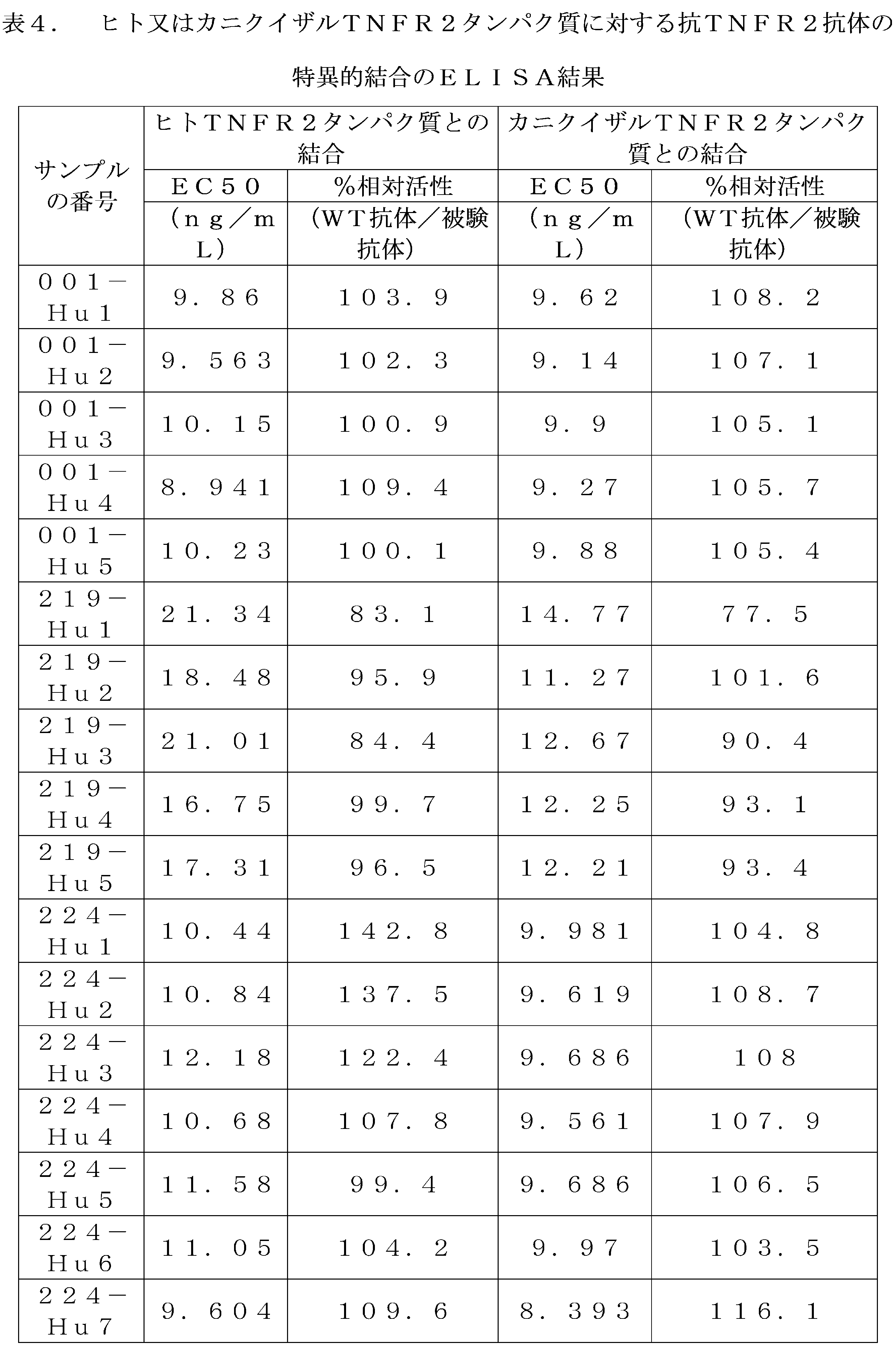
001-WT VHアミノ酸配列(配列番号38):
EVQLQESGGGLVQPGGSLNLSCAASGFAFSTYDLSWVRQTPEKRLEWVAYINNGGISTYYSDTVKGRFTISRDNAKNTLYLQMSSLKSEDTAMYYCVRGPFYGSANYFDYWGQGTTVTVSS
001-WT VLアミノ酸配列(配列番号39):
EIVMTQSPASLSLSVGETVTITCRTSESIYSNLPWYQQKQGKSPQLLVYDATKLAEGVPSRFSGSESGTQYSLKINSLQSEDFGTYYCQHFWVTPWTFGGGTKLEIK
219-WT VHアミノ酸配列(配列番号40):
EVQLQESGAELVRPGASVKLSCKASGYSFTNYWMNWVKQRPGQGLEWIGMIHPSDTETRLNQNFKDKATLTVDKSSSTSYMQLSSPTSEDSAVYYCARGEGLGAARSVSMDYWGQGTTVTVSS
219-WT VLアミノ酸配列(配列番号41):
DILMTQSPSSLSASLGDRVTISCRASQDISNYLNWYQQKPDGTVKVLIYYTAILHSGVPSRFSGSGSGTDYSLTISNLEQEDIATYFCQQGNTLPWTFGGGTKLEIK
224-WT VHアミノ酸配列(配列番号42):
EVQLQESGPEIVKPGASVKLSCTASGFNNKDIYMHWVKQRPEQGLEWIGRIDPATGNTKHDPKFQDKATLSSDTSSNTAYLQFSSLTSEDTAVYYCAHSPYGDFGAMDYWGQGTTVTVSS
224-WT VLアミノ酸配列(配列番号43):
DIQMTQSPAIMSASLGERVTMTCTASSSVSSNYLHWYQQKPGSSPKLWIYSTSNLPSGVPARFSGSGSGTSYSLTISSMEAEDAATYYCHQYHRSPWTFGGGTKLEIK
001-WT VH amino acid sequence (SEQ ID NO:38):
EVQLQESGGGLVQPGGSLNLSCAASGFAFSTYDLSWVRQTPEKRLEWVAYINNGGISTYY SDTVKGRFTISRDNAKNTLYLQMSSLKSEDTAMYYCVRGPFYGSANYFDYWGQGTTVTVSS
001-WT VL amino acid sequence (SEQ ID NO:39):
EIVMTQSPASLSLSVGETVTITCRTSESIYSNLPWYQQKQGKSPQLLVYDATKLAEGVPSRFSGSESGTQYSLKINSLQSEDFGTYYCQHFWVTPWTFGGGGTKLEIK
219-WT VH amino acid sequence (SEQ ID NO:40):
EVQLQESGAELVRPGASVKLSCKASGYSFTNYWMNWVKQRPGQGLEWIGMIHPSDTETRLN QNFKDKATLTVDKSSSTSYMQLSSPTSEDSAVYYCARGEGLGAARSVSMDYWGQGTTVTVSS
219-WT VL amino acid sequence (SEQ ID NO:41):
DILMTQSPSSLSASLGDRVTISCRASQDISNYLNWYQQKPDGTVKVLIYYTAILHSGVPSRFSGSGSGTDYSLTISNLEQEDIATYFCQQGNTLPWTFGGGGTKLEIK
224-WT VH amino acid sequence (SEQ ID NO:42):
EVQLQESGPEIVKPGASVKLSCTASGFNNKDIYMHWVKQRPEQGLEWIGRIDPATGNTKH DPKFQDKATLSSDTSSNTAYLQFSSLTSEDTAVYYCAHSPYGDFGAMDYWGQGTTVTVSS
224-WT VL amino acid sequence (SEQ ID NO:43):
DIQMTQSPAIMSASLGERVTMTCTASSSVSSNYLHWYQQKPGSSPKLWIYSTSNLPSGVPARFSGSGSGTSYSLTISSMEAEDAATYYCHQYHRSPWTFGGGGTKLEIK
実施例3 BIAcoreによるTNFR2ヒト化抗体とヒト及びカニクイザルTNFR2タンパク質との親和性の検出
BIAcoreにより被験抗TNFR2抗体とヒト及びカニクイザルTNFR2タンパク質との特異的結合を検出した。この実験は、Protein Aチップを使用し、結合抗原を飽和させることができるRmaxが50RUとなるように、チップが希釈された抗体を捕捉するのに必要な時間をマニュアル操作(manual run)で測定した。ヒト(Human TNFR2-His、Sino 10417-H08H)とカニクイザル(Cynomolgus TNFR2-His、Sino 90102-C08H)TNFR2の両方を、32、16、8、4、2nMに段階希釈した。抗原に対する抗体の親和性は、マルチサイクル速度論を使用して測定された。各サイクルで、抗体を注入した後、TNFR2タンパク質を勾配濃度で注入し、抗原と抗体の結合及び解離プロセスを生じさせた。各サイクルの後、Glycine pH1.5を使用してProtein Aチップの再生(チップ上のタンパク質を除去する)を行った。BIAcore T200解析ソフトウェアを使用して、抗体抗原の親和性KDをフィッティングした。表5の結果から、すべての被験抗TNFR2抗体とヒト又はカニクイザルTNFR2タンパク質との間に特異的結合があり、親和性レベルが比較的高いことが分かる。
Example 3: Detection of affinity of TNFR2 humanized antibody to human and cynomolgus monkey TNFR2 protein by BIAcore Specific binding of the test anti-TNFR2 antibody to human and cynomolgus monkey TNFR2 protein was detected by BIAcore. In this experiment, Protein A chip was used, and the time required for the chip to capture the diluted antibody was measured manually so that Rmax was 50 RU, which can saturate the bound antigen. Both human (Human TNFR2-His, Sino 10417-H08H) and cynomolgus monkey (Cynomolgus TNFR2-His, Sino 90102-C08H) TNFR2 were serially diluted to 32, 16, 8, 4, and 2 nM. The affinity of the antibody to the antigen was measured using multi-cycle kinetics. In each cycle, after the antibody was injected, TNFR2 protein was injected at a gradient concentration to allow the antigen-antibody binding and dissociation process to occur. After each cycle, Glycine pH 1.5 was used to regenerate the Protein A chip (remove the protein on the chip). The affinity KD of the antibody-antigen was fitted using BIAcore T200 analysis software. The results in Table 5 show that there is specific binding between all tested anti-TNFR2 antibodies and human or cynomolgus monkey TNFR2 protein, and the affinity level is relatively high.
実施例4 FACSによるTNFR2抗体とCHO-TNFR2細胞表面ヒトTNFR2との結合の検出
ヒトTNFR2高発現プラスミドをトランスフェクトしたCHO-K1安定細胞(CHO-TNFR2と命名)を取り、トランスフェクトしたヒトTNFR2全長プラスミドはSino Biological(カタログ番号:HG10417-UT、NCBI Ref Seq:NM_001066.2)から購入され、細胞密度が80%を超えないときに実験を行った。細胞培地を捨て、PBSで洗浄し、1mLのトリプシンを加えて2分間消化し、10%FBSを含むHam’s F12完全培地で消化を停止して細胞懸濁液を作った。計数後、適量の細胞懸濁液を取り、350×gで遠心分離し、上清を捨て、1×10⁷細胞/mLの密度で、対応する量のブロッキング溶液(10%FBS+PBS)を加えて細胞を再懸濁し、4℃で30分間インキュベートした。インキュベート終了後、350×gで遠心分離して上清を除去し、細胞を染色バッファー(2%FBS+PBS)で2×106細胞/mLの密度に再懸濁し、96ウェルプレートに広げ、各ウェルに50μLの細胞懸濁液を加え、後で使用するようにした。PBSを使用して抗体を最高濃度の20μg/mL(2倍濃度)から10倍段階希釈し、希釈した抗体を既に50μLの細胞懸濁液を含むウェルに加え、マイクロプレートシェーカーに置き、500rpmで1分間振とうし、抗体と細胞を完全に混合させ、4℃で1hインキュベートした。インキュベート終了後、細胞を染色バッファー100μL/ウェルで2回洗浄し、350×gで5分間遠心分離して上清を捨てた。PE goat anti-Human IgG Fc抗体(ebioscience、カタログ番号:12-4998-82)を染色バッファーで250倍希釈し、洗浄した細胞ウェルに1ウェルあたり100μLの量で加え、よく混合し、4℃で30分間染色した。染色終了後、同様に染色バッファーで2回洗浄し、最後に200μLの染色バッファーで細胞を再懸濁し、フローサイトメーターでシグナルを検出し(Thermo Attune NxT)、シグナルが強いほど、TNFR2に対する抗体の結合能が強いことを示した。抗体濃度を横軸、対応する平均蛍光強度MFIの倍数を縦軸として抗体の結合曲線をプロットし、4パラメータフィッティング(GraphPad Prism9)を行い、EC50値を算出した。EC50値が小さいほど、抗体がCHO-TNFR2細胞に結合する能力が強いことを示した。ヒト化抗体の結合効果を、対応するヒト-マウスキメラWT抗体に正規化し、パーセント値が高いほど、ヒト化抗体の結合効果が良好であることを示した。図3及び表6に示すように、すべての被験抗TNFR2抗体はCHO-TNFR2細胞表面ヒトTNFR2に結合することができ、且つヒト化抗体の結合能力は基本的にヒト-マウスキメラWT抗体の結合能力と同じである。
Example 4 Detection of TNFR2 antibody binding to human TNFR2 on CHO-TNFR2 cell surface by FACS Human TNFR2 high expression plasmid-transfected CHO-K1 stable cells (named CHO-TNFR2) were taken, and the transfected human TNFR2 full-length plasmid was purchased from Sino Biological (Cat. No.: HG10417-UT, NCBI Ref Seq: NM_001066.2), and experiments were performed when the cell density was not more than 80%. The cell medium was discarded, washed with PBS, and digested with 1 mL of trypsin for 2 minutes, and digestion was stopped with Ham's F12 complete medium containing 10% FBS to make a cell suspension. After counting, an appropriate amount of cell suspension was taken, centrifuged at 350×g, the supernatant was discarded, and the cells were resuspended at a density of 1×10 7 cells/mL by adding a corresponding amount of blocking solution (10% FBS+PBS), and incubated at 4°C for 30 minutes. After the incubation was completed, the cells were centrifuged at 350×g to remove the supernatant, and the cells were resuspended at a density of 2×10 6 cells/mL in staining buffer (2% FBS+PBS), spread on a 96-well plate, and 50 μL of cell suspension was added to each well for later use. The antibody was serially diluted 10-fold from the highest concentration of 20 μg/mL (2x concentration) using PBS, and the diluted antibody was added to the wells already containing 50 μL of cell suspension, placed on a microplate shaker, shaken at 500 rpm for 1 minute, and the antibody and cells were thoroughly mixed, and incubated at 4°C for 1 h. After incubation, the cells were washed twice with 100 μL/well of staining buffer, centrifuged at 350×g for 5 minutes, and the supernatant was discarded. PE goat anti-Human IgG Fc antibody (ebioscience, catalog number: 12-4998-82) was diluted 250-fold with staining buffer, added to the washed cell wells in an amount of 100 μL per well, mixed well, and stained at 4° C. for 30 minutes. After staining, the cells were washed twice with staining buffer in the same manner, and finally resuspended in 200 μL of staining buffer, and the signal was detected with a flow cytometer (Thermo Attune NxT). The stronger the signal, the stronger the binding ability of the antibody to TNFR2. The antibody binding curves were plotted with the antibody concentration on the horizontal axis and the corresponding multiple of the mean fluorescence intensity MFI on the vertical axis, and 4-parameter fitting (GraphPad Prism 9) was performed to calculate the EC50 value. The smaller the EC50 value, the stronger the antibody's ability to bind to CHO-TNFR2 cells. The binding efficiency of the humanized antibodies was normalized to the corresponding human-mouse chimeric WT antibody, and the higher the percentage value, the better the binding efficiency of the humanized antibody. As shown in FIG. 3 and Table 6, all the tested anti-TNFR2 antibodies could bind to human TNFR2 on the surface of CHO-TNFR2 cells, and the binding ability of the humanized antibodies was essentially the same as that of the human-mouse chimeric WT antibody.
実施例5 FACSによる抗体とヒト制御性T細胞(Treg細胞)表面TNFR2との結合の検出実験
ヒトTreg細胞は、選別キット(Stemcell、カタログ番号:18063)を使用してヒトPBMCから単離し、Dynabeads Human Treg Expander(Gibco、カタログ番号:11129D)によってインビトロで15日間刺激及び増幅を行い、その後個別包装して凍結保存した。インビトロで単離増輻したTreg細胞を取り、一晩回復し、翌日300×gで5分間遠心分離し、DPBSで再懸濁して細胞懸濁液を得、計数した。実験に必要な数の細胞を遠心分離管に入れ、300×gで5min遠心分離し、上清を除去し、染色バッファーで細胞密度を2×10⁶/mLに調整し、96ウェルプレートに1ウェルあたり50μLで広げた。すべての被験抗体と対照抗体anti-Hel isotype(それぞれPBSで200ng/mLに希釈し、総量は100μLとした)を取った。各サンプルを各ウェルに50μL/ウェルで添加した。細胞懸濁液と抗体を加えたプレートをマイクロプレートシェーカーに置き、500rpmの速度で1min振とうし、細胞と抗体を十分に混合した。混合後、プレートを4℃の冷蔵庫に入れ、60minインキュベートした。インキュベート終了後、各ウェルに100μLの染色バッファーを加え、350×gで5min遠心分離し、上清を捨て、また、各ウェルに200μLの染色バッファーを加えて細胞を再懸濁し、350×gで5min遠心分離し、上清を捨てた。染色バッファーを、250:1(染色バッファー:蛍光染色抗体)の割合でPE goat anti-Human IgG Fcに加え、染色液を作成し、よく混合して1ウェルあたり100μLで細胞ウェルに加え、プレートをマイクロプレートシェーカーに置き、500rpmの速度で1min振とうし、細胞と染色液を十分に混合した。混合後、4℃の冷蔵庫に入れ、30minインキュベートした。細胞を2回洗浄し、最後に200μLのPBSを各ウェルに加えて細胞を再懸濁し、フローサイトメーターでシグナルを検出した(Thermo Attune NxT)。図4は、すべての抗TNFR2ヒト化抗体が効果的にヒトTreg細胞に結合できることを示す。
Example 5: Detection of binding of antibodies to TNFR2 on the surface of human regulatory T cells (Treg cells) by FACS Human Treg cells were isolated from human PBMCs using a sorting kit (Stemcell, catalog number: 18063), stimulated and expanded in vitro for 15 days using Dynabeads Human Treg Expander (Gibco, catalog number: 11129D), and then individually packaged and cryopreserved. The in vitro isolated and expanded Treg cells were taken and allowed to recover overnight, centrifuged at 300×g for 5 minutes the next day, resuspended in DPBS to obtain a cell suspension, and counted. The number of cells required for the experiment was placed in a centrifuge tube, centrifuged at 300×g for 5 minutes, the supernatant was removed, and the cell density was adjusted to 2×10 6 /mL with staining buffer, and spread in a 96-well plate at 50 μL per well. All test antibodies and control antibody anti-Hel isotype (each diluted to 200 ng/mL with PBS, total volume 100 μL) were taken. Each sample was added to each well at 50 μL/well. The plate with the cell suspension and antibody added was placed on a microplate shaker and shaken at a speed of 500 rpm for 1 min to thoroughly mix the cells and antibody. After mixing, the plate was placed in a 4°C refrigerator and incubated for 60 min. After the incubation was completed, 100 μL of staining buffer was added to each well, centrifuged at 350×g for 5 min, the supernatant was discarded, and 200 μL of staining buffer was added to each well to resuspend the cells, centrifuged at 350×g for 5 min, and the supernatant was discarded. The staining buffer was added to the PE goat anti-Human IgG Fc at a ratio of 250:1 (staining buffer: fluorescent staining antibody) to prepare a staining solution, which was mixed well and added to the cell wells at 100 μL per well. The plate was placed on a microplate shaker and shaken at a speed of 500 rpm for 1 min to thoroughly mix the cells and the staining solution. After mixing, the plate was placed in a refrigerator at 4°C and incubated for 30 min. The cells were washed twice, and finally 200 μL of PBS was added to each well to resuspend the cells, and the signals were detected by a flow cytometer (Thermo Attune NxT). Figure 4 shows that all anti-TNFR2 humanized antibodies can effectively bind to human Treg cells.
実施例6 FACSによる抗体と活性化されたカニクイザルPBMC細胞との結合の検出実験
凍結保存したカニクイザルPMBC細胞を回復させた後、完全培地(RPMI1640-Glutamax+10%FBS+1×P/S+1×ITS+50μMPMメルカプトエタノール)に再懸濁して細胞懸濁液を得、計数した。計数結果に従い、細胞を5×106/mLの密度に再懸濁し、96ウェルU底プレートに1ウェルあたり100μLの細胞懸濁液を広げた。20%濃度のT細胞活性化試薬Immunocult T cell(Stemcell、1:50)activator+rhIL-2(R&D、200IU)を調製し、20%濃度の活性化試薬を1ウェルあたり100μLで細胞に加え、よく混合して37℃、5%CO2のインキュベーターで3日間培養した。活性化された細胞を収集して計数し、染色バッファー(2%FBSを含むDPBS溶液)を使用して細胞密度を4×10⁶/mLに調整し、96ウェルV底プレートに1ウェルあたり50μLで広げた。すべての被験抗体とanti-Hel対照抗体(それぞれ染色バッファーで0.4μg/mL、4μg/mLに希釈した)を取り、各サンプルを各ウェルに50μL/ウェルで添加した。細胞懸濁液と抗体を加えたプレートをマイクロプレートシェーカーに置き、500rpmの速度で1min振とうし、細胞と抗体を十分に混合した。混合後、プレートを4℃の冷蔵庫に入れ、30minインキュベートした。インキュベート終了後、各ウェルに100μLの染色バッファーを加え、350×gで5min遠心分離し、上清を捨て、また、各ウェルに200μLの染色バッファーを加えて細胞を再懸濁し、350×gで5min遠心分離し、上清を捨てた。抗体Anti-Human CD8-FTIC(BD-555366、1μL/test)とPE goat anti-Human IgG Fc(Invitrogen-124998-82、0.5μL/test)を加え、プレートをマイクロプレートシェーカーに置き、500rpmの速度で1min振とうし、細胞と染色液を十分に混合した。混合後、4℃の冷蔵庫に入れ、30minインキュベートした。細胞を2回洗浄し、最後に200μLの染色バッファーを各ウェルに加えて細胞を再懸濁し、フローサイトメーターでシグナルを検出した(Thermo Attune NxT)。図5は、anti-Hel陰性アイソタイプ対照抗体がカニクイザルCD8T細胞に結合せず、すべての被験抗TNFR2ヒト化抗体が活性化カニクイザルPBMCにおけるCD8+T細胞に効果的に結合できることを示す。
Example 6: Detection of binding between antibodies and activated cynomolgus monkey PBMC cells by FACS Cryopreserved cynomolgus monkey PBMC cells were recovered and then resuspended in complete medium (RPMI1640-Glutamax+10% FBS+1xP/S+1xITS+50μM PM mercaptoethanol) to obtain a cell suspension, which was then counted. According to the counting results, the cells were resuspended to a density of 5x10 6 /mL, and 100μL of the cell suspension was spread per well on a 96-well U-bottom plate. A 20% concentration of T cell activation reagent Immunocult T cell (Stemcell, 1:50) activator + rhIL-2 (R&D, 200 IU) was prepared, and the 20% concentration of the activation reagent was added to the cells at 100 μL per well, mixed well, and cultured in a 37°C, 5% CO 2 incubator for 3 days. The activated cells were collected and counted, and the cell density was adjusted to 4×10 6 /mL using staining buffer (DPBS solution containing 2% FBS), and spread in a 96-well V-bottom plate at 50 μL per well. All test antibodies and anti-Hel control antibodies (diluted to 0.4 μg/mL and 4 μg/mL in staining buffer, respectively) were taken, and each sample was added to each well at 50 μL/well. The plate containing the cell suspension and antibody was placed on a microplate shaker and shaken at 500 rpm for 1 min to thoroughly mix the cells and antibody. After mixing, the plate was placed in a refrigerator at 4°C and incubated for 30 min. After incubation, 100 μL of staining buffer was added to each well, centrifuged at 350 × g for 5 min, the supernatant was discarded, and 200 μL of staining buffer was added to each well to resuspend the cells, centrifuged at 350 × g for 5 min, and the supernatant was discarded. Antibody Anti-Human CD8-FTIC (BD-555366, 1 μL/test) and PE goat anti-Human IgG Fc (Invitrogen-124998-82, 0.5 μL/test) were added, the plate was placed on a microplate shaker, and shaken at a speed of 500 rpm for 1 min to thoroughly mix the cells and staining solution. After mixing, the plate was placed in a refrigerator at 4°C and incubated for 30 min. The cells were washed twice, and finally 200 μL of staining buffer was added to each well to resuspend the cells, and the signal was detected with a flow cytometer (Thermo Attune NxT). FIG. 5 shows that the anti-Hel negative isotype control antibody does not bind to cynomolgus monkey CD8 T cells, and all tested anti-TNFR2 humanized antibodies can effectively bind to CD8+ T cells in activated cynomolgus monkey PBMCs.
実施例7 ELISAにより抗TNFR2抗体がTNFαとTNFR2の結合を遮断するのを検出すること
ヒトTNFR2(Novoprotein、カタログ番号C830)を100μL/ウェル、1μg/mLでELISAプレートに予め被包し、すべての被験TNFR2抗体を最高濃度の30μg/mLから2.5倍段階希釈し、希釈した抗体をそれぞれ15ng/mLのヒトTNFα-Biotin(Acro Biosystem、カタログ番号TNA-H82E3)となど量混合し、100μL/ウェルでELISAプレートに加え、室温で振とうしながら2.0時間インキュベートし、プレート洗浄後、Streptavidin-HRP標準溶液(1:10000希釈)を100μL/ウェルで加え、室温で振とうしながら40分間インキュベートし、プレートを再度洗浄し、HRPの基質であるTMBを加えて発色させ、停止液を加えて反応を停止させ、マイクロプレートリーダー(MD I3)で吸光度を読み取った。抗体濃度を横軸、対応するOD値を縦軸として抗体の阻害曲線をプロットし、4パラメータフィッティング(GraphPad Prism9)を行い、IC50値を算出した。IC50値が小さいほど、抗体がヒトTNFαとヒトTNFR2の結合を阻害する能力が強いことを示した。ヒト化抗体の遮断効果を、対応するヒト-マウスキメラ抗体WTに正規化し、パーセント値が高いほど、ヒト化抗体の阻害効果が良好であることを示した。被験抗体の遮断曲線を図6に、阻害活性を表7に示す。図6及び表7から、すべての被験抗体がヒトTNFαとヒトTNFR2タンパク質の結合を有意に阻害でき、且つヒト化抗体の阻害能力が基本的にヒト-マウスキメラWT抗体の阻害能力と同じであることが分かる。
Example 7: Detection of anti-TNFR2 antibodies blocking the binding of TNFα to TNFR2 by ELISA Human TNFR2 (Novoprotein, Cat. No. C830) was pre-coated on an ELISA plate at 100 μL/well, 1 μg/mL. All tested TNFR2 antibodies were serially diluted 2.5-fold from the highest concentration of 30 μg/mL, and each diluted antibody was diluted with 15 ng/mL human TNFα-Biotin (Acro). The mixture was mixed with 100 μL/well of Streptavidin-HRP standard solution (1:10000 dilution) and incubated at room temperature for 40 minutes with shaking. The plate was washed, and then Streptavidin-HRP standard solution (1:10000 dilution) was added at 100 μL/well. The plate was then incubated at room temperature for 40 minutes with shaking. The plate was washed again, and TMB, a substrate for HRP, was added to develop color. A stop solution was added to stop the reaction, and the absorbance was read using a microplate reader (MD I3). The antibody inhibition curve was plotted with the antibody concentration on the horizontal axis and the corresponding OD value on the vertical axis, and 4-parameter fitting (GraphPad Prism 9) was performed to calculate the IC50 value. The smaller the IC50 value, the stronger the antibody's ability to inhibit the binding of human TNFα to human TNFR2. The blocking effect of the humanized antibodies was normalized to the corresponding human-mouse chimeric antibody WT, and the higher the percentage value, the better the inhibitory effect of the humanized antibody. The blocking curves of the tested antibodies are shown in Figure 6, and the inhibitory activities are shown in Table 7. From Figure 6 and Table 7, it can be seen that all the tested antibodies can significantly inhibit the binding of human TNFα to human TNFR2 protein, and the inhibitory ability of the humanized antibodies is basically the same as that of the human-mouse chimeric antibody WT.

実施例8 抗TNFR2抗体がヒトTNFR2を高度に発現するCHO細胞上でのTNFR2とTNFαの結合を遮断すること
ヒトTNFR2を高度に発現するCHO安定細胞(実施例4のCHO-TNFR2と同じ)を用いて実験を行った。CHO-TNFR2細胞を消化した後、DPBSで2回洗浄し、Live/Dead(L/D)で染色し(室温で20分間)、その後、1×105細胞/50μL/ウェルでプレートに広げ、染色バッファーで被験抗TNFR2抗体を6μg/mLに希釈して初期濃度とし、3.33倍段階希釈して合計7点の濃度ポイントを設定し、希釈した抗体を50μL/ウェルで播種した細胞に加え、穏やかに吹き混ぜて4℃で30分間インキュベートした。その後、100ng/mLに希釈したヒトTNFα-biotinを100μL/ウェルで加え、穏やかに吹き混ぜて、混合後4℃で30分間インキュベートした。染色バッファーで2回洗浄した後、PE-streptavidin(BioLegend、カタログ番号405204)を100μL/ウェル、0.12μg/mLで加え、4℃で30分間インキュベートした。染色バッファーで2回洗浄した後、150μLで再懸濁し、フローサイトメーターで検出した(Thermo Attune NxT)。抗体濃度を横軸、対応するMFI値を縦軸として抗体の阻害曲線をプロットし、4パラメータフィッティング(GraphPad Prism9)を行い、IC50値を算出した。IC50値が小さいほど、抗体がヒトTNFαとヒトTNFR2の結合を阻害する能力が強いことを示した。ヒト化抗体の遮断効果を、対応するヒト-マウスキメラWT抗体に正規化し、パーセント値が高いほど、ヒト化抗体の阻害効果が良好であることを示した。被験抗体の遮断曲線を図7に、阻害活性を表8に示す。図7及び表8から、すべての被験抗体がTNFαとCHO-TNFR2細胞上でのTNFR2の結合を有意に阻害できることが分かる。
Example 8 Anti-TNFR2 antibodies block the binding of TNFR2 to TNFα on CHO cells highly expressing human TNFR2 An experiment was carried out using CHO stable cells highly expressing human TNFR2 (same as CHO-TNFR2 in Example 4). After digestion of CHO-TNFR2 cells, they were washed twice with DPBS and stained with Live/Dead (L/D) (20 min at room temperature), and then spread on a plate at 1 x 10⁵ cells/50 μL/well. The test anti-TNFR2 antibodies were diluted with staining buffer to 6 μg/mL as the initial concentration, and serially diluted 3.33 times to set a total of 7 concentration points. The diluted antibodies were added to the seeded cells at 50 μL/well, gently mixed, and incubated at 4°C for 30 min. Then, human TNFα-biotin diluted to 100 ng/mL was added at 100 μL/well, gently mixed by blowing, and incubated at 4° C. for 30 minutes after mixing. After washing twice with staining buffer, PE-streptavidin (BioLegend, catalog number 405204) was added at 100 μL/well, 0.12 μg/mL, and incubated at 4° C. for 30 minutes. After washing twice with staining buffer, the plate was resuspended in 150 μL and detected with a flow cytometer (Thermo Attune NxT). The antibody inhibition curve was plotted with the antibody concentration on the horizontal axis and the corresponding MFI value on the vertical axis, and 4-parameter fitting (GraphPad Prism9) was performed to calculate the IC50 value. The smaller the IC50 value, the stronger the antibody's ability to inhibit the binding of human TNFα to human TNFR2. The blocking effect of the humanized antibodies was normalized to the corresponding human-mouse chimeric WT antibody, and the higher the percentage value, the better the inhibitory effect of the humanized antibody. The blocking curves of the tested antibodies are shown in Figure 7, and the inhibitory activities are shown in Table 8. From Figure 7 and Table 8, it can be seen that all the tested antibodies can significantly inhibit the binding of TNFα to TNFR2 on CHO-TNFR2 cells.
実施例9 FACSによる、TNF(誘導ヒトTreg細胞IκBαの分解に対する抗体の阻害の検出実験
ヒトTreg細胞は、選別kit(Stemcell、カタログ番号:18063)を使用してヒトPBMCから単離し、Dynabeads Human Treg Expander(Gibco、カタログ番号:11129D)によってインビトロで15日間刺激及び増幅を行い、その後個別包装して凍結保存した。インビトロで単離増幅したTreg細胞を取り、一晩回復し、翌日300×gで5分間遠心分離し、AIM-V培地(ブランドGibco、カタログ番号31035025)で培養して計数し、細胞の計数結果に従って細胞密度を6×10⁶細胞/mLに調整し、96ウェルプレートに1ウェルあたり25μLで広げた。すべての被験抗体とanti-Hel対照抗体(それぞれAIM-V培地で40%濃度に希釈し、1μg/mL、10μg/mL、100μg/mLとした)を取った。各濃度の抗体を対応する細胞ウェルに1ウェルあたり25μLで加えた。マルチチャンネルピペットで混合した後、37℃、5%CO2のインキュベーターに入れ、30minプレインキュベートした。AIM-V培地を使用してTNFα(ブランドNovoprotein、カタログ番号C008)を作業濃度の2倍である20ng/mLに希釈し、TNFα活性化を必要とするウェルに50μLのTNFαを、非活性化対照ウェルに50μLのAIM-V培地を、抗体の最終濃度が1倍となり、TNFαの最終濃度が10ng/mLとなるように加え、ピペットで吹き混ぜて、培養プレートを37℃のインキュベーターに入れて10minインキュベートした。インキュベート終了後、培養プレートを取り出し、100μLの予冷したDPBSを加え、4℃、300gで5min遠心分離した。次に、細胞について、1×live/dead violet(予冷したDPBSで500倍希釈)100μLを使用して、氷上で20min反応させた。各ウェルに100μLの染色バッファーを加え、4℃、300gで5min遠心分離した。各ウェルに1×fix-perm buffer(ブランドBD、カタログ番号554714)を100μLずつ加えてよく混ぜ、4℃で30min固定した。固定終了後、各ウェルに100μLの1×perm bufferを加えて固定を停止させ、400gで5min遠心分離し、上清を捨てた。次に、各ウェルに200μLの1×perm bufferを加えて、細胞を2回洗浄した。100μLのIκBα染色液(1×perm bufferで100倍希釈)を実験ウェルに加え、1μLのRat IgG2b kappa Isotype Control抗体を対照ウェルに加え、よく混ぜ、4℃で30minインキュベートした。インキュベート終了後、各ウェルに100μLの1×perm bufferを加えて固定を停止させ、400gで5min遠心分離し、上清を捨てた。次に、各ウェルに200μLの1×perm bufferを加えて、細胞を2回洗浄した。フローサイトメーターで検出した(Thermo Attune NxT)。生細胞中のIκBαlowのパーセント又は全ての生細胞のIκBαのMFIをフローサイトメトリーにより分析した。IκBαlowの割合が低いほど、抗TNFR2抗体がTNFα誘導IκBαの分解を阻害する能力が強くなった。図8の結果から、陰性対照抗体anti-HelはIκBαの発現に影響を及ぼさないが、被験抗TNFR2ヒト化抗体はいずれもTNFα誘導IκBαの分解を阻害することができることが分かる。
Example 9: Detection of antibody inhibition of TNF-induced human Treg cell IκBα degradation by FACS Human Treg cells were isolated from human PBMCs using a sorting kit (Stemcell, catalog number: 18063), stimulated and expanded in vitro for 15 days using Dynabeads Human Treg Expander (Gibco, catalog number: 11129D), and then individually packaged and cryopreserved. The in vitro isolated and expanded Treg cells were taken and allowed to recover overnight, centrifuged at 300×g for 5 minutes the next day, cultured in AIM-V medium (brand Gibco, catalog number 31035025), counted, and the cell density was adjusted to 6×10 according to the cell counting results. The concentration of each antibody was adjusted to 6 cells/mL and spread at 25 μL per well on a 96-well plate. All test antibodies and anti-Hel control antibodies were diluted to 40% concentration with AIM-V medium to give 1 μg/mL, 10 μg/mL, and 100 μg/mL. Each antibody concentration was added to the corresponding cell well at 25 μL per well. After mixing with a multichannel pipette, the plate was incubated at 37°C, 5% CO 2 incubator and pre-incubated for 30 min. TNFα (brand Novoprotein, catalog number C008) was diluted to 20 ng/mL, which is twice the working concentration, using AIM-V medium, and 50 μL of TNFα was added to wells requiring TNFα activation, and 50 μL of AIM-V medium was added to non-activated control wells so that the final concentration of antibody was 1x and the final concentration of TNFα was 10 ng/mL, and the plates were mixed with a pipette and incubated in a 37°C incubator for 10 min. After incubation, the plates were removed, 100 μL of pre-cooled DPBS was added, and the plates were centrifuged at 4°C and 300 g for 5 min. Next, the cells were diluted with 1x live/dead medium. 100 μL of violet (pre-cooled DPBS diluted 500-fold) was used and reacted on ice for 20 min. 100 μL of staining buffer was added to each well and centrifuged at 4 ° C and 300 g for 5 min. 100 μL of 1 × fix-perm buffer (brand BD, catalog number 554714) was added to each well, mixed well, and fixed at 4 ° C for 30 min. After fixation, 100 μL of 1 × perm buffer was added to each well to stop fixation, centrifuged at 400 g for 5 min, and the supernatant was discarded. Next, 200 μL of 1 × perm buffer was added to each well, and the cells were washed twice. 100 μL of IκBα staining solution (diluted 100-fold with 1×perm buffer) was added to the experimental wells, and 1 μL of Rat IgG2b kappa Isotype Control antibody was added to the control wells, mixed well, and incubated at 4°C for 30 min. After incubation, 100 μL of 1×perm buffer was added to each well to stop fixation, centrifuged at 400 g for 5 min, and the supernatant was discarded. Next, 200 μL of 1×perm buffer was added to each well, and the cells were washed twice. Detection was performed with a flow cytometer (Thermo Attune NxT). The percentage of IκBα low in live cells or the MFI of IκBα in all live cells was analyzed by flow cytometry. The lower the ratio of IκBα low , the stronger the ability of the anti-TNFR2 antibody to inhibit TNFα-induced IκBα degradation. The results in Figure 8 show that the negative control antibody anti-Hel has no effect on the expression of IκBα, but all of the tested anti-TNFR2 humanized antibodies can inhibit TNFα-induced IκBα degradation.
実施例10 TNFα及びIL-2誘導Treg細胞の増殖に対するTNFR2抗体の阻害
TNFR2抗体の存在下でのTNFα及びIL-2誘導Treg細胞の増殖を測定することにより、TNFα及びIL-2誘導Treg細胞の増殖に対するこの抗体の阻害効果を判断した(Zaragoza B et al.,Nat Med.2016 Jan;22(1):16-7)。インビトロで増輻単離してから凍結保存したTreg細胞を取り、一晩回復し、翌日400×gで5分間遠心分離し、培地で再懸濁して細胞懸濁液を得、計数した。TregをCellTrace Violet cell proliferation kit(Thermo カタログ番号C34557)で染色し、1×10⁷細胞あたり1μLの保存液を用いて1mLの染色液を調製した。37℃で20min染色し、少なくとも5倍量の完全培地で反応を中和し、5min静置し、400×gで5min遠心分離した。培地で細胞を再懸濁して1回洗浄し、再度遠心分離して上清を取った。96ウェルプレートに1ウェルあたり1×10⁵細胞の密度で広げ、1×10⁵細胞あたり50μLの培地の密度で細胞を再懸濁し、96ウェルプレートに移し替えた。抗体の最終濃度が12.5μg/mLになるように、各ウェルに50μLの被験抗体を加え、インキュベーターで30分間インキュベートした。その後、IL-2(最終濃度300IU)及びTNFα(最終濃度50ng/mL)を含む培地50μLと、anti-CD3/CD28 Dynabeads(Gibco、カタログ番号:11129D、beads/Treg細胞比1:20)を含む培地50μLを各ウェルに加え、各ウェルの最終容量を200μLとした。条件毎に2つの平行なウェルを設定し、前記のプレートを混合し、37℃のインキュベーターに入れて3日間培養してFACS検出(Thermo Attune NxT)を行った。TNFα及びIL-2誘導Treg増殖に対する本抗体の阻害効果は、Treg増殖の割合で判断した。その結果を図9に示すが、対照anti-Hel抗体と比較して、すべての抗TNFR2ヒト化抗体は、いずれもTNFα及びIL-2誘導Treg細胞の増殖を効果的に阻害でき、且つ有意差があった。
Example 10 Inhibition of TNFR2 Antibody on TNFα- and IL-2-Induced Treg Cell Proliferation The inhibitory effect of TNFR2 antibody on TNFα- and IL-2-induced Treg cell proliferation was determined by measuring proliferation of TNFα- and IL-2-induced Treg cells in the presence of TNFR2 antibody (Zaragoza B et al., Nat Med. 2016 Jan;22(1):16-7). Treg cells that were expanded and isolated in vitro and then cryopreserved were taken, allowed to recover overnight, centrifuged the next day at 400×g for 5 minutes, and resuspended in culture medium to obtain a cell suspension and counted. Tregs were stained with CellTrace Violet cell proliferation kit (Thermo Cat. No. C34557) and 1 mL of staining solution was prepared using 1 μL of storage solution per 1× 10⁷ cells. The cells were stained at 37°C for 20 min, neutralized with at least 5 volumes of complete medium, allowed to stand for 5 min, and centrifuged at 400 x g for 5 min. The cells were resuspended and washed once with medium, centrifuged again, and the supernatant was removed. The cells were spread at a density of 1 x 10⁵ cells per well in a 96-well plate, resuspended at a density of 50 μL of medium per 1 x 10⁵ cells, and transferred to the 96-well plate. 50 μL of the test antibody was added to each well to give a final antibody concentration of 12.5 μg/mL, and incubated in an incubator for 30 minutes. Then, 50 μL of medium containing IL-2 (final concentration 300 IU) and TNFα (final concentration 50 ng/mL) and 50 μL of medium containing anti-CD3/CD28 Dynabeads (Gibco, catalog number: 11129D, beads/Treg cell ratio 1:20) were added to each well, making the final volume of each well 200 μL. Two parallel wells were set up for each condition, and the plates were mixed and cultured in a 37°C incubator for 3 days before FACS detection (Thermo Attune NxT). The inhibitory effect of the antibody on TNFα- and IL-2-induced Treg proliferation was determined by the percentage of Treg proliferation. The results are shown in FIG. 9. Compared with the control anti-Hel antibody, all of the anti-TNFR2 humanized antibodies were able to effectively inhibit the proliferation of TNFα- and IL-2-induced Treg cells, and the differences were significant.
実施例11 Treg機能に対する抗TNFR2抗体の阻害
抗TNFR2抗体を含む条件下でエフェクター(responder)T細胞に対するTreg細胞の阻害機能を測定することにより、Treg細胞の阻害機能に対する抗体の阻害効果を判断した。実験に必要なTreg(CD4+CD25+FoxP3+T細胞)とConventional T細胞(Tcon)エフェクター細胞(CD4+CD25-T細胞)を取り、一晩回復し、翌日400×gで5分間遠心分離し、完全培地(RPMI1640-Glutamax+10%FBS+1×P/S+1×ITS+50μMPMメルカプトエタノール)に再懸濁して細胞懸濁液を得、計数した。Tcon細胞をCellTrace Violet cell proliferation kit(Thermo カタログ番号C34557)で染色し、1×10⁷細胞あたり1μLの保存液を用いて1mLの染色液を調製した。37℃で20min染色し、少なくとも5倍量の完全培地で反応を中和し、5min静置し、400×gで5min遠心分離した。培地で細胞を再懸濁して1回洗浄し、再度遠心分離して上清を捨てた。96ウェルプレートに1ウェルあたり1×10⁵細胞の密度で広げ、1×10⁵細胞あたり50μLの培地の密度で細胞を再懸濁し、50mLの遠心分離管に移し替えた。同時にTreg細胞を準備し、Treg細胞とTcon細胞を1:1の比率で共培養した。Tcon細胞の数に応じて、対応するanti-CD3/CD28 Dynabeads(Gibco、カタログ番号:11129D)を1/10の比率で準備した。1ウェルあたり50μLの容量でTreg及びTconの培地に加えた。抗体の最終濃度が12.5μg/mLになるように、各ウェルに被験抗体を加えた。条件毎に3つの平行なウェルを設定し、前記のプレートを混合し、37℃のインキュベーターに入れて4日間培養してフローサイトメーターで検出を行った。Tcon増殖の割合が高いほど、抗体がTregを阻害する能力が強くなった。阻害結果を図10に示すが、3つの被験抗TNFR2ヒト化抗体は、いずれもTcon細胞に対するTregの阻害効果を有意に阻害し、Tconの増殖を促進することができ、且つ統計的差異を有した。
Example 11 Inhibition of Treg Function by Anti-TNFR2 Antibody The inhibitory effect of the antibody on the inhibitory function of Treg cells was determined by measuring the inhibitory function of Treg cells against effector (responder) T cells under conditions containing anti-TNFR2 antibody. Treg (CD4+CD25+FoxP3+T cells) and conventional T cell (Tcon) effector cells (CD4+CD25-T cells) required for the experiment were taken, allowed to recover overnight, centrifuged at 400×g for 5 minutes the next day, and resuspended in complete medium (RPMI1640-Glutamax+10%FBS+1×P/S+1×ITS+50 μM PM mercaptoethanol) to obtain a cell suspension, which was then counted. Tcon cells were stained with CellTrace Violet cell proliferation kit (Thermo Cat. No. C34557) and 1 mL of staining solution was prepared using 1 μL of stock solution per 1 × 10 7 cells. Staining was performed for 20 min at 37 °C, the reaction was neutralized with at least 5 volumes of complete medium, left to stand for 5 min, and centrifuged at 400 × g for 5 min. The cells were resuspended and washed once with medium, centrifuged again, and the supernatant was discarded. The cells were spread at a density of 1 × 10 5 cells per well in a 96-well plate, resuspended at a density of 50 μL of medium per 1 × 10 5 cells, and transferred to a 50 mL centrifuge tube. Treg cells were prepared at the same time, and Treg cells and Tcon cells were co-cultured at a ratio of 1:1. According to the number of Tcon cells, corresponding anti-CD3/CD28 Dynabeads (Gibco, Catalog No.: 11129D) were prepared at a ratio of 1/10. The Treg and Tcon medium was added at a volume of 50 μL per well. The test antibody was added to each well so that the final concentration of the antibody was 12.5 μg/mL. Three parallel wells were set up for each condition, and the plate was mixed and cultured in a 37°C incubator for 4 days, and detection was performed with a flow cytometer. The higher the rate of Tcon proliferation, the stronger the ability of the antibody to inhibit Treg. The inhibition results are shown in Figure 10. All three tested anti-TNFR2 humanized antibodies could significantly inhibit the inhibitory effect of Treg on Tcon cells and promote the proliferation of Tcon, and had statistical differences.
実施例12 FACSによる抗TNFR2抗体媒介抗体依存性細胞傷害(ADCC)活性の検出
PBMCと凍結保存したTreg細胞を完全培地(RPMI1640-Glutamax+10%FBS+1×P/S+1×ITS+50μMPMメルカプトエタノール)で1日前に培養し、そのうち、PBMCはIL-2を100IU/mL添加する必要があった。実験当日、分離キット(Stemcell、カタログ番号17955)の要件に従ってPBMCからNK細胞を分離し、完全培地(IL-2を含まない)で0.8×106/mLの密度で再懸濁した。標的細胞であるTreg細胞又はCHO-TNFR2細胞をCell Trace violet試薬で標識し、標識後に密度を0.4×10⁶/mLに調整した。被験抗体を完全培地で最終濃度の4倍に段階希釈し、後で使用するようにした。実験用プレートの分布図に従って標的細胞を1ウェルあたり50μLで細胞プレートに播種し、希釈した薬物を対応するウェルに広げ、標的細胞とともに37℃で30分間インキュベートした。インキュベート終了後、エフェクター細胞を添加する必要のあるウェルにエフェクター細胞懸濁液を100μL添加し、エフェクター細胞を添加する必要のないウェルに培地を100μL補充し、37℃で4hインキュベートした。反応終了後、各ウェルに1μLのPI色素を加え、機器で検出した。標的細胞におけるPIの陽性の割合が高いほど、より顕著なADCC効果を示した。ADCC実験の結果を図11に示すが、anti-Hel陰性対照抗体には作用標的がないため、ADCC殺傷効果がなく、3つの被験ヒト化抗体は、標的細胞であるTreg又はCHO-TNFR2細胞の両方でいずれもADCC活性を示した。
Example 12 Detection of Anti-TNFR2 Antibody-Mediated Antibody-Dependent Cellular Cytotoxicity (ADCC) Activity by FACS PBMCs and cryopreserved Treg cells were cultured in complete medium (RPMI1640-Glutamax+10%FBS+1xP/S+1xITS+50μM PM mercaptoethanol) one day in advance, of which PBMCs needed to be supplemented with IL-2 at 100 IU/mL. On the day of the experiment, NK cells were isolated from PBMCs according to the requirements of the isolation kit (Stemcell, Cat. No. 17955) and resuspended in complete medium (without IL-2) at a density of 0.8x10 6 /mL. Target cells, Treg cells or CHO-TNFR2 cells, were labeled with Cell Trace violet reagent, and the density was adjusted to 0.4x10 6 /mL after labeling. The test antibodies were serially diluted in complete medium to 4 times the final concentration for later use. Target cells were seeded on the cell plate at 50 μL per well according to the distribution diagram of the experimental plate, and the diluted drugs were spread into the corresponding wells and incubated with the target cells at 37°C for 30 minutes. After the incubation was completed, 100 μL of effector cell suspension was added to the wells that needed to add effector cells, and 100 μL of medium was replenished to the wells that did not need to add effector cells, and incubated at 37°C for 4 h. After the reaction was completed, 1 μL of PI dye was added to each well and detected by the instrument. The higher the PI positive rate in the target cells, the more significant the ADCC effect. The results of the ADCC experiment are shown in Figure 11. The anti-Hel negative control antibody had no target and therefore no ADCC killing effect, and all three test humanized antibodies showed ADCC activity in both target cells, Treg and CHO-TNFR2 cells.
実施例13 TNFR2トランスジェニックヒト化マウス(TNFR2 HuGEMM)におけるhTNFR2及びmTNFR2発現の検出
中美冠科生物技術有限公司との協力により、CRISPR/Cas9技術を用いて相同組換えにより、hTNFRSF1B-WPRE-PA発現枠をTnfrsf1b遺伝子のexon2部位にノックインし、Tnfrsf1b遺伝子がhTNFRSF1B-WPRE-PAにノックインされたヘテロ接合性BALB/cマウスを得て、次に繁殖によりヒトTnfrsf1b(TNFR2)遺伝子を発現するホモ接合性トランスジェニックマウス(TNFR2 HuGEMM)を得た。6~8週齢の雌BALB/cマウス(北京維通利華実験動物技術有限公司から)及びTNFR2 HuGEMMマウスから末梢血を採取し、ACK(Gibco、カタログ番号A1049201)を使用して赤血球を除去し、FACSの免疫表現型検査を実施し、マウス末梢血中のCD4+T細胞、CD8+T細胞、及びCD4+CD25+Foxp3+Treg細胞におけるmTNFR2及びhTNFR2の発現を検出し、すべての細胞集団のゲーティングはいずれもFMO対照を基準とした(図12A)。その結果、TNFR2 HuGEMMマウスはmTNFR2を発現せず、CD4+T細胞、CD8+T細胞、Treg細胞の表面におけるhTNFR2の発現プロファイル/発現レベルはBALB/cバックグラウンドマウスのmTNFR2と一致し、TNFR2 HuGEMMマウスがhTNFR2によるmTNFR2の置換を完了したことが示された(図12B~12C)。
Example 13 Detection of hTNFR2 and mTNFR2 Expression in TNFR2 Transgenic Humanized Mice (TNFR2 HuGEMM) In collaboration with Zhongmei Crown Bio-Tech Co., Ltd., the hTNFRSF1B-WPRE-PA expression frame was knocked into the exon 2 site of the Tnfrsf1b gene by homologous recombination using CRISPR/Cas9 technology to obtain heterozygous BALB/c mice in which the Tnfrsf1b gene was knocked into hTNFRSF1B-WPRE-PA, and then homozygous transgenic mice expressing the human Tnfrsf1b (TNFR2) gene (TNFR2 HuGEMM) were obtained by breeding. Peripheral blood was collected from 6-8 week-old female BALB/c mice (from Beijing Weitong Lihua Laboratory Animal Technology Co., Ltd.) and TNFR2 HuGEMM mice, red blood cells were removed using ACK (Gibco, Cat. No. A1049201), and FACS immunophenotyping was performed to detect the expression of mTNFR2 and hTNFR2 in CD4+ T cells, CD8+ T cells, and CD4+CD25+Foxp3+ Treg cells in mouse peripheral blood, and all cell populations were gated based on FMO control ( FIG. 12A ). The results showed that TNFR2 HuGEMM mice did not express mTNFR2, and the expression profile/level of hTNFR2 on the surface of CD4+ T cells, CD8+ T cells, and Treg cells was consistent with mTNFR2 in BALB/c background mice, indicating that TNFR2 HuGEMM mice had completed replacement of mTNFR2 with hTNFR2 (Figures 12B-12C).
実施例14 TNFα誘導TNFR2 HuGEMMのTreg細胞増殖に対する抗TNFR2抗体の阻害
TNFR2抗体の存在下でTNFα誘導TNFR2 HuGEMMマウスのTreg細胞増殖を測定することにより、遺伝子改変後のTNFR2 HuGEMMマウスにおいてTNFR2シグナル伝達経路と対応する機能が保存されていることが確認され、改変後のトランスジェニックマウスが候補抗TNFR2抗体の体内薬効評価に使用できることが確保された。TNFR2 HuGEMMマウス5匹と野生型Balb/cマウス5匹を用意し、安全キャビネット内でマウスの脾臓を無菌的に取り出して粉砕し、粉砕した細胞懸濁液をすべて15mL遠心分離管に移し替えて遠心分離(320g、7分間、4℃)した。上清を捨てた。約2~3mLのACK赤血球溶解バッファー(Gibco、カタログ番号A1049201)を加えてよく混ぜ、赤血球が完全に溶解するまで3~5分間室温で放置し、6~8mLのRPMI1640培地を加えて溶解を停止させ、反転してよく混ぜた。遠心分離(200g、8分間、4℃)した。遠心分離後、上清を捨て、EasySep bufferで細胞を再懸濁し、40μmメンブレンフィルタで細胞を濾過し、細胞の計数を行った。次に、Mouse CD4+CD25+Regulatory T kit(ブランドStemcell、カタログ番号18783A)の説明書に従って、Treg細胞を選別した。選別したTreg細胞をCellTrace Violet cell proliferation kit(Thermo カタログ番号C34557)で染色し、1×10⁷細胞あたり1μLの保存液を用いて1mLの染色液を調製した。37℃で20min染色し、少なくとも5倍量の完全培地で反応を中和し、5min静置し、400×gで5min遠心分離した。培地で細胞を再懸濁して1回洗浄し、再度遠心分離して上清を取った。96ウェルプレートに1ウェルあたり1×10⁵細胞の密度で広げ、1×10⁵細胞あたり50μLの培地の密度で細胞を再懸濁し、96ウェルプレートに移し替えた。各ウェルに50μLの被験抗体(3つのヒト化TNFR2抗体、1つの抗マウスTNFR2置換抗体Surrogate antibody clone#75-54.7(BioXcell、BE0247)及びアイソタイプ対照抗体であるanti-Hel-mIgG1を含む)を加え、抗体の最終濃度が10μg/mLであり、インキュベーターで30分間インキュベートした。その後、マウスIL-2(ブランド義翹神州、カタログ番号51061-MNAE、最終濃度300IU)及びマウスTNFα(ブランド義翹神州、カタログ番号20180502、最終濃度50ng/mL)を含む培地50μLと、Dynabeads mouse T-activator CD3/CD28(Gibco、カタログ番号:11452D、beadsとTreg細胞比1:20)を含まないか又は含む培地50μLを各ウェルに加え、各ウェルの最終容量を200μLとした。同時に、Treg細胞+マウスIL-2のみ、Treg細胞+マウスTNFαのみ、及びTreg細胞+マウスIL-2とヒトTNFαの対照ウェルを設定した。条件毎に2つの平行なウェルを設定し、前記のプレートを混合し、37℃のインキュベーターに入れて3日間培養してFACS検出(Thermo Attune NxT)を行った。TNFα誘導Treg増殖に対する本抗体の阻害効果は、Treg増殖の割合で判断した。図13Aは、TNFR2 HuGEMMマウスのTreg細胞がヒトTNFR2を発現するがマウスTNFR2を発現しないことと、WT Balb/cマウスのTreg細胞がマウスTNFR2のみを発現することを示す。図13B及び13Cは、システムにDynabeads mouse T-activator CD3/CD28アゴニストが存在するかどうかに関係なく、mIL-2及びmTNFα又はhTNFαを添加すると、TNFR2 HuGEMM Tregの増殖が著しく増加したことを示すが、TNFR2 HuGEMMマウスの遺伝子改変後にTNFR2媒介シグナルが保存されていると考えられる。一方、WT balb/cでは、mIL-2とmTNFαの添加でのみ増殖が増加したが、hTNFαは増殖に影響を与えなかったことから、hTNFαはmTNFR2に結合しないことが示唆された。TNFR2 HuGEMM Tregについて、mIL-2とmTNFαの存在下で抗TNFR2ヒト化抗体を添加すると、陰性対照anti-Hel-mIgG1と比較して、001-Hu3-mIgG1及び219-Hu1-mIgG1はTNFR2 HuGEMM Tregの増殖を有意に阻害することができた、且つ統計的差異(***p<0.001)を有する一方で、抗マウスTNFR2置換抗体TR75-54.7には阻害効果がなく、WT Balb/c Tregについて、mIL-2とmTNFαの存在下で抗TNFR2抗体を添加すると、陰性対照Hamster IgGと比較して、抗マウスTNFR2置換抗体TR75-54.7はWT Balb/c Tregの増殖を有意に阻害することができ、且つ統計的差異(*** p<0.001)を有する一方で、001-Hu3-mIgG1及び219-Hu1-mIgG1には阻害効果がなかった。
Example 14 Inhibition of Treg cell proliferation in TNFα-induced TNFR2 HuGEMM by anti-TNFR2 antibodies By measuring Treg cell proliferation in TNFα-induced TNFR2 HuGEMM mice in the presence of TNFR2 antibodies, it was confirmed that the TNFR2 signaling pathway and corresponding functions were preserved in the genetically modified TNFR2 HuGEMM mice, ensuring that the modified transgenic mice could be used for in vivo efficacy evaluation of candidate anti-TNFR2 antibodies. Five TNFR2 HuGEMM mice and five wild-type Balb/c mice were prepared, and the mouse spleens were aseptically removed and crushed in a safety cabinet, and all the crushed cell suspensions were transferred to 15 mL centrifuge tubes and centrifuged (320 g, 7 minutes, 4° C.). The supernatant was discarded. Approximately 2-3 mL of ACK red blood cell lysis buffer (Gibco, Catalog No. A1049201) was added, mixed well, and left at room temperature for 3-5 minutes until red blood cells were completely lysed. 6-8 mL of RPMI1640 medium was added to stop the lysis, and the mixture was inverted and mixed well. Centrifugation (200 g, 8 minutes, 4°C) was performed. After centrifugation, the supernatant was discarded, and the cells were resuspended in EasySep buffer, filtered through a 40 μm membrane filter, and counted. Treg cells were then selected according to the instructions of the Mouse CD4+CD25+Regulatory T kit (Brand Stemcell, Catalog No. 18783A). Sorted Treg cells were stained with CellTrace Violet cell proliferation kit (Thermo Cat. No. C34557) and 1 mL of staining solution was prepared using 1 μL of stock solution per 1× 10 cells. Staining was performed for 20 min at 37°C, the reaction was neutralized with at least 5 volumes of complete medium, left to stand for 5 min, and centrifuged at 400×g for 5 min. Cells were resuspended and washed once with medium, centrifuged again, and the supernatant was removed. Cells were spread at a density of 1× 10 cells per well in a 96-well plate and resuspended at a density of 50 μL of medium per 1× 10 cells and transferred to a 96-well plate. 50 μL of test antibodies (including three humanized TNFR2 antibodies, one anti-mouse TNFR2 replacement antibody Surrogate antibody clone #75-54.7 (BioXcell, BE0247), and an isotype control antibody, anti-Hel-mIgG1) were added to each well to give a final antibody concentration of 10 μg/mL, and the wells were incubated in an incubator for 30 minutes. Then, 50 μL of medium containing mouse IL-2 (brand Gibco, catalog number 51061-MNAE, final concentration 300 IU) and mouse TNFα (brand Gibco, catalog number 20180502, final concentration 50 ng/mL) and 50 μL of medium without or with Dynabeads mouse T-activator CD3/CD28 (Gibco, catalog number: 11452D, beads to Treg cell ratio 1:20) were added to each well, making the final volume of each well 200 μL. At the same time, control wells of Treg cells + mouse IL-2 only, Treg cells + mouse TNFα only, and Treg cells + mouse IL-2 and human TNFα were set up. Two parallel wells were set up for each condition, and the plates were mixed and cultured in a 37°C incubator for 3 days, followed by FACS detection (Thermo Attune NxT). The inhibitory effect of the antibody on TNFα-induced Treg proliferation was determined by the percentage of Treg proliferation. Figure 13A shows that Treg cells from TNFR2 HuGEMM mice express human TNFR2 but not mouse TNFR2, and that Treg cells from WT Balb/c mice express only mouse TNFR2. Figures 13B and 13C show that the addition of mIL-2 and mTNFα or hTNFα significantly increased the proliferation of TNFR2 HuGEMM Tregs, regardless of whether Dynabeads mouse T-activator CD3/CD28 agonist was present in the system, suggesting that TNFR2-mediated signaling is preserved after genetic modification of TNFR2 HuGEMM mice. On the other hand, in WT balb/c, only the addition of mIL-2 and mTNFα increased proliferation, whereas hTNFα had no effect on proliferation, suggesting that hTNFα does not bind to mTNFR2. For TNFR2 HuGEMM Tregs, when anti-TNFR2 humanized antibodies were added in the presence of mIL-2 and mTNFα, 001-Hu3-mIgG1 and 219-Hu1-mIgG1 could significantly inhibit the proliferation of TNFR2 HuGEMM Tregs compared to the negative control anti-Hel-mIgG1, with a statistical difference (***p<0.001), whereas the anti-mouse TNFR2 substituted antibody TR75-54.7 had no inhibitory effect. For WT Balb/c Tregs, when anti-TNFR2 antibodies were added in the presence of mIL-2 and mTNFα, the anti-mouse TNFR2 substituted antibody TR75-54.7 inhibited the proliferation of WT Balb/c Tregs compared to the negative control Hamster IgG. It could significantly inhibit the proliferation of Tregs and had a statistical difference (***p<0.001), while 001-Hu3-mIgG1 and 219-Hu1-mIgG1 had no inhibitory effect.
実施例15 TNFR2 HuGEMMマウス腫瘍モデルにおける抗TNFR2抗体の薬効評価
百奥賽図基因生物技術有限公司との協力により、同社がCRISPR/Cas9に基づいて開発したEGEシステムを使用して、遺伝子ノックアウトを目的としてマウスCT26細胞株のmTNFR2遺伝子のExon2及びExon3の一部をPuroR cassetteに置換し、マウス内因性mTNFR2の発現を破壊し、そして、マウス細胞内領域、及び膜貫通領域をヒト細胞外領域キメラタンパク質のCDSに挿入し、hTNFR2遺伝子のみを発現するCT26細胞株(CT26-hTNFR2 KI)を作製した。TNFR2 HuGEMMを使用して、hTNFR2遺伝子をノックインしたCT26-hTNFR2 KI細胞株結腸癌動物モデルを確立し、抗TNFR2抗体の薬効実験を行った。
セルバンクからCT26-hTNFR2 KI細胞を1個取り出し、完全1640培地(1640(Gibco、61870-036)+10%FBS(Gibco、10099-141C)+1%P/S(Gibco、15140-122)+1%NEAA(Gibco、11140-050)+1%Sodium Pyruvate(Gibco、11360-070)+16μg/mL puromycin(Gibco、A11138-03))で細胞を回復させ、回復した細胞を細胞培養フラスコに入れ(フラスコの壁に細胞の種類、世代、日付、培養者の名前などを記載)、CO2インキュベーター(インキュベーター温度37℃、CO2濃度5%)で培養し、細胞が培養フラスコの底部の80%~90%を覆った後、継代し、継代後の細胞をCO2インキュベーターで培養を続け、細胞数が体内薬効実験の要件を満たすまでこのプロセスを繰り返し、その後、細胞懸濁液を調製し、全自動セルカウンター(BECKMAN、Vi-Cell XR)で計数し、計数結果に従い、PBS(HyClone、SH30256.01)溶液で細胞を再懸濁し、細胞懸濁液(密度2×10⁶/mL)を調製した。前記の細胞懸濁液をマウスの右側皮下に100μL/匹で接種し、腫瘍体積が約100mm3に達した時点で、腫瘍体積が中程度のマウスを選択して群に入れ、腫瘍体積に応じてEXCELの乱数に基づいた無作為群分け法を用いて、動物を8つの実験群に割り当て、群分けを表9に示し、同日(Day 0と表記)から投与を開始し、投与量は10mL/kg、溶媒はPBSとし、3日に1回、腹腔内注射により、合計4回投与した。腫瘍体積と体重を週3回、決まった時間に測定した。腫瘍体積の計算式:腫瘍体積(mm3)=長さ(mm)×幅(mm)×幅(mm)/2、腫瘍抑制率の計算式:TGI(%)=[1-T/C]×100%、いずれもTwo-way ANOVAの統計的手法を用いて実験データを分析した。投与後11日目に、対照群と比較して、実験した抗TNFR2抗体群の各用量群は、PBS対照群と比較していずれも統計的差異を有し、有効性は明らかであり、また、同じ抗体の異なる用量群間にも用量依存関係があり、同時に、陽性薬anti-mPD-1(BioXcell、BE0146)の10mpkでの有効性は、3mpk(mg/kg)の用量で実験した抗TNFR2抗体219-Hu1-mIgG2aと001-Hu3-mIgG2aの有効性と同等であり、これらの結果から、実験した抗TNFR2ヒト化抗体がインビボで有意な腫瘍増殖阻害効果を有し、用量依存関係があることを示した(図14A及び図14B、表10)。同時に、各群の実験動物は、投与期間中に活動及び摂食の状態が良好であり、体重と体重変化率はいずれもある程度増加したから、実験した抗TNFR2ヒト化抗体のインビボでの安全性リスクが低いことを示した(図15A~D)。
Example 15: Evaluation of efficacy of anti-TNFR2 antibodies in a TNFR2 HuGEMM mouse tumor model In collaboration with Baiwositu Gene Bio-Technology Co., Ltd., a part of Exon 2 and Exon 3 of the mTNFR2 gene of a mouse CT26 cell line was replaced with PuroR cassette for the purpose of gene knockout using the EGE system developed by the company based on CRISPR/Cas9, disrupting the expression of mouse endogenous mTNFR2, and then the mouse intracellular domain and transmembrane domain were inserted into the CDS of a human extracellular domain chimeric protein to generate a CT26 cell line (CT26-hTNFR2 KI) expressing only the hTNFR2 gene. TNFR2 Using HuGEMM, a colon cancer animal model was established using the CT26-hTNFR2 KI cell line in which the hTNFR2 gene was knocked in, and efficacy studies of anti-TNFR2 antibodies were performed.
One CT26-hTNFR2 KI cell was taken from the cell bank and recovered in complete 1640 medium (1640 (Gibco, 61870-036) + 10% FBS (Gibco, 10099-141C) + 1% P/S (Gibco, 15140-122) + 1% NEAA (Gibco, 11140-050) + 1% Sodium Pyruvate (Gibco, 11360-070) + 16 μg/mL puromycin (Gibco, A11138-03)). The recovered cell was placed in a cell culture flask (cell type, generation, date, name of culturer, etc. are written on the flask wall) and placed in a CO2 incubator (incubator temperature 37°C, CO2 The cells were cultured in a 5 % CO2 incubator and the cells were cultured until they covered 80%-90% of the bottom of the culture flask. This process was repeated until the cell number met the requirements for in vivo drug efficacy experiments. Then, a cell suspension was prepared and counted using an automated cell counter (BECKMAN, Vi-Cell XR). According to the counting results, the cells were resuspended in PBS (HyClone, SH30256.01) solution to prepare a cell suspension (density 2 x 10⁶ /mL). The cell suspension was inoculated subcutaneously into the right side of the mouse at 100 μL/mouse. When the tumor volume reached about 100 mm 3 , mice with medium tumor volume were selected and placed into groups. According to the tumor volume, random grouping based on random numbers in EXCEL was used to assign the animals to 8 experimental groups, as shown in Table 9. The administration was started on the same day (represented as Day 0), the dose was 10 mL/kg, the solvent was PBS, and the mice were administered intraperitoneally once every 3 days for a total of 4 times. The tumor volume and body weight were measured at a fixed time 3 times a week. The formula for calculating the tumor volume was: tumor volume (mm 3 ) = length (mm) × width (mm) × width (mm) / 2, and the formula for calculating the tumor inhibition rate was: TGI (%) = [1-T/C] × 100%, and the experimental data was analyzed using the statistical method of two-way ANOVA. On day 11 after administration, compared with the control group, each dose group of the anti-TNFR2 antibody group tested had a statistical difference compared with the PBS control group, and the efficacy was obvious. There was also a dose-dependent relationship between different dose groups of the same antibody. At the same time, the efficacy of the positive drug anti-mPD-1 (BioXcell, BE0146) at 10 mpk was equivalent to that of the anti-TNFR2 antibodies 219-Hu1-mIgG2a and 001-Hu3-mIgG2a tested at a dose of 3 mpk (mg/kg). These results showed that the anti-TNFR2 humanized antibodies tested had a significant tumor growth inhibitory effect in vivo, and had a dose-dependent relationship (Figures 14A and 14B, Table 10). At the same time, the experimental animals in each group were active and fed well during the administration period, and both their body weight and body weight change rate increased to a certain extent, indicating that the tested anti-TNFR2 humanized antibodies had low safety risks in vivo (Figures 15A-D).


実施例16 ヒトPBMC免疫再構成ヒト化マウス結腸腫瘍モデルにおける抗TNFR2抗体の薬効評価
7~9週齢のNOG雌マウス(北京維通利華実験動物技術有限公司から購入)を選択し、皮下に3.5×10⁶個のHT-29細胞を接種し(実験0日目とする)、当日に腹腔内に7×10⁶個のヒト末梢血単核細胞(PBMC、donor#193及びdonor#272、中美冠科生物技術(太倉)有限公司によりSOP-CP-042に従ってスクリーニングしたドナーの全血から抽出)を注射し、PBMCヒト化結腸癌モデルを確立した。腫瘍体積が95mm3に達した時点で、腫瘍体積が中程度のマウスを選択して群に入れ、腫瘍体積に応じてStudy DirectorTM(バージョン番号3.1.399.19、供給者Studylog System,Inc.、S.San Francisco、CA、USA)を用いて無作為に群分けして、動物を5つの実験群(G1はPBS対照群、G2は対照抗体anti-Hel hIgG1群、G3は抗TNFR2抗体001-Hu3-hIgG1群、G4は抗TNFR2抗体219-Hu1-hIgG1群、G5はKeytruda群)に割り当て、2つのドナーで各群に5~7匹が割り当てられ、体内薬力学の研究に使用された。群分け(13日目)の同日に投与を開始した。投与量はいずれも10mg/kgで、週2回、腹腔内注射で投与した。腫瘍体積と体重を週2回、決まった時間に測定した。腫瘍体積(tumor volume、TV)の計算式は、V=1/2×a×b2である。そのうち、aとbはそれぞれ長さ、幅を示す。donor#193において、腫瘍接種後29日目に、対照抗体群と比較して、219-Hu1群の腫瘍成長は有意に阻害され、統計的差異を有し(p<0.0001)、他の群は対照抗体群と比較して統計的差異がなかった。donor#193において、抗TNFR2抗体219-Hu1が有意な有効性を有することを示した(図16A)。donor#272において、腫瘍接種後29日目に、対照抗体群と比較して、001-Hu3、219-Hu1、及びKeytruda群の腫瘍成長はすべて有意に阻害され、統計的差異を有した(p<0.0001)。donor#272において、Keytrudaと抗TNFR2抗体001-Hu3、219-Hu1はいずれも有効性を有することを示した(図16B)。
実験中、対照群と投与群のマウスはいずれも体重が減少し、且ついずれもマウスが死亡し、獣医の剖検結果によると、マウスはPBMCの注射によって引き起こされた宿主免疫拒絶反応(GvHD)が原因で死亡したと推測された。被験薬001-Hu3、219-Hu1及び陽性薬Keytrudaの被験用量10mg/kgでは、動物は明らかな薬物毒性を示さず、治療に耐えることができた(図16C~D)。
最後の投与から48h後に、それぞれG2対照抗体群、G3抗TNFR2抗体001-Hu3群、及びG4抗TNFR2抗体219-Hu1群のマウスを選択して、腫瘍浸潤免疫細胞(TIL)を単離し(Miltenyi、カタログ番号130-095-929)、FACSの免疫表現型検査を実施し、そして123 count eBeads(eBioscience、カタログ番号01-1234-42)を絶対細胞計数のために加え、すべての細胞集団のゲーティングはいずれもFMO対照を基準とした(図17A)。実験結果は、対照抗体群と比較して、donor#193抗TNFR2抗体治療群のCD4+T細胞の割合が減少したが、数に有意差はなく、donor#272抗TNFR2抗体治療群のCD4+T細胞の割合と数に有意差はなく(図17B~C、E~F)、対照抗体群と比較して、donor#193抗TNFR2抗体治療群のCD8+T細胞の割合が増加したが、数に有意差はなく、CD8+T細胞の割合に有意差はなく、しかし、001-Hu3抗体治療群のCD8+T細胞の数は有意に増加し(図17B~C、E~F)、2つのdonorの結果は、抗TNFR2抗体治療群のTreg(CD4+CD25+Foxp3+細胞)細胞の割合と数が対照抗体群よりも有意に低く、有意差があり(図17B~C、E~F)、同時に、抗TNFR2抗体治療群のTreg細胞数に対するCD8+T細胞の総数の比率(CD8+T/Treg)も、対照抗体群よりも有意に高く、統計的差異を有した(図17D、G)ことを示した。以上のデータから、抗TNFR2抗体の投与により、TILのCD8+T/Treg比を有意に増加させ、腫瘍の成長を抑制できることが分かる。
Example 16: Efficacy Evaluation of Anti-TNFR2 Antibody in a Human PBMC Immune Reconstituted Humanized Mouse Colon Tumor Model 7-9 week-old NOG female mice (purchased from Beijing Weitong Lihua Laboratory Animal Technology Co., Ltd.) were selected and subcutaneously inoculated with 3.5× 10 HT-29 cells (day 0 of the experiment). On the same day, 7× 10 PBMCs (donor #193 and donor #272, extracted from whole blood of donors screened by Zhongmei Crown Bio-Technology (Taicang) Co., Ltd. according to SOP-CP-042) were intraperitoneally injected to establish a PBMC humanized colon cancer model. When the tumor volume reached 95 mm3 , mice with medium tumor volume were selected and put into groups, and randomly grouped according to tumor volume using Study DirectorTM (version number 3.1.399.19, supplier Studylog System, Inc., S. San Francisco, CA, USA) to assign animals to five experimental groups (G1, PBS control group; G2, control antibody anti-Hel hIgG1 group; G3, anti-TNFR2 antibody 001-Hu3-hIgG1 group; G4, anti-TNFR2 antibody 219-Hu1-hIgG1 group; G5, Keytruda group), with 5-7 mice in each group from two donors, used for in vivo pharmacodynamics study. Dosing was started on the same day of grouping (day 13). The dose of each was 10 mg/kg, and the mice were administered intraperitoneally twice a week. Tumor volume and body weight were measured twice a week at a fixed time. The formula for calculating tumor volume (TV) was V=1/2×a× b2 , where a and b represent length and width, respectively. In donor #193, on the 29th day after tumor inoculation, tumor growth in the 219-Hu1 group was significantly inhibited compared to the control antibody group, with a statistical difference (p<0.0001), and the other groups had no statistical difference compared to the control antibody group. In donor #193, the anti-TNFR2 antibody 219-Hu1 was shown to have significant efficacy (Figure 16A). In donor #272, 29 days after tumor inoculation, tumor growth in the 001-Hu3, 219-Hu1, and Keytruda groups was all significantly inhibited compared to the control antibody group, with statistical differences (p<0.0001). In donor #272, Keytruda and the anti-TNFR2 antibodies 001-Hu3 and 219-Hu1 all demonstrated efficacy (Figure 16B).
During the experiment, both the control and treated mice lost weight and died. Veterinarian autopsy results suggested that the mice died due to host immune rejection (GvHD) caused by the injection of PBMC. At the dose of 10 mg/kg of the test drugs 001-Hu3, 219-Hu1 and the positive drug Keytruda, the animals showed no obvious drug toxicity and were able to tolerate the treatment (Figures 16C-D).
48 h after the last dose, mice from G2 control antibody group, G3 anti-TNFR2 antibody 001-Hu3 group, and G4 anti-TNFR2 antibody 219-Hu1 group were selected to isolate tumor infiltrating immune cells (TILs) (Miltenyi, Cat. No. 130-095-929), FACS immunophenotyping was performed, and 123 count eBeads (eBioscience, Cat. No. 01-1234-42) were added for absolute cell counting, and all cell populations were gated based on FMO control ( FIG. 17A ). The experimental results showed that, compared with the control antibody group, the proportion of CD4+ T cells in the donor #193 anti-TNFR2 antibody treatment group was reduced, but there was no significant difference in the number, and there was no significant difference in the proportion and number of CD4+ T cells in the donor #272 anti-TNFR2 antibody treatment group (Figures 17B-C, E-F). Compared with the control antibody group, the proportion of CD8+ T cells in the donor #193 anti-TNFR2 antibody treatment group was increased, but there was no significant difference in the number and there was no significant difference in the proportion of CD8+ T cells, but there was no significant difference in the proportion of CD8+ T cells in the 001-Hu3 antibody treatment group. The number of CD8+ T cells increased significantly (Fig. 17B-C, E-F), and the results of two donors showed that the proportion and number of Treg (CD4+CD25+Foxp3+ cells) cells in the anti-TNFR2 antibody treatment group was significantly lower than that in the control antibody group, with a significant difference (Fig. 17B-C, E-F), and at the same time, the ratio of the total number of CD8+ T cells to the number of Treg cells in the anti-TNFR2 antibody treatment group (CD8+T/Treg) was also significantly higher than that in the control antibody group, with a statistical difference (Fig. 17D, G). From the above data, it can be seen that administration of anti-TNFR2 antibody can significantly increase the CD8+T/Treg ratio of TILs and inhibit tumor growth.
実施例17 TNFR2ヒト化BALBcマウスモデルにおける219-Hu1-hIgG1モノクローナル抗体と抗マウスPD-L1モノクローナル抗体の併用効果の検出
ヒトTNFR2遺伝子改変CT26.1C03結腸癌細胞(hTNFR2 KI-CT26)をヒトTNFR2発現遺伝子改変BALB/cマウス(TNFR2 HuGEMM)に皮下接種する動物モデルにより、219-Hu1-hIgG1抗体と抗マウスPD-L1(anti-mPD-L1、BioXcell、BE0101)の併用効果を評価した。マウスの右側皮下に5×10⁵のCT26.1C03結腸癌細胞を接種した(実験0日目とする)。腫瘍体積が100mm3に達した時点で、腫瘍体積が中程度のマウスを選択して群に入れ、腫瘍体積に応じてEXCELの乱数に基づいた無作為群分け法を用いて、動物を5つの実験群(G1は対照群、G2はanti-mPD-L1 10mg/kg抗体群、G3は219-Hu1-hIgG1 10mg/kg抗体群、G4は219-Hu1-hIgG1 30mg/kg抗体群、G5は219-Hu1-hIgG1 10mg/kg+anti-mPD-L1 10mg/kg抗体併用群)に割り当て、各群に10匹の動物が割り当てられ、体内薬力学の研究に使用され、群分け(10日目)の同日に投与を開始した。週2回、腹腔内注射で投与した。腫瘍体積と体重を週3回、決まった時間に測定した。
腫瘍接種後18日目(即ち、投与8日目)に、対照群(G1)と比較して、G2 anti-mPD-L1 10mg/kg群、G3 219-Hu1-hIgG1 10mg/kg群及びG4 219-Hu1-hIgG1 30mg/kg群のTGITV値は、それぞれ35.1%、28.2%、63.8%であり、腫瘍体積の増加は、これら3つの治療群で有意に抑制され(G2 vs.G1、p値<0.0001;G3 vs.G1、p値=0.0002;G4 vs.G1、P値<0.0001)、219-Hu1-hIgG1 10mg/kg群とanti-mPD-L1 10mg/kg群のTGITV値は同等であり、しかし、219-Hu1-hIgG1 30mg/kg群のTGITV値は219-Hu1-hIgG1 10mg/kg群とanti-mPD-L1 10mg/kg群よりも有意に高かった(G2 vs.G4、P値=0.0002;G3 vs.G4、P値<0.0001)。前記の結果は、219-Hu1-hIgG1が10~30mg/kgの用量範囲で用量依存的な効果を示し、用量の増加とともに腫瘍体積が減少したことを示す。詳細は、表11及び図18に示す。
腫瘍接種後18日目に、対照群(G1)と比較して、G5 219-Hu1-hIgG1 10mg/kg+anti-mPD-L1 10mg/kg併用群では、腫瘍体積の増加も有意に抑制され(TGITV値=66.5、vs.G1、P値<0.0001)、そして、G5 219-Hu1-hIgG1 10mg/kg+anti-mPD-L1 10mg/kg併用群の平均腫瘍体積も、G3 219-Hu1-hIgG1 10mg/kg及びG2 anti-mPD-L1 10mg/kgの単剤群よりも有意に小さく(TGITV G5 vs.G3 vs.G2=66.5% vs.28.2% vs.35.1%;P値G5 vs.G2<0.0001、P値G5 vs.G3<0.0001)、これは、219-Hu1-hIgG1抗体とanti-mPD-L1抗体を併用すると、2つの単剤群と比較して、腫瘍成長をより抑制でき、統計的差異を有すること(****、p<0.0001、単剤群vs.併用群)を示した。具体的には表11、図18に示す。
Example 17 Detection of the combined effect of 219-Hu1-hIgG1 monoclonal antibody and anti-mouse PD-L1 monoclonal antibody in a TNFR2 humanized BALBc mouse model The combined effect of 219-Hu1-hIgG1 antibody and anti-mouse PD-L1 (anti-mPD-L1, BioXcell, BE0101) was evaluated using an animal model in which human TNFR2 gene-modified CT26.1C03 colon cancer cells (hTNFR2 KI-CT26) were subcutaneously inoculated into human TNFR2-expressing gene-modified BALB/c mice (TNFR2 HuGEMM). 5 x 10 5 CT26.1C03 colon cancer cells were inoculated subcutaneously into the right side of the mouse (day 0 of the experiment). When the tumor volume reached 100 mm3 , mice with medium tumor volume were selected and placed into groups, and the animals were assigned to five experimental groups (G1, control group; G2, anti-mPD-L1 10 mg/kg antibody group; G3, 219-Hu1-hIgG1 10 mg/kg antibody group; G4, 219-Hu1-hIgG1 30 mg/kg antibody group; G5, 219-Hu1-hIgG1 10 mg/kg + anti-mPD-L1 10 mg/kg antibody combination group) according to the tumor volume using a random grouping method based on random numbers in EXCEL, with 10 animals assigned to each group and used for in vivo pharmacodynamics studies, starting dosing on the same day of grouping (day 10). Administration was by intraperitoneal injection twice a week. Tumor volume and body weight were measured three times a week at regular times.
On the 18th day after tumor inoculation (i.e., the 8th day of administration), compared with the control group (G1), the TGI TV values of the G2 anti-mPD-L1 10 mg/kg group, the G3 219-Hu1-hIgG1 10 mg/kg group, and the G4 219-Hu1-hIgG1 30 mg/kg group were 35.1%, 28.2%, and 63.8%, respectively, and the increase in tumor volume was significantly suppressed in these three treatment groups (G2 vs. G1, p value <0.0001; G3 vs. G1, p value = 0.0002; G4 vs. G1, P value < 0.0001). The TV values were comparable, but the TGI TV value of the 219-Hu1-hIgG1 30 mg/kg group was significantly higher than that of the 219-Hu1-hIgG1 10 mg/kg group and the anti-mPD-L1 10 mg/kg group (G2 vs. G4, P value = 0.0002; G3 vs. G4, P value < 0.0001). The results show that 219-Hu1-hIgG1 showed a dose-dependent effect in the dose range of 10 to 30 mg/kg, and the tumor volume decreased with increasing dose. Details are shown in Table 11 and Figure 18.
On day 18 after tumor inoculation, the increase in tumor volume was also significantly suppressed in the G5 219-Hu1-hIgG1 10 mg/kg + anti-mPD-L1 10 mg/kg combination group compared to the control group (G1) (TGI TV value = 66.5, vs. G1, P value < 0.0001), and the average tumor volume of the G5 219-Hu1-hIgG1 10 mg/kg + anti-mPD-L1 10 mg/kg combination group was also significantly smaller than the G3 219-Hu1-hIgG1 10 mg/kg and G2 anti-mPD-L1 10 mg/kg single agent groups (TGI TV G5 vs. G3 vs. G2 = 66.5% vs. 28.2% vs. 35.1%; P value G5 vs. G2<0.0001, P value G5 vs. G3<0.0001), which indicates that the combined use of 219-Hu1-hIgG1 antibody and anti-mPD-L1 antibody can suppress tumor growth more than the two single agent groups, with a statistical difference (***, p<0.0001, single agent group vs. combination group). Specifically, this is shown in Table 11 and FIG. 18.
注記:
a.平均値±SD。
b.相対腫瘍増殖率T/C%=TRTV/CRTV×100%(TRTV:治療群の平均RTV、CRTV:陰 性対照群の平均RTV)。相対腫瘍体積(relative tumor volume、RTV )は、腫瘍測定の結果に従って計算され、計算式はRTV=Vt/V0であり、そのうち、V0は 群分けして投与した時(即ちPO-D0)に測定した腫瘍体積であり、Vtはある時に測定した腫 瘍体積である。
c.TGITV(%)=[1-(Ti-T0)/(Vi-V0)]×100%計算(Ti:投与i日目の 治療群の平均腫瘍体積、T0:投与0日目の治療群の平均腫瘍体積、Vi:投与i日目の溶媒対照 群の平均腫瘍体積、V0:投与0日目の溶媒対照群の平均腫瘍体積)。
d.データは、Two-way ANOVAによって分析され、vs.G1 Veh、p<0.05は 有意差があることを意味し、***:p<0.001、****:p<0.0001。
e.データは、Two-way ANOVAによって分析され、vs.G5併用群、p<0.05は有 意差があることを意味し、****:p<0.0001。
f.データは、Two-way ANOVAによって分析され、vs.G4 219-Hu1-hIgG1 30mpk、p<0.05は有意差があることを意味し、***:p<0.001、****:p<0.0001。
Notes:
a Mean ± SD.
b. Relative tumor growth rate T/C%=T RTV /C RTV ×100% (T RTV : mean RTV of treatment group, C RTV : mean RTV of negative control group). Relative tumor volume (RTV) was calculated according to the results of tumor measurement, using the formula RTV=V t /V 0 , where V 0 is the tumor volume measured at the time of group administration (i.e. PO-D0), and V t is the tumor volume measured at a certain time.
c. TGI TV (%) = [1-(Ti-T0)/(Vi-V0)] x 100% (Ti: mean tumor volume of treatment group on day i of administration, T0: mean tumor volume of treatment group on day 0 of administration, Vi: mean tumor volume of solvent control group on day i of administration, V0: mean tumor volume of solvent control group on day 0 of administration).
d. Data were analyzed by two-way ANOVA, vs. G1 Veh, p<0.05 indicates significant difference, ***: p<0.001, ***: p<0.0001.
e. Data were analyzed by two-way ANOVA, vs. G5 combination group, p<0.05 indicates significant difference, ****: p<0.0001.
f. Data were analyzed by two-way ANOVA, vs. G4 219-Hu1-hIgG1 30 mpk, p<0.05 indicates significant difference, ***: p<0.001, ***: p<0.0001.
各群の実験動物は、投与期間中に、活動及び摂食の状態が良好であり、体重には一定の程度の上昇が認められた。実験期間中の観察において、各群には動物の精神状態、活動状態、及び体表観察の悪化はいずれも認められず、動物は薬物毒性を示さず、安全性は良好であり、219-Hu1-hIgG1抗体及び219-Hu1-hIgG1+anti-mPD-L1抗体の併用は高い安全性を有することを示した。詳細は、図19~20及び表12に示す。 During the administration period, the experimental animals in each group were well-active and had good feeding conditions, and a certain degree of increase in body weight was observed. During the observations of the experimental period, no deterioration in the mental state, activity state, or body surface observations of the animals in each group was observed, the animals showed no drug toxicity, and the safety was good, indicating that the 219-Hu1-hIgG1 antibody and the combination of 219-Hu1-hIgG1 + anti-mPD-L1 antibody are highly safe. Details are shown in Figures 19-20 and Table 12.
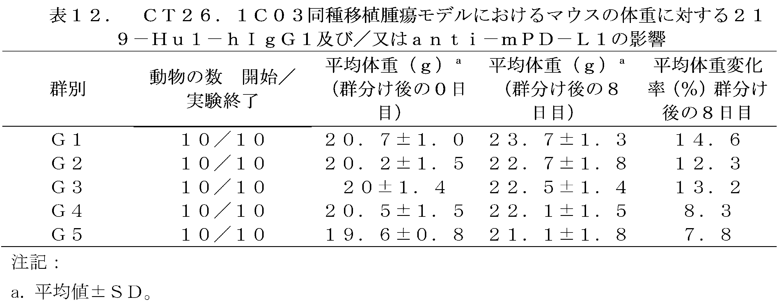
腫瘍接種後18日目に、各群の動物を安楽死させた後、腫瘍を分離して重量を測定し、対照群(G1)と比較して、G2 anti-mPD-L1 10mg/kg群の腫瘍重量は有意に減少し(TGITW=37.3%、P値0.0087)、G3 219-Hu1-hIgG1 10mg/kg群の腫瘍重量は対照群よりも低かったが、統計的差異はなく(TGITW=25.9%、P値0.0830)、G4 219-Hu1-hIgG1 30mg/kg群の腫瘍重量も対照群よりも有意に低く(TGITW=47.9%、P値0.0013)、G5 219-Hu1-hIgG1 10mg/kg+anti-mPD-L1 10mg/kg併用群の腫瘍重量も対照群よりも有意に低く(TGITW=43.0%、P値0.0042)、219-Hu1-hIgG1の2つの用量での腫瘍重量は用量依存的な効果を示し、腫瘍重量は用量の増加とともに減少し、併用群の腫瘍重量は各単剤群(G2、G3)の腫瘍重量よりも小さかったが、統計的差異はなかった(図21、表13)。 On the 18th day after tumor inoculation, the animals in each group were euthanized, and the tumors were isolated and weighed. Compared with the control group (G1), the tumor weight of the G2 anti-mPD-L1 10 mg/kg group was significantly reduced (TGI TW = 37.3%, P value 0.0087), the tumor weight of the G3 219-Hu1-hIgG1 10 mg/kg group was lower than the control group, but there was no statistical difference (TGI TW = 25.9%, P value 0.0830), the tumor weight of the G4 219-Hu1-hIgG1 30 mg/kg group was also significantly lower than the control group (TGI TW = 47.9%, P value 0.0013), and the tumor weight of the G5 219-Hu1-hIgG1 10 mg/kg + anti-mPD-L1 The tumor weight in the 10 mg/kg combination group was also significantly lower than that in the control group (TGI TW =43.0%, P value 0.0042). The tumor weights at the two doses of 219-Hu1-hIgG1 showed a dose-dependent effect, with tumor weight decreasing with increasing dose, and the tumor weight in the combination group was smaller than that in each single-agent group (G2, G3), but there was no statistical difference (Figure 21, Table 13).

Claims (10)
(1)前記重鎖可変領域は、配列番号21、22、23、24及び25から選ばれるいずれかに示されるVH配列
を含み、
(2)前記軽鎖可変領域は、配列番号32に示されるVL配列
を含む、ことを特徴とする、
TNFR2に特異的に結合するヒト化抗体又は抗原結合断片。 1. A humanized antibody or antigen-binding fragment that specifically binds to TNFR2, the antibody or antigen-binding fragment comprising a heavy chain variable region and a light chain variable region,
(1) The heavy chain variable region is a VH sequence selected from SEQ ID NOs: 21, 22, 23, 24, and 25.
Including,
(2) The light chain variable region has the VL sequence shown in SEQ ID NO: 32.
Characterized in that it comprises
A humanized antibody or antigen-binding fragment that specifically binds to TNFR2.
請求項1に記載の抗体又は抗原結合断片。 comprising a human heavy chain constant region and/or a human light chain constant region,
The antibody or antigen-binding fragment of claim 1.
請求項1に記載の抗体又は抗原結合断片。 The antibody or antigen-binding fragment thereof is conjugated to a therapeutic agent or a tracer.
The antibody or antigen-binding fragment of claim 1.
前記他の抗原は、CD3、CD4、CD5、CD8、CD14、CD15、CD16、CD16A、CD19、CD20、CD21、CD22、CD23、CD25、CD33、CD37、CD38、CD40、CD40L、CD46、CD52、CD54、CD70、CD74、CD80、CD86、CD126、CD138、B7、PD-1、PD-L1、PD-2、CTLA4、PRVIG、TIGHT、HAS、CLDN18.2、MSLN、MUC、Ia、HLA-DR、テネイシン、EGFR、VEGF、P1GF、ED-Bフィブロネクチン、癌遺伝子産物、IL-2、IL-6、IL-15、IL-21、TRAIL-R1又はTRAIL-R2から選ばれる、ことを特徴とする、
多重特異性抗原結合分子。 The antibody or antigen-binding fragment of claim 1 , wherein the second antigen-binding module specifically binds to an antigen other than TNFR2 or binds to a different TNFR2 epitope than the first antigen-binding module ;
The other antigens include CD3, CD4, CD5, CD8, CD14, CD15, CD16, CD16A, CD19, CD20, CD21, CD22, CD23, CD25, CD33, CD37, CD38, CD40, CD40L, CD46, CD52, CD54, CD70, CD74, CD80, CD86, CD126, CD138, B7, PD-1, P D-L1, PD-2, CTLA4, PRVIG, TIGHT, HAS, CLDN18.2, MSLN, MUC, Ia, HLA-DR, tenascin, EGFR, VEGF, P1GF, ED-B fibronectin, oncogene products, IL-2, IL-6, IL-15, IL-21, TRAIL-R1, or TRAIL-R2 ;
Multispecific antigen binding molecules.
キメラ抗原受容体(CAR)。 The antibody or antigen-binding fragment of the present invention comprises an extracellular antigen-binding domain, a transmembrane domain, and an intracellular signaling domain, and the extracellular antigen-binding domain comprises a TNFR2 humanized antibody or antigen-binding fragment of the present invention according to any one of claims 1 to 3 .
Chimeric antigen receptors (CARs).
免疫エフェクター細胞。 6. An immune effector cell, characterized in that the immune effector cell comprises the chimeric antigen receptor of claim 5 or comprises a nucleic acid fragment encoding the chimeric antigen receptor of claim 5 .
Immune effector cells.
単離された核酸断片。 Characterized in that it encodes the antibody or antigen-binding fragment according to any one of claims 1 to 3 .
Isolated nucleic acid fragments.
方法。 10. A method of preparing an immune effector cell in vitro according to claim 7, said method comprising introducing into said immune effector cell a nucleic acid fragment encoding a CAR according to claim 6, and optionally said method further comprising initiating expression of the CAR according to claim 5 by said immune effector cell.
method.
医薬組成物。 A pharmaceutical composition, characterized in that the composition comprises an antibody or antigen-binding fragment according to any one of claims 1 to 3 , a multispecific antigen-binding molecule according to claim 4 , a chimeric antigen receptor according to claim 5 , an immune effector cell according to claim 6 , or a nucleic acid fragment according to claim 7 .
Pharmaceutical compositions.
前記疾患は、癌又は自己免疫疾患である、ことを特徴とする、
薬物。 A drug for treating and/or preventing a disease associated with immune abnormality, comprising the antibody or antigen-binding fragment according to any one of claims 1 to 3 , the multispecific antigen-binding molecule according to claim 4 , the chimeric antigen receptor according to claim 5 , the immune effector cell according to claim 6, or the nucleic acid fragment according to claim 7 ,
The disease is cancer or an autoimmune disease.
Drugs .
Applications Claiming Priority (5)
| Application Number | Priority Date | Filing Date | Title |
|---|---|---|---|
| CN202110140980.5 | 2021-01-29 | ||
| CN202110140980 | 2021-01-29 | ||
| CN202111016307.7 | 2021-08-31 | ||
| CN202111016307 | 2021-08-31 | ||
| PCT/CN2022/074228 WO2022161425A1 (en) | 2021-01-29 | 2022-01-27 | Humanized antibody against tnfr2 and use thereof |
Publications (2)
| Publication Number | Publication Date |
|---|---|
| JP2024504820A JP2024504820A (en) | 2024-02-01 |
| JP7635401B2 true JP7635401B2 (en) | 2025-02-25 |
Family
ID=82653002
Family Applications (1)
| Application Number | Title | Priority Date | Filing Date |
|---|---|---|---|
| JP2023546194A Active JP7635401B2 (en) | 2021-01-29 | 2022-01-27 | Anti-TNFR2 humanized antibodies and uses thereof |
Country Status (9)
| Country | Link |
|---|---|
| US (1) | US20240109974A1 (en) |
| EP (1) | EP4286411A4 (en) |
| JP (1) | JP7635401B2 (en) |
| KR (1) | KR20230132524A (en) |
| CN (1) | CN117120469A (en) |
| AU (1) | AU2022212842A1 (en) |
| CA (1) | CA3206910A1 (en) |
| TW (1) | TWI835061B (en) |
| WO (1) | WO2022161425A1 (en) |
Families Citing this family (9)
| Publication number | Priority date | Publication date | Assignee | Title |
|---|---|---|---|---|
| CN118369117A (en) * | 2021-12-28 | 2024-07-19 | 山东先声生物制药有限公司 | An anti-TNFR2 antibody pharmaceutical composition |
| CN119768434A (en) * | 2022-08-19 | 2025-04-04 | 盛禾(中国)生物制药有限公司 | A bispecific antibody and its application |
| US20260049151A1 (en) * | 2022-08-19 | 2026-02-19 | Shenghe (China) Biopharmaceutical Co., Ltd. | Bispecific antibody binding to tnfr2 and 4-1bb |
| US20260055201A1 (en) * | 2022-08-19 | 2026-02-26 | Shenghe (China) Biopharmaceutical Co., Ltd. | Bispecific antibody and use thereof |
| WO2024233258A1 (en) * | 2023-05-05 | 2024-11-14 | Cureimmune Therapeutics Inc. | Anti-tnfr2 antibodies and uses thereof |
| CN121969644A (en) * | 2023-09-07 | 2026-05-01 | 盛禾(中国)生物制药有限公司 | Anti-TNFR 2 antibody and preparation method and application thereof |
| WO2025220804A1 (en) * | 2024-04-18 | 2025-10-23 | 주식회사 큐로셀 | Novel antibody against cd5 and uses thereof |
| CN119591711B (en) * | 2024-11-12 | 2026-01-13 | 澳门大学 | An inhibitory TNFR2 single-domain antibody and its application |
| CN119798439A (en) * | 2024-12-23 | 2025-04-11 | 澳门大学 | A monoclonal antibody library targeting TNFR2 and its application |
Citations (2)
| Publication number | Priority date | Publication date | Assignee | Title |
|---|---|---|---|---|
| WO2020089474A1 (en) | 2018-11-01 | 2020-05-07 | Bioinvent International Ab | Novel antagonistic anti tnfr2 antibody molecules |
| JP7378509B2 (en) | 2019-08-02 | 2023-11-13 | チャンスー シムサー ファーマシューティカル カンパニー リミテッド | Anti-TNFR2 antibodies and their uses |
Family Cites Families (3)
| Publication number | Priority date | Publication date | Assignee | Title |
|---|---|---|---|---|
| CA3004830A1 (en) * | 2015-11-11 | 2017-05-18 | Opi Vi- Ip Holdco Llc | Composition and methods for anti-tnfr2 antibodies |
| US11859002B2 (en) * | 2016-05-13 | 2024-01-02 | The General Hospital Corporation | Antagonistic anti-tumor necrosis factor receptor 2 antibodies |
| CN107541499B (en) * | 2017-07-27 | 2020-04-14 | 山东兴瑞生物科技有限公司 | Preparation and application of CIK of TNFR2 in targeted immunodetection point |
-
2022
- 2022-01-27 EP EP22745298.4A patent/EP4286411A4/en active Pending
- 2022-01-27 AU AU2022212842A patent/AU2022212842A1/en active Pending
- 2022-01-27 TW TW111103685A patent/TWI835061B/en active
- 2022-01-27 CA CA3206910A patent/CA3206910A1/en active Pending
- 2022-01-27 JP JP2023546194A patent/JP7635401B2/en active Active
- 2022-01-27 WO PCT/CN2022/074228 patent/WO2022161425A1/en not_active Ceased
- 2022-01-27 CN CN202280011902.8A patent/CN117120469A/en active Pending
- 2022-01-27 US US18/274,845 patent/US20240109974A1/en active Pending
- 2022-01-27 KR KR1020237027516A patent/KR20230132524A/en active Pending
Patent Citations (2)
| Publication number | Priority date | Publication date | Assignee | Title |
|---|---|---|---|---|
| WO2020089474A1 (en) | 2018-11-01 | 2020-05-07 | Bioinvent International Ab | Novel antagonistic anti tnfr2 antibody molecules |
| JP7378509B2 (en) | 2019-08-02 | 2023-11-13 | チャンスー シムサー ファーマシューティカル カンパニー リミテッド | Anti-TNFR2 antibodies and their uses |
Also Published As
| Publication number | Publication date |
|---|---|
| TW202237658A (en) | 2022-10-01 |
| AU2022212842A1 (en) | 2023-08-31 |
| US20240109974A1 (en) | 2024-04-04 |
| WO2022161425A1 (en) | 2022-08-04 |
| CN117120469A (en) | 2023-11-24 |
| KR20230132524A (en) | 2023-09-15 |
| EP4286411A4 (en) | 2025-06-25 |
| EP4286411A1 (en) | 2023-12-06 |
| JP2024504820A (en) | 2024-02-01 |
| CA3206910A1 (en) | 2022-08-04 |
| TWI835061B (en) | 2024-03-11 |
Similar Documents
| Publication | Publication Date | Title |
|---|---|---|
| JP7635401B2 (en) | Anti-TNFR2 humanized antibodies and uses thereof | |
| KR102821105B1 (en) | Anti-TNFR2 antibodies and uses thereof | |
| CN110066338B (en) | Bispecific IgG antibodies as T cell engagers | |
| US20200024358A1 (en) | Trispecific antigen binding proteins | |
| JP7727086B2 (en) | Anti-PVRIG/anti-TIGIT bispecific antibodies and applications | |
| AU2018360800A1 (en) | Chimeric antigen receptors specific for B-cell maturation antigen (BCMA) | |
| CN110214150A (en) | Combination therapy of chimeric antigen receptor and PD-1 inhibitor | |
| KR20170128234A (en) | Antibodies specific for ROR1 and chimeric antigen receptors | |
| TW201619200A (en) | Humanized anti-OX40 antibodies and uses thereof | |
| JP7646768B2 (en) | Anti-human programmed cell death ligand-1 (PD-L1) antibodies and uses thereof | |
| CA3008440A1 (en) | Combination therapy comprising a superagonistic antibody against interleukin-2 and a checkpoint blockade agent | |
| WO2023125888A1 (en) | Gprc5d antibody and application thereof | |
| TW202313689A (en) | Anti-pvrig/anti-tigit bispecific antibody and applications | |
| US20250333527A1 (en) | Antibodies capable of binding to ox40, variants thereof and uses thereof | |
| WO2023186100A1 (en) | Anti-ror1 antibody and use thereof | |
| CN115181180B (en) | Antibodies against human programmed death ligand-1 (PD-L1) and their applications | |
| US20250339529A1 (en) | B-cell maturation antigen (bcma) chimeric antigen receptor invariant natural killer t cells and uses thereof | |
| WO2026008059A1 (en) | Anti-dll3 constructs and uses thereof | |
| WO2024165049A1 (en) | Anti-cdh6 antibody and use thereof | |
| HK40007657A (en) | Bispecific igg antibodies as t cell engagers | |
| EA040393B1 (en) | BISPECIFIC IgG ANTIBODIES AS T-CELL RECRUITERS |
Legal Events
| Date | Code | Title | Description |
|---|---|---|---|
| A521 | Request for written amendment filed |
Free format text: JAPANESE INTERMEDIATE CODE: A523 Effective date: 20230807 Free format text: JAPANESE INTERMEDIATE CODE: A523 Effective date: 20230728 |
|
| A621 | Written request for application examination |
Free format text: JAPANESE INTERMEDIATE CODE: A621 Effective date: 20230807 |
|
| A131 | Notification of reasons for refusal |
Free format text: JAPANESE INTERMEDIATE CODE: A131 Effective date: 20240723 |
|
| A521 | Request for written amendment filed |
Free format text: JAPANESE INTERMEDIATE CODE: A523 Effective date: 20241023 |
|
| TRDD | Decision of grant or rejection written | ||
| A01 | Written decision to grant a patent or to grant a registration (utility model) |
Free format text: JAPANESE INTERMEDIATE CODE: A01 Effective date: 20250121 |
|
| A61 | First payment of annual fees (during grant procedure) |
Free format text: JAPANESE INTERMEDIATE CODE: A61 Effective date: 20250212 |
|
| R150 | Certificate of patent or registration of utility model |
Ref document number: 7635401 Country of ref document: JP Free format text: JAPANESE INTERMEDIATE CODE: R150 |