JPWO2002024903A1 - Testing methods for allergic diseases - Google Patents
Testing methods for allergic diseases Download PDFInfo
- Publication number
- JPWO2002024903A1 JPWO2002024903A1 JP2002529497A JP2002529497A JPWO2002024903A1 JP WO2002024903 A1 JPWO2002024903 A1 JP WO2002024903A1 JP 2002529497 A JP2002529497 A JP 2002529497A JP 2002529497 A JP2002529497 A JP 2002529497A JP WO2002024903 A1 JPWO2002024903 A1 JP WO2002024903A1
- Authority
- JP
- Japan
- Prior art keywords
- polynucleotide
- seq
- gene
- protein
- nucleotide sequence
- Prior art date
- Legal status (The legal status is an assumption and is not a legal conclusion. Google has not performed a legal analysis and makes no representation as to the accuracy of the status listed.)
- Pending
Links
- 208000026935 allergic disease Diseases 0.000 title claims abstract description 73
- 238000012360 testing method Methods 0.000 title claims abstract description 52
- 108090000623 proteins and genes Proteins 0.000 claims abstract description 304
- 230000014509 gene expression Effects 0.000 claims abstract description 165
- 238000000034 method Methods 0.000 claims abstract description 144
- 210000003979 eosinophil Anatomy 0.000 claims abstract description 86
- 150000001875 compounds Chemical class 0.000 claims abstract description 71
- 206010012438 Dermatitis atopic Diseases 0.000 claims abstract description 51
- 201000008937 atopic dermatitis Diseases 0.000 claims abstract description 51
- 238000012216 screening Methods 0.000 claims abstract description 29
- 230000001965 increasing effect Effects 0.000 claims abstract description 21
- 108091033319 polynucleotide Proteins 0.000 claims description 142
- 102000040430 polynucleotide Human genes 0.000 claims description 142
- 239000002157 polynucleotide Substances 0.000 claims description 142
- 102000004169 proteins and genes Human genes 0.000 claims description 110
- 125000003729 nucleotide group Chemical group 0.000 claims description 74
- 239000002773 nucleotide Substances 0.000 claims description 72
- 210000004027 cell Anatomy 0.000 claims description 59
- 241001465754 Metazoa Species 0.000 claims description 53
- 125000003275 alpha amino acid group Chemical group 0.000 claims description 41
- 230000000694 effects Effects 0.000 claims description 38
- 239000002299 complementary DNA Substances 0.000 claims description 33
- 230000001105 regulatory effect Effects 0.000 claims description 29
- 238000013518 transcription Methods 0.000 claims description 27
- 230000035897 transcription Effects 0.000 claims description 27
- FWMNVWWHGCHHJJ-SKKKGAJSSA-N 4-amino-1-[(2r)-6-amino-2-[[(2r)-2-[[(2r)-2-[[(2r)-2-amino-3-phenylpropanoyl]amino]-3-phenylpropanoyl]amino]-4-methylpentanoyl]amino]hexanoyl]piperidine-4-carboxylic acid Chemical compound C([C@H](C(=O)N[C@H](CC(C)C)C(=O)N[C@H](CCCCN)C(=O)N1CCC(N)(CC1)C(O)=O)NC(=O)[C@H](N)CC=1C=CC=CC=1)C1=CC=CC=C1 FWMNVWWHGCHHJJ-SKKKGAJSSA-N 0.000 claims description 26
- 108020004414 DNA Proteins 0.000 claims description 25
- 210000000265 leukocyte Anatomy 0.000 claims description 25
- 230000000295 complement effect Effects 0.000 claims description 23
- 108091034117 Oligonucleotide Proteins 0.000 claims description 19
- 239000013598 vector Substances 0.000 claims description 19
- 206010020751 Hypersensitivity Diseases 0.000 claims description 16
- 238000009396 hybridization Methods 0.000 claims description 15
- 230000009261 transgenic effect Effects 0.000 claims description 15
- 108700008625 Reporter Genes Proteins 0.000 claims description 14
- 150000001413 amino acids Chemical class 0.000 claims description 13
- 108091026890 Coding region Proteins 0.000 claims description 12
- 239000000126 substance Substances 0.000 claims description 12
- 230000007815 allergy Effects 0.000 claims description 10
- 238000010998 test method Methods 0.000 claims description 10
- 239000003814 drug Substances 0.000 claims description 9
- 229940126585 therapeutic drug Drugs 0.000 claims description 9
- 239000003153 chemical reaction reagent Substances 0.000 claims description 8
- 108090000765 processed proteins & peptides Proteins 0.000 claims description 6
- 238000012258 culturing Methods 0.000 claims description 5
- 230000002829 reductive effect Effects 0.000 claims description 5
- 229940124597 therapeutic agent Drugs 0.000 claims description 5
- 239000004480 active ingredient Substances 0.000 claims description 4
- 230000008105 immune reaction Effects 0.000 claims description 3
- 238000004519 manufacturing process Methods 0.000 claims description 2
- 208000015891 sexual disease Diseases 0.000 claims 1
- 230000005713 exacerbation Effects 0.000 abstract description 12
- 230000001225 therapeutic effect Effects 0.000 abstract description 5
- 239000000523 sample Substances 0.000 description 34
- 239000013566 allergen Substances 0.000 description 20
- 108091032973 (ribonucleotides)n+m Proteins 0.000 description 18
- 108020004999 messenger RNA Proteins 0.000 description 17
- 238000006243 chemical reaction Methods 0.000 description 16
- 210000004369 blood Anatomy 0.000 description 12
- 239000008280 blood Substances 0.000 description 12
- 238000001514 detection method Methods 0.000 description 12
- 238000005259 measurement Methods 0.000 description 12
- 239000012634 fragment Substances 0.000 description 10
- 210000002966 serum Anatomy 0.000 description 10
- 108010085238 Actins Proteins 0.000 description 9
- 230000000172 allergic effect Effects 0.000 description 9
- 208000010668 atopic eczema Diseases 0.000 description 9
- 239000000047 product Substances 0.000 description 9
- 239000000203 mixture Substances 0.000 description 8
- 102000007469 Actins Human genes 0.000 description 7
- 238000004458 analytical method Methods 0.000 description 7
- 201000010099 disease Diseases 0.000 description 7
- 208000037265 diseases, disorders, signs and symptoms Diseases 0.000 description 7
- 235000013601 eggs Nutrition 0.000 description 7
- 238000002372 labelling Methods 0.000 description 7
- 238000002360 preparation method Methods 0.000 description 7
- 239000000243 solution Substances 0.000 description 7
- 238000011282 treatment Methods 0.000 description 7
- 210000001671 embryonic stem cell Anatomy 0.000 description 6
- 102000039446 nucleic acids Human genes 0.000 description 6
- 108020004707 nucleic acids Proteins 0.000 description 6
- 150000007523 nucleic acids Chemical class 0.000 description 6
- 210000005259 peripheral blood Anatomy 0.000 description 6
- 239000011886 peripheral blood Substances 0.000 description 6
- 208000024891 symptom Diseases 0.000 description 6
- 238000012408 PCR amplification Methods 0.000 description 5
- 230000009471 action Effects 0.000 description 5
- 208000030961 allergic reaction Diseases 0.000 description 5
- 239000000427 antigen Substances 0.000 description 5
- 108091007433 antigens Proteins 0.000 description 5
- 102000036639 antigens Human genes 0.000 description 5
- 238000003556 assay Methods 0.000 description 5
- 229940000406 drug candidate Drugs 0.000 description 5
- 239000013604 expression vector Substances 0.000 description 5
- 230000001605 fetal effect Effects 0.000 description 5
- 238000002744 homologous recombination Methods 0.000 description 5
- 230000006801 homologous recombination Effects 0.000 description 5
- 238000000338 in vitro Methods 0.000 description 5
- 238000012544 monitoring process Methods 0.000 description 5
- 230000002441 reversible effect Effects 0.000 description 5
- 108020004705 Codon Proteins 0.000 description 4
- 230000004544 DNA amplification Effects 0.000 description 4
- NTYJJOPFIAHURM-UHFFFAOYSA-N Histamine Chemical compound NCCC1=CN=CN1 NTYJJOPFIAHURM-UHFFFAOYSA-N 0.000 description 4
- 241000699670 Mus sp. Species 0.000 description 4
- 108091028043 Nucleic acid sequence Proteins 0.000 description 4
- 239000011324 bead Substances 0.000 description 4
- 210000000601 blood cell Anatomy 0.000 description 4
- 238000011161 development Methods 0.000 description 4
- 230000018109 developmental process Effects 0.000 description 4
- 238000003745 diagnosis Methods 0.000 description 4
- 229940079593 drug Drugs 0.000 description 4
- 238000010195 expression analysis Methods 0.000 description 4
- 230000002163 immunogen Effects 0.000 description 4
- 238000001727 in vivo Methods 0.000 description 4
- 210000004185 liver Anatomy 0.000 description 4
- 210000001616 monocyte Anatomy 0.000 description 4
- 230000035772 mutation Effects 0.000 description 4
- 210000000440 neutrophil Anatomy 0.000 description 4
- 230000036961 partial effect Effects 0.000 description 4
- 230000006798 recombination Effects 0.000 description 4
- 238000007619 statistical method Methods 0.000 description 4
- 230000002103 transcriptional effect Effects 0.000 description 4
- 238000013519 translation Methods 0.000 description 4
- 238000011144 upstream manufacturing Methods 0.000 description 4
- 238000005406 washing Methods 0.000 description 4
- 241000283707 Capra Species 0.000 description 3
- IAZDPXIOMUYVGZ-UHFFFAOYSA-N Dimethylsulphoxide Chemical compound CS(C)=O IAZDPXIOMUYVGZ-UHFFFAOYSA-N 0.000 description 3
- 238000002965 ELISA Methods 0.000 description 3
- 229920001917 Ficoll Polymers 0.000 description 3
- 101000917858 Homo sapiens Low affinity immunoglobulin gamma Fc region receptor III-A Proteins 0.000 description 3
- 101000917839 Homo sapiens Low affinity immunoglobulin gamma Fc region receptor III-B Proteins 0.000 description 3
- 102100029185 Low affinity immunoglobulin gamma Fc region receptor III-B Human genes 0.000 description 3
- 239000004698 Polyethylene Substances 0.000 description 3
- 241000700159 Rattus Species 0.000 description 3
- 210000001744 T-lymphocyte Anatomy 0.000 description 3
- 241000251539 Vertebrata <Metazoa> Species 0.000 description 3
- 102000005421 acetyltransferase Human genes 0.000 description 3
- 230000003321 amplification Effects 0.000 description 3
- 238000010171 animal model Methods 0.000 description 3
- 230000000692 anti-sense effect Effects 0.000 description 3
- 210000004556 brain Anatomy 0.000 description 3
- 238000005119 centrifugation Methods 0.000 description 3
- 238000010367 cloning Methods 0.000 description 3
- 238000012937 correction Methods 0.000 description 3
- 239000008187 granular material Substances 0.000 description 3
- 230000028993 immune response Effects 0.000 description 3
- 239000006166 lysate Substances 0.000 description 3
- 239000003550 marker Substances 0.000 description 3
- 239000012528 membrane Substances 0.000 description 3
- 238000003199 nucleic acid amplification method Methods 0.000 description 3
- 210000000056 organ Anatomy 0.000 description 3
- 230000004952 protein activity Effects 0.000 description 3
- 238000005215 recombination Methods 0.000 description 3
- 238000011160 research Methods 0.000 description 3
- 238000003757 reverse transcription PCR Methods 0.000 description 3
- 210000000952 spleen Anatomy 0.000 description 3
- BZTDTCNHAFUJOG-UHFFFAOYSA-N 6-carboxyfluorescein Chemical compound C12=CC=C(O)C=C2OC2=CC(O)=CC=C2C11OC(=O)C2=CC=C(C(=O)O)C=C21 BZTDTCNHAFUJOG-UHFFFAOYSA-N 0.000 description 2
- 108700028369 Alleles Proteins 0.000 description 2
- 208000012657 Atopic disease Diseases 0.000 description 2
- 206010003645 Atopy Diseases 0.000 description 2
- FERIUCNNQQJTOY-UHFFFAOYSA-N Butyric acid Chemical compound CCCC(O)=O FERIUCNNQQJTOY-UHFFFAOYSA-N 0.000 description 2
- 241000218645 Cedrus Species 0.000 description 2
- HEDRZPFGACZZDS-UHFFFAOYSA-N Chloroform Chemical compound ClC(Cl)Cl HEDRZPFGACZZDS-UHFFFAOYSA-N 0.000 description 2
- 108010017826 DNA Polymerase I Proteins 0.000 description 2
- 102000004594 DNA Polymerase I Human genes 0.000 description 2
- 108010053770 Deoxyribonucleases Proteins 0.000 description 2
- 102000016911 Deoxyribonucleases Human genes 0.000 description 2
- 229920002307 Dextran Polymers 0.000 description 2
- 102000004190 Enzymes Human genes 0.000 description 2
- 108090000790 Enzymes Proteins 0.000 description 2
- ZHNUHDYFZUAESO-UHFFFAOYSA-N Formamide Chemical compound NC=O ZHNUHDYFZUAESO-UHFFFAOYSA-N 0.000 description 2
- 241000238631 Hexapoda Species 0.000 description 2
- KFZMGEQAYNKOFK-UHFFFAOYSA-N Isopropanol Chemical compound CC(C)O KFZMGEQAYNKOFK-UHFFFAOYSA-N 0.000 description 2
- 241000124008 Mammalia Species 0.000 description 2
- 229930193140 Neomycin Natural products 0.000 description 2
- 241000283973 Oryctolagus cuniculus Species 0.000 description 2
- 239000002202 Polyethylene glycol Substances 0.000 description 2
- 108020002494 acetyltransferase Proteins 0.000 description 2
- 230000004913 activation Effects 0.000 description 2
- 210000000628 antibody-producing cell Anatomy 0.000 description 2
- 208000006673 asthma Diseases 0.000 description 2
- 210000003719 b-lymphocyte Anatomy 0.000 description 2
- 210000003651 basophil Anatomy 0.000 description 2
- 238000010170 biological method Methods 0.000 description 2
- 230000035584 blastogenesis Effects 0.000 description 2
- 210000001185 bone marrow Anatomy 0.000 description 2
- 229910000389 calcium phosphate Inorganic materials 0.000 description 2
- 239000001506 calcium phosphate Substances 0.000 description 2
- 235000011010 calcium phosphates Nutrition 0.000 description 2
- 238000003163 cell fusion method Methods 0.000 description 2
- 230000008859 change Effects 0.000 description 2
- 238000007796 conventional method Methods 0.000 description 2
- 238000004520 electroporation Methods 0.000 description 2
- 230000007613 environmental effect Effects 0.000 description 2
- 210000003617 erythrocyte membrane Anatomy 0.000 description 2
- 238000002474 experimental method Methods 0.000 description 2
- 238000000605 extraction Methods 0.000 description 2
- 239000007850 fluorescent dye Substances 0.000 description 2
- 210000003714 granulocyte Anatomy 0.000 description 2
- 229960001340 histamine Drugs 0.000 description 2
- 230000001900 immune effect Effects 0.000 description 2
- 230000007233 immunological mechanism Effects 0.000 description 2
- 230000006872 improvement Effects 0.000 description 2
- 230000004968 inflammatory condition Effects 0.000 description 2
- 230000003902 lesion Effects 0.000 description 2
- 210000001165 lymph node Anatomy 0.000 description 2
- 210000004698 lymphocyte Anatomy 0.000 description 2
- 230000007246 mechanism Effects 0.000 description 2
- 238000000520 microinjection Methods 0.000 description 2
- 238000002156 mixing Methods 0.000 description 2
- 238000012986 modification Methods 0.000 description 2
- 230000004048 modification Effects 0.000 description 2
- 238000010369 molecular cloning Methods 0.000 description 2
- 229960004927 neomycin Drugs 0.000 description 2
- BOPGDPNILDQYTO-NNYOXOHSSA-N nicotinamide-adenine dinucleotide Chemical compound C1=CCC(C(=O)N)=CN1[C@H]1[C@H](O)[C@H](O)[C@@H](COP(O)(=O)OP(O)(=O)OC[C@@H]2[C@H]([C@@H](O)[C@@H](O2)N2C3=NC=NC(N)=C3N=C2)O)O1 BOPGDPNILDQYTO-NNYOXOHSSA-N 0.000 description 2
- 229930027945 nicotinamide-adenine dinucleotide Natural products 0.000 description 2
- 210000004940 nucleus Anatomy 0.000 description 2
- 239000013610 patient sample Substances 0.000 description 2
- 238000000053 physical method Methods 0.000 description 2
- 210000002826 placenta Anatomy 0.000 description 2
- 229920001223 polyethylene glycol Polymers 0.000 description 2
- 230000002250 progressing effect Effects 0.000 description 2
- 238000000746 purification Methods 0.000 description 2
- 238000004445 quantitative analysis Methods 0.000 description 2
- 238000003753 real-time PCR Methods 0.000 description 2
- 238000003756 stirring Methods 0.000 description 2
- 239000000758 substrate Substances 0.000 description 2
- 230000002194 synthesizing effect Effects 0.000 description 2
- ABZLKHKQJHEPAX-UHFFFAOYSA-N tetramethylrhodamine Chemical compound C=12C=CC(N(C)C)=CC2=[O+]C2=CC(N(C)C)=CC=C2C=1C1=CC=CC=C1C([O-])=O ABZLKHKQJHEPAX-UHFFFAOYSA-N 0.000 description 2
- 210000001541 thymus gland Anatomy 0.000 description 2
- 210000001519 tissue Anatomy 0.000 description 2
- 230000001131 transforming effect Effects 0.000 description 2
- QORWJWZARLRLPR-UHFFFAOYSA-H tricalcium bis(phosphate) Chemical compound [Ca+2].[Ca+2].[Ca+2].[O-]P([O-])([O-])=O.[O-]P([O-])([O-])=O QORWJWZARLRLPR-UHFFFAOYSA-H 0.000 description 2
- BFSVOASYOCHEOV-UHFFFAOYSA-N 2-diethylaminoethanol Chemical compound CCN(CC)CCO BFSVOASYOCHEOV-UHFFFAOYSA-N 0.000 description 1
- YUDPTGPSBJVHCN-DZQJYWQESA-N 4-methylumbelliferyl beta-D-galactoside Chemical compound C1=CC=2C(C)=CC(=O)OC=2C=C1O[C@@H]1O[C@H](CO)[C@H](O)[C@H](O)[C@H]1O YUDPTGPSBJVHCN-DZQJYWQESA-N 0.000 description 1
- FVFVNNKYKYZTJU-UHFFFAOYSA-N 6-chloro-1,3,5-triazine-2,4-diamine Chemical compound NC1=NC(N)=NC(Cl)=N1 FVFVNNKYKYZTJU-UHFFFAOYSA-N 0.000 description 1
- 208000035285 Allergic Seasonal Rhinitis Diseases 0.000 description 1
- 241000219195 Arabidopsis thaliana Species 0.000 description 1
- 241000283690 Bos taurus Species 0.000 description 1
- 102000017420 CD3 protein, epsilon/gamma/delta subunit Human genes 0.000 description 1
- 241000282472 Canis lupus familiaris Species 0.000 description 1
- 108700010070 Codon Usage Proteins 0.000 description 1
- 108020004635 Complementary DNA Proteins 0.000 description 1
- 102000053602 DNA Human genes 0.000 description 1
- 238000000018 DNA microarray Methods 0.000 description 1
- 102000004163 DNA-directed RNA polymerases Human genes 0.000 description 1
- 108090000626 DNA-directed RNA polymerases Proteins 0.000 description 1
- 201000004624 Dermatitis Diseases 0.000 description 1
- 241000255601 Drosophila melanogaster Species 0.000 description 1
- 241000196324 Embryophyta Species 0.000 description 1
- 206010014958 Eosinophilic leukaemia Diseases 0.000 description 1
- 241000588724 Escherichia coli Species 0.000 description 1
- 108700039887 Essential Genes Proteins 0.000 description 1
- 108060002716 Exonuclease Proteins 0.000 description 1
- 108700039691 Genetic Promoter Regions Proteins 0.000 description 1
- 238000002738 Giemsa staining Methods 0.000 description 1
- 241000282412 Homo Species 0.000 description 1
- 206010048643 Hypereosinophilic syndrome Diseases 0.000 description 1
- 206010061218 Inflammation Diseases 0.000 description 1
- 108010002616 Interleukin-5 Proteins 0.000 description 1
- 102100035877 Monocyte differentiation antigen CD14 Human genes 0.000 description 1
- 241000711408 Murine respirovirus Species 0.000 description 1
- 241000699666 Mus <mouse, genus> Species 0.000 description 1
- 101710202061 N-acetyltransferase Proteins 0.000 description 1
- 241000244206 Nematoda Species 0.000 description 1
- 238000000636 Northern blotting Methods 0.000 description 1
- 108010038807 Oligopeptides Proteins 0.000 description 1
- 102000015636 Oligopeptides Human genes 0.000 description 1
- 241001494479 Pecora Species 0.000 description 1
- 206010035226 Plasma cell myeloma Diseases 0.000 description 1
- 108010021757 Polynucleotide 5'-Hydroxyl-Kinase Proteins 0.000 description 1
- 102000008422 Polynucleotide 5'-hydroxyl-kinase Human genes 0.000 description 1
- LCTONWCANYUPML-UHFFFAOYSA-M Pyruvate Chemical compound CC(=O)C([O-])=O LCTONWCANYUPML-UHFFFAOYSA-M 0.000 description 1
- 239000013614 RNA sample Substances 0.000 description 1
- 102000007056 Recombinant Fusion Proteins Human genes 0.000 description 1
- 108010008281 Recombinant Fusion Proteins Proteins 0.000 description 1
- 206010039085 Rhinitis allergic Diseases 0.000 description 1
- 102000001712 STAT5 Transcription Factor Human genes 0.000 description 1
- 108010029477 STAT5 Transcription Factor Proteins 0.000 description 1
- 240000004808 Saccharomyces cerevisiae Species 0.000 description 1
- 241000235347 Schizosaccharomyces pombe Species 0.000 description 1
- 108091058545 Secretory proteins Proteins 0.000 description 1
- 102000040739 Secretory proteins Human genes 0.000 description 1
- 229920002684 Sepharose Polymers 0.000 description 1
- DBMJMQXJHONAFJ-UHFFFAOYSA-M Sodium laurylsulphate Chemical compound [Na+].CCCCCCCCCCCCOS([O-])(=O)=O DBMJMQXJHONAFJ-UHFFFAOYSA-M 0.000 description 1
- 241000282887 Suidae Species 0.000 description 1
- 108700026226 TATA Box Proteins 0.000 description 1
- 108010006785 Taq Polymerase Proteins 0.000 description 1
- 101710120037 Toxin CcdB Proteins 0.000 description 1
- 102100033444 Tyrosine-protein kinase JAK2 Human genes 0.000 description 1
- JLCPHMBAVCMARE-UHFFFAOYSA-N [3-[[3-[[3-[[3-[[3-[[3-[[3-[[3-[[3-[[3-[[3-[[5-(2-amino-6-oxo-1H-purin-9-yl)-3-[[3-[[3-[[3-[[3-[[3-[[5-(2-amino-6-oxo-1H-purin-9-yl)-3-[[5-(2-amino-6-oxo-1H-purin-9-yl)-3-hydroxyoxolan-2-yl]methoxy-hydroxyphosphoryl]oxyoxolan-2-yl]methoxy-hydroxyphosphoryl]oxy-5-(5-methyl-2,4-dioxopyrimidin-1-yl)oxolan-2-yl]methoxy-hydroxyphosphoryl]oxy-5-(6-aminopurin-9-yl)oxolan-2-yl]methoxy-hydroxyphosphoryl]oxy-5-(6-aminopurin-9-yl)oxolan-2-yl]methoxy-hydroxyphosphoryl]oxy-5-(6-aminopurin-9-yl)oxolan-2-yl]methoxy-hydroxyphosphoryl]oxy-5-(6-aminopurin-9-yl)oxolan-2-yl]methoxy-hydroxyphosphoryl]oxyoxolan-2-yl]methoxy-hydroxyphosphoryl]oxy-5-(5-methyl-2,4-dioxopyrimidin-1-yl)oxolan-2-yl]methoxy-hydroxyphosphoryl]oxy-5-(4-amino-2-oxopyrimidin-1-yl)oxolan-2-yl]methoxy-hydroxyphosphoryl]oxy-5-(5-methyl-2,4-dioxopyrimidin-1-yl)oxolan-2-yl]methoxy-hydroxyphosphoryl]oxy-5-(5-methyl-2,4-dioxopyrimidin-1-yl)oxolan-2-yl]methoxy-hydroxyphosphoryl]oxy-5-(6-aminopurin-9-yl)oxolan-2-yl]methoxy-hydroxyphosphoryl]oxy-5-(6-aminopurin-9-yl)oxolan-2-yl]methoxy-hydroxyphosphoryl]oxy-5-(4-amino-2-oxopyrimidin-1-yl)oxolan-2-yl]methoxy-hydroxyphosphoryl]oxy-5-(4-amino-2-oxopyrimidin-1-yl)oxolan-2-yl]methoxy-hydroxyphosphoryl]oxy-5-(4-amino-2-oxopyrimidin-1-yl)oxolan-2-yl]methoxy-hydroxyphosphoryl]oxy-5-(6-aminopurin-9-yl)oxolan-2-yl]methoxy-hydroxyphosphoryl]oxy-5-(4-amino-2-oxopyrimidin-1-yl)oxolan-2-yl]methyl [5-(6-aminopurin-9-yl)-2-(hydroxymethyl)oxolan-3-yl] hydrogen phosphate Polymers Cc1cn(C2CC(OP(O)(=O)OCC3OC(CC3OP(O)(=O)OCC3OC(CC3O)n3cnc4c3nc(N)[nH]c4=O)n3cnc4c3nc(N)[nH]c4=O)C(COP(O)(=O)OC3CC(OC3COP(O)(=O)OC3CC(OC3COP(O)(=O)OC3CC(OC3COP(O)(=O)OC3CC(OC3COP(O)(=O)OC3CC(OC3COP(O)(=O)OC3CC(OC3COP(O)(=O)OC3CC(OC3COP(O)(=O)OC3CC(OC3COP(O)(=O)OC3CC(OC3COP(O)(=O)OC3CC(OC3COP(O)(=O)OC3CC(OC3COP(O)(=O)OC3CC(OC3COP(O)(=O)OC3CC(OC3COP(O)(=O)OC3CC(OC3COP(O)(=O)OC3CC(OC3COP(O)(=O)OC3CC(OC3COP(O)(=O)OC3CC(OC3CO)n3cnc4c(N)ncnc34)n3ccc(N)nc3=O)n3cnc4c(N)ncnc34)n3ccc(N)nc3=O)n3ccc(N)nc3=O)n3ccc(N)nc3=O)n3cnc4c(N)ncnc34)n3cnc4c(N)ncnc34)n3cc(C)c(=O)[nH]c3=O)n3cc(C)c(=O)[nH]c3=O)n3ccc(N)nc3=O)n3cc(C)c(=O)[nH]c3=O)n3cnc4c3nc(N)[nH]c4=O)n3cnc4c(N)ncnc34)n3cnc4c(N)ncnc34)n3cnc4c(N)ncnc34)n3cnc4c(N)ncnc34)O2)c(=O)[nH]c1=O JLCPHMBAVCMARE-UHFFFAOYSA-N 0.000 description 1
- 238000002835 absorbance Methods 0.000 description 1
- 238000007792 addition Methods 0.000 description 1
- 239000002671 adjuvant Substances 0.000 description 1
- 201000010105 allergic rhinitis Diseases 0.000 description 1
- BFNBIHQBYMNNAN-UHFFFAOYSA-N ammonium sulfate Chemical compound N.N.OS(O)(=O)=O BFNBIHQBYMNNAN-UHFFFAOYSA-N 0.000 description 1
- 229910052921 ammonium sulfate Inorganic materials 0.000 description 1
- 235000011130 ammonium sulphate Nutrition 0.000 description 1
- 238000012197 amplification kit Methods 0.000 description 1
- 210000004102 animal cell Anatomy 0.000 description 1
- 238000002820 assay format Methods 0.000 description 1
- 102000005936 beta-Galactosidase Human genes 0.000 description 1
- 108010005774 beta-Galactosidase Proteins 0.000 description 1
- 210000001124 body fluid Anatomy 0.000 description 1
- 239000010839 body fluid Substances 0.000 description 1
- 239000011449 brick Substances 0.000 description 1
- 206010006451 bronchitis Diseases 0.000 description 1
- 239000002775 capsule Substances 0.000 description 1
- 230000015556 catabolic process Effects 0.000 description 1
- 125000002091 cationic group Chemical group 0.000 description 1
- 230000007910 cell fusion Effects 0.000 description 1
- 108091092328 cellular RNA Proteins 0.000 description 1
- 229960005091 chloramphenicol Drugs 0.000 description 1
- YTRQFSDWAXHJCC-UHFFFAOYSA-N chloroform;phenol Chemical compound ClC(Cl)Cl.OC1=CC=CC=C1 YTRQFSDWAXHJCC-UHFFFAOYSA-N 0.000 description 1
- 238000004587 chromatography analysis Methods 0.000 description 1
- 210000000349 chromosome Anatomy 0.000 description 1
- 208000021668 chronic eosinophilic leukemia Diseases 0.000 description 1
- 238000012790 confirmation Methods 0.000 description 1
- 239000000470 constituent Substances 0.000 description 1
- 239000012228 culture supernatant Substances 0.000 description 1
- 210000004748 cultured cell Anatomy 0.000 description 1
- 230000003247 decreasing effect Effects 0.000 description 1
- 230000007547 defect Effects 0.000 description 1
- 238000006731 degradation reaction Methods 0.000 description 1
- 238000012217 deletion Methods 0.000 description 1
- 230000037430 deletion Effects 0.000 description 1
- 238000002405 diagnostic procedure Methods 0.000 description 1
- 239000003085 diluting agent Substances 0.000 description 1
- 239000012153 distilled water Substances 0.000 description 1
- 239000002552 dosage form Substances 0.000 description 1
- 238000001962 electrophoresis Methods 0.000 description 1
- 210000002308 embryonic cell Anatomy 0.000 description 1
- 239000000839 emulsion Substances 0.000 description 1
- 238000005516 engineering process Methods 0.000 description 1
- 239000003623 enhancer Substances 0.000 description 1
- 210000003743 erythrocyte Anatomy 0.000 description 1
- 238000012869 ethanol precipitation Methods 0.000 description 1
- 102000013165 exonuclease Human genes 0.000 description 1
- 239000000284 extract Substances 0.000 description 1
- 238000001917 fluorescence detection Methods 0.000 description 1
- 238000001943 fluorescence-activated cell sorting Methods 0.000 description 1
- 238000010230 functional analysis Methods 0.000 description 1
- 230000004927 fusion Effects 0.000 description 1
- 238000001502 gel electrophoresis Methods 0.000 description 1
- 230000002068 genetic effect Effects 0.000 description 1
- 239000001963 growth medium Substances 0.000 description 1
- 210000002216 heart Anatomy 0.000 description 1
- 210000005260 human cell Anatomy 0.000 description 1
- 210000004408 hybridoma Anatomy 0.000 description 1
- 238000003384 imaging method Methods 0.000 description 1
- 210000002865 immune cell Anatomy 0.000 description 1
- 210000000987 immune system Anatomy 0.000 description 1
- 238000001114 immunoprecipitation Methods 0.000 description 1
- 238000007901 in situ hybridization Methods 0.000 description 1
- 230000001939 inductive effect Effects 0.000 description 1
- 230000004054 inflammatory process Effects 0.000 description 1
- 208000030603 inherited susceptibility to asthma Diseases 0.000 description 1
- 230000002401 inhibitory effect Effects 0.000 description 1
- 230000005764 inhibitory process Effects 0.000 description 1
- 239000007924 injection Substances 0.000 description 1
- 238000002347 injection Methods 0.000 description 1
- 238000003780 insertion Methods 0.000 description 1
- 230000037431 insertion Effects 0.000 description 1
- 230000003993 interaction Effects 0.000 description 1
- 239000007927 intramuscular injection Substances 0.000 description 1
- 238000010255 intramuscular injection Methods 0.000 description 1
- 239000007928 intraperitoneal injection Substances 0.000 description 1
- 238000004255 ion exchange chromatography Methods 0.000 description 1
- 238000002955 isolation Methods 0.000 description 1
- 210000003734 kidney Anatomy 0.000 description 1
- 210000002429 large intestine Anatomy 0.000 description 1
- 208000032839 leukemia Diseases 0.000 description 1
- 239000003446 ligand Substances 0.000 description 1
- 239000002502 liposome Substances 0.000 description 1
- 210000004072 lung Anatomy 0.000 description 1
- 238000002826 magnetic-activated cell sorting Methods 0.000 description 1
- 210000001161 mammalian embryo Anatomy 0.000 description 1
- 238000002844 melting Methods 0.000 description 1
- 230000008018 melting Effects 0.000 description 1
- 244000005700 microbiome Species 0.000 description 1
- 239000003094 microcapsule Substances 0.000 description 1
- 238000000386 microscopy Methods 0.000 description 1
- 238000013508 migration Methods 0.000 description 1
- 230000005012 migration Effects 0.000 description 1
- 244000309715 mini pig Species 0.000 description 1
- 230000000877 morphologic effect Effects 0.000 description 1
- 201000000050 myeloid neoplasm Diseases 0.000 description 1
- 230000002018 overexpression Effects 0.000 description 1
- 239000002245 particle Substances 0.000 description 1
- 230000008506 pathogenesis Effects 0.000 description 1
- 230000001717 pathogenic effect Effects 0.000 description 1
- 230000007170 pathology Effects 0.000 description 1
- 230000037361 pathway Effects 0.000 description 1
- 239000008194 pharmaceutical composition Substances 0.000 description 1
- 239000000546 pharmaceutical excipient Substances 0.000 description 1
- 239000013612 plasmid Substances 0.000 description 1
- 229920002401 polyacrylamide Polymers 0.000 description 1
- 238000006116 polymerization reaction Methods 0.000 description 1
- 102000054765 polymorphisms of proteins Human genes 0.000 description 1
- 239000000843 powder Substances 0.000 description 1
- 239000002244 precipitate Substances 0.000 description 1
- 238000001556 precipitation Methods 0.000 description 1
- 230000008569 process Effects 0.000 description 1
- 210000001938 protoplast Anatomy 0.000 description 1
- 238000011002 quantification Methods 0.000 description 1
- 108020003175 receptors Proteins 0.000 description 1
- 230000009467 reduction Effects 0.000 description 1
- 230000022532 regulation of transcription, DNA-dependent Effects 0.000 description 1
- 230000004044 response Effects 0.000 description 1
- 108091008146 restriction endonucleases Proteins 0.000 description 1
- 238000005185 salting out Methods 0.000 description 1
- 150000003839 salts Chemical class 0.000 description 1
- 239000013049 sediment Substances 0.000 description 1
- 238000000926 separation method Methods 0.000 description 1
- 238000012163 sequencing technique Methods 0.000 description 1
- 238000013207 serial dilution Methods 0.000 description 1
- 230000019491 signal transduction Effects 0.000 description 1
- 230000011664 signaling Effects 0.000 description 1
- 238000002741 site-directed mutagenesis Methods 0.000 description 1
- 210000002027 skeletal muscle Anatomy 0.000 description 1
- 210000000813 small intestine Anatomy 0.000 description 1
- 239000002904 solvent Substances 0.000 description 1
- 210000001082 somatic cell Anatomy 0.000 description 1
- 238000011895 specific detection Methods 0.000 description 1
- 230000006641 stabilisation Effects 0.000 description 1
- 238000011105 stabilization Methods 0.000 description 1
- 238000010186 staining Methods 0.000 description 1
- 150000003431 steroids Chemical class 0.000 description 1
- 230000000638 stimulation Effects 0.000 description 1
- 239000012089 stop solution Substances 0.000 description 1
- 239000007929 subcutaneous injection Substances 0.000 description 1
- 238000010254 subcutaneous injection Methods 0.000 description 1
- 238000006467 substitution reaction Methods 0.000 description 1
- 239000000725 suspension Substances 0.000 description 1
- 239000003826 tablet Substances 0.000 description 1
- 238000012546 transfer Methods 0.000 description 1
- 230000014616 translation Effects 0.000 description 1
- 241001430294 unidentified retrovirus Species 0.000 description 1
- 108700026220 vif Genes Proteins 0.000 description 1
- 239000013603 viral vector Substances 0.000 description 1
- XLYOFNOQVPJJNP-UHFFFAOYSA-N water Chemical compound O XLYOFNOQVPJJNP-UHFFFAOYSA-N 0.000 description 1
- 238000001262 western blot Methods 0.000 description 1
Images
Classifications
-
- C—CHEMISTRY; METALLURGY
- C12—BIOCHEMISTRY; BEER; SPIRITS; WINE; VINEGAR; MICROBIOLOGY; ENZYMOLOGY; MUTATION OR GENETIC ENGINEERING
- C12Q—MEASURING OR TESTING PROCESSES INVOLVING ENZYMES, NUCLEIC ACIDS OR MICROORGANISMS; COMPOSITIONS OR TEST PAPERS THEREFOR; PROCESSES OF PREPARING SUCH COMPOSITIONS; CONDITION-RESPONSIVE CONTROL IN MICROBIOLOGICAL OR ENZYMOLOGICAL PROCESSES
- C12Q1/00—Measuring or testing processes involving enzymes, nucleic acids or microorganisms; Compositions therefor; Processes of preparing such compositions
- C12Q1/68—Measuring or testing processes involving enzymes, nucleic acids or microorganisms; Compositions therefor; Processes of preparing such compositions involving nucleic acids
- C12Q1/6876—Nucleic acid products used in the analysis of nucleic acids, e.g. primers or probes
- C12Q1/6883—Nucleic acid products used in the analysis of nucleic acids, e.g. primers or probes for diseases caused by alterations of genetic material
-
- A—HUMAN NECESSITIES
- A61—MEDICAL OR VETERINARY SCIENCE; HYGIENE
- A61P—SPECIFIC THERAPEUTIC ACTIVITY OF CHEMICAL COMPOUNDS OR MEDICINAL PREPARATIONS
- A61P17/00—Drugs for dermatological disorders
-
- A—HUMAN NECESSITIES
- A61—MEDICAL OR VETERINARY SCIENCE; HYGIENE
- A61P—SPECIFIC THERAPEUTIC ACTIVITY OF CHEMICAL COMPOUNDS OR MEDICINAL PREPARATIONS
- A61P17/00—Drugs for dermatological disorders
- A61P17/04—Antipruritics
-
- A—HUMAN NECESSITIES
- A61—MEDICAL OR VETERINARY SCIENCE; HYGIENE
- A61P—SPECIFIC THERAPEUTIC ACTIVITY OF CHEMICAL COMPOUNDS OR MEDICINAL PREPARATIONS
- A61P37/00—Drugs for immunological or allergic disorders
- A61P37/08—Antiallergic agents
-
- A—HUMAN NECESSITIES
- A61—MEDICAL OR VETERINARY SCIENCE; HYGIENE
- A61P—SPECIFIC THERAPEUTIC ACTIVITY OF CHEMICAL COMPOUNDS OR MEDICINAL PREPARATIONS
- A61P43/00—Drugs for specific purposes, not provided for in groups A61P1/00-A61P41/00
-
- C—CHEMISTRY; METALLURGY
- C07—ORGANIC CHEMISTRY
- C07K—PEPTIDES
- C07K14/00—Peptides having more than 20 amino acids; Gastrins; Somatostatins; Melanotropins; Derivatives thereof
- C07K14/435—Peptides having more than 20 amino acids; Gastrins; Somatostatins; Melanotropins; Derivatives thereof from animals; from humans
- C07K14/46—Peptides having more than 20 amino acids; Gastrins; Somatostatins; Melanotropins; Derivatives thereof from animals; from humans from vertebrates
- C07K14/47—Peptides having more than 20 amino acids; Gastrins; Somatostatins; Melanotropins; Derivatives thereof from animals; from humans from vertebrates from mammals
-
- A—HUMAN NECESSITIES
- A01—AGRICULTURE; FORESTRY; ANIMAL HUSBANDRY; HUNTING; TRAPPING; FISHING
- A01K—ANIMAL HUSBANDRY; AVICULTURE; APICULTURE; PISCICULTURE; FISHING; REARING OR BREEDING ANIMALS, NOT OTHERWISE PROVIDED FOR; NEW BREEDS OF ANIMALS
- A01K2217/00—Genetically modified animals
- A01K2217/05—Animals comprising random inserted nucleic acids (transgenic)
-
- A—HUMAN NECESSITIES
- A01—AGRICULTURE; FORESTRY; ANIMAL HUSBANDRY; HUNTING; TRAPPING; FISHING
- A01K—ANIMAL HUSBANDRY; AVICULTURE; APICULTURE; PISCICULTURE; FISHING; REARING OR BREEDING ANIMALS, NOT OTHERWISE PROVIDED FOR; NEW BREEDS OF ANIMALS
- A01K2217/00—Genetically modified animals
- A01K2217/07—Animals genetically altered by homologous recombination
- A01K2217/075—Animals genetically altered by homologous recombination inducing loss of function, i.e. knock out
-
- A—HUMAN NECESSITIES
- A61—MEDICAL OR VETERINARY SCIENCE; HYGIENE
- A61K—PREPARATIONS FOR MEDICAL, DENTAL OR TOILETRY PURPOSES
- A61K38/00—Medicinal preparations containing peptides
-
- C—CHEMISTRY; METALLURGY
- C12—BIOCHEMISTRY; BEER; SPIRITS; WINE; VINEGAR; MICROBIOLOGY; ENZYMOLOGY; MUTATION OR GENETIC ENGINEERING
- C12Q—MEASURING OR TESTING PROCESSES INVOLVING ENZYMES, NUCLEIC ACIDS OR MICROORGANISMS; COMPOSITIONS OR TEST PAPERS THEREFOR; PROCESSES OF PREPARING SUCH COMPOSITIONS; CONDITION-RESPONSIVE CONTROL IN MICROBIOLOGICAL OR ENZYMOLOGICAL PROCESSES
- C12Q2600/00—Oligonucleotides characterized by their use
- C12Q2600/158—Expression markers
Landscapes
- Health & Medical Sciences (AREA)
- Chemical & Material Sciences (AREA)
- Life Sciences & Earth Sciences (AREA)
- Organic Chemistry (AREA)
- Proteomics, Peptides & Aminoacids (AREA)
- Engineering & Computer Science (AREA)
- General Health & Medical Sciences (AREA)
- Bioinformatics & Cheminformatics (AREA)
- Genetics & Genomics (AREA)
- Zoology (AREA)
- Medicinal Chemistry (AREA)
- Nuclear Medicine, Radiotherapy & Molecular Imaging (AREA)
- Wood Science & Technology (AREA)
- Animal Behavior & Ethology (AREA)
- General Chemical & Material Sciences (AREA)
- Public Health (AREA)
- Veterinary Medicine (AREA)
- Pharmacology & Pharmacy (AREA)
- Chemical Kinetics & Catalysis (AREA)
- Analytical Chemistry (AREA)
- Biochemistry (AREA)
- Biophysics (AREA)
- Molecular Biology (AREA)
- Immunology (AREA)
- Microbiology (AREA)
- Pathology (AREA)
- Gastroenterology & Hepatology (AREA)
- Physics & Mathematics (AREA)
- Biotechnology (AREA)
- Toxicology (AREA)
- General Engineering & Computer Science (AREA)
- Dermatology (AREA)
- Pulmonology (AREA)
- Measuring Or Testing Involving Enzymes Or Micro-Organisms (AREA)
- Medicines That Contain Protein Lipid Enzymes And Other Medicines (AREA)
- Peptides Or Proteins (AREA)
Abstract
ディファレンシャルディスプレイ法により、アトピー性皮膚炎患者の増悪期と寛解期に採取した好酸球において発現に差の見られる遺伝子を探索した。その結果、寛解期にある患者の好酸球において、有意に発現が上昇している遺伝子「2090−05」が単離された。本発明者らは、この遺伝子をアレルギー性疾患の検査、および治療薬候補化合物のスクリーニングに使用できることを見出した。The differential display method was used to search for genes with differential expression in eosinophils collected during the exacerbation and remission periods of patients with atopic dermatitis. As a result, the gene “2090-05” whose expression was significantly increased in eosinophils of patients in remission was isolated. The present inventors have found that this gene can be used for testing allergic diseases and for screening candidate therapeutic compounds.
Description
技術分野
本発明は、アレルギー性疾患に関連する遺伝子、並びに該遺伝子の発現を指標としたアレルギー性疾患の検査方法およびアレルギー性疾患治療薬候補化合物のスクリーニング方法に関する。
背景技術
アトピー性皮膚炎等のアレルギー性疾患は、多因子性の病気(multifactorial diseases)と考えられている。これらの病気は多くの異なる遺伝子の発現の相互作用によって起こり、これらの個々の遺伝子の発現は、複数の環境要因によって影響を受ける。このため、特定の病気を起こす特定の遺伝子を解明することは、非常に困難である。
またアレルギー性疾患には、変異や欠陥を有する遺伝子の発現や、特定の遺伝子の過剰発現や発現量の減少が関わっていると考えられている。病気に関して遺伝子発現が果たしている役割を解明するためには、遺伝子が発症にどのように関わり、薬剤などの外的な刺激が遺伝子発現をどのように変化させるのかを理解する必要がある。
近年の遺伝子発現の解析技術の発達により、多くの臨床試料で、遺伝子の発現を解析・比較することが可能となった。このような方法としては、ディファレンシャルディスプレイ(DD)法が有用である。ディファレンシャルディスプレイ法は、ライアンおよびパディー(Liang and Pardee)によって1992年に最初に開発された(Science,1992,257:967−971)。この方法を用いることによって、1回に数十種類以上のサンプルをスクリーニングすることができ、それらのサンプル中で発現が変化した遺伝子を検出することが可能である。このような方法を用いて、変異が生じた遺伝子や、時間や環境とともに発現が変わるような遺伝子を調べることによって、病因遺伝子の解明のために重要な情報がもたらされることが期待される。これらの遺伝子には、環境要因によって発現に影響を受けるような遺伝子も含まれる。
さて、現在アレルギー疾患の診断においては、一般に、問診、家族歴、そして本人の既往症の確認が重要な要素となっている。またアレルギーをより客観的な情報に基づいて診断するために、血液を試料とする試験方法や、アレルゲンに対する患者の免疫学的な応答を観察する方法も実施されている。前者の例として、アレルゲン特異的IgE測定、白血球ヒスタミン遊離試験、あるいはリンパ球幼若化試験等が挙げられる。アレルゲン特異的IgEの存在は、そのアレルゲンに対するアレルギー反応の証明である。しかし患者によっては、必ずしもアレルゲン特異的なIgEを検出できるとは限らない場合もある。また、その測定原理上、診断に必要なアレルゲンの全てに対して、試験を実施しなければならない。白血球ヒスタミン遊離試験やリンパ球幼若化試験は、免疫システムのアレルゲンに対する反応をin vitroで観察する方法である。これらの方法は、操作が煩雑である。
一方、患者を実際にアレルゲンに接触させたときに観察される免疫応答をアレルギーの診断に役立てる方法(後者)も公知である。ブリック・テスト、スクラッチ・テスト、パッチ・テスト、皮内反応、あるいは誘発試験等が、この種の試験に含まれる。これらの試験では、患者のアレルギー反応を直接診断することができる反面、実際に被検者をアレルゲンに曝露する侵襲性の高い検査であると言うことができる。
この他、アレルゲンに関わらず、アレルギー反応の関与を証明するための試験方法も試みられている。たとえば、血清IgE値が高値である場合、その患者にはアレルギー反応が起きていると推定することができる。血清IgE値は、アレルゲン特異IgEの総量に相当する情報である。アレルゲンの種類に関わらずIgEの総量を決定することは容易であるが、非アトピー型気管支炎等の疾患を持つ患者では、IgEが低値となる場合がある。
好酸球数とECP値は、I型アレルギーに引き続いて起きる遅延型反応や、アレルギー性炎症反応に関連する診断項目である。好酸球の数は、アレルギー症状の進展を反映するとされている。また、好酸球の顆粒に含まれる蛋白質であるECP(eosinophil cationic protein)も、喘息患者の発作に伴って強く活性化される。これらの診断項目は、確かにアレルギー症状を反映するものではある。しかし、実際に診断の指標とできる範囲は限られている。
したがって、アレルゲンに関わらず、アレルギー患者の病態の把握や治療方針の決定に役立てることができる診断指標が求められていた。加えて、患者に対する危険が少なく、しかも診断に必要な情報を容易に得ることができる、アレルギー疾患のマーカーが提供されれば有用である。
発明の開示
本発明は、アレルギー性疾患に関連する遺伝子を提供することを課題とする。さらに、本発明は該遺伝子の発現を指標とした、アレルギー性疾患の検査方法およびアレルギー性疾患治療薬候補化合物のスクリーニング方法を提供することを課題とする。
本発明者らは、既に確立された「蛍光DD(Fluorescent DD)法」(T.Itoら,1994,FEBS Lett.351:231−236)の手順に基づき、複数のヒトの血液から調製した白血球細胞RNAサンプルを解析できるDDシステムを開発した(WO 00/65046)。そして該システムを、アレルギー性疾患特異的に発現量が異なる遺伝子の単離に応用した。
すなわち、まず本発明者らは、アトピー性皮膚炎の患者について、皮膚炎症状の増悪期と寛解期とでアレルギー症状に関連するいくつかのパラメーターを比較した。その結果、一部の患者で寛解期における好酸球の減少が観察された。一般に好酸球は、アトピー性皮膚炎の代表的な臨床指標とされていることから、本発明者らはこの知見に着目した。そして、同一患者の増悪期と寛解期の間で、好酸球において発現レベルが変化する遺伝子を単離することができれば、アトピー性皮膚炎に直接的に関与する遺伝子の単離が可能となるのではないかと考えた。
そこで、複数の被検者について、アトピー性皮膚炎の増悪期と寛解期に好酸球を採取して、前記システムを利用して好酸球で発現量が変化する遺伝子のスクリーニングを行った。その結果、寛解期の患者において有意に高い発現量を示す遺伝子「2090−05」の単離に成功した。この遺伝子は、公知の遺伝子データベースに同一の塩基配列を見出すことができず、新規な遺伝子であると考えられた。さらに本発明者らは、この遺伝子の発現量を指標として、アレルギー性疾患の検査、およびアレルギー性疾患治療薬候補化合物のスクリーニングを行うことが可能であることを見出し、本発明を完成した。
すなわち、本発明は、アトピー性皮膚炎の寛解期において高い発現を示す遺伝子、該遺伝子によってコードされる蛋白質、並びにそれらの応用に関する。より具体的には、該遺伝子の発現を指標としたアレルギー性疾患の検査方法、および候補化合物の該遺伝子の発現に与える影響を検出する方法、更にこの検出方法に基づくアレルギー性疾患治療薬候補化合物のスクリーニング方法に関する。
〔1〕次の工程を含む、アレルギー性疾患の検査方法。
a)被検者の好酸球細胞における、配列番号:1に記載の塩基配列を含む遺伝子の発現レベルを測定する工程
b)健常者の好酸球細胞における前記遺伝子の発現レベルと比較する工程
〔2〕アレルギー性疾患がアトピー性皮膚炎である、〔1〕に記載の検査方法。
〔3〕遺伝子の発現レベルを、cDNAのPCRによって測定する〔1〕に記載の検査方法。
〔4〕遺伝子の発現レベルを、前記遺伝子によってコードされる蛋白質の検出によって測定する〔1〕に記載の検査方法。
〔5〕配列番号:1に記載の塩基配列を含むポリヌクレオチド、またはその相補鎖に相補的な塩基配列を有する少なくとも15塩基の長さを有するオリゴヌクレオチドからなる、アレルギー性疾患検査用試薬。
〔6〕配列番号:2に記載のアミノ酸配列を含むペプチドを認識する抗体からなる、アレルギー性疾患検査用試薬。
〔7〕次の工程を含む、候補化合物が下記(a)−(d)のいずれかに記載のポリヌクレオチドの発現レベルに与える影響を検出する方法。
(1)下記(a)−(d)のいずれかに記載のポリヌクレオチドを発現する細胞に候補化合物を接触させる工程
(a)配列番号:1に記載の塩基配列のコード領域を含むポリヌクレオチド。
(b)配列番号:2に記載のアミノ酸配列からなる蛋白質をコードするポリヌクレオチド。
(c)配列番号:2に記載のアミノ酸配列において、1若しくは複数個のアミノ酸が置換、欠失、挿入、および/または付加したアミノ酸配列からなり、アトピー性皮膚炎の寛解期に好酸球において発現が増加する蛋白質と機能的に同等な蛋白質をコードするポリヌクレオチド。
(d)配列番号:1に記載の塩基配列を含むDNAとストリンジェントな条件下でハイブリダイズするポリヌクレオチドであって、アトピー性皮膚炎の寛解期に好酸球において発現が増加する蛋白質をコードするポリヌクレオチド。
(2)前記(a)−(d)のいずれかに記載のポリヌクレオチドの発現レベルを測定する工程、
〔8〕細胞が株化白血球細胞である〔7〕に記載の方法。
〔9〕次の工程を含む、候補化合物が下記(a)−(d)のいずれかに記載のポリヌクレオチドの発現レベルに与える影響を検出する方法。
(a)配列番号:1に記載の塩基配列のコード領域を含むポリヌクレオチド。
(b)配列番号:2に記載のアミノ酸配列からなる蛋白質をコードするポリヌクレオチド。
(c)配列番号:2に記載のアミノ酸配列において、1若しくは複数個のアミノ酸が置換、欠失、挿入、および/または付加したアミノ酸配列からなり、アトピー性皮膚炎の寛解期に好酸球において発現が増加する蛋白質と機能的に同等な蛋白質をコードするポリヌクレオチド。
(d)配列番号:1に記載の塩基配列を含むDNAとストリンジェントな条件下でハイブリダイズするポリヌクレオチドであって、アトピー性皮膚炎の寛解期に好酸球において発現が増加する蛋白質をコードするポリヌクレオチド。
(1)被験動物に候補化合物を投与する工程、および
(2)被験動物の好酸球細胞における前記(a)−(d)のいずれかに記載のポリヌクレオチドの発現強度を測定する工程、
〔10〕〔7〕、または〔9〕に記載の方法によって、前記発現レベルに与える影響を検出し、対照と比較して前記発現レベルを上昇させる化合物を選択する工程を含む、前記(a)−(d)のいずれかに記載のポリヌクレオチドの発現レベルを上昇させる化合物のスクリーニング方法。
〔11〕次の工程を含む、候補化合物が配列番号:1に記載の塩基配列を含む遺伝子の転写調節領域の活性に与える影響を検出する方法。
(1)配列番号:1に記載の塩基配列を含む遺伝子の転写調節領域と、この転写調節領域の制御下に発現するレポーター遺伝子とを含むベクターを導入した細胞と候補物質を接触させる工程、および
(2)前記レポーター遺伝子の活性を測定する工程、
〔12〕〔11〕に記載の方法によって、候補化合物の前記活性に与える影響を検出し、対照と比較して前記活性を上昇させる化合物を選択する工程を含む、配列番号:1に記載の塩基配列を含む遺伝子の転写調節領域の活性を上昇させる化合物のスクリーニング方法。
〔13〕配列番号:1に記載の塩基配列を含む遺伝子の転写調節領域と、この転写調節領域の制御下に発現するレポーター遺伝子とを含むベクター。
〔14〕〔13〕に記載のベクターを導入した細胞。
〔15〕〔10〕、または〔12〕に記載のスクリーニング方法によって得ることができる化合物を有効成分として含有する、アレルギー性疾患の治療薬。
〔16〕下記の(a)−(d)のいずれかに記載のポリヌクレオチド。
(a)配列番号:1に記載の塩基配列のコード領域を含むポリヌクレオチド。
(b)配列番号:2に記載のアミノ酸配列からなる蛋白質をコードするポリヌクレオチド。
(c)配列番号:2に記載のアミノ酸配列において、1若しくは複数個のアミノ酸が置換、欠失、挿入、および/または付加したアミノ酸配列からなり、アトピー性皮膚炎の寛解期に好酸球において発現が増加する蛋白質と機能的に同等な蛋白質をコードするポリヌクレオチド。
(d)配列番号:1に記載の塩基配列を含むDNAとストリンジェントな条件下でハイブリダイズするポリヌクレオチドであって、アトピー性皮膚炎の寛解期に好酸球において発現が増加する蛋白質をコードするポリヌクレオチド。
〔17〕〔16〕に記載のポリヌクレオチドによってコードされる蛋白質。
〔18〕〔16〕に記載のポリヌクレオチドを発現可能に保持するベクター。
〔19〕〔16〕に記載のポリヌクレオチド、または〔18〕に記載のベクターを保持する形質転換細胞。
〔20〕〔19〕に記載の形質転換細胞を培養し、その発現産物を回収する工程を含む、〔17〕に記載の蛋白質の製造方法。
〔21〕〔17〕に記載の蛋白質に対する抗体。
〔22〕〔21〕に記載の抗体と、〔17〕に記載の蛋白質の免疫学的な反応を観察する工程を含む、〔17〕に記載の蛋白質の免疫学的測定方法。
〔23〕配列番号:1に記載の塩基配列を含むポリヌクレオチド、またはその相補鎖に相補的な塩基配列からなるオリゴヌクレオチドであって、少なくとも15塩基の長さを持つオリゴヌクレオチド。
〔24〕〔23〕に記載のオリゴヌクレオチドと、〔16〕に記載のポリヌクレオチドとのハイブリダイズを観察する工程を含む、〔16〕に記載のポリヌクレオチドの測定方法。
〔25〕下記の(a)−(d)のいずれかに記載のポリヌクレオチドの好酸球細胞における発現強度を低下させたトランスジェニック非ヒト脊椎動物からなるアレルギー性疾患モデル動物。
(a)配列番号:1に記載の塩基配列のコード領域を含むポリヌクレオチド。
(b)配列番号:2に記載のアミノ酸配列からなる蛋白質をコードするポリヌクレオチド。
(c)配列番号:2に記載のアミノ酸配列において、1若しくは複数個のアミノ酸が置換、欠失、挿入、および/または付加したアミノ酸配列からなり、アトピー性皮膚炎の寛解期に好酸球において発現が増加する蛋白質と機能的に同等な蛋白質をコードするポリヌクレオチド。
(d)配列番号:1に記載の塩基配列を含むDNAとストリンジェントな条件下でハイブリダイズするポリヌクレオチドであって、アトピー性皮膚炎の寛解期に好酸球において発現が増加する蛋白質をコードするポリヌクレオチド。
〔26〕トランスジェニック動物が、ノックアウト動物である〔25〕に記載のモデル動物。
〔27〕配列番号:1に記載の塩基配列、またはその相補配列にハイブリダイズする少なくとも15塩基の長さを有するポリヌクレオチドと、配列番号:1に記載の塩基配列からなる遺伝子を発現する細胞からなる、アレルギー性疾患の治療薬候補化合物をスクリーニングするためのキット。
〔28〕配列番号:2に記載のアミノ酸配列からなるペプチドを認識する抗体と、配列番号:1に記載の塩基配列からなる遺伝子を発現する細胞からなる、アレルギー性疾患の治療薬候補化合物をスクリーニングするためのキット。
本発明は、新規な遺伝子「2090−05」、並びに好酸球細胞における「2090−05」の発現レベルを指標とする、アレルギー疾患の検査方法に関する。「2090−05」は、アトピー性皮膚炎患者の寛解期において発現量が増加する遺伝子である。本発明の遺伝子「2090−05」は、特に構造的に同一性の高い遺伝子を見出すことができず、新規な遺伝子であると考えられた。「2090−05」は、配列番号:1に示す塩基配列を含む。
「2090−05」は、5’側にORFの一部と推測される領域(1−561)が見つかり、187アミノ酸をコードしていた。このアミノ酸配列はキイロショウジョウバエ(Drosophila melanogaster)、線虫(Caenorhabditis elegans)、シロイヌナズナ(Arabidopsis thaliana)、分裂酵母(Schizosaccharomyces pombe)、および出芽酵母(Saccharomyces cerevisiae)のAcetyltransferaseと50〜74%の相同性があった。これらの蛋白質のアミノ酸配列との相同性は次のとおりであった。
配列番号:1に示す塩基配列は、全長cDNAの部分配列である。この部分配列を含む全長cDNAは、白血球細胞のcDNAライブラリーを、配列番号:1に示した塩基配列から選択された塩基配列からなるプローブでスクリーニングすることによって取得することができる。「2090−05」は、好酸球のみならず、好塩基球においても発現している。したがって、これらの細胞や、これらの細胞を含む細胞集団に由来するcDNAライブラリーは、いずれも本発明の遺伝子「2090−05」の取得に利用することができる。また、RACE法(Frohman,M.A.et al.:Proc.Natl.Acad.Sci.USA,85:8992,1988)によって「2090−05」の配列を延長することもできる。すなわち、「2090−05」由来の配列をプライマーとして用いて、白血球細胞などのmRNAを一本鎖cDNAに変換し、末端にオリゴマーを付加してからPCRを行えば、延長されたcDNAを取得することができる。
本発明における「配列番号:1に記載の塩基配列を含むポリヌクレオチド」には、このように配列番号:1に記載の「2090−05」cDNAの配列情報を基に単離しうる、「2090−05」の全長cDNAが含まれる。更にこのようにして取得したcDNAの塩基配列に基づいて、cDNAによってコードされるアミノ酸配列を推定することができる。
本発明は、配列番号:1に示す塩基配列を含むポリヌクレオチドに関する。本発明はまた、このポリヌクレオチドとストリンジェントな条件下でハイブリダイズし、このポリヌクレオチドによってコードされる蛋白質と機能的に同等な蛋白質をコードするポリヌクレオチドに関する。本発明において、ポリヌクレオチドとは、DNAやRNAのような天然の核酸分子の他、標識を付した分子や、各種のヌクレオチド誘導体によって構成された人工的な分子をも含む。人工的なポリヌクレオチドには、ホスホロチオエート結合やペプチド結合をバックボーンとするポリヌクレオチドが含まれる。
これら本発明のポリヌクレオチドは、化学的に合成することもできるし、mRNA、cDNAライブラリー、あるいはゲノムライブラリー等の天然の核酸から単離することもできる。本発明のポリヌクレオチド分子は、それによってコードされる蛋白質の生産、「2090−05」の発現を阻害するためのアンチセンス核酸、あるいはその存在をハイブリダイゼーションによって検出するためのプローブ等として有用である。
また本発明において、ある蛋白質がアトピー性皮膚炎の寛解期に好酸球において発現が増加するとき、本発明の蛋白質と機能的に同等と言う。ある蛋白質が、アトピー性皮膚炎の寛解期に好酸球において発現が増加することは、増悪期と寛解期に採取された好酸球における当該蛋白質をコードする遺伝子の発現レベルを比較することによって確認することができる。
このポリヌクレオチドとストリンジェントな条件下でハイブリダイズし、機能的に同等な蛋白質をコードするポリヌクレオチドは、配列番号:1に記載の塩基配列に基づいて、ハイブリダイズやPCRなどの公知の手法によって取得することができる。たとえば、配列番号:1に記載の塩基配列から選択された塩基配列からなるオリゴヌクレオチドをプローブとして用い、ストリンジェントな条件下で、白血球細胞のcDNAライブラリーをスクリーニングすることにより、配列番号:1に記載の塩基配列と同一性の高い塩基配列からなるcDNAを取得することができる。あるポリヌクレオチドがストリンジェントな条件下で配列番号:1に示す塩基配列を含むポリヌクレオチドとハイブリダイズするとき、このポリヌクレオチドがコードする蛋白質は本発明の蛋白質と類似した活性を持つものが多いと考えられる。ストリンジェントな条件とは、一般的には以下のような条件を示すことができる。すなわち、4×SSC、65℃でハイブリダイゼーションさせ、0.1×SSCを用いて65℃で1時間洗浄する。ストリンジェンシーを大きく左右するハイブリダイゼーションや洗浄の温度条件は、融解温度(Tm)に応じて調整することができる。Tmはハイブリダイズする塩基対に占める構成塩基の割合、ハイブリダイゼーション溶液組成(塩濃度、ホルムアミドやドデシル硫酸ナトリウム濃度)によって変動する。したがって、当業者であればこれらの条件を考慮して同等のストリンジェンシーを与える条件を経験的に設定することができる。
同一性の高い塩基配列からなるcDNAによってコードされる蛋白質は、本発明における機能的に同等な蛋白質である可能性が高い。本発明において、同一性が高い塩基配列とは、一般的に75%以上、通常は80%以上、好ましくは90%以上、より好ましくは95%以上、更に好ましくは98%以上、特に好ましくは99%以上の同一性を示す塩基配列を言う。塩基配列の同一性は、BLASTN等の公知のアルゴリズムによって計算することができる。
あるいは、配列番号:1に記載の塩基配列からなるオリゴヌクレオチドをプライマーとして用い、やはり白血球細胞のcDNAライブラリーを鋳型としてPCRを行うことにより、同一性の高いcDNAを取得することもできる。cDNAのソースとしてヒトの細胞を用いれば、ヒトのcDNAを取得することができる。またヒト以外の、脊椎動物細胞を利用すれば、異種動物におけるカウンターパートを取得することができる。このような非ヒト動物としては、マウス、ラット、イヌ、ブタ、ヤギなどの多くの実験動物を例示することができる。実験動物における「2090−05」のカウンターパートは、各動物種におけるアレルギーモデル動物の作成や、アレルギーの治療薬の開発におけるマーカーとして有用である。
あるいは「2090−05」タンパク質のアミノ酸配列に対して、たとえば90%以上、望ましくは95%以上、更に望ましくは99%以上の相同性を有するタンパク質をコードする遺伝子は、「2090−05」遺伝子と機能的な遺伝子として示すことができる。また、実施例において用いた配列番号:1に記載の塩基配列から選択された塩基配列からなるオリゴヌクレオチドをプライマーとして増幅することができる遺伝子であって、アトピー性皮膚炎の寛解期に好酸球において発現が増加する蛋白質をコードする遺伝子も、機能的に同等な遺伝子である。本発明において、「2090−05」遺伝子、あるいはこの遺伝子と機能的に同等な遺伝子を指標遺伝子と言う。また指標遺伝子によってコードされる蛋白質を指標蛋白質と言う。
本発明のポリヌクレオチドには、配列番号:2で表されるアミノ酸配列に対して1若しくは複数個のアミノ酸が置換、欠失、付加、および/または挿入されたアミノ酸配列からなり、本発明の蛋白質と機能的に同等な蛋白質をコードするポリヌクレオチドが含まれる。たとえば真核生物の遺伝子には、しばしば多型が見出される。多型によって1個あるいはそれ以上のアミノ酸が置換される場合もあるが、通常タンパク質の活性は維持される。また、一般に、1個または数個のアミノ酸配列の改変によって、タンパク質の活性が維持される場合が多いことが知られている。したがって、配列番号:2に示されるアミノ酸配列をコードするポリヌクレオチドの人為的な改変によってアミノ酸配列に変異を与えた蛋白質をコードするポリヌクレオチドは、該蛋白質が本発明の遺伝子の特徴的な機能を有する限り、すべて本発明に含まれる。このようなアミノ酸配列は、好ましくは配列番号:2で表されるアミノ酸配列に対して90%以上のホモロジーを持つ配列を含む。アミノ酸配列のホモロジーは、FASTAによって決定することができる。
なお、アミノ酸に対するコドンは公知であり、その選択も任意でよい。例えば利用する宿主のコドン使用頻度を考慮して常法に従い決定できる[Grantham,R.et al.Nucleic Acids Res.9,r43(1981)]。したがって、コドンの縮重を考慮して、DNAを適宜改変したものもまた本発明のポリヌクレオチドに含まれる。所望の改変をコードする合成オリゴヌクレオチドからなるプライマーを利用した部位特異的変位導入法(sitespecific mutagenesis)[Mark,D.F.et al.Proc.Natl.Acad.Sci.U.S.A.81,5662(1984)]等にしたがって、これら塩基配列のコドンを一部改変することができる。
本発明はまた、配列番号:1に記載の塩基配列を含むポリヌクレオチド、またはその相補鎖に相補的な塩基配列からなるオリゴヌクレオチドであって、少なくとも15塩基の長さを持つオリゴヌクレオチドに関する。ここで「相補鎖」とは、A:T(RNAにおいてはTをUに読みかえる)、G:Cの塩基対からなる2本鎖ポリヌクレオチドの一方の鎖に対する他方の鎖を指す。また、「相補的」とは、少なくとも15個の連続したヌクレオチド領域で完全に相補配列である場合に限られず、少なくとも70%、好ましくは少なくとも80%、より好ましくは90%、さらに好ましくは95%以上の塩基配列上の相同性を有すればよい。塩基配列の相同性は、本明細書に記載したアルゴリズムにより決定することができる。
本発明のオリゴヌクレオチドは、本発明のポリヌクレオチドの検出や合成に有用である。オリゴヌクレオチドをプローブやプライマーとして、標的ポリヌクレオチドを検出、あるいは合成する手法は公知である。たとえば、mRNAを標的ポリヌクレオチドとするノーザンブロット法は、RNAの代表的な検出方法である。mRNAを鋳型としてRT−PCRを行えば、本発明のポリヌクレオチドを合成することができる。また、その合成産物の有無や量を指標としてmRNAの有無やその発現レベルを知ることができる。あるいは、好酸球中に発現している本発明のポリヌクレオチドを、in situハイブリダイゼーションによって検出することもできる。
更に本発明のポリヌクレオチドを利用して、それがコードする蛋白質を組み換え体として製造することができる。より具体的には、配列番号:1の塩基配列を含むポリヌクレオチドのコード領域を公知の発現ベクターに組み込み、適切な宿主に形質転換することによって形質転換体を得る。あるいは、前記コード領域を含むポリヌクレオチドを適切な宿主のゲノムにインテグレートすることにより、形質転換体とすることもできる。
得られた形質転換体を、導入された本発明のポリヌクレオチドが発現可能な条件下で培養し、発現生成物を回収することにより、本発明の蛋白質を得ることができる。発現生成物は、公知の方法によって精製蛋白質とすることができる。
加えて本発明は、本発明のポリヌクレオチドによってコードされる蛋白質に関する。本発明の蛋白質は、アトピー性皮膚炎等のアレルギー疾患の診断のための指標として有用である。あるいは本発明のポリヌクレオチドが、寛解期にある好酸球で発現を増加させることから、このポリヌクレオチドによってコードされる蛋白質自体にアレルギーの治療効果を期待できる。
あるいは本発明の蛋白質やその断片は、本発明の蛋白質に対する抗体を作成するための免疫原として有用である。与えられた免疫原を利用して抗体を得る手法は、公知である。すなわち、蛋白質、あるいはその断片を、適切なアジュバントと混合して免疫原とし、免疫動物に接種する。免疫動物は限定されない。代表的な免疫動物としては、マウス、ラット、ウサギ、あるいはヤギ等の動物が挙げられる。抗体価の上昇を確認した後に採血し、血清を分取すれば抗血清とすることができる。あるいは、更にIgG分画を精製することにより、精製抗体を取得することもできる。抗体の精製には、硫安塩析、イオン交換クロマトグラフィー、プロテインA結合セファロースや本発明の蛋白質をリガンドとするイムノアフィニティクロマトグラフィー等の手法を利用することができる。
更に、抗体産生細胞を細胞融合などの手法によって形質転換し、クローン化することによって、モノクローナル抗体を得ることもできる。あるいは、抗体産生細胞の遺伝子を取得し、ヒト化抗体やキメラ抗体を構築する方法も公知である。このようにして得ることができる抗体は、本発明の蛋白質を免疫学的に測定するためのツールとして有用である。本発明の蛋白質をその抗体と接触させ、両者の免疫学的な反応を観察することにより、本発明の蛋白質を免疫学的に測定することができる。本発明の免疫学的な測定には、公知の多くのアッセイフォーマットを応用することができる。たとえば、血清等に含まれる蛋白質であれば、ELISA等の手法によって測定することができる。あるいは好酸球に発現する蛋白質を抗体によって検出するには、免疫組織学的な手法や、あるいは蛍光標識抗体を用いたFACS等を利用することができる。
本発明において、アレルギー性疾患(allergic disease)とはアレルギー反応の関与する疾患の総称である。より具体的には、アレルゲンが同定され、アレルゲンへの曝露と病変の発症に深い結びつきが証明され、その病変に免疫学的な機序が証明されることと定義することができる。ここで、免疫学的な機序とは、アレルゲンの刺激によって白血球細胞が免疫応答を示すことを意味する。アレルゲンとしては、ダニ抗原や花粉抗原等を例示することができる。
代表的なアレルギー性疾患には、気管支喘息、アレルギー性鼻炎、アトピー性皮膚炎、花粉症、あるいは昆虫アレルギー等を示すことができる。アレルギー素因(allergic diathesis)とは、アレルギー性疾患を持つ親から子に伝えられる遺伝的な因子である。家族性に発症するアレルギー性疾患はアトピー性疾患とも呼ばれ、その原因となる遺伝的に伝えられる因子がアトピー素因である。アトピー性皮膚炎は、アトピー性疾患のうち、特に皮膚炎症状を伴う疾患に対して与えられた総称である。
本発明における遺伝子「2090−05」は、アトピー性皮膚炎患者の増悪期と寛解期との比較において、寛解期にある患者の好酸球で発現量の増加を示した。従って、「2090−05」遺伝子の発現レベルを指標として、アレルギー性疾患の検査を行うことができる。
本発明におけるアレルギー疾患の検査とは、たとえば以下のような検査が含まれる。たとえば本発明によって、アレルギー症状が改善に向かっているのかどうかを判断するための検査が可能となる。本発明の遺伝子「2090−05」は、特に寛解期にあるアトピー性皮膚炎患者の好酸球で発現量の増加を示した。好酸球はアトピー性皮膚炎の代表的な臨床マーカーであることから、好酸球で発現する臨床マーカーは、治療効果の判定に有用である。より具体的には、本発明の遺伝子「2090−05」の発現の上昇は、アレルギー疾患の改善が進んでいることを示している。
アトピー性皮膚炎の重症度と好酸球数は密接に関連しているので、好酸球に特異的に誘導されてくるこの遺伝子を測定するとともに、細胞の外から積極的に誘導するような方法や物質を見つけ出せば、アトピー性皮膚炎の新しい治療法及びそれを評価するための診断法につながる可能性がある。
本発明において、「2090−05」の遺伝子の発現レベルとは、これらの遺伝子のmRNAへの転写、並びに蛋白質への翻訳を含む。したがって本発明によるアレルギー疾患の検査方法は、前記遺伝子に対応するmRNAの発現強度、あるいは前記遺伝子によってコードされる蛋白質の発現レベルの比較に基づいて行われる。
本発明におけるアレルギー性疾患の検査における「2090−05」遺伝子の発現レベルの測定は、公知の遺伝子解析方法にしたがって実施することができる。具体的には、たとえばこの遺伝子にハイブリダイズする核酸をプローブとしたハイブリダイゼーション技術、または本発明の遺伝子にハイブリダイズするDNAをプライマーとした遺伝子増幅技術等を利用することができる。
本発明の検査に用いられるプローブまたはプライマーとしては、配列番号:1に記載の塩基配列からなるポリヌクレオチド、またはその相補鎖に相補的な少なくとも15ヌクレオチドを含むポリヌクレオチドを利用することができる。ここで「相補鎖」とは、A:T(RNAの場合はU)、G:Cの塩基対からなる2本鎖DNAの一方の鎖に対する他方の鎖を指す。また、「相補的」とは、少なくとも15個の連続したヌクレオチド領域で完全に相補配列である場合に限られず、少なくとも70%、好ましくは少なくとも80%、より好ましくは90%、さらに好ましくは95%以上の塩基配列上の相同性を有すればよい。塩基配列の相同性は、BLASTN等のアルゴリズムにより決定することができる。
このようなポリヌクレオチドは、本発明の蛋白質をコードするポリヌクレオチドを検出、単離するためのプローブとして、また、本発明のポリヌクレオチドを増幅するためのプライマーとして利用することが可能である。プライマーとして用いる場合には、通常、15bp〜100bp、好ましくは15bp〜35bpの鎖長を有する。また、プローブとして用いる場合には、本発明のポリヌクレオチドの少なくとも一部若しくは全部の配列を有し、少なくとも15bpの鎖長のDNAが用いられる。プライマーとして用いる場合、3’側の領域は相補的である必要があるが、5’側には制限酵素認識配列やタグなどを付加することができる。
なお、本発明における「ポリヌクレオチド」は、DNAあるいはRNAであることができる。これらポリヌクレオチドは、合成されたものでも天然のものでもよい。また、ハイブリダイゼーションに用いるプローブDNAは、通常、標識したものが用いられる。標識方法としては、例えば次のような方法を示すことができる。なお用語オリゴヌクレオチドは、ポリヌクレオチドのうち、重合度が比較的低いものを意味している。オリゴヌクレオチドは、ポリヌクレオチドに含まれる。
・DNAポリメラーゼIを用いるニックトランスレーションによる標識
・ポリヌクレオチドキナーゼを用いる末端標識
・クレノーフラグメントによるフィルイン末端標識(Berger SL,Kimmel AR.(1987)Guide to Molecular Cloning Techniques,Method in Enzymology,Academic Press;Hames BD,Higgins SJ(1985)Genes Probes:A Practical Approach.IRL Press;Sambrook J,Fritsch EF,Maniatis T.(1989)Molecular Cloning:a Laboratory Manual,2nd Edn.Cold Spring Harbor Laboratory Press)
・RNAポリメラーゼを用いる転写による標識(Melton DA,Krieg,PA,Rebagkiati MR,Maniatis T,Zinn K,Green MR.(1984)Nucleic Acid Res.,12,7035−7056)
・放射性同位体を用いない修飾ヌクレオチドをDNAに取り込ませる方法(Kricka LJ.(1992)Nonisotopic DNA Probing Techniques.Academic Press)
ハイブリダイゼーション技術を利用したアレルギー性疾患の検査は、例えば、ノーザンハイブリダイゼーション法、ドットブロット法、DNAマイクロアレイを用いた方法などを使用して行うことができる。さらには、RT−PCR法等の遺伝子増幅技術を利用することができる。RT−PCR法においては、遺伝子の増幅過程においてPCR増幅モニター法を用いることにより、本発明の遺伝子の発現について、より定量的な解析を行うことが可能である。
PCR遺伝子増幅モニター法においては、両端に互いの蛍光を打ち消し合う異なった蛍光色素で標識したプローブを用い、検出対象(DNAもしくはRNAの逆転写産物)にハイブリダイズさせる。PCR反応が進んでTaqポリメラーゼの5’−3’エクソヌクレアーゼ(exonuclease)活性により同プローブが分解されると二つの蛍光色素が離れ、蛍光が検出されるようになる。この蛍光の検出をリアルタイムに行う。検出対象についてコピー数の明らかな標準試料について同時に測定することにより、PCR増幅の直線性のあるサイクル数で目的試料中の検出対象のコピー数を決定する(Holland,P.M.et al.,1991,Proc.Natl.Acad.Sci.USA 88:7276−7280;Livak,K.J.et al.,1995,PCR Methods and Applications 4(6):357−362;Heid,C.A.et al.,Genome Research 6:986−994;Gibson,E.M.U.et al.,1996,Genome Research 6:995−1001)。PCR増幅モニター法においては、例えば、ABI PRISM7700(PEバイオシステムズ社)を用いることができる。
また本発明のアレルギー性疾患の検査方法は、前記遺伝子「2090−05」によりコードされる蛋白質を検出することにより行うこともできる。このような検査方法としては、例えば、これら遺伝子でコードされる蛋白質に結合する抗体を利用したウェスタンブロッティング法、免疫沈降法、ELISA法などを利用することができる。
この検出に用いる「2090−05」蛋白質に結合する抗体は、当業者に周知の技法を用いて得ることができる。本発明に用いる抗体は、ポリクローナル抗体、あるいはモノクローナル抗体(Milstein C,et al.,1983,Nature 305(5934):537−40)であることができる。例えば、本発明の蛋白質に対するポリクローナル抗体は、抗原を感作した哺乳動物の血液を取り出し、この血液から公知の方法により血清を分離する。ポリクローナル抗体としては、ポリクローナル抗体を含む血清を使用することができる。あるいは必要に応じてこの血清からポリクローナル抗体を含む画分をさらに単離することもできる。また、モノクローナル抗体を得るには、上記抗原を感作した哺乳動物から免疫細胞を取り出して骨髄腫細胞などと細胞融合させる。こうして得られたハイブリドーマをクローニングして、その培養物から抗体を回収しモノクローナル抗体とすることができる。
「2090−05」蛋白質の検出には、これらの抗体を適宜標識して用いればよい。また、この抗体を標識せずに、該抗体に特異的に結合する物質、例えば、プロテインAやプロテインGを標識して間接的に検出することもできる。具体的な検出方法としては、例えば、ELISA法を挙げることができる。
抗原に用いる蛋白質もしくはその部分ペプチドは、例えば該遺伝子もしくはその一部を発現ベクターに組込み、これを適当な宿主細胞に導入して、形質転換体を作成し、該形質転換体を培養して組み換え蛋白質を発現させ、発現させた組み換え蛋白質を培養体または培養上清から精製することにより得ることができる。あるいは、これらの遺伝子によってコードされるアミノ酸配列、あるいは配列番号:1に基づいて得られる全長cDNAによってコードされるアミノ酸配列の部分アミノ酸配列からなるオリゴペプチドを化学的に合成し、免疫原として用いることもできる。
本発明においては、被検者の好酸球細胞を試料とする。好酸球細胞は、末梢血から公知の方法によって調製することができる。すなわち、たとえばヘパリン採血した血液を遠心分離によって分画し、白血球細胞を分離する。次に白血球細胞から、フィコールによる遠心分離等によって顆粒球細胞を分取し、更にCD16抗体を用いた好中球のディプリーション等によって好酸球細胞を分離することができる。分離された好酸球を破壊してライセートとすれば、前記蛋白質の免疫学的な測定のための試料とすることができる。あるいはこのライセートからmRNAを抽出すれば、前記遺伝子に対応するmRNAの測定のための試料とすることができる。好酸球のライセートやmRNAの抽出には、市販のキットを利用すると便利である。
あるいは、好酸球の分離を行わず、全血や、末梢血白血球集団を対象として、本発明において指標とすべき遺伝子の発現レベルを測定しても良い。この場合には、測定値の補正を行うことによって、細胞における遺伝子の発現レベルの変化を求めることができる。たとえば好酸球に特異的に発現し、かつ細胞の状態に関わらず発現レベルが大きく変動しない遺伝子(ハウスキーピング遺伝子)の発現レベルの測定値に基づいて、本発明において指標とすべき遺伝子の発現レベルの測定値を補正することができる。
また検出すべき蛋白質が分泌型の蛋白質である場合には、被検者の血液や血清などの体液試料に含まれる目的とする蛋白質の量を測定することによって、それをコードする遺伝子の発現レベルの比較が可能である。
本発明によるアレルギー性疾患の検査の結果、特にアトピー性皮膚炎等のアレルギー性疾患の患者において本発明の遺伝子の発現レベルが上昇している場合に、アレルギー症状の改善が進んでいることが推定される。
また本発明は、下記の(a)−(d)のいずれかに記載のポリヌクレオチドの好酸球細胞における発現レベルを低下させたトランスジェニック非ヒト動物からなるアレルギー疾患モデル動物に関する。
(a)配列番号:1に記載の塩基配列のコード領域を含むポリヌクレオチド。
(b)配列番号:2に記載のアミノ酸配列からなる蛋白質をコードするポリヌクレオチド。
(c)配列番号:2に記載のアミノ酸配列において、1若しくは複数個のアミノ酸が置換、欠失、挿入、および/または付加したアミノ酸配列からなり、アトピー性皮膚炎の寛解期に好酸球において発現が増加する蛋白質と機能的に同等な蛋白質をコードするポリヌクレオチド。
(d)配列番号:1に記載の塩基配列を含むDNAとストリンジェントな条件下でハイブリダイズするポリヌクレオチドであって、アトピー性皮膚炎の寛解期に好酸球において発現が増加する蛋白質をコードするポリヌクレオチド。
本発明において、発現レベルの低下とは、遺伝子の機能を実質的に消失させたノックアウト状態が含まれる。本発明において、遺伝子の機能が実質的に消失した状態とは、遺伝子の発現や、この遺伝子によってコードされるタンパク質の活性を見出すことができない状態を言う。遺伝子の発現レベルは、たとえば実施例に示すような定量的なPCRにより確認することができる。また翻訳産物であるタンパク質の活性が実質的に見出せないことは、正常な状態と比較することにより確認することができる。
このようなトランスジェニック動物には、たとえば遺伝子のコード領域に変異を導入し、人為的にアミノ酸配列の変異や終止コドンを生じさせて、本来のタンパク質の活性を発現できない状態とした動物などを示すことができる。アミノ酸配列の変異には、置換、欠失、挿入、あるいは付加を示すことができる。その他、遺伝子の転写調節領域を変異させることにより、本発明の遺伝子の発現そのものを調節することもできる。
特定の遺伝子を対象として、トランスジェニック動物を得る方法は公知である。すなわち、遺伝子と卵を混合してリン酸カルシウムで処理する方法や、位相差顕微鏡下で前核期卵の核に、微小ピペットで遺伝子を直接導入する方法(マイクロインジェクション法、米国特許第4873191号)、胚性幹細胞(ES細胞)を使用する方法などによってトランスジェニック動物を得ることができる。その他、レトロウィルスベクターに遺伝子を挿入し、卵に感染させる方法、また、精子を介して遺伝子を卵に導入する精子ベクター法等も開発されている。精子ベクター法とは、精子に外来遺伝子を付着またはエレクトロポレーション等の方法で精子細胞内に取り込ませた後に、卵子に受精させることにより、外来遺伝子を導入する遺伝子組換え法である(M.LavitranoetらCell,57,717,1989)。
本発明のトランスジェニック動物は、ヒト以外のあらゆる脊椎動物を利用して作成することができる。具体的には、マウス、ラット、ウサギ、ミニブタ、ヤギ、ヒツジ、あるいはウシ等の脊椎動物において様々な遺伝子の導入や発現レベルを改変されたトランスジェニック動物が作り出されている。
本発明のトランスジェニック動物には、配列番号:1に示す塩基配列を含むヒト遺伝子の当該動物種におけるホモログの発現が抑止されたノックアウト動物が含まれる。ノックアウト動物の表現型を観察することにより、ノックアウトした遺伝子の働きを具体的に知ることができる。配列番号:1に示す塩基配列を含む遺伝子は、ヒトにおいてアトピー皮膚炎の寛解期における好酸球中で発現が上昇していた。したがって、そのホモログをノックアウトした動物は、アレルギー疾患のモデル動物として有用である。
たとえば本発明によるノックアウト動物が皮膚炎を発症したり、何らかのアレルギー疾患に関連した測定値の変化を示せば、それを回復させる作用を持った化合物を探索するスクリーニングシステムが構築できる。この場合の薬剤の作用点は、2090−05遺伝子とは別の、アレルギー疾患の重症化に関係する遺伝子産物ということになる。
ノックアウト動物の作成方法は公知である。例えばマウスにおいて、胚性幹細胞を用いて相同組換えを行い、一方の対立遺伝子を改変・破壊した胚性幹細胞を選別し、ノックアウト動物を作製する方法が公知である。例えば、受精卵に遺伝子を操作した胚性幹細胞を注入して、胚性幹細胞由来の細胞と胚由来の細胞が混ざったキメラ動物を得る。このキメラ動物(キメラとは、2個以上の受精卵に基づいた体細胞で形成される単一個体をいう)と正常マウスを交配すると、一方の対立遺伝子の全てが改変・破壊されたヘテロ接合体を作製することができる。さらに、ヘテロ接合体同士を交配すれば、ホモ接合体が作製できる。本発明によるトランスジェニック動物は、これらヘテロ接合体と、ホモ接合体のいずれをも含む。
相同組換えとは、遺伝子組換え機構で塩基配列が同じ、または非常に類似している2つの遺伝子間で起こる組換えのことをいう。相同組換えを起こした細胞の選別にはPCRを使用することができる。挿入遺伝子の一部と挿入が期待される領域の一部をプライマーとして使ったPCR反応を行い、増幅産物ができた細胞で相同組換えを起こしていることが判明する。また、胚幹細胞で発現している遺伝子に相同組み換えを起こさせる場合には、導入遺伝子にネオマイシン耐性遺伝子を結合させておき、導入後に細胞をネオマイシン耐性にさせることにより選択することができる等、公知の方法およびそれらの変法を用いて容易に選択することができる。
本発明によるトランスジェニック動物は、アレルギー性疾患のメカニズムの解明、さらにはスクリーニングされた化合物の安全性の試験に有用である。
本発明によって、前記遺伝子「2090−05」の発現レベルが、寛解期にあるアトピー性皮膚炎患者の好酸球において上昇することが明らかとなった。したがって、好酸球細胞においてこれら遺伝子、またはこれらの遺伝子と機能的に同等な遺伝子の発現レベルを人為的に低下させた動物は、アレルギー疾患のモデル動物として利用することができる。なお好酸球における発現レベルの低下とは、白血球集団全体における前記遺伝子の発現レベルの低下を含む。すなわち、前記遺伝子の発現レベルを低下させるのは好酸球のみである場合のみならず、白血球集団全体において前記遺伝子の発現レベルが低下している場合を含む。本発明において機能的に同等な遺伝子とは、前記(a)−(d)のいずれかに記載した遺伝子のいずれかを意味する。本発明におけるモデル動物には、たとえば前記トランスジェニック動物等を利用することができる。
更に本発明は、候補化合物が本発明のポリヌクレオチドの発現レベルに与える影響を検出する方法に関する。本発明において、「2090−05」遺伝子は、寛解期にあるアトピー性皮膚炎患者の好酸球において有意に発現レベルが上昇している。したがって、これらの遺伝子の発現レベルに与える影響を検出する方法に基づいて、その発現レベルを上昇させることができる化合物を選択することによって、アレルギー疾患の治療薬を得ることができる。本発明において遺伝子の発現レベルを上昇させる化合物とは、遺伝子の転写、翻訳、蛋白質の活性発現のいずれかのステップを誘導する作用を持つ化合物である。
候補化合物が本発明のポリヌクレオチドの発現レベルに与える影響の検出方法は、in vivoで行なうこともin vitroで行うこともできる。in vivoでの影響を検出するには、適当な被験動物を利用する。被験動物には、たとえば指標遺伝子を発現することができるアレルギー疾患モデル動物や実験動物を利用することができる。本発明に基づくin vivoでの発現レベルに与える影響の検出は、たとえば以下のような工程にしたがって実施することができる。
(1)被験動物に候補化合物を投与する工程、
(2)被験動物の好酸球細胞における前記(a)−(d)のいずれかに記載のポリヌクレオチドの発現レベルを測定する工程、
このようにして被験動物に薬剤候補化合物を投与し、被験動物の好酸球における2090−05遺伝子の発現に対する化合物の作用をモニターすることにより、2090−05遺伝子の発現レベルに与える薬剤候補化合物の影響を検出することができる。更にこの検出の結果に基づいて、2090−05遺伝子の発現レベルを上昇させる薬剤候補化合物を選択すれば、薬剤候補化合物をスクリーニングすることができる。
このようなスクリーニングにより、2090−05遺伝子の発現に様々な形で関与する薬剤を選択することができる。具体的には、たとえば次のような作用点を持つ薬剤候補化合物を見出すことができる。
2090−05遺伝子の発現をもたらすシグナル伝達経路の活性化、
2090−05遺伝子の転写活性の上昇、
2090−05遺伝子の転写産物の安定化もしくは分解の阻害等、
また、in vitroにおいては、例えば、前記(a)−(d)のいずれかに記載した遺伝子を発現する細胞に候補化合物を接触させ、これら遺伝子の発現レベルを検出する方法を利用することができる。具体的には、たとえば以下のような工程にしたがって実施することができる。
(1)前記(a)−(d)のいずれかに記載したポリヌクレオチドを発現する細胞に候補化合物を接触させる工程
(2)前記(a)−(d)のいずれかに記載したポリヌクレオチドの発現レベルを測定する工程、
本発明において、工程(1)に用いるための細胞は、これらポリヌクレオチドを適当な発現ベクターに挿入し、該ベクターを適当な宿主細胞に導入することにより得ることができる。利用できるベクター、および宿主細胞は、本発明の遺伝子を発現し得るものであればよい。宿主−ベクター系における宿主細胞としては、大腸菌、酵母、昆虫細胞、動物細胞等が例示でき、それぞれ利用できるベクターを適宜選択することができる。
ベクターの宿主への導入方法としては、生物学的方法、物理的方法、化学的方法などを示すことができる。生物学的方法としては、例えば、ウイルスベクターを使用する方法、特異的受容体を利用する方法、細胞融合法(HVJ(センダイウイルス)、ポリエチレングリコール(PEG)、電気的細胞融合法、微少核融合法(染色体移入))が挙げられる。また、物理的方法としては、マイクロインジェクション法、エレクトロポレーション法、ジーンパーティクルガン(gene gun)を用いる方法が挙げられる。化学的方法としては、リン酸カルシウム沈殿法、リポソーム法、DEAEデキストラン法、プロトプラスト法、赤血球ゴースト法、赤血球膜ゴースト法、マイクロカプセル法が挙げられる。
本発明の検出方法においては、前記(a)または(b)に記載したポリヌクレオチドを発現する細胞として、株化白血球細胞を用いることもできる。株化白血球細胞としては、Eol、YY−1、HL−60、TF−1、およびAML14.3D10など白血球由来の株化細胞を例示できる。白血球細胞株の中でも、好酸球に由来する細胞株は、本発明の検出方法に好適である。好酸球に由来する細胞株は以下に示すとおりである。
Eol
YY−1
AML14.3D10
Eol(Eol−1:Saito H et al,Establishment and characterization of a new human eosinophilic leukemia cell line.Blood 66,1233−1240,1985)は、林原研究所より入手することができる。同様にYY−1(Ogata N et al,The activation of the JAK2/STAT5 pathway is commonly involved in signaling through the human IL−5 receptor.Int.Arch.Allergy Immunol.,SuppI 1,24−27,1997)は、サイトシグナル研究所より分与される。またAML14.3D10(Baumann MA et al,The AML14 and AML14.3D10 cell lines:a long−overdue model for the study of eosinophils and more.Stem Cells,16,16−24,1998)は、米国オハイオ州、Research Service,VA Medical Center DaytonのPaul CCより、商業的に入手可能である。
その他、未分化白血球細胞株であるHL−60クローン15(ATCC CRL−1964)は、酪酸存在下で1週間程度培養すれば、好酸球に分化し好酸球細胞株とすることができる。好酸球であることは、形態的に、多形核で好酸球顆粒が認められることにより判別することができる。形態的な観察は、ギムザ染色やディフクイック染色によって行われる。一般に、好酸球を含むヒト白血球細胞株は、白血病の患者サンプルから不死化した細胞をクローニングすることにより樹立することができる。したがって、当業者は、必要に応じて好酸球細胞株を公知の方法によって得ることもできる。
スクリーニングの方法は、まず前記株化白血球細胞に候補化合物を添加する。その後、該株化白血球細胞における前記(a)または(b)に記載のポリヌクレオチドの発現レベルを測定し、該遺伝子の発現レベルを上昇させる化合物を選択する。
in vitroにおける検出方法のための細胞として、前記(a)−(d)のいずれかに記載したポリヌクレオチドの発現を調節した形質転換細胞を用いることができる。このような形質転換細胞としては、たとえば当該ポリヌクレオチドのアンチセンス発現ベクターを形質転換した細胞を挙げることができる。アンチセンス発現ベクターによる形質転換細胞は、前記トランスジェニック動物の作成と同様の原理によって得ることができる。得られた形質転換細胞を用いて該遺伝子の発現レベルに与える候補化合物の影響を検出することもできる。
なお本発明の方法において、前記(a)−(d)のいずれかに記載のポリヌクレオチドの発現レベルは、これらの遺伝子がコードする蛋白質の発現レベルのみならず、対応するmRNAを検出することにより比較することもできる。mRNAによって発現レベルの比較を行うには、蛋白質試料の調製工程に代えて、先に述べたようなmRNA試料の調製工程を実施する。mRNAや蛋白質の検出は、先に述べたような公知の方法によって実施することができる。
さらに本発明の開示に基づいて本発明の遺伝子の転写調節領域を取得し、レポーターアッセイ系を構築することができる。レポーターアッセイ系とは、転写調節領域の下流にこの転写調節領域の制御下に発現するレポーター遺伝子の発現量を指標として、該転写調節領域に作用する転写調節因子をスクリーニングするアッセイ系をいう。
すなわち本発明は、次の工程を含む、アレルギー性疾患の治療薬のスクリーニング方法であって、指標遺伝子が、「2090−05」およびこの遺伝子と機能的に同等な遺伝子からなる群から選択されるいずれかの遺伝子である方法に関する。
(1)指標遺伝子の転写調節領域と、この転写調節領域の制御下に発現するレポーター遺伝子を含むベクターを導入した細胞と候補物質を接触させる工程、
(2)前記レポーター遺伝子の活性を測定する工程、および
(3)対照と比較してレポーター遺伝子の発現レベルを上昇させる化合物を選択する工程
転写調節領域としては、プロモーター、エンハンサー、さらには、通常プロモーター領域に見られるCAATボックス、TATAボックス等を例示することができる。またレポーター遺伝子としては、CAT(chloramphenicol acetyltransferase)遺伝子、ルシフェラーゼ(luciferase)遺伝子、成長ホルモン遺伝子等を利用することができる。
本発明の遺伝子の転写調節領域は、次のようにして取得することができる。すなわち、まず本発明で開示したcDNAの塩基配列に基づいて、BACライブラリー、YACライブラリー等のヒトゲノムDNAライブラリーから、PCRまたはハイブリダイゼーションを用いる方法によりスクリーニングを行い、該cDNAの配列を含むゲノムDNAクローンを得る。得られたゲノムDNAの配列を基に、本発明で開示したcDNAの転写調節領域を推定し、該転写調節領域を取得する。得られた転写調節領域を、レポーター遺伝子の上流に位置するようにクローニングしてレポーターコンストラクトを構築する。得られたレポーターコンストラクトを培養細胞株に導入してスクリーニング用の形質転換体とする。この形質転換体に候補化合物を接触させ、レポーター遺伝子の発現を検出することによって、転写調節領域に対する候補化合物の作用を評価することができる。
本発明の前記ポリヌクレオチドの発現レベルに与える影響を検出する方法に基づいて、前記ポリヌクレオチドの発現レベルを変化させる化合物のスクリーニングを行うことができる。本発明は、次の工程を含む前記(a)−(d)のいずれかに記載のポリヌクレオチドの発現レベルを変化させる化合物のスクリーニング方法に関する。
すなわち本発明は、in vivoおよび/またはin vitroにおいて、候補化合物による前記ポリヌクレオチドの発現レベルに与える影響を検出し、対照と比較して前記発現レベルを上昇させる化合物を選択する工程を含む、前記(a)−(d)のいずれかに記載のポリヌクレオチドの発現レベルを上昇させる化合物のスクリーニング方法に関する。
あるいは本発明は、配列番号:1に示す塩基配列を含む遺伝子の転写調節領域を利用するレポーターアッセイによる、転写調節領域に作用する化合物のスクリーニング方法に関する。本発明によるレポーターアッセイの結果に基づいて、対象と比較してレポーター遺伝子の発現を上昇させる化合物を選択することにより、配列番号:1に示す塩基配列を含む遺伝子の発現を誘導する化合物を取得することができる。
本発明による各種のスクリーニング方法に必要な、ポリヌクレオチド、抗体、細胞株、あるいはモデル動物は、予め組み合わせてキットとすることができる。より具体的には、たとえば指標遺伝子を発現する細胞と、これらの指標遺伝子の発現レベルを測定するための試薬とで構成される。指標遺伝子の発現レベルを測定するための試薬としては、たとえば少なくとも1つの指標遺伝子の塩基配列を含むポリヌクレオチド、若しくはその相補鎖に相補的な塩基配列を有する少なくとも15塩基の長さを有するオリゴヌクレオチドが用いられる。あるいは、少なくとも1つの指標蛋白質のアミノ酸配列を含むペプチドを認識する抗体を試薬として用いることができる。これらのキットには、標識の検出に用いられる基質化合物、細胞の培養のための培地や容器、陽性や陰性の標準試料、更にはキットの使用方法を記載した指示書等をパッケージしておくこともできる。本発明に基づく候補化合物の2090−05遺伝子の発現レベルに与える影響を検出するためのキットは、2090−05遺伝子の発現レベルを修飾する化合物のスクリーニング用キットとして利用することができる。
これらの方法に用いる被験候補化合物としては、ステロイド誘導体等既存の化学的方法により合成された化合物標品、コンビナトリアルケミストリーにより合成された化合物標品のほか、動・植物組織の抽出物もしくは微生物培養物等の複数の化合物を含む混合物、またそれらから精製された標品などが挙げられる。
本発明のスクリーニング方法によって選択される化合物は、アレルギー性疾患の治療薬として有用である。本発明のアレルギー性疾患の治療薬は、前記スクリーニング方法によって選択された化合物を有効成分として含み、生理学的に許容される担体、賦形剤、あるいは希釈剤等と混合することによって製造することができる。本発明のアレルギー性疾患の治療剤は、アレルギー症状の改善を目的として、経口、あるいは非経口的に投与することができる。
経口剤としては、顆粒剤、散剤、錠剤、カプセル剤、溶剤、乳剤、あるいは懸濁剤等の剤型を選択することができる。注射剤には、皮下注射剤、筋肉注射剤、あるいは腹腔内注射剤等を示すことができる。
投与量は、患者の年齢、性別、体重および症状、治療効果、投与方法、処理時間、あるいは該医薬組成物に含有される活性成分の種類などにより異なるが、通常成人一人あたり、一回につき0.1mgから500mgの範囲で、好ましくは0.5mgから20mgの範囲で投与することができる。しかし、投与量は種々の条件により変動するため、上記投与量よりも少ない量で充分な場合もあり、また上記の範囲を超える投与量が必要な場合もある。
発明を実施するための最良の形態
以下、本発明を実施例により具体的に説明するが、本発明はこれら実施例に制限されるものではない。
[実施例1]ディファレンシャルディスプレイ解析
アトピー性皮膚炎の同一患者における増悪期と、薬物治療その他による寛解期の末梢血より単離した血球細胞を比較して、発現変動している新しい治療関連遺伝子あるいは診断に有用な遺伝子を見出すことを目的としてスクリーニングを行った。
(1)被検者
血液を採取した7例のアトピー性皮膚炎患者のプロフィールを表1に示す。アレルゲン非特異的(Total IgE)、ダニおよびスギ特異的IgEはEIA法により測定した。すなわち、抗ヒトIgE抗体を結合させたキャップに被検血清を反応させ、血清中のアレルゲン非特異的IgE抗体、またはダニ、スギ特異的IgE抗体を結合させた。次に、β−D−ガラクトシダーゼ標識抗ヒトIgE抗体と基質液(4−メチルウンベルフェリル−β−D−ガラクトピラノシド)を加え、反応させて蛍光物質を生成させた。反応停止液を加えて反応を停止させ、同時測定の標準IgEの蛍光強度より抗体濃度を決定した。LDHの測定は、UV法(Wroblewski−La Due法)により、ピルビン酸とNADHの反応によるNADHの減少速度を吸光度の減少から算出した。LDH値の測定には、LタイプワコーLDH(和光純薬)と7170型自動分析装置(日立)を用いた。好酸球数は、EDTA添加血液2mlを試料として鏡検法と自動血球分析装置SE−9000(RF/DCインピーダンス方式、Sysmex製造)により測定した。
患者から採取した全血に3%デキストラン溶液を加えて30分室温放置し、赤血球を沈降させた。上層の白血球画分を回収し、フィコール溶液(Ficoll−Paque PLUS;アマシャムファルマシアバイオテク)の上に載せて1500rpm、30分室温で遠心した。下層に回収された顆粒球画分をCD16抗体磁気ビーズと4℃で30分反応させ、MACSを用いた分離でトラップさせずに溶出する細胞を好酸球として実験に用いた。
上記のように調製した好酸球をIsogen(日本ジーン;和光純薬)に溶解し、この溶液から、Isogenに添付されているプロトコルに従ってRNAを分離した。クロロホルムを加え、攪拌遠心して水層を回収した。次にイソプロパノールを加え、攪拌遠心して沈殿の全RNAを回収した。回収した全RNAは、DNase(日本ジーン;和光純薬)を加えて37℃15分反応させ、フェノール−クロロホルム抽出してエタノール沈殿でRNAを回収した。
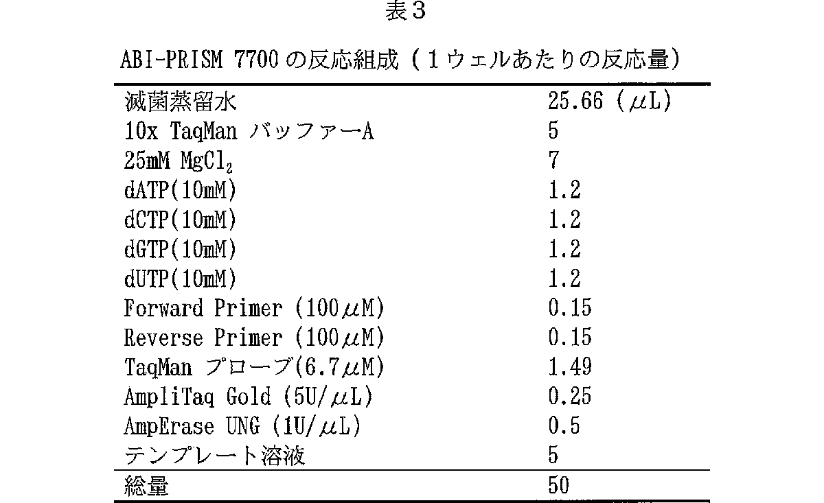
このように調製した全RNAを用いて蛍光ディファレンシャルディスプレイ(Fluorescent Differential Display,「DD」と略記する)解析を行った。DD解析は文献(T.Itoら,1994,FEBS Lett.351:231−236)に記載の方法に準じて行った。まず全RNAを逆転写し、cDNAを得た。第一次DD−PCR反応用には3種のアンカープライマーの各々について全RNAの各0.2μgを用いてcDNAを調製した。第二次DD−PCR反応用には、3種のアンカープライマーの各々についてRNA 0.4μgを用いてcDNAを調製した。いずれのcDNAも、0.4ng/μl RNA相当の最終濃度に希釈し、実験に用いた。1反応あたり1ng RNA相当のcDNAを用いてDD−PCR反応を行った。反応液の組成は表2の通りである。
使用したプライマー対はアンカープライマーであるGT15A(配列番号:3)、GT15C(配列番号:4)、およびGT15G(配列番号:5)に対して任意プライマーをそれぞれAG1〜110、AG111〜199、およびAG200〜287を組み合わせ、計287組の反応をおこなった。なお、任意プライマーとしてはGC含量50%の10ヌクレオチドからなるオリゴマーを設計し、合成して用いた。
ゲル電気泳動は、6%変性ポリアクリルアミドゲルを作製し、2.5μlの試料をアプライし、40Wで210分間泳動した。その後、日立製蛍光イメージアナライザーFMBIO IIを用いてゲル板をスキャンし、蛍光検出によって泳動画像を得た。
増悪期及び寛解期の両サンプルを並べて泳動し、ほとんどの患者で同方向に発現が変動している遺伝子バンドを目で判定し、切り取ってTAクローニングおよび配列決定を行った。その結果、増悪期に比べ寛解期に有意に発現の亢進する好酸球遺伝子(DD解析のバンドID2090−05;以後、この遺伝子を「2090−05」と称す)を同定した。バンドID2090−05の増幅に用いたプライマーセットを以下に示す。また、2090−05のDDバンドの塩基配列を配列番号:15に示す。
バンドID:2090−05
断片の長さ:430bp(プライマーの塩基配列を除く)
アンカープライマー:GT15A
任意プライマーの名前:AG00055
任意プライマーの配列:CCGTGAATTC(配列番号:6)
(3)発現解析
2090−05遺伝子の発現量を定量的に確認するために、同一臨床サンプルを用いてさらにABI7700による定量的PCRを行った。ABI7700による測定に用いたプライマーおよびTaqManプローブは、ディファレンシャルディスプレイ法によって得られた配列情報からPrimer Express(PEバイオシステムズ)により設計した。TaqManプローブの5’末端はFAM(6−carboxy−fluorescein)で、また3’末端はTAMRA(6−carboxy−N,N,N’,N’−tetramethylrhodamine)で標識されている。
2090−05フォーワードプライマー(配列番号:7)
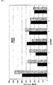
βアクチン測定用のプライマーとプローブは、TaqMan β−actin Control Reagents(PEバイオシステムズ)に添付のものを用いて行った。塩基配列は以下の通りである。βアクチンにより補正した「2090−05」遺伝子発現量(copy/ng RNA)を表4および図1に示す。
βアクチンフォーワードプライマー(配列番号:10)
TAMRA:6−carboxy−N,N,N’,N’−tetramethylrhodamine
統計解析の結果、増悪期に比べ寛解期で有意に発現が上昇することが確認された(p=0.011)。以上の知見は、この遺伝子がアトピー性皮膚炎の寛解期において発現が上昇することを示している。これは、この遺伝子の発現を測定することが、アトピー性皮膚炎において診断的価値をもつことを示している。この好酸球由来遺伝子は、アトピー性皮膚炎の治療標的あるいは診断マーカーとして医療上有用である。
[実施例2]各種血液細胞での2090−05遺伝子の発現
5人の健常人の末梢血から分離した細胞での2090−05遺伝子の発現を調べた。好酸球(E)の分離は上記の通り行った。好中球(N)は好酸球を溶出させた後、CD16抗体磁気ビーズでトラップされた細胞を磁界から外して溶出、回収して調製した。一方、フィコール遠心分離で中間層に回収される単球画分を、MACS CD3抗体磁気ビーズにより溶出画分(M:monocyteとB cellの混合物)とトラップされる画分(T cell画分)に分離した。次に、溶出画分をMACS CD14抗体磁気ビーズにより、溶出画分(B cell画分)とトラップされる画分(moocyte画分)に分け、それぞれを精製T細胞、精製B細胞、そして精製単球とした。
好酸球はIsogen、好中球、T細胞、B細胞、そして単球はRNeasy(Qiagen)を用いて可溶化し、全RNA抽出、DNase処理後(方法は前述の通り)遺伝子発現解析に供した。用いたプライマー、プローブ等は上記と同一である。これらの血球細胞での平均発現量(AVERAGE:copy/ng(補正値))は以下の通りであった。
好酸球(E):1454
好中球(N):139
好塩基球(B):1056
T細胞(T):760
単球(M):473
[実施例3]塩基配列の伸長
実施例1で決定した塩基配列に基づいて、5’RACE法により本発明の遺伝子の塩基配列解析を進めた。Marathon cDNA Amplification Kit(CLONTECH)のプロトコールに従いHuman Placenta Poly A+ RNA(CLONTECH)よりMarathon cDNAを合成し、これを鋳型としてキット付属のAP1プライマーとB2090特異的なプライマー2090−05FでPCRを行った。さらに、この増幅断片を鋳型にアダプター内の配列[AP2]とプライマー2090−5Fを用いてPCRを行った。増幅した断片をサブクローニングし配列決定をしたところc209−05の配列を含む約1.8kbの配列が得られた。同様に、キット付属のAP1プライマーとB2090特異的なプライマー2090−05RでPCRを行った。さらに、この増幅断片を鋳型にアダプター内の配列[AP2]とプライマー2090−05Rを用いてPCRを行った。増幅した断片をサブクローニングし配列決定をしたところc2090−05の配列を含む0.7kbの配列が得られ、合計2214bpとなった。決定された2214bpの塩基配列を配列番号:19に示す。また、この塩基配列に基づく推定アミノ酸配列を配列番号:2に示す。
プライマー配列
ESTを検索して上流配列になりそうなもの(配列番号:19の塩基配列を含み、更に5’側の塩基配列を含んでいるEST)を見つけ、以下のプライマーを構築してPCRを行った。
プライマー配列
その結果、胎児肝臓、リンパ節、胎児脳、脾臓、及び骨髄より約0.6kbのバンドが認められた。このDNA断片の塩基配列を常法に従って決定した。その結果、配列番号:19に対して上流(5’側)に204塩基ほど長い、配列番号:1に記載の塩基配列を見出すことができた。配列番号:1に示す塩基配列において、配列番号:19に対して新たに見出された塩基配列は、以下のとおりである。
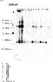
各臓器(脳、心臓、骨格筋、大腸、胸腺、脾臓、腎臓、肝臓、小腸、胎盤、肺、末梢血白血球)から調製したmRNAが転写された膜、Human 12−Lane MTN Blot(Clontech社)を用いてノーザンハイブリダイゼーションを行った。Primer1FとPrimer9Rのプライマーセットを用いてMarathon−Ready DNA(CLONTECH)の胎児肝臓を鋳型にPCR増幅を行い、得られたDNA断片を、PCR Product PurificationKit(QIAGEN)により精製した。精製DNA断片をRandom Primer Labeling Kit(TAKARA)を用いて32Pにより標識し、プローブとして用いた。ExpressHybridization Solution(CLONTECH)を用いて、添付使用書通りにノーザンハイブリダイゼーションおよび膜洗浄を行った。洗浄後の膜をイメージングプレートに暴露し、Molecular Imager System(BIO−RAD)によって画像を取得した(図2)。使用した組織で約5.0kbと約0.8kbのmRNAの発現が認められた。本発明によって見出された2090−05遺伝子の全長は、約5.0kbの長さを有する可能性が示唆された。
産業上の利用の可能性
本発明により、アトピー性皮膚炎患者の増悪期と寛解期とで発現に差の見られる遺伝子が提供された。本発明の遺伝子の発現を指標にすることにより、アレルギー性疾患の検査、および治療薬候補化合物のスクリーニングを行うことが可能となった。
本発明によって提供されたアレルギー疾患関連遺伝子は、アレルゲンの種類に関わらず、簡便にその発現レベルを知ることができる。したがって、アレルギー反応の病態を総合的に把握することができる。
また本発明によるアレルギーの検査方法は、末梢血好酸球を試料としてその発現レベルを解析することができるので、患者に対する侵襲性が低い。しかも遺伝子発現解析に関しては、たとえばECPなどの蛋白質測定と異なって、微量サンプルによる高感度な測定が可能である。遺伝子解析技術は、年々ハイスループット化、低価格化が進行している。したがって本発明によるアレルギーの検査方法は、近い将来、ベッドサイドにおける重要な診断方法となることが期待される。この意味でこれらの病態関連遺伝子の診断的価値は高い。
【配列表】
【図面の簡単な説明】
図1は、アトピー性皮膚炎患者(患者番号1〜7)の、増悪期と寛解期におけるβアクチンにより補正した2090−05遺伝子発現量(copy/ng RNA)を示す図である。
図2は、各臓器中における2090−05遺伝子の発現を調べたノーザンハイブリダイゼーションの結果を示す電気泳動写真である。 Technical field
The present invention relates to a gene associated with an allergic disease, a method for testing an allergic disease using the expression of the gene as an index, and a method for screening a candidate compound for a therapeutic drug for allergic disease.
Background art
Allergic diseases such as atopic dermatitis are considered to be multifactorial diseases. These diseases are caused by the interaction of the expression of many different genes, and the expression of these individual genes is affected by multiple environmental factors. For this reason, it is very difficult to elucidate a specific gene that causes a specific disease.
It is thought that allergic diseases are related to the expression of genes having mutations and defects, or overexpression or reduced expression of specific genes. To understand the role of gene expression in disease, it is necessary to understand how genes are involved in the pathogenesis and how external stimuli such as drugs alter gene expression.
Recent developments in gene expression analysis techniques have made it possible to analyze and compare gene expression in many clinical samples. As such a method, a differential display (DD) method is useful. The differential display method was first developed by Ryan and Paddy in 1992 (Science, 1992, 257: 967-971). By using this method, tens or more types of samples can be screened at one time, and it is possible to detect a gene whose expression has changed in those samples. Using such a method, examining a gene in which a mutation has occurred or a gene whose expression changes with time or environment is expected to provide important information for elucidating a pathogenic gene. These genes also include those whose expression is affected by environmental factors.
By the way, in the diagnosis of allergic diseases, questionnairing, family history, and confirmation of the patient's history are important factors in general. In order to diagnose allergy based on more objective information, a test method using blood as a sample and a method of observing a patient's immunological response to an allergen have been implemented. Examples of the former include an allergen-specific IgE measurement, a leukocyte histamine release test, and a lymphocyte blastogenesis test. The presence of allergen-specific IgE is evidence of an allergic reaction to the allergen. However, in some patients, it is not always possible to detect allergen-specific IgE. In addition, due to its measurement principle, tests must be performed for all allergens required for diagnosis. The leukocyte histamine release test and lymphocyte blastogenesis test are methods for observing the response of the immune system to allergens in vitro. These methods are complicated in operation.
On the other hand, a method of using an immune response observed when a patient is actually contacted with an allergen to diagnose allergy (the latter) is also known. Such tests include brick tests, scratch tests, patch tests, intradermal reactions, or provocation tests. While these tests can directly diagnose a patient's allergic reaction, they can be said to be highly invasive tests that actually expose the subject to an allergen.
In addition, test methods for proving the involvement of allergic reactions regardless of allergens have been attempted. For example, when the serum IgE level is high, it can be estimated that the patient has an allergic reaction. The serum IgE value is information corresponding to the total amount of allergen-specific IgE. Although it is easy to determine the total amount of IgE regardless of the type of allergen, IgE may be low in patients with diseases such as non-atopic bronchitis.
Eosinophil counts and ECP values are diagnostic items related to delayed-type reactions following type I allergy and allergic inflammatory reactions. The number of eosinophils is said to reflect the development of allergic symptoms. ECP (eosinophil o cationic protein), which is a protein contained in eosinophil granules, is also strongly activated with asthma attack. These diagnostic items certainly reflect allergic symptoms. However, the range that can be actually used as a diagnostic index is limited.
Therefore, regardless of the allergen, there has been a demand for a diagnostic index that can be used for understanding the condition of an allergic patient and determining a treatment policy. In addition, it would be useful to provide a marker for an allergic disease, which can reduce the risk to the patient and can easily obtain information necessary for diagnosis.
Disclosure of the invention
An object of the present invention is to provide a gene associated with an allergic disease. Another object of the present invention is to provide a method for testing an allergic disease and a method for screening a candidate compound for a therapeutic drug for allergic disease, using the expression of the gene as an index.
The present inventors have prepared leukocytes prepared from a plurality of human blood based on the already established procedure of the “fluorescent DD (Fluorescent DD) method” (T. Ito et al., 1994, FEBS Lett. 351: 231-236). A DD system capable of analyzing cellular RNA samples was developed (WO $ 00/65046). Then, the system was applied to the isolation of genes having different expression levels specifically for allergic diseases.
That is, first, the present inventors compared some parameters related to allergic symptoms between the exacerbation period and the remission period of the skin inflammatory condition in patients with atopic dermatitis. As a result, a decrease in eosinophils during remission was observed in some patients. In general, eosinophils are regarded as a representative clinical index of atopic dermatitis, and the present inventors have paid attention to this finding. And if it is possible to isolate a gene whose expression level changes in eosinophils between the exacerbation period and remission period of the same patient, it will be possible to isolate genes directly involved in atopic dermatitis I thought it might be.
Therefore, for a plurality of subjects, eosinophils were collected during the exacerbation period and remission period of atopic dermatitis, and the above system was used to screen for a gene whose expression level changes in eosinophils. As a result, the gene “2090-05” showing a significantly higher expression level in patients in remission was successfully isolated. This gene could not find the same nucleotide sequence in a known gene database, and was considered to be a novel gene. Furthermore, the present inventors have found that it is possible to perform an allergic disease test and to screen a candidate compound for a therapeutic drug for allergic disease using the expression level of this gene as an index, and completed the present invention.
That is, the present invention relates to a gene showing high expression in the remission period of atopic dermatitis, a protein encoded by the gene, and an application thereof. More specifically, a method for testing an allergic disease using the expression of the gene as an index, a method for detecting the effect of the candidate compound on the expression of the gene, and a candidate compound for a therapeutic drug for an allergic disease based on the detection method A screening method.
[1] A method for testing an allergic disease, comprising the following steps:
a) a step of measuring the expression level of a gene containing the nucleotide sequence of SEQ ID NO: 1 in eosinophil cells of the subject
b) a step of comparing the expression level of the gene in eosinophil cells of a healthy subject
[2] The test method according to [1], wherein the allergic disease is atopic dermatitis.
[3] The test method according to [1], wherein the expression level of the gene is measured by cDNA PCR.
[4] The test method according to [1], wherein the expression level of the gene is measured by detecting a protein encoded by the gene.
[5] A reagent for testing an allergic disease, comprising a polynucleotide comprising the nucleotide sequence of SEQ ID NO: 1 or an oligonucleotide having a nucleotide sequence complementary to a complementary strand thereof and having a length of at least 15 bases.
[6] A reagent for testing an allergic disease, comprising an antibody that recognizes a peptide containing the amino acid sequence of SEQ ID NO: 2.
[7] A method for detecting the effect of a candidate compound on the expression level of a polynucleotide according to any of the following (a) to (d), comprising the following steps:
(1) a step of bringing a candidate compound into contact with a cell expressing the polynucleotide according to any one of the following (a) to (d):
(A) a polynucleotide comprising the coding region of the nucleotide sequence of SEQ ID NO: 1.
(B) a polynucleotide encoding a protein consisting of the amino acid sequence of SEQ ID NO: 2;
(C) the amino acid sequence of SEQ ID NO: 2 in which one or more amino acids are substituted, deleted, inserted, and / or added, and are eosinophils during the remission of atopic dermatitis. A polynucleotide that encodes a protein that is functionally equivalent to the protein whose expression is to be increased.
(D) a polynucleotide that hybridizes with a DNA comprising the nucleotide sequence of SEQ ID NO: 1 under stringent conditions and encodes a protein whose expression increases in eosinophils during the remission of atopic dermatitis Polynucleotide.
(2) a step of measuring the expression level of the polynucleotide according to any of (a) to (d),
[8] The method of [7], wherein the cells are established leukocyte cells.
[9] A method for detecting the effect of a candidate compound on the expression level of a polynucleotide according to any one of the following (a) to (d), comprising the following steps:
(A) a polynucleotide comprising the coding region of the nucleotide sequence of SEQ ID NO: 1.
(B) a polynucleotide encoding a protein consisting of the amino acid sequence of SEQ ID NO: 2;
(C) the amino acid sequence of SEQ ID NO: 2 in which one or more amino acids are substituted, deleted, inserted, and / or added, and are eosinophils during the remission of atopic dermatitis. A polynucleotide that encodes a protein that is functionally equivalent to the protein whose expression is to be increased.
(D) a polynucleotide that hybridizes with a DNA comprising the nucleotide sequence of SEQ ID NO: 1 under stringent conditions and encodes a protein whose expression increases in eosinophils during the remission of atopic dermatitis Polynucleotide.
(1) administering a candidate compound to a test animal, and
(2) measuring the expression intensity of the polynucleotide according to any one of (a) to (d) in eosinophil cells of a test animal,
[10] The method according to [7] or [9], comprising detecting the effect on the expression level and selecting a compound that increases the expression level as compared to a control. -A method for screening a compound that increases the expression level of the polynucleotide according to any one of (d).
[11] A method for detecting the effect of a candidate compound on the activity of a transcription regulatory region of a gene comprising the nucleotide sequence of SEQ ID NO: 1, comprising the following steps:
(1) contacting a candidate substance with a cell into which a vector containing a transcription regulatory region of a gene containing the nucleotide sequence of SEQ ID NO: 1 and a reporter gene expressed under the control of this transcription regulatory region has been introduced; and
(2) measuring the activity of the reporter gene;
[12] The method according to [11], comprising detecting the effect of the candidate compound on the activity and selecting a compound that increases the activity as compared to a control. A method for screening a compound that increases the activity of a transcription regulatory region of a gene containing a sequence.
[13] A vector comprising a transcription regulatory region of a gene containing the nucleotide sequence of SEQ ID NO: 1, and a reporter gene expressed under the control of the transcription regulatory region.
[14] A cell into which the vector of [13] has been introduced.
[15] A therapeutic agent for allergic diseases, comprising as an active ingredient a compound obtainable by the screening method according to [10] or [12].
[16] the polynucleotide according to any one of the following (a) to (d):
(A) a polynucleotide comprising the coding region of the nucleotide sequence of SEQ ID NO: 1.
(B) a polynucleotide encoding a protein consisting of the amino acid sequence of SEQ ID NO: 2;
(C) the amino acid sequence of SEQ ID NO: 2 in which one or more amino acids are substituted, deleted, inserted, and / or added, and are eosinophils during the remission of atopic dermatitis. A polynucleotide that encodes a protein that is functionally equivalent to the protein whose expression is to be increased.
(D) a polynucleotide that hybridizes with a DNA comprising the nucleotide sequence of SEQ ID NO: 1 under stringent conditions and encodes a protein whose expression increases in eosinophils during the remission of atopic dermatitis Polynucleotide.
[17] A protein encoded by the polynucleotide of [16].
[18] A vector that holds the polynucleotide of [16] in an expressible manner.
[19] A transformed cell carrying the polynucleotide of [16] or the vector of [18].
[20] The method of producing a protein according to [17], comprising culturing the transformed cell according to [19] and collecting an expression product thereof.
[21] an antibody against the protein of [17];
[22] The method for immunologically measuring a protein according to [17], comprising a step of observing an immunological reaction between the antibody according to [21] and the protein according to [17].
[23] a polynucleotide comprising the nucleotide sequence of SEQ ID NO: 1, or an oligonucleotide having a nucleotide sequence complementary to a complementary strand thereof, wherein the oligonucleotide has a length of at least 15 bases;
[24] The method for measuring a polynucleotide according to [16], comprising a step of observing hybridization between the oligonucleotide according to [23] and the polynucleotide according to [16].
[25] An allergic disease model animal comprising a transgenic non-human vertebrate having a reduced expression level of the polynucleotide according to any one of the following (a) to (d) in eosinophil cells.
(A) a polynucleotide comprising the coding region of the nucleotide sequence of SEQ ID NO: 1.
(B) a polynucleotide encoding a protein consisting of the amino acid sequence of SEQ ID NO: 2;
(C) the amino acid sequence of SEQ ID NO: 2 in which one or more amino acids are substituted, deleted, inserted, and / or added, and are eosinophils during the remission of atopic dermatitis. A polynucleotide that encodes a protein that is functionally equivalent to the protein whose expression is to be increased.
(D) a polynucleotide that hybridizes with a DNA comprising the nucleotide sequence of SEQ ID NO: 1 under stringent conditions and encodes a protein whose expression increases in eosinophils during the remission of atopic dermatitis Polynucleotide.
[26] the model animal of [25], wherein the transgenic animal is a knockout animal;
[27] a polynucleotide having a length of at least 15 bases that hybridizes to the nucleotide sequence of SEQ ID NO: 1 or its complementary sequence, and a cell that expresses a gene consisting of the nucleotide sequence of SEQ ID NO: 1 A kit for screening candidate compounds for the treatment of allergic diseases.
[28] Screening for a candidate compound for a therapeutic drug for an allergic disease, comprising an antibody recognizing a peptide consisting of the amino acid sequence of SEQ ID NO: 2 and a cell expressing a gene consisting of the nucleotide sequence of SEQ ID NO: 1 Kit to do.
The present invention relates to a method for testing an allergic disease using the novel gene “2090-05” and the expression level of “2090-05” in eosinophil cells as an index. “2090-05” is a gene whose expression level increases during the remission phase of atopic dermatitis patients. The gene “2090-05” of the present invention could not find a gene having a particularly high structural identity, and was considered to be a novel gene. "2090-05" includes the nucleotide sequence of SEQ ID NO: 1.
In “2090-05”, a region (1-561) presumed to be a part of the ORF was found on the 5 ′ side, and encoded 187 amino acids. This amino acid sequence is obtained from Drosophila melanogaster, nematodes (Caenorhabditis @ elegans), Arabidopsis thaliana (Arabidopsis @ thaliana), fission yeast (Schizosaccharomyces @ pombe) Was. The homology of these proteins to the amino acid sequences was as follows.
The base sequence shown in SEQ ID NO: 1 is a partial sequence of the full-length cDNA. A full-length cDNA containing this partial sequence can be obtained by screening a white blood cell cDNA library with a probe consisting of a nucleotide sequence selected from the nucleotide sequence shown in SEQ ID NO: 1. “2090-05” is expressed not only in eosinophils but also in basophils. Therefore, any of these cells and a cDNA library derived from a cell population containing these cells can be used for obtaining the gene “2090-05” of the present invention. The sequence of “2090-05” can be extended by the RACE method (Frohman, MA et al .: Proc. Natl. Acad. Sci. USA, 85: 8992, 1988). That is, using a sequence derived from “2090-05” as a primer, mRNA of leukocyte cells or the like is converted to single-stranded cDNA, an oligomer is added to the end, and PCR is performed to obtain an extended cDNA. be able to.
The “polynucleotide comprising the base sequence of SEQ ID NO: 1” in the present invention includes “2090-isolated” based on the sequence information of “2090-05” cDNA described in SEQ ID NO: 1. 05 "full-length cDNA. Further, the amino acid sequence encoded by the cDNA can be estimated based on the nucleotide sequence of the cDNA thus obtained.
The present invention relates to a polynucleotide comprising the nucleotide sequence shown in SEQ ID NO: 1. The present invention also relates to a polynucleotide that hybridizes to the polynucleotide under stringent conditions and encodes a protein functionally equivalent to the protein encoded by the polynucleotide. In the present invention, the term “polynucleotide” includes not only natural nucleic acid molecules such as DNA and RNA, but also labeled molecules and artificial molecules constituted by various nucleotide derivatives. Artificial polynucleotides include polynucleotides having a phosphorothioate bond or a peptide bond as a backbone.
These polynucleotides of the present invention can be chemically synthesized, or can be isolated from natural nucleic acids such as mRNA, cDNA library, or genomic library. The polynucleotide molecule of the present invention is useful as an antisense nucleic acid for inhibiting the production of the protein encoded thereby and for expressing “2090-05”, or as a probe for detecting its presence by hybridization. .
In the present invention, when the expression of a certain protein increases in eosinophils during the remission of atopic dermatitis, it is said to be functionally equivalent to the protein of the present invention. Increased expression of a protein in eosinophils during remission of atopic dermatitis can be determined by comparing the expression level of the gene encoding the protein in eosinophils collected during the exacerbation and remission periods. You can check.
A polynucleotide that hybridizes with this polynucleotide under stringent conditions and encodes a functionally equivalent protein can be obtained by a known method such as hybridization or PCR based on the nucleotide sequence of SEQ ID NO: 1. Can be obtained. For example, a cDNA library of leukocyte cells is screened under stringent conditions using an oligonucleotide consisting of a base sequence selected from the base sequence described in SEQ ID NO: 1 as a probe, thereby obtaining SEQ ID NO: 1. A cDNA consisting of a base sequence having high identity to the described base sequence can be obtained. When a certain polynucleotide hybridizes under stringent conditions to a polynucleotide containing the nucleotide sequence shown in SEQ ID NO: 1, it is considered that the protein encoded by the polynucleotide often has an activity similar to the protein of the present invention. Conceivable. Stringent conditions generally include the following conditions. That is, hybridization is performed at 4 × SSC at 65 ° C., and washing is performed at 65 ° C. for 1 hour using 0.1 × SSC. Hybridization and washing temperature conditions that greatly affect stringency can be adjusted according to the melting temperature (Tm). Tm varies depending on the ratio of the constituent bases to the hybridized base pairs and the composition of the hybridization solution (salt concentration, formamide and sodium dodecyl sulfate concentration). Therefore, those skilled in the art can empirically set conditions that give equivalent stringency in consideration of these conditions.
A protein encoded by a cDNA comprising a highly identical base sequence is likely to be a functionally equivalent protein in the present invention. In the present invention, a base sequence having high identity generally means 75% or more, usually 80% or more, preferably 90% or more, more preferably 95% or more, still more preferably 98% or more, and particularly preferably 99% or more. % Or more. The identity of the nucleotide sequence can be calculated by a known algorithm such as BLASTN.
Alternatively, a cDNA having high identity can also be obtained by performing PCR using an oligonucleotide consisting of the nucleotide sequence of SEQ ID NO: 1 as a primer and also using a cDNA library of white blood cells as a template. If human cells are used as the source of cDNA, human cDNA can be obtained. In addition, if a vertebrate cell other than a human is used, a counterpart in a heterologous animal can be obtained. Examples of such non-human animals include many experimental animals such as mice, rats, dogs, pigs, goats, and the like. The counterpart of “2090-05” in experimental animals is useful as a marker in the preparation of allergy model animals in each animal species and in the development of therapeutic drugs for allergy.
Alternatively, a gene encoding a protein having, for example, 90% or more, preferably 95% or more, more preferably 99% or more homology with the amino acid sequence of the “2090-05” protein is referred to as the “2090-05” gene. It can be shown as a functional gene. In addition, a gene that can be amplified using, as a primer, an oligonucleotide consisting of a base sequence selected from the base sequence of SEQ ID NO: 1 used in the Examples, and is used for eosinophils during the remission period of atopic dermatitis. Are also functionally equivalent genes. In the present invention, the “2090-05” gene or a gene functionally equivalent to this gene is referred to as an indicator gene. The protein encoded by the indicator gene is called an indicator protein.
The polynucleotide of the present invention comprises an amino acid sequence in which one or more amino acids have been substituted, deleted, added, and / or inserted into the amino acid sequence represented by SEQ ID NO: 2; And polynucleotides encoding proteins functionally equivalent to the above. For example, polymorphisms are often found in eukaryotic genes. Although one or more amino acids may be substituted depending on the polymorphism, the activity of the protein is usually maintained. In addition, it is generally known that protein activity is often maintained by modifying one or several amino acid sequences. Therefore, a polynucleotide encoding a protein in which the amino acid sequence is mutated by artificial modification of the polynucleotide encoding the amino acid sequence shown in SEQ ID NO: 2 has a characteristic function of the gene of the present invention. All that is included in the present invention. Such an amino acid sequence preferably includes a sequence having 90% or more homology to the amino acid sequence represented by SEQ ID NO: 2. Amino acid sequence homology can be determined by FASTA.
In addition, the codon for an amino acid is known, and the selection may be arbitrary. For example, it can be determined according to a conventional method in consideration of the codon usage of the host used [Grantham, R. et al. et @ al. Nucleic Acids Res. 9, r43 (1981)]. Accordingly, those obtained by appropriately modifying DNA in consideration of the degeneracy of codons are also included in the polynucleotide of the present invention. Site-specific mutagenesis using a primer consisting of a synthetic oligonucleotide encoding the desired modification [Mark, D .; F. et @ al. Proc. Natl. Acad. Sci. U. S. A. 81, 5662 (1984)] and the like, the codons of these nucleotide sequences can be partially modified.
The present invention also relates to a polynucleotide comprising the nucleotide sequence of SEQ ID NO: 1 or an oligonucleotide having a nucleotide sequence complementary to a complementary strand thereof, wherein the oligonucleotide has a length of at least 15 bases. As used herein, the term "complementary strand" refers to one strand of a double-stranded polynucleotide consisting of A: T (T is read as U in RNA) and G: C base pairs with respect to the other strand. The term "complementary" is not limited to a case where the sequence is completely complementary to at least 15 contiguous nucleotide regions, but is at least 70%, preferably at least 80%, more preferably 90%, and still more preferably 95%. It is only necessary to have the above homology on the base sequence. The homology of the nucleotide sequences can be determined by the algorithm described in the present specification.
The oligonucleotide of the present invention is useful for detecting or synthesizing the polynucleotide of the present invention. Techniques for detecting or synthesizing a target polynucleotide using an oligonucleotide as a probe or primer are known. For example, Northern blotting using mRNA as a target polynucleotide is a typical method for detecting RNA. If RT-PCR is performed using mRNA as a template, the polynucleotide of the present invention can be synthesized. In addition, the presence or absence of mRNA and its expression level can be known using the presence or absence and amount of the synthetic product as an index. Alternatively, the polynucleotide of the present invention expressed in eosinophils can be detected by in situ hybridization.
Further, the polynucleotide encoded by the polynucleotide of the present invention can be produced as a recombinant. More specifically, a transformant is obtained by incorporating the coding region of the polynucleotide containing the nucleotide sequence of SEQ ID NO: 1 into a known expression vector, and transforming it into an appropriate host. Alternatively, a transformant can be obtained by integrating the polynucleotide containing the coding region into the genome of an appropriate host.
The protein of the present invention can be obtained by culturing the obtained transformant under conditions under which the introduced polynucleotide of the present invention can be expressed, and collecting the expression product. The expression product can be a purified protein by a known method.
In addition, the present invention relates to a protein encoded by the polynucleotide of the present invention. The protein of the present invention is useful as an index for diagnosing allergic diseases such as atopic dermatitis. Alternatively, since the polynucleotide of the present invention increases expression in eosinophils in remission, the protein itself encoded by the polynucleotide can be expected to have a therapeutic effect on allergy.
Alternatively, the protein of the present invention or a fragment thereof is useful as an immunogen for preparing an antibody against the protein of the present invention. Techniques for obtaining antibodies using a given immunogen are known. That is, the protein or a fragment thereof is mixed with an appropriate adjuvant to prepare an immunogen, and the immunized animal is inoculated. The immunized animal is not limited. Representative immunized animals include animals such as mice, rats, rabbits, and goats. Blood is collected after confirming an increase in the antibody titer, and serum is collected to obtain an antiserum. Alternatively, a purified antibody can be obtained by further purifying the IgG fraction. For purification of the antibody, techniques such as ammonium sulfate salting out, ion exchange chromatography, protein A-bound Sepharose, and immunoaffinity chromatography using the protein of the present invention as a ligand can be used.
Furthermore, a monoclonal antibody can also be obtained by transforming the antibody-producing cell by a technique such as cell fusion and cloning. Alternatively, a method for obtaining a gene of an antibody-producing cell and constructing a humanized antibody or chimeric antibody is also known. The antibody thus obtained is useful as a tool for immunologically measuring the protein of the present invention. The protein of the present invention can be measured immunologically by bringing the protein of the present invention into contact with the antibody and observing the immunological reaction between the two. Many known assay formats can be applied to the immunological measurement of the present invention. For example, a protein contained in serum or the like can be measured by a technique such as ELISA. Alternatively, in order to detect a protein expressed in eosinophils using an antibody, an immunohistological technique, FACS using a fluorescently labeled antibody, or the like can be used.
In the present invention, the allergic disease (allergic disease) is a general term for diseases involving an allergic reaction. More specifically, it can be defined as identifying an allergen, demonstrating a deep link between allergen exposure and the development of a lesion, and demonstrating an immunological mechanism for the lesion. Here, the immunological mechanism means that white blood cells show an immune response by allergen stimulation. Examples of the allergen include a mite antigen and a pollen antigen.
Representative allergic diseases include bronchial asthma, allergic rhinitis, atopic dermatitis, hay fever, and insect allergy. An allergic diathesis is a genetic factor transmitted from a parent having an allergic disease to a child. An allergic disease that develops familially is also called atopic disease, and a genetically transmitted factor that causes it is atopic predisposition. Atopic dermatitis is a general term given to atopic diseases, particularly those accompanied by skin inflammatory conditions.
The gene “2090-05” of the present invention showed an increased expression level in eosinophils of patients in remission in comparison with the exacerbation and remission of atopic dermatitis patients. Therefore, an allergic disease test can be performed using the expression level of the “2090-05” gene as an index.
The test for an allergic disease in the present invention includes, for example, the following tests. For example, the present invention allows for tests to determine whether allergic symptoms are improving. The gene “2090-05” of the present invention showed an increased expression level particularly in eosinophils of atopic dermatitis patients in remission. Since eosinophils are typical clinical markers of atopic dermatitis, clinical markers expressed on eosinophils are useful for determining the therapeutic effect. More specifically, an increase in the expression of the gene “2090-05” of the present invention indicates that improvement of allergic diseases is progressing.
Since the severity of atopic dermatitis is closely related to the number of eosinophils, it is important to measure this gene, which is specifically induced by eosinophils, and to actively induce it from outside the cell. Finding methods and substances could lead to new treatments for atopic dermatitis and diagnostics to evaluate them.
In the present invention, the expression level of the “2090-05” gene includes transcription of these genes into mRNA and translation into proteins. Therefore, the method for testing an allergic disease according to the present invention is performed based on a comparison of the expression intensity of mRNA corresponding to the gene or the expression level of a protein encoded by the gene.
The measurement of the expression level of the “2090-05” gene in the test for an allergic disease according to the present invention can be performed according to a known gene analysis method. Specifically, for example, a hybridization technique using a nucleic acid that hybridizes to the gene as a probe or a gene amplification technique using a DNA that hybridizes to the gene of the present invention as a primer can be used.
As the probe or primer used in the test of the present invention, a polynucleotide consisting of the nucleotide sequence of SEQ ID NO: 1 or a polynucleotide containing at least 15 nucleotides complementary to a complementary strand thereof can be used. Here, the “complementary strand” refers to one strand of a double-stranded DNA consisting of A: T (U for RNA) and G: C base pairs with respect to the other strand. The term "complementary" is not limited to a case where the sequence is completely complementary to at least 15 contiguous nucleotide regions, but is at least 70%, preferably at least 80%, more preferably 90%, and still more preferably 95%. It is only necessary to have the above homology on the base sequence. The homology of the nucleotide sequences can be determined by an algorithm such as BLASTN.
Such a polynucleotide can be used as a probe for detecting and isolating a polynucleotide encoding the protein of the present invention, and as a primer for amplifying the polynucleotide of the present invention. When used as a primer, it usually has a chain length of 15 bp to 100 bp, preferably 15 bp to 35 bp. When used as a probe, a DNA having at least a part or the entire sequence of the polynucleotide of the present invention and having a chain length of at least 15 bp is used. When used as a primer, the region on the 3 'side must be complementary, but a restriction enzyme recognition sequence, a tag, or the like can be added to the 5' side.
The “polynucleotide” in the present invention can be DNA or RNA. These polynucleotides may be synthetic or natural. The probe DNA used for hybridization is usually labeled. As the labeling method, for example, the following method can be shown. The term oligonucleotide means a polynucleotide having a relatively low degree of polymerization among polynucleotides. Oligonucleotides are included in polynucleotides.
Labeling by nick translation using DNA polymerase I
・ End labeling using polynucleotide kinase
Fillin terminal labeling with Klenow fragment (Berger SL, Kimmel AR (1987) Guide to Molecular Cloning Techniques, Method in Enzymology, Academic Press; HameshP.A.R.P. J, Fritsch EF, Maniatis T. (1989) Molecular Cloning: a Laboratory Manual, 2nd Edn. Cold Spring Harbor Laboratory Press.
Labeling by transcription using RNA polymerase (Melton DA, Krieg, PA, Rebagkiati MR, Maniatis T, Zinn K, Green MR. (1984) Nucleic Acid Res., 12, 7035-7056)
A method of incorporating a modified nucleotide without using a radioisotope into DNA (Kricka LJ. (1992) Nonisotopic DNA Probing Techniques Academic Press)
Testing for allergic diseases using hybridization techniques can be performed using, for example, Northern hybridization, dot blotting, a method using a DNA microarray, and the like. Further, a gene amplification technique such as an RT-PCR method can be used. In the RT-PCR method, more quantitative analysis can be performed on the expression of the gene of the present invention by using the PCR amplification monitoring method in the gene amplification process.
In the PCR gene amplification monitoring method, probes are labeled at both ends with different fluorescent dyes that cancel each other's fluorescence, and are hybridized to a detection target (reverse transcript of DNA or RNA). When the PCR proceeds and the probe is decomposed by the 5'-3 'exonuclease activity of Taq polymerase, the two fluorescent dyes are separated and the fluorescence is detected. This fluorescence is detected in real time. The number of copies of the detection target in the target sample is determined by the number of linear cycles of PCR amplification by simultaneously measuring a standard sample whose copy number is clearly known as the detection target (Holland, PM et al., Natl. Acad. Sci. USA {88: 7276-7280; Livak, KJ. Et al., 1995, PCR Methods and Applications 4 (6): 357-362; Heid, CA et al. , Genome Research 6: 986-994; Gibson, EMU et al., 1996, Genome Research 6: 995-1001). In the PCR amplification monitoring method, for example, ABI @ PRISM7700 (PE Biosystems) can be used.
The method for testing an allergic disease of the present invention can also be performed by detecting the protein encoded by the gene “2090-05”. As such a test method, for example, a Western blotting method, an immunoprecipitation method, an ELISA method, or the like using an antibody that binds to a protein encoded by these genes can be used.
An antibody that binds to the “2090-05” protein used for this detection can be obtained using techniques well known to those skilled in the art. The antibody used in the present invention can be a polyclonal antibody or a monoclonal antibody (Milstein C, et al., 1983, Nature 305 (5934): 537-40). For example, a polyclonal antibody against the protein of the present invention is obtained by extracting blood of a mammal sensitized with an antigen and separating serum from the blood by a known method. Serum containing the polyclonal antibody can be used as the polyclonal antibody. Alternatively, if necessary, a fraction containing a polyclonal antibody can be further isolated from the serum. In addition, to obtain a monoclonal antibody, immune cells are removed from a mammal sensitized with the above antigen, and are fused with myeloma cells or the like. The hybridoma thus obtained is cloned, and the antibody is recovered from the culture to obtain a monoclonal antibody.
For detection of the “2090-05” protein, these antibodies may be appropriately labeled and used. Further, without labeling the antibody, a substance that specifically binds to the antibody, for example, protein A or protein G can be labeled and detected indirectly. As a specific detection method, for example, an ELISA method can be mentioned.
A protein or a partial peptide thereof used as an antigen can be prepared, for example, by incorporating the gene or a part thereof into an expression vector, introducing this into an appropriate host cell, preparing a transformant, culturing the transformant, and performing recombination. It can be obtained by expressing a protein and purifying the expressed recombinant protein from a culture or a culture supernatant. Alternatively, an oligopeptide consisting of an amino acid sequence encoded by these genes or a partial amino acid sequence of the amino acid sequence encoded by the full-length cDNA obtained based on SEQ ID NO: 1 is chemically synthesized and used as an immunogen. You can also.
In the present invention, eosinophil cells of a subject are used as a sample. Eosinophil cells can be prepared from peripheral blood by a known method. That is, for example, heparin-collected blood is fractionated by centrifugation to separate white blood cells. Next, granulocyte cells are separated from the leukocyte cells by centrifugation using Ficoll or the like, and further, eosinophil cells can be separated by depletion of neutrophils using the CD16 antibody. If the separated eosinophils are destroyed to form a lysate, it can be used as a sample for immunological measurement of the protein. Alternatively, if mRNA is extracted from this lysate, it can be used as a sample for measuring mRNA corresponding to the gene. For extraction of eosinophil lysate and mRNA, it is convenient to use a commercially available kit.
Alternatively, the expression level of the gene to be used as an index in the present invention may be measured for whole blood or peripheral blood leukocyte populations without separating eosinophils. In this case, a change in the expression level of the gene in the cell can be obtained by correcting the measured value. For example, the expression of a gene to be used as an index in the present invention based on a measured value of the expression level of a gene (housekeeping gene) which is specifically expressed in eosinophils and whose expression level does not largely fluctuate regardless of the state of the cell Level measurements can be corrected.
When the protein to be detected is a secretory protein, the expression level of the gene encoding the protein is determined by measuring the amount of the target protein contained in a body fluid sample such as blood or serum of the subject. Can be compared.
As a result of the test for an allergic disease according to the present invention, it is presumed that improvement of allergic symptoms is progressing particularly when the expression level of the gene of the present invention is increased in patients with allergic diseases such as atopic dermatitis. Is done.
In addition, the present invention relates to an allergic disease model animal comprising a transgenic non-human animal in which the expression level of any of the following polynucleotides (a) to (d) in eosinophil cells is reduced.
(A) a polynucleotide comprising the coding region of the nucleotide sequence of SEQ ID NO: 1.
(B) a polynucleotide encoding a protein consisting of the amino acid sequence of SEQ ID NO: 2;
(C) the amino acid sequence of SEQ ID NO: 2 in which one or more amino acids are substituted, deleted, inserted, and / or added, and are eosinophils during the remission of atopic dermatitis. A polynucleotide that encodes a protein that is functionally equivalent to the protein whose expression is to be increased.
(D) a polynucleotide that hybridizes with a DNA comprising the nucleotide sequence of SEQ ID NO: 1 under stringent conditions and encodes a protein whose expression increases in eosinophils during the remission of atopic dermatitis Polynucleotide.
In the present invention, a decrease in the expression level includes a knockout state in which the function of a gene is substantially lost. In the present invention, a state in which the function of a gene is substantially lost means a state in which expression of a gene or activity of a protein encoded by the gene cannot be found. The expression level of the gene can be confirmed, for example, by quantitative PCR as described in Examples. Further, the fact that the activity of the protein as a translation product is not substantially found can be confirmed by comparing with the normal state.
Such transgenic animals include, for example, animals in which a mutation has been introduced into the coding region of a gene and an amino acid sequence has been mutated or a stop codon has been artificially generated so that the original protein activity cannot be expressed. be able to. Mutations in the amino acid sequence can indicate substitutions, deletions, insertions, or additions. In addition, the expression itself of the gene of the present invention can be regulated by mutating the transcriptional regulatory region of the gene.
Methods for obtaining a transgenic animal for a specific gene are known. That is, a method in which a gene and an egg are mixed and treated with calcium phosphate, a method in which a gene is directly introduced into a nucleus of a pronuclear stage egg with a micropipette under a phase-contrast microscope (microinjection method, US Pat. A transgenic animal can be obtained by a method using embryonic stem cells (ES cells) or the like. In addition, a method of inserting a gene into a retrovirus vector and infecting an egg, a method of introducing a gene into an egg via sperm, and a sperm vector method have also been developed. The sperm vector method is a genetic recombination method in which a foreign gene is introduced into sperm cells by a method such as attachment or electroporation to a sperm and then fertilized by an egg to introduce the foreign gene (M. Lavitranoet et al., Cell, 57, 717, 1989).
The transgenic animal of the present invention can be prepared using any vertebrate other than human. Specifically, transgenic animals in which various genes have been introduced and expression levels of which have been modified in vertebrates such as mice, rats, rabbits, minipigs, goats, sheep, and cattle have been created.
The transgenic animal of the present invention includes a knockout animal in which the expression of a homolog of a human gene containing the nucleotide sequence shown in SEQ ID NO: 1 in the animal species is suppressed. By observing the phenotype of the knockout animal, the function of the knocked out gene can be specifically known. The expression of the gene containing the nucleotide sequence shown in SEQ ID NO: 1 was increased in eosinophils during the remission of atopic dermatitis in humans. Therefore, an animal in which the homolog is knocked out is useful as a model animal for an allergic disease.
For example, if a knockout animal according to the present invention develops dermatitis or shows a change in a measured value related to any allergic disease, a screening system for searching for a compound having an action to restore it can be constructed. In this case, the action point of the drug is a gene product different from the 2090-05 gene and related to the allergic disease.
Methods for producing knockout animals are known. For example, a method is known in which homologous recombination is performed using embryonic stem cells in mice, and embryonic stem cells in which one allele has been modified or disrupted are selected to produce a knockout animal. For example, a fertilized egg is injected with a genetically modified embryonic stem cell to obtain a chimeric animal in which cells derived from embryonic stem cells and cells derived from an embryo are mixed. When this chimeric animal (a chimera is a single individual formed from somatic cells based on two or more fertilized eggs) is bred to a normal mouse, a heterozygote in which all of one allele has been altered or destroyed is obtained. A body can be made. Furthermore, homozygotes can be prepared by crossing heterozygotes. The transgenic animals according to the present invention include both these heterozygotes and homozygotes.
Homologous recombination refers to recombination occurring between two genes having the same or very similar nucleotide sequences in the gene recombination mechanism. PCR can be used to select cells that have undergone homologous recombination. A PCR reaction was performed using a part of the inserted gene and a part of the region expected to be inserted as primers, and it was found that homologous recombination had occurred in the cells in which the amplification product was formed. In addition, when homologous recombination is caused to a gene expressed in embryonic stem cells, a neomycin resistance gene can be linked to the introduced gene, and cells can be selected by neomycin resistance after introduction. And their variations can be readily selected.
The transgenic animals according to the present invention are useful for elucidating the mechanism of allergic diseases and for testing the safety of screened compounds.
The present invention has revealed that the expression level of the gene “2090-05” is increased in eosinophils of atopic dermatitis patients in remission. Therefore, animals in which the expression levels of these genes or genes functionally equivalent to these genes in eosinophil cells are artificially reduced can be used as model animals for allergic diseases. The decrease in the expression level in eosinophils includes a decrease in the expression level of the gene in the whole leukocyte population. That is, the expression level of the gene is decreased not only by eosinophils but also by the expression level of the gene in the whole leukocyte population. In the present invention, a functionally equivalent gene means any of the genes described in any of (a) to (d) above. As the model animal in the present invention, for example, the above-mentioned transgenic animal or the like can be used.
Furthermore, the present invention relates to a method for detecting the effect of a candidate compound on the expression level of the polynucleotide of the present invention. In the present invention, the expression level of the “2090-05” gene is significantly increased in eosinophils of atopic dermatitis patients in remission. Therefore, a therapeutic agent for allergic diseases can be obtained by selecting a compound capable of increasing the expression level based on a method for detecting the effect on the expression level of these genes. In the present invention, the compound that increases the expression level of a gene is a compound that has an action of inducing any of the steps of gene transcription, translation, and protein activity expression.
The method for detecting the effect of the candidate compound on the expression level of the polynucleotide of the present invention can be performed in vivo or in vitro. To detect effects in vivo, appropriate test animals are utilized. As the test animal, for example, an allergic disease model animal or an experimental animal that can express an indicator gene can be used. The detection of the effect on the expression level in vivo according to the present invention can be performed, for example, according to the following steps.
(1) administering a candidate compound to a test animal,
(2) measuring the expression level of the polynucleotide according to any of (a) to (d) in eosinophil cells of a test animal,
By administering the drug candidate compound to the test animal in this way and monitoring the effect of the compound on the expression of the 2090-05 gene in eosinophils of the test animal, the drug candidate compound The effect can be detected. Furthermore, if a drug candidate compound that increases the expression level of the 2090-05 gene is selected based on the result of this detection, the drug candidate compound can be screened.
By such a screening, drugs involved in various ways in the expression of the 2090-05 gene can be selected. Specifically, for example, drug candidate compounds having the following action points can be found.
Activation of a signaling pathway leading to expression of the 2090-05 gene,
Increased transcriptional activity of the 2090-05 gene,
Such as stabilization or inhibition of degradation of the transcript of the 2090-05 gene,
In addition, in in vitro, for example, a method of contacting a candidate compound with a cell expressing the gene described in any of the above (a) to (d) and detecting the expression level of these genes can be used. . Specifically, for example, it can be carried out according to the following steps.
(1) a step of contacting a candidate compound with a cell expressing the polynucleotide according to any one of the above (a) to (d)
(2) measuring the expression level of the polynucleotide according to any one of (a) to (d),
In the present invention, cells to be used in step (1) can be obtained by inserting these polynucleotides into an appropriate expression vector and introducing the vector into an appropriate host cell. Usable vectors and host cells may be any as long as they can express the gene of the present invention. Examples of host cells in the host-vector system include Escherichia coli, yeast, insect cells, animal cells, and the like, and any available vector can be appropriately selected.
Examples of a method for introducing a vector into a host include a biological method, a physical method, and a chemical method. Examples of biological methods include a method using a viral vector, a method using a specific receptor, a cell fusion method (HVJ (Sendai virus), polyethylene glycol (PEG), an electric cell fusion method, micronucleus fusion). Method (chromosome transfer)). Examples of the physical method include a microinjection method, an electroporation method, and a method using a gene particle gun (gene @ gun). Examples of the chemical method include a calcium phosphate precipitation method, a liposome method, a DEAE dextran method, a protoplast method, an erythrocyte ghost method, an erythrocyte membrane ghost method, and a microcapsule method.
In the detection method of the present invention, established leukocytes can also be used as cells expressing the polynucleotide described in (a) or (b). Examples of the established leukocyte cells include leukocyte-derived established cells such as Eol, YY-1, HL-60, TF-1, and AML14.3D10. Among leukocyte cell lines, cell lines derived from eosinophils are suitable for the detection method of the present invention. Cell lines derived from eosinophils are as shown below.
Eol
YY-1
AML14.3D10
Eol (Eol-1: Saito H et al, Establishment and charactrization of of a new human human eosinophilic leukemia cell line, Blood 66, 1233-1240, 1985) can be obtained from Hayashibara Research Institute. Similarly, YY-1 (Ogata @ et @ al, The activation * of the @ JAK2 / STAT5 \ pathway \ is \ commonly \ involved \ in \ signaling \ throw \ the \ human \ IL-5@receptor.Il. , Distributed by Site Signal Laboratory. AML14.3D10 (Baumann MA @ et @ al, The AML14 @ and @ AML14.3D10 @ cell @ lines: a @ long-overdue @ model @ for @ the @ study @ ofe @ seophils @ and @ m.m.e., Os., Republic of Oseofils, 16 M., Ore. It is commercially available from Paul, CC of Service, VA, Medical, Center, Dayton.
In addition, HL-60 clone 15 (ATCC @ CRL-1964), which is an undifferentiated leukocyte cell line, can be differentiated into eosinophils and used as an eosinophil cell line if cultured for about one week in the presence of butyric acid. Eosinophils can be distinguished morphologically by the presence of eosinophil granules in polymorphonuclear nuclei. Morphological observation is performed by Giemsa staining or Diff Quick staining. In general, human leukocyte cell lines containing eosinophils can be established by cloning immortalized cells from a leukemia patient sample. Therefore, those skilled in the art can also obtain an eosinophil cell line by a known method, if necessary.
In the screening method, first, a candidate compound is added to the established leukocyte cells. Thereafter, the expression level of the polynucleotide described in the above (a) or (b) in the established leukocyte cells is measured, and a compound that increases the expression level of the gene is selected.
As a cell for the in vitro detection method, a transformed cell in which the expression of the polynucleotide described in any of the above (a) to (d) is regulated can be used. Examples of such a transformed cell include a cell transformed with an antisense expression vector of the polynucleotide. Transformed cells with the antisense expression vector can be obtained by the same principle as in the preparation of the transgenic animal. Using the obtained transformed cells, the effect of the candidate compound on the expression level of the gene can also be detected.
In the method of the present invention, the expression level of the polynucleotide according to any one of the above (a) to (d) is determined by detecting not only the expression level of the protein encoded by these genes but also the corresponding mRNA. You can also compare. In order to compare the expression levels using mRNA, the mRNA sample preparation step as described above is performed instead of the protein sample preparation step. The detection of mRNA or protein can be carried out by a known method as described above.
Further, based on the disclosure of the present invention, a transcription regulatory region of the gene of the present invention can be obtained, and a reporter assay system can be constructed. The reporter assay system refers to an assay system that screens for a transcription regulatory factor acting on the transcription regulatory region by using, as an index, the expression level of a reporter gene expressed under the control of the transcription regulatory region downstream of the transcription regulatory region.
That is, the present invention is a method for screening a therapeutic agent for an allergic disease, comprising the following steps, wherein the indicator gene is selected from the group consisting of “2090-05” and a gene functionally equivalent to this gene. The method is any gene.
(1) contacting a candidate substance with a cell into which a vector containing a reporter gene expressed under the control of the transcriptional regulatory region of the indicator gene and the transcriptional regulatory region has been introduced;
(2) measuring the activity of the reporter gene;
(3) selecting a compound that increases the reporter gene expression level as compared to a control
Examples of the transcription control region include a promoter, an enhancer, and a CAAT box, a TATA box, and the like usually found in a promoter region. As the reporter gene, a CAT (chloramphenicol @ acetyltransferase) gene, a luciferase gene, a growth hormone gene and the like can be used.
The transcription regulatory region of the gene of the present invention can be obtained as follows. That is, first, based on the nucleotide sequence of the cDNA disclosed in the present invention, screening is performed by a method using PCR or hybridization from a human genomic DNA library such as a BAC library or a YAC library, and a genomic sequence containing the cDNA sequence is obtained. Obtain a DNA clone. Based on the sequence of the obtained genomic DNA, the transcription regulatory region of the cDNA disclosed in the present invention is estimated, and the transcription regulatory region is obtained. The obtained transcription regulatory region is cloned so as to be located upstream of the reporter gene to construct a reporter construct. The resulting reporter construct is introduced into a cultured cell line to obtain a transformant for screening. By contacting a candidate compound with this transformant and detecting the expression of a reporter gene, the action of the candidate compound on the transcription regulatory region can be evaluated.
Based on the method of the present invention for detecting an effect on the expression level of the polynucleotide, a compound that changes the expression level of the polynucleotide can be screened. The present invention relates to a method for screening a compound that changes the expression level of the polynucleotide according to any one of the above (a) to (d), comprising the following steps.
That is, the present invention comprises a step of detecting the effect of a candidate compound on the expression level of the polynucleotide in in vivo and / or in vitro, and selecting a compound that increases the expression level compared to a control. It relates to a method for screening a compound that increases the expression level of the polynucleotide according to any one of (a) to (d).
Alternatively, the present invention relates to a method for screening a compound acting on a transcription regulatory region by a reporter assay using a transcription regulatory region of a gene containing the nucleotide sequence shown in SEQ ID NO: 1. Based on the results of the reporter assay according to the present invention, by selecting a compound that increases the expression of a reporter gene as compared with a subject, a compound that induces the expression of a gene containing the nucleotide sequence shown in SEQ ID NO: 1 is obtained. be able to.
Polynucleotides, antibodies, cell lines, or model animals required for various screening methods according to the present invention can be combined in advance to form a kit. More specifically, it is composed of, for example, cells expressing the indicator genes and reagents for measuring the expression levels of these indicator genes. As a reagent for measuring the expression level of the indicator gene, for example, a polynucleotide containing a nucleotide sequence of at least one indicator gene, or an oligonucleotide having a nucleotide sequence complementary to a complementary strand thereof and having a length of at least 15 bases Is used. Alternatively, an antibody that recognizes a peptide containing the amino acid sequence of at least one indicator protein can be used as a reagent. These kits must be packaged with the substrate compound used to detect the label, culture media and containers for culturing cells, positive and negative standard samples, and instructions describing how to use the kit. You can also. The kit for detecting the effect of the candidate compound on the expression level of the 2090-05 gene according to the present invention can be used as a kit for screening a compound that modifies the expression level of the 2090-05 gene.
The test candidate compounds used in these methods include compound preparations synthesized by existing chemical methods such as steroid derivatives, compound preparations synthesized by combinatorial chemistry, extracts of animal and plant tissues, and cultures of microorganisms. And a mixture containing a plurality of compounds, and a sample purified therefrom.
The compound selected by the screening method of the present invention is useful as a therapeutic drug for allergic diseases. The remedy for allergic diseases of the present invention can be produced by mixing the compound selected by the screening method as an active ingredient and mixing with a physiologically acceptable carrier, excipient, or diluent. it can. The therapeutic agent for an allergic disease of the present invention can be administered orally or parenterally for the purpose of improving allergic symptoms.
As oral preparations, dosage forms such as granules, powders, tablets, capsules, solvents, emulsions, and suspensions can be selected. Injections include subcutaneous injections, intramuscular injections, and intraperitoneal injections.
The dose varies depending on the age, sex, weight and condition of the patient, therapeutic effect, administration method, treatment time, or the type of active ingredient contained in the pharmaceutical composition. It can be administered in the range 0.1 mg to 500 mg, preferably in the range 0.5 mg to 20 mg. However, since the dose varies depending on various conditions, a dose smaller than the above dose may be sufficient, and a dose exceeding the above range may be required in some cases.
BEST MODE FOR CARRYING OUT THE INVENTION
Hereinafter, the present invention will be described specifically with reference to Examples, but the present invention is not limited to these Examples.
[Example 1] Differential display analysis
To compare the exacerbation period in the same patient with atopic dermatitis with the blood cells isolated from peripheral blood during remission by drug treatment or other to find new treatment-related genes whose expression is fluctuating or useful for diagnosis Screening was performed for the purpose.
(1) Examinee
Table 1 shows the profiles of the seven patients with atopic dermatitis from which blood was collected. Allergen non-specific (Total @ IgE), tick and cedar-specific IgE were measured by EIA method. That is, the test serum was reacted with the cap to which the anti-human IgE antibody had been bound, and the allergen non-specific IgE antibody or the mite or cedar-specific IgE antibody in the serum was bound. Next, a β-D-galactosidase-labeled anti-human IgE antibody and a substrate solution (4-methylumbelliferyl-β-D-galactopyranoside) were added and reacted to produce a fluorescent substance. The reaction was stopped by adding a reaction stop solution, and the antibody concentration was determined from the fluorescence intensity of standard IgE measured simultaneously. In the measurement of LDH, the reduction rate of NADH due to the reaction between pyruvate and NADH was calculated from the decrease in absorbance by the UV method (Wrobleski-La @ Due method). For measuring the LDH value, an L-type Wako LDH (Wako Pure Chemical Industries) and a 7170-type automatic analyzer (Hitachi) were used. The eosinophil count was measured by microscopy and an automatic blood cell analyzer SE-9000 (RF / DC impedance method, manufactured by Sysmex) using 2 ml of EDTA-added blood as a sample.
A 3% dextran solution was added to whole blood collected from the patient and left at room temperature for 30 minutes to sediment erythrocytes. The leukocyte fraction in the upper layer was collected, placed on a Ficoll solution (Ficoll-Paque @ PLUS; Amersham Pharmacia Biotech), and centrifuged at 1500 rpm for 30 minutes at room temperature. The granulocyte fraction collected in the lower layer was reacted with CD16 antibody magnetic beads at 4 ° C. for 30 minutes, and the cells eluted without trapping by separation using MACS were used as eosinophils in the experiment.
The eosinophil prepared as described above was dissolved in Isogen (Nippon Gene; Wako Pure Chemical Industries), and RNA was separated from this solution according to the protocol attached to Isogen. Chloroform was added, and the mixture was centrifuged with stirring to collect an aqueous layer. Next, isopropanol was added, and the mixture was centrifuged with stirring to collect the total RNA in the precipitate. The collected total RNA was added with DNase (Nippon Gene; Wako Pure Chemical Industries, Ltd.) and reacted at 37 ° C. for 15 minutes, extracted with phenol-chloroform, and recovered with ethanol precipitation.
Using the total RNA thus prepared, a fluorescent differential display (Fluorescent Differential Display, abbreviated as “DD”) analysis was performed. The DD analysis was performed according to the method described in the literature (T. Ito et al., 1994, FEBS @ Lett. 351: 231-236). First, total RNA was reverse transcribed to obtain cDNA. For the primary DD-PCR reaction, cDNA was prepared using 0.2 μg of total RNA for each of the three anchor primers. For the second DD-PCR reaction, cDNA was prepared using 0.4 μg of RNA for each of the three anchor primers. All cDNAs were diluted to a final concentration of 0.4 ng / μμ RNA and used in the experiments. A DD-PCR reaction was performed using cDNA corresponding to 1 ng RNA per reaction. Table 2 shows the composition of the reaction solution.
The primer pairs used were arbitrary primers for the anchor primers GT15A (SEQ ID NO: 3), GT15C (SEQ ID NO: 4), and GT15G (SEQ ID NO: 5), respectively AG1-110, AG111-199, and AG200. 287 were combined to perform a total of 287 reactions. As an arbitrary primer, an oligomer composed of 10 nucleotides having a GC content of 50% was designed, synthesized, and used.
For gel electrophoresis, a 6% denaturing polyacrylamide gel was prepared, 2.5 μl of the sample was applied, and electrophoresis was performed at 40 W for 210 minutes. Thereafter, the gel plate was scanned using a Hitachi fluorescent image analyzer FMBIO II, and a migration image was obtained by fluorescence detection.
Both exacerbation and remission samples were run side-by-side, and gene bands with variable expression in most patients in the same direction were visually determined and excised for TA cloning and sequencing. As a result, an eosinophil gene whose expression was significantly enhanced in the remission period compared to the exacerbation period (band ID 2090-05 in DD analysis; hereinafter, this gene is referred to as “2090-05”) was identified. The primer set used for amplification of band ID 2090-05 is shown below. The base sequence of the DD band of 2090-05 is shown in SEQ ID NO: 15.
Band ID: 2090-05
Fragment length: 430 bp (excluding base sequence of primer)
Anchor primer: GT15A
Optional primer name: AG00055
Arbitrary primer sequence: CCGTGAATTC (SEQ ID NO: 6)
(3) Expression analysis
In order to quantitatively confirm the expression level of the 2090-05 gene, quantitative PCR was further performed using the same clinical sample using ABI7700. The primer and TaqMan probe used for the measurement by ABI7700 were designed by Primer @ Express (PE Biosystems) from the sequence information obtained by the differential display method. The 5 'end of the TaqMan probe is labeled with FAM (6-carboxy-fluorescein), and the 3' end is labeled with TAMRA (6-carboxy-N, N, N ', N'-tetramethylrhodamine).
2090-05 forward primer (SEQ ID NO: 7)
The primers and probes for β-actin measurement were those attached to TaqMan \ β-actin \ Control \ Reagents (PE Biosystems). The base sequence is as follows. The expression level of “2090-05” gene (copy / ngΔRNA) corrected by β-actin is shown in Table 4 and FIG.
β-actin forward primer (SEQ ID NO: 10)
TAMRA: 6-carboxy-N, N, N ', N'-tetramethylrhodamine
As a result of the statistical analysis, it was confirmed that the expression significantly increased in the remission period compared with the exacerbation period (p = 0.111). These findings indicate that the expression of this gene increases during the remission of atopic dermatitis. This indicates that measuring the expression of this gene has diagnostic value in atopic dermatitis. This eosinophil-derived gene is medically useful as a therapeutic target or a diagnostic marker for atopic dermatitis.
[Example 2] Expression of 2090-05 gene in various blood cells
The expression of the 2090-05 gene in cells isolated from peripheral blood of five healthy individuals was examined. Eosinophils (E) were separated as described above. Neutrophils (N) were prepared by eluting eosinophils and then eluting and recovering the cells trapped by the CD16 antibody magnetic beads out of the magnetic field. On the other hand, the monocyte fraction collected in the intermediate layer by Ficoll centrifugation is converted into a fraction eluted by MACS @ CD3 antibody magnetic beads (M: a mixture of monocyte and B @ cell) and a fraction trapped (T @ cell fraction). separated. Next, the eluted fraction was separated by MACS @ CD14 antibody magnetic beads into an eluted fraction (B @ cell fraction) and a trapped fraction (moocyte fraction), each of which was purified T cells, purified B cells, and purified single cells. A sphere.
Eosinophils are solubilized using Isogen, neutrophils, T cells, B cells, and monocytes using RNeasy (Qiagen), and subjected to gene expression analysis after extraction of total RNA and DNase treatment (as described above). did. The used primers and probes are the same as above. The average expression level in these blood cells (AVERAGE: copy / ng (correction value)) was as follows.
Eosinophil (E): 1454
Neutrophils (N): 139
Basophils (B): 1056
T cells (T): 760
Monocyte (M): 473
[Example 3] Extension of base sequence
Based on the nucleotide sequence determined in Example 1, the nucleotide sequence of the gene of the present invention was analyzed by the 5'RACE method. Human Placenta Poly A according to the protocol of Marathon cDNA Amplification Kit (CLONTECH)+Marathon cDNA was synthesized from RNA (CLONTECH), and PCR was performed using this as a template with the AP1 primer attached to the kit and the B2090-specific primer 2090-05F. Furthermore, PCR was performed using this amplified fragment as a template and the sequence [AP2] in the adapter and primer 2090-5F. When the amplified fragment was subcloned and sequenced, a sequence of about 1.8 kb including the sequence of c209-05 was obtained. Similarly, PCR was performed using the AP1 primer attached to the kit and the B2090-specific primer 2090-05R. Furthermore, PCR was performed using this amplified fragment as a template and the sequence [AP2] in the adapter and primer 2090-05R. When the amplified fragment was subcloned and sequenced, a 0.7 kb sequence including the sequence of c2090-05 was obtained, for a total of 2,214 bp. The nucleotide sequence of 2214 bp determined is shown in SEQ ID NO: 19. The deduced amino acid sequence based on this nucleotide sequence is shown in SEQ ID NO: 2.
Primer sequence
An EST was searched to find one likely to become an upstream sequence (EST containing the base sequence of SEQ ID NO: 19 and further containing the base sequence on the 5 ′ side), and the following primers were constructed and PCR was performed. .
Primer sequence
As a result, a band of about 0.6 kb was observed in the fetal liver, lymph node, fetal brain, spleen, and bone marrow. The nucleotide sequence of this DNA fragment was determined according to a conventional method. As a result, a base sequence described in SEQ ID NO: 1 that was about 204 bases longer (5 ′ side) upstream of SEQ ID NO: 19 could be found. In the nucleotide sequence shown in SEQ ID NO: 1, the nucleotide sequence newly found for SEQ ID NO: 19 is as follows.
A membrane to which mRNA prepared from each organ (brain, heart, skeletal muscle, large intestine, thymus, spleen, kidney, liver, small intestine, placenta, lung, peripheral blood leukocyte) is transcribed, Human 12-Lane MTN Blot (Clontech) Was used to perform Northern hybridization. Using a primer set of Primer1F and Primer9R, PCR amplification was performed using Marathon-Ready DNA (CLONTECH) fetal liver as a template, and the obtained DNA fragment was purified by PCR Product Purification Kit (QIAGEN). The purified DNA fragment was prepared using Random Primer Labeling Kit (TAKARA).32It was labeled with P and used as a probe. Northern Hybridization and membrane washing were performed using ExpressHybridization @ Solution (CLONTECH) as per the attached instructions. The membrane after washing was exposed to an imaging plate, and an image was acquired by Molecular Imager System (BIO-RAD) (FIG. 2). Expression of about 5.0 kb and about 0.8 kb of mRNA was observed in the tissues used. It was suggested that the full length of the 2090-05 gene found by the present invention may have a length of about 5.0 kb.
Industrial potential
According to the present invention, a gene whose expression is different between the exacerbation period and the remission period of an atopic dermatitis patient is provided. By using the expression of the gene of the present invention as an index, it has become possible to carry out an allergic disease test and a screening for a therapeutic drug candidate compound.
The expression level of the allergic disease-related gene provided by the present invention can be easily determined regardless of the type of allergen. Therefore, the pathology of the allergic reaction can be comprehensively grasped.
In addition, the method for testing allergy according to the present invention can analyze the expression level of peripheral blood eosinophils as a sample, and therefore has low invasiveness to patients. Moreover, regarding gene expression analysis, unlike a protein measurement such as ECP, for example, highly sensitive measurement using a small amount of sample is possible. Gene analysis technology has been increasing in throughput and lowering its price year by year. Therefore, the method of testing for allergy according to the present invention is expected to be an important diagnostic method at the bedside in the near future. In this sense, the diagnostic value of these disease-related genes is high.
[Sequence list]
[Brief description of the drawings]
FIG. 1 is a graph showing the expression level of 2090-05 gene (copy / ng @ RNA) in patients with atopic dermatitis (patient numbers 1 to 7) corrected by β-actin during the exacerbation period and the remission period.
FIG. 2 is an electrophoretic photograph showing the results of Northern hybridization in which the expression of the 2090-05 gene in each organ was examined.
Claims (28)
a)被検者の好酸球細胞における、配列番号:1に記載の塩基配列を含む遺伝子の発現レベルを測定する工程
b)健常者の好酸球細胞における前記遺伝子の発現レベルと比較する工程A method for testing an allergic disease, comprising the following steps.
a) a step of measuring the expression level of the gene containing the nucleotide sequence of SEQ ID NO: 1 in the eosinophil cells of the subject; b) a step of comparing the expression level of the gene in the eosinophil cells of a healthy subject
(1)下記(a)−(d)のいずれかに記載のポリヌクレオチドを発現する細胞に候補化合物を接触させる工程
(a)配列番号:1に記載の塩基配列のコード領域を含むポリヌクレオチド。
(b)配列番号:2に記載のアミノ酸配列からなる蛋白質をコードするポリヌクレオチド。
(c)配列番号:2に記載のアミノ酸配列において、1若しくは複数個のアミノ酸が置換、欠失、挿入、および/または付加したアミノ酸配列からなり、アトピー性皮膚炎の寛解期に好酸球において発現が増加する蛋白質と機能的に同等な蛋白質をコードするポリヌクレオチド。
(d)配列番号:1に記載の塩基配列を含むDNAとストリンジェントな条件下でハイブリダイズするポリヌクレオチドであって、アトピー性皮膚炎の寛解期に好酸球において発現が増加する蛋白質をコードするポリヌクレオチド。
(2)前記(a)−(d)のいずれかに記載のポリヌクレオチドの発現レベルを測定する工程、A method for detecting the effect of a candidate compound on the expression level of a polynucleotide according to any one of the following (a) to (d), comprising the following steps:
(1) A step of contacting a candidate compound with a cell that expresses the polynucleotide according to any one of the following (a) to (d): (a) a polynucleotide comprising the coding region of the nucleotide sequence of SEQ ID NO: 1;
(B) a polynucleotide encoding a protein consisting of the amino acid sequence of SEQ ID NO: 2;
(C) the amino acid sequence of SEQ ID NO: 2 in which one or more amino acids are substituted, deleted, inserted, and / or added, and are eosinophils during the remission of atopic dermatitis. A polynucleotide that encodes a protein that is functionally equivalent to the protein whose expression is to be increased.
(D) a polynucleotide that hybridizes with a DNA comprising the nucleotide sequence of SEQ ID NO: 1 under stringent conditions and encodes a protein whose expression increases in eosinophils during the remission of atopic dermatitis The polynucleotide to be expressed.
(2) a step of measuring the expression level of the polynucleotide according to any of (a) to (d),
(a)配列番号:1に記載の塩基配列のコード領域を含むポリヌクレオチド。
(b)配列番号:2に記載のアミノ酸配列からなる蛋白質をコードするポリヌクレオチド。
(c)配列番号:2に記載のアミノ酸配列において、1若しくは複数個のアミノ酸が置換、欠失、挿入、および/または付加したアミノ酸配列からなり、アトピー性皮膚炎の寛解期に好酸球において発現が増加する蛋白質と機能的に同等な蛋白質をコードするポリヌクレオチド。
(d)配列番号:1に記載の塩基配列を含むDNAとストリンジェントな条件下でハイブリダイズするポリヌクレオチドであって、アトピー性皮膚炎の寛解期に好酸球において発現が増加する蛋白質をコードするポリヌクレオチド。
(1)被験動物に候補化合物を投与する工程、および
(2)被験動物の好酸球細胞における前記(a)−(d)のいずれかに記載のポリヌクレオチドの発現強度を測定する工程、A method for detecting the effect of a candidate compound on the expression level of a polynucleotide according to any one of the following (a) to (d), comprising the following steps:
(A) a polynucleotide comprising the coding region of the nucleotide sequence of SEQ ID NO: 1.
(B) a polynucleotide encoding a protein consisting of the amino acid sequence of SEQ ID NO: 2;
(C) the amino acid sequence of SEQ ID NO: 2 in which one or more amino acids are substituted, deleted, inserted, and / or added, and are eosinophils during the remission of atopic dermatitis. A polynucleotide that encodes a protein that is functionally equivalent to the protein whose expression is to be increased.
(D) a polynucleotide that hybridizes with a DNA comprising the nucleotide sequence of SEQ ID NO: 1 under stringent conditions and encodes a protein whose expression increases in eosinophils during the remission of atopic dermatitis The polynucleotide to be expressed.
(1) a step of administering a candidate compound to a test animal, and (2) a step of measuring the expression intensity of the polynucleotide according to any one of the above (a) to (d) in eosinophil cells of the test animal;
(1)配列番号:1に記載の塩基配列を含む遺伝子の転写調節領域と、この転写調節領域の制御下に発現するレポーター遺伝子とを含むベクターを導入した細胞と候補物質を接触させる工程、および
(2)前記レポーター遺伝子の活性を測定する工程、A method for detecting the effect of a candidate compound on the activity of a transcription regulatory region of a gene comprising the nucleotide sequence of SEQ ID NO: 1, comprising the following steps:
(1) contacting a candidate substance with a cell into which a vector containing a transcription regulatory region of a gene containing the nucleotide sequence of SEQ ID NO: 1 and a reporter gene expressed under the control of the transcription regulatory region has been introduced; and (2) measuring the activity of the reporter gene;
(a)配列番号:1に記載の塩基配列のコード領域を含むポリヌクレオチド。
(b)配列番号:2に記載のアミノ酸配列からなる蛋白質をコードするポリヌクレオチド。
(c)配列番号:2に記載のアミノ酸配列において、1若しくは複数個のアミノ酸が置換、欠失、挿入、および/または付加したアミノ酸配列からなり、アトピー性皮膚炎の寛解期に好酸球において発現が増加する蛋白質と機能的に同等な蛋白質をコードするポリヌクレオチド。
(d)配列番号:1に記載の塩基配列を含むDNAとストリンジェントな条件下でハイブリダイズするポリヌクレオチドであって、アトピー性皮膚炎の寛解期に好酸球において発現が増加する蛋白質をコードするポリヌクレオチド。The polynucleotide according to any one of the following (a) to (d).
(A) a polynucleotide comprising the coding region of the nucleotide sequence of SEQ ID NO: 1.
(B) a polynucleotide encoding a protein consisting of the amino acid sequence of SEQ ID NO: 2;
(C) the amino acid sequence of SEQ ID NO: 2 in which one or more amino acids are substituted, deleted, inserted, and / or added, and are eosinophils during the remission of atopic dermatitis. A polynucleotide that encodes a protein that is functionally equivalent to the protein whose expression is to be increased.
(D) a polynucleotide that hybridizes with a DNA comprising the nucleotide sequence of SEQ ID NO: 1 under stringent conditions and encodes a protein whose expression increases in eosinophils during the remission of atopic dermatitis The polynucleotide to be expressed.
(a)配列番号:1に記載の塩基配列のコード領域を含むポリヌクレオチド。
(b)配列番号:2に記載のアミノ酸配列からなる蛋白質をコードするポリヌクレオチド。
(c)配列番号:2に記載のアミノ酸配列において、1若しくは複数個のアミノ酸が置換、欠失、挿入、および/または付加したアミノ酸配列からなり、アトピー性皮膚炎の寛解期に好酸球において発現が増加する蛋白質と機能的に同等な蛋白質をコードするポリヌクレオチド。
(d)配列番号:1に記載の塩基配列を含むDNAとストリンジェントな条件下でハイブリダイズするポリヌクレオチドであって、アトピー性皮膚炎の寛解期に好酸球において発現が増加する蛋白質をコードするポリヌクレオチド。An allergic disease model animal comprising a transgenic non-human vertebrate having a reduced expression level of the polynucleotide according to any one of the following (a) to (d) in eosinophil cells.
(A) a polynucleotide comprising the coding region of the nucleotide sequence of SEQ ID NO: 1.
(B) a polynucleotide encoding a protein consisting of the amino acid sequence of SEQ ID NO: 2;
(C) the amino acid sequence of SEQ ID NO: 2 in which one or more amino acids are substituted, deleted, inserted, and / or added, and are eosinophils during the remission of atopic dermatitis. A polynucleotide that encodes a protein that is functionally equivalent to the protein whose expression is to be increased.
(D) a polynucleotide that hybridizes with a DNA comprising the nucleotide sequence of SEQ ID NO: 1 under stringent conditions and encodes a protein whose expression increases in eosinophils during the remission of atopic dermatitis The polynucleotide to be expressed.
Applications Claiming Priority (3)
| Application Number | Priority Date | Filing Date | Title |
|---|---|---|---|
| JP2000291318 | 2000-09-25 | ||
| JP2000291318 | 2000-09-25 | ||
| PCT/JP2001/008246 WO2002024903A1 (en) | 2000-09-25 | 2001-09-21 | Method of examining allergic disease |
Publications (1)
| Publication Number | Publication Date |
|---|---|
| JPWO2002024903A1 true JPWO2002024903A1 (en) | 2004-03-04 |
Family
ID=18774408
Family Applications (1)
| Application Number | Title | Priority Date | Filing Date |
|---|---|---|---|
| JP2002529497A Pending JPWO2002024903A1 (en) | 2000-09-25 | 2001-09-21 | Testing methods for allergic diseases |
Country Status (3)
| Country | Link |
|---|---|
| US (1) | US7148011B2 (en) |
| JP (1) | JPWO2002024903A1 (en) |
| WO (1) | WO2002024903A1 (en) |
Families Citing this family (3)
| Publication number | Priority date | Publication date | Assignee | Title |
|---|---|---|---|---|
| WO2005085851A2 (en) * | 2004-03-03 | 2005-09-15 | Adra Chaker N | Granulocyte subtype-selective receptors and ion channels and uses thereof |
| US8134137B2 (en) | 2008-06-18 | 2012-03-13 | Micron Technology, Inc. | Memory device constructions, memory cell forming methods, and semiconductor construction forming methods |
| CN109781998B (en) * | 2019-02-20 | 2022-03-25 | 首都医科大学附属北京中医医院 | Application of HMGB1 gene or protein expression in evaluating effect of Qigui silver on treating pneumonia |
Family Cites Families (6)
| Publication number | Priority date | Publication date | Assignee | Title |
|---|---|---|---|---|
| JPH11332567A (en) * | 1998-05-22 | 1999-12-07 | Dai Ichi Seiyaku Co Ltd | Judgment of atopic predisposition |
| US6504083B1 (en) | 1998-10-06 | 2003-01-07 | Pioneer Hi-Bred International, Inc. | Maize Gos-2 promoters |
| JP2000106879A (en) | 1998-10-06 | 2000-04-18 | Jenokkusu Soyaku Kenkyusho:Kk | Hay fever-related genes |
| JP2003125775A (en) | 1999-04-27 | 2003-05-07 | Jenokkusu Soyaku Kenkyusho:Kk | Hay fever-related gene, 373 |
| JP2004514416A (en) * | 2000-07-07 | 2004-05-20 | インサイト・ゲノミックス・インコーポレイテッド | Drug metabolizing enzymes |
| JPWO2002026962A1 (en) | 2000-09-26 | 2004-02-05 | 株式会社ジェノックス創薬研究所 | Testing methods for allergic diseases |
-
2001
- 2001-09-21 WO PCT/JP2001/008246 patent/WO2002024903A1/en not_active Ceased
- 2001-09-21 JP JP2002529497A patent/JPWO2002024903A1/en active Pending
- 2001-09-21 US US10/380,254 patent/US7148011B2/en not_active Expired - Fee Related
Also Published As
| Publication number | Publication date |
|---|---|
| US20040038252A1 (en) | 2004-02-26 |
| US7148011B2 (en) | 2006-12-12 |
| WO2002024903A1 (en) | 2002-03-28 |
Similar Documents
| Publication | Publication Date | Title |
|---|---|---|
| US7115373B2 (en) | Method of testing for atopic dermatitis by measuring expression of the NOR-1 gene | |
| US7172867B2 (en) | Methods of testing for allergic diseases, and therapeutic agents for treating same | |
| EP1260816A1 (en) | Method of examining allergic diseases | |
| KR20170041955A (en) | Mutant Genes as Diagnosis Marker for Amyotrophic Lateral Sclerosis and Diagnosis Method Using the Same | |
| JPWO2002050269A1 (en) | Testing methods for allergic diseases | |
| US20040197786A1 (en) | Method of examining steroid resnponsiveness | |
| US7148011B2 (en) | Method of testing for allergic diseases | |
| JPWO2002026962A1 (en) | Testing methods for allergic diseases | |
| US20040058351A1 (en) | Method of examining for allergic disease | |
| JP2002095500A (en) | Testing methods for allergic diseases | |
| KR101634047B1 (en) | Biomarkers for detection of ADHD | |
| US20220316008A1 (en) | Compositions comprising rare genetic sequence variants associated with pulmonary function and methods of use thereof for diagnosis and treatment of asthma in african american patients | |
| JP2002119281A (en) | Testing methods for allergic diseases | |
| KR101576008B1 (en) | Biomarkers for detection of ADHD | |
| WO2003072778A1 (en) | Method of examining allergic disease | |
| WO2000065046A1 (en) | Pollinosis-associated gene 373 | |
| JP2004267090A (en) | Testing methods for allergic diseases | |
| EP1498493A1 (en) | Method of examining allergic disease | |
| JPWO2003010310A1 (en) | Testing methods for allergic diseases | |
| WO2004016783A1 (en) | Method of examining atopic dermatitis | |
| JPWO2001065259A1 (en) | Testing methods for allergic diseases | |
| WO2002075304A1 (en) | Method of examining allergic disease | |
| KR20160047986A (en) | Biomarkers for detection of ADHD | |
| WO2003054231A1 (en) | Method of examining allergic disease | |
| JPWO2000065045A1 (en) | Hay fever-related genes, 419 |
Legal Events
| Date | Code | Title | Description |
|---|---|---|---|
| A521 | Request for written amendment filed |
Free format text: JAPANESE INTERMEDIATE CODE: A523 Effective date: 20050119 |
|
| A521 | Request for written amendment filed |
Free format text: JAPANESE INTERMEDIATE CODE: A821 Effective date: 20050119 |
|
| A711 | Notification of change in applicant |
Free format text: JAPANESE INTERMEDIATE CODE: A712 Effective date: 20060908 |
|
| RD04 | Notification of resignation of power of attorney |
Free format text: JAPANESE INTERMEDIATE CODE: A7424 Effective date: 20060929 |