KR20170016461A - 메디컬 이미징에서 공간 및 시간 제약들을 이용하는 랜드마크 검출 - Google Patents
메디컬 이미징에서 공간 및 시간 제약들을 이용하는 랜드마크 검출 Download PDFInfo
- Publication number
- KR20170016461A KR20170016461A KR1020177000509A KR20177000509A KR20170016461A KR 20170016461 A KR20170016461 A KR 20170016461A KR 1020177000509 A KR1020177000509 A KR 1020177000509A KR 20177000509 A KR20177000509 A KR 20177000509A KR 20170016461 A KR20170016461 A KR 20170016461A
- Authority
- KR
- South Korea
- Prior art keywords
- detecting
- valve
- frames
- bounding box
- candidate
- Prior art date
- Legal status (The legal status is an assumption and is not a legal conclusion. Google has not performed a legal analysis and makes no representation as to the accuracy of the status listed.)
- Granted
Links
Images


Classifications
-
- G—PHYSICS
- G06—COMPUTING OR CALCULATING; COUNTING
- G06T—IMAGE DATA PROCESSING OR GENERATION, IN GENERAL
- G06T7/00—Image analysis
- G06T7/10—Segmentation; Edge detection
- G06T7/11—Region-based segmentation
-
- G—PHYSICS
- G06—COMPUTING OR CALCULATING; COUNTING
- G06F—ELECTRIC DIGITAL DATA PROCESSING
- G06F18/00—Pattern recognition
- G06F18/20—Analysing
- G06F18/21—Design or setup of recognition systems or techniques; Extraction of features in feature space; Blind source separation
- G06F18/213—Feature extraction, e.g. by transforming the feature space; Summarisation; Mappings, e.g. subspace methods
- G06F18/2136—Feature extraction, e.g. by transforming the feature space; Summarisation; Mappings, e.g. subspace methods based on sparsity criteria, e.g. with an overcomplete basis
-
- G—PHYSICS
- G06—COMPUTING OR CALCULATING; COUNTING
- G06F—ELECTRIC DIGITAL DATA PROCESSING
- G06F18/00—Pattern recognition
- G06F18/20—Analysing
- G06F18/21—Design or setup of recognition systems or techniques; Extraction of features in feature space; Blind source separation
- G06F18/214—Generating training patterns; Bootstrap methods, e.g. bagging or boosting
-
- G—PHYSICS
- G06—COMPUTING OR CALCULATING; COUNTING
- G06F—ELECTRIC DIGITAL DATA PROCESSING
- G06F18/00—Pattern recognition
- G06F18/20—Analysing
- G06F18/24—Classification techniques
-
- G—PHYSICS
- G06—COMPUTING OR CALCULATING; COUNTING
- G06F—ELECTRIC DIGITAL DATA PROCESSING
- G06F18/00—Pattern recognition
- G06F18/20—Analysing
- G06F18/29—Graphical models, e.g. Bayesian networks
- G06F18/295—Markov models or related models, e.g. semi-Markov models; Markov random fields; Networks embedding Markov models
-
- G06K9/6256—
-
- G06K9/6297—
-
- G—PHYSICS
- G06—COMPUTING OR CALCULATING; COUNTING
- G06T—IMAGE DATA PROCESSING OR GENERATION, IN GENERAL
- G06T7/00—Image analysis
- G06T7/0002—Inspection of images, e.g. flaw detection
- G06T7/0012—Biomedical image inspection
-
- G—PHYSICS
- G06—COMPUTING OR CALCULATING; COUNTING
- G06T—IMAGE DATA PROCESSING OR GENERATION, IN GENERAL
- G06T7/00—Image analysis
- G06T7/0002—Inspection of images, e.g. flaw detection
- G06T7/0012—Biomedical image inspection
- G06T7/0014—Biomedical image inspection using an image reference approach
- G06T7/0016—Biomedical image inspection using an image reference approach involving temporal comparison
-
- G—PHYSICS
- G06—COMPUTING OR CALCULATING; COUNTING
- G06T—IMAGE DATA PROCESSING OR GENERATION, IN GENERAL
- G06T7/00—Image analysis
- G06T7/10—Segmentation; Edge detection
- G06T7/143—Segmentation; Edge detection involving probabilistic approaches, e.g. Markov random field [MRF] modelling
-
- G—PHYSICS
- G06—COMPUTING OR CALCULATING; COUNTING
- G06T—IMAGE DATA PROCESSING OR GENERATION, IN GENERAL
- G06T7/00—Image analysis
- G06T7/10—Segmentation; Edge detection
- G06T7/174—Segmentation; Edge detection involving the use of two or more images
-
- G—PHYSICS
- G06—COMPUTING OR CALCULATING; COUNTING
- G06V—IMAGE OR VIDEO RECOGNITION OR UNDERSTANDING
- G06V10/00—Arrangements for image or video recognition or understanding
- G06V10/70—Arrangements for image or video recognition or understanding using pattern recognition or machine learning
- G06V10/77—Processing image or video features in feature spaces; using data integration or data reduction, e.g. principal component analysis [PCA] or independent component analysis [ICA] or self-organising maps [SOM]; Blind source separation
- G06V10/774—Generating sets of training patterns; Bootstrap methods, e.g. bagging or boosting
-
- G—PHYSICS
- G06—COMPUTING OR CALCULATING; COUNTING
- G06V—IMAGE OR VIDEO RECOGNITION OR UNDERSTANDING
- G06V10/00—Arrangements for image or video recognition or understanding
- G06V10/70—Arrangements for image or video recognition or understanding using pattern recognition or machine learning
- G06V10/84—Arrangements for image or video recognition or understanding using pattern recognition or machine learning using probabilistic graphical models from image or video features, e.g. Markov models or Bayesian networks
- G06V10/85—Markov-related models; Markov random fields
-
- G—PHYSICS
- G06—COMPUTING OR CALCULATING; COUNTING
- G06V—IMAGE OR VIDEO RECOGNITION OR UNDERSTANDING
- G06V30/00—Character recognition; Recognising digital ink; Document-oriented image-based pattern recognition
- G06V30/10—Character recognition
- G06V30/19—Recognition using electronic means
- G06V30/192—Recognition using electronic means using simultaneous comparisons or correlations of the image signals with a plurality of references
- G06V30/194—References adjustable by an adaptive method, e.g. learning
-
- G06K2209/051—
-
- G—PHYSICS
- G06—COMPUTING OR CALCULATING; COUNTING
- G06T—IMAGE DATA PROCESSING OR GENERATION, IN GENERAL
- G06T2207/00—Indexing scheme for image analysis or image enhancement
- G06T2207/10—Image acquisition modality
- G06T2207/10132—Ultrasound image
-
- G—PHYSICS
- G06—COMPUTING OR CALCULATING; COUNTING
- G06T—IMAGE DATA PROCESSING OR GENERATION, IN GENERAL
- G06T2207/00—Indexing scheme for image analysis or image enhancement
- G06T2207/20—Special algorithmic details
- G06T2207/20076—Probabilistic image processing
-
- G—PHYSICS
- G06—COMPUTING OR CALCULATING; COUNTING
- G06T—IMAGE DATA PROCESSING OR GENERATION, IN GENERAL
- G06T2207/00—Indexing scheme for image analysis or image enhancement
- G06T2207/20—Special algorithmic details
- G06T2207/20081—Training; Learning
-
- G—PHYSICS
- G06—COMPUTING OR CALCULATING; COUNTING
- G06T—IMAGE DATA PROCESSING OR GENERATION, IN GENERAL
- G06T2207/00—Indexing scheme for image analysis or image enhancement
- G06T2207/20—Special algorithmic details
- G06T2207/20112—Image segmentation details
- G06T2207/20132—Image cropping
-
- G—PHYSICS
- G06—COMPUTING OR CALCULATING; COUNTING
- G06T—IMAGE DATA PROCESSING OR GENERATION, IN GENERAL
- G06T2207/00—Indexing scheme for image analysis or image enhancement
- G06T2207/20—Special algorithmic details
- G06T2207/20112—Image segmentation details
- G06T2207/20164—Salient point detection; Corner detection
-
- G—PHYSICS
- G06—COMPUTING OR CALCULATING; COUNTING
- G06T—IMAGE DATA PROCESSING OR GENERATION, IN GENERAL
- G06T2207/00—Indexing scheme for image analysis or image enhancement
- G06T2207/30—Subject of image; Context of image processing
- G06T2207/30004—Biomedical image processing
- G06T2207/30048—Heart; Cardiac
-
- G—PHYSICS
- G06—COMPUTING OR CALCULATING; COUNTING
- G06T—IMAGE DATA PROCESSING OR GENERATION, IN GENERAL
- G06T2210/00—Indexing scheme for image generation or computer graphics
- G06T2210/12—Bounding box
-
- G—PHYSICS
- G06—COMPUTING OR CALCULATING; COUNTING
- G06V—IMAGE OR VIDEO RECOGNITION OR UNDERSTANDING
- G06V2201/00—Indexing scheme relating to image or video recognition or understanding
- G06V2201/03—Recognition of patterns in medical or anatomical images
- G06V2201/031—Recognition of patterns in medical or anatomical images of internal organs
Landscapes
- Engineering & Computer Science (AREA)
- Theoretical Computer Science (AREA)
- Physics & Mathematics (AREA)
- Computer Vision & Pattern Recognition (AREA)
- General Physics & Mathematics (AREA)
- Data Mining & Analysis (AREA)
- Artificial Intelligence (AREA)
- Evolutionary Computation (AREA)
- Software Systems (AREA)
- Health & Medical Sciences (AREA)
- General Health & Medical Sciences (AREA)
- Medical Informatics (AREA)
- Bioinformatics & Cheminformatics (AREA)
- Evolutionary Biology (AREA)
- Bioinformatics & Computational Biology (AREA)
- General Engineering & Computer Science (AREA)
- Life Sciences & Earth Sciences (AREA)
- Multimedia (AREA)
- Databases & Information Systems (AREA)
- Computing Systems (AREA)
- Nuclear Medicine, Radiotherapy & Molecular Imaging (AREA)
- Radiology & Medical Imaging (AREA)
- Quality & Reliability (AREA)
- Probability & Statistics with Applications (AREA)
- Ultra Sonic Daignosis Equipment (AREA)
- Apparatus For Radiation Diagnosis (AREA)
- Image Analysis (AREA)
- Closed-Circuit Television Systems (AREA)
Abstract
Description
[0013] 도 1-도 3은 해부학적 구조를 위치결정하기 위한 방법의 상이한 실시예들의 흐름도 다이어그램(diagram)들이다.
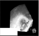
[0014] 도 4는 검출된 바운딩 박스를 와이어(wire) 프레임으로서 보여 주는 예시적인 렌더링된(rendered) 이미지이다.
[0015] 도 5a 및 도 5b는 각각, 타이트(tight)한 패턴(pattern) 및 넓은 패턴으로, 유두상 끝(papillary tip)들의 후보 지점들의 하이라이팅된 위치들을 갖는 예시적인 렌더링된 이미지들을 도시한다.
[0016] 도 6은, 공간 제약을 이용하여 제한한 이후에 남아 있으며 시간 제약을 사용하여 선택된, 검출기에 의해 출력된 예시적 후보 유두상 끝 위치와 지상 검증자료(ground truth) 유두상 끝 위치를 도시한다.
[0017] 도 7은 승모판막의 개개의 오버레이된 그래픽들(overlaid graphics)을 갖는 예시적 초음파 이미지들을 도시한다.
[0018] 도 8 및 도 9는 기계 학습 분류기를 이용하여 해부학적 구조를 검출하고, 광학적 흐름을 이용하여 해부학적 구조를 추적하기 위한 방법의 상이한 실시예들의 흐름도 다이어그램들이다.
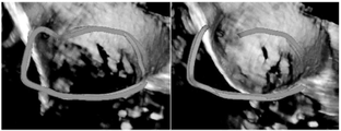
[0019] 도 10은 지상 검증자료와 오버레이된, 검출된 승모판막 고리(mitral valve annulus)의 예시적 이미지들을 도시한다.
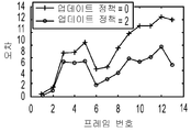
[0020] 도 11a 및 도 11b는, 기계 학습 검출에 의한 위치의 가끔의 업데이팅(occasional updating)을 이용하는, 그리고 이용하지 않는 광학적 흐름 추적에 대한 예시적 오차들을 도시한다.
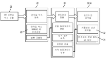
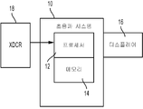
[0021] 도 12는 해부학적 검출을 위한 시스템의 일 실시예의 블록(block) 다이어그램이다.


Claims (21)
- 메디컬 이미징(medical imaging)에서 판막밑 위치(sub-valvular location)를 검출하기 위한 방법으로서, 상기 방법은,
메디컬 이미징 데이터(data)의 프레임(frame)들의 시퀀스(sequence)를 취득(22)하는 단계 ―상기 프레임들 각각은 환자의 심장의 적어도 일 구역의 볼륨(volume)을 표현함―;
상기 심장의 판막을 에워싸는 판막 바운딩 박스(bounding box)를 상기 프레임들 각각에서 검출(26)하는 단계;
상기 판막 바운딩 박스에 기반하여, 판막밑 구조 위치를 에워싸는 판막밑 바운딩 박스를 상기 프레임들 각각에서 검출(28)하는 단계;
상기 판막밑 바운딩 박스 내의 판막밑 위치의 후보 위치들을 상기 프레임들 각각에서 검출(30)하는 단계;
공간 제약 밖의 후보 위치들을 제거(32)하는 단계;
상기 제거(32) 이후에 남아 있는 후보 위치들 중 하나의 후보 위치를 상기 판막밑 위치로서 상기 프레임들 각각에서 선택(34)하는 단계 ―상기 선택(34)은 상기 프레임들에 걸친 후보 위치들의 함수임―; 및
상기 환자의 심장의 적어도 일 구역의 이미지(image)에서 상기 판막밑 위치를 하이라이팅(highlighting)(36)하는 단계
를 포함하는,
메디컬 이미징에서 판막밑 위치를 검출하기 위한 방법. - 제 1 항에 있어서,
상기 취득(22)하는 단계는 경식도 심초음파(transesophageal echocardiography) 데이터를 취득(22)하는 단계를 포함하는,
메디컬 이미징에서 판막밑 위치를 검출하기 위한 방법. - 제 1 항에 있어서,
상기 판막 바운딩 박스를 검출(26)하는 단계는 가장자리 공간 학습 분류기(marginal space learnt classifier)를 이용하여 직사각형 프리즘(prism)을 검출하는 단계를 포함하는,
메디컬 이미징에서 판막밑 위치를 검출하기 위한 방법. - 제 1 항에 있어서,
상기 판막밑 바운딩 박스를 검출(28)하는 단계는 상기 판막 바운딩 박스에 대한 상기 판막밑 바운딩 박스의 평균 포지션(position)에 기반하여 직사각형 프리즘을 검출하는 단계를 포함하는,
메디컬 이미징에서 판막밑 위치를 검출하기 위한 방법. - 제 1 항에 있어서,
상기 판막 바운딩 박스를 검출(26)하는 단계는 복수의 후보 승모판막 바운딩 박스들을 각각의 프레임에서 검출하는 단계를 포함하고, 상기 판막밑 바운딩 박스를 검출(28)하는 단계는 개개의 복수의 후보 유두상 바운딩 박스들을 각각의 프레임에서 검출하는 단계 및 상기 프레임들에 걸친 상기 후보 유두상(papillary) 바운딩 박스들의 랜덤 샘플 컨센서스(Random Sample Consensus)로부터 각각에 대한 유두상 바운딩 박스를 선택하는 단계를 포함하는,
메디컬 이미징에서 판막밑 위치를 검출하기 위한 방법. - 제 1 항에 있어서,
상기 후보 위치들을 검출(30)하는 단계는 가장자리 공간 학습 분류기를 이용하여 검출하는 단계를 포함하는,
메디컬 이미징에서 판막밑 위치를 검출하기 위한 방법. - 제 1 항에 있어서,
상기 제거(32)하는 단계는 상기 후보 위치들을 상기 승모판막의 랜드마크(landmark)에 대해 바운딩된(bounded) 구역으로 제한하는 단계를 포함하는,
메디컬 이미징에서 판막밑 위치를 검출하기 위한 방법. - 제 1 항에 있어서,
상기 선택(34)하는 단계는 마르코프(Markov) 제약을 이용하여 선택(34)하는 단계를 포함하는,
메디컬 이미징에서 판막밑 위치를 검출하기 위한 방법. - 제 1 항에 있어서,
상기 선택(34)하는 단계는 상기 후보 위치들의 상이한 세트(set)들로부터 상기 후보 위치들 중 단 한 개의 후보 위치를 갖는 세트를 각각의 프레임에서 선택(34)하는 단계를 포함하고, 상기 선택(34)은 상기 세트 내의 후보 위치들 사이의 시간 변위의 함수인,
메디컬 이미징에서 판막밑 위치를 검출하기 위한 방법. - 제 1 항에 있어서,
상기 하이라이팅(36)하는 단계는, 유두근 위치 및 상기 유두근 위치를 승모판막의 소엽(leaflet)들에 연결하는 끈(chordae)의 표현을 포함하는 상기 승모판막의 모델(model)의 그래픽(graphic)을 디스플레이(displaying)하는 단계, 또는 다른 판막 구조에 대한 그래픽들 없이 유두상 끝(papillary tip)들의 그래픽을 디스플레이하는 단계를 포함하는,
메디컬 이미징에서 판막밑 위치를 검출하기 위한 방법. - 제 1 항에 있어서,
상기 취득(22)하는 단계는, 시간에 따라 상기 심장을 표현하는 프레임들의 세트의 시퀀스의 희소 샘플링(sparse sampling)을 선택하는 단계를 포함하고,
상기 방법은, 광학적 흐름을 사용하여, 상기 희소 샘플링에서는 없는 판막밑 위치를 상기 프레임들에서 위치결정(38)하는 단계를 더 포함하는,
메디컬 이미징에서 판막밑 위치를 검출하기 위한 방법. - 제 11 항에 있어서,
상기 광학적 흐름에 대한 상기 판막밑 위치를 선택된 판막밑 위치로 주기적으로 업데이팅(updating)(40)하는 단계를 더 포함하는,
메디컬 이미징에서 판막밑 위치를 검출하기 위한 방법. - 비-일시적 컴퓨터(computer) 판독가능 저장 매체로서,
랜드마크 검출을 위해, 프로그래밍된 프로세서(programmed processor)(12)에 의해 실행가능한 명령들을 표현하는 데이터를 저장하고 있고, 상기 저장 매체는,
기계 학습 분류기를 이용하여 후보 해부학적 랜드마크들을 위치결정(30)하고;
공간 제약을 이용하여 상기 후보 해부학적 랜드마크들을 제한(32)하고;
상기 제한 이후에 남아 있는 후보 해부학적 랜드마크들로부터의 랜드마크의 위치를 복수의 볼륨들 각각에서 선택(34)하고 ―상기 선택은 시간에 따른 후보 랜드마크들의 함수임―; 그리고
상기 랜드마크의 이미지를 생성(36)하기 위한
명령들을 포함하는,
비-일시적 컴퓨터 판독가능 저장 매체. - 제 13 항에 있어서,
상기 위치결정(30)하는 것은 더 큰 해부학적 구조의 바운딩 박스로부터 상기 랜드마크의 바운딩 박스를 추정하는 것을 포함하는,
비-일시적 컴퓨터 판독가능 저장 매체. - 제 14 항에 있어서,
상기 위치결정(30)하는 것은 랜덤 샘플 컨센서스를 이용하여 상기 추정을 미세조정하는 것을 더 포함하는,
비-일시적 컴퓨터 판독가능 저장 매체. - 제 13 항에 있어서,
상기 기계 학습 분류기를 이용하여 위치결정(30)하는 것은 가장자리 공간 학습 분류기를 이용하여 위치결정하는 것을 포함하는,
비-일시적 컴퓨터 판독가능 저장 매체. - 제 13 항에 있어서,
상기 선택(34)하는 것은 마르코프 제약을 이용하여 선택(34)하는 것을 포함하는,
비-일시적 컴퓨터 판독가능 저장 매체. - 해부학적 랜드마크를 검출하기 위한 시스템(system)으로서, 상기 시스템은,
환자의 심장 볼륨을 스캔(scan)하도록 구성된 초음파 스캐너(scanner)(10) ―상기 스캔은 시간에 따라 상기 심장의 적어도 일부를 표현하는 메디컬 진단 초음파 데이터를 제공함―; 및
상기 스캔을 이용하여 실시간으로 상기 해부학적 랜드마크의 위치를 결정하도록 구성된 프로세서(12)
를 포함하고,
상기 위치는, 희소 프레임들에 대한 기계 학습 분류 및 상기 희소 프레임들 사이에 끼어 있는 프레임들에 대한 광학적 흐름을 이용하여 결정되며, 상기 위치는 상기 희소 프레임들 및 인터리빙된(interleaved) 프레임들이 취득될 때 순차적으로 결정되는,
해부학적 랜드마크를 검출하기 위한 시스템. - 제 18 항에 있어서,
상기 프로세서(12)는 상기 희소 프레임들에 대해 결정된, 상기 기계 학습 분류기로부터의 위치들을 이용하여 상기 광학적 흐름을 주기적으로 재초기화하도록 구성되는,
해부학적 랜드마크를 검출하기 위한 시스템. - 제 18 항에 있어서,
상기 프로세서(12)는 공간 제약 및 시간 마르코프 제약을 사용하는 기계 학습 분류를 이용하여 상기 위치를 결정하도록 구성되는,
해부학적 랜드마크를 검출하기 위한 시스템. - 제 18 항에 있어서,
상기 프로세서(12)는 시간에 따라 상기 해부학적 랜드마크의 특성을 측정하도록 구성되는,
해부학적 랜드마크를 검출하기 위한 시스템.
Applications Claiming Priority (5)
| Application Number | Priority Date | Filing Date | Title |
|---|---|---|---|
| US201462009512P | 2014-06-09 | 2014-06-09 | |
| US62/009,512 | 2014-06-09 | ||
| US201562128177P | 2015-03-04 | 2015-03-04 | |
| US62/128,177 | 2015-03-04 | ||
| PCT/US2015/034618 WO2015191414A2 (en) | 2014-06-09 | 2015-06-08 | Landmark detection with spatial and temporal constraints in medical imaging |
Publications (2)
| Publication Number | Publication Date |
|---|---|
| KR20170016461A true KR20170016461A (ko) | 2017-02-13 |
| KR101908520B1 KR101908520B1 (ko) | 2018-10-16 |
Family
ID=53487426
Family Applications (1)
| Application Number | Title | Priority Date | Filing Date |
|---|---|---|---|
| KR1020177000509A Active KR101908520B1 (ko) | 2014-06-09 | 2015-06-08 | 메디컬 이미징에서 공간 및 시간 제약들을 이용하는 랜드마크 검출 |
Country Status (5)
| Country | Link |
|---|---|
| US (1) | US10297027B2 (ko) |
| EP (1) | EP3152736B1 (ko) |
| KR (1) | KR101908520B1 (ko) |
| CN (1) | CN106605257B (ko) |
| WO (1) | WO2015191414A2 (ko) |
Cited By (3)
| Publication number | Priority date | Publication date | Assignee | Title |
|---|---|---|---|---|
| KR20190103048A (ko) * | 2018-02-27 | 2019-09-04 | 지멘스 메디컬 솔루션즈 유에스에이, 인크. | 정량적 초음파 이미징을 위한 관심 구역 배치 |
| KR20190113089A (ko) * | 2018-03-27 | 2019-10-08 | 울산대학교 산학협력단 | 근감소증 분석지원을 위한 인공 신경망 기반의 인체 형태 분석법을 채용하는 영상 처리 장치 및 이를 이용한 영상 처리 방법 |
| WO2022197074A1 (ko) * | 2021-03-17 | 2022-09-22 | 재단법인 아산사회복지재단 | 의료영상 처리 장치와 그 의료영상 학습 방법 및 의료영상 처리 방법 |
Families Citing this family (33)
| Publication number | Priority date | Publication date | Assignee | Title |
|---|---|---|---|---|
| KR20160032586A (ko) * | 2014-09-16 | 2016-03-24 | 삼성전자주식회사 | 관심영역 크기 전이 모델 기반의 컴퓨터 보조 진단 장치 및 방법 |
| US20170086789A1 (en) * | 2015-09-30 | 2017-03-30 | General Electric Company | Methods and systems for providing a mean velocity |
| WO2017140352A1 (en) * | 2016-02-16 | 2017-08-24 | Brainlab Ag | DETERMINATION OF DYNAMIC DRRs |
| US9928875B2 (en) * | 2016-03-22 | 2018-03-27 | Nec Corporation | Efficient video annotation with optical flow based estimation and suggestion |
| US11151721B2 (en) | 2016-07-08 | 2021-10-19 | Avent, Inc. | System and method for automatic detection, localization, and semantic segmentation of anatomical objects |
| KR101949114B1 (ko) | 2016-12-02 | 2019-02-15 | 아벤트, 인크. | 의료적 이미징 기반 절차에서 타겟 해부 대상으로의 내비게이션을 위한 시스템 및 방법 |
| JP6925824B2 (ja) * | 2017-02-28 | 2021-08-25 | キヤノンメディカルシステムズ株式会社 | 超音波診断装置、画像処理装置、及び画像処理プログラム |
| CN110914866B (zh) * | 2017-05-09 | 2024-04-30 | 哈特弗罗公司 | 用于在图像分析中进行解剖结构分割的系统和方法 |
| US11432875B2 (en) | 2017-09-28 | 2022-09-06 | Siemens Medical Solutions Usa, Inc. | Left atrial appendage closure guidance in medical imaging |
| US20190125295A1 (en) * | 2017-10-30 | 2019-05-02 | Siemens Medical Solutions Usa, Inc. | Cardiac flow detection based on morphological modeling in medical diagnostic ultrasound imaging |
| KR101919847B1 (ko) * | 2018-01-18 | 2018-11-19 | 주식회사 뷰노 | 동일 피사체에 대하여 시간 간격을 두고 촬영된 영상 간에 동일 관심구역을 자동으로 검출하는 방법 및 이를 이용한 장치 |
| EP3537447A1 (en) * | 2018-03-07 | 2019-09-11 | Koninklijke Philips N.V. | Display of medical image data |
| CN108510475B (zh) * | 2018-03-09 | 2022-03-29 | 南京合迈美家智能科技有限公司 | 一种肌肉连续超声图像中肌肉肌腱结的测量方法及系统 |
| EP3549529A1 (en) * | 2018-04-05 | 2019-10-09 | Koninklijke Philips N.V. | Ultrasound imaging system and method |
| WO2019226803A1 (en) | 2018-05-22 | 2019-11-28 | Boston Scientific Scimed, Inc. | Percutaneous papillary muscle relocation |
| US10733474B2 (en) | 2018-07-03 | 2020-08-04 | Sony Corporation | Method for 2D feature tracking by cascaded machine learning and visual tracking |
| GB201817238D0 (en) * | 2018-10-23 | 2018-12-05 | Univ Sheffield | Medical image processing |
| EP3705049A1 (en) | 2019-03-06 | 2020-09-09 | Piur Imaging GmbH | Apparatus and method for determining motion of an ultrasound probe including a forward-backward directedness |
| CN112150370B (zh) * | 2019-06-28 | 2024-02-06 | 深圳市恩普电子技术有限公司 | 一种空间复合成像方法和装置 |
| US12171592B2 (en) | 2019-08-30 | 2024-12-24 | Avent, Inc. | System and method for identification, labeling, and tracking of a medical instrument |
| US11244446B2 (en) * | 2019-10-25 | 2022-02-08 | Shanghai United Imaging Intelligence Co., Ltd. | Systems and methods for imaging |
| CN111192356B (zh) * | 2019-12-30 | 2023-04-25 | 上海联影智能医疗科技有限公司 | 感兴趣区域的显示方法、装置、设备和存储介质 |
| US12140525B2 (en) | 2020-04-22 | 2024-11-12 | Sony Group Corporation | Measuring device and imaging control method |
| CN111611909A (zh) * | 2020-05-18 | 2020-09-01 | 桂林电子科技大学 | 多子空间域自适应人脸识别方法 |
| GB202017510D0 (en) * | 2020-09-29 | 2020-12-23 | Univ Oxford Innovation Ltd | Method of training a machine learning model, method of assessing ultrasound measurement data, method of determining information about an anatomical featur |
| EP4280964A4 (en) * | 2021-01-19 | 2024-11-20 | ImaCor Inc. | Hemodynamic monitoring system implementing ultrasound imaging systems and machine learning-based image processing techniques |
| EP4092621B1 (en) | 2021-05-21 | 2026-04-22 | Siemens Healthineers AG | Technique for assigning a perfusion metric to dce mr images |
| EP4111982A1 (en) * | 2021-06-29 | 2023-01-04 | Koninklijke Philips N.V. | Systems and apparatuses for navigation and procedural guidance of laser leaflet resection under intracardiac echocardiography |
| US12315213B2 (en) * | 2021-08-20 | 2025-05-27 | Canon Medical Systems Corporation | Medical image processing apparatus and medical image processing method |
| US12315218B2 (en) * | 2022-07-06 | 2025-05-27 | Shanghai United Imaging Intelligence Co., Ltd. | Systems and methods for tracking groups of objects in medical images |
| CN115908330A (zh) * | 2022-11-23 | 2023-04-04 | 杭州脉流科技有限公司 | 基于dsa影像的冠脉自动化选帧分类推荐方法及装置 |
| WO2025255165A1 (en) * | 2024-06-04 | 2025-12-11 | Shifamed Holdings, Llc | Autonomous tee probe with graph model generation and landmark-based navigation |
| CN119214796B (zh) * | 2024-11-28 | 2025-02-07 | 西安市第九医院 | 一种心血管介入手术中的实时图像导航与定位系统 |
Citations (1)
| Publication number | Priority date | Publication date | Assignee | Title |
|---|---|---|---|---|
| JP2009153600A (ja) * | 2007-12-25 | 2009-07-16 | Toshiba Corp | 超音波診断装置、画像処理装置及びプログラム |
Family Cites Families (10)
| Publication number | Priority date | Publication date | Assignee | Title |
|---|---|---|---|---|
| US8771189B2 (en) | 2009-03-18 | 2014-07-08 | Siemens Medical Solutions Usa, Inc. | Valve assessment from medical diagnostic imaging data |
| US8682626B2 (en) | 2010-07-21 | 2014-03-25 | Siemens Aktiengesellschaft | Method and system for comprehensive patient-specific modeling of the heart |
| US8532352B2 (en) | 2010-10-06 | 2013-09-10 | Siemens Aktiengesellschaft | Method and system for intraoperative guidance using physiological image fusion |
| US9245091B2 (en) | 2011-03-09 | 2016-01-26 | Siemens Aktiengesellschaft | Physically-constrained modeling of a heart in medical imaging |
| US9824302B2 (en) | 2011-03-09 | 2017-11-21 | Siemens Healthcare Gmbh | Method and system for model-based fusion of multi-modal volumetric images |
| US9687204B2 (en) | 2011-05-20 | 2017-06-27 | Siemens Healthcare Gmbh | Method and system for registration of ultrasound and physiological models to X-ray fluoroscopic images |
| US8934693B2 (en) | 2011-11-23 | 2015-01-13 | Siemens Aktiengesellschaft | Method and system for intervention planning for transcatheter aortic valve implantation from 3D computed tomography data |
| US9730643B2 (en) * | 2013-10-17 | 2017-08-15 | Siemens Healthcare Gmbh | Method and system for anatomical object detection using marginal space deep neural networks |
| US10194888B2 (en) | 2015-03-12 | 2019-02-05 | Siemens Medical Solutions Usa, Inc. | Continuously oriented enhanced ultrasound imaging of a sub-volume |
| US10835210B2 (en) | 2015-03-30 | 2020-11-17 | Siemens Medical Solutions Usa, Inc. | Three-dimensional volume of interest in ultrasound imaging |
-
2015
- 2015-06-08 CN CN201580030902.2A patent/CN106605257B/zh active Active
- 2015-06-08 EP EP15731439.4A patent/EP3152736B1/en active Active
- 2015-06-08 WO PCT/US2015/034618 patent/WO2015191414A2/en not_active Ceased
- 2015-06-08 KR KR1020177000509A patent/KR101908520B1/ko active Active
- 2015-06-08 US US15/317,353 patent/US10297027B2/en active Active
Patent Citations (1)
| Publication number | Priority date | Publication date | Assignee | Title |
|---|---|---|---|---|
| JP2009153600A (ja) * | 2007-12-25 | 2009-07-16 | Toshiba Corp | 超音波診断装置、画像処理装置及びプログラム |
Cited By (4)
| Publication number | Priority date | Publication date | Assignee | Title |
|---|---|---|---|---|
| KR20190103048A (ko) * | 2018-02-27 | 2019-09-04 | 지멘스 메디컬 솔루션즈 유에스에이, 인크. | 정량적 초음파 이미징을 위한 관심 구역 배치 |
| US11006926B2 (en) | 2018-02-27 | 2021-05-18 | Siemens Medical Solutions Usa, Inc. | Region of interest placement for quantitative ultrasound imaging |
| KR20190113089A (ko) * | 2018-03-27 | 2019-10-08 | 울산대학교 산학협력단 | 근감소증 분석지원을 위한 인공 신경망 기반의 인체 형태 분석법을 채용하는 영상 처리 장치 및 이를 이용한 영상 처리 방법 |
| WO2022197074A1 (ko) * | 2021-03-17 | 2022-09-22 | 재단법인 아산사회복지재단 | 의료영상 처리 장치와 그 의료영상 학습 방법 및 의료영상 처리 방법 |
Also Published As
| Publication number | Publication date |
|---|---|
| US10297027B2 (en) | 2019-05-21 |
| CN106605257A (zh) | 2017-04-26 |
| CN106605257B (zh) | 2019-10-11 |
| WO2015191414A3 (en) | 2016-02-25 |
| KR101908520B1 (ko) | 2018-10-16 |
| WO2015191414A2 (en) | 2015-12-17 |
| EP3152736B1 (en) | 2020-01-08 |
| EP3152736A2 (en) | 2017-04-12 |
| US20170116748A1 (en) | 2017-04-27 |
Similar Documents
| Publication | Publication Date | Title |
|---|---|---|
| KR101908520B1 (ko) | 메디컬 이미징에서 공간 및 시간 제약들을 이용하는 랜드마크 검출 | |
| US12329565B2 (en) | Cardiac flow detection based on morphological modeling in medical diagnostic ultrasound imaging | |
| US12042232B2 (en) | Left atrial appendage closure guidance in medical imaging | |
| US10321892B2 (en) | Computerized characterization of cardiac motion in medical diagnostic ultrasound | |
| US10271817B2 (en) | Valve regurgitant detection for echocardiography | |
| US8812431B2 (en) | Method and system for medical decision support using organ models and learning based discriminative distance functions | |
| US5889524A (en) | Reconstruction of three-dimensional objects using labeled piecewise smooth subdivision surfaces | |
| US9033887B2 (en) | Mitral valve detection for transthoracic echocardiography | |
| US8218845B2 (en) | Dynamic pulmonary trunk modeling in computed tomography and magnetic resonance imaging based on the detection of bounding boxes, anatomical landmarks, and ribs of a pulmonary artery | |
| JP5108905B2 (ja) | 3dデータセット中の画像ビューを自動的に特定する方法および装置 | |
| CN112955934B (zh) | 识别医学图像中的介入设备 | |
| CN107403446A (zh) | 用于使用智能人工代理的图像配准的方法和系统 | |
| CN116883322A (zh) | 一种三维超声模型应用于心脏参数测量与管理方法及终端 | |
| CN117729890A (zh) | 用于二尖瓣反流评估的具有用户指导和自动图像设置选择的方法和装置 | |
| Carnahan | Towards Patient Specific Mitral Valve Modelling via Dynamic 3D Transesophageal Echocardiography | |
| Park et al. | Automatic computation of 2D cardiac measurements from B-mode echocardiography | |
| Mao | Three-dimensional Ultrasound Fusion for Transesophageal Echocardiography | |
| CN112336378B (zh) | 一种用于动物超声诊断的m型超声心动图处理方法和系统 | |
| MUNAFÒ | Deep learning-driven segmentation of echocardiographic images for intraprocedural support in percutaneous mitral valve repair | |
| Lenting | Automated echocardiographic surgical guidance in mitral valve repair | |
| Chen | QUiLT (Quantitative Ultrasound in Longitudinal Tissue Tracking): Stitching 2D images into 3D Volumes for Organ Health Monitoring | |
| Pedrosa et al. | Left ventricular segmentation of the heart in real-time in 3D echocardiography | |
| Vannelli et al. | How Accurately Does Transesophageal Echocardiography Identify the Mitral Valve? | |
| Schneider | Semi-Automatic Delineation of the Mitral Valve from Clinical Four-Dimensional Ultrasound Imaging | |
| CN120770840A (zh) | 引导式超声成像系统 |
Legal Events
| Date | Code | Title | Description |
|---|---|---|---|
| PA0105 | International application |
St.27 status event code: A-0-1-A10-A15-nap-PA0105 |
|
| A201 | Request for examination | ||
| E13-X000 | Pre-grant limitation requested |
St.27 status event code: A-2-3-E10-E13-lim-X000 |
|
| P11-X000 | Amendment of application requested |
St.27 status event code: A-2-2-P10-P11-nap-X000 |
|
| P13-X000 | Application amended |
St.27 status event code: A-2-2-P10-P13-nap-X000 |
|
| PA0201 | Request for examination |
St.27 status event code: A-1-2-D10-D11-exm-PA0201 |
|
| R15-X000 | Change to inventor requested |
St.27 status event code: A-3-3-R10-R15-oth-X000 |
|
| R16-X000 | Change to inventor recorded |
St.27 status event code: A-3-3-R10-R16-oth-X000 |
|
| PG1501 | Laying open of application |
St.27 status event code: A-1-1-Q10-Q12-nap-PG1501 |
|
| R18-X000 | Changes to party contact information recorded |
St.27 status event code: A-3-3-R10-R18-oth-X000 |
|
| E902 | Notification of reason for refusal | ||
| PE0902 | Notice of grounds for rejection |
St.27 status event code: A-1-2-D10-D21-exm-PE0902 |
|
| P11-X000 | Amendment of application requested |
St.27 status event code: A-2-2-P10-P11-nap-X000 |
|
| P13-X000 | Application amended |
St.27 status event code: A-2-2-P10-P13-nap-X000 |
|
| E701 | Decision to grant or registration of patent right | ||
| PE0701 | Decision of registration |
St.27 status event code: A-1-2-D10-D22-exm-PE0701 |
|
| GRNT | Written decision to grant | ||
| PR0701 | Registration of establishment |
St.27 status event code: A-2-4-F10-F11-exm-PR0701 |
|
| PR1002 | Payment of registration fee |
St.27 status event code: A-2-2-U10-U12-oth-PR1002 Fee payment year number: 1 |
|
| PG1601 | Publication of registration |
St.27 status event code: A-4-4-Q10-Q13-nap-PG1601 |
|
| PN2301 | Change of applicant |
St.27 status event code: A-5-5-R10-R11-asn-PN2301 |
|
| PN2301 | Change of applicant |
St.27 status event code: A-5-5-R10-R14-asn-PN2301 |
|
| PN2301 | Change of applicant |
St.27 status event code: A-5-5-R10-R11-asn-PN2301 |
|
| PN2301 | Change of applicant |
St.27 status event code: A-5-5-R10-R14-asn-PN2301 |
|
| PR1001 | Payment of annual fee |
St.27 status event code: A-4-4-U10-U11-oth-PR1001 Fee payment year number: 4 |
|
| P22-X000 | Classification modified |
St.27 status event code: A-4-4-P10-P22-nap-X000 |
|
| PR1001 | Payment of annual fee |
St.27 status event code: A-4-4-U10-U11-oth-PR1001 Fee payment year number: 5 |
|
| P22-X000 | Classification modified |
St.27 status event code: A-4-4-P10-P22-nap-X000 |
|
| P22-X000 | Classification modified |
St.27 status event code: A-4-4-P10-P22-nap-X000 |
|
| PR1001 | Payment of annual fee |
St.27 status event code: A-4-4-U10-U11-oth-PR1001 Fee payment year number: 6 |
|
| P22-X000 | Classification modified |
St.27 status event code: A-4-4-P10-P22-nap-X000 |
|
| PR1001 | Payment of annual fee |
St.27 status event code: A-4-4-U10-U11-oth-PR1001 Fee payment year number: 7 |
|
| PN2301 | Change of applicant |
St.27 status event code: A-5-5-R10-R11-asn-PN2301 |
|
| PN2301 | Change of applicant |
St.27 status event code: A-5-5-R10-R14-asn-PN2301 |
|
| PR1001 | Payment of annual fee |
St.27 status event code: A-4-4-U10-U11-oth-PR1001 Fee payment year number: 8 |
|
| U11 | Full renewal or maintenance fee paid |
Free format text: ST27 STATUS EVENT CODE: A-4-4-U10-U11-OTH-PR1001 (AS PROVIDED BY THE NATIONAL OFFICE) Year of fee payment: 8 |