KR20170094792A - Methods of measuring signaling pathway activity to diagnose and treat patients - Google Patents
Methods of measuring signaling pathway activity to diagnose and treat patients Download PDFInfo
- Publication number
- KR20170094792A KR20170094792A KR1020177018267A KR20177018267A KR20170094792A KR 20170094792 A KR20170094792 A KR 20170094792A KR 1020177018267 A KR1020177018267 A KR 1020177018267A KR 20177018267 A KR20177018267 A KR 20177018267A KR 20170094792 A KR20170094792 A KR 20170094792A
- Authority
- KR
- South Korea
- Prior art keywords
- sample
- cell
- subject
- cells
- agent
- Prior art date
- Legal status (The legal status is an assumption and is not a legal conclusion. Google has not performed a legal analysis and makes no representation as to the accuracy of the status listed.)
- Granted
Links
Images


Classifications
-
- G—PHYSICS
- G01—MEASURING; TESTING
- G01N—INVESTIGATING OR ANALYSING MATERIALS BY DETERMINING THEIR CHEMICAL OR PHYSICAL PROPERTIES
- G01N33/00—Investigating or analysing materials by specific methods not covered by groups G01N1/00 - G01N31/00
- G01N33/48—Biological material, e.g. blood, urine; Haemocytometers
- G01N33/50—Chemical analysis of biological material, e.g. blood, urine; Testing involving biospecific ligand binding methods; Immunological testing
- G01N33/53—Immunoassay; Biospecific binding assay; Materials therefor
- G01N33/575—Immunoassay; Biospecific binding assay; Materials therefor for cancer
- G01N33/5758—Immunoassay; Biospecific binding assay; Materials therefor for cancer involving compounds serving as markers for tumours, cancers or neoplasias, e.g. cellular determinants, receptors, heat shock/stress proteins, A-protein, oligosaccharides or metabolites
- G01N33/5759—Immunoassay; Biospecific binding assay; Materials therefor for cancer involving compounds serving as markers for tumours, cancers or neoplasias, e.g. cellular determinants, receptors, heat shock/stress proteins, A-protein, oligosaccharides or metabolites involving compounds localised on the membrane of tumour or cancer cells
-
- G—PHYSICS
- G01—MEASURING; TESTING
- G01N—INVESTIGATING OR ANALYSING MATERIALS BY DETERMINING THEIR CHEMICAL OR PHYSICAL PROPERTIES
- G01N33/00—Investigating or analysing materials by specific methods not covered by groups G01N1/00 - G01N31/00
- G01N33/48—Biological material, e.g. blood, urine; Haemocytometers
- G01N33/50—Chemical analysis of biological material, e.g. blood, urine; Testing involving biospecific ligand binding methods; Immunological testing
- G01N33/5005—Chemical analysis of biological material, e.g. blood, urine; Testing involving biospecific ligand binding methods; Immunological testing involving human or animal cells
- G01N33/5008—Chemical analysis of biological material, e.g. blood, urine; Testing involving biospecific ligand binding methods; Immunological testing involving human or animal cells for testing or evaluating the effect of chemical or biological compounds, e.g. drugs, cosmetics
- G01N33/5011—Chemical analysis of biological material, e.g. blood, urine; Testing involving biospecific ligand binding methods; Immunological testing involving human or animal cells for testing or evaluating the effect of chemical or biological compounds, e.g. drugs, cosmetics for testing antineoplastic activity
-
- A—HUMAN NECESSITIES
- A61—MEDICAL OR VETERINARY SCIENCE; HYGIENE
- A61K—PREPARATIONS FOR MEDICAL, DENTAL OR TOILETRY PURPOSES
- A61K31/00—Medicinal preparations containing organic active ingredients
- A61K31/33—Heterocyclic compounds
- A61K31/335—Heterocyclic compounds having oxygen as the only ring hetero atom, e.g. fungichromin
- A61K31/337—Heterocyclic compounds having oxygen as the only ring hetero atom, e.g. fungichromin having four-membered rings, e.g. taxol
-
- A—HUMAN NECESSITIES
- A61—MEDICAL OR VETERINARY SCIENCE; HYGIENE
- A61K—PREPARATIONS FOR MEDICAL, DENTAL OR TOILETRY PURPOSES
- A61K31/00—Medicinal preparations containing organic active ingredients
- A61K31/33—Heterocyclic compounds
- A61K31/395—Heterocyclic compounds having nitrogen as a ring hetero atom, e.g. guanethidine or rifamycins
- A61K31/435—Heterocyclic compounds having nitrogen as a ring hetero atom, e.g. guanethidine or rifamycins having six-membered rings with one nitrogen as the only ring hetero atom
- A61K31/47—Quinolines; Isoquinolines
- A61K31/4709—Non-condensed quinolines and containing further heterocyclic rings
-
- A—HUMAN NECESSITIES
- A61—MEDICAL OR VETERINARY SCIENCE; HYGIENE
- A61K—PREPARATIONS FOR MEDICAL, DENTAL OR TOILETRY PURPOSES
- A61K31/00—Medicinal preparations containing organic active ingredients
- A61K31/33—Heterocyclic compounds
- A61K31/395—Heterocyclic compounds having nitrogen as a ring hetero atom, e.g. guanethidine or rifamycins
- A61K31/435—Heterocyclic compounds having nitrogen as a ring hetero atom, e.g. guanethidine or rifamycins having six-membered rings with one nitrogen as the only ring hetero atom
- A61K31/47—Quinolines; Isoquinolines
- A61K31/4738—Quinolines; Isoquinolines ortho- or peri-condensed with heterocyclic ring systems
- A61K31/4745—Quinolines; Isoquinolines ortho- or peri-condensed with heterocyclic ring systems condensed with ring systems having nitrogen as a ring hetero atom, e.g. phenantrolines
-
- A—HUMAN NECESSITIES
- A61—MEDICAL OR VETERINARY SCIENCE; HYGIENE
- A61K—PREPARATIONS FOR MEDICAL, DENTAL OR TOILETRY PURPOSES
- A61K31/00—Medicinal preparations containing organic active ingredients
- A61K31/33—Heterocyclic compounds
- A61K31/395—Heterocyclic compounds having nitrogen as a ring hetero atom, e.g. guanethidine or rifamycins
- A61K31/495—Heterocyclic compounds having nitrogen as a ring hetero atom, e.g. guanethidine or rifamycins having six-membered rings with two or more nitrogen atoms as the only ring heteroatoms, e.g. piperazine or tetrazines
- A61K31/505—Pyrimidines; Hydrogenated pyrimidines, e.g. trimethoprim
- A61K31/506—Pyrimidines; Hydrogenated pyrimidines, e.g. trimethoprim not condensed and containing further heterocyclic rings
-
- A—HUMAN NECESSITIES
- A61—MEDICAL OR VETERINARY SCIENCE; HYGIENE
- A61K—PREPARATIONS FOR MEDICAL, DENTAL OR TOILETRY PURPOSES
- A61K31/00—Medicinal preparations containing organic active ingredients
- A61K31/33—Heterocyclic compounds
- A61K31/395—Heterocyclic compounds having nitrogen as a ring hetero atom, e.g. guanethidine or rifamycins
- A61K31/495—Heterocyclic compounds having nitrogen as a ring hetero atom, e.g. guanethidine or rifamycins having six-membered rings with two or more nitrogen atoms as the only ring heteroatoms, e.g. piperazine or tetrazines
- A61K31/505—Pyrimidines; Hydrogenated pyrimidines, e.g. trimethoprim
- A61K31/517—Pyrimidines; Hydrogenated pyrimidines, e.g. trimethoprim ortho- or peri-condensed with carbocyclic ring systems, e.g. quinazoline, perimidine
-
- A—HUMAN NECESSITIES
- A61—MEDICAL OR VETERINARY SCIENCE; HYGIENE
- A61K—PREPARATIONS FOR MEDICAL, DENTAL OR TOILETRY PURPOSES
- A61K31/00—Medicinal preparations containing organic active ingredients
- A61K31/33—Heterocyclic compounds
- A61K31/395—Heterocyclic compounds having nitrogen as a ring hetero atom, e.g. guanethidine or rifamycins
- A61K31/495—Heterocyclic compounds having nitrogen as a ring hetero atom, e.g. guanethidine or rifamycins having six-membered rings with two or more nitrogen atoms as the only ring heteroatoms, e.g. piperazine or tetrazines
- A61K31/505—Pyrimidines; Hydrogenated pyrimidines, e.g. trimethoprim
- A61K31/519—Pyrimidines; Hydrogenated pyrimidines, e.g. trimethoprim ortho- or peri-condensed with heterocyclic rings
-
- A—HUMAN NECESSITIES
- A61—MEDICAL OR VETERINARY SCIENCE; HYGIENE
- A61K—PREPARATIONS FOR MEDICAL, DENTAL OR TOILETRY PURPOSES
- A61K31/00—Medicinal preparations containing organic active ingredients
- A61K31/33—Heterocyclic compounds
- A61K31/395—Heterocyclic compounds having nitrogen as a ring hetero atom, e.g. guanethidine or rifamycins
- A61K31/535—Heterocyclic compounds having nitrogen as a ring hetero atom, e.g. guanethidine or rifamycins having six-membered rings with at least one nitrogen and one oxygen as the ring hetero atoms, e.g. 1,2-oxazines
- A61K31/5375—1,4-Oxazines, e.g. morpholine
- A61K31/5377—1,4-Oxazines, e.g. morpholine not condensed and containing further heterocyclic rings, e.g. timolol
-
- A—HUMAN NECESSITIES
- A61—MEDICAL OR VETERINARY SCIENCE; HYGIENE
- A61K—PREPARATIONS FOR MEDICAL, DENTAL OR TOILETRY PURPOSES
- A61K31/00—Medicinal preparations containing organic active ingredients
- A61K31/33—Heterocyclic compounds
- A61K31/555—Heterocyclic compounds containing heavy metals, e.g. hemin, hematin, melarsoprol
-
- A—HUMAN NECESSITIES
- A61—MEDICAL OR VETERINARY SCIENCE; HYGIENE
- A61K—PREPARATIONS FOR MEDICAL, DENTAL OR TOILETRY PURPOSES
- A61K31/00—Medicinal preparations containing organic active ingredients
- A61K31/70—Carbohydrates; Sugars; Derivatives thereof
- A61K31/7042—Compounds having saccharide radicals and heterocyclic rings
- A61K31/7052—Compounds having saccharide radicals and heterocyclic rings having nitrogen as a ring hetero atom, e.g. nucleosides, nucleotides
- A61K31/706—Compounds having saccharide radicals and heterocyclic rings having nitrogen as a ring hetero atom, e.g. nucleosides, nucleotides containing six-membered rings with nitrogen as a ring hetero atom
- A61K31/7064—Compounds having saccharide radicals and heterocyclic rings having nitrogen as a ring hetero atom, e.g. nucleosides, nucleotides containing six-membered rings with nitrogen as a ring hetero atom containing condensed or non-condensed pyrimidines
- A61K31/7068—Compounds having saccharide radicals and heterocyclic rings having nitrogen as a ring hetero atom, e.g. nucleosides, nucleotides containing six-membered rings with nitrogen as a ring hetero atom containing condensed or non-condensed pyrimidines having oxo groups directly attached to the pyrimidine ring, e.g. cytidine, cytidylic acid
-
- A—HUMAN NECESSITIES
- A61—MEDICAL OR VETERINARY SCIENCE; HYGIENE
- A61K—PREPARATIONS FOR MEDICAL, DENTAL OR TOILETRY PURPOSES
- A61K33/00—Medicinal preparations containing inorganic active ingredients
- A61K33/24—Heavy metals; Compounds thereof
- A61K33/243—Platinum; Compounds thereof
-
- A—HUMAN NECESSITIES
- A61—MEDICAL OR VETERINARY SCIENCE; HYGIENE
- A61K—PREPARATIONS FOR MEDICAL, DENTAL OR TOILETRY PURPOSES
- A61K45/00—Medicinal preparations containing active ingredients not provided for in groups A61K31/00 - A61K41/00
- A61K45/06—Mixtures of active ingredients without chemical characterisation, e.g. antiphlogistics and cardiaca
-
- C—CHEMISTRY; METALLURGY
- C07—ORGANIC CHEMISTRY
- C07K—PEPTIDES
- C07K16/00—Immunoglobulins [IG], e.g. monoclonal or polyclonal antibodies
- C07K16/18—Immunoglobulins [IG], e.g. monoclonal or polyclonal antibodies against material from animals or humans
- C07K16/28—Immunoglobulins [IG], e.g. monoclonal or polyclonal antibodies against material from animals or humans against receptors, cell surface antigens or cell surface determinants
- C07K16/2863—Immunoglobulins [IG], e.g. monoclonal or polyclonal antibodies against material from animals or humans against receptors, cell surface antigens or cell surface determinants against receptors for growth factors, growth regulators
-
- C—CHEMISTRY; METALLURGY
- C07—ORGANIC CHEMISTRY
- C07K—PEPTIDES
- C07K16/00—Immunoglobulins [IG], e.g. monoclonal or polyclonal antibodies
- C07K16/18—Immunoglobulins [IG], e.g. monoclonal or polyclonal antibodies against material from animals or humans
- C07K16/32—Immunoglobulins [IG], e.g. monoclonal or polyclonal antibodies against material from animals or humans against translation products of oncogenes
-
- G—PHYSICS
- G01—MEASURING; TESTING
- G01N—INVESTIGATING OR ANALYSING MATERIALS BY DETERMINING THEIR CHEMICAL OR PHYSICAL PROPERTIES
- G01N33/00—Investigating or analysing materials by specific methods not covered by groups G01N1/00 - G01N31/00
- G01N33/48—Biological material, e.g. blood, urine; Haemocytometers
- G01N33/50—Chemical analysis of biological material, e.g. blood, urine; Testing involving biospecific ligand binding methods; Immunological testing
- G01N33/5005—Chemical analysis of biological material, e.g. blood, urine; Testing involving biospecific ligand binding methods; Immunological testing involving human or animal cells
- G01N33/5008—Chemical analysis of biological material, e.g. blood, urine; Testing involving biospecific ligand binding methods; Immunological testing involving human or animal cells for testing or evaluating the effect of chemical or biological compounds, e.g. drugs, cosmetics
- G01N33/5044—Chemical analysis of biological material, e.g. blood, urine; Testing involving biospecific ligand binding methods; Immunological testing involving human or animal cells for testing or evaluating the effect of chemical or biological compounds, e.g. drugs, cosmetics involving specific cell types
-
- G—PHYSICS
- G01—MEASURING; TESTING
- G01N—INVESTIGATING OR ANALYSING MATERIALS BY DETERMINING THEIR CHEMICAL OR PHYSICAL PROPERTIES
- G01N33/00—Investigating or analysing materials by specific methods not covered by groups G01N1/00 - G01N31/00
- G01N33/48—Biological material, e.g. blood, urine; Haemocytometers
- G01N33/50—Chemical analysis of biological material, e.g. blood, urine; Testing involving biospecific ligand binding methods; Immunological testing
- G01N33/53—Immunoassay; Biospecific binding assay; Materials therefor
- G01N33/575—Immunoassay; Biospecific binding assay; Materials therefor for cancer
- G01N33/57515—Immunoassay; Biospecific binding assay; Materials therefor for cancer of the breast
-
- G—PHYSICS
- G01—MEASURING; TESTING
- G01N—INVESTIGATING OR ANALYSING MATERIALS BY DETERMINING THEIR CHEMICAL OR PHYSICAL PROPERTIES
- G01N2333/00—Assays involving biological materials from specific organisms or of a specific nature
- G01N2333/435—Assays involving biological materials from specific organisms or of a specific nature from animals; from humans
- G01N2333/475—Assays involving growth factors
- G01N2333/4756—Neuregulins, i.e. p185erbB2 ligands, glial growth factor, heregulin, ARIA, neu differentiation factor
-
- G—PHYSICS
- G01—MEASURING; TESTING
- G01N—INVESTIGATING OR ANALYSING MATERIALS BY DETERMINING THEIR CHEMICAL OR PHYSICAL PROPERTIES
- G01N2333/00—Assays involving biological materials from specific organisms or of a specific nature
- G01N2333/435—Assays involving biological materials from specific organisms or of a specific nature from animals; from humans
- G01N2333/475—Assays involving growth factors
- G01N2333/485—Epidermal growth factor [EGF] (urogastrone)
-
- G—PHYSICS
- G01—MEASURING; TESTING
- G01N—INVESTIGATING OR ANALYSING MATERIALS BY DETERMINING THEIR CHEMICAL OR PHYSICAL PROPERTIES
- G01N2333/00—Assays involving biological materials from specific organisms or of a specific nature
- G01N2333/435—Assays involving biological materials from specific organisms or of a specific nature from animals; from humans
- G01N2333/705—Assays involving receptors, cell surface antigens or cell surface determinants
- G01N2333/71—Assays involving receptors, cell surface antigens or cell surface determinants for growth factors; for growth regulators
-
- G—PHYSICS
- G01—MEASURING; TESTING
- G01N—INVESTIGATING OR ANALYSING MATERIALS BY DETERMINING THEIR CHEMICAL OR PHYSICAL PROPERTIES
- G01N2800/00—Detection or diagnosis of diseases
- G01N2800/52—Predicting or monitoring the response to treatment, e.g. for selection of therapy based on assay results in personalised medicine; Prognosis
-
- Y—GENERAL TAGGING OF NEW TECHNOLOGICAL DEVELOPMENTS; GENERAL TAGGING OF CROSS-SECTIONAL TECHNOLOGIES SPANNING OVER SEVERAL SECTIONS OF THE IPC; TECHNICAL SUBJECTS COVERED BY FORMER USPC CROSS-REFERENCE ART COLLECTIONS [XRACs] AND DIGESTS
- Y02—TECHNOLOGIES OR APPLICATIONS FOR MITIGATION OR ADAPTATION AGAINST CLIMATE CHANGE
- Y02A—TECHNOLOGIES FOR ADAPTATION TO CLIMATE CHANGE
- Y02A90/00—Technologies having an indirect contribution to adaptation to climate change
- Y02A90/10—Information and communication technologies [ICT] supporting adaptation to climate change, e.g. for weather forecasting or climate simulation
Landscapes
- Health & Medical Sciences (AREA)
- Life Sciences & Earth Sciences (AREA)
- Chemical & Material Sciences (AREA)
- Engineering & Computer Science (AREA)
- Immunology (AREA)
- Biomedical Technology (AREA)
- Medicinal Chemistry (AREA)
- General Health & Medical Sciences (AREA)
- Molecular Biology (AREA)
- Hematology (AREA)
- Urology & Nephrology (AREA)
- Cell Biology (AREA)
- Public Health (AREA)
- Animal Behavior & Ethology (AREA)
- Epidemiology (AREA)
- Pharmacology & Pharmacy (AREA)
- Veterinary Medicine (AREA)
- Biochemistry (AREA)
- General Physics & Mathematics (AREA)
- Pathology (AREA)
- Analytical Chemistry (AREA)
- Physics & Mathematics (AREA)
- Food Science & Technology (AREA)
- Microbiology (AREA)
- Biotechnology (AREA)
- Tropical Medicine & Parasitology (AREA)
- Toxicology (AREA)
- Bioinformatics & Cheminformatics (AREA)
- Organic Chemistry (AREA)
- Inorganic Chemistry (AREA)
- Proteomics, Peptides & Aminoacids (AREA)
- Genetics & Genomics (AREA)
- Biophysics (AREA)
- Oncology (AREA)
- Measuring Or Testing Involving Enzymes Or Micro-Organisms (AREA)
- Investigating Or Analysing Biological Materials (AREA)
- Pharmaceuticals Containing Other Organic And Inorganic Compounds (AREA)
- Medicines That Contain Protein Lipid Enzymes And Other Medicines (AREA)
- Medicines Containing Antibodies Or Antigens For Use As Internal Diagnostic Agents (AREA)
Abstract
개별 대상체로부터 수득된 이환 세포 샘플 내 세포성 경로의 기능성 상태의 계측 방법이 본원에 제공된다. 이러한 방법은, 경로가 정상적으로 기능화하는 경우 특이적 세포성 경로를 교란시킨다고 공지된 교란제 (예를 들면, 활성화제)와, 상기 대상체로부터 수득된 이환 세포 샘플을 접촉시키는 단계를 포함한다. 상기 교란제의 존재 하에서, 하나 이상의 생리적 반응 파라미터에서의 변화는, 상기 교란제에 의해 표적된 상기 세포성 경로가 상기 개별 대상체에서 기능성이라는 것을 나타낸다. 개별 개상체에 대하여 표적화된 치료제를 선택하는 방법이 또한 제공된다.Methods of measuring the functional status of cellular pathways in a sample of diseased cells obtained from an individual subject are provided herein. Such methods include contacting a sample of a diseased cell obtained from the subject with an agonist ( e.g., an activator) known to disrupt the specific cellular pathway when the pathway normally functions. In the presence of the agonist, a change in one or more physiological response parameters indicates that the cellular pathway targeted by the agonist is functional in the individual subject. Also provided is a method for selecting a targeted therapeutic agent for an individual.
Description
관련 출원Related application
본 출원은, 2014년 12월 12일 출원된, 미국 가특허 출원 시리얼 번호 62/091,180에 우선권을 주장하고, 이의 내용은 본원에서 참조로 편입된다.This application claims priority to U.S. Provisional Application Serial No. 62 / 091,180, filed December 12, 2014, the contents of which are incorporated herein by reference.
발명의 배경BACKGROUND OF THE INVENTION
화학물질 및 외인성 단백질이 특이적 세포성 표적에 대해 유효한 인간 치료제일 수 있다는 발견 이후로 이환 개인의 치료는 유의미한 진전을 가졌다. 그러나, 많은 통상 질환 예컨대 암의 치료에서 개선에 대한 유의미한 여지가 여전히 있다. 인간 게놈 프로젝트의 주요 동인 중 하나는, 치료 중재의 발전 및 처방을 진전시키기 위해, 질환의 유전적 원인을 발견하는 것이었다. 보고가 믿어진다면, 전체 인간 유전자는 인간 게놈 프로젝트를 통해 확인되었다. 많은 이들 유전자는 인간 집단에서 질환에 통계적으로 연결된다. 그렇지만 질환의 유전적 연결 또는 유전적 바이오마커의 검출의 지식이 질환 과정 또는 치료적 결과를 항상 효과적으로 예측하지 않는다. 그래서 유전자로부터 유전적 연결 및 심지어 단백질 발현 수준의 정량화도 적절한 치료적 과정의 결정에서 그렇게 매우 제한된다.Since the discovery that chemicals and extrinsic proteins can be effective human therapeutics for specific cellular targets, the treatment of individuals with mental disorders has had significant advances. However, there is still significant room for improvement in the treatment of many common diseases, such as cancer. One of the key drivers of the human genome project was to discover the genetic cause of the disease, in order to advance the development and prescription of therapeutic interventions. If the report is believed, the entire human gene has been identified through the Human Genome Project. Many of these genes are statistically linked to disease in human populations. However, the knowledge of the genetic linkage of a disease or the detection of a genetic biomarker does not always predict the disease course or therapeutic outcome effectively. Thus, genetic linkage from the gene and even quantification of the level of protein expression are so very limited in determining the appropriate therapeutic process.
페타바이트의 유전 정보는 수집되었다. 상당한 통계적인 및 분석적 모델링 컴퓨터처리 능력은 많은 상이한 유형의 질환을 분석하기 위해 수집된 유전 데이터에 적용되었다. 적어도 2개의 중요한 사실이 상기 과정에서 부각되었다. 첫째, 한 개인에서의 유방암이 유전자 구성, 단백질 발현 수준, 및 치료 중재에 대한 반응에서 또 다른 개인에서의 동일한 암과 완전히 상이할 수 있기 때문에 유방암같은 "질환"은 부분적으로 이종성이다. 둘째, 현행 유전 바이오마커의 검출이 대다수의 사례에서 좋지 못한 예측의 값을 갖는다.The petabytes of genetic information were collected. Significant Statistical and Analytical Modeling Computer processing capabilities have been applied to genetic data collected to analyze many different types of diseases. At least two important facts have been highlighted in the process. First, the "disease", such as breast cancer, is partially heterogeneous because breast cancer in one individual can be completely different from the same cancers in another individual in response to gene composition, protein expression levels, and therapeutic intervention. Second, detection of current genetic biomarkers has poor predictive value in most cases.
동시대의 표적된 약물은 특이적 제한된 수의 인간 세포 모델을 염두에 둔 과정을 따라 발견되고 개발된다. 많은 이들 세포주는 특정한 세포성 표적에 대해 원하는 활성을 갖는 것을 선택하기 위해 포텐셜 약물의 큰 라이브러리의 최적화된 스크리닝 환경을 제공하도록 조작된다. 상기 과정의 사용은 각 환자의 질환이 동일한 질환을 가진 다른 환자와 상이하다는 것을 나타내는 임상 정보의 면에서 포텐셜 약물의 효능에 관하여 오도할 수 있다. 약물 발견 및 발전 과정은 현재까지 임상시험에 앞서 반응성 인간의 확인에서 매우 유효하지 않고 임상 발전 과정 전반에 걸쳐 높은 실패율을 계속해서 겪고 있다. 환자에 손해 감소에 집중하는 조절 임상 발전 과정을 통해 승인되는 많은 약물은 실제의 질환 환자 집단에서 좋지 못한 유효율로 고통받는다.Contemporary targeted drugs are discovered and developed following a process with a specific, limited number of human cell models in mind. Many of these cell lines are engineered to provide an optimized screening environment for large libraries of potential drugs to select those that have the desired activity for a particular cellular target. The use of this procedure may be misleading as to the efficacy of the potential drug in terms of clinical information indicating that the disease of each patient is different from other patients with the same disease. The drug discovery and development process is currently far ineffective in identifying reactive individuals prior to clinical trials and continues to experience high failure rates throughout the clinical development process. Many drugs that are approved through controlled clinical development processes that focus on reducing the patient's injury suffer from poor efficacy rates in the actual patient population.
임상 의사에 모든 질환 상태 설명이 동일한 원인에서 발생하지 않는다. 단순 예에서, 골 관절의 염증은 몇 개의 공급원, 일부 내부, 일부 외부, 일부 "유전자 상으로 연결된", 및 일부 아직 미공지된 원인에서 발생할 수 있다. 의학은 외부 병원체가 적절하게 확인될 수 있는 경우 감염성 질환에 대한 환자의 부상자 분류에 상당히 유효하다. 의사는 현재 이용가능한 요법이 내부 원인으로부터 염증의 감소로 이어질 예측을 위하여 가까이에 소수의 도구를 갖는다. 의사는 특이적 환자의 세포가 어떻게 기능하고 있는지, 또는 더욱 적절하게 기능부전하는지, 및 이들이 "염증"으로서 임상적으로 나타내는 질환의 치료에 이용가능한 많은 치료제 중 하나에 어떻게 반응할지의 지식이 부족하다. 이들은 비정상적인 유전자가 존재한다는 것을 알지만 그러나 특이적 환자에서 질환 과정에 어떻게 영향을 미치는지는 알지 못한다. 이들은 약물이 특이적으로 어떻게 작용하는지는 알 수 있지만 그러나 특정한 환자가 그 약물 활성에 무반응성 또는 내성일 수 있는 이유는 모를 수 있다.Not all disease state descriptions in clinical doctors occur in the same cause. In a simple example, inflammation of the osteoarthritis can occur from several sources, some internal, some external, some "genetically linked", and some yet unknown causes. Medicine is quite effective in classifying patients' injuries to infectious diseases if external pathogens can be properly identified. The physician has a handful of tools at hand to predict which therapies currently available will lead to reduced inflammation from internal causes. Physicians lack knowledge of how the cells of a particular patient function, or more appropriately dysfunction, and how to respond to one of the many therapies available for the treatment of diseases clinically indicated as "inflammation" . They know that abnormal genes are present, but they do not know how they affect disease processes in specific patients. They may know how a drug specifically works, but may not know why a particular patient may be unresponsive or tolerant to its drug activity.
환자는 그의 특정한 질환 원인의 더 나은 확인 및 유효한 치료적 과정의 의사결정을 필요로 한다. 인간 게놈 서열분석 및 다른 유전적 정량화 도구는 각 환자의 질환이 그 환자에게 어느 정도 고유한지를 의사에게 정보제공하였다. 상기 정보는 개인맞춤형 의약에 관한 전체 사업을 낳았고, 여기에서 각 환자는 그의 질환에 맞춤화된 맞춤형 치료 레지멘을 잠재적으로 받을 수 있다. 약물은 특이적 유전자-관련 질환 징후에 대하여 개발되고 있다. 상기 이상적 접근법은 현행 예후 도구세트의 유의미한 단점에 주로 기인하여 아직 입증되지 않았다. 유전자는 존재할 수 있지만 특정한 개인의 맥락에서 그의 기능은 상관되지 않는다.The patient needs better identification of his specific disease cause and decision of an effective therapeutic process. Human genome sequencing and other genetic quantification tools informed the physician that each patient's disease was unique to the patient. This information has resulted in a whole business on personalized medicines where each patient can potentially receive tailored treatment regimens tailored to his disease. Drugs are being developed for specific gene-related disease indications. This ideal approach has not yet been proven due to the significant shortcomings of the current prognostic toolset. Genes can exist, but their function is not correlated in the context of a particular individual.
각 환자가 상이하다는 것 및 여러번 요법이 양성 반응을 효과내는데 실패한다는 것의 인식에 대한 하나의 반응은 동반 진단의 발전이었다. 진단 시험의 상기 유형은 특정한 약물에 더욱 반응할 것 같은 환자를 확인하려고 노력하기 위한 동시대의 바이오마커 검출을 이용하여 설계된다. 시험은 증가된 유전자 수, 유전자 돌연변이, 또는 특정한 유전자의 변경된 발현 수준 찾기를 포함한다. 예를 들어, 많은 암은 요즘 질환과 관련된 특이적 유전적 돌연변이 또는 과-발현된 수용체 단백질의 존재를 계측하기 위한 시험의 도움으로 진단된다. 그러나, 대부분의 바이오마커 시험은 질환 및 그의 근본적인 원인에 대하여 간접적 및 추론적 정보만을 제공한다. 따라서, 유의미한 치료적 반응의 예측에서 대부분의 이들 시험에 대한 성공률은 종종 50% 보다 훨씬 적다.One response to the recognition that each patient was different and that multiple therapies failed to effect a positive response was the development of a comorbid diagnosis. This type of diagnostic test is designed using contemporary biomarker detection to try to identify patients who are more likely to respond to a particular drug. Testing includes finding increased gene numbers, gene mutations, or altered expression levels of a particular gene. For example, many cancers are diagnosed with the help of tests to measure the presence of specific genetic mutations or over-expressed receptor proteins associated with current disease. However, most biomarker tests provide only indirect and speculative information about the disease and its underlying causes. Thus, in predicting meaningful therapeutic responses, the success rate for most of these trials is often much less than 50%.
따라서 개인에 대한 치료제의 유효성에 대하여 더 나은 진단 검정 및 더 나은 예후 지표를 제공할 필요성이 남아 있다.Thus, there remains a need to provide better diagnostic tests and better prognostic indicators of the efficacy of therapeutic agents for individuals.
발명의 요약SUMMARY OF THE INVENTION
질환의 진단을 위하여 그리고 치료적 의사결정의 기반으로서 바이오마커의 사용은 질환에 관여된 신호전달 경로(들)의 활성이 평가되지 않는다는 사실에 의해 제한된다. 따라서, 본 발명은 질환에 관여된 신호전달 경로(들)이 환자의 이환 세포의 생리적 활성에서 변화에 기반하여 평가되는 진단 방법을 제공한다. 따라서, 본 발명은, 신호전달 경로가 질환 과정에서 연루되는지를 사실상 나타내지 않을 수 있는 바이오마커 (예컨대 세포 표면 수용체)의 발현 보다는, 상기 생리적 활성에 기반하여 치료 레지멘이 선택될 수 있는 정도로 환자의 이환 세포에서 특정한 신호전달 경로가 활성인지의 계측을 허용한다. 특히, 본 발명은 "바이오마커 음성"이고 그렇지만 질환 과정에서 연루된 활성 신호전달 경로를 갖는 환자의 확인 방법을 제공한다. 이는 그의 바이오마커 음성 상태에 기반된 상기 치료에 대하여 이전에 건너뛴 그 신호전달 경로에 영향을 미치는 표적화 치료제를 이용한 환자의 선택 및 치료를 허용한다. 예를 들어, 본 발명은 활성 ErbB 신호전달 경로를 아직 갖고 있는 ErbB2 음성 암을 가진 환자의 확인 방법을 제공하여 이로써 그 환자는 ErbB 신호전달 경로에 영향을 미치는 표적화 치료제를 이용한 치료에 대해 선택될 수 있고, 반면에 본 발명에 앞서 그 환자는 ErbB 표적화 치료제를 이용한 치료로 간과될 것이다. 이는 ErbB2를 표적하는 약물용 FDA 표지가 과-발현된 또는 증폭된 ErbB2 수용체를 가진 환자에 사용으로만 승인된다는 사실을 반영한다. 마찬가지로, 본 발명은 활성 ER 신호전달 경로를 아직 갖는 에스트로겐 수용체 (ER) 음성 암을 가진 환자의 확인 방법을 제공하여 이로써 그 환자가 ER 신호전달 경로 (예를 들면, 호르몬 요법)에 영향을 미치는 표적화 치료제를 이용한 치료에 선택될 수 있고, 반면에 본 발명에 앞서 그 환자는 ER-관련 요법 (예를 들면, 호르몬 요법)에 간과될 것이다. 따라서, 본 발명은 유의미하게 표적화 치료제를 이용한 치료받음에서 유익할 수 있는 환자 집단을 확대한다.The use of biomarkers for diagnosis of disease and as a basis for therapeutic decision making is limited by the fact that the activity of the pathway (s) involved in the disease is not assessed. Thus, the present invention provides a diagnostic method wherein the disease-associated signaling pathway (s) is evaluated based on a change in the physiological activity of the patient's miliary cells. Accordingly, the present invention provides a method of treating a patient, such as a patient, to the extent that a therapeutic regimen can be selected based on said physiological activity rather than the expression of a biomarker (e.g., a cell surface receptor) that may not actually indicate that the signaling pathway is involved in a disease process Allowing the measurement of the specific signaling pathway in the diseased cells to be active. In particular, the present invention provides a method of identifying a patient having "biomarker negative" but having an active signal transduction pathway involved in the disease process. This allows the selection and treatment of patients with targeted therapies that affect their signaling pathway previously skipped for the treatment based on their biomarker negative state. For example, the present invention provides a method of identifying a patient with an ErbB2 negative cancer that still has an active ErbB signaling pathway, whereby the patient can be selected for treatment with a targeting agent that affects the ErbB signaling pathway , Whereas prior to the present invention the patient would be overlooked as treatment with an ErbB targeted therapeutic. This reflects the fact that the FDA labeling for drugs targeting ErbB2 is approved for use only in patients with over-expressed or amplified ErbB2 receptors. Likewise, the present invention provides a method of identifying a patient with an estrogen receptor (ER) negative cancer that still has an active ER signaling pathway, such that the patient has a targetizing effect that affects the ER signaling pathway (e. G., Hormone therapy) May be selected for treatment with a therapeutic agent, whereas prior to the present invention the patient will be overlooked for ER-related therapies (e. G., Hormone therapy). Thus, the present invention extends the population of patients that may benefit from treatment with a targeted therapeutic agent.
본 발명의 한 측면은, 하기 단계를 포함하는, 이환 세포에서 비정상 신호전달 경로와 관련된 질환에 대하여 대상체의 진단 및 치료 방법으로서, 상기 대상체가 질환에 대하여 바이오마커 음성인, 방법을 제공한다:One aspect of the present invention provides a method of diagnosing and treating a subject for a disease associated with an abnormal signal transduction pathway in a mature cell comprising the steps of:
대상체로부터 수득된 생존 원발 이환 세포의 샘플 제조 및 기본 배지에서 샘플의 배양 단계; Preparing a sample of the primary primary respiratory cells obtained from the subject and culturing the sample in the primary culture medium;
이들이 교란제와 접촉되기 최소 12 시간 내지 최대 72 시간 이전에 신선한 기본 배지로 샘플을 배양시키기 위해 사용된 기본 배지의 대체 단계;A replacement step of the basal medium used to incubate the sample with fresh basal medium for at least 12 hours up to 72 hours before they are contacted with the perturbant;
교란제와 접촉되지 않는 세포에 비하여, 생리적 반응 파라미터, 예컨대 세포 접착 또는 부착에 관한 효과에서 변화에 의해 측정된 바와 같이 신호전달 경로를 상향조절 또는 하향조절하기 위해 질환과 관련된 신호전달 경로에 선택적으로 영향을 미친다고 공지되는 교란제와 이환 세포의 접촉 단계; As compared to cells that are not in contact with the perturbing agent, can be selectively administered to the disease-associated signaling pathway to up-regulate or down-regulate the signaling pathway, as measured by a change in physiological response parameters, Contacting the miliary cell with an agonist known to affect;
교란제와 접촉된 생존 이환 세포의 생리적 반응 파라미터 예컨대 세포 접착 또는 부착의 연속적 측정 단계; 및A step of measuring physiological response parameters of survival cells that are contacted with the perturbing agent, such as cell adhesion or adhesion; And
이환 세포에서 발생한 생리적 반응 파라미터에서 변화의 양을 확인하기 위해 생리적 반응 파라미터의 연속적 측정의 수학적 분석에 의한 측정량의 계측 단계, A step of measuring a measurement amount by a mathematical analysis of a continuous measurement of a physiological response parameter in order to confirm the amount of change in a physiological response parameter generated in a morbid cell,
신호전달 경로 활성의 정상 참조 구간으로부터 유도된 컷-오프 값과 측정량의 비교 단계Comparing the measured amount with the cut-off value derived from the normal reference section of the signal transduction pathway activity
(여기서 대상체는 비정상 신호전달 경로와 관련된 질환으로 진단되고 측정량이 컷-오프 값 초과인 경우 신호전달 경로에 영향을 미치는 표적된 요법을 이용한 치료에 대해 선택될 수 있다).Wherein the subject is diagnosed with a disease associated with an abnormal signal transduction pathway and can be selected for treatment with a targeted therapy that affects the signaling pathway if the measured amount is above the cut-off value.
변화는, 예를 들어, 적합한 대조군과 비교된 경우 세포 접착 또는 부착에서 증가 또는 감소일 수 있다.The change may be, for example, an increase or decrease in cell adhesion or adhesion when compared to a suitable control.
하나의 구현예에서, 질환은 암, 예컨대 유방암, 폐암, 결장암, 난소암, 식도암, 두경부암, 또는 위암이다. 비정상 신호전달 경로와 관련된 질환의 다른 유형, 예컨대 자가면역 질환 및 감염성 질환은 또한 포함된다.In one embodiment, the disease is cancer, such as breast cancer, lung cancer, colon cancer, ovarian cancer, esophageal cancer, head and neck cancer, or stomach cancer. Other types of diseases associated with abnormal signal transduction pathways, such as autoimmune diseases and infectious diseases, are also included.
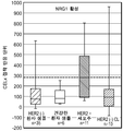
하나의 구현예에서, 질환은 암이고 따라서 이환 세포는 종양 세포의 샘플이고 건강한 세포는 종양 세포와 동일한 세포 유형의 비-종양 세포의 샘플이다. 바람직한 구현예에서, 암은 유방암이다. 유방암 진단을 위한 하나의 구현예에서, 교란제는 PI3K 신호전달 경로, 예컨대 NRG1에 선택적으로 영향을 미친다. 유방암 진단을 위한 하나의 구현예에서, 교란제는 ERα 신호전달 경로, 예컨대 에스트라디올에 선택적으로 영향을 미친다. 유방암 진단을 위한 더욱 또 다른 구현예에서, 교란제는 ErbB 신호전달 경로, 예컨대 HER2 신호전달 경로에 선택적으로 영향을 미친다. ErbB 신호전달 경로에 선택적으로 영향을 미치는 제제는 당해 기술에 공지되어 있다.In one embodiment, the disease is cancer and thus the diseased cell is a sample of tumor cells and the healthy cell is a sample of non-tumor cells of the same cell type as the tumor cells. In a preferred embodiment, the cancer is breast cancer. In one embodiment for the diagnosis of breast cancer, the perturbant selectively affects the PI3K signaling pathway, such as NRG1. In one embodiment for the diagnosis of breast cancer, the perturbant selectively affects the ERa signaling pathway, such as estradiol. In yet another embodiment for breast cancer diagnosis, the perturbant selectively affects the ErbB signaling pathway, such as the HER2 signaling pathway. Agents that selectively affect the ErbB signaling pathway are known in the art.
다양한 구현예에서, 신호전달 경로는 하기로 이루어진 군으로부터 선택된다 신호전달 경로는 하기로 이루어진 군으로부터 선택된다: MAPK-PK, RAS/RAF, RHO, FAK1, MEK/MAPK, MAK, MKK, AKT, EGF 수용체, Her2 수용체, Her 3 수용체, Her 4 수용체, 에스트로겐 수용체, 프로게스테론 수용체, 안드로겐 수용체, GPER30, PIK3/PTEN, VEGF 수용체 경로 억제제, 세포 접착, TGF베타/SMAD, WNT, 헤지혹/GLI, HIF1 알파, JAK/STAT, 노치, G1/S 전이의 대조군, DNA 손상 대조군, 및 세포자멸사. 추가의 적합한 신호전달 경로는 본원에서 개시된 다른 신호전달 경로를 포함한다. 다양한 구현예에서, 교란제는, 예를 들어, 단백질, 펩타이드, 핵산, 대사물질, 리간드, 시약, 유기 분자, 신호전달 인자, 성장 인자, 생화학물질, 또는 이들의 조합일 수 있다.In various embodiments, the signaling pathway is selected from the group consisting of: a signaling pathway selected from the group consisting of MAPK-PK, RAS / RAF, RHO, FAK1, MEK / MAPK, MAK, MKK, AKT, TGF beta / SMAD, WNT, hedgehog / GLI, HIF1 alpha, EGF receptor, Her2 receptor, Her 3 receptor, Her 4 receptor, estrogen receptor, progesterone receptor, androgen receptor, GPER30, PIK3 / PTEN, VEGF receptor pathway inhibitor , JAK / STAT, Notch, Control of G1 / S metastasis, DNA damage control, and apoptosis. Additional suitable signaling pathways include other signaling pathways disclosed herein. In various embodiments, the perturbing agent can be, for example, a protein, a peptide, a nucleic acid, a metabolite, a ligand, a reagent, an organic molecule, a signaling factor, a growth factor, a biochemical, or a combination thereof.
측정량은 시험에서 측정되도록 의도된 양으로서 일반적으로 정의된다. 본원에서 기재된 본 발명에 대하여, 측정되도록 의도된 양은 교란에 대한 생존 세포의 생리적 반응에서의 변화이다. 본원에서 추가로 기재된 바와 같이, 세포 접착 또는 부착은 임피던스 바이오센서 또는 광학적 바이오센서로 측정될 수 있는 세포에서 생리적 변화의 예이다. 하나의 구현예에서, 측정량은 교란되는 세포 샘플의 세포 접착 또는 부착에서의 변화를 나타내고 유클리드 분석을 이용하여 평가된다. 유클리드 분석은 하기로 이루어진 군으로부터 선택될 수 있다: 다중 시점에서 차이 벡터의 산술적 합산, 일시적 최대치, 일시적 최소치, 최대치 또는 최소치에 도달하는 시간, 경사의 변화, 바이오센서 신호의 절대 저하, 전체 측정의 합계, 및 이들의 조합. 또 다른 구현예에서, 세포 접착 또는 부착에서의 변화는 일시적 최대치 또는 최소치에서의 변화에 의해 측정된다. 바람직한 구현예에서, 측정량 계산은 아래에서 기재된 바와 같이 알고리즘 적용에 의해 수행된다.The measurand is generally defined as the amount intended to be measured in the test. For the present invention described herein, the amount intended to be measured is a change in the physiological response of the surviving cell to disturbance. As further described herein, cell adhesion or attachment is an example of a physiological change in a cell that can be measured with an impedance biosensor or an optical biosensor. In one embodiment, the measurand represents a change in cell adhesion or adhesion of the disturbed cell sample and is assessed using Euclidean analysis. The Euclidean analysis may be selected from the group consisting of: arithmetic summation of the difference vectors at multiple time points, temporal maximum, temporal minimum, time to reach maximum or minimum, change in slope, absolute degradation of the biosensor signal, Sums, and combinations thereof. In another embodiment, the change in cell adhesion or adhesion is measured by a change in transient maxima or minima. In a preferred embodiment, the metric calculations are performed by algorithm application as described below.
시점 대 임피던스 정보는 다양한 세포 조건 (세포 + 배지, 세포 + 교란 인자 F, 세포 + 확인 시약 + 교란 인자 F)에 대하여 적어도 240 분 동안 각 분 기록된다. 1 초과 교란 인자 F는 질환 기전에 따라 사용될 수 있고; 질환이 다중 관련된 경로를 따라 번식되는 사례에서, 개별적으로 또는 조합으로 각각의 다중 경로를 교란시키는데 유용할 수 있다. 예를 들어, ErbB2 암은 수용체의 ErbB 계열 및 관련된 경로 - MAPK 및 PI3K 경로를 포함한다. 다중 경로가 교란되는 사례에서 측정량은 각 경로에 대하여 계측된 별개의 측정량의 조합일 수 있다. 이들 조건에서 차이에 상응하는 벡터는 아래 상세히 보여진 방정식에 따라 측정량 (NED, 비-선형 유클리드 거리로도 공지됨)을 제공하도록 합계된다.The point-to-point impedance information is recorded for at least 240 minutes for various cell conditions (cell + medium, cell + disturbance factor F, cell + confirmation reagent + disturbance factor F) for at least 240 minutes. 1 excess disturbance factor F may be used depending on the disease mechanism; In cases where the disease is to be propagated along multiple related pathways, it may be useful to disturb each multipath individually or in combination. For example, ErbB2 cancers include the ErbB family of receptors and related path-MAPK and PI3K pathways. In the case where the multipath is disturbed, the measurand may be a combination of discrete measurands measured for each path. The vector corresponding to the difference in these conditions is summed to provide a metric (NED, also known as non-linear Euclidean distance) according to the equation detailed below.
시간 대 CAS 시험 데이터로부터 유도된 비-선형 유클리드 시점 벡터로부터 측정량을 계산하기 위해 사용된 방정식: The equation used to calculate the measurand from the non-linear Euclidean viewpoint vector derived from the time-to-CAS test data:

여기에서 변수는 아래와 같이 정의된다:Here the variables are defined as follows:
i = CAS가 시험 동안 기록되는 각 분에 대한 단계 i = step for each minute in which CAS is recorded during the test
F1 = 교란 인자 1F1 =
F2 = 교란 인자 2 (하나가 사용되면)F2 = disturbance factor 2 (if one is used)
Ci = 대조군, 시험 세포에 부가된 교란 인자 없음C i = control, no disturbance added to test cells
CF1i = 교란 인자 1 (F1)을 가진 세포CF1 i = cells with disturbance factor 1 (F1)
CF2 = 교란 인자 2 (F2)를 가진 세포 (하나가 사용되면)CF2 = cells with disturbance factor 2 (F2) (if one is used)
CCF1i = HER2 경로 확인제 부가를 가진 세포Cells with CCF1 i = HER2 pathway recognition agent
CCF2 = HER2 경로 확인제 부가를 가진 세포 (교란 인자 2가 사용되면)Cells with CCF2 = HER2 pathway recognition agent (if
다중 확인 인자는 이들 단계를 수행하기 위해 선택될 수 있다.Multiple acknowledgment factors may be selected to perform these steps.
본 발명은 구현예를 포함하고 이로써 측정량 알고리즘의 간소화된 버전이 유용한 정보를 또한 제공할 수 있다. 예를 들어:The present invention includes implementations whereby a streamlined version of the metric algorithm can also provide useful information. E.g:

추가로, 임의의 상기 알고리즘에서 합산의 한계는 분석의 값의 손실 없이 분석을 추가로 향상시키기 위해 i>240 또는 i<240까지 확장될 수 있다.In addition, the limit of summation in any of the above algorithms can be extended to i > 240 or i < 240 to further improve the analysis without loss of the value of the analysis.
또 다른 구현예에서, 이환 세포는 질환과 관련된 신호전달 경로를 표적하는 확인제와 추가로 접촉되고 세포 접착 또는 부착에 관한 확인제의 효과는 측정된다. 적합한 확인제는 당해 분야의 숙련가에게 공지된다. 당해 질환 기전에 관련된 경로내 한 지점에서 당해 신호전달 경로를 억제시킨다고 공지되기 때문에 확인제는 선택된다. 상기 구현예는 당해 신호전달 경로와 관련된 교란제와 이환 세포의 접촉에서 비롯된 생리적 반응의 양을 정량화하는 것을 가능하게 한다. 이는 측정된 생리적 반응이 당해 신호전달 경로에 특이적인지 아닌지를 확인할 수 있다.In another embodiment, the bifurcated cell is further contacted with a detection agent that targets the signal transduction pathway associated with the disease and the effect of the detection agent on cell adhesion or attachment is measured. Suitable detectors are known to those skilled in the art. The detergent is selected because it is known to inhibit the signal transduction pathway at one point in the pathway involved in the pathogenesis of the disease. This embodiment makes it possible to quantify the amount of physiological response resulting from contact of the mast cell with the perturbing agent associated with the signal transduction pathway. This confirms whether the measured physiological response is specific to the signal transduction pathway in question.
더욱 또 다른 구현예에서, 이환 세포는 질환과 관련된 신호전달 경로를 표적하는 표적화 치료제와 추가로 접촉되고 세포 접착 또는 부착에 관한 표적화 치료제의 효과는 측정된다.In yet another embodiment, the bifurcated cell is further contacted with a targeting agent that targets a signaling pathway associated with the disease and the effect of the targeting agent on cell adhesion or attachment is measured.
표적화 치료제의 비-제한 예는 하기를 포함한다: 트라스투주맙, 페르투주맙, 라파티닙, 도세탁셀, 타목시펜, 시스플라틴, 아브락산, 파클리탁셀 주사, 브렌툭시맙 베도톤, 에버롤리무스, 페메트렉세드, 엑세메스탄, 오파투무맙, 베바시주맙, 알렘투주맙, 이리노테칸, 바이칼루타마이드, 옥살리플라틴, 세툭시맙, 비소메데깁, 토레미펜 시트레이트, 풀베스트란트, 젬시타빈, 이마티닙, 익사베필론, 토페오테칸, 악시티닙, 로미뎁신, 카브라지탁셀, 소라페닙, 인플릭시맙, 레날리도마이드, 리툭시맙, 다사티닙, 수니티닙, 에를로티닙, 닐로티닙, 파클리탁셀, 테모졸로마이드, 트리옥사이드, 파니투무맙, 보르테조밉, 아자시티딘, 파조파닙, 크리조티닙, 카페시타빈, 이필리무맙, 베무라페닙, 고세렐린 아세테이트, 아비라테론, BH3 모방체, 나비토클락스, 아나스트로졸, 레트로졸, 방향화효소 억제제, 사이클로포스파마이드, 독소루비신, 메토트렉세이트, 플루오로우라실, 익사베필론, 카보플라틴, 아플리베르셉트, 템시롤리무스, 이르브리투모맙, 아비라테론, 쿠스티르센, 네라티닙, 엔잘루타마이드, 니볼루맙, 팔보시클립, 레고라페닙, 엔티노스태트, 아파티닙, ARN-509, ARN-810, BIND-014, 다브라페닙, 다라투무맙, 람브롤리주맙, LDK378, MM-121, sym004, 트라스투주맙-엠탄신, 티보자닙, 트라메티닙, 악시티닙, LY2835219, MPDL320A, 오비누투주맙, Sym004, 토시투모맙, 트라메티닙, 네시투무맙, 라무시루맙, ADXS-HER2, TAK-285, MM-302, MM-121, MM-111, REGN 1400, 사파티닙, 다코미티닙, 포지오티닙, ASLAN001, LIM716, AV-203, 둘리고투주맙, 룸레투주맙, 파니투무맙, REGN955, MM-151, 게피티닙, U3-1287(AMG888), TK-A3/TK-A4, AZD9291, 로실레티닙, 및 이들의 조합. 다른 적합한 표적화 치료제는 본원에서 개시된다. 더욱 또 다른 구현예에서, 방법은 대상체에 표적화 치료제의 투여를 추가로 포함한다.Non-limiting examples of targeted therapeutic agents include: Trastuzumab, pertuzumab, lapatinib, docetaxel, tamoxifen, cisplatin, ablucan, paclitaxel injection, brenuxux mabedotone, everolimus, , Eicemethan, ophatumatum, bevacizumab, alemtuzumab, irinotecan, bicalutamide, oxaliplatin, cetuximab, nonosomedib, toremifens citrate, fulvestrant, gemcitabine, imatinib, But are not limited to, nicotinamide, nicotinic acid, nicotinic acid, nicotinic acid, nicotinic acid, nicotinic acid, nicotinic acid, nicotinic acid, nicotinic acid, Paclitaxel, temolozomide, trioxide, pannituomat, bortezomib, azacytidine, pazopanib, creototinib, capecitabine, eicilimumab, bemula penip, goserelin acetate, aviratorone, mimic BH3 Che, Butterfly Clarks, Anas But are not limited to, cortisol, rosol, retrosol, aromatase inhibitors, cyclophosphamide, doxorubicin, methotrexate, fluorouracil, xavapilone, carboplatin, affluxcept, temsirolimus, ARN-509, ARN-810, BIND-014, Dabra feNib, DaraTumuMAP, LAMBE, and others. Ratrijumat, LDK378, MM-121, sym004, trastuzumab-mantansin, tiboazanib, trametinib, acctinib, LY2835219, MPDL320A, obunituzumab, Sym004, tositumam, trametinib, MM-121, MM-111, REGN 1400, Zapatidin, Dakomitimin, Posiotinib, ASLAN001, LIM716, AV-203, Dulryotouzumat, Roomretuumum, Panitumumat, REGN955, MM-151, Gefitinib, U3-1287 (AMG888), TK-A3 / TK-A4, AZD9291, Rosiletinib, and combinations thereof. Other suitable targeting agents are disclosed herein. In yet another embodiment, the method further comprises administration of a targeting agent to the subject.
또 다른 측면에서, 본 발명은, 하기 단계를 포함하는, 에스트로겐 관련된 신호전달 경로 (예컨대 호르몬 요법)에 영향을 미치는 표적된 요법을 위한 비-과발현된 에스트로겐 수용체 알파 (ERα-음성) 유방암을 가진 대상체의 선택 방법을 제공한다:In another aspect, the invention provides a method of treating a subject with a non-overexpressed estrogen receptor alpha (ERa-negative) breast cancer for targeted therapies that affects an estrogen-related signal transduction pathway (e.g., hormone therapy) Lt; RTI ID = 0.0 > of:
비-과발현된 ERα (ERα 음성) 유방암을 가진 대상체로부터 수득된 생존 원발 유방암 세포의 샘플의 제조 및 기본 배지에서 샘플의 배양 단계;Preparing a sample of survival primary breast cancer cells obtained from a subject with non-overexpressed ERa (ERa negative) breast cancer and culturing the sample in a basal medium;
생리적 반응 파라미터 예컨대 세포 접착 또는 부착에 관한 효과에 의해 측정된 바와 같이 신호전달 경로를 상향조절 또는 하향조절하기 위한 ERα 신호전달 경로를 선택적으로 영향을 미친다고 공지되는 교란제와 생존 원발 유방암 세포; 및 (2) 동일한 에스트로겐-관련 신호전달 경로 활성을 억제한다고 공지된 확인제 및 교란제와 샘플의 제2 부분의 접촉 단계;Disturbing agents and survival primary breast cancer cells known to selectively affect the ERa signaling pathway for upregulating or downregulating the signal transduction pathway as measured by physiological response parameters such as effects on cell adhesion or adhesion; And (2) contacting the second portion of the sample with a detergent and an agonist known to inhibit the same estrogen-associated signal transduction pathway activity;
샘플의 각 부분에서 생존 원발 유방암 세포의 생리적 반응 파라미터 예컨대 세포 접착 또는 부착의 연속적 측정 단계; 및A step of measuring physiological response parameters of surviving primary breast cancer cells such as cell adhesion or attachment in each part of the sample; And
샘플의 제1 부분과 샘플의 제2 부분 간 값 차이를 확인하기 위해 생리적 반응 파라미터의 연속적 측정의 수학적 분석에 의한 측정량의 계측; 및Measurement of a measurand by a mathematical analysis of successive measurements of physiological response parameters to ascertain a value difference between the first portion of the sample and the second portion of the sample; And
에스트로겐 수용체 경로 활성에 대하여 정상 참조 구간으로부터 유도된 컷-오프 값에 측정량의 비교 단계;Comparing the measured amount to a cut-off value derived from the normal reference interval for estrogen receptor pathway activity;
(여기에서 측정량이 컷-오프 값 초과인 경우 대상체는 에스트로겐 관련된 신호전달 경로에 영향을 미치는 표적된 요법을 이용한 치료에 대해 선택된다).Wherein the subject is selected for treatment with a targeted therapy that affects the estrogen-related signaling pathway if the measurand is above the cut-off value.
하나의 구현예에서, 방법은 샘플이 교란제와 접촉되기 최소 12 시간 내지 최대 72 시간 이전에 신선한 기본 배지로 샘플을 배양시키기 위해 사용된 기본 배지의 대체를 포함한다.In one embodiment, the method comprises replacing the basal medium used to incubate the sample with fresh basal medium for at least 12 hours up to 72 hours before the sample is contacted with the perturbant.
측정량은, 예를 들어, 미교란된 생존 암 세포 샘플에 비교된 경우 교란제와 접촉된 생존 암 세포 샘플의 부분에서 세포 접착 또는 부착의 증가 또는 감소 혹은 교란제 및 확인제와 접촉된 이후 생존 세포 샘플의 부분에서 세포 접착 또는 부착의 증가 또는 감소일 수 있다. ERα 신호전달 경로에 영향을 미치는 적합한 교란제는 당해 기술에 공지되어 있고 에스트라디올처럼 본원에서 개시된다. 또 다른 구현예에서, 생존 종양 세포는 ERα 신호전달 경로를 억제시킨다고 공지된 확인제와 추가로 접촉되고 세포 접착 또는 부착에 관한 확인제의 효과는 측정된다. 적합한 확인제, 예컨대 선택적 에스트로겐 수용체 다운-조절물질은 당해 분야의 숙련가에게 공지된다. 또 다른 구현예에서, 생존 종양 세포는 ERα 신호전달 경로를 표적하는 표적화 치료제와 추가로 접촉되고 세포 접착 또는 부착에 관한 표적화 치료제의 효과는 측정된다. ERα 신호전달 경로를 표적하는 적합한 표적화 치료제는 당해 기술에 공지되어 있고 풀베스트란트 및 타목시펜처럼 본원에서 개시된다. 방법은 대상체에 표적화 치료제의 투여를 추가로 포함할 수 있다.The measurand may include, for example, an increase or decrease in cell adhesion or adhesion in a portion of a survival cancer cell sample contacted with an agonist, as compared to an unfrozen survival cancer cell sample, It may be an increase or decrease in cell adhesion or adhesion in the portion of the sample. Suitable perturbants that affect the ERa signaling pathway are known in the art and are disclosed herein as estradiol. In another embodiment, the surviving tumor cells are further contacted with a known agent that inhibits the ERa signaling pathway and the effect of the agent on cell adhesion or adhesion is measured. Suitable detectors, such as selective estrogen receptor down-regulating substances, are known to those skilled in the art. In another embodiment, the surviving tumor cells are further contacted with a targeting agent that targets the ERa signaling pathway and the effect of the targeting agent on cell adhesion or attachment is measured. Suitable targeting agents that target the ERa signaling pathway are known in the art and are disclosed herein, such as fulvestrant and tamoxifen. The method may further comprise administration of a targeted therapeutic agent to the subject.
더욱 또 다른 측면에서, 본 발명은, 하기 단계를 포함하는, ErbB 신호전달 경로에 영향을 미치는 표적화 치료제를 이용한 요법을 위한 비-과발현된 또는 증폭된 ErbB2 암 (예를 들면, 유방암)을 가진 대상체의 선택 방법을 제공한다:In yet another aspect, the invention provides a method of treating a subject with a non-overexpressed or amplified ErbB2 cancer (e. G., Breast cancer) for therapy with a targeting agent that affects the ErbB signaling pathway, Lt; RTI ID = 0.0 > of:
대상체로부터 수득된 생존 원발 암 세포의 샘플 제조 및 기본 배지에서 샘플의 배양;Preparing a sample of the primary cancer cell obtained from the subject and culturing the sample in the primary medium;
(1) 뉴레굴린과 대상체로부터 수득된 생존 암 세포의 샘플의 제1 부분, 및 (2) 뉴레굴린 및 확인제 (여기서 상기 확인제는 뉴레굴린과 동일한 ErbB 신호전달 경로를 선택적으로 억제시킨다)와 샘플의 제2 부분의 접촉; 및/또는 (3) 상피 성장 인자와 샘플의 제3 부분 및 (4) 상피 성장 인자 및 확인제 (여기서 상기 확인제는 상피 성장 인자와 동일한 ErbB 신호전달 경로를 선택적으로 억제시킨다)와 샘플의 제4 부분의 접촉 단계;(1) a first portion of a sample of the surviving cancer cells obtained from the neuregulin and the subject, and (2) a second portion of the sample comprising the neuregulin and the identifying agent, wherein the identifying agent selectively inhibits the same ErbB signaling pathway as the neuregulin, Contact of the second portion; And / or (3) an epithelial growth factor and a third portion of the sample; and (4) an epithelial growth factor and a detergent, wherein the inhibitor selectively inhibits the ErbB signaling pathway identical to the epithelial growth factor, A contact step of;
샘플의 각 부분에서 생존 원발 암 세포의 생리적 반응 파라미터의 연속적 측정 단계;Continuously measuring the physiological response parameters of the primary cancer cells in each part of the sample;
샘플의 제1 부분과 샘플의 제2 부분 간 값의 차이 및/또는 샘플의 제3 부분과 샘플의 제4 부분 간 값의 차이를 확인하기 위해 생리적 반응 파라미터의 연속적 측정의 수학적 분석에 의한 측정량의 계측 단계; 및A measured amount by a mathematical analysis of successive measurements of physiological response parameters to determine the difference between the values of the first portion of the sample and the second portion of the sample and / or the difference between the third portion of the sample and the fourth portion of the sample A measuring step of And
ErbB 신호전달 경로 활성의 정상 참조 구간으로부터 유도된 컷-오프 값과 측정량의 비교 단계;Comparing the measured amount with a cut-off value derived from a normal reference section of ErbB signaling pathway activity;
(여기서 측정량이 컷-오프 값 초과인 경우 대상체는 ErbB 신호전달 경로에 영향을 미치는 표적화 치료제를 이용한 치료에 대해 선택된다).Wherein the subject is selected for treatment with a targeting agent that affects the ErbB signaling pathway if the measurand is above the cut-off value.
하나의 구현예에서, 방법은 이들이 교란제와 접촉되기 최소 12 시간 내지 최대 72 시간 이전에 신선한 기본 배지로 샘플을 배양시키기 위해 사용된 기본 배지의 대체를 포함한다.In one embodiment, the method comprises replacing the basal medium used for culturing the sample with fresh basal medium from a minimum of 12 hours up to a maximum of 72 hours before they are contacted with the perturbant.
측정량은, 예를 들어, 미교란된 생존 암 세포 샘플과 비교된 경우 교란제와 접촉된 생존 암 세포 샘플의 교란된 부분에서 세포 접착 또는 부착에서 증가 또는 감소 혹은 교란제 및 확인제와 접촉된 이후 생존 세포 샘플의 부분에서 세포 접착 또는 부착의 증가 또는 감소일 수 있다.The measurand may include, for example, an increase or decrease in cell adhesion or adhesion in the disturbed portion of the survival cancer cell sample contacted with the perturbing agent, as compared to an unfrozen survival cancer cell sample, It may be an increase or decrease in cell adhesion or attachment in the portion of the surviving cell sample.
ErbB 신호전달 경로에 영향을 미치는 다른 적합한 교란제는 당해 기술에 공지되어 있고 본원에서 개시된다. 또 다른 구현예에서, 종양 세포는 ErbB 신호전달 경로를 표적하는 표적화 치료제와 추가로 접촉되고 세포 접착 또는 부착에 관한 표적화 치료제의 효과는 측정된다. ErbB 신호전달 경로를 표적하는 적합한 표적화 치료제는 당해 기술에 공지되어 있고 본원에서 개시된다. 방법은 대상체에 표적화 치료제의 투여를 추가로 포함할 수 있다.Other suitable perturbants that affect the ErbB signaling pathway are known in the art and are disclosed herein. In another embodiment, the tumor cells are further contacted with a targeting agent that targets the ErbB signaling pathway and the effect of the targeting agent on cell adhesion or adhesion is measured. Suitable targeting agents that target the ErbB signaling pathway are known in the art and are disclosed herein. The method may further comprise administration of a targeted therapeutic agent to the subject.
또 다른 측면에서, 본 발명은 표적화 치료제를 투여함으로써 대상체의 치료 방법을 제공하고, 여기서 상기 대상체는 신호전달 경로가 대상체의 세포에서 활성인지의 계측을 위하여 본 발명의 방법을 이용한 치료에 대해 선택되었다. 따라서, 본 발명은 특이적 신호전달 경로를 표적하는 표적화 치료제가 있는 바이오마커 음성 암을 갖는 대상체의 치료 방법을 제공하고, 여기서 대상체가 바이오마커 음성 암을 가짐에도 불구하고 활성 신호전달 경로를 갖는 것으로 보여졌기 때문에 상기 대상체는 치료에 선택되었다.In another aspect, the invention provides a method of treating a subject by administering a targeted therapeutic agent, wherein said subject is selected for treatment with the method of the invention for the measurement of signal transduction pathway activity in a subject's cells . Accordingly, the present invention provides a method of treating a subject having a biomarker negative cancer with a targeted therapeutic agent that targets a specific signal transduction pathway, wherein the subject has an active signal transduction pathway, even though the subject has a biomarker negative arm The subject was selected for treatment because it was shown.
따라서, 하나의 구현예에서, 본 발명은 암 세포가 비-과발현된, 비-증폭된 ErbB2 수용체를 갖는 암을 가진 (예를 들면, 암으로 진단된) 인간 대상체의 치료 방법을 제공하고, 상기 방법은 ErbB 신호전달 경로에 영향을 미치는 표적화 치료제를 대상체에 투여하는 것을 포함하고, 상기 대상체가 하기 단계를 포함하는 방법을 이용한 치료에 대해 선택된다:Thus, in one embodiment, the invention provides a method of treating a human subject having cancer (e.g., diagnosed with cancer) with cancer having a non-overexpressed, non-amplified ErbB2 receptor, The method comprises administering to the subject a targeted therapeutic agent that affects an ErbB signaling pathway and wherein the subject is selected for treatment using a method comprising the steps of:
대상체로부터 수득된 생존 원발 암 세포의 샘플 제조 및 기본 배지에서 샘플의 배양 단계;Preparing a sample of the primary cancer cell obtained from the subject and culturing the sample in the primary medium;
(1) 교란제와 대상체로부터 수득된 생존 원발 암 세포의 샘플의 제1 부분, 및 (2) 교란제 및 확인제 (여기서 상기 교란제는 ErbB 신호전달 경로에 선택적으로 영향을 미치고 확인제는 동일한 ErbB 신호전달 경로에 관해 교란제의 효과를 선택적으로 억제시킨다)와 샘플의 제2 부분의 접촉 단계;(1) a first part of a sample of the surviving primary cancer cells obtained from the disturbing agent and the subject, and (2) a disturbing agent and a detergent, wherein the disturbant selectively affects the ErbB signaling pathway and the identifying agent is the same ErbB signal Selectively inhibiting the effect of the perturbing agent on the delivery path) and the second portion of the sample;
샘플의 각 부분에서 생존 원발 암 세포의 생리적 반응 파라미터의 연속적 측정 단계;Continuously measuring the physiological response parameters of the primary cancer cells in each part of the sample;
샘플의 제1 부분과 샘플의 제2 부분 간 값의 차이를 확인하기 위해 생리적 반응 파라미터의 연속적 측정의 수학적 분석에 의한 측정량의 계측 단계; 및Measuring a measured quantity by a mathematical analysis of successive measurements of physiological response parameters to identify differences in values between the first portion of the sample and the second portion of the sample; And
ErbB 신호전달 경로 활성의 정상 참조 구간으로부터 유도된 컷-오프 값과 측정량의 비교 단계; Comparing the measured amount with a cut-off value derived from a normal reference section of ErbB signaling pathway activity;
(여기서 측정량이 컷-오프 값 초과인 경우 대상체는 ErbB 신호전달 경로에 영향을 미치는 표적화 치료제를 이용한 치료에 대해 선택된다).Wherein the subject is selected for treatment with a targeting agent that affects the ErbB signaling pathway if the measurand is above the cut-off value.
하나의 구현예에서, 방법은 샘플이 교란제와 접촉되기 최소 12 시간 내지 최대 72 시간 이전에 신선한 기본 배지로 샘플을 배양시키기 위해 사용된 기본 배지의 대체를 포함한다. In one embodiment, the method comprises replacing the basal medium used to incubate the sample with fresh basal medium for at least 12 hours up to 72 hours before the sample is contacted with the perturbant.
다양한 구현예에서, 교란제는 하기로 이루어진 군으로부터 선택된다: EGF, TGF-α, HB-EGF, 암피레굴린, 베타셀룰린, 에피겐, 에피레굴린, 뉴레굴린-1, 뉴레굴린-2, 뉴레굴린-3, 뉴레굴린-4, 및 이들의 조합. 구현예에서, 방법은 하기를 포함한다:In various embodiments, the perturbing agent is selected from the group consisting of: EGF, TGF- alpha, HB-EGF, ampirilgroline, betacellulin, epigene, epiregulin, , Neuregulin-3, neuregulin-4, and combinations thereof. In an embodiment, the method comprises:
(1) 뉴레굴린과 대상체로부터 수득된 생존 암 세포의 샘플의 제1 부분, 및 (2) 뉴레굴린 및 확인제 (여기서 상기 확인제는 뉴레굴린과 동일한 ErbB 신호전달 경로를 선택적으로 억제시킨다)와 샘플의 제2 부분의 접촉; 및/또는 (3) 상피 성장 인자와 샘플의 제3 부분 및 (4) 상피 성장 인자 및 확인제 (여기서 상기 확인제는 상피 성장 인자와 동일한 ErbB 신호전달 경로를 선택적으로 억제시킨다)와 샘플의 제4 부분의 접촉 단계; 및(1) a first portion of a sample of the surviving cancer cells obtained from the neuregulin and the subject, and (2) a second portion of the sample comprising the neuregulin and the identifying agent, wherein the identifying agent selectively inhibits the same ErbB signaling pathway as the neuregulin, Contact of the second portion; And / or (3) an epithelial growth factor and a third portion of the sample; and (4) an epithelial growth factor and a detergent, wherein the inhibitor selectively inhibits the ErbB signaling pathway identical to the epithelial growth factor, A contact step of; And
샘플의 제1 부분과 샘플의 제2 부분 간 값의 차이 및/또는 샘플의 제3 부분과 샘플의 제4 부분 간 값의 차이를 확인하기 위해 생리적 반응 파라미터의 연속적 측정의 수학적 분석에 의한 측정량의 계측 단계.A measured amount by a mathematical analysis of successive measurements of physiological response parameters to determine the difference between the values of the first portion of the sample and the second portion of the sample and / or the difference between the third portion of the sample and the fourth portion of the sample Measurement phase of.
방법은 ErbB 신호전달 경로를 표적하는 교란제 및 표적화 치료제와 생존 원발 암 세포의 접촉 및 생리적 반응 파라미터에 관한 표적화 치료제의 효과의 측정을 추가로 포함할 수 있다.The method may further comprise a disturbance targeting the ErbB signaling pathway and measuring the effect of the targeted therapeutic agent on the contact and physiological response parameters of the primary remnant cancer cell with the targeted therapeutic agent.
하나의 구현예에서, 생리적 반응 파라미터는 세포 접착 또는 부착이다. 또 다른 구현예에서, 생리적 반응 파라미터는 세포 대사물질의 수준이다. 또 다른 구현예에서, 생리적 반응 파라미터는 미토콘드리아 기능에 관련된 세포 효소 수준이다.In one embodiment, the physiological response parameter is cell adhesion or attachment. In another embodiment, the physiological response parameter is the level of cellular metabolites. In another embodiment, the physiological response parameter is a cellular enzyme level associated with mitochondrial function.
하나의 구현예에서, 대상체는 비-과발현된, 비-증폭된 HER2 암을 갖는다. 또 다른 구현예에서, 대상체는 EGFR 음성 및 HER2 음성 암을 갖는다. 또 다른 구현예에서, 대상체는 비-과발현된, 비-증폭된 ErbB2 유방암을 갖는다. 다른 구현예에서, 대상체는 하기로 이루어진 군으로부터 선택되는 비-과발현된, 비-증폭된 ErbB 암을 갖는다: 결장암, 직장암, 자궁내막암, 위 암종, 위장 카르시노이드 종양, 위장 기질 종양, 교모세포종, 간세포 암종, 소세포 폐암, 비-소세포 폐암 (NSCLC), 흑색종, 난소암, 자궁경부암, 췌장암, 전립선 암종, 급성 골수성 백혈병 (AML), 만성적 골수성 백혈병 (CML), 비-호지킨의 림프종 및 갑상선 암종.In one embodiment, the subject has a non-overexpressed, non-amplified HER2 cancer. In another embodiment, the subject has EGFR negative and HER2 negative cancer. In another embodiment, the subject has a non-overexpressed, non-amplified ErbB2 breast cancer. In another embodiment, the subject has a non-overexpressed, non-amplified ErbB cancer selected from the group consisting of: colon cancer, rectal cancer, endometrial cancer, gastric carcinoma, gastric carcinoid tumor, gastric stromal tumor, (AML), chronic myelogenous leukemia (CML), lymphoma of the non-Hodgkin's lymphoma, lymphoma of the uterine cervix, pancreatic cancer, prostate carcinoma, And thyroid carcinoma.
하나의 구현예에서, 확인제는 2C4 마우스 단클론성 항체이다. 또 다른 구현예에서, 확인제는 하기로 이루어진 군으로부터 선택되는 부위에 결합한다: HER2의 세포외 분절의 도메인 II; HER2의 세포외 분절의 도메인 IV; EGFR 또는 HER2 또는 HER3 또는 HER4의 세포질 아데노신 삼인산-결합 부위; EGFR 또는 HER2의 아데노신 삼인산 결합 부위에서의 시스테인 잔기; 및 이들의 조합.In one embodiment, the determinant is a 2C4 mouse monoclonal antibody. In another embodiment, the identifying agent binds to a site selected from the group consisting of domain II of the extracellular segment of HER2; Domain IV of the extracellular segment of HER2; Cytoplasmic adenosine triphosphate-binding site of EGFR or HER2 or HER3 or HER4; A cysteine residue at the adenosine triphosphate binding site of EGFR or HER2; And combinations thereof.
하나의 구현예에서, 표적화 치료제는 라파티닙이다. 또 다른 구현예에서, 표적화 치료제는 하기로 이루어진 군으로부터 선택된다: 트라스투주맙, 페르투주맙, 아파티닙, 네라티닙, ADXS-HER2, TAK-285, MM-302, MM-121, MM-111, REGN 1400, 사피티닙, 다코미티닙, 포지오티닙, ASLAN001, LIM716, AV-203, 둘리고투주맙, 룸레투주맙, 파니투무맙, REGN955, MM-151, 세툭시맙, 게피티닙, 에를로티닙, 트라스투주맙-엠탄신, 및 이들의 조합. 다른 구현예에서, 표적화 치료제는 하기로 이루어진 군으로부터 선택되는 부위에 결합한다: HER2의 세포외 분절의 도메인 II; HER2의 세포외 분절의 도메인 IV; EGFR 또는 HER2 또는 HER3 또는 HER4의 세포질 아데노신 삼인산-결합 부위; EGFR 또는 HER2의 아데노신 삼인산 결합 부위에서의 시스테인 잔기; 및 이들의 조합.In one embodiment, the targeting agent is lapatinib. In another embodiment, the targeting therapeutic agent is selected from the group consisting of: trastuzumab, pertuzumab, apatinate, neratinib, ADXS-HER2, TAK-285, MM-302, REGN 955, MM-151, Cetuximab, Gefitti, REGN 1400, Zapitinib, Dakomutinip, Posiotinib, ASLAN001, LIM716, AV-203, Dulryotouzumum, Nip, erlotinib, trastuzumab-mthansin, and combinations thereof. In another embodiment, the targeting therapeutic binds to a site selected from the group consisting of domain II of the extracellular segment of HER2; Domain IV of the extracellular segment of HER2; Cytoplasmic adenosine triphosphate-binding site of EGFR or HER2 or HER3 or HER4; A cysteine residue at the adenosine triphosphate binding site of EGFR or HER2; And combinations thereof.
또 다른 구현예에서, 대상체는 ErbB 신호전달 경로에 영향을 미치지 않는 치료제, 예컨대 표준 화학요법 제제 또는 ErbB와 상이한 신호전달 경로에 영향을 미치는 상이한 표적화 치료제로 또한 치료된다.In another embodiment, the subject is also treated with a therapeutic agent that does not affect the ErbB signaling pathway, such as a standard chemotherapeutic agent or a different targeting agent that affects the signaling pathway different from ErbB.
또 다른 구현예에서, 본 발명은 비-과발현된, 비-증폭된 ErbB2 유방암을 가진 (예를 들면, 상기로 진단된) 인간 대상체의 치료 방법으로서, 라파티닙을 대상체에 투여를 포함하는 방법을 제공하고, 여기서 상기 대상체는 하기 단계를 포함한 방법을 이용하여 라파티닙을 이용한 치료에 대해 선택되었고;In another embodiment, the invention provides a method of treating a human subject with a non-overexpressed, non-amplified ErbB2 breast cancer (e. G., Diagnosed above), which method comprises administering lapatinib to a subject Wherein said subject has been selected for treatment with lapatinib using a method comprising the steps of:
대상체로부터 수득된 생존 원발 유방암 세포의 샘플 제조 및 기본 배지에서 샘플의 배양 단계;Preparing a sample of survival primary breast cancer cells obtained from the subject and culturing the sample in a basal medium;
샘플이 교란제와 접촉되기 최소 12 시간 내지 최대 72 시간 이전에 신선한 기본 배지로 샘플을 배양시키기 위해 사용된 기본 배지의 대체 단계;A replacement step of the basal medium used to incubate the sample with fresh basal medium for at least 12 hours up to 72 hours before the sample is contacted with the perturbant;
(1) 교란제로서의 뉴레굴린과 대상체로부터 수득된 생존 암 세포의 샘플의 제1 부분, 및 (2) 뉴레굴린 및 확인제 (여기서 상기 확인제는 뉴레굴린과 동일한 ErbB 신호전달 경로를 선택적으로 억제시킨다)와 샘플의 제2 부분의 접촉; 및/또는 (3) 교란제로서 상피 성장 인자와 샘플의 제3 부분 및 (4) 상피 성장 인자 및 확인제 (여기서 상기 확인제는 상피 성장 인자와 동일한 ErbB 신호전달 경로를 선택적으로 억제시킨다)와 샘플의 제4 부분의 접촉 단계;(1) a first portion of a sample of the surviving cancer cells obtained from the subject, and (2) a second portion of the sample comprising the nervuline and the identifying agent, wherein the identifying agent selectively inhibits the same ErbB signaling pathway as the neuregulin, And a second portion of the sample; And / or (3) an epithelial growth factor as a perturbant and a third portion of the sample, and (4) an epithelial growth factor and a determinant, wherein said inhibitor selectively inhibits the same ErbB signaling pathway as the epithelial growth factor, A contact step of a fourth portion;
교란제와 접촉된 생존 원발 유방암 세포의 세포 접착 또는 부착의 연속적 측정 단계;Continuous measurement of cell adhesion or attachment of survival primary breast cancer cells in contact with an agonist;
샘플의 제1 부분과 샘플의 제2 부분 간 값의 차이 및/또는 샘플의 제3 부분과 샘플의 제4 부분 간 값의 차이를 확인하기 위해 생리적 반응 파라미터의 연속적 측정의 수학적 분석에 의한 측정량의 계측 단계; 및A measured amount by a mathematical analysis of successive measurements of physiological response parameters to determine the difference between the values of the first portion of the sample and the second portion of the sample and / or the difference between the third portion of the sample and the fourth portion of the sample A measuring step of And
ErbB 경로 활성에 대하여 정상 참조 구간으로부터 유도된 컷-오프 값과 측정량의 비교 단계;Comparing the measured amount with a cut-off value derived from a normal reference section for ErbB path activity;
여기서 측정량이 컷-오프 값 초과인 경우 대상체는 라파티닙을 이용한 치료에 대해 선택된다.Wherein the subject is selected for treatment with lapatinib if the measured quantity is above the cut-off value.
상기 제시된 동일한 방법은 라파티닙 이외의 표적화 치료제에 적용될 수 있다. 따라서, 다른 구현예에서, 표적화 치료제는 하기로 이루어진 군으로부터 선택된다: 트라스투주맙, 페르투주맙, 아파티닙, 네라티닙, ADXS-HER2, TAK-285, MM-302, MM-121, MM-111, REGN 1400, 사피티닙, 다코미티닙, 포지오티닙, ASLAN001, LIM716, AV-203, 둘리고투주맙, 룸레투주맙, 파니투무맙, REGN955, MM-151, 세툭시맙, 게피티닙, 에를로티닙, 트라스투주맙-엠탄신, 및 이들의 조합. 다른 구현예에서, 표적화 치료제는 하기로 이루어진 군으로부터 선택되는 부위에 결합한다: HER2의 세포외 분절의 도메인 II 또는 또 다른 ErbB 계열 구성원의 동족 도메인; HER2의 세포외 분절의 도메인 IV 또는 또 다른 ErbB 계열 구성원의 동족 도메인; EGFR 또는 HER2 또는 HER3 또는 HER4의 세포질 아데노신 삼인산-결합 부위; EGFR 또는 HER2의 아데노신 삼인산 결합 부위에서의 시스테인 잔기; 및 이들의 조합.The same method presented above can be applied to a targeting agent other than lapatinib. Thus, in other embodiments, the targeting therapeutic agent is selected from the group consisting of: Trastuzumab, pertuzumab, apatinate, nertinib, ADXS-HER2, TAK-285, MM- MM-111, REGN 1400, sapitinib, dacomitinib, positotinib, ASLAN001, LIM716, AV-203, dorigothoumumine, roomretujumat, panituumatum, REGN955, MM-151, cetuximab, crab Phytinib, erlotinib, trastuzumab-mantansin, and combinations thereof. In other embodiments, the targeting therapeutic agent binds to a site selected from the group consisting of: a domain II of the extracellular segment of HER2 or a cognate domain of another ErbB family member; A kinin domain of domain IV of another extracellular segment of HER2 or another ErbB family member; Cytoplasmic adenosine triphosphate-binding site of EGFR or HER2 or HER3 or HER4; A cysteine residue at the adenosine triphosphate binding site of EGFR or HER2; And combinations thereof.
하나의 구현예에서, 대상체는 EGFR 음성 및 HER2 음성 유방암을 갖는다.In one embodiment, the subject has EGFR negative and HER2 negative breast cancer.
하나의 구현예에서, 대상체는 ErbB 신호전달 경로에 영향을 미치지 않는 치료제, 예컨대 표준 화학요법 제제 또는 ErbB와 상이한 신호전달 경로에 영향을 미치는 상이한 표적화 치료제로 또한 치료된다.In one embodiment, the subject is also treated with a therapeutic agent that does not affect the ErbB signaling pathway, such as a standard chemotherapeutic agent or a different targeting agent that affects the signaling pathway different from ErbB.
또 다른 구현예에서, 본 발명은 비-과발현된 에스트로겐 수용체 (ER) 유방암을 가진 (예를 들면, 상기로 진단된) 인간 대상체의 치료 방법으로서, ER 신호전달 경로에 영향을 미치는 표적화 치료제를 대상체에 투여하는 것을 포함하는 방법을 제공하고, 여기서 상기 대상체는 하기 단계를 포함한 방법을 이용하여 치료에 대해 선택되었고;In another embodiment, the invention provides a method of treating a human subject having a non-overexpressed estrogen receptor (ER) breast cancer (eg, diagnosed above), comprising administering a targeting therapeutic agent that affects the ER signaling pathway Wherein said subject has been selected for treatment using a method comprising the steps of;
대상체로부터 수득된 생존 원발 유방암 세포의 샘플 제조 및 기본 배지에서 샘플의 배양 단계;Preparing a sample of survival primary breast cancer cells obtained from the subject and culturing the sample in a basal medium;
(1) 에스트로겐-관련 신호전달 경로를 상향조절 또는 하향조절한다고 공지된 교란제와 대상체로부터 수득된 생존 원발 유방암 세포의 샘플의 제1 부분, 및 (2) 동일한 에스트로겐-관련 신호전달 경로 활성을 억제시킨다고 공지된 교란제 및 확인제와 생존 원발 암 세포의 샘플의 제2 부분의 접촉 단계;(1) a first part of a disturbance agent known to upregulate or downregulate an estrogen-related signaling pathway and a sample of a survival breast cancer cell obtained from a subject, and (2) a second part of the same estrogen- Contacting a second portion of a sample of surviving primary cancer cells with a perturbing agent and a known agent that are known to induce a cancer cell;
샘플의 각 부분에서 생존 원발 암 세포의 생리적 반응 파라미터의 연속적 측정 단계;Continuously measuring the physiological response parameters of the primary cancer cells in each part of the sample;
샘플의 제1 부분과 샘플의 제2 부분 간 값의 차이를 확인하기 위해 생리적 반응 파라미터의 연속적 측정의 수학적 분석에 의한 측정량의 계측 단계; Measuring a measured quantity by a mathematical analysis of successive measurements of physiological response parameters to identify differences in values between the first portion of the sample and the second portion of the sample;
에스트로겐 수용체 경로 활성에 대하여 정상 참조 구간으로부터 유도된 컷-오프 값과 측정량의 비교 단계;Comparing the measured amount with a cut-off value derived from a normal reference interval for estrogen receptor pathway activity;
여기서 측정량이 컷-오프 값 초과인 경우 대상체는 에스트로겐 관련된 신호전달 경로에 영향을 미치는 표적화 치료제를 이용한 치료에 대해 선택된다.Where the measured amount is greater than the cut-off value, the subject is selected for treatment with a targeting agent that affects the estrogen-related signaling pathway.
하나의 구현예에서, 방법은 이들이 교란제와 접촉되기 최소 12 시간 내지 최대 72 시간 이전에 신선한 기본 배지로 샘플을 배양시키기 위해 사용된 기본 배지의 대체를 포함한다.In one embodiment, the method comprises replacing the basal medium used for culturing the sample with fresh basal medium from a minimum of 12 hours up to a maximum of 72 hours before they are contacted with the perturbant.
방법은 ER 신호전달 경로를 표적하는 표적화 치료제와 원발 유방암 세포의 접촉 및 생리적 반응 파라미터에 관한 표적화 치료제의 효과 측정을 추가로 포함할 수 있다.The method may further include measuring the effect of the targeted therapeutic agent on the contact of the primary breast cancer cell with the targeting agent targeting the ER signaling pathway and the physiological response parameter.
하나의 구현예에서, 생리적 반응 파라미터는 세포 접착 또는 부착이다. 또 다른 구현예에서, 생리적 반응 파라미터는 세포 대사물질의 수준이다. 또 다른 구현예에서, 생리적 반응 파라미터는 미토콘드리아 기능에 관련된 세포 효소 수준이다.In one embodiment, the physiological response parameter is cell adhesion or attachment. In another embodiment, the physiological response parameter is the level of cellular metabolites. In another embodiment, the physiological response parameter is a cellular enzyme level associated with mitochondrial function.
하나의 구현예에서, 대상체는 ERα 음성 유방암을 갖는다.In one embodiment, the subject has ERa negative breast cancer.
하나의 구현예에서, 교란제는 하기로 이루어진 군으로부터 선택된다: 에스트라디올, 에스트론, 랄록시펜, 에스트리올, 게니스테인, DHEA, 안드로스테네디온, 안드로스테네디올, 프로게스테론, 하이드록시프로게스테론, 테스토스테론, 이들의 황산화된 형태, 및 이들의 조합.In one embodiment, the perturbing agent is selected from the group consisting of: estradiol, estrone, raloxifene, estriol, genistein, DHEA, androstenedione, androstenediol, progesterone, hydroxyprogesterone, testosterone , Their sulfated forms, and combinations thereof.
하나의 구현예에서, 표적화 치료제는 풀베스트란트, 타목시펜, 레트로졸, 팔보시클립, 아베마시클립, 및 이들의 조합으로 이루어진 군으로부터 선택된다. 다른 구현예에서, 표적화 치료제는 하기로 이루어진 군으로부터 선택되는 부위에 결합한다: 방향화효소 효소 (CYP19A)의 사이토크롬 P450 서브유닛, 에스트로겐 수용체의 활성화 기능 1 (AFP-1) 도메인, 사이클린-의존적 키나제 4 (사이클린 D1), 사이클린-의존적 키나제 6 (사이클린 D3), 및 이들의 조합.In one embodiment, the targeting therapeutic agent is selected from the group consisting of fulvestrant, tamoxifen, letrozole, palboshi clips, Abemarishi clips, and combinations thereof. In other embodiments, the targeting therapeutic agent binds to a site selected from the group consisting of the cytochrome P450 subunit of the aromatase enzyme (CYP19A), the activation function 1 (AFP-1) domain of the estrogen receptor, the cyclin-dependent Kinase 4 (cyclin D1), cyclin-dependent kinase 6 (cyclin D3), and combinations thereof.
하나의 구현예에서, 대상체는 ER 신호전달 경로에 영향을 미치지 않는 치료제, 예컨대 표준 화학요법 제제 또는 ER과 상이한 신호전달 경로에 영향을 미치는 상이한 표적화 치료제로 치료된다. 하나의 구현예에서, 대상체는 또한 화학요법, CDK4/CDK6 억제제, PD-1 억제제, PD-L1 억제제, CTLA-4 억제제, 및 이들의 조합으로 이루어진 군으로부터 선택되는 요법으로 치료된다. 하나의 구현예에서, 대상체는 ErbB 신호전달 경로에 영향을 미치는 표적화 치료제로 치료된다. 예를 들어, 하나의 구현예에서, 대상체는 에스트로겐-관련 표적된 요법을 이전에 받았고 이에 반응하는데 실패하였고 그 다음 ErbB 신호전달 경로에 영향을 미치지 않는 표적화 치료제로 치료되었고, 여기서 ErbB 신호전달 경로에 영향을 미치지 않는 표적화 치료제를 이용한 치료가 그 다음 에스트로겐-관련 표적된 요법의 기능을 향상시킨다.In one embodiment, the subject is treated with a therapeutic agent that does not affect the ER signaling pathway, such as a standard chemotherapeutic agent or a different targeting agent that affects the signaling pathways different from the ER. In one embodiment, the subject is also treated with a therapy selected from the group consisting of chemotherapy, CDK4 / CDK6 inhibitors, PD-I inhibitors, PD-L1 inhibitors, CTLA-4 inhibitors, and combinations thereof. In one embodiment, the subject is treated with a targeting agent that affects the ErbB signaling pathway. For example, in one embodiment, the subject has previously failed to respond to and received estrogen-related targeted therapies and was then treated with a targeting agent that does not affect the ErbB signaling pathway, where the ErbB signaling pathway Treatment with a targeting agent that does not affect will then improve the function of the estrogen-related targeted therapies.
일부 약물은 특이적 유전자-관련 질환 징후를 표적화되고 있다. 상기 접근법은 주로 현행 예후 도구세트의 유의미한 단점 때문에 아직 광범위하게 이용되지 않았다. 본원에서 기재된 바와 같이 키트 및 방법은 개인의 질환에 대해 효능을 보이는 치료제의 선택 방법을 제공한다. 특정 구현예에서, 치료제는 세포성 반응 측정 시스템 (CReMS)에서 이환 조직으로부터 무 표지 전체의 생세포에 접촉되고 세포의 생리적 파라미터에서 이의 변화 또는 부족은 치료제의 존재하에서 검출된다. 치료제는 기준선 측정과 비교된 경우 질환 세포의 생리적 파라미터에서 변화를 초래하는 대상체를 치료하도록 선택된다.Some drugs have been targeted for specific gene-related disease indications. This approach has not yet been widely used, mainly due to the significant shortcomings of the current prognostic toolset. Kits and methods, as described herein, provide a method of selecting a therapeutic agent that is efficacious against an individual ' s disease. In certain embodiments, the therapeutic agent is contacted with live cell-free whole cells from the diseased tissue in a cellular response measurement system (CReMS) and the change or lack thereof in the physiological parameters of the cells is detected in the presence of the therapeutic agent. The therapeutic agent is selected to treat a subject that results in a change in the physiological parameters of the diseased cell when compared to baseline measurements.
따라서, 하나의 측면에서, 본 발명은 초기 진단에서 또는 치료 전반에 걸쳐 하나 이상의 치료제의 선택 방법을 제공한다. 특정 구현예에서, 치료제는 개인에서 질환 또는 장애를 치료하기 위한 용도로 상업적으로 승인된다. 방법은 하기를 포함한다: 세포성 반응 측정 시스템에서 대상체로부터 적어도 하나의 단리된 질환 세포 샘플에 하나 이상의 치료제의 투여; 치료제 또는 치료제들의 투여 이전 세포성 반응 파라미터의 기준선 측정과 비교된 경우 치료제 또는 치료제들에 반응으로 질환 세포 샘플의 세포성 반응 파라미터에서 변화가 발생하는지의 계측 (여기서 세포성 반응 파라미터에서 상기 변화는 제제 또는 제제들이 개별 대상체에서 질환에 대하여 치료적 효능을 갖는 것을 나타낸다). 특정 구현예에서, 단리된 질환 세포 샘플은 무 표지 전체의 세포를 포함한다. 특정 구현예에서, 단리된 질환 세포에서 세포성 반응 파라미터의 변화는 시간의 연속적으로 규정된 기간 동안 모니터링된다. 다른 구현예에서, 방법은 추가로 적어도 하나의 세포성 반응 또는 생리적 파라미터의 변화를 초래하는 치료제의 조합 또는 치료제의 선택 및 건강 관리 제공자에 선택된 제제의 전달을 포함한다. 다른 구현예에서, 방법은 추가로 대상체에 적어도 하나의 세포성 반응 또는 생리적 파라미터의 변화를 초래하는 치료제의 조합 또는 치료제의 투여를 포함한다. Thus, in one aspect, the invention provides a method of selecting one or more therapeutic agents in an initial diagnosis or throughout treatment. In certain embodiments, the therapeutic agent is commercially approved for use in treating a disease or disorder in an individual. The method includes: administering one or more therapeutic agents to a sample of at least one isolated disease cell from a subject in a cellular response measurement system; Determining whether a change in cellular cellular response parameters of the disease cell sample occurs in response to the therapeutic agent or therapeutic agents when compared to a baseline measurement of cellular cellular response parameters prior to administration of the therapeutic agent or therapeutic agents, Or that the agents have therapeutic efficacy for the disease in the individual subject). In certain embodiments, the isolated disease cell sample comprises whole non-labeled cells. In certain embodiments, changes in cellular response parameters in isolated disease cells are monitored for a defined period of time in succession. In other embodiments, the method further comprises selecting a combination or treatment of a therapeutic agent that results in at least one cellular response or a change in physiological parameters, and delivery of the agent selected to the healthcare provider. In another embodiment, the method further comprises administration of a therapeutic agent or a combination of therapies that results in a change in at least one cellular response or physiological parameter to the subject.
다른 구현예에서, 개별 대상체용 치료의 선택 방법은 하기를 포함한다: 세포성 반응 측정 시스템 (CReMS)에서 개별 대상체로부터 적어도 하나의 단리된 무 표지 질환 세포 샘플에 제제의 투여를 포함한 개별 대상체에서 질환용 제제의 치료적 효능의 계측 (여기서 상기 질환 세포 샘플은 하기로 이루어진 군으로부터 선택된다: 암 세포 샘플, 자가면역 질환을 가진 대상체로부터 세포 샘플, 외래 제제로 감염된 조직으로부터 세포 샘플 및 이들의 조합); 치료제의 존재하에 시간의 규정된 기간 동안 세포 샘플의 적어도 하나의 생리적 반응 파라미터에서의 변화의 연속적 측정; 및 기준선 측정과 비교된 경우 제제에 대한 세포 샘플의 생리적 반응 파라미터에서의 변화가 발생하는지의 계측 (여기서 생리적 반응에서의 상기 변화는 제제가 개별 대상체에서 질환에 대하여 치료적 효능을 갖는 것을 나타낸다).In another embodiment, the method of selecting treatment for an individual subject comprises: administering to the individual subject at least one isolated, unlabeled disease cell sample in a cellular response measurement system (CReMS) Wherein the disease cell sample is selected from the group consisting of: a cancer cell sample, a cell sample from a subject having an autoimmune disease, a cell sample from an infected tissue with an exogenous agent, and combinations thereof) ; Continuous measurement of changes in at least one physiological response parameter of a cell sample for a defined period of time in the presence of a therapeutic agent; And measuring whether a change in the physiological response parameter of the cell sample relative to the formulation occurs when compared to a baseline measurement wherein said change in physiological response indicates that the agent has a therapeutic effect on the disease in the individual subject.
다른 구현예에서, 암을 가진 개별 대상체용 치료의 선택 방법은 하기를 포함한다: 하기를 포함하는 개별 대상체에서 암에 대한 제제의 치료적 효능의 계측: 바이오센서에서 개별 대상체로부터 적어도 하나의 단리된 무 표지 암 세포 샘플에 제제의 투여; 치료제의 존재하에 시간의 규정된 기간 동안 세포 샘플의 적어도 하나의 생리적 반응 파라미터에서의 변화의 연속적 측정; 및 기준선 측정과 비교된 경우 세포 샘플의 생리적 반응 파라미터에서의 변화를 나타내는 대상체의 치료용 치료제의 선택.In another embodiment, the method of selecting a treatment for an individual subject with cancer comprises: measuring the therapeutic efficacy of the preparation for cancer in an individual subject, including: determining at least one isolated Administration of the agent to a sample of cancer-free cancer cells; Continuous measurement of changes in at least one physiological response parameter of a cell sample for a defined period of time in the presence of a therapeutic agent; And selecting a therapeutic agent for treatment of a subject exhibiting a change in a physiological response parameter of the cell sample when compared to a baseline measurement.
또 다른 측면에서, 본 발명은, 경로가 정상적으로 기능하는 경우 세포성 경로를 작용화 또는 길항화한다고 공지된 교란제와 대상체로부터 수득된 이환 세포 샘플의 접촉에 의해, 개별 대상체로부터 수득된 이환 세포에서 세포성 경로의 기능성 상태의 계측 방법을 제공한다. 하나 이상의 생리적 반응 파라미터는 샘플내 생존 세포에서 연속적으로 측정될 수 있다. 연속적 측정의 분석은, 적합한 대조군에 비하여, 교란제의 존재하에 이환 세포 샘플에서 하나 이상의 생리적 반응 파라미터에서의 변화가 발생하는지를 계측하기 위해 사용될 수 있다. 적합한 기준선 또는 대조군에 비하여, 교란제의 존재하에 하나 이상의 생리적 반응 파라미터에서의 변화는, 교란제에 의해 표적된 세포성 경로가 개별 대상체에서 기능성이라는 것을 나타낸다.In another aspect, the present invention provides a method of treating a cell, comprising the steps of: contacting a cell of a diseased cell obtained from an individual subject with a disturbing agent known to functionalize or antagonize a cellular pathway, Thereby providing a method of measuring the functional state of the cellular pathway. One or more physiological response parameters can be continuously measured in living cells in the sample. Analysis of serial measurements can be used to determine if a change in one or more physiological response parameters occurs in a morbid cell sample in the presence of an agonist, relative to a suitable control. A change in one or more physiological response parameters in the presence of an agonist, relative to an appropriate baseline or control, indicates that the cellular pathway targeted by the agonist is functional in the individual subject.
또 다른 측면에서, 본 발명은 당해 신호전달 경로에 특이적인 신호전달 경로에 영향을 미친다고 공지된 제제에 의해 야기된 경로 교란의 양의 계측 방법을 제공한다. 상기 구현예에서, 이환 세포 샘플은 교란제 및 확인제와 접촉되고, 이 둘 모두는 신호전달 경로에 영향을 미친다고 공지된다. 생리적 반응 파라미터는 측정되고 교란제와 단지 접촉된 세포 샘플에 비하여 변화는 계측된다. 확인제가 억제시키는 신호전달 경로 활성의 양은 당해 신호전달 경로 활성의 양에 상응한다. 교란제가 신호전달 경로에 매우 특이적이지 않다면, 그리고 당해 신호전달 경로에 더하여 다른 세포 신호전달 활성을 개시한다면, 확인제는 교란제에 의해 개시된 그 다른 세포 활성을 억제시킬 수 없을 것이다. 확인제는 따라서 당해 신호전달 경로에 관련되는 교란제와 세포 샘플의 접촉에서 비롯된 생리적 반응 파라미터에서의 변화의 양의 계측 방법을 제공한다. 이는 교란제가 당해 신호전달 경로에 관련없는 활성을 개시하는 사례에서 위 양성 결과의 목적을 제공한다.In yet another aspect, the present invention provides a method of measuring the amount of pathway disturbance caused by a formulation known to affect signal transduction pathways specific to that signal transduction pathway. In this embodiment, the morbid cell sample is contacted with an agonist and a detective agent, both of which are known to affect the signal transduction pathway. Physiological response parameters are measured and changes are measured relative to a cell sample that is only in contact with the perturbing agent. The amount of signal transduction pathway activity that the inhibitor inhibits corresponds to the amount of signal transduction pathway activity in question. If the agonist is not very specific to the signaling pathway and initiates other cell signaling activities in addition to the signaling pathway, then the agent will not be able to inhibit its other cellular activity initiated by the agonist. The determinant thus provides a method of measuring the amount of change in physiological response parameters resulting from contact of a cell sample with an agonist associated with the signal transduction pathway. This provides the objective of the positive results in the case where the perturbing agent initiates activity unrelated to the signaling pathway in question.
또 다른 측면에서, 본 발명은, 경로가 정상적으로 기능하는 경우 세포성 경로를 작용화 또는 길항화한다고 공지된 교란제와 대상체로부터 수득된 이환 세포 샘플의 접촉, 샘플내 생존 세포에서 하나 이상의 생리적 반응 파라미터의 연속적 측정, 및 적합한 기준선 또는 대조군에 비하여 교란제의 존재하에 이환 세포 샘플에서 하나 이상의 생리적 반응 파라미터에서의 변화가 발생하는지의 연속적 측정의 분석에 의한 계측에 의해, 개별 대상체용 표적화 치료제의 선택 방법을 제공하고, 여기서 적합한 기준선 또는 대조군에 비하여 교란제의 존재하에 하나 이상의 생리적 반응 파라미터에서의 변화는 대상체가 세포성 경로를 표적하는 표적화 치료제에 반응성일 것을 나타낸다.In another aspect, the present invention provides a method of treating a cell, comprising: contacting a sample of a diseased cell obtained from a subject with an agonist known to functionalize or antagonize the cellular pathway when the pathway functions normally; And a method of selecting a targeted therapeutic agent for an individual subject by measuring by continuous measurement of whether a change in one or more physiological response parameters occurs in a diseased cell sample in the presence of an agonist relative to an appropriate baseline or a control Wherein changes in one or more physiological response parameters in the presence of an agonist relative to an appropriate baseline or control indicates that the subject is responsive to the targeting treatment targeting the cellular pathway.
하나의 구현예에서, 전술한 방법은 또한 대상체에 표적화 치료제의 투여를 포함할 수 있다.In one embodiment, the methods described above may also include administration of a targeting therapeutic agent to a subject.
특정 구현예에서, 생리적 반응 파라미터는 하기일 수 있다: 세포 접착, 세포 부착, 세포 형태학, 세포 증식, 세포 신호전달, 세포 밀도, 세포 크기, 세포 형상, 세포 극성, pH, O2, CO2, 글루코오스, 및 이들의 조합. 예를 들어, 생리적 반응 파라미터는 세포 접착 또는 부착일 수 있다.In certain embodiments, the physiological response parameters may be: cell adhesion, cell attachment, cytopathology, cell proliferation, cell signaling, cell density, cell size, cell shape, cell polarity, pH, O 2 , CO 2 , Glucose, and combinations thereof. For example, the physiological response parameter may be cell adhesion or attachment.
하나의 구현예에서, 교란제는 하기를 포함한 하나 이상의 세포성 경로를 표적한다: MAPK-PK, RAS/RAF, RHO, FAK1, MEK/MAPK, MAK, MKK, AKT, EGF 수용체, Her2 수용체, Her 3 수용체, Her 4 수용체, 에스트로겐 수용체, 프로게스테론 수용체, 안드로겐 수용체, GPER30, PIK3/PTEN, VEGF 수용체 경로 억제제, 세포 접착, TGF베타/SMAD, WNT, 헤지혹/GLI, HIF1 알파, JAK/STAT, 노치, G1/S 전이의 대조군, DNA 손상 대조군, 및 세포자멸사. 교란제는, 예를 들어, 단백질, 펩타이드, 핵산, 대사물질, 리간드, 유기 분자, 신호전달 인자, 생화학물질, 또는 이들의 조합일 수 있다. 하나의 구현예에서, 교란제는 CDK4, CDK6, PD-1, 사이클린 A, 사이클린 B, 사이클린 C, 사이클린 D, 사이클린 E, 사이클린 F, 및 G1/S 사이클린으로 이루어진 군으로부터 선택되는 세포 사이클 조절에 관여된 세포 경로 성분에 표적된다.In one embodiment, the disrupter targets one or more cellular pathways, including: MAPK-PK, RAS / RAF, RHO, FAK1, MEK / MAPK, MAK, MKK, AKT, EGF receptor, Her2 receptor, Her TGF beta / SMAD, WNT, HIF1 alpha, JAK / STAT, Notch, TGF-beta receptor, Her 4 receptor, Herceptin receptor, Progesterone receptor, Androgen receptor, GPER30, PIK3 / PTEN, VEGF receptor pathway inhibitor, Control of G1 / S metastasis, DNA damage control, and apoptosis. The perturbing agent may be, for example, a protein, a peptide, a nucleic acid, a metabolite, a ligand, an organic molecule, a signal transduction factor, a biochemical, or a combination thereof. In one embodiment, the perturbant is selected from the group consisting of CDK4, CDK6, PD-1, cyclin A, cyclin B, cyclin C, cyclin D, cyclin E, cyclin F, and G1 / Are targeted to the involved cell pathway components.
표적화 치료제는, 특정 구현예에서, 하나 이상의 하기일 수 있다: 트라스투주맙, 페르투주맙, 라파티닙, 도세탁셀, 타목시펜, 시스플라틴, 아브락산, 파클리탁셀 주사, 브렌툭시맙 베도톤, 에버롤리무스, 페메트렉세드, 엑세메스탄, 오파투무맙, 베바시주맙, 알렘투주맙, 이리노테칸, 바이칼루타마이드, 옥살리플라틴, 세툭시맙, 비소메데깁, 토레미펜 시트레이트, 풀베스트란트, 젬시타빈, 이마티닙, 익사베필론, 토페오테칸, 악시티닙, 로미뎁신, 카브라지탁셀, 소라페닙, 인플릭시맙, 레날리도마이드, 리툭시맙, 다사티닙, 수니티닙, 에를로티닙, 닐로티닙, 파클리탁셀, 테모졸로마이드, 트리옥사이드, 파니투무맙, 보르테조밉, 아자시티딘, 파조파닙, 크리조티닙, 카페시타빈, 이필리무맙, 베무라페닙, 고세렐린 아세테이트, 아비라테론, BH3 모방체, 나비토클락스, 아나스트로졸, 레트로졸, 방향화효소 억제제, 사이클로포스파마이드, 독소루비신, 메토트렉세이트, 플루오로우라실, 익사베필론, 카보플라틴, 아플리베르셉트, 템시롤리무스, 이르브리투모맙, 아비라테론, 쿠스티르센, 네라티닙, 엔잘루타마이드, 니볼루맙, 팔보시클립, 레고라페닙, 엔티노스태트, 아파티닙, ARN-509, ARN-810, BIND-014, 다브라페닙, 다라투무맙, 람브롤리주맙, LDK378, MM-121, sym004, 트라스투주맙 엠탄신, 티보자닙, 트라메티닙, 악시티닙, LY2835219, MPDL320A, 오비누투주맙, Sym004, 토시투모맙, 트라메티닙, 네시투무맙, 라무시루맙, 및 이들의 조합. Targeting therapeutic agents may, in certain embodiments, be one or more of the following: Trastuzumab, pertuzumab, lapatinib, docetaxel, tamoxifen, cisplatin, ablactic acid, paclitaxel injection, brenuxux mabedotone, everolimus, But are not limited to, femetrexed, exemestane, oppartum, bevacizumab, alemtuzumab, irinotecan, bicalutamide, oxaliplatin, cetuximab, isomedeib, toremifens citrate, fulvestrant, gemcitabine, , Ixabecilone, Toxothecene, Acquitinib, Romidopsin, Cabrazetaxel, Sorapenib, Infliximab, Linalidomide, Rituximab, Dasatinib, Suminitinib, Errotinib, Neal But are not limited to, but are not limited to, cyclophosphamide, cyclophosphamide, rhodinib, paclitaxel, temolozomide, trioxide, panitumumat, bortezomib, azacytidine, pazopanib, , BH3 mimic, butterfly Clarins, Anastrozole, Letrozole, an aromatase inhibitor, cyclophosphamide, doxorubicin, methotrexate, fluorouracil, xavapilone, carboplatin, influenzacept, temsirolimus, ARN-509, ARN-810, BIND-014, Daburupenip, Bacillus thuringiensis, Lactobacillus, Lactobacillus, Lactobacillus, Paclitaxel, paclitaxel, paclitaxel, paclitaxel, paclitaxel, dalarothiomat, rhamrurolizumab, LDK378, MM-121, sym004, trastuzumab manganic, tiboazanib, trametinib, acctinib, LY2835219, MPDL320A, Methinib, neshi thumatum, lambsylum, and combinations thereof.
하나의 구현예에서, 이환 세포 샘플은 하기이다: 암 세포 샘플, 예를 들면, 유방암, 결장암, 직장암, 자궁내막암, 위 암종, 위장 카르시노이드 종양, 위장 기질 종양, 교모세포종, 간세포 암종, 소세포 폐암, 비-소세포 폐암 (NSCLC), 흑색종, 난소암, 자궁경부암, 췌장암, 전립선 암종, 급성 골수성 백혈병 (AML), 만성적 골수성 백혈병 (CML), 비-호지킨의 림프종 및 갑상선 암종 또는 두경부암.In one embodiment, the diseased cell sample is as follows: a cancer cell sample, such as breast cancer, colon cancer, rectal cancer, endometrial cancer, gastric carcinoma, gastric carcinoid tumor, gastric cancer tumor, glioblastoma, hepatocellular carcinoma, Small cell lung cancer (NSCLC), melanoma, ovarian cancer, cervical cancer, pancreatic cancer, prostate carcinoma, acute myelogenous leukemia (AML), chronic myelogenous leukemia (CML), non-Hodgkin's lymphoma and thyroid carcinoma or two Cervical cancer.
하나 이상의 생리적 반응 파라미터에서의 변화는, 일부 구현예에서, 비-선형 유클리드 분석을 이용하여 평가될 수 있다. 예를 들어, 하나 이상의 생리적 반응 파라미터에서의 변화는 하기를 포함하는 분석적 방법을 이용하여 평가될 수 있다: 다중 시점에서 차이의 산술적 합산, 일시적 최대치, 일시적 최소치, 최대치 또는 최소치를 달성하는 시간, 기울기의 변화, 바이오센서 신호에서의 절대적 저하, 전체 측정의 총계, 또는 이들의 조합. 하나의 구현예에서, 하나 이상의 생리적 반응 파라미터에서의 변화는 일시적 최대치 또는 최소치에서의 변화에 의해 측정된다.Changes in one or more physiological response parameters may, in some embodiments, be assessed using non-linear Euclidean analysis. For example, a change in one or more physiological response parameters can be assessed using an analytical method that includes: an arithmetic sum of the differences at multiple points in time, a temporal maximum, a temporal minimum, a time to achieve a maximum or minimum, Absolute decay in the biosensor signal, total of all measurements, or a combination thereof. In one embodiment, the change in one or more physiological response parameters is measured by a change in the transient maximum or minimum.
또 다른 측면에서, 본 발명은 개별 대상체에서 교란제 및/또는 치료제에 의해 영향받는 세포 경로 성분의 확인 방법을 제공한다. 이들 방법은 하기를 포함한다: 교란제 및/또는 치료제와 대상체로부터 수득된 단리된, 무 표지 세포성 샘플의 접촉, 샘플내 생존 세포에서 적어도 하나의 생리적 반응 파라미터의 연속적 측정에 의한 제제의 효과 모니터링, 생리적 반응 파라미터에서의 변화가 발생하는지의 연속적 측정의 분석에 의한 계측, 그렇게 함으로써 제제(들)에 대한 샘플의 감수성의 특성화, 및 프로테오믹스, qPCR, 유전체학, RNA 정량화, 탠덤 액체 크로마토그래피-질량 분광법, 및 대사체학으로부터 선택된 방법을 이용하여 제제(들)에 의해 표적된 세포 경로의 성분의 분석, 그렇게 함으로써 세포 경로의 성분이 세포성 샘플에서 교란제 및/또는 치료제의 존재에 의해 변경되는지의 계측. 하나의 구현예에서, 샘플상에서 교란제의 활성은 세포 경로 성분의 분석에 앞서 중단된다.In yet another aspect, the invention provides a method of identifying cell pathway components that are affected by an agent and / or therapeutic agent in an individual subject. These methods include: monitoring the effect of the agent by contacting the isolated, unlabeled cellular sample obtained from the agent with the perturbant and / or therapeutic agent, by continuous measurement of at least one physiological response parameter in the surviving cells in the sample; , Measurement by analysis of a continuous measurement of whether a change in physiological response parameter occurs, thereby characterizing the susceptibility of the sample to the agent (s) and determining the sensitivity of the sample to proteomics, qPCR, genomics, RNA quantification, tandem liquid chromatography- , And metabolomics, thereby determining whether the components of the cell pathway are altered by the presence of an agonist and / or therapeutic agent in the cellular sample (s) by analyzing the components of the cellular pathway targeted by the agent (s) . In one embodiment, the activity of the perturbant on the sample is discontinued prior to analysis of the cellular pathway components.
또 다른 구현예에서, 본 발명은 표적화 치료제에 반응할 것 같은 또는 반응할 것 같지 않은 환자를 확인하는 시험용 컷-오프 값의 계측 방법을 제공한다. 상기 방법은 하기를 포함할 수 있다: 환자의 그룹의 선택 (각각의 이들은 동일한 질환을 갖고 동일한 치료제가 처방된다); 환자의 그룹 내에서 각 대상체에 대한 시험 값의 유도 (여기서 상기 시험 값은 치료제 및/또는 교란제로 치료 동안 환자 세포 샘플에서 하나 이상의 생리적 반응 파라미터의 연속적 측정의 분석에서 비롯된다); 사전규정된 임상 종료점을 달성하기 위해 시험된 총 환자의 유의미한 백분율로 충분한 일정 기간에 걸쳐 시험된 환자의 그룹의 각 구성원의 건강 상황의 관찰; 이들이 했다면, 사전규정된 임상 종료점을 달성하기 위해 각각의 환자에 요구된 시간의 길이의 기록; 다른 것에 대한 값에서 등거리인 2 이상의 후보자 컷-오프 값의 확인 (여기서 각 후보자 컷-오프 값은 환자가 반응한다고 또는 반응하지 않는다고 예측되는 값 미만 및 환자가 컷-오프 값 미만에 스코어가 해당했던 것의 반대 방식으로 반응한다고 예상되는 값 초과를 나타낸다); 시험 값이 컷-오프인 또는 미만인 환자에 대하여 임상 종료점 기간과 시험 값이 컷-오프 초과인 환자에 대하여 임상 종료점 기간 사이에서 차이를 분석하기 위한 통계적인 방법의 이용; 및 컷-오프 값 초과 및 미만 환자의 그룹 사이에서 통계적으로 유의차가 있음을 나타내는 후보자 컷-오프 값의 그룹 중에서 요법에 반응하지 않는 것으로 예측되는 환자의 최대 백분율을 초래하는 컷-오프 값의 선택.In another embodiment, the present invention provides a method of measuring cut-off values for testing to identify patients who are likely to respond to or are unlikely to respond to a targeted therapeutic. The method may include: selecting a group of patients (each of which has the same disease and the same treatment is prescribed); Inducing a test value for each subject within a group of patients wherein said test value results from an analysis of a continuous measurement of one or more physiological response parameters in a patient cell sample during treatment with a therapeutic agent and / Observation of the health status of each member of a group of patients tested over a sufficient period of time to a significant percentage of the total patients tested to achieve a predefined clinical endpoint; If they did, record the length of time required for each patient to achieve a pre-defined clinical endpoint; Wherein each candidate cut-off value is less than a predicted value that the patient is responding or not responding and the score is less than the cut-off value when the patient is less than the cut-off value ≪ / RTI > indicating a value exceeding what is expected to react in the opposite manner of one); The use of statistical methods to analyze the difference between the clinical endpoint duration and the clinical endpoint duration for patients whose test value is cut-off above the patient for a cut-off or less test value; And a cut-off value resulting in a maximum percentage of patients not expected to respond to therapy among the group of candidate cut-off values indicating that there is a statistically significant difference between groups of patients above and below the cut-off value.
또 다른 측면에서, 본 발명은, 동일한 질환을 가지고 동일한 치료제로 시험되었던 개별 대상체의 그룹에 대한 시험 결과 값의 기록, 및 개별 대상체의 시험 값의 백분위 등급의 계측 (여기서 개별 대상체의 시험 값의 상기 백분위 등급은 개별 대상체에서 질환용 치료제의 효능을 예측한다)에 의해, 개별 대상체에서 치료제의 효능의 예측 방법을 제공한다. 하나의 구현예에서, 방법은 기록된 시험 결과 값의 목록으로의 열거, 및 각 개별 대상체의 절대적인 수치 시험 값에 기초하여 시험된 개별 대상체의 시험 결과 값에 기초하여 목록의 배치를 포함한다.In another aspect, the present invention relates to a method for the recording of test results for a group of individual subjects having the same disease and having been tested with the same therapeutic agent, and measurement of the percentile rating of the test value of the individual subject, Percentile rating predicts the efficacy of a therapeutic agent for a disease in an individual subject) provides a method of predicting the efficacy of a therapeutic agent in an individual subject. In one implementation, the method includes enumeration into a list of recorded test result values and placement of the list based on the test result values of the individual subjects tested based on the absolute numerical test values of each individual subject.
또 다른 측면에서, 본 발명은 하기를 포함하는 키트를 제공하고: 수송 배지를 함유한 개별 대상체로부터 질환 세포 샘플용 용기; 수송 배지를 함유한 개별 대상체로부터 대조군 세포 샘플용 용기; 바이오센서; 및 바이오센서로부터 데이터의 출력으로의 전환용 컴퓨터 실행가능 지침을 가진 비-일시적 컴퓨터 판독가능한 배지, 여기서 상기 출력은 정의된 기간에 걸쳐 세포성 생리적 반응 파라미터의 변화를 보여주고, 여기서 상기 세포성 생리적 반응 파라미터는 하기로 이루어진 군으로부터 선택된다: pH, 세포 접착, 세포 부착 패턴, 세포 증식, 세포 신호전달, 세포 생존, 세포 밀도, 세포 크기, 세포 형상, 세포 극성, O2, CO2, 글루코오스, 세포 사이클, 동화작용, 이화, 소분자 합성 및 생성, 턴오버, 및 호흡, ATP, 칼슘, 마그네슘, 및 다른 충전된 이온, 단백질, 특이적 경로 구성원 분자, 다양한 세포성 구획내 DNA 및 또는 RNA, 유전체학, 및 프로테오믹스, 번역후 변형 및 기전, 생리적 기능을 가진 2차 메신저, cAMP, mRNA, RNAi, 마이크로RNAs 및 다른 RNA의 수준, 및 이들의 조합; 무 반응, 약 반응성, 및 반응성으로 출력의 분류; 및 분류에 따라 보고서 작성.In another aspect, the invention provides a kit comprising: a container for a diseased cell sample from an individual subject containing a transport medium; A container for a control cell sample from an individual subject containing the transport medium; A biosensor; And a computer readable medium having computer executable instructions for switching from a biosensor to outputting of data, wherein the output shows a change in a cellular physiological response parameter over a defined time period, wherein the cellular physiological The reaction parameters are selected from the group consisting of pH, cell adhesion, cell adhesion pattern, cell proliferation, cell signaling, cell viability, cell density, cell size, cell shape, cell polarity, O 2 , CO 2, glucose, ATP, calcium, magnesium, and other charged ions, proteins, specific pathway member molecules, DNA and / or RNA in a variety of cellular compartments, genomics, cell cycle, assimilation, catabolism, small molecule synthesis and generation, turnover and respiration. , And levels of proteomics, post-translational modifications and mechanisms, secondary messengers with physiological functions, cAMP, mRNA, RNAi, microRNAs and other RNAs, and their Combination; Classification of output as unreacted, weakly reactive, and reactive; And report on classification.
더욱 또 다른 측면, 본 발명은, 하기 단계를 포함하는, 개별 대상체에서 교란제 및/또는 치료제에 의해 영향받는 세포 경로 성분의 확인 방법을 제공한다: In yet another aspect, the invention provides a method of identifying a cell pathway component that is affected by an agonist and / or therapeutic agent in an individual subject, comprising the steps of:
교란제 및/또는 치료제와 대상체로부터 수득된 단리된, 무 표지 세포성 샘플의 접촉 단계;Contacting the isolated, unlabeled cellular sample obtained from the subject with a disrupter and / or therapeutic agent;
샘플내 생존 세포에서 적어도 하나의 생리적 반응 파라미터의 연속적 측정에 의한 제제의 효과의 모니터링 단계;Monitoring the effect of the agent by continuous measurement of at least one physiological response parameter in the living cells in the sample;
생리적 반응 파라미터에서의 변화가 발생할지의 연속적 측정의 분석의 계측, 그렇게 함으로써 제제(들)에 샘플의 감수성의 특성화 단계;Measuring an analysis of a continuous measure of whether a change in the physiological response parameter will occur, thereby characterizing the susceptibility of the sample to the agent (s);
샘플에서 교란제의 활성의 중단 단계; 및Stopping the activity of the perturbant in the sample; And
프로테오믹스, qPCR, 유전체학, RNA 정량화, 탠덤 액체 크로마토그래피-질량 분광법, 및 대사체학으로부터 선택된 방법을 이용하여 제제(들)에 의해 표적된 세포 경로의 성분의 분석, 그렇게 함으로써 세포 경로의 성분이 세포성 샘플에서 교란제 및/또는 치료제의 존재에 의해 변경되는지의 계측 단계.Analysis of the components of the cell pathway targeted by the agent (s) using a method selected from the group consisting of proteomics, qPCR, genomics, RNA quantitation, tandem liquid chromatography-mass spectrometry, and metabolism, Measuring whether the sample is altered by the presence of an agonist and / or therapeutic agent.
더욱 또 다른 측면에서, 본 발명은, 하기 단계를 포함하는, 기능성 수준에서 그의 질환을 한정할 목적으로 개별 대상체로부터 수득된 이환 세포에서 세포성 경로의 기능성 상태의 계측 방법을 제공하고:In yet another aspect, the invention provides a method of measuring the functional status of a cellular pathway in a diseased cell obtained from an individual subject for the purpose of defining its disease at the functional level, comprising the steps of:
경로가 정상적으로 기능하는 경우 세포성 경로를 작용화 또는 길항화한다고 공지된 교란제와 대상체로부터 수득된 이환 세포 샘플의 접촉 단계;Contacting the diseased cell sample obtained from the subject with an agonist known to functionalize or antagonize the cellular pathway if the pathway functions normally;
샘플내 생존 세포에서 하나 이상의 생리적 반응 파라미터의 측정 단계; 및Measuring one or more physiological response parameters in the living cells in the sample; And
하나 이상의 생리적 반응 파라미터에서의 변화가 적합한 기준선 또는 대조군에 비하여 교란제의 존재하에 이환 세포 샘플에서 발생하는지의 측정의 분석에 의한 계측 단계; Measuring by a determination of whether a change in one or more physiological response parameters occurs in a morbid cell sample in the presence of an agonist relative to an appropriate baseline or control;
여기서 적합한 기준선 또는 대조군에 비하여 교란제의 존재하에 하나 이상의 생리적 반응 파라미터의 변화는 교란제에 의해 표적된 세포성 경로가 개별 대상체에서 비정상 또는 정상을 나타낸다.Wherein a change in one or more physiological response parameters in the presence of an agonist relative to an appropriate baseline or control indicates that the cellular pathway targeted by the agonist is abnormal or normal in the individual subject.
측정 단계는, 예를 들어, 샘플내 생존 세포에서 하나 이상의 생리적 반응 파라미터의 연속적 측정 또는 간헐적 측정일 수 있다The measuring step may be, for example, a continuous or intermittent measurement of one or more physiological response parameters in a living cell in a sample
더욱 또 다른 측면에서, 본 발명은, 하기 단계를 포함하는, 표적화 치료제가 대상체의 암 세포에서 기능성인지를 계측하기 위해 표적화 치료제인 제1 제제가 대상체로부터 수득된 생존 암 세포의 샘플에서 다루도록 의도되는 신호전달 경로에서 효과를 갖는지의 평가 방법을 제공하고;In yet another aspect, the invention provides a method for determining whether a targeted therapeutic agent is functional in a cancer cell of a subject, comprising the steps of: administering to the subject a first agent, Lt; RTI ID = 0.0 > a < / RTI > signaling pathway;
대상체로부터 수득된 생존 원발 암 세포의 샘플 제조 및 기본 배지에서 이들의 배양 단계;The production of samples of surviving primary cancer cells obtained from the subject and their culturing in a basal medium;
이들이 제제와 접촉되기 최소 12 시간 내지 최대 72 시간 이전에 신선한 기본 배지로 샘플을 배양하기 위해 사용된 기본 배지의 대체 단계;A replacement step of the basal medium used to culture the sample with fresh basal medium from at least 12 hours up to 72 hours before they are contacted with the preparation;
세포 접착 또는 부착에 관한 효과에 의해 측정된 바와 같이 신호전달 경로를 상향조절 또는 하향조절하기 위해, 제1 제제가 다루도록 의도되는 신호전달 경로에 선택적으로 영향을 미친다고 공지되는 제2 제제와 그리고 제1 제제와 샘플의 접촉 단계;A second agent known to selectively affect the signal transduction pathway intended by the first agent to up-regulate or down-regulate the signal transduction pathway as measured by the effect on cell adhesion or adhesion, and Contacting the first agent with the sample;
샘플이 제1 제제 또는 제2 제제와 단독으로 접촉되는 대상체로부터 수득된 생존 암 세포의 샘플에 비하여, 제1 제제 및 제2 제제 모두와 접촉된 샘플에서 생존 세포의 세포 접착 또는 부착의 연속적 측정 단계; 및Continuous measurement of cell adhesion or attachment of viable cells in a sample contacted with both the first and second agents as compared to a sample of surviving cancer cells obtained from a subject in which the sample is in single contact with the first or second agent ; And
제1 제제 또는 제2 제제와 단독으로 접촉된 샘플과 비교된 경우, 제1 제제 및 제2 제제 모두와 접촉된 샘플에서 세포 접착 또는 부착에서의 변화가 발생했는지의 연속적 측정의 수학적 분석에 의한 계측 단계; 및Measurement by mathematical analysis of successive measurements of whether changes in cell adhesion or adhesion occurred in samples contacted with both the first and second agents, when compared to samples that were in single contact with the first or second agent step; And
세포 신호전달 경로에서 변화를 나타내는, 제1 또는 제2 제제 단독과 비교된 경우, 제2 제제와 조합으로 세포 접착 또는 부착에서의 변화를 일으키는 및 따라서 표적화 치료제가 대상체의 암 세포에서 기능성인 것으로 예상되는 제1 제제의, 대상체에서 치료 용도를 위한 선택 단계.When compared to the first or second agent alone, which exhibits a change in the cellular signaling pathway, causes a change in cell adhesion or adhesion in combination with the second agent and thus the targeting agent is expected to be functional in the cancer cells of the subject Of the first agent for therapeutic use in a subject.
도 1a, 1b, 및 1c는 HER2 과발현 유방암, 및 인간 상피 성장 인자 (EGF)에 대하여 나타낸 2개 HER2 과발현 유방암 환자 (환자 B1 및 B4), 2개 표적된 경로 약물 (라파티닙 및 트라스투주맙)으로부터 세포로 수행된 "CELx" 시험의 결과를 보여준다. 시험 동안 B1 및 B4 세포의 생리적 변화는 도에서 기록되는 CReMS로부터 출력 및 세포성 반응 측정 시스템 (CReMS)으로 측정되었다. 하나의 샘플 각각의 B1 및 B4 세포는 라파티닙으로 전처리되었고 또 다른 샘플 각각의 B1 및 B4 세포는 트라스투주맙으로 전처리되었고 후속의 EGF 교란에 대한 각 세트의 세포의 생리적 반응은 시험 전반에 걸쳐 연속적 기준으로 기록된다. 도 1a에서 보이는 CELx 경로 폐쇄 시험은 환자 B1이 트라스투주맙에 반응하지 않지만 라파티닙에 반응할 것을 예측한다. 도 1b에서 보이는 결과는 또한 환자 B4가 트라스투주맙 및 라파티닙 모두에 반응할 것을 예측한다. CELx 시험 예측과 제삼자 임상 참조에 의해 기록된 결과의 비교는 도 1c에서 보여지고; CELx 시험이 임상 참조 표준에 의해 기록된 결과를 정확하게 예측한 것을 보여주고, 여기에서 환자 B1은 트라스투주맙에 미반응성 및 라파티닙에 반응성인 것이 밝혀졌고 환자 B4는 모두에 반응성인 것이 밝혀졌다.
도 2a, 2b, 및 2c는 2 유방암 환자 (환자 B1 및 B2) 및 항-증식성 약물 파클리탁셀로부터 세포로 수행된 CELx 시험의 결과를 보여준다. 시험 동안 B1 및 B2 세포의 생리적 변화는 도에서 기록되는 CReMS로부터 출력 및 CReMS로 측정되었다. 하나의 세트 각각의 B1 및 B2 세포는 파클리탁셀로 처리되었고 또 다른 대조군 세트의 B1 및 B2 세포는 약물을 받지 않았고; 각 세트의 세포의 생리적 반응은 48 시간 동안 연속적으로 기록되었다. B2 시험 세포는, B2 대조군 세포와 비교된 CReM 출력에서의 유의미한 감소에서 반영된 경우, 파클리탁셀에 초기 반응성을 보여주었지만, 그러나 거의 24 시간 이후, CReM 출력은 반대가 되어, 시험 세포가 증식하기 시작하고 약물에 더이상 반응성이 없음을 나타낸다. B1 시험 세포는, 시험 기간 전반에 걸쳐 B1 대조군 세포와 비교된 CReM 출력에서의 감소에서 반영된 경우, 파클리탁셀에 즉각적 및 연속적 반응성을 보여준다. 도 2a 및 2b에서 나타낸 CELx 시험 결과는 모든 환자 B1 및 B2가 파클리탁셀에 반응할 것을 예측한다. 제삼자 임상 참조에 의해 기록된 결과 및 CELx 시험 예측의 비교는 도 2c에서 보여지고; 환자 B1 및 B2가 파클리탁셀에 반응성인 것으로 모두 밝혀지는 경우, CELx 시험이 임상 참조 표준에 의해 기록된 결과를 정확하게 예측하였음을 보여준다.
도 3a, 3b, 및 3c는 2명 결장암 환자 (환자 C1 및 C2)로부터 세포, EGF, 및 결장 암에 대하여 나타낸 2개의 약물, 세툭시맙 및 이리노테칸의 조합으로 수행된 CELx 시험의 실험의 전체 시간 경과에 걸친 결과를 보여준다. 시험 동안 C1 및 C2 세포의 생리적 변화는 도에서 기록되는 것인 CReMS로부터 출력 및 CReMS로 측정되었다. 하나의 세트 각각의 C1 및 C2 시험 세포는 세툭시맙 및 이리노테칸으로 처리되었고 또 다른 세트의 대조군 C1 및 C2 세포는 약물을 받지 않았고; 각 세트의 세포의 생리적 반응은 연속적으로 기록되었다. 모든 C1 및 C2 시험 세포는 그의 각각의 대조군 세포에 비교된 시험 세포에 대하여 감소된 CReMS 출력에서 반영된 경우 약물 조합에 대한 반응성을 보여주었다. 이들 결과는 모든 환자 C1 및 C2가 세툭시맙 및 이리노테칸의 조합에 반응할 것을 예측한다. CELx 시험 예측과 제삼자 임상 참조에 의해 기록된 결과의 비교는 도 3c에서 보여지고; 환자 C1 및 C2가 세툭시맙 및 이리노테칸 조합에 모두 반응성인 것으로 밝혀진 경우, CELx 시험이 임상 참조 표준에 의해 기록된 결과를 정확하게 예상하였음을 보여준다.
도 4는 11 상이한 환자 세포 (환자 B1, B2, B3, B4, B5, B6, B7로부터 유방암 세포, 환자 C1 및 C2로부터 결장암 세포, 및 환자 L1 및 L2로부터 폐암 세포) 및 15 상이한 약물 (카페시타빈, 세툭시맙, 도세탁셀, 플루오로우라실, 게피티닙, GSK1059615, GSK1120212, 라파티닙, 파클리탁셀, 파조파닙, 트라스투주맙, 토포테칸, 시스플라틴, 에를로티닙, 및 옥실리플라틴)의 선택으로부터 조합 가능한 세포 및 약물의 일부를 이용하여 수행된 57 CELx 시험의 요약 결과를 보여준다. 도 4는 또한 환자 L1 및 L2 세포상에서 파클리탁셀 및 시스플라틴의 약물 조합을 이용하여 수행된 2개의 CELx 조합 시험 및 환자 B1, B2, B3, 및 B4 세포상에서 트라스투주맙 및 라파티닙의 약물 조합을 이용한 4개의 CELx 시험으로부터의 결과를 보여준다. 11개의 상이한 세포성 경로를 표적하는 총 18개의 상이한 약물은 실험의 상기 세트에서 세포 샘플에 도입되었다. 각 실험에 대하여, 그의 대조군 세포와 비교된 시험 세포의 생리적 반응의 변화는 계산되었다. 도 4에서 각 박스는 50% 초과 (고체 박스), 5% - 50% 사이 (수직 그늘진 박스), 5% 미만 (수평 그늘진 박스), 또는 시험되지 않는 것 (개방형 박스) 으로서 각 실험에서 측정된 생리적 반응에서의 변화를 분류한다. 상기 도에서 나타낸 실험의 시리즈는 이들이 광범위의 약물에 노출된 이후 다양한 암 세포 유형에서 발생하는 생리적 변화를 측정하기 위한 CELx 시험의 능력을 예시한다.
도 5는 약물 세툭시맙 및 EGF를 가진 4명의 유방암 환자 (B1, B2, B3, 및 B4)로부터 세포상에서 분리하여 수행된 8개 CELx 시험의 요약 결과를 보여준다. 세포 B1, B2, B3, 및 B4상에서 시험의 하나의 세트는 "광학적" 바이오센서 CReMS를 이용하여 수행되었고 동일한 세포에 관한 시험의 또 다른 세트는 "임피던스" 바이오센서 CReMS를 이용하여 수행되었다. 결과는 CReMS에 의해 기록된 출력에서 백분율 변화의 범위를 보여주는 요약 방식으로 나타난다. 시험된 각 환자 세포에 대하여, 각 CReMS에 의해 기록된 생리적 변화의 양은 동일하였다. 이들 결과는 CELx 시험 방법이 세포에서 상이한 생리적 변화를 측정하는 CReMS의 상이한 유형을 이용할 수 있다는 것을 예시한다.
도 6은 실시예 1-4에서 기재된 65 시험의 요약 결과를 제공한다. 11개의 상이한 세포성 경로를 표적하는 총 16개의 상이한 약물은 암의 3개 상이한 유형을 가진 11명의 환자로부터 세포 샘플에 대한 상기 세트의 실험에 도입되었다. 각 실험에 대하여, 그의 기준선, 또는 대조군 세포에 비교된 시험 세포의 생리적 반응의 변화는 계산되었다. 도 6에서 각 박스는 50% 초과, 5% - 50%, 또는 5% 미만인 것으로서 각 실험에서 측정된 생리적 반응에서의 변화를 분류한다. CELx 시험은 생리적 반응에서의 변화가 5%-50% 또는 50% 초과인 경우 요법에 대해 양성 환자 반응을 예측하고 생리적 반응에서의 변화가 5% 미만인 경우 요법에 대해 무 환자 반응을 예측한다. 반응은 아래와 같이 보여진다: 50% 초과 (고체 박스), 5% - 50%, (수직 그늘진 박스), 5% 미만 (수평 그늘진 박스), 또는 시험되지 않음 (개방향 박스). 상기 도에서 나타난 실험의 시리즈는 이들이 광범위한 세포성 경로에 영향을 미치는 광범위의 약물에 노출된 이후 다양한 암 세포 유형에서 발생하는 생리적 변화를 측정하는 CELx 시험의 능력을 예시한다.
도 7은 도 6 (개별 약물에 대한 시험 세포 반응)에서 기재된 CELx 시험 예측 사이에서 상관관계 (0% 또는 100%) 및 약물에 대한 환자의 반응성을 기록한 제삼자로부터 결과를 기록한다. 고체 박스는 치료제에 대한 반응 또는 비반응용 세포 샘플에 관한 시험 결과와 세포 샘플의 공지된 상태 사이에서 100% 일치를 나타내고, 블랭크 박스는 시험되지 않고, 회색 그늘진 박스는 치료제에 대한 반응 또는 비 반응용 공지된 세포 샘플 상태와 무 일치를 나타낸다. 시험된 사례에서, CELx 시험 및 제삼자는, 광범위한 세포성 경로를 표적하는 16개의 상이한 약물에 유방, 폐, 및 결장 환자 반응을 예측하기 위한 CELx 시험의 힘을 예시하는, 하나의 사례에서 제외하고 동일한 결과를 생성하였다.
도 8a, 8b, 8c 및 8d는 약물에 대한 환자의 반응성의 기록한 제삼자로부터 결과 대 상이한 환자 암 세포 및 약물에 대하여 CELx 시험 결과를 기록한다. 도 8a는 시험된 전체 12개의 암 환자 세포 및 16개의 상이한 약물에 대하여 결과의 비교를 기록한다. 도 8b는 단독으로 및 13개의 상이한 약물과 조합으로 시험된 8개의 유방암 환자 세포에 대하여 결과의 비교를 기록한다. 도 8c는 단독으로 및 3개의 상이한 약물과 조합으로 시험된 2개의 상이한 결장 암 환자 세포에 대하여 결과의 비교를 기록한다. 도 8d는 단독으로 및 3개의 상이한 약물과 조합으로 시험된 2개의 상이한 폐암 환자 세포에 대하여 결과의 비교를 기록한다. 각 도에서, CELx 시험은 환자가 하나의 사례에서 제외하고 특정한 약물 또는 약물의 조합에 반응할지 또는 반응하지 않을지를 정확하게 예측하는 것으로 보여진다.
도 9는 시험된 전체 환자 세포 및 약물에 대하여 뿐만 아니라 시험된 환자, 약물, 경로, 및 CReMS 유형의 하위-그룹에 대하여 CELx 시험의 감수성 및 특이성을 기록한다. 연구된 전체 및 각각의 하위-그룹 내에서, CELx 시험은 고감수성 (98%+) 및 특이성 (99.9%+)을 생성하였다. 이들 결과는 상이한 암 세포 유형, 약물 유형, CReMS 유형, 및 표적된 경로를 거친 시험의 예측의 능력을 예시한다.
도 10a-c는 PI3K 경로 인자 (NRG1) 단독의 존재하에 또는 PI3K 경로 억제제 (라파티닙)와 함께 유방암 세포주 및 원발 종양 세포에서 세포 부착 신호전달 (CAS)에 의해 측정된 바와 같이, 특이적 PI3K 경로 활성의 측정을 기록한다. 도 10a 및 10b는 NRG1이 BT474 세포 (도 10a) 및 환자 54 종양 세포 (도 10b)에서 CAS를 증가시키는 것 및 라파티닙이 CAS를 감소시키는 것을 보여주어, 단지 NRG1로 측정된 CAS가 PI3K 경로를 따라 활성에 특이적이었음을 확인한다. 도 10c는 원발 유방암 세포 (환자 54 세포)와 건강한 유방암 세포 (H62 세포) 사이에서 NRG1에 대한 차별적인 반응을 보여주어, 질환 활성의 표현형 바이오마커로서 신호전달 경로 활성 이용의 유효성을 증명한다.
도 11a-c는 ERα 경로 인자 (에스트라디올) 단독의 존재하에 또는 ERα 경로 억제제 (풀베스트란트)와 함께 유방암 세포주 및 원발 종양 세포에서 세포 부착 신호전달 (CAS)에 의해 측정된 바와 같이, 특이적 ERα 경로 활성의 측정을 기록한다. 도 11a 및 11b는 에스트라디올이 BT474 세포 (도 11a) 및 환자 54 종양 세포 (도 11b)에서 CAS를 증가시키는 것 및 풀베스트란트가 CAS를 감소시켰음을 보여주어, 단지 에스트라디올로 측정된 CAS가 ERα 경로를 따라 활성에 특이적이었음을 확인한다. 도 11c는 원발 유방암 세포 (환자 54 세포)와 건강한 유방암 세포 (H62 세포) 사이에서 에스트라디올에 차별적인 반응을 보여주어, 질환 활성의 표현형 바이오마커로서 신호전달 경로 활성 이용의 유효성을 증명한다.
도 12는 실시예 7에서 시험된 상이한 환자 물질에 관한 HER2의 발현에 대하여 FACS 데이터를 보여주는 막대 그래프이다.
도 13은 상이한 생존가능성의 세포에 대하여 세포 부착 (임피던스) CAS 결과를 보여주는 그래프이다.
도 14는 HER2 유도된 질환을 가진 HER2-낮은 환자의 EGF 및 NRG1 교란에 대하여 확인제를 이용한 CAS 결과를 보여주는 그래프이다.
도 15a-d는 상이한 세포주 및 환자 샘플에 대하여 확인제 있거나 없이 NRG1 교란에 대하여 CAS 결과의 비교를 보여주는 막대 그래프이다. 도 15a는 과발현된 HER2 세포주 (SKBR3)에 대한 결과를 보여주고, 도 15b는 HER2-유도된 질환 (R39)을 가진 비-과발현된 HER2 환자에 대한 결과를 보여주고, 도 15c는 HER2-유도된 질환 (R49)이 없는 비-과발현된 HER2 환자에 대한 결과를 보여주고 도 15d는 건강한 환자 (R62)에 대한 결과를 보여준다.
도 16은 HER2 경로 컴파일링된 샘플 데이터의 NRG1 교란에 대하여 CAS 결과를 보여주는 그래프이다.
도 17은 HER2 경로 컴파일링된 샘플 데이터의 EGF 교란에 대하여 CAS 결과를 보여주는 그래프이다.
도 18은 신호전달 경로 활성의 측정에 관한 기본 배지의 수명의 효과를 보여주는 그래프이다.Figures 1a, 1b, and 1c show the results of two HER2-overexpressing breast cancer patients (Patients B1 and B4), two targeted pathways drugs (lapatinib and trastuzumab) that were shown for HER2 overexpressing breast cancer and human epithelial growth factor (EGF) The results of the "CELx" test performed on the cells are shown. The physiological changes of B1 and B4 cells during the test were measured by CReMS output and cellular response measurement system (CReMS) from the charts recorded in the chart. Each of the B1 and B4 cells of one sample was pretreated with lapatinib and the B1 and B4 cells of each of the other samples were pretreated with trastuzumab and the physiological response of each set of cells to subsequent EGF disturbance was assessed over a continuous basis Lt; / RTI > The CELx pathway occlusion test shown in Figure 1A predicts that patient B1 does not respond to trastuzumab but respond to lapatinib. The results shown in Figure 1b also predict that patient B4 will respond to both trastuzumab and lapatinib. A comparison of the results recorded by the CELx test predictions and third-party clinical references is shown in Figure 1c; CELx test accurately predicted the results recorded by the clinical reference standard, where patient B1 was found to be unresponsive to trastuzumab and reactive to lapatinib, and patient B4 was found to be responsive to all.
Figures 2a, 2b, and 2c show the results of CELx tests performed in cells from 2 breast cancer patients (Patients B1 and B2) and the anti-proliferative drug paclitaxel. During the test, the physiological changes of B1 and B2 cells were measured as CReMS and output from CReMS recorded in the chart. One set of each B1 and B2 cells was treated with paclitaxel and another control set of B1 and B2 cells received no drug; The physiological response of each set of cells was recorded continuously for 48 hours. B2 test cells showed initial reactivity to paclitaxel when reflected in a significant decrease in CReM output compared to B2 control cells but after nearly 24 hours the CReM output was reversed so that the test cells began to multiply Indicating that the drug is no longer reactive. B1 test cells show immediate and continuous reactivity to paclitaxel when reflected in a decrease in CReM output compared to B1 control cells throughout the test period. The CELx test results shown in Figures 2a and 2b predict that all patients B1 and B2 will respond to paclitaxel. A comparison of the results recorded by the third-party clinical reference and the CELx test predictions is shown in Figure 2c; If patients B1 and B2 were all found to be reactive to paclitaxel, the CELx test correctly predicted the results recorded by the clinical reference standard.
Figures 3a, 3b, and 3c show the total time of experiments of the CELx test performed with the combination of two drugs, cetuximab and irinotecan, shown for cells, EGF, and colorectal cancer from two colon cancer patients (patients C1 and C2) Show results over time. Physiological changes of C1 and C2 cells during the test were measured by CReMS output and CReMS, which is recorded in the chart. One set of each of the C1 and C2 test cells was treated with cetuximab and irinotecan, and the other set of control C1 and C2 cells received no drug; The physiological responses of each set of cells were recorded continuously. All C1 and C2 test cells showed reactivity to drug combinations when reflected at reduced CReMS output relative to test cells compared to their respective control cells. These results predict that all patients C1 and C2 will respond to the combination of cetuximab and irinotecan. A comparison of the results recorded by the CELx test predictions and third-party clinical references is shown in Figure 3c; When patients C1 and C2 were found to be both reactive with cetuximab and irinotecan combinations, the CELx test correctly predicted the results recorded by the clinical reference standard.
Figure 4 is a graph showing the effect of 15 different patient cells (breast cancer cells from patients B1, B2, B3, B4, B5, B6, B7, colon cancer cells from patients C1 and C2, and lung cancer cells from patients L1 and L2) The combination from the selection of tablets, cetuximab, docetaxel, fluorouracil, gefitinib, GSK1059615, GSK1120212, lapatinib, paclitaxel, pazopanib, trastuzumab, topotecan, cisplatin, erlotinib, and oxylplatin The results of the summarization of the 57 CELx test performed using available cells and some of the drug are shown. Figure 4 also shows two CELx combination tests performed using the drug combination of paclitaxel and cisplatin on patients L1 and L2 cells and four CELx combination tests on patient B1, B2, B3, and B4 cells using the drug combination of trastuzumab and lapatinib The results from the CELx test are shown. A total of 18 different drugs targeting eleven different cellular pathways were introduced into the cell sample in the above set of experiments. For each experiment, changes in the physiological response of the test cells compared to its control cells were calculated. In Figure 4, each box was measured in each experiment as more than 50% (solid box), between 5% and 50% (vertical shaded box), less than 5% (horizontal shaded box), or untested Classify changes in physiological responses. The series of experiments shown in the figures illustrate the ability of the CELx test to measure physiological changes that occur in a variety of cancer cell types after they have been exposed to a wide range of drugs.
Figure 5 shows the summary results of eight CELx tests performed on cells separated from four breast cancer patients (B1, B2, B3, and B4) with drugs Cetuximab and EGF. One set of tests on cells B1, B2, B3, and B4 was performed using the "optical" biosensor CReMS and another set of tests on the same cells was performed using the "Impedance" biosensor CReMS. The result is a summary method that shows the range of percent change in the output recorded by CReMS. For each patient cell tested, the amount of physiological change recorded by each CReMS was the same. These results demonstrate that the CELx test method can utilize different types of CReMS to measure different physiological changes in the cells.
Figure 6 provides a summary result of the 65 tests described in Examples 1-4. A total of sixteen different drugs targeting eleven different cellular pathways were introduced into the set of experiments for cell samples from eleven patients with three different types of cancer. For each experiment, changes in the baseline, or the physiological response of the test cells compared to the control cells, were calculated. In Figure 6, each box is classified as greater than 50%, less than 5% -50%, or less than 5%, and the change in physiological response measured in each experiment. The CELx test predicts benign patient response to therapy if the change in physiological response is between 5% -50% or 50%, and predicts a no-patient response to therapy if the change in physiological response is less than 5%. The reaction is shown below: 50% over (solid box), 5% - 50%, (vertical shaded box), less than 5% (horizontal shaded box), or not tested (open box). The series of experiments shown in the figures illustrate the ability of the CELx test to measure physiological changes that occur in a variety of cancer cell types after they have been exposed to a wide range of drugs affecting a wide range of cellular pathways.
Figure 7 records the results from a third party recording the correlation (0% or 100%) between the CELx test predictions described in Figure 6 (test cell response for individual drugs) and the patient's response to the drug. The solid box shows 100% agreement between the test results for the response to the therapeutic agent or for the non-applied cell sample and the known state of the cell sample, the blank box is not tested, and the gray shaded box indicates the response or non- Lt; RTI ID = 0.0 > well-known < / RTI > In the tested case, the CELx test and a third party were randomized to receive the same drug, except in one case, illustrating the power of the CELx test to predict breast, lung, and colon patient responses to 16 different drugs targeting a wide range of cellular pathways Results were generated.
Figures 8a, 8b, 8c, and 8d document the results of CELx test on patient cancer cells and drugs that differ from the outcome of the recorded third party of the patient's response to the drug. Figure 8a records a comparison of results for a total of 12 cancer patient cells tested and 16 different drugs. Figure 8b records a comparison of results for eight breast cancer patient cells tested alone and in combination with thirteen different drugs. Figure 8c records a comparison of results for two different colon cancer patient cells tested alone and in combination with three different drugs. Figure 8d records a comparison of results for two different lung cancer patient cells tested alone and in combination with three different drugs. In each figure, the CELx test appears to precisely predict whether a patient will or will not respond to a particular drug or combination of drugs, except in one case.
Figure 9 records the sensitivity and specificity of the CELx test for the tested patient, drug, route, and sub-group of the CReMS type as well as for the entire patient cell and drug tested. Within the studied whole and each sub-group, the CELx test produced high sensitivity (98% +) and specificity (99.9% +). These results illustrate the ability of predicting the test through different cancer cell types, drug types, CReMS types, and target pathways.
Figures 10a-c show specific PI3K pathway activity (as measured by cell adhesion signaling (CAS) in breast cancer cell lines and primary tumor cells in the presence of the PI3K pathogen (NRG1) alone or with the PI3K pathway inhibitor (lapatinib) ≪ / RTI > Figures 10a and 10b show that NRG1 increases CAS in BT474 cells (Figure 10a) and patient 54 tumor cells (Figure 10b), and that lapatinib reduces CAS, suggesting that CAS measured only by NRG1 follows the PI3K pathway Specific < / RTI > Figure 10c shows the differential response to NRG1 between primary breast cancer cells (patient 54 cells) and healthy breast cancer cells (H62 cells), demonstrating the effectiveness of using signaling pathway activity as a phenotypic biomarker of disease activity.
Figures 11a-c are specific, as measured by cell adhesion signaling (CAS) in breast cancer cell lines and primary tumor cells in the presence of the ERa pathogen (estradiol) alone or in combination with the ERa pathway inhibitor Record the measurement of the relative ERα pathway activity. Figures 11a and 11b show that estradiol increases CAS in BT474 cells (Figure 11a) and patient 54 tumor cells (Figure 11b), and that full bestort reduced CAS, suggesting that only CAS measured with estradiol Were specific for activity along the ERa pathway. Figure 11c shows differential response to estradiol between primary breast cancer cells (patient 54 cells) and healthy breast cancer cells (H62 cells), demonstrating the effectiveness of signal transduction pathway activity utilization as a phenotypic biomarker of disease activity.
12 is a bar graph showing FACS data for the expression of HER2 on the different patient materials tested in Example 7. FIG.
Figure 13 is a graph showing cell attachment (impedance) CAS results for cells of different viability.
Figure 14 is a graph showing CAS results using a detergent for EGF and NRG1 perturbation in HER2-low patients with HER2 induced disease.
15A-D are bar graphs showing a comparison of CAS results for NRG1 perturbation with or without a confirmatory agent for different cell lines and patient samples. Figure 15a shows the results for over-expressing HER2 cell line (SKBR3), Figure 15b shows the results for non-overexpressed HER2 patients with HER2-induced disease (R39), Figure 15c shows the results for HER2- The results are shown for non-overexpressed HER2 patients without the disease (R49), and Figure 15d shows the results for the healthy patient (R62).
16 is a graph showing the CAS results for NRG1 perturbation of HER2 path compiled sample data.
17 is a graph showing CAS results for EGF disturbance of HER2 path-compiled sample data.
18 is a graph showing the effect of lifetime of the basal medium on measurement of signal transduction pathway activity.
본 발명이 더욱 쉽게 이해될 수 있기 위해, 특정 용어들이 먼저 정의된다. 추가의 정의는 상세한 설명 전반에 걸쳐 설정된다.In order that the invention may be more readily understood, certain terms are first defined. Additional definitions are set throughout the detailed description.
A. 정의A. Definitions
달리 정의되지 않는 한, 본원에서 사용된 전체 기술 및 과학적 용어들은 본 발명이 속하는 당해 분야의 숙련가에 의해 통상적으로 이해되는 바와 같이 동일한 의미를 갖는다. 본원에서 지칭된 전체 특허, 출원, 공개 출원 및 다른 공개는 그 전체가 참고로 편입된다. 본 섹션에서 제시된 정의가 본원에서 참고로 편입되는 특허, 출원, 공개 출원 및 다른 공개에서 제시된 정의와 상반되거나 또는 달리 불일치되면, 본 섹션에서 제시된 정의는 본원에서 참조로 편입되는 정의에 우세를 차지한다. 하기 용어들은, 본 명세서에서 사용된 바와 같이, 하기 정의를 갖도록 의도된다.Unless otherwise defined, all technical and scientific terms used herein have the same meaning as commonly understood by one of ordinary skill in the art to which this invention belongs. All patents, applications, published applications and other disclosures referred to herein are incorporated by reference in their entirety. If the definitions set forth in this section are inconsistent with, or inconsistent with, the definitions set forth in patents, applications, applications and other disclosures incorporated herein by reference, the definitions set forth in this section are preceded by the definitions incorporated herein by reference . The following terms, as used herein, are intended to have the following definitions.
용어 "약"은, 본 명세서에서 사용된 바와 같이, 거의, 또는 주변의 영역에서 대략을 의미한다. 용어 "약"이 수치 범위와 함께 사용되는 경우, 제시된 수치 값 초과 및 미만에서 경계를 확대함으로써 그 범위를 변경한다. 일반적으로, 용어 "약"은 10%의 변동에 의해 언급된 값 초과 및 미만에서 수치 값을 변경하기 위해 본원에서 사용된다. 하나의 측면에서, 용어 "약"은 사용되는 수의 수치 값의 플러스 또는 마이너스 20%를 의미한다. 따라서, 약 50%는 45%-55%의 범위에서 의미한다. 종료점에 의해 본원에서 인용된 수치 범위는 전체 수 및 그 범위내에 포함된 분수를 포함한다 (예를 들면 1 내지 5는 1, 1.5, 2, 2.75, 3, 3.90, 4, 및 5를 포함한다).The term " about ", as used herein, means approximately, or approximately, in the peripheral region. Where the term "about" is used in conjunction with a numerical range, the range is modified by enlarging the boundary above and below the numerical value presented. In general, the term " about "is used herein to change numerical values above and below the values mentioned by a variation of 10%. In one aspect, the term " about "means plus or minus 20% of the numerical value used. Thus, about 50% means in the range of 45% -55%. The numerical ranges recited herein by endpoints include the total number and fractions included within the range (e.g., 1 to 5 include 1, 1.5, 2, 2.75, 3, 3.90, 4, and 5) .
세포와 관련하여 함께 사용된 경우 용어들 "활성제", "활성화하다", "교란제", "교란하다", 및 "교란"은 당해 기술에서 실시된 것으로 잘 알려진 세포에 관한 효과를 갖는 시약, 승인된 약물, 개발 중인분자 및 실험적인 약물 같은 실험적인 화합물 및 약물, 유기 분자, 성장 인자, 신호전달 인자, 생화학물질, 핵산, 사이토카인, 케모카인, 또는 단백질을 이용한 세포의 생리적 조작의 특이적 대상체 또는 활성을 지칭한다. 조작은 세포성 생리적 활성의 임의의 조절을 지칭하고 비제한적으로 상향 또는 하향 조절을 포함할 수 있다. 활성제 제제 또는 교란제는 비제한적으로 임의의 하기 단일 제제 또는 이들의 조합을 추가로 포함할 수 있다: 하기 목록으로부터 이들 구성원 또는 구성원의 조합에 제한되지 않고 구성원 및 구성원의 조합을 포함하는 특이적 성장 인자: 혈관 내피 성장 인자, 포스파티딜 이노시톨, 상피 성장 인자 및 EGF 펩타이드 서열을 가진 인자, 간세포 성장 인자, m-CSF, RANK 리간드, 종양 괴사 인자 (TNF-α), 인슐린 성장 인자, 형질전환 성장 인자, 간세포 성장 인자, 뉴레굴린 또는 신경 레굴린, 헤레굴린 및 수용체의 ErbB 계열과 관련된 인자, 에스트로겐 및 그의 호르몬 전구체 및 분해 생성물 (Ex. DHEA, 에스트론, 안드로스테네디온), 프로게스테론, 테스토스테론, 폴레이트, 아데노신 삼인산, AMP, 사이클릭 AMP, 및 FAS 리간드, 혈소판 유도된 성장 인자 (PDGF), 또는 세포성 경로 또는 신호전달 교란의 다른 제제 예컨대 환자 세포 (특히 섬유아세포 및 상피)로부터 유도된 대상체의 플라즈마 또는 혈청 또는 상청액 분획, Na+, K+, Mg+, Cl-, Ca+2, 글루코오스, 글루타민, 히스티딘, 만니톨, 및 트립토판, 항생제 (라파마이신), 필수 및 비-필수 아미노산, 비타민, 다른 유기 화합물, 미량 미네랄 및 무기 염, 나트륨 셀레나이트, 혈청, 세포 추출물, 분획화된 세포 추출물 또는 분획화된 혈청, 세포외 신호전달 인자, 세포내 신호전달 인자, 인슐린, 트랜스페린, 하이드로코르티손, 에탄올아민, 포스포포릴에탄올로아민, 트리이도티로닌, 나트륨 피루베이트, 미토겐, 옥시토신, 이소프로테레놀, L-글루타민.When used in conjunction with a cell, the terms " activator ", "activating "," A specific object of physiological manipulation of cells using approved compounds, experimental compounds such as molecules under development and experimental drugs and drugs, organic molecules, growth factors, signaling factors, biochemicals, nucleic acids, cytokines, chemokines, Or activity. The manipulation refers to any modulation of cellular physiological activity and may include, without limitation, upregulation or downregulation. The active agent or excipient may additionally include, but is not limited to, any of the following single agents or combinations thereof: Specific growth, including, but not limited to, the combination of members and members, Factors such as: vascular endothelial growth factor, phosphatidylinositol, factors with epithelial growth factor and EGF peptide sequences, hepatocyte growth factor, m-CSF, RANK ligand, TNF-a, insulin growth factor, Estrogen and its hormone precursors and degradation products (Ex. DHEA, estrone, androstenedione), progesterone, testosterone, folate, erythropoietin, Adenosine triphosphate, AMP, cyclic AMP, and FAS ligand, platelet derived growth factor (PDGF) Plasma or serum or supernatant fraction of a subject derived from other agents such as, for example, patient cells (particularly fibroblasts and epithelium), or Na +, K +, Mg +, Cl-, Ca + 2, glucose, glutamine, histidine, mannitol , And tryptophan, antibiotics (rapamycin), essential and non-essential amino acids, vitamins, other organic compounds, trace minerals and inorganic salts, sodium selenite, serum, cell extract, fractionated cell extract or fractionated serum, An insulin, a transferrin, a hydrocortisone, an ethanolamine, an amine with phospholipid ethanol, triiodothyronine, sodium pyruvate, mitogens, oxytocin, isoproterenol, L-glutamine.
용어 "접착"은 직접적으로, 간접적으로, 및 경로 소통에 의해 간접적으로 ECM 또는 다른 세포에 세포의 연결을 책임지는 임의의 수의 분자를 포함한 공정을 포함할 수 있다. 예를 들어, 인테그린은 세포골격 조직화, 세포성 운동성, 세포 사이클의 조절, 인테그린 친화도의 세포성의 조절, 바이러스에 세포의 부착, 다른 세포 또는 ECM에 세포의 부착을 책임진다. 인테그린은 또한 신호 전달, 이로써 세포가 한 종류의 신호 또는 자극을 또 다른-세포내- 및 세포간 중 하나를 변환하는 공정을 책임진다. 인테그린은 ECM부터 세포까지의 정보 및 세포부터 (예를 들면, 다른 세포상의 인테그린을 통해) 다른 세포 또는 ECM까지의 정보를 전달할 수 있다. α- 및 β-서브유닛의 조합은 인테그린의 리간드 특이성을 계측한다. 많은 인테그린은 동일한 리간드에 대하여 결합 특이성을 갖고 생체내 상호작용을 구체화하는 리간드의 이용가능성과 인테그린 발현/활성화 패턴의 조합이다. 접착은 세포 영역 또는 세포의 집단의 영역 내에 밀도에서 변화할 수 있다. 접착은 세포 또는 세포의 집단 내에 양에서 변화할 수 있다. 접착은 접착 공정에서 관여된 특이성 또는 단백질 유형에 의해 품질에서 변화할 수 있다. 접착은 극성에서 변화할 수 있다.The term "adherence" may include processes involving direct, indirect, and indirectly by pathway communication any number of molecules responsible for the connection of cells to the ECM or other cells. For example, integrins are responsible for cytoskeletal organization, cellular motility, regulation of cell cycle, regulation of cellularity of integrin affinity, attachment of cells to the virus, and attachment of cells to other cells or ECMs. Integrins are also responsible for signal transduction, whereby the cell transforms one kind of signal or stimulus into another - intracellular - and intercellular. Integrins can transfer information from the ECM to the cell and from the cell (e.g., through the integrin on other cells) to other cells or ECMs. The combination of [alpha] and [beta] -subunit measures the ligand specificity of the integrin. Many integrins are combinations of integrin expression / activation patterns with the availability of ligands that have binding specificity for the same ligand and embody in vivo interactions. Adhesion may vary in density within the region of the cell or population of cells. Adhesion can vary in quantity within a cell or a population of cells. Adhesion can vary in quality due to the specificity or type of protein involved in the bonding process. Adhesion can vary in polarity.
용어 "검정" 또는 "검정하는"은, 예를 들어, 표적의 존재, 부재, 양, 정도, 동력학, 동력학, 또는 유형, 예컨대 외인성 자극 (예를 들면, 치료제 또는 리간드)으로 교란시 세포의 광학적 또는 생체임피던스 반응을 계측하기 위한 분석을 지칭한다.The term "assaying" or "assaying " refers to the determination of the presence or absence, presence, absence, amount, extent, kinetics, kinetics, or type of a target, e.g., an exogenous stimulus (e.g., therapeutic agent or ligand) Or bio-impedance response.
용어들 "부착한다", 또는 "부착"은, 표면에, 예컨대 물리적 흡수, 화학 결합, 화학 인력, 및 유사 공정, 또는 이들의 조합에 의해 연결된, 예를 들어, 표면 조절제 물질, 세포, 리간드 후보자 화합물, 및 개시내용의 유사 독립체를 지칭한다. 특히, "세포 부착", "세포 접착", 또는 "세포 샘플 부착"은 표면에, 예컨대 배양에 의해 함께 또는 상호작용하는, 또는 세포 고착 물질, 등등과 상호작용하는 세포의 결합을 지칭한다.The terms "adhered ", or" adherent ", refers to a surface modifier material, cell, ligand candidate, Compounds, and similar entities of the disclosure. In particular, "cell adhesion "," cell adhesion ", or "cell sample attachment" refers to the binding of cells that interact with the surface, e.g., by culture, or interact with cell adhesion materials,
용어 "부착 패턴"은 표면에 세포 또는 세포 샘플의 연결의 관측가능한 특성 또는 특징을 지칭한다. 부착 패턴은 정량적, 예를 들면, 부착 부위의 수일 수 있다. 부착 패턴은 또한 정성적, 예를 들면, 세포외 기질에 부착의 바람직한 분자 부위일 수 있다.The term "attachment pattern" refers to an observable characteristic or characteristic of the connection of a cell or a cell sample to a surface. The attachment pattern may be quantitative, e.g., number of attachment sites. The attachment pattern may also be a qualitative, e.g., desirable, molecular site of attachment to an extracellular matrix.
용어 "세포 부착 신호 (CAS)"는 마이크로플레이트의 웰에 배치된 경우 세포에 의해 생성된 및 임피던스 바이오센서로 분석된 세포 부착의 정량적 측정을 지칭한다. 전형적으로, 임의의 제제의 부재하에 세포의 CAS는 특정한 신호전달 경로에 영향을 미치는 교란제 단독의 존재하에 세포의 CAS와 및/또는 그 특정한 신호전달 경로의 특이적 억제제와 조합으로 특정한 신호전달 경로에 영향을 미치는 교란제의 존재하에 세포의 CAS와 비교될 수 있다. 전형적으로, 세포의 CAS는 옴으로 측정된다. The term " cell adhesion signal (CAS) " refers to a quantitative measurement of cell attachment produced by a cell and analyzed by an impedance biosensor when placed in a well of a microplate. Typically, in the absence of any agent, the CAS of a cell can bind to a specific signaling pathway in combination with CAS of the cell and / or a specific inhibitor of that particular signaling pathway in the presence of a perturbant that affects a particular signaling pathway Can be compared to the CAS of a cell in the presence of a perturbing agent that affects < RTI ID = 0.0 > Typically, the CAS of a cell is measured in ohms.
용어 "항체"는 가장 넓은 의미로 사용되고 특이적으로 단클론성 항체 (전장 단클론성 항체 포함), 인간화된 항체, 키메라성 항체, 다중특이적 항체 (예를 들면, 이중특이적 항체), 및 원하는 생물학적 활성 또는 기능을 나타내는 항체 단편을 포함한다.The term "antibody" is used in its broadest sense and specifically refers to a monoclonal antibody (including a full length monoclonal antibody), a humanized antibody, a chimeric antibody, a multispecific antibody ( e. G., Bispecific antibody) Lt; / RTI > antibody fragments that exhibit activity or function.
항체는 키메라성, 인간화된, 또는 인간일 수 있고, 예를 들어, 이들의 항원-결합 단편일 수 있다. "항체 단편"은 전장 항체, 일반적으로 이의 항원 결합 또는 가변 영역의 부분을 포함한다. 항체 단편의 예는 하기를 포함한다: Fab, Fab', F(ab')2, 및 Fv 단편; 디아바디; 선형 항체; 단일-사슬 항체 분자; 및, 예를 들어 항체 단편으로부터 형성된 다중특이적 항체 예컨대 이중특이적 항체. "기능성 단편"은 실질적으로 전장 항체의 항원에 결합을 보유하고, 생물학적 활성을 보유한다. 항체는 개별 약물 분자가 분리하여 성취할 수 있는 것보다 더 큰 효력, 특이성, 및 효능을 성취하기 위해 공유 또는 다른 부착을 통해 하나 이상의 다른 약물과 조합시킴으로써 "아암된" 또는 "접합된" 것일 수 있다.Antibodies can be chimeric, humanized, or human, and can be, for example, antigen-binding fragments thereof. An "antibody fragment" includes a full-length antibody, generally an antigen-binding portion thereof or a portion of a variable region. Examples of antibody fragments include: Fab, Fab ', F (ab') 2 , and Fv fragments; Diabody; Linear antibodies; Single-chain antibody molecules; And multispecific antibodies such as bispecific antibodies formed from, for example, antibody fragments. A "functional fragment" substantially retains binding to the antigen of the full length antibody and retains biological activity. The antibody may be "armed" or "conjugated" by combining with one or more other drugs via covalent or other attachment to achieve greater efficacy, specificity, and efficacy than the individual drug molecule have.
용어 "확인제"는 본 명세서에서 사용된 바와 같이 본원에서 기재된 시험에서 이용되는 당해 경로 활성을 방해 또는 영향을 미친다고 공지된, 소분자, 공지된 기능의 특이적 리간드, 또는 항체 또는 친화도/특이성 시약을 지칭한다. 교란제가 당해 경로 활성과 직접적으로 관련된 활성을 특이적으로 개시하는 세포 샘플에 도입되는 경우 생성된 특이적 작용 기전과 관련된 경로 활성의 양을 확인 및 정량화하기 위해 시험에서 사용된다. 예를 들어, 교란제가 세포 표면 수용체용 공지된 리간드이면, 확인제의 도입 이후 변화되는 본원에서 기재된 방법에서 측정된 활성은 방법이 분석할 의도인 경로와 관련된 활성의 양을 나타낼 것이다. 추가 예에서, 리간드 결합 영역, 수용체 이량체화 영역, 및 수용체 티로신 키나제 영역으로 구성된 수용체의 사례일 수 있는 바와 같이, 교란제는 수용체 리간드 결합 부위에 결합하는 리간드일 수 있다. 확인 시약은 그 다음 리간드 결합 이전 또는 리간드 결합 이후 사례(들) 즉 수용체 이량체화 또는 수용체 이량체화 이후 수용체 티로신 키나제 활성을 예방한 제제일 수 있다. 더욱이, 확인 시약은 당해 환자에 활성화된 또는 기능이상성 다운스트림 신호전달 경로의 특정한 부분을 한정하는 제제일 수 있다.The term "reagent" as used herein refers to a small molecule, a specific ligand of known function, or an antibody or affinity / specificity reagent known to interfere with or affect the pathway activity used in the tests described herein, Quot; When an agonist is introduced into a cell sample that specifically initiates an activity directly associated with the pathway activity, it is used in the test to identify and quantify the amount of pathway activity associated with the specific mechanism of action that is generated. For example, if the perturbants are known ligands for cell surface receptors, the activity measured in the methods described herein varying after introduction of the detectors will indicate the amount of activity associated with the path for which the method is intended to be analyzed. In a further example, the perturbing agent may be a ligand which binds to a receptor ligand binding site, as may be the case of a receptor consisting of a ligand binding domain, a receptor dimerization domain, and a receptor tyrosine kinase domain. The identifying reagent may then be the agent preventing the receptor tyrosine kinase activity prior to ligand binding or after ligand binding (i.e., receptor dimerization or receptor dimerization). Moreover, the confirmation reagent may be a reagent that defines a particular portion of the activated or dysfunctional downstream signaling pathway in the patient.
용어 "단클론성 항체"는 본 명세서에서 사용된 바와 같이 실질적으로 균질한 항체의 집단으로부터 수득된 항체를 지칭하고, 즉, 집단의 개별 항체는 소량으로 존재할 수 있는 가능한 천연 발생 돌연변이를 제외하고 동일하다. 단클론성 항체는, 단일 항원성 부위에 관련되는, 매우 특이적이다. 더욱이, 상이한 계측인자 (에피토프)에 관련된 상이한 항체를 전형적으로 포함하는 종래의 (다클론성) 항체 제제와 대조로, 각 단클론성 항체는 항원에 관해 단일 계측인자에 관련된다. 조절제 "단클론성"은 항체의 실질적으로 균질한 집단으로부터 수득된 것으로서 항체의 특성을 나타내고, 임의의 특정한 방법에 의해 항체의 생산을 요구하는 것으로서 해석되지 않는다.The term "monoclonal antibody" as used herein refers to an antibody obtained from a population of substantially homogeneous antibodies, i.e. individual antibodies of the population are identical except for possible naturally occurring mutations that may be present in minor amounts . Monoclonal antibodies are highly specific, involving a single antigenic site. Moreover, in contrast to conventional (polyclonal) antibody preparations, which typically comprise different antibodies related to different metrics (epitopes), each monoclonal antibody is associated with a single metrological factor with respect to the antigen. Modulator "monoclonal" refers to the characteristics of an antibody obtained from a substantially homogeneous population of antibodies and is not to be construed as requiring production of the antibody by any particular method.
용어 "면역포착 시약"은 임의의 유형의 항체를 지칭하고 RNA, DNA, 및 염기의 합성 변이체를 함유한 폴리머로 구성된 압타머, 또는 압타머 또는 시약이 작제되고 또 다른 분자 및 신호 그의 존재, 양, 및 또는 품질을 특이적으로 인식 및 결합하도록 선택된 임의의 합성 분자를 추가로 포함한다.The term "immunosorbent reagent" refers to any type of antibody and refers to an abatumer, or platamer, or reagent, composed of a polymer containing a synthetic variant of RNA, DNA, and a base, , ≪ / RTI > and / or any synthetic molecule that is specifically chosen to specifically recognize and combine quality.
용어 "배양"은 본 발명을 수행하기 위한 세포의 준비를 지칭한다. 준비는 본 발명의 실시에서 상이한 시간에, 다양한 배지, 배지 보충물, 온도, 습도, CO2%, 종자 밀도, 세포 유형 순도 또는 혼합물의 다양한 조건 및 세포 배양의 당해 기술에서 실시된 것에 공지된 다른 조건을 포함할 수 있다. 준비는 세포를 증식, 정지, 노쇠하게 하고, 그리고 세포 사이클의 다양한 단계에서 진입, 통과 또는 체크되는 조건을 포함할 수 있다. 배양은, 본 발명의 이상적 실시를 허용 및 또는 최적화하는, 당해 기술에서 실시된 것에 공지된 임의의 수의 배지 또는 보충물 예컨대 비제한적으로 비타민, 사이토카인, 성장 인자, 혈청 (Ex. 공급원 동물은 소, 태아 소, 인간, 말 또는 다른 포유동물이다), 대사물질, 아미노산, 미량 미네랄, 이온, pH 버퍼, 및 또는 글루코오스를 포함할 수 있다. 세포의 배양은 본 발명에 의한 교란 이전 또는 이후 무혈청 및 또는 무 교란제 배지로 실시될 수 있다. 배양은 환자의 종양 미세환경을 모방하도록 설계된 조건을 이상적으로 포함할 수 있다. 배양 준비는 경로 활성의 작용 또는 길항작용의 측정을 허용하기 위해 기저 또는 고조된 수준에 특정한 경로를 배치하도록 설계되는 조건을 이상적으로 포함할 수 있다.The term "culturing" refers to the preparation of cells for carrying out the present invention. The preparation may be carried out at different times in the practice of the invention under various conditions of various media, medium replenishment, temperature, humidity, CO2%, seed density, cell type purity or mixture, and other conditions known to those skilled in the art of cell culture . ≪ / RTI > Preparations can include conditions in which cells multiply, stop, degrade, and are entered, passed, or checked at various stages of the cell cycle. The culture may be any number of media or supplements known to those skilled in the art that allow and / or optimize the ideal practice of the present invention, such as, but not limited to, vitamins, cytokines, growth factors, Humans, horses or other mammals), metabolites, amino acids, trace minerals, ions, pH buffers, and / or glucose. The cultivation of the cells may be carried out before or after the disturbance according to the present invention with serum-free and / or non-disturbant medium. Culturing may ideally include conditions designed to mimic the patient ' s tumor microenvironment. Culturing preparations may ideally include conditions designed to place specific pathways at basal or elevated levels to allow for the measurement of pathogenic activity or antagonism.
용어 "기본 배지"는, 양호하게 정의된 양으로, 무기 염, 필수 아미노산, 글루코오스, 비타민, 및 pH 버퍼를 함유하지만 방법이 분석하도록 의도되는 신호전달 경로를 자극시키는 제제를 함유하지 않는 배양 배지의 유형을 지칭한다. 많은 기본 배지는 당해 기술에서 실시된 것으로 공지되고 예를 들어 DMEM, F12, MEM, MEGM, RPMI-1640 및 이들의 조합을 포함할 수 있다. 예를 들어, ErbB 신호전달 경로가 분석되는 경우, 기본 배지는 ErbB 경로를 교란시킨다고 공지된 시약을 함유하지 않는다. 기본 배지는 세포 배양을 유지하기 위해 사용되어 이로써 세포 집단이 생존하고 개별 세포 유형의 그의 이종성 및 세포 사이클의 상이한 단계를 나타내는 세포의 정상 분포를 보유한다. 질환 세포의 샘플이 교란제와 접촉되는 방법에서 단계 직전에 본원에서 기재된 방법에서 대상체로부터 수득된 이환 세포의 샘플을 배양시키기 위해 사용된다.The term "basal medium" refers to a culture medium which contains, in well-defined amounts, inorganic salts, essential amino acids, glucose, vitamins, and pH buffers but which do not contain agents that stimulate the signal transduction pathways Type. Many basic media are known to be practiced in the art and may include, for example, DMEM, F12, MEM, MEGM, RPMI-1640 and combinations thereof. For example, when the ErbB signaling pathway is analyzed, the basal medium does not contain reagents known to disturb the ErbB pathway. The basal medium is used to maintain cell culture so that the cell population is alive and has a normal distribution of cells showing different degrees of cell cycle and its heterogeneity of individual cell types. Is used to culture a sample of diseased cells obtained from the subject in a manner described herein just prior to the step in which the sample of diseased cells is contacted with the perturbing agent.
물질에 적용된 경우 용어 "신선한"은 아직 사용되지 않은 물질을 지칭한다. 신선한 기본 배지는 따라서 아직 사용되지 않은 기본 배지이다. 신선한 기본 배지는 세포 배양액을 함유한 용기에서 기본 배지의 용적을 증가시키기 위해 기본 배지에서 이미 배양되는 질환 세포의 샘플을 함유한 용기에 부가될 수 있다. 대안적으로, 용기에서 이환 세포의 샘플을 배양시키기 위해 사용된 기본 배지의 부분은 세포 배양 용기로부터 제거될 수 있고 신선한 기본 배지로 대체될 수 있다. 어느 경우에나, 세포 배양 용기에서 총 기본 배지 용적의 50% 초과가 신선한 기본 배지인 경우, 세포 배양 용기는 신선한 기본 배지를 함유하는 것으로 간주된다. 확장된 기간의 시간 (예를 들면 72 시간 초과) 동안 동일한 기본 배지에서 배양된 세포는 개별 세포 유형의 이종성을 잃을 것이고 대다수의 세포는 신호전달 경로 활성의 측정을 방해할 수 있는 G0/G1 세포 사이클 단계에 진입할 수 있다. 신선한 배지에 배치된 세포 샘플은 신규 배지로 조정하기 위해 일정 기간 (예를 들면 적어도 12 시간)을 필요로 하여 이로써 세포 샘플은 최초 세포 샘플에서 발견된 개별 세포 유형의 이종성 및 세포 사이클의 상이한 단계를 나타내는 세포의 정상 분포를 반영한다.When applied to a substance, the term "fresh" refers to a substance not yet used. The fresh primary medium is thus the unused primary medium. Fresh primary media may be added to a container containing a sample of disease cells that have already been cultured in the primary medium to increase the volume of the primary medium in a container containing the cell culture medium. Alternatively, the portion of the basal medium used to culture the sample of infective cells in the vessel may be removed from the cell culture vessel and replaced with fresh basal medium. In any case, if the cell culture vessel is more than 50% of the total basal medium volume is fresh basal medium, the cell culture vessel is considered to contain fresh basal medium. Cells cultured in the same basal medium for extended periods of time (e.g., greater than 72 hours) will lose heterogeneity of individual cell types and the majority of cells will undergo a G0 / G1 cell cycle Step can be entered. Cell samples placed in fresh media require a period of time (e.g., at least 12 hours) to adjust to the fresh medium so that the cell sample is free of heterogeneity of individual cell types found in the original cell sample and different steps of the cell cycle Lt; RTI ID = 0.0 > normal < / RTI >
용어 "버퍼 배지"는 pH 버퍼 및 등장의 용액을 함유하는 용액을 지칭한다. 버퍼 배지는 세포의 샘플을 아사 또는 세포를 휴지기 또는 노화로 유도하기 위해 전형적으로 사용되고 예컨대 세포가 쉬는 경우 찾은 것은 세포-사이클 G0/G1이다.The term "buffer medium" refers to a solution containing a solution of pH buffer and isotope. The buffered medium is typically used to induce a sample of cells asphyxia or cells to be rested or aged, for example, cell-cycle G 0 / G 1, if the cells are free.
"키메라성" 항체 (면역글로불린)은, 이들이 원하는 생물학적 활성을 나타내는 한 (미국 특허 번호 4,816,567; 및 Morrison et al., 1984, Proc . Natl . Acad . Sci. USA 81:6851-6855), 특정한 종으로부터 유도된 또는 특정한 항체 클래스 또는 서브클래스에 속하는 항체에서 상응하는 서열과 동일한 또는 상동성인 중쇄 및/또는 경쇄의 부분을 함유하고, 반면에 사슬(들)의 나머지는 또 다른 종으로부터 유도된 또는 또 다른 항체 클래스 또는 서브클래스에 속하는 항체, 뿐만 아니라 상기 항체의 단편에서 상응하는 서열과 동일하거나 상동성이다. "Chimeric" antibodies (immunoglobulins), as long as they exhibit the desired biological activity (U.S. Patent No. 4,816,567; and Morrison et al., 1984, Proc . Natl . Acad . Sci. USA 81: 6851-6855), containing a portion of the heavy and / or light chain which is identical or homologous to the corresponding sequence in an antibody derived from a particular species or belonging to a particular antibody class or subclass, The remainder is identical or homologous to the corresponding sequence derived from another species or belonging to another antibody class or subclass, as well as a fragment of said antibody.
용어 "인간화된 항체"는, 본 명세서에서 사용된 바와 같이, 비인간 면역글로불린으로부터 유도된 최소 서열을 함유하는 항체이다. 대개, 인간화된 항체는 수령체 항체의 가변 도메인 초가변 영역 잔기가 비인간 종 (공여체 항체), 예컨대 마우스, 랫트, 토끼, 또는 원하는 특이성, 친화도, 및 수용력을 갖는 비인간 영장류로부터 초가변 영역 잔기에 의해 대체되는 인간 면역글로불린 (수령체 또는 수용체 항체)이다. 초가변 영역은 서열에 의해 정의된 상보성-계측 영역 (CDRs) (참고 예를 들어 카밧 1991, 1987, 1983), 또는 구조에 의해 정의된 초가변 루프 (HVLs) (참고 예를 들어, 초티아 1987), 또는 모두일 수 있다.The term "humanized antibody ", as used herein, is an antibody that contains a minimal sequence derived from a non-human immunoglobulin. Generally, humanized antibodies are generated by contacting a variable domain hypervariable region residue of the recipient antibody with a non-human species (donor antibody), such as a mouse, rat, rabbit, or a non-human primate having desired specificity, affinity, (Recipient or receptor antibody). ≪ / RTI > The hypervariable region comprises complementarity-measuring regions (CDRs) defined by the sequence (see for example Kabat 1991, 1987, 1983) or hypervariable loops (HVLs) defined by the structure ), Or both.
"생체분자 코팅물"은 천연 발생 생체분자 또는 생화학물질, 또는 하나 이상의 천연 발생 생체분자 또는 생화학물질로부터 유도된 또는 상기에 기반된 생화학물질인 분자를 포함하는 표면상의 코팅물이다. 예를 들어, 생체분자 코팅물은 세포외 기질 성분 (예를 들면, 파이브로넥틴, 콜라겐, 라미닌, 다른 당단백질, 펩타이드, 글리코사미노글리칸, 프로테오글리칸, 빈트로넥틴, 세포간CAMs, 혈관CAMs, MAdCAMs), 또는 그들의 유도체를 포함할 수 있거나, 또는 천연 발생 생화학물질 라이신 및 오르니틴에 기반된 폴리머성 분자인, 생화학물질 예컨대 폴리라이신 또는 폴리오르니틴을 포함할 수 있다. 천연 발생 생화학물질 예컨대 아미노산에 기반된 폴리머성 분자는 천연 발생 생화학물질의 이성질체 또는 거울상이성질체를 이용할 수 있다. 코팅물은 또한 세포 표면 수용체 또는 세포 표면 동족 결합 단백질 또는 상기 세포 표면 단백질에 대하여 친화도를 가진 단백질을 포함할 수 있다.A "biomolecule coating" is a coating on a surface comprising molecules that are naturally occurring biomolecules or biochemicals, or molecules that are derived from or based on one or more naturally occurring biomolecules or biochemicals. For example, biomolecule coatings can be formed from extracellular matrix components (e.g., fibronectin, collagen, laminin, other glycoproteins, peptides, glycosaminoglycans, proteoglycans, vinertronectin, intercellular CAMs, vascular CAMs , MAdCAMs), or derivatives thereof, or biochemicals such as polylysine or polyornithine, which are polymeric molecules based on naturally occurring biochemicals lysine and ornithine. Polymeric molecules based on naturally occurring biochemicals such as amino acids may utilize isomers or enantiomers of naturally occurring biochemicals. The coating may also include a cell surface receptor or a cell surface coat kinase binding protein or a protein having affinity for the cell surface protein.
용어 "기준선 측정"은 시험되는 세포의 세트에 대하여 생리적 시작점을 지칭하고 약물이 부가되기 이전 일정 기간에 걸쳐 측정의 평가에 기반된다. 이는 외인성 교란에 앞서 기저 세포성 대사 측정 또는 CReMS 판독을 포함할 수 있다. 이는 외인성 교란 있거나 없이 정상 건강한 세포 대사성 기능의 CReMS 측정을 포함하는 것에 제한되지 않고 대안적으로 포함할 수 있다. The term "baseline measurement" refers to the physiological starting point for a set of cells to be tested and is based on evaluation of the measurement over a period of time before the addition of the drug. This may include basal cellular metabolic measurements or CReMS readings prior to exogenous disturbances. This may be alternatively included without being limited to including CReMS measurements of normal healthy cellular metabolic function with or without exogenous disturbances.
용어 "측정량"은 임상 시험에서 측정되도록 의도된 양으로서 정의된다. 본원에서 기재된 본 발명에 대하여, 측정되도록 의도된 양은 교란에 대한 세포의 생리적 반응에서의 변화이다. 교란에 대한 세포의 생리적 반응의 측정에서의 변화는 다양한 유클리드 수학적 분석을 이용하여 수학적으로 계측될 수 있고 정량적 시험의 사례에서 수치로 보고 또는 정성적 시험의 사례에서 양성 또는 음성 결과로서 보고될 수 있다. 정량적 및 정성적 시험 모두에서, 측정량 (예를 들면 시험 결과)는 상이한 임상 계측 또는 해석이 실시되는 초과 및 미만 컷-오프 값과 비교된다.The term "measurand" is defined as the amount intended to be measured in a clinical trial. For the invention described herein, the intended amount to be measured is a change in the physiological response of the cell to disturbance. Changes in the measurement of cellular physiological responses to disturbances can be mathematically measured using a variety of Euclidean mathematical analyzes and can be reported as positive or negative results in cases of quantitative or qualitative tests in the case of quantitative tests . In both quantitative and qualitative tests, a measurand (e.g., a test result) is compared with excess and undercut-off values for which different clinical measurements or interpretations are performed.
용어 "기저 형태학"은 제제, 교란제, 또는 자극의 도입에 앞서 세포 또는 세포 샘플의 형태 및 구조를 지칭한다.The term " underlying morphology " refers to the morphology and structure of a cell or cell sample prior to introduction of the agent, perturbant, or stimulus.
용어 "세포 접착"은 또 다른 세포, 세포외 기질 성분, 또는 표면 (예를 들면, 미세적정 플레이트)에 세포의 결합을 지칭한다.The term "cell adhesion" refers to the attachment of cells to another cell, extracellular matrix component, or surface (e.g., microtiter plate).
용어 "세포성 반응 측정 시스템" 또는 "CReMS"는 세포에서, 세포에서 그리고 세포 사이에서, 및 세포와 기기장치 디바이스 사이에서 생리적 또는 세포성 반응 파라미터에서의 변화를 정량적으로 계측할 수 있는 디바이스를 지칭한다. 구현예에서 세포는 전체의 무 표지 세포이다. 생리적 또는 세포성 반응 파라미터에서의 변화는 분석물 예컨대 글루코오스, 산소, 이산화탄소, 아민 함유 물질 예컨대 단백질, 아미노산, 또는 세포외 기질에서의, 또는 세포 신호전달 분자, 또는 세포 증식, 세포 형태학, 또는 세포골격 재배열의 변화 계측에 의해 측정된다. CReMS의 예는 바이오센서이다.The term "cellular response measurement system" or "CReMS" refers to a device capable of quantitatively measuring changes in physiological or cellular response parameters between cells and in cells and between cells and between cell and device device devices do. In an embodiment, the cell is a whole, non-labeled cell. Changes in physiological or cellular response parameters can be measured in an analyte such as glucose, oxygen, carbon dioxide, an amine containing material such as a protein, an amino acid, or an extracellular matrix, or a cell signaling molecule, It is measured by the change measurement of the rear row. An example of a CReMS is a biosensor.
용어 "CReMS 신호"는 본 명세서에서 사용된 바와 같이 그 세포가 화학-전기 CReMS에 의해 분석되는 경우 세포의 세포성 생리적 변화의 측정으로서 정의된다. CReMS 신호 및 CReMS 신호에서의 변화는 생리적 변화를 측정하는 특정한 화학-전기 변환기에 관련된 경우 다양한 단위를 가질 수 있다. 예를 들어, CReMS 신호는 비제한적으로 세포 지수, 임피던스, 파장 단위, pH 단위, 전압, 전류의 단위를 가질 수 있거나, 또는 단위의 비를 이용함으로써 무차원이 될 수 있다. 임의의 이들 단위는 시간 성분을 가질 수 있다. CReMS 신호는, 예를 들어 정규화, 기준선설정, 곡선 소거법, 또는 이들의 임의의 조합을 포함한, 생물학, 생화학 및 생물물리학의 당해 기술에서 실시된 것에 의해 빈번하게 실시되는 바와 같이 해석을 명백하게 하기 위해 수학적으로 변형될 수 있다. CReMS 신호는 단일 시점에서, 또는, 더 바람직하게는, 세포성 생리적 반응의 완벽한 패턴을 나타내는 연속적 시리즈의 시점에 걸쳐 측정될 수 있다.The term "CReMS signal" is defined herein as a measure of the cellular physiological change of a cell when the cell is analyzed by chemo-electrical CReMS as used herein. Changes in the CReMS signal and the CReMS signal can have various units when associated with a particular chemo-electrical transducer measuring physiological changes. For example, the CReMS signal can have cell exponents, impedances, wavelength units, pH units, voltages, current units, or can be dimensionless by using a ratio of units, without limitation. Any of these units may have a time component. The CReMS signal may be mathematically expressed to clarify interpretation, as is frequently done by those skilled in the art of biology, biochemistry and biophysics, including, for example, normalization, baseline setting, curve erasing, Lt; / RTI > The CReMS signal can be measured at a single time point, or, more preferably, at the time of a series of consecutive series representing a perfect pattern of cellular physiological response.
용어 CReM "광학적 신호"는 세포가 쉬고 있는 광자 계측 생물감지 CReMS로부터 광이 반사되는 경우 측정된 파장 값 또는 파장 값의 변화로서 정의된다. 단위는 변화의 비가 보고되면 또한 무차원이 될 수 있어도 전형적으로 피코미터 또는 나노미터이다. "광학적 신호"는 시간과 조합된 상기 단위에서 발현될 수 있다. 광의 반사된 파장에서 시프트는 광자 계측 표면상의 질량에 비례한다. 따라서 "광학적 신호"는 CReMS상에서 세포의 수의 정량적 측정이다. 더욱이, 예를 들어 세포 형태학, 세포 접착, 세포 생존력, 세포의 구조적 재배열에서의 변화가 파장 시프트로서 검출되는 센서에서 질량의 양에서 차이로 이어지는 경우 "광학적 신호"는 세포 생리적 상태의 측정이다.The term CReM "optical signal" is defined as the change in measured wavelength value or wavelength value when light is reflected from a photon-measuring biosensor CReMS in which the cell is resting. A unit is typically a picometer or nanometer, even if the ratio of change is reported to be dimensionless as well. An "optical signal" can be expressed in said unit in combination with time. The shift in the reflected wavelength of light is proportional to the mass on the photometric surface. An "optical signal" is therefore a quantitative measurement of the number of cells on a CReMS. Moreover, an "optical signal" is a measure of the cellular physiological state, for example when the change in cellular morphology, cell adhesion, cell viability, structural rearrangement of the cell leads to a difference in the amount of mass in the sensor detected as a wavelength shift.
용어 "세포 지수"는 본 명세서에서 사용된 바와 같이 임피던스의 측정으로서 정의되고, 예를 들어, 10 kHz의 고정된 전기 주파수 및 고정된 전압에서 측정에 의해 본 발명의 한 사례에 적용될 수 있다.The term "cell index" is defined as a measure of impedance, as used herein, and may be applied to one example of the present invention by measurement, for example, at a fixed electrical frequency of 10 kHz and at a fixed voltage.
그리고 방정식 세포 지수i = (Rtn - Rt0)/F로 계산된다And the equation cell index i = (R tn - R t0 ) / F
식 중:Where:
i = 1, 2, 또는 3 시점i = 1, 2, or 3 time points
F = 기기가 10kHz 주파수에서 작동되는 경우 하나의 예에서 15옴F = 15 ohms in one example when the device is operating at 10 kHz frequency
Rt0는 시점 T0에서 측정된 배경 저항성이다.R t0 is the background resistivity measured at time T0.
Rtn은 세포 부가, 세포 생리적 변화, 또는 세포 교란 이후 시점 Tn에서 측정된 저항성이다.R tn is the resistance measured at the time point T n after cell attachment, cell physiological change, or cell disturbance.
세포 지수는 세포 상태를 나타내기 위해 측정된 전기적 임피던스에서의 상대적 변화로서 유도된 무차원 파라미터이다. 세포가 존재하지 않거나 또는 전극상에서 양호하게-부착되지 않는 경우, CI는 0 이다. 동일한 생리적 조건하에서, 더 많은 세포가 전극상에 부착되는 경우, CI 값은 더 크다. CI는 따라서 웰에 존재한 세포 수의 정량적 측정이다. 추가로, 세포 생리적 상태, 예를 들어 세포 형태학, 세포 접착, 또는 세포 생존력에서의 변화는 CI에서의 변화로 이어질 것이다.Cellular index is a dimensionless parameter derived as a relative change in electrical impedance measured to represent cellular conditions. If the cell is not present or is not well-adhered on the electrode, the CI is zero. Under the same physiological conditions, when more cells are attached on the electrode, the CI value is greater. CI is thus a quantitative measure of the number of cells present in the well. In addition, changes in cellular physiological status, such as cellular morphology, cell adhesion, or cell viability, will lead to changes in CI.
용어 "바이오마커"는, 가장 일반적인 의미로, 세포의 상태 또는 환자 건강 또는 질환 상태의 생물학적 측정기준을 지칭한다. 일반적인 바이오마커의 비-제한 열거는 하기를 포함한다: 포유동물에서 발견된 생물학적으로 유도된 분자, 포유동물 세포 또는 조직의 생물학적 활성, 유전자 복제수, 유전자 돌연변이, 단일 뉴클레오타이드 다형성, 유전자 발현 수준, mRNA 수준, 스플라이스 변이체, 전사 변형, 후-전사 변형, 에피-유전적 변형, 세포 표면 마커, (전체 형태의 작용성 RNA 포함한) 단백질 또는 핵산의 차별적인 발현, 핵산의 증폭, 세포 형태학, 번역후 변형, 단백질 절단, 인산화, 탈인산화, 유비퀴틴화, 탈-유비퀴틴화, 대사물질, 생합성의 임의의 단계에서의 호르몬, 사이토카인, 케모카인, 및 이들의 조합. 바이오마커의 서브셋은 질환을 진단하는 병리학자를 돕기 위해 및 요법을 처방하는 의사를 돕기 위해 진단적 및 치료적 선택 목적으로 사용된다. 바이오마커는 전형적으로, 고정된, 실장된 조직에서, 유전자 복제수, 유전적 돌연변이, 또는 단백질의 상태 또는 활성의 구체화 없이 단백질의 수준을 측정한다. 본 발명은, 살아있는 환자 세포 샘플로부터 활성 또는 동적 결과인 생리적 반응 파라미터인, 신규 유형의 바이오마커를 포함한다.The term "biomarker ", in its most general sense, refers to a biological measurement criterion of a cell's condition or patient's health or disease state. Non-limiting enumerations of generic biomarkers include: biologically derived molecules found in mammals, biological activity of mammalian cells or tissues, number of gene copies, gene mutations, single nucleotide polymorphisms, gene expression levels, mRNA Level expression, splice variants, transcriptional modifications, post-transcriptional modifications, epi-genetic modifications, cell surface markers, differential expression of proteins or nucleic acids (including all forms of functional RNA), nucleic acid amplification, A hormone, cytokine, chemokine, and combinations thereof at any stage of transformation, protein cleavage, phosphorylation, dephosphorylation, ubiquitination, de-ubiquitination, metabolites, biosynthesis. A subset of biomarkers are used for diagnostic and therapeutic selection purposes to aid pathologists diagnosing disease and to assist physicians prescribing therapy. A biomarker typically measures the level of a protein in a fixed, mounted tissue without the number of copies of the gene, genetic mutation, or specificity of the state or activity of the protein. The present invention includes a novel type of biomarker that is a physiological response parameter that is an active or dynamic result from a live patient cell sample.
용어 "바이오마커 상태"는 환자, 또는 환자의 세포에서 바이오마커(들)의 평가를 지칭하고, 전형적으로 바이오마커가 존재하는 경우 "바이오마커 양성" 또는 바이오마커가 부재인 경우 "바이오마커 음성"으로서 보고된다. 단백질 수용체가 바이오마커 (예를 들면 HER2/ErbB2 또는 ER)로서 사용되는 경우, 바이오마커 양성 결과는 또한 과-발현된 또는 증폭된 수용체로 지칭되고 바이오마커 음성 결과는 정상적으로 발현된 또는 비-증폭된 수용체로 지칭된다. 바이오마커 또는 바이오마커 시그니처가 질환 진행의 예후 지표이거나 또는 치료적 효능을 예측하는 질환에 대하여, 현행 임상 실시는 바이오마커 음성 또는 양성으로서 환자를 분류함으로써 환자의 진단을 한정하기 위해 바이오마커 또는 그의 관련된 돌연변이의 양의 측정에 의존한다. 바이오마커 상태의 계측은 환자를 치료하기 위해 약물 치료제의 선택을 안내하는데 종종 사용된다. 바이오마커 양성 및 바이오마커 음성 환자를 확인하기 위해 사용되는 바이오마커 측정의 컷-오프 값은 바이오마커부터 바이오마커까지 다양하다. 바이오마커가 약물 표적인 경우, 컷-오프 값은 환자가 바이오마커를 표적하는 치료제를 받을 조건 초과 및 환자가 바이오마커를 표적하는 치료제를 받지 않을 조건 미만이다. 임상시험은 바이오마커의 임상 관련성을 확인하기 위해 전형적으로 요구된다.The term "biomarker condition" refers to an evaluation of a biomarker (s) in a patient, or in a patient's cells, and is typically "biomarker positive" . When the protein receptor is used as a biomarker (e. G., HER2 / ErbB2 or ER), the biomarker positive result is also referred to as over-expressing or amplified receptor and the biomarker negative result is expressed normally or non- Receptor. ≪ / RTI > For biomarker or biomarker signature prognostic indicators of disease progression or for diseases predictive of therapeutic efficacy, the current clinical practice is to classify the patient as biomarker negative or positive to identify biomarkers or their associated It depends on the measurement of the amount of mutation. The measurement of the biomarker status is often used to guide the selection of a drug treatment to treat the patient. Biomarker Positive and Biomarker Negative Markers The cut-off values of the biomarker measurements used to identify patients range from biomarkers to biomarkers. If the biomarker is a drug target, the cut-off value is below the conditions under which the patient will receive the therapeutic targeting the biomarker and the condition under which the patient will not receive the therapeutic targeting the biomarker. Clinical trials are typically required to confirm the clinical relevance of biomarkers.
용어 "HER2/ErbB2 상태"는 바이오마커로서 환자, 또는 환자의 세포 (예를 들면, 암 세포)에서 HER2/ErbB2의 발현의 평가를 지칭하고, 상태는 전형적으로 바이오마커가 정상 건강한 비-암 유방 조직 샘플과 비교시 과잉으로 존재한 경우 "HER2/ErbB2 양성" 또는 바이오마커가 고정된 조직 샘플의 IHC 염색 시험에 의해 계측된 바와 같이 정상 건강한 비-암 유방 조직 샘플 이하의 수준에서 존재한 경우 "HER2/ErbB2 음성"으로서 보고된다. 다양한 방법은 HER2/ErbB2 상태의 평가, 전형적으로 환자의 세포에 의해 발현되어 그렇게 함으로써 환자를 HER2/ErbB 양성 (이 수용체가 환자의 세포에서 과발현된 또는 증폭된 경우) 또는 HER2/ErbB 음성 (이 수용체가 환자의 세포상에서 과발현되지 않거나 또는 증폭되지 않는 경우)으로서 진단하는, 수용체 (IHC)의 양, 또는 mRNA 수준 (qPCR), 또는 유전자 복제수 (FISH)에 집중에 대하여 당해 기술에 공지되어 있다. 과발현 및 증폭은 정상 무병 개인으로부터 유사한 조직에서 발견된 것 초과로 상승된 수준을 기재하는 기술분야의 용어들이다. DAKO IHC 시험에 대하여, HER2 음성 결과는 보고된 스코어가 2+ 미만인 환자에 대하여 전형적으로 보고된다. DAKO HER2 IHC 시험 염색 해석 지침은 표 1에서 아래 보여진다:The term "HER2 / ErbB2 status" refers to an assessment of the expression of HER2 / ErbB2 in a patient, or in a patient's cells (e.g., cancer cells) as a biomarker and the condition typically refers to a normal healthy non- When "HER2 / ErbB2 positive" or biomarkers were present at levels below normal healthy non-cancer breast tissue samples as measured by IHC staining test of fixed tissue samples when present in excess compared to tissue samples, HER2 / ErbB2 < / RTI > A variety of methods are available for the evaluation of HER2 / ErbB2 status, typically expressed by the cells of a patient, thereby allowing the patient to be HER2 / ErbB positive (when this receptor is overexpressed or amplified in the patient's cells) or HER2 / ErbB negative (QPCR), or gene copy number (FISH), which diagnose the disease as a condition that is not overexpressed or not amplified on the patient's cells. Overexpression and amplification are terms of the art describing an elevated level above that found in similar tissues from normal healthy subjects. For the DAKO IHC test, HER2 negative results are typically reported for patients with a reported score of less than 2+. The DAKO HER2 IHC test dye analysis instructions are shown below in Table 1:
표 1: DAKO HER2 IHC 시험 염색 해석 가이드라인Table 1: DAKO HER2 IHC Test Dyeing Interpretation Guidelines

HER2 FISH 시험은 정상 세포 각각이 염색체 17상에서 HER2 유전자의 2개의 카피 - 모친에서 물려받은 하나 및 부친에서 물려받은 하나를 갖는 것을 고려한다. HER2-양성 암 세포에서, 유전자는 증폭될 수 있고 - 각 세포는 2 초과 카피를 갖는다. 암 세포는 또한 이수성 염색체를 가질 수 있고, 그의 HER2 유전자를 포함한, 모든 그 DNA가 증폭되는 것을 의미한다. HER2 FISH 시험은, 염색체 17 (CEP17)에서, 동원체로 불리는, 상이한 영역에 결합하는 녹색 형광 태그와 혼재 변수에 대하여 대조한다. HER2-양성 세포는 이들이 동원체 신호를 하는 것보다 더 많은 HER2 신호를 항상 가질 것이다. ASCO-CAP HER2 시험 지침 권고 2013에 의해, HER2 양성 환자는 내부 대조군 프로브 없이 그 시험 시스템에 대하여 > 2.2의 HER2 대 CEP17의 FISH 증폭된 (비 또는 평균 HER2 유전자 복제수 > 6 신호/ 핵을 갖고 HER2 음성 환자는 내부 대조군 프로브 없이 시험 시스템에 대하여 < 1.8의 FISH HER2/CEP17 비 또는 < 4 신호/핵의 평균 HER2 유전자 복제수를 갖는다. 시험은 인증된 기술자 및 병리학자에 의해 수행되고 등급화되어야 한다.The HER2 FISH test considers that each normal cell has one inherited from two copy-mothers of the HER2 gene on chromosome 17 and one inherited from the father. In HER2-positive cancer cells, genes can be amplified - each cell has an overexpression of 2 copies. Cancer cells can also have an isomeric chromosome and means that all of their DNA, including its HER2 gene, is amplified. The HER2 FISH test, on chromosome 17 (CEP17), contrasts with the hybridization parameter, called the chromosome, with a green fluorescent tag that binds to a different region. HER2-positive cells will always have more HER2 signal than they do electrode signal. By ASCO-CAP HER2 Test Guideline Recommendation 2013, HER2-positive patients were tested for HER2 vs. CEP17 with> 2.2 FISH amplified (non or mean HER2 gene copy number> 6 signals / nucleus with HER2 Negative patients have an average HER2 gene copy number of FISH HER2 / CEP17 ratio or <4 signal / nucleus <1.8 for the test system without internal control probe. The test should be performed and graded by a certified technician and pathologist .
HER2 유도된 암의 계측에 대하여 HER2 mRNA 존재의 양의 해석을 위한 표준은 설정되지 않았다. No standard has been established for quantitative analysis of the presence of HER2 mRNA for the measurement of HER2-derived cancers.
본 명세서에서 사용된 바와 같이, 용어들 "HER2 음성 암", "ErbB2 음성 암", "비-과발현된, 비-증폭된 HER2 암" 및 "비-과발현된, 비-증폭된 ErbB2 암"은 대상체의 암 세포 (예를 들면, 유방암 세포)의 HER2/ErbB2 상태에 대해 상호교환적으로 사용된다.As used herein, the terms "HER2 negative cancer", "ErbB2 negative cancer", "non-overexpressed, non-amplified HER2 cancer" and "non-overexpressed, non-amplified ErbB2 cancer" It is used interchangeably for the HER2 / ErbB2 status of cancer cells (e.g., breast cancer cells) of a subject.
용어들 "에스트로겐 수용체 상태" 또는 "ER 상태"는 바이오마커로서 환자, 또는 환자의 세포 (예를 들면, 암 세포)에서 ER의 발현의 평가를 지칭하고, 상태는 전형적으로 바이오마커가 염색된 고정된 시료의 핵에서 과발현되는 경우 "ER 양성" 또는 바이오마커가 정상적으로 발현되거나 또는 염색된 고정된 시료의 핵에서 부재인 경우 "ER 음성"으로서 보고된다. 다양한 방법은 ER 상태의 평가, 전형적으로 환자의 세포에 의해 발현되어 그렇게 함으로써 환자를 ER 양성 (이 수용체가 환자의 세포에서 과발현된 또는 증폭된 경우) 또는 ER 음성 (이 수용체가 환자의 세포상에서 발현되지 않는 경우)으로서 진단하는, 수용체 (IHC)의 양, 또는 mRNA 수준 (qPCR)에 집중에 대하여 당해 기술에 공지되어 있다.The term "estrogen receptor status" or "ER status" refers to an assessment of the expression of ER in a patient, or in a patient's cells (eg, cancer cells) as a biomarker, and the condition typically refers to a biomarker- &Quot; ER negative "if overexpressed in the nucleus of a sample that has been overexpressed or if the biomarker is normally expressed or absent from the nucleus of a stained fixed sample. A variety of methods can be used to assess the status of the ER, typically expressed by the cells of the patient so that the patient is either ER positive (when this receptor is over- or amplified in the patient's cells) or ER negative The amount of receptor (IHC), or mRNA level (qPCR), which is diagnosed as a disease or disorder (if not).
ER 상태에 관한 ASCO CAP 2010 승인된 지침은 ER 검정이 내부 (정상 상피 요소) 및 외부 대조군의 예상된 반응성의 존재하에 시험시 샘플에서 적어도 1% 양성 종양 핵이 있다면 양성으로 고려되어야 한다는 것이다. ER 염색 시험 결과는 샘플이 ER 또는 PgR을 발현시킬 수 있다는 (양성 고유 대조군이 보여진다는) 증거의 존재하에 종양 세포 핵의 < 1%의 발견이 면역반응성이면 ER에 대하여 음성이다. ER 염색 시험 결과는 무 종양 핵이 면역반응성이고 샘플에서 존재한 또는 동일한 샘플로부터 분리하여 제출된 내부 상피 요소가 임의의 핵 염색이 부족한 것이 발견되면 ER에 대하여 해석불가능하다. 시험은 인증된 기술자 및 병리학자에 의해 수행되고 등급화되어야 한다.The ASCO CAP 2010 Approved Guideline on ER status is that the ER test should be considered positive if there is at least 1% benign tumor nuclei in the sample in the presence of the expected reactivity of the internal (normal epithelial elements) and external controls. The ER staining test results are negative for ER if <1% of the tumor cell nuclei are immunoreactive in the presence of evidence that the sample is capable of expressing ER or PgR (positive intrinsic controls are shown). The results of the ER staining test can not be interpreted for the ER if the tumor-free nuclei are immunoreactive and the internal epithelial elements present in the sample or isolated from the same sample are found to lack any nuclear staining. The tests should be performed and graded by certified technicians and pathologists.
본 명세서에서 사용된 바와 같이, 용어들 "ER 음성 암" 및 "비-과발현된 ER 암"은 대상체의 암 세포 (예를 들면, 유방암 세포)의 ER 상태에 대해 상호교환적으로 사용된다.As used herein, the terms "ER negative cancer" and "non-overexpressed ER cancer" are used interchangeably for the ER status of cancer cells (eg, breast cancer cells) of a subject.
용어 "바이오센서"는 분석물 또는 세포의 생리적 조건 또는 분석물에서의 변화를 측정하는 디바이스를 지칭한다. 구현예에서, 바이오센서는 전형적으로 하기 3개의 부품을 함유한다: (비-제한 예 예컨대 세포외 기질, 세포 신호전달 분자, 또는 세포 증식, 조직, 세포, 대사물질, 이화대사물, 생체분자, 이온, 산소, 이산화탄소, 탄수화물, 단백질 등을 포함한) 분석물을 결합 또는 인식하는 생물학적 성분 또는 요소, (물리화학 방식 예컨대 광학, 압전, 전기화학적, 온도측정, 또는 자기에서 작동한) 검출기 요소, 및 모든 성분와 관련된 변환기.The term "biosensor" refers to a device that measures the physiological condition of an analyte or cell or a change in an analyte. In an embodiment, a biosensor typically contains three components: (non-limiting examples such as extracellular matrix, cell signaling molecule, or cell proliferation, tissue, cell, metabolite, metabolite, biomolecule, A biological component or element that binds or recognizes an analyte (e.g., an ion, oxygen, carbon dioxide, carbohydrate, protein, etc.), a detector element (operating in physical chemistry such as optical, piezoelectric, electrochemical, temperature measurement, All components and associated transducers.
용어 "광학적 바이오센서"는 형광, 흡수, 투과율, 밀도, 굴절률, 및 광의 반사를 측정하는 디바이스를 지칭한다. 구현예에서, 광학적 바이오센서는 생세포, 병원체, 또는 이들의 조합에서 분자 인식 또는 분자 교란 사례를 정량화가능 신호로 전환하기 위한 광학적 변환기를 포함할 수 있다. 추가로, 구현예는 광자 계측 디바이스, 광학적 도파관 디바이스, 및 표면 플라즈몬 공명 디바이스를 포함할 수 있다.The term "optical biosensor" refers to a device that measures fluorescence, absorption, transmittance, density, refractive index, and reflection of light. In an embodiment, the optical biosensor may comprise an optical transducer for converting molecular recognition or molecular disturbance cases into viable signals in viable cells, pathogens, or combinations thereof. Additionally, implementations may include photonic measurement devices, optical waveguide devices, and surface plasmon resonance devices.
용어 "임피던스 바이오센서"는 임피던스 (Z)가 옴 법칙 (Z = V/I)에 의해 기재된 바와 같이 전압 대 전류의 비에 관련되는 살아있는 환자 세포의 복합 임피던스 변화 (델타 Z, 또는 dZ)를 측정하는 디바이스를 지칭한다. 세포가 있는 전극 계면에서 국부 이온성 환경에 감수성이고 전압 및 전류 변동의 기능으로서 이들 변화를 검출한다. 정상 기능의 결과로서 세포의 생리적 변화 또는 이들의 교란은 전극 주변의 전류의 흐름에 정량화가능 변화를 초래하고 측정된 신호의 규모 및 특징에 영향을 미친다. 구현예에서, 임피던스 바이오센서는 생세포, 병원체, 또는 이들의 조합에서 분자 인식 또는 분자 교란 사례를 정량화가능 신호로 전환시키기 위한 전극 또는 전기 회로를 포함할 수 있다. 구현예에서, ISFET 바이오센서는 생세포, 병원체, 또는 이들의 조합에서 분석물 인식 또는 세포성 교란 사례를 정량화가능 신호로 전환시키기 위한 이온 선택적 전계 효과 전기 변환기를 포함할 수 있다. ISFET 바이오센서에서 분석물 농도가 변화하는 경우, 트랜지스터에서 전류는 변화하고, 이는 정량화 신호를 창출한다.The term "impedance biosensor" measures the complex impedance change (delta Z, or dZ) of a living patient cell that is related to the ratio of voltage to current, as described by Ohm's law (Z = V / I) Lt; / RTI > The cell is susceptible to local ionic environments at the electrode interface and detects these changes as a function of voltage and current variations. Physiological changes or disturbances of the cells as a result of normal function result in a quantifiable change in the current flow around the electrodes and affect the magnitude and characteristics of the measured signal. In an embodiment, the impedance biosensor may include electrodes or electrical circuitry for converting molecular recognition or molecular disturbance cases into quantifiable signals in living cells, pathogens, or combinations thereof. In an embodiment, the ISFET biosensor may include an ion selective field effect electrical transducer for converting analyte recognition or cellular disturbance cases into viable signals in living cells, pathogens, or combinations thereof. In an ISFET biosensor, when the analyte concentration changes, the current in the transistor changes, which creates a quantization signal.
용어 "세포 신호전달"은 정보의 세포내 또는 세포간 전달을 지칭한다. 세포 신호전달은 세포 사이에서 직접 접촉에 의해 또는 또 다른 세포에 의해 취해지는 한 세포로부터 물질의 방출에 의해 달성될 수 있다. 세포간 신호전달은 2개의 분자 (예를 들면, 리간드 및 수용체) 사이에서 상호작용을 통해 발생할 수 있다. 수용체 결합은 세포내 신호전달의 캐스케이드 (예를 들면, 세포 내에서 생화학물질 변화 또는 막 전위의 변형의 개시)를 유발시킬 수 있다.The term "cell signaling" refers to intracellular or intracellular delivery of information. Cell signaling can be achieved by direct contact between cells or by release of a substance from a cell as taken by another cell. Intercellular signaling can occur through interaction between two molecules (e. G., Ligands and receptors). Receptor binding can lead to cascades of intracellular signaling (e. G., Biochemical changes in cells or initiation of deformation of membrane potentials).
용어 "HER 계열-관련 신호전달 경로"는 HER 계열 수용체 (HER1/ErbB1/EGFR, HER2/ErbB2, HER2/ErbB3 및 HER4/ErbB4)를 통해 신호전달과 관련된 세포내 신호전달 경로를 지칭한다. 각 수용체에 결합한다고 공지된 HER 계열 수용체 및 상응하는 리간드는 표 2에서 아래 요약된다:The term "HER sequence-related signaling pathway" refers to the intracellular signaling pathway involved in signaling through the HER family receptors (HER1 / ErbB1 / EGFR, HER2 / ErbB2, HER2 / ErbB3 and HER4 / ErbB4). The HER family receptors known to bind to each receptor and the corresponding ligands are summarized below in Table 2:
표 2:Table 2:

용어 "ER-관련 신호전달 경로"는, ERα 및 ERβ를 포함한, 에스트로겐 수용체 (ER)를 통한 신호전달과 관련된 세포내 신호전달 경로를 지칭한다. (ER의 알파 또는 베타 동형체에 대하여 그의 친화도가 상이할 수 있는) ER용 공지된 리간드는 에스트라디올, 에스트론, 랄록시펜, 에스트리올 및 게니스테인을 포함한다.The term "ER-related signaling pathway" refers to the intracellular signaling pathway involved in signaling through the estrogen receptor (ER), including ERα and ERβ. Known ligands for ER (whose affinity for the alpha or beta isomer of ER may be different) include estradiol, estrone, raloxifene, estriol and genistein.
용어 "세포골격 조직화"는 세포의 내부 스캐폴드의 배열을 지칭한다. 세포의 세포골격은 세포질 또는 막 요소 및/또는 세포내 세포소기관을 지지하는 작용을 하는 필라멘트를 포함한다. 세포골격은 또한 세포의 형상을 유지하는데 도움을 준다.The term "cytoskeleton organization" refers to the arrangement of the internal scaffold of a cell. The cytoskeleton of the cell includes filaments that act to support the cytoplasm or membrane elements and / or intracellular organelles. The cytoskeleton also helps maintain the shape of the cell.
용어 "세포 증식"은 세포 성장 및 세포 분할의 결과로서 세포의 수에서의 증가를 지칭한다.The term "cell proliferation" refers to an increase in the number of cells as a result of cell growth and cell division.
용어 "세포 생존"은 특정 기능 예컨대 대사, 성장, 운동, 재생산, 반응성의 일부 형태, 및 순응성을 수행하기 위한 수용력에 의해 특성화된 세포의 생존력을 지칭한다.The term "cell survival" refers to the viability of cells characterized by specific functions such as metabolism, growth, motility, reproduction, some form of reactivity, and capacity to perform compliance.
용어 "효능"은 특이적 개입이 유익한 결과를 생산하는 정도를 지칭한다. 구현예에서, 개입은 치료제, 예컨대 소분자 또는 항체일 수 있다. 유익한 결과는 비제한적으로 증상의 억제, 세포 성장의 감소, 세포 사멸의 증가, 염증의 감소, 및 면역 반응성의 증가를 포함한다.The term " efficacy "refers to the extent to which a specific intervention produces beneficial results. In an embodiment, the intervention may be a therapeutic agent, such as a small molecule or an antibody. Beneficial results include, but are not limited to, inhibition of symptoms, decreased cell growth, increased cell death, decreased inflammation, and increased immunoreactivity.
"세포외 기질 성분"은 동물의 세포외 기질에서 발생하는 분자이다. 임의의 종으로부터 및 임의의 조직 유형으로부터 세포외 기질의 성분일 수 있다. 세포외 기질 성분의 비-제한 예는 라미닌, 콜라겐, 파이브로넥틴, 다른 당단백질, 펩타이드, 글리코사미노글리칸, 프로테오글리칸, 등을 포함한다. 세포외 기질 성분은 또한 성장 인자를 포함할 수 있다.An "extracellular matrix component" is a molecule that occurs in the extracellular matrix of an animal. And may be an extracellular matrix component from any species and from any tissue type. Non-limiting examples of extracellular matrix components include laminin, collagen, fibronectin, other glycoproteins, peptides, glycosaminoglycans, proteoglycans, and the like. Extracellular matrix components may also include growth factors.
용어 "전면적인 표현형"은 전체적으로 세포 또는 세포 샘플의 관측가능한 특성의 복수 또는 복합체를 지칭하고 발달, 생화학물질 또는 생리적 특성, 생물계절학, 행동, 및 행동의 산물을 반영한다. 전면적인 표현형은 비제한적으로 하기를 포함할 수 있다: 세포 크기, 세포 형상, 고유 돌출부, 결과물, 확산, 부착 병소 밀도, 세포골격 배열, 세포 증식 패턴, 수용체 식균작용, 또는 부착 병소 수, pH의 변화, 대사물질, 신호전달 단백질 및 성장 인자, 산소, CO2, 글루코오스, ATP, 및 이온 예컨대 마그네슘, 칼슘, 칼륨의 흡수 또는 유출.The term "overall phenotype" refers collectively to a plurality or complex of observable characteristics of a cell or cell sample and reflects the products of development, biochemical or physiological characteristics, biological seasonality, behavior, and behavior. The overall phenotype may include, but is not limited to: cell size, cell shape, native lobes, outcome, diffusion, adherence lesion density, cytoskeletal pattern, cellular proliferative pattern, receptor fungicidal action, Absorption or outflow of metabolites, signaling proteins and growth factors, oxygen, CO2, glucose, ATP, and ions such as magnesium, calcium, and potassium.
용어 "사례 특이성"은 세포의 특이적 특성의 물리적 관찰을 지칭한다. 그와 같은 특이적 특성은 특정한 활성제 또는 치료제에 대한 세포의 의도된 및/ 또는 예상된 생리적 반응의 일부로서 특이적 세포성 기능, 외인성 교란, 또는 경로 작용/길항작용에 관련한다. 활성제 및 치료제는 세포 기능의 특정 측면 예컨대 세포골격 구조, 또는 세포성 경로에 영향을 미치기 위해 표적된다고 공지될 수 있다. 물리적으로 관측가능한 사례는 활성제 또는 치료제의 존재하에 세포에서 물리적으로 관측가능한 사례가 세포에서 활성제 또는 치료제의 의도된 및/또는 예상된 효과의 반영이기 때문에 사례 특이성이라 불린다. 예를 들어, CReMS의 부착 바이오센서 유형에서 대부분의 세포 샘플에 빈블라스틴의 부가는 신호에서 심오한 감소를 생산한다. 빈블라스틴은 장애물질을 스캐폴딩한 세포성 세포골격이다. 신호의 감소는 미세소관 분자에서 약물 작용에 의해 야기된 세포 형상 및 부착의 손실에 특이적으로 연결된 세포의 물리적으로 관측가능한 사례이다.The term "case specificity" refers to a physical observation of the specific characteristics of a cell. Such specificity relates to specific cellular function, exogenous disturbance, or pathway / antagonism as part of the intended and / or anticipated physiological response of the cell to a particular activator or therapeutic agent. It is known that active agents and therapeutic agents are targeted to affect particular aspects of cell function, such as cytoskeletal structure, or cellular pathways. A physically observable example is referred to as case specificity because the physically observable case in the cell in the presence of the activator or therapeutic agent is a reflection of the intended and / or expected effect of the activator or therapeutic agent in the cell. For example, the attachment of bin blastin to most cell samples in the attachment biosensor type of CReMS produces a profound reduction in signal. Vinblastin is a cellular cytoskeleton that scaffolds a substance of interest. The reduction in signal is a physically observable example of a cell that is specifically linked to cell shape and loss of adhesion caused by drug action in the microtubule molecule.
용어 "임피던스"는 본 명세서에서 사용된 바와 같이 하기 방정식에 의해 전압 및 전류에 관한 물리적 법칙에 의해 정의된다: 임피던스 (옴) = 전압 (볼트) /전류 (암페어) 또는 Z= V/I.The term "impedance" is defined by the physical laws of voltage and current, as used herein, by the following equation: Impedance (ohms) = Volts / Volts / Amps or Z = V / I.
치료 또는 요법의 목적을 위하여 "포유동물"은, 인간, 가정용 및 농장 동물, 및 동물원, 스포츠, 또는 애완 동물, 예컨대 개, 말, 고양이, 소, 등등을 포함한, 포유동물로서 분류된 임의의 동물을 지칭한다. 바람직하게는, 포유동물은 인간이다.For purposes of therapy or therapy, the term "mammal" includes any animal classified as a mammal, including humans, domestic and farm animals, and zoos, sports or pets such as dogs, horses, cats, cows, Quot; Preferably, the mammal is a human.
용어 "미세 캔틸레버 디바이스", "미세 캔틸레버 어레이", 또는 미세 캔틸레버 장치"는, 그의 적용에 의존하여, 적어도 하나의 캔틸레버, 막대-형상화, V-형상화될 수 있는, 또는 다른 형상을 가질 수 있는 가요성 빔을 포함한 CREMS 기기의 유형을 지칭한다. 미세 캔틸레버의 한 말단은 지지 기부상에 고정되고, 또 다른 말단은 자유롭게 서있다. 미세 캔틸레버는 천연 공명 주파수 (미국 특허 번호 6,041,642에 기재된, 2000년 3월 28일 발행된, 이로써 참고로 편입된 바와 같은 예)와 매칭될 수 있는 위상차 신호를 검출하기 위한 전기적 방법을 이용한 농도를 측정할 수 있다 공지된 분자, 예를 들어, 거대분자 생체분자 예컨대 DNA, RNA, 또는 단백질이 배치되는 미세 캔틸레버의 공명 특성에서의 변화를 이용한 표적 종의 농도 계측. 편향은 광학적 및 압전 방법을 이용하여 측정된다.The term "fine cantilever device "," fine cantilever array, or fine cantilever device ", depending on its application, may have at least one cantilever, rod-shaped, V- Refers to the type of CREMS device including the sultone beam. One end of the fine cantilever is fixed on the support and the other end is free standing. The fine cantilever has a natural resonance frequency (described in U.S. Patent No. 6,041,642, March 2000 Such as those incorporated by reference herein, which are incorporated herein by reference, can be used to determine the concentration using an electrical method for detecting phase difference signals that can be matched with known molecules such as macromolecular biomolecules such as DNA, Measurement of the concentration of a target species using a change in resonance characteristics of a fine cantilever in which RNA or protein is placed. It is measured.
용어 "정상 기능성"은 신호전달의 비천연 수준, 복제, 접촉 억제의 손실, 및 비정상적인 유전자 복사 및 증폭으로부터 기능이상에서 세포를 예방하는 체크 및 균형의 정의된 시스템을 갖는 세포에서 경로를 지칭한다. 많은 사례에서, 일부 정지 또는 정체 기초 상태에서 시작하는 경로로, 경로 구성원의 EC50 농도에서 교란제의 소량의 부가는 세포 시스템이 교란제를 인식하고, 경로 활성을 개시하고, 그 다음 다른 세포성 기능과 균형을 유지하기 위해 교란제 효과를 하향 조절함에 따라 단지 작은 일시적 효과를 가질 것이다. 이환 기능은 종종 교란제와 과-반응, 경로를 따른 고/저 활성, 교란제 효과를 축적하기 위한 부적절한 경로간 활성, 및 최소 교란제 효과를 하향조절하는데 실패로서 인식가능하다. 추가로, 일부 이환 상태로, 일부 경로 구성원의 기초 상태는 경로로 도달될 수 없다. 이들 시스템은 구성적으로 활성화될 때 기재된다.The term "normal functionality" refers to a pathway in a cell having a defined system of checks and balances to prevent cells from functioning abnormally from unnatural levels of signal transduction, replication, loss of contact inhibition, and abnormal gene copy and amplification. In many cases, with a pathway beginning at some stationary or basal state, the addition of a small amount of disturbing agent at the pathway member's EC50 concentration allows the cell system to recognize the perturbing agent, initiate pathway activation, And will have only a small transient effect as the effect of disturbing agents is lowered to maintain balance. Mortality is often perceived as a failure to down-regulate the perturbation agent and over-response, high / low activity along pathways, improper pathway activity to accumulate disturbing effects, and minimal perturbative effects. Additionally, in some mental states, the base state of some path members can not be reached by path. These systems are described when they are configured actively.
용어 "정상 참조 구간"은, 건강한 대상체 (예를 들면 당해 질환이 없는 대상체)의 집단으로부터 수득된 값의 지정된 백분율을 포함하도록 추정되는, 2개 숫자, 상한치 및 하한치 사이 및 이를 포함한 범위로서 본원에서 정의된다. 대부분의 분석물에 대하여, 하한치 및 상한치는 참조 집단, 각각에 대하여 시험 결과의 분포의 2.5번째 및 97.5번째 백분위로서 추정된다. 일부 경우에서, 단 하나의 한계치는 의료적 중요성이고, 보통 상한이고, 97.5번째 백분위를 말한다. 참조 구간의 한계의 추정치에 대한 신뢰 구간은 참조 집단 - 일반적으로 약 120 참조 대상체의 랜덤 샘플링을 가정하면 작성될 수 있다. 각 신뢰 구간의 폭은 참조 대상체의 수, 뿐만 아니라 관측된 참조 값의 분포에 좌우된다.The term "normal reference interval" refers to a range of values between and including two numbers, upper and lower limits, and ranges encompassed by the two numbers, which are estimated to include a specified percentage of values obtained from a population of healthy subjects (e. Is defined. For most analytes, the lower and upper limits are estimated as the 2.5th and 97.5th percentiles of the distribution of test results for the reference population, respectively. In some cases, the single threshold is of medical importance, usually upper limit, and refers to the 97th percentile. The confidence interval for the estimate of the limit of the reference interval can be created by assuming random sampling of the reference population - typically about 120 reference objects. The width of each confidence interval depends on the number of reference objects as well as the distribution of the observed reference values.
정상 참조 구간 컷오프는 참조 개인의 선택의 과정, 그들 참조 개인에 적용된 분석 방법에 의해 계측 및 설정되고, 내용이 본원에서 그 전체가 참고로 편입되는 간행물 임상 실험실 표준 협회 승인된 지침 EP28-A3C "Defining, Establishing, and Verifying Reference Intervals in the Clinical Laboratory"에 의해 정의된 바와 같이 데이터 수집 및 분석으로 결론낸다.The reference cut-offs are measured and set by the process of selection of the reference individuals, their analytical methods applied to their reference individuals, and the contents of which are incorporated herein by reference in their entirety. Clinical Laboratory Standards Association Approved Guideline EP28-A3C "Defining , &Quot; Establishing, and Verifying Reference Intervals in the Clinical Laboratory ".
하나의 구현예에서, 참조 개인은 질환, 특히 모든 형태의 암이 없는 개인일 것이다. 정상 참조 구간은 본원에서 기재된 방법을 이용하여 정상 참조 개인의 시험에 의해 계측될 것이다. 정상 참조 구간의 상한은 정상 경로 활성의 상한을 나타낼 것이다. 하나의 구현예에서, 양성 및 음성 시험 결과 사이에서 확인하는 컷-오프 값은 정상 참조 구간의 상한과 비등할 것이다. 다른 구현예에서, 컷-오프 값은 정상 참조 구간의 상한 플러스 하나의, 조합, 또는 모든 하기 값의 임의의, 조합, 전체, 또는 배수와 비등할 것이다: 검출의 한계, 블랭크의 한계, 정량화의 한계, 측정량의 표준 편차.In one embodiment, the reference individual will be a person without a disease, particularly all types of cancer. The normal reference intervals will be measured by testing of normal reference individuals using the methods described herein. The upper limit of the normal reference interval will represent the upper limit of normal path activity. In one embodiment, the cut-off value identified between the positive and negative test results will be equal to the upper limit of the normal reference interval. In other implementations, the cut-off value may be equal to any combination, whole, or multiple of the upper limit of the normal reference interval plus one, combination, or all of the following values: detection limit, blank limit, Limit, standard deviation of measurand.
질환이 없는 참조 개인의 그룹은 당해 질환을 넘어선 다양한 특징에 의해 추가로 정의될 수 있다. 예를 들어, 유방암에 대한 본 발명의 적용에서, 질환이 없는 참조 여성의 그룹은 임의의, 조합, 또는 모든 하기 특징을 갖는 것으로 추가로 정의될 수 있다: 폐경전 또는 폐경후, 수유 또는 비-수유, 출산 경험, BRCA 유전자 돌연변이 보유, 당뇨병의 존재, BMI (체질량 지수)로 계측된 대로 비만. 약리학적 제제 예컨대 호르몬 또는 다른 약물 중독으로부터 금단, 식이 물질 예컨대 알코올로부터 금단 및 또는 암의 가족력.A group of reference individuals without disease can be further defined by various features beyond the disease. For example, in the application of the present invention to breast cancer, the group of reference women without disease can be further defined as having any, combination, or all of the following characteristics: pre- or postmenopausal, lactating or non- Breastfeeding, birth experience, presence of BRCA gene mutations, presence of diabetes, obesity as measured by BMI (body mass index). Family history of withdrawal from pharmacological agents such as hormone or other drug addiction, withdrawal from dietary substances such as alcohol, and / or cancer.
용어들 "비정상 신호전달 경로" 또는 "기능이상 신호전달 경로"는 상호교환적으로 사용되고 그의 정상 기능을 수행하기 위해 세포의 능력을 손상시키는 그와 같은 방식으로 파괴되는 세포 신호전달 경로를 지칭한다. 세포 신호전달 파괴 및 수득한 기능이상의 원천은 전형적으로 신호전달 경로의 정상 기능을 방해하는 게놈에 대한 손상의 결과이다. 상기 손상은 내인성 과정의 결과 예컨대 DNA의 복제에서의 오류, 특정 DNA 염기의 고유 화학 불안정 또는 대사 동안 생성된 유리 라디칼에 의한 공격으로부터일 수 있다. 일부 불활성화 돌연변이는 추가의 돌연변이의 취득을 용이하게 하는 게놈 완전성을 책임지는 유전자에서 발생한다. 세포성 대조군의 게놈 수준에 영향을 미치는 추가의 기전은 후성유전적 기전을 포함하여 이로써 특이적 유전자의 발현이 히스톤 단백질의 기능에 대한 변화에 의해 변경되었다. 에피게놈 기능은 질환 병인 및 번식에 참여하는 조건을 포함한 많은 상이한 환경적 조건에 고 적응성 또는 반응성인 것으로 입증되었다. 경로 기능이상의 다양한 RNA-기반 기전은 전사, 전사후, 번역, 및 번역후 수준에서 기재되었다.The term "abnormal signal transduction pathway" or "dysfunctional signal transduction pathway" refers to a cell signaling pathway that is used interchangeably and is destroyed in such a way as to impair the ability of the cell to perform its normal function. Cell signaling breakdown and sources beyond that obtained are typically the result of damage to the genome that interferes with the normal function of the signaling pathway. Such damage may result from endogenous processes, such as errors in replication of DNA, inherent chemical instability of a particular DNA base, or attack by free radicals generated during metabolism. Some inactivating mutations occur in genes responsible for genomic completeness that facilitate the acquisition of additional mutations. Additional mechanisms that influence the genomic level of cellular controls include the postnatal genetic mechanism whereby the expression of the specific gene has been altered by a change in the function of the histone protein. Epigenome functions have been demonstrated to be highly adaptive or reactive to many different environmental conditions, including conditions that participate in disease pathogenesis and reproduction. A variety of RNA-based mechanisms beyond pathway function have been described at transcription, post-translational, translational, and post-translational levels.
추가로, 단백질 수준에서 경로 기능이상의 많은 작용이 세포성 분자 생물학의 당해 분야의 숙련가에 공지된다. 경로 기능이상은 경로 구성원 또는 구성원들 또는 보조-인자(들)의 과발현 또는 저발현, 비천연 세포 유형 또는 세포성 위치에 존재한 단백질 활성, 경로 교차-반응성으로도 공지된 비천연 경로 구성원과의 단백질 상호작용, 기능이상 피드백 루프의 결과일 수 있다. 경로 기능이상은 추가로 단백체, 프로테아솜, 키놈, 대사체, 핵 단백질 및 인자, 세포질 단백질 및 인자, 및 또는 미토콘드리아 단백질 및 인자의 활성의 결과일 수 있다.In addition, many functions beyond pathway function at the protein level are known to those skilled in the art of cellular molecular biology. Pathway anomalies include overexpression or low expression of pathway members or members or co-factor (s), protein activity present in non-native cell types or cellular locations, pathway cross-reactivity with non-native pathway members Protein interactions, and dysfunctional feedback loops. Pathway abnormalities may additionally result from the activity of proteases, proteasomes, key molecules, metabolites, nuclear proteins and factors, cytoplasmic proteins and factors, and / or mitochondrial proteins and factors.
기능이상 경로를 가진 세포가 복제하는 경우, 이들은 그의 자손에 비정상으로 통과할 수 있고, 이는 세포가 이환되는 가능성을 증가시킨다. 살아있는 세포에서 세포 신호전달 경로의 활성을 분석함으로써, 세포의 신호전달 경로가 정상적으로 또는 비정상적으로 기능하는지를 계측하는 것이 가능하다.When a cell with a dysfunctional pathway replicates, they can pass abnormally to his offspring, which increases the likelihood that the cell will become diseased. By analyzing the activity of the cell signaling pathway in living cells it is possible to measure whether the cell's signaling pathway functions normally or abnormally.
"폴리뉴클레오타이드", 또는 "핵산"은, 본원에서 상호교환적으로 사용되는 바와 같이, 임의의 길이의 뉴클레오타이드의 폴리머를 지칭하고, DNA 및 RNA를 포함한다. 뉴클레오타이드는 데옥시리보뉴클레오타이드, 리보뉴클레오타이드, 변형된 뉴클레오타이드 또는 염기, 및/또는 그의 유사체, 또는 DNA 또는 RNA 폴리머라제에 의해, 또는 합성 반응에 의해 폴리머에 편입될 수 있는 임의의 기질일 수 있다. 폴리뉴클레오타이드는 변형된 뉴클레오타이드, 예컨대 메틸화된 뉴클레오타이드 및 그의 유사체를 포함할 수 있다. 존재한다면, 뉴클레오타이드 구조에 대한 변형은 폴리머의 어셈블리 이전 또는 이후 부여될 수 있다. 뉴클레오타이드의 서열은 비-뉴클레오타이드 성분에 의해 방해될 수 있다. 폴리뉴클레오타이드는 합성 이후, 예컨대 표지를 이용한 콘주게이션에 의해 추가로 변형될 수 있다. 변형의 다른 유형은, 예를 들어, 하기를 포함한다: "캡스", 천연 발생 뉴클레오타이드의 하나 이상의 유사체로의 치환, 뉴클레오타이드간 변형 예컨대, 예를 들어, 미하전된 연결기 (예를 들면, 메틸 포스포네이트, 포스포트리에스테르, 포스포아미데이트, 카바메이트, 등) 및 하전된 연결기 (예를 들면, 포스포로티오에이트, 포스포로디티오에이트, 등)을 갖는 것, 현수 모이어티, 예컨대, 예를 들어, 단백질 (예를 들면, 뉴클레아제, 독소, 항체, 신호 펩타이드, ply-L-라이신, 등)을 함유한 것, 삽입제 (예를 들면, 아크리딘, 소랄렌, 등)을 가진 것, 킬레이터 (예를 들면, 금속, 방사성 금속, 붕소, 산화적 금속, 등)을 함유한 것, 알킬화제를 함유한 것, 변형된 연결기 (예를 들면, 알파 아노머성 핵산, 등)을 가진 것, 뿐만 아니라 폴리뉴클레오타이드(들)의 비변형된 형태. 추가로, 당류에서 통상적으로 존재한 임의의 하이드록실기는, 예를 들어, 포스포네이트기, 포스페이트기에 의해 대체될 수 있거나, 표준 보호기에 의해 보호될 수 있거나, 또는 추가의 뉴클레오타이드에 추가의 연결기를 제조하기 위해 활성화될 수 있거나, 또는 고형 또는 반-고형 지지체에 접합될 수 있다. 5' 및 3' 말단 OH는 1 내지 20개의 탄소 원자의 아민 또는 유기 캡핑 기 모이어티로 인산화 또는 치환될 수 있다. 다른 하이드록실은 또한 표준 보호기에 유도될 수 있다. 폴리뉴클레오타이드는 또한, 예를 들어, 하기를 포함한, 당해 기술에서 일반적으로 공지되는 비슷한 형태의 리보오스 또는 데옥시리보스 당류를 함유할 수 있다: 2'-O-메틸-, 2'-O-알릴, 2'-플루오로- 또는 2'-아지도-리보오스, 카보사이클릭 당 유사체, 알파-아노머성 당, 에피머 당류 예컨대 아라비노오스, 자일로스 또는 릭소스, 파이라노스 당, 푸라노스 당, 세도헵툴로스, 비환식 유사체 및 염기성 뉴클레오사이드 유사체 예컨대 메틸 리보사이드. 하나 이상의 포스포디에스테르 연결기는 대안적인 연결기에 의해 대체될 수 있다. 이들 대안적인 연결기는, 비제한적으로, 포스페이트가 P(O)S ("티오에이트"), P(S)S ("디티오에이트"), "(O)NR.sub.2 ("아미데이트"), P(O)R', P(O)또는', CO 또는 CH.sub.2 ("포름아세탈") (여기에서 각 R 또는 R'는 독립적으로 H 또는 에테르 (--O--) 연결, 아릴, 알케닐, 사이클로알킬, 사이클로알케닐 또는 아랄딜을 선택적으로 함유한 치환된 또는 비치환된 알킬 (1-20 C)이다)에 의해 대체되는 구현예를 포함한다. 폴리뉴클레오타이드에서 전체 연결기가 동일할 필요는 없다. 이전의 설명은, RNA 및 DNA를 포함한, 본원에서 참조된 전체 폴리뉴클레오타이드에 적용한다."Polynucleotide" or "nucleic acid " refers to a polymer of nucleotides of any length, as used interchangeably herein, and includes DNA and RNA. The nucleotide may be any substrate that can be incorporated into the polymer by deoxyribonucleotides, ribonucleotides, modified nucleotides or bases, and / or analogues thereof, or DNA or RNA polymerases, or by synthetic reactions. Polynucleotides may include modified nucleotides such as methylated nucleotides and analogs thereof. If present, modifications to the nucleotide structure may be imparted before or after assembly of the polymer. The sequence of the nucleotide can be interrupted by the non-nucleotide component. The polynucleotide can be further modified after synthesis, for example, by conjugation using a label. Other types of modifications include, for example, "capping ", substitution of one or more of the naturally occurring nucleotides with one or more analogs, inter-nucleotide modification such as, for example, (E.g., phosphorothioate, phosphorodithioate, and the like), haptic moieties such as, for example, phosphotriester, phosphatidylate, phosphatidylate, (E. G., Acridine, psoralen, and the like) containing a protein (e. G., A nuclease, a toxin, an antibody, a signal peptide, a ply-L- Containing an alkylating agent, a modified linking group (for example, an alpha-anomeric nucleic acid, etc.), a chelating agent, a chelator (e.g., metal, radioactive metal, boron, oxidative metal, etc.) , As well as the non-deformation of the polynucleotide (s) Form. In addition, any hydroxyl groups typically present in the saccharide may be replaced by, for example, a phosphonate group, a phosphate group, or may be protected by a standard protecting group, or may be protected by a further linking group , Or it may be bonded to a solid or semi-solid support. The 5 'and 3' terminal OH may be phosphorylated or substituted with an amine of 1 to 20 carbon atoms or an organic capping group moiety. Other hydroxyls can also be derivatized to a standard protecting group. The polynucleotides may also contain similar forms of ribose or deoxyribose sugars commonly known in the art, including, for example, 2'-O-methyl-, 2'-O-allyl, 2-fluoro-or 2'-azido-ribose, carbocyclic sugar analogs, alpha-anomeric sugars, epimeric saccharides such as arabinose, xylose or ricin sauce, pyuranose sugars, Heptuloses, acyclic analogs, and basic nucleoside analogs such as methyl riboside. One or more phosphodiester linkages may be replaced by alternative linkers. These alternative linkages include, but are not limited to, P (O) S ("thioate"), P (S) S ("dithioate" (Wherein each R or R 'is independently H or an ether (--O--), or - (O) R' ) Linking, substituted or unsubstituted alkyl (1-20 C) optionally containing aryl, alkenyl, cycloalkyl, cycloalkenyl, or aralyl. In a polynucleotide, The overall linker need not be the same. The previous description applies to the entire polynucleotide referred to herein, including RNA and DNA.
"폴리펩타이드"는 펩타이드 결합에 의해 연결된 2 이상의 아미노산을 함유한 펩타이드 또는 단백질을 지칭하고, 펩타이드, 올리기머, 단백질, 등등을 포함한다. 폴리펩타이드는 천연, 변형된, 또는 합성 아미노산을 함유할 수 있다. 폴리펩타이드는 또한 자연적으로, 예컨대 번역후 가공에 의해, 또는 화학적으로, 예컨대 아미드화, 아실화, 가교결합, 등등으로 변형될 수 있다."Polypeptide" refers to a peptide or protein containing two or more amino acids linked by peptide bonds and includes peptides, oligomers, proteins, and the like. Polypeptides may contain natural, modified, or synthetic amino acids. Polypeptides may also be modified naturally, e.g., by post-translational processing, or chemically, such as amidation, acylation, cross-linking, and the like.
용어 "석영 계측 공명체/미량천칭"은 작은 질량 예컨대 바이러스 또는 측정되도록 의도된 임의의 다른 미소물의 부가에 의해 방해되는 경우 압전 석영 계측의 빈도의 변화를 측정함으로써 질량을 측정하는 CREMS 디바이스의 유형을 지칭한다. 빈도 측정은 높은 정확성으로 쉽게 실시되고, 그러므로, 작은 질량을 측정하는데 용이하다.Refers to a type of CREMS device that measures mass by measuring the change in the frequency of piezoelectric quartz measurements when disturbed by the addition of a small mass such as virus or any other minute water intended to be measured do. Frequency measurements are easily carried out with high accuracy and are therefore easy to measure small masses.
본 명세서에서 사용된 바와 같이, "샘플"은 본 개시내용에서 장치, 마이크로플레이트 또는 방법을 이용하여 단리된, 조작된, 측정된, 정량화된, 검출된 또는 분석된 모이어티를 함유할 수 있는 임의의 것을 지칭한다. 샘플은 생물학적 샘플, 예컨대 생체액 또는 생물학적 조직일 수 있다. 생체액의 예는 배지 예컨대 세포 배양 배지내 세포의 현탁액, 소변, 혈액, 플라즈마, 혈청, 타액, 정액, 대변, 가래, 뇌 척수액, 눈물, 점액, 양수 등등을 포함한다. 생물학적 조직은, 연결, 상피, 근육 및 신경 조직을 포함한, 인간, 동물, 식물, 박테리아, 진균 또는 바이러스 구조의 구조적 물질 중 하나를 형성하는 그의 세포간 물질과 함께 세포의, 보통 특정한 종류의 응집물이다. 생물학적 조직의 예는 또한 기관, 종양, 림프절, 동맥 및 개별 세포(들)을 포함한다. 생물학적 샘플은 추가로 세포 현탁액, 생물학적 분자 (예를 들면 단백질, 효소, 핵산, 탄수화물, 생물학적 분자에 결합하는 화학 분자)를 함유한 용액을 포함할 수 있다.As used herein, a "sample" is intended to include any and all materials which may contain, manipulated, measured, quantified, detected or analyzed moieties, isolated, Quot; The sample may be a biological sample, such as a biological fluid or biological tissue. Examples of the biological fluid include a medium such as a suspension of cells in a cell culture medium, urine, blood, plasma, serum, saliva, semen, feces, sputum, brain spinal fluid, tears, mucus, amniotic fluid and the like. Biological tissue is usually a specific type of aggregate of cells with their intercellular material that forms one of the structural materials of human, animal, plant, bacterial, fungal or viral structures, including connective, epithelial, muscular and neural tissues . Examples of biological tissues also include organs, tumors, lymph nodes, arteries and individual cell (s). The biological sample may further comprise a solution containing a cell suspension, a biological molecule (e.g., a chemical molecule that binds to a protein, an enzyme, a nucleic acid, a carbohydrate, a biological molecule).
용어 "세포 샘플"은 특정한 대상체로부터 단리된 세포를 지칭하고, 여기에서 세포는 대상체의 생체액, 배설물, 또는 조직으로부터 단리된다. 조직으로부터 단리된 세포는 종양 세포를 포함할 수 있다. 조직으로부터 단리된 세포는 균질화된 조직, 및 세포성 추출물, 및 이들의 조합을 포함한다. 세포 샘플은, 비제한적으로, 혈액, 혈액 혈청, 혈당, 소변, 정액, 정액, 정액 장액, 전립선 유체, 사정전 유체 (쿠퍼액), 배설물, 눈물, 타액, 땀, 생검, 복수, 뇌척수액, 림프, 골수, 또는 모발로부터 단리물을 포함한다.The term "cell sample" refers to a cell isolated from a particular subject, wherein the cell is isolated from the biological fluid, feces, or tissue of the subject. Cells isolated from tissues may include tumor cells. Isolated cells from tissue include homogenized tissue, and cellular extracts, and combinations thereof. Cell samples include, but are not limited to, blood, blood serum, blood glucose, urine, semen, semen, semen fluid, prostate fluid, cachexia fluid (cupper solution), excrement, tear, saliva, sweat, biopsy, ascites, cerebrospinal fluid, lymph, , Or isolate from hair.
용어 "CELx" 시험은 일반적으로 본원에서 기재된 방법의 다양한 구현예를 지칭한다.The term "CELx" test generally refers to various embodiments of the methods described herein.
용어 "질환 세포 샘플"은 질환의 특징을 갖는 세포 또는 질환의 부위로부터 복수의 세포를 지칭한다.The term "diseased cell sample" refers to a plurality of cells from a cell or disease site having the characteristics of the disease.
용어 "건강한 세포 샘플"은 세포가 시험되는 질환을 갖지 않는 조직으로부터 추출되거나 상기 조직을 갖지 않는 세포 샘플을 지칭한다. 예를 들어, 특정한 대상체가 대상체의 유방암에 대해 치료제의 효과에 대하여 시험되는 경우, 비-암성 세포 또는 비-유방 조직으로부터 세포는 "건강한" 것으로 고려된다. 용어 "건강한 세포 샘플"은 대상체의 전체의 건강 상황에서 계측 또는 반영은 아니다. 정상 참조 구간의 유도 목적으로, 종종 사용된 건강한 세포 샘플이 시험되는 질환을 갖지 않는 대상체로부터 수득되는 사례가 있다.The term "healthy cell sample" refers to a cell sample that is extracted from, or does not have, the tissue from which the cell is not diseased to be tested. For example, when a particular subject is tested for the effect of a therapeutic against breast cancer in a subject, the cells are considered "healthy" from non-cancerous or non-breast tissues. The term "healthy cell sample" is not a measurement or reflection in the overall health status of the subject. For the purpose of inducing normal reference intervals, there are instances where often used healthy cell samples are obtained from a subject that does not have the disease being tested.
용어 분석적 "감수성"은 시험 또는 검출 한계를 지칭하고, 0 부터 분화된 최저 양으로서 정의된다. (예를 들면 제로 대조군의 평균 초과의 95% 신뢰 구간 또는 2 표준 편차 (SD)는 통상적으로 사용된다).The term analytical "susceptibility" refers to the test or detection limit and is defined as the lowest amount differentiated from zero. (E.g., 95% confidence interval or 2 standard deviations (SD) of the mean excess of the zero control are commonly used).
용어 임상 "감수성"은 시험이 양성인 표적 조건을 가진 대상체의 분율 또는 당해 조건이 존재한 경우 얼마나 자주 시험이 양성인지를 지칭한다. 시험의 임상 "감수성"은 하기 계산에 의해 제공된 정확도의 추정치로서 정의된다: 100% x TP/(TP+FN) (여기에서 TP는 시험된 결과에 대하여 진 양성 사례의 수이고 FN은 위 음성 사례, 음성으로서 부정확하게 계측된 사례의 수이다).The term clinical "susceptibility" refers to the fraction of a subject having a test condition that is positive for the test or how often the test is positive if the condition is present. The clinical "susceptibility" of the test is defined as an estimate of the accuracy provided by the following calculation: 100% x TP / (TP + FN) (where TP is the number of true positive cases for the tested result, , And the number of cases that were incorrectly measured as speech).
임상 "특이성"은 시험이 음성인 표적 조건 없이 대상체의 분율 또는 당해 조건이 부재인 경우 얼마나 자주 시험이 음성인지를 지칭한다. 임상 특이성은 하기 계산에 의해 추정된다: 100% x TN/(FP +TN) (여기에서 TN은 시험된 결과에 대하여 진 음성 사례의 수이고 FP는 위 양성, 양성으로서 부정확하게 계측된 사례의 수이다).Clinical "specificity" refers to how often a test is negative if the fraction of the subject or absence of the condition is absent without the test condition being negative. Clinical specificity is estimated by the following calculation: 100% x TN / (FP + TN), where TN is the number of true negative cases for the results tested and FP is the number of false positive, to be).
용어 "표면 플라즈몬 공명 디바이스"는 국부 굴절률에서의 변화를 검출함으로써 금속 표면에서 생체분자의 결합 사례를 측정하는 CReMS의 광학적 바이오센서 유형을 지칭한다.The term "surface plasmon resonance device" refers to a type of CReMS optical biosensor that measures the binding of biomolecules on a metal surface by detecting changes in local refractive index.
용어 "치료제"는, 유기체 (인간 또는 비인간 동물)에 투여된 경우, 국부 및/또는 전신 작용에 의해 원하는 약리학적, 면역원성, 및/또는 생리적 효과를 유도하는 물질의 임의의 합성 또는 천연 발생 생물학적 활성 화합물 또는 조성물을 지칭한다. 용어는 분자 예컨대 단백질, 펩타이드, 호르몬, 핵산, 유전자 작제물 등등을 포함한 약물, 백신, 및 생물의약품으로서 전통적으로 여겨지는 화합물 또는 화학물질을 포함한다. 제제는 수의과, 응용을 포함한, 의료에서 그리고 예컨대 식물을 이용한 농업에서, 뿐만 아니라 다른 영역에서 사용된 생물학적 활성 제제일 수 있다. 용어 치료제는 또한 비제한적으로, 하기를 포함한다: 약제; 비타민; 미네랄 보충물; 질환 또는 병의 치료, 예방, 진단, 치유 또는 완화에 사용된 물질; 또는 신체의 구조 또는 기능에 영향을 미치는 물질; 또는 이들이 생물학적 활성이 되거나 또는 예정된 생리적 환경에서 배치된 이후 더욱 활성이 되는, 전구-약물. 치료제는, 비제한적으로, 항암 치료제, 항정신병약, 항-염증제, 및 항생제를 포함한다.The term "therapeutic agent" is intended to encompass any synthetic or naturally-occurring biological (e. G., Biologically, biologically, Active compound or composition. The term includes compounds or chemicals that are traditionally regarded as drugs, vaccines, and biologics, including molecules such as proteins, peptides, hormones, nucleic acids, gene constructs and the like. Agents can be biologically active agents used in medicine, including in veterinary medicine, in applications, in medicine and in agriculture, for example, in plants, as well as in other areas. The term therapeutic agent also includes, but is not limited to, agents; vitamin; Mineral supplements; A substance used for the treatment, prevention, diagnosis, healing or alleviation of a disease or a disease; Or substances that affect the structure or function of the body; Or they become more active after being biologically active or placed in a predetermined physiological environment. Therapeutic agents include, but are not limited to, chemotherapeutic agents, antipsychotic agents, anti-inflammatory agents, and antibiotics.
용어들 "세포독성 요법" 및 "화학요법"은 하나 이상의 치료제를 이용한 치료를 지칭하고, 여기서 상기 제제(들)은 이환 세포 (뿐만 아니라, 가능하게는, 비-이환 세포)에 대해 비-특이적 또는 비-표적된 세포독성을 나타낸다.The terms "cytotoxic therapy" and "chemotherapy" refer to treatment with one or more therapeutic agents, wherein the agent (s) is non-specific for the diseased cell (as well as possibly non- Indicating either cytotoxic or non-targeted cytotoxicity.
용어 "표적된 경로 약물", "경로 약물", 또는 "표적된 약물"은, 질환 과정에 관여된, 그렇게 함으로써 그의 활성을 조절하는 특이적 생체분자 (예를 들면 단백질)에 결합하도록 설계된 치료적 수용력을 가진 임의의 분자 또는 항체를 지칭한다.The term "targeted pathway drug "," pathway drug ", or "targeted drug" refers to a therapeutic agent designed to bind to a specific biomolecule (e.g., a protein) involved in a disease process, Quot; refers to any molecule or antibody having capacity.
용어들 "HER2 요법" 또는 "HER2-표적된 요법"은, 비제한적으로, 예를 들어 HER2 분자 및/또는 신호전달 경로(들)을 표적하는 항체 및 소분자를 포함한, HER2 분자 및/또는 신호전달 경로(들)을 특이적으로 표적하도록 설계되는 하나 이상의 치료제를 이용한 치료를 지칭한다. 상기 HER2 요법은 또한 HER 계열의 다른 구성원, 예를 들어 모든 HER1 및 HER2, HER1, HER2, 및 HER4, 또는 HER3 단독을 표적하는 요법을 표적할 수 있다.The terms "HER2 therapy" or "HER2-targeted therapy" include, but are not limited to, HER2 molecules and / or signaling pathways including, for example, antibodies and small molecules that target HER2 molecules and / Refers to treatment with one or more therapeutic agents designed to specifically target the path (s). The HER2 regimen may also target therapy targeting other members of the HER family, such as all HER1 and HER2, HER1, HER2, and HER4, or HER3 alone.
용어들 "ER 요법", "ER-표적된 요법" 또는 "호르몬 요법"은, 비제한적으로 방향화효소 억제제, 선택적 에스트로겐 수용체 조절물질 및 선택적 에스트로겐 수용체 하향조절물질을 포함한, ER 분자 및/또는 신호전달 경로(들)을 특이적으로 표적하도록 설계되는 하나 이상의 치료제를 이용한 치료, 뿐만 아니라 상기 요법과 사이클린-의존적 키나제 CDK4 및 CDK6을 억제시키는 요법의 조합을 지칭한다.The terms "ER therapy "," ER- targeted therapy ", or "hormone therapy" include, but are not limited to, ER molecules and / or signaling pathways, including but not limited to aromatase inhibitors, selective estrogen receptor modulators and selective estrogen receptor downregulators Refers to the combination of therapy with one or more therapeutic agents designed to specifically target the delivery path (s), as well as therapies and therapies that inhibit the cyclin-dependent kinases CDK4 and CDK6.
용어 "항-증식성 약물", "항-증식성 제제", 또는 "세포자멸사 유도 약물"은, 세포 분할을 감소시키는, 세포 성장을 감소시키는, 또는 세포를 사멸시키는 기능을 하는 치료적 수용력을 가진 임의의 분자 또는 항체를 지칭한다. 많은 사례에서, 이들 약물의 활성은 정상 세포성 공정에 관여된 생체분자의 넓은 클래스 (예를 들면 DNA 삽입)에 관련하고 따라서 약물은 세포 질환 상태에 대하여 덜 변별적일 수 있다.The term "anti-proliferative drug "," anti-proliferative agent ", or "apoptosis inducing drug" refers to a drug that decreases the cell division, decreases the cell growth, ≪ / RTI > In many instances, the activity of these drugs is related to a broad class of biomolecules involved in normal cellular processes (e. G., DNA insertions) and therefore the drug may be less distinctive of the cellular disease state.
폴리펩타이드의 "변이체"는 참조 서열과 상이한 아미노산 서열을 함유하는 폴리펩타이드를 지칭한다. 참조 서열은 전장 원상태 폴리펩타이드 서열 또는 전장 폴리펩타이드 서열의 임의의 다른 단편일 수 있다. 일부 구현예에서, 참조 서열은 가변 도메인 중쇄 또는 가변 도메인 경쇄 공통 서열이다. 폴리펩타이드 변이체는 일반적으로 참조 서열과 적어도 약 80% 아미노산 서열 동일성을 갖는다."Variant" of a polypeptide refers to a polypeptide containing an amino acid sequence that differs from the reference sequence. The reference sequence may be a full length native polypeptide sequence or any other fragment of the full length polypeptide sequence. In some embodiments, the reference sequence is a variable domain heavy chain or a variable domain light chain common sequence. Polypeptide variants generally have at least about 80% amino acid sequence identity with a reference sequence.
B. 신호전달 경로 활성 측정의 진단 검정 및 방법B. Diagnostic Assays and Methods of Measurement of Signal Transduction Pathway Activity
오늘날 많은 암은 질환과 관련된 특이적 유전적 돌연변이 또는 과-발현된 수용체 단백질의 존재를 계측하는 시험의 도움으로 진단된다. 예를 들어, 유방암의 경우, 에스트로겐 수용체 (ER) 또는 인간 상피 성장 인자 수용체 2 (HER2)가 암 세포에서 과발현 또는 증폭되는지를 계측하기 위해 시험이 수행된다. 이들 시험의 결과는 질환을 치료하는데 가장 알맞은 요법을 계측하는데 사용된다.Today, many cancers are diagnosed with the aid of tests that measure the presence of disease-specific genetic mutations or over-expressed receptor proteins. For example, in the case of breast cancer, a test is performed to determine whether the estrogen receptor (ER) or human epithelial growth factor receptor 2 (HER2) is overexpressed or amplified in cancer cells. The results of these tests are used to measure the most appropriate therapy to treat the disease.
대부분의 표적된 요법의 실제 표적이 신호전달 경로의 요소인 반면, 대부분의 바이오마커 시험은 단지 신호전달 경로 활성 자체에 대한 간접적 및 추정적 정보를 제공한다. 이들 시험은 전형적으로 활성화된 신호 전달 단백질을 인코딩한 유전자에서 돌연변이에 대한 DNA 분석 또는 고정된 조직에서 과-발현된 수용체 단백질의 면역조직화학으로 이루어진다. 실제로는, 질환은 개별 단백질에 의해 유도되지 않지만, 종양 세포 생존 및 증식으로 이어지는 신호전달 경로 네트워크의 동적, 복합 회로에 의해 유도된다. 상기 네트워크는 단순한, 선형 경로는 아니지만, 그러나 다소 유전적 분석을 이용하여 평가하기가 불가능한 복합 바이패스 기전 및 피드백 루프를 포함한다. While the actual target of most targeted therapies is a component of the signaling pathway, most biomarker tests provide only indirect and predictive information on the signaling pathway activity itself. These tests typically involve DNA analysis of mutations in the gene encoding the activated signaling protein or immunohistochemistry of the over-expressed receptor protein in the fixed tissue. Indeed, the disease is not induced by individual proteins, but is induced by a dynamic, complex circuit of the signaling pathway network leading to tumor cell survival and proliferation. The network includes a complex bypass mechanism and feedback loop that is not a simple, linear path, but which is somewhat impossible to evaluate using genetic analysis.
현행 바이오마커 시험의 제한 때문에 오진에 대하여 포텐셜의 예는 현행 유방암 바이오마커 시험으로 알아낼 수 있다. 대략 300,000 여성은 연간 침습성 또는 비-침습성 유방암으로 진단된다. 임상 결과는 에스트로겐 수용체 알파 (ERα) 양성 유방암으로 진단된 여성의 70%에 대하여 극적으로 개선하였다. 이들 개선된 결과는 주로 ERα에 결합과 관련된 에스트라디올 또는 그의 활성의 생산을 파괴하는 표적된 요법의 이용가능성의 결과이다. 암이 ERα-음성인, 여성의 잔여 30%, 거의 90,000명은 훨씬 더 나쁜 예측을 직면한다.Because of the limitations of current biomarker testing, examples of potential for misdiagnosis can be found through current breast cancer biomarker testing. Approximately 300,000 women are diagnosed with annual invasive or non-invasive breast cancer. Clinical results have dramatically improved for 70% of women diagnosed with estrogen receptor alpha (ERα) -based breast cancer. These improved results are primarily a result of the availability of targeted therapies that destroy the production of estradiol or its activity associated with binding to ERa. The remaining 30% of women, nearly 90,000 women with ERα-negative cancer, face even worse predictions.
기능이상 에스트로겐 신호전달 네트워크 (ESN)가 유방암에서 실시하는 널리 기술적으로 인식된 역할에도 불구하고, 단일 반응물, ERα를 측정한 유일한 시험은 병상에서 수행되고; 환자의 종양 상피 세포 내에서 ESN 활성을 측정하는 진단 시험은 오늘날 이용불가능하다. 그러나, 단지 ERα 발현이 측정된 경우, ERα-음성으로서 분류된 그러나 종양 세포에서 비정상 ESN 활성을 갖는 환자는, 사실상, 이들이 하는 경우, 에스트로겐-유도된 유방암을 갖지 않는 것으로 진단된다. ERα-음성 유방암이 ERα-양성 유방암에 대하여 처방된 것보다 훨씬 덜 유효한 약물로 치료되기 때문에 상기 오진은 상기 환자에 대하여 유의미한 음성 결과를 갖는다. ERα-양성 대 ERα-음성 환자 사이에서 결과의 차이는 극명하다.Despite the widely recognized technologically recognized role that estrogen signaling networks (ESNs) play in breast cancer, the only test that measures ERα is performed on the bedside; Diagnostic tests to measure ESN activity in patient ' s tumor epithelial cells are not available today. However, when only ERa expression is measured, patients with abnormal ESN activity in tumor cells, classified as ERa-negative, are, in fact, diagnosed as not having estrogen-induced breast cancer if they do. Because the ERα-negative breast cancer is treated with a drug that is far less effective than the one prescribed for ERα-positive breast cancer, the misdiagnosis has significant negative consequences for the patient. The difference in results between ERα-positive versus ERα-negative patients is appreciated.
임상시험으로부터 발견은 일부 ERα-음성 환자가 내분비 요법의 덕을 본다는 것을 나타낸다. 이는 이들이 확인될 수 있다면 그의 생존 결과를 개선하는 것이 가능할 것이라는 것을 시사한다. 이는 ERα-음성 환자에 대하여 내분비 요법의 추천된 시도로 NCCN 유방암 패널을 이끌었다.Discovery from clinical trials indicates that some ERα-negative patients benefit from endocrine therapy. Suggesting that it would be possible to improve his survival outcome if they could be identified. This led to the NCCN breast cancer panel as a recommended endocrine therapy for ERα-negative patients.
병상에서 사용된 현행 방법이 질환 활성을 구동한다고 공지된 특이적 기능성 활성을 측정하지 않는 또 다른 예는 과발현된 HER2 유방암 (HER2 양성)이다. HER2의 과발현된 수준의 존재가 필연적으로 암 세포 증식을 구동하는 HER2-관련 경로의 활성에 상관하지 않기 때문에 HER2 단백질을 표적한 약물은 종종 효과없다. 이 경우에, HER2-관련 경로가 활성이었는지를 계측하는 것이 바람직할 것이다.Another example where the current method used in the beds does not measure the specific functional activity known to drive disease activity is over-expressed HER2 breast cancer (HER2 positive). Drugs that target the HER2 protein are often ineffective because the presence of over-expressed levels of HER2 inevitably does not correlate with the activity of the HER2-related pathway driving cancer cell proliferation. In this case, it would be desirable to measure whether the HER2-related pathway was active.
따라서, 원발 이환 세포에서 신호전달 경로 활성을 측정하는 방법은 질환이 고정된 유전적 또는 단백질 분석의 도움으로 먼저 특성화되는 더욱 정확하게 진단 환자에 대한 시험으로서 유의미한 상업적 적용을 갖는다. 상기 접근법은 과학적 총의의 오래된 영역을 넘어뜨리고, 유전자 또는 단백질의 상태 그대로가 아니라, 생존 원발 이환 세포에서 신호전달 경로 활성의 측정이 환자의 질환의 정확한 진단에 필수적인 것을 보여준다.Thus, methods for measuring signaling pathway activity in primary metastatic cells have significant commercial applications as tests for more accurately diagnosed patients in which the disease is characterized primarily by the help of genetic or protein analysis that is fixed. This approach overturns the old realm of scientific calculations and shows that measurement of signaling pathway activity in primary survival cells is essential for accurate diagnosis of a patient's disease, rather than the state of the gene or protein.
유전적 또는 단백질 분석에 관한 집중은 환자의 바이오마커 상태가 그의 암을 진단하는데 충분하다는 널리-퍼진 견해를 반영한다. 그러나 유전적 또는 단백체 발현 수준의 평가는 이환 신호전달 경로 자체의 활성에 대해 간접적 및 추정적 정보만을 제공한다. 현행 연구는 질환이 신호전달 경로 네트워크의 동적, 복합 회로에 의해 전형적으로 유도된다는 것을 확립하였다. 상기 네트워크는 단순한, 선형 경로가 아니지만, 다소 고정된 세포의 고정된 유전적 및 면역조직화학 분석을 이용하여 사정하는 것이 불가능한 복합 바이패스 기전 및 피드백 루프를 포함한다. 신호전달 경로 활성의 분석은 경시적으로 이들 살아있는 세포에서 동적 세포성 변화를 측정할 수 있는 방법의 개발 및 살아있는, 고정되지 않은, 세포의 사용을 필요로 한다. 고정된 유전적 또는 단백질 분석에 관한 상기 신뢰는, 부분적으로, 통상적으로 사용된 시험 방법에 의해 유도된다. 이들은 먼저 많은 세포 고착 기술 중 하나를 이용하여 종양 조직의 제조, 및 그 다음 다양한 기술, 예컨대 면역조직화학 또는 형광 제자리 하이브리드화 (FISH)를 이용하여 고정된 세포의 분석을 포함한다. 이들 방법은 종양 시료의 유전적 또는 단백체 조성물을 검출할 수 있지만, 그러나 이들은 동적 세포성 경로 활성을 측정할 수 없다.The focus on genetic or protein analysis reflects the widespread view that the patient's biomarker status is sufficient to diagnose his cancer. However, evaluation of genetic or proteome expression levels provides only indirect and predictive information on the activity of the mRNA pathway itself. Current studies have established that the disease is typically induced by dynamic, complex circuits in the signaling pathway network. The network includes a complex bypass mechanism and feedback loop that is not a simple, linear path, but is impossible to assess using fixed genetic and immunohistochemical analysis of rather fixed cells. Analysis of signal transduction pathway activity requires the development of a method to measure dynamic cellular changes in these living cells over time and the use of living, unfixed, cells. This confidence in fixed genetic or protein assays is, in part, driven by the test methods commonly used. They first involve the production of tumor tissue using one of many cell fusion techniques, and then analysis of immobilized cells using a variety of techniques, such as immunohistochemistry or fluorescence in situ hybridization (FISH). These methods can detect the genetic or proteomic composition of a tumor sample, but they can not measure dynamic cellular pathway activity.
신호전달 경로 활성의 측정은 질환과 일치된 비정상의 존재를 검출할 수 있다. 이를 성취하기 위해, 세포성 신호전달 경로 작동과 세포 접착 공정 사이에서 밀접한 연결을 활용하는 플랫폼은 개발되었다. 세포외 기질에서 또는 다른 세포상에서 그의 동족 결합 부위와, 막통과 세포 접착 수용체, 예컨대 인테그린, 카드헤린, Ig CAMs, 및 셀렉틴의 상호작용은 다중 세포성 신호전달 공정에 연결을 입증하였다. 접착 연결은 세포내 신호전달 캐스케이드에 직접적으로 연결되는 조직화된 막-근위 세포골격 구조를 통해 전달한다. 이는 경로 교란제의 적용시 특이적 세포성 경로를 통해 특이적 접착 분자에 영향을 미치는 것을 가능하게 한다.Measurement of signal transduction pathway activity can detect the presence of abnormalities consistent with the disease. To achieve this, a platform has been developed that exploits the close connection between the cellular signaling pathway pathway and the cell adhesion process. The interaction of the transmembrane cell adhesion receptors such as integrin, carderine, Ig CAMs, and selectin with its kinable binding site in the extracellular matrix or on other cells has proven to be a link to multiple cellular signaling processes. Adhesive connections are delivered through a structured membrane-proximal cytoskeleton that is directly connected to the intracellular signaling cascade. This makes it possible to influence specific adhesion molecules through specific cellular pathways in the application of pathway disruptors.
세포성 경로의 교란이 세포 접착에 얼마나 효과를 보이는지를 측정하기 위해, 미소전극을 코팅한 특이적 세포외 기질 (ECM) 물질에 부착된 생존 환자 세포의 복합 임피던스 변화를 측정하는 디바이스는 사용된다. 세포성 임피던스 바이오센서로서 공지되면, 이들 디바이스는 각 웰의 최하부를 커버하는 박형 금 전극을 가진 표준 마이크로플레이트로 구성된다. 선택적 세포외 기질과 함께 이용된 웰들은 특이적 방식으로 마이크로플레이트 웰 전극에 생존 세포를 부착시킨다. 웰 전극의 최상부상에서 생존 세포의 존재는 전극/세포 인터페이스에서 국부 이온성 환경에 영향을 미쳐, 전극 임피던스에서의 증가로 이어진다. 세포가 교란 또는 자극되어 그의 기능을 변화시키는 경우, 세포 접착에서 수반한 변화는 따라서 임피던스를 변경시킨다. 접착 반응의 특이성은 경로내 다양한 지점에서 작용한다고 공지된 특이적 ECM 또는 도구 화합물 또는 약물의 적용에 의해 계측될 수 있다. 임피던스 결과는 임피던스 일시적 패턴에 의해 나타낸 시점에서 특이적 단백체 변화의 면역검출에 의해 추가로 지지된다. 시스템은 서브-나노미터 내지 마이크로미터 범위로 접착 변화를 검출할 수 있고 살아있는 세포상에서 다양한 경로 약리학의 카테고리화를 위한 데이터를 생성한다. 옴으로 발현된, 세포 부착 신호 (CAS)로 지칭된, 측정된 임피던스의 양은 세포 생존력, 접착, 및 신호전달 경로 교란을 모니터링하기 위해 사용될 수 있다. 생성된 데이터는 임피던스 대 시간이다.To measure the effect of disturbance of cellular pathways on cell adhesion, a device is used to measure the complex impedance change of viable patient cells attached to a specific extracellular matrix (ECM) material coated with a microelectrode. Known as a cellular impedance biosensor, these devices consist of a standard microplate with a thin gold electrode covering the bottom of each well. The wells used with the selective extracellular matrix attach the viable cells to the microplate well electrodes in a specific manner. The presence of viable cells on the top of the well electrode affects the local ionic environment at the electrode / cell interface leading to an increase in electrode impedance. When the cell is disturbed or stimulated to change its function, the change in cell adhesion thus changes the impedance. The specificity of the adhesion reaction can be measured by the application of specific ECMs or tool compounds or drugs known to act at various points in the pathway. Impedance results are further supported by immunodetection of specific proteolytic changes at the time indicated by the impedance transient pattern. The system can detect adhesion changes in sub-nanometer to micrometer range and generate data for categorization of diverse pathophysiology on living cells. The amount of measured impedance, referred to as cell adhesion signal (CAS), expressed in ohms can be used to monitor cell viability, adhesion, and signal transduction pathway disturbances. The generated data is impedance versus time.
따라서, 본 발명의 진단 시험에서, 분석물은, 마이크로플레이트의 웰에 배치된 및 바이오센서 예컨대 임피던스 바이오센서로 분석된 경우, 생존 환자 세포가 단독으로 또는 세포 교란제의 존재하에 발생하는 세포 부착 신호 (CAS)이다. 모든 시험에 대하여, CAS는 환자 세포 샘플의 2개 그룹에 대하여 측정 및 분석된다.Accordingly, in the diagnostic test of the present invention, the analyte is analyzed by a biosensor, such as an impedance biosensor, placed in a well of a microplate, and the viability of the viable patient cell alone or in the presence of a cell- (CAS). For all tests, CAS is measured and analyzed for two groups of patient cell samples.
1) 환자 세포 단독 (C)1) patient cell alone (C)
2) 환자 세포 + 교란 경로 인자(들) (CF)2) patient cell + disturbance pathway factor (s) (CF)
3) 환자 세포 + 교란 경로 인자(들) + 확인제 (CCF)3) Patient cell + disturbance pathway factor (s) + confirmatory agent (CCF)
신호전달 경로가 정상적으로 또는 비정상적으로 기능하는지를 검출하기 위해, 환자의 이환 세포에서 신호전달 경로는 하나 이상의 경로 인자 및 확인제로 교란되고 수득한 활성은 교란제 및/또는 확인제가 컷-오프 값에서 갖는 효과와 비교된다. 컷-오프 값은 암을 갖지 않는 대상체로부터 수득된 건강한 세포의 샘플 세트의 신호전달 경로 활성의 분석을 포함한 연구로부터 유도될 수 있다. 검정 측정량은 환자의 이환 세포내 CF와 C 세포 사이 CAS에서의 변화 및 CF와 CCF 이환 세포 사이 CAS에서의 변화를 반영한다. 신호전달 경로가 비정상이면, CF와 C 이환 세포 사이 CAS 변화 및/또는 CF와 CCF 이환 세포 사이 CAS 변화는 컷-오프 값 초과일 것이다. 컷-오프 값은 당해 경로 활성에 대하여 전형적으로 정상 참조 구간의 상한 초과이다. 간소화한 구현예에서, 경로 인자 또는 확인제로 교란 직전 시점에서 CAS에 비교된 교란의 지점 이후 CF 및 CDF 샘플의 CAS의 측정은 또한 유용한 측정량일 수 있다. 상기 분석의 예는, 실시예 6에서 추가로 기재된, 도 10c 및 11C에서 보여진다. 이들 실험에서, 건강한 유방암 세포 (H62 세포) 및 유방암 환자 세포 (환자 54 원발 종양 세포)는 PI3K 경로 (NRG1, 도 10c) 또는 에스트로겐 수용체 알파 (ERα) 경로 (에스트라디올, 도 11c)를 특이적으로 활성화하는 교란제로 치료되었고 CAS는 세포에서 측정되었다. 결과는 PI3K 경로의 신호전달 활성은 이환 세포 대 건강한 세포 (도 10c)에서 상승되는 것을 입증한다. 마찬가지로, ERα 경로에서 신호전달 활성은 이환 세포 대 건강한 세포 (도 11c)에서 상승된다. In order to detect whether the signal transduction pathway normally or abnormally functions, the signal transduction pathway in the patient ' s miliary cells is disturbed by one or more pathways and a checker, and the activity obtained is the effect of the perturbant and / . The cut-off value can be derived from studies involving the analysis of signal transduction pathway activity of a sample of healthy cells obtained from a subject with no cancer. The assay measures reflect changes in CAS between CF and C cells in the patient's mucosal cells and changes in CAS between the CF and CCF diseased cells. If the signaling pathway is abnormal, the CAS change between CF and C-diseased cells and / or the CAS change between CF and CCF-infected cells will be above the cut-off value. The cut-off value is typically above the upper limit of the normal reference interval for that path activity. In a simplified embodiment, measurement of the CAS of the CF and CDF samples after the point of disturbance compared to the CAS at the point just before disturbance with the path factor or the determinant can also be a useful metric. Examples of such analyzes are shown in Figures 10C and 11C, further described in Example 6. In these experiments, healthy breast cancer cells (H62 cells) and breast cancer patient cells (patient 54 primary tumor cells) were specifically challenged with the PI3K pathway (NRG1, Figure 10c) or the estrogen receptor alpha (ERa) pathway (estradiol, Figure 11c) CAS was measured in the cells. The results demonstrate that the signaling activity of the PI3K pathway is elevated in mature versus healthy cells (Figure 10c). Likewise, signal transduction activity in the ERa pathway is elevated in mature versus healthy cells (Figure 11c).
신호전달 경로에 관한 특이적 표적된 치료적 억제제의 효과는 또한 CAS를 이용하여 평가될 수 있다. 도 10a-b 및 11a-b에서 나타낸 바와 같이, PI3K 경로 또는 ERα 경로의 특이적 억제제는 유방암 세포주 (도 10a, 11a) 및 원발 유방암 세포 (도 10b, 11b)에서 감소된 신호전달 경로 활성으로 보여주었다.The effect of a specific targeted therapeutic inhibitor on the signaling pathway can also be assessed using CAS. As shown in Figures 10a-b and 11a-b, specific inhibitors of the PI3K pathway or ERa pathway show reduced signaling pathway activity in breast cancer cell lines (Figures 10a, 11a) and primary breast cancer cells (Figures 10b, 11b) gave.
본 발명의 진단 검정은 본질적으로 임의의 임상 상황, 특히 현재 유전적 또는 단백질 바이오마커가 질환의 지표로서 및 따라서 치료적 의사결정용 지표로서 사용되는 것에서 사용될 수 있다. 아래 표 3은 다양한 질환 상태의 치료에서 그의 용도와 관련된 바이오마커와 함께 FDA 승인된 치료제의 목록을 보여준다. 본 발명의 방법에 있어서, 본원에서 기재된 접근법은 치료제에 의해 영향을 받는 관여된 신호전달 경로의 활성을 조사하여 그렇게 함으로써, 이들이 표준 바이오마커 검정을 이용한 양성 결과를 나타내는지와 무관하게, 치료제를 이용한 치료가 환자에 처방되어야 하는지를 계측하기 위해 사용될 수 있다. The diagnostic assays of the present invention can be used in essentially any clinical situation, particularly where the current genetic or protein biomarker is used as an indicator of disease and thus is used as an indicator for therapeutic decision-making. Table 3 below lists FDA approved treatments with biomarkers related to their use in the treatment of various disease states. In the methods of the present invention, the approaches described herein may be used to investigate the activity of the involved signaling pathways affected by the therapeutic agent, so that, regardless of whether they exhibit positive results using standard biomarker assays, It can be used to measure whether therapy should be prescribed to the patient.
표 3. 약물 표지 내 Table 3. Inside the drug label 약물유전체학적Pharmacogenomics 바이오마커Biomarker

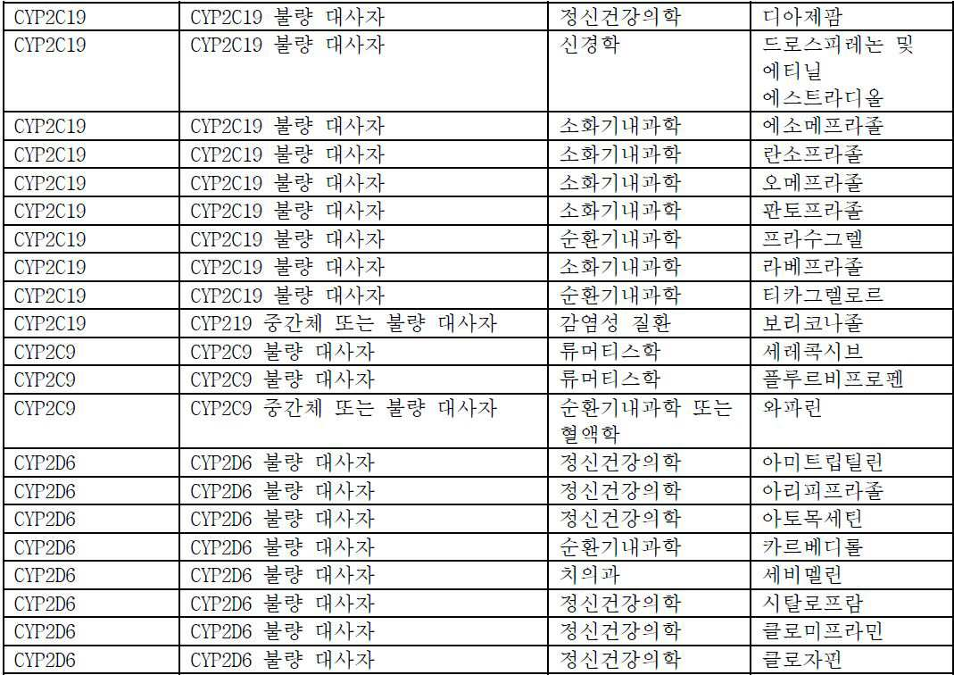
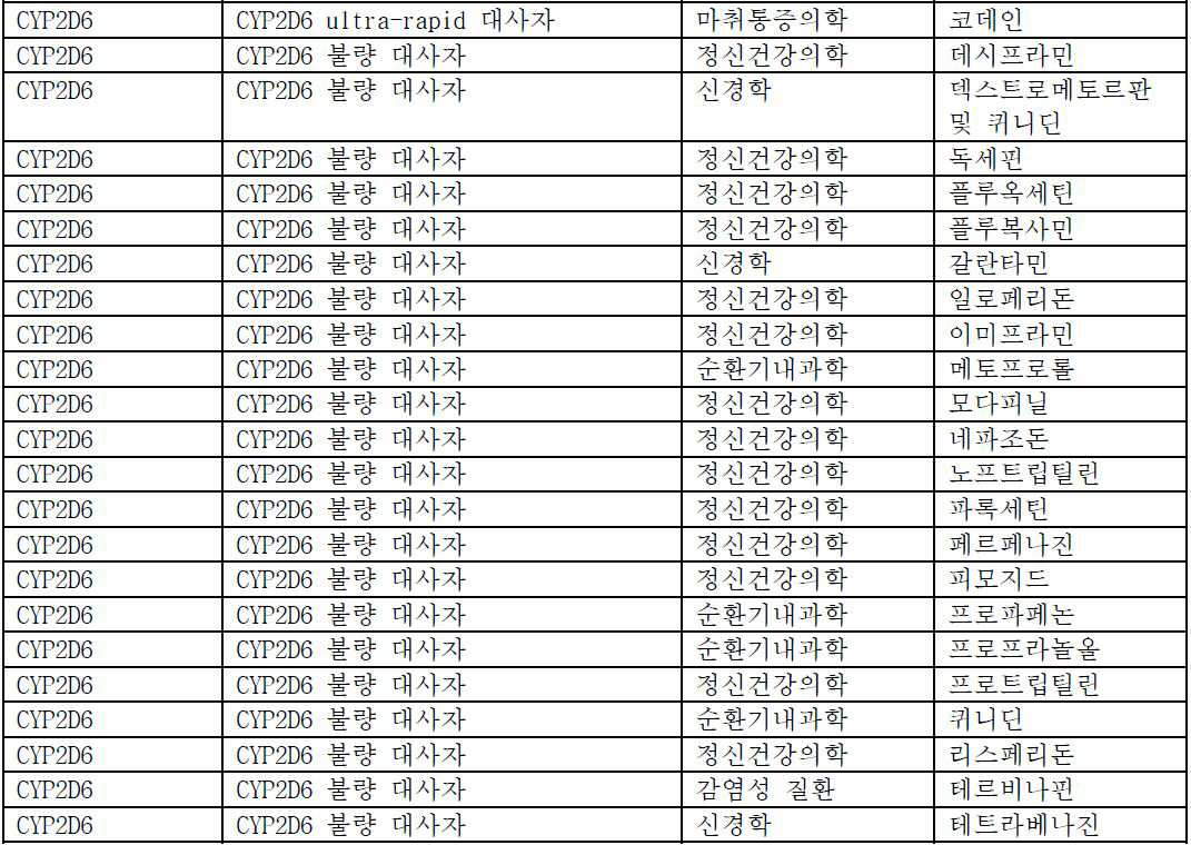


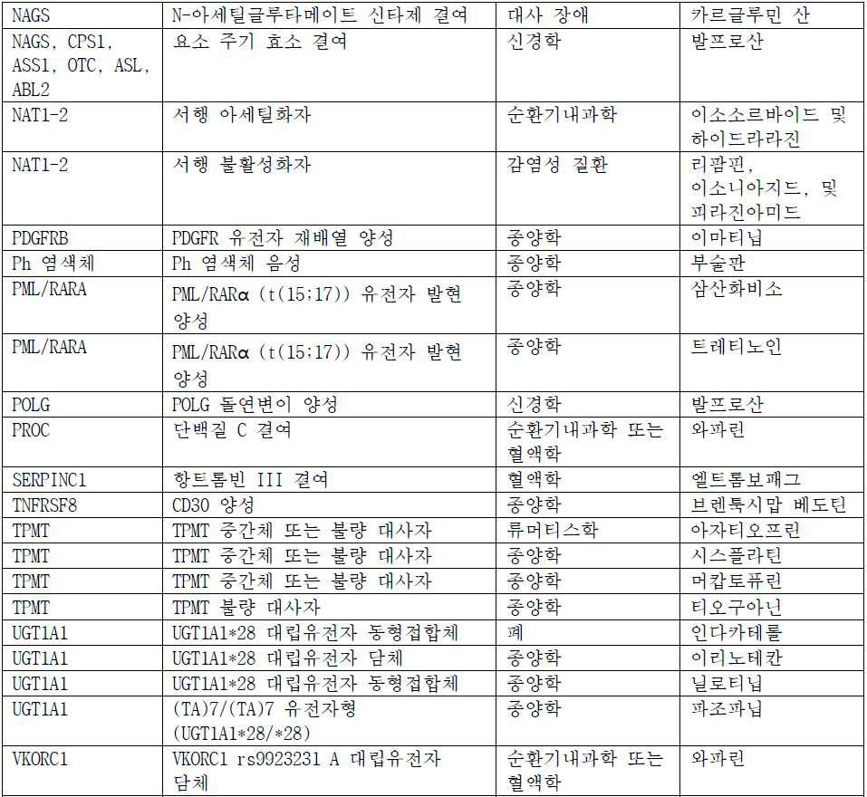
따라서, 본 발명은, 하기 단계를 포함하는, 이환 세포에서 비정상 신호전달 경로와 관련된 질환에 대하여 대상체의 진단 방법으로서, 상기 대상체가 바이오마커 음성인, 방법을 제공하고; Accordingly, the present invention provides a method of diagnosing a subject for a disease associated with an abnormal signal transduction pathway in a mature cell, comprising the steps of: providing the subject with a biomarker negative;
대상체로부터 수득된 생존 원발 이환 세포의 샘플의 제조 및 기본 배지에서 샘플의 배양 단계;The production of a sample of the surviving primary respiratory cells obtained from the subject and the culture of the sample in the primary culture;
(1) 교란제와 대상체로부터 수득된 생존 원발 이환 세포의 샘플의 제1 부분, 및 (2) 교란제 및 확인제와 샘플의 제2 부분의 접촉 단계 (여기서 상기 교란제는 선택적으로 질환-관련된 신호전달 경로에 영향을 미치고 확인제는 동일한 질환-관련된 신호전달 경로에 관한 교란제의 효과를 선택적으로 억제시킨다);(1) a first part of a sample of a survival primary respiratory cell obtained from the disturbing agent and a subject, and (2) a second step of contacting the second part of the sample with the perturbing agent and the identifying agent, Affects the delivery pathway and the inhibitor selectively inhibits the effect of the perturbant on the same disease-related signaling pathway);
샘플의 각 부분에서 생존 원발 이환 세포의 세포 접착 또는 부착의 연속적 측정 단계; 및Continuous measurement of cell adhesion or attachment of primary surviving primary cells in each part of the sample; And
샘플의 제1 부분과 샘플의 제2 부분 간 값의 차이를 확인하기 위해 생리적 반응 파라미터의 연속적 측정의 수학적 분석에 의한 측정량의 계측 단계;Measuring a measured quantity by a mathematical analysis of successive measurements of physiological response parameters to identify differences in values between the first portion of the sample and the second portion of the sample;
ErbB 신호전달 경로 활성의 정상 참조 구간으로부터 유도된 컷-오프 값과 측정량의 비교 단계;Comparing the measured amount with a cut-off value derived from a normal reference section of ErbB signaling pathway activity;
여기서 상기 대상체는 측정량이 컷-오프 값 초과인 경우 이환 세포에서 비정상 신호전달 경로와 관련된 질환에 대하여 진단된다.Wherein the subject is diagnosed for a disease associated with an abnormal signal transduction pathway in a bifurcated cell when the measured quantity is above a cut-off value.
추가 구현예에서, 샘플을 배양시키기 위해 사용된 기본 배지는 이들이 교란제와 접촉되기 최소 12 시간 내지 최대 72 시간 이전에 신선한 기본 배지로 대체된다.In a further embodiment, the basal medium used to incubate the sample is replaced with fresh basal medium from at least 12 hours up to a maximum of 72 hours before they are contacted with the perturbant.
또 다른 구현예에서, 본원에서 기재된 방법을 이용하는 비정상 신호전달 경로 활성으로 진단된 대상체는 대상체가 진단되는 비정상 신호전달 경로 활성을 억제시킨다고 공지된 요법으로 선택된다. 추가 구현예에서, 대상체는 본원에서 기재된 방법을 이용하여 선택된 요법이 투여된다.In another embodiment, a subject diagnosed with an abnormal signal transduction pathway activity using the methods described herein is selected for therapy that is known to inhibit the abnormal signaling pathway activity in which the subject is diagnosed. In a further embodiment, the subject is administered a therapy selected using the methods described herein.
하나의 구현예에서, 질환은 하기이다: 암, 예컨대 유방암, 결장암, 직장암, 자궁내막암, 위 암종, 위장 카르시노이드 종양, 위장 기질 종양, 교모세포종, 간세포 암종, 소세포 폐암, 비-소세포 폐암 (NSCLC), 흑색종, 난소암, 자궁경부암, 췌장암, 전립선 암종, 급성 골수성 백혈병 (AML), 만성적 골수성 백혈병 (CML), 비-호지킨 림프종 및 갑상선 암종 또는 두경부암. 비정상 신호전달 경로와 관련된 질환의 다른 유형, 예컨대 자가면역 질환 및 감염성 질환은 또한 포함된다.In one embodiment, the disease is: cancer such as breast cancer, colon cancer, rectal cancer, endometrial cancer, gastric carcinoma, gastric carcinoid tumor, gastric cancer tumor, glioblastoma, hepatocellular carcinoma, small cell lung cancer, non- small cell lung cancer (NSCLC), melanoma, ovarian cancer, cervical cancer, pancreatic cancer, prostate carcinoma, acute myelogenous leukemia (AML), chronic myelogenous leukemia (CML), non-Hodgkin's lymphoma and thyroid carcinoma or head and neck cancer. Other types of diseases associated with abnormal signal transduction pathways, such as autoimmune diseases and infectious diseases, are also included.
하나의 구현예에서, 질환은 암이고 따라서 이환 세포는 종양 세포의 샘플이다. 또 다른 구현예에서, 질환에 관여된 조직 유형으로부터 수득된 이환 환자의 건강한 세포는 대조군으로서 사용된다. 바람직한 구현예에서, 암은 유방암이다. 유방암 진단을 위한 하나의 구현예에서, 교란제는 PI3K 신호전달 경로, 예컨대 NRG1에 선택적으로 영향을 미친다. 또 다른 구현예에서, 교란제는 MAPK 신호전달 경로, 예컨대 상피 성장 인자를 선택적으로 영향을 미친다. 유방암 진단을 위한 하나의 구현예에서, 교란제는 ERα 신호전달 경로, 예컨대 에스트라디올을 선택적으로 영향을 미친다. 유방암 진단을 위한 더욱 또 다른 구현예에서, 교란제는 ErbB 신호전달 경로, 예컨대 HER2 신호전달 경로에 선택적으로 영향을 미친다. ErbB 신호전달 경로에 선택적으로 영향을 미치는 제제는 당해 기술에 공지되어 있다.In one embodiment, the disease is cancer and thus the diseased cell is a sample of tumor cells. In another embodiment, a healthy cell of a diseased patient obtained from a tissue type associated with the disease is used as a control. In a preferred embodiment, the cancer is breast cancer. In one embodiment for the diagnosis of breast cancer, the perturbant selectively affects the PI3K signaling pathway, such as NRG1. In another embodiment, the perturbant selectively affects the MAPK signaling pathway, such as epithelial growth factor. In one embodiment for the diagnosis of breast cancer, the perturbant selectively affects the ERa signaling pathway, such as estradiol. In yet another embodiment for breast cancer diagnosis, the perturbant selectively affects the ErbB signaling pathway, such as the HER2 signaling pathway. Agents that selectively affect the ErbB signaling pathway are known in the art.
다양한 구현예에서, 신호전달 경로는 MAPK, RHO, AKT, FAK1, RAS/RAF, PI3K/PTEN, MAK, MKK, 및 MEK로 이루어진 군으로부터 선택된다. 추가의 적합한 신호전달 경로는 본원에서 개시된 다른 신호전달 경로를 포함한다. 다양한 구현예에서, 교란제는, 예를 들어, 단백질, 펩타이드, 핵산, 대사물질, 리간드, 시약, 유기 분자, 신호전달 인자, 성장 인자, 생화학물질, 또는 이들의 조합일 수 있다.In various embodiments, the signaling pathway is selected from the group consisting of MAPK, RHO, AKT, FAKl, RAS / RAF, PI3K / PTEN, MAK, MKK, and MEK. Additional suitable signaling pathways include other signaling pathways disclosed herein. In various embodiments, the perturbing agent can be, for example, a protein, a peptide, a nucleic acid, a metabolite, a ligand, a reagent, an organic molecule, a signaling factor, a growth factor, a biochemical, or a combination thereof.
본원에서 추가로 기재된 바와 같이, 세포 접착 또는 부착은 임피던스 바이오센서 또는 광학적 바이오센서를 이용하여 측정될 수 있다. 하나의 구현예에서, 세포 접착 또는 부착에서의 변화는 유클리드 분석을 이용하여 평가된다. 예를 들어, 유클리드 분석은 하기로 이루어진 군으로부터 선택될 수 있다: 다중 시점에서 차이의 산술적 합산, 일시적 최대치, 일시적 최소치, 최대치 또는 최소치를 달성하는 시간, 기울기의 변화, 바이오센서 신호에서의 절대적 저하, 전체 측정의 총계, 또는 이들의 조합. 또 다른 구현예에서, 세포 접착 또는 부착에서의 변화는 일시적 최대치 또는 최소치에서의 변화에 의해 측정된다.As further described herein, cell adhesion or attachment can be measured using an impedance biosensor or an optical biosensor. In one embodiment, changes in cell adhesion or adhesion are assessed using Euclidean analysis. For example, Euclidean analysis can be selected from the group consisting of: arithmetic summation of differences at multiple time points, temporal maximum, temporal minimum, time to achieve maximum or minimum, change in slope, absolute decrease in biosensor signal , The total of the total measurements, or a combination thereof. In another embodiment, the change in cell adhesion or adhesion is measured by a change in transient maxima or minima.
다양한 구현예에서, 생존 원발 이환 세포는 질환과 관련된 신호전달 경로를 표적하는 확인제와 추가로 접촉되고 세포 접착 또는 부착에서 확인제의 효과는 측정된다. 또 다른 구현예에서, 확인제는 에스트로겐 관련된 신호전달 경로 활성을 억제시킨다고 공지된 선택적 에스트로겐 수용체 하향 조절물질 (SERD), 또는 선택적 에스트로겐 수용체 조절물질 (SERM)이다.In various embodiments, the primary surviving primary cells are further contacted with a detection agent that targets the signal transduction pathway associated with the disease and the effect of the detection agent on cell adhesion or attachment is measured. In another embodiment, the agent is a selective estrogen receptor downregulator (SERD), or selective estrogen receptor modulator (SERM), known to inhibit estrogen-associated signal transduction pathway activity.
더욱 또 다른 구현예에서, 이환 세포는 질환과 관련된 신호전달 경로를 표적하는 표적화 치료제와 추가로 접촉되고 세포 접착 또는 부착에서 표적화 치료제의 효과는 측정된다. 표적화 치료제의 비-제한 예는 하기를 포함한다: 세툭시맙, 도세탁셀, 에를로티닙, 게피티닙, 이리노테칸, 라파티닙, 파클리탁셀, 파조파닙, 토포테칸, 트라스투주맙, 풀베스트란트, 타목시펜, 레트로졸, 아나스트로졸, 엑세메스탄, 에버롤리무스, 아비라테론, 바이칼루타마이드, 보르테조밉, 베무라페닙, 이필리무맙 및 이들의 조합. 다른 적합한 표적화 치료제는 본원에서 개시된다. 더욱 또 다른 구현예에서, 방법은 추가로 대상체에 표적화 치료제의 투여를 포함한다.In yet another embodiment, the bifunctional cell is further contacted with a targeting agent that targets the signaling pathway associated with the disease and the effect of the targeting agent is determined in cell adhesion or attachment. Non-limiting examples of targeted therapeutic agents include: cetuximab, docetaxel, erlotinib, gefitinib, irinotecan, lapatinib, paclitaxel, pazopanib, topotecan, trastuzumab, fulvestrant, tamoxifen , Letrozole, anastrozole, exemestane, everolimus, aviraterone, bicalutamide, bortezomib, bemurafenid, eicilimumab, and combinations thereof. Other suitable targeting agents are disclosed herein. In yet another embodiment, the method further comprises administering a targeting therapeutic agent to the subject.
또 다른 구현예에서, 본 발명은, 하기 단계를 포함하는, 호르몬 요법을 위하여 에스트로겐 수용체 알파 (ERα) 음성 유방암을 가진 대상체의 선택 방법을 제공하고;In another embodiment, the invention provides a method of selecting a subject with estrogen receptor alpha (ERa) negative breast cancer for hormone therapy, comprising the steps of:
대상체로부터 수득된 생존 원발 유방암 세포의 샘플의 제조 및 기본 배지에서 샘플의 배양 단계;Producing a sample of survival primary breast cancer cells obtained from a subject and culturing the sample in a basal medium;
(1) 에스트로겐-관련 신호전달 경로를 상향조절 또는 하향조절한다고 공지된 교란제와 대상체로부터 수득된 생존 원발 유방암 세포의 샘플의 제1 부분, 및 (2) 동일한 에스트로겐-관련 신호전달 경로 활성을 억제시킨다고 공지된 교란제 및 확인제와 샘플의 제2 부분의 접촉 단계; (1) a first part of a disturbance agent known to upregulate or downregulate an estrogen-related signaling pathway and a sample of a survival breast cancer cell obtained from a subject, and (2) a second part of the same estrogen- Contacting the second portion of the sample with a perturbing agent and a known agent that are known to make the sample;
샘플의 각 부분에서 생존 원발 유방암 세포의 생리적 반응 파라미터의 연속적 측정 단계;Continuously measuring physiological response parameters of surviving primary breast cancer cells in each part of the sample;
샘플의 제1 부분과 샘플의 제2 부분 간 값의 차이를 확인하기 위해 생리적 반응 파라미터의 연속적 측정의 수학적 분석에 의한 측정량의 계측 단계; 및Measuring a measured quantity by a mathematical analysis of successive measurements of physiological response parameters to identify differences in values between the first portion of the sample and the second portion of the sample; And
에스트로겐 수용체 경로 활성에 대하여 정상 참조 구간으로부터 유도된 컷-오프 값과 측정량의 비교 단계;Comparing the measured amount with a cut-off value derived from a normal reference interval for estrogen receptor pathway activity;
여기서 대상체는 측정량이 컷-오프 값 초과인 경우 호르몬 요법으로 선택된다.Where the subject is selected for hormone therapy if the measured quantity is above the cut-off value.
추가 구현예, 샘플을 배양시키기 위해 사용된 기본 배지는 이들이 교란제와 접촉되기 최소 12 시간 내지 최대 72 시간 이전에 신선한 기본 배지와 대체된다.In a further embodiment, the basal medium used to incubate the sample is replaced with fresh basal medium at least 12 hours up to 72 hours before they are contacted with the perturbant.
ERα 신호전달 경로에 영향을 미치는 적합한 교란제는 당해 기술에 공지되어 있고 에스트라디올과 같이 본원에서 개시된다. 또 다른 구현예에서, 생존 원발 암 세포는 에스트로겐 관련된 신호전달 경로 활성을 억제시키는 확인제, 예컨대 선택적 에스트로겐 수용체 하향 조절물질과 추가로 접촉된다. 또 다른 구현예에서, 종양 세포는 ERα 신호전달 경로를 표적하는 표적화 치료제와 추가로 접촉되고 세포 접착 또는 부착에 관한 표적화 치료제의 효과는 측정된다. ERα 신호전달 경로를 표적하는 적합한 표적화 치료제는 당해 기술에 공지되어 있고 풀베스트란트 및 타목시펜과 같이 본원에서 개시된다. 방법은 대상체에 표적화 치료제의 투여를 추가로 포함할 수 있다.Suitable perturbants that affect ERa signaling pathways are known in the art and are disclosed herein, such as estradiol. In another embodiment, the primary survival cancer cells are further contacted with a detectable agent that inhibits estrogen-associated signaling pathway activity, such as a selective estrogen receptor down-regulating substance. In another embodiment, the tumor cells are further contacted with a targeting agent that targets the ERa signaling pathway and the effect of the targeting agent on cell adhesion or attachment is measured. Suitable targeting agents that target the ERa signaling pathway are known in the art and are disclosed herein, such as fulvestrant and tamoxifen. The method may further comprise administration of a targeted therapeutic agent to the subject.
더욱 또 다른 구현예에서, 본 발명은, 하기 단계를 포함하는, ErbB 신호전달 경로에 영향을 미치는 표적화 치료제를 이용한 요법을 위하여 비-과발현, 비-증폭된 ErbB2 암을 가진 대상체의 선택 방법을 제공하고;In yet another embodiment, the invention provides a method of selecting a subject with a non-overexpressed, non-amplified ErbB2 cancer for therapy with a targeting agent that affects the ErbB signaling pathway, comprising the steps of and;
대상체로부터 수득된 생존 원발 암 세포의 샘플의 제조 및 기본 배지에서 샘플의 배양 단계;Preparing a sample of the primary cancer cell obtained from the subject and culturing the sample in the primary culture medium;
(1) 교란제와 대상체로부터 수득된 생존 원발 암 세포의 샘플의 제1 부분, 및 (2) 교란제 및 확인제와 샘플의 제2 부분의 접촉 단계 (여기서 상기 교란제는 선택적으로 ErbB 신호전달 경로에 영향을 미치고 확인제는 선택적으로 동일한 ErbB 신호전달 경로상에서 교란제의 효과를 억제시킨다);(1) a first portion of a sample of the surviving primary cancer cells obtained from the disturbing agent and the subject, and (2) a contacting step of the second portion of the sample with the perturbant and the identifying agent, wherein the perturbator selectively binds to the ErbB signaling pathway And the inhibitor selectively inhibits the effect of the disrupter on the same ErbB signaling pathway);
샘플의 각 부분에서 생존 원발 암 세포의 생리적 반응 파라미터의 연속적 측정 단계;Continuously measuring the physiological response parameters of the primary cancer cells in each part of the sample;
샘플의 제1 부분과 샘플의 제2 부분 간 값의 차이를 확인하기 위해 생리적 반응 파라미터의 연속적 측정의 수학적 분석에 의한 측정량의 계측 단계;Measuring a measured quantity by a mathematical analysis of successive measurements of physiological response parameters to identify differences in values between the first portion of the sample and the second portion of the sample;
ErbB 신호전달 경로 활성의 정상 참조 구간으로부터 유도된 컷-오프 값과 측정량의 비교 단계;Comparing the measured amount with a cut-off value derived from a normal reference section of ErbB signaling pathway activity;
여기서 대상체는 측정량이 컷-오프 값 초과인 경우 ErbB 신호전달 경로에 영향을 미치는 표적화 치료제를 이용한 치료에 대해 선택된다.Wherein the subject is selected for treatment with a targeting agent that affects the ErbB signaling pathway when the measurand is above the cut-off value.
추가 구현예에서, 샘플을 배양시키기 위해 사용된 기본 배지는 이들이 교란제와 접촉되기 최소 12 시간 내지 최대 72 시간 이전에 신선한 기본 배지로 대체된다.In a further embodiment, the basal medium used to incubate the sample is replaced with fresh basal medium from at least 12 hours up to a maximum of 72 hours before they are contacted with the perturbant.
ErbB 신호전달 경로에 영향을 미치는 적합한 교란제는 당해 기술에 공지되어 있고 본원에서 개시된다. 하나의 구현예에서, 생존 원발 암 세포는 ErbB 관련된 신호전달 경로 신호전달을 억제시킨다고 공지된 확인제, 예컨대 2C4 마우스 단클론성 항체 또는 티로신 키나제 억제제와 접촉된다. 또 다른 구현예에서, 생존 원발 종양 세포는 ErbB 신호전달 경로를 표적하는 표적화 치료제와 추가로 접촉되고 세포 접착 또는 부착에서 표적화 치료제의 효과는 측정된다. ErbB 신호전달 경로를 표적하는 적합한 표적화 치료제는 당해 기술에 공지되어 있고 본원에서 개시된다. 방법은 대상체에 표적화 치료제의 투여를 추가로 포함할 수 있다.Suitable perturbants that affect the ErbB signaling pathway are known in the art and are disclosed herein. In one embodiment, the primary cancer cell of survival is contacted with a known inhibitor of inhibiting ErbB-associated signaling pathway signaling, such as a 2C4 mouse monoclonal antibody or tyrosine kinase inhibitor. In another embodiment, the surviving primary tumor cells are further contacted with a targeting agent that targets the ErbB signaling pathway and the effect of the targeting agent is determined in cell adhesion or attachment. Suitable targeting agents that target the ErbB signaling pathway are known in the art and are disclosed herein. The method may further comprise administration of a targeted therapeutic agent to the subject.
또 다른 구현예에서, 본 발명은, 하기 단계를 포함하는, 라파티닙을 이용한 요법을 위하여 비-과발현, 비-증폭된 ErbB2 유방암을 가진 대상체의 선택 방법을 제공한다:In another embodiment, the invention provides a method of selecting a subject with non-overexpressed, non-amplified ErbB2 breast cancer for lapatinib-based therapy, comprising the steps of:
대상체로부터 수득된 생존 원발 유방암 세포의 샘플의 제조 및 기본 배지에서 샘플의 배양 단계;Producing a sample of survival primary breast cancer cells obtained from a subject and culturing the sample in a basal medium;
샘플이 교란제와 접촉되기 최소 12 시간 내지 최대 72 시간 이전에 신선한 기본 배지와 샘플을 배양시키기 위해 사용된 기본 배지의 대체 단계;A fresh basal medium from at least 12 hours up to a maximum of 72 hours before the sample is contacted with the perturbant and an alternative to the basal medium used to culture the sample;
(1) 교란제로서의 뉴레굴린과 대상체로부터 수득된 생존 원발 유방암 세포의 샘플의 제1 부분, 및 (2) 뉴레굴린 및 확인제 (여기서 상기 확인제는 선택적으로 뉴레굴린과 동일한 ErbB 신호전달 경로를 억제시킨다)와 샘플의 제2 부분의 접촉; 및/또는 (3) 교란제로서 상피 성장 인자와 샘플의 제3 부분 및 (4) 상피 성장 인자 및 확인제 (여기서 상기 확인제는 선택적으로 상피 성장 인자와 동일한 ErbB 신호전달 경로를 억제시킨다)와 샘플의 제4 부분의 접촉 단계;(1) a first portion of a sample of surviving primary breast cancer cells obtained from the subject and (2) a second portion of a sample comprising a nuroligin and a detergent, wherein said identifying agent selectively inhibits the same ErbB signaling pathway as the neuregulin ) And the second portion of the sample; And / or (3) an epithelial growth factor as a perturbation agent and a third portion of the sample, and (4) an epithelial growth factor and a detector, wherein the determiner selectively inhibits the same ErbB signaling pathway as the epithelial growth factor, A contact step of a fourth portion;
교란제와 접촉된 생존 원발 유방암 세포의 세포 접착 또는 부착의 연속적 측정 단계;Continuous measurement of cell adhesion or attachment of survival primary breast cancer cells in contact with an agonist;
샘플의 제1 부분과 샘플의 제2 부분 간 값의 차이 및/또는 샘플의 제3 부분과 샘플의 제4 부분 간 값의 차이를 확인하기 위해 생리적 반응 파라미터의 연속적 측정의 수학적 분석에 의한 측정량의 계측 단계; 및A measured amount by a mathematical analysis of successive measurements of physiological response parameters to determine the difference between the values of the first portion of the sample and the second portion of the sample and / or the difference between the third portion of the sample and the fourth portion of the sample A measuring step of And
ErbB 경로 활성에 대하여 정상 참조 구간으로부터 유도된 컷-오프 값과 측정량의 비교 단계;Comparing the measured amount with a cut-off value derived from a normal reference section for ErbB path activity;
(여기서 대상체는 측정량이 컷-오프 값 초과인 경우 라파티닙을 이용한 치료에 대해 선택된다).(Wherein the subject is selected for treatment with lapatinib if the measurand is above the cut-off value).
대안적으로, 상기 설정된 동일한 방법은, ErbB 신호전달 경로에 영향을 미치는, 라파티닙 이외의, 다른 표적화 치료제로 적용될 수 있다. 아래 표 (표 4)는 ErbB 신호전달 경로 암을 치료하도록 의도된 경로, 약물 표적, 및 표적된 요법을 요약한다.Alternatively, the same set of methods described above can be applied to other targeting agents other than lapatinib, which affects the ErbB signaling pathway. The following table (Table 4) summarizes the pathways, drug targets, and targeted therapies intended to treat ErbB signaling pathway cancer.
표 4:Table 4:
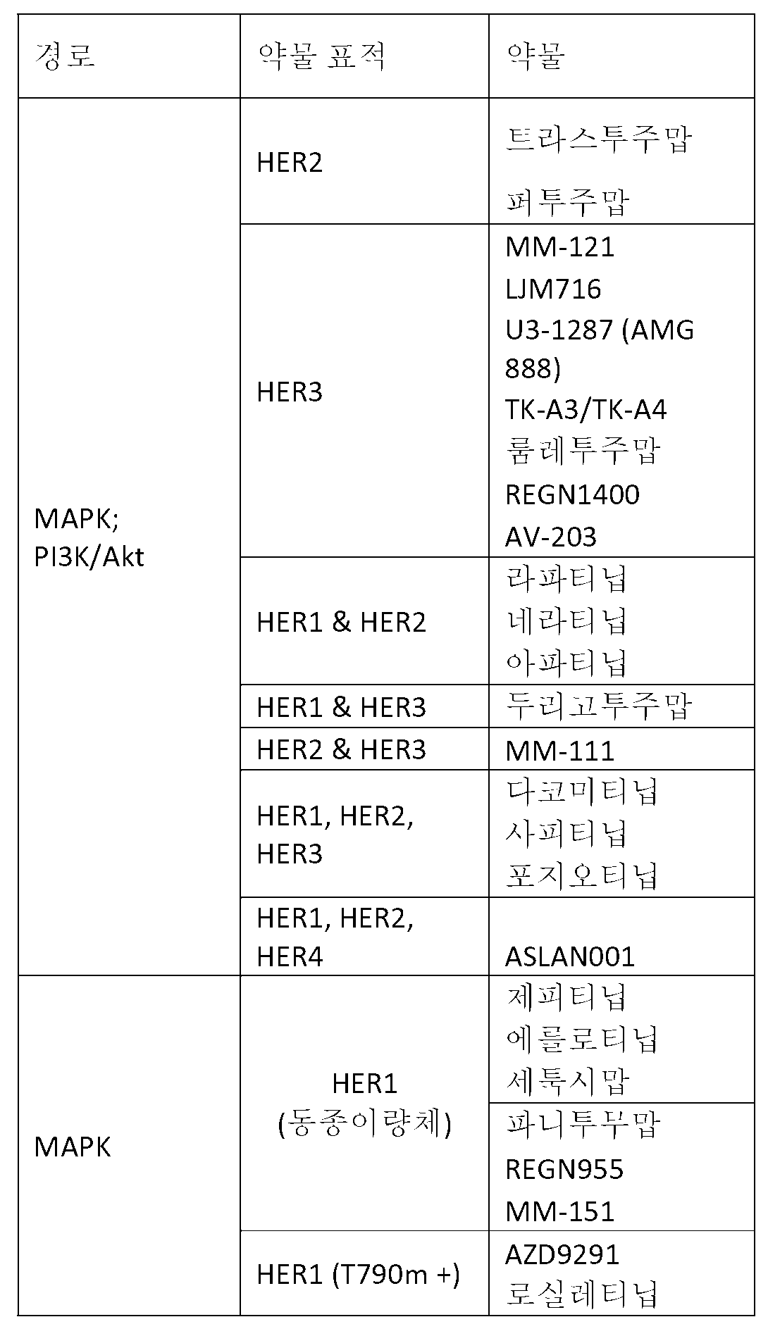
표적화 치료제를 이용한 치료를 위하여 대상체의 상기-기재된 선택 방법에 더하여, 본 발명은 또한 대상체에 표적화 치료제의 투여를 포함하는, 표적화 치료제를 이용한 대상체의 치료 방법을 포함하고, 여기서 상기 대상체는, 대상체로부터 생존 원발 세포의 샘플을 이용하여, 치료용 대상체의 상기-기재된 선택 방법 중 하나의 적용에 의해 표적화 치료제를 이용한 치료에 대해 선택되었다.In addition to the above-described selection method of a subject for treatment with a targeted therapeutic agent, the present invention also includes a method of treating a subject with a targeted therapeutic agent, comprising administering a targeted therapeutic agent to the subject, A sample of surviving primary cells was selected for treatment with a targeted therapeutic agent by application of one of the above-described selection methods of therapeutic agents.
C. 치료제의 효능의 선택 또는 C. selection of efficacy of the therapeutic agent or 모니터링monitoring 방법 Way
한 개인내 한 부위로부터 암이 유전자 구성, 단백질 발현 수준, 및 치료 중재에 대한 반응에서 또 다른 개인내 동일한 부위의 암과 완전히 상이할 수 있기 때문에 암 같은 질환은 부분적으로 이종성이다. 이환 조직은 유전자 발현 또는 유전자 변경에서 서로 상당히 다양할 수 있다. 예를 들어, 전이성 종양은 원발 종양과 상이할 수 있다. 인간 게놈 서열분석 및 다른 유전적 정량화 도구는 각 환자의 질환이 그 환자에 어느 정도 고유한지를 의사에서 정보제공하였다. 상기 정보는 개인맞춤형 의약에 맞춰 전체 사업을 낳았고, 여기에서 각 환자는 그의 질환에 대하여 맞춤형 치료 레지멘을 잠재적으로 받을 수 있다.Diseases such as cancer are partially heterogeneous because cancer from one part of an individual can be completely different from cancer in the same part of another individual in response to gene composition, protein expression levels, and therapeutic intervention. The diseased tissues can vary considerably from each other in gene expression or gene alteration. For example, metastatic tumors may be different from primary tumors. Human genome sequencing and other genetic quantification tools informed the physician that each patient's disease was unique to the patient. The information produced an entire business in accordance with the personalized medicament, where each patient could potentially receive a tailor-made treatment regimen for his disease.
일부 약물은 특이적 유전자-관련 질환 징후로 표적되고 있다. 상기 접근법은 현행 예후 도구세트의 유의미한 단점에 주로 기인하여 아직 광범위하게 이용되지 않는다. 본원에서 기재된 바와 같은 방법은 개인의 질환에 대해 효능을 보여주는 치료제의 선택 방법을 제공한다. 구현예에서, 치료제는 CReMS에서 이환 조직으로부터 단리된 무 표지 살아있는 전체의 세포에 접촉되고 세포의 생리적 파라미터에서 이의 변화 또는 부족은 치료제의 존재하에 검출된다. 치료제는 기준선 측정과 비교된 경우 질환 세포의 생리적 파라미터에서의 변화를 초래하는 대상체를 치료하기 위해 선택된다.Some drugs are targeted as specific gene-related disease indications. This approach is not yet widely used due mainly to the significant shortcomings of the current prognostic toolset. The method as described herein provides a method of selecting a therapeutic agent that demonstrates efficacy against an individual ' s disease. In an embodiment, the therapeutic agent is contacted with whole, unlabeled live cells isolated from the diseased tissue in CReMS and its change or lack in physiological parameters of the cells is detected in the presence of the therapeutic agent. The therapeutic agent is selected to treat a subject that results in a change in physiological parameters of the diseased cell when compared to a baseline measurement.
개시내용의 하나의 측면은, 초기 진단에서 또는 대상체의 치료 전반에 걸쳐, 질환 또는 장애를 치료하기 위한 용도로 상업적으로 승인되는 약물을 포함한, 하나 이상의 치료제의 선택 방법을 포함한다. 구현예에서, 방법은 하기 단계를 포함한다: 세포성 반응 측정 시스템에서 대상체로부터 적어도 하나의 단리된 질환 세포 샘플에 하나 이상의 치료제의 투여 단계; 치료제 또는 치료제들의 투여 이전 세포성 반응 파라미터의 기준선 측정과 비교된 경우 치료제 또는 치료제들의 반응에서 질환 세포 샘플의 세포성 반응 파라미터에서 변화가 발생하는지의 계측 단계 (여기서 세포성 반응 파라미터에서의 상기 변화는 제제 또는 제제들이 개별 대상체에서 질환에 대하여 치료적 효능을 갖는다는 것을 나타낸다). 특정 구현예에서, 단리된 질환 세포 샘플은 무 표지 전체의 세포를 포함한다. 다른 구현예에서, 단리된 질환 세포에서 세포성 반응 파라미터의 변화는 시간의 규정된 기간 동안 연속적으로 모니터링된다. 다른 구현예에서, 방법은 추가로 적어도 하나의 세포성 반응 또는 생리적 파라미터의 변화를 초래하는 치료제 또는 치료제의 조합의 선택 단계 및 건강 관리 제공자에 선택된 제제의 제공 단계를 포함한다. 다른 구현예에서, 방법은 추가로 적어도 하나의 세포성 반응 또는 생리적 파라미터의 변화를 초래하는 치료제 또는 치료제의 조합의 투여 단계를 포함한다.One aspect of the disclosure encompasses methods of selecting one or more therapeutic agents, including drugs that are commercially approved for use in treating a disease or disorder, either in an initial diagnosis or throughout the treatment of a subject. In an embodiment, the method comprises the steps of: administering at least one therapeutic agent to at least one isolated disease cell sample from a subject in a cellular response measurement system; Determining whether a change in a cellular response parameter of a diseased cell sample occurs in a response of the therapeutic agent or therapeutic agent when compared to a baseline measurement of cellular response parameters prior to administration of the therapeutic agent or therapeutic agents, The agent or agent has therapeutic efficacy against the disease in the individual subject). In certain embodiments, the isolated disease cell sample comprises whole non-labeled cells. In another embodiment, changes in cellular response parameters in isolated disease cells are continuously monitored for a defined period of time. In another embodiment, the method further comprises the step of selecting a therapeutic agent or combination of therapies that results in at least one cellular response or a change in physiological parameters, and the step of providing the agent selected by the healthcare provider. In another embodiment, the method further comprises the step of administering a therapeutic or a combination of therapies which results in a change in at least one cellular response or physiological parameter.
또 다른 구현예에서, 본 발명은, 하기 단계를 포함하는, 개별 대상체에서 질환에 대하여 제제의 치료적 효능을 계측함으로써 개별 대상체용 치료의 선택 방법을 제공한다: 세포성 반응 측정 시스템 (CReMS)에서 개별 대상체로부터 적어도 하나의 단리된 무 표지 질환 세포 샘플에 제제의 투여 단계 (여기서 상기 질환 세포 샘플은 하기로 이루어진 군으로부터 선택된다: 암 세포 샘플, 자가면역 질환을 가진 대상체로부터의 세포 샘플, 외래 제제로 감염된 조직으로부터의 세포 샘플 및 이들의 조합); 치료제의 존재 및/또는 부재하에 시간의 규정된 기간 동안 세포 샘플의 적어도 하나의 생리적 반응 파라미터에서의 변화의 연속적 측정 단계; 및 제제에 대한 세포 샘플의 생리적 반응 파라미터에서의 변화가 기준선 측정과 비교된 경우 발생하는지의 계측 단계 (여기서 생리적 반응에서의 상기 변화는 제제가 개별 대상체에서 질환에 대하여 치료적 효능을 갖는다). 구현예에서, 질환 세포는 암 세포이다.In another embodiment, the invention provides a method of selecting a treatment for an individual subject by measuring the therapeutic efficacy of the preparation against a disease in an individual subject, comprising the steps of: The method comprising: administering an agent to at least one isolated, unlabeled disease cell sample from an individual subject, wherein the disease cell sample is selected from the group consisting of: a cancer cell sample, a cell sample from a subject having an autoimmune disease, Cell samples from infected tissues and combinations thereof); Continuously measuring changes in at least one physiological response parameter of a cell sample during a defined period of time in the presence and / or absence of a therapeutic agent; And determining whether a change in a physiological response parameter of the cell sample to the agent occurs when compared to a baseline measurement, wherein the change in physiological response has therapeutic efficacy for the disease in the individual subject. In an embodiment, the disease cell is a cancer cell.
다른 구현예에서, 본 발명은 하기 단계를 포함하는 특정한 대상체에 대하여 치료제의 효능의 비교 방법을 제공한다: 세포의 적어도 하나의 생리적 파라미터를 측정하는 디바이스에서 동일한 대상체로부터 별개의 질환 세포 샘플에 적어도 2개의 상이한 치료제의 투여 단계; 기준선 측정에 비교된 각각의 치료제에 대한 각 세포 샘플의 생리적 반응의 계측 단계 (여기서 상기 생리적 반응은 각 치료제의 효능을 나타낸다). 특정 구현예에서, 단리된 질환 세포 샘플은 무 표지 전체의 세포를 포함한다. 다른 구현예에서, 단리된 질환 세포에서 세포성 반응 파라미터의 변화는 시간의 규정된 기간 동안 연속적으로 모니터링된다. 구현예에서, 방법은 추가로 더 나은 효능을 초래하는 치료제 또는 치료제의 조합의 선택 단계; 및 건강 관리 제공자에 선택의 제공 단계를 포함한다. 다른 구현예에서, 방법은 추가로 대상체에 더 나은 효능을 초래하는 치료제 또는 치료제의 조합의 투여 단계를 포함한다.In another embodiment, the invention provides a method of comparing the efficacy of a therapeutic agent to a particular subject comprising the steps of: measuring at least one physiological parameter of a cell; Administering different therapeutic agents; Measuring the physiological response of each cell sample to each therapeutic agent compared to a baseline measurement, wherein said physiological response indicates efficacy of each therapeutic agent. In certain embodiments, the isolated disease cell sample comprises whole non-labeled cells. In another embodiment, changes in cellular response parameters in isolated disease cells are continuously monitored for a defined period of time. In an embodiment, the method further comprises the step of selecting a combination of therapeutic agent or therapeutic agent resulting in a better efficacy; And providing a choice to the health care provider. In another embodiment, the method further comprises the step of administering a therapeutic or a combination of therapies which results in a better efficacy in the subject.
개시내용의 또 다른 측면은 종양 세포의 성장 속도의 계측 방법을 제공한다. 종양의 성장 속도를 측정함으로써, 치료는 종양 세포가 얼마나 빨리 성장할 수 있는지에 의존하여 선택될 수 있다. 종양 세포가 고속 성장 종양이면, 건강 관리 작업자는 저속 성장 종양에 대한 치료의 것과 비교된 경우 더욱 공격성 치료를 선택할 것이다. 특정 구현예에서, 방법은 하기 단계를 포함한다: 세포성 반응 측정 시스템에서 단리된 종양 세포 샘플의 제공 단계, 정의된 기간에 걸쳐 연속적으로 종양 세포 샘플의 성장 속도의 모니터링 단계, 및 고속 성장 속도를 나타내는 그 종양 세포를 위하여 더욱 공격성 치료의 선택 단계 및/또는 건강 관리 제공자에 선택된 치료의 제공 단계. 다른 구현예에서, 단리된 질환 세포 샘플은 무 표지 전체의 세포를 포함한다. 다른 구현예에서, 방법은 추가로 대상체에 선택된 치료의 투여 단계를 포함한다. 구현예에서, 고속 성장 종양은 약 100 시간 미만, 바람직하게는 20 시간 미만의 세포 배가 속도를 갖고, 반면에 저속 성장 종양은 100 시간 이상인 세포 배가 속도를 갖고, 여기에서 세포 배가 속도는 하나의 세포가 2개 세포로 분할하기 위한 시간이다.Another aspect of the disclosure provides a method of measuring the growth rate of tumor cells. By measuring the growth rate of the tumor, the treatment can be selected depending on how fast the tumor cells can grow. If the tumor cell is a fast-growing tumor, the healthcare worker will choose more aggressive treatment when compared to treatment for slow-growing tumors. In certain embodiments, the method comprises the following steps: providing a sample of isolated tumor cells in a cellular response measurement system, continuously monitoring the growth rate of the tumor cell sample over a defined period of time, Selecting a further aggressive treatment for the tumor cell and / or providing a treatment selected to the healthcare provider. In other embodiments, the isolated disease cell sample comprises whole non-labeled cells. In another embodiment, the method further comprises administering a selected treatment to the subject. In an embodiment, the fast-growing tumor has a cell doubling rate of less than about 100 hours, preferably less than 20 hours, while the slow-growing tumor has a cell doubling rate of at least 100 hours, wherein the cell- Is the time to divide into two cells.
또 다른 측면에서, 본 발명은 특정한 경로가 개별 대상체로부터 생존 원발 질환 세포 샘플에서 비정상인지, 및/또는 특정한 경로가 질환 세포 샘플에서 경로의 기능이상을 확인하기 위해 확인제에 감수성인지의 계측 방법을 제공한다. 상기 방법에서, 개인의 질환 세포 샘플에서 기능한 세포성 경로의 프로파일은 치료가 계속함에 따라 경시적으로 수득 및 모니터링될 수 있다. 특정 구현예에서, 방법은 하기 단계를 포함한다: 세포성 반응 측정 시스템에서 대상체로부터 적어도 하나의 단리된 생존 원발 질환 세포 샘플에 하나 이상의 교란제 및/또는 확인제를 투여함으로써 비정상 경로 기능의 존재로 질환 세포 샘플의 특성화 단계; 교란제 및/또는 확인제의 투여 이전 세포성 반응 파라미터의 기준선 측정과 비교된 경우 교란제 및/또는 확인제에 대한 반응에서 생존 원발 질환 세포 샘플의 세포성 반응 파라미터에서 변화가 발생하는지의 계측 단계 (여기서 세포성 반응 파라미터에서의 상기 변화는 교란제에 의해 활성화된 또는 확인제에 의해 억제된 세포성 경로가 개별 대상체로부터 단리된 생존 원발 질환 세포 샘플에서 비정상인 것을 나타낼 수 있다). 특정 구현예에서, 교란제는 하기를 포함한다: 성장 인자, 그 다음 세포성 경로를 활성화시키는 수용체 및 세포 표면 단백질 예컨대 헤레굴린에 결합하는 단백질 또는 다른 리간드, 질환 세포 샘플에서 경로를 활성화시키는 세포 표면 수용체를 갖는 형질전환된 세포를 포함한 세포, 또는 작은 유기 분자 (10,000 달톤 이하), 펩타이드, 원하는 방식으로 세포성 생리적 기능을 세포내로 교란시키는 핵산 (예를 들면 간섭하는 RNA). 다른 구현예에서, 확인제는 비-제한 목록으로부터 성장 인자 수용체 예컨대 EGFR, Her2, PDGFR, TGFR, FGFR, TNFR, HGF, FGF, IGF, TNFα, TGFβ, 또는 VEGF 수용체, 토포이소머라제 활성, 키나제, G-단백질 커플링된 수용체, 수용체 티로신 키나제, 미세소관 중합, 세포골격 조직화, 세포 기능 및 세포 접착을 억제시키는 것을 포함한다.In another aspect, the invention provides a method of determining whether a particular pathway is abnormal in a sample of surviving primary disease cells from an individual subject, and / or whether a particular pathway is susceptible to a detective agent to identify pathway dysfunction in a diseased cell sample do. In this method, the profile of cellular pathways functioning in a patient ' s disease cell sample can be obtained and monitored over time as treatment continues. In certain embodiments, the method comprises the steps of: administering to a sample of at least one isolated survival disease cell from a subject in a cellular response measurement system one or more perturbants and / Characterization of cell samples; Measuring whether a change in cellular cellular response parameters of the primary survival disease cell sample occurs in response to an agonist and / or a detective agent, when compared to a baseline measurement of cellular response parameters prior to administration of the agonist and / Such changes in cellular response parameters may indicate that the cellular pathway activated by the agonist or inhibited by the agonist is abnormal in the primary survival disease cell sample isolated from the individual subject). In certain embodiments, the disrupter comprises: a growth factor, a receptor that activates the cellular pathway, and a protein or other ligand that binds to cell surface proteins such as, for example, weirelegine, a cell surface that activates the pathway in the disease cell sample Cells containing transformed cells with receptors, or small organic molecules (less than 10,000 daltons), peptides, nucleic acids that interfere with intracellular cellular physiological function in a desired way (eg interfering RNA). In other embodiments, the identifying agent is selected from the non-limiting list, such as growth factor receptors, such as EGFR, Her2, PDGFR, TGFR, FGFR, TNFR, HGF, FGF, IGF, TNFa, TGFβ, or VEGF receptors, topoisomerase activity, G-protein coupled receptors, receptor tyrosine kinases, microtubule polymerization, cytoskeletal organization, cell function and cell adhesion.
예를 들어, 하나의 구현예에서, 세포성 경로를 작용화 또는 길항화한다고 공지된 교란제 (예를 들면, 활성제 제제)와 대상체로부터 수득된 이환 세포 샘플의 접촉, 샘플에서 생존 원발 세포에서 하나 이상의 생리적 반응 파라미터의 연속적 측정, 및 적합한 대조군에 비하여, 교란제의 존재하에 생존 원발 이환 세포 샘플내 하나 이상의 생리적 반응 파라미터에서 변화의 양의 연속적 측정에 의해, 개별 대상체로부터 수득된 이환 세포에서 세포성 경로의 기능성 상태의 계측 방법이 제공되고, 여기서, 적합한 대조군에 비하여, 교란제의 존재하에 하나 이상의 생리적 반응 파라미터에서 변화의 양은 교란제에 의해 표적된 세포성 경로가 개별 대상체에서 비정상인 것을 나타낸다.For example, in one embodiment, contacting a sample of a diseased cell obtained from a subject with an agonist known to functionalize or antagonize the cellular pathway ( e. G. , An active agent formulation) A continuous measurement of the above physiological response parameters and a continuous measurement of the amount of change in one or more physiological response parameters in a primary survival primary cell sample in the presence of an agonist, relative to a suitable control, Wherein the amount of change in one or more physiological response parameters in the presence of an agonist, relative to a suitable control, indicates that the cellular pathway targeted by the agonist is abnormal in the individual subject.
개별 대상체에서 세포성 경로의 상태의 지식은, 하나의 구현예에서, 신호전달 경로 유도된 질환의 존재를 진단하기 위해 사용될 수 있다. 추가 구현예에서, 본원에서 기재된 방법으로 진단된 대상체는 신호전달 경로 질환을 표적하는 치료제에 반응성인 것으로 예상된다. 예를 들어, 대상체로부터 이환 세포가, 교란제의 존재하에 하나 이상의 생리적 반응 파라미터의 연속적 측정의 분석에 의해 계측된 바와 같이, 교란제 (예를 들면, 활성제 제제)에 비정상적으로 반응성이면, 대상체는 교란제와 동일한 세포성 경로에 표적화 치료제에 유사하게 반응성일 것이다. 적합한 대조군에 비하여, 교란제의 존재하에 이환 세포 샘플의 생존 세포에서 하나 이상의 생리적 반응 파라미터에서의 변화가 발생하면 대상체로부터 이환 세포는 교란제에 비정상적으로 반응성이다. Knowledge of the state of the cellular pathway in an individual subject can, in one embodiment, be used to diagnose the presence of a signaling pathway induced disease. In a further embodiment, a subject diagnosed by the methods described herein is expected to be responsive to a therapeutic agent targeting a signaling pathway disease. For example, if a respiratory cell from an object is abnormally reactive with an agonist ( e. G. , An active agent formulation), as measured by analysis of a continuous measurement of one or more physiological response parameters in the presence of an agonist, Will be similarly reactive to the targeting agent in the same cellular pathway as the agonist. Compared to a suitable control, when a change in one or more physiological response parameters occurs in a living cell of a diseased cell sample in the presence of an agonist, the immune cell from the subject is abnormally reactive to the agonist.
또 다른 예에서, 대상체로부터 이환 세포가, 교란제의 존재하에 하나 이상의 생리적 반응 파라미터의 연속적 측정의 분석에 의해 계측된 바와 같이, 교란제 (예를 들면, 활성제 제제)에 비정상적으로 반응성이지 않으면, 대상체는 교란제와 동일한 세포성 경로에 표적화 치료제의 투여의 도움을 받는데 유사하게 실패할 것이다. 교란제의 존재하에 이환 세포 샘플의 생존 세포내 하나 이상의 생리적 반응 파라미터에서의 변화가 적합한 대조군 미만이면 대상체로부터 이환 세포는 교란제에 정상적으로 반응성이다.In another example, if the infective cell from a subject is not abnormally reactive to an agonist ( e. G. , An active agent formulation), as measured by analysis of a continuous measurement of one or more physiological response parameters in the presence of the agonist, The subject will similarly fail to receive the same targeted cellular pathway as the perturbing agent, assisted by the administration of the targeted therapeutic agent. If the change in one or more physiological response parameters in the surviving cells of the diseased cell sample in the presence of an agonist is less than the appropriate control, the immune cells from the subject are normally responsive to the agonist.
따라서, 또 다른 측면에서, 세포성 경로를 작용화 또는 길항화한다고 공지된 교란제와 대상체로부터 수득된 이환 세포 샘플의 접촉 단계, 샘플내 생존 세포에서 하나 이상의 생리적 반응 파라미터의 연속적 측정 단계, 및 적합한 대조군에 비하여, 교란제의 존재하에 이환 세포 샘플에서 발생하는 하나 이상의 생리적 반응 파라미터에서 변화의 양의 연속적 측정의 수학적 분석에 의한 계측 단계에 의해, 개별 대상체에 대하여 표적화 치료제의 선택 방법이 제공되고, 여기서 적합한 대조군에 비하여, 교란제의 존재하에 하나 이상의 생리적 반응 파라미터에서 변화의 양은 대상체가 세포성 경로를 표적하는 표적화 치료제에 반응성일 것을 나타낸다.Thus, in another aspect, there is provided a method of treating a cell, comprising contacting a sample of a diseased cell obtained from a subject with an agonist known to functionalize or antagonize the cellular pathway, successively measuring one or more physiological response parameters in the surviving cells in the sample, Compared to the control, a method of selecting a targeting therapeutic agent for an individual subject is provided by a measurement step by a mathematical analysis of a continuous measurement of the amount of change in one or more physiological response parameters occurring in a sample of diseased cells in the presence of an agonist, Where the amount of change in one or more physiological response parameters in the presence of an agonist, relative to a suitable control, indicates that the subject is responsive to the targeting treatment targeting the cellular pathway.
특정 구현예에서, 대상체로부터 수득된 세포 샘플의 제1 부분은 교란제와 접촉되고 세포 샘플의 제2 부분은 교란제 및 확인제와 접촉된다. 방법은 그 다음 추가로 샘플의 각각의 부분에서 생리적 반응 파라미터 (예를 들면, 세포 접착 또는 부착)의 연속적 측정 단계, 그 다음 샘플의 제1 부분과 샘플의 제2 부분 간 값의 차이를 확인하기 위해 생리적 반응 파라미터의 연속적 측정의 수학적 분석에 의한 측정량의 계측 단계; 그 다음 정상 참조 구간으로부터 유도된 컷-오프 값과 측정량의 비교 단계를 포함한다. 표적화 치료제는 측정량이 컷-오프 값 초과인 경우 선택된다.In certain embodiments, a first portion of a cell sample obtained from a subject is contacted with an agonist and a second portion of the cell sample is contacted with an agonist and a detective. The method may then be further followed by a successive measurement step of a physiological response parameter (e.g., cell adhesion or attachment) in each portion of the sample, then a difference between the first portion of the sample and the second portion of the sample Measuring a measurand by a mathematical analysis of successive measurements of the physiological response parameters; And then comparing the measured amount with the cut-off value derived from the normal reference interval. The targeting agent is selected when the measured quantity exceeds the cut-off value.
특정 구현예에서, 방법은 추가로 대상체에 (즉, 대상체가 제제에 반응성인 것으로 계측되면) 표적화 치료제의 투여를 포함한다.In certain embodiments, the method further comprises administering a targeted therapeutic agent to the subject ( i . E., When the subject is measured to be responsive to the agent).
다른 구현예에서, 방법은 세포성 반응 측정 시스템에서 대상체로부터 단리된 질환 세포 샘플에 하나 이상의 활성제 제제의 투여 단계; 교란제의 투여 이전 세포성 반응 파라미터의 기준선 측정과 비교된 경우 정의된 기간에 걸쳐 교란제와 반응으로 질환 세포 샘플의 세포성 반응 파라미터에서 변화가 발생하는지의 계측 단계, 단리된 질환 세포 샘플에 하나 이상의 확인제의 투여 단계 및 교란제의 투여 이전 또는 이후 세포성 반응 파라미터에 비교된 경우 정의된 기간에 걸쳐 확인제와 반응으로 질환 세포 샘플의 세포성 반응 파라미터에서 변화가 발생하는지의 계측 단계를 포함하고, 여기서 적합한 대조군에 비하여 세포성 반응 파라미터에서 변화의 상기 양은 교란제에 의해 활성화된 및 확인제에 의해 억제된 세포성 경로가 개별 대상체로부터 단리된 질환 세포 샘플에서 비정상적으로 기능하고 있다는 것을 나타낸다.In another embodiment, the method comprises the steps of administering one or more activator agents to a diseased cell sample isolated from the subject in a cellular response measurement system; Determining whether a change in cellular cellular response parameters of the disease cell sample occurs in response to the perturbant over a defined period of time compared to baseline measurement of cellular cellular response parameters prior to administration of the perturbant; Determining whether a change in cellular cellular response parameters of the diseased cell sample occurs in response to the determinant over a defined period of time, when compared to the cellular response parameters before or after administration of the above-identified agent, Wherein the amount of change in the cellular response parameter relative to the appropriate control indicates that the cellular pathway activated by the perturbing agent and inhibited by the agent is functioning abnormally in the diseased cell sample isolated from the individual subject.
추가의 구현예는 치료, 임상시험, 및/또는 후보자 치료제에 대한 환자의 반응성 평가용 대상체의 선택 방법을 포함한다. 구현예에서, 대상체는 후보자 치료제에 아마도 반응하는 그 환자만을 선택하기 위해 그 후보자 치료제의 임상시험에 앞서 선택되고; 상기 접근법은, 특히 그 질환으로 진단되는 전체 집단의 부분에 대하여 유효한 결과를 제공할 수 있을 뿐인 치료제로, 후보자 치료제가 조절 승인을 보증하기에 충분한 선택된 환자 집단 내에서 효능을 입증할 수 있다는 가능성을 증가시킬 것이다. 상기 접근법으로 미승인된 치료제의 임상시험으로 고려된 환자는 미승인된 약물에 대한 그의 반응성을 예측하기 위해 비정상 신호전달 경로 활성의 존재를 계측하도록 평가된 그의 이환 세포를 가질 것이다. 비정상 신호전달 경로 활성의 존재를 입증하는 것만이 실험에 선택될 것이다. 다른 구현예에서, 세포성 반응 측정 시스템에서 대상체로부터 적어도 하나의 단리된 질환 세포 샘플에 하나 이상의 치료제의 투여 단계; 치료제 또는 치료제들의 투여 이전 세포성 반응 파라미터의 기준선 측정에 비교된 경우 치료제 또는 치료제들에 반응으로 질환 세포 샘플의 세포성 반응 파라미터에서 변화가 발생하는지의 계측 단계를 포함하는 방법에 의해 대상체의 세포의 샘플이 반응군으로서 확인된 경우 대상체는 치료에 선택되고, 여기서 세포성 반응 파라미터에서 상기 변화는 제제 또는 제제들이 개별 대상체에서 질환에 대하여 치료적 효능을 갖는 것을 나타낸다. 특정 구현예에서, 방법은 추가로 세포가 치료용 또는 임상시험용 치료제 또는 치료제들에 반응으로 세포성 반응 파라미터에서 변화를 나타내는 대상체의 선택 단계를 포함한다.Additional embodiments include methods of selecting a subject for evaluation of a patient ' s response to treatment, clinical trials, and / or candidate therapeutic agents. In an embodiment, the subject is selected prior to the clinical trial of the candidate therapeutic to select only those patients who are likely to respond to the candidate therapeutic; This approach has the potential to provide a potential therapeutic benefit for a candidate therapeutic that is capable of providing efficacious results, particularly for parts of the entire population diagnosed with the disease, that the candidate therapeutic can demonstrate efficacy in selected patient populations sufficient to warrant regulatory approval . A patient considered as a clinical trial of an unapproved therapeutic agent in this approach will have its morbid cells evaluated to measure the presence of abnormal signaling pathway activity to predict its responsiveness to unapproved drugs. Only the presence of abnormal signaling pathway activity will be selected for the experiment. In another embodiment, there is provided a method of treating a cell disease, comprising: administering one or more therapeutic agents to a sample of at least one isolated disease cell from a subject in a cellular response measurement system; The method comprising the step of determining whether a change in the cellular response parameter of the diseased cell sample occurs in response to the therapeutic agent or therapeutic agents when compared to a baseline measurement of the cellular response parameter prior to administration of the therapeutic agent or therapeutic agents When the sample is identified as a response group, the subject is selected for treatment, wherein the change in cellular response parameters indicates that the agent or agents have therapeutic efficacy against the disease in the individual subject. In certain embodiments, the method further comprises the step of selecting a subject for which the cell exhibits a change in cellular response parameters in response to a therapeutic or therapeutic agent for treatment or treatment.
추가 측면에서, 본 발명은 치료제에 반응성 또는 비-반응성을 입증하는 대상체로부터 질환 샘플의 바이오마커의 확인 방법을 제공한다. 하나의 구현예에서, 방법은 세포성 반응 측정 시스템에서 치료제와 대상체로부터 단리된 질환 세포 샘플의 접촉 단계; 치료제 또는 치료제들의 투여 이전 세포성 반응 파라미터의 기준선 측정과 비교된 경우 치료제 또는 치료제들과 반응으로 질환 세포 샘플의 세포성 반응 파라미터에서 변화가 발생하는지의 계측 단계를 포함하고, 여기서 세포성 반응 파라미터에서 상기 변화는 제제 또는 제제들이 개별 대상체 (반응군)에서 질환에 대하여 치료적 효능을 갖는 것을 나타내고 변화의 부족은 치료제가 그 대상체의 질환 (비반응군)에 대하여 효능을 갖지 않는 것을 나타낸다. 구현예에서, 방법은 추가로 다른 바이오마커용 치료제에 반응성인 대상체로부터 세포의 추가 특성화 단계 및/또는 다른 바이오마커용 치료제에 반응성이 아닌 대상체로부터 세포의 추가 특성화 단계를 포함한다. 바이오마커는 하기를 포함한다: 유전자 돌연변이, 단일 뉴클레오타이드 다형성, 유전자 발현 수준, 단백질, 단백질 돌연변이, 번역후 단백질 변형, 스플라이스 변이체, 세포 표면 마커, 단백질 또는 핵산의 과발현, 핵산의 증폭 (비-제한 예는 mRNA, miRNA 또는 다른 RNA를 포함한다), 세포 형태학, 및 이들의 조합. In a further aspect, the invention provides a method of identifying a biomarker of a disease sample from a subject that demonstrates responsiveness or non-reactivity to the therapeutic agent. In one embodiment, the method comprises contacting a therapeutic cell sample isolated from a therapeutic agent with a cellular sample in a cellular response measurement system; Determining whether a change in the cellular response parameter of the diseased cell sample occurs in response to the therapeutic agent or therapeutic agents when compared to a baseline measurement of the cellular response parameter prior to administration of the therapeutic agent or therapeutic agents, The change indicates that the agent or agent has therapeutic efficacy against the disease in the individual subject (the responding group) and the lack of change indicates that the therapeutic agent has no effect on the disease (unreacted group) of the subject. In an embodiment, the method further comprises the step of further characterizing the cells from a subject that is responsive to the therapeutic agent for another biomarker and / or further characterizing the cells from the subject that is not responsive to the therapeutic agent for the other biomarker. Biomarkers include: gene mutations, single nucleotide polymorphisms, gene expression levels, proteins, protein mutations, posttranslational protein modifications, splice variants, cell surface markers, overexpression of proteins or nucleic acids, Examples include mRNA, miRNA, or other RNA), cytomorphology, and combinations thereof.
표적화 치료제의 효능은 표적된 요법이 영향받도록 의도되는 (예를 들면 비정상 신호전달 경로 활성) 질환을 환자가 갖는지의 여부, 그의 표적에 결합하는지의 여부 및 비정상 신호전달 활성에서 우연한 변화를 일으키는지 여부에 좌우된다. 본원에서 기재된 방법은 세포 샘플에서 제제(들)에 생리적 반응을 측정함으로써 제제가 효과에 의도되는 경로상에서 교란제, 및/또는 확인제, 및/또는 치료제가 갖는 효과를 측정한다. 게놈 및 단백체 도구는 치료제 및/또는 교란제 (예를 들면, 활성화제)에 의해 영향받는 세포 경로 성분에서 변경을 확인하기 위해 사용될 수 있다. 따라서, 하나의 구현예에서, 교란제, 및/또는 확인제, 및/또는 치료제와 대상체로부터 수득된 단리된, 무 표지 세포성 샘플의 접촉 단계, 샘플내 생존 세포에서 적어도 하나의 생리적 반응 파라미터의 연속적 측정에 의한 제제의 효과의 모니터링 단계, 생리적 반응 파라미터에서 변화가 발생하는지의 연속적 측정의 수학적 분석에 의한 계측 단계, 그렇게 함으로써 제제(들)에 대한 샘플의 감수성의 특성화 단계, 샘플상에서 교란제의 활성의 중단 단계, 및 세포 경로의 성분이 세포성 샘플에서 제제(들)에 의해 변경되는지를 계측하기 위해, 프로테오믹스, qPCR, 유전체학, RNA 정량화, 탠덤 액체 크로마토그래피-질량 분광법, 및 대사체학으로부터 선택된 방법을 이용하여 제제(들)에 의해 표적된 세포 경로의 성분 또는 바이오마커의 분석 단계에 의해, 개별 대상체에서 교란제, 및/또는 확인제, 및/또는 치료제에 의해 영향받는 세포 경로 성분의 확인 방법이 제공된다. 상기 방법은, 예를 들어, 환자 세포에서 경로의 교란시 세포 경로 성분이 발현 또는 활성화 (예를 들면, 인산화)를 변화시키는 것을 계측하기 위해 사용될 수 있다.The efficacy of a targeted therapeutic will depend on whether the patient has a disease for which the targeted therapy is intended to be effected (e. G., Abnormal signal transduction pathway activity), whether it binds to its target, and whether it causes an ad hoc change in an abnormal signaling activity . The methods described herein measure the physiological response to the agent (s) in a cell sample to determine the effects of the agent (s), and / or the agent (s) and / or therapeutic agent (s) on the path to which the agent is intended to effect. Genomic and proteomic tools can be used to identify changes in cellular pathway components affected by therapeutic agents and / or perturbants ( e. G. , Activators). Thus, in one embodiment, the contacting step of an isolated, unlabeled cellular sample obtained from a subject with an agonist, and / or a reagent, and / or a therapeutic agent, a continuous step of at least one physiological response parameter A step of monitoring the effect of the formulation by measurement, a measurement step by a mathematical analysis of a continuous measurement of whether a change in the physiological response parameter occurs, thereby characterizing the susceptibility of the sample to the formulation (s) QPCR, genomics, RNA quantitation, tandem liquid chromatography-mass spectrometry, and metabolism to determine whether the components of the cellular pathway are altered by the agent (s) in the cellular sample By the steps of analyzing the components of the cell pathway or the biomarkers targeted by the agent (s) Methods of identifying cell pathway components that are affected by a perturbant, and / or a detergent, and / or a therapeutic agent in a subject are provided. The method can be used, for example , to measure the change in expression or activation ( e.g. , phosphorylation) of a cellular pathway component in a pathway disturbance in a patient cell.
이들 방법은 또한 동일한 개별 대상체로부터 상이한 조직에서 2개 세포 샘플의 경로 활성을 비교하기 위해 사용될 수 있다.These methods can also be used to compare the pathway activity of two cell samples in different tissues from the same individual subject.
특정 구현예에서, 교란제는 약물, 교란제의 조합, 경로의 활성제 및 억제제를 포함하는 교란제의 조합, 경로의 상이한 구성원의 작용제 및 길항제를 포함하는 교란제의 조합, 또는 치료제를 포함하는 교란제의 조합일 수 있다.In certain embodiments, the perturbants are selected from the group consisting of drugs, combinations of perturbants, combinations of perturbants including pathway activators and inhibitors, combinations of perturbants comprising agonists and antagonists of different members of the pathway, Combination.
이들 방법은 또한 약물 후보자인 소분자 또는 항체가 이들이 표적하는 경로에서 효과를 갖는지를 평가함으로써 약물 발견 공정을 향상시키기 위해 사용될 수 있다.These methods can also be used to improve the drug discovery process by assessing whether drug candidates, small molecules or antibodies, have an effect on the pathway they are targeted for.
더욱 다른 구현예에서, 본 발명은 세포의 적어도 하나의 생리적 파라미터를 측정하는 디바이스에서 동일한 대상체로부터 질환 세포 샘플을 분리시키기 위해 복수의 치료제 조합의 투여 단계 (여기서 각 치료제 조합은 동일한 대상체로부터 질환 세포 샘플을 분리시키기 위해 투여된다); 및 기준선 측정과 비교된 각 치료제 조합에 대한 각 세포 샘플의 생리적 반응의 계측 단계 (여기서 상기 생리적 반응은 포텐셜 치료제 조합의 가장 유효한 치료제 조합을 나타낸다)에 의해 특정한 대상체용 약물의 조합 또는 최적의 치료적 체제의 계측 방법을 제공한다. 특정 구현예에서, 방법은 추가로 적어도 하나의 세포성 반응 또는 생리적 파라미터의 변화를 초래하는 치료제 또는 치료제의 조합의 선택 단계를 포함한다. 다른 구현예에서, 방법은 추가로 대상체에 적어도 하나의 세포성 반응 또는 생리적 파라미터의 변화를 초래하는 치료제 또는 치료제의 조합의 투여 단계를 포함한다.In yet another embodiment, the present invention provides a method of measuring a physiological parameter of a cell, the method comprising the steps of administering a combination of a plurality of therapeutic agents to separate a diseased cell sample from the same subject in a device that measures at least one physiological parameter of the cell, ≪ / RTI > And a measurement of the physiological response of each cell sample to each therapeutic agent combination compared to a baseline measurement, wherein the physiological response represents the most effective therapeutic agent combination of the potential therapeutic agent combinations. Provides a measurement method of the system. In certain embodiments, the method further comprises the step of selecting a therapeutic agent or combination of therapies that results in a change in at least one cellular response or physiological parameter. In another embodiment, the method further comprises the step of administering a therapeutic agent or combination of therapies that results in a change in at least one cellular response or physiological parameter to the subject.
또 다른 측면에서, 본 발명은 하기 단계에 의해 질환에 대하여 환자의 치료를 포함하는 방법을 제공한다: 세포성 반응 측정 시스템에서 대상체로부터 적어도 하나의 단리된 질환 세포 샘플에 하나 이상의 치료제의 투여를 포함하는 질환 치료용 치료제의 선택 단계; 치료제 또는 치료제들의 투여 이전 세포성 반응 파라미터의 기준선 측정과 비교된 경우 치료제 또는 치료제들과 반응으로 질환 세포 샘플의 세포성 반응 파라미터에서 변화가 발생하는지의 계측 단계, 세포성 반응 파라미터에서 변화를 야기하는 치료제의 선택 단계; 대상체에 적어도 하나의 세포성 반응 또는 생리적 파라미터의 변화를 초래하는 치료제의 투여 단계. 치료제는 특이적 생물학적 경로에 표적되는 것, 세포 증식을 억제시키는 것, 세포 사멸을 향상시키는 것, 염증을 억제시키는 것, 미생물을 사멸시키는 것 및/또는 면역 반응을 향상시키는 것을 포함한다. 특정 구현예에서, 치료제가 특이적 생물학적 경로에 표적되는 경우, 세포 표면 수용체와 상호작용할 수 있고 수용체용 리간드의 작용을 억제시킬 수 있다. 본 발명의 또 다른 구현예에서 생리적 반응 파라미터 또는 바이오마커는 환자 살아있는 세포 또는 조직 샘플로부터 상청액 분획을 시험함으로써 측정된다. 예를 들어, 일부 유방암 세포는 상피 성장 인자 수용체 (EGFR)에 대하여 양성이고 상피 성장 인자 (EGF)에 반응한다. 개별 대상체의 세포용 EGF의 상호작용을 억제시키는 치료제의 효능은 리간드의 존재 및 부재하에 계측될 수 있다.In another aspect, the invention provides a method of treating a patient for a disease by the following steps: administering one or more therapeutic agents to at least one isolated disease cell sample from the subject in a cellular response measurement system Selecting a therapeutic agent for treating the disease; Determining whether a change in the cellular response parameter of the diseased cell sample occurs in response to the therapeutic agent or therapeutic agents when compared to a baseline measurement of the cellular response parameter prior to administration of the therapeutic agent or therapeutic agents; A selection step of the therapeutic agent; The step of administering a therapeutic agent which results in a change in at least one cellular response or physiological parameter in the subject. Therapeutic agents include targeting to a specific biological pathway, inhibiting cell proliferation, enhancing cell death, inhibiting inflammation, killing microorganisms and / or improving immune response. In certain embodiments, where a therapeutic agent is targeted to a specific biological pathway, it can interact with cell surface receptors and inhibit the action of ligands for receptors. In another embodiment of the present invention, the physiological response parameter or biomarker is determined by testing the supernatant fraction from the patient live cell or tissue sample. For example, some breast cancer cells are positive for epithelial growth factor receptor (EGFR) and respond to epithelial growth factor (EGF). The efficacy of therapeutic agents to inhibit the interaction of EGF for cells of individual subjects with the presence of ligands can be measured.
다른 구현예에서, 치료제는 세포 증식 및/또는 세포 사멸을 억제시킨다. 이 경우에, 세포성 반응 또는 생리적 파라미터에서 변화의 속도는 샘플상에서 측정될 수 있고 세포 사멸의 유발 또는 세포 증식의 억제를 위하여 치료제의 효능을 나타낸다. 구현예에서, 세포성 반응의 변화의 속도는 증식을 향상시키고/향상시키거나 세포 사멸을 억제시키는 공지된 제제 및 치료제의 존재 및/또는 부재하에 계측된다.In other embodiments, the therapeutic agent inhibits cell proliferation and / or apoptosis. In this case, the rate of change in cellular response or physiological parameters can be measured on the sample and indicates the efficacy of the therapeutic agent for inducing apoptosis or inhibiting cell proliferation. In embodiments, the rate of change in the cellular response is measured in the presence and / or absence of known agents and treatments that enhance / enhance proliferation or inhibit apoptosis.
본 발명의 다른 측면에서, 키트가 제공된다. 하나의 구현예에서, 키트는 하기를 포함한다: 수송 배지를 함유한 개별 대상체로부터 질환 세포 샘플용 용기; 수송 배지를 함유한 대조군 세포 샘플용 용기; 바이오센서; 및 바이오센서로부터 출력까지 데이터 전송 (여기서 상기 출력은 정의된 기간에 걸쳐 세포성 생리적 반응 파라미터에서 변화를 보여주고, 여기서 상기 세포성 생리적 반응 파라미터는 pH, 세포 접착, 세포 부착 패턴, 세포 증식, 세포 신호전달, 세포 생존, 세포 밀도, 세포 크기, 세포 형상, 세포 극성, O2, CO2, 글루코오스, 및 이들의 조합으로 이루어진 군으로부터 선택된다); 무 반응, 약 반응성, 및 반응성으로서 출력의 분류; 및 분류를 이용한 리포트의 발생용 컴퓨터 실행가능 지침을 갖는 비 일시적 컴퓨터 판독가능한 배지.In another aspect of the present invention, a kit is provided. In one embodiment, the kit comprises: a container for a diseased cell sample from an individual subject containing a transport medium; A container for a control cell sample containing the transport medium; A biosensor; And data transmission from the biosensor to the output, wherein the output shows a change in a cellular physiological response parameter over a defined period of time, wherein the cellular physiological response parameter is selected from the group consisting of pH, cell adhesion, cell adhesion pattern, Cell proliferation, signal transduction, cell survival, cell density, cell size, cell shape, cell polarity, O 2, CO 2, glucose, and combinations thereof; Classification of power as unreacted, weakly reactive, and reactive; And non-transitory computer readable media having computer executable instructions for generating a report using the classification.
D. 세포 샘플D. Cell sample
본 발명의 구현예는 치료제의 유효성을 계측하기 위한, 유효성을 모니터링하기 위한, 또는 대상체의 이환 세포에 투여된 경우 치료제의 용량을 확인하기 위한 시스템, 키트, 및 방법을 포함한다.Embodiments of the invention include systems, kits, and methods for monitoring the efficacy of a therapeutic agent, for monitoring effectiveness, or for determining the dose of a therapeutic agent when administered to a diseased cell of a subject.
전통적으로, 질환은 질환이 영향을 미치는 조직 또는 장기에 의해 분류되었다. 기저 기전 (예를 들면, 유전적, 자가면역 반응, 등)의 더 나은 지식 때문에, 동일한 조직/장기에 영향을 미치는, 또는 동일한 증상을 생산하는 질환이 상이한 원인론을 가질 수 있고 이종 유전자 발현 프로파일을 가질 수 있음이 이제 이해된다. 또한, 치료제에 반응군 및 비-반응군이 있는 많은 질환에서 나타났다. 구현예에서, 반응군 및 비-반응군이 확인된 경우, 임의의 질환 유형은 약물의 특정한 치료적 약물 조합이 특정한 개인에 유효할지를 예측 또는 예지하기 위해 본원에서 방법, 예를 들면 개인이 반응군 또는 비-반응군인지의 계측에서 이용될 수 있다.Traditionally, diseases have been classified by the tissues or organs that the disease affects. Because of better knowledge of the underlying mechanisms (eg, genetic, autoimmune response, etc.), diseases that affect the same tissues / organs, or that produce the same symptoms can have different etiologies and have heterogeneous gene expression profiles It can now be understood. In addition, it appeared in many diseases with both reactive and non-reactive groups in the treatment. In embodiments, where a reactive group and a non-reactive group are identified, any type of disease can be used herein to predict or predict whether a particular therapeutic drug combination of the drug will be effective for a particular individual, Or non-responsive group.
자연에서 이종이라고 공지된 그리고 반응군 및 많은 비-반응군을 갖는다고 공지된 질환 유형의 한 예는 암이다. 암은 조직 유형에 따라 전형적으로 분류된다. 그러나, 암의 이종성의 더욱 정확한 설명은 상이한 암의 상이한 돌연변이에서 반영된다. 암의 이종성의 더욱더 정확한 설명은 특정한 환자의 세포에서 돌연변이의 실제 기능성, 생리적 결과이다. 예를 들어, 유방암은 상기 장기의 암을 유발시키는 상이한 유형 및 상이한 돌연변이를 갖는다. 결과 및 치료는 암을 유발하는 돌연변이가 기능의 이득 (예를 들면, 단백질 생산 증가를 유발한 원종양유전자) 또는 기능 돌연변이의 손실 (예를 들면, 종양 억제제) 및 그 유전자인지에 기반하여 상이할 수 있다. 특정한 암의 이종성 때문에, 특정한 치료제에 이종 반응이 있을 것이 예상될 것이다. 본 발명의 구현예는 대상체에 대하여 치료를 선택하기 위해 특정한 대상체의 암을 위한 특이적 치료제 또는 가장 유효한 치료제의 효능을 계측하기 위해 치료제 또는 치료제의 패널에 특정한 대상체의 암 세포의 시험을 허용한다.One example of a disease type known to be heterozygous in nature and known to have a reactive group and many non-reactive groups is cancer. Cancer is typically categorized by tissue type. However, a more accurate description of the heterogeneity of cancer is reflected in different mutations of different cancers. A more accurate description of the heterogeneity of cancer is the actual functional and physiological consequences of the mutation in a particular patient's cell. For example, breast cancer has different types and different mutations that cause cancer in the organs. The results and treatment are based on whether mutations that cause cancer are different based on the gain of function (e. G., A gonadal tumor gene that caused an increase in protein production) or a loss of functional mutation (e. G., A tumor suppressor) . Due to the heterogeneity of certain cancers, it is anticipated that there will be a heterogeneous response to certain therapies. Embodiments of the invention allow testing of cancer cells of a particular subject on a panel of therapeutic or therapeutic agents to measure the efficacy of a specific therapeutic or most effective therapeutic agent for the cancer of a particular subject in order to select therapy for the subject.
본 발명의 구현예는 개별 대상체로부터 암 세포의 질환 세포 샘플을 포함한다. 그와 같은 암 세포는, 비제한적으로, 하기로부터 유도될 수 있다: 급성 림프아구성 백혈병 (ALL), 급성 골수 백혈병 (AML), 부신피질 암종, 항문암, 충수암, 별아교세포종, 기저 세포 암종, 간외 담관암, 방광암, 골암, 골육종, 악성 섬유질 조직구종, 뇌 줄기 신경아교종, 중심 신경 시스템 비정형 기형/횡문근양 종양, 중심 신경 시스템 배아 종양, 중심 신경 시스템 세균 세포 종양, 두개인두종, 상의모세포종, 뇌실막세포종, 수모세포종, 수질상피종, 유방암, 중간체 분화의 송과체 실질 종양, 천막상 원시 신경외배엽성 종양, 송과체아세포종, 기관지 종양, 카르시노이드 종양, 자궁경부암, 만성 림프구성 백혈병 (CLL), 만성 골수성 백혈병 (CML), 만성 골수증식성 장애, 결장암, 대장암, 피부 T-세포 림프종, 유관상피내암 (DCIS), 자궁내막암, 식도암, 비강신경교세포종, 유잉 육종, 고환외 세균 세포 종양, 안구내 흑색종, 망막모세포종, 섬유질 조직구종, 담낭암, 위암, 위장 카르시노이드 종양, 위장 기질 종양 (GIST), 임신성 융모성 종양, 신경아교종, 모발 세포 백혈병, 심장암, 간세포암, 랑게르한스 세포 조직구증, 호지킨 림프종, 하인두암, 소도 세포 종양, 카포시 육종, 신장 세포 암, 후두암, 구순암, 간암, 소엽상피내암 (LCIS), 폐암, 메르켈 세포 암종, 흑색종, 중피종, 구강암, 다발성 골수종, 비강 및 부비동 암, 비인두암, 신경교세포종, 비-호지킨 림프종, 비-소세포 폐암, 구강 암, 구강인두암, 난소암, 췌장암, 유두종증, 부신경절종, 부갑상선암, 음경암, 인두암, 크롬친화세포종, 송과체 실질, 뇌하수체 종양, 흉막폐 모세포종, 전립선암, 직장암, 횡문근육종, 타액샘 암, 편평상피 세포 암종, 소장암, 고환암, 목암, 갑상선암, 요관암, 요도암, 자궁암, 질암, 외음부암, 및 빌름스 종양.Embodiments of the invention include disease cell samples of cancer cells from individual subjects. Such cancer cells can be derived from, but are not limited to, the following: acute lymphoblastic leukemia (ALL), acute myelogenous leukemia (AML), adrenocortical carcinoma, anal cancer, appendicitis, astrocytoma, basal cell carcinoma , Extrahepatic cholangiocarcinoma, bladder cancer, osteosarcoma, osteosarcoma, malignant fibrous histiocytoma, cerebral stem glioma, central nervous system atypical malformation / rhabdomyosarcoma, central nervous system embryo tumor, central nervous system bacterial cell tumor, craniopharyngioma, (CLL), chronic lymphocytic leukemia (CLL), chronic myelogenous leukemia (CML), papillary thyroid carcinoma, papillary thyroid carcinoma, pancreaticoblastoma, bronchogenic carcinoma tumor, cervical cancer, Leukemia (CML), chronic myeloproliferative disorder, colon cancer, colorectal cancer, skin T-cell lymphoma, ductal carcinoma (DCIS), endometrial cancer, esophageal cancer, (GIST), a gestational trophoblastic tumor, a glioma, a germ cell tumor, a gynecologic tumor, a gynecologic malignant tumor, a gynecologic malignant tumor, a gynecologic malignant tumor, (LCIS), lung cancer, Merkel cell carcinoma, pancreatic cancer, pancreatic cancer, renal cell carcinoma, laryngeal cancer, laryngeal cancer, laryngeal cancer, liver cancer, laryngeal cancer, The present invention also relates to a method for treating or preventing a cancer selected from the group consisting of melanoma, mesothelioma, oral cancer, multiple myeloma, nasal and sinus cancer, nasopharyngeal cancer, glioma, non-Hodgkin's lymphoma, non- small cell lung cancer, Prostate cancer, rectal cancer, rhabdomyosarcoma, salivary gland carcinoma, squamous cell carcinoma, small bowel cancer, testicular cancer, cholangiocarcinoma, papillary carcinoma, papillary thyroid carcinoma, Thyroid cancer, ureter cancer, urethral cancer, uterine cancer, vaginal cancer, vulvar cancer, and Wilms' tumor.
자가면역 질환은 자기 항원에 대해 면역 시스템 활성화로 인한 증가된 염증에 의해 특성화된다. 현행 요법은 면역 시스템 세포 예컨대 B 세포 및 염증성 분자 예컨대 항 TNFα를 표적한다. 요법은 면역조절제 또는 면역억제제로서 광범위하게 특성화될 수 있다. 약물은 특정한 분자 예컨대 TNF 알파, 인테그린, 스핑고신 수용체, 및 인터류킨에 표적화될 수 있다. 다른 약물은 항-염증제 예컨대 코르티코스테로이드로서 작용한다. 더욱 다른 사례에서, 약물은 면역억제제 예컨대 머캅토퓨린 및 사이클로포스파마이드이다. 자가면역 조건에 대하여, 말초 혈액 세포는 특정 치료제에 대한 반응으로 검사될 수 있다. 다른 구현예에서, 염증의 부위의 조직 샘플, 예를 들어, 류마티스성 관절염에서의 활막 조직 또는 궤양성 대장염용 결장 조직.Autoimmune diseases are characterized by increased inflammation due to activation of the immune system against self-antigens. Current therapies target immune system cells such as B cells and inflammatory molecules such as anti-TNFa. Therapies can be extensively characterized as immunomodulators or immunosuppressants. The drug can be targeted to specific molecules such as TNF alpha, integrin, sphingosine receptor, and interleukin. Other drugs act as anti-inflammatory agents such as corticosteroids. In yet another example, the drug is an immunosuppressant such as mercaptopurine and cyclophosphamide. For autoimmune conditions, peripheral blood cells can be tested for response to a particular therapeutic agent. In another embodiment, a tissue sample of the site of inflammation, for example, synovial tissue in rheumatoid arthritis or colon tissue for ulcerative colitis.
예를 들어, 류마티스성 관절염을 가진 일부 환자는 항-TNFα 항체에 비-반응군이라고 공지된다. 하나의 구현예에서, 말초 혈액 세포는 RA를 갖는 것으로 의심되는 환자로부터 수득될 수 있고 환자의 TNF 수용체의 세포 신호전달 능력 및 관련된 MAPK 경로에서의 감소는 환자가 면역조절 또는 면역억제 화합물에 반응군 또는 비-반응군이 될 것 같은지를 계측하기 위해 사용될 수 있다. 마찬가지로 다른 치료제 예컨대 IL-6, 인터페론 알파, 인터페론 감마, 등등을 표적하는 것은 동일한 방식으로 시험될 수 있다. 다른 구현예에서, 다발성 경화증을 갖는 환자가 인터페론 베타에 비반응군인 것이 공지된다. 대상체로부터 세포 샘플은 세포성 생리적 파라미터에서 변화를 유도함으로써 특정한 대상체에 임의의 약물이 유효한지를 보기 위해 약물의 패널에 대해 시험될 수 있다. 유리한 결과의 예는 미국 류머티스학회 (ACR) 기준에 의해 계측된 바와 같이 세포성 염증 파라미터에서의 감소 또는 혈액-뇌 장벽 기능의 강화를 위하여 세포 접착의 증가일 것이다.For example, some patients with rheumatoid arthritis are known to be non-responsive to anti-TNFa antibodies. In one embodiment, the peripheral blood cells can be obtained from a patient suspected of having RA, and the reduction in cell signaling capacity and the associated MAPK pathway of a patient ' s TNF receptor is determined by the patient ' s response to an immunomodulatory or immunosuppressive compound Or non-responsive group. Likewise, targeting other therapeutic agents such as IL-6, interferon alpha, interferon gamma, etc. may be tested in the same manner. In another embodiment, it is known that a patient with multiple sclerosis is a non-responding group to interferon beta. Cell samples from the subject can be tested against a panel of drugs to see if any drug is effective for a particular subject by inducing a change in cellular physiological parameters. An example of an advantageous outcome would be an increase in cell adhesion for decreased cellular inflammatory parameters or enhanced blood-brain barrier function, as measured by the American College of Rheumatology (ACR) criteria.
다른 구현예에서, 환자는 미생물, 이물질, 또는 외래 제제에 의한 세포의 감염에 의해 야기된 질환을 가질 수 있다. 미생물로 감염된 혈액 세포 또는 조직 샘플은 다양한 항생제, 항바이러스제, 또는 다른 치료적 후보자에 대한 반응성으로 평가될 수 있다. 예를 들어, 바이러스 기능을 감소시키는 C형 간염 감염에 대하여 수많은 상이한 치료제가 있고, 감염된 조직 샘플은 하나 이상의 치료제로 접촉될 수 있고 세포성 생리적 파라미터에서의 변화는 검출된다. 감염된 조직의 세포성 생리적 파라미터에서 변화를 제공하는 치료제, 및/또는 최저 용량으로 세포성 생리적 파라미터에서 변화를 제공하는 치료제는 선택된다. 결과 예컨대 세포 생존에서의 증가 또는 세포 성장에서의 증가는 유리한 것으로 고려될 것이다. 특이적 수용체 유형을 통해 바이러스 진입의 차단 또는 세포성 경로의 교란 차단처럼 직접적으로 인간 세포를 초래하도록 치료제가 설계되는 다른 구현예에서, 환자 세포는 본원에서 다른 구현예에 기재된 바와 같이 상기 치료제에 의한 경로 교란 또는 수용체 결합에 대하여 시험될 수 있다.In another embodiment, the patient may have a disease caused by infection of the cell with a microorganism, foreign matter, or foreign agent. Blood cells or tissue samples infected with the microorganism can be evaluated for reactivity to various antibiotics, antiviral agents, or other therapeutic candidates. For example, there are a number of different therapeutic agents for hepatitis C infections that reduce viral function, infected tissue samples can be contacted with one or more therapeutic agents, and changes in cellular physiological parameters are detected. Therapeutic agents that provide a change in the cellular physiological parameters of the infected tissue, and / or therapeutic agents that provide a change in cellular physiological parameters to the lowest dose are selected. The result, for example, an increase in cell viability or an increase in cell growth, will be considered advantageous. In other embodiments where the therapeutic agent is designed to directly result in a human cell, such as blocking the entry of the virus through a specific receptor type or interrupting the cellular pathway, the patient cell is treated with the therapeutic agent as described herein in other embodiments Pathway disturbance or receptor binding.
구현예에서, 세포 샘플은 요법이 개시되기 이전, 요법 동안, 요법 이후, 차도 동안, 및 재발시 수득될 수 있다. 본원에서 기재된 바와 같은 방법은 치료에 앞서, 치료 동안, 환자가 저항성을 발전시키는 경우, 및 재발시 치료적 효능을 예측하는데 유용하다. 개시내용의 방법은 치료제 또는 제제의 조합에 반응군 또는 비-반응군을 예측하는 것으로 또한 유용하다.In embodiments, the cell sample may be obtained prior to, prior to, after, and during re-emergence of the therapy. Methods as described herein are useful for predicting treatment, during treatment, when the patient develops resistance, and to predict therapeutic efficacy at relapse. The methods of the disclosure are also useful for predicting a response or non-response group to a therapeutic agent or combination of agents.
특정 구현예에서, 세포는 임의의 종류의 고정제와 접촉되지 않거나 또는 상기로 처리되지 않거나, 또는 파라핀 또는 다른 물질, 또는 임의의 검출가능한 표지에 포매된다. 다른 구현예에서, 바람직하게는, 세포는 전체의, 생존 및/또는 무 표지로 남아있다. 따라서, 생존 원발 세포는 세포 샘플로서 사용될 수 있다. 일부 다른 구현예에서, 세포 샘플은 모든 이환 조직 및 건강한 조직에 제공된다. 일부 구현예에서, 세포 샘플은 모든 생존 및 고정된 형태로 제공된다. 고정된 형태로 제공된 세포 샘플은 추가의 바이오마커의 특히 개선된 확인 및 상관관계에 대하여 본원에서 기재된 바와 같은 방법에 따라 분석되는 생존 세포와 비교를 위하여 대조군으로서 작용할 수 있다. In certain embodiments, the cells are not in contact with or treated with any kind of fixative, or are embedded in paraffin or other material, or any detectable label. In other embodiments, preferably the cells remain whole, viable and / or no label. Thus, the surviving primary cells can be used as a cell sample. In some other embodiments, the cell sample is provided to all diseased and healthy tissues. In some embodiments, the cell sample is provided in all viable and fixed forms. Cell samples provided in fixed form may serve as a control for comparison with viable cells analyzed according to the method as described herein for particularly improved identification and correlation of additional biomarkers.
본 발명의 다른 구현예에서, 개별 대상체로부터 세포는 치료적 유효성을 계측하기 위해 사용된다. 세포는 공지된 방법 (즉, 면봉, 생검, 등)에 의해 수집 및 단리될 수 있다. 모든 이환 및 비-이환 세포는 사용될 수 있다. 비-이환 세포는 음성 대조군, 기준선 측정, 경시적으로 측정용 비교, 등으로서 사용될 수 있다. 구현예에서, 동일한 대상체로부터 조직 세포의 대조군 샘플이 또한 수득될 수 있다. 이환 조직 샘플 또는 더 바람직하게는 건강한 조직이 질환 없이 개인으로부터 얻어짐에 따라 대조군 샘플은 대상체내 또 다른 건강한 조직 또는 동일한 장기에서 건강한 조직으로부터 얻을 수 있다. 이환 세포는 활성 질환을 가진 조직으로부터 추출된 세포이다. 하나의 구현예에서, 이환 세포는 종양 세포, 예컨대 유방암 세포일 수 있다. 암성 세포는 필연적으로 종양으로부터 추출되지 않아야 한다. 예를 들어, 백혈병성 세포는 백혈병을 가진 환자의 혈액으로부터 수집될 수 있다. 세포는 상이한 조직 부위 예컨대 전이의 부위, 순환 종양 세포, 원발 종양 부위, 및 반복되는 종양 부위, 및 서로 비교된 세포성 반응성으로부터 수집될 수 있다. 또 다른 구현예에서, 이환 세포는 자가면역 질환, 예컨대 류마티스성 관절염의 부위로부터 추출될 수 있다. 특정 구현예에서, 각 조직 샘플내 세포의 수는 바람직하게는 적어도 약 5000 세포이다. 다른 구현예에서, 조직 샘플내 세포 수는 약 5000 내지 1 백만 세포 이상 범위일 수 있다. 세포 샘플은, 비제한적으로, 혈액, 혈청, 혈장, 소변, 정액, 정액 유체, 정액 장액, 전립선 유체, 사정전 유체 (쿠퍼액), 배설물, 눈물, 타액, 땀, 생검, 복수, 뇌척수액, 림프, 골수, 또는 모발로부터 단리물을 포함한다. 일부 다른 구현예에서, 세포 샘플은 환자 혈청, 이의 분획, 오가노이드, 섬유아세포, 기질 세포, 간엽 세포, 상피 세포, 백색 혈액 세포, 적색 혈액 세포, B 세포, T 세포, 면역 세포, 줄기 세포, 또는 이들의 조합을 함유할 수 있거나 또는 상기로부터 유도될 수 있다. In another embodiment of the invention, cells from individual subjects are used to measure therapeutic efficacy. Cells can be harvested and isolated by known methods (i. E., Swabs, biopsies, etc.). All diseased and non-diseased cells can be used. Non-diseased cells can be used as a negative control, baseline measurement, comparison for measurement over time, and the like. In an embodiment, a control sample of tissue cells from the same subject can also be obtained. As the diseased tissue sample or more preferably the healthy tissue is obtained from the individual without disease, the control sample can be obtained from healthy tissue in another healthy tissue or the same organ in the subject. Mortal cells are cells extracted from tissues with active disease. In one embodiment, the bifunctional cell may be a tumor cell, such as a breast cancer cell. Cancer cells should not necessarily be extracted from the tumor. For example, leukemic cells can be collected from the blood of patients with leukemia. Cells can be harvested from different tissue sites such as sites of metastasis, circulating tumor cells, primary tumor sites, and repeated tumor sites, and cellular reactivity compared to each other. In another embodiment, the bifunctional cell can be extracted from a site of autoimmune disease, such as rheumatoid arthritis. In certain embodiments, the number of cells in each tissue sample is preferably at least about 5000 cells. In other embodiments, the number of cells in a tissue sample can range from about 5000 to over 1 million cells. Cell samples include, but are not limited to, blood, serum, plasma, urine, semen, seminal fluid, seminal fluid, prostate fluid, semen fluid (cupper solution), excrement, tear, saliva, sweat, biopsy, ascites, cerebrospinal fluid, lymph, , Or isolate from hair. In some other embodiments, the cell sample is selected from the group consisting of patient serum, a fraction thereof, an organoid, a fibroblast, a stromal cell, a mesenchymal cell, an epithelial cell, a white blood cell, a red blood cell, a B cell, a T cell, Or a combination thereof, or may be derived therefrom.
하나의 구현예에서, 대상체로부터 세포의 추출은 CReMS (예를 들면, 실험실, 병원)과 동일한 위치이다. 이와 같이, 세포는 바이오센서에 대상체로부터 시간을 가교시키기 위해 공지된 수송 배지에서 현탁 또는 보존될 수 있다. 또 다른 구현예에서, 대상체로부터 세포의 추출은 CReMS로부터 상이한 위치이다. 일단 수득되면 세포 샘플은 세포 생존력을 보유하는 배지에서 유지된다. 분석의 부위에 수송용 시간의 길이에 의존하여, 상이한 배지는 이용될 수 있다. 구현예에서, 조직 샘플의 수송이 최대 10 시간 필요할 수 있는 경우, 배지는 400 mosm/L 미만의 오스몰농도를 갖고 Na+, K+, Mg+, Cl-, Ca+2, 글루코오스, 글루타민, 히스티딘, 만니톨, 및 트립토판, 페니실린, 스트렙토마이신을 포함하고, 필수 아미노산을 함유하고 그리고 추가로, 인간 원발 세포의 증식 및 플레이팅 효율을 지지하기 위해, 비-필수 아미노산, 비타민, 다른 유기 화합물, 미량 미네랄 및 무기 염, 혈청, 세포 추출물, 또는 성장 인자, 인슐린, 트랜스페린, 나트륨 셀레나이트, 하이드로코르티손, 에탄올아민, 포스포포릴에탄올로아민, 트리아이오도티로닌, 나트륨 피루베이트, L-글루타민을 함유할 수 있다. 그와 같은 배지의 예는 원발 세포 치유의 실시를 위하여 셀시오 배지, 로즈웰 파크 메모리얼 연구소 배지 (RPMI), 항크스 버퍼링된 염수, 및 McCoy's 5A, 이글스 필수 최소 배지 (EMEM), 둘베코 변형된 이글스 배지 (DMEM), Leibovitz L-15, 또는 이들의 변형을 포함한다. 구현예에서, 배지 및 용기는 무 내독소, 비발열성 및 DNase- 및 RNase-프리이다.In one embodiment, the extraction of cells from the subject is in the same location as the CReMS (e.g., laboratory, hospital). As such, the cells can be suspended or preserved in a known transport medium for cross-linking time to the biosensor from the subject. In another embodiment, the extraction of cells from the subject is a different location from the CReMS. Once obtained, the cell sample is maintained in a medium having cell viability. Depending on the length of time for transportation at the site of analysis, different media can be used. In embodiments, where transport of the tissue sample may require up to 10 hours, the medium has an osmolality of less than 400 mosm / L and may contain Na +, K +, Mg +, Cl-, Ca + 2, glucose, glutamine, histidine, mannitol Vitamins, other organic compounds, trace minerals, and inorganic compounds, including tryptophan, penicillin, streptomycin, and contain essential amino acids and further to support the growth and plating efficiency of human primary cells. Or may contain salts, sera, cell extracts, or growth factors, insulin, transferrin, sodium selenite, hydrocortisone, ethanolamine, phospholipid ethanolamine, triiodothyronine, sodium pyruvate, L-glutamine. Examples of such media include, but are not limited to, celsio medium, Roswell Park Memorial Institute medium (RPMI), antix buffered saline, and McCoy's 5A, Eagles Essential Minimal Medium (EMEM), Dulbecco's Modified Eagles Medium (DMEM), Leibovitz L-15, or a modification thereof. In embodiments, the media and containers are non-endotoxin, non-heat-labile, and DNase- and RNase-free.
다른 구현예에서, 개별 대상체의 조직 시료로부터 수득된 이환 세포는 조직 시료의 다짐 및 효소 소화, 세포 유형에 의한 추출된 세포의 분리, 및/또는 추출된 세포의 배양을 포함하는 단계를 이용하여 추출된다. 배양 시약은 다양한 보충물, 예를 들어, 환자 혈청 또는 환자 유도된 인자를 포함할 수 있다.In another embodiment, the infected cells obtained from a tissue sample of an individual subject are extracted using a step comprising compaction of the tissue sample and enzymatic digestion, isolation of the extracted cells by the cell type, and / or culturing of the extracted cells do. The culture reagent may contain various replenishers, for example, patient serum or a patient-derived factor.
추가 측면은, 이후에 개별 대상체에서 치료제의 효능을 계측하기 위해 사용될 수 있는, 조직 시료로부터 오가노이드의 추출 방법을 포함한다. 그와 같은 방법은 다짐 및 효소 소화 단계를 포함한다. 추가 측면은, 이후에 개별 대상체에서 치료제의 효능을 계측하기 위해 사용될 수 있는, 조직 시료로부터 오가노이드의 배양 방법을 포함한다. 그와 같은 방법은 다짐, 효소 소화, 세포 유형에 의한 분리, 및 배양 단계를 포함한다. 추가 측면은 본원에서 기재된 방법을 수행하기 위해 상당히 분리된 세포의 특이적 재조합을 포함할 수 있다.A further aspect includes a method of extracting an organoid from a tissue sample, which can then be used to measure the efficacy of the therapeutic agent in an individual subject. Such methods include compaction and enzyme digestion steps. A further aspect includes a method of culturing an organoid from a tissue sample, which can then be used to measure the efficacy of the therapeutic agent in an individual subject. Such methods include compaction, enzymatic digestion, isolation by cell type, and incubation. Additional aspects may include specific recombination of cells that are highly isolated to perform the methods described herein.
특정 구현예에서, 대상체로부터 생존 세포의 샘플에서 신호전달 경로 활성의 평가에 앞서, 세포는 평가되는 신호전달 경로 (즉, 대상체의 세포에서 유효성에 대하여 평가되는 표적화 치료제에 의해 다루어진 신호전달 경로)를 교란시킬 수 있는 임의의 제제 및 혈청이 없는 배지에서 먼저 배양되어 이로써 세포는 생리적 상태 및 경로 교란에 관해 동조화된다. 특정 구현예에서, 세포 샘플은 혈청 및 성장 인자가 없는 배지에서 배양된다. 다른 구현예에서, 세포는 인간 원발 세포의 증식 및 플레이팅 효능을 지지하기 위해 기능성 세포성 사이클릭 아데노신 모노포스페이트 (cAMP), 기능성 갑상선 수용체 및/또는 기능성 G-단백질-커플링된 수용체 (또는 상기 언급된 특성의 2개 또는 3개의 임의의 조합)을 유지하는 배지에서 배양된다.In certain embodiments, prior to the evaluation of signal transduction pathway activity in a sample of viable cells from a subject, the cell is selected from the group consisting of the signal transduction pathway to be evaluated (i. E., The signal transduction pathway addressed by the targeting therapeutic agent that is assessed for efficacy in the cells of the subject) Lt; RTI ID = 0.0 > and / or < / RTI > serum-free medium so that the cells are synchronized with respect to physiological condition and pathway disturbances. In certain embodiments, the cell sample is cultured in medium lacking serum and growth factors. In another embodiment, the cell is selected from the group consisting of a functional cellular cyclic adenosine monophosphate (cAMP), a functional thyroid receptor and / or a functional G-protein-coupled receptor Two or three optional combinations of the mentioned properties).
E. 세포성E. Cellular 반응 측정 시스템 (" The reaction measurement system (" CReMSCReMS ")")
본 발명의 시스템 및 방법은 세포성 반응 측정 시스템 (CReMS)을 이용한다. CReMS는 세포내, 세포내 및 사이, 및 세포와 기기장치 디바이스 사이 생리적 파라미터에서 변화를 정량적으로 계측할 수 있는 디바이스를 지칭한다. 생리적 파라미터에서 변화는 (비-제한 예 예컨대 세포외 기질, 세포 신호전달 분자, 또는 세포 증식, 조직, 세포, 대사물질, 이화대사물, 생체분자, 이온, 산소, 이산화탄소, 탄수화물, 단백질 등을 포함한) 분석물에서 변화를 계측함으로써 측정된다. 구현예에서, 바이오센서는 단리된 전체의 무 표지 생존 세포내 생리적 파라미터에서 변화의 측정이다. 구현예에서, 치료제 및/또는 활성제 제제의 유형 때문에 예상된 변화를 측정할 수 있는 바이오센서가 선택된다.The system and method of the present invention utilizes a cellular response measurement system (CReMS). CReMS refers to a device capable of quantitatively measuring changes in physiological parameters between intracellular, intracellular, and intercellular, and cell and device device devices. Changes in physiological parameters include, but are not limited to, non-limiting examples including, but not limited to, extracellular matrix, cell signaling molecule, or cell proliferation, tissue, cell, metabolite, metabolite, biomolecule, ion, oxygen, carbon dioxide, carbohydrate, ) Is measured by measuring changes in the assay. In an embodiment, the biosensor is a measure of the change in physiological parameters within an entirely unlabeled viable cell isolated. In an embodiment, a biosensor is selected that is capable of measuring the expected change due to the type of therapeutic agent and / or activator agent.
CReMS의 예는 바이오센서이다. 바이오센서의 예는 전기화학적 바이오센서, 전기적 바이오센서, 광학적 바이오센서, 질량 감수성 바이오센서, 열적 바이오센서, 및 ISFET 바이오센서이다. 전기화학적 바이오센서는 전위차, 전류측정 및/또는 전압전류 특성을 측정한다. 전기적 바이오센서는 표면 전도도, 임피던스, 저항성 또는 전해질 전도도를 측정한다. 광학적 바이오센서는 형광, 흡수, 투과율, 밀도, 굴절률, 및 반사를 측정한다. 질량 감수성 바이오센서는 압전계측의 공명 주파수를 측정한다. 열적 바이오센서는 반응 및 흡착의 열을 측정한다. ISFET 바이오센서는 이온, 요소, 및 산소같은 단순 분자, 이산화탄소, 글루코오스, 및 생활과학에서 당해 다른 대사물질을 측정한다. 구현예에서, 바이오센서는 임피던스 디바이스, 광자 계측 디바이스, 광학적 도파관 디바이스, 표면 플라즈몬 공명 디바이스, 석영 계측 공명체/미량천칭, 및 미세 캔틸레버 디바이스로 이루어진 군으로부터 선택된다. 구현예에서, 광학적 바이오센서는 생세포, 병원체, 또는 이들의 조합에서 분자 인식 또는 분자 교란 사례 전환용 광학적 변환기를 포함할 수 있다. 특이적 구현예에서, 디바이스는 임피던스 디바이스이다.An example of a CReMS is a biosensor. Examples of biosensors are electrochemical biosensors, electrical biosensors, optical biosensors, mass-sensitive biosensors, thermal biosensors, and ISFET biosensors. The electrochemical biosensor measures potential difference, current measurement and / or voltage-current characteristics. Electrical biosensors measure surface conductivity, impedance, resistance or electrolyte conductivity. Optical biosensors measure fluorescence, absorption, transmittance, density, refractive index, and reflection. A mass-sensitive biosensor measures the resonant frequency of a piezoelectric instrument. Thermal biosensors measure the heat of reaction and adsorption. ISFET biosensors measure simple molecules such as ions, urea, and oxygen, carbon dioxide, glucose, and other metabolites in life sciences. In an embodiment, the biosensor is selected from the group consisting of an impedance device, a photon measurement device, an optical waveguide device, a surface plasmon resonance device, a quartz instrument / minor balance, and a fine cantilever device. In an embodiment, the optical biosensor may comprise an optical transducer for molecular recognition or molecular disturbance conversion in live cells, pathogens, or combinations thereof. In a specific embodiment, the device is an impedance device.
단백질 또는 다른 시험관내 생체분자 상호작용을 측정하기 위해 사용된 바이오센서의 예에서, 특이적 단백질 질량의 포착은 유의미한 생화학물질 및 생체물리학적 값으로 번역된다. 포착된 특이적 단백질의 분자량을 포함한 포착된 질량을 이용한 단순 계산의 적용으로, 몰수가 평가되어, 당해 기술에서 경험된 것으로 공지된 평형 결합 상수 및 다른 상호작용 서술적인 값으로 이어진다. 세포 검정에 사용된 바이오센서의 예에서, 세포 표면상에서 특이적 접착 분자는 특이적 세포성 경로를 통해 외부 화학 또는 다른 자극의 적용시 센서 및 다른 근접 세포의 표면에 인접한 그의 부착 및 형태학을 조절한다.In the example of a biosensor used to measure protein or other in vitro biomolecule interactions, the capture of specific protein mass is translated into meaningful biochemical and biophysical values. With the application of simple calculations using captured masses, including the molecular mass of the specific proteins captured, the moles are evaluated and lead to equilibrium binding constants and other interactive descriptive values known to those skilled in the art. In the example of a biosensor used for cell assays, a specific adhesion molecule on the cell surface controls its adhesion and morphology adjacent to the surface of the sensor and other proximal cells upon application of external chemistry or other stimuli through a specific cellular pathway .
바이오센서는 자극에 반응하기 위해 이용된 세포 내에서 자극 및 경로에 고유하도록 그와 같은 방식으로 선택될 수 있는 이들 조절을 검출할 수 있다. 적절하게 설계된 경우, 상기 세포 검정용 바이오센서 결과는 분자 및 기능성 용어들에서 최상으로 정량적일 수 있다. 상기 바이오센서 결과는 추가 고유성에 대하여 반응의 일시적 패턴일 수 있다. 세포 내에서 특이적 경로의 턴 온 및 턴 오프로 공지된 생체분자 활성제 또는 교란제는 CReMS 바이오센서 신호의 특이성 계측용 대조군으로서 사용될 수 있다. 바이오센서 결과의 일시적 반응의 곡선 디콘볼루션의 방법 (예를 들면 대조군 반응을 이용한 비-선형 유클리드 비교)는 추가로 더욱 미세하게 상세한 특이적 세포성 반응에 적용될 수 있다. 세포성 바이오센서 검정에서 적정 외부 자극의 사용은 또한 추가로 생화학물질 및 생체물리학적 파라미터 설명을 제공할 수 있다.The biosensor can detect these controls that can be selected in such a way that they are unique to the stimulus and pathway within the cell used to respond to the stimulus. When properly designed, the biosensor results for cell assays can be best quantitative in molecular and functional terms. The biosensor result may be a temporal pattern of response to additional uniqueness. Biomolecule activators or perturbants, known as turn-on and turn-off of specific pathways in the cell, can be used as controls for the specificity measurement of CReMS biosensor signals. Curve of the transient response of the biosensor results The method of deconvolution (e.g. non-linear euclide comparison using a control reaction) can be further applied to more finely detailed specific cellular responses. The use of titration external stimuli in cellular biosensor assays can also provide additional biochemical and biophysical parameter descriptions.
무 표지 센서의 하나의 예는 고주파수 석영 공명체 또는 석영 계측 미량천칭 (QCM) 또는 공명 캔틸레버이다. 공명체는 그의 표면상에 패턴화된 금속 전극을 가진 석영 계측을 포함한다. 석영 물질은 전압이 인가된 경우 확립된 공명 특성을 갖는다. 특정 주파수로 전극에 교대 전압을 인가함으로써, 계측은 특징적인 주파수로 진동할 것이다. 진동 주파수는 질량이 센서 표면에서 포착된 경우 정량적 방식으로 조절되고; 부가 질량은 더 낮은 공명체 주파수를 초래한다. 따라서, 석영 진동자의 공명 주파수에서 작은 변화를 측정함으로써, 침착된 질량에서 매우 작은 변화는 연구중 생체분자 또는 세포에 표지 부착 없이 측정될 수 있다.One example of a non-marking sensor is a high frequency quartz resonator or quartz metrology microbalance (QCM) or resonant cantilever. The resonator includes quartz metrology with a patterned metal electrode on its surface. The quartz material has an established resonance characteristic when a voltage is applied. By applying an alternating voltage to the electrode at a specific frequency, the measurement will oscillate at a characteristic frequency. The oscillating frequency is adjusted in a quantitative manner when mass is captured at the sensor surface; The added mass results in a lower resonance frequency. Thus, by measuring small changes in the resonance frequency of the quartz oscillator, very small changes in the deposited mass can be measured without labeling the biomolecules or cells during the study.
이온 선택적 전계 효과 트랜지스터 (ISFET) 디바이스는 선택된 이온, 요소, 및 산소 같은 단순 분자, 이산화탄소, 글루코오스, 및 생활과학에서 당해 다른 대사물질을 측정할 수 있는 소형화된, 나노규모, 디바이스이다. 이들은 전기기계적 작동의 수준 뿐만 아니라 생물적용 수준에서 광범위하게 기재되었다. 현재까지 이들은 질환 과정에서 제안된 치료 중재에 이들의 반응 또는 저항성 또는 일시적 패턴을 확인하기 위해 특이적 환자의 세포를 가진 용도로 기재되지 않았다.An ion selective field effect transistor (ISFET) device is a miniaturized, nanoscale device that can measure simple molecules such as selected ions, urea, and oxygen, carbon dioxide, glucose, and other metabolites in life sciences. They have been extensively described at the level of biocompatibility as well as at the level of electromechanical operation. To date, they have not been described for use with specific patient cells to identify their response or resistance or transient pattern to the proposed treatment interventions in the course of the disease.
광학적 바이오센서는 센서 표면에 커플링되는 광의 일부 특징에서 측정가능한 변화를 산출하도록 설계된다. 상기 접근법의 이점은 여기 공급원 (센서의 조명의 공급원), 검출 변환기 (반사광 또는 투과광을 수집하는 디바이스), 및 변환기 표면 자체 사이 직접적인 물리적 연결이 요구되지 않는다는 것이다. 환언하면, 대부분의 생물학적 시스템의 안정화 및 연구에 요구된 유체와 센서의 결부 간소화 방법인, 광학적 바이오센서에 전기적 연결 필요성은 없다. 직접적으로 질량 검출 보다는, 전체 광학적 바이오센서는 측정가능한 신호를 생산하기 위한 검출된 물질의 유전체 유전율에 의존한다. 유전체 유전율에서 변화는 자유 공간에서 광의 속도 대 배지에서의 것의 비에서 차이와 관련된다. 상기 변화는 본질적으로 배지의 굴절률을 나타낸다. 굴절률은 배지의 유전 상수의 제곱근으로서 공식적으로 정의된다 (참고 상기 관계의 더욱 명백한 처리를 위하여 맥스웰 방정식). 광학적 바이오센서는 전체 생물학적 물질, 예컨대 단백질, 세포, 및 DNA가 자유 공간의 것보다 더 높은 유전 상수를 갖는다는 사실에 의존한다. 따라서, 이들 물질은 모두 이들을 통과시킨 광의 속도를 저속화하기 위한 고유 능력을 보유한다. 광학적 바이오센서는 센서 표면상에서 포착되는 생물학적 물질의 양에 비례하는 정량화가능 신호로, 생물학적 물질을 함유하는 배지를 통해 광의 전파 속도에서 변화를 번역하도록 설계된다.Optical biosensors are designed to produce measurable changes in some of the characteristics of light coupled to the sensor surface. The advantage of this approach is that no direct physical connection is required between the excitation source (the source of illumination of the sensor), the detection transducer (the device that collects reflected or transmitted light), and the transducer surface itself. In other words, there is no need for an electrical connection to an optical biosensor, which is the method of simplification of fluid and sensor coupling required for stabilization and study of most biological systems. Rather than directly detecting mass, the entire optical biosensor relies on the dielectric permittivity of the detected material to produce a measurable signal. The change in dielectric permittivity is related to the difference in the ratio of the speed of light in free space to that in the medium. The change essentially represents the refractive index of the medium. The refractive index is formally defined as the square root of the dielectric constant of the medium (see Maxwell's equation for a more explicit treatment of the above relationship). Optical biosensors rely on the fact that whole biological materials such as proteins, cells, and DNA have a higher dielectric constant than that of free space. Thus, all of these materials possess a unique ability to slow down the speed of light passed through them. Optical biosensors are designed to translate changes in the propagation rate of light through a medium containing biological material, with a quantifiable signal proportional to the amount of biological material captured on the sensor surface.
광학적 바이오센서의 상이한 유형은 비제한적으로 하기를 포함한다: 엘립소미터, 표면 플라즈몬 공명 (SPR) 디바이스, 화상형성 SPR 디바이스, 격자 커플링된 화상형성 SPR 디바이스, 홀로그램 바이오센서, 간섭 바이오센서, 반사형 간섭 분광법 (RIFS), 비색 간섭 바이오센서, 차이 간섭계, 하트만 간섭계, 이중 국소 분극화 간섭계 (DPI), 도파관 센서 칩, 통합된 입력 격자 커플러 디바이스, 처핑된 도파관 격자 디바이스, 광자 계측 디바이스, 우드의 이상에 기반된 유도된 방식 공명 필터 디바이스, 삼각형 은 입자 배열. 그리고 추가로 비제한적으로 가시선, 자외선, 근적외선, 및 적외선을 포함한 전자기 스펙트럼의 다양한 파장을 측정하는 디바이스를 포함한다. 작동 방식은 비제한적으로 산란, 비탄성 산란, 반사, 흡광도, 라만, 투과율, 횡행 전파, 및 횡행 자파를 포함한다.Different types of optical biosensors include, but are not limited to, ellipsometers, surface plasmon resonance (SPR) devices, image forming SPR devices, grating coupled imaging SSR devices, hologram biosensors, interference biosensors, (RIFS), a colorimetric interferometric biosensor, a differential interferometer, a Hartmann interferometer, a dual local polarization interferometer (DPI), a waveguide sensor chip, an integrated input grating coupler device, Chirped waveguide grating devices, photonic measurement devices, induced resonant filter devices based on Wood et al. And devices that measure various wavelengths of the electromagnetic spectrum including, but not limited to, visible radiation, ultraviolet radiation, near-infrared radiation, and infrared radiation. The modes of operation include, but are not limited to, scattering, inelastic scattering, reflection, absorbance, Raman, transmittance, transverse propagation, and transverse Jaffna.
표면 플라즈몬 공명 디바이스는 국부 굴절률에서 변화 검출에 의해 금속 표면에서 생체분자의 결합 사례를 측정하는 광학적 바이오센서이다. 일반적으로, 고-처리량 SPR 기기는 오토-샘플링 로보트, 고해상도 CCD (전하-커플링된 디바이스) 카메라, 및 플라스틱 지지체 플랫폼에서 포매된 4 초과 배열 세포를 각각 가진 금 또는 은-코팅된 유리 슬라이드 칩으로 이루어진다. SPR 기술은 특정 금속 인터페이스, 가장 현저히 은 및 금에서 여기될 수 있는 표면 플라즈몬 (특별한 전자기파)를 활용한다. 입사광이 임계각 초과의 각에서 금속 인터페이스와 커플링되는 경우, 반사광은 입사광부터 표면 플라즈몬까지 에너지의 공명 전달 때문에 반사율에서의 급격한 감쇠 (SPR 최소)를 나타낸다. 표면에서 생체분자의 결합은 국부 굴절률을 변화시키고 SPR 최소의 변화를 초래한다. SPR 신호에서 변화의 모니터링에 의해, 실시간으로 표면에서 결합 활성을 측정하는 것이 가능하다.Surface plasmon resonance devices are optical biosensors that measure the binding of biomolecules on metal surfaces by detecting changes in local refractive index. Generally, a high-throughput SPR device is a gold or silver-coated glass slide chip each having an auto-sampling robot, a high resolution CCD (charge-coupled device) camera, and four over array cells embedded in a plastic support platform . SPR technology utilizes surface plasmons (special electromagnetic waves) that can be excited at specific metal interfaces, most notably silver and gold. When the incident light is coupled to the metal interface at an angle greater than the critical angle, the reflected light exhibits a sharp attenuation in reflectance (SPR minimum) due to the resonance transfer of energy from incident light to surface plasmon. The binding of biomolecules on the surface changes the local refractive index and causes a change in the minimum SPR. By monitoring changes in the SPR signal, it is possible to measure binding activity on the surface in real time.
SPR 측정이 굴절률 변화에 기반하기 때문에, 분석물의 검출은 무 표지이고 직접적이다. 분석물은 임의의 특별한 특징 또는 표지 (방사성 또는 형광성)를 요구하지 않고 다단계 검출 프로토콜에 대한 필요성 없이 직접적으로 검출될 수 있다. 측정은 실시간으로 수행될 수 있어서, 동력학 데이터 및 열역학적 데이터의 수집을 허용한다. 마지막으로, SPR은 광범위한 분자량에 대한 다수의 분석물 및 결합 친화도를 검출할 수 있다. 따라서, SPR 기술은 세포성 반응 측정 시스템으로서 상당히 유용하다.Since the SPR measurement is based on refractive index changes, the detection of the analyte is non-labeling and direct. The analyte can be detected directly without the need for a multistage detection protocol without requiring any particular feature or label (radioactive or fluorescent). Measurements can be performed in real time, allowing the collection of kinetic data and thermodynamic data. Finally, SPRs can detect multiple assays and binding affinities for a wide range of molecular weights. Thus, the SPR technique is quite useful as a cellular response measurement system.
살아있는 환자 세포의 복합 임피던스 변화 (델타 Z, 또는 dZ)의 측정용 CReMS는 임피던스 (Z)가 옴의 법칙 (Z = V/I)에 의해 기재된 바와 같이 전압 대 전류의 비와 관련되는 본 구현예에서 기재된다. 예를 들어 일정한 전압은 환자 세포가 부착되는 전극에 인가되어, 차별적인 주파수로 세포 주변, 세포 사이 및 세포를 통해 흐르는 전류를 생산한다. 상기 CReMS는 세포와 전극 계면에서 국부 이온성 환경에 감수성이고 전압 및 전류 변동의 기능으로서 이들 변화를 검출한다. 이들의 정상 기능 또는 교란의 결과로서 세포의 생리적 변화는 전극 주위 전류의 흐름에 대한 정량화가능 변화를 초래하고 그와 같은 CReMS에서 측정된 신호의 규모 및 특징에 영향을 미친다. The CReMS for the measurement of the complex impedance change (delta Z, or dZ) of a living patient cell is similar to that of the present embodiment in which the impedance Z is related to the ratio of voltage to current as described by Ohm's law (Z = V / I) Lt; / RTI > For example, a constant voltage is applied to an electrode to which a patient cell is attached, producing a current that flows around, around, and through cells at a different frequency. The CReMS is sensitive to local ionic environments at the cell and electrode interface and detects these changes as a function of voltage and current variations. Physiological changes in the cells as a result of their normal function or disturbance result in a quantifiable change in current flow around the electrodes and affect the magnitude and characteristics of the signals measured in such CReMS.
특정 구현예에서, 바이오센서는 사례 특이성을 가진 전면적인 표현형에서의 변화를 검출한다. 전면적인 표현형은 하기로 이루어진 군으로부터 선택되는 하나 이상의 세포성 반응 파라미터를 포함한다: pH, 세포 접착, 세포 부착 패턴, 세포 증식, 세포 신호전달, 세포 생존, 세포 밀도, 세포 크기, 세포 형상, 세포 극성, O2, CO2, 글루코오스, 세포 사이클, 동화작용, 이화, 소분자 합성 및 생성, 턴오버, 및 호흡, ATP, 칼슘, 마그네슘, 및 다른 충전된 이온, 단백질, 특이적 경로 구성원 분자, 다양한 세포성 구획, 유전체학, 및 프로테오믹스내 DNA 및 또는 RNA, 번역후 변형 및 기전, 2차 메신저의 수준, 생리적 기능을 가진 cAMP, mRNA, RNAi, 마이크로RNAs 및 다른 RNA, 및 이들의 조합. 사례 특이성에 관해, 치료제 및/또는 활성제 제제의 그 유형에 대하여 예상된 변화인 세포 샘플에서 변화를 반영하는 세포성 파라미터는 선택된다. 예를 들어, 치료제가 세포골격 요소를 표적하도록 공지되면, 그와 같은 제제와 접촉된 세포는 제제의 존재하에 세포 접착에서 변화를 보여준다고 예상될 것이다.In certain embodiments, the biosensor detects a change in an overall phenotype with case specificity. The overall phenotype includes one or more cellular response parameters selected from the group consisting of: pH, cell adhesion, cell adhesion pattern, cell proliferation, cell signaling, cell viability, cell density, cell size, ATP, calcium, magnesium, and other charged ions, proteins, specific pathway members, molecules, and the like, as well as other pathways, including , for example , polarity, O 2, CO 2, glucose, cell cycles, assimilation, Cellular compartment, genomics, and DNA and / or RNA in proteomics, post translational modifications and mechanisms, levels of secondary messenger, cAMP, mRNA, RNAi, microRNAs and other RNAs with physiological functions, and combinations thereof. With respect to case specificity, cellular parameters that reflect changes in cell samples are selected that are the expected changes for that type of therapeutic and / or active agent formulations. For example, if a therapeutic agent is known to target a cytoskeletal element, the cells contacted with such agent will be expected to show a change in cell adhesion in the presence of the agent.
다른 구현예에서, 부착 패턴에서 변화는 세포 접착에서 변화이다. 일부 경우에서, 세포 접착에서 변화는 굴절률에서 변화 또는 임피던스에서 변화에 의해 나타낸다. 더욱 다른 구현예에서, 부착 패턴에서 변화는 기저 형태학에서 변화, 세포 밀도에서 변화, 또는 세포 크기 또는 세포 형상에서 변화이다. 특이적 구현예에서, 기저 형태학에서 변화는 세포 극성에서 변화이다. 구현예에서, 세포 신호전달에서 감소는 세포골격 조직화에서 변화를 나타낸다.In another embodiment, the change in the attachment pattern is a change in cell adhesion. In some cases, changes in cell adhesion are indicated by changes in refractive index or changes in impedance. In still other embodiments, the change in attachment pattern is a change in basal morphology, a change in cell density, or a change in cell size or cell shape. In a specific embodiment, the change in basal morphology is a change in cell polarity. In an embodiment, the decrease in cell signaling indicates a change in cytoskeletal organization.
다른 구현예에서, 개시내용의 방법은 무 표지이고 실시간으로 측정될 수 있는 세포 샘플의 분석을 제공한다. 하나의 구현예에서, 분석된 세포 샘플은 무 표지이고, 생존성이고, 그리고 세포를 고정시키기 위해 임의의 처리되지 않는다. 또 다른 구현예에서, 개시내용의 방법 및 키트에서 사용된 치료제 및/또는 활성제 제제는 또한 무 표지이다. 현재까지 무 표지 방법은 유효한 방식으로 치료적 효능의 계측에 적용되지 않았다.In another embodiment, the method of disclosure provides an analysis of a cell sample that is unlabeled and can be measured in real time. In one embodiment, the analyzed cell sample is non-labeled, viable, and is not subjected to any treatment to immobilize the cell. In yet another embodiment, the method of the disclosure and the therapeutic agent and / or activator agent used in the kit are also non-label. Until now, the no-label method has not been applied to the measurement of therapeutic efficacy in a valid way.
무 표지 검정은 시간의 감소 및 표지 검정의 합병증의 오도에 의해 스크리닝 캠페인의 시간 및 비용을 감소시킬 수 있다. 유전자 발현, 유전자 돌연변이, 및 단백질 기능을 확인 및 정량화할 수 있는 검정은 대규모 평행관계를 가능하게 하는 포맷으로 수행된다. 단백질-단백질 또는 DNA-DNA 상호작용의 수만 내지 백만은 무 표지 검정으로 동시에 더욱 경제적으로 수행될 수 있다.Non-marker assays can reduce the time and cost of screening campaigns by reducing time and misleading complications of label testing. Assays to identify and quantify gene expression, gene mutation, and protein function are performed in a format that enables large-scale parallel relationships. Thousands to millions of protein-protein or DNA-DNA interactions can be performed more economically at the same time with no-label assays.
매우 다양한 표지된 방법과 대조로, 분자 상호작용의 검출을 허용하는 상대적으로 소수의 방법이 있고 표지 없이 세포성 기능에 대하여 여전히 심지어 더욱 적다. 무 표지 검출은 치료제 및 활성제 제제의 분자 폴딩에서 표지의 효과에 의해 창출된 실험적인 불확실성, 세포에서 활성 부위의 차단, 또는 세포 내에서 효과적으로 배치될 수 있는 실험에서 전체 분자에 대하여 동등하게 기능하는 적절한 표지를 찾기 위한 무능을 제거한다. 무 표지 검출 방법은 검정 발전에 요구된 시간 및 노력을 크게 간소화시키고, 반면에 켄칭, 유통 기한, 및 배경 간섭으로부터 실험적인 인공물을 제거한다.In contrast to a wide variety of labeled methods, there are relatively few methods that allow for the detection of molecular interactions and are still even less marked for cellular function without labeling. Non-label detection can be accomplished by experimental uncertainty created by the effect of the label on the molecular folding of therapeutic and activator formulations, blocking of the active site in the cell, or appropriate Eliminate the inability to find the cover. The no-label detection method greatly simplifies the time and effort required for test generation, while eliminating experimental artifacts from quenching, shelf life, and background interference.
표지는 생화학물질 및 세포-기반 검정의 버팀목이다. 표지는 대다수의 전체 검정 방법을 포함하고 몇 개의 문제를, 특히 살아있는 인간 세포에서 복합 동적 활성의 연구의 맥락에서 극복해야 한다. 방사성 표지의 사용은 오염된 물질의 큰 양을 창출하고 이들을 사용하는 것에 (세포성 수준으로) 해를 예방하기 위해 조절 방법을 가진 특화된 설비에서 사용되어야 한다. 형광단의 여기/방출 효율은 시간 및 광에 노츨에 의해 떨어지고, 정확하고 정밀하게 되기 위해 표지의 능력을 감소시키고, 일시적 정보가 수득될 수 없는 종료점 방식으로만 검정이 일단 판독되도록 요구한다. 전체 표지-기반 검정은, 표지가 선형으로 정량적일지를 계측하는, 균질한 및 균일한 방식으로 표적의 부착 공정을 개발하기 위한 시간의 유의미한 양을 요구하고, 측정되는 상호작용 또는 공정을 방해 또는 영향을 미치지 않을 것이다. 복합 혼합물에서 표지의 균일한 적용은 자연적으로 진행하는 공정에 필요한 전체 분자의 존재에 의해 복잡해진다. 표지의 부가는 그 분자 기능의 가시화를 간접적으로만 허용하고, 직접적으로 전체 시스템 기능은 아니다 (즉 일부 확장된 가정은 필요할 수 있다). 세포성 활성은 정확하게 표지로 측정하기 더욱더 어렵다. 유용한 시험은 표지가 세포에 관해 올바른 분자, 올바른 방식, 올바른 위치에서 얻을 방법을 해결해야 하고, 표지가 정상 세포성 공정을 방해하지 않는 것이 특정되어야 한다.The label is the backbone of biochemical and cell-based assays. The label should cover the majority of the whole assay methods and overcome several problems, especially in the context of the study of complex dynamics in living human cells. The use of radioactive labels should be used in specialized facilities with regulating methods to prevent harm (by cellular levels) in creating large amounts of contaminated material and using them. The excitation / emission efficiency of the fluorophore is reduced by the exposure to time and light, reducing the ability of the label to be accurate and precise, and requiring the assay to be read once only in an endpoint manner in which transient information can not be obtained. Whole label-based assays require a significant amount of time to develop a target attachment process in a homogeneous and uniform manner, measuring whether the label is linearly quantitative, . The uniform application of the label in the complex mixture is complicated by the presence of the entire molecule required for the naturally occurring process. The addition of a label allows only indirect visualization of its molecular function and is not directly a function of the whole system (ie some extended assumptions may be needed). Cellular activity is more difficult to measure accurately by labeling. A useful test should be to identify the labeling of the cell with the correct molecule, the right way, the way to get it in the right place, and that the label does not interfere with normal cellular processing.
무 표지 검출은 일반적으로 생물학적 화합물 또는 생물독립체 예컨대 DNA 분자, 펩타이드, 단백질, 또는 세포의 일부 물리적 특성을 직접적으로 측정할 수 있는 변환기의 사용을 포함한다. 전체 생화학물질 분자 및 세포는 그의 존재 또는 부재, 증가 또는 감소, 및 센서의 유형을 이용한 변형을 나타내기 위해 사용될 수 있는 용적, 질량, 점탄성, 유전체 유전율, 열용량 및 전도도에 대하여 유한한 물리적 값을 갖는다. 추가 생 시스템은 에너지를 제공하기 위해 및 그의 생활 과정, 예컨대 유한한 기간을 넘어 그의 환경에서 pH 같은 측정가능한 변화를 야기하는 O2/CO2 소비/생성, 글루코오스 생산/ 소비, ATP 생산/소비를 실시하기 위해 분자를 이용한다. 센서는 이들 물리적 특성 중 하나를 정량화가능 신호 예컨대 측정될 수 있는 전류 또는 전압으로 전환시킬 수 있는 변환기로서 기능한다.Non-label detection generally involves the use of transducers capable of directly measuring biological compounds or biological entities such as DNA molecules, peptides, proteins, or some physical properties of cells. All biochemical molecules and cells have finite physical values for volume, mass, viscoelasticity, dielectric permittivity, heat capacity, and conductivity that can be used to indicate their presence or absence, increase or decrease, and strain using the type of sensor . Additional biosystems may be used to provide O 2 / CO 2 consumption / production, glucose production / consumption, ATP production / consumption, which provide measurable changes such as pH in their environment and beyond their lifetime We use molecules to carry out. The sensor functions as a transducer that can convert one of these physical characteristics into a quantifiable signal, e.g., a current or voltage that can be measured.
일부 경우에서, 바이오센서로서 변환기를 사용하기 위해, 변환기의 표면은 특이적 물질 예컨대 단백질 또는 특이적 세포 유형을 선택적으로 포착하고, 반면에 원하지 않는 물질을 부착시키지 않는 능력을 가져야 한다. 선택적 검출 능력은 변환기의 표면상에 화학 분자의 특이적 코팅층의 결합에 의해 제공된다. 센서 표면에 부착되는 물질은 센서 코팅물로 지칭되고 반면에 검출된 물질은 분석물로 불린다. 따라서, 일부 경우에서, 바이오센서는 변환기에 부착하는 물질로부터 측정가능한 신호를 생성할 수 있는 변환기, 및 시험 샘플로부터 표적된 분석물을 결합시킬 수 있는 수용체 리간드를 함유한 특이적 인식 표면 코팅물의 조합이다.In some cases, in order to use a transducer as a biosensor, the surface of the transducer must have the ability to selectively capture a specific substance, such as a protein or specific cell type, while not sticking undesired material. The selective detection capability is provided by the binding of a specific coating of chemical molecules on the surface of the transducer. The material attached to the sensor surface is referred to as the sensor coating while the detected material is referred to as the analyte. Thus, in some cases, the biosensor may comprise a combination of a transducer capable of producing a measurable signal from the substance attached to the transducer, and a specific recognition surface coating comprising a receptor ligand capable of binding the target analyte from the test sample to be.
특정 구현예에서, 코팅물은 특정한 세포성 성분 또는 경로와 관련되는 바이오센서에 대하여 선택된다. 예를 들어, 이 경우에, 세포성 생리적 파라미터는 세포 접착에서 변화이고, 바이오센서 표면에 세포 샘플내 세포의 접착을 제공하는 코팅물이 선택된다. 구현예에서, 바이오센서에 세포의 접착을 향상시키는 코팅물은 세포외 기질, 파이브로넥틴, 인테그린 등등을 포함한다. 다른 구현예에서, 세포 표면 마커에 기반된 특정한 세포 유형에 결합하는 코팅물이 선택된다. 구현예에서, 상기 세포 표면 마커는 CD20, CD30, EGFR, EGFR-TK, PI3K, MEK1, MEK2, HER2 수용체, Her3 수용체, Her4 수용체, VEGFR, 및 다른 세포 표면 암 바이오마커를 포함한다.In certain embodiments, the coating is selected for a biosensor that is associated with a particular cellular component or pathway. For example, in this case, the cellular physiological parameter is a change in cell adhesion, and a coating is selected that provides adhesion of the cells in the cell sample to the biosensor surface. In embodiments, the coatings that enhance cell adhesion to the biosensor include extracellular matrix, fibronectin, integrin, and the like. In another embodiment, a coating that binds to a particular cell type based on a cell surface marker is selected. In an embodiment, the cell surface marker comprises CD20, CD30, EGFR, EGFR-TK, PI3K, MEK1, MEK2, HER2 receptor, Her3 receptor, Her4 receptor, VEGFR, and other cell surface cancer biomarkers.
다른 구현예에서, 바이오센서는 생체분자 코팅물로 코팅된다. 세포를 접촉하는 CReMS 표면은 세포의 부가에 앞서, 세포의 부가 동안, 또는 세포의 부가 이후 생체 분자 코팅물을 함유할 수 있다. 코팅 물질은 합성, 천연, 동물 유도된, 포유동물일 수 있거나, 또는 센서상에 배치된 세포에 의해 창출될 수 있다. 예를 들어, 생체분자 코팅물은 인테그린, 어드헤린, 카드헤린 및 다른 세포성 접착 분자 및 세포 표면 단백질 (예를 들면, 파이브로넥틴, 라미닌, 빈트로넥틴, 콜라겐, 세포간CAMs, 혈관CAMs, MAdCAMs)에 관여한다고 공지된 세포외 기질 성분을 포함할 수 있거나, 또는 천연 발생 생화학물질 라이신 및 오르니틴에 기반된 폴리머성 분자인, 생화학물질 예컨대 폴리라이신 또는 폴리오르니틴을 포함할 수 있고, 천연 발생 생화학물질 예컨대 아미노산에 기반된 폴리머성 분자는, 바이오센서에 특이적 세포 표면 단백질을 부착시키도록 설계된, 천연 발생 생화학물질, 항체, 항체의 단편 또는 펩타이드 유도체, 보체 계측 영역 (CDR)의 이성질체 또는 거울상이성질체를 이용할 수 있다. In another embodiment, the biosensor is coated with a biomolecule coating. The CReMS surface contacting the cell may contain a biomolecule coating prior to the addition of the cell, during the addition of the cell, or after the addition of the cell. The coating material can be synthetic, natural, animal-derived, mammalian, or can be created by cells placed on the sensor. For example, biomolecule coatings can be used to coat cells with integrins, adherin, cardelin and other cellular adhesion molecules and cell surface proteins (e.g., fibronectin, laminin, vinertronectin, collagen, intercellular CAMs, vascular CAMs, MAdCAMs), or may comprise biochemicals such as polylysine or polyornithine, which are polymeric molecules based on naturally occurring biochemicals lysine and ornithine, and may contain natural Polymeric molecules based on developmental biochemicals, such as amino acids, include naturally-occurring biochemicals, antibodies, fragments or peptide derivatives of antibodies, isomers of complementarity determining regions (CDRs), or the like, designed to attach specific cell surface proteins to a biosensor Enantiomers can be used.
마이크로플레이트에 생존 세포의 부착 방법은, 예를 들어, 바이오센서의 표면과 상호작용하도록 설계된 하나의 말단, 및 펩타이드상에서 작용기와 반응하도록 설계된 또 다른 말단을 갖는 반응성 분자로 센서 마이크로플레이트 표면의 코팅을 포함할 수 있다. 예를 들어, 금-코팅된 바이오센서를 이용하는 경우, 반응성 분자는 바이오센서 표면과 화학적으로 상호작용하도록 설계된 황 원자 또는 다른 화학 모이어티를 포함할 수 있다. 분자의 다른 말단은, 예를 들어, 펩타이드상에서 아미드 또는 카복시 기와 특이적으로 반응할 수 있다.The method of attaching viable cells to a microplate can be accomplished, for example, by coating a sensor microplate surface with a reactive molecule having one end designed to interact with the surface of the biosensor and another end designed to react with the functional group on the peptide . For example, when using a gold-coated biosensor, the reactive molecule may comprise sulfur atoms or other chemical moieties designed to chemically interact with the biosensor surface. The other end of the molecule may specifically react with an amide or a carboxy group on the peptide, for example.
또 다른 예에서, 바이오센서 표면은 반데르발스력, 수소 결합, 정전기 인력, 소수성 상호작용, 또는 이들의 임의의 조합을 통해 부착할 수 있는 분자로 코팅될 수 있고 예컨대 당해 기술에서 실시된 것이 단백질을 적용하기 위해 사용한다. 세포외 기질 (ECM) 분자는 또한 제1 표면 분자 코팅물에 부가될 수 있다. Glance. Journal of Cell Science 119 (19) p3901-03에서 Humphries 2006 인테그린 리간드는 본 발명에서 유용한 접착 분자를 기재한다. 특이적 세포 접착 분자에 사용될 수 있는 추가의 ECM 분자는 Takada et al., Genome Biology 8:215 (2007)의 표 1에 기재된 것을 포함한다. 상기 예는 세포-ECM 및 세포-세포 접착에서 관여된 인테그린용이다. 많은 다른 접착 분자는 생리적 대조군 및 반응에 관련된 특성으로 기재되었다. (참고 표 5 아래).In another example, the biosensor surface can be coated with a molecule that is attachable via Van der Waals forces, hydrogen bonding, electrostatic attraction, hydrophobic interaction, or any combination thereof, Is applied. Extracellular matrix (ECM) molecules may also be added to the first surface molecule coating. Glance. Humphries 2006 Integrin ligand in Journal of Cell Science 119 (19) p3901-03 describes adhesion molecules useful in the present invention. Additional ECM molecules that can be used for specific cell adhesion molecules include those listed in Table 1 of Takada et al ., Genome Biology 8: 215 (2007). These examples are for integrins involved in cell-ECM and cell-cell adhesion. Many other adhesion molecules have been described as properties related to physiological controls and reactions. (See Table 5 below).
표 5:Table 5:
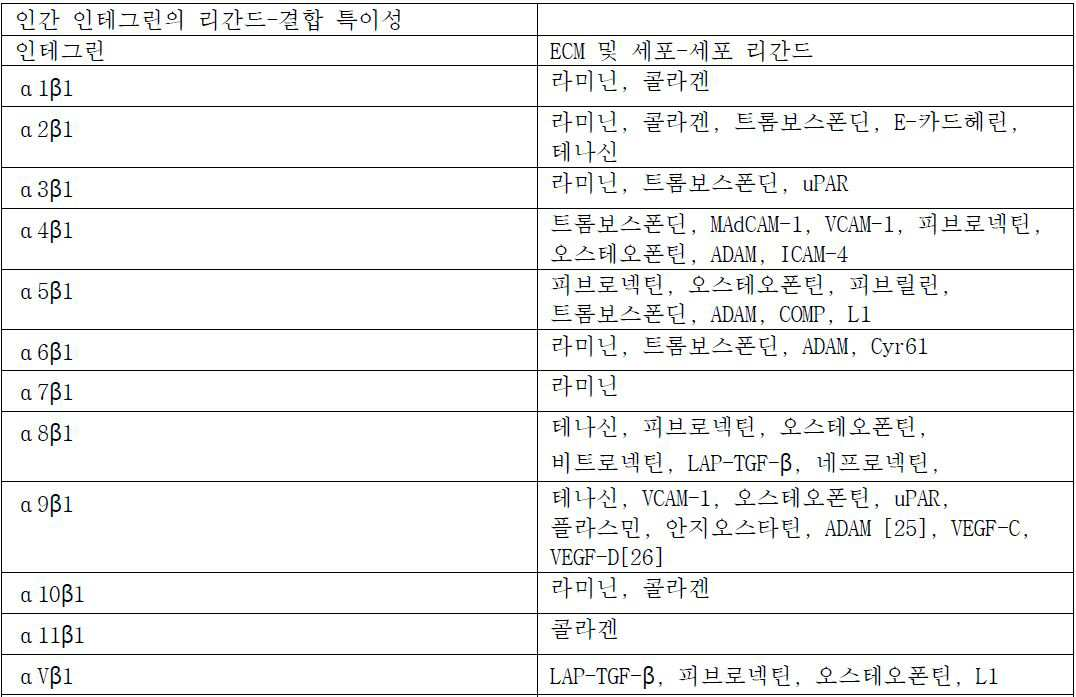

추가의 코팅물은 본원에서 기재된 방법의 수행 목적으로 바이오센서에 환자 세포를 매우 근접시키기 위해 환자 세포 표면 단백질에 대하여 친화도를 갖는다고 공지된 항체 또는 다른 단백질을 포함할 수 있다. 환자 세포가 마이크로플레이트에 원하는 방식으로 부착되는 것을 확인하는 것이 또한 유익할 수 있다. 특이적 바이오센서 코팅물은 특이적 환자 세포 유형으로부터 특이적 세포 신호 및 특이적 세포성 경로에 센서 코팅물의 연결에 의한 교란 및 치료제에 대한 세포 신호전달 반응을 향상, 개선, 명확화, 격리, 또는 검출하기 위해 추가로 사용될 수 있다 (참고, 예를 들면, Hynes, Integrins, Cell, 110:673-687 (2002)). 바이오센서는 종자 세포에 대한 영역을 포함한다. 예를 들어, 바이오센서는 종자 세포에 대한 웰들을 함유한 미세적정 플레이트를 포함할 수 있다. 하나 이상의 세포 샘플은 상이한 위치에서 표면에 물리적 흡착에 의해 바이오센서상에 씨딩될 수 있다. 바이오센서는 1, 10, 24, 48, 96, 384, 또는 초과의 상이한 위치를 포함할 수 있다. 세포 샘플은 중간에 약 100 내지 약 100,000 개별 세포 또는 임의의 세포 수를 포함할 수 있다. 최적의 세포 샘플은 바이오센서상에서 상이한 위치의 크기 및 성질에 의존한다. 세포 샘플은 약 5000 세포 이하; 약 10,000 세포 이하; 약 15,000 세포 이하; 약 20,000 세포 이하; 약 25,000 세포 이하; 또는 약 50,000 세포 이하를 포함할 수 있다. 세포 샘플은 약 1000 내지 약 2500 세포; 약 1000 내지 약 5000 세포; 5000 내지 약 10,000 세포; 약 5000 내지 약 15,000 세포; 약 5000 내지 약 25,000 세포; 약 1000 내지 약 10,000 세포; 약 1000 내지 약 50,000 세포; 및 약 5000 내지 약 50,000 세포를 포함할 수 있다. Additional coatings may include antibodies or other proteins known to have affinity for the patient cell surface proteins in order to bring the patient cells very close to the biosensor for purposes of performing the methods described herein. It may also be beneficial to confirm that the patient cells are attached to the microplate in the desired manner. Specific biosensor coatings can be used to enhance, improve, clarify, isolate, or detect cellular signaling from a specific patient cell type and cell signal transduction responses to the therapeutic agent by disturbance by the connection of the sensor coating to a specific cellular pathway (See, for example , Hynes, Integrins , Cell, 110: 673-687 (2002)). The biosensor includes regions for seed cells. For example, the biosensor may include a microtiter plate containing wells for seed cells. The one or more cell samples may be seeded onto the biosensor by physical adsorption to the surface at different locations. The biosensor may comprise 1, 10, 24, 48, 96, 384, or more different locations. The cell sample may comprise between about 100 and about 100,000 individual cells or any number of cells in the middle. The optimal cell sample depends on the size and nature of the different sites on the biosensor. The cell sample is less than about 5000 cells; About 10,000 cells or less; About 15,000 cells or less; About 20,000 cells or less; About 25,000 cells or less; Or about 50,000 cells or less. The cell sample may comprise from about 1000 to about 2500 cells; About 1000 to about 5000 cells; 5000 to about 10,000 cells; About 5000 to about 15,000 cells; About 5000 to about 25,000 cells; About 1000 to about 10,000 cells; From about 1000 to about 50,000 cells; And about 5000 to about 50,000 cells.
특정 구현예에서, 세포성 반응 또는 생리적 파라미터에서 변화는 정의된 기간 동안 측정된다. 다른 구현예에서, 정의된 기간은 생리적 파라미터의 출력에서 변화가 20% 이하만큼 변하는 정상 상태를 대조군 세포가 달성하는데 걸리는 시간의 양이다. 다른 구현예에서, 변화는 1 시간 이하로 세포에서 관측된다. 다른 구현예에서, 변화는 적어도 1 분 내지 약 60 분 동안 및 중간에 매 분 세포에서 관측된다. 다른 구현예에서, 세포 반응에서 변화는 약 10 분 내지 약 1 주 또는 200 시간 측정된다. 다른 구현예에서, 치료제가 세포성 경로에 표적되는 경우, 세포성 반응은 약 10 분 내지 약 5 시간, 약 10 분 내지 약 4 시간, 약 10 분 내지 약 3 시간, 약 10 분 내지 약 2 시간, 약 10 분 내지 약 1 시간, 또는 약 10 분 내지 약 30 분 또는 중간에 임의의 시점에서 측정된다. 다른 구현예에서, 치료제가 세포 증식 또는 세포 사멸 또는 세포성 저항성에 영향을 미치는 경우, 세포성 반응은 약 1 시간 내지 약 200 시간 측정된다. 더욱 다른 구현예에서, 1 분과 200 시간 사이 (달리 완전 일시적 패턴으로서 기재된) 반응의 조합은 세포상에서 화합물의 치료 효과 및 저항성을 개발하기 위한 세포 능력을 계측하기 위해 사용된다. 상기 기간은 환자에서 정황적 반응 및 이의 유지 평가에 중요한 단기 경로 신호전달, 동적 재프로그래밍 및 더 긴 기간 세포성 반응의 중요한 공정을 포함한다. In certain embodiments, changes in cellular responses or physiological parameters are measured over a defined period of time. In another embodiment, the defined time period is the amount of time it takes for the control cells to achieve a steady state in which the change in the output of the physiological parameter is less than 20%. In another embodiment, the change is observed in the cells in less than one hour. In another embodiment, the change is observed for at least 1 minute to about 60 minutes and in an intermediate minute cell. In other embodiments, the change in the cellular response is measured from about 10 minutes to about 1 week or 200 hours. In other embodiments, where the therapeutic agent is targeted to a cellular pathway, the cellular response may be from about 10 minutes to about 5 hours, from about 10 minutes to about 4 hours, from about 10 minutes to about 3 hours, from about 10 minutes to about 2 hours , From about 10 minutes to about 1 hour, or from about 10 minutes to about 30 minutes, or at any point in the middle. In other embodiments, where the therapeutic agent affects cell proliferation or apoptosis or cellular resistance, the cellular response is measured from about 1 hour to about 200 hours. In yet another embodiment, a combination of reactions between 1 minute and 200 hours (otherwise described as a full transient pattern) is used to measure the cellular ability to develop therapeutic effects and resistance of the compounds on the cells. These periods include critical processes of short-term pathway signaling, dynamic reprogramming and longer duration cellular responses important for the contextual response and its maintenance assessment in patients.
일단 특정한 대상체의 세포가 바이오센서상에 씨딩되면, 기준선 측정은 계측될 수 있다. 기준선 측정은 동일한 세포 샘플, 또는 대조군 세포 샘플상에서 실시될 수 있다. 대조군 샘플은 동일한 환자 및/또는 동일한 조직으로부터 건강한 세포 또는 이환 세포를 포함할 수 있다. 대조군 샘플은 교란제 또는 제제를 받지 않는 이환 세포를 포함할 수 있다. 대조군 샘플은 제제에 반응한다고 공지된 질환 세포를 포함할 수 있다. 다른 구현예에서, 대조군 샘플은 제제에 반응하지 않는다고 공지된 질환 세포를 포함한다. 대조군 샘플은, 세포 건강, 세포 대사, 또는 세포 경로 활성에 관련한 표준화된 반응을 유도하도록 설계된, 특정한 환자의 건강한 또는 이환 세포에 활성제 제제의 적용을 포함할 수 있다. Once the cells of a particular subject are seeded on the biosensor, the baseline measurement can be measured. Baseline measurements can be performed on the same cell sample, or on a control cell sample. Control samples can include healthy cells or diseased cells from the same patient and / or from the same tissue. The control sample may comprise a diffusing cell that is not subjected to an agonist or agent. The control sample may comprise disease cells known to respond to the agent. In other embodiments, the control sample comprises disease cells known to be unresponsive to the agent. Control samples may include application of the active agent formulation to healthy or diseased cells of a particular patient designed to induce a standardized response in relation to cellular health, cell metabolism, or cell pathway activity.
대조군은 각 질환 및/또는 약물 유형에 대하여 계측될 것이다. 하나의 구현예에서, 이는 동일한 환자로부터 건강한 세포 대조군에 대한 비교 또는 비-이환 환자 (예를 들면 정상 참조 구간)의 풀에 대하여 결과에 대한 비교를 포함한다. 예를 들어, 세포 사멸 약물의 경우, 방법은 유의미한 치료적 지수를 달성하기 위해 건강한 세포에 대해 질환 세포의 사멸의 이점을 보여줄 것이다. 다른 구현예는 약물에 의해 경로 기능 및 대조군을 계측하기 위한 경로 도구의 사용을 포함한다. 표적화 치료제에 대하여, 도구는 교란을 방해하는 표적된 약물의 능력을 계측 및 경로를 교란하기 위한 대조군으로서 사용되는 교란제 (예를 들면, 활성제 제제), 생체시약 또는 소분자일 수 있다. 더욱 다른 구현예에서, 세포상에서 약물의 생리적 효과는, 예를 들어, 산소 소비의 일시적 패턴 또는 속도, 산성화의 속도 또는 일시적 패턴, 이온 유동, 또는 대사물질 턴오버를 언급한 활성제 제제의 외인성 교란 없이 측정된다.Controls will be measured for each disease and / or drug type. In one embodiment, this includes a comparison to the results for a pool of healthy or non-diseased patients (e.g., normal reference intervals) from the same patient to a healthy cell control. For example, in the case of an apoptotic drug, the method will show the benefit of the death of disease cells on healthy cells to achieve a significant therapeutic index. Other embodiments include the use of pathway tools to measure pathway function and control by drug. For targeted therapeutics, the tool may be a perturbant ( e. G. , An active agent), a biochemical reagent or a small molecule used as a control to disturb measurement and pathway of the ability of the targeted drug to interfere with the perturbation. In yet other embodiments, the physiological effect of the drug on the cell can be assessed without any external disturbance of the active agent formulation, such as, for example, a transient pattern or rate of oxygen consumption, a rate or transient pattern of acidification, ionic flux, or metabolite turnover .
특정한 구현예에서, 바이오센서 신호는 계속되는 시간 경과 동안 측정된다. 약물 치료에 대한 환자 세포 반응을 나타내는 시간 대 바이오센서 신호 플롯상에서 고유 패턴이 있다. 이들 패턴의 평가는 유효한 사례의 존재를 확인하는데 유용하다. 살아있는 및 완전히 기능성 세포의 시간 경과 또는 일정하게 변화 측정은 시점을 단지 나타내는 전형적인 전체의 세포 검정에서 사용된 현행 실시보다 더욱 유익하다. 본원에서 기재된 방법은 이들이 환자에서 발생하고 환자 반응 계측의 가장 정확한 수단을 나타냄에 따라 동적 시스템을 측정한다. 경로 반응의 경우에서, 완벽한 시간 경과 또는 일시적 패턴의 기록은 더욱 복잡한 분석을 지지하기 위한 및 단일 측정에 대하여 최적의 시점 선택을 방지하기 위한 능력이 우수하다.In certain embodiments, the biosensor signal is measured over time. There is a unique pattern on a time vs. biosensor signal plot showing patient cell response to drug therapy. Evaluation of these patterns is useful for confirming the existence of valid cases. The time course or constant change measurement of live and fully functional cells is more beneficial than the current practice used in a typical whole cell assay, which simply represents a point in time. The methods described herein measure dynamic systems as they occur in a patient and represent the most accurate means of measuring patient response. In the case of the path response, the recording of perfect time lapse or transient patterns is excellent for supporting more complex analysis and for preventing optimal point-in-time selection for a single measurement.
대조군에 대한 비교는 일시적 최대치, 최소치에서, 또는 최대 신호-최소 신호 사이 차이로서, 또는 시간 경과 플롯에 대하여 통합된 곡선하 면적 (AUC)의 비교 또는 양성 또는 음성 대조군 웰들에 대해 시험 웰의 다른 비-선형 비교 (예를 들면 차이 벡터의 합산) 또는 동일한 환자 생존 이환 세포에 대하여 교란된 및 미혼란된 웰들의 비교에 의해 발생할 수 있다. 바이오센서를 이용한 측정으로만 지지된 추가의 분석은 최대치/최소치, 및 일시적 시간 경과의 다른 유도체를 달성하기 위한 시간이다. 더 긴 기간 반응의 경우에서, 비교의 시간은 약물의 치료 또는 다중 적용의 며칠 또는 1 주 이후 특이적 시점일 수 있다. 더 긴 시간 경과는 또한 기울기에서 변화를 비교할 수 있거나 또는 약물 치료의 1 주의 시작, 중간 또는 말기에서 시간 대 바이오센서 신호 플롯의 제2 유도체를 포함할 수 있다. 대조군에 비교된 유의미한 변화는 세포성 대사의 단축에 관련된 바이오센서 신호에서 절대적 하락을 포함할 수 있다. 대안적으로, 하락은 그 다음 검정 동안 약물에 대한 저항성의 발달을 나타낼 수 있는 증가일 수 있다. 추가로, 비-선형 유클리드 분석은 완벽한 시간 경과 동안 대조군과 환자 샘플 사이 총 차이의 측정을 만들어내는데 사용될 수 있다. 이는 또한 환자에 대하여 결과 예측에 관해 유의미할 것이다.Comparisons to the control can be made by comparing the integrated curve area (AUC) for temporal maxima, at the minimum, or as the difference between the maxima and minima signals, or for the time course plots, or for the positive or negative control wells, - can be caused by linear comparisons (e.g., summing up the difference vectors) or by comparison of disturbed and un-confused wells for the same patient survival cells. Further analysis supported only by biosensor-based measurements is the time to achieve maximum / minimum, and other derivatives of transient time course. In the case of a longer duration of the reaction, the time of the comparison may be a specific point in time after a few days or a week of the treatment or multiple application of the drug. Longer time lapses may also compare the change in slope or may include a second derivative of the time versus biosensor signal plot at the beginning, middle, or end of one week of drug therapy. Significant changes compared to the control group may include an absolute decline in the biosensor signal associated with shortening of cellular metabolism. Alternatively, the drop may be an increase that may indicate the development of resistance to the drug during the next assay. In addition, non-linear Euclidean analysis can be used to produce a measure of the total difference between the control and patient samples over a complete time course. It will also be meaningful to predict the outcome for the patient.
특정 구현예에서, 정의된 기간 동안 바이오센서의 출력은 세포 지수로서 나타난다. 세포 지수는 시험 개시점으로부터 임피던스에서의 변화이다. 세포 지수는 임피던스의 측정으로서 정의되고, 예를 들어, 10 kHz의 고정된 전기 주파수 및 고정된 전압에서 측정함으로써 본 발명의 일 사례에서 적용될 수 있다.In certain embodiments, the output of the biosensor appears for a defined period of time as a cell index. The cell index is the change in impedance from the test start point. The cell index is defined as a measure of the impedance and can be applied in one example of the present invention, for example, by measuring at a fixed electrical frequency of 10 kHz and a fixed voltage.
그리고 하기 방정식에 의해 계산된다: 세포 지수i = (Rtn - Rt0)/FAnd is calculated by the following equation: Cell index i = (R tn - R t0 ) / F
식 중:Where:
i = 1, 2, 또는 3 시점i = 1, 2, or 3 time points
F = 기기가 10kHz 주파수에서 작동되는 경우 하나의 예에서 15옴F = 15 ohms in one example when the device is operating at 10 kHz frequency
Rt0는 시점 T0에서 측정된 배경 저항성이다.R t0 is the background resistivity measured at time T0.
Rtn은 세포 부가, 세포 생리적 변화, 또는 세포 교란 이후 시점 Tn에서 측정된 저항성이다.R tn is the resistance measured at the time point T n after cell attachment, cell physiological change, or cell disturbance.
세포 지수는 세포 상태를 나타내기 위해 측정된 전기적 임피던스에서 상대적 변화로서 유도된 무차원 파라미터이다. 세포가 존재하지 않거나 또는 전극에 잘-부착되지 않는 경우, CI는 0이다. 동일한 생리적 조건하에서, 더 많은 세포가 전극상에 부착되는 경우, CI 값이 더 크다. CI는 따라서 웰에 존재한 세포 수의 정량적 측정이다. 추가로, 세포 형태학을 포함한, 세포 생리적 상태에서 변화, 기저, 안정한, 또는 정지 상태에서 변화, 세포 접착, 또는 세포 생존력은 CI에서 변화로 이어질 것이다.The cell index is a dimensionless parameter derived as a relative change in the electrical impedance measured to represent the cell state. If the cell is not present or is not well-attached to the electrode, the CI is zero. Under the same physiological conditions, when more cells are attached on the electrode, the CI value is greater. CI is thus a quantitative measure of the number of cells present in the well. In addition, changes in cell physiological conditions, including cellular morphology, changes in basal, stable, or resting state, cell adhesion, or cell viability will lead to changes in CI.
세포 지수는 개시점 또는 기준선 임피던스 측정에 기반된 이의 존재, 밀도, 부착 또는 변화의 정량적 측정이다. 기준선 개시점 임피던스는 약물 또는 다른 교란제를 이용한 임의의 치료에 앞서 세포의 건강, 생존력, 및 생리적 상태의 적응증 및 물리적 관측가능한 특징이다. 기준선 개시점은 CELx 시험에 대하여 정성적 대조군으로서 사용될 수 있다. 약물 또는 교란제의 부가는 세포에 의해 경험된 세포성 생리적 변화의 특이성의 일시적 패턴 반사에서 변화에 임피던스를 발생시킨다. 세포 생리적 상태, 예를 들어 세포 형태학, 세포 수, 세포 밀도, 세포 접착, 또는 세포 생존력에서 변화는 세포 지수에서 변화로 이어질 것이다.Cellular index is a quantitative measure of its presence, density, attachment or change based on a starting point or baseline impedance measurement. Baseline onset impedance is an indication and physically observable characteristic of the health, viability, and physiological state of the cell prior to any treatment with drugs or other perturbations. The baseline starting point can be used as a qualitative control for the CELx test. The addition of drugs or perturbants induces an impedance to the change in the transient pattern reflection of the specificity of the cellular physiological change experienced by the cell. Changes in cellular physiological status, such as cellular morphology, cell number, cell density, cell adhesion, or cell viability, will lead to changes in cell index.
생리적 반응 파라미터는 추가로 세포 사이클 분석을 포함할 수 있고 기능성 및 기능이상 경로와 관련된 환자 세포에서 다른 비색 변화 또는 임의의 수의 화학 바이오센서 예컨대 접합된 또는 비접합된 형광 염료를 이용하여 측정될 수 있다. 예를 들어 비접합된 염료를 이용한 세포의 집단에 대하여 세포 사이클에서 변화는 DNA에 삽입한다고 공지된 프로피듐 아이오다이드 또는 유사한 염료로 정량화될 수 있고 DNA의 양에서 변화를 평가함으로써 성장 및 복제의 G0, G1, S, G2, Gm 시기를 통해 세포 사이클링과 상관할 수 있다. 하나의 염료 유형, 프로피듐 아이오다이드의 경우, G2/M 시기에서 세포의 형광은 G0/G1 시기에서 세포의 것보다 2배만큼 높을 것이다. 프로피듐 아이오다이드는 또한 RNA에 삽입할 수 있고 종종 리보뉴클레아제는 RNA에 비교된 DNA로부터 형광 신호를 구별하기 위해 사용된다. 예는 또한 세포 사이클 체크 포인트에 연결된 특정한 단백질에 특이적인 염료를 포함하고 추가의 세포 사이클 상태 측정을 제공한다. 이들 측정 실시에 유용한 통상 기기 비제한적으로 본원에서 열거된 것은 형광 현미경검사, 공초점 레이저 스캐닝 현미경검사, 유세포측정, 형광분석법, 형광 국소 분극화, 균질한 시간 분해 형광, 및 형광 활성화된 세포 분류 (FACS)이다.The physiological response parameters may further include cell cycle assays and may be measured using different colorimetric changes or any number of chemical biosensors, such as conjugated or unconjugated fluorescent dyes, in the patient's cells in relation to the functional and dysfunctional pathways have. For example, for a population of cells using unconjugated dyes, changes in the cell cycle can be quantified with propidium iodide or similar dyes known to insert into DNA, and by assessing changes in the amount of DNA, G0, G1, S, G2, and Gm. In the case of one dye type, propidium iodide, the fluorescence of the cells in the G2 / M phase will be twice as high as that of the cells in the G0 / G1 phase. Propidium iodide can also be inserted into RNA and ribonuclease is often used to distinguish the fluorescent signal from DNA compared to RNA. Examples also include dyes specific to specific proteins linked to cell cycle checkpoints and provide additional cell cycle status measurements. Examples of conventional devices useful for carrying out these measurements include but are not limited to fluorescence microscopy, confocal laser scanning microscopy, flow cytometry, fluorescence analysis, fluorescence localization, homogeneous time-resolved fluorescence, and fluorescence activated cell sorting )to be.
비접합된 염료는 세포의 생리적 상태 또는 경로의 화학 바이오센서로서 본 발명과 함께 이용될 수 있으면서 대사성 파라미터 예컨대 동화작용, 이화, 소분자 합성 및 생성, 턴오버, 및 호흡을 측정한다. 와버그 효과로 명명된, 공지된 세포 생리적 반응은 종양 및 다른 이환 세포, 및 주요 신호전달 경로에서 에너지 생성을 위하여 산화적 인산화부터 락테이트 생산까지 시프트를 기재하고, 종양유전자 및 종양 억제제 (예를 들어 비제한적으로 Akt, mTor, c-myc, p53)는 본원에서 기재된 임의의 화학 바이오센서 방법에 의해 또는 광-전자 바이오센서에 의해 측정될 수 있다. 세포성 반응에서 세포성 산소 소비 또는 호흡 및 당분해는 양성자를 생산하고 용해된 산소 및 자유 양성자의 농도 또는 산도에 급속, 쉽게 측정가능한 변화를 발생시킨다.Unconjugated dyes measure metabolic parameters such as assimilation, catabolism, small molecule synthesis and generation, turnover, and respiration while being able to be used with the present invention as chemical biosensors in the physiological state or pathway of a cell. Known cellular physiological responses, named as bug effects, describe shifts from tumors and other diseased cells, and oxidative phosphorylation to lactate production for energy production in key signaling pathways, and include tumor genes and tumor suppressors Including but not limited to Akt, mTor, c-myc, p53) can be measured by any chemical biosensor method described herein or by photo-electronic biosensor. In cellular reactions, cellular oxygen consumption or respiration and glycosylation produce protons and produce rapid, easily measurable changes in the concentration or pH of dissolved oxygen and free protons.
화학 바이오센서를 이용한 생리적 반응 파라미터의 추가의 그러나 비제한 예는, 대사작용으로 활성 및 불활성 세포성 기능의 지표인, 존재한 ATP의 정량화 (Ex. CellTiterGlo 및 유사한 루시퍼라아제 유도된 검정)에 기반된 배양액에서 세포에 의해 이용되는 ATP의 양이다.Additional but non-limiting examples of physiological response parameters using chemical biosensors are based on the quantification of existing ATPs (Ex. CellTiterGlo and similar luciferase-induced assays), which are indicators of active and inactive cellular function by metabolism Lt; RTI ID = 0.0 > ATP < / RTI >
생체분자 폴딩 및 기능에 중요한 칼슘, 마그네슘, 및 다른 충전된 이온은 생리적 반응 때문에 유입된다. 이들은 또한 화학 바이오센서 예컨대 Cal-520, 오레곤 그린 BAPTA-1, 후라-2, 인도-1, 플루오-3, 플루오-4, 칼슘 녹색-1, 및 특이적 이온 착화 및 측정용 다른 EGTA 또는 EDTA-유사 화학에 의해 측정될 수 있다. 이들 생리적 반응 파라미터는 많은 유형의 비접합된 반응성 또는 결합 염료 또는 다른 전자 또는 분광 수단을 이용하여 측정될 수 있다. 많은 이들 방법은 동일한 세포 집단의 생리적 기능을 경시적으로 반복적으로 연속적으로 측정되도록 하는 세포에 비-파괴적이기 위해 배열될 수 있다.Calcium, magnesium, and other charged ions important for biomolecule folding and function are introduced due to physiological reactions. These also include chemical biosensors such as Cal-520, Oregon Green BAPTA-1, Fura-2, Indo-1, Fluo-3, Fluo-4, Calcium Green-1, and other EGTA or EDTA- Can be measured by similar chemistry. These physiological response parameters can be measured using many types of unconjugated reactive or binding dyes or other electronic or spectroscopic means. Many of these methods can be arranged to be non-destructive to cells that allow repeated physiological functioning of the same cell population to be repeatedly and continuously measured.
접합된 염료 예컨대 천연 세포 단백질 결합 리간드에 부착된 또는 면역입자 (항체 또는 항체의 단편 또는 높은 특이성 높은 친화도 합성 분자 예컨대 압타머)에 부착된 것, 또는 핵산 폴리머 하이브리드화 프로브는 다양한 세포성 구획, 유전체학, 및 프로테오믹스에서 단백질, 특이적 경로 구성원 분자, DNA 및 또는 RNA에 관련된 생리적 반응 파라미터를 측정하기 위해 사용될 수 있고, 특이적 번역후 변형 및 기전을 측정할 수 있다. 세포성 대조군의 번역후 변형 및 후성유전적 수단은 비제한적으로 리보자임, 키나제, 포스파타제, 유비퀴티나제, 데유비퀴티나제, 메틸화효소, 탈메틸효소, 및 프로테아제를 포함하는 경로 기능을 수행하는 다수의 효소에 의한 조절을 포함할 수 있다. 죽은 세포의 포르말린 고정 파라핀 실장된 샘플의 염색에 사용된 이들 분자의 예는 아래에서 찾아질 수 있다: DAKO Immunohistochemical Staining Methods Education Guide - Sixth Edition or at Cell Signaling Technology tutorials and application guides http://www.cellsignal.com/common/content/content.jspαid=tutorials-and-application-guides. 이들 2개의 예는 살아있는 세포 반응의 측정을 위하여 본 발명과 함께 더욱더 유용할 것이다. 비제한적으로 이들 방법의 상기 측정 수행에 유용한 통상 기기는 형광 현미경검사, 공초점 레이저 스캐닝 현미경검사, 유세포측정, 형광분석법, 균질한 시간 분해 형광, 형광 국소 분극화, 및 형광 활성화된 세포 분류 (FACS)이다.Attached to conjugated dyes such as natural cell protein binding ligands or attached to immunoparticles (fragments of antibodies or antibodies or high specificity high affinity synthetic molecules such as platamer), or nucleic acid polymer hybridization probes can be used in a variety of cellular compartments, Can be used to measure physiological response parameters related to proteins, specific pathway member molecules, DNA and / or RNA in genomics, and in proteomics, and specific post-translational modifications and mechanisms can be measured. Post-translational modifications and cellular genetic tools of cellular control include, but are not limited to, performing pathways involving ribozyme, kinase, phosphatase, ubiquitinase, deubiquitinase, methylating enzyme, demethyl enzyme, and protease ≪ RTI ID = 0.0 > and / or < / RTI > Examples of these molecules used in the staining of formalin-fixed paraffin-embedded samples of dead cells can be found below: DAKO Immunohistochemical Staining Methods Education Guide - Sixth Edition or Cell Signaling Technology tutorials and application guides http: // www. cellsignal.com/common/content/content.jspαid=tutorials-and-application-guides. These two examples will be even more useful with the present invention for the measurement of live cellular responses. Typical instruments useful for performing such measurements of these methods include but are not limited to fluorescence microscopy, confocal laser scanning microscopy, flow cytometry, fluorescence analysis, homogeneous time-resolved fluorescence, fluorescence localization, and fluorescence activated cell sorting (FACS) to be.
접합된 및 비-접합된 염료의 조합은 또한 세포의 생리적 반응을 측정하기 위해 본 발명에 의해 이용될 수 있다. 활성화 이후, 생리적 반응 제어를 책임지는 수용체의 한 유형은 GPCRs이다. 이들은 2개의 신호전달 경로: 2차 메신저 이노시톨 (1,4,5) 삼인산 (IP3)에 의해 해방되는, 2차 메신저 cAMP의 수준에서 변화, 또는 세포내 Ca2+의 수준에서 변화를 통해 정보 및 대조군 세포를 전달한다. cAMP 검출은 예를 들어 크립테이트-표지된 항-cAMP 항체 (또는 다른 면역포착 분자) 및 GPCR 반응 및 후속의 항체 결합에 대하여 세포성 cAMP와 경쟁하는 d2-표지된 cAMP를 이용하여 경쟁적 면역검정상에서 기반될 수 있다. 특이적 신호 (즉 에너지 전달)은 표준 또는 샘플에서 cAMP의 농도에 반비례한다.The combination of conjugated and non-conjugated dyes can also be used by the present invention to measure the physiological response of a cell. After activation, one type of receptor responsible for physiological response control is GPCRs. These are information and control cells that undergo two signaling pathways: a change in the level of the secondary messenger cAMP, released by the secondary messenger inositol (1,4,5) triphosphate (IP3), or a change in the level of intracellular Ca2 + . cAMP detection can be carried out on competitive immunoassays using d2-labeled cAMP competing with cellular cAMP for, for example, cryptate-labeled anti-cAMP antibodies (or other immunocyte molecules) and GPCR reactions and subsequent antibody binding Can be based. The specific signal (ie, energy transfer) is inversely proportional to the concentration of cAMP in the standard or sample.
생리적 기능을 가진 mRNA, RNAi, 마이크로RNAs 및 다른 RNA의 정량화에 의한 생리적 반응의 측정은 전사 수준에서 세포성 변화의 교란 계측을 위하여 본 발명의 실시와 함께 이용된 매우 감수성 방법일 수 있다. 모두 화학 센서를 사용하는 rtPCR, qPCR, 선택적 서열 탐침검사, 선택적 서열 포착, 및 서열 하이브리드화 방법을 이용함으로써 RNA 예를 들어 비제한적으로 본원에서 열거된 것은 정량화될 수 있다.Measurement of physiological responses by quantification of mRNA, RNAi, microRNAs and other RNAs with physiological function can be a highly sensitive method used in conjunction with the practice of the present invention for the disturbance measurement of cellular changes at the transcriptional level. RNAs enumerated herein, including but not limited to, by using rtPCR, qPCR, selective sequence probe detection, selective sequence capture, and sequence hybridization methods, all using chemical sensors, can be quantified.
면역-포착 및 하이브리드화 방법은, 비드 기반 방법 예컨대 루미넥스 또는 섬유 옵틱 팁 기술 예컨대 일루미나 또는 단백질, DNA, RNA, 또는 세포로부터 생리적 반응 분자(들)을 꺼내기 위해, 단리하기 위해, 및 정확하게 측정하기 위해 사용되는 고체 표면상에 특이적 포착 시약이 고정되는 다른 하이브리드화 마이크로어레이 기술을 이용한 것을 포함한다. 이들 방법은 단일 실험에서 다수의 반응 파라미터의 측정의 이점을 제공한다.Immuno-capture and hybridization methods can be used to remove, isolate, and accurately measure the physiological response molecule (s) from bead-based methods such as luminex or fiber optic tip techniques such as an illuminator or protein, DNA, RNA, Lt; RTI ID = 0.0 > microarray < / RTI > technique in which a specific capture reagent is immobilized on the solid surface used for the assay. These methods provide the advantage of measuring multiple reaction parameters in a single experiment.
세포성 반응 또는 생리적 파라미터에서 변화는 기준선 측정과 비교에 의해 계측된다. 세포성 파라미터 또는 생리적 반응에서 변화는 CReMS의 유형에 좌우된다. 예를 들어, 세포성 반응에서 변화가 광학적으로 계측되면, 물리적으로 관측가능한 변화는 예를 들어 화학 흡광도 또는 투과율용 스펙트럼 파장에서 광학 밀도의 기능, 표면 플라즈몬 측정 디바이스에서 변화, 또는 광자 계측 디바이스에 의해 검출된 변화로서 측정될 수 있다. 세포성 파라미터 또는 생리적 반응에서 변화가 전기적으로 계측되면, 물리적으로 관측가능한 변화는 예를 들어 전극에 부착된 세포의 밀리 또는 마이크로 임피던스 변화를 이용하여 측정될 수 있다. pH, 글루코오스, 이산화탄소, 또는 이온에서 변화는 이온 선택적 전계 효과 트랜지스터 (ISFET)를 이용하여 전자적으로 측정될 수 있다.Changes in cellular responses or physiological parameters are measured by comparison with baseline measurements. Changes in cellular parameters or physiological responses depend on the type of CReMS. For example, if a change in the cellular response is optically measured, the physically observable change can be measured by, for example, the function of the optical density at the spectral wavelength for the chemical absorbance or transmittance, the change in the surface plasmon measurement device, Can be measured as the detected change. If changes in cellular parameters or physiological responses are electrically measured, physically observable changes can be measured, for example, using the millimeter or micro-impedance changes of the cells attached to the electrodes. Changes in pH, glucose, carbon dioxide, or ions can be measured electronically using ion selective field effect transistors (ISFETs).
다른 구현예에서, 변화의 속도는 치료제에 대한 세포성 생리적 반응에서 차이를 계측하도록 요구된 일정한 기간 동안 CReMS 반응을 측정하는 방법에 의해 계측된다. 변화의 속도는 시간 경과 데이터의 다양한 해석에 의해 기재되고 속도로서 또는 추가로 속도의 가속을 포함한 속도의 유도 기능으로서 발현될 수 있다.In other embodiments, the rate of change is measured by measuring the CReMS response over a period of time required to measure the difference in cellular physiological response to the therapeutic agent. The rate of change can be described by various interpretations of time lapse data and can be expressed as a rate or as an induction function of the rate, including further acceleration of the rate.
환자의 생리적 조건을 측정하는 시험은 환자가 상이한 임상 결과를 경험하는 것으로 예상되는 것 초과 및 미만으로 하나 이상의 컷오프 값을 유도할 수 있다. 구현예에서, 세포성 반응에서 변화 계측용 하나 이상의 컷오프 값은 하기 단계를 포함하는 방법에 의해 계측된다: 표준 편차, 신호 대 잡음 비, 표준 오차, 변동의 분석, 또는 공지된 비반응 환자로부터 샘플의 세트로부터 및 공지된 반응 세포 샘플로부터 샘플의 세트의 통계적 유의도로 적절한 신뢰 구간의 계측을 위하여 당해 기술에서 실시된 것에 의해 공지된 다른 통계적인 시험 값의 계측 단계; 및 둘 사이에서 차이의 계측 및 모든 그룹에 대하여 신뢰 구간 사이에서 컷오프 값의 셋팅 단계. 추가의 구현예는 이환되지 않은 공지된 환자로부터 CReMS 반응에 의해 정의된 정상 참조 범위로부터 계측된 컷오프를 이용한다. 상기 구현예에서, 단일 환자 질환 물질 시험 결과는 정상 (건강한) 참조 범위 구간으로 본 발명내 추가로 다른 곳에 기재된 바와 같이 교란된 및 미혼란된 생존 암 세포 결과의 비교에 의해 보고된다.Testing to measure the physiological condition of a patient can lead to one or more cutoff values above and below what the patient is expected to experience different clinical outcomes. In an embodiment, the one or more cutoff values for the change measurements in the cellular response are measured by a method comprising the steps of: analyzing the standard deviation, the signal to noise ratio, the standard error, the variation, or the sample from a known non- Measuring other statistical test values known from those set forth in the art for the measurement of a confidence interval from a set of samples and a set of samples from a known reaction cell sample to a statistical significance; And measuring the difference between the two and setting the cutoff value between confidence intervals for all groups. A further embodiment utilizes the measured cutoff from the normal reference range defined by the CReMS response from known uninjured patients. In this embodiment, the single patient disease material test results are reported by comparison of disturbed and untouched survival cancer cell results as described elsewhere in the present invention with normal (healthy) reference range intervals.
정상 (건강한) 참조 범위 시험 결과는 "정상" 경로 활성이 건강한 대상체로부터 정상 조직을 이용한 연구의 수행이다. 시험 결과는 그 다음 이환 경로를 갖는다고 예상되지 않는 임의의 환자 (예를 들면 바이오마커 음성 환자)가 사실상 정상 참조 구간 연구로부터 유도된 값과 비교된 경우 비정상 경로 활성을 갖는지를 평가한다. 본 발명의 결과는 또한 바이오마커 음성인 환자가 모두 비정상이고 바이오마커 양성 환자의 경로 활성에 비교할만한 경로 활성을 갖는지를 보기 위해 질환으로 현재 진단되는 대상체 (바이오마커 양성 환자)로부터 실시된 측정에 대하여 바이오마커-음성 환자로부터 관측된 비정상 측정에 대하여 비교된다. 경로 활성을 방해하기 위해 의도된 요법을 현재 받고 덕을 보는 환자의 경로 활성에 비교할만한 비정상 경로 활성을 갖는 환자는 따라서 경로 질환을 갖는 것으로서 진단될 것이고 따라서 경로 활성을 방해하기 위해 바이오마커를 표적하는 약물로 치료되어야 한다.Normal (healthy) reference range test results are the performance of studies using normal tissues from healthy subjects with "normal" pathway activity. The test results then assess whether any patient (eg, a biomarker negative patient) not expected to have a pathway has abnormal pathway activity when compared to a value derived from virtually the normal reference interval study. The results of the present invention also demonstrate that measurements made from subjects (biomarker-positive patients) currently diagnosed with the disease to see if the biomarker negative patients are all abnormal and have pathway activity comparable to the pathway activity of the biomarker- Biomarkers - are compared for abnormal measurements observed from patients with speech. Patients with abnormal pathway activity that are comparable to the pathway activity of a patient who is currently receiving the intended therapy to interfere with pathway activity will thus be diagnosed as having pathway disease and thus will be required to target the biomarker It should be treated with medication.
본 발명은 따라서 이환 경로의 기능성 활성에 기반된 질환 진단의 더욱 정확한 수단을 의사가 창출하게 한다. 이는 상관적인, 원인이 되지 않는 모델에 의존하는 단일 바이오마커가 측정되는 경우 실시된 접근법과 대조된다. 현행 바이오마커 접근법은 위 음성 및 위 양성 결과의 높은 백분율을 초래할 수 있다. 본 발명은 위 결과의 백분율을 감소시킬 것이다.The present invention thus allows physicians to create more accurate means of diagnosing diseases based on the functional activity of the pathway. This contrasts with the approach taken when a single biomarker is measured that relies on a model that is not correlated or causative. Current biomarker approaches can result in a high percentage of false negative and false positive results. The present invention will reduce the percentage of above results.
바람직한 구현예는 80-90% 신뢰 구간을 포함하고, 더 바람직한 구현예는 >90% 신뢰 구간을 포함하고 가장 바람직한 구현예는 >95% 또는 >99% 신뢰 구간을 포함한다.A preferred implementation includes an 80-90% confidence interval, a more preferred implementation includes a> 90% confidence interval, and a most preferred implementation includes a> 95% confidence interval or a> 99% confidence interval.
구현예에서, 컷오프 값은 컷오프 값을 이용하여 반응군 또는 비반응군으로서 맹검 공지된 샘플의 생태의 계측 및 샘플의 비맹검 및 샘플의 상태 예측의 정확도의 계측에 의해 입증된다. 단일 컷오프 값의 경우에서, 컷오프 값 미만에 해당하거나 또는 공지된 반응군에 대한 값에 더 근접한 값은 환자 샘플이 치료제에 반응성을 나타내고 있음을 가리킨다. 값이 컷오프 값이거나 또는 초과이거나 또는 공지된 비 반응군 값에 대한 값에 더 근접하면, 세포 샘플은 치료제에 비 반응군으로서 확인된다. 일부 구현예에서 정의된 기간에서 바이오센서의 출력은 무 반응, 약 반응성 또는 반응성으로서 분류된다.In an embodiment, the cutoff value is verified by measuring the ecology of the blind known sample as a reaction group or non-responding group using the cutoff value and by measuring the accuracy of the non-blinded and sample state prediction of the sample. In the case of a single cut-off value, a value that is less than the cut-off value or closer to the value for a known response group indicates that the patient sample is responsive to the therapeutic. If the value is greater than or equal to the cutoff value or closer to the value for the known non-responsive group value, the cell sample is identified as a non-responsive group to the therapeutic. In some embodiments, the output of the biosensor in the defined time period is classified as unresponsive, weakly reactive or reactive.
바람직한 구현예에서, 컷오프 값은 컷오프 값을 이용하여 질환 경로 반응 (예를 들면 암 환자)를 갖는 또는 질환 경로 반응 (예를 들면 건강한 무병 환자)를 갖지 않는 것으로서 맹검 공지된 샘플의 상태 계측 및 샘플의 비맹검 및 샘플의 상태 예측의 정확도의 계측에 의해 입증된다. 단일 컷오프 값의 경우에서, 컷오프 값 미만에 해당하거나 또는 공지된 반응군에 대한 값에 더 근접한 값은 환자 샘플이 치료제에 반응성을 나타내고 있음을 가리킨다. 컷오프 값이거나 또는 초과이거나 또는 질환 경로 반응을 갖지 않는 샘플의 값에 더 근접한 값은 환자 샘플이 무 경로 질환을 나타내는 것을 가리킨다. 값이 컷오프 값이거나 또는 초과이거나 또는 공지된 이환 경로 샘플 값에 대한 값에 더 근접하면, 세포 샘플은 존재한 이환 경로로서 확인된다. 일부 구현예에서 정의된 기간에서 바이오센서의 출력은 존재하지 않는 경로 질환, 비확정적인 경로 질환, 또는 존재한 경로 질환으로서 분류된다.In a preferred embodiment, the cutoff value is determined by using the cutoff value to determine the status of the blindly known sample as having no disease pathway response (e. G., A cancer patient) or a disease pathway response (e. And by measuring the accuracy of the state prediction of the sample. In the case of a single cut-off value, a value that is less than the cut-off value or closer to the value for a known response group indicates that the patient sample is responsive to the therapeutic. A value closer to the value of the sample that is greater than or equal to the cutoff value or that does not have a disease pathway response indicates that the patient sample exhibits no pathological disease. If the value is greater than or equal to the cutoff value or closer to the value for the known pathway sample value, the cell sample is identified as an existing pathway. In some embodiments, the output of the biosensor in the defined time period is classified as non-existent pathology, non-definitive pathology, or pathology present.
정의된 기간에서 출력은 카테고리에 출력을 분류하기 위해 선택된다. 다른 구현예에서, 정의된 기간은 세포가 바이오센서에서 연속적으로 모니터링되었던 기간의 종료점이다. 다른 구현예에서, 기간은 적어도 60 분, 240 분, 300 분, 10 시간, 24 시간, 60 시간, 또는 120 시간이다. 바람직한 구현예에서 정의된 기간에서 출력은 30 분 및 10 시간이다. 더 바람직한 구현예에서, 정의된 기간에서 출력은 180 분 내지 600 분 또는 240 분이다.In a defined period, the output is selected to classify the output into categories. In another embodiment, the defined time period is the end point of the period in which the cells were continuously monitored in the biosensor. In other embodiments, the period is at least 60 minutes, 240 minutes, 300 minutes, 10 hours, 24 hours, 60 hours, or 120 hours. In the period defined in the preferred embodiment, the output is 30 minutes and 10 hours. In a more preferred embodiment, the output in the defined period is from 180 minutes to 600 minutes or 240 minutes.
구현예에서, 무 반응으로서 분류된 출력은 치료제의 투여에 앞서 기준선 또는 적어도 20% 이하, 15% 이하, 10% 이하, 또는 5% 이하 치료제로 치료되지 않은 대조군 세포의 출력 값과 상이한 출력 값으로 나타낸다. In an embodiment, the output classified as non-responsive is a baseline or an output value that is different from the baseline or at least 20%, 15%, 10%, or 5% of the output of the control cells not treated with the therapeutic agent prior to administration of the therapeutic .
다른 구현예에서, 약 반응성으로서 분류된 출력은 치료제의 투여에 앞서 기준선 또는 적어도 50% 이하 및 5% 초과의 치료제로 치료되지 않은 대조군 세포의 출력 값과 상이한 출력 값으로 나타낸다. 다른 구현예에서, 반응성으로서 분류된 출력은 치료제의 투여에 앞서 기준선 또는 적어도 50% 초과의 치료제로 치료되지 않은 대조군 세포와 상이한 출력 값으로 나타낸다. 구현예에서, 대조군 샘플은 동일한 대상체로부터 및 치료제로 치료되지 않은 질환 세포의 샘플이다.In another embodiment, the output classified as weakly reactive is indicative of a baseline or an output value that is different from the baseline or at least 50% and the output value of the control cells not treated with more than 5% of the therapeutic agent prior to administration of the therapeutic agent. In another embodiment, the output labeled as reactive is indicative of an output value that differs from the baseline or at least 50% of the control cells not treated with the therapeutic agent prior to administration of the therapeutic agent. In an embodiment, the control sample is a sample of diseased cells from the same subject and not treated with the therapeutic agent.
본원에서 기재된 방법의 추가 측면은 개별 대상체에서 치료제의 효능을 예측하기 위해 사용될 수 있는 알고리즘의 개발을 포함한다. 알고리즘은, 개별 대상체의 건강의 측면을 한정하는 하나 이상의 환자 특징에 배정된 값과 조합으로, 본원에서 기재된 방법을 이용하여 유도된 값을 편입시킨다. 환자 특징은, 비제한적으로, 하기를 포함할 수 있다: 전이의 존재, 전이의 위치, 결절 상태, 암의 초기 진단부터 전이의 진단까지 무병 구간, 아쥬반트 화학요법의 수령, 다른 약물 요법의 수령, 방사선 요법의 수령, 질환의 우세한 부위, 종양 질량 크기, 바디-질량 지수, 연한 관절의 수, 팽윤된 관절의 수, 통증, 장애 지수, 의사 전면적인 평가, 환자 전면적인 평가, 배쓰 강직 척추염 작용성 지수, 배쓰 강직 척추염 질환 활성 지수, 배쓰 강직 척추염 도량형 지수, C-반응성 단백질, 총 요통, 염증, 유전적 상태, 다른 병의 이력, 다른 생명 유지 관련 건강 통계 상태, 및 임의의 이들의 조합. 이들 값을 편입시키는 알고리즘은 치료제가 표적하기 위해 의도되는 질환을 가진 환자의 집단에서 질환 진행과 그의 상관관계에 따라 이들 값을 가감할 것이다. 차별적인 반응과 임의의 상관관계를 입증하지 않았던 질환 특징은 알고리즘에 포함되지 않을 것이다.Additional aspects of the methods described herein include the development of algorithms that can be used to predict the efficacy of therapeutic agents in individual subjects. The algorithm incorporates values derived using the methods described herein, in combination with values assigned to one or more patient characteristics that define aspects of health of the individual subject. Patient characteristics may include, but are not limited to: presence of metastases, location of metastases, nodular status, disease-free interval from initial diagnosis of cancer to diagnosis of metastasis, receipt of adjuvant chemotherapy, receipt of other drug therapies , Radiotherapy receipt, predominant site of disease, tumor mass size, body-mass index, number of soft joints, number of swollen joints, pain, disability index, overall assessment of the patient, overall assessment of the patient, C-reactive protein, total back pain, inflammation, genetic status, history of other diseases, other life support related health statistics status, and any combination thereof. The algorithm incorporating these values will add or subtract these values depending on the disease progression and its correlation in the population of patients with the disease for which the therapeutic agent is intended to be targeted. Disease characteristics that did not demonstrate any correlation with discriminative responses will not be included in the algorithm.
하나의 구현예에서, 환자 특징상에 배치된 값은 시험 결과의 퇴행 분석 (즉, 본원에서 기재된 바와 같이 치료제, 교란제, 활성제 제제, 등에 대한 반응성의 계측 방법으로부터 유도된 값), 환자 특징, 및 연구된 환자의 그룹의 임상 결과로부터 유도될 수 있다. 상기 분석으로부터, 알고리즘 값은 유도될 수 있다. 하나의 예에서, 환자 특징 데이터에 기반된 변수와 조합으로 시험을 이용한 알고리즘의 최적화는 0.10으로 시작하여 폭 0.10의 9 동등하게 이격된 컷-포인트에 기반하여 10 구간으로 시험 값을 분할함으로써 수행될 수 있다. 각 컷-포인트에 대하여, 콕스 퇴행은 대상체가 컷-포인트 미만 또는 동등인 알고리즘 값을 가지면 값 "1" 및 달리 "0"으로 표시하는 지표 변수를 이용하여 실시될 수 있다. 컷-오프에서 또는 그 미만에서의 것 대 컷-오프 초과의 것과 비교인, 위험 비율은 각 컷-포인트에 대하여 계측될 것이다. 추정된 위험 비율을 최소화하는 컷-포인트의 값은 그 다음 선택된다.In one embodiment, the values deployed on the patient characteristics may be determined by a regression analysis of the test results ( i . E., A value derived from a measure of reactivity to a therapeutic agent, an agonist, an activator agent, etc. as described herein) And the clinical results of a group of patients studied. From this analysis, the algorithm value can be derived. In one example, the optimization of the algorithm using the test in combination with the parameters based on the patient characteristic data is performed by dividing the test value into 10 intervals based on 9 evenly spaced cut-points starting at 0.10 and a width of 0.10 . For each cut-point, the Cox regression may be performed using an indicator variable indicating a value of "1" and otherwise "0" if the object has an algorithm value less than or equal to the cut- A hazard ratio, as compared to that at or below cut-off, will be measured for each cut-point. The value of the cut-point that minimizes the estimated risk percentage is then selected.
예를 들어, 환자의 총 종양 질량이 특정 값 초과인 경우, 본원에서 기재된 방법에 의해 계측된 바와 같이, 약물에 대한 그의 반응성이 약물에 대한 비 반응성인 환자에 대하여 발견된 중앙 결과를 넘어서 환자의 포텐셜 무진행 기간을 연장시키기에 충분하지 않을 것이 발견될 수 있다. 시험 결과가 약물이 환자에서 기능성이라는 것, 및 이들이 달리 이의 덕분으로 예상될 것이라는 것을 나타내는 경우에서, 환자 특징 변수를 포함하는 알고리즘은 종양 질량 변수가 시험 결과 값을 상쇄시키기 때문에 결과가 불확정임을 보고할 것이다.For example, if the patient ' s total tumor mass is above a certain value, the patient ' s responsiveness to the drug, as measured by the methods described herein, is greater than the median result found for a patient who is non- It may be found that it is not enough to prolong the potential progression period. In the case where the test results indicate that the drug is functional in the patient and that they would otherwise be expected due to this reason, the algorithm comprising the patient characteristic variable reports that the result is indeterminate because the tumor mass variable offsets the test result value will be.
본원에서 기재된 방법의 또 다른 측면은 표적화 치료제에 반응할 것 같은 또는 반응하지 않을 것 같은 환자를 확인하는 시험용 컷-오프 값의 계측 방법을 제공한다. 상기 방법은 하기를 포함한다: a) 각각의 이들이 동일한 질환을 갖고 동일한 치료제로 처방되는, 환자의 그룹의 선택, b) 환자의 그룹 내에서 각 대상체에 대하여 시험 값을 유도하기 위해 본원에서 기재된 방법의 이용, c) 사전규정된 임상 종료점을 달성하기 위해 시험된 총 환자의 유의미한 백분율로 충분한 일정 기간에 걸쳐 시험된 환자의 그룹의 각 구성원의 건강 상황 및, 이들이 했다면, 사전규정된 임상 종료점을 달성하기 위해, 각각의 환자에 대하여 요구된 시간의 길이의 기록의 관찰, d) 다른 것에 값에서 등거리인 2 이상의 후보자 컷-오프 값의 확인 (여기서 각 후보자 컷-오프 값은 환자가 반응할 또는 반응하지 않을 것으로 예상되는 것 미만 및 스코어가 컷-오프 값 미만에 해당한 것의 반대편 방식으로 환자가 반응한다고 예상되는 것 초과의 값을 나타낸다), e) 시험 값이 컷-오프 또는 미만인 환자에 대하여 임상 종료점 기간과 시험 값이 컷-오프 초과인 환자에 대하여 임상 종료점 기간 사이의 차이를 분석하기 위한 통계적인 방법의 이용, 및 f) 컷-오프 값 초과 및 미만의 환자의 그룹 사이에서 통계적으로 유의차가 있는 것을 나타내는 후보자 컷-오프 값의 그룹 중에서 요법에 반응하지 않는다고 예상되는 환자의 최대 백분율을 초래하는 컷-오프 값의 선택.Another aspect of the methods described herein provides a method for measuring cut-off values for testing to identify patients who are likely to respond to or are unlikely to respond to a targeted therapeutic. The method comprises: a) selecting a group of patients, each of which has the same disease and being prescribed with the same therapeutic agent; b) the method described herein to derive a test value for each subject in a group of patients C) the health status of each member of the group of patients tested over a sufficient period of time to a significant percentage of the total patients tested to achieve a predefined clinical endpoint and, if they did, achieve a predefined clinical endpoint Observation of a record of the length of time required for each patient, d) Confirmation of two or more candidate cut-off values equidistant from the value to another, where each candidate cut- Less than what is expected to be done and that the score is less than the cut-off value. E) use of a statistical method to analyze the difference between the clinical endpoint duration and the clinical endpoint duration for patients whose test value is cut-off above the test value for cut-off or less, and f Selection of cut-off values resulting in a maximum percentage of patients expected to not respond to therapy among a group of candidate cut-off values indicating a statistically significant difference between groups of patients with cut-off values of less than or equal to the cut-off values.
본원에서 기재된 방법을 이용하여, 세포가 동일한 치료제로 시험된 동일한 질환을 가진 다른 개인으로부터 유도된 시험 값에 비교될 수 있는 개별 대상체에 대하여 숫자적 시험 결과 값을 유도하는 것이 가능하다. 이는 하기에 의해 개별 대상체상에 치료제의 효능을 감소시킬 수 있다: a) 동일한 질환을 갖는 및 동일한 치료제로 시험된 그룹 개별 대상체에 대하여 시험 결과 값의 기록, b) 상기 값의 목록으로의 열거, c) 각 개별 대상체의 절대적인 숫자적 시험 값을 기준으로 시험된 개별 대상체에 대하여 시험 결과 값을 기준으로 목록의 배치, 및 d) 개별 대상체의 시험 값의 백분위 순위의 계측 (여기서 개별 대상체의 시험 값의 상기 백분위 순위는 개별 대상체에서 질환에 대하여 치료제의 효능을 예측한다). Using the methods described herein, it is possible to derive the numerical test results for individual subjects whose cells can be compared to test values derived from other individuals with the same disease tested with the same therapeutic agent. This can reduce the efficacy of a therapeutic agent on an individual subject by: a) recording the test result values for individual subjects having the same disease and tested with the same therapeutic agent, b) listing in the list of values, c) placement of the list on the basis of the test result values for the individual subjects tested on the basis of the absolute numerical test values of each individual subject, and d) measurement of the percentile ranking of the test values of the individual subjects, Quot; predicts the efficacy of the therapeutic agent against a disease in an individual subject).
또 다른 구현예는 특정한 결과의 백분위 순위화를 추정하기 위해 동일한 요법의 효능을 시험하는 임상시험으로부터 수득된 결과의 분석 및 그 다음 개별 대상체의 시험 값에 대하여 백분위 순위의 확인, 및 동일한 백분위 순위화에 상응하는 임상시험 종료점 결과의 확인을 포함하고, 여기서 개별 대상체의 시험 값으와 동일한 백분위 순위화에서 상기 임상시험 종료점 결과는 개별 대상체가 질환용 치료제로부터 수득할 수 있을 것 같은 임상 결과를 예측한다. 임상시험 종료점은, 예를 들어, 하기를 포함할 수 있다: 시간-대-진행 기간, 무진행 생존 기간, 전체 생존율 기간, 객관적 반응 기간, ACR 반응, 토탈 샤프 스코어 (Total Sharp Score)에서 변화, 침식 스코어, 및 관절 공간 협소화(Joint Space Narrowing), 임상 반응, 통증, 장애 지수, 임상 차도, 체표면적 관여, 의사 전면적인 평가, 및 건선 면적 및 중증도 지수.Another embodiment is an analysis of the results obtained from clinical trials testing the efficacy of the same therapies to estimate a percentile ranking of a particular outcome and then an identification of the percentile ranking for the test values of the individual subjects, Wherein the clinical endpoint results in the same percentile ranking as the test values of the individual subjects predict clinical outcomes such that the individual subject is likely to be obtainable from the therapeutic agent for the disease. Clinical trial endpoints may include, for example, time-to-progression, progression-free survival, total survival time, objective response time, ACR response, change in Total Sharp Score, Erosion score, and joint space narrowing, clinical response, pain, disability index, clinical course, body surface involvement, physician-wide assessment, and psoriasis area and severity index.
또 다른 구현예는 본원에서 기재된 방법으로부터 유도된 시험 결과 값과 시험된 치료제를 받은 개인에 대한 임상 결과 사이에서 통계적인 상관관계의 계측 방법을 포함한다. 상기 방법은 하기를 포함한다: a) 각각 동일한 질환을 갖고 동일한 치료제 처방되는, 환자의 그룹의 선택, b) 개인에 대하여 시험 결과 값을 유도하기 위해 본원에서 기재된 방법의 이용, c) 동일한 질환을 갖고 동일한 치료제로 시험된 환자의 그룹 내에서 각 대상체에 대하여 시험 결과 값의 목록의 열거, d) 사전규정된 임상 종료점을 달성하기 위해 시험된 총 환자의 유의미한 백분율로 충분한 일정 기간에 걸쳐 시험된 환자의 그룹의 각 구성원의 건강 상황의 관찰, e) 이들이 했다면, 사전규정된 임상 종료점을 달성하기 위해 각각의 환자에게 요구된 시간의 길이의 기록, f) 종료점 결과와 시험 값 사이에서 통계적인 관계가 계측되는 그와 같은 방식으로 종료점 데이터 (예를 들면 시간-대-진행 기간, 무진행 생존, ACR 반응)의 분석.Another embodiment includes a method of measuring a statistical correlation between a test result value derived from a method described herein and a clinical result for an individual receiving the tested treatment. The method comprising: a) selecting a group of patients each having the same disease and being prescribed the same treatment, b) using the method described herein to derive the test result value for the individual, c) A list of test results values for each subject within a group of patients tested with the same therapeutic agent; d) a patient who has been tested over a sufficient period of time to a significant percentage of the total patients tested to achieve a pre-defined clinical endpoint E) a record of the length of time required for each patient to achieve a pre-defined clinical endpoint if they did, f) a statistical relationship between the endpoint results and the test value Analysis of endpoint data ( eg time-to-run duration, progression-free survival, ACR response) in such a manner as measured.
예의 방식으로, 일단 임상시험로부터 결과가 이용가능하면, 컷-오프 값 - "C*" -의 추정치의 계측은 아래와 같이 진행한다. 시험 값이 환자 결과, 예컨대 시간-대-진행 (TTP)을 예측하는 것을 콕스 퇴행 시험이 나타낸다는 가정하에, 시험 값은 0.10으로 시작하는 폭 0.10의 9 동등하게 이격된 컷-포인트에 기반하여 10 구간으로 분할될 것이다. 각 컷-오프에 대하여, 콕스 퇴행은 대상체가 컷-포인트 미만 또는 동등인 검정 값을 가지면 값 "1" 및 달리 "0"으로 표시하는 지표 변수를 이용하여 실시될 것이다. 컷-오프에서 또는 그 미만에서의 것 대 컷-오프 초과의 것과 비교인, 위험 비율은 각 컷-포인트에 대하여 계측될 것이다. 추정된 위험 비율을 최대화하는 컷-포인트의 값은 연구의 후속 중추적 시기에서 사용을 위하여 선택될 것이다. 최종 분석을 위하여, 콕스 비례 위험 퇴행은 지표 변수 (컷-포인트 미만 대 컷-포인트 초과)로 실시될 수 있다. 최종 분석은 또한 TTP의 다른 추정 예측의 환자 특징적인 변수를 포함할 수 있다.In an exemplary fashion, once the results are available from the clinical trial, the measurement of the estimate of the cut-off value - "C * " - proceeds as follows. Assuming that the Cox Regression Test indicates that the test value predicts a patient outcome, such as a time-to-progression (TTP), the test value is based on 9 equally spaced cut- Section. For each cut-off, the Cox regression will be performed using an indicator variable indicating a value of "1" and a value of "0" otherwise if the subject has a cut-point less than or equal to a black value. A hazard ratio, as compared to that at or below cut-off, will be measured for each cut-point. The cut-point value that maximizes the estimated risk ratio will be selected for use in the successive epochs of the study. For final analysis, the Cox proportional hazards regression can be performed with an indicator variable (cut-point less than cut-point). The final analysis may also include patient characteristic variables of other estimated predictions of TTP.
F. 치료제 및 활성제 제제F. Therapeutic and active agent formulations
종종 환자가 특정한 질환 또는 병태로 진단된 경우, 다양한 치료 옵션이 있다. 일부 경우에서, 치료는 매우 고가일 수 있거나 또는 환자가 치료에 반응군 또는 비-반응군이 될 것 같은지를 알기에 유용하도록 치료와 관련된 부작용이 중증일 수 있다. 또한, 환자가 내성이 있는 경우, 환자의 이환 세포가 내성이 있는 다른 치료가 현재 유효할지를 아는 것이 유용할 것이다.Often, when the patient is diagnosed with a particular disease or condition, there are various treatment options. In some cases, the treatment can be very expensive, or the side effects associated with treatment can be severe so that it is useful to know if the patient is likely to become a responding or non-responding group to treatment. It will also be useful if the patient is resistant, knowing whether other treatments resistant to the patient's miliary cells are currently available.
특정 구현예에서, 일부 환자가 반응하고 다른 환자가 반응하지 않는 병태의 치료에서 사용되는 임의의 치료제 또는 치료제들은 본원에서 기재된 방법에서 분석될 수 있다. 예를 들어, 암에 대하여, 수많은 상이한 키메라성 및 인간화된 항체를 포함한 수많은 표적된 면역요법이 이용가능하다. 자가면역 병태에 대하여, 분자 예컨대 염증성 사이토카인 또는 그의 수용체에 표적된 것은 분석될 수 있다. 염증성 사이토카인에 표적된 제제의 예는 항-TNF α 제제, 인터페론 알파를 표적한 제제, 인터류킨, 등등이다. 면역억제성 제제 예컨대 코르티코스테로이드, 타크롤리무스 (FK-506 또는 TACR) (T-세포 대사 및 증식을 억제시킨다), 시롤리무스 (SIRO/81768), 미오코페놀산, 마이코페놀레이트 모페틸 (MMF), 칼시뉴린 억제제 (CI), 사이클로스포린 (CsA), 및 라파마이신 (mTOR 억제제).In certain embodiments, any therapeutic or therapeutic agent used in the treatment of conditions in which some patients respond and other patients do not respond may be analyzed in the methods described herein. For cancer, for example, numerous targeted immunotherapies are available, including a number of different chimeric and humanized antibodies. For autoimmune conditions, targeting to a molecule such as an inflammatory cytokine or its receptor can be analyzed. Examples of agents that are targeted to inflammatory cytokines include anti-TNF alpha agents, agents that target interferon alpha, interleukins, and the like. Immunosuppressive agents such as corticosteroids, tacrolimus (FK-506 or TACR) (inhibits T-cell metabolism and proliferation), sirolimus (SIRO / 81768), myocophenolic acid, mycophenolate mopetil MMF), calcineurin inhibitor (CI), cyclosporine (CsA), and rapamycin (mTOR inhibitor).
다른 구현예에서, 방법은, 개별 대상체로부터 이환 세포의 생리적 파라미터에서 변화를 일으키는 능력에 대하여, 하나 이상의 치료제, 교란제 (예를 들면, 활성제 제제), 확인제, 또는 이들의 조합의 시험을 포함한다. 구현예에서, 치료제는 또한 무 표지이다. 일부 구현예에서, 2 이상의 치료제는 분리하여 또는 동일한 환자로부터 이환 세포의 별도 샘플에서 조합으로 시험될 수 있다. 다른 치료제보다 더 적은 용량에서 세포성 반응 또는 생리적 특징에서 최대 변화를 일으키는 치료제가 선택된다. 최대 치료 효과를 제공하는 화합물의 조합이 계측될 수 있다. 구현예에서, 치료적 지수 및 다른 개인 안전성 및 내성 효과를 계측하기 위해 환자의 건강한 세포 또는 비-이환 환자의 건강한 세포 또는 비-이환 환자 결과의 풀과 비교된 경우 계측이 될 수 있다.In other embodiments, the method includes testing of one or more therapeutic agents, an agonist ( e . G., An active agent formulation), a detergent, or a combination thereof, with respect to its ability to cause a change in physiological parameters of a morbid cell from an individual subject . In an embodiment, the therapeutic agent is also non-label. In some embodiments, two or more therapeutic agents may be tested in isolation or in combination in separate samples of the diseased cells from the same patient. Therapeutic agents that cause maximal changes in cellular responses or physiological characteristics at lower doses than other therapeutic agents are selected. Combinations of compounds that provide the greatest therapeutic effect can be measured. In an embodiment, measurement can be instrumented when measuring the therapeutic index and other personal safety and tolerance effects compared to a pool of healthy cells or non-diseased patient outcomes in a patient's healthy cells or non-diseased patients.
일부 구현예에서, 치료제가 세포성 경로에 영향을 미치는 표적화 치료제인 경우, 세포성 반응성에서 변화는 경로의 활성제 제제 또는 교란제 (즉, 교란 제제)의 부재 또는 존재하에 측정된다. 기준선 측정 또는 컷-오프 값과 비교된 경우 및 선택적으로, 다른 치료제와 비교된 경우 경로의 교란제에 세포성 반응성을 억제시키는 치료제가 선택된다.In some embodiments, when the therapeutic agent is a targeting agent that affects the cellular pathway, the change in cellular reactivity is measured in the absence or presence of an activator agent or an agonist (i. Therapeutic agents are selected that, when compared to baseline measurements or cut-off values, and optionally, inhibit cellular responses to pathway disruptants when compared to other therapeutic agents.
다른 구현예에서, 치료제가 세포 표면 수용체에 결합하는 표적화 치료제인 경우, 세포성 반응성에서 변화는 수용체에 결합하는 활성제 제제 또는 교란제의 부재 또는 존재하에 측정된다. 구현예에서, 치료제는 활성제 또는 교란제 이전 또는 이후 세포 샘플에 투여된다. 구현예에서, 활성제 제제 또는 교란제는 무 표지이다. 세포 표면 수용체의 밀도와 무관하게, 기준선 측정과 비교된 경우 및 선택적으로, 다른 치료제와 비교된 경우 활성제 제제 또는 교란제에 세포성 반응성을 억제시키는 치료제가 선택된다. 일부 구현예에서, 세포 수용체의 밀도에 독립적인 활성제 제제 또는 교란제의 작용을 억제시키는 치료제가 선택된다.In another embodiment, when the therapeutic agent is a targeting agent that binds to a cell surface receptor, the change in cellular reactivity is measured in the absence or presence of activator agent or agonist binding to the receptor. In an embodiment, the therapeutic agent is administered to a cell sample before or after the activator or perturbant. In an embodiment, the activator agent or perturbant is non-label. Regardless of the density of cell surface receptors, therapeutic agents are selected that, when compared to baseline measurements, and optionally, inhibit cellular responses to activator or perturbants when compared to other therapeutic agents. In some embodiments, therapeutic agents that inhibit the action of an activator agent or an agonist independent of the density of the cell receptor are selected.
생리적 파라미터에서 변화는 기준선 또는 건강한 또는 미교란된 세포 대조군과 비교된 경우 파라미터에서 증가 또는 감소일 수 있다. 변화는 하기를 나타낼 수 있다: 전체 작용, 초작용, 비가역적 작용, 선택적 작용, 공동-작용, 역 작용, 또는 부분 제한 작용, 가역적 및 비가역적 길항작용, 경쟁적 길항작용, 비-경쟁적 길항작용, 비-경쟁적 길항작용. 변화는 적절한 대조군과 비교된 경우 더 먼저, 더 후에 또는 전혀 발생할 수 있다. 변화는 더 긴 또는 더 짧은 기간 동안 발생시키기 위해 선택될 수 있다. 가역적 또는 비가역적인 변화가 선택될 수 있다.The change in physiological parameters may be an increase or a decrease in parameters when compared to baseline or healthy or unaffected cell controls. The change may indicate: total action, super action, irreversible action, selective action, co-action, adverse action or partial action, reversible and irreversible antagonism, competitive antagonism, non-competitive antagonism, Non-competitive antagonism. Changes may occur earlier, later, or at all, when compared to appropriate controls. The change may be selected to occur for a longer or shorter period of time. Reversible or irreversible changes may be selected.
예를 들어, 세포 신호전달에서 감소를 초래하는 치료제는 자가면역 병태의 치료를 위하여 선택될 것이다. 사이토카인의 작용을 억제시키는 제제에 반응하는 말초 혈액 세포는 세포 신호전달에서 감소를 보여준다. 또 다른 예에서, 항암제, 예컨대 Her2 같은 수용체에 표적된 인간화된 항체에 반응성인 질환 세포에 대하여, 질환 세포는 EGF 계열 경로 신호전달에서 유의미한 감소를 보여줄 것이다. 다른 사례에서, 항-혈관형성제에 반응성인 질환 세포에 대하여, 질환 세포는 VEGF 경로 신호전달에서 감소 또는 증식성 능력에서 감소를 보여줄 것이다. 제제의 각 유형에 대하여 측정된 CReMS 반응 또는 물리적으로 관측가능한 특징은 약물이 불법으로 설계되었고 필요에 따라 특이적 또는 일반적일 수 있는 의도된 생리적 반응에 의존한다. 비결은 약물이 변경하도록 의도된 반응을 시험하기 위해 일정한 기간 동안 살아있는 세포의 생리적 측정에 대하여 CReMS의 용도이다.For example, a therapeutic agent that results in a decrease in cellular signaling will be selected for the treatment of autoimmune disease. Peripheral blood cells responding to agents that inhibit the action of cytokines show a decrease in cell signaling. In another example, for disease cells that are responsive to humanized antibodies targeted to receptors such as anticancer agents, such as Her2, the disease cells will show a significant reduction in EGF family pathway signaling. In another example, for disease cells that are reactive to anti-angiogenic agents, the disease cells will exhibit a decrease in VEGF pathway signaling or a decrease in proliferative capacity. The measured CReMS response or physically observable characteristic for each type of agent depends on the intended physiological response that the drug is designed to be illegal and may be specific or general as needed. The secret is the use of CReMS for physiological measurements of living cells for a period of time to test the response intended for the drug to change.
특정한 치료제 또는 치료제들은 특정한 치료제 또는 치료제들의 유효성을 계측하기 위해 이환 세포, 및 선택적으로, 건강한 세포에 투여될 수 있다. 이환 세포 및/또는 건강한 세포는 또한 치료된 및 미치료된 이환 및/또는 건강한 세포에서 치료제 또는 치료제들의 효과를 비교하기 위해 미치료될 수 있다. 단일 치료제는 대상체가 치료적 치료에 얼마나 반응할지를 계측하기 위해 투여될 수 있다. 또 다른 구현예에서, 상이한 치료제의 패널은 특정한 대상체의 세포에 투여될 수 있다.Certain therapeutic agents or therapeutic agents may be administered to the respiratory cells, and optionally, healthy cells, to measure the effectiveness of a particular therapeutic or therapeutic agent. Mortal cells and / or healthy cells may also be untreated to compare the effectiveness of therapeutic or therapeutic agents in treated and untreated diseased and / or healthy cells. A single therapeutic may be administered to determine how much the subject will respond to therapeutic treatment. In another embodiment, a panel of different therapeutic agents can be administered to the cells of a particular subject.
특정 구현예에서, 세포성 경로의 활성화를 억제시키기 위해 치료제의 효능에 대하여 컷오프 값은 약물의 부가 및 생리적 반응의 측정에 의해 하나의 구현예에서 계측된다. 또 다른 구현예에서, 경로는 약물 사전-치료 있거나 없이 교란된다. 생리적 기준선 신호에 대한 변화 또는 85% 신뢰 구간에서 또는 이상적으로 90% 신뢰 구간 초과에서 또는 더욱 이상적으로 95% 또는 99% 신뢰 구간 초과에서 약물에 의한 교란 신호의 감소는 유효한 것으로 간주된다. 구현예에서, 세포 증식을 억제시키는 또는 세포 사멸을 향상시키는 치료제의 효능에 대하여 컷오프 값은 경시적으로 생리적 반응의 기록에 의해 계측된다. 85% 신뢰 구간 또는 이상적으로 90% 신뢰 구간 초과 또는 더욱 이상적으로 95% 또는 99% 신뢰 구간 초과에서 약물에 의해 비-치료된 또는 건강한 세포 또는 이의 조합에 비교된 경우 일시적 패턴으로부터 생리적 기준선 신호 또는 편차에 대한 감소는 유효한 것으로 간주된다.In certain embodiments, the cutoff value for the efficacy of a therapeutic agent to inhibit activation of a cellular pathway is measured in one embodiment by measurement of the addition and physiological response of the drug. In another embodiment, the pathway is disturbed with or without drug pre-treatment. A change in the physiological baseline signal or a decrease in the disturbance signal due to the drug in the 85% confidence interval, or ideally above the 90% confidence interval or ideally above 95% or 99% confidence interval, is considered valid. In embodiments, for efficacy of a therapeutic agent that inhibits cell proliferation or enhances apoptosis, the cutoff value is measured by recording physiological responses over time. A physiological baseline signal or deviation from a temporal pattern when compared to a non-treated or healthy cell or combination thereof by the drug at 85% confidence interval or ideally above 90% confidence interval or ideally above 95% or 99% confidence interval Is considered to be valid.
개별 대상체의 질환 치료용 치료제의 감수성 및 특이성은 약물이 특이적 표적상에서 설계된 대로 작동하는 것을 계측하기 위해 CReMS에 의해 측정된 바와 같이 세포성 생리적 경로 반응의 비교 및 효능에 대하여 컷오프 값이 획득되었는지 계측에 의해 계측된다.The sensitivity and specificity of therapeutic agents for the treatment of diseases of individual subjects may be determined by measuring the cutoff value for the comparison and efficacy of cellular physiological pathway responses as measured by CReMS to determine that the drug works as designed on a specific target .
일부 구현예에서, 활성제 제제 및/또는 치료제는 한쪽 제제에 대하여 힐 슬로프 (Hill Slope), EC50 또는 IC50 값을 수득하기 위해 적정된다. 활성화제 적정 및/또는 치료제 적정으로부터 수득된 데이터는 한쪽 제제의 효력 (농도가 절반 최대 효과를 달성하는 것) 및 또는 효능 (최대 달성가능한 효과)를 평가하기 위해 사용될 수 있다. 추가 측면은, IC50 값을 개발하기 위해 활성제 제제 또는 치료제를 적정함으로써, 대상체로부터 수득된 이환 세포를 이용한 개별 대상체에서 치료제의 효능의 예측 방법을 포함하고, 여기서 활성화제는 세포성 경로 활성을 감소시키고 치료제는 세포성 경로 활성을 작용화한다.In some embodiments, the active agent formulation and / or therapeutic agent is titrated to obtain a Hill Slope, EC 50 or IC 50 value for one agent. Data obtained from the active agent titration and / or therapeutic agent titration can be used to evaluate the efficacy of one agent (achieving half maximal effect of concentration) and / or efficacy (maximum achievable effect). A further aspect includes a method for predicting the efficacy of a therapeutic agent in an individual subject using a respiratory cell obtained from a subject by titrating an active agent or therapeutic agent to develop an IC50 value wherein the activator reduces cellular pathway activity The therapeutic agent functions cellular pathway activity.
치료제는 비제한적으로 세포 증식을 억제시키는 또는 세포 사멸을 일으키는 특정한 세포성 경로 및/또는 제제에 표적되는 제제를 포함할 수 있다. 치료제가 표적하는 경로의 예는 하기를 포함한다: MAPK-PK, RAS/RAF, RHO, FAK1, MEK/MAPK, MAK, MKK, AKT, EGF 수용체, Her2 수용체, Her 3 수용체, Her 4 수용체, 에스트로겐 수용체, 프로게스테론 수용체, 안드로겐 수용체, GPER30, PIK3/PTEN, VEGF 수용체 경로 억제제, 세포 접착, TGF베타/SMAD, WNT, 헤지혹/GLI, HIF1 알파, JAK/STAT, 노치, G1/S 전이의 대조군, DNA 손상 대조군, 및 세포자멸사. 일부 구현예에서, 치료제는 세포 사이클 조절에 관여된 세포성 경로를 표적한다. 세포 사이클 조절에 영향을 미치는 예시적인 표적화 치료제는 CDK4, CDK6, PD-1, 및 사이클린 (예를 들면, 사이클린 A, B, C, D, E, 또는 F, 및 G1/S 사이클린)에 표적된 것을 포함한다. 일부 구현예에서, 표적화 치료제는 방향화효소 효소를 표적한다.Therapeutic agents may include, but are not limited to, agents targeted to specific cellular pathways and / or agents that inhibit cell proliferation or cause apoptosis. Examples of pathways targeted by therapeutic agents include: MAPK-PK, RAS / RAF, RHO, FAK1, MEK / MAPK, MAK, MKK, AKT, EGF receptor, Her2 receptor, Her3 receptor, Her4 receptor, estrogen HGF1 alpha, JAK / STAT, notch, control of G1 / S metastasis, DNA, progesterone receptor, androgen receptor, GPER30, PIK3 / PTEN, VEGF receptor pathway inhibitor, cell adhesion, TGF beta / SMAD, WNT, Impaired control, and apoptosis. In some embodiments, the therapeutic agent targets a cellular pathway involved in cell cycle regulation. Exemplary targeted therapeutic agents that affect cell cycle regulation are CDK4, CDK6, PD-I, and cyclin ( e.g. , cyclin A, B, C, D, E, or F, and G1 / S cyclins) . In some embodiments, the targeting agent targets an aromatase enzyme.
다른 구현예에서, 치료제는 하기로부터 선택된다: 수많은 소분자 및 항체 약물 예컨대 트라스투주맙, 페르투주맙, 라파티닙, 도세탁셀, 타목시펜, 시스플라틴, 아브락산, 파클리탁셀 주사, 브렌툭시맙 베도톤, 에버롤리무스, 페메트렉세드, 엑세메스탄, 오파투무맙, 베바시주맙, 알렘투주맙, 이리노테칸, 바이칼루타마이드, 옥살리플라틴, 세툭시맙, 비소메데깁, 토레미펜 시트레이트, 풀베스트란트, 젬시타빈, 이마티닙, 익사베필론, 토페오테칸, 악시티닙, 로미뎁신, 카브라지탁셀, 소라페닙, 인플릭시맙, 레날리도마이드, 리툭시맙, 다사티닙, 수니티닙, 에를로티닙, 닐로티닙, 파클리탁셀, 테모졸로마이드, 트리옥사이드, 파니투무맙, 보르테조밉, 아자시티딘, 파조파닙, 크리조티닙, 카페시타빈, 이필리무맙, 베무라페닙, 고세렐린 아세테이트, 아비라테론, BH3 모방체, 나비토클락스, 아나스트로졸, 레트로졸, 방향화효소 억제제, 사이클로포스파마이드, 독소루비신, 메토트렉세이트, 플루오로우라실, 익사베필론, 카보플라틴, 아플리베르셉트, 템시롤리무스, 이르브리투모맙, 아비라테론, 쿠스티르센, 네라티닙, 엔잘루타마이드, 니볼루맙, 팔보시클립, 레고라페닙, 엔티노스태트, 아파티닙, ARN-509, ARN-810, BIND-014, 다브라페닙, 다라투무맙, 람브롤리주맙, LDK378, MM-121, sym004, 트라스투주맙 엠탄신, 티보자닙, 트라메티닙, 악시티닙, LY2835219, MPDL320A, 오비누투주맙, Sym004, 토시투모맙, 트라메티닙, 네시투무맙, 라무시루맙, 및 이들의 조합. 이들 치료제의 표적은 공지되어 있다. 치료제의 추가의 조합은 Chou and Talalay 방법 (Chou, Cancer Res., 70(2):440-446 (2010))을 이용하여 선택될 수 있다.In another embodiment, the therapeutic agent is selected from the group consisting of: a number of small molecule and antibody drugs such as trastuzumab, pertuzumab, lapatinib, docetaxel, tamoxifen, cisplatin, ablactic acid, paclitaxel injection, brenuxux mabedotone, But are not limited to, mousse, femetrexed, exemestane, opampumep, bevacizumab, alemtuzumab, irinotecan, bicalutamide, oxaliplatin, cetuximab, isomedefib, toremifens citrate, , Erythroinib, erythropib, erythromycin, rituximab, dasatinib, sunitinib, erlotinib, fentanyl, fentanyl, fentanyl, , Nilotinib, paclitaxel, temozolomide, trioxide, panitumumab, bortezomib, azacytidine, pazopanib, creototinib, capecitabine, eicilimumab, bemurafenib, goserelin acetate, Latte Anthraquinone, anthraquinone, anthraquinone, anthraquinone, anthraquinone, anthraquinone, anthraquinone, anthraquinone, ARN-810, ARN-509, ARN-509, ARN-810, ARN-509, LDL 378, MM-121, sym004, trastuzumab manganic, tibozanib, trametinib, acctinib, LY2835219, MPDL320A, ovinuzumuip , Sym004, tositumomab, trametinib, neshi thumatum, ramushirum, and combinations thereof. Targets of these therapeutic agents are known. Additional combinations of therapeutic agents may be selected using the Chou and Talalay method (Chou, Cancer Res., 70 (2): 440-446 (2010)).
하나의 구현예에서, 개별 대상체에서 질환용 제제의 치료적 효능의 계측 방법은 하기를 포함한다: 세포성 반응 측정 시스템 (CReMS)에서 개별 대상체로부터 적어도 하나의 단리된 질환 세포 샘플에 제제의 투여; 및 기준선 측정과 비교된 경우 제제에 대한 세포 샘플의 세포성 반응 파라미터에서 변화가 발생하는지의 계측 (여기서 세포성 반응에서 상기 변화는 제제가 개별 대상체에서 질환에 대하여 치료적 효능을 갖는 것을 나타낸다). 구현예에서, 방법은 추가로 세포성 반응 측정 시스템에서 개별 대상체로부터 적어도 하나의 단리된 질환 세포 샘플에 치료제의 투여 이전 또는 이후 세포성 반응 경로를 교란시키는 교란제 또는 활성제 제제의 투여를 포함한다.In one embodiment, a method of measuring the therapeutic efficacy of a disease agent in an individual subject comprises: administering the agent to at least one isolated disease cell sample from the individual subject in a cellular response measurement system (CReMS); And measuring whether a change in the cellular response parameters of the cell sample relative to the formulation as compared to baseline measurements occurs wherein the change in cellular response indicates that the agent has a therapeutic effect on the disease in the individual subject. In embodiments, the method further comprises administering to the at least one isolated disease cell sample from the individual subject in a cellular response measurement system an agonist or activator agent that disrupts the cellular response pathway prior to or subsequent to administration of the therapeutic agent.
일부 구현예에서, 치료제는 세포성 경로의 구성원인 세포 표면 수용체에 표적된다. 이들 샘플은 샘플이 경로의 활성제 제제 또는 교란제로 활성화되기 이전 치료제와 접촉될 수 있다. 다른 구현예에서, 활성제 제제 또는 교란제는 하기를 포함한다: 특이적 성장 인자, 혈관 내피 성장 인자, 포스파티딜 이노시톨, 상피 성장 인자, 간세포 성장 인자, m-CSF, RANK 리간드, 종양 괴사 인자 (TNF-α), 뉴레굴린, 에스트로겐, 프로게스테론, 폴레이트, 아데노신 삼인산, 및 FAS 리간드, 혈소판 유도된 성장 인자 (PDGF), 또는 세포성 경로 또는 신호전달 교란의 다른 제제 예컨대 대상체의 플라즈마 또는 혈청, Na+, K+, Mg+, Cl-, Ca+2, 글루코오스, 글루타민, 히스티딘, 만니톨, 및 트립토판, 항생제 (라파마이신), 필수 및 비-필수 아미노산, 비타민, 다른 유기 화합물, 미량 미네랄 및 무기 염, 혈청, 세포 추출물, 분획화된 세포 추출물 또는 분획화된 혈청, 세포외 신호전달 인자, 세포내 신호전달 인자, 인슐린, 트랜스페린, 나트륨 셀레나이트, 하이드로코르티손, 에탄올아민, 포스포포릴에탄올로아민, 트리이도티로닌, 나트륨 피루베이트, L-글루타민. 다른 구현예에서, 치료제는 세포 증식 억제, 세포 사멸 향상, 및 이환 상태로 이어지는 신호에 세포의 미반응성 또는 덜 반응성화에 의해 이환 세포에 영향을 미치는 것이다. 상기 치료제의 예는 하기를 포함한다: 사이클로포스파마이드, 5-FU, 카페시타빈, 및 다른 피리미딘 약물, 다른 것 SN-38 대사물질 유사체 (Ex. 이리노테칸), 탁솔, 및 백금 함유 약물 (Ex. 시스플라틴).In some embodiments, the therapeutic agent is targeted to a cell surface receptor that is a member of the cellular pathway. These samples may be contacted with the therapeutic agent prior to activation of the sample with the active agent or agonist of the pathway. In other embodiments, the active agent or agonist includes: a specific growth factor, a vascular endothelial growth factor, a phosphatidylinositol, an epithelial growth factor, a hepatocyte growth factor, an m-CSF, a RANK ligand, a tumor necrosis factor (TNF- Na +, K +) of the subject, such as, for example, alpha, beta, alpha, beta, alpha, beta, alpha, alpha), neuregulin, estrogen, progesterone, folate, adenosine triphosphate, and FAS ligand, platelet derived growth factor (Rapamycin), essential and non-essential amino acids, vitamins, other organic compounds, trace minerals and inorganic salts, serum, cell extracts, yeast extracts, , Fractionated cell extracts or fractionated sera, extracellular signaling factors, intracellular signaling factors, insulin, transferrin, sodium selenite, hydrocortisone, Ethanol amine, phosphine Po reel ethanol amine, teuriyi Dottie Ronin, sodium pyruvate, L- glutamine. In other embodiments, the therapeutic agent is one that affects the immune cell by inhibiting cell proliferation, enhancing cell death, and unreactive or less responsive to the signal leading to the condition. Examples of such therapeutic agents include: cyclophosphamide, 5-FU, capecitabine, and other pyrimidine drugs, other SN-38 metabolite analogs (ex. Irinotecan), taxol, and platinum-containing drugs Ex. Cisplatin).
일부 구현예에서, 하나 이상의 이들 제제들에 샘플의 반응은 또한 세포 증식을 교란시키는 성장 인자 또는 항-세포자멸적 제제의 존재 또는 부재하에 측정될 수 있다. 세포 증식을 교란시키는 성장 인자는 하기를 포함한다: 성장 호르몬, 상피 성장 인자, 혈관 내피 성장 인자, 혈소판 유도된 성장 인자, 간세포 성장 인자, 형질전환 성장 인자, 섬유아세포 성장 인자, 신경 성장 인자, 및 당해 기술에 실시된 것에 공지된 다른 것. 항-세포자멸적 제제는 항-세포자멸적 단백질 또는 경로를 조절하는 화합물 (예를 들면, 항-세포자멸적 Ras 신호전달 캐스케이드의 대조군용 게피티닙 및 Bcl-2 단백질 활성에 관한 탁솔)을 포함한다.In some embodiments, the response of the sample to one or more of these agents may also be measured in the presence or absence of growth factors or anti-apoptotic agents that disturb cell proliferation. Growth factors that disrupt cell proliferation include: growth hormone, epithelial growth factor, vascular endothelial growth factor, platelet derived growth factor, hepatocyte growth factor, transforming growth factor, fibroblast growth factor, nerve growth factor, and And others known to those skilled in the art. An anti-apoptotic agent may be an anti-apoptotic protein or a compound that modulates the pathway (for example, gepetinib for the control of an anti-apoptotic Ras signaling cascade and Taxol for Bcl-2 protein activity) .
예를 들어, 유방암으로 진단된 및 과발현된 또는 증폭된 HER2 수용체를 갖도록 계측된 특정한 대상체에 대하여, 그 대상체로부터 단리된 세포는 특정한 항-암 치료제, 특히 항-HER2 치료제에 대한 반응성으로 시험될 수 있다. 예를 들어, 과발현된 HER2 대상체로부터 세포는 상피 성장 인자 (EGF) 및/또는 상동성 구조화된 펩타이드, 뉴레굴린, 또는 헤레굴린의 존재 또는 부재하에 트라스투주맙 또는 라파티닙에 대한 반응성으로 시험될 수 있다. 하나의 구현예에서, 대상체로부터 세포는 바이오센서상에서 씨딩될 수 있다. 구현예에서, 세포는 무 표지 전체의 세포이다. 상기 세포 둘 모두는 유방암 종양 및 건강한 유방 조직으로부터 세포일 수 있다. 트라스투주맙 또는 라파티닙은 이환 세포의 샘플 및, 선택적으로, 건강한 세포의 샘플에 투여될 수 있다. 일부 구현예에서, 트라스투주맙으로 치료된 세포 샘플은 그 다음 Her 수용체 활성제 예컨대 뉴레굴린과 접촉된다. 모든 이환 및 건강한 세포의 샘플은 미치료된채 남아있을 수 있다. 세포성 반응은 세포성 반응 측정 시스템 (CReMS)을 이용하여 계측된다. 구현예에서, 세포성 반응은 1 시간 이하 이후 계측된다. 특정한 대상체의 세포를 표적하는 트라스투주맙의 유효성은 그 다음 경로의 교란의 존재 또는 부재하에 계측될 수 있다.For example, for a particular subject diagnosed with breast cancer and measured to have an over-expressed or amplified HER2 receptor, cells isolated from the subject can be tested for reactivity to a particular anti-cancer therapeutic agent, particularly an anti-HER2 therapeutic agent have. For example, cells from over-expressing HER2 subjects can be tested for reactivity to trastuzumab or lapatinib in the presence or absence of epithelial growth factor (EGF) and / or homologous structured peptide, neuregulin, or herelegin . In one embodiment, the cells from the subject can be seeded on the biosensor. In an embodiment, the cell is a whole non-labeled cell. Both of these cells may be cells from breast cancer tumors and healthy breast tissue. Trastuzumab or lapatinib may be administered to a sample of diseased cells and, optionally, a sample of healthy cells. In some embodiments, the cell sample treated with trastuzumab is then contacted with a Her receptor activator, such as, for example, neuregulin. Samples of all diseased and healthy cells may remain untreated. The cellular response is measured using a cellular response measurement system (CReMS). In embodiments, the cellular response is measured after 1 hour or less. The effectiveness of trastuzumab targeting cells of a particular subject can then be measured in the presence or absence of disturbances in the pathway.
특정 구현예에서, 세포성 경로의 활성제, 세포 증식의 활성제, 또는 세포자멸사의 억제제에 개별 대상체의 세포 샘플의 세포성 반응을 억제시키는 제제가 선택된다. 동일한 또는 상이한 경로를 활성화시키는 수많은 상이한 치료제가 개시내용의 방법에서 평가되는 경우, 다른 것보다 더 낮은 농도에서 활성제 또는 억제제 반응을 억제시킬 수 있는 제제가 바람직하게는 선택된다.In certain embodiments, agents are selected that inhibit the cellular response of a cellular sample of an individual subject to an activator of a cellular pathway, an activator of cell proliferation, or an inhibitor of apoptosis. When a number of different therapeutic agents activating the same or different pathway are evaluated in the methods of the disclosure, agents that are capable of inhibiting the activator or inhibitor response at lower concentrations than others are preferably selected.
유사한 구현예에서, 치료제는 감소된 활성이 이환 상태로 이어진 세포성 활성의 작용화 또는 부분적으로 작용화에 의해 이환 세포에 영향을 미치는 것이다.In a similar embodiment, the therapeutic agent is one that affects the diseased cell by functionalization or partial functionalization of the cellular activity resulting in reduced activity.
시험은 일반적으로 20% 미만 표준 편차 및 최적으로 5% 미만 표준 편차를 이용하여 1nM 미만 내지 100uM 초과 농도의 범위에서 약물의 유효성을 측정할 수 있다. 화합물 시험 범위는 최대 내성 용량으로서 공지된 약물 패키징 표지상에 정의된 바와 같이 투약 수준에 상응할 것이다. 시험내 세포의 실제 세트에서 살아있는 세포의 수를 확인할 수 없는 대부분의 시험과 달리, 상기 시험은 시험의 시작에서 품질 대조군 및 기준선 생리적 계측 단계에서 계측된 바와 같이 살아있는 세포와 함께 단지 작용한다. 상기 특성의 결과는 시험 결과의 변동을 감소시킨다. 시험은 온도, 산소, 습도, 및 당해 기술에서 실시된 것에 통상적으로 공지된 세포 생존력으로 일반적으로 허용가능한 이산화탄소 범위를 이용하여 수행될 수 있다. 일부 경우에서, 바람직한 온도 범위는 25℃-40℃이다. 다른 사례에서 온도는 특이적 작은 변화를 위하여 상기 범위 내에서 추가로 +0.5℃까지 최적화될 수 있고 표준 온도 제어된 인큐베이터 캐비닛을 이용하여 유지될 수 있다.The test can generally measure the efficacy of the drug in the range of less than 1 nM to more than 100 uM concentration using a standard deviation of less than 20% and optimally less than 5% standard deviation. The compound test range will correspond to the dosage level as defined on the known drug packaging label as the maximum tolerated dose. Unlike most tests in which the number of living cells in an actual set of cells in a test can not be ascertained, the test only works with living cells as measured in the quality control and baseline physiological measurement steps at the start of the test. The result of the characteristic reduces the variation of the test result. The test may be performed using temperature, oxygen, humidity, and a generally acceptable carbon dioxide range with cell viability commonly known in the art. In some cases, the preferred temperature range is 25 ° C to 40 ° C. In other cases the temperature can be further optimized to + 0.5 ° C within this range for specific small changes and can be maintained using standard temperature controlled incubator cabinets.
또 다른 구현예에서, 개인으로부터 이환 세포의 샘플은 항암 치료제의 패널에 대한 반응성에 대하여 시험될 수 있다. 암에 대하여, 수많은 소분자 및 항체 약물은 이용가능하다. 상기 치료제의 예는 하기를 포함한다: 트라스투주맙, 페르투주맙, 라파티닙, 도세탁셀, 타목시펜, 시스플라틴, 아브락산, 파클리탁셀 주사, 브렌툭시맙 베도톤, 에버롤리무스, 페메트렉세드, 엑세메스탄, 오파투무맙, 베바시주맙, 알렘투주맙, 이리노테칸, 바이칼루타마이드, 옥살리플라틴, 세툭시맙, 비소메데깁, 토레미펜 시트레이트, 풀베스트란트, 젬시타빈, 이마티닙, 토페오테칸, 악시티닙, 로미뎁신, 카브라지탁셀, 소라페닙, 인플릭시맙, 레날리도마이드, 리툭시맙, 다사티닙, 수니티닙, 에를로티닙, 닐로티닙, 파클리탁셀, 테모졸로마이드, 트리옥사이드, 파니투무맙, 보르테조밉, 아자시티딘, 파조파닙, 크리조티닙, 카페시타빈, 이필리무맙, 베무라페닙, 고세렐린 아세테이트, 아비라테론, BH3 모방체, 나비토클락스, 아나스트로졸, 레트로졸, 방향화효소 억제제, 사이클로포스파마이드, 독소루비신, 메토트렉세이트, 플루오로우라실, 및 이들의 조합.In another embodiment, a sample of infected cells from an individual can be tested for reactivity to a panel of anticancer agents. Against cancer, numerous small molecule and antibody drugs are available. Examples of such therapeutic agents include: Trastuzumab, pertuzumab, lapatinib, docetaxel, tamoxifen, cisplatin, ablactic acid, paclitaxel injection, brenuxux mabedotone, everolimus, femetrexed, But are not limited to, thalidomide, thallus, oppartum, bevacizumab, alemtuzumab, irinotecan, bicalutamide, oxaliplatin, cetuximab, The compounds of the present invention may be selected from the group consisting of Citinib, Romidopsin, Cabrazetaxel, Sorapenib, Infliximab, Linalidomide, Rituximab, Dasatinib, Suminitinib, Errotinib, Neilotinib, Paclitaxel, Temozolomide, But are not limited to, oxides, pannitu mum, bortezomib, azacytidine, pazopanib, creototinib, capecitabine, eicilimumab, bemula penip, goserelin acetate, aviratorone, BH3 mimetics, Anastrozole, Letrozole, Orientate Inhibitor, cyclophosphamide, uracil as doxorubicin, methotrexate, fluorouracil, and combinations thereof.
예를 들어, 과발현된 HER2 대상체로부터 수집된 세포의 샘플은, 항-HER2 치료제를 포함한, 항-유방암 치료제의 패널에 대해 시험될 수 있다. 하나의 구현예에서, 대상체로부터 세포의 각 샘플은 항-유방암 치료제 중 하나가 투여될 수 있다. 항-유방암 치료제의 패널은, 비제한적으로, 하기를 포함할 수 있다: 트라스투주맙, 페르투주맙, 라파티닙, 도세탁셀, 타목시펜, 시스플라틴, BH3 모방체, 방향화효소 억제제, 사이클로포스파마이드, 독소루비신, 메토트렉세이트, 플루오로우라실, NeuVax™(아쥬반트 사르그라모스팀 (rGM-CSF)으로 투여된 E75 펩타이드), 및 이들의 조합. 방향화효소 억제제는 방향화효소 억제제의 적어도 하나일 수 있다 아나스트로졸, 레트로졸, 또는 엑세메스탄이다. BH3 모방체는 나비토클락스일 수 있다.For example, a sample of cells collected from an over-expressing HER2 subject can be tested against a panel of anti-breast cancer therapeutics, including anti-HER2 therapeutics. In one embodiment, each sample of cells from a subject can be administered one of the anti-breast cancer therapeutics. Panels of anti-breast cancer therapies may include, but are not limited to: Trastuzumab, pertuzumab, lapatinib, docetaxel, tamoxifen, cisplatin, BH3 mimetics, aromatase inhibitors, cyclophosphamide, doxorubicin , Methotrexate, fluorouracil, NeuVax (E75 peptide administered with adjuvant sargamos team (rGM-CSF)), and combinations thereof. The aromatase inhibitor can be at least one of the aromatase inhibitors anastrozole, letrozole, or exemestane. The BH3 mimic can be a butterfly clark.
하나의 구현예에서, 항-유방암 치료제는 하기일 수 있다: Her/Neu 수용체 계열 활성 조절물질 (예를 들면, 페르투주맙), 세포성 성장 인자 수용체 조절물질 (예를 들면, 혈관 내피 성장 인자 (VEGF) 수용체의 조절물질), 미토겐 활성화된 단백질 키나제 (MAPK) 경로 조절물질, (PI3K) 경로 조절물질, BH3 모방체, 방향화효소 억제제, 또는 이들의 조합.In one embodiment, the anti-breast cancer therapeutic agent may be: a Her / Neu receptor family activity modulator (eg, pertuzumab), a cell growth factor receptor modulator (eg, a vascular endothelial growth factor (VEGF) receptors), mitogen activated protein kinase (MAPK) pathway regulators, (PI3K) pathway regulators, BH3 mimics, aromatase inhibitors, or combinations thereof.
본 발명의 방법은 안전성을 계측하기 위해 및 치료적 유효성을 계측하기 위해 대상체의 세포에 후보자 치료제의 투여를 포함한다. 추가로, 대상체의 이환 세포에 후보자 치료제의 투여는 II 또는 III 기 임상시험에 대하여 적절한 환자 집단의 선택 방법으로서 사용될 수 있다. 본 발명의 방법은 공지된 치료적 조합에 대해 이환 세포의 시험을 포함한다. 추가로, 본 발명의 방법은 공지된 및 후보자 치료제의 시험을 포함한다.The methods of the invention include administration of candidate therapeutic agents to the cells of a subject to measure safety and to measure therapeutic efficacy. In addition, administration of candidate therapeutic agents to the mucosal cells of the subject can be used as a suitable method of patient population selection for the II or III study. The methods of the invention include the examination of the morbid cells for known therapeutic combinations. In addition, the methods of the invention include testing of known and candidate therapeutic agents.
본 발명의 방법은 또한 제제의 특정한 조합이 더 효과적인 결과 (즉, 질환 증상의 완화 또는 치유)를 생산하는지를 계측하기 위해 치료제의 조합의 투여를 포함한다. 치료제의 조합은 동일한 세포 샘플에 투여된 2 이상의 치료제이다. 본 발명의 하나의 구현예에서, 치료제의 조합은 동반하여 세포 샘플에 투여된다. 하나의 구현예에서, 적어도 하나의 치료제는 조합의 다른 적어도 하나의 치료제의 투여보다 상이한 한번에 세포 샘플에 투여된다.The methods of the invention also include administration of a combination of therapeutic agents to determine whether a particular combination of agent produces a more effective outcome (i. E., Relieving or curing a symptom of the disease). The combination of therapeutic agents is two or more therapeutic agents administered to the same cell sample. In one embodiment of the invention, the combination of therapeutic agents is co-administered to a cell sample. In one embodiment, at least one therapeutic agent is administered to the cell sample at a time that is different than the administration of the other at least one therapeutic agent in the combination.
세포 샘플에 치료제의 투여 이후, 실시간 데이터는 세포 샘플의 다중 측면상에서 수집될 수 있다. 예를 들어, pH 및 온도는 측정될 수 있다. 추가로, 다른 인자, 예컨대 "세포 사멸 인자"는 계측될 수 있다. CReMS에 의해 계측된 바와 같이 세포 사멸 인자는 CReMS에 의해 측정된 바와 같이 물리화학 특성에서 변화일 수 있다. 예를 들어, 암 세포는 표면에 부착할 것이고 굴절률용 기준선 판독을 제공할 것이다. 암 세포 사멸을 촉진시키는 치료제의 투여는 샘플내 암 세포가 모아지고 표면으로부터 탈착하기 때문에 굴절률에서 변화를 야기할 것이다. 이는 연속적 실시간 방식으로 표면 플라즈몬 공명을 이용한 광학적 바이오센서에 의해 측정될 수 있다.After administration of the therapeutic agent to the cell sample, real-time data can be collected on multiple sides of the cell sample. For example, pH and temperature can be measured. In addition, other factors such as "apoptotic factors" can be measured. As measured by CReMS, the apoptotic factor may be a change in the physico-chemical properties as measured by CReMS. For example, cancer cells will attach to the surface and provide baseline readings of refractive index. Administration of a therapeutic agent that promotes cancer cell death will cause a change in the refractive index because cancer cells in the sample collect and desorb from the surface. This can be measured by an optical biosensor using surface plasmon resonance in a continuous real-time manner.
하나의 구현예에서, 본 발명은 CReMS에서 대상체로부터 질환 세포 샘플에 제제의 투여 및 기준선 측정에 비교된 제제에 세포 샘플의 생리적 반응의 계측에 의해 특정한 대상체용 제제의 치료적 효능의 계측 방법을 제공하고, 여기서 상기 생리적 반응은 제제의 치료적 효능을 나타낸다. 질환 세포 샘플에 투여된 제제는 단일 제제 또는 2 이상의 제제일 수 있다. 제제가 2 이상의 제제인 경우, 2 이상의 제제는 동반하여 또는 상이한 시간에 투여될 수 있다. 예를 들어, 하나의 제제가 세포 샘플에 투여될 수 있고 제2 제제가 이후의 시간 (예를 들면, 10 분 이후)에 투여될 수 있다. 방법은 또한 이환 세포 샘플에 위약의 투여를 포함할 수 있다. 방법은 또한 건강한 세포 샘플상에 시험되도록 제제(들)의 투여를 포함할 수 있다.In one embodiment, the present invention provides a method of measuring the therapeutic efficacy of a particular agent formulation by measuring the physiological response of a cell sample to the agent compared to the administration of the agent to the diseased cell sample and the baseline measurement from the subject in CReMS Wherein the physiological response indicates the therapeutic efficacy of the agent. The agent administered to the diseased cell sample may be a single agent or two or more agents. Where the agent is two or more agents, the two or more agents may be administered concurrently or at different times. For example, one agent may be administered to a cell sample and the second agent may be administered at a later time (e.g., after 10 minutes). The method may also include administration of a placebo to a sample of diseased cells. The method may also include administration of the agent (s) to be tested on a healthy cell sample.
특정 구현예에서, 방법은 특정한 치료제에 대하여 최적의 용량 범위의 계측을 포함한다. 용량 범위의 계측은 임상시험의 적절한 설계를 허용하고/하거나 의사가 해로운 부작용과 효능에 균형을 이루게 한다. 구현예에서, 방법은 동일한 환자로부터 이환 세포의 샘플을 분리시키기 위해 치료제의 다양한 용량의 투여, 및 기준선 및/또는 건강한 대조군 세포에 비교된 경우 본원에서 기재된 바와 같이 세포의 생리적 파라미터에서 변화를 초래하는 용량 범위의 계측을 포함한다.In certain embodiments, the methods include the measurement of an optimal dose range for a particular therapeutic agent. Measuring the dose range allows for appropriate design of clinical trials and / or allows doctors to balance harmful side effects and efficacy. In an embodiment, the method further comprises administering various doses of the therapeutic agent to isolate a sample of respiratory cells from the same patient, and, if compared to baseline and / or healthy control cells, causes a change in the physiological parameters of the cell as described herein Includes measurement of capacity range.
일단 본원에서 기재된 임의의 방법이 개별 대상체의 질환 세포가 하나 이상의 치료제에 반응하는지를 계측하기 위해 사용되면, 결과는 대상체의 치료용 치료제의 선택을 허용하도록 건강 관리 작업자에 보고된다. 구현예에서, 방법은 추가로 대상체에 선택된 치료제의 투여를 포함한다.Once any of the methods described herein are used to measure whether the disease cells of an individual subject respond to one or more therapeutic agents, the results are reported to the healthcare worker to allow selection of therapeutic agents for treatment of the subject. In an embodiment, the method further comprises administration of the selected therapeutic agent to the subject.
G. 개별 G. Individual 대상체에서From the object 세포성 경로의 상태 계측 Cellular pathway status measurement
많은 질환은, 상기 기재된 바와 같이, 기능이상 세포성 경로에 의해 야기된다. 많은 사례에서, 특정한 유전적 돌연변이는 종종 이들 기능이상 경로와 관련되어, 예를 들어, 과- 또는 저-발현되는 단백질 수용체로 이어진다. 다양한 치료제는 주어진 장애의 특징이 된다고 생각되는 바이오마커를 특이적으로 표적하도록 설계되었다. 불행하게도, 표적화 치료제가 이들을 받는 환자의 절반 미만에서 종종 단지 유효한 것은, 적어도 부분적으로 환자의 질환의 성질이 단순하지 않게 특이적 유전적 바이오마커의 존재의 기능이기 때문이다. 따라서, 질환 바이오마커 또는 유전자 시그니처의 확인은 약물 효능을 정확하게 예측하기에 불충분하다. 질환 과정에 관여된 세포성 경로의 활성은 유전적 조건의 정적 정량화로 포착되기에는 너무 복잡하다. 상기 기재된 바와 같이, 개별 대상체에서 치료제의 효능은, 단독으로 또는 교란제와 조합으로, 치료제에 대상체로부터 이환 세포 샘플의 노출, 및 치료된 세포상에 제제의 생리적 효과의 측정에 의해 계측될 수 있다.Many diseases are caused by dysfunctional cellular pathways, as described above. In many cases, certain genetic mutations are often associated with these dysfunctional pathways leading to, for example, over- or under-expressed protein receptors. Various therapeutic agents are designed to specifically target biomarkers that are thought to be characteristic of a given disorder. Unfortunately, less than half of the patients receiving the targeted therapies are often only effective because, at least in part, the nature of the disease in the patient is simply a function of the presence of a specific genetic biomarker. Thus, identification of disease biomarkers or gene signatures is insufficient to accurately predict drug efficacy. The activity of cellular pathways involved in disease processes is too complex to be captured by the static quantification of genetic conditions. As described above, the efficacy of a therapeutic agent in an individual subject can be measured, either alone or in combination with an agonist, by exposing the subject to a therapeutic cell sample and measuring the physiological effect of the agent on the treated cell .
또 다른 구현예에서, 대상체로부터 수득된 세포 샘플에서 제제에 의해 표적된 세포성 경로의 상태를 계측함으로써 표적화 치료제에 개별 대상체의 반응성을 계측하는 것이 또한 가능하다. 세포성 경로가 정상적으로 기능하는 경우, 질환-유발 경로의 활성은 특이적 세포성 경로를 교란시킨다고 공지된 제제를 이용하여 측정될 수 있다. 상기 구현예는 생존 이환 세포의 맥락에서 세포성 경로의 기능화가 질환 활성의 으뜸이라는 관찰을 반영하고, 이는 단순히 그 경로 내에서 잔류하는 단백질의 과-발현의 정도 때문이 아니다.In another embodiment, it is also possible to measure the reactivity of an individual subject to a targeted therapeutic agent by measuring the state of the cellular pathway targeted by the agent in the cell sample obtained from the subject. When the cellular pathway functions normally, the activity of the disease-causing pathway can be measured using a formulation known to disturb the specific cellular pathway. This embodiment reflects the observation that functionalization of cellular pathways in the context of survival cells is the prime for disease activity, not simply due to the degree of over-expression of proteins remaining in the pathway.
따라서, 하나의 측면에서, 본 발명은, 경로가 정상적으로 기능화하는 경우 특이적 세포성 경로를 교란시킨다고 공지된 교란제 (예를 들면, 활성화제)와 대상체로부터 수득된 이환 세포 샘플의 접촉에 의해, 개별 대상체로부터 수득된 이환 세포에서 세포성 경로의 기능성 상태의 계측 방법을 제공한다. 하나 이상의 생리적 반응 파라미터는 샘플내 생존 세포에서 (예를 들면, 세포성 반응 측정 시스템 (CReMS)에서) 측정될 수 있고, 연속적 측정의 수학적 분석은 생리적 반응 파라미터에서 변화의 양이 교란제 및/또는 확인제의 존재하에 발생하는지를 계측하기 위해 사용될 수 있고, 적합한 대조군 (예를 들면 정상 참조 구간) 초과이다. 적합한 대조군에 비하여, 교란제의 존재하에 하나 이상의 생리적 반응 파라미터에서 변화의 양은 교란제에 의해 표적된 세포성 경로가 개별 대상체에서 비정상 또는 정상인지를 나타낸다.Thus, in one aspect, the present invention provides a method of treating a cell, comprising contacting a sample of a diseased cell obtained from a subject with an agonist ( e.g., an activator) known to disrupt a specific cellular pathway when the pathway normally functions, The present invention provides a method of measuring the functional state of a cellular pathway in a diseased cell obtained from an individual subject. One or more physiological response parameters may be measured in living cells in the sample ( e.g. , in a cellular response measurement system (CReMS)), and a mathematical analysis of the continuous measurements may be performed to determine if the amount of change in the physiological response parameter is a disturbing agent and / Can be used to measure if it occurs in the presence of a detectable agent and is in excess of the appropriate control group (e.g., the normal reference period). Compared to a suitable control, the amount of change in one or more physiological response parameters in the presence of an agonist indicates whether the cellular pathway targeted by the agonist is abnormal or normal in the individual subject.
또 다른 측면에서, 본 발명은, 경로가 비정상적으로 기능화하는 경우 특이적 세포성 경로를 교란시킨다고 공지된 교란제와 대상체로부터 수득된 이환 세포 샘플의 접촉에 의해, 개별 대상체용 표적화 치료제의 선택 방법을 제공한다. 하나 이상의 생리적 반응 파라미터는 샘플내 생존 세포에서 (예를 들면, 세포성 반응 측정 시스템 (CReMS)에서) 측정될 수 있고, 연속적 측정의 수학적 분석은 생리적 반응 파라미터에서 변화가 교란제의 존재하에 발생하는지를 계측하기 위해 사용될 수 있다. 적합한 대조군에 비하여, 교란제의 존재하에 하나 이상의 생리적 반응 파라미터에서 변화는 교란제에 의해 표적된 세포성 경로가 개별 대상체에서 기능이상 (예를 들면 비정상)이라는 것, 및, 따라서, 대상체가 동일한 세포성 경로를 표적하는 표적화 치료제에 반응성일 것을 나타낼 수 있다. 일부 구현예에서, 방법은 추가로 대상체에 표적화 치료제의 투여를 포함한다. 상기 구현예는, 치료제가 영향을 미치는 세포성 경로가 정상적으로 또는 비정상적으로 기능화하는지의 계측에 의해, 표적화 치료제에 개별 대상체의 반응성이 계측되도록 한다.In another aspect, the present invention provides a method of selecting a targeted therapeutic agent for an individual subject, by contacting a sample of a diseased cell obtained from the subject with an agonist known to disturb a specific cellular pathway when the pathway is abnormally functionalized to provide. One or more physiological response parameters may be measured in living cells in the sample ( e.g. , in a cellular response measurement system (CReMS)) and a mathematical analysis of the successive measurements may be performed to determine whether changes in physiological response parameters occur in the presence of an agonist It can be used for measuring. In contrast to a suitable control, a change in one or more physiological response parameters in the presence of an agonist indicates that the cellular pathway targeted by the agonist is dysfunctional (e. G., Abnormal) in the individual subject, Lt; RTI ID = 0.0 > target < / RTI > pathway. In some embodiments, the method further comprises administering a targeting therapeutic agent to the subject. This embodiment allows the targeting agent to measure the reactivity of an individual subject by measuring whether the cellular pathways affected by the therapeutic agent are functioning normally or abnormally.
특정한 구현예에서, 개별 대상체에서 세포성 경로의 기능성 상태는 본원에서 기재된 방법의 적용에 앞서 미공지된다. 또 다른 특정한 구현예에서, 대상체로부터 수득된 세포성 샘플은 이환 세포를 함유하고/하거나, 무 표지일 수 있다.In certain embodiments, the functional state of the cellular pathway in an individual subject is unknown prior to application of the methods described herein. In another specific embodiment, the cellular sample obtained from the subject may contain and / or be unlabeled.
교란제의 효과는, 교란제 및/또는 확인제의 존재하에, 샘플내 생존 세포에서 상기 파라미터에서의 변화를 검출하는데 충분한 시간의 규정된 기간 동안 세포 샘플의 적어도 하나의 생리적 반응 파라미터의 모니터링에 의해 측정될 수 있다. 일부 구현예에서, 샘플은 본질적으로 생존 세포로 이루어진다.The effect of the perturbing agent may be measured by monitoring at least one physiological response parameter of the cell sample for a defined period of time sufficient to detect a change in the parameter in living cells in the sample in the presence of an agonist and / . In some embodiments, the sample consists essentially of living cells.
교란제는 본원에서 기재된 교란제 (예를 들면, 활성화제)를 포함할 수 있다. 예를 들어, 교란제는 하기일 수 있다: 단백질, 펩타이드, 핵산, 대사물질, 리간드, 유기 분자, 신호전달 인자, 생화학물질, 또는 이들의 조합. 특정 교란제는 비제한적으로 하기를 포함한다: 작용제 또는 길항제, 성장 인자, 사이토카인, 호르몬, 특이적 세포성 활성을 작용화 또는 길항화하도록 설계된 소분자, 효소, 임의의 상기의 펩타이드 및 펩타이드 분획, 항체 또는 항체의 단편.The perturbing agent may include the perturbants described herein ( e. G. , An activator). For example, the perturbing agent can be: a protein, a peptide, a nucleic acid, a metabolite, a ligand, an organic molecule, a signal transduction factor, a biochemical, or a combination thereof. Specific agonists include, but are not limited to: agonists or antagonists, growth factors, cytokines, hormones, small molecules designed to functionalize or antagonize specific cellular activities, enzymes, any of the above peptide and peptide fractions, Antibody or fragment thereof.
확인제는 본원에서 교란제가 기재된 바와 같이 개시할 수 있는 신호전달 경로 활성을 억제시킨다고 공지된 제제들을 포함할 수 있다. 교란제 및 확인제에 의해 표적된 경로의 예는 하기를 포함한다: MAPK-PK, RAS/RAF, RHO, FAK1, MEK/MAPK, MAK, MKK, AKT, EGF 수용체, Her2 수용체, Her 3 수용체, Her 4 수용체, PIK3/PTEN, VEGF 수용체 경로 억제제, 세포 접착, TGF베타/SMAD, WNT, 헤지혹/GLI, HIF1 알파, JAK/STAT, 노치, G1/S 전이의 대조군, DNA 손상 대조군, 및 세포자멸사. 일부 구현예에서, 제제는 세포 사이클 조절에 관여된 세포성 경로를 표적한다. 세포 사이클 조절에 영향을 미치는 예시적인 제제는 CDK4, CDK6, PD-1, 및 사이클린 (예를 들면, 사이클린 A, B, C, D, E, 또는 F, 및 G1/S 사이클린)에 표적된 것을 포함한다. 일부 구현예에서, 제제는 방향화효소 효소를 표적한다.Detectives may include agents known in the art to inhibit signal transduction pathway activity that may be initiated as described herein. Examples of pathways targeted by perturbants and agents include: MAPK-PK, RAS / RAF, RHO, FAK1, MEK / MAPK, MAK, MKK, AKT, EGF receptor, Her2 receptor, Her3 receptor, Her 4 receptor, PIK3 / PTEN, VEGF receptor pathway inhibitor, cell adhesion, TGF beta / SMAD, WNT, hedgehog / GLI, HIF1 alpha, JAK / STAT, Notch, control of G1 / S metastasis, DNA damage control, and apoptosis. In some embodiments, the agent targets a cellular pathway involved in cell cycle regulation. Exemplary agents that affect cell cycle regulation include those that are targeted to CDK4, CDK6, PD-I, and cyclins ( e.g. , cyclin A, B, C, D, E, or F, and G1 / . In some embodiments, the agent targets an aromatase enzyme.
예시적인 구현예에서, 교란제 및 확인 치료제는 하기 세포성 공정의 적어도 하나에 관여된 세포성 경로상에서 작용한다: MAP 키나제 신호전달, 세포자멸사, PI3K/Akt/mTOR 신호전달, 염색질/후성유전적 조절, 세포성 대사, 세포 사이클 대조군, 면역학 및 염증, 발달 및 분화, 및/또는 세포골격 조절 및 접착. 예시적인 교란제, 및 이들이 표적하는 경로는 아래 표 6-14에서 제공된다.In an exemplary embodiment, the agonist and the confirmatory therapeutic agent act on a cellular pathway involved in at least one of the following cellular processes: MAP kinase signaling, apoptosis, PI3K / Akt / mTOR signaling, chromatin / Regulation, cellular metabolism, cell cycle control, immunology and inflammation, development and differentiation, and / or cytoskeletal regulation and adhesion. Exemplary disturbances, and the pathways they target, are provided in Tables 6-14 below.
표 6. 세포 처리 - MAP 키나아제 신호전달Table 6. Cell processing - MAP kinase signaling
경로Route
미토겐-활성화 단백질 키나아제 캐스케이드Mitogen-activated protein kinase cascade
성장 및 분화에서의 MAPK/ErkMAPK / Erk in growth and differentiation
MAPK/Erk으로의 G-단백질-커플링된 수용체 신호전달G-protein-coupled receptor signaling to MAPK / Erk
SAPK/JNK 신호전달 캐스케이드SAPK / JNK signaling cascade
신호전달 경로 활성화 p38 MAPKSignal transduction pathway activation p38 MAPK

표 7. 세포 처리 - 세포사멸 경로 Table 7. Cell processing - cell death pathway
세포사멸 (Overview) Cell death (Overview)
세포사멸 억제Cell death suppression
사멸 수용체 신호전달Death receptor signaling
세포사멸의 미토콘드리아 대조군Mitochondrial control of apoptosis

표 8. 세포 처리 - PI3 키나아제/Table 8. Cell treatment - PI3 kinase / AktAkt / / mTORmTOR 신호전달 Signal transmission


2개의 TORC 및 Akt. Dev . Cell 12(4), 487-502 (2007)Two TORC and Akt. Dev . Cell 12 (4), 487-502 (2007)
표 9. 세포 처리 - 염색질 / Table 9. Cell treatment - chromatin / 후성적Post-sexual 조절 control
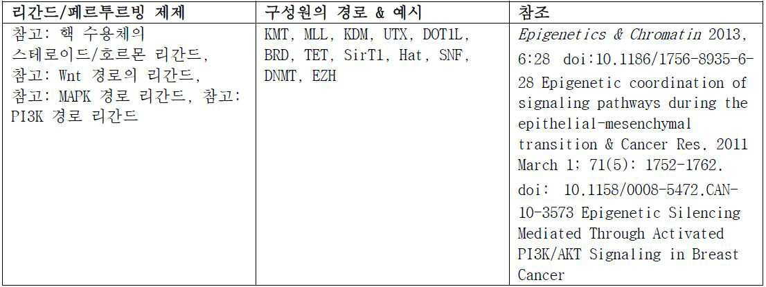
표 10. 세포 처리 - 세포성 대사 Table 10. Cell processing - cellular metabolism
경로 Route
인슐린 수용체 신호전달 Insulin receptor signaling
AMPK 신호전달AMPK signaling
와르버그(Warburg) 효과Warburg effect

표 11. 세포 처리 - 세포 주기 / DNA 손상Table 11. Cell processing - cell cycle / DNA damage
경로 Route
세포 주기 대조군: G1/S 관문Cell cycle control: G1 / S gateway
세포 주기 대조군: G2/M DNA 손상 관문Cell cycle control: G2 / M DNA damage gateway
표 12. 세포 처리 - 면역학 및 염증Table 12. Cell processing - immunology and inflammation
경로 Route
JaK/Stat 신호전달: IL-6 수용체 패밀리JaK / Stat signaling: IL-6 receptor family
NF-κB 신호전달NF-κB signaling
TLR 경로TLR path
B 세포 수용체 신호전달B cell receptor signaling
T 세포 수용체 신호전달T cell receptor signaling
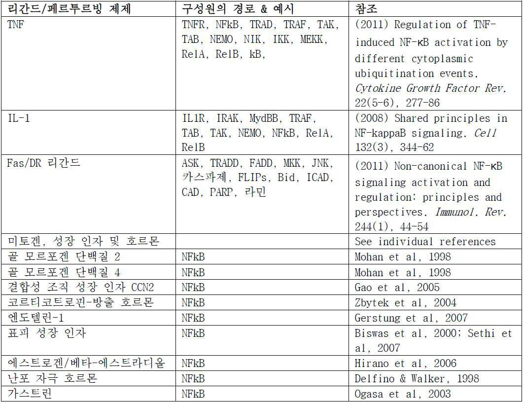

표 13. 세포 처리 - 진행, 및 분화Table 13. Cell processing - progression and differentiation
경로 Route
히포(Hippo) 신호전달Hippo signaling
TGF-β 신호전달 TGF-β signaling
헤지호그(Hedgehog) 신호전달 Hedgehog signaling
노치(Notch) 신호전달Notch signaling
Wnt/β-카테닌 신호전달Wnt / beta -catenin signaling
혈관형성 Angiogenesis
핵 수용체 신호전달 Nuclear receptor signaling
ErbB/HER 신호전달 ErbB / HER signaling
RasRas 신호전달 Signal transmission
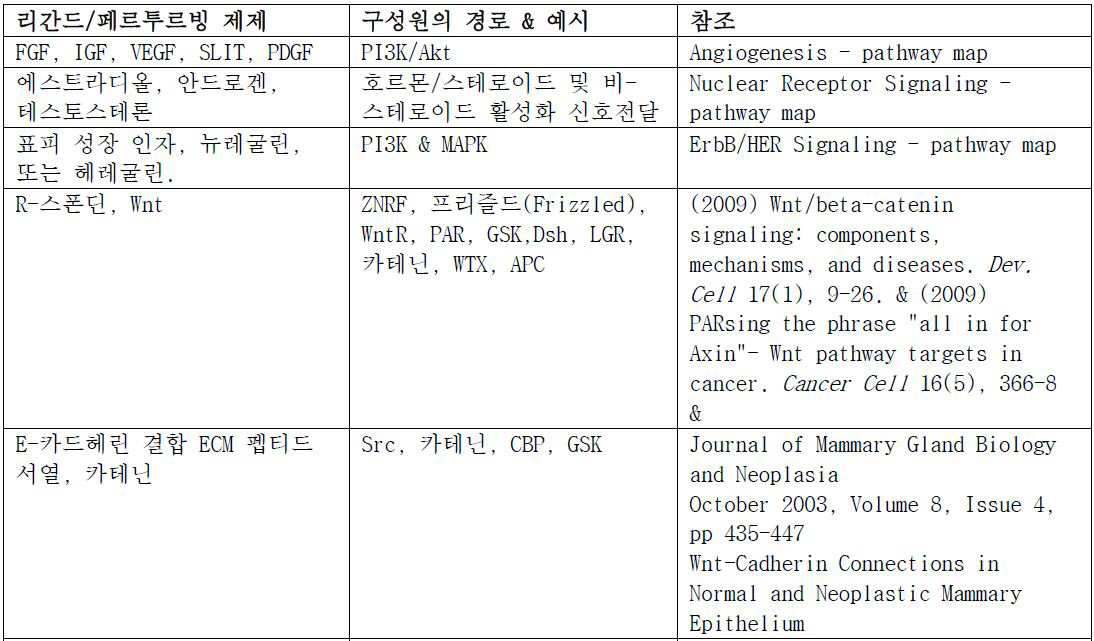

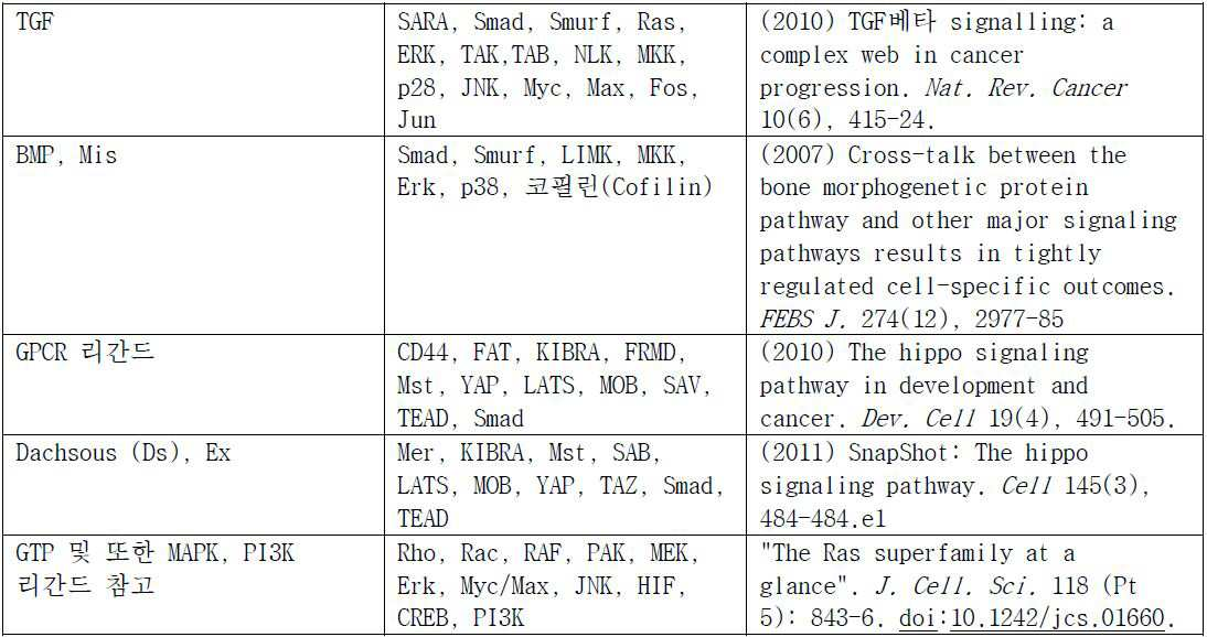
표 14. 세포 공정- Table 14. Cell Processing - 세포골격Cytoskeleton 조절 및 접착 Adjustment and Adhesion
·액틴 동력학의 조절 - 많은 경로에 관련됨, 참고 예를 들어 인테그린 수용체는 ECM 리간드, GPCR 리간드, 성장 인자 결합 수용체 (수용체 티로신 키나제)를 결합시킨다Control of actin kinetics - related to many pathways, for example, integrin receptors bind ECM ligands, GPCR ligands, growth factor binding receptors (receptor tyrosine kinases)
·미세소관 동력학의 조절 - 상기 많은 경로에 관련됨, 참고 예를 들어 Wnt 신호전달, 뉴트로핀/트로핀Control of microtubule dynamics - related to many of the above pathways, see for example Wnt signaling, neurotropin / tropin
·애드헤렌 접합 동력학 - 상기 많은 경로에 관련됨, 참고 예를 들어 PI3K, MAPKAdherent junction kinetic-related to the many routes mentioned above, for example PI3K, MAPK
생리적 반응 파라미터는 본원에서 기재된 것, 예를 들면, 세포 접착, 세포 부착, 세포 형태학, 세포 증식, 세포 신호전달, 세포 밀도, 세포 크기, 세포 형상, 세포 극성, pH, O2, CO2, 글루코오스, 및 이들의 조합을 포함한다. 예시적인 구현예에서, 생리적 반응 파라미터는 세포 접착 또는 부착에서 변화이다.The physiological response parameters may be selected from those described herein, for example , cell adhesion, cell adhesion, cellular morphology, cell proliferation, cell signaling, cell density, cell size, cell shape, cell polarity, pH, O 2 , CO 2 , glucose , And combinations thereof. In an exemplary embodiment, the physiological response parameter is a change in cell adhesion or adhesion.
생리적 반응 파라미터에서 변화가 세포성 샘플에서 발생하는지를 계측하기 위해 연속적 측정의 분석 방법은 본원에서 기재된다 (예를 들면, 반응의 규모 (양성 또는 음성), 최대 또는 최소까지 시간, 반응 연대표의 임의의 시점에서 규모, 등). 비-선형 분석의 이들 및 다른 방법은 생리적 반응 파라미터에서 변화가 교란제의 존재하에 발생하는지를 계측하기 위해 사용될 수 있다.Methods of analysis of serial measurements to measure whether changes in physiological response parameters occur in cellular samples are described herein ( e.g., scale of reaction (positive or negative), maximum or minimum time, Scale from point of view, etc.). These and other methods of non-linear analysis can be used to measure whether changes in physiological response parameters occur in the presence of an agonist.
기준선 및 대조군은 세포성 경로의 상태를 판단하기 위해 사용될 수 있다. 적합한 기준선은, 비제한적으로, 하기를 포함할 수 있다: 교란제 없는 샘플, 교란제의 무한한 희석의 샘플, 교란제의 부가 이후 충분히 오랜 시간 이후 또는 이전 동일한 샘플, 및 세포 기반 검정의 당해 분야의 숙련가에 공지된 다른 상기 베이스라이닝 활성.Baseline and control groups can be used to determine the status of cellular pathways. Suitable baselines may include, but are not limited to, samples without perturbations, samples of infinite dilution of perturbants, samples long enough or longer after the addition of perturbants, and those of the art of cell-based assays Other base lining actives known to the skilled artisan.
적합한 대조군은, 비제한적으로, 하기를 포함할 수 있다: 동일한 환자로부터 건강한 물질의 샘플, 정상 참조 구간을 유도하기 위해 당해 질환이 부족한 충분한 수의 환자로부터 건강한 물질의 샘플의 세트, 유사한 그러나 상이한 활성화제를 가진 샘플, 공지된 양성 또는 음성 반응의 세포주, 활성화제의 역 활성으로 처리된 샘플, 하나 이상의 환자로부터 이환 물질의 샘플, 및 세포 기반 검정의 당해 기술에서 실시된 것으로 공지된 다른 상기 양성 및 음성 대조군.A suitable control may include, but is not limited to, a sample of healthy material from the same patient, a set of samples of healthy material from a sufficient number of patients lacking the disease to derive a normal reference interval, a similar but different activation A cell line of a known positive or negative response, a sample treated with the reverse activity of an activator, a sample of a substance from one or more patients, and other such positive and negative cells known to be practiced in the art of cell- Negative control.
일부 구현예에서, 교란제의 양의 적정으로부터 수득된 데이터는 개별 대상체에서 표적화 치료제의 효능을 계측하기 위해 사용될 수 있다. 예를 들어, 이환 세포 샘플이 교란제의 증가하는 농도에 노출되면, 세포 샘플에서 생존 세포에 관한 교란제의 EC50 값은 생성될 수 있고, 여기서 세포성 경로가 정상적으로 기능하는 경우, 그 세포 샘플에 대한 상기 EC50 값은 교란제에 의해 영향받은 세포성 경로를 방해한다고 공지된 표적화 치료제의 효능을 예측한다. EC50 값의 예측에 의해, 교란제에 대한 환자 세포 샘플의 감수성은 획득되고 따라서 관련된 세포성 경로가 그 환자에서 기능성 또는 기능이상인 정도를 평가하기 위해 사용될 수 있다. In some embodiments, data obtained from titration of the amount of perturbants may be used to measure the efficacy of a targeted therapeutic agent in an individual subject. For example, if a mature cell sample is exposed to an increasing concentration of an agonist, the EC50 value of the perturbing agent for the viable cell in the cell sample can be generated, where the cellular pathway normally functions, The above EC50 values for the anticipated efficacy of the targeted therapeutic agents which are known to interfere with the cellular pathways affected by the agonist. By predicting the EC50 value, the susceptibility of the patient cell sample to perturbants can be obtained and thus used to assess the degree to which the relevant cellular pathway is functional or functional in the patient.
하나의 예에서, EC50은 매우 작은 수이어서, 환자가 최소량의 교란제에 매우 감수성인 것을 나타낸다. 교란제가 정상 인간 유체에서 발견된 천연 제제이면, 상기 제제를 감소시키기 위해 설계된 치료는 낮은 EC50 미만의 상기 제제 수준의 약화에서 효과없을 수 있다. 또 다른 예에서, 경로의 구성원은 구성적이고 교란제에 대해 경로를 미반응성 또는 약 반응성으로 만들 수 있어서, 세포성 경로가 기능이상인 것을 나타낸다. 상기 실시예에서, EC50은 매우 큰 수이다. 또 다른 예에서, 경로 구성원은 과발현될 수 있어서, 교란제의 부가시 경로의 지나친 생리적 반응을 초래하고, 기능이상 경로 상태에 기여한다. 더욱 또 다른 실시예에서, 경로 구성원은, 예를 들어, 불활성 상태에서 잠겨진, 발현하에, 돌연변이, 또는 격리를 통해 저활동성일 수 있어서, 기능이상 상태에 기여한다. 또 다른 구현예에서, 경로 구성원은 다른 세포성 기능에 부적절하게 연결될 수 있어서, 정상 경로 반응의 생리적 반응 미특징을 생성하고, 기능이상 상태에 기여한다.In one example, the EC50 is a very small number, indicating that the patient is highly susceptible to minimal amounts of perturbing agents. If the perturbant is a natural formulation found in normal human fluids, therapies designed to reduce the formulation may be ineffective at lowering the formulation level below the lower EC50. In another example, the members of the pathway are constitutive and can make the path unreactive or weakly reactive with an agonist, thus indicating that the cellular pathway is dysfunctional. In this embodiment, the EC50 is a very large number. In yet another example, pathway members can be overexpressed, resulting in excessive physiological responses of pathways upon addition of perturbants and contributing to dysfunctional pathways. In yet another embodiment, the pathway member may be hypoactive through, for example, locked in an inactive state, under expression, mutated, or isolated, thereby contributing to a dysfunctional state. In another embodiment, the pathway member may be improperly linked to other cellular functions, producing physiological response characteristics of the normal pathway response and contributing to dysfunctional conditions.
교란제의 적정으로부터 수득된 데이터는 또한 교란제의 효력 (즉, 농도가 절반최대 효과를 달성한 것) 및/또는 효능 (즉, 최대 달성가능한 효과)를 평가하기 위해 사용될 수 있다.The data obtained from the titration of the disturbance may also be used to assess the effect (that is, the concentration of one half to achieve the maximum effect), and / or efficiency (i.e., the maximum achievable effect) of the disturbance.
한 개별 대상체로부터 단리된 무 표지 질환 세포 샘플에 대하여 EC50 결과는 본질적으로 동일한 세포 유형을 함유한 다른 개별 대상체로부터 단리된 무 표지 세포 샘플에 대하여 EC50 결과와 비교될 수 있고 동일한 교란제로 시험될 수 있다. 통계적인 분석은 수행될 수 있고, 여기에서 통계적인 분석은 세포성 경로의 기능성 상태를 예측한다. 개별 대상체에서 세포성 경로의 기능성 상태는 그 경로에 표적화 치료제에 대한 대상체의 반응성을 나타낸다.For EC50 results for a sample of unlabeled disease cells isolated from one individual subject, EC50 results can be compared to EC50 results for unlabeled cell samples isolated from other individual subjects containing essentially the same cell type and tested with the same agonist . Statistical analysis can be performed, where the statistical analysis predicts the functional status of the cellular pathway. The functional state of the cellular pathway in an individual subject indicates the reactivity of the subject to the targeted therapeutic agent in that pathway.
H. 약물 효능을 측정하기 위한H. To measure drug efficacy 세포 사이클 상태의 분석 방법Method of analysis of cell cycle status
사이클린-의존적 키나제 (CDKs)는 세포 사이클의 주요 조절물질로서 작용한다. 다양한 유형의 암을 치료하기 위해 개발된 수많은 표적화 치료제는 CDKs를 표적한다. 본원에서 기재된 방법의 하나의 구현예에서, CDK 경로의 활성은 세포 사이클의 상태를 측정하기 위해 단리되고, 상기 측정은 약물 효능과 상관될 수 있다.Cyclin-dependent kinases (CDKs) act as key regulators of the cell cycle. A number of targeted therapies developed to treat various types of cancer target CDKs. In one embodiment of the methods described herein, the activity of the CDK pathway is isolated to determine the condition of the cell cycle, and the measurement can be correlated with drug efficacy.
따라서, 일부 구현예에서, 본원에서 기재된 방법은 교란제, 및/또는 확인제, 및/또는 특정한 세포 사이클 상태로 세포의 이동에 관여된 세포성 경로를 교란시키는 표적화 치료제를 이용할 수 있다. 예를 들어, 약물 트라스투주맙이 의도된 바와 같이 작용하는 경우, G1/S 시기에서 정적이도록 세포 사이클을 명확히 유발시켜, 세포가 추가로 번식할 수 없게 만들고 암성 상태를 방해한다. 본원에서 기재된 방법은 G1/S 시기에서 세포의 기능성 상태의 측정, 예를 들면, G1/S 상태와 관련된 경로의 상태의 시험에 의해 환자 샘플에서 트라스투주맙의 효능을 확인할 것이다. 상기 경로는 당해 기술에 공지되어 있다. 다른 실시예에서, G0 시기에서 세포를 중단시키는 교란제가 선택될 수 있고, 본원에서 기재된 방법은 상기 시험 또는 정지 세포 시기와 관련된 경로 기능을 시험하기 위해 사용될 수 있다. 더욱 다른 실시예에서, 세포가 세포자멸사에 진입 또는 진입하기 시작하도록 하는 제제가 선택될 수 있고, 본원에서 기재된 방법은 세포 사멸 사이클에서 진입과 관련된 경로 기능을 시험할 수 있다.Thus, in some embodiments, the methods described herein can utilize targeting agents that disrupt perturbants, and / or detectors, and / or cellular pathways involved in cell migration to a particular cell cycle state. For example, when the drug Trastuzumab acts as intended, it clearly induces cell cycles to be static in the G1 / S period, rendering the cells unable to reproduce further and disturb the cancerous state. The methods described herein will confirm the efficacy of trastuzumab in a patient sample by measuring the functional status of the cells in the G1 / S phase, for example, by testing the condition of the pathway associated with the Gl / S status. Such pathways are known in the art. In another embodiment, an agonist that disrupts the cell at the G0 time may be selected and the methods described herein may be used to test pathway function associated with the test or arrest cell timing. In yet another embodiment, agents may be selected that allow the cells to enter or enter apoptosis, and the methods described herein may test pathway functions associated with entry in a cell death cycle.
I. 시험 결과의 분석 및 해석I. Analysis and interpretation of test results
본원에서 기재된 방법을 이용하여 수득된 시험 결과는 임상의 및/또는 환자에 정보를 제공하기 위해 다양한 방식으로 분석 및 해석될 수 있다. 특정 구현예는 아래와 같이 설정된다.Test results obtained using the methods described herein may be analyzed and interpreted in a variety of ways to provide information to the clinician and / or patient. A specific implementation is set as follows.
(i) 이환 경로 분석. 상기 분석은 이환 경로 활성이 생체외 환자에서 발견되는지를 확인한다. 분석은 의사에게, 처음으로, 질환 과정이 환자의 이환 세포에서 존재하는지의 동적 평가를 제공할 것이다. 상기 구현예에서, 시험된 경로는 4개의 그룹 카테고리 중 하나로 분류될 수 있다: 구성적으로 활성, 과반응, 전체에서 활성 없음 (저-활성), 또는 정상적으로 활성. 경로가 이환되는지 및 따라서 당해 경로 활성을 억제시킨다고 공지된 표적된 요법을 이용한 치료에 적합한지를 계측하기 위해, 질환을 갖는 것으로 의심되는 환자에 대하여 본원에서 기재된 방법에 의해 계측된 바와 같이 경로 활성은 그 경로 활성에 대하여 정상 참조 구간으로부터 유도된 컷-오프 값에 비교된다. 비정상인 경로 활성 (예를 들면 비정상 및 정상 경로 활성을 기술하는 컷-오프 초과)를 갖기 위해 밝혀진 경로를 표적하는 약물은 그 활성을 방해하는 것이 예상될 것이고, 그렇게 함으로써 환자에서 의도된 효과를 생산한다. (i) pathway analysis . The assay confirms that pathway pathway activity is found in in vitro patients. The analysis will provide the physician with, for the first time, a dynamic assessment of whether the disease process is present in the patient's miliary cells. In this embodiment, the tested pathway can be categorized into one of four group categories: constitutively active, overactive, totally inactive (low-active), or normally active. The pathway activity is measured by the method described herein for a patient suspected of having a disease, to determine whether the pathway is affected and thus is appropriate for treatment with a targeted therapy known to inhibit the pathway activity, Is compared to the cut-off value derived from the normal reference interval for path activity. A drug that targets a pathway that has been found to have abnormal pathway activity (e.g., cut-off excess that describes abnormal and normal pathway activity) would be expected to interfere with its activity, thereby producing an intended effect in the patient do.
(ii) 약물 작용기 분석. 상기 분석은 생체외 약물의 작용기의 2개 평가를 제공한다. (ii) drug functional group analysis. The assay provides two assessments of the functional groups of the in vitro drug.
1) 반응 스코어 (RS): 반응 스코어는 시험된 약물이 표적된 경로상에서 가졌던 기능성 효과를 특성화한다. 0 - 1 범위 상에서 보고될 수 있고, 여기서 더 높은 스코어는 더 큰 약물 작용기를 나타낸다. 1) Response Score (RS): The response score characterizes the functional effect that the tested drug had on the target pathway. 0 - 1, where a higher score indicates a larger drug functional group.
2) 반응 스코어 백분위 등급화 (RSPR): RSPR은 환자의 반응 스코어가 동일한 제제로 시험된 다른 환자에 의해 받은 스코어에 비하여 등급하는 방법을 특성화한다. 각 환자에 대하여, 총 그룹 내에서 그의 반응 스코어의 백분위는 계측된다. 일단 백분위 등급화가 배정되면, 환자는 그 다음 3개의 그룹: a) 중앙 미만, b) 중앙 근처, 또는 c) 중앙 초과로 분류될 수 있다. 특정 약물에 대하여, 임상 종료점에 의해 측정된 바와 같이 환자 약물 반응 예컨대 진행까지의 시간 (TTP)에서 광범위 변화는 반응 스코어에서의 변화를 비춰질 것이다. 임상시험에서 75번째 백분위 환자의 TTP 기간이 25번째 백분위 환자의 TTP 기간의 5-10 배 초과인 것이 종종 사실이기 때문에, 의사에게 그의 환자의 반응 스코어의 상대적 등급의 제공은 이들에게 중요한 해석적 맥락을 제공한다. 예를 들어, 이들은 개별 환자의 반응 스코어 백분위에 상응하는 백분위 범위에서 임상시험내 환자의 TTP 기간에 기반된 개별 환자에 대하여 TTP 기간을 추정할 수 있다. 2) Response Score Percentile Grading (RSPR): The RSPR characterizes the way the patient's response score is graded relative to the score received by other patients tested with the same formulation. For each patient, the percentile of his response score within the total group is measured. Once a percentile grading has been assigned, the patient can be classified into the following three groups: a) below the center, b) near the center, or c) above the center. For a particular drug, a broad-ranging change in the patient drug response, such as time to progression (TTP), as measured by the clinical endpoint, will reflect a change in the response score. Since it is often the case that the TTP period of the 75th percentile patient is more than 5-10 times the TTP period of the 25th percentile patient in clinical trials, the provision of a relative rating of his patient's response score to the physician is important to their interpretive context . For example, they can estimate the TTP duration for individual patients based on the patient ' s TTP duration in the clinical trial in the percentile range corresponding to the individual patient ' s response score percentile.
(iii) 유사하게 임상 결과의 예측. 상기 분석은 반응 스코어와 문제의 제제의 수용 이후 시험된 및 관측된 환자에 대한 임상 종료점 사이 임상시험에서 찾아진 상관관계를 반영한다. 상기 상관관계의 경우, 임상시험에서 특정 반응 스코어를 받은 환자와 일치하는 임상 결과를 확인하는 것이 가능하다. 예를 들어, TTP가 측정된 임상 결과이었으면, 환자의 결과는 하기 3개의 카테고리 중 하나로 분류될 수 있다. (iii) a prediction of clinical outcomes similarly. The analysis reflects the correlation found in the clinical trial between the response score and the clinical endpoint of the tested and observed patient after receipt of the agent in question. In the case of this correlation, it is possible to identify clinical results consistent with the patient who received a specific response score in the clinical trial. For example, if TTP was the measured clinical outcome, the outcome of the patient could be categorized into one of the following three categories.
1) 유사한 TTP 기간 - 최저: 본 하위-집단에 해당하는 환자는 환자의 전체 집단이 경험할 중앙 TTP 기간 미만에서 양호한 TTP 기간을 경험할 것 같다.1) Similar TTP periods - lowest : Patients in this subgroup are likely to experience a good TTP period under the median TTP period that the entire population of patients will experience.
2) 유사한 TTP 기간 - 불확정: 본 카테고리에 해당하는 반응 스코어를 받은 환자에 대하여 평가는 제공되지 않는다.2) Similar TTP durations - Indeterminate : No evaluation is provided for patients who received a response score corresponding to this category.
3) 유사한 TTP 기간 - 최고: 본 하위-집단에 해당하는 환자는 환자의 전체 그룹의 중앙 TTP 기간 초과에서 양호한 TTP 기간을 경험할 것 같다.3) Similar TTP periods - Best : Patients in this subgroup are likely to experience a good TTP period in the entire group of patients over the median TTP period.
임상의는 이들이 선택하는 약물 요법을 계측함에 따라 안내로서 CELx 프로파일 시험의 결과를 이용할 것이다. 환자의 세포가 다중 약물로 시험된 경우, 각 약물의 유사한 임상 결과는 의사가 최대 유사하게 임상 결과를 상관하는 시험 결과를 가진 약물을 선택할 수 있을 정도로 비교될 수 있다.The clinician will use the results of the CELx profile test as a guide as they measure the pharmacotherapy they choose. When a patient's cells are tested with multiple drugs, similar clinical results for each drug can be compared so that the physician can select the drug with the test results that most closely correlate to the clinical outcome.
J. J. 키트Kit
본 발명의 또 다른 측면에서, 키트가 제공된다. 특정 구현예에서, 키트는 하기를 포함하고: 수송 배지를 함유한 개별 대상체로부터 질환 세포 샘플용 용기; 수송 배지를 함유한 개별 대상체로부터 대조군 세포 샘플용 용기; 바이오센서; 바이오센서로부터 출력으로 데이터 전환용 컴퓨터 실행가능 지침을 갖는 비-일시적 컴퓨터 판독가능한 배지, 여기서 상기 출력은 정의된 기간 동안 세포성 생리적 반응 파라미터에서 변화를 보여주고, 여기서 상기 세포성 생리적 반응 파라미터는 하기로 이루어진 군으로부터 선택된다: pH, 세포 접착, 세포 부착 패턴, 세포 증식, 세포 신호전달, 세포 생존, 세포 밀도, 세포 크기, 세포 형상, 세포 극성, O2, CO2, 글루코오스, 세포 사이클, 동화작용, 이화, 소분자 합성 및 생성, 턴오버, 및 호흡, ATP, 칼슘, 마그네슘, 및 다른 충전된 이온, 단백질, 특이적 경로 구성원 분자, 다양한 세포성 구획에서 DNA 및 또는 RNA, 유전체학, 및 프로테오믹스, 번역후 변형 및 기전, 2차 메신저의 수준, cAMP, mRNA, RNAi, 마이크로RNAs 및 생리적 기능을 가진 다른 RNA, 및 이들의 조합; 반응군 또는 비반응군으로서의 상태를 나타내는 컷오프 값 초과 또는 미만으로서 출력의 분류 및/또는 무 반응, 약 반응성, 및 반응성을 갖는 것으로서 샘플의 분류; 및 분류를 이용한 보고서 작성.In yet another aspect of the present invention, a kit is provided. In certain embodiments, the kit comprises: a container for a diseased cell sample from an individual subject containing a transport medium; A container for a control cell sample from an individual subject containing the transport medium; A biosensor; A non-transitory computer readable medium having computer executable instructions for switching data from a biosensor to an output, wherein the output shows a change in a cellular physiological response parameter for a defined period of time, Cell adhesion, cell attachment pattern, cell proliferation, cell signaling, cell survival, cell density, cell size, cell shape, cell polarity, O 2, CO 2, glucose, cell cycle, assimilation DNA and / or RNA in various cellular compartments, genomics, and proteomics, including, but not limited to, enzymatic activity, catabolism, small molecule synthesis and generation, turnover and respiration, ATP, calcium, magnesium and other charged ions, Post-translational modifications and mechanisms, level of secondary messenger, cAMP, mRNA, RNAi, microRNAs and other RNAs with physiological functions, and combinations thereof; A classification of the sample as having a classification and / or no reaction, a weak reactivity, and a reactivity as a cut-off value indicating a state as a reaction group or a non-reaction group, And creating reports using classification.
질환 세포 샘플의 유형 및 양은 본원에서 기재된다. 특정 구현예에서, 질환 세포 샘플은 적어도 5,000 세포를 갖는 전체의 세포 무 표지 생존 세포 샘플이다. 구현예에서, 대조군 세포 샘플은 하기로 이루어진 군으로부터 선택된다: 동일한 대상체로부터 질환 세포 샘플, 동일한 대상체로부터 건강한 세포 샘플, 질환이 없다고 공지된 대상체로부터 건강한 세포 샘플, 정상 참조 구간을 유도하기 위해 당해 질환이 부족한 환자의 충분한 수로부터 건강한 물질의 샘플의 세트, 치료제에 반응한다고 공지된 세포 샘플, 치료제에 반응하지 않는다고 공지된 세포 샘플, 및 이들의 조합.The types and amounts of disease cell samples are described herein. In certain embodiments, the disease cell sample is a whole, cell-free survival cell sample having at least 5,000 cells. In an embodiment, a control cell sample is selected from the group consisting of: a diseased cell sample from the same subject, a healthy cell sample from the same subject, a healthy cell sample from a subject known to be free of disease, A set of samples of healthy material from a sufficient number of these deficient patients, a cell sample known to respond to the therapeutic, a cell sample known not to respond to the therapeutic, and combinations thereof.
용기 및 수송 배지는 세포 생존력을 유지하기 위해 및 세포 활성화를 최소화하기 위해 설계된다. 구현예에서, 배지 및 용기는 무 내독소, 비발열성 및 DNase- 및 RNase-프리이다. 일단 수득되면 세포 샘플은 세포 생존력을 보유하는 수송 배지에서 유지된다. 분석의 부위에 대한 수송용 시간의 길이에 의존하여, 상이한 배지가 이용될 수 있다. 구현예에서, 조직 샘플의 수송이 최대 10 시간을 필요로 하는 경우, 배지는 400 mosm/L 미만의 오스몰농도를 갖고 Na+, K+, Mg+, Cl-, Ca+2, 글루코오스, 글루타민, 히스티딘, 만니톨, 및 트립토판, 페니실린, 스트렙토마이신을 포함하고, 필수 아미노산을 함유하고 그리고 인간 원발 세포의 증식 및 플레이팅 효율을 지지하기 위해, 추가로 하기를 함유할 수 있다: 비-필수 아미노산, 비타민, 다른 유기 화합물, 미량 미네랄 및 무기 염, 혈청, 세포 추출물, 또는 성장 인자, 인슐린, 트랜스페린, 나트륨 셀레나이트, 하이드로코르티손, 에탄올아민, 포스포포릴에탄올로아민, 트리도티로닌, 나트륨 피루베이트, L-글루타민. 그와 같은 배지의 예는 하기를 포함한다: 셀시오 배지, 로즈웰 파크 메모리얼 연구소 배지 (RPMI), 항크스 버퍼링된 염수, 및 McCoy's 5A, 이글스 필수 최소 배지 (EMEM), 둘베코 변형된 이글스 배지 (DMEM), Leibovitz L-15, 또는 이들의 변형.Containers and transport medium are designed to maintain cell viability and minimize cell activation. In embodiments, the media and containers are non-endotoxin, non-heat-labile, and DNase- and RNase-free. Once obtained, the cell sample is maintained in a transport medium that retains cell viability. Depending on the length of time for transport to the site of analysis, different media may be used. In embodiments, if transport of the tissue sample requires up to 10 hours, the medium has an osmolality of less than 400 mosm / L and has Na +, K +, Mg +, Cl-, Ca + 2, glucose, glutamine, histidine, Mannitol, and tryptophan, penicillin, streptomycin, contain essential amino acids, and to further support the growth and plating efficiency of human primary cells, including: non-essential amino acids, vitamins, other The present invention relates to a pharmaceutical composition comprising an organic compound, a trace minerals and an inorganic salt, a serum, a cell extract or a growth factor, insulin, transferrin, sodium selenite, hydrocortisone, ethanolamine, phospholipid ethanolamine, tridothyronine, sodium pyruvate, . Examples of such media include: Celcio medium, Roswell Park Memorial Labs medium (RPMI), Heps buffered saline, and McCoy's 5A, Eagles Essential Minimal Medium (EMEM), Dulbecco's Modified Eagles Medium DMEM), Leibovitz L-15, or a modification thereof.
바이오센서는 본원에서 기재된다. 특정 구현예에서 바이오센서는 하기로 이루어진 군으로부터 선택된 세포성 파라미터를 검출하는 바이오센서로 이루어진 군으로부터 선택된다: 세포 접착, 세포 부착, 세포 형태학, 세포 표현형, 세포 증식, 세포 신호전달, 세포 밀도, 세포 극성, pH, O2, CO2, 글루코오스, 및 이들의 조합. 구현예에서, 디바이스는 임피던스 또는 광학적 디바이스이다. 바이오센서는 본원에서 기재된 바와 같이 선택적으로 코팅될 수 있다. 구현예에서, 본원에서 기재된 바와 같이 치료적 및/또는 활성제 제제의 유형과 관련된 생리적 파라미터에서 변화를 측정하는 바이오센서가 선택된다.Biosensors are described herein. In certain embodiments, the biosensor is selected from the group consisting of a biosensor that detects cellular parameters selected from the group consisting of: cell adhesion, cell attachment, cellular morphology, cell phenotype, cell proliferation, cell signaling, cell density, Cell polarity, pH, O 2, CO 2, glucose, and combinations thereof. In an embodiment, the device is an impedance or an optical device. The biosensor may optionally be coated as described herein. In an embodiment, a biosensor is selected that measures changes in physiological parameters associated with the type of therapeutic and / or active agent formulation as described herein.
다른 구현예에서, 키트는 바이오센서로부터 출력으로 데이터 전환용 컴퓨터 실행가능 지침을 갖는 비-일시적 컴퓨터 판독가능한 배지를 포함하고, 여기서 상기 출력은 정의된 기간 동안 세포성 생리적 반응 파라미터에서 변화를 보여주고, 여기서 상기 세포성 생리적 반응 파라미터는 하기로 이루어진 군으로부터 선택된다: pH, 세포 접착, 세포 부착 패턴, 세포 증식, 세포 신호전달, 세포 생존, 세포 밀도, 세포 크기, 세포 형상, 세포 극성, O2, CO2, 글루코오스, 및 이들의 조합; 반응군 또는 비반응군 및/또는 무 반응, 약 반응성, 및 반응성으로서 출력의 분류; 및 분류를 이용한 보고서 작성.In another embodiment, the kit comprises a non-transitory computer readable medium having computer executable instructions for switching data from a biosensor to an output, wherein the output shows a change in a cellular physiological response parameter for a defined period of time Wherein the cellular physiological response parameters are selected from the group consisting of pH, cell adhesion, cell attachment pattern, cell proliferation, cell signaling, cell viability, cell density, cell size, cell shape, cell polarity, O 2 , CO 2, glucose, and combinations thereof; Classification of output as reactive or unreactive groups and / or unreacted, weakly reactive, and reactive; And creating reports using classification.
다른 구현예에서, 본 발명은 개시내용의 방법을 이행하기 위한 지침으로 컴퓨터처리 디바이스 또는 컴퓨터 판독가능한 배지를 제공한다. 컴퓨터 판독가능한 배지는 비-일시적 CD, DVD, 플래시 드라이브, 외부 하드 드라이브, 및 모바일 디바이스를 포함한다.In another embodiment, the present invention provides a computer processing device or a computer readable medium as a guide for implementing the method of the disclosure. Computer readable media include non-volatile CD, DVD, flash drives, external hard drives, and mobile devices.
본원에서 기재된 키트 및 방법은 프로세서/컴퓨터 시스템의 용도를 사용할 수 있다. 예를 들어, 방법을 이행하기 위해 컴퓨터 프로그램 코드를 분류하는 프로그램 메모리, 작업 메모리, 및 인터페이스 예컨대 종래의 컴퓨터 스크린, 키보드, 마우스, 및 프린터, 뿐만 아니라 다른 인터페이스, 예컨대 네트워크 인터페이스, 및 데이터베이스 인터페이스를 포함한 소프트웨어 인터페이스에 결합된 프로세서를 포함한 다목적 컴퓨터 시스템은 본원에서 기재된 하나의 구현예의 용도를 찾는다.The kits and methods described herein may use the use of a processor / computer system. Including, for example, program memory for classifying computer program code to implement the method, working memory, and interfaces, such as conventional computer screens, keyboards, mice, and printers, as well as other interfaces such as network interfaces and database interfaces A multipurpose computer system including a processor coupled to a software interface finds use of one implementation described herein.
컴퓨터 시스템은 데이터 입력 디바이스, 예컨대 키보드, 입력 데이터 파일, 또는 네트워크 인터페이스, 또는 또 다른 시스템, 예컨대, 예를 들어, 정의된 기간 동안 바이오센서에 의해 생성된 데이터를 해석한 시스템으로부터 사용자 입력을 허용하고, 출력 디바이스 예컨대 프린터, 디스플레이, 네트워크 인터페이스, 또는 데이터 저장 디바이스에 출력을 제공한다. 입력 디바이스, 예를 들어 네트워크 인터페이스는 본원에서 기재된 바와 같이 세포성 생리적 파라미터에서 변화 및/또는 이들 변화의 정량화를 포함한 입력을 받는다. 출력 디바이스는, 세포성 파라미터에서 변화의 검출 및/또는 정량화를 도시하는 하나 이상의 수 및/또는 그래프를 포함한, 출력 예컨대 디스플레이를 제공한다.The computer system may allow user input from a data input device, such as a keyboard, an input data file, or a network interface, or another system, e.g., a system that interprets data generated by the biosensor for a defined period of time , An output device such as a printer, a display, a network interface, or a data storage device. An input device, e.g., a network interface, receives inputs including changes in cellular physiological parameters and / or quantification of these changes, as described herein. The output device provides an output, e.g., a display, comprising one or more numbers and / or graphs that illustrate detection and / or quantification of changes in cellular parameters.
컴퓨터 시스템은 본원에서 기재된 방법에 의해 생성된 데이터를 저장하는 데이터 스토어에 결합될 수 있다. 상기 데이터는 각 측정 및/또는 각 대상체에 대하여 저장되고; 각각의 이들 데이터 유형의 선택적으로 복수의 세트는 각 대상체에 상응하여 저장된다. 하나 이상의 컴퓨터/프로세서는, 예를 들어, 네트워크를 거쳐 컴퓨터 시스템에 연결된, 예를 들어, 별도의 기계로서 사용될 수 있거나, 또는 컴퓨터 시스템상에서 실시하는 별도 또는 통합된 프로그램을 포함할 수 있다. 어느 방법이 이용되든 이들 시스템은 데이터를 받고 대신 검출/진단에 관한 데이터를 제공한다.The computer system may be coupled to a data store that stores data generated by the methods described herein. Said data being stored for each measurement and / or for each object; A plurality of sets of each of these data types is stored corresponding to each object. One or more of the computers / processors may be, for example, a separate machine, connected to the computer system via a network, or may include separate or integrated programs running on the computer system. Whichever method is used, these systems receive the data and instead provide the detection / diagnosis data.
일부 구현예에서, 컴퓨터처리 디바이스는 단일 컴퓨터처리 디바이스, 예컨대 서버 컴퓨터를 포함할 수 있다. 다른 구현예에서, 컴퓨터처리 디바이스는 네트워크 (도시되지 않음)를 거쳐 서로 통신하기 위해 구성된 다중 컴퓨터처리 디바이스를 포함할 수 있다. 컴퓨터처리 디바이스는 메모리 내에서 다중 데이터베이스를 저장할 수 있다. 컴퓨터처리 디바이스상에서 저장된 데이터베이스는 임상, 실시 임상의, 프로그래머 확인 코드, 또는 임의의 다른 원하는 카테고리에 의해 조직화될 수 있다. In some implementations, the computer processing device may comprise a single computer processing device, e.g., a server computer. In other implementations, the computer processing devices may include multiple computer processing devices configured to communicate with one another via a network (not shown). The computer processing device may store multiple databases in memory. The database stored on the computer processing device may be organized by clinical, practitioner, programmer identification code, or any other desired category.
바이오센서로부터 데이터는 원격 컴퓨터처리 시스템 또는 또 다른 데이터 저장 디바이스에 보내질 수 있다. 통신 과정은 출발 모듈에서 개시 및 시작하고 연결 작업으로 진행한다. 연결 작업은 건강 관리 제공자의 저장된 정보를 원격 컴퓨터처리 시스템에, 예를 들어, 케이블 연결, 무선 랜 (WLAN 또는 Wi-Fi) 연결, 세포성 네트워크, 무선 개인 통신망 (WPAN) 연결, 예를 들면, BLUETOOTH®, 또는 임의의 원하는 통신 회선을 통해 통신적으로 연결한다.Data from the biosensor may be sent to a remote computer processing system or another data storage device. The communication process starts and starts in the departure module and proceeds to the connection operation. The connection operation may be used to connect the healthcare provider's stored information to a remote computer processing system, for example, a cable connection, a wireless LAN (WLAN or Wi-Fi) connection, a cellular network, a wireless personal digital network BLUETOOTH®, or any desired communication line.
수송 작업은 데이터를 바이오센서로부터 컴퓨터처리 디바이스에 수송한다. 하나의 구현예에서, 수송 작업은 디바이스 사이에서 데이터 전송 이전 데이터를 암호화시킨다. 통신 과정은 완료할 수 있고 중지 모듈에서 종료할 수 있다. 일단 바이오센서 데이터가 원격 컴퓨터처리 디바이스에 이동되면, 데이터는 출력, 예컨대 경시적으로 세포 지수 측정으로 전환된다. 특정 구현예에서, 정의된 종료점은 선택되고 본원에서 기재된 바와 같이 무 반응, 약 반응성 또는 반응성으로서 세포 샘플을 분류하는데 사용된다. 구현예에서, 반응군 또는 비 반응군으로서 샘플의 분석의 상태는 케이블 연결, 무선 랜 (WLAN 또는 Wi-Fi) 연결, 세포성 네트워크, 무선 개인 통신망 (WPAN) 연결, 예를 들면, BLUETOOTH®, 또는 임의의 원하는 통신 회선을 거쳐 유사한 과정을 이용하여 건강 관리 제공자에 역으로 통신된다.The transport operation transports data from the biosensor to the computer processing device. In one implementation, the transport operation encrypts data prior to data transfer between devices. The communication process can be completed and terminated in the pause module. Once the biosensor data is transferred to the remote computer processing device, the data is converted to an output, e. G., Cell index measurement over time. In certain embodiments, the defined endpoints are selected and used to sort the cell sample as unreactive, weakly reactive, or reactive, as described herein. In embodiments, the state of the analysis of the sample as a reaction group or a non-reaction group may be a cable connection, a wireless LAN (WLAN or Wi-Fi) connection, a cellular network, a wireless personal area network (WPAN) connection, e.g., BLUETOOTH, Or communicated back to the health care provider using a similar process over any desired communication line.
특정 구현예에서, 컴퓨터 판독가능한 저장 배지는, 컴퓨터처리 디바이스에 의해 실행된 경우, 컴퓨터처리 디바이스가 하기를 포함하는 단계를 수행하도록 하는 컴퓨터-실행가능 지침을 갖는다: 데이터를 바이오센서로부터 출력으로의 전환 단계 (여기서 상기 출력은 정의된 기간에 걸쳐 세포성 생리적 반응 파라미터에서 변화를 보여주고, 여기서 상기 세포성 생리적 반응 파라미터는 하기로 이루어진 군으로부터 선택된다: pH, 세포 접착, 세포 부착 패턴, 세포 증식, 세포 신호전달, 세포 생존, 세포 밀도, 세포 크기, 세포 형상, 세포 극성, O2, CO2, 글루코오스, 및 치료제의 존재 및/또는 부재 하에 이들의 조합); 치료제의 존재하에 세포 샘플에서 바이오센서로부터 출력과 치료제의 부재 하에 세포 샘플에서 바이오센서로부터 출력의 비교에 의해 정의된 종료점에서 무 반응, 및 반응성으로서 출력의 분류 단계; 및 분류를 이용한 보고서 작성 단계. 구현예에서, 컴퓨터 실행가능 지침은 건강 관리 제공자에 분류 통신용 지침을 포함한다.In certain embodiments, a computer-readable storage medium has computer-executable instructions that, when executed by a computer processing device, cause the computer processing device to perform the steps comprising: transferring data from the biosensor to the output Wherein the output shows a change in a cellular physiological response parameter over a defined period wherein the cellular physiological response parameter is selected from the group consisting of: pH, cell adhesion, cell adhesion pattern, cell proliferation Cell signaling, cell viability, cell density, cell size, cell shape, cell polarity, O 2, CO 2, glucose, and combinations thereof in the presence and / or absence of therapeutic agents); A step of classifying the output as non-responsive and responsive at an end point defined by comparison of the output from the biosensor in the cell sample with the output from the biosensor in the presence of the therapeutic agent and the absence of the therapeutic agent in the cell sample; And reporting step using classification. In an embodiment, the computer executable instructions include instructions for classifying communications to the health care provider.
다른 구현예에서, 컴퓨터 판독가능한 저장 배지는 경로가 대상체의 질환 세포 샘플에서 작동가능한지 확인용 지침을 포함할 수 있다. 컴퓨터처리 디바이스에 의해 실행된 경우 지침은 제1 활성제 또는 교란제 제제에 대한 경로 반응성이 질환 세포 샘플에서 활성인지를 계측하기 위해 서로 제1 활성화제 또는 교란제로 치료된 대상체에서 질환 세포 샘플로부터 바이오센서 데이터의 출력과 제1 활성화제 또는 교란제로 치료되지 않은 동일한 대상체에서 제2 질환 세포 샘플로부터 바이오센서 데이터의 출력 사이에서 차이가 있는지의 계측; 경로의 활성의 적응증으로서 출력에서 차이의 존재 확인, 및 건강 관리 제공자에 경로의 활성 전달을 포함한다. 활성제 또는 교란제 제제 및 그의 경로는 본원에서 기재된다.In other embodiments, the computer readable storage medium may comprise instructions for verifying that the pathway is operable in a diseased cell sample of the subject. The instructions, when executed by a computer processing device, may be used to determine whether pathway reactivity to the first active or perturbing agent is active in the diseased cell sample, from a diseased cell sample in a subject treated with the first activator or perturbant, Measuring whether there is a difference between the output of the data and the output of the biosensor data from the second diseased cell sample in the same subject not treated with the first activator or disturbance; Indication of the activity of the pathway includes confirmation of the presence of the difference in output, and active delivery of the pathway to the healthcare provider. Active or perturbative agents and their routes are described herein.
적어도 5개 유형의 CELx 시험은 본원에서 기재된 방법을 이용하여 구상된다.At least five types of CELx tests are envisioned using the methods described herein.
1) 표적된 경로 약물의 효능을 계측하는 경로 폐쇄 시험. 본 시험에서, 그의 세포성 표적에 표적된 경로 약물의 결합에 의해 야기된 시험 세포의 생리적 변화는 측정되고 기준선 측정과 비교된다.1) Pathway closure testing to measure the efficacy of the targeted pathway drug. In this test, the physiological changes of the test cells caused by the binding of pathway drugs targeted to its cellular targets are measured and compared to baseline measurements.
2) 항-증식 약물의 효능을 계측하는 항-증식 시험. 본 시험에서, 그의 증식성 수용력의 저해에 의해 야기된 시험 세포의 생리적 변화는 측정되고 기준선 측정과 비교된다.2) Anti-proliferation test to measure the efficacy of anti-proliferative drugs. In this test, the physiological changes of the test cells caused by the inhibition of its proliferative capacity are measured and compared with baseline measurements.
3) 조합으로 이용된 2 이상의 약물의 효능을 계측하는 조합 시험. 본 시험에서, 약물에 의해 야기된 시험 세포의 생리적 변화는 측정되고 기준선 측정과 비교된다. 조합 시험은 2 이상의 표적된 경로 약물, 2 이상의 항-증식 약물, 또는 하나 이상의 각 유형의 약물을 포함할 수 있다.3) Combination test to measure the efficacy of two or more drugs used in combination. In this test, the physiological changes of the test cells caused by the drug are measured and compared with baseline measurements. Combination tests may include two or more target pathways, two or more anti-proliferative drugs, or one or more of each type of drug.
4) 특이적 환자에서 경로의 기능을 입증하는 기능성 경로 시험. 본 시험에서, 교란제 및/또는 확인제의 부가에 의해 야기된 시험 세포의 생리적 변화는 환자에서 신호전달 경로의 기능성 상태를 나타낸다. 상기 결과는 경로 기능이 비정상 (예를 들면 질환-관련) 또는 정상인지를 계측하기 위해 분리하여 계측된 컷-오프 값과 비교된다.4) Functional pathway test demonstrating pathway function in specific patients. In this test, the physiological changes of the test cells caused by the addition of perturbants and / or agents are indicative of the functional status of the signaling pathway in the patient. The result is compared to a separately measured cut-off value to measure whether the path function is abnormal (e.g., disease-related) or normal.
5) 리간드 및 또는 표적이 조합된 리간드 및 또는 표적의 존재하에 측정함으로써 존재하는지를 계측하기 위한 리간드/표적 시험.5) A ligand / target test for measuring whether a ligand and / or target is present by combining in the presence of a ligand and / or a target.
실시예Example
실시예Example 1- One- 5에 대한 실험 디자인의Experimental design for 5 논의 Argument
방법은 세포 또는 세포 경로 내에서 단백질 결합이 발생한 이후 세포 또는 세포 경로의 생리적 변화를 측정하기 위해 CReMS를 이용한다. 결합되지 않는 한 약물이 작업할 수 없는 것, 그리고 거의 모든 질환 유전자가 코어 신호전달 경로에 해당하는 것이 통상적으로 이해된다. 상기 및 단백질 결합의 생화학물질 원리가 세포 유형을 거쳐 보편적이라는 사실의 관점에서, 본원에서 기재된 방법은 따라서 단백질 및 다른 생체분자 결합이 발생할 수 있는 전체 세포 및 세포 경로에 광범위하게 적용가능하다.The method uses CReMS to measure physiological changes in the cell or cell pathway after protein binding occurs within the cell or cell pathway. It is commonly understood that drugs can not work unless combined, and that almost all disease genes correspond to the core signaling pathway. In view of the above and the fact that the biochemical principles of protein binding are universal across cell types, the methods described herein are thus broadly applicable to whole cells and cell pathways in which protein and other biomolecule binding can occur.
종래 기술의 유전적 시험의 현황은 약물 또는 경로가 결합되는지를 직접적으로 나타낼 수 없고, 따라서 이들은 약물 반응을 신뢰성있게 예측할 수 없다. 약물이 도입된 이후 세포 내에서 발생하는 생리적 변화의 확인에 의해, CELx 시험은 약물에 대한 대상체의 세포의 반응을 신뢰성있게 예측할 수 있다. The status of prior art genetic tests can not directly indicate whether the drug or pathway is bound, and therefore they can not reliably predict the drug response. By confirming the physiological changes that occur within the cell after the drug is introduced, the CELx test can reliably predict the cell response of the subject to the drug.
방법이 이용하는 시험의 유형의 구현예의 일부를 입증하기 위해, 3개의 상이한 유형의 암을 가진 11명의 상이한 환자로부터 세포에 관한 65 실험은 수행되었다. 11 상이한 세포 경로에 영향을 미치는 16개의 상이한 약물은 시험되었고 2개의 상이한 CReMS 유형은 이용되었다. 결과가 본 출원의 실시예에서 보고되는 시험의 목록은 아래 표 15에서 제공된다:To demonstrate some of the implementations of the type of test the method uses, 65 experiments on cells from 11 different patients with three different types of cancer were performed. Sixteen different drugs affecting 11 different cell pathways were tested and two different CReMS types were used. A list of tests for which the results are reported in the examples of the present application are provided in Table 15 below:
표 15. 시험 수행 목록Table 15. Test performance list
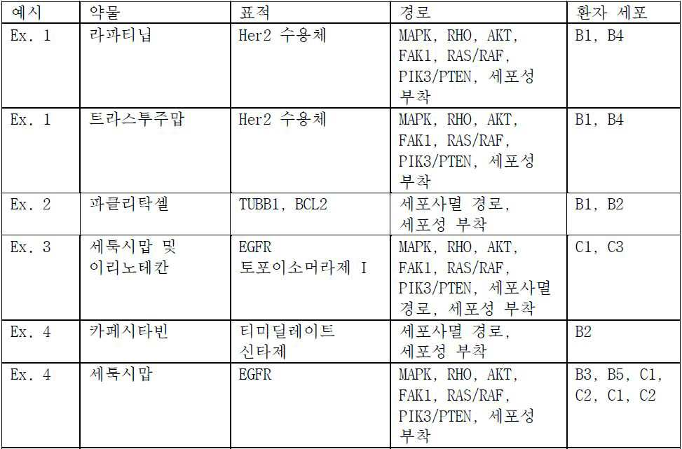

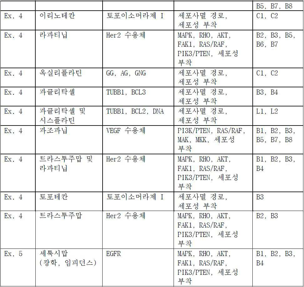
실험 디자인에 대한 근거Basis for experimental design
조직: group:
최고 발생률을 가진 3개의 암으로부터 조직이 선택되었다.Tissue was selected from three cancers with the highest incidence.
유방암 . 유방암 모델이 진행, 다양한 세포성 형태학, 가변 대사율, 및 생존에 관하여 많은 다른 암을 대표하고 많은 다른 조직에서 발견된 암에 공통인 비정상적인 분자 및 경로를 갖기 때문에 유방암 세포는 시험의 64%로 이용되었다. Breast cancer . Breast cancer cells were used in 64% of the trials because the breast cancer model represented many other cancers in terms of progression, variable cellular morphology, variable metabolic rate, and survival and had abnormal molecules and pathways common to cancers found in many different tissues .
대장암 및 폐암 . 대장암 및 폐암 세포는 다른 유의미한 암 유형으로 개시내용의 시스템 및 방법의 적용가능성을 입증하기 위해 이용되었다. Colon cancer and lung cancer . Colon cancer and lung cancer cells have been used to demonstrate the applicability of systems and methods of disclosure to other significant cancer types.
세포cell ::
유방암에 대하여 상피 세포 유형의 공통 임상 제시를 가진 8명 환자로부터 세포는 시험용으로 선택되었다. 환자로부터 세포는 조직 수집의 당해 기술에서 실시된 것에 의해 규칙적으로 사용된 세포 샘플 수집 기술을 이용하여 수득되었다.Cells from 8 patients with a common clinical presentation of epithelial cell types for breast cancer were selected for testing. Cells from the patients were obtained using cell sample collection techniques routinely used by those skilled in the art of tissue collection.
환자 B1 : 세포는 61세 여성의 유방의 TNM IIA 기, 등급 3 원발 침습성 관상 암종에서 유도된다. 세포는 대략 31 시간의 배가 시간을 갖고, 가끔의 비정질-형상화된 상피 세포 형태학으로 확장된 바와 같이 나타나고, ERB B1 및 ERB B2 수용체의 매우 높은 발현 수준을 갖는다. 에스트로겐 수용체 (ER), 프로게스테론 수용체 (PR) 및 종양유전자 TP53 상태는 모두 3개 음성이다. Patient B1 : Cells are derived from TNM IIA,
환자 B2 : 세포는 51-세 코카서스 여성의 유방의 선암종의 늑막 삼출에서 유도된다. 세포는 대략 28 시간의 배가 시간을 갖고, 침습성, eel-유사 형태학으로 보여지고 정상 ERB B2 수용체 수준 초과 약간 상승된 및 ERB B1의 높은 발현 수준을 갖고, 에스트로겐 수용체 (ER) 음성, 프로게스테론 수용체 (PR) 음성이고 높은 종양유전자 TP53 상태를 갖는다. Patient B2 : Cells are derived from pleural effusion of adenocarcinoma of the breast of a 51-year-old Caucasian female. (ER) negative, the progesterone receptor (PR), the progesterone receptor (PR), and the progesterone receptor (PR) ) Negative and has a high tumor gene TP53 status.
환자 B3 : 세포는 43-세 백인 여성의 유방의 선암종의 늑막 삼출에서 유도되고; 대략 20 시간 배가 시간, 자갈 상피 형태학, ERB B1 및 ERB B2 수용체의 매우 높은 발현 수준, 및 에스트로겐 수용체 (ER) 음성, 프로게스테론 수용체 (PR) 음성, 및 종양유전자 TP53 양성 상태. Patient B3 : cells are induced in the pleural effusion of adenocarcinoma of the breast of a 43-year-old Caucasian female; Very high expression levels of ERB B1 and ERB B2 receptors, and estrogen receptor (ER) negative, progesterone receptor (PR) negative, and oncogenic gene TP53-positive status, approximately 20 hours doubling time, gravel epithelial morphology, ERB B1 and ERB B2 receptors.
환자 B4 : 세포는 47-세 흑인 여성의 유방의 침습성 관상 암종의 복수 유체에서 유도되고; 110 시간의 배가 시간, 원형, 포도-유사 클러스터 형태학을 갖고, ERB B1 및 ERB B2 수용체의 매우 높은 발현 수준, 및 에스트로겐 수용체 (ER) 양성, 프로게스테론 수용체 (PR) 음성, 및 종양유전자 TP53 야생형-낮은 상태를 갖는다. Patient B4 : Cells are induced in ascites fluid of invasive ductal carcinoma of the breast of a 47-year-old black woman; (ER) positive, progesterone receptor (PR) negative, and tumor gene TP53 wild-type, with a 110 hour doubling time, round, grape-like cluster morphology, very high expression levels of ERB B1 and ERB B2 receptors and estrogen receptor State.
환자 B5 : 세포는 60-세 백인 여성의 원발 유방 침습성 관상 암종에서 유도되고; 28 시간 배가 시간, 비정질 확대의 혼합물 및 침습성 형태학, ERB B1 및 ERB B2 수용체의 매우 높은 발현 수준, 및 에스트로겐 수용체 (ER) 양성, 프로게스테론 수용체 (PR) 양성, 및 종양유전자 TP53 양성 상태. Patient B5 : cells are induced in primary breast-invasive tubular carcinoma of 60-year-old Caucasian female; 28 hour doubling time, mixture of amorphous enlargement and invasive morphology, very high expression levels of ERB B1 and ERB B2 receptors, and estrogen receptor (ER) positive, progesterone receptor (PR) positive, and tumor gene TP53 positive status.
환자 B6 : 세포는 70-세 흑인 여성의 원발 유방 화생 암종 TNM IV 기 등급 3에서 유도되고; 대략 30 시간 배가 시간, 거칠어진 확대 형태학, ERB B1 및 ERB B2 수용체의 매우 높은 발현 수준, 및 에스트로겐 수용체 (ER) 음성, 프로게스테론 수용체 (PR) 음성, 및 종양유전자 TP53 돌연변이된 낮은 상태. Patient B6 : Cells were induced in primary mammary carcinoma
환자 B7 : 세포는 69-새 백인 여성의 유방의 침습성 관상 암종의 늑막 삼출에서 유도되고; 30 시간 배가 시간, 작은 모자이크 상피 형태학, ERB B1 및 ERB B2 수용체의 낮은 발현 수준, 및 에스트로겐 수용체 (ER) 양성, 프로게스테론 수용체 (PR) 양성, 및 종양유전자 TP53 야생형 상태. Patient B7 : Cells are induced in the pleural effusion of invasive ductal carcinoma of the breast of a 69-New White woman; 30 hour doubling time, small mosaic epithelium morphology, low expression levels of ERB B1 and ERB B2 receptors, and estrogen receptor (ER) positive, progesterone receptor (PR) positive, and tumor gene TP53 wild-type status.
환자 B8 : 세포는 48-세 백인 여성의 유방의 선암종의 늑막 삼출에서 유도되고; 24 시간 배가 시간, 매우 작은 포도-유사 클러스터 형태학, ERB B1 수용체의 매우 낮은 발현 수준, ERB B2 수용체의 높은 발현 수준, 및 에스트로겐 수용체 (ER) 음성, 프로게스테론 수용체 (PR) 음성, 및 종양유전자 TP53 야생형 낮은 상태. Patient B8 : Cells are induced in the pleural effusion of adenocarcinoma of the breast of a 48-year-old Caucasian female; (ER) negative, progesterone receptor (PR) negative, and oncogenic gene TP53 wild-type, as well as the expression levels of ERB B2 receptors, 24 hours doubling time, very small grape-like cluster morphology, very low expression levels of ERB B1 receptor, Low state.
대장암에 대한 상피 세포 유형의 공통 임상 제시를 가진 2명의 환자로부터 세포가 시험용으로 선택되었다:Cells were selected for testing from two patients with a common clinical presentation of epithelial cell types for colorectal cancer:
환자 C1 : 세포는 남성 결장직장 암종에서 유도된다. 세포는 14 시간의 회전타원체 용적 배가 시간, ERB B1의 높은 수준, 돌연변이체 K-Ras, 돌연변이체 PIK3CA 및 종양유전자TP53 양성 상태를 갖는다. Patient C1 : Cells are induced in male colon rectal carcinoma. Cells have a 14 hour oval volume doubling time, a high level of ERB B1, a mutant K-Ras, a mutant PIK3CA and a tumor gene TP53-positive status.
환자 C2 : 세포는 44 세 백인 여성의 원발 결장 선암종, 등급 2에서 유도된다. 세포는 46 시간의 회전타원체 용적 배가 시간, ERB B1의 높은 수준, 돌연변이체 BRAF, 및 종양유전자 TP53 음성 상태를 갖는다. Patient C2 : Cells are induced in primary colorectal adenocarcinoma,
비-소세포 폐암에 대하여 상피 세포 유형의 공통 임상 제시를 갖는 2명의 환자로부터 세포는 시험용으로 선택되었다:Cells from two patients with a common clinical presentation of epithelial cell types for non-small cell lung cancer were selected for testing:
환자 L1 : 세포는 25-세 남성의 비-소세포 폐 암종의 늑막 삼출에서 유도되고; 48 시간 배가 시간, 상피 형태학, ERB B1 및 ERB B2 수용체의 상승된 발현 수준, PIK3CA 양성, 및 KRAS, BRAF 모두 음성 상태. Patient L1 : Cells are induced in pleural effusion of non-small cell lung carcinoma of 25-year-old male; 48 hours doubling time, epithelial morphology, elevated expression levels of ERB B1 and ERB B2 receptors, PIK3CA positive, and both KRAS and BRAF negative status.
환자 L2 : 세포는 52-세 백인 남성의 세기관지폐포 선암종에서 유도되고; 대략 30 시간 배가 시간, 상피 형태학, ERB B1 및 ERB B2 수용체의 정상 발현 수준, 및 BRAF, HRAS, PIK3CA, 및 KRAS 모두 음성 상태. Patient L2 : Cells are induced in bronchoalveolar adenocarcinoma of 52-year-old white male; Approximately 30 hours doubling time, epithelial morphology, normal expression levels of ERB B1 and ERB B2 receptors, and both BRAF, HRAS, PIK3CA, and KRAS negative status.
세포 경로 표적: Cell pathway target:
이들 실험에 대하여 선택된 약물은, 이들이 광범위하게 상호연결되고, 결합을 통해 조절되고, 효소 활성 예컨대 인산화 및 탈-인산화를 포함하고, 임계적 세포성 기능을 대조하는 방법에서 대부분의 세포성 조절 경로를 대표하는 11개의 세포성 경로에 영향을 미친다.The drugs chosen for these experiments are those that are broadly interconnected, regulated through binding, contain enzymatic activities such as phosphorylation and de-phosphorylation, and most cellular regulatory pathways in a way to contrast critical cellular functions Affecting the 11 representative cellular pathways.
MAPK . ( EGFR , EGFR -TK, HER1 , HER 2) . 미토겐-활성화된 단백질 (MAP) 키나제는 전체 세포 유형에서 발견되고 그리고 세포외 자극 (미토겐, 삼투 스트레스, 열충격 및 전-염증 사이토카인)에 반응하는 및 다양한 세포성 활성, 예컨대 유전자 발현, 유사분열, 분화, 증식, 및 세포 생존/세포자멸사를 조절하는 필수 세린/트레오닌-특이적 단백질 키나제이다. 그의 단단한 조절은 세포성 생존력의 유지에 중요하다. 상피 성장 인자 수용체 (인간에서 EGFR; ErbB-1; HER1)은 세포외 단백질 리간드의 상피 성장 인자 계열 (EGF-계열)의 구성원에 대한 세포-표면 수용체이다. (상향조절로서 공지된) EGFR 과발현 또는 과-활성으로 이어지는 돌연변이는, 폐암, 항문암 및 교모세포종 다형성을 포함한, 수많은 암과 관련되었다. 돌연변이, 증폭 또는 EGFR 또는 계열 구성원의 오-조절은 전체 상피암의 약 30%에서 연루되고, 항암 요법의 확장 클래스의 표적이다. MAPK . ( EGFR , EGFR- TK, HER1 , HER2) . Mitogen-activated protein (MAP) kinases have been found in whole cell types and have been shown to act in response to extracellular stimuli (mitogen, osmotic stress, thermal shock and pre-inflammatory cytokines) and to inhibit various cellular activities such as gene expression, Is an essential serine / threonine-specific protein kinase that regulates mitosis, differentiation, proliferation, and cell survival / apoptosis. His tight regulation is important in maintaining cellular viability. The epithelial growth factor receptor (EGFR; ErbB-1; HER1 in humans) is a cell-surface receptor for members of the epithelial growth factor family (EGF-family) of extracellular protein ligands. Mutations leading to EGFR overexpression or hyper-activity (known as upregulation) have been associated with a number of cancers, including lung cancer, anal cancer, and glioblastoma multiforme. Mutations, amplification or o-regulation of EGFR or family members are implicated in approximately 30% of all epithelial cancers and are targets of an extended class of anti-cancer therapies.
PI3K / PTEN ( Her2 , 3, 4, VEGF ) . 거의 전체 세포 유형에서 발견된 포스파티딜이노시톨 3-키나제 (PI3K) 경로는 세포 생존 및 세포 성장에 임계적이고, 세포 표면 수용체에 성장 인자 결합에 의해 활성화될 수 있다. 암에서 가장 빈번하게 활성화된 경로중에 있는 복잡한 신호전달 캐스케이드이다. 인간 암에서 임의의 다른 경로보다 더 자주 돌연변이, 증폭 및 재배열을 포함한 게놈 변이에 의해 표적된다. VEGF 수용체는 광범위한 인간 종양 및 세포주를 거쳐 발현된다. VEGF의 발현은 종양 성장 및 전이를 촉진시키는 혈관 네트워크의 개발 및 유지로 이어지기 위해 나타났다. VEGF는 대다수의 비-소세포 폐암 (NSCLC), 결장직장, 및 다른 종양에서 발현된다. VEGF는 폐암이 진행함에 따라 더 높은 수준에서 발현된다. 또한, 거대하고 확실해지는 증거는 VEGF 유전자 발현이 좋지 못한 예측과 밀접하게 관련되는 것을 나타낸다. PI3K / PTEN ( Her2 , 3, 4, VEGF ) . The phosphatidylinositol 3-kinase (PI3K) pathway found in nearly all cell types is critical for cell survival and cell growth, and can be activated by growth factor binding to cell surface receptors. It is a complex signaling cascade in the most frequently activated pathway in cancer. Are more frequently targeted by genomic variations including mutations, amplifications and rearrangements than in any other pathway in human cancers. VEGF receptors are expressed across a wide range of human tumors and cell lines. Expression of VEGF appeared to lead to the development and maintenance of vascular networks that promote tumor growth and metastasis. VEGF is expressed in the majority of non-small cell lung cancer (NSCLC), colon rectum, and other tumors. VEGF is expressed at higher levels as lung cancer progresses. In addition, the evidence of gigantic and confirmatory evidence indicates that VEGF gene expression is closely related to poor predictions.
세포 접착 . 세포 접착 경로는 거의 전체 주요 생리적 기능을 교차한다. 경로는 세포 접착 분자 예컨대 셀렉틴, 인테그린, 및 카드헤린을 이용하여 표면, 세포외 기질 또는 또 다른 세포에 세포의 결합을 포함한다. 정확한 세포성 접착은 다중세포 구조의 유지에 필수적이다. 세포성 접착은 세포의 세포질을 연결할 수 있고 신호 전달에 관여될 수 있다. 전체 접착은, 직접적으로 원형질막의 통합 성분을 포함한, 또는 간접적으로 세포의 외부상에서 배출된 및 침착된 물질을 통해, 세포 표면에 의해 매개된다. Cell adhesion . The cell adhesion pathway crosses almost the entire major physiological function. The pathway involves binding of cells to the surface, extracellular matrix or another cell using cell adhesion molecules such as selectin, integrin, and carderine. Exact cell adhesion is essential for the maintenance of multiple cellular structures. Cellular adhesion can connect the cytoplasm of a cell and can be involved in signal transduction. Total adhesion is mediated by the cell surface, either directly through the inclusion of an integral component of the plasma membrane, or indirectly through the excreted and deposited material outside the cell.
MEK . MEK는, 암 세포 증식 및 생존에 대하여 신호전달하는, RAS/RAF/MEK/ERK 경로에서 주요 단백질 키나제이다. MEK는 암, 특히 RAS 및 RAF 종양유전자에서 돌연변이를 갖는 종양에서 빈번하게 활성화된다. MEK는 또한, 암에서 성장 및 생존 인자로서 작용할 수 있는, 염증성 사이토카인 TNF, IL-6 및 IL-1의 생합성을 조절한다. MEK 경로는 흑색종, 비-소세포 폐암, 두경부암 및 췌장암을 포함한 상이한 종양의 증식에서 중심 축으로서 작용한다. 그리고 단독 또는 다른 제제와 조합으로 MEK 저해는 암 치료에서 중요한 치료적 전략이다. MEK . MEK is the major protein kinase in the RAS / RAF / MEK / ERK pathway, which signals cancer cell proliferation and survival. MEK is frequently activated in tumors with mutations in cancer, particularly RAS and RAF oncogenes. MEK also regulates the biosynthesis of the inflammatory cytokines TNF, IL-6 and IL-1, which may act as growth and survival factors in cancer. The MEK pathway acts as a central axis in the proliferation of different tumors, including melanoma, non-small cell lung cancer, head and neck cancer, and pancreatic cancer. And MEK inhibition alone or in combination with other agents is an important therapeutic strategy in cancer therapy.
RHO . Rho 단백질은 다양한 세포성 기능 예컨대 세포 극성, 소포성 추적, 세포 사이클 및 전사체 동력학에서 관여된다. Rho 활성화는 암성 세포에서 수많은 상이한 효과를 가질 수 있다. 종양의 개시에서, Rho 활성의 변형은 세포자멸사를 억제시킬 수 있고 따라서 인공 세포 장수에 기여할 수 있다. 천연 세포자멸사가 억제된 이후, 비정상 종양 성장은 Rho 단백질이 통합적 역할을 하는 극성의 손실을 통해 관측될 수 있다. 다음으로, 성장하는 덩어리는 Rho 단백질에 의해 잠재적으로 야기된 접착 단백질의 변경을 통해 그의 정상 경계를 거쳐 침범할 수 있다. RHO . Rho proteins are involved in a variety of cellular functions such as cell polarity, bubble tracking, cell cycle and transcriptional dynamics. Rho activation can have a number of different effects in cancerous cells. At the onset of the tumor, a modification of Rho activity can inhibit apoptosis and thus contribute to artificial cell longevity. After suppression of natural apoptosis, abnormal tumor growth can be observed through the loss of polarity, in which Rho proteins play an integral role. Next, the growing mass may invade through its normal boundary through alteration of the adhesive protein potentially caused by the Rho protein.
AKT . AKT는 세린/트레오닌 키나제이고, 수용체 티로신 키나제 예컨대 IGF-1R, HER2/Neu, VEGF-R, PDGF-R의 자극, 및 수용체-PI-3K의 막-국재화된 복합체의 어셈블리 및 2차 메신저 PIP를 통한 Akt의 활성화를 포함하는, 수렴성 업스트림 신호전달 경로의 무리에 대하여 중요한 결절 포인트로서 세포내로 기능한다. AKT 및 그의 업스트림 조절물질이 광범위한 고체 종양 및 혈액성 악성종양에서 탈조절되고, 상기 경로의 상기 언급된 생물학적 후유증의 면에서, AKT 경로는 이들 종양의 생물학적 공격성의 주요 계측인자, 및 신규 항-암 요법용 주요 잠재 표적으로 간주된다. AKT . AKT is a serine / threonine kinase and is involved in stimulation of the receptor tyrosine kinases such as IGF-1R, HER2 / Neu, VEGF-R, PDGF-R and assembly of membrane-localized complex of receptor- Lt; / RTI > function as an important nodal point for a multitude of astringent upstream signaling pathways, including the activation of Akt through < RTI ID = 0.0 > AKT and its upstream regulatory substances are deregulated in a wide range of solid tumors and hematological malignancies and in terms of the aforementioned biological sequelae of the pathway the AKT pathway is a key metrological factor in the biologic aggressiveness of these tumors, It is considered a major potential target for therapy.
FAK1 . 초점 접착 키나제 1 (FAK1)-매개된 신호 전달의 생물학적 중요성은 상기 티로신 키나제가 배아 발생에서, 세포 이동, 세포 사이클 진행의 대조에서, 및 세포자멸사에서 근본적인 역할을 한다는 사실에 의해 밑줄 표시된다. 정착-의존적 세포의 생존에서 중추적 역할을 하고 악성종양의 개발에서 중요한 역할을 하는 인테그린-연결된 세포 이동-과정에 필수적이다. FAK는 다양한 인간 상피암에서 상향조절되고, 발현은 침습성 포텐셜에 밀접하게 상관된다. 최근에, FAK 발현은 종양 세포의 악성종양으로의 진행 또는 암의 발병에 연루되었다. FAK1은 유방암 항-에스트로겐 저항성 조절에서 주요 역할을 한다. FAK1 . The biological significance of focal adhesion kinase 1 (FAK1) -mediated signaling is underlined by the fact that the tyrosine kinases play a fundamental role in embryonic development, in cell migration, in the control of cell cycle progression, and in apoptosis. It is essential for integrin-linked cell migration-processes that play a pivotal role in the survival of fixation-dependent cells and play an important role in the development of malignant tumors. FAK is upregulated in a variety of human epithelial cancers, and expression is closely correlated with invasive potential. Recently, FAK expression has been implicated in the progression of tumor cells to malignant tumors or the development of cancer. FAK1 plays a major role in the regulation of breast cancer anti-estrogen resistance.
RAS/ RAF . RAS 경로는 암에서 가장 빈번하게 탈조절된 경로 중 하나이다. RAS는, RAF/미토겐-활성화된 단백질 키나제 (MAPK)/세포외 신호-조절된 키나제 (ERK) 키나제 (MEK)/ERK MAPK 및 포스파티딜이노시톨 3-키나제 (PI3K)-AKT 신호전달 캐스케이드를 포함한, 다중 효과기 경로를 통해 신호전달한다. 이들 효과기 경로의 종양발생 포텐셜은 BRAF 및 PIK3CA에서 활성화 돌연변이의 빈번한 발생 뿐만 아니라 종양 억제제 PTEN, PI3K의 음성 조절물질에서 기능 상실 돌연변이에 의해 예시된다. 종양형성에서 Ras의 상기 중요한 역할 때문에, Ras-신호전달 경로는 항암 요법용 표적으로서 상당한 주의력을 유인하였다. RAS / RAF . The RAS pathway is one of the most frequently controlled pathways in cancer. RAS has been shown to be involved in a number of pathways including RAF / mitogen-activated protein kinase (MAPK) / extracellular signal-regulated kinase (ERK) kinase (MEK) / ERK MAPK and phosphatidylinositol 3-kinase (PI3K) Signaling through multiple effector paths. The tumorigenic potentials of these effector pathways are exemplified by the frequent occurrence of activating mutations in BRAF and PIK3CA, as well as by loss-of-function mutations in the negative regulators of the tumor suppressors PTEN, PI3K. Because of this important role of Ras in tumorigenesis, the Ras-signaling pathway attracted considerable attention as a target for chemotherapy.
MAK 경로. 전이-관련된 키나제 (MAK)는 세포 성장에 요구된 전사 인자의 신규 조절물질이다. 상기 경로의 저해는 세포 사이클 저지 활성으로 이어진다. MAK path . Metastasis-related kinases (MAK) are novel regulators of the transcription factors required for cell growth. Inhibition of this pathway leads to cell cycle arresting activity.
MKK . 미토겐-활성화된 단백질 키나제 키나제 (MKK) 신호전달 경로는 세포 분화, 증식, 운동성 및 생존을 포함한 생명 유지 관련 세포성 과정의 전사 및 번역후 조절 모두에 실시되었다. MKK 신호전달 경로가 VEGF의 방출, 및 VEGF에 대한 반응 조절에서 필수적 역할을 하기 때문에, MKK가 종양 혈관형성 촉진에서 중요한 역할을 한다고 믿어진다. MKK . The mitogen-activated protein kinase kinase (MKK) signaling pathway has been implicated in both transcriptional and post-translational regulation of vital-related cellular processes including cell differentiation, proliferation, motility and survival. It is believed that MKK plays an important role in promoting tumor angiogenesis, since the MKK signaling pathway plays an essential role in the release of VEGF and in the regulation of response to VEGF.
세포자멸적 경로 . 세포자멸사 경로의 활성화는세포독성 약물이 종양 세포를 사멸시키는 주요 기전이다. 세포자멸사는 2개의 주요 경로를 통해 발생한다. 외적인 또는 세포질 경로로 언급된, 제1은 종양 괴사 인자 (TNF) 수용체 슈퍼패밀리의 구성원인, Fas 사멸 수용체를 통해 유발된다. 제2 경로는 교란된 경우 미토콘드리아로부터 사이토크롬-c의 방출 및 사멸 신호의 활성화로 이어지는 고유 또는 미토콘드리아 경로이다. 모든 경로는, 세포의 사멸이 되는, 조절 및 구조 분자를 절개하는 카스파제로 불리는 프로테아제의 캐스케이드의 활성화를 포함한 최종 공통 경로로 수렴한다. 세포자멸사 신호전달에서 결함은 종양의 저항성에 기여한다. Apoptotic pathway . Activation of apoptosis pathway is the main mechanism by which cytotoxic drugs kill tumor cells. Apoptosis occurs through two major pathways. The first, referred to as extrinsic or cytoplasmic pathway, is induced through the Fas death receptor, a member of the tumor necrosis factor (TNF) receptor superfamily. The second pathway is the intrinsic or mitochondrial pathway, which, when disturbed, leads to the release of cytochrome c from the mitochondria and activation of the death signal. All pathways converge to the final common path, including activation of the cascade of proteases called caspases, which cleave the regulatory and structural molecules, which are cell deaths. Defects in apoptosis signaling contribute to tumor resistance.
치료제:remedy:
선택된 치료제는 소분자 약물을 대표하는 것 및 항체로부터 유도된 것을 포함한다. 시험된 치료제는 세포 내에서 기능성인 특이적 경로를 폐쇄하기 위해 설계된 작용 기전을 가진 일부 및 세포 세포자멸사를 유발시키기 위해 설계된 다른 것을 포함한다.Selected therapeutic agents include those that represent small molecule drugs and those that are derived from antibodies. The therapeutic agents tested include those with a mechanism of action designed to shut down a specific pathway that is functional in the cell and others designed to induce apoptosis.
세툭시맙 . 세툭시맙 (Erbitux)는 전이성 결장직장암 및 두경부암의 치료를 위하여 정맥내 주입에 의해 주어진, 키메라성 (마우스/인간) 단클론성 항체, 상피 성장 인자 수용체 (EGFR) 억제제이다. 성장 인자가 세포의 표면상에서 그의 수용체에 결합하는 경우, 수용체는 세포를 분할시키는 신호를 제공한다. 일부 암은 심지어 성장 인자 없이 분할하기 위해 신호를 제공하는 돌연변이된 수용체에 의해 야기된다. 그것이 세포를 통제불가능하게 분할시킨다. 세툭시맙은 그와 같은 수용체에 결합하고 그 신호를 턴오프한다. Cetuximab . Cetuxim (Erbitux) is a chimeric (mouse / human) monoclonal antibody, epithelial growth factor receptor (EGFR) inhibitor, given by intravenous infusion for the treatment of metastatic colorectal cancer and head and neck cancer. When the growth factor binds to its receptor on the surface of the cell, the receptor provides a signal that divides the cell. Some cancers are caused by mutated receptors that provide signals to divide even without growth factors. It divides the cell uncontrollably. Cetuximab binds to such a receptor and turns off its signal.
에를로티닙 . 에를로티닙 하이드로클로라이드 (Tarceva)는 비-소세포 폐암, 췌장암 및 몇 개의 다른 유형의 암을 치료하기 위해 사용된 약물이다. 상피 성장 인자 수용체 (EGFR)상에서 작용하는, 가역적 티로신 키나제 억제제이다. 에를로티닙은, 다양한 형태의 암에서 크게 발현되고 가끔 돌연변이되는, 상피 성장 인자 수용체 (EGFR) 티로신 키나제를 특이적으로 표적한다. 수용체의 아데노신 삼인산 (ATP) 결합 부위에 가역적 방식으로 결합한다. Erlotinib . Errotinib hydrochloride (Tarceva) is a drug used to treat non-small cell lung cancer, pancreatic cancer and several other types of cancer. Is a reversible tyrosine kinase inhibitor that acts on the epithelial growth factor receptor (EGFR). Errotinib specifically targets epithelial growth factor receptor (EGFR) tyrosine kinase, which is largely expressed and sometimes mutated in various forms of cancer. Binds reversibly to the adenosine triphosphate (ATP) binding site of the receptor.
라파티닙 . 라파티닙 (Tykerb/Tyverb)은 유방암 및 다른 고체 종양용 경구 활성 약물이다. HER2 성장 수용체 경로를 방해하는 이중 티로신 키나제 억제제이다. HER2-양성 유방암을 위하여 조합 요법으로 사용된다. 라파티닙은 2개의 종양유전자, EGFR (상피 성장 인자 수용체) 및 HER2/neu (인간 EGFR 유형 2)와 관련된 티로신 키나제 활성을 억제시킨다. HER2/neu의 과 발현은 여성에서 고위험 유방암의 특정 유형을 책임진다. Lapatinib . Tykerb / Tyverb is an orally active drug for breast cancer and other solid tumors. It is a dual tyrosine kinase inhibitor that interferes with the HER2 growth receptor pathway. It is used as a combination therapy for HER2-positive breast cancer. Lapatinib inhibits tyrosine kinase activity associated with two oncogenes, EGFR (epithelial growth factor receptor) and HER2 / neu (human EGFR type 2). Overexpression of HER2 / neu is responsible for certain types of high-risk breast cancer in women.
트라스투주맙 . 트라스투주맙 (Herceptin)은 HER2/neu 수용체를 방해하는 단클론성 항체이다. 그의 주요 용도는 특정 유방암을 치료하는 것이다. 불량성 HER2 단백질에 결합하는 경우, HER2 단백질은 더 이상 유방내 세포를 통제불가능하게 재생시키지 않는다. Trastuzum . Trastuzumab (Herceptin) is a monoclonal antibody that interferes with the HER2 / neu receptor. His main use is to treat certain types of breast cancer. When bound to a defective HER2 protein, the HER2 protein no longer regenerates intramammary cells uncontrollably.
도세탁셀 . 도세탁셀 (Taxotere)은 임상적으로 정착된 항-유사분열 화학요법 약물이다 (즉, 세포 분할을 방해한다). 유방, 난소, 전립선, 및 비-소세포 폐 암의 치료에 주로 사용된다. 도세탁셀은 화학요법 약물 부류; 탁산이고, 파클리탁셀 (탁솔)의 반-합성 유사체이다. Docetaxel . Taxotere is a clinically established anti-mitotic chemotherapeutic drug (i. E., Prevents cell division). Breast, ovary, prostate, and non-small cell lung cancer. Docetaxel is a chemotherapeutic drug class; Taxane, and is a semi-synthetic analog of paclitaxel (Taxol).
GSK1059615 . 포스포이노시티드 3-키나제 억제제 (PI3K 억제제)는, 암에서 주요 역할을 하는, PI3K/AKT/mTOR 경로의 일부인 포스포이노시티드 3-키나제 효소의 억제에 의해 기능하는 포텐셜 의료 약물이다. 상기 경로의 억제는 종종 종양 성장을 억압시킨다. GSK1059615 . The phosphoinositide 3-kinase inhibitor (PI3K inhibitor) is a potential medical drug that functions by inhibition of the phosphoinositide 3-kinase enzyme, which is part of the PI3K / AKT / mTOR pathway, which plays a major role in cancer. Inhibition of this pathway often suppresses tumor growth.
GSK1120212 . GSK1120212는 유망한 항종양 활성을 가진 MEK1 및 MEK2 (MEK1/2) 효소의 강력한 및 선택적 알로스테릭 억제제이다. GSK1120212 . GSK1120212 is a potent and selective allosteric inhibitor of MEK1 and MEK2 (MEK1 / 2) enzymes with promising antitumor activities.
파조파닙 . 파조파닙 (Votrient)은 VEGFR-1, VEGFR-2, VEGFR-3, PDGFR-a/β의 강력한 및 선택적 다중-표적된 수용체 티로신 키나제 억제제, 및 종양 성장을 차단하고 혈관신생을 억제시키는 c-키트이다. Pazopanib . Pazopent is a potent and selective multi-targeted receptor tyrosine kinase inhibitor of VEGFR-1, VEGFR-2, VEGFR-3, PDGFR-a / It is a kit.
파클리탁셀 . 파클리탁셀은 폐, 난소, 유방, 두경부암, 및 진전된 형태의 카포시 육종을 가진 환자를 치료하기 위해 사용된 유사분열 억제제이다. 파클리탁셀은 미세소관을 안정화시키고 결과적으로, 세포 분할 동안 미세소관의 정상 파손을 방해한다. 도세탁셀과 함께, 탁산의 약물 카테고리를 형성한다. Paclitaxel . Paclitaxel is a mitotic inhibitor used to treat patients with pulmonary, ovarian, breast, head and neck cancer, and advanced forms of Kaposi 's sarcoma. Paclitaxel stabilizes microtubules and, as a result, prevents normal breakage of microtubules during cell division. Together with docetaxel, it forms the drug category of taxane.
플루오로우라실 . 플루오로우라실 (5-FU 또는 f5U) (Adrucil, Carac, Efudix, Efudex 및 Fluoroplex)은 암의 치료에서 사용되는 피리미딘 유사체인 약물이다. 타이미딜레이트 합성효소의 비가역적 저해를 통해 작용하고 자살 억제제이다. 항대사물질로 불리는 약물의 계열에 속한다. Fluorouracil . Fluorouracil (5-FU or f5U) (Adrucil, Carac, Efudix, Efudex and Fluoroplex) are pyrimidine analogues used in the treatment of cancer. It acts through the irreversible inhibition of tamilate synthase and is a suicide inhibitor. It belongs to a family of drugs called antimetabolites.
카페시타빈 . 카페시타빈 (Xeloda)은 전이성 유방암 및 결장직장암의 치료에서 사용된 경구로-투여된 화학치료제이다. 카페시타빈은, DNA 합성을 억제시키고 종양 조직의 성장을 느리게 하는, 종양에서 5-플루오로우라실까지 효소적으로 전환되는, 프로드러그이다. Caffeicitabine . Capecitabine (Xeloda) is an orally-administered chemotherapeutic used in the treatment of metastatic breast cancer and colorectal cancer. Capecitabine is a prodrug that enzymatically converts from tumor to 5-fluorouracil, which inhibits DNA synthesis and slows the growth of tumor tissue.
토포테칸 . 토포테칸 (Hycamtin)은 토포이소머라제 억제제인 화학치료제이다. 난소 암 및 폐암, 뿐만 아니라 다른 암 유형을 치료하기 위해 사용된다. 토포이소머라제-I은 DNA 복제를 방제하는, 및 궁극적으로 세포 사멸로 이어지는 핵 효소이다. 상기 과정은 세포자멸사를 초래하는 DNA 가닥의 파괴로 이어진다. Topotecan . Topotecan (Hycamtin) is a topical isomerase inhibitor, a chemotherapeutic agent. Ovarian cancer and lung cancer, as well as other cancer types. Topoisomerase-I is a nuclear enzyme that controls DNA replication, and ultimately leads to apoptosis. This process leads to the destruction of DNA strands resulting in apoptosis.
이리노테칸 . 이리노테칸 (Camptosar)는 결장암의 치료에 사용된 약물이다. 이리노테칸은 토포이소머라제 I의 억제제인, SN-38에 가수분해에 의해 활성화된다. 활성 대사물질 SN-38에 의한 토포이소머라제 I의 억제는 결국 DNA 복제 및 전사 모두의 억제로 이어진다. Irinotecan . Irinotecan (Camptosar) is a drug used in the treatment of colon cancer. Irinotecan is activated by hydrolysis to SN-38, an inhibitor of topoisomerase I. Inhibition of topoisomerase I by the active metabolite SN-38 results in inhibition of both DNA replication and transcription.
옥살리플라틴 . 옥살리플라틴은 암 화학요법에서 사용되는 배위 착물이다. 이들 백금-기반 약물은 보통 알킬화제로서 분류된다. 옥살리플라틴은 DNA에서 모든 가닥간 및 가닥내 가교 형성에 의해 기능하는 알킬화제이다. DNA에서 가교는 DNA 복제 및 전사를 예방하여, 세포 사멸을 초래한다. Oxaliplatin . Oxaliplatin is a coordination complex used in cancer chemotherapy. These platinum-based drugs are usually classified as alkylating agents. Oxaliplatin is an alkylating agent that functions by crosslinking in all strands and strands in DNA. Cross-linking in DNA prevents DNA replication and transcription, leading to apoptosis.
시스플라틴 . 시스플라틴 (Platin)은, 육종, 일부 암종 (예를 들면 소세포 폐암, 및 난소암), 림프종, 및 세균 세포 종양을 포함한, 다양한 유형의 암을 치료하기 위해 사용된다. 이제 또한 카보플라틴 및 옥살리플라틴을 포함하는, 백금-함유 항-암 약물의 클래스의 제1 구성원이었다. 이들 백금 착물은 생체내 반응하여, DNA에 결합하고 이의 가교결합을 일으켜, 궁극적으로 세포자멸사를 유발시킨다. Cisplatin . Cisplatin is used to treat various types of cancer, including sarcoma, some carcinomas (e.g., small cell lung cancer, and ovarian cancer), lymphoma, and bacterial cell tumors. It was now also the first member of a class of platinum-containing anti-cancer drugs, including carboplatin and oxaliplatin. These platinum complexes react in vivo, bind to DNA and cross-link it, ultimately leading to apoptosis.
CReMSCReMS 유형 type
2개 유형의 CReMS, 광학적 바이오센서 및 임피던스 바이오센서는 시험 동안 세포의 생리적 반응을 측정하기 위해 및 발생하는 생리적 변화의 양이 상이한 유형의 CReMS상에서 측정될 수 있는 방법을 입증하기 위해 이용되었다.Two types of CReMS, optical biosensors and impedance biosensors have been used to measure the physiological response of cells during testing and to demonstrate how the amount of physiological change that occurs can be measured on different types of CReMS.
예측 기준Prediction criteria
질환 진단:Disease diagnosis:
본원에서 기재된 방법으로부터 유도된 시험 결과는 컷-오프 값과 비교되고; 여기서 컷-오프 초과의 시험 결과는 시험 양성으로서 기록되고 컷-오프 값 미만은 시험 음성으로서 기록된다. 환자의 신호전달 경로 활성의 측정량이 그 특정한 신호전달 경로에 대하여 유도된 컷-오프 값 초과인 것으로 발견되는 경우, 환자는 비정상 신호전달 경로 활성으로 간주되고 따라서 그 비정상 신호전달 경로 활성과 관련된 질환을 갖는 것으로 진단된다. 반대로, 환자의 신호전달 경로 활성이 그 특정한 신호전달 경로에 대하여 유도된 컷-오프 값 미만인 것으로 발견되는 경우, 환자는 정상 신호전달 경로 활성을 갖는 것으로 간주되고 따라서 비정상 신호전달 경로 활성과 관련된 질환을 갖지 않는 것으로 진단된다.Test results derived from the methods described herein are compared to cut-off values; Here, the test result of the cut-off exceedance is recorded as test positive and less than the cut-off value is recorded as test negative. If the measurand of the patient's signaling pathway activity is found to be above the cut-off value induced for that particular signaling pathway, the patient is considered to be an abnormal signaling pathway pathway and thus is free of disease associated with the abnormal signaling pathway activity . Conversely, if the patient ' s signaling pathway activity is found to be less than the cut-off value induced for that particular signaling pathway, the patient is considered to have normal signaling pathway activity and thus has a disease associated with abnormal signaling pathway activity It is diagnosed that it does not have.
사례에서, 환자가 신호전달 경로와 관련된 바이오마커로 먼저 진단되는 경우, 본원에서 기재된 방법은 바이오마커-기반 진단을 보정할 수 있다.In an example, if the patient is first diagnosed with a biomarker associated with a signaling pathway, the methods described herein may correct biomarker-based diagnostics.
약물 효능:Drug efficacy:
표적된 경로 또는 세포자멸적 경로의 억제에 의해 CELx 시험 동안 야기된 생리적 변화의 양은 3개 카테고리 중 하나로 기록되었다:The amount of physiological changes caused during the CELx test by inhibition of the target pathway or apoptotic pathway was recorded in one of three categories:
1) 비-반응군: 미치료된 대조군 세포에 비교된 경우 2개 약물의 최고 생리적으로 관련된 농도에 의해 세포 지수의 <25% 감소. 상기 결과는 환자가 시험된 약물 조합에 반응하지 않을 것임을 나타낼 것이다;1) Non-Reactive group: <25% reduction in cell index by the highest physiologically relevant concentration of the two drugs when compared to untreated control cells. The result will indicate that the patient will not respond to the tested drug combination;
2) 반응군 (약): 임의의 수준의 농도에서 약물에 의한 세포 지수의 25-50% 감소. 이는 환자가 어느 정도까지 시험 약물의 조합에 반응할 것을 나타낼 것이다.2) Reaction group (approx.): 25-50% reduction of cell index by drug at any level of concentration. This will indicate to what extent the patient will respond to the combination of test drugs.
3) 반응군 (강): 임의의 수준의 농도에서 약물에 의한 세포 지수의 >50% 감소. 이는 환자가 시험 약물에 반응할 것을 나타낼 것이다.3) Reaction group (steel):> 50% reduction of cell index by drug at any level of concentration. This will indicate that the patient will respond to the test drug.
임피던스 또는 광학적 바이오센서를 이용한 세포 지수는 임피던스 측정 또는 굴절률 측정의 기준선 개시점을 이용하여 계산된다. 기준선 개시점 임피던스 또는 굴절률은 물리적 관측가능하고 약물 또는 다른 교란제를 이용한 임의의 치료에 앞서 세포의 건강, 생존력, 및 생리적 상태의 암시이다. 약물 또는 교란제의 부가는 세포에 의해 경험된 세포성 생리적 변화의 특이성의 일시적 패턴 반사에서 변화에 대한 임피던스 또는 굴절률의 기준선 판독을 야기한다.Impedance or cell index using an optical biosensor is calculated using the baseline point of the impedance measurement or the refractive index measurement. Baseline onset impedance or refractive index is a physical observable and is indicative of the health, viability, and physiological state of the cell prior to any treatment with drugs or other perturbants. The addition of drug or agonist causes a baseline reading of the impedance or refractive index for the change in transient pattern reflection of the specificity of the cellular physiological change experienced by the cell.
실시예Example 1 One
2개 약물에 대한 2명 환자의 분화된 반응을 보여주는 경로 폐쇄 시험Pathway closure test showing the differential response of two patients to two drugs
CELx 경로 폐쇄 시험은 2명의 HER2 과발현 유방암 환자 (환자 B1 및 B4)로부터 세포, HER2 양성 유방암, 및 인간 상피 성장 인자 (EGF)로 나타내는 2개의 약물 (라파티닙 및 트라스투주맙)을 이용하여 수행되었다. 시험 동안 B1 및 B4 세포의 생리적 변화는 임피던스 바이오센서 CReMS로 측정되었고 CReMS로부터 출력은 도 1a 및 1B에서 기록된다. CELx 시험 결과과 제삼자 임상 참조의 비교는 도 1c에서 기록된다. 본 실시예는 환자가 수 시간의 기간에 걸쳐 연속적으로 환자의 세포에서 생리적 변화를 측정하는 CReMS의 이용에 의해 상이한 표적된 경로 약물에 CELx 시험이 가질 반응성을 예측할 수 있는 방법을 예시한다. 본 실시예는 또한 유전적 바이오마커, 이 경우에 과발현 HER2 수용체의 존재가, 약물의 효능을 예측하는데 충분한 조건이 아닌 방법을 예시한다.The CELx pathway closure test was performed using two drugs (lapatinib and trastuzumab), expressed as cells, HER2-positive breast cancer, and human epithelial growth factor (EGF) from two HER2 overexpressing breast cancer patients (patients B1 and B4). During the test, the physiological changes of B1 and B4 cells were measured with an impedance biosensor CReMS and the output from CReMS was recorded in Figs. 1A and 1B. A comparison of CELx test results with third-party clinical references is documented in Figure 1c. This example illustrates how a patient can predict the reactivity a CELx test will have on a different target pathway drug by the use of CReMS, which continuously measures physiological changes in the cells of a patient over a period of several hours. This example also illustrates a method in which the presence of a genetic biomarker, in this case an overexpressing HER2 receptor, is not a sufficient condition to predict the efficacy of the drug.
물질 및 방법Materials and methods
CReM 및 마이크로플레이트: 4"x 6", 96-웰 임피던스 마이크로플레이트는 모든 웰의 임피던스를 동시에 측정하면서 일정한 전압을 유지하도록 설계된 Roche Applied Science (Indianapolis, IN) xCELLigence SP 임피던스 바이오센서에 배치되었다. 특정한 웰에 대하여 임피던스의 변화는 임피던스 마이크로플레이트를 이용하여 세포의 수 및 세포가 갖는 부착의 유형에 비례한다. 임피던스의 변화는 이들 작은 세포 집단의 교란에 대한 반응을 나타낸다. CReM and Microplates: A 4 "x 6", 96-well impedance microplate was placed in a Roche Applied Science (Indianapolis, IN) xCELLigence SP impedance biosensor designed to maintain a constant voltage while simultaneously measuring the impedance of all wells. The change in impedance for a particular well is proportional to the number of cells and the type of attachment of the cells using an impedance microplate. Changes in impedance represent responses to disturbances in these small cell populations.
세포 : 환자 B1 및 B4로부터 세포는 이용되었다. 세포는 -80℃에서 받아졌고, 전형적으로 37℃, 5% CO2에서 혈청과 버퍼링된 배지를 함유한 T75 배양 플라스크에서, 표준 인간 상피 세포 취급 절차에 따라 해동 및 배양되었다. 임피던스 마이크로플레이트에 부가에 앞서, 세포는 베르센을 가진 그의 성장 용기로부터 제거되었고, 카운트되었고, 혈청 또는 다른 성장 인자 없이 배지에서 재현탁되었다. Cells : Cells from patients B1 and B4 were used. Cells were harvested at -80 ° C and thawed and cultured according to standard human epithelial cell handling procedures, typically in T75 culture flasks containing serum and buffered medium at 37 ° C, 5
버퍼 및 시약: 표준 배지, 혈청, 항생제 (예를 들면 페니실린, 스트렙토마이신), 및 다른 버퍼는 ATCC (Manassas, VA, USA) 또는 Life Technologies (Grand Island, NY)로부터 전달된 대로 사용되었고 구매되었다. 추가의 성장 인자 (성숙한 인간 EGF ca6KDa)는 R&D Systems (Minneapolis, MN)로부터 구매되었고 성장 인자 또는 혈청 없이 버퍼링된 세포 배지에서 제조되었다. 치료제 라파티닙, 소분자 약물은 Selleck Chemicals (TX, USA)로부터 구매되었고; 트라스투주맙, 항체 약물은 임상 조제실로부터 수득되었다. Buffers and reagents: Standard media, serum, antibiotics (eg, penicillin, streptomycin), and other buffers were used and purchased as delivered from ATCC (Manassas, VA, USA) or Life Technologies (Grand Island, NY). Additional growth factors (mature human EGF ca6KDa) were purchased from R & D Systems (Minneapolis, MN) and prepared in buffered cell media without growth factors or serum. Therapeutic agent Lapatinib, small molecule drug was purchased from Selleck Chemicals (TX, USA); Trastuzumab, an antibody drug, was obtained from a clinical preparation room.
절차 : 각 웰에서 6,000-12,000 세포는 혈청과 120uL 표준 배지를 함유한 임피던스 마이크로플레이트상에서 씨딩되었다. 용액은 생리적 상태 및 경로 교란에 대하여 세포를 동조화하기 위해 무 혈청 함유 배지로 대체되었다. 20 마이크로리터의 약물은 경로 교란보다 2 시간 앞에 무-혈청 배지에 부가되었다. 경로 교란은 EC80 용량의 수용체 리간드 (전형적으로 20uL내 6nM)를 이용하여 개시되었다. 생리적 변화의 CReMS 기록은 완벽한 세포성 반응을 통해 경로 교란에 버퍼 교환으로부터 수 시간 동안 연속적으로 유지되었다. 경로 시험은 37℃, 5% CO2 및 상대 습도 75%에서 수행되었다. Procedure : 6,000-12,000 cells in each well were seeded on an impedance microplate containing serum and 120uL standard medium. The solution was replaced with a serum-free medium to synchronize cells against physiological conditions and pathway disturbances. Twenty microliters of drug was added to the serum-free medium two hours prior to the pathway disturbance. Pathway disturbances were initiated using EC80 dose of receptor ligand (typically 6 nM in 20 uL). The CReMS record of physiological changes was maintained continuously for several hours from buffer exchange to pathway disturbance through a perfect cellular response. Route testing was performed at 37 ° C, 5% CO2 and 75% relative humidity.
CReMS는 시험 전반에 걸쳐 연속 기준으로 데이터를 기록하였고, 여기서 데이터는 B1 및 B4 세포상에서 2개 치료제의 효과를 나타냈다.CReMS recorded data on a continuous basis throughout the test, where the data showed the effect of two treatments on B1 and B4 cells.
결과: 도 1a 및 1B는 항체 약물 트라스투주맙 및 소분자 약물 라파티닙을 각각 갖는 B1 및 B4 세포상에서 CELx 시험 동안 수집된 데이터를 나타낸다. 임피던스 CReMS에 의해 수집된 데이터는 X-축에서 분 및 Y-축에서 세포 지수로 경시적으로 각 도에서 나타난다. 세포 지수는 시험 동안 B1 및 B4 세포의 생리적 변화를 나타낸다. Results: Figures 1A and 1B show data collected during the CELx test on B1 and B4 cells, respectively, with the antibody drug Traustuzumab and small molecule drug lapatinib. Impedance The data collected by CReMS appear at angles in minutes from the X-axis to the cell index in minutes and the Y-axis. The cell counts represent the physiological changes of the B1 and B4 cells during the test.
결과는 부가된 약물 없이 리간드 수용체를 가진 전체 경로의 교란이 최고 세포 지수를 생성하였음을 나타낸다. 약물 트라스투주맙이 교란된 B1 세포에 부가된 이후, 시험 세포의 세포 지수는 5% 미만 변화하였고, B1 시험 세포가 트라스투주맙의 부가에 의해 영향받지 않는다는 것을 나타낸다. 반대로, 약물 라파티닙이 B1 세포에 부가된 이후, 시험 세포에 대한 세포 지수는 50% 초과만큼 감소하였고, 표적된 경로 내에서 활성이 유의미하게 감소되는 것을 나타낸다. 약물, 라파티닙 및 트라스투주맙 각각이 B4 세포의 샘플을 분리시키기 위해 부가된 이후, 각 시험 세포 샘플의 세포 지수는 50% 초과만큼 감소하였다. 이는 각 시험 세포 샘플의 표적된 경로 내에서 활성이 유의미하게 감소되었음을 나타냈다.The results indicate that the disturbance of the whole pathway with the ligand receptor without the added drug produced the highest cell index. The cell index of the test cell was changed by less than 5% after the addition of the drug trastuzumab to disturbed B1 cells, indicating that the B1 test cell is not affected by the addition of trastuzumab. Conversely, after drug lapatinib was added to
이들 결과에 기반하여, 도 1a에서 보여진 CELx 경로 폐쇄 시험은 환자 B1이 트라스투주맙에 반응하지 않지만 라파티닙에 반응할 것을 예측한다. 도 1b에서 보여준 결과는 또한 환자 B4가 트라스투주맙 및 라파티닙 모두에 반응할 것을 예측한다. CELx 시험 예측과 제삼자 임상 참조에 의해 기록된 결과의 비교는 도 1c에서 보여지고; CELx 시험이 임상 참조 표준에 의해 기록된 결과를 정확하게 예상하였음을 보여주고, 여기서 환자 B1는 트라스투주맙에 미반응성 및 라파티닙에 반응성임을 알아냈고 환자 B4는 모두에 반응성임을 알아냈다.Based on these results, the CELx pathway closure test shown in Fig. Ia predicts that patient B1 does not respond to trastuzumab, but will respond to lapatinib. The results shown in Figure 1b also predict that patient B4 will respond to both trastuzumab and lapatinib. A comparison of the results recorded by the CELx test predictions and third-party clinical references is shown in Figure 1c; CELx test accurately predicted the results recorded by the clinical reference standard, where patient B1 was found to be unresponsive to trastuzumab and reactive to lapatinib, and patient B4 was found to be reactive to all.
논의: 본 발명의 본 실시예에서, CELx 시험은, 세포 환자 B1 및 B4를 이용하여, 2개 약물, 트라스투주맙 및 라파티닙의 효능을 정확하게 예측하였다. B1 및 B4 세포는 수용체 리간드를 이용하여 HER2 경로의 교란에 반응하였고, 환자가 그 경로 내에서 활성을 폐쇄할 수 있는 약물에 반응할 수 있음을 나타낸다. 본 실시예에서, B1 세포는, 유사한 작용 기전을 가진 약물에도 불구하고, 2개 약물에 분화된 반응을 입증한다. 환자 B1은 라파티닙에 반응성 및 트라스투주맙에 비-반응성인 것을 알아내었다. Discussion: In this example of the invention, the CELx test correctly predicted the efficacy of the two drugs, trastuzumab and lapatinib, using cell patients B1 and B4. B1 and B4 cells responded to the disturbance of the HER2 pathway using a receptor ligand, indicating that the patient could respond to drugs that could shut down the activity in that pathway. In this example, B1 cells demonstrate a differentiated response to two drugs, despite drugs with similar mechanisms of action. Patient B1 was found to be reactive to lapatinib and non-reactive to trastuzumab.
본 실시예는, 트라스투주맙, 항체 약물의 경우에서와 같이, 세포 표면에서 작업하는 것, 또는 키나제 억제제 약물, 라파티닙의 경우에서와 같이, 세포질에서 작업하는 것을 포함한, 상이한 유형의 치료제에 CELx 시험이 적용될 수 있는 방법을 예시한다. 개시내용의 시스템 및 방법이 MAPK, RHO, AKT, FAK1, RAS/RAF, PIK3 및 세포 접착 경로를 표적하는 약물에 반응으로 변화를 검출하는데 유효한 방법을 또한 예시한다. 본 실시예는 또한 관련된 유전적 바이오마커, 이 경우에 과발현 HER2 유전자의 존재의 지식이 그의 의도된 작용 기전에 따라 약물이 기능할지를 예측하기 위한 충분한 조건이 아님을 예시한다. 본 실시예에서, 약물 트라스투주맙은, 의도되는 바와 같이, 모든 Her2 양성 암 세포 유형에서 HER2 성장 인자 신호전달 경로를 항상 폐쇄시키지 않는다. 유사한 유전적 프로파일에도 불구하고, 환자 B1 및 B4는 CELx 시험에 의해 확인된 바와 같이 트라스투주맙에 상이하게 반응한다. 반대로, 본 발명의 방법의 구현예는, HER2 부위에서 작업하는, 또 다른 약물, 라파티닙이 모든 환자에 대하여 설계된 바와 같이 경로를 폐쇄할 수 있다는 것을 정확하게 예측한다. 본 실시예의 결과는 제삼자에 의해 보고된 반응, 치료제가 의도된 효능을 제공할지를 예측하기 위해 환자의 이환 세포에서 생리적 변화의 측정을 사용하는 능력의 확인과 상관한다. 본 발명의 경우에, 의사는 약물에 대한 종양 세포의 실제 반응성에 기반하여 유방암 환자용 치료를 선택한다.This example demonstrates that different types of therapeutic agents, including those working at the cell surface, or working in the cytoplasm, such as in the case of kinase inhibitor drugs, lapatinib, as in the case of trastuzumab, antibody drugs, ≪ / RTI > The system and method of the disclosure also illustrate methods effective to detect changes in response to drugs that target MAPK, RHO, AKT, FAKl, RAS / RAF, PIK3 and the cell adhesion pathway. This example also illustrates that the knowledge of the presence of the relevant genetic biomarker, in this case the overexpressed HER2 gene, is not a sufficient condition to predict whether the drug will function according to its intended mechanism of action. In this example, the drug trastuzumab does not always occlude the HER2 growth factor signaling pathway in all Her2 positive cancer cell types, as intended. Notwithstanding a similar genetic profile, patients B1 and B4 respond differently to trastuzumab, as confirmed by the CELx test. In contrast, an embodiment of the method of the present invention accurately predicts that another drug, lapatinib, working at the HER2 site, can close the pathway as designed for all patients. The results of this example correlate with the response reported by a third party, the identification of the ability to use the measurement of physiological changes in the patient's miliary cells to predict whether the therapeutic will provide the intended efficacy. In the case of the present invention, the physician selects treatment for breast cancer patients based on the actual reactivity of the tumor cells to the drug.
실시예 2Example 2
하나의 약물에 대한 2명 환자의 분화된 반응을 보여주는 항-증식성 시험An anti-proliferative test showing the differential response of two patients to one drug
CELx 항-증식성 시험은 2명 유방암 환자 (환자 B1 및 B2)로부터 세포 및 약물 파클리탁셀을 이용하여 수행되었다. 시험 동안 B1 및 B2 세포의 생리적 변화는 임피던스 바이오센서 CReMS로 측정되었고 CReMS로부터 출력은 도 2a 및 2b에 기록된다. CELx 시험 결과와 제삼자 임상 참조의 비교는 도 2c에 기록된다. 본 실시예는 항-증식성 약물이 도입된 이후 환자의 암 세포에서 며칠 동안 생리적 변화의 측정에 의해 치료제의 효능 예측에 대한 CELx 시험의 능력을 입증한다. 본 실시예는 또한 결과의 측정에서 기준선, 이 경우에, 미치료된 환자 세포의 역활을 입증한다. 또한, 환자 B2에 대하여 기록된 결과는 약물에 대한 세포의 반응성에서 경시적으로 발생할 수 있는 변화 때문에 며칠 동안 연속적 기준상에서 세포의 생리적 반응의 모니터링의 중요성을 입증한다.The CELx anti-proliferative test was performed using cell and drug paclitaxel from two patients with breast cancer (patients B1 and B2). During the test, the physiological changes of the B1 and B2 cells were measured with an impedance biosensor CReMS and the output from CReMS was recorded in Figs. 2a and 2b. A comparison of CELx test results with third-party clinical references is shown in Figure 2c. This example demonstrates the ability of the CELx test to predict the efficacy of a therapeutic agent by measuring physiological changes over several days in a patient's cancer cells after the introduction of an anti-proliferative drug. This example also demonstrates the baseline, in this case, the role of untreated patient cells in the measurement of results. In addition, the recorded results for patient B2 demonstrate the importance of monitoring the physiological response of the cells on a continuous basis for several days due to changes over time that may occur in the responsiveness of the cells to the drug.
물질 및 방법Materials and methods
CReMS , 마이크로플레이트, 시약, 및 버퍼 : 실시예 1에서 사용된 CReMS, 마이크로플레이트, 시약, 및 버퍼는, 시험된 치료제를 제외하고, 실시예 2에서 이용된 것과 동일하다. 실시예 2에서, 치료제, 파클리탁셀은 시험되었다. 파클리탁셀은 Selleck Chemicals (TX, USA)로부터 구매되었다. CReMS , Microplate, Reagent, and Buffer : The CReMS , microplate, reagent, and buffer used in Example 1 are the same as those used in Example 2, except for the therapeutic agent tested. In Example 2, the therapeutic agent, paclitaxel, was tested. Paclitaxel was purchased from Selleck Chemicals (TX, USA).
세포 : 환자 B1 및 B2로부터 유방암 세포는 실시예 1에서 기재된 바와 동일한 방식으로 이용되었고 다루어졌다. Cells : Breast cancer cells from patients B1 and B2 were used and treated in the same manner as described in Example 1.
절차 : 각 웰에서 6,000 - 12,000 세포는 혈청과 120uL 침강 배지를 함유한 임피던스 마이크로플레이트상에 씨딩되었다. 약물 파클리탁셀의 40 마이크로리터는 B1 및 B2 세포 각각의 한 세트에 부가되었고; B1 및 B2 세포의 또 다른 대조군 세트는 약물을 받지 않았다. 세포가 48-72 시간인 완벽한 세포성 반응을 통해 마이크로플레이트상에서 먼저 씨딩된 경우 생리적 변화의 CReMS 기록은 연속적으로 유지되었다. 시험은 37℃, 5% CO2 및 75% 상대 습도에서 수행되었다. Procedure : 6,000 - 12,000 cells in each well were seeded on an impedance microplate containing serum and 120 uL sedimentation medium. 40 microliters of drug paclitaxel was added to each set of B1 and B2 cells, respectively; Another control set of B1 and B2 cells received no drug. The CReMS record of physiological changes was maintained continuously when the cells were first seeded on the microplate through a complete cellular reaction with 48-72 hours. The test was performed at 37 ° C, 5
결과: 도 2a 및 2b는 약물 파클리탁셀을 이용하여 B1 및 B2 세포에 관한 CELx 시험 동안 수집된 데이터를 나타낸다. 임피던스 CReMS에 의해 수집된 데이터는 X-축에서 시간 및 Y-축에서 세포 지수로 경시적으로 도에서 나타낸다. 세포 지수는 시험 동안 B1 및 B2 세포의 생리적 변화를 나타낸다. 세포 지수에서 증가는 일반적으로 세포 증식에서 증가의 암시이다. 반면에 장기간 세포 지수에서 감소는 일반적으로 세포 생존력 또는 살아있는 세포수 감소의 손실을 암시한다. B2 시험 세포는, B2 대조군 세포에 비교된 CReM 출력에서의 유의미한 감소에서 반영된 바와 같이, 파클리탁셀에 대한 초기 반응성을 보여주었지만, 그러나 거의 24 시간 이후, CReM 출력은 반전시켜, 시험 세포가 증식하기 시작하고 더 이상 약물에 반응성이 없음을 나타낸다. B1 시험 세포는, 시험 기간 전반에 걸쳐 B1 대조군 세포에 비교된 CReM 출력에서의 감소에서 반영된 바와 같이, 파클리탁셀에 대한 즉각적 및 연속적 반응성을 보여준다. 도 2a 및 2b에서 나타낸 CELx 시험 결과는 환자 B1 및 B2 모두가 파클리탁셀에 반응할 것을 예측한다. CELx 시험 예측과 제삼자 임상 참조에 의해 기록된 결과의 비교는 도 2c에서 보여지고; 환자 B1 및 B2가 모두 파클리탁셀에 반응성인 것으로 발견되는, 임상 참조 표준에 의해 기록된 결과를 CELx 시험이 정확하게 예측하였음을 보여준다. Results: Figures 2a and 2b show data collected during the CELx test for B1 and B2 cells using the drug paclitaxel. Impedance The data collected by CReMS are plotted in time from the X-axis to the cell index in time and from the Y-axis. Cellular exposures represent the physiological changes of B1 and B2 cells during the test. An increase in cell count is generally an indication of an increase in cell proliferation. On the other hand, a decrease in long-term cell indices implies a loss of cell viability or a decrease in viable cell count in general. B2 test cells showed initial reactivity to paclitaxel, as reflected in a significant reduction in CReM output compared to B2 control cells, but after nearly 24 hours, the CReM output was reversed so that the test cells began to multiply And no more reactivity to the drug. The B1 test cells show immediate and continuous reactivity to paclitaxel, as reflected in the reduction in CReM output compared to the B1 control cells throughout the test period. The CELx test results shown in Figures 2a and 2b predict that both patients B1 and B2 will respond to paclitaxel. A comparison of the results recorded by the CELx test predictions and third-party clinical references is shown in Figure 2c; Shows that the CELx test accurately predicted the results recorded by the clinical reference standard, in which both patients B1 and B2 were found to be reactive to paclitaxel.
논의 : 본 실시예에서, CELx 시험은 2명 유방암 환자, B1 및 B2의 경우 항-증식성 약물, 파클리탁셀의 효능을 정확하게 예측하였다. 추가로, 환자 B2에 대한 CELx 시험 결과는 파클리탁셀에 대한 저항성이 단기에서 발달하는 것을 나타내어, 장시간에 걸쳐 연속적 기준으로 세포의 생리적 반응의 모니터링의 중요성을 예시하였다. 약물 요법을 이용한 주요 사안 중 하나가 약물에 대한 저항성의 급속 개발이기 때문에 상기 결과는 중요하다. 환자가 효과없는 요법으로 처방된 경우 시간은 허비된다. 화학독성의 위험 증가 및 화학요법의 통상 부작용 발생 이외에, 많은 사례에서, 하나의 약물을 이용한 치료는 더 효과적일 수 있는 또 다른 약물을 이용한 치료의 가능성을 제거한다. Discussion : In this example, the CELx test correctly predicted the efficacy of the anti-proliferative drug, paclitaxel, in two patients with breast cancer, B1 and B2. In addition, the results of the CELx test on patient B2 showed that the resistance to paclitaxel developed in the short term, demonstrating the importance of monitoring the physiological response of the cells on a continuous basis over a prolonged period of time. The above results are important because one of the major uses of pharmacotherapy is the rapid development of resistance to drugs. Time is wasted if the patient is prescribed as ineffective therapy. In addition to the increased risk of chemical toxicity and the usual side effects of chemotherapy, in many cases, treatment with one drug eliminates the possibility of treatment with another drug, which may be more effective.
실시예 3Example 3
함께 합쳐져서 2개 약물에 대한 2명 환자의 반응을 보여주는 조합 시험Combination tests that combine to show the response of two patients to two drugs
CELx 조합 시험은 2명의 결장암 환자 (환자 C1 및 C2)로부터 세포, EGF, 및 결장암에 대하여 나타낸 2개 약물, 세툭시맙 및 이리노테칸의 조합을 이용하여 수행되었다. 시험 동안 C1 및 C2 세포의 생리적 변화는 임피던스 바이오센서 CReMS로 측정되었고 CReMS로부터 출력은 도 3a 및 3b에서 기록된다. CELx 시험 결과와 제삼자 임상 참조의 비교는 도 3c에서 기록된다. 본 실시예는 개별 환자가 유전 검사 또는 발현 프로파일링을 이용하여 실시될 수 없는 방식으로 2 이상의 약물의 조합을 가질 반응성을 CELx 시험이 예측할 수 있는 방법을 입증한다. 시험은 또한 CELx 시험이, 유방암 세포에 더하여, 결장암 세포를 이용하여 작동하는 방법을 예시한다.The CELx combination test was performed using a combination of two drugs, cetuximab and irinotecan, shown for cells, EGF, and colon cancer from two colon cancer patients (patients C1 and C2). During the test, the physiological changes of the C1 and C2 cells were measured with the impedance biosensor CReMS and the output from CReMS was recorded in Figures 3a and 3b. A comparison of CELx test results with third-party clinical references is documented in Figure 3c. This example demonstrates how a CELx test can predict the reactivity of an individual patient to have a combination of two or more drugs in a manner that can not be performed using genetic testing or expression profiling. The test also illustrates how the CELx test, in addition to breast cancer cells, works using colon cancer cells.
물질 및 방법Materials and methods
CReMS , 마이크로플레이트, 시약, 및 버퍼 : 실시예 1 및 2에서 사용된 CReMS, 마이크로플레이트, 시약, 및 버퍼는, 사용된 치료제를 제외하고, 실시예 3에서 이용된 것과 동일하다. 실시예 3에서, 2개 치료제, 세툭시맙 및 이리노테칸은 시험되었다. 이리노테칸은 Selleck Chemicals (TX, USA)로부터 구매되었고 세툭시맙은 임상 조제실로부터 수득되었다. CReMS , Microplate, Reagent, and Buffer : The CReMS , microplate, reagent, and buffer used in Examples 1 and 2 are the same as those used in Example 3, except for the therapeutic agent used. In Example 3, two treatments, cetuximab and irinotecan were tested. Irinotecan was purchased from Selleck Chemicals (TX, USA) and cetuximab was obtained from the clinical preparation room.
세포 : 환자 C1 및 C2로부터 결장암 세포는 실시예 1에서 기재된 바와 동일한 방식으로 이용되었고 다루어졌다. Cells : Colon cancer cells from patients C1 and C2 were used and treated in the same manner as described in Example 1. [
절차 : 각 웰에서 6,000 - 12,000 세포는 혈청과 120uL 침강 배지를 함유한 임피던스 마이크로플레이트에 씨딩되었다. 용액은 생리적 상태에 대해 세포를 동조화하기 위해 혈청을 함유하지 않은 배지로 대체되었다. 이리노테칸 및 세툭시맙의 20 마이크로리터 각각은 C1 및 C2 세포 각각의 한 세트에 부가되었고; C1 및 C2 세포의 또 다른 대조군 세트는 약물을 받지 않았다. 세포가 48-72 시간인 완벽한 세포성 반응을 통해 마이크로플레이트상에서 먼저 씨딩된 경우 생리적 변화의 CReMS 기록은 연속적으로 유지되었다. 시험은 37℃, 5% CO2 및 75% 상대 습도에서 수행되었다. Procedure : 6,000 - 12,000 cells in each well were seeded in impedance microplate containing serum and 120 uL sedimentation medium. The solution was replaced with a medium that did not contain serum to synchronize the cells for physiological conditions. 20 microliters of irinotecan and cetuximab were added to each set of C1 and C2 cells, respectively; Another control set of C1 and C2 cells received no drug. The CReMS record of physiological changes was maintained continuously when the cells were first seeded on the microplate through a complete cellular reaction with 48-72 hours. The test was performed at 37 ° C, 5
결과 : 도 3a 및 3b는 C1 및 C2 세포 및 항체 약물 세툭시맙 및 소분자 약물 이리노테칸의 조합에 관한 CELx 시험 동안 수집된 데이터를 나타낸다. 임피던스 CReMS에 의해 수집된 데이터는 X-축에서 시간 및 Y-축에서 세포 지수로 경시적으로 도에서 나타난다. 세포 지수는 시험 동안 C1 및 C2 세포의 생리적 변화를 나타낸다. 결과는 미치료된 대조군 C1 및 C2 세포가 최고 세포 지수를 생성하였음을 보여준다. 2개 약물이 C1 및 C2 시험 세포에 부가된 이후 결과는 50% 초과의 각 세포 샘플에 대한 세포 지수의 감소를 보여준다. 이들 결과는 환자 C1 및 C2 모두가 세툭시맙 및 이리노테칸의 조합에 반응할 것을 예측한다. CELx 시험 예측과 제삼자 임상 참조에 의해 기록된 결과의 비교는 도 3c에서 보여지고; CELx 시험이, 환자 C1 및 C2가 세툭시맙 및 이리노테칸 조합에 반응성인 것으로 발견된, 임상 참조 표준에 의해 기록된 결과를 정확하게 예측하였음을 보여준다. Results : Figures 3a and 3b show data collected during the CELx test for the combination of C1 and C2 cells and the antibody drug cetuximab and small molecule drug irinotecan. Impedance The data collected by CReMS are shown in degrees over time in the X-axis and cell exponent in the Y-axis. Cellular exposures represent physiological changes of C1 and C2 cells during the test. The results show that untreated controls C1 and C2 cells produced the highest cell index. The results after addition of two drugs to the C1 and C2 test cells show a decrease in cell index for each cell sample in excess of 50%. These results predict that both patients C1 and C2 will respond to the combination of cetuximab and irinotecan. A comparison of the results recorded by the CELx test predictions and third-party clinical references is shown in Figure 3c; The CELx test shows that patients C1 and C2 accurately predicted the results recorded by clinical reference standards found to be reactive with cetuximab and irinotecan combinations.
논의 : 본 실시예에서, CELx 시험은 2명의 결장암 환자, C1 및 C2의 경우 2개 약물, 세툭시맙 및 이리노테칸의 효능을 정확하게 예측하였다. Discussion : In this example, the CELx test correctly predicted the efficacy of two colon cancer patients, two drugs C1 and C2, cetuximab and irinotecan.
그러나, 2개 약물을 이용하여 환자 C1에 대한 전체 결과가 세포 지수에서 50% 초과 감소를 보여주었어도, CELx 시험 결과는 약물 중 하나, 세툭시맙이 환자 C1의 세포에서 생리적 변화를 야기하지 않는 것을 나타냈다. 이는 환자 C1에서 약물 조합의 전체 치료적 이점이 유사하게 이리노테칸 때문이었음을 시사할 것이다. 의사가 조합 요법 내에서 단 하나의 약물, 이 경우에 이리노테칸이 유효하였다는 것을 안다면, 이들은 그 다음 유효한 약물을 단지 처방할 것이다. CELx 시험 결과는 환자 2가 각 개별 약물에 반응성이었음을 나타내어, 약물의 조합이 단지 단일 약물의 사용보다 더욱 유효할 것임을 시사한다.However, even though the overall outcome for patient C1 using the two drugs showed an over 50% reduction in cell index, the CELx test results showed that one of the drugs, cetuximab, did not cause a physiological change in the cells of patient C1 . This would suggest that the overall therapeutic benefit of the drug combination in patient C1 was similarly due to irinotecan. If the physician knows that only one drug in combination therapy, in this case irinotecan, is effective, they will then only prescribe the active drug. The CELx test results indicate that
결과는 CELx 시험이 2 이상의 치료제의 조합에 대한 개별 환자의 반응성을 예측할 수 있는 방법을 예시한다. 시험은 CELx 시험이 결장암 세포로 작동하는 방법을 예시한다. 세포 표면에 결합에 의해 작동하는 약물, 이 경우에, 항체 약물 세툭시맙, 및 세포 핵에 결합에 의해 작동하는 세포자멸적 경로 억제제, 이 경우에 이리노테칸의 상이한 유형에 대한 암 세포의 생리적 반응성을 추가로 예시한다. 그리고 MAPK, RHO, AKT, FAK1, RAS/RAF, PIK3, 및 세포 접착 경로 및 세포자멸적 경로를 표적하는 약물에 대한 암 세포의 생리적 반응성을 또한 예시한다. 결과는 의사가 결장암 환자에 대하여 더욱 유효한 치료를 선택하게 할 것이다The results illustrate how the CELx test can predict the responsiveness of individual patients to a combination of two or more therapeutic agents. The test illustrates how the CELx test works as a colon cancer cell. A drug acting by binding to the cell surface, in this case, the antibody drug Cetuximab, and a cell apoptotic pathway inhibitor that is activated by binding to the cell nucleus, in this case the physiological reactivity of cancer cells to different types of irinotecan Further illustrated. And also the physiological responsiveness of cancer cells to drugs targeting MAPK, RHO, AKT, FAK1, RAS / RAF, PIK3, and cell adhesion and apoptotic pathways. The results will allow the physician to choose a more effective treatment for colon cancer patients
실시예Example 4 4
상이한 약물을 이용한 추가의 The addition of different drugs CELxCELx 시험 exam
51 CELx 경로 폐쇄 및 항-증식성 단일 약물 시험은 11 상이한 환자 세포 (환자 B1, B2, B3, B4, B5, B6, B7로부터 유방암 세포), 환자 C1 및 C2로부터 결장암 세포, 및 환자 L1 및 L2로부터 폐암 세포) 및 15 상이한 약물 (카페시타빈, 세툭시맙, 도세탁셀, 플루오로우라실, 게피티닙, GSK1059615, GSK1120212, 라파티닙, 파클리탁셀, 파조파닙, 트라스투주맙, 토포테칸, 시스플라틴, 에를로티닙, 및 옥실리플라틴)의 선택으로부터 가능한 세포 및 약물 조합의 일부를 이용하여 수행되었다. 6 CELx 조합 시험은 수행되었고, 2개는 파클리탁셀 및 시스플라틴 및 환자 L1 및 L2 세포의 약물 조합이었고, 4개는 트라스투주맙 및 라파티닙 및 환자 B1, B2, B3, 및 B4 세포의 약물 조합이었다. 시험된 세포 및 약물의 생리적 변화는 임피던스 바이오센서 CReMS로 측정되었고 CReMS로부터 요약 출력은 도 4에서 기록된다. 이들 CELx 시험 결과와 제삼자 임상 참조 사이 상관관계는 도 7에 기록된다.51 CELx pathway occlusion and anti-proliferative single drug test consisted of 11 different patient cells (breast cancer cells from patients B1, B2, B3, B4, B5, B6, B7), colon cancer cells from patients C1 and C2, Lung cancer cells) and 15 different drugs (capecitabine, cetuximab, docetaxel, fluorouracil, gefitinib, GSK1059615, GSK1120212, lapatinib, paclitaxel, pazopanib, trastuzumab, topotecan, cisplatin, ≪ / RTI > nip, and oxyliplatin). 6 CELx combination tests were performed, two were drug combinations of paclitaxel and cisplatin and patient L1 and L2 cells, and four were drug combinations of Trastuzumab and lapatinib and patient B1, B2, B3, and B4 cells. The physiological changes of the tested cells and drug were measured with the impedance biosensor CReMS and the summary output from CReMS is recorded in FIG. The correlation between these CELx test results and third-party clinical references is noted in FIG.
물질 및 방법Materials and methods
CReMS , 마이크로플레이트, 시약, 및 버퍼 : 도 4에 열거된 각각의 57 시험은 실시예 1-3에 기재된 것과 동일한 CReMS, 마이크로플레이트, 시약, 및 버퍼에 의존한다. CReMS , Microplate, Reagent, and Buffer : Each of the 57 tests listed in Figure 4 relies on the same CReMS, microplate, reagent, and buffer as described in Examples 1-3.
세포 : 환자 B1, B2, B3, B4, B5, B6, B7, C1, C2, L1, 및 L2로부터 세포는 실시예 1에서 기재된 바와 동일한 방식으로 이용되었고 다루어졌다. Cells : Cells from patients B1, B2, B3, B4, B5, B6, B7, C1, C2, L1 and L2 were used and treated in the same manner as described in Example 1.
절차 : 표적된 경로 약물 (세툭시맙, 게피티닙, GSK1059615, GSK1120212, 라파티닙, 파조파닙, 트라스투주맙, 및 에를로티닙)을 포함한 실험에서 실시예 1에 기재된 절차는 이용되었다. 항-증식성 약물 (카페시타빈, 도세탁셀, 플루오로우라실, 파클리탁셀, 토포테칸, 시스플라틴, 및 옥실리플라틴)을 포함한 실험에서, 실시예 2에 기재된 절차는 이용되었다. 약물의 조합을 포함한 실험에서, 실시예 3에 기재된 절차는 이용되었다. 환자 세포 및 세포로 시험된 약물의 목록은 도 4에서 특성화된다. Procedure : The procedure described in Example 1 was used in experiments involving the targeted pathway drugs (cetuximab, gefitinib, GSK1059615, GSK1120212, lapatinib, pazopanib, trastuzumab, and erlotinib). In experiments involving anti-proliferative drugs (capecitabine, docetaxel, fluorouracil, paclitaxel, topotecan, cisplatin, and oxyliplatin), the procedure described in Example 2 was used. In experiments involving combinations of drugs, the procedure described in Example 3 was used. The list of drugs tested in patient cells and cells is characterized in Fig.
결과 : 열거된 세포 및 약물의 다양한 조합상에서 수행된 57 CELx 시험의 요약 결과는 도 4에서 보여진다. 각 실험에 대하여, 그의 대조군 세포에 비교된 시험 세포의 생리적 반응의 변화는 계산되었다. 도 4에서 각 박스는 50% 초과, 5% - 50%, 또는 5% 미만으로서 각 실험에서 측정된 생리적 반응에서 변화를 분류한다. 도 4에서 나타낸 시험의 시리즈는 광범위한 세포성 경로를 표적하는 광범위한 약물에 이들이 노출된 이후 다양한 통상 암 세포 유형에서 발생하는 생리적 변화를 측정하기 위한 CELx 시험의 능력을 예시한다. CELx 시험 예측과 제삼자 임상 참조에 의해 기록된 결과의 비교는 도 7에서 보여지고; CELx 시험 결과가 환자 및 약물 조합에 대하여 보고된 제삼자 임상 참조와 상관하였음을 보여준다. Results : A summary result of the 57 CELx test performed on various combinations of enumerated cells and drugs is shown in FIG. For each experiment, the change in the physiological response of the test cell compared to its control cells was calculated. Each box in FIG. 4 classifies the changes in the physiological response measured in each experiment as greater than 50%, 5% -50%, or less than 5%. The series of tests shown in Figure 4 illustrate the ability of the CELx test to measure the physiological changes occurring in various common cancer cell types after they have been exposed to a wide range of drugs targeting a wide range of cellular pathways. A comparison of the results recorded by the CELx test predictions and the third-party clinical references is shown in Figure 7; CELx test results correlated with third-party clinical references reported for patient and drug combinations.
논의 : 본 실시예에 기재된 57 시험에서 본원에서 기재된 본 발명은 하기를 이용한 효능을 입증하였다: DISCUSSION : The invention described herein in Test 57 described in this Example demonstrates efficacy using:
결장, 유방, 및 폐 암 세포;Colon, breast, and lung cancer cells;
EGFR, EGFR-TK, PI3K, MEK1, MEK2, HER2 수용체, 및 VEGFR를 포함하는 표적을 통해 MAPK, RHO, AKT, FAK1, RAS/RAF, PI3K, MAK, MKK, MEK 및 세포 접착 경로를 억제시키는 표적된 경로 약물; 및The target that inhibits MAPK, RHO, AKT, FAK1, RAS / RAF, PI3K, MAK, MKK, MEK and cell adhesion pathway through targets including EGFR, EGFR-TK, PI3K, MEK1, MEK2, HER2 receptor, and VEGFR Pathway drugs; And
토포이소머라제 I, TUBB1, BCL2, DNA, 퓨린 가교결합 (GG, AG, GNG), 및 타이미딜레이트 합성효소를 포함하는 표적을 통해 세포자멸적 경로를 표적하는 항-증식성 약물.An antiproliferative drug that targets the apoptotic pathway through a target comprising topoisomerase I, TUBB1, BCL2, DNA, purine cross-linking (GG, AG, GNG), and tamilate synthase.
하나를 제외한 각각의 CELx 시험 결과는 상기 환자 세포 및 약물 조합에 대한 결과와 상관하였다.The results of each CELx test except one correlated with the results for the patient cell and drug combination.
실시예 5Example 5
상이한 Different CReMS로부터From CReMS 생산된 결과 사이에서 조화 시험 Harmonization test between produced results
CELx 경로 폐쇄 시험은 과발현 상피 성장 인자 (EGF) 수용체, 1 약물 세툭시맙, 및 인간 상피 성장 인자 (EGF)를 가진 4명의 유방암 환자 (환자 B1, B2, B3, B4)로부터 세포를 이용하여 수행되었다. 시험 동안 4명 환자의 세포의 생리적 변화는 2개의 상이한 CReMS로부터 생산된 결과의 상관관계를 입증하기 위해 임피던스 바이오센서 CReMS 및 광학적 바이오센서 CReMs로 측정되었다. CReMS로부터 출력은 도 5에 기록된다. 본 실시예는 CELx 시험이 환자의 세포에서 생리적 변화의 동일한 측정을 수득하기 위해 2개의 상이한 CReMS를 이용할 수 있는 방법을 예시한다.The CELx pathway closure test was performed using cells from four breast cancer patients (Patients B1, B2, B3, B4) with an overexpression epithelial growth factor (EGF) receptor, 1 drug cetuximab, and human epithelial growth factor . Physiological changes in the cells of four patients during the test were measured with the impedance biosensor CReMS and the optical biosensor CReMs to demonstrate the correlation of the results produced from the two different CReMS. The output from the CReMS is recorded in FIG. This example illustrates how a CELx test can utilize two different CReMS to obtain the same measurement of physiological changes in a patient's cells.
물질 및 방법Materials and methods
CReMS 및 마이크로플레이트 : 2개의 상이한 CReMS는 본 실시예에서 사용되었다. 시험의 한 시리즈에서, 4"x 6", 96-웰 임피던스 마이크로플레이트는 모든 웰의 임피던스를 동시에 측정하면서 일정한 전압을 유지하기 위해 설계된 Roche Applied Science (Indianapolis, IN) xCELLigence SP 임피던스 바이오센서에 배치되었다. 특정한 웰에 대하여 임피던스에서의 변화는 세포의 수 및 세포가 임피던스 마이크로플레이트와 함께 갖는 부착의 유형에 비례한다. 임피던스에서의 변화는 이들 작은 세포 집단의 교란에 대한 반응을 나타낸다. 시험의 다른 시리즈에서, 4"x 6", 384-웰 광학적 마이크로플레이트는 각 웰에서 850 나노미터 근적외선 반사광을 스캔하기 위해 설계된 PerkinElmer Instruments (Waltham, MA) EnSpire Multimode 광학적 바이오센서에 배치되었다. 특정한 웰에 대하여 반사된 파장에서의 변화는 세포의 수 및 세포가 광학적 마이크로플레이트와 함께 갖는 부착의 유형에 비례한다. 반사된 파장에서의 변화는 웰에서 소세포 집단의 교란에 대한 반응을 나타낸다. CReMS and Microplate : Two different CReMS were used in this example. In one series of tests, a 4 "x 6", 96-well impedance microplate was placed in a Roche Applied Science (Indianapolis, IN) xCELLigence SP impedance biosensor designed to maintain a constant voltage while simultaneously measuring the impedance of all wells . The change in impedance for a particular well is proportional to the number of cells and the type of attachment the cell has with the impedance microplate. Changes in impedance represent responses to disturbances in these small cell populations. In another series of tests, a 4 "x 6", 384-well optical microplate was placed on a PerkinElmer Instruments (Waltham, MA) EnSpire Multimode optical biosensor designed to scan 850 nm near-infrared reflected light in each well. The change in the reflected wavelength for a particular well is proportional to the number of cells and the type of attachment the cells have with the optical microplate. Changes at the reflected wavelengths indicate the response to disturbance of the small cell population in the wells.
시약 및 버퍼 : 실시예 1에서 사용된 시약 및 버퍼는, 이용된 치료제를 제외하고, 실시예 5에서 이용된 것과 동일하다. 실시예 5에서, 치료제 세툭시맙이 시험되었다. 세툭시맙은 의료 조제실로부터 획득되었다. Reagents and buffers : The reagents and buffers used in Example 1 are the same as those used in Example 5, except for the therapeutic agent used. In Example 5, the therapeutic agent cetuximab was tested. Cetuximab was obtained from the medical dispensary.
세포 : 환자 B1, B2, B3 및 B4로부터 유방암 세포는 시험의 모든 세트에서 이용되었고 실시예 1에 기재된 바와 동일한 방식으로 다루어졌다. Cells : Breast cancer cells from patients B1, B2, B3 and B4 were used in all sets of tests and were handled in the same manner as described in Example 1.
절차 : 임피던스 바이오센서 CReMS를 이용하여 수행된 시험의 세트에서, 각 웰내 6,000 - 12,000 세포는 혈청과 120uL 침강 배지를 함유한 임피던스 마이크로플레이트에 씨딩되었다. 약물 세툭시맙의 40 마이크로리터는 경로 교란에 2 시간 앞서 B1, B2, B3, 및 B4 환자 세포 각각의 한 세트를 함유한 무-혈청 배지에 부가되었고; B1, B2, B3 및 B4 세포의 또 다른 대조군 세트는 약물을 받지 않았다. 경로 교란은 수용체 리간드 (20uL내 6nM)의 EC80 용량을 이용하여 개시되었다. 세포가 20-48 시간 범위인 완벽한 세포성 반응을 통해 마이크로플레이트상에서 먼저 씨딩된 경우 생리적 변화의 CReMS 기록은 연속적으로 유지되었다. 시험은 37℃, 5% CO2 및 75% 상대 습도에서 수행되었다. Procedure : In a set of tests performed using the Impedance Biosensor CReMS, 6,000 - 12,000 cells in each well were seeded in an impedance microplate containing serum and 120 uL sedimentation medium. Forty microlitres of the drug Cetuximab was added to the serum-free medium containing one set of each of the B1, B2, B3, and B4 patient cells two hours prior to the pathway disturbance; Another control set of B1, B2, B3 and B4 cells received no drug. Pathway perturbation was initiated using an EC80 dose of receptor ligand (6 nM in 20 uL). The CReMS record of physiologic changes was maintained continuously when the cells were first seeded on microplates through a complete cellular reaction with a range of 20-48 hours. The test was performed at 37 ° C, 5
광학적 바이오센서 CReMS를 이용하여 수행된 시험의 세트에서, 각 웰내 6,000 - 12,000 세포는 혈청과 60uL 침강 배지를 함유한 광학적 마이크로플레이트에 씨딩되었다. 약물 세툭시맙의 20 마이크로리터는 경로 교란에 2 시간 앞서 B1, B2, B3, 및 B4 환자 세포 각각의 한 세트를 함유한 무-혈청 배지에 부가되었고; B1, B2, B3 및 B4 세포의 또 다른 대조군 세트는 약물을 받지 않았다. 경로 교란은 수용체 리간드 (20uL내 6nM)의 EC80 용량을 이용하여 개시되었다. 세포가 20-48 시간 범위인 완벽한 세포성 반응을 통해 마이크로플레이트상에서 먼저 씨딩된 경우 생리적 변화의 광학적 CReMS 기록은 연속적으로 유지되었다. 시험은 25℃-30℃, <5% CO2 및 30% 상대 습도에서 수행되었다.In the set of tests performed using the optical biosensor CReMS, 6,000 - 12,000 cells in each well were seeded in an optical microplate containing serum and 60 uL sedimentation medium. Twenty microliters of the drug Cetuximab was added to the serum-free medium containing one set of each of the B1, B2, B3, and B4 patient cells two hours prior to the pathway disturbance; Another control set of B1, B2, B3 and B4 cells received no drug. Pathway perturbation was initiated using an EC80 dose of receptor ligand (6 nM in 20 uL). The optical CReMS record of physiological changes was maintained continuously when the cells were first seeded on the microplate through a complete cellular reaction with a range of 20-48 hours. The tests were carried out at 25 ° C-30 ° C, <5
결과: 도 5는 약물 세툭시맙 및 EGF를 가진 4명 유방암 환자 (B1, B2, B3, 및 B4)로부터 세포에 관해 분리하여 수행된 8 CELx 시험의 요약 결과를 보여준다. 세포 B1, B2, B3, 및 B4에 관한 시험의 1 세트는 광학적 바이오센서 CReMS를 이용하여 수행되었고 동일한 세포에 관한 시험의 또 다른 세트는 임피던스 바이오센서 CReMS를 이용하여 수행되었다. 결과는 CReMS에 의해 기록된 출력에서 백분율 변화의 범위를 보여주는 요약 방식으로 나타난다. 시험된 각 환자 세포에 대하여, 각 CReMS에 의해 기록된 생리적 변화의 양은 동일하였다. 이들 결과는 CELx 시험 방법이 세포에서 상이한 생리적 변화를 측정하는 CReMSs의 상이한 유형을 이용할 수 있음을 예시한다. Results: Figure 5 shows a summary result of 8 CELx tests performed on cells separated from 4 breast cancer patients (B1, B2, B3, and B4) with drugs Cetuximab and EGF. One set of tests on cells B1, B2, B3, and B4 was performed using the optical biosensor CReMS and another set of tests on the same cells was performed using the impedance biosensor CReMS. The result is a summary method that shows the range of percent change in the output recorded by CReMS. For each patient cell tested, the amount of physiological change recorded by each CReMS was the same. These results demonstrate that the CELx test method can utilize different types of CReMSs to measure different physiological changes in the cells.
논의 : 본 실시예에서, CELx 시험은 세포성 생리적 변화를 측정하기 위해 상이한 변환기 인터페이스를 갖는 2개의 상이한 CReMS상에서 수행되었다. 치료에 대한 생리적 반응의 획득에 이용된 디바이스에서 유의차에도 불구하고, 광학적 바이오센서 CReMS 및 임피던스 바이오센서 CReMS는 각각의 환자 샘플에 동일한 결과를 제공하였다. 상기 결과는 약물에 대한 치료적 반응을 예측하기 위해 개별 환자의 세포성 생리적 변화의 이용의 본 발명의 보편성의 실례 및 많은 CReMS 유형까지 본 발명의 확대에 중요하다. Discussion : In this example, the CELx test was performed on two different CReMSs with different transducer interfaces to measure cellular physiological changes. Despite the significant differences in the devices used to obtain the physiological response to treatment, the optical biosensor CReMS and the impedance biosensor CReMS provided the same results for each patient sample. These results are important for the expansion of the present invention to illustrative examples of the universality of the invention and many CReMS types of utilization of cellular physiological changes in individual patients to predict therapeutic response to drugs.
실시예Example 1-5로부터 From 1-5 CELxCELx 시험 결과 및 임상 예측의 요약 Summary of test results and clinical predictions
실시예 1-4에서 기재된 전체 65 총 CELx 시험의 요약 결과는 도 6에 나타낸다. 도 6에 기재된 CELx 시험 결과와 단일 약물 또는 약물 조합에 대한 환자의 반응성을 기록한 제삼자 임상 참조로부터의 결과 사이에서 상관관계 (0% 또는 100%)는 도 7에 보여진다. 하나를 제외한 전체 65 시험에서, CELx 시험 예측 및 제삼자 측정은 동일한 결과를 생성하여, 광범위한 세포성 경로를 표적하는 16 상이한 약물에 대한 유방, 폐, 및 결장 환자 반응을 예측하기 위한 CELx 시험의 능력을 예시하였다.The summary results of the total 65 total CELx test described in Examples 1-4 are shown in FIG. The correlation (0% or 100%) between the results of the CELx test described in FIG. 6 and the results from a third-party clinical reference documenting the patient's response to a single drug or drug combination is shown in FIG. In all 65 trials except one, CELx trial predictions and third-party measures yielded the same results, demonstrating the ability of the CELx test to predict breast, lung, and colon patient responses to 16 different drugs targeting a wide range of cellular pathways .
실시예 1-4에서 시험된 다양한 환자 암 세포에 대한 CELx 시험 예측 대 제삼자 기록은 도 8a, 8b, 8c 및 8d에서 제공된다. 환자가 약물 또는 약물 조합에 반응할 것을 정확하게 예측하는 CELx 시험 결과는 진 양성 (TP) 결과로서 표시된다. 환자가 약물 또는 약물 조합에 반응하지 않을 정확한 예측은 진 음성 (TN) 결과로서 표시된다. 환자가 약물 또는 약물 조합에 반응할 부정확한 예측은 위 양성 (FP)으로 표시되고 환자가 약물에 반응하지 않을 부정확한 예측은 위 음성 (FN)으로 표시된다.CELx test predictions vs. various third-party records for the various patient cancer cells tested in Examples 1-4 are provided in Figures 8a, 8b, 8c and 8d. CELx test results that accurately predict that a patient will respond to a drug or drug combination are indicated as true positive (TP) results. The precise prediction that the patient will not respond to the drug or drug combination is indicated as a true negative (TN) result. Inaccurate predictions that a patient will respond to a drug or drug combination are marked as false positive (FP) and incorrect predictions that the patient will not respond to the drug are marked as false negative (FN).
도 8a는 단독으로 또는 16 상이한 약물과 조합으로 시험된 12 암 환자 세포로 실시예 1-4에서 수행된 전체 시험에 대한 결과 대 제삼자 기록의 비교를 기록한다. 도 8b는 단독으로 및 13 상이한 약물과 조합으로 시험된 8명 유방암 환자 세포에 대한 결과 대 제삼자 기록의 비교를 기록한다. 도 8c는 단독으로 및 3 상이한 약물과 조합으로 시험된 2 상이한 결장암 환자 세포에 대한 결과의 비교를 기록한다. 도 8d는 단독으로 및 3 상이한 약물과 조합으로 시험된 2 상이한 폐암 환자 세포에 대한 결과의 비교를 기록한다. 각 도에서, CELx 시험은 한 경우에서 제외하고 환자가 특정한 약물 또는 약물의 조합에 반응할지 또는 반응하지 않을지를 정확하게 예측한다고 보여진다 도 8b에서, 게피티닙에 대한 반응군인 것으로 예상된 1 환자 유방암 세포 샘플이 CReMS 시험에서 반응을 보여주지 않았음을 볼 수 있다.Figure 8a records the comparison of results versus results for the entire study performed in Examples 1-4 with 12 cancer patient cells tested alone or in combination with 16 different drugs. Figure 8b records a comparison of results versus outcomes for 8 breast cancer patient cells tested alone and in combination with 13 different drugs. Figure 8c records a comparison of the results for two different colon cancer patient cells tested alone and in combination with three different drugs. Figure 8d records a comparison of the results for two different lung cancer patient cells tested alone and in combination with three different drugs. In each figure, the CELx test is shown to accurately predict whether a patient will respond to or not respond to a particular drug or combination of drugs, except in one case. In Figure 8b, one patient breast cancer expected to be a response to gefitinib It can be seen that the cell sample did not show a response in the CReMS test.
실시예 1-4에서 시험된 환자 세포 및 약물 뿐만 아니라 시험된 환자, 약물, 경로, 및 CReMS 유형의 하위-그룹에 대한 CELx 시험의 감수성 및 특이성은 도 9에서 제공된다. 연구된 전체 및 각각의 하위-그룹 내에서, CELx 시험은 고 감수성 (98%+) 및 특이성 (99.9%+)을 생성하였다. 이들 결과는 실시예 1-5에 기재된 시험에서 표적된 상이한 암 세포 유형, 약물 유형, CReMS 유형, 및 경로를 거쳐 시험의 예측의 능력을 예시한다.The sensitivity and specificity of the CELx test for the tested patient, drug, route, and sub-group of the CReMS type as well as the patient cell and drug tested in Examples 1-4 are provided in FIG. Within the studied whole and each sub-group, the CELx test produced high sensitivity (98% +) and specificity (99.9% +). These results illustrate the ability of the test to predict by different cancer cell types, drug types, CReMS types, and pathways targeted in the tests described in Examples 1-5.
실시예 6Example 6
세포 부착 신호 (CAS)를 이용한 진단 검정 시스템Diagnostic test system using cell attachment signal (CAS)
본원에서 사용된 진단 시험의 분석물은, 마이크로플레이트의 웰에 배치되고 임피던스 바이오센서로 분석된 경우, 단독 또는 세포 길항제의 존재하에, 생존 환자 세포가 생성하는 세포 부착 신호 (CAS)이다. 전체 시험에 대하여, CAS는 환자 세포 샘플의 2개 그룹에 대하여 측정 및 분석된다.The analyte of the diagnostic test used herein is the cell adhesion signal (CAS) produced by surviving patient cells, either alone or in the presence of a cell antagonist when placed in a well of a microplate and analyzed by an impedance biosensor. For the whole test, CAS is measured and analyzed for two groups of patient cell samples.
1) 환자 세포 단독 (C)1) patient cell alone (C)
2) 환자 세포 + 경로 인자(들) (CF)2) patient cell + pathway factor (s) (CF)
3) 환자 세포 + 경로 인자(들) + 확인제 (CCF)3) patient cell + pathway factor (s) + CCF
신호전달 경로가 정상적으로 기능하고 있는지를 검출하기 위해, 환자의 이환 세포에서 신호전달 경로는 교란되고 수득한 활성은 그의 건강한 세포의 신호전달 경로 활성에 관해 교란제가 갖는 효과 또는 질환이 없다고 공지된 환자 또는 환자들로부터 건강한 세포의 신호전달 경로 활성에 관해 교란제의 효과에 비교된다. 검정은 이환 세포내 CF와 C 세포 사이 CAS에서의 변화에 비하여 환자의 건강한 세포내 CF와 C 세포 사이 CAS에서의 변화를 측정한다. 대안적으로, 검정은 질환이 없다고 공지된 환자 또는 환자의 풀로부터 공지된 결과에 비교될 수 있는 환자의 이환 세포내 CF와 C 세포 사이 CAS에서의 변화를 측정할 수 있다. 본질적으로, 신호전달 경로가 비정상이면, 건강한 세포와 이환 세포 사이 CAS 변화는 상이할 것이다. 대상체로부터 CF와 CCF 이환 세포 사이 CAS에서의 변화의 측정은 CF CAS가 당해 신호전달 경로의 활성을 나타내는 확인을 제공한다. CCF가 CF에 비하여 변화되지 않는 경우에서, 이는 CF에서 신호전달 경로 활성이 당해 조건에 관련되지 않은 일부 다른 신호전달 경로 활성을 나타내는 것을 암시할 것이다. 이들 경우에, 환자는 분석된 질환을 갖는 것으로 고려되지 않을 것이다.In order to detect whether the signaling pathway is functioning normally, the signaling pathway in the patient ' s miliary cell is disturbed and the activity obtained is known to have no effect or disorder on the signaling pathway activity of its healthy cell, The effect of disturbing agents on the signaling pathway activity of healthy cells from patients is compared. The assay measures the change in CAS between CF and C cells in a patient's healthy cells compared to the change in CAS between CF and C cells in the pituitary cell. Alternatively, the assay can measure changes in CAS between CF and C cells in a patient ' s morbid intracellular compartment that can be compared to known outcomes from a patient or pool of patients known to be free of disease. In essence, if the signaling pathway is abnormal, the CAS changes between healthy cells and mature cells will be different. Measurement of changes in the CAS between CF and CCF mature cells from a subject provides confirmation that CF CAS indicates activity of the signaling pathway. In the case where CCF is not changed relative to CF, this would imply that signaling pathway activity in CF exhibits some other signaling pathway activity not related to the condition in question. In these cases, the patient will not be considered to have the disease being analyzed.
원발 비-전환된 악성 및 건강한 상피 세포 뿐만 아니라 세포주에서 특이적 경로 활성을 측정하기 위한 상기 세포 분석 방법의 능력을 확인하는 연구가 수행되었다. 이들 연구로부터 대표적인 데이터는 유방암, PI3K 및 ERα에 관여된 2개의 임계적 경로에 대하여 도 10 및 11에서 보여진다.Studies have been conducted to confirm the ability of the cell analysis method to measure specific pathway activity in cell lines as well as primary non-transformed malignant and healthy epithelial cells. Representative data from these studies are shown in Figures 10 and 11 for two critical pathways involved in breast cancer, PI3K and ERa.
도 10a 및 10b는 NRG1, PI3K 신호전달 경로 인자 (즉, PI3K 신호전달 경로를 교란시키는 제제)가 예상대로 시험된 세포 (도 10a에서 BT474 유방암 세포주 세포 및 도 10b에서 환자 54 원발 유방암 세포)에서 세포 부착 신호 (CAS)를 증가시켰다는 것 및 확인제, 라파티닙, 이중 PI3K 및 MAPK 억제제가 CAS를 감소시켜, NRG1으로만 측정된 CAS가 PI3K 경로를 따라 활성에 특이적이었음을 확인하는 것을 보여준다. 도 10c는 원발 유방암 세포 (환자 54 세포)와 건강한 유방암 세포 (H62 세포) 사이 NRG1에 대한 차별적인 반응을 보여주어, 질환 활성의 표현형 바이오마커로서 신호전달 경로 활성 이용의 유효성을 입증한다.Figures 10a and 10b show that cells in which the NRG1, PI3K signaling pathway factor (i. E., The agent that disrupts the PI3K signaling pathway) were transfected into cells (BT474 breast cancer cell line in Figure 10a and patient 54 primary breast cancer cells in Figure 10b) Increased adhesion signal (CAS) and confirmatory agent, lapatinib, dual PI3K and MAPK inhibitors reduced CAS, confirming that CAS measured only with NRG1 was activity specific along the PI3K pathway. Figure 10c shows the differential response to NRG1 between primary breast cancer cells (patient 54 cells) and healthy breast cancer cells (H62 cells), demonstrating the effectiveness of signal transduction pathway activity utilization as a phenotypic biomarker of disease activity.
도 11a 및 11b는 에스트라디올, ERα 경로 인자가 예상대로 시험된 세포 (도 11a에서 BT474 유방암 세포주 세포 및 도 11b에서 환자 54 원발 유방암 세포)에서 CAS를 변화시켰다는 것 및 확인제, 풀베스트란트, ERα 억제제가 CAS를 역전시켜, 에스트라디올로만 측정된 CAS가 ERα 경로를 따라 활성에 특이적이었음을 확인하는 것을 보여준다. 도 11c는 원발 유방암 세포 (환자 54 세포)와 건강한 유방암 세포 (H62 세포) 사이 에스트라디올에 대한 차별적인 반응을 보여주어, 질환 활성의 표현형 바이오마커로서 경로 활성 이용의 유효성을 입증한다.Figures 11a and 11b show that estradiol, CAa, was altered in cells where the ERa pathway factor was tested as expected (BT474 breast cancer cell line in Figure 11a and patient 54 primary breast cancer cells in Figure 11b), and that the determinants, Inhibitors reversed CAS, confirming that CAS, measured only with estradiol, was activity specific along the ERa pathway. Figure 11c shows the differential response to estradiol between primary breast cancer cells (patient 54 cells) and healthy breast cancer cells (H62 cells), demonstrating the effectiveness of pathway utilization as a phenotypic biomarker of disease activity.
실시예 7Example 7
그의 이환 세포에서 비-과발현된 Overexpressing < RTI ID = 0.0 > HER2HER2 수용체 및 비정상 Receptors and abnormal HER2HER2 신호전달 경로 활성을 갖는 유방암 환자를 확인하기 위한 신호전달 경로 활성의 측정 Measurement of signaling pathway activity to identify breast cancer patients with signaling pathway activity
본 발명의 가치 및 유용성 그리고 본 발명이 임상 시험으로서 유용해지는데 충분히 정밀한 및 정확한 것을 입증하기 위해, 유의미한 수의 건강한 및 이환 환자로부터 세포는 본 발명으로 분석되었다. 특이적으로, 본 실시예는 본 발명이 암 세포가 HER2 수용체의 비-과발현 또는 비-증폭된 수준을 갖고 또한 현행 IHC 또는 FISH 진단에 기반된 HER2-표적된 요법을 정성화하는 환자의 경로 활성만큼 적어도 기능이상인 기능이상 HER2-관련 경로 활성을 갖는 환자의 하위-그룹을 확인하는 방법을 입증한다. 이는 예기치 못한 및 임상적으로 중요한 결과이다.Cells from a significant number of healthy and diseased patients were analyzed in accordance with the present invention to demonstrate the value and utility of the present invention and that it is sufficiently precise and accurate to be useful as a clinical trial. Specifically, this example demonstrates that the present invention is as effective as the pathological activity of a patient with cancer cells having a non-overexpressed or non-amplified level of HER2 receptor and also qualifying HER2-targeted therapy based on current IHC or FISH diagnosis Demonstrate a method for identifying sub-groups of patients with at least functional dysfunctional HER2-related pathway activity. This is an unexpected and clinically significant result.
HER2 -유도된 암 : HER2 유도된 유방암은 그의 유방 상피 세포상에서 (IHC 또는 FISH에 의해) 과발현된 또는 증폭된 HER2 수용체를 갖는 환자에서 단지 발생하는 것으로서 현재 진단된다. HER2 -Induced Cancer : HER2-induced breast cancer is currently diagnosed as occurring only in patients with HER2 receptors over- or amplified (by IHC or FISH) on its mammary epithelial cells.
암 세포가 비-과발현된 또는 비-증폭된 HER2 수용체 수준을 갖고 또한 HER2-유도된 암을 갖는 환자를 확인할 수 있는 공지된 현행 방법은 없다. 본 발명의 본 실시예는 본원에서 기재된 방법이 HER2 수용체 수준이 비-과발현 또는 비-증폭되고 또한 HER2-유도된 암을 갖는 환자를 확인하기 위해 유효한 시험을 제공한다는 것을 입증한다.There is no known current method by which cancer cells can identify patients with non-overexpressed or non-amplified HER2 receptor levels and also with HER2-induced cancers. This example of the present invention demonstrates that the methods described herein provide a valid test to identify patients with HER2-induced cancer and with HER2 receptor levels that are non-overexpressed or non-amplified.
HER2 수용체는 계열의 임의의 다른 구성원과 짝짓기를 할 수 있다는 점에서 ErbB 수용체 계열 (HER1, HER2, HER3, 및 -HER4)의 도처에 존재하는 구성원이다. ErbB 수용체 계열의 구성원은 그의 세포 신호전달 기능을 실시하기 위해 짝짓기 또는 이량체화한다. ErbB 수용체 계열의 구성원은, 혼합된 계열 구성원 짝짓기 (Ex. HER1-HER2 또는 HER2-HER3)와 함께하는, 동형이량체 (Ex. HER2-HER2) 또는 이종이량체로서 자체로 짝짓기할 수 있다. HER2는 수용체의 상기 계열과 짝짓기를 통해 상기 경로에서 신호전달 개시에서 임계적 성분인, 수용체 티로신 키나제 효소 활성을 강력하게 공급할 수 있다. HER2 수용체의 도처에 존재하는 성질은 세포 집단 내에서 그의 작용성 활성의 정량화를 어렵게 할 수 있다. HER2가 ErbB 계열의 상이한 구성원과 짝짓기하는 경우, 상이한 경로 신호는 생성될 수 있다. HER2-유도된 암은 HER1 수용체와 HER2 수용체 짝짓기를 통해 및 HER3 수용체와 HER2 짝짓기를 통해 주로 발생한다고 생각된다. HER2 수용체는 공지된 직접적으로 교란 리간드를 갖지 않고 따라서 HER2-HER2 짝짓기 교란은 발생한다고 공지되지 않는다. HER1-HER2 수용체 짝짓기는 상피 성장 인자 (EGF)의 부가에 의해 생존 세포상에서 교란될 수 있다. HER2-HER3 수용체 짝짓기는 뉴레굴린 1 (NRG1)의 부가에 의해 생존 세포상에서 교란될 수 있다. 본 발명의 한 구현예는, 본 실시예에서 입증된 바와 같이, HER2 수용체가 짝짓기되는 ErbB 계열 구성원과 무관하게 경로 신호전달 활성에 HER2 수용체의 특이적 기여를 계측하기 위해 2 교란제를 사용한다. 교란제는 ErbB 경로 수용체에 특이적인 것이고 확인제는 교란제 기능에서 HER2 수용체의 역활 방해에 특이적인 것이다.HER2 receptors are members all over the ErbB receptor family (HER1, HER2, HER3, and-HER4) in that they can mate with any other member of the family. Members of the ErbB receptor family mate or dimerize to carry out their cell signaling functions. Members of the ErbB receptor family can self mate as homozymes (Ex. HER2-HER2) or heterodimers, with mixed sequence member matings (Ex. HER1-HER2 or HER2-HER3). HER2 can potently supply the receptor tyrosine kinase enzyme activity, which is a critical component in the initiation of signal transduction in this pathway through the mating with this family of receptors. The properties present all over the HER2 receptor can make it difficult to quantify its functional activity in a population of cells. If HER2 mates with different members of the ErbB family, different path signals can be generated. HER2-derived cancers are thought to occur primarily through HER1 receptor-mediated HER2 receptor mating and HER3 receptor-mediated HER2 mating. The HER2 receptor does not have known directly disrupting ligand and thus it is not known that HER2-HER2 mating disturbance occurs. HER1-HER2 receptor mating can be disturbed on living cells by the addition of epithelial growth factor (EGF). HER2-HER3 receptor mating can be disturbed on the viable cell by the addition of nuregerin 1 (NRG1). One embodiment of the present invention uses a two disrupter to measure the specific contribution of the HER2 receptor to pathway signaling activity, independent of the ErbB family members to which the HER2 receptor is mated, as demonstrated in this example. The disturbing agent is specific to the ErbB pathway receptor and the agent is specific to the inhibition of the HER2 receptor in the disturbing function.
분석물 및 측정량 : 본원에서 사용된 진단 시험의 분석물은, 단독 또는 특이적 세포 신호전달 경로 교란제의 존재하에, 임피던스 바이오센서 마이크로플레이트의 웰에 배치되고 전기적 임피던스로 연속적으로 측정된 경우, 생존 환자 세포가 생성하는 세포 부착 신호 (CAS)이다. 전체 시험에 대하여, CAS는 동일한 환자로부터 환자 세포 샘플의 최대 4 부분에 대하여 측정량 알고리즘에서 연속적으로 측정되고 사용된다. Analyte and Scale : The analyte of the diagnostic test used herein is placed in the well of an impedance biosensor microplate in the presence of a single or specific cell signaling pathway disrupter and, when continuously measured at electrical impedance, It is a cell attachment signal (CAS) generated by surviving patient cells. For the whole test, CAS is continuously measured and used in a metric algorithm for up to four portions of patient cell samples from the same patient.
1. 환자 세포 단독 (C)1. Patient cell alone (C)
2. 환자 세포 + 교란 경로 인자 1 (CF1)2. Patient cell + disturbance pathway factor 1 (CF1)
3. 환자 세포 + 교란 경로 인자 2 (CF2) (필요하면)3. Patient cell + disturbance pathway factor 2 (CF2) (if necessary)
4. 환자 세포 + 교란 경로 인자(들) + 확인제 (CCF)4. Patient cell + disturbance pathway factor (s) + confirmatory agent (CCF)
CAS는 시험된 환자 샘플의 최대 4 부분 (C, CF1, CF2, CCF)의 각각에 대하여 정량화된다. 환자의 이환 세포의 신호전달 경로 활성을 정량화하기 위해, CF1 및/또는 CF2 부분에서 측정된 CAS는 C 부분에서 측정된 CAS에 비교된다. 이는 CF와 C 사이 얼마나 많은 변화가 당해 신호전달 경로에 관련되는지를 확인하기 위해 CCF와 CF 샘플 부분 사이 CAS에서의 변화에 비하여 CF와 C 샘플 부분 사이 CAS에서의 변화를 계측할 수 있다. CAS is quantified for each of up to four parts (C, CF1, CF2, CCF) of the patient sample tested. To quantify the signaling pathway activity of the patient ' s mature cells, the CAS measured at the CF1 and / or CF2 moieties is compared to the CAS measured at the C moiety. This can measure the change in CAS between CF and C sample parts compared to the change in CAS between CCF and CF sample parts to see how many changes between CF and C are related to the signaling path involved.
본 실시예에서 사용된 확인제, 항체 약물은 작용 기전을 갖고 이로써 HER2 수용체의 이량체화 아암은 다른 HER2 수용체, HER1 수용체, HER3 수용체, 또는 HER4 수용체와 짝짓기로부터 입체적으로 차단된다. 상기 확인제의 사용에서, CF1 및/또는 CF2와 C의 CAS 사이 차이는 HER2 참여에 정확하게 배분될 수 있다. 상기 확인제가 교란제 전에 세포 샘플에 적용되는 경우, 교란제는 HER2-수용체-관련 활성으로부터 차단되고 그렇게 함으로써 C에 비하여 CF1 및/또는 CF2 변화에서 HER2 수용체의 참여에 비례하는 양에 의해 CF1 및/CF2 CAS 값 미만인 CCF에 대한 CAS 값을 생성한다.The detectors and antibody drugs used in this example have a mechanism of action whereby the dimerization arm of the HER2 receptor is sterically blocked from mating with other HER2, HER1, HER3, or HER4 receptors. In the use of the above-mentioned reagents, the difference between CF1 and / or CF2 and the CAS of C can be accurately distributed to HER2 participation. When the agent is applied to a cell sample prior to the perturbation, the perturbant is blocked from HER2-receptor-associated activity and thereby inhibit CF1 and / or CF2 by a quantity proportional to the participation of the HER2 receptor in CF1 and / Generates a CAS value for the CCF that is less than the CF2 CAS value.
상이한 확인제, 예컨대 소분자가 사용될 수 있다. 이들은 작용 기전을 갖고 이로써 이들은 ErbB 계열에서 수용체 티로신 키나제의 ATP 결합 부위에 결합한다. 이들 확인제는 HER2 수용체, HER1 수용체, HER3 수용체, 또는 HER4 수용체의 신호전달 효소 기능의 개시를 입체적으로 차단한다. 이들 소분자 확인제는 교란제의 부가시 그 CAS 신호 변화에서 수용체 티로신 키나제 효소 참여에 교란제 신호를 정확하게 분배되게 할 수 있다. 이들 확인제가 교란제 전에 세포 샘플에 개별적으로 적용된 경우, 교란제는 수용체-관련 효소 활성으로부터 차단되고 그렇게 함으로써 수용체 효소 기능의 참여에 비례하는 양에 의해 CF1 및/또는 CF2 CAS 값 미만인 CCF CAS 값을 생성한다.Different identifiers, such as small molecules, may be used. They have a mechanism of action whereby they bind to the ATP binding site of the receptor tyrosine kinase in the ErbB family. These detectors sterically block the initiation of the signaling enzyme function of the HER2 receptor, HER1 receptor, HER3 receptor, or HER4 receptor. These small molecule identifiers can precisely distribute the disturbance signal to the receptor tyrosine kinase enzyme participation in its CAS signal changes upon addition of an agonist. When these agents are individually applied to a cell sample prior to perturbation, the perturbant is blocked from receptor-associated enzyme activity and thereby reduces the CCF CAS values below the CF1 and / or CF2 CAS values by an amount proportional to the participation of the receptor enzyme function .
시간으로부터 유도된 비-선형 유클리드 시점 벡터로부터 측정량 대 CAS 시험 데이터를 계산하기 위해 사용된 방정식:The equation used to calculate the measurand to CAS test data from the non-linear Euclidean view vector derived from the time:

여기서 변수는 아래와 같이 정의된다:Here the variable is defined as:
i = CAS가 시험 동안 기록된 각 분용 단계 i = each CAS recorded during the test
F1 = NRG1 교란 인자F1 = NRG1 disturbance factor
F2 = EGF 교란 인자F2 = EGF disturbance factor
Ci = 대조군, 시험 세포에 부가된 교란 인자 없음C i = control, no disturbance added to test cells
CF1i = 교란 인자 (F1)를 가진 세포CF1 i = cells with disturbance factor (F1)
CF2i = 교란 인자 (F2)를 가진 세포CF2 i = cells with disturbance factor (F2)
CCF1i = F1 및 HER2 경로 확인 (C) 제제를 가진 세포Cells with CCF1 i = F1 and HER2 pathway (C) preparations
CCF2i = F2 및 HER2 경로 확인 (C) 제제를 가진 세포Cells with CCF2 i = F2 and HER2 pathway (C) formulation
개별 교란 인자 기여에 대하여 측정량 계산용 알고리즘이 상기 보여진 완벽한 알고리즘에서 부가 오퍼레이터 (+)의 한쪽에서 발견되는 것을 주목.Note that the algorithms for the computation of the measurand for individual disturbance factor contributions are found on one side of the supplementary operator (+) in the complete algorithm shown above.
시약 및 버퍼 : 혈청 없이, 항생제 (예를 들면 페니실린, 스트렙토마이신, 암포테리신), 및 HEPES 버퍼를 가진 DMEM/F12에 기반된 표준 배지는 VWR (Radnor, PA) 또는 Life Technologies (Grand Island, NY)로부터 전달된 바와 같이 사용되었고 구매되었다. 추가의 성장 인자/교란제 (인간 EGF ca6KDa, NRG1 ca8KDa)는 R&D Systems (Minneapolis, MN)로부터 구매되었고 성장 인자 또는 혈청 없이 버퍼링된 세포 배지에서 제조되었다. 사용된 소분자 확인제는 (라파티닙, 아파티닙, 네라티닙)이었고 성장 인자 또는 혈청 없이 버퍼링된 세포 배지에서 제조되었고 Selleck Chemicals (TX, USA)로부터 구매되었고; 사용된 항체 확인제는 페르투주맙, 인간화된 단클론성 항체이었고 임상 조제실로부터 수득되었다. Reagents and buffers: without serum, antibiotics (e.g., penicillin, streptomycin, amphotericin), and HEPES standard medium based on DMEM / F12 with buffer VWR (Radnor, PA) or Life Technologies (Grand Island, NY ) And purchased. Additional growth factors / perturbants (human EGF ca6KDa, NRG1 ca8KDa) were purchased from R & D Systems (Minneapolis, MN) and prepared in buffered cell media without growth factors or serum. The small molecule identifiers used were (lapatinib, apatinate, neratinium) and were prepared in buffered cell media without growth factors or serum and purchased from Selleck Chemicals (TX, USA); The antibody determinant used was pertuzumab, a humanized monoclonal antibody and was obtained from a clinical preparation room.
세포 샘플 : 상업적 공급원 (Ex. DSMZ, Leibniz-Institut DSMZ-Deutsche Sammlung von Mikroorganismen und Zellkulturen GmbH, Braunschweig, GERMANY)으로부터 표준 장수하는 유방암 상피 세포주, 산부인과 수술을 경험한 건강한 여성으로부터 유방 상피 세포, 및 환자로부터 유방암 세포가 이용되었다. Cell sample : Standard longevity breast cancer epithelial cell line from commercial sources (Ex. DSMZ, Leibniz-Institut DSMZ-Deutsche Sammlung von Mikroorganismen und Zellkulturen GmbH, Braunschweig, GERMANY), breast epithelial cells from healthy women who have undergone obstetric surgery, Breast cancer cells were used.
세포 샘플 목록Cell sample list
·서른다섯 (35) HER2 음성 환자 · Thirty-five (35) HER2 negative patients
·여섯 (6) 무병 건강한 환자· Six (6) healthy patients
·열 (10) 과발현된 HER2 양성 세포주· Heat (10) overexpressed HER2-positive cell line
·아홉 (9) 비-과발현된 HER2 음성 세포주Nine (9) non-overexpressed HER2 negative cell lines
상업적-공급원 세포주는 드라이아이스상에서 수령되었고 상업적 제공자의 권고에 따라 배양되었다. 생존 원발 환자 물질은, 전형적으로 37℃, 5% CO2, 85% 습도에서 혈청 없이, 항생제와 함께 버퍼링된 유선 상피 성장 배지를 함유한 플라스틱 세포 배양-준비 플라스크에서 표준 인간 상피 세포 취급 절차에 따라 이들이 배양된 병원으로부터 직접적으로 받았다. 임피던스 마이크로플레이트의 부가에 앞서, 세포는 트립신/EDTA 용액으로 그의 성장 용기로부터 제거되었고, 카운트되었고, 혈청, 또는 다른 성장 인자 없이 항생제를 가진 배지에서 재현탁되었다.Commercial-source cell lines were picked on dry ice and cultured according to the recommendations of commercial suppliers. Survival primary patient material is subjected to standard human epithelial cell handling procedures in plastic cell culture-preparation flasks containing mammary gland epithelial growth medium buffered with antibiotics, typically without serum at 37 ° C, 5% CO 2 , 85% They were received directly from the hospital where they were cultivated. Prior to addition of the impedance microplate, the cells were removed from their growth vessel with trypsin / EDTA solution, counted, and resuspended in medium with antibiotics without serum or other growth factors.
절차 : 사용된 시험 절차 (이하에서 CELx로 칭함)는, 모두 5%CO2, 85% 습도, 37℃에서 수행된, 하기 단계를 포함하였다: Procedure : The test procedure used (hereinafter referred to as CELx) included the following steps, all performed at 5% CO 2 , 85% humidity, 37 ° C:
1. 세포 씨딩: 혈청, 또는 다른 성장 인자 없이 항생제를 가진 유선 상피 성장 배지에서 각 환자로부터 15,000 생존 상피 세포 또는 세포주를 함유한 180 uL 분취액은 임피던스 마이크로플레이트의 각 지정된 웰 (C, CCF, CF1, CF2)에 분포되었다. 임피던스는 아래 기재된 단계 2-4 전반에 걸쳐 연속적으로 측정되었다.1. Cell Seeding: 180 uL aliquots containing 15,000 viable epithelial cells or cell lines from each patient in mammary gland epithelial growth medium with antibiotics without serum or other growth factors were injected into each designated well (C, CCF, CF1 , And CF2). Impedance was continuously measured throughout steps 2-4 described below.
2. 확인제 (20 uL)는 각 세포 샘플에 대하여 사전-지정된 CCF 웰들에 씨딩 이후 6 시간 부가되었다. 확인제를 받지 않은 웰들은 대조군으로서 배지를 수령했다.2. Detecting reagent (20 uL) was added for 6 hours after seeding in pre-designated CCF wells for each cell sample. Wells that did not receive the confirmation agent received the badge as a control.
3. 교란 인자는 각 샘플에 대하여 사전-지정된 CF 웰들에 씨딩 이후 18 시간 부가되었다 (20uL). 교란 인자를 받지 않은 웰들은 대조군으로서 배지를 수령했다.3. The perturbation factor was added (18 uL) for 18 hours after seeding in pre-designated CF wells for each sample. Wells not receiving the disturbance factor received the medium as a control.
4. 연속적 임피던스 기록은 교란제가 부가된 이후 적어도 15 시간 동안 전형적으로 각 환자에 대하여 각 시험 웰에 대하여 작성되었다.4. Continuous impedance recording was typically done for each test well for each patient for at least 15 hours after the perturbing agent was added.
결과 및 도 설명 : Result and description :
임피던스 CReMS에 의해 수집된 데이터는 X-축에서 분으로 및 Y-축에서 세포 지수로 경시적으로 각 도에서 표시된다. 세포 지수는 시험 동안 부가된 상이한 제제에 대해 시험 동안 상이한 세포의 생리적 변화를 표시한다.Impedance Data collected by CReMS is plotted in degrees from the X-axis to the minute and from the Y-axis to the cell index over time. The cell index represents the physiological changes of different cells during the test for different agents added during the test.
형광성 활성화된 세포 분류 (FACS)는 본 실시예에 사용된 상이한 환자 세포의 수용체 수준을 계측하기 위해 사용되었다. 환자 생존 세포는 HER2 수용체의 세포외 부분에 결합하기 위해 관련된 형광성 항체로 태그되었다. 도 12는 본 실시예에서 시험된 상이한 환자 물질에 대하여 HER2 수용체 수준용 FACS 데이터를 보여준다. 상기 FACS 데이터는 본 실시예에 대하여 시험된 세포의 상이한 클래스가 HER2 수용체의 상이한 수준을 갖는다는 것을 확인한다. 데이터는 비-발현된 HER2 암 환자가 건강한 환자와 유사한 HER2 수용체 단백질 수준 및 비-발현된 HER2 세포주를 가졌고 이들이 과발현 HER2 세포주의 수용체 수준의 훨씬 아래인 것을 나타낸다.Fluorescence activated cell sorting (FACS) was used to measure the receptor levels of different patient cells used in this example. Patient survival cells were tagged with associated fluorescent antibodies to bind to the extracellular portion of the HER2 receptor. Figure 12 shows FACS data for HER2 receptor levels for different patient materials tested in this example. The FACS data confirms that for this example, different classes of tested cells have different levels of the HER2 receptor. The data show that non-expressed HER2 cancer patients had similar HER2 receptor protein levels and non-expressed HER2 cell lines to healthy patients and that they were well below the receptor levels of the overexpressing HER2 cell line.
도 13은 생존 세포 대 덜 생존 (시험불가능한 세포)에 대한 세포 부착 CAS를 보여준다. 본 발명은 실시간으로 연속적으로 단지 생존 세포를 시험하기 위해 의도된다. 각 시험은 단지 생존 세포가 시험되는 것을 보장한다.Figure 13 shows cell-associated CAS for viable versus less viable (non-testable) cells. The present invention is intended to test live cells only in real time continuously. Each test ensures that only viable cells are tested.
도 14는 1명의 암 환자 (R112)에 대하여 CELx 시험 동안 수집된 데이터를 나타낸다. EGF 및 NRG 교란 인자를 이용한 암 환자 R112 예 시간 경과 (X-축) 대 임피던스 (Y-축)가 보여진다. 각 교란 인자 부가는 또한 HER2 참여 확인 인자 부가와 함께 도에서 보여진다.Figure 14 shows data collected during the CELx test for one cancer patient (R112). Cancer patient R112 with EGF and NRG disturbance factor Time course (X-axis) versus impedance (Y-axis) is shown. Each disturbance factor attachment is also shown in the figure along with the HER2 participation confirmation factor attachment.
C에 대하여 환자 R112, NRG에 대하여 CF, EGF에 대하여 CF, 및 각 교란 인자에 대하여 CCF용 계산된 측정량 값의 표는 아래에서 발견될 수 있다: 표 16 아래.A table of calculated measured values for patient R112 for C, CF for NRG, CF for EGF, and CCF for each disturbance factor can be found below: Table 16 below.
표 16Table 16
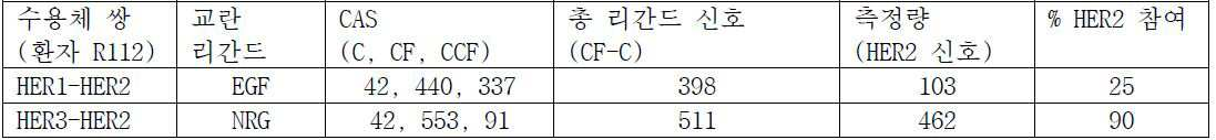
주석: C = 배지 단독 부가 대조군 샘플, CF = 교란 인자 단독 부가 샘플, CCF = 확인 인자 + 교란 인자 부가 샘플. (CF-C)는 교란제가 개시한 신호의 총량이다. 리간드 측정량은 ~ (CF-C) - (CCF-C)이다. % HER2 참여는 ((CF-C)-(CCF-C))/(CF-C))*100에 의해 계산된다. 여기서 CF 및 CCF에 대한 값은 상기 제공된 비-선형 벡터 알고리즘의 합산에 의해 계산된다.Note: C = medium alone control sample, CF = disturbance factor alone additional sample, CCF = confirmation factor + disturbance factor addition sample. (CF-C) is the total amount of the signal initiated by the perturbation agent. The ligand measurand is ~ (CF-C) - (CCF-C). % HER2 participation is calculated by ((CF-C) - (CCF-C)) / (CF-C) * 100. Where the values for CF and CCF are calculated by summing up the non-linear vector algorithms provided above.
이들 결과는 확인 인자 부가 없이 수용체 리간드를 가진 전체 경로의 교란이 상기 비-과발현 HER2 환자 (CF-C)에 대하여 최고 세포 지수를 생성하였음을 나타낸다. 2개의 상이한 수용체 교란제 샘플에 확인 인자의 부가는 HER2-HER3 수용체 연결된 경로 신호전달 또는 HER2-HER4 수용체 연결된 경로 신호전달의 NRG1 교란이 90%이었고 반면에 HER1-HER2 수용체 연결된 경로 신호전달 교란이 상기 환자에 대하여 유의미하게 더 낮은 HER2 수용체 참여를 가졌음을 입증한다. 각 환자는 각각의 2개 작은 변화에서 HER2 참여의 상이한 수준을 갖고, 추가로 각 환자에서 HER2-유도된 경로 신호전달의 모든 측면의 개별화된 시험의 중요성을 입증한다.These results indicate that disturbance of the whole pathway with receptor ligand without confirmation factor produced the highest cell index for the non-overexpressing HER2 patient (CF-C). Addition of the confirmation factor to two different receptor perturbation samples resulted in 90% of the NRG1 disturbance of the HER2-HER3 receptor linked pathway signaling or the HER2-HER4 receptor linked pathway signaling pathway, while the HER1- Demonstrate that they have significantly lower HER2 receptor participation in the patient. Each patient has a different level of HER2 participation in each of the two small changes and further demonstrates the importance of individualized testing of all aspects of HER2-induced pathway signaling in each patient.
도 15a-d는 하나의 과발현된 HER2 세포주 (SKBr3; 도 15a), HER2-유도된 질환 (R39; 도 15b)을 가진 하나의 비-과발현된 HER2 환자, HER2-유도된 질환 (R49; 도 15c)이 없는 하나의 비-과발현된 HER2 환자, 및 하나의 건강한 환자 (R62; 도 15d)의 NRG1 교란 데이터 비교에 대한 막대 그래프 측정량을 나타낸다. 과발현된 HER2 세포주 (도 15a, SKBR3) 및 비-과발현된 HER2 환자 샘플 (도 15b, R39)은 비교할만한 NRG1 유도된 활성을 보여준다. HER2 이량체 차단제 (페르투주맙) 확인제 저해 결과는 측정된 PI3K 활성의 벌크가 이들 환자에 대하여 HER2 수용체를 포함하는 것을 확인한다. 페르투주맙 저해의 양이 비-과발현된 HER2 환자 샘플 (R39)보다 더 크다는 것을 주목한다. HER2-유도된 질환이 없는 비-과발현된 HER2 암 환자 샘플에 대한 경로 신호전달 활성의 NRG1 교란 (도 15c, R49)은 본 실시예에서 시험된 비-과발현된 HER2 환자 샘플의 80%를 대표하고 대표적인 건강한 환자 샘플 (도 15d, R62)에 비교할만하다. 모든 종양 (도 15d; R49) 및 건강한 (도 15d, R62) 샘플에 대하여 측정된 NRG1 활성의 수준이 좌측에서 SKBR3 및 R39 샘플에 대하여 측정된 활성의 25% 미만임을 주목한다.Figures 15a-d show one non-overexpressed HER2 patient with one over-expressing HER2 cell line (SKBr3; Figure 15a), HER2-induced disease (R39; Figure 15b), HER2- ), And one healthy patient (R62; FIG. 15d), without the presence of the non-overexpressed HER2 patient. Overexpressed HER2 cell lines (Figure 15a, SKBR3) and non-overexpressed HER2 patient samples (Figure 15b, R39) show comparable NRG1 induced activity. The inhibitor of HER2 dimeric blocker (pertuzumab) confirms that the bulk of the PI3K activity measured contains the HER2 receptor for these patients. Note that the amount of perturbum inhibition is greater than the non-overexpressed HER2 patient sample (R39). NRG1 disturbance of pathway signaling activity (Fig. 15c, R49) for samples of non-overexpressed HER2 cancer patient without HER2-induced disease represents 80% of the non-overexpressed HER2 patient sample tested in this example It is comparable to a representative healthy patient sample (Fig. 15D, R62). Note that the level of NRG1 activity measured for all tumors (Figure 15d; R49) and healthy (Figure 15d, R62) samples is less than 25% of the activity measured for SKBR3 and R39 samples on the left.
연구는 비-과발현된 HER2 암 환자가 CELx 시험을 이용한 건강한 비-이환 환자로부터 통계적으로 불가분인 귀무 가설을 시험하기 위해 수행되었다. 도 16은 본 실시예에서 시험된 상이한 환자 집단의 사분위를 한정하는 위스커-박스 플롯을 나타낸다. CELx 시험은 HER2/HER3 수용체상에서 경로 신호전달 반응의 NRG1 교란을 측정한다. 세포주 및 원발 샘플 결과는 일치한다. 데이터는 비정상 HER2 경로가 비-과발현 HER2 환자에서 존재하였음을 보여준다. 과발현 HER2 세포주 및 비-과발현 HER2 환자의 최상부 20번째 백분위에서 HER2 경로 신호전달 활성의 NRG1 교란은 더 낮은 CAS를 갖는 건강한 집단으로부터 고유하다.The study was conducted to test the statistically insignificant null hypothesis from healthy non-morbid patients using the CELx test in patients with non-overexpressed HER2 cancer. Figure 16 shows a whisker-box plot that defines quadrants of different patient populations tested in this example. The CELx test measures the NRG1 disturbance of the pathway signaling response on the HER2 / HER3 receptor. The cell line and the original sample results are consistent. The data show that the abnormal HER2 pathway was present in non-overexpressing HER2 patients. NRG1 disturbance of HER2 pathway signaling activity at the top 20th percentile of over-expressing HER2 cell line and non-overexpressing HER2 patients is unique from healthy populations with lower CAS.
비-과발현된 HER2 암 환자의 대략 20%는 과발현 HER2 환자의 최상부 50번째 백분위 (점선 초과)와 일치된 HER2 경로 신호전달 활성의 NRG1 교란을 갖는다. 이들 결과는 귀무 가설이 건강한 및 비-과발현된 HER2 집단 사이에서 차이로 거부되고 과발현된 HER2 그룹 활성과 비-과발현된 HER2 그룹 신호전달 경로 활성 사이에서 중첩을 확인하는 것을 보여준다.Approximately 20% of non-overexpressed HER2 cancer patients have NRG1 disturbance of HER2 pathway signaling activity consistent with the top 50th percentile (above the dotted line) of over-expressing HER2 patients. These results show that the null hypothesis is rejected as a difference between healthy and non-overexpressed HER2 populations and confirms overlap between overexpressed HER2 group activity and non-overexpressed HER2 group signaling pathway activity.
도 17은 HER2/HER1 수용체 이량체에 의해 CELx EGF 교란된 경로 신호전달 활성용 데이터를 나타낸다. HER2/HER1이 HER2-유도된 질환에 참여한다고 공지되기 때문에 제공된다. 데이터는 EGF 교란된 경로 신호전달 활성이 과발현된 HER2 및 비-과발현된 HER2 샘플에서 유사하다는 것을 보여준다. 데이터는 NRG1 교란된 경로 신호전달 활성에서 발견된 바와 같이 최상부 사분위 비-과발현된 HER2 및 과발현된 HER2 샘플의 CELx EGF 교란된 경로 신호전달 활성에서 유사한 상승을 보여준다. 데이터는 또한 비-과발현된 HER2 CELx EGF 교란된 경로 신호전달 활성이 건강한 집단에 비교된 경우 상승되는 것을 보여준다.Figure 17 shows data for CELx EGF perturbed pathway signaling activity by HER2 / HER1 receptor dimer. Is provided because HER2 / HER1 is known to participate in HER2-induced disease. The data show that EGF disturbed pathway signaling activity is similar in overexpressed HER2 and non-overexpressed HER2 samples. The data show a similar increase in the CELx EGF disturbed pathway signaling activity of the top quartile non-overexpressed HER2 and overexpressed HER2 samples as found in NRG1 disturbed pathway signaling activity. The data also show that the non-overexpressed HER2 CELx EGF disturbed pathway signaling activity is elevated when compared to healthy populations.
아래 표 17은 HER2-유도된 질환을 가진 비-과발현 HER2 암 환자에 대하여 대표적인 NRG1 확인제 데이터를 나타내고, 반면에 확인제는 CELx 시험에 따라 HER2-유도된 질환을 갖도록 계측된 5명 환자에 대하여 치료적 약물이다.Table 17 below shows representative NRG1 confirmatory data for non-overexpressing HER2 cancer patients with HER2-induced disease, whereas the five drugs for which HER2-induced disease was determined to have a HER2-induced disease according to the CELx test It is a drug.
표 17Table 17

*NRG1 신호의 감쇠%* Attenuation of NRG1 signal%
표 17에서 데이터는 HER2 경로 신호전달 치료적 억제제 반응을 보여준다. 각 억제제는 각 환자에 대하여 약물 반응을 입증하기 위해 임상적으로 관련된 단일 농도에서 사용되었다. 공유 수용체 티로신 키나제 억제제 약물 (RTKi) 네라티닙 및 아파티닙은 이들 환자에 대하여 NRG1 및 EGF 유도된 활성 (단지 NRG1 데이터 보여짐)의 최고 백분율 감쇠를 생산하였다. 비-공유 RTKi, 라파티닙, 및 단클론성 항체 (페르투주맙)은 경로 신호전달의 그러나 공유 RTKi 미만의 유의미한 감쇠를 생산하였다. 예상외로, HER2 약물은 과발현 HER2 세포주보다 비-과발현 HER2 환자에서 HER2-유도된 경로 신호전달 활성의 더 높은 백분율을 억제시킨다.The data in Table 17 show the HER2 pathway signaling therapeutic inhibitor response. Each inhibitor was used at a single clinically relevant concentration to demonstrate drug response for each patient. Shared Receptor Tyrosine Kinase Inhibitor Drugs (RTKi) Neratinib and apatinate have produced the highest percent attenuation of NRG1 and EGF induced activity (only NRG1 data shown) for these patients. Non-shared RTKi, lapatinib, and monoclonal antibodies (pertuzumab) produced pathway signaling but significant attenuation below the shared RTKi. Unexpectedly, HER2 drugs inhibit a higher percentage of HER2-induced pathway signaling activity in non-overexpressing HER2 patients than overexpressing HER2 cell lines.
실시예Example 7에 대한 결과의 논의 Discussion of results for 7
본 실시예에 기재된 결과는 본 발명의 주요 측면 및 본원에서 기재된 방법을 입증하고 예기치 못한 결과를 제공한다. 비-과발현된 HER2 암 세포 샘플은 건강한 유방 세포에서 발견된 것만큼 낮은 HER2 수용체 수준을 갖는 것으로 그리고 유의미하게 과발현된 HER2 암 환자의 HER2 수용체 수준보다 더 낮은 것으로 확인되었다. 따라서, 비-과발현된 HER2 암 세포에서 측정된 경로 신호전달 활성은 대상체의 이들 그룹 사이의 HER2 수용체 수준에서 차이에 기인될 수 없다. 본 실시예는 또한 방법이 특이적 HER2/HER3 및 HER2/HER1 경로 활성을 단리 및 정량화할 수 있다는 것을 확인하였다. 추가로, 실시예는 비정상 HER2 신호전달 활성이 비-과발현 HER2 암 환자에 존재하고 비-과발현 HER2 암 환자의 하위-그룹 (15%)이 과발현된 HER2 암 환자에 유사한 HER2 경로 신호전달 활성을 갖는다는 가설을 확인하였다. 이 예기치 못한 결과는 이들 방법의 가치를 입증한다. 또 다른 예기치 못한 결과는 과발현된 HER2 암 환자보다 비-과발현된 HER2 암 환자에서 HER2 신호전달 경로 활성의 더 높은 백분율을 억제시키기 위한 HER2 표적된 약물의 능력이었다. 집합적으로, 본 실시예에 기재된 결과는 방법이 활성 HER2-신호전달 네트워크를 갖는 비-과발현된 HER2 환자를 확인, 진단 및 치료할 수 있다는 것을 확인한다. 전체 비-과발현된 HER2 유방암 환자의 대략 15%가 미진단된 및 미치료된 HER2-유도된 유방암을 갖는다는 것을 실시예가 시사하기 때문에 이는 유의미한 결과이다. 본 발명은 상기 환자 집단의 확인을 허용하고 이로써 이들은 ErbB 신호전달 경로를 표적하는 치료제를 이용한 치료에 선택될 수 있다.The results described in this Example demonstrate the main aspects of the present invention and the methods described herein and provide unexpected results. Samples of non-overexpressed HER2 cancer cells were found to have HER2 receptor levels as low as those found in healthy breast cells and to be significantly lower than HER2 receptor levels in significantly overexpressed HER2 cancer patients. Thus, pathway signaling activity measured in non-over-expressing HER2 cancer cells can not be attributed to differences in HER2 receptor levels between these groups of subjects. This example also confirms that the method can isolate and quantify specific HER2 / HER3 and HER2 / HER1 pathway activities. Additionally, the example has similar HER2 pathway signaling activity in HER2 cancer patients in which abnormal HER2 signaling activity is present in non-overexpressing HER2 cancer patients and over-expressing subgroup (15%) of non-overexpressing HER2 cancer patients . These unexpected results demonstrate the value of these methods. Another unexpected result was the ability of the HER2 targeted drug to inhibit a higher percentage of HER2 signaling pathway activity in non-overexpressing HER2 cancer patients than in overexpressed HER2 cancer patients. Collectively, the results described in this Example confirm that the method can identify, diagnose and treat non-overexpressed HER2 patients with an active HER2-signaling network. This is a significant result as the example suggests that approximately 15% of total non-overexpressed HER2 breast cancer patients have undiagnosed and untreated HER2-induced breast cancer. The present invention allows for identification of the patient population so that they can be selected for treatment with a therapeutic agent targeting the ErbB signaling pathway.
실시예 8Example 8
신호전달 경로 활성의 측정에 관한 신선한 기본 배지의 효과Effect of fresh basal medium on measurement of signaling pathway activity
많은 세포 선별 검정은 세포가 이들이 시험되는 단시간 동안 및 그 이전에 배치되는 간소화한 배지-버퍼를 처방한다. 생존 원발 환자 세포를 이용한 본 발명은 생존 원발 세포 샘플을 유지하기 위해 사용된 기본 배지로 수행되어 이로써 세포 집단은 생존하고 연속적 측정의 완벽한 시험 시간 동안 세포 사이클의 상이한 시기를 나타내는 세포의 정상 분포 및 개별 세포 유형의 그의 이종성을 보유한다. 기본 배지는 당해 신호전달 경로를 교란시킨다고 공지된 시약을 함유하지 않는다. 방법의 추가 측면은 교란제 또는 확인제와 접촉 개시의 72 시간 이내에 신선한 기본 배지로 기본 배지의 대체를 포함한다.Many cell sorting assays prescribe a simplified medium-buffer in which cells are placed for a short period of time and before they are tested. The present invention using primary surviving patient cells is carried out with the basal medium used to maintain the surviving primary cell samples so that the population of cells is in a normal distribution of cells showing different times of the cell cycle during the complete test time of the continuous measurement, It retains its heterogeneity of cell type. The basal medium does not contain reagents known to disturb the signal transduction pathway. A further aspect of the method involves replacing the basal medium with fresh basal medium within 72 hours of initiating contact with perturbants or agents.
도 18은 세포 샘플의 MAPK 신호전달 경로 활성에 관해 비-신선한 기본 배지 이용의 효과를 입증하는 데이터를 나타낸다. 세포 샘플 C54가 비-신선한 기본 배지에서 교란에 앞서 배양된 이후 EGF로 교란된 경우, CAS는 C54가 EGF로 교란되지 않은 경우 미만이다. 사실상, 신호전달 경로 활성은 측정되지 않는다. 이는 세포 샘플 C54가 EGF와 접촉에 앞서 신선한 기본 배지에서 배양된 경우 측정된 CAS에 유의미한 대조이다. 신선한 기본 배지 예에서, 측정된 CAS는 C54 샘플에서 유의미한 MAPK 신호전달 경로 활성이 있음을 시사한다. 데이터는 세포 샘플이 교란제 또는 확인제와 접촉되기 전에 신선한 기본 배지에서 세포 배양의 중요성을 강조한다.Figure 18 shows data demonstrating the effect of using a non-fresh basal medium on the MAPK signaling pathway activity of a cell sample. When cell sample C54 is disturbed by EGF after being cultured prior to disturbance in non-fresh basal medium, CAS is less than when C54 is not disturbed by EGF. In fact, signaling pathway activity is not measured. This is a significant contrast to the CAS measured when cell sample C54 was cultured in fresh basal medium prior to contact with EGF. In the fresh basal medium example, the measured CAS suggests a significant MAPK signaling pathway activity in the C54 sample. The data emphasizes the importance of cell culture in fresh basal media before cell samples are contacted with perturbants or agents.
Claims (28)
ErbB 신호전달 경로에 영향을 미치는 표적화 치료제를 상기 대상체에 투여하는 단계를 포함하고,
상기 대상체가 하기 단계를 포함하는 방법을 이용한 치료에 대해 선택되며:
상기 대상체로부터 수득된 생존 원발 암 세포의 샘플을 제조하고, 기본 배지에서 상기 샘플을 배양하는 단계;
(1) 교란제와, 상기 대상체로부터 수득된 생존 원발 암 세포의 샘플의 제1 부분, 및 (2) 교란제 및 확인제와, 상기 샘플의 제2 부분의 접촉 단계 (여기서 상기 교란제는 ErbB 신호전달 경로에 선택적으로 영향을 미치고, 상기 확인제는 동일한 ErbB 신호전달 경로상에서 상기 교란제의 효과를 선택적으로 억제시킨다);
상기 샘플의 각 부분에서 상기 생존 원발 암 세포의 생리적 반응 파라미터의 연속적 측정 단계;
상기 샘플의 상기 제1 부분과 상기 샘플의 상기 제2 부분 간 값 차이를 확인하기 위해 상기 생리적 반응 파라미터의 연속적 측정의 수학적 분석에 의하여 측정량을 계측하는 단계; 및
ErbB 신호전달 경로 활성의 정상 참조 구간으로부터 유도된 컷-오프 값과 상기 측정량의 비교 단계;
대상체는 상기 측정량이 상기 컷-오프 값 초과인 경우 ErbB 신호전달 경로에 영향을 미치는 표적화 치료제를 이용한 치료에 대해 선택되는, 방법.CLAIMS What is claimed is: 1. A method for treating a human subject diagnosed as having a cancer with a non-overexpressed, non-amplified ErbB2 receptor,
Administering to said subject a targeted therapeutic agent that affects an ErbB signaling pathway,
Wherein the subject is selected for treatment using a method comprising the steps of:
Preparing a sample of the surviving cancer cells obtained from the subject, and culturing the sample in a basal medium;
(1) contacting a first portion of a sample of surviving primary cancer cells obtained from said subject with an agonist, and (2) an agonist and a reagent, and a second portion of said sample, wherein said agonist is an ErbB signal Selectively inhibits the effect of the perturbant on the same ErbB signaling pathway);
Continuously measuring physiological response parameters of the surviving primary cancer cells in each part of the sample;
Measuring a measured quantity by a mathematical analysis of successive measurements of the physiological response parameter to identify a value difference between the first portion of the sample and the second portion of the sample; And
Comparing the measured quantity with a cut-off value derived from a normal reference section of ErbB signaling pathway activity;
Wherein the subject is selected for treatment with a targeting agent that affects the ErbB signaling pathway when the measured amount is above the cut-off value.
(1) 뉴레굴린과, 상기 대상체로부터 수득된 생존 암 세포의 상기 샘플의 제1 부분, 및 (2) 뉴레굴린 및 확인제와, 상기 샘플의 제2 부분을 접촉시키고/시키거나 (3) 상피 성장 인자와 상기 샘플의 제3 부분 및 (4) 상피 성장 인자 및 확인제와 상기 샘플의 제4 부분을 접촉시키는 단계; 및
상기 샘플의 상기 제1 부분과 상기 샘플의 상기 제2 부분 간 값의 차이 및/또는 상기 샘플의 상기 제3 부분과 상기 샘플의 상기 제4 부분 간 값의 차이를 확인하기 위해 상기 생리적 반응 파라미터의 연속적 측정의 수학적 분석에 의하여 측정량을 계측하는 단계를 포함하는, 방법.8. The method of claim 7,
(1) contacting and / or contacting a second portion of the sample with (2) a second portion of the sample of surviving cancer cells obtained from the subject, and (2) Contacting a third portion of the sample with the factor; and (4) contacting the fourth portion of the sample with an epithelial growth factor and a determinant; And
Determining a difference between a value of the first portion of the sample and the second portion of the sample and / or a difference between the third portion of the sample and the fourth portion of the sample, And measuring the measured quantity by a mathematical analysis of the continuous measurement.
ErbB 신호전달 경로에 영향을 미치는 라파티닙을 상기 대상체에 투여하는 단계를 포함하고,
상기 대상체가 하기 단계를 포함하는 방법을 이용한 치료에 대해 선택되며:
상기 대상체로부터 수득된 생존 원발 유방암 세포의 샘플을 제조하고, 기본 배지에서 상기 샘플을 배양하는 단계;
상기 샘플이 교란제와 접촉되기 최소 12 시간 내지 최대 72 시간 이전에 신선한 기본 배지와 상기 샘플을 배양시키기 위해 사용된 상기 기본 배지의 대체 단계;
(1) 교란제로서의 뉴레굴린과 상기 대상체로부터 수득된 생존 원발 유방암 세포의 상기 샘플의 제1 부분, 및 (2) 뉴레굴린 및 확인제와 상기 샘플의 제2 부분을 접촉시키고/시키거나(여기서 상기 확인제는 뉴레굴린과 동일한 ErbB 신호전달 경로를 선택적으로 억제시킨다) (3) 교란제로서의 상피 성장 인자와 상기 샘플의 제3 부분 및 (4) 상피 성장 인자 및 확인제와 상기 샘플의 제4 부분을 접촉시키는 단계(여기서 상기 확인제는 상피 성장 인자와 동일한 ErbB 신호전달 경로를 선택적으로 억제시킨다);
상기 교란제와 접촉된 상기 생존 원발 유방암 세포의 세포 접착 또는 부착의 연속적 측정 단계;
상기 샘플의 상기 제1 부분과 상기 샘플의 상기 제2 부분 간 값의 차이 및/또는 상기 샘플의 상기 제3 부분과 상기 샘플의 상기 제4 부분 간 값의 차이를 확인하기 위해 상기 생리적 반응 파라미터의 연속적 측정의 수학적 분석에 의하여 측정량을 계측하는 단계; 및
ErbB 경로 활성에 대하여 정상 참조 구간으로부터 유도된 컷-오프 값과 측정량의 비교 단계;
대상체는 상기 측정량이 상기 컷-오프 값 초과인 경우 라파티닙을 이용한 치료에 대해 선택되는, 방법.A method of treating a human subject diagnosed as having a non-overexpressed, non-amplified ErbB2 breast cancer,
Administering to said subject lapatinib that affects the ErbB signaling pathway,
Wherein the subject is selected for treatment using a method comprising the steps of:
Preparing a sample of survival breast cancer cells obtained from the subject, and culturing the sample in a basal medium;
A replacement of the fresh basal medium and the basal medium used for culturing the sample for at least 12 hours up to 72 hours before the sample is contacted with the perturbant;
(1) contacting a first portion of the sample of the survival breast cancer cells obtained from the subject with (2) a second portion of the sample with (2) a second portion of the sample, and (3) an epithelial growth factor as an agonist and a third portion of the sample; and (4) an epithelial growth factor and a determinant as a disturbing agent in contact with the fourth portion of the sample Wherein the identifying agent selectively inhibits the same ErbB signaling pathway as the epithelial growth factor;
Continuously measuring cell adhesion or adhesion of the surviving primary breast cancer cells in contact with the perturbing agent;
Determining a difference between a value of the first portion of the sample and the second portion of the sample and / or a difference between the third portion of the sample and the fourth portion of the sample, Measuring a measured quantity by a mathematical analysis of the continuous measurement; And
Comparing the measured amount with a cut-off value derived from a normal reference section for ErbB path activity;
Wherein the subject is selected for treatment with lapatinib if the measured amount is above the cut-off value.
에스트로겐-관련된 신호전달 경로에 영향을 미치는 표적화된 치료제를 상기 대상체에 투여하는 단계를 포함하고,
상기 대상체가 하기 단계를 포함하는 방법을 이용한 치료에 대해 선택되며:
상기 대상체로부터 수득된 생존 원발 유방암 세포의 샘플을 제조하고, 기본 배지에서 상기 샘플을 배양하는 단계;
(1) 에스트로겐-관련 신호전달 경로를 상향조절 또는 하향조절한다고 공지된 교란제와, 상기 대상체로부터 수득된 생존 원발 유방암 세포의 샘플의 제1 부분, 및 (2) 상기 교란제 및 동일한 에스트로겐-관련 신호전달 경로 활성을 억제시킨다고 공지된 확인제와, 상기 샘플의 제2 부분을 접촉시키는 단계;
상기 샘플의 각 부분에서 상기 생존 원발 암 세포의 생리적 반응 파라미터의 연속적 측정 단계;
상기 샘플의 상기 제1 부분과 상기 샘플의 상기 제2 부분 간 값 차이를 확인하기 위해 상기 생리적 반응 파라미터의 연속적 측정의 수학적 분석에 의하여 측정량을 계측하는 단계; 및
에스트로겐 수용체 경로 활성에 대하여 정상 참조 구간으로부터 유도된 컷-오프 값에 상기 측정량을 비교하는 단계;
상기 측정량이 상기 컷-오프 값 초과인 경우, 대상체는 에스트로겐 관련된 신호전달 경로에 영향을 미치는 표적화 치료제를 이용한 치료에 대해 선택되는, 방법.A method of treating a human subject diagnosed as having a non-overexpressed, estrogen receptor breast cancer,
Administering to the subject a targeted therapeutic agent that affects an estrogen-related signaling pathway,
Wherein the subject is selected for treatment using a method comprising the steps of:
Preparing a sample of survival breast cancer cells obtained from the subject, and culturing the sample in a basal medium;
(1) a disturbing agent known to up-regulate or down-regulate the estrogen-related signal transduction pathway, a first portion of a sample of surviving primary breast cancer cells obtained from the subject, and (2) a first portion of the disturbing agent and the same estrogen- Contacting a second portion of the sample with a known agent that inhibits signal transduction pathway activity;
Continuously measuring physiological response parameters of the surviving primary cancer cells in each part of the sample;
Measuring a measured quantity by a mathematical analysis of successive measurements of the physiological response parameter to identify a value difference between the first portion of the sample and the second portion of the sample; And
Comparing the measured amount to a cut-off value derived from a normal reference interval for estrogen receptor pathway activity;
Wherein the subject is selected for treatment with a targeting agent that affects the estrogen-related signaling pathway when the amount of the measurement is above the cut-off value.
Applications Claiming Priority (3)
| Application Number | Priority Date | Filing Date | Title |
|---|---|---|---|
| US201462091180P | 2014-12-12 | 2014-12-12 | |
| US62/091,180 | 2014-12-12 | ||
| PCT/US2015/065584 WO2016094904A1 (en) | 2014-12-12 | 2015-12-14 | Methods of measuring erbb signaling pathway activity to diagnose and treat cancer patients |
Publications (2)
| Publication Number | Publication Date |
|---|---|
| KR20170094792A true KR20170094792A (en) | 2017-08-21 |
| KR102439094B1 KR102439094B1 (en) | 2022-08-31 |
Family
ID=55083491
Family Applications (1)
| Application Number | Title | Priority Date | Filing Date |
|---|---|---|---|
| KR1020177018267A Active KR102439094B1 (en) | 2014-12-12 | 2015-12-14 | Methods for measuring signaling pathway activity for diagnosing and treating a patient |
Country Status (9)
| Country | Link |
|---|---|
| US (1) | US20170343554A1 (en) |
| EP (2) | EP3699589A1 (en) |
| JP (1) | JP6782698B2 (en) |
| KR (1) | KR102439094B1 (en) |
| CN (1) | CN107003300B (en) |
| AU (1) | AU2015360260B2 (en) |
| CA (1) | CA2969471C (en) |
| DK (1) | DK3230736T3 (en) |
| WO (1) | WO2016094904A1 (en) |
Families Citing this family (27)
| Publication number | Priority date | Publication date | Assignee | Title |
|---|---|---|---|---|
| WO2007079139A2 (en) | 2005-12-28 | 2007-07-12 | Vertex Pharmaceuticals, Inc. | Solid forms of n-[2,4-bis(1,1-dimethylethyl)-5-hydroxyphenyl]-1,4-dihydro-4-oxoquinoline-3-carboxamide |
| US20130330761A1 (en) | 2012-06-12 | 2013-12-12 | Celcuity, LLC | Whole cell assays and methods |
| WO2015089380A2 (en) | 2013-12-12 | 2015-06-18 | Celcuity Llc | Assays and methods for determining the responsiveness of an individual subject to a therapeutic agent |
| EP3274476B1 (en) | 2015-03-25 | 2023-06-07 | The General Hospital Corporation | Digital analysis of circulating tumor cells in blood samples |
| TWI808055B (en) | 2016-05-11 | 2023-07-11 | 美商滬亞生物國際有限公司 | Combination therapies of hdac inhibitors and pd-1 inhibitors |
| TWI794171B (en) | 2016-05-11 | 2023-03-01 | 美商滬亞生物國際有限公司 | Combination therapies of hdac inhibitors and pd-l1 inhibitors |
| EP3985127A1 (en) * | 2016-10-27 | 2022-04-20 | The General Hospital Corporation | Digital analysis of blood samples to determine efficacy of cancer therapies for specific cancers |
| WO2018175251A1 (en) | 2017-03-20 | 2018-09-27 | Celcuity Llc | Methods of measuring signaling pathway activity for selection of therapeutic agents |
| KR20180117529A (en) * | 2017-04-19 | 2018-10-29 | 주식회사 프로티나 | Method for predicting drug responsiveness by protein-protein interaction analysis |
| WO2019055493A1 (en) * | 2017-09-12 | 2019-03-21 | Board of Supervisors for the University of Louisiana System | Methods and chemicals for treatment of mek related pathologies |
| AU2019243738B2 (en) * | 2018-03-27 | 2024-05-30 | Board Of Regents, The University Of Texas System | Compounds with anti-tumor activity against cancer cells bearing HER2 exon 19 mutations |
| CN110680919A (en) * | 2018-07-06 | 2020-01-14 | 江苏恒瑞医药股份有限公司 | Application of CDK4/6 inhibitor in preparation of medicine for treating tumors in combination with immunotherapy |
| US12331320B2 (en) | 2018-10-10 | 2025-06-17 | The Research Foundation For The State University Of New York | Genome edited cancer cell vaccines |
| US20210393621A1 (en) | 2018-10-26 | 2021-12-23 | The Research Foundation For The State University Of New York | Combination serotonin specific reuptake inhibitor and serotonin 1a receptor partial agonist for reducing l-dopa-induced dyskinesia |
| CN109652376B (en) * | 2019-01-08 | 2021-10-15 | 创芯国际生物科技(广州)有限公司 | Culture medium for 3D culture of ovarian cancer tissues |
| JP7510998B2 (en) * | 2019-08-05 | 2024-07-04 | 北京基石生命科技有限公司 | Method and kit for culturing primary cells from solid lung cancer tumors and primary tumor cells from lung cancer pleural effusion |
| CN112840402B (en) * | 2019-09-02 | 2024-05-07 | 北京哲源科技有限责任公司 | Method and electronic device for obtaining deterministic events in cells |
| WO2021050760A1 (en) * | 2019-09-11 | 2021-03-18 | Recursion Pharmaceuticals, Inc. | Systems and methods for pairwise inference of drug-gene interaction networks |
| EP4073516A1 (en) * | 2019-12-09 | 2022-10-19 | Celcuity Inc. | Methods of treating cancer patients with ras node or rtk targeted therapeutic agents |
| CN111443068B (en) * | 2020-03-06 | 2023-06-27 | 天津大学 | Pure organic room-temperature phosphorescent materials with multiple stimuli-responsive properties and screening methods and applications |
| CN111588359B (en) * | 2020-04-09 | 2023-07-25 | 北京雪扬科技有限公司 | New coronaries pneumonia early warning system |
| CN112270952B (en) * | 2020-10-30 | 2022-04-05 | 广西师范大学 | A method to identify cancer driver pathways |
| CN115341028A (en) * | 2021-05-14 | 2022-11-15 | 上海思路迪医学检验所有限公司 | SMARCA1 as biomarker for sensitivity of malignant biliary tract tumor ErbB inhibitor and application thereof |
| EP4568563A1 (en) * | 2022-08-10 | 2025-06-18 | University of Cincinnati | Biosensor system for drug efficacy monitoring |
| CN116042836B (en) * | 2023-02-23 | 2025-05-23 | 南通大学附属医院 | Electrochemical biosensor for detecting ctDNA EGFR L858R and preparation method thereof |
| CN116286998A (en) * | 2023-05-16 | 2023-06-23 | 北京百普赛斯生物科技股份有限公司 | Cell model for screening EGFR inhibitor and construction method and application thereof |
| CN116735683B (en) * | 2023-06-08 | 2025-08-19 | 河南中医药大学 | Electrochemical detection kit for detecting ER alpha with high sensitivity based on competitive action of ER alpha and GO on aptamer and application thereof |
Citations (1)
| Publication number | Priority date | Publication date | Assignee | Title |
|---|---|---|---|---|
| WO2010052225A1 (en) * | 2008-11-04 | 2010-05-14 | F. Hoffmann-La Roche Ag | Modulators for her2 signaling in normal her2 expressing settings |
Family Cites Families (13)
| Publication number | Priority date | Publication date | Assignee | Title |
|---|---|---|---|---|
| US4816567A (en) | 1983-04-08 | 1989-03-28 | Genentech, Inc. | Recombinant immunoglobin preparations |
| US6041642A (en) | 1998-06-04 | 2000-03-28 | Lockheed Martin Energy Systems, Inc. | Method and apparatus for sensing the natural frequency of a cantilevered body |
| CN1836051A (en) * | 2003-04-01 | 2006-09-20 | 莫诺格兰姆生物科技公司 | ErbB surface receptor complexes as biomarkers |
| CN101155932A (en) * | 2005-04-14 | 2008-04-02 | 默克专利有限公司 | anti-EGFR antibody treatment based on increased EGFR gene copy number in tumor tissue |
| AU2009219437B2 (en) * | 2008-02-25 | 2014-10-16 | Nestec S.A. | Drug selection for breast cancer therapy using antibody-based arrays |
| WO2010085845A1 (en) * | 2009-01-28 | 2010-08-05 | The University Of Queensland | Cancer therapy and/or diagnosis |
| NZ596468A (en) * | 2009-05-14 | 2013-11-29 | Nestec Sa | Biomarkers for determining sensitivity of breast cancer cells to her2-targeted therapy |
| UY33740A (en) * | 2010-11-18 | 2012-05-31 | Takeda Pharmaceutical | METHOD FOR TREATING BREAST CANCER AND OVARIAN CANCER |
| EP2751562B1 (en) * | 2011-09-02 | 2015-09-16 | Nestec S.A. | Profiling of signal pathway proteins to determine therapeutic efficacy |
| WO2013086031A1 (en) * | 2011-12-05 | 2013-06-13 | Nestec S.A. | Method of therapy selection for patients with cancer |
| US20130330761A1 (en) * | 2012-06-12 | 2013-12-12 | Celcuity, LLC | Whole cell assays and methods |
| KR102291355B1 (en) * | 2012-11-30 | 2021-08-19 | 에프. 호프만-라 로슈 아게 | Identification of patients in need of pd-l1 inhibitor cotherapy |
| WO2015089380A2 (en) * | 2013-12-12 | 2015-06-18 | Celcuity Llc | Assays and methods for determining the responsiveness of an individual subject to a therapeutic agent |
-
2015
- 2015-12-14 CN CN201580067127.8A patent/CN107003300B/en active Active
- 2015-12-14 WO PCT/US2015/065584 patent/WO2016094904A1/en not_active Ceased
- 2015-12-14 EP EP20152262.0A patent/EP3699589A1/en not_active Withdrawn
- 2015-12-14 JP JP2017530685A patent/JP6782698B2/en not_active Expired - Fee Related
- 2015-12-14 AU AU2015360260A patent/AU2015360260B2/en active Active
- 2015-12-14 US US15/533,897 patent/US20170343554A1/en not_active Abandoned
- 2015-12-14 EP EP15823249.6A patent/EP3230736B1/en active Active
- 2015-12-14 CA CA2969471A patent/CA2969471C/en active Active
- 2015-12-14 DK DK15823249.6T patent/DK3230736T3/en active
- 2015-12-14 KR KR1020177018267A patent/KR102439094B1/en active Active
Patent Citations (1)
| Publication number | Priority date | Publication date | Assignee | Title |
|---|---|---|---|---|
| WO2010052225A1 (en) * | 2008-11-04 | 2010-05-14 | F. Hoffmann-La Roche Ag | Modulators for her2 signaling in normal her2 expressing settings |
Non-Patent Citations (3)
| Title |
|---|
| Gianni et al., Journal of Clinical Oncology, Vol. 28, No. 7, 2010, pp. 1131-1137. * |
| Guerra et al., Journal of Clinical Oncology, Meeting Abstract(2013 ASCO Annual Meeting), Vol. 31, No. 15. * |
| M.Schuler et al., Breast Cancer Research and Treatment, Vol. 134, NO. 3, 2012, pp. 1149-1159. * |
Also Published As
| Publication number | Publication date |
|---|---|
| JP2018505138A (en) | 2018-02-22 |
| CN107003300B (en) | 2020-02-07 |
| CA2969471A1 (en) | 2016-06-16 |
| EP3230736A1 (en) | 2017-10-18 |
| EP3230736B1 (en) | 2020-03-11 |
| US20170343554A1 (en) | 2017-11-30 |
| JP6782698B2 (en) | 2020-11-11 |
| DK3230736T3 (en) | 2020-06-08 |
| KR102439094B1 (en) | 2022-08-31 |
| CN107003300A (en) | 2017-08-01 |
| CA2969471C (en) | 2024-02-20 |
| AU2015360260B2 (en) | 2021-10-28 |
| WO2016094904A1 (en) | 2016-06-16 |
| AU2015360260A1 (en) | 2017-06-01 |
| EP3699589A1 (en) | 2020-08-26 |
| WO2016094904A8 (en) | 2017-06-29 |
Similar Documents
| Publication | Publication Date | Title |
|---|---|---|
| JP7108323B2 (en) | Whole-cell assays and methods | |
| KR102439094B1 (en) | Methods for measuring signaling pathway activity for diagnosing and treating a patient | |
| JP7485391B2 (en) | Methods for measuring signal transduction pathway activity for therapeutic agent selection - Patents.com | |
| US11073509B2 (en) | Assays and methods for determining the responsiveness of an individual subject to a therapeutic agent | |
| HK40014847A (en) | Whole cell assays and methods | |
| HK40050635A (en) | Methods of measuring signaling pathway activity for selection of therapeutic agents | |
| HK1245400B (en) | Methods of measuring erbb signaling pathway activity to diagnose and treat cancer patients |
Legal Events
| Date | Code | Title | Description |
|---|---|---|---|
| PA0105 | International application |
St.27 status event code: A-0-1-A10-A15-nap-PA0105 |
|
| PG1501 | Laying open of application |
St.27 status event code: A-1-1-Q10-Q12-nap-PG1501 |
|
| PN2301 | Change of applicant |
St.27 status event code: A-3-3-R10-R13-asn-PN2301 St.27 status event code: A-3-3-R10-R11-asn-PN2301 |
|
| A201 | Request for examination | ||
| E13-X000 | Pre-grant limitation requested |
St.27 status event code: A-2-3-E10-E13-lim-X000 |
|
| P11-X000 | Amendment of application requested |
St.27 status event code: A-2-2-P10-P11-nap-X000 |
|
| P13-X000 | Application amended |
St.27 status event code: A-2-2-P10-P13-nap-X000 |
|
| PA0201 | Request for examination |
St.27 status event code: A-1-2-D10-D11-exm-PA0201 |
|
| E902 | Notification of reason for refusal | ||
| PE0902 | Notice of grounds for rejection |
St.27 status event code: A-1-2-D10-D21-exm-PE0902 |
|
| T11-X000 | Administrative time limit extension requested |
St.27 status event code: U-3-3-T10-T11-oth-X000 |
|
| E13-X000 | Pre-grant limitation requested |
St.27 status event code: A-2-3-E10-E13-lim-X000 |
|
| P11-X000 | Amendment of application requested |
St.27 status event code: A-2-2-P10-P11-nap-X000 |
|
| P13-X000 | Application amended |
St.27 status event code: A-2-2-P10-P13-nap-X000 |
|
| E701 | Decision to grant or registration of patent right | ||
| PE0701 | Decision of registration |
St.27 status event code: A-1-2-D10-D22-exm-PE0701 |
|
| GRNT | Written decision to grant | ||
| PR0701 | Registration of establishment |
St.27 status event code: A-2-4-F10-F11-exm-PR0701 |
|
| PR1002 | Payment of registration fee |
St.27 status event code: A-2-2-U10-U12-oth-PR1002 Fee payment year number: 1 |
|
| PG1601 | Publication of registration |
St.27 status event code: A-4-4-Q10-Q13-nap-PG1601 |
|
| R18-X000 | Changes to party contact information recorded |
St.27 status event code: A-5-5-R10-R18-oth-X000 |
|
| PR1001 | Payment of annual fee |
St.27 status event code: A-4-4-U10-U11-oth-PR1001 Fee payment year number: 4 |
|
| U11 | Full renewal or maintenance fee paid |
Free format text: ST27 STATUS EVENT CODE: A-4-4-U10-U11-OTH-PR1001 (AS PROVIDED BY THE NATIONAL OFFICE) Year of fee payment: 4 |