KR20200038121A - 실시간으로 획득되는 위 내시경 이미지를 기반으로 위 병변을 진단하는 내시경 장치 및 방법 - Google Patents
실시간으로 획득되는 위 내시경 이미지를 기반으로 위 병변을 진단하는 내시경 장치 및 방법 Download PDFInfo
- Publication number
- KR20200038121A KR20200038121A KR1020180117824A KR20180117824A KR20200038121A KR 20200038121 A KR20200038121 A KR 20200038121A KR 1020180117824 A KR1020180117824 A KR 1020180117824A KR 20180117824 A KR20180117824 A KR 20180117824A KR 20200038121 A KR20200038121 A KR 20200038121A
- Authority
- KR
- South Korea
- Prior art keywords
- unit
- lesion
- gastric
- neural network
- image
- Prior art date
- Legal status (The legal status is an assumption and is not a legal conclusion. Google has not performed a legal analysis and makes no representation as to the accuracy of the status listed.)
- Granted
Links
Images


Classifications
-
- A—HUMAN NECESSITIES
- A61—MEDICAL OR VETERINARY SCIENCE; HYGIENE
- A61B—DIAGNOSIS; SURGERY; IDENTIFICATION
- A61B1/00—Instruments for performing medical examinations of the interior of cavities or tubes of the body by visual or photographical inspection, e.g. endoscopes; Illuminating arrangements therefor
-
- A—HUMAN NECESSITIES
- A61—MEDICAL OR VETERINARY SCIENCE; HYGIENE
- A61B—DIAGNOSIS; SURGERY; IDENTIFICATION
- A61B1/00—Instruments for performing medical examinations of the interior of cavities or tubes of the body by visual or photographical inspection, e.g. endoscopes; Illuminating arrangements therefor
- A61B1/00002—Operational features of endoscopes
- A61B1/00004—Operational features of endoscopes characterised by electronic signal processing
- A61B1/00009—Operational features of endoscopes characterised by electronic signal processing of image signals during a use of endoscope
- A61B1/000094—Operational features of endoscopes characterised by electronic signal processing of image signals during a use of endoscope extracting biological structures
-
- A—HUMAN NECESSITIES
- A61—MEDICAL OR VETERINARY SCIENCE; HYGIENE
- A61B—DIAGNOSIS; SURGERY; IDENTIFICATION
- A61B1/00—Instruments for performing medical examinations of the interior of cavities or tubes of the body by visual or photographical inspection, e.g. endoscopes; Illuminating arrangements therefor
- A61B1/00002—Operational features of endoscopes
- A61B1/00043—Operational features of endoscopes provided with output arrangements
- A61B1/00045—Display arrangement
-
- A—HUMAN NECESSITIES
- A61—MEDICAL OR VETERINARY SCIENCE; HYGIENE
- A61B—DIAGNOSIS; SURGERY; IDENTIFICATION
- A61B1/00—Instruments for performing medical examinations of the interior of cavities or tubes of the body by visual or photographical inspection, e.g. endoscopes; Illuminating arrangements therefor
- A61B1/00064—Constructional details of the endoscope body
- A61B1/00071—Insertion part of the endoscope body
- A61B1/0008—Insertion part of the endoscope body characterised by distal tip features
- A61B1/00101—Insertion part of the endoscope body characterised by distal tip features the distal tip features being detachable
-
- A—HUMAN NECESSITIES
- A61—MEDICAL OR VETERINARY SCIENCE; HYGIENE
- A61B—DIAGNOSIS; SURGERY; IDENTIFICATION
- A61B1/00—Instruments for performing medical examinations of the interior of cavities or tubes of the body by visual or photographical inspection, e.g. endoscopes; Illuminating arrangements therefor
- A61B1/04—Instruments for performing medical examinations of the interior of cavities or tubes of the body by visual or photographical inspection, e.g. endoscopes; Illuminating arrangements therefor combined with photographic or television appliances
-
- A—HUMAN NECESSITIES
- A61—MEDICAL OR VETERINARY SCIENCE; HYGIENE
- A61B—DIAGNOSIS; SURGERY; IDENTIFICATION
- A61B5/00—Measuring for diagnostic purposes; Identification of persons
-
- A—HUMAN NECESSITIES
- A61—MEDICAL OR VETERINARY SCIENCE; HYGIENE
- A61B—DIAGNOSIS; SURGERY; IDENTIFICATION
- A61B5/00—Measuring for diagnostic purposes; Identification of persons
- A61B5/72—Signal processing specially adapted for physiological signals or for diagnostic purposes
- A61B5/7271—Specific aspects of physiological measurement analysis
- A61B5/7275—Determining trends in physiological measurement data; Predicting development of a medical condition based on physiological measurements, e.g. determining a risk factor
-
- G—PHYSICS
- G06—COMPUTING OR CALCULATING; COUNTING
- G06T—IMAGE DATA PROCESSING OR GENERATION, IN GENERAL
- G06T7/00—Image analysis
- G06T7/0002—Inspection of images, e.g. flaw detection
- G06T7/0012—Biomedical image inspection
-
- G—PHYSICS
- G16—INFORMATION AND COMMUNICATION TECHNOLOGY [ICT] SPECIALLY ADAPTED FOR SPECIFIC APPLICATION FIELDS
- G16H—HEALTHCARE INFORMATICS, i.e. INFORMATION AND COMMUNICATION TECHNOLOGY [ICT] SPECIALLY ADAPTED FOR THE HANDLING OR PROCESSING OF MEDICAL OR HEALTHCARE DATA
- G16H30/00—ICT specially adapted for the handling or processing of medical images
- G16H30/40—ICT specially adapted for the handling or processing of medical images for processing medical images, e.g. editing
-
- G—PHYSICS
- G16—INFORMATION AND COMMUNICATION TECHNOLOGY [ICT] SPECIALLY ADAPTED FOR SPECIFIC APPLICATION FIELDS
- G16H—HEALTHCARE INFORMATICS, i.e. INFORMATION AND COMMUNICATION TECHNOLOGY [ICT] SPECIALLY ADAPTED FOR THE HANDLING OR PROCESSING OF MEDICAL OR HEALTHCARE DATA
- G16H50/00—ICT specially adapted for medical diagnosis, medical simulation or medical data mining; ICT specially adapted for detecting, monitoring or modelling epidemics or pandemics
- G16H50/20—ICT specially adapted for medical diagnosis, medical simulation or medical data mining; ICT specially adapted for detecting, monitoring or modelling epidemics or pandemics for computer-aided diagnosis, e.g. based on medical expert systems
-
- A—HUMAN NECESSITIES
- A61—MEDICAL OR VETERINARY SCIENCE; HYGIENE
- A61B—DIAGNOSIS; SURGERY; IDENTIFICATION
- A61B18/00—Surgical instruments, devices or methods for transferring non-mechanical forms of energy to or from the body
- A61B2018/00315—Surgical instruments, devices or methods for transferring non-mechanical forms of energy to or from the body for treatment of particular body parts
- A61B2018/00482—Digestive system
-
- G—PHYSICS
- G06—COMPUTING OR CALCULATING; COUNTING
- G06T—IMAGE DATA PROCESSING OR GENERATION, IN GENERAL
- G06T2207/00—Indexing scheme for image analysis or image enhancement
- G06T2207/20—Special algorithmic details
- G06T2207/20081—Training; Learning
-
- G—PHYSICS
- G06—COMPUTING OR CALCULATING; COUNTING
- G06T—IMAGE DATA PROCESSING OR GENERATION, IN GENERAL
- G06T2207/00—Indexing scheme for image analysis or image enhancement
- G06T2207/30—Subject of image; Context of image processing
- G06T2207/30004—Biomedical image processing
- G06T2207/30092—Stomach; Gastric
Landscapes
- Health & Medical Sciences (AREA)
- Life Sciences & Earth Sciences (AREA)
- Engineering & Computer Science (AREA)
- Medical Informatics (AREA)
- Public Health (AREA)
- Surgery (AREA)
- General Health & Medical Sciences (AREA)
- Biomedical Technology (AREA)
- Pathology (AREA)
- Radiology & Medical Imaging (AREA)
- Nuclear Medicine, Radiotherapy & Molecular Imaging (AREA)
- Physics & Mathematics (AREA)
- Heart & Thoracic Surgery (AREA)
- Molecular Biology (AREA)
- Animal Behavior & Ethology (AREA)
- Biophysics (AREA)
- Veterinary Medicine (AREA)
- Optics & Photonics (AREA)
- Primary Health Care (AREA)
- Epidemiology (AREA)
- Data Mining & Analysis (AREA)
- Databases & Information Systems (AREA)
- Signal Processing (AREA)
- Computer Vision & Pattern Recognition (AREA)
- Artificial Intelligence (AREA)
- Physiology (AREA)
- Psychiatry (AREA)
- Theoretical Computer Science (AREA)
- General Physics & Mathematics (AREA)
- Quality & Reliability (AREA)
- Endoscopes (AREA)
- Image Analysis (AREA)
- Image Processing (AREA)
Abstract
Description
도 2는 본원의 일 실시예에 따른 내시경 장치의 개략적인 블록도이다.
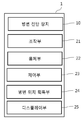
도 3은 본원의 일 실시예에 따른 내시경 장치의 병변 진단부의 개략적인 블록도이다.
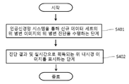
도 4는 본원의 일 실시예에 따른 내시경 장치가 실시간으로 획득하는 위 내시경 이미지를 이용하여 병변을 진단하는 방법에 대한 동작 흐름도이다.
10: 병변 진단부
11: 이미지 획득부 12: 데이터 생성부
13: 데이터 전처리부 14: 학습부
15: 위 병변 진단부
21: 조작부 22: 몸체부
23: 제어부 24: 병변 위치 획득부
25: 디스플레이부
Claims (15)
- 실시간으로 획득되는 위 내시경 이미지를 이용하여 병변을 진단하는 내시경 장치에 있어서,
복수의 유닛 장치를 수용하고, 피검자의 체내로 삽입되는 몸체부;
상기 몸체부의 후단에 마련되어 사용자의 입력 정보에 기반하여 상기 몸체부를 조작하는 조작부;
복수의 위 병변 이미지를 입력으로 하고 위 병변 진단 결과에 관한 항목을 출력으로 하는 학습을 통해 인공신경망 시스템을 구축하고, 실시간으로 획득되는 신규 위 내시경 이미지를 환자 정보와 연계하여 신규 데이터 세트를 생성하고, 구축된 상기 인공신경망 시스템을 통해 위 병변 진단을 수행하는 병변 진단부; 및
상기 병변 진단부의 진단 결과 및 실시간으로 획득되는 신규 위 내시경 이미지를 표시하는 디스플레이부를 포함하는 내시경 장치. - 제1항에 있어서,
상기 조작부에서 제공받은 사용자의 입력 정보 및 상기 병변 진단 장치의 진단 결과에 기반하여 상기 몸체부의 동작을 제어하는 제어 신호를 생성하는 제어부를 더 포함하는 내시경 장치. - 제2항에 있어서,
상기 몸체부는,
상기 몸체부의 전단에 구비되어, 신규 위 병변 이미지를 촬영하고, 촬영한 상기 신규 위 내시경 이미지를 상기 병변 진단부로 제공하는 촬영부를 포함하되,
상기 제어부는,
상기 조작부로부터 상기 촬영부의 동작을 제어하기 위한 사용자의 입력을 수신하고, 상기 촬영부를 제어하기 위한 제어 신호를 생성하는 것인, 내시경 장치. - 제3항에 있어서,
상기 촬영부에서 제공한 상기 신규 위 내시경 이미지를 위치 정보와 연계하여 위 병변 정보를 생성하는 병변 위치 획득부를 더 포함하되,
상기 제어부는 상기 병변 진단 장치의 진단 결과 및 상기 위 병변 정보에 기반하여 대상체의 조직의 일부를 채취하기 위한 생검(biopsy) 유닛의 동작을 제어하기 위한 제어 신호를 생성하는 것인, 내시경 장치. - 제2항에 있어서,
상기 병변 진단부는,
상기 신규 위 병변 이미지를 제공받는 이미지 획득부;
상기 신규 위 병변 이미지와 환자 정보를 연계하여 신규 데이터 세트를 생성하는 데이터 생성부;
딥러닝 알고리즘에 적용 가능하도록 상기 신규 데이터 세트를 전처리하는 데이터 전처리부;
복수의 위 병변 이미지를 입력으로 하고 위 병변 진단 결과에 관한 항목을 출력으로 하는 학습을 통해 인공신경망 시스템을 구축하는 인공신경망 구축부; 및
상기 신규 데이터 세트를 상기 전처리 과정을 거친 후 상기 인공신경망 시스템을 통해 위 병변 진단을 수행하는 위 병변 진단부를 포함하는 것인, 내시경 장치. - 제5항에 있어서,
상기 데이터 생성부는 상기 복수의 위 병변 이미지 각각을 환자 정보와 연계하여 데이터 세트를 생성하되, 상기 데이터 세트는 상기 인공신경망 시스템의 학습에 요구되는 학습용 데이터 세트 및 상기 인공신경망 시스템의 학습의 진행 정도를 검증하기 위한 검증용 데이터 세트로 분류하여 생성되는 것인, 내시경 장치. - 제6항에 있어서,
상기 검증용 데이터 세트는 상기 학습용 데이터 세트와 중복되지 않는 데이터 세트인 것인, 내시경 장치. - 제5항에 있어서,
상기 전처리부는,
상기 신규 데이터 세트에 포함된 위 병변 이미지를 이용하여 상기 위 병변을 중심으로 하여 상기 위 병변이 포함되지 않은 이미지의 주변부 영역을 자르기(crop), 이동(shift), 회전(rotation), 뒤집기(flipping) 및 색상 조정(color adjustment) 중 어느 하나의 전처리 과정을 수행하여 상기 위 병변 이미지를 상기 딥러닝 알고리즘에 적용 가능한 상태로 전처리하는 것인, 내시경 장치. - 제8항에 있어서,
상기 전처리부는,
상기 신규 위 병변 이미지 데이터의 데이터 수를 증가시키기 위한 증폭부를 포함하되, 상기 증폭부는 상기 신규 위 병변 이미지 데이터의 회전, 뒤집기, 자르기 및 소음 섞기를 적용하여 상기 신규 위 병변 이미지 데이터를 증폭하는 것인, 내시경 장치. - 제6항에 있어서,
상기 인공신경망 구축부는,
전처리 과정을 거친 상기 데이터 세트를 입력으로 하고 상기 위 병변 진단 결과에 관한 항목을 출력으로 하는 합성곱신경망(Convolutional Neural Networks) 및 완전연결 심층 신경망 (Fully-connected Neural Networks)의 학습을 통한 훈련 모델을 구축하는 것인, 내시경 장치. - 제10항에 있어서,
전처리 과정을 거친 상기 데이터 세트는 상기 합성곱신경망의 입력으로 하고, 상기 완전연결 심층 신경망은 상기 합성곱신경망의 출력 및 상기 환자 정보를 입력으로 하는 것인, 내시경 장치. - 제11항에 있어서,
상기 합성곱신경망은
상기 복수의 위 병변 이미지로부터 복수의 특징 패턴을 출력하고,
상기 복수의 특징 패턴은 완전연결 심층 신경망에 의해 최종 분류되는 것인, 내시경 장치. - 제5항에 있어서,
상기 위 병변 진단부는,
진행 위암(advanced gastric cancer), 조기 위암(early gastric cancer), 고도 이형성증(high-grade dysplasia), 저이형성증(low-grade dysplasia) 및 비종양(non-neoplasm) 중 적어도 어느 하나로 어느 하나로 상기 위 병변 진단 분류를 수행하는 것인, 내시경 장치. - 피검자의 체내로 삽입되는 몸체부 및 상기 몸체부의 후단에 마련되어 사용자의 입력 정보에 기반하여 상기 몸체부를 조작하는 조작부를 포함하는 내시경 장치가 실시간으로 획득하는 위 내시경 이미지를 이용하여 병변을 진단하는 방법에 있어서,
복수의 위 병변 이미지를 입력으로 하고 위 병변 진단 결과에 관한 항목을 출력으로 하는 학습을 통해 인공신경망 시스템을 구축하고, 실시간으로 획득되는 신규 위 내시경 이미지를 환자 정보와 연계하여 신규 데이터 세트를 생성하고, 구축된 상기 인공신경망 시스템을 통해 위 병변 진단을 수행하는 단계; 및
진단 결과 및 실시간으로 획득되는 신규 위 내시경 이미지를 표시하는 단계를 포함하는, 병변 진단 방법. - 제14항의 방법을 컴퓨터에서 실행시키기 위한 프로그램을 기록한 컴퓨터로 읽을 수 있는 기록매체.
Priority Applications (5)
| Application Number | Priority Date | Filing Date | Title |
|---|---|---|---|
| KR1020180117824A KR102168485B1 (ko) | 2018-10-02 | 2018-10-02 | 실시간으로 획득되는 위 내시경 이미지를 기반으로 위 병변을 진단하는 내시경 장치 및 방법 |
| JP2021516766A JP7218432B2 (ja) | 2018-10-02 | 2019-09-25 | リアルタイムに取得される胃内視鏡イメージに基づいて胃病変を診断する内視鏡装置及び方法 |
| PCT/KR2019/012449 WO2020071678A2 (ko) | 2018-10-02 | 2019-09-25 | 실시간으로 획득되는 위 내시경 이미지를 기반으로 위 병변을 진단하는 내시경 장치 및 방법 |
| CN201980064310.0A CN112823396A (zh) | 2018-10-02 | 2019-09-25 | 基于实时获得的胃内窥镜图像诊断胃病变的内窥镜装置及方法 |
| US17/278,983 US20260108133A1 (en) | 2018-10-02 | 2019-09-25 | Endoscopic device and method for diagnosing gastric lesion based on gastroendoscopic images acquired in real time |
Applications Claiming Priority (1)
| Application Number | Priority Date | Filing Date | Title |
|---|---|---|---|
| KR1020180117824A KR102168485B1 (ko) | 2018-10-02 | 2018-10-02 | 실시간으로 획득되는 위 내시경 이미지를 기반으로 위 병변을 진단하는 내시경 장치 및 방법 |
Publications (2)
| Publication Number | Publication Date |
|---|---|
| KR20200038121A true KR20200038121A (ko) | 2020-04-10 |
| KR102168485B1 KR102168485B1 (ko) | 2020-10-21 |
Family
ID=70055574
Family Applications (1)
| Application Number | Title | Priority Date | Filing Date |
|---|---|---|---|
| KR1020180117824A Active KR102168485B1 (ko) | 2018-10-02 | 2018-10-02 | 실시간으로 획득되는 위 내시경 이미지를 기반으로 위 병변을 진단하는 내시경 장치 및 방법 |
Country Status (5)
| Country | Link |
|---|---|
| US (1) | US20260108133A1 (ko) |
| JP (1) | JP7218432B2 (ko) |
| KR (1) | KR102168485B1 (ko) |
| CN (1) | CN112823396A (ko) |
| WO (1) | WO2020071678A2 (ko) |
Cited By (5)
| Publication number | Priority date | Publication date | Assignee | Title |
|---|---|---|---|---|
| KR20220039307A (ko) * | 2020-09-22 | 2022-03-29 | 한림대학교 산학협력단 | 인공지능을 이용한 비조영 컴퓨터 단층촬영 이미지를 바탕으로 대동맥 박리증을 판단하는 시스템의 제어 방법, 장치 및 프로그램 |
| KR20220043271A (ko) * | 2020-09-29 | 2022-04-05 | 한림대학교 산학협력단 | 인공지능을 이용한 비조영 컴퓨터 단층촬영 이미지를 바탕으로 폐색전증을 판단하는 시스템의 제어 방법, 장치 및 프로그램 |
| KR20220072761A (ko) | 2020-11-25 | 2022-06-02 | 주식회사 아이도트 | 내시경 영상 병변 진단 시스템 |
| KR20230097646A (ko) * | 2021-12-24 | 2023-07-03 | 주식회사 인피니트헬스케어 | 위장 질환 및 위암 검출률 향상을 위한 인공 지능 기반 위 내시경 영상 진단 보조 시스템 및 방법 |
| KR20230163723A (ko) | 2022-05-24 | 2023-12-01 | 주식회사 아이도트 | 내시경 검사 판독 보조 시스템 |
Families Citing this family (7)
| Publication number | Priority date | Publication date | Assignee | Title |
|---|---|---|---|---|
| JP7546270B2 (ja) * | 2020-09-23 | 2024-09-06 | 株式会社Aiメディカルサービス | 検査支援装置、検査支援方法および検査支援プログラム |
| CN112435246A (zh) * | 2020-11-30 | 2021-03-02 | 武汉楚精灵医疗科技有限公司 | 窄带成像放大胃镜下胃癌的人工智能诊断方法 |
| CN113539476A (zh) * | 2021-06-02 | 2021-10-22 | 复旦大学 | 基于人工智能的胃内窥活检拉曼图像辅助诊断方法和系统 |
| CN114171187B (zh) * | 2021-12-06 | 2024-09-10 | 浙江大学 | 一种基于多模态深度学习的胃癌tnm分期预测系统 |
| US12062169B2 (en) | 2022-04-25 | 2024-08-13 | Hong Kong Applied Science and Technology Research Institute Company Limited | Multi-functional computer-aided gastroscopy system optimized with integrated AI solutions and method |
| CN116230208B (zh) * | 2023-02-15 | 2023-09-19 | 北京透彻未来科技有限公司 | 基于深度学习的胃粘膜炎症分型辅助诊断系统 |
| CN118430818B (zh) * | 2024-07-04 | 2024-10-22 | 山东大学齐鲁医院 | 基于人工智能的内镜下胃癌风险分级系统、介质及设备 |
Citations (4)
| Publication number | Priority date | Publication date | Assignee | Title |
|---|---|---|---|---|
| JP2015085152A (ja) * | 2013-09-26 | 2015-05-07 | 富士フイルム株式会社 | 内視鏡システム、内視鏡システムのプロセッサ装置、内視鏡システムの作動方法、プロセッサ装置の作動方法 |
| WO2018020558A1 (ja) * | 2016-07-25 | 2018-02-01 | オリンパス株式会社 | 画像処理装置、画像処理方法およびプログラム |
| KR101857624B1 (ko) * | 2017-08-21 | 2018-05-14 | 동국대학교 산학협력단 | 임상 정보를 반영한 의료 진단 방법 및 이를 이용하는 장치 |
| US20180263568A1 (en) * | 2017-03-09 | 2018-09-20 | The Board Of Trustees Of The Leland Stanford Junior University | Systems and Methods for Clinical Image Classification |
Family Cites Families (23)
| Publication number | Priority date | Publication date | Assignee | Title |
|---|---|---|---|---|
| JPH07250812A (ja) * | 1994-03-15 | 1995-10-03 | Olympus Optical Co Ltd | 蛍光診断装置 |
| JP2008301968A (ja) * | 2007-06-06 | 2008-12-18 | Olympus Medical Systems Corp | 内視鏡画像処理装置 |
| JP5322794B2 (ja) * | 2009-06-16 | 2013-10-23 | 株式会社東芝 | 内視鏡検査支援システム |
| CN101623191B (zh) * | 2009-08-14 | 2011-02-16 | 北京航空航天大学 | 一种胃部组织性质无创检测装置 |
| KR101497662B1 (ko) * | 2013-03-26 | 2015-03-03 | 재단법인대구경북과학기술원 | 진단 보조용 내시경 시스템 및 그 제어 방법 |
| KR101719322B1 (ko) * | 2015-07-20 | 2017-03-23 | 계명대학교 산학협력단 | vSLAM 기법을 이용하여 병변과 주변조직의 3차원 정보를 측정할 수 있는 내시경 기기 및 이를 이용한 병변 측정 방법 |
| JP6959218B2 (ja) * | 2015-08-12 | 2021-11-02 | モレキュラー デバイシーズ, エルエルシー | 細胞の表現型応答を自動的に分析するためのシステムおよび方法 |
| JP6528608B2 (ja) * | 2015-08-28 | 2019-06-12 | カシオ計算機株式会社 | 診断装置、及び診断装置における学習処理方法、並びにプログラム |
| WO2017065170A1 (ja) * | 2015-10-16 | 2017-04-20 | オリンパス株式会社 | 挿入装置 |
| KR20170061222A (ko) * | 2015-11-25 | 2017-06-05 | 한국전자통신연구원 | 건강데이터 패턴의 일반화를 통한 건강수치 예측 방법 및 그 장치 |
| JP6927211B2 (ja) * | 2016-07-04 | 2021-08-25 | 日本電気株式会社 | 画像診断学習装置、画像診断装置、方法およびプログラム |
| US9589374B1 (en) * | 2016-08-01 | 2017-03-07 | 12 Sigma Technologies | Computer-aided diagnosis system for medical images using deep convolutional neural networks |
| JP6737502B2 (ja) * | 2016-09-05 | 2020-08-12 | 独立行政法人国立高等専門学校機構 | 学習用データ生成方法およびこれを用いた対象空間状態認識方法 |
| KR102224510B1 (ko) * | 2016-12-09 | 2021-03-05 | 베이징 호라이즌 인포메이션 테크놀로지 컴퍼니 리미티드 | 데이터 관리를 위한 시스템들 및 방법들 |
| CN107423576A (zh) * | 2017-08-28 | 2017-12-01 | 厦门市厦之医生物科技有限公司 | 一种基于深度神经网络的肺癌识别系统 |
| CN107564580B (zh) * | 2017-09-11 | 2019-02-12 | 合肥工业大学 | 基于集成学习的胃镜图像辅助处理系统及方法 |
| CN107730489A (zh) * | 2017-10-09 | 2018-02-23 | 杭州电子科技大学 | 无线胶囊内窥镜小肠病变计算机辅助检测系统及检测方法 |
| CN107658028A (zh) * | 2017-10-25 | 2018-02-02 | 北京华信佳音医疗科技发展有限责任公司 | 一种获取病变数据的方法、识别病变方法及计算机设备 |
| WO2019088121A1 (ja) * | 2017-10-30 | 2019-05-09 | 公益財団法人がん研究会 | 画像診断支援装置、資料収集方法、画像診断支援方法および画像診断支援プログラム |
| CN107705852A (zh) * | 2017-12-06 | 2018-02-16 | 北京华信佳音医疗科技发展有限责任公司 | 一种医用电子内窥镜的实时病变智能识别方法及装置 |
| CN107967946B (zh) * | 2017-12-21 | 2021-05-11 | 武汉楚精灵医疗科技有限公司 | 基于深度学习的胃镜操作实时辅助系统及方法 |
| CN108272437A (zh) * | 2017-12-27 | 2018-07-13 | 中国科学院西安光学精密机械研究所 | 用于皮肤病诊断的光谱检测系统和分类器模型构建方法 |
| GB201812050D0 (en) * | 2018-07-24 | 2018-09-05 | Dysis Medical Ltd | Computer classification of biological tissue |
-
2018
- 2018-10-02 KR KR1020180117824A patent/KR102168485B1/ko active Active
-
2019
- 2019-09-25 JP JP2021516766A patent/JP7218432B2/ja active Active
- 2019-09-25 CN CN201980064310.0A patent/CN112823396A/zh active Pending
- 2019-09-25 US US17/278,983 patent/US20260108133A1/en active Pending
- 2019-09-25 WO PCT/KR2019/012449 patent/WO2020071678A2/ko not_active Ceased
Patent Citations (4)
| Publication number | Priority date | Publication date | Assignee | Title |
|---|---|---|---|---|
| JP2015085152A (ja) * | 2013-09-26 | 2015-05-07 | 富士フイルム株式会社 | 内視鏡システム、内視鏡システムのプロセッサ装置、内視鏡システムの作動方法、プロセッサ装置の作動方法 |
| WO2018020558A1 (ja) * | 2016-07-25 | 2018-02-01 | オリンパス株式会社 | 画像処理装置、画像処理方法およびプログラム |
| US20180263568A1 (en) * | 2017-03-09 | 2018-09-20 | The Board Of Trustees Of The Leland Stanford Junior University | Systems and Methods for Clinical Image Classification |
| KR101857624B1 (ko) * | 2017-08-21 | 2018-05-14 | 동국대학교 산학협력단 | 임상 정보를 반영한 의료 진단 방법 및 이를 이용하는 장치 |
Non-Patent Citations (2)
| Title |
|---|
| Hussain Z외 3인, "Differential Data Augmentation Techniques for Medical Imaging Classification Tasks". AMIA Annu Symp Proc. 2017. pp979-984, 2018.04.16. 공개 * |
| Toshiaki Hirasawa 외 10인. "Application of AI using a CNN for detecting gastric cancer in endoscopic images". The International Gastric Cancer Association. 2018.01.15. 공개 1부.* * |
Cited By (6)
| Publication number | Priority date | Publication date | Assignee | Title |
|---|---|---|---|---|
| KR20220039307A (ko) * | 2020-09-22 | 2022-03-29 | 한림대학교 산학협력단 | 인공지능을 이용한 비조영 컴퓨터 단층촬영 이미지를 바탕으로 대동맥 박리증을 판단하는 시스템의 제어 방법, 장치 및 프로그램 |
| KR20220043271A (ko) * | 2020-09-29 | 2022-04-05 | 한림대학교 산학협력단 | 인공지능을 이용한 비조영 컴퓨터 단층촬영 이미지를 바탕으로 폐색전증을 판단하는 시스템의 제어 방법, 장치 및 프로그램 |
| KR20220072761A (ko) | 2020-11-25 | 2022-06-02 | 주식회사 아이도트 | 내시경 영상 병변 진단 시스템 |
| WO2022114357A1 (ko) * | 2020-11-25 | 2022-06-02 | 주식회사 아이도트 | 영상 병변 진단 시스템 |
| KR20230097646A (ko) * | 2021-12-24 | 2023-07-03 | 주식회사 인피니트헬스케어 | 위장 질환 및 위암 검출률 향상을 위한 인공 지능 기반 위 내시경 영상 진단 보조 시스템 및 방법 |
| KR20230163723A (ko) | 2022-05-24 | 2023-12-01 | 주식회사 아이도트 | 내시경 검사 판독 보조 시스템 |
Also Published As
| Publication number | Publication date |
|---|---|
| KR102168485B1 (ko) | 2020-10-21 |
| CN112823396A (zh) | 2021-05-18 |
| US20260108133A1 (en) | 2026-04-23 |
| JP7218432B2 (ja) | 2023-02-06 |
| JP2022507002A (ja) | 2022-01-18 |
| WO2020071678A2 (ko) | 2020-04-09 |
| WO2020071678A3 (ko) | 2020-05-28 |
Similar Documents
| Publication | Publication Date | Title |
|---|---|---|
| KR102210806B1 (ko) | 위 내시경 이미지의 딥러닝을 이용하여 위 병변을 진단하는 장치 및 방법 | |
| KR102168485B1 (ko) | 실시간으로 획득되는 위 내시경 이미지를 기반으로 위 병변을 진단하는 내시경 장치 및 방법 | |
| JP6657480B2 (ja) | 画像診断支援装置、画像診断支援装置の作動方法および画像診断支援プログラム | |
| Ohmori et al. | Endoscopic detection and differentiation of esophageal lesions using a deep neural network | |
| JP7037220B2 (ja) | 消化器官の内視鏡画像による疾患の診断支援システム、診断支援システムの作動方法、診断支援プログラム及びこの診断支援プログラムを記憶したコンピュータ読み取り可能な記録媒体 | |
| Shimamoto et al. | Real-time assessment of video images for esophageal squamous cell carcinoma invasion depth using artificial intelligence | |
| Naz et al. | Detection and classification of gastrointestinal diseases using machine learning | |
| US10482313B2 (en) | Method and system for classification of endoscopic images using deep decision networks | |
| KR102505791B1 (ko) | 실시간 영상을 통해 획득되는 병변 판단 시스템의 제어 방법, 장치 및 프로그램 | |
| US20180263568A1 (en) | Systems and Methods for Clinical Image Classification | |
| JPWO2018225448A1 (ja) | 消化器官の内視鏡画像による疾患の診断支援方法、診断支援システム、診断支援プログラム及びこの診断支援プログラムを記憶したコンピュータ読み取り可能な記録媒体 | |
| CN110367913B (zh) | 无线胶囊内窥镜图像幽门和回盲瓣定位方法 | |
| TW202006742A (zh) | 藉由消化器官之內視鏡影像之疾病的診斷支援方法、診斷支援系統、診斷支援程式及記憶此診斷支援程式之電腦可讀取之記錄媒體 | |
| Barbalata et al. | Laryngeal tumor detection and classification in endoscopic video | |
| CN116206741A (zh) | 一种消化科医疗信息处理系统及方法 | |
| Bejakovic et al. | Analysis of Crohn's disease lesions in capsule endoscopy images | |
| KR102095730B1 (ko) | 딥러닝 기반의 대장 병변 검출 방법 | |
| Al-Rahayfeh et al. | Detection of bleeding in wireless capsule endoscopy images using range ratio color | |
| CN112950601A (zh) | 用于食管癌模型训练的图片的筛选方法、系统及存储介质 | |
| Chuquimia et al. | Polyp follow-up in an intelligent wireless capsule endoscopy | |
| Khryashchev et al. | Analysis of pathologies on endoscopic images of the stomach using ssd and retinanet neural network architecture | |
| TWI917264B (zh) | 人工智慧模型協助偵測巴瑞特氏食道黏膜病變之方法 | |
| Vilariño et al. | A machine learning framework using SOMs: Applications in the intestinal motility assessment | |
| Nadimi et al. | Towards full integration of artificial intelligence in colon capsule endoscopy's pathway | |
| Ramakrishnan et al. | Development of Framework to Find Lung Canker Using GAN Technique |
Legal Events
| Date | Code | Title | Description |
|---|---|---|---|
| PA0109 | Patent application |
Patent event code: PA01091R01D Comment text: Patent Application Patent event date: 20181002 |
|
| PA0201 | Request for examination | ||
| AMND | Amendment | ||
| PA0302 | Request for accelerated examination |
Patent event date: 20200110 Patent event code: PA03022R01D Comment text: Request for Accelerated Examination Patent event date: 20181002 Patent event code: PA03021R01I Comment text: Patent Application |
|
| E902 | Notification of reason for refusal | ||
| PE0902 | Notice of grounds for rejection |
Comment text: Notification of reason for refusal Patent event date: 20200217 Patent event code: PE09021S01D |
|
| AMND | Amendment | ||
| PG1501 | Laying open of application | ||
| E601 | Decision to refuse application | ||
| PE0601 | Decision on rejection of patent |
Patent event date: 20200831 Comment text: Decision to Refuse Application Patent event code: PE06012S01D Patent event date: 20200217 Comment text: Notification of reason for refusal Patent event code: PE06011S01I |
|
| X091 | Application refused [patent] | ||
| AMND | Amendment | ||
| PX0901 | Re-examination |
Patent event code: PX09011S01I Patent event date: 20200831 Comment text: Decision to Refuse Application Patent event code: PX09012R01I Patent event date: 20200305 Comment text: Amendment to Specification, etc. Patent event code: PX09012R01I Patent event date: 20190925 Comment text: Amendment to Specification, etc. |
|
| PX0701 | Decision of registration after re-examination |
Patent event date: 20201012 Comment text: Decision to Grant Registration Patent event code: PX07013S01D Patent event date: 20200915 Comment text: Amendment to Specification, etc. Patent event code: PX07012R01I Patent event date: 20200831 Comment text: Decision to Refuse Application Patent event code: PX07011S01I Patent event date: 20200305 Comment text: Amendment to Specification, etc. Patent event code: PX07012R01I Patent event date: 20190925 Comment text: Amendment to Specification, etc. Patent event code: PX07012R01I |
|
| X701 | Decision to grant (after re-examination) | ||
| GRNT | Written decision to grant | ||
| PR0701 | Registration of establishment |
Comment text: Registration of Establishment Patent event date: 20201015 Patent event code: PR07011E01D |
|
| PR1002 | Payment of registration fee |
Payment date: 20201015 End annual number: 3 Start annual number: 1 |
|
| PG1601 | Publication of registration | ||
| PR1001 | Payment of annual fee |