KR20200042228A - System for measuring properties of soft tissue quantitatively - Google Patents
System for measuring properties of soft tissue quantitatively Download PDFInfo
- Publication number
- KR20200042228A KR20200042228A KR1020180122544A KR20180122544A KR20200042228A KR 20200042228 A KR20200042228 A KR 20200042228A KR 1020180122544 A KR1020180122544 A KR 1020180122544A KR 20180122544 A KR20180122544 A KR 20180122544A KR 20200042228 A KR20200042228 A KR 20200042228A
- Authority
- KR
- South Korea
- Prior art keywords
- soft tissue
- properties
- quantitative
- reference material
- property value
- Prior art date
- Legal status (The legal status is an assumption and is not a legal conclusion. Google has not performed a legal analysis and makes no representation as to the accuracy of the status listed.)
- Granted
Links
Images

Classifications
-
- A—HUMAN NECESSITIES
- A61—MEDICAL OR VETERINARY SCIENCE; HYGIENE
- A61B—DIAGNOSIS; SURGERY; IDENTIFICATION
- A61B8/00—Diagnosis using ultrasonic, sonic or infrasonic waves
- A61B8/08—Clinical applications
- A61B8/0858—Clinical applications involving measuring tissue layers, e.g. skin, interfaces
-
- A—HUMAN NECESSITIES
- A61—MEDICAL OR VETERINARY SCIENCE; HYGIENE
- A61B—DIAGNOSIS; SURGERY; IDENTIFICATION
- A61B8/00—Diagnosis using ultrasonic, sonic or infrasonic waves
- A61B8/44—Constructional features of the ultrasonic, sonic or infrasonic diagnostic device
- A61B8/4444—Constructional features of the ultrasonic, sonic or infrasonic diagnostic device related to the probe
-
- A—HUMAN NECESSITIES
- A61—MEDICAL OR VETERINARY SCIENCE; HYGIENE
- A61B—DIAGNOSIS; SURGERY; IDENTIFICATION
- A61B8/00—Diagnosis using ultrasonic, sonic or infrasonic waves
- A61B8/44—Constructional features of the ultrasonic, sonic or infrasonic diagnostic device
- A61B8/4477—Constructional features of the ultrasonic, sonic or infrasonic diagnostic device using several separate ultrasound transducers or probes
-
- A—HUMAN NECESSITIES
- A61—MEDICAL OR VETERINARY SCIENCE; HYGIENE
- A61B—DIAGNOSIS; SURGERY; IDENTIFICATION
- A61B8/00—Diagnosis using ultrasonic, sonic or infrasonic waves
- A61B8/48—Diagnostic techniques
- A61B8/485—Diagnostic techniques involving measuring strain or elastic properties
-
- A—HUMAN NECESSITIES
- A61—MEDICAL OR VETERINARY SCIENCE; HYGIENE
- A61B—DIAGNOSIS; SURGERY; IDENTIFICATION
- A61B8/00—Diagnosis using ultrasonic, sonic or infrasonic waves
- A61B8/54—Control of the diagnostic device
Landscapes
- Health & Medical Sciences (AREA)
- Life Sciences & Earth Sciences (AREA)
- Biomedical Technology (AREA)
- Biophysics (AREA)
- Nuclear Medicine, Radiotherapy & Molecular Imaging (AREA)
- Pathology (AREA)
- Radiology & Medical Imaging (AREA)
- Engineering & Computer Science (AREA)
- Physics & Mathematics (AREA)
- Heart & Thoracic Surgery (AREA)
- Medical Informatics (AREA)
- Molecular Biology (AREA)
- Surgery (AREA)
- Animal Behavior & Ethology (AREA)
- General Health & Medical Sciences (AREA)
- Public Health (AREA)
- Veterinary Medicine (AREA)
- Gynecology & Obstetrics (AREA)
- Ultra Sonic Daignosis Equipment (AREA)
Abstract
본 발명의 실시예에 따른 연조직의 물성을 정량적으로 측정하기 위한 시스템은, 초음파를 생성하고 연조직에 의해 반사된 초음파를 수신하는 초음파 프로브; 본체 및 상기 초음파 프로브와 상기 연조직 사이에 위치하며 미리 알려진 정량적인 물성값을 갖는 기준물질로 구성되는, 상기 초음파 프로브의 전단에 탈착 가능한 커플러; 변형탄성기법을 이용하여 상기 연조직과 상기 기준물질의 물성을 비교 측정하고, 상기 기준물질의 정량적인 물성값에 기초하여 상기 연조직의 정량적인 물성값을 연산하는 처리부; 및 상기 연조직의 정량적인 물성값을 실시간으로 출력하는 출력부를 포함한다. 본 시스템에 의하면, 미리 알려진 정량적인 물성값을 갖는 기준물질과 연조직의 물성을 비교하여, 기준물질의 물성값을 정량적으로 측정하고 실시간으로 나타낼 수 있다. 본 시스템의 구성요소는 기존의 초음파 측정 장비에 쉽게 호환되므로, 고가의 장비를 이용하지 않고도 연조직의 물성을 정량적으로 측정할 수 있어 비용을 절감할 수 있다.A system for quantitatively measuring the properties of soft tissue according to an embodiment of the present invention includes: an ultrasonic probe that generates ultrasonic waves and receives ultrasonic waves reflected by soft tissue; A coupler located between the main body and the ultrasonic probe and the soft tissue and consisting of a reference material having a known quantitative property value, a coupler detachable at a front end of the ultrasonic probe; A processing unit for comparing and measuring the properties of the soft tissue and the reference material using a modified elastic technique, and calculating the quantitative properties of the soft tissue based on the quantitative properties of the reference material; And an output unit which outputs the quantitative physical property value of the soft tissue in real time. According to the present system, it is possible to quantitatively measure a physical property value of a reference material and display it in real time by comparing a physical property of a reference material having a known quantitative property value and a soft tissue. Since the components of this system are easily compatible with existing ultrasonic measuring equipment, it is possible to quantitatively measure the properties of soft tissues without using expensive equipment, thereby reducing costs.
Description
본 발명은 연조직의 물성을 측정하기 위한 시스템에 관한 것으로서, 보다 구체적으로는 초음파 프로브를 이용하여 연조직의 물성을 측정할 때, 초음파 프로브의 전단에 부착된 기준물질의 정량적인 물성값과 비교하여 연조직의 물성값을 정량적으로 연산하고 실시간으로 출력하기 위한 시스템에 관한 것이다.The present invention relates to a system for measuring the properties of soft tissues, and more specifically, when measuring the properties of soft tissues using an ultrasonic probe, the soft tissues are compared with a quantitative physical property value of a reference substance attached to the front end of the ultrasonic probe. It relates to a system for quantitatively calculating a property value and outputting it in real time.
[국가지원 연구개발에 대한 설명][Explanation on national support R & D]
본 연구는 한국과학기술연구원의 주관 하에 산업통상자원부부의 글로벌중견기업육성프로젝트지원 사업(정합정밀도 1mm 이하 수술용 내비게이션 융합 두경부 수술로봇 시스템 개발, 과제고유번호: 1425117773)의 지원에 의하여 이루어진 것이다.This study was conducted by the Ministry of Trade, Industry and Energy under the supervision of the Korea Institute of Science and Technology, and supported by the Ministry of Trade, Industry and Energy's Global Midsize Business Development Project (development of a navigation fused head and neck surgical robot system with a matching accuracy of 1 mm or less, task number: 1425117773).
최근 의료 기술의 발달로 인해, 컴퓨터단층촬영(Computed Tomography) 장치(이하, 'CT' 장치), 자기공명영상(Magnetic Resonance Imaging) 촬영장치(이하, 'MRI 장치'), 형광 투시경 또는 X-ray 촬영을 통해 기 촬영된 환자의 영상을 이용하여 진행하는 영상유도 수술(image-guided surgery)이 많이 활용되고 있다. 이와 같은 수술 방법은, 뇌신경 수술이나 임플란트 수술과 같이 시술자가 직접 환자의 환부를 보기 어렵고, 환자 신체내의 주요 신경과 장기들을 회피하면서 수술을 진행하여야 하는 경우에 이용된다. Due to recent developments in medical technology, computed tomography (hereinafter referred to as 'CT' devices), magnetic resonance imaging (hereinafter referred to as 'MRI devices'), fluoroscopy or X-ray Image-guided surgery, which is performed using images of a patient who has already been photographed through imaging, has been widely used. Such a surgical method is used when it is difficult for the operator to directly see the patient's affected area, such as cranial nerve surgery or implant surgery, and surgery must be performed while avoiding major nerves and organs in the patient's body.
스캐닝 영상을 이용하여 수술을 진행하는 경우, 미리 촬영된 환자의 스캐닝 영상 좌표와 환자의 실제 신체 부위 좌표를 컴퓨터를 이용하여 정확히 매칭시키는 정합(registration) 과정을 거침으로써, 시술자는 마치 환자의 신체를 실시간으로 보는 것과 같이 미리 촬영된 스캐닝 영상을 모니터로 보면서 수술을 진행할 수 있다.When the surgery is performed using a scanned image, the operator undergoes a registration process that accurately matches the coordinates of the patient's actual body part and the scanned image coordinates of the patient, which is previously photographed, so that the operator is as if the patient's body is As seen in real time, surgery can be performed while viewing the pre-imaged scanning image on a monitor.
영상유도 수술 시 발생하는 과대 절개 또는 많은 양의 X-ray 피폭은 환자에게 심각한 부작용을 초래할 수 있는데, 비침습적 또는 적은 양의 절개만으로 수술을 진행하거나, X-ray 피폭량을 감소시키기 위해서는 보다 향상된 영상 정합(image registration) 기술이 요구된다.Excessive incision or a large amount of X-ray exposure that occurs during image-guided surgery can cause serious side effects to the patient. In order to perform surgery with a non-invasive or small amount of incision, or to reduce X-ray exposure, improved imaging Image registration technology is required.
영상 정합 기술의 발전을 위해서는, 신체 기관 및 조직의 역학적 원리를 설명하기 위한 '동적 생체 역학 모델(Dynamic Biomechanics Model)'이 필요한데, CT로 촬영 가능한 인체 경조직(hard tissue)뿐만 아니라 연조직(soft tissue)의 해부학적 형상 및 물성(physical properties) 정보가 필수적으로 요구된다.In order to develop image matching technology, a 'Dynamic Biomechanics Model' is needed to explain the mechanical principles of body organs and tissues, as well as soft tissue as well as hard tissue that can be imaged by CT. The anatomical shape and physical properties of the information are essential.
연조직의 물성 중에서도 경도(hardness), 즉 딱딱한 정도를 측정하는 기술은 의학적인 활용도가 높다. 일반적인 초음파 탐지 장비는 변형탄성기법(Strain elastography)을 이용하여 신체 내부 조직의 상대적인 경도를 측정함으로써 병변을 검출해낼 수 있다. 예를 들어, 측정 부위의 경도를 RGB 색상으로 나타내어 의료 시술자가 환자의 이상 부위를 직관적으로 찾아낼 수 있다.Among the properties of soft tissues, the technology for measuring hardness, that is, the degree of hardness, has high medical utility. Typical ultrasonic detection equipment can detect lesions by measuring the relative hardness of internal tissues of the body using strain elastography. For example, the hardness of the measurement site is represented in RGB color, so that the medical operator can intuitively find the patient's abnormal area.
다만, 종래의 기술은 연조직 부위의 상대적인 경도를 RGB 색상으로 나타낼 수 있을 뿐, 대상 물질의 경도를 수치화하여 나타내거나 영률(Young's modulus) 등의 정량적인 물성 정보를 알 수는 없었으므로 의료 정보를 얻는데 한계가 있었다.However, in the conventional technique, the relative hardness of the soft tissue region can be expressed in RGB color, and the hardness of the target material is numerically displayed or quantitative physical property information such as Young's modulus is not known. There were limits.
최근에는 횡파탄성영상(shear wave elastography) 초음파 영상 기술이 도입되어 연조직의 딱딱한 정도를 정량적으로 수치화할 수 있게 되었으나, 이를 위해서는 고가의 장비가 별도로 필요하고 기존의 초음파 탐지 장비와 호환되지 않아 비경제적이라는 문제점이 있었다.Recently, shear wave elastography ultrasound imaging technology has been introduced to quantitatively quantify the degree of stiffness of soft tissues.However, for this, expensive equipment is required separately, and it is not economical because it is not compatible with existing ultrasonic detection equipment. There was a problem.
이에, 본 발명의 실시예는 일반적인 초음파 탐지 시스템에 있어서, 정량적인 물성값을 갖는 기준물질과의 비교 측정을 통해 연조직의 정량적인 물성값을 실시간으로 연산할 수 있는 물성 측정 시스템을 제공하는 것을 목적으로 한다.Accordingly, an object of the present invention is to provide a physical property measurement system capable of calculating a quantitative physical property value of a soft tissue in real time through comparative measurement with a reference material having a quantitative physical property value in a general ultrasonic detection system. .
본 발명의 과제를 해결하기 위한 일 실시예에 따른 연조직의 물성을 정량적으로 측정하기 위한 시스템은, 초음파를 생성하고 연조직에 의해 반사된 초음파를 수신하는 초음파 프로브; 본체 및 상기 초음파 프로브와 상기 연조직 사이에 위치하며 미리 알려진 정량적인 물성값을 갖는 기준물질로 구성되는, 상기 초음파 프로브의 전단에 탈착 가능한 커플러; 변형탄성기법을 이용하여 상기 연조직과 상기 기준물질의 물성을 비교 측정하고, 상기 기준물질의 정량적인 물성값에 기초하여 상기 연조직의 정량적인 물성값을 연산하는 처리부; 및 상기 연조직의 정량적인 물성값을 실시간으로 출력하는 출력부를 포함한다.A system for quantitatively measuring the properties of soft tissue according to an embodiment for solving the problems of the present invention includes: an ultrasonic probe that generates ultrasonic waves and receives ultrasonic waves reflected by soft tissue; A coupler located between the main body and the ultrasonic probe and the soft tissue and consisting of a reference material having a known quantitative property value, a coupler detachable at a front end of the ultrasonic probe; A processing unit for comparing and measuring the physical properties of the soft tissue and the reference material using a modified elastic technique, and calculating the quantitative physical property values of the soft tissue based on the quantitative physical property values of the reference material; And an output unit which outputs the quantitative physical property value of the soft tissue in real time.
일 실시예에서, 상기 커플러는, 상기 초음파 프로브의 전단에 가해지는 압력을 측정하는 압력센서를 더 포함하고, 상기 처리부는, 상기 압력센서에 의해 측정된 압력에 기초하여 상기 연조직의 정량적인 물성값을 조정함으로써, 불규칙한 압력에 따른 영향을 감소시킬 수 있다.In one embodiment, the coupler, further comprising a pressure sensor for measuring the pressure applied to the front end of the ultrasonic probe, the processing unit, based on the pressure measured by the pressure sensor, the quantitative property value of the soft tissue By adjusting, the influence of irregular pressure can be reduced.
일 실시예에서, 상기 시스템은 상기 초음파 프로브의 동작을 제어하는 제어부를 더 포함하되, 상기 제어부는, 상기 압력센서에 의해 측정된 압력에 기초하여 상기 초음파 프로브가 일정한 압력을 유지하면서 동작하도록 제어할 수 있다.In one embodiment, the system further includes a control unit for controlling the operation of the ultrasonic probe, the control unit, based on the pressure measured by the pressure sensor to control the ultrasonic probe to operate while maintaining a constant pressure You can.
일 실시예에서, 상기 기준물질은, 미리 알려진 물성값을 갖는 제1 물질, 및 상기 제1 물질과 상이한 물성값을 갖는 제2 물질의 적층 구조로 구성될 수 있다.In one embodiment, the reference material may include a stacked structure of a first material having a known physical property value, and a second material having a different physical property value from the first material.
일 실시예에서, 상기 제1 물질은 상기 연조직보다 낮은 경도(硬度)를 갖고, 상기 제2 물질은 상기 제1 물질보다 높은 경도를 가질 수 있다.In one embodiment, the first material may have a lower hardness than the soft tissue, and the second material may have a higher hardness than the first material.
일 실시예에서, 상기 기준물질 및 연조직의 물성값은, 상기 기준물질 및 연조직의 형상 파라미터 또는 영률(Young's modulus) 을 포함할 수 있다.In one embodiment, the physical property values of the reference material and the soft tissue may include shape parameters or Young's modulus of the reference material and the soft tissue.
일 실시예에 따른 연조직의 물성을 정량적으로 측정하기 위한 방법은 프로세서에 의해 수행되며, 연조직에 의해 반사된 초음파를 분석하여 상기 연조직의 물성을 측정하는 단계; 기준물질에 의해 반사된 초음파를 분석하여 상기 기준물질의 물성을 측정하는 단계; 상기 연조직과 상기 기준물질의 물성을 비교하는 단계; 상기 비교결과 및 상기 기준물질의 미리 알려진 정량적인 물성값에 기초하여, 상기 연조직의 정량적인 물성값을 연산하는 단계; 및 상기 연조직의 정량적인 물성값을 출력부에 전송하는 단계를 포함한다.A method for quantitatively measuring the properties of soft tissue according to an embodiment is performed by a processor, and analyzing the ultrasonic waves reflected by the soft tissue to measure the properties of the soft tissue; Measuring the physical properties of the reference material by analyzing ultrasonic waves reflected by the reference material; Comparing the physical properties of the soft tissue and the reference material; Calculating a quantitative property value of the soft tissue based on the comparison result and a known quantitative property value of the reference material; And transmitting a quantitative physical property value of the soft tissue to an output unit.
일 실시예에서, 압력센서로부터 수신된 초음파 프로브의 전단에 가해지는 압력에 기초하여, 상기 연조직의 정량적인 물성값을 조정하는 단계를 더 포함할 수 있다.In one embodiment, based on the pressure applied to the front end of the ultrasonic probe received from the pressure sensor, may further include the step of adjusting the quantitative property value of the soft tissue.
일 실시예에 따른 물성 측정 방법을 수행하기 위한 컴퓨터로 판독 가능한 기록매체에 저장된 컴퓨터 프로그램이 제공될 수 있다.A computer program stored in a computer-readable recording medium for performing a property measurement method according to an embodiment may be provided.
본 발명의 실시예에 따르면, 미리 알려진 정량적인 물성값을 갖는 기준물질과 변형탄성기법(Strain elastography)을 이용하여 측정된 연조직의 물성을 비교하여, 기준물질의 물성값을 정량적으로 측정하고 실시간으로 나타낼 수 있다.According to an embodiment of the present invention, a physical property value of a reference material is quantitatively measured and displayed in real time by comparing a physical property of a reference material having a known quantitative property value and a soft tissue measured using strain elastography. have.
실시예에 따른 시스템의 구성요소는 기존의 초음파 측정 장비와 쉽게 호환되므로, 고가의 장비를 이용하지 않고도 연조직의 물성을 정량적으로 측정할 수 있어 비용을 절감할 수 있다.Since the components of the system according to the embodiment are easily compatible with existing ultrasonic measuring equipment, it is possible to quantitatively measure the properties of soft tissue without using expensive equipment, thereby reducing costs.
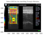
도 1은 일 실시예에 따른 연조직 물성 측정 시스템을 이용하여 연조직의 정량적인 물성값을 출력하는 것을 나타낸다.
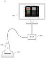
도 2은 일 실시예에 따른 연조직 물성 측정 시스템을 나타낸 도면이다.
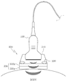
도 3는 일 실시예에 따른 초음파 프로브 및 커플러를 나타낸 도면이다.
도 4는 일 실시예에 따른 로봇-암을 더 포함하는 연조직 물성 측정 시스템을 나타낸 도면이다.
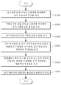
도 5는 일 실시예에 따른 연조직 물성 측정 방법을 나타낸 순서도이다.1 shows outputting a quantitative physical property value of a soft tissue using a soft tissue physical property measurement system according to an embodiment.
2 is a view showing a soft tissue property measurement system according to an embodiment.
3 is a view showing an ultrasonic probe and a coupler according to an embodiment.
4 is a view showing a soft tissue property measurement system further comprising a robot-arm according to an embodiment.
5 is a flowchart illustrating a method for measuring soft tissue properties according to an embodiment.
이하 첨부 도면들 및 첨부 도면들에 기재된 내용들을 참조하여 실시예를 상세하게 설명하지만, 청구하고자 하는 범위는 실시 예들에 의해 제한되거나 한정되는 것은 아니다.Hereinafter, embodiments will be described in detail with reference to the accompanying drawings and the contents described in the accompanying drawings, but the scope to be claimed is not limited or limited by the embodiments.
본 명세서에서 사용되는 용어는 기능을 고려하면서 가능한 현재 널리 사용되는 일반적인 용어를 선택하였으나, 이는 당 분야에 종사하는 기술자의 의도 또는 관례 또는 새로운 기술의 출현 등에 따라 달라질 수 있다. 또한, 특정한 경우는 출원인이 임의로 선정한 용어도 있으며, 이 경우 해당되는 명세서의 설명 부분에서 그 의미를 기재할 것이다. 따라서 본 명세서에서 사용되는 용어는, 단순한 용어의 명칭이 아닌 그 용어가 가지는 실질적인 의미와 본 명세서의 전반에 걸친 내용을 토대로 해석되어야 함을 밝혀두고자 한다.The terminology used in the present specification has been selected as a general terminology that is currently widely used while considering functions, but this may be changed according to intentions or customs of a person skilled in the art or the appearance of new technologies. Also, in certain cases, some terms are arbitrarily selected by the applicant, and in this case, their meanings will be described in the description of the corresponding specification. Therefore, the terms used in the present specification are intended to clarify that the terms should be interpreted based on the actual meaning of the terms and the contents of the present specification, not simply the names of the terms.
또한, 본 명세서에 기술된 실시예는 전적으로 하드웨어이거나, 부분적으로 하드웨어이고 부분적으로 소프트웨어이거나, 또는 전적으로 소프트웨어인 측면을 가질 수 있다. 본 명세서에서 "부(unit)", "모듈(module)", "장치(device)", "서버(server)" 또는 "시스템(system)" 등은 하드웨어, 하드웨어와 소프트웨어의 조합, 또는 소프트웨어 등 컴퓨터 관련 엔티티(entity)를 지칭한다. 예를 들어, 부, 모듈, 장치, 서버 또는 시스템은 플랫폼(platform)의 일부 또는 전부를 구성하는 하드웨어 및/또는 상기 하드웨어를 구동하기 위한 애플리케이션(application) 등의 소프트웨어를 지칭하는 것일 수 있다.In addition, the embodiments described herein can have aspects that are entirely hardware, partly hardware and partly software, or entirely software. In this specification, "unit (unit)", "module (module)", "device (device)", "server (server)" or "system (system)", such as hardware, a combination of hardware and software, or software Refers to a computer related entity. For example, a part, module, device, server, or system may refer to software, such as hardware constituting part or all of a platform and / or applications for driving the hardware.
이하, 도면들을 참조하여 바람직한 실시예들을 보다 상세하게 설명하기로 한다.Hereinafter, preferred embodiments will be described in more detail with reference to the drawings.
도 1은 일 실시예에 따라 측정한 연조직의 정량적인 물성값을 출력하는 화면을 나타낸다.1 shows a screen for outputting a quantitative physical property value of a soft tissue measured according to an embodiment.
본 발명의 실시예에서는 변형탄성기법(strain elastography)을 이용하여 연조직의 상대적인 경도(hardness)를 RGB 색상으로 나타낸다. 변형탄성기법은 생체조직을 누를 때와 누르지 않을 때의 길이 차이를 RF echo 신호와의 상관관계로 측정하여, 연조직의 경도를 RGB 색상으로 표현되는 탄성영상(elastogram)으로 나타내는 기술이다. 다시 말해, 초음파 프로브에서 출력된 초음파와 연조직에 의해 반사된 초음파의 파형의 변화를 분석하여 연조직의 경도를 측정한다. 측정된 경도는 단단한 정도에 따라 디스플레이에 RGB 색상으로 표시되며, 부드러움(Soft)에서 딱딱함(Hard)으로 갈수록 색상이 적색에서 청색으로 변화한다. 즉, 측정 대상의 경도에 따라 출력부에 색상이 다르게 표시되므로, 의료 시술자는 다른 조직과 색상이 다르게 표시되는 부위에 병변이 존재함을 인지할 수 있다.In an embodiment of the present invention, the relative hardness of the soft tissue is expressed in RGB colors using strain elastography. Deformation elasticity technique is a technique that measures the difference in length between when the biological tissue is pressed and when it is not pressed, by correlating it with the RF echo signal, and shows the hardness of the soft tissue as an elastic image expressed in RGB color. In other words, the hardness of the soft tissue is measured by analyzing changes in the waveform of the ultrasound waves output from the ultrasound probe and the ultrasound waves reflected by the soft tissue. The measured hardness is displayed in RGB color on the display according to the degree of rigidity, and the color changes from red to blue as it goes from soft to hard. That is, since the color is differently displayed on the output part according to the hardness of the measurement object, the medical practitioner can recognize that a lesion exists in a region where colors are different from other tissues.
종래의 초음파 탐지 장치는 상기 방식으로 연조직의 경도를 측정하였으나 조직 부위의 상대적인 경도를 알 수 있을 뿐이며, 경도를 수치화하여 나타내거나 형상 파라미터 또는 영률(Young's modulus) 등의 정량적인 물성값을 알아낼 수는 없었다. 최근에는 횡파탄성영상(shear wave elastography) 초음파 영상 기술이 도입되면서, 연조직의 딱딱한 정도를 정량적으로 수치화할 수 있게 되었으나, 이를 위해서는 고가의 별도의 장비가 필요하며 일반적인 초음파 프로브 장치로는 물성값을 정량적으로 측정할 수 없었다.The conventional ultrasonic detection device measures the hardness of the soft tissue in the above manner, but can only know the relative hardness of the tissue site, and it is not possible to figure out the hardness or quantify the physical properties such as shape parameters or Young's modulus. . Recently, with the introduction of shear wave elastography ultrasound imaging technology, it has been possible to quantitatively quantify the degree of stiffness of soft tissues.However, for this, an expensive separate equipment is required, and the general ultrasonic probe device quantitatively measures the physical properties. It could not be measured.
이에, 본 발명의 실시예는 종래의 초음파 탐지 시스템과 호환 가능하며, 정량적인 물성값을 갖는 기준물질과의 비교 측정을 통해 연조직의 정량적인 물성값을 실시간으로 연산할 수 있는 물성 측정 시스템을 제공하고자 한다.Accordingly, an embodiment of the present invention is compatible with a conventional ultrasonic detection system, and intends to provide a property measurement system capable of real-time calculating a quantitative property value of soft tissue through comparative measurement with a reference material having a quantitative property value. .
도 2는 일 실시예에 따른 물성 측정 시스템을 나타낸다. 연조직의 물성을 정량적으로 측정하기 위한 시스템(10)은, 초음파 프로브(100), 초음파 프로브의 전단에 탈착 가능한 커플러(200), 연조직의 정량적인 물성값을 연산하기 위한 처리부(300), 및 측정된 물성값을 실시간으로 출력하기 위한 출력부(400)를 포함한다.2 shows a physical property measurement system according to an embodiment. The
초음파 프로브(100)는 초음파를 생성하는 트랜스듀서 및 반사된 초음파를 수신하는 센서를 포함하며, 수신한 반사파를 전기 신호로 변환하여 처리부(300)에 전송하기 위한 다른 구성요소를 포함할 수 있다.The
커플러(200)는 상기 초음파 프로브(100)의 전단에 탈착 가능한 부재이다. 도 3을 참조하면, 커플러(200)는 본체(210), 기준물질(220) 및 압력센서(230)를 포함할 수 있다. 본체(210)는 초음파 프로브(100)의 전단(초음파가 출력되는 부분)에 결합되도록 맞춤형으로 제작될 수 있고, 내부에는 실시예에 따라서 압력센서(230) 및 측정된 압력을 전기 신호로 변환하여 처리부(300)에 전송하기 위한 다른 구성요소를 포함할 수 있다. The
커플러(200)는 초음파 프로브 전단과 신체 사이에 밀착되는 기준물질(220)을 포함한다. 기준물질(220)은 미리 알려진 정량적인 물성값을 가지며, 상기 물성값 정보는 처리부(300)에 미리 저장되어 연조직의 물성값을 추정하는데 이용된다. 기준물질(220)은 초음파 투과성이 있는 부드러운 재질로서, 예를 들어 한천, 활석, 끈적거리는 물질, 젤 등으로 구성된 조직유사물질로 형성될 수 있으나 이에 한정되는 것은 아니다. The
기준물질(220)은 단일 물질로 구성될 수도 있고 복수의 물질층이 적층된 다층 구조일 수도 있다. 예를 들어, 기준물질(220)은 도 3에 도시된 것처럼 제1 물질(220a) 및 제2 물질(220b), 또는 그 이상의 물질이 적층된 다층 구조로 구성될 수 있다. 이 경우 단일 물질을 이용하는 경우에 비해 정확한 기준값을 얻을 수 있고, 측정 과정에서 어느 하나의 물질이 손상되더라도 시스템이 동작 가능하도록 안정성을 향상시킬 수 있다. 일 실시예에서, 상기 제1 물질(220a)은 측정 대상 연조직보다 낮은 경도를 갖고, 상기 제2 물질(220b)은 상기 제1 물질보다 높은 경도를 가질 수 있다. 예를 들어, GS 근육의 경도는 약 28-34kPa 이며, 이 경우 제1 물질은 7kPa, 제2 물질은 30 kPa 의 경도를 갖는 물질 중에서 선택될 수 있다.The
이하에서는 기준물질(220)과 연조직의 물성을 비교하여 연조직의 정량적인 물성값을 연산하는 과정을 설명한다.Hereinafter, a process of calculating a quantitative physical property value of the soft tissue by comparing the physical properties of the
처리부(300)는 상기 연조직과 기준물질의 물성을 비교 측정하고, 상기 기준물질의 정량적인 물성값에 기초하여 상기 연조직의 정량적인 물성값을 연산한다. 처리부는 하드웨어 및 소프트웨어가 결합된 컴퓨터 장치일 수 있다. 처리부(300)는 초음파 프로브(100)로부터 수신한 정보(트랜스듀서가 출력하는 초음파 정보, 기준물질 및 연조직에 의해 반사된 초음파 정보 등)를 분석함으로써 기준물질과 연조직의 물성(예를 들어, 물질의 경도)을 상대적으로 측정하여 나타낼 수 있다. 일반적인 초음파 측정 분야에서 널리 알려진 변형탄성기법(strain elastography)이 이용될 수 있다.The
이를 이용하면, 도 1에 도시된 것처럼 측정 대상 연조직의 경도를 영역 A와 같이, 기준물질의 경도를 영역 B와 같이 RGB 색상으로 출력된다. 전술한 바와 같이, 초음파를 반사하는 객체가 부드러울수록 적색에 가깝고 딱딱할 수록 청색에 가깝게 나타난다. Using this, as shown in FIG. 1, the hardness of the soft tissue to be measured is output in RGB color as in the region A and the hardness of the reference material as in the region B. As described above, the softer the object reflecting the ultrasound, the closer it is to red and the harder it is to blue.
본 발명의 일 실시예에 의하면 출력된 기준물질과 측정 대상 연조직의 색상대비를 분석하여 연조직의 정량적인 물성값을 추정할 수 있다. 대상범위는 도 1과 같이 ROI(region of interest) 영역으로 지정할 수 있으며, 사용자는 출력부에 출력된 초음파 분석영상에서 ROI를 직접 선택함으로써 해당 영역의 연조직 물성 정보를 연산 및 출력할 것을 명령할 수 있다.According to an embodiment of the present invention, it is possible to estimate a quantitative physical property value of the soft tissue by analyzing the color contrast of the output reference material and the soft tissue to be measured. The target range can be designated as a region of interest (ROI) region as shown in FIG. 1, and the user can command to calculate and output soft tissue property information of the region by directly selecting the ROI from the ultrasound analysis image output to the output unit. have.
색상대비를 통한 정량적인 물성값 연산의 예시는 다음과 같다. 측정 대상 연조직의 경도를 1kPa 내지 10 kPa 의 수치로 정량화하여 나타내는 경우, 미리 알려진 기준물질의 경도가 "2 kPa" 이고 출력되는 색상이 R: 80, G: 15, B: 5 라면, 연조직의 출력 색상이 R: 5, G: 15, B: 80 인 것으로부터 RGB 색상의 수치 비교를 통해 연조직의 경도가 "8 kPa" 이라는 결과를 도출할 수 있을 것이다. 본 실시예에서는 RGB 색상대비를 통해 물성을 비교 측정하였지만, 이에 한정되는 것은 아니며 각 물질의 반사 초음파에 의해 알 수 있는 파라미터의 차이를 이용해 물성을 비교 측정할 수 있다.An example of quantitative property value calculation through color contrast is as follows. When the hardness of the soft tissue to be measured is quantified and expressed as a value of 1 kPa to 10 kPa, the hardness of the known reference material is “2 kPa” and the output color is R: 80, G: 15, B: 5, and the soft tissue is output. From the colors R: 5, G: 15, and B: 80, a numerical comparison of RGB colors may lead to the result that the hardness of the soft tissue is "8 kPa". In the present embodiment, the physical properties were compared and measured through RGB color contrast, but the present invention is not limited thereto, and physical properties may be compared and measured using differences in parameters known by reflected ultrasound of each material.
이외에도 연산되는 정량적인 물성값은 연조직의 경도(hardness), 영률(Young's modulus) 등 연조직의 물성을 나타내는 정량적인 파라미터, 또는 연조직의 해부학적인 형태와 관련된 형상 파라미터 등을 포함할 수 있다. In addition, the calculated quantitative physical property values may include quantitative parameters indicating physical properties of soft tissues, such as hardness and softness of soft tissues, or shape parameters related to anatomical shape of soft tissues.
유사한 방식으로, 기준물질과 연조직의 경도를 나타내는 RGB 색상의 비교를 통해 연조직의 물성값을 추정할 수 있다. 상기 추정된 물성값은 출력부(400)에 의해 실시간으로 출력된다.In a similar manner, the physical property value of the soft tissue can be estimated by comparing the reference material and the RGB color representing the hardness of the soft tissue. The estimated physical property value is output in real time by the
출력부(400)는 연조직과 기준물질의 상대적인 경도를 RGB 색상으로 출력하고, ROI 영역에 해당하는 연조직의 형상 파라미터 또는 영률 등의 정량적인 물성값을 처리부(300)로부터 수신하여 실시간으로 출력한다. 출력부(400)는 LCD 또는 LED 디스플레이 장치 등 일반적인 출력 장치를 포함한다.The
일 실시예에서, 상기 커플러(200)는 도 3에 도시된 것처럼 초음파 프로브(100)의 전단에 가해지는 압력을 측정하는 압력센서(230)를 더 포함할 수 있다. 이 경우 처리부(300)는 상기 압력센서(230)에 의해 측정된 압력에 기초하여 연조직의 정량적인 물성값을 조정함으로써, 불규칙한 압력에 따른 영향을 감소시킬 수 있다.In one embodiment, the
예를 들어, 종래의 초음파 측정 장치를 이용하여 탐색할 시 초음파 프로브를 환자의 신체에 밀착시킨 채 일정한 세기를 유지하는 것이 중요한데, 숙련되지 않은 의료 시술자는 초음파 프로브를 환자의 신체에 지나치게 밀착시키거나 밀착시키지 않음으로써 측정 결과가 변동되는 경우가 있었다.For example, when searching using a conventional ultrasonic measuring device, it is important to maintain a constant intensity while keeping the ultrasonic probe in close contact with the patient's body. The measurement result may fluctuate by not sticking.
실시예에 따른 압력센서는 이와 같은 문제를 해결하기 위한 것으로서, 커플러(200) 내부의 압력센서(230)는 초음파 프로브(100)에 의해 신체에 가해지는 압력을 측정하여 처리부(300)에 전송하고, 상기 처리부(300)는 측정된 압력에 기초하여 출력되는 연조직의 물성값을 조정한다. 예를 들어, 압력이 증가하여 연조직의 경도가 더 단단하게 측정된다면, 압력에 비례하여 경도를 낮게 조정함으로써 사용자가 가하는 불규칙한 압력에 따른 영향을 감소시킬 수 있다. 결과적으로 RGB 색상이 안정적으로 출력되고 이에 따라 연조직의 물성값도 안정적으로 측정할 수 있다.Pressure sensor according to the embodiment is intended to solve this problem, the
일 실시예에서, 상기 시스템은 상기 초음파 프로브(100)의 동작을 제어하는 제어부(500)를 더 포함할 수 있다. 상기 제어부(500)는 상기 압력센서(230)에 의해 측정된 압력에 기초하여 상기 초음파 프로브(100)가 일정한 압력을 유지하면서 동작하도록 제어할 수 있다. 여기서 제어부(500)는 도 4에 도시된 것처럼 다관절 로봇 암(arm)과 같은 구동장치를 포함할 수 있다. In one embodiment, the system may further include a
종래의 초음파 측정 장치는 사용자가 초음파 프로브를 환자의 신체에 밀착시킨 채 직접 움직임으로써 병변을 검출하는 방식이지만, 본 실시예에 따르면 제어부가 일정한 압력을 유지하면서 초음파 프로브를 구동시키므로 보다 안정적으로 물성값을 측정할 수 있다. 초음파 프로브와 다관절 로봇 암은 개별 장치로서 결합 가능하거나 일체로서 형성될 수 있다.The conventional ultrasonic measuring device is a method in which a user detects a lesion by directly moving the ultrasonic probe in close contact with the patient's body, but according to this embodiment, the control unit drives the ultrasonic probe while maintaining a constant pressure, so that the physical property value is more stably. Can be measured. The ultrasonic probe and the multi-joint robot arm can be combined as separate devices or can be formed integrally.
도 5는 일 실시예에 따른 연조직 물성 측정 방법을 나타낸 순서도이다. 실시예에 따른 방법은, 애플리케이션으로 구현되거나 다양한 컴퓨터 구성요소를 통하여 수행될 수 있는 프로그램 명령어의 형태로 구현되어 컴퓨터 판독 가능한 기록 매체에 기록될 수 있다. 상기 컴퓨터 판독 가능한 기록 매체는 프로그램 명령어, 데이터 파일, 데이터 구조 등을 단독으로 또는 조합하여 포함할 수 있다.5 is a flowchart illustrating a method for measuring soft tissue properties according to an embodiment. The method according to the embodiment may be recorded in a computer-readable recording medium by being implemented in the form of program instructions that can be implemented as an application or executed through various computer components. The computer-readable recording medium may include program instructions, data files, data structures, or the like alone or in combination.
먼저, 연조직에 의해 반사된 초음파를 분석하여 상기 연조직의 물성을 측정하는 단계(S100), 기준물질에 의해 반사된 초음파를 분석하여 상기 기준물질의 물성을 측정하는 단계(S200)가 수행된다. 초음파 프로브의 트랜스듀서에서 초음파를 생성하고, 연조직과 기준물질에 의해 반사된 초음파를 수신하고 파형의 변화를 분석하여 각각의 경도를 측정한다. 전술한 것처럼 변형탄성기법(strain elastography)을 이용할 수 있다.First, a step of measuring the properties of the soft tissue by analyzing the ultrasound waves reflected by the soft tissue (S100), and a step of measuring the properties of the reference material by analyzing the ultrasonic waves reflected by the reference material (S200) are performed. The ultrasonic probe generates ultrasonic waves, receives the ultrasonic waves reflected by the soft tissue and the reference material, and analyzes changes in the waveform to measure each hardness. As described above, strain elastography can be used.
이어서, 상기 연조직과 상기 기준물질의 물성을 비교하는 단계(S300), 기준물질의 미리 알려진 정량적인 물성값에 기초하여 상기 연조직의 정량적인 물성값을 연산하는 단계(S400)가 수행된다. 측정된 물성(경도)은 단단한 정도에 따라 디스플레이에 RGB 색상으로 표시된다. 시스템은 기준물질의 경도, 형상 파라미터 또는 영률(Young's modulus)과 같은 정량적인 파라미터값을 미리 입력 받아 저장해 두므로, 이를 이용하면 기준물질과 측정 대상 연조직의 RGB 출력값을 비교 분석하여 연조직의 물성값을 정량적으로 나타낼 수 있다.Subsequently, comparing the properties of the soft tissue and the reference material (S300), and calculating the quantitative properties of the soft tissue based on a known quantitative property value of the reference material (S400). The measured physical properties (hardness) are displayed in RGB colors on the display according to the degree of rigidity. Since the system receives and stores quantitative parameter values such as hardness, shape parameter, or Young's modulus of the reference material in advance, it uses this to compare and analyze the RGB output values of the reference material and the soft tissue to be measured to quantitatively evaluate the physical property values of the soft tissue. Can be represented as
일 실시예에서, 압력센서로부터 수신된 초음파 프로브의 전단에 가해지는 압력에 기초하여, 상기 연조직의 정량적인 물성값을 조정하는 단계(S500)가 더 수행될 수 있다. 예를 들어, 압력이 증가하여 연조직의 경도가 더 단단하게 측정된다면, 압력에 비례하여 경도를 낮게 조정함으로써 사용자가 가하는 불규칙한 압력에 따른 영향을 감소시킬 수 있다. 결과적으로 RGB 색상이 안정적으로 출력되고 이에 따라 연조직의 물성값도 안정적으로 측정할 수 있다.In one embodiment, based on the pressure applied to the front end of the ultrasonic probe received from the pressure sensor, the step of adjusting the quantitative property value of the soft tissue (S500) may be further performed. For example, if the pressure increases and the hardness of the soft tissue is measured more firmly, the influence of the irregular pressure exerted by the user can be reduced by adjusting the hardness in proportion to the pressure. As a result, the RGB color is stably output, and accordingly, the physical property value of the soft tissue can also be stably measured.
이어서, 상기 연조직의 정량적인 물성값을 출력부에 전송하는 단계가 수행된다(S600). 사용자는 출력부(디스플레이)에 표시된 관심영역(ROI)의 연조직의 물성값을 실시간으로 알 수 있다. 사용자는 관심영역을 임의로 설정할 수 있고, 설정된 영역이 현재 지정된 영역과 다를 경우, 해당 관심영역에 대하여 단계(S300) 내지 단계(S600)을 반복한다. Subsequently, a step of transmitting the quantitative property value of the soft tissue to the output unit is performed (S600). The user can know in real time the physical property value of the soft tissue of the region of interest (ROI) displayed on the output unit (display). The user can arbitrarily set the region of interest, and if the set region is different from the currently designated region, the steps S300 to S600 are repeated for the region of interest.
상기 단계(S100 내지 S600)를 포함하는 물성 측정 방법은 컴퓨터로 판독 가능한 기록매체에 프로그램 명령어 형태로 저장될 수 있다. 프로그램 명령어는 본 발명을 위하여 특별히 설계되고 구성된 것들이거니와 컴퓨터 소프트웨어 분야의 당업자에게 공지되어 사용 가능한 것일 수도 있다. 컴퓨터 판독 가능한 기록 매체의 예에는, 하드 디스크, 플로피 디스크 및 자기 테이프와 같은 자기 매체, CD-ROM, DVD와 같은 광기록 매체, 플롭티컬 디스크(floptical disk)와 같은 자기-광 매체(magneto-optical media), 및 ROM, RAM, 플래시 메모리 등과 같은 프로그램 명령어를 저장하고 수행하도록 특별히 구성된 하드웨어 장치가 포함된다. The method for measuring physical properties including the steps (S100 to S600) may be stored in the form of program instructions in a computer-readable recording medium. The program instructions are specially designed and configured for the present invention, and may be known and available to those skilled in the computer software field. Examples of computer-readable recording media include magnetic media such as hard disks, floppy disks, and magnetic tapes, optical recording media such as CD-ROMs, DVDs, and magneto-optical media such as floptical disks. media), and hardware devices specifically configured to store and execute program instructions such as ROM, RAM, flash memory, and the like.
이상에서 설명한 연조직의 물성을 정량적으로 측정하기 위한 시스템에 의하면, 미리 알려진 정량적인 물성값을 갖는 기준물질과 변형탄성기법(Strain elastography)을 이용하여 측정된 연조직의 물성을 비교하여, 기준물질의 물성값을 정량적으로 측정하고 실시간으로 나타낼 수 있다. 본 시스템의 구성요소는 기존의 초음파 측정 장비에 쉽게 호환되므로, 고가의 장비를 이용하지 않고도 연조직의 물성을 정량적으로 측정할 수 있어 비용을 절감할 수 있다.According to the system for quantitatively measuring the properties of the soft tissue described above, the physical properties of the reference material are compared by comparing the physical properties of the reference material having a known quantitative property value and the soft tissue measured using strain elastography. It can be quantitatively measured and displayed in real time. Since the components of this system are easily compatible with existing ultrasonic measuring equipment, it is possible to quantitatively measure the properties of soft tissues without using expensive equipment, thereby reducing costs.
이상에서는 실시예들을 참조하여 설명하였지만, 해당 기술 분야의 숙련된 당업자는 하기의 특허 청구의 범위에 기재된 본 발명의 사상 및 영역으로부터 벗어나지 않는 범위 내에서 본 발명을 다양하게 수정 및 변경시킬 수 있음을 이해할 수 있을 것이다.Although described above with reference to embodiments, those skilled in the art can variously modify and change the present invention without departing from the spirit and scope of the present invention as set forth in the claims below. You will understand.
10: 물성 측정 시스템
100: 초음파 프로브
200: 커플러
210: 커플러 본체
220: 기준물질
220a: 제1 기준물질
220b: 제2 기준물질
230: 압력센서
300: 처리부
400: 출력부
500: 제어부10: physical property measurement system
100: ultrasonic probe
200: coupler
210: coupler body
220: reference substance
220a: first reference substance
220b: second reference substance
230: pressure sensor
300: processing unit
400: output
500: control unit
Claims (9)
초음파를 생성하고 연조직에 의해 반사된 초음파를 수신하는 초음파 프로브;
본체 및 상기 초음파 프로브와 상기 연조직 사이에 위치하며 미리 알려진 정량적인 물성값을 갖는 기준물질로 구성되는, 상기 초음파 프로브의 전단에 탈착 가능한 커플러;
변형탄성기법을 이용하여 상기 연조직과 상기 기준물질의 물성을 비교 측정하고, 상기 기준물질의 정량적인 물성값에 기초하여 상기 연조직의 정량적인 물성값을 연산하는 처리부; 및
상기 연조직의 정량적인 물성값을 실시간으로 출력하는 출력부를 포함하는 것을 특징으로 하는, 연조직의 물성을 정량적으로 측정하기 위한 시스템.
A system for quantitatively measuring the properties of soft tissue,
An ultrasonic probe that generates ultrasonic waves and receives ultrasonic waves reflected by soft tissue;
A coupler located between the main body and the ultrasonic probe and the soft tissue and consisting of a reference material having a known quantitative property value, a coupler detachable at a front end of the ultrasonic probe;
A processing unit for comparing and measuring the physical properties of the soft tissue and the reference material using a modified elastic technique, and calculating the quantitative physical property values of the soft tissue based on the quantitative physical property values of the reference material; And
A system for quantitatively measuring the physical properties of the soft tissue, characterized in that it comprises an output for outputting the quantitative physical property values of the soft tissue in real time.
상기 커플러는, 상기 초음파 프로브의 전단에 가해지는 압력을 측정하는 압력센서를 더 포함하고,
상기 처리부는, 상기 압력센서에 의해 측정된 압력에 기초하여 상기 연조직의 정량적인 물성값을 조정함으로써, 불규칙한 압력에 따른 영향을 감소시키는 것을 특징으로 하는, 연조직의 물성을 정량적으로 측정하기 위한 시스템.
According to claim 1,
The coupler, further comprising a pressure sensor for measuring the pressure applied to the front end of the ultrasonic probe,
The processing unit, by adjusting the quantitative physical property value of the soft tissue based on the pressure measured by the pressure sensor, characterized in that to reduce the influence of the irregular pressure, a system for quantitatively measuring the physical properties of the soft tissue.
상기 시스템은 상기 초음파 프로브의 동작을 제어하는 제어부를 더 포함하되,
상기 제어부는, 상기 압력센서에 의해 측정된 압력에 기초하여 상기 초음파 프로브가 일정한 압력을 유지하면서 동작하도록 제어하는 것을 특징으로 하는, 연조직의 물성을 정량적으로 측정하기 위한 시스템.
According to claim 2,
The system further includes a control unit for controlling the operation of the ultrasonic probe,
The control unit, based on the pressure measured by the pressure sensor, characterized in that to control the ultrasonic probe to operate while maintaining a constant pressure, a system for quantitatively measuring the properties of soft tissue.
상기 기준물질은, 미리 알려진 물성값을 갖는 제1 물질, 및 상기 제1 물질과 상이한 물성값을 갖는 제2 물질의 적층 구조로 구성되는 것을 특징으로 하는, 연조직의 물성을 정량적으로 측정하기 위한 시스템.
According to claim 1,
The reference material is a system for quantitatively measuring physical properties of soft tissues, characterized in that it consists of a stacked structure of a first material having a known physical property value, and a second material having a different physical property value from the first material.
상기 제1 물질은 상기 연조직보다 낮은 경도(硬度)를 갖고,
상기 제2 물질은 상기 제1 물질보다 높은 경도를 갖는 것을 특징으로 하는, 연조직의 물성을 정량적으로 측정하기 위한 시스템.
According to claim 4,
The first material has a lower hardness (상기 度) than the soft tissue,
The second material is characterized in that it has a higher hardness than the first material, a system for quantitatively measuring the properties of soft tissue.
상기 기준물질 및 연조직의 물성값은, 경도(hardness), 영률(Young's modulus) 또는 상기 연조직의 형태와 관련된 형상 파라미터를 포함하는 것을 특징으로 하는, 연조직의 물성을 정량적으로 측정하기 위한 시스템.
According to claim 1,
A system for quantitatively measuring physical properties of soft tissue, characterized in that the reference material and the soft tissue physical property values include shape parameters related to hardness, Young's modulus, or the shape of the soft tissue.
연조직에 의해 반사된 초음파를 분석하여 상기 연조직의 물성을 측정하는 단계;
기준물질에 의해 반사된 초음파를 분석하여 상기 기준물질의 물성을 측정하는 단계;
상기 연조직과 상기 기준물질의 물성을 비교하는 단계;
상기 비교결과 및 상기 기준물질의 미리 알려진 정량적인 물성값에 기초하여, 상기 연조직의 정량적인 물성값을 연산하는 단계; 및
상기 연조직의 정량적인 물성값을 출력부에 전송하는 단계를 포함하는 것을 특징으로 하는, 연조직의 물성을 정량적으로 측정하기 위한 방법.
As a method for quantitatively measuring the properties of soft tissue, the method is performed by a processor,
Analyzing the ultrasound waves reflected by the soft tissue to measure the properties of the soft tissue;
Measuring the physical properties of the reference material by analyzing ultrasonic waves reflected by the reference material;
Comparing the physical properties of the soft tissue and the reference material;
Calculating a quantitative property value of the soft tissue based on the comparison result and a known quantitative property value of the reference material; And
Method for quantitatively measuring the physical properties of the soft tissue, characterized in that it comprises the step of transmitting a quantitative physical property value of the soft tissue.
압력센서로부터 수신된 초음파 프로브의 전단에 가해지는 압력에 기초하여, 상기 연조직의 정량적인 물성값을 조정하는 단계를 더 포함하는 것을 특징으로 하는, 연조직의 물성을 정량적으로 측정하기 위한 방법.
The method of claim 7,
Method for quantitatively measuring the physical properties of the soft tissue, characterized in that it further comprises the step of adjusting the quantitative physical property value of the soft tissue, based on the pressure applied to the front end of the ultrasonic probe received from the pressure sensor.
Priority Applications (1)
| Application Number | Priority Date | Filing Date | Title |
|---|---|---|---|
| KR1020180122544A KR102174348B1 (en) | 2018-10-15 | 2018-10-15 | System for measuring properties of soft tissue quantitatively |
Applications Claiming Priority (1)
| Application Number | Priority Date | Filing Date | Title |
|---|---|---|---|
| KR1020180122544A KR102174348B1 (en) | 2018-10-15 | 2018-10-15 | System for measuring properties of soft tissue quantitatively |
Publications (2)
| Publication Number | Publication Date |
|---|---|
| KR20200042228A true KR20200042228A (en) | 2020-04-23 |
| KR102174348B1 KR102174348B1 (en) | 2020-11-04 |
Family
ID=70472636
Family Applications (1)
| Application Number | Title | Priority Date | Filing Date |
|---|---|---|---|
| KR1020180122544A Active KR102174348B1 (en) | 2018-10-15 | 2018-10-15 | System for measuring properties of soft tissue quantitatively |
Country Status (1)
| Country | Link |
|---|---|
| KR (1) | KR102174348B1 (en) |
Cited By (1)
| Publication number | Priority date | Publication date | Assignee | Title |
|---|---|---|---|---|
| GB2614912A (en) * | 2022-01-24 | 2023-07-26 | Dotplot Ltd | Soft tissue monitoring device and method |
Citations (5)
| Publication number | Priority date | Publication date | Assignee | Title |
|---|---|---|---|---|
| JP2002136520A (en) * | 2000-11-06 | 2002-05-14 | Aloka Co Ltd | Ultrasonic probe for hard texture diagnosis |
| JP2005066041A (en) * | 2003-08-25 | 2005-03-17 | Hitachi Medical Corp | Ultrasonic probe and ultrasonic diagnostic equipment |
| WO2009131028A1 (en) * | 2008-04-25 | 2009-10-29 | 株式会社 日立メディコ | Ultrasonic diagnostic apparatus |
| US20140180116A1 (en) * | 2009-10-08 | 2014-06-26 | C. R. Bard, Inc. | Coupling Structures for an Ultrasound Probe |
| US20180272157A1 (en) * | 2004-10-06 | 2018-09-27 | Guided Therapy Systems, L.L.C. | Ultrasound probe for treating skin laxity |
-
2018
- 2018-10-15 KR KR1020180122544A patent/KR102174348B1/en active Active
Patent Citations (6)
| Publication number | Priority date | Publication date | Assignee | Title |
|---|---|---|---|---|
| JP2002136520A (en) * | 2000-11-06 | 2002-05-14 | Aloka Co Ltd | Ultrasonic probe for hard texture diagnosis |
| JP2005066041A (en) * | 2003-08-25 | 2005-03-17 | Hitachi Medical Corp | Ultrasonic probe and ultrasonic diagnostic equipment |
| US20180272157A1 (en) * | 2004-10-06 | 2018-09-27 | Guided Therapy Systems, L.L.C. | Ultrasound probe for treating skin laxity |
| WO2009131028A1 (en) * | 2008-04-25 | 2009-10-29 | 株式会社 日立メディコ | Ultrasonic diagnostic apparatus |
| JP5329532B2 (en) * | 2008-04-25 | 2013-10-30 | 株式会社日立メディコ | Ultrasonic diagnostic equipment |
| US20140180116A1 (en) * | 2009-10-08 | 2014-06-26 | C. R. Bard, Inc. | Coupling Structures for an Ultrasound Probe |
Non-Patent Citations (1)
| Title |
|---|
| Reliability and Validity of Quantifying Absolute Muscle Hardness Using Ultrasound Elastography |
Cited By (1)
| Publication number | Priority date | Publication date | Assignee | Title |
|---|---|---|---|---|
| GB2614912A (en) * | 2022-01-24 | 2023-07-26 | Dotplot Ltd | Soft tissue monitoring device and method |
Also Published As
| Publication number | Publication date |
|---|---|
| KR102174348B1 (en) | 2020-11-04 |
Similar Documents
| Publication | Publication Date | Title |
|---|---|---|
| US8172754B2 (en) | Ultrasonograph | |
| CN115334963A (en) | System and method for generating tissue image biomarkers | |
| US10524768B2 (en) | Medical image diagnostic apparatus and medical image processing apparatus | |
| US11241221B2 (en) | Method for processing morphology information and elasticity information of tissue and device for elasticity detection | |
| US20090143676A1 (en) | Ultrasonograph for Creating Elastic Image | |
| US20100185090A1 (en) | Ultrasonographic device | |
| JP2017527401A (en) | Ultrasonic imaging device | |
| US11497475B2 (en) | Ultrasound image acquisition optimization according to different respiration modes | |
| KR20130080640A (en) | Method and apparatus for providing ultrasound images | |
| US20210052255A1 (en) | Ultrasound guidance dynamic mode switching | |
| JP6538280B2 (en) | Apparatus and method for characterizing a subject's tissue | |
| Sassaroli et al. | Breast ultrasound technology and performance evaluation of ultrasound equipment: B-mode | |
| KR20210093049A (en) | Ultrasonic diagnostic apparatus and operating method for the same | |
| CN204600529U (en) | Elastomeric check equipment | |
| US20200405264A1 (en) | Region of interest positioning for longitudinal montioring in quantitative ultrasound | |
| JP5075040B2 (en) | Ultrasonic diagnostic equipment | |
| KR102174348B1 (en) | System for measuring properties of soft tissue quantitatively | |
| CN115551416B (en) | Measurement of hip | |
| JP2004267464A (en) | Ultrasonic diagnostic device | |
| US11712223B2 (en) | Ultrasound imaging system and method | |
| CN120859553A (en) | Determination of enhanced quantitative ultrasound medical imaging by interventional tissue | |
| JP2005270351A (en) | Method and device for ultrasonic three-dimensional imaging | |
| WO2024203331A1 (en) | Ultrasonic diagnostic device and control method for ultrasonic diagnostic device | |
| CN117157013A (en) | Method for ultrasound imaging | |
| KR20130077410A (en) | Method and apparatus for providing acoustic radiation force impulse imaging |
Legal Events
| Date | Code | Title | Description |
|---|---|---|---|
| PA0109 | Patent application |
St.27 status event code: A-0-1-A10-A12-nap-PA0109 |
|
| PA0201 | Request for examination |
St.27 status event code: A-1-2-D10-D11-exm-PA0201 |
|
| D13-X000 | Search requested |
St.27 status event code: A-1-2-D10-D13-srh-X000 |
|
| D14-X000 | Search report completed |
St.27 status event code: A-1-2-D10-D14-srh-X000 |
|
| E902 | Notification of reason for refusal | ||
| PE0902 | Notice of grounds for rejection |
St.27 status event code: A-1-2-D10-D21-exm-PE0902 |
|
| PG1501 | Laying open of application |
St.27 status event code: A-1-1-Q10-Q12-nap-PG1501 |
|
| E13-X000 | Pre-grant limitation requested |
St.27 status event code: A-2-3-E10-E13-lim-X000 |
|
| P11-X000 | Amendment of application requested |
St.27 status event code: A-2-2-P10-P11-nap-X000 |
|
| P13-X000 | Application amended |
St.27 status event code: A-2-2-P10-P13-nap-X000 |
|
| E701 | Decision to grant or registration of patent right | ||
| PE0701 | Decision of registration |
St.27 status event code: A-1-2-D10-D22-exm-PE0701 |
|
| GRNT | Written decision to grant | ||
| PR0701 | Registration of establishment |
St.27 status event code: A-2-4-F10-F11-exm-PR0701 |
|
| PR1002 | Payment of registration fee |
St.27 status event code: A-2-2-U10-U11-oth-PR1002 Fee payment year number: 1 |
|
| PG1601 | Publication of registration |
St.27 status event code: A-4-4-Q10-Q13-nap-PG1601 |
|
| PR1001 | Payment of annual fee |
St.27 status event code: A-4-4-U10-U11-oth-PR1001 Fee payment year number: 4 |
|
| PN2301 | Change of applicant |
St.27 status event code: A-5-5-R10-R13-asn-PN2301 St.27 status event code: A-5-5-R10-R11-asn-PN2301 |
|
| PR1001 | Payment of annual fee |
St.27 status event code: A-4-4-U10-U11-oth-PR1001 Fee payment year number: 5 |
|
| PR1001 | Payment of annual fee |
St.27 status event code: A-4-4-U10-U11-oth-PR1001 Fee payment year number: 6 |
|
| U11 | Full renewal or maintenance fee paid |
Free format text: ST27 STATUS EVENT CODE: A-4-4-U10-U11-OTH-PR1001 (AS PROVIDED BY THE NATIONAL OFFICE) Year of fee payment: 6 |