RU2198001C2 - Subcortical brain structures electric stimulation method usable in chronic experimental studies - Google Patents
Subcortical brain structures electric stimulation method usable in chronic experimental studies Download PDFInfo
- Publication number
- RU2198001C2 RU2198001C2 RU2000111595A RU2000111595A RU2198001C2 RU 2198001 C2 RU2198001 C2 RU 2198001C2 RU 2000111595 A RU2000111595 A RU 2000111595A RU 2000111595 A RU2000111595 A RU 2000111595A RU 2198001 C2 RU2198001 C2 RU 2198001C2
- Authority
- RU
- Russia
- Prior art keywords
- stimulation
- structures
- brain
- stimulating
- time
- Prior art date
Links
- 230000000638 stimulation Effects 0.000 title claims abstract description 37
- 238000000034 method Methods 0.000 title claims abstract description 15
- 210000004556 brain Anatomy 0.000 title claims abstract description 13
- 230000002739 subcortical effect Effects 0.000 title claims abstract description 8
- 230000001684 chronic effect Effects 0.000 title claims abstract description 6
- 238000002474 experimental method Methods 0.000 claims abstract description 19
- 230000000694 effects Effects 0.000 claims abstract description 13
- 210000000056 organ Anatomy 0.000 claims abstract description 12
- 230000033228 biological regulation Effects 0.000 claims abstract description 10
- 241001465754 Metazoa Species 0.000 claims abstract description 7
- 230000004936 stimulating effect Effects 0.000 claims abstract description 7
- 210000003169 central nervous system Anatomy 0.000 claims abstract description 6
- 230000015572 biosynthetic process Effects 0.000 claims description 7
- 210000005036 nerve Anatomy 0.000 claims description 4
- 238000010171 animal model Methods 0.000 claims description 2
- 239000000126 substance Substances 0.000 abstract description 2
- 239000003814 drug Substances 0.000 abstract 1
- 210000003016 hypothalamus Anatomy 0.000 description 6
- 238000005755 formation reaction Methods 0.000 description 5
- 230000007794 irritation Effects 0.000 description 5
- 230000004044 response Effects 0.000 description 5
- 230000033764 rhythmic process Effects 0.000 description 5
- 241000283707 Capra Species 0.000 description 4
- 238000013459 approach Methods 0.000 description 4
- 230000002267 hypothalamic effect Effects 0.000 description 3
- 239000008267 milk Substances 0.000 description 3
- 210000004080 milk Anatomy 0.000 description 3
- 235000013336 milk Nutrition 0.000 description 3
- 230000001105 regulatory effect Effects 0.000 description 3
- 230000028327 secretion Effects 0.000 description 3
- 102000006877 Pituitary Hormones Human genes 0.000 description 2
- 108010047386 Pituitary Hormones Proteins 0.000 description 2
- 102000003946 Prolactin Human genes 0.000 description 2
- 108010057464 Prolactin Proteins 0.000 description 2
- 239000004020 conductor Substances 0.000 description 2
- 230000006378 damage Effects 0.000 description 2
- 238000011161 development Methods 0.000 description 2
- 239000000960 hypophysis hormone Substances 0.000 description 2
- 210000005075 mammary gland Anatomy 0.000 description 2
- 229940097325 prolactin Drugs 0.000 description 2
- 230000011514 reflex Effects 0.000 description 2
- 206010001497 Agitation Diseases 0.000 description 1
- 230000004913 activation Effects 0.000 description 1
- 210000004727 amygdala Anatomy 0.000 description 1
- 230000003851 biochemical process Effects 0.000 description 1
- 239000008280 blood Substances 0.000 description 1
- 210000004369 blood Anatomy 0.000 description 1
- 210000000133 brain stem Anatomy 0.000 description 1
- 210000000481 breast Anatomy 0.000 description 1
- 230000008859 change Effects 0.000 description 1
- 238000006243 chemical reaction Methods 0.000 description 1
- 238000005352 clarification Methods 0.000 description 1
- 230000008878 coupling Effects 0.000 description 1
- 238000010168 coupling process Methods 0.000 description 1
- 238000005859 coupling reaction Methods 0.000 description 1
- 230000003247 decreasing effect Effects 0.000 description 1
- 238000003745 diagnosis Methods 0.000 description 1
- 238000010586 diagram Methods 0.000 description 1
- 230000000763 evoking effect Effects 0.000 description 1
- 230000005714 functional activity Effects 0.000 description 1
- 210000001320 hippocampus Anatomy 0.000 description 1
- 229940088597 hormone Drugs 0.000 description 1
- 239000005556 hormone Substances 0.000 description 1
- 238000009434 installation Methods 0.000 description 1
- 238000011835 investigation Methods 0.000 description 1
- 230000006651 lactation Effects 0.000 description 1
- 230000000009 lactational effect Effects 0.000 description 1
- 230000036403 neuro physiology Effects 0.000 description 1
- 230000035790 physiological processes and functions Effects 0.000 description 1
- 230000008569 process Effects 0.000 description 1
- 230000002035 prolonged effect Effects 0.000 description 1
- 238000011160 research Methods 0.000 description 1
- 230000003248 secreting effect Effects 0.000 description 1
- 238000003786 synthesis reaction Methods 0.000 description 1
- 238000012360 testing method Methods 0.000 description 1
- 210000001103 thalamus Anatomy 0.000 description 1
- 230000001256 tonic effect Effects 0.000 description 1
- 230000001228 trophic effect Effects 0.000 description 1
- 210000001835 viscera Anatomy 0.000 description 1
Images

Landscapes
- Electrotherapy Devices (AREA)
Abstract
Description
Изобретение относится к методикам научных исследований в нейтрофизиологии, в частности, для изучения нервной регуляции сравнительно медленно протекающих физиологических и биохимических процессов тонического, трофического характера. The invention relates to methods of scientific research in neutrophysiology, in particular, to study the nervous regulation of the relatively slowly occurring physiological and biochemical processes of the tonic, trophic nature.
Задача изобретения - повысить объективность результатов исследований роли отдельных подкорковых образований мозга в регуляции деятельности внутренних органов и систем организма за счет унификации методических подходов в хронических экспериментах. Заявляемый способ может послужить основой в разработке нового подхода в создании биоэлектрических систем управления функциональным состоянием отдельных систем организма. The objective of the invention is to increase the objectivity of the results of studies of the role of individual subcortical brain formations in the regulation of the activity of internal organs and body systems due to the unification of methodological approaches in chronic experiments. The inventive method can serve as the basis for the development of a new approach in the creation of bioelectric control systems for the functional state of individual body systems.
В электрофизиологических исследованиях центральной регуляции вегето-висцеральных функций узким местом остается отсутствием рекомендаций по оптимальным частотно-силовым параметрам и форме импульсов для стимуляции отдельных подкорковых структур мозга. Использование для этого импульсов прямоугольной, синусоидальной, пилообразной и других форм представляет собой ни что иное, как эмпирический подход в моделировании повышенной функциональной активности в изучаемых нервных образованиях. Существенное отличие их по форме и длительности от естественных биопотенциалов, в частности, возникающих в ответ на адекватную стимуляцию рецепторного аппарата органов, является одной из возможных причин противоречивости литературных данных о роли того или иного образования мозга в регуляции данных органов. Не менее важно и выяснение оптимальных силовых параметров тока, требования к которым особенно возрастают в условиях хронических экспериментов при необходимости неоднократных и длительных воздействий на такие легко уязвимые органы, как подкорковые структуры мозга. In electrophysiological studies of the central regulation of vegeto-visceral functions, the bottleneck remains the lack of recommendations on the optimal frequency-power parameters and pulse shape for stimulation of individual subcortical brain structures. The use of rectangular, sinusoidal, sawtooth, and other pulses for this is nothing more than an empirical approach in modeling increased functional activity in the studied nerve formations. Their significant difference in form and duration from natural biopotentials, in particular, arising in response to adequate stimulation of the receptor apparatus of organs, is one of the possible reasons for the inconsistency of literature data on the role of this or that brain formation in the regulation of these organs. No less important is the clarification of the optimal power parameters of the current, the requirements for which are especially increased in the conditions of chronic experiments, if necessary, repeated and prolonged exposure to such easily vulnerable organs as the subcortical structures of the brain.
Запредельные воздействия могут вызывать как функциональное истощение, так и деструкцию раздражаемых центров, минимальными побочными явлениями при которых будут либо повышение порога возбудимости. либо искажение характера ответной реакции (1). Transcendental effects can cause both functional depletion and destruction of irritated centers, the minimum side effects of which will be either an increase in the threshold of excitability. or a distortion in the nature of the response (1).
Специфика рефлекторной связи регулирующего центра с регулируемым органом отражается, как известно, в форме и частоте биоэлектрической активности в ответ на раздражение рецепторов или нервов этого органа и, таким образом, обладает определенной информативностью, по которой судят об особенностях их взаимоотношений (2). Использование для стимуляции центра импульсов, по форме представляющих его же вызванные биопотенциалы при оптимальных силовых параметрах, позволяет получать уже не косвенный, а прямой и достоверный ответ о роли исследуемого центра и, кроме того, унифицировать методический подход в экспериментах в данной области нейрофизиологии. The specificity of the reflex connection of the regulatory center with the regulated organ is reflected, as is known, in the form and frequency of bioelectric activity in response to irritation of the receptors or nerves of this organ and, thus, has a certain information content, which is used to judge the features of their relationship (2). The use of impulses to stimulate the center, which in the form represent its evoked biopotentials with optimal power parameters, allows us to obtain not just an indirect, but a direct and reliable answer about the role of the center under study and, in addition, to unify the methodological approach in experiments in this field of neurophysiology.
При необходимости, с целью повышения достоверности получаемых результатов, неоднократных повторений сеансов стимуляции исследуемого центра важным условием является идентификация исходного функционального состояния центральной нервной системы, в целом, перед нанесением каких-либо воздействий на отдельные ее образования. Ответная реакция на стимуляцию может отсутствовать или извращаться, если исходное состояние центральной нервной системы, ее сиюминутное побуждение (драйв) функционально несовместимо с развитием той реакции, которую экспериментатор искусственно пытается вызвать (3). If necessary, in order to increase the reliability of the results obtained, repeated repetitions of stimulation sessions of the center under study, an important condition is the identification of the initial functional state of the central nervous system, in general, before applying any effects on its individual formations. The response to stimulation may be absent or distorted if the initial state of the central nervous system, its momentary prompting (drive) is functionally incompatible with the development of the reaction that the experimenter artificially tries to cause (3).
Аналог заявляемому способу автору не известен. Прототипом является экспериментально доказанное увеличение в 2-3 раза времени поддержания в жизнеспособном состоянии изолированного сердца лягушки при раздражении его нервов импульсами, по форме соответствующими кардиограмме по сравнению с обычно применяемыми в экспериментах и клинике прямоугольными импульсами (4). An analog of the claimed method is not known to the author. The prototype is an experimentally proven increase of 2-3 times the time of maintaining the frog’s isolated heart in a viable state upon irritation of its nerves by pulses corresponding in shape to a cardiogram compared to rectangular pulses commonly used in experiments and clinics (4).
Использование заявляемого способа в эксперименте складывается из следующих приемов:
1. Для стимуляции используются импульсы, названные "естественной ритмикой", представляющие собой по форме биопотенциалы исследуемого центра, предварительно зарегистрированные на магнитофонную ленту (широко распространенный прием в нейтрофизиологических исследованиях) в момент специфической стимуляции рецепторов органа, регуляция которого исследуется. Принципиальная схема такого рода регистрации на примере опытов на козах с раздражением рецепторов молочной железы доильными стимулами представлена на фиг.1. При использовании для регистрации бытовых магнитофонов биоэлектрическую активность следует моделировать по частоте, а при применении ее в качестве стимулирующих посылок - демодулировать. Демодулятором регулируется и выходное напряжение. Схемы рекомендуемых модулятора и демодулятора, описанные в монографии (5), представлены на фиг.2.Using the proposed method in the experiment consists of the following techniques:
1. For stimulation, impulses called "natural rhythm" are used, which are biopotentials of the center under study that are previously recorded on a tape (a widespread technique in neutrophysiological studies) at the time of specific stimulation of the organ receptors whose regulation is being studied. A schematic diagram of this kind of registration on the example of experiments on goats with irritation of mammary receptors by milking stimuli is presented in figure 1. When using household tape recorders for recording, bioelectric activity should be modeled in frequency, and when used as stimulating premises, it should be demodulated. The output voltage is also regulated by the demodulator. Schemes of the recommended modulator and demodulator described in the monograph (5) are presented in Fig. 2.
2. Следует использовать минимальные из результативных силы тока для ростральных структур стволовой части мозга, это от 30 до 50 мкА. 2. The smallest of the effective currents should be used for the rostral structures of the brain stem, this is from 30 to 50 μA.
3. Стимуляция производится прерывисто: 15-секундная непрерывная подача чередуется с 45-секундными перерывами. 3. Stimulation is intermittent: a 15-second continuous feed alternates with 45-second intervals.
4. "Чистое" время стимуляций (за вычетом времени перерывов) не должно превышать 2-х часов в сутки. Это время может быть растянуто на несколько сеансов в сутки. 4. The "pure" stimulation time (minus break time) should not exceed 2 hours per day. This time can be extended to several sessions per day.
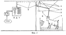
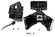
5. В момент стимуляций животное не должно испытывать иммобилизационный стресс. В качестве примера может рекомендоваться схема установки, применявшейся в опытах на козах (фиг.3). При этом для предотвращения повреждений внешних концов электродов проводники от них, прикрепленные к муфте, следует фиксировать на неподвижном каркасе, находящемся на животном. В качестве образца можно рекомендовать съемный резиновый бандаж с укрепленной на нем разъемной муфтой для соединения с проводниками от генератора стимулов (фиг.4), который, в частности, в опытах на рогатых козах можно крепить на рогах. 5. At the time of stimulation, the animal should not experience immobilization stress. As an example, the installation scheme used in experiments on goats (Figure 3) can be recommended. In this case, to prevent damage to the outer ends of the electrodes, the conductors from them, attached to the coupling, should be fixed on a fixed frame located on the animal. As a sample, a removable rubber band with a detachable sleeve mounted on it for connection with conductors from a stimulus generator (Fig. 4), which, in particular, in experiments on horned goats, can be mounted on horns, can be recommended.
6. Непосредственно перед началом электрических стимуляций у животного производится раздражение (механическое, химическое и др.) рецепторного аппарата исследуемых органов, вызывающее рефлекторное изменение функционального состояния центральной нервной системы для его идентификации перед каждым сеансом стимуляций. В порядке примера - в опытах по изучению влияния стимуляций отдельных структур гипоталамуса на секрецию молока и синтез его отдельных органических компонентов такую стимуляцию начинали сразу же по окончании очередного доения. Известно, что доение вызывает всегда определенные изменения в деятельности целого ряда органов и систем и, таким образом, центральной нервной системы в целом (6, 7, 8). 6. Immediately before the start of electrical stimulations, the animal produces irritation (mechanical, chemical, etc.) of the receptor apparatus of the organs under investigation, causing a reflex change in the functional state of the central nervous system to identify it before each stimulation session. As an example, in experiments to study the effect of stimulations of individual structures of the hypothalamus on the secretion of milk and the synthesis of its individual organic components, such stimulation began immediately after the end of the next milking. It is known that milking always causes certain changes in the activity of a number of organs and systems and, thus, the central nervous system as a whole (6, 7, 8).
Примеры конкретного использования заявляемого способа. Examples of specific use of the proposed method.
Заявляемый способ разработан в опытах на лактирующих козах, на которых изучалась роль отдельных структур гипоталамуса в регуляции секреторной активности молочной железы. Подопытным и ложнооперированным животным в отдельные гипоталамические образования вживляли биополярные электроды. В период установившейся лактации производили электрические раздражения подэлектродных структур с помощью различных по форме электрических импульсов у подопытных животных, ложнооперированные служили контролем. Сравнивали молочную продуктивность в период стимуляций в течение 5 дней с продуктивностью в дни перерывов в стимуляции (5-7 дней) и сопоставляли эти показатели с таковыми же у контрольных животных. Сравнивались результаты, получаемые при стимуляции после ручных доений и без таковых, когда молоко получали из вымени методом постоянного оттока, описанного Г.Б. Тверским (10). The inventive method was developed in experiments on lactating goats, which studied the role of individual structures of the hypothalamus in the regulation of secretory activity of the mammary gland. Biological polar electrodes were implanted into experimental and falsely operated animals in separate hypothalamic formations. During steady-state lactation, electrical irritations of the sub-electrode structures were produced with the help of different electrical impulses in experimental animals, the false-operated ones served as a control. Milk productivity was compared during stimulation for 5 days with productivity on days of breaks in stimulation (5-7 days) and these indices were compared with those in control animals. The results obtained during stimulation after manual milking and without it were compared when milk was obtained from the udder by the method of constant outflow described by G. B. Tversky (10).
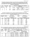
Изучалась роль латерального гипоталамического поля, для которой известна важная роль в регуляции ряда вегето-висцеральных систем организма (9). Сравнение результатов стимуляции одних и тех же структур в разные периоды опыта с помощью прямоугольных импульсов (с параметрами силы тока и длительности импульсов, определяемыми как оптимальные по результатам предшествующих экспериментов) и заявляемых в данной заявке импульсов "естественной ритмики", представлено в табл.1. The role of the lateral hypothalamic field was studied, for which an important role in the regulation of a number of vegeto-visceral systems of the body is known (9). A comparison of the results of stimulation of the same structures in different periods of the experiment using rectangular pulses (with parameters of current strength and pulse duration, defined as optimal according to the results of previous experiments) and pulses of "natural rhythm" declared in this application are presented in Table 1.
Таким образом, под влиянием "естественной ритмики" наблюдалась несколько более заметная активизация секторных процессов в молочной железе по сравнению с прямоугольными импульсами при их адекватных параметрах. При этом также несколько снижалась и амплитуда вариационного ряда. Thus, under the influence of “natural rhythm”, a slightly more noticeable activation of sector processes in the mammary gland was observed compared with rectangular pulses with their adequate parameters. At the same time, the amplitude of the variation series also decreased slightly.
Если начало стимуляций указанной области гипоталамуса откладывалось после окончания доения на 30-60 мин, то ответная реакция оказывалась маловыразительной. Такая же картина наблюдалась и при применении других параметров подачи импульсации, т. е. при других соотношениях времени непрерывной стимуляции и времени перерывов. If the beginning of stimulation of the indicated area of the hypothalamus was postponed after milking for 30-60 minutes, then the response was not very pronounced. The same picture was observed when applying other parameters of the impulse supply, i.e., with other ratios of continuous stimulation time and break time.
Сравнение результатов стимуляции латерального гипоталамического поля разными по средней силе тока импульсами "естественной ритмики" представлено в табл.2. A comparison of the results of stimulation of the lateral hypothalamic field by pulses of "natural rhythm" different in average current strength is presented in Table 2.
Таким образом, оптимальными являются импульсы с силой тока в пределах от 30 до 50 мкА, до 30 мкА - подпороговыми, а свыше 50 - пессимальными. Thus, pulses with a current strength in the range from 30 to 50 μA, up to 30 μA are subthreshold, and more than 50 are pessimal.
Известно, что одним из важнейших путей влияния гипоталамуса на лактационную функцию организма является трансгипофизарный, т.е. через секрецию гипофизарных гормонов. Это достаточно четко доказано в многочисленных экспериментах (6, 10). Поэтому одним из объективных критериев адекватности стимулирующих посылок при стимуляции гипоталамической области мозга может служить показатель изменения концентрации в крови гипофизарных гормонов при таких воздействиях. It is known that one of the most important ways the hypothalamus affects the lactational function of the body is transhypophysial, i.e. through the secretion of pituitary hormones. This is quite clearly proven in numerous experiments (6, 10). Therefore, one of the objective criteria for the adequacy of stimulating premises during stimulation of the hypothalamic region of the brain can serve as an indicator of changes in the concentration of pituitary hormones in the blood under such influences.
Результаты использования в качестве тест-объекта гормона аденогипофиза - пролактина, - отличающегося многогранным действием в организме и отзывающегося на стимуляцию рецепторов молочной железы (6, 8, 10), представлены в табл.3. The results of using the hormone of the adenohypophysis, prolactin, as a test object, which has a multifaceted effect in the body and responds to the stimulation of mammary receptors (6, 8, 10), are presented in Table 3.
Таким образом, импульсы "естественной ритмики" и по влиянию на секрецию пролактина оказались более адекватными по сравнению с прямоугольными. Более адекватными оказались они и в опытах по исследованию роли соседних с гипоталамусом подкорковых структур, таких как таламус, миндалевидный комплекс, прозрачная перегородка, гиппокамп. Thus, the impulses of "natural rhythm" and the influence on the secretion of prolactin were more adequate than rectangular ones. They turned out to be more adequate in experiments on the role of subcortical structures adjacent to the hypothalamus, such as the thalamus, amygdala complex, transparent septum, hippocampus.
Заявляемый способ может рекомендоваться также и для диагностики и лечения при нарушениях центральной регуляции вегето-висцеральных функций организма своеобразным их "протезированием". The inventive method can also be recommended for diagnosis and treatment for violations of the central regulation of vegetative-visceral functions of the body by their peculiar "prosthetics".
Литература
1. Чалисова Н. И. Изменения в тканях мозга при хронически вживленных электродах. - Тр. Ленинградского научного общества паталого-анатомов. Л., 1971, в.12, с.109-113.Literature
1. Chalisova N. I. Changes in brain tissue with chronically implanted electrodes. - Tr. Leningrad Scientific Society of Pathologist Anatomists. L., 1971, p. 12, pp. 109-113.
2. Грачев И.И., Галанцев В.П. Физиология лактации. Л., "Наука", 1972. 2. Grachev I.I., Galantsev V.P. The physiology of lactation. L., "Science", 1972.
3. Хольст Н. , Сент-Пол У. О функциональной организации драйвов. Поведение животных. 1962, в.ХI, с.1-19. 3. Holst N., St. Paul W. On the functional organization of drives. Animal behavior. 1962, century ХI, p.1-19.
4. Гурфинкель В.С., Малкин В.В., Цетлин М.Л., Шнейдер А.Ю. Биоэлектрическое управление. М., "Наука", 1972. 4. Gurfinkel V.S., Malkin V.V., Tsetlin M.L., Schneider A.Yu. Bioelectric control. M., "Science", 1972.
5. Кратин Ю. Г., Гусельников В.И. Техника и методика электроэнцефалографии. Л., 1971. 5. Kratin Yu. G., Guselnikov V.I. Technique and technique of electroencephalography. L., 1971.
6. Тараненко А.Г. Пролактин и лактация сельскохозяйственных животных. В сб. : "Эндокринология и трансплантация сельско-хозяйственных животных". М., Колос, 1982, с.328-342. 6. Taranenko A.G. Prolactin and lactation of farm animals. On Sat : "Endocrinology and transplantation of farm animals." M., Kolos, 1982, p. 328-342.
7. Огородний Ю.М. Основные закономерности секреторной деятельности молочной железы и ее регуляция. Тр. Кишиневского СХИ, Кишинев, 1967, т.48. 7. Ogorodniy Yu.M. The main laws of the secretory activity of the mammary gland and its regulation. Tr. Chisinau Agricultural Institute, Chisinau, 1967, t. 48.
8. Марченко Г.М. О закономерностях секреторной деятельности молочной железы жвачных животных. - Автореферат дисс. доктора биологических наук. Л., 1971. 8. Marchenko G.M. On the patterns of secretory activity of the mammary gland of ruminants. - Abstract of diss. Doctors of biological sciences. L., 1971.
9. Гращенков Н. И. Гипоталамус. Его роль в физиологии и патологии. М., Медицина, 1964. 9. Grashchenkov N. I. Hypothalamus. Its role in physiology and pathology. M., Medicine, 1964.
10. Тверской Г.Б. Регуляция секреции молока. Л., "Наука", 1972. 10. Tverskoy G.B. Regulation of milk secretion. L., "Science", 1972.
Claims (1)
Priority Applications (1)
| Application Number | Priority Date | Filing Date | Title |
|---|---|---|---|
| RU2000111595A RU2198001C2 (en) | 2000-05-10 | 2000-05-10 | Subcortical brain structures electric stimulation method usable in chronic experimental studies |
Applications Claiming Priority (1)
| Application Number | Priority Date | Filing Date | Title |
|---|---|---|---|
| RU2000111595A RU2198001C2 (en) | 2000-05-10 | 2000-05-10 | Subcortical brain structures electric stimulation method usable in chronic experimental studies |
Publications (2)
| Publication Number | Publication Date |
|---|---|
| RU2000111595A RU2000111595A (en) | 2002-02-27 |
| RU2198001C2 true RU2198001C2 (en) | 2003-02-10 |
Family
ID=20234385
Family Applications (1)
| Application Number | Title | Priority Date | Filing Date |
|---|---|---|---|
| RU2000111595A RU2198001C2 (en) | 2000-05-10 | 2000-05-10 | Subcortical brain structures electric stimulation method usable in chronic experimental studies |
Country Status (1)
| Country | Link |
|---|---|
| RU (1) | RU2198001C2 (en) |
Cited By (1)
| Publication number | Priority date | Publication date | Assignee | Title |
|---|---|---|---|---|
| RU2368402C2 (en) * | 2007-04-23 | 2009-09-27 | Государственное образовательное учреждение высшего профессионального образования "Тверская государственная медицинская академия Федерального агентства по здравоохранению и социальному развитию" | Method of obtaining feeding behavioural responses during chronic experiment and device for obtaining feeding behavioural responses during chronic experiment |
Citations (3)
| Publication number | Priority date | Publication date | Assignee | Title |
|---|---|---|---|---|
| SU908378A1 (en) * | 1976-05-03 | 1982-02-28 | Харьковский Научно-Исследовательский Институт Неврологии И Психиатрии | Method of control of animal's behaviour |
| RU2092102C1 (en) * | 1993-04-30 | 1997-10-10 | Научно-исследовательский институт кардиологии | Method of recording of electrical activity of nerve in chronic experiment |
| RU2113839C1 (en) * | 1989-11-01 | 1998-06-27 | ЭсПиИЭс Пейтентс Лимитед | Electrode and device for performing transcranial electrotherapy |
-
2000
- 2000-05-10 RU RU2000111595A patent/RU2198001C2/en active
Patent Citations (3)
| Publication number | Priority date | Publication date | Assignee | Title |
|---|---|---|---|---|
| SU908378A1 (en) * | 1976-05-03 | 1982-02-28 | Харьковский Научно-Исследовательский Институт Неврологии И Психиатрии | Method of control of animal's behaviour |
| RU2113839C1 (en) * | 1989-11-01 | 1998-06-27 | ЭсПиИЭс Пейтентс Лимитед | Electrode and device for performing transcranial electrotherapy |
| RU2092102C1 (en) * | 1993-04-30 | 1997-10-10 | Научно-исследовательский институт кардиологии | Method of recording of electrical activity of nerve in chronic experiment |
Cited By (1)
| Publication number | Priority date | Publication date | Assignee | Title |
|---|---|---|---|---|
| RU2368402C2 (en) * | 2007-04-23 | 2009-09-27 | Государственное образовательное учреждение высшего профессионального образования "Тверская государственная медицинская академия Федерального агентства по здравоохранению и социальному развитию" | Method of obtaining feeding behavioural responses during chronic experiment and device for obtaining feeding behavioural responses during chronic experiment |
Similar Documents
| Publication | Publication Date | Title |
|---|---|---|
| Nakamura et al. | Direct and indirect activation of human corticospinal neurons by transcranial magnetic and electrical stimulation | |
| Sinclair | Role of the pharyngeal plexus in initiation of swallowing | |
| US20100292527A1 (en) | Device and method for hypertension treatment by non-invasive stimulation to vascular baroreceptors | |
| ATE537872T1 (en) | DEVICE FOR THE TREATMENT OF NEUROLOGICAL DISORDERS USING CHRONIC ADAPTIVE BRAIN STIMULATION DEPENDENT ON LOCAL BIOPOTENTIALS | |
| TW202341924A (en) | An apparatus for delivering variable pulse | |
| Gebodh et al. | Transcranial direct current stimulation among technologies for low-intensity transcranial electrical stimulation: classification, history, and terminology | |
| US20240189614A1 (en) | Apparatus and method for treatment of mental and behavioral conditions and disorders with electromegnetic fields | |
| YAMAMOTO et al. | Microelectrode studies on sensory afferents in the posterior funiculus of cat | |
| US6298265B1 (en) | Electrode design and stimulator for antler-bearing animals | |
| Bartels et al. | Representation of lateral line and electrosensory systems in the midbrain of the axolotl, Ambystoma mexicanum | |
| Wester | Habituation to electrical stimulation of the thalamus in unanaesthetized cats | |
| US4243043A (en) | Apparatus for electrical stimulation of mammae | |
| Phillips | Evoked potential study of connections of avian archistriatum and caudal neostriatum | |
| Delgado | Radio Stimulation of the Brain in Primates and Man Fourth Becton, Dickinson and Company Oscar Schwidetzky Memorial Lecture | |
| Durelli et al. | Single-unit chronic recordings from the sensorimotor cortex of unrestrained cats during locomotion | |
| Hayward et al. | Hypothalamic and cortical DC potential changes induced by stimulation of the midbrain reticular formation | |
| Schjetnan et al. | Anodal transcranial direct current stimulation with monopolar pulses improves limb use after stroke by enhancing inter‑hemispheric coherence | |
| Delgado | Modulation of emotions by cerebral radio stimulation | |
| Whalen et al. | Electrophysiologic and behavioral studies of the cutaneous nerves of the concave surface of the pinna and the external ear canal of the dog | |
| Galin et al. | Reaction time and heart rate response pattern: Effects of reticular stimulation in cats | |
| Shannon et al. | Medullary neurons mediating the inhibition of inspiration by intercostal muscle tendon organs? | |
| Laming et al. | Sustained potential shifts in the toad tectum reflect prey-catching and avoidance behavior. | |
| Majkowski et al. | Cross-modality comparisons of averaged evoked potentials, their relation to vigilance and stimulus parameters in cats | |
| SU1183120A1 (en) | Method of attaching an electrode for temporary stimulation of the nerve | |
| Saienko | Brain stimulation therapies |