RU2646103C2 - Method for renatured protein g isolation from marked polyacrylamide gel - Google Patents
Method for renatured protein g isolation from marked polyacrylamide gel Download PDFInfo
- Publication number
- RU2646103C2 RU2646103C2 RU2016111874A RU2016111874A RU2646103C2 RU 2646103 C2 RU2646103 C2 RU 2646103C2 RU 2016111874 A RU2016111874 A RU 2016111874A RU 2016111874 A RU2016111874 A RU 2016111874A RU 2646103 C2 RU2646103 C2 RU 2646103C2
- Authority
- RU
- Russia
- Prior art keywords
- protein
- gel
- hours
- stage
- buffer
- Prior art date
Links
- 238000000034 method Methods 0.000 title claims abstract description 17
- 229920002401 polyacrylamide Polymers 0.000 title claims abstract description 9
- 238000002955 isolation Methods 0.000 title abstract description 4
- 102000004169 proteins and genes Human genes 0.000 title description 36
- 108090000623 proteins and genes Proteins 0.000 title description 36
- 101710120037 Toxin CcdB Proteins 0.000 claims abstract description 74
- 239000000243 solution Substances 0.000 claims abstract description 46
- XSQUKJJJFZCRTK-UHFFFAOYSA-N Urea Chemical compound NC(N)=O XSQUKJJJFZCRTK-UHFFFAOYSA-N 0.000 claims abstract description 26
- FAPWRFPIFSIZLT-UHFFFAOYSA-M Sodium chloride Chemical compound [Na+].[Cl-] FAPWRFPIFSIZLT-UHFFFAOYSA-M 0.000 claims abstract description 20
- 239000000872 buffer Substances 0.000 claims abstract description 20
- 239000007983 Tris buffer Substances 0.000 claims abstract description 13
- 239000007853 buffer solution Substances 0.000 claims abstract description 13
- LENZDBCJOHFCAS-UHFFFAOYSA-N tris Chemical compound OCC(N)(CO)CO LENZDBCJOHFCAS-UHFFFAOYSA-N 0.000 claims abstract description 13
- 239000004202 carbamide Substances 0.000 claims abstract description 11
- 239000012634 fragment Substances 0.000 claims abstract description 11
- 239000011780 sodium chloride Substances 0.000 claims abstract description 10
- UMGDCJDMYOKAJW-UHFFFAOYSA-N thiourea Chemical compound NC(N)=S UMGDCJDMYOKAJW-UHFFFAOYSA-N 0.000 claims abstract description 8
- KCXVZYZYPLLWCC-UHFFFAOYSA-N EDTA Chemical compound OC(=O)CN(CC(O)=O)CCN(CC(O)=O)CC(O)=O KCXVZYZYPLLWCC-UHFFFAOYSA-N 0.000 claims abstract description 7
- 239000013504 Triton X-100 Substances 0.000 claims abstract description 6
- 229920004890 Triton X-100 Polymers 0.000 claims abstract description 6
- 239000013578 denaturing buffer Substances 0.000 claims abstract description 5
- PSBDWGZCVUAZQS-UHFFFAOYSA-N (dimethylsulfonio)acetate Chemical compound C[S+](C)CC([O-])=O PSBDWGZCVUAZQS-UHFFFAOYSA-N 0.000 claims abstract description 4
- 230000008859 change Effects 0.000 claims abstract description 4
- 239000008057 potassium phosphate buffer Substances 0.000 claims abstract description 4
- 229940117986 sulfobetaine Drugs 0.000 claims abstract description 4
- 238000004153 renaturation Methods 0.000 claims description 15
- 238000003756 stirring Methods 0.000 claims description 5
- QKNYBSVHEMOAJP-UHFFFAOYSA-N 2-amino-2-(hydroxymethyl)propane-1,3-diol;hydron;chloride Chemical compound Cl.OCC(N)(CO)CO QKNYBSVHEMOAJP-UHFFFAOYSA-N 0.000 claims description 4
- 239000012134 supernatant fraction Substances 0.000 claims description 3
- 230000001580 bacterial effect Effects 0.000 claims description 2
- 238000004925 denaturation Methods 0.000 claims description 2
- 230000036425 denaturation Effects 0.000 claims description 2
- 238000012545 processing Methods 0.000 claims description 2
- 239000000725 suspension Substances 0.000 claims description 2
- 238000005119 centrifugation Methods 0.000 claims 2
- 239000008188 pellet Substances 0.000 claims 1
- 238000001226 reprecipitation Methods 0.000 claims 1
- 238000002415 sodium dodecyl sulfate polyacrylamide gel electrophoresis Methods 0.000 claims 1
- 238000000926 separation method Methods 0.000 abstract description 5
- 230000001900 immune effect Effects 0.000 abstract description 3
- 230000000694 effects Effects 0.000 abstract 1
- 238000010828 elution Methods 0.000 abstract 1
- 239000000126 substance Substances 0.000 abstract 1
- 235000018102 proteins Nutrition 0.000 description 35
- 239000000499 gel Substances 0.000 description 34
- 239000012528 membrane Substances 0.000 description 29
- 238000005406 washing Methods 0.000 description 29
- 238000011534 incubation Methods 0.000 description 26
- 241000699666 Mus <mouse, genus> Species 0.000 description 16
- 241000283973 Oryctolagus cuniculus Species 0.000 description 15
- 238000001962 electrophoresis Methods 0.000 description 14
- 210000002966 serum Anatomy 0.000 description 14
- 108060003951 Immunoglobulin Proteins 0.000 description 13
- 102000018358 immunoglobulin Human genes 0.000 description 13
- 229940072221 immunoglobulins Drugs 0.000 description 13
- WCUXLLCKKVVCTQ-UHFFFAOYSA-M Potassium chloride Chemical compound [Cl-].[K+] WCUXLLCKKVVCTQ-UHFFFAOYSA-M 0.000 description 12
- 238000010790 dilution Methods 0.000 description 12
- 239000012895 dilution Substances 0.000 description 12
- 239000000020 Nitrocellulose Substances 0.000 description 10
- DBMJMQXJHONAFJ-UHFFFAOYSA-M Sodium laurylsulphate Chemical compound [Na+].CCCCCCCCCCCCOS([O-])(=O)=O DBMJMQXJHONAFJ-UHFFFAOYSA-M 0.000 description 10
- 229920001220 nitrocellulos Polymers 0.000 description 10
- 238000002360 preparation method Methods 0.000 description 10
- 239000000758 substrate Substances 0.000 description 10
- 108010001336 Horseradish Peroxidase Proteins 0.000 description 8
- MHAJPDPJQMAIIY-UHFFFAOYSA-N Hydrogen peroxide Chemical compound OO MHAJPDPJQMAIIY-UHFFFAOYSA-N 0.000 description 8
- 238000003119 immunoblot Methods 0.000 description 8
- 238000001042 affinity chromatography Methods 0.000 description 7
- 230000000984 immunochemical effect Effects 0.000 description 7
- 238000001179 sorption measurement Methods 0.000 description 7
- 238000002965 ELISA Methods 0.000 description 6
- 239000003593 chromogenic compound Substances 0.000 description 6
- 239000001103 potassium chloride Substances 0.000 description 6
- 235000011164 potassium chloride Nutrition 0.000 description 6
- 108010088751 Albumins Proteins 0.000 description 5
- 102000009027 Albumins Human genes 0.000 description 5
- 108091003079 Bovine Serum Albumin Proteins 0.000 description 5
- 229920002684 Sepharose Polymers 0.000 description 5
- 239000002981 blocking agent Substances 0.000 description 5
- 229940098773 bovine serum albumin Drugs 0.000 description 5
- 239000013592 cell lysate Substances 0.000 description 5
- 239000000203 mixture Substances 0.000 description 5
- QAOWNCQODCNURD-UHFFFAOYSA-N Sulfuric acid Chemical compound OS(O)(=O)=O QAOWNCQODCNURD-UHFFFAOYSA-N 0.000 description 4
- 238000004458 analytical method Methods 0.000 description 4
- 230000027455 binding Effects 0.000 description 4
- 238000006243 chemical reaction Methods 0.000 description 4
- 230000003993 interaction Effects 0.000 description 4
- 238000012546 transfer Methods 0.000 description 4
- XLYOFNOQVPJJNP-UHFFFAOYSA-N water Chemical compound O XLYOFNOQVPJJNP-UHFFFAOYSA-N 0.000 description 4
- DGVVWUTYPXICAM-UHFFFAOYSA-N β‐Mercaptoethanol Chemical compound OCCS DGVVWUTYPXICAM-UHFFFAOYSA-N 0.000 description 4
- PEDCQBHIVMGVHV-UHFFFAOYSA-N Glycerine Chemical compound OCC(O)CO PEDCQBHIVMGVHV-UHFFFAOYSA-N 0.000 description 3
- 241001465754 Metazoa Species 0.000 description 3
- 230000000903 blocking effect Effects 0.000 description 3
- 235000018417 cysteine Nutrition 0.000 description 3
- 238000002474 experimental method Methods 0.000 description 3
- 238000011049 filling Methods 0.000 description 3
- 230000005714 functional activity Effects 0.000 description 3
- 229920001184 polypeptide Polymers 0.000 description 3
- 102000004196 processed proteins & peptides Human genes 0.000 description 3
- 108090000765 processed proteins & peptides Proteins 0.000 description 3
- 238000010186 staining Methods 0.000 description 3
- GEYOCULIXLDCMW-UHFFFAOYSA-N 1,2-phenylenediamine Chemical compound NC1=CC=CC=C1N GEYOCULIXLDCMW-UHFFFAOYSA-N 0.000 description 2
- GHCZTIFQWKKGSB-UHFFFAOYSA-N 2-hydroxypropane-1,2,3-tricarboxylic acid;phosphoric acid Chemical compound OP(O)(O)=O.OC(=O)CC(O)(C(O)=O)CC(O)=O GHCZTIFQWKKGSB-UHFFFAOYSA-N 0.000 description 2
- HSTOKWSFWGCZMH-UHFFFAOYSA-N 3,3'-diaminobenzidine Chemical compound C1=C(N)C(N)=CC=C1C1=CC=C(N)C(N)=C1 HSTOKWSFWGCZMH-UHFFFAOYSA-N 0.000 description 2
- UMCMPZBLKLEWAF-BCTGSCMUSA-N 3-[(3-cholamidopropyl)dimethylammonio]propane-1-sulfonate Chemical compound C([C@H]1C[C@H]2O)[C@H](O)CC[C@]1(C)[C@@H]1[C@@H]2[C@@H]2CC[C@H]([C@@H](CCC(=O)NCCC[N+](C)(C)CCCS([O-])(=O)=O)C)[C@@]2(C)[C@@H](O)C1 UMCMPZBLKLEWAF-BCTGSCMUSA-N 0.000 description 2
- IJGRMHOSHXDMSA-UHFFFAOYSA-N Atomic nitrogen Chemical compound N#N IJGRMHOSHXDMSA-UHFFFAOYSA-N 0.000 description 2
- BVKZGUZCCUSVTD-UHFFFAOYSA-L Carbonate Chemical compound [O-]C([O-])=O BVKZGUZCCUSVTD-UHFFFAOYSA-L 0.000 description 2
- LEVWYRKDKASIDU-QWWZWVQMSA-N D-cystine Chemical compound OC(=O)[C@H](N)CSSC[C@@H](N)C(O)=O LEVWYRKDKASIDU-QWWZWVQMSA-N 0.000 description 2
- LFQSCWFLJHTTHZ-UHFFFAOYSA-N Ethanol Chemical compound CCO LFQSCWFLJHTTHZ-UHFFFAOYSA-N 0.000 description 2
- DHMQDGOQFOQNFH-UHFFFAOYSA-N Glycine Chemical compound NCC(O)=O DHMQDGOQFOQNFH-UHFFFAOYSA-N 0.000 description 2
- 102000008394 Immunoglobulin Fragments Human genes 0.000 description 2
- 108010021625 Immunoglobulin Fragments Proteins 0.000 description 2
- 229910021586 Nickel(II) chloride Inorganic materials 0.000 description 2
- 108010090804 Streptavidin Proteins 0.000 description 2
- 230000002776 aggregation Effects 0.000 description 2
- 238000004220 aggregation Methods 0.000 description 2
- 238000003776 cleavage reaction Methods 0.000 description 2
- 238000004040 coloring Methods 0.000 description 2
- XUJNEKJLAYXESH-UHFFFAOYSA-N cysteine Natural products SCC(N)C(O)=O XUJNEKJLAYXESH-UHFFFAOYSA-N 0.000 description 2
- 229960003067 cystine Drugs 0.000 description 2
- 239000008367 deionised water Substances 0.000 description 2
- 229910021641 deionized water Inorganic materials 0.000 description 2
- 238000007865 diluting Methods 0.000 description 2
- 238000004090 dissolution Methods 0.000 description 2
- 239000012153 distilled water Substances 0.000 description 2
- 238000010438 heat treatment Methods 0.000 description 2
- 229910052739 hydrogen Inorganic materials 0.000 description 2
- QMMRZOWCJAIUJA-UHFFFAOYSA-L nickel dichloride Chemical compound Cl[Ni]Cl QMMRZOWCJAIUJA-UHFFFAOYSA-L 0.000 description 2
- 102000013415 peroxidase activity proteins Human genes 0.000 description 2
- 108040007629 peroxidase activity proteins Proteins 0.000 description 2
- 239000008363 phosphate buffer Substances 0.000 description 2
- 230000008569 process Effects 0.000 description 2
- 238000001742 protein purification Methods 0.000 description 2
- 230000007017 scission Effects 0.000 description 2
- 235000020183 skimmed milk Nutrition 0.000 description 2
- 238000006467 substitution reaction Methods 0.000 description 2
- 238000012360 testing method Methods 0.000 description 2
- 238000004448 titration Methods 0.000 description 2
- 238000000108 ultra-filtration Methods 0.000 description 2
- 229920000936 Agarose Polymers 0.000 description 1
- 241000486679 Antitype Species 0.000 description 1
- 102000004506 Blood Proteins Human genes 0.000 description 1
- 108010017384 Blood Proteins Proteins 0.000 description 1
- 241000588724 Escherichia coli Species 0.000 description 1
- 241000701533 Escherichia virus T4 Species 0.000 description 1
- 239000004471 Glycine Substances 0.000 description 1
- 241000699670 Mus sp. Species 0.000 description 1
- 229920001213 Polysorbate 20 Polymers 0.000 description 1
- 101710172711 Structural protein Proteins 0.000 description 1
- 230000000840 anti-viral effect Effects 0.000 description 1
- 230000002155 anti-virotic effect Effects 0.000 description 1
- 230000000890 antigenic effect Effects 0.000 description 1
- 238000013459 approach Methods 0.000 description 1
- 230000001174 ascending effect Effects 0.000 description 1
- 230000004888 barrier function Effects 0.000 description 1
- 230000015572 biosynthetic process Effects 0.000 description 1
- UDSAIICHUKSCKT-UHFFFAOYSA-N bromophenol blue Chemical compound C1=C(Br)C(O)=C(Br)C=C1C1(C=2C=C(Br)C(O)=C(Br)C=2)C2=CC=CC=C2S(=O)(=O)O1 UDSAIICHUKSCKT-UHFFFAOYSA-N 0.000 description 1
- 238000004364 calculation method Methods 0.000 description 1
- 239000003795 chemical substances by application Substances 0.000 description 1
- 238000004587 chromatography analysis Methods 0.000 description 1
- 150000001945 cysteines Chemical class 0.000 description 1
- 238000010586 diagram Methods 0.000 description 1
- 238000000502 dialysis Methods 0.000 description 1
- LOKCTEFSRHRXRJ-UHFFFAOYSA-I dipotassium trisodium dihydrogen phosphate hydrogen phosphate dichloride Chemical compound P(=O)(O)(O)[O-].[K+].P(=O)(O)([O-])[O-].[Na+].[Na+].[Cl-].[K+].[Cl-].[Na+] LOKCTEFSRHRXRJ-UHFFFAOYSA-I 0.000 description 1
- 230000003203 everyday effect Effects 0.000 description 1
- 238000013401 experimental design Methods 0.000 description 1
- 239000011521 glass Substances 0.000 description 1
- 230000001759 immunoprophylactic effect Effects 0.000 description 1
- 210000003000 inclusion body Anatomy 0.000 description 1
- 239000007788 liquid Substances 0.000 description 1
- 230000007774 longterm Effects 0.000 description 1
- 229920002521 macromolecule Polymers 0.000 description 1
- MYWUZJCMWCOHBA-VIFPVBQESA-N methamphetamine Chemical compound CN[C@@H](C)CC1=CC=CC=C1 MYWUZJCMWCOHBA-VIFPVBQESA-N 0.000 description 1
- 229910052757 nitrogen Inorganic materials 0.000 description 1
- 230000009871 nonspecific binding Effects 0.000 description 1
- 239000002953 phosphate buffered saline Substances 0.000 description 1
- 238000002264 polyacrylamide gel electrophoresis Methods 0.000 description 1
- 239000000256 polyoxyethylene sorbitan monolaurate Substances 0.000 description 1
- 235000010486 polyoxyethylene sorbitan monolaurate Nutrition 0.000 description 1
- 235000010482 polyoxyethylene sorbitan monooleate Nutrition 0.000 description 1
- 229920000053 polysorbate 80 Polymers 0.000 description 1
- 239000002244 precipitate Substances 0.000 description 1
- 238000001556 precipitation Methods 0.000 description 1
- 230000002035 prolonged effect Effects 0.000 description 1
- 239000012460 protein solution Substances 0.000 description 1
- 238000000746 purification Methods 0.000 description 1
- 230000002441 reversible effect Effects 0.000 description 1
- 229920006395 saturated elastomer Polymers 0.000 description 1
- 238000007789 sealing Methods 0.000 description 1
- 239000007790 solid phase Substances 0.000 description 1
- 230000001629 suppression Effects 0.000 description 1
- 238000000954 titration curve Methods 0.000 description 1
- GPRLSGONYQIRFK-MNYXATJNSA-N triton Chemical compound [3H+] GPRLSGONYQIRFK-MNYXATJNSA-N 0.000 description 1
- 238000012800 visualization Methods 0.000 description 1
Images

Classifications
-
- C—CHEMISTRY; METALLURGY
- C07—ORGANIC CHEMISTRY
- C07K—PEPTIDES
- C07K1/00—General methods for the preparation of peptides, i.e. processes for the organic chemical preparation of peptides or proteins of any length
- C07K1/14—Extraction; Separation; Purification
-
- C—CHEMISTRY; METALLURGY
- C07—ORGANIC CHEMISTRY
- C07K—PEPTIDES
- C07K1/00—General methods for the preparation of peptides, i.e. processes for the organic chemical preparation of peptides or proteins of any length
- C07K1/14—Extraction; Separation; Purification
- C07K1/24—Extraction; Separation; Purification by electrochemical means
- C07K1/26—Electrophoresis
Landscapes
- Chemical & Material Sciences (AREA)
- Organic Chemistry (AREA)
- Biophysics (AREA)
- General Health & Medical Sciences (AREA)
- Analytical Chemistry (AREA)
- Health & Medical Sciences (AREA)
- Life Sciences & Earth Sciences (AREA)
- Biochemistry (AREA)
- Electrochemistry (AREA)
- Chemical Kinetics & Catalysis (AREA)
- Genetics & Genomics (AREA)
- Medicinal Chemistry (AREA)
- Molecular Biology (AREA)
- Proteomics, Peptides & Aminoacids (AREA)
- General Chemical & Material Sciences (AREA)
- Peptides Or Proteins (AREA)
Abstract
Description
Изобретение относится к биотехнологии и описывает сочетанный способ очистки и ренатурации белка G, который может использоваться при создании специфических иммунодиагностических и иммунопрофилактических средств.The invention relates to biotechnology and describes a combined method of purification and renaturation of protein G, which can be used to create specific immunodiagnostic and immunoprophylactic agents.
Известны многочисленные методы очистки белков, включающие, в том числе, электроэлюцию из агарозных и полиакриламидных гелей, которые широко распространены в повседневной лабораторной практике [Михайлов А.Т., Симирский В.Н. Методы иммунохимического анализа в биологии развития. - М.: Наука, 1991. - 290 с., Финдлей Д.Б. Биологические мембраны: Методы / Под ред. Дж. Финдлея, У. Эванза. М.: Мир, 1990. С. 271-273]. Их популярность связана с возможностью наработать некоторые необходимые для исследователей количества белка и избегать при этом трудоемких этапов выделения индивидуальных белков как и фрагментов ДНК (для выделения последних электроэлюция применяется значительно чаще). При очистке белков электроэлюция может не только выступать в качестве единственного и самостоятельного метода выделения, но и дополняется в некоторых случаях хроматографией, дифференциальным осажденим и другими.Numerous methods for protein purification are known, including, inter alia, electroelution from agarose and polyacrylamide gels, which are widespread in everyday laboratory practice [Mikhailov AT, Simirsky VN Methods of immunochemical analysis in developmental biology. - M .: Nauka, 1991 .-- 290 p., Findlay D.B. Biological membranes: Methods / Ed. J. Findlay, W. Evanza. M.: Mir, 1990. S. 271-273]. Their popularity is associated with the ability to produce some amounts of protein necessary for researchers and to avoid the laborious steps of isolating individual proteins as well as DNA fragments (to isolate the latter, electroelution is used much more often). During protein purification, electroelution can not only act as the only and independent method of isolation, but also is supplemented in some cases by chromatography, differential precipitation, and others.
Известны ренатурационные способы, имеющие одно принципиальное сходство, которое заключается в разбавлении концентрированного раствора денатурированного белка специальными буферами, что сопровождается образованием нативной, биологически активной структурой полипептида [Richard R, Burgess 2009. Meth Enzym 463: 259-281].Renaturation methods are known that have one fundamental similarity, which consists in diluting a concentrated solution of denatured protein with special buffers, which is accompanied by the formation of a native, biologically active structure of the polypeptide [Richard R, Burgess 2009. Meth Enzym 463: 259-281].
К настоящему времени опубликованы многочисленные протоколы ренатурации отдельных белков способом, в основе которого лежит разведение ренатурационных растворов, однако, метод имеет несколько существенных недостатков [Cabrita LD, Bottomley SP. 2004. Biotech Ann Rev 10: 31-49]. Известна альтернативная техника перевода белков в растворы с низкой ионной силой, использующая диализ или ультрафильтрацию [Yoshii Н, Furuta Т, Yonehara Т, Ito D, Linko Y-Y, Linko P: Biosci Biotechnol Biochem 2000, 64:1159-1165].To date, numerous protocols have been published for the renaturation of individual proteins by a method based on dilution of renaturation solutions, however, the method has several significant drawbacks [Cabrita LD, Bottomley SP. 2004. Biotech Ann Rev 10: 31-49]. An alternative technique is known for converting proteins to low ionic strength solutions using dialysis or ultrafiltration [Yoshii H, Furuta T, Yonehara T, Ito D, Linko Y-Y, Linko P: Biosci Biotechnol Biochem 2000, 64: 1159-1165].
Предложен ряд методических подходов, имеющих целью преодолеть концентрационный барьер [Li М, Poliakov A, Danielson UH, Su Z, Janson JC: Biotechnol Lett 2003, 5:1729-1734].A number of methodological approaches have been proposed with the goal of overcoming the concentration barrier [Li M, Poliakov A, Danielson UH, Su Z, Janson JC: Biotechnol Lett 2003, 5: 1729-1734].
Однако все вышеуказанные методы приводят к агрегации, очень низкому выходу и сильному разбавлению конечного раствора.However, all of the above methods lead to aggregation, a very low yield and a strong dilution of the final solution.
Технический результат предлагаемого изобретения заключается в том, что не изменяется антигенная и функциональная активность белка, что сохраняет иммунобиологические и функциональные свойства очищенного белка и позволяет включать его в состав иммунохимических тест-систем.The technical result of the invention is that the antigenic and functional activity of the protein does not change, which preserves the immunobiological and functional properties of the purified protein and allows it to be included in the immunochemical test systems.
Технический результат достигается путем получения очищенного белка при помощи препаративного электрофореза в денатурирующих условиях в присутствии додецилсульфата натрия с последующей ренатурацией в геле и электороэлюции биологически активного восстановленного белка, а ренатурация биологически активной третичной структуры белка G оценивается по восстановлению его антителсвязывающей способности в иммунохимических тестах.The technical result is achieved by obtaining purified protein by preparative electrophoresis under denaturing conditions in the presence of sodium dodecyl sulfate, followed by gel renaturation and electroelution of the biologically active reduced protein, and renaturation of the biologically active tertiary structure of protein G is evaluated by restoring its antibody-binding ability in immunochemical tests.
Технический результат достигается тем, что лизированную бактериальную культуру, продуцировавшую белок G преимущественно в составе телец включения, центрифугируют в течение 15 мин при 1500×g. Полученный осадок переосаждают при тех же условиях при 10°С, ресуспендируют в растворе 8 М мочевины, содержащем 10 мM Трис-HCI (рН 8,0) и 1,5 мM ЭДТА и перемешивают длительное время для наиболее полного растворения. Полученную суспензию центрифугируют при 14000×g в течение 30 мин и 12°С. Отбирают надосадочную фракцию и используют ее для фракционирования белков при помощи препаративного электрофореза в полиакриламидном геле в редуцирующих условиях (ДСН-ПААГ). По завершении электрофореза гели подвергают экспресс окраске холодным 0,4 М раствором калия хлористого с целью экспресс-идентификации. Идентифицированную полосу белка G вырезают из геля и промывают денатурирующим буфером, содержащим 20 мМ Трис (рН 8.0), 100 мМ NaCl, 1% Тритон Х-100, 6 М мочевину, 2 М тиомочевину в течение 1 час, после чего погружают гель в ренатурационный буфер, содержащий 20 мМ Трис (рН 8.0), 100 мМ NaCl, 2 мМ сульфобетаин, 1 мМ ЭДТА. Ренатурацию проводят при умеренном перемешивании емкости с гелем в течение 3 час при 20°С, буфер меняют три раза, строго контролируя его температуру, после чего промывают деионизованной водой и замораживают при - 80°С или жидким азотом. Гель измельчают в замороженном состоянии и проводят электроэлюцию ренатурированного белка в течение 2 час и силой тока 200 мА в буферном растворе, содержащем 10 мМ калий-фосфатный буфер (рН 7,5). Элюированный белок концентрируют ультрафильтрацией над мембраной YM-10 и диализуют против того же буфера, раствор меняют три раза. Полученный таким образом белок G представляет собой высокоочищенный препарат, обладающий присущими ему иммунобиологическими свойствами.The technical result is achieved by the fact that the lysed bacterial culture, which produced protein G mainly in the composition of inclusion bodies, is centrifuged for 15 min at 1500 × g. The precipitate obtained is reprecipitated under the same conditions at 10 ° C, resuspended in a solution of 8 M urea containing 10 mM Tris-HCI (pH 8.0) and 1.5 mM EDTA and stirred for a long time for the most complete dissolution. The resulting suspension is centrifuged at 14000 × g for 30 min and 12 ° C. The supernatant fraction was selected and used to fractionate proteins using preparative polyacrylamide gel electrophoresis under reducing conditions (SDS-PAGE). Upon completion of electrophoresis, the gels are subjected to rapid staining with a cold 0.4 M potassium chloride solution for rapid identification. The identified strip of protein G is excised from the gel and washed with denaturing buffer containing 20 mM Tris (pH 8.0), 100 mM NaCl, 1% Triton X-100, 6 M urea, 2 M thiourea for 1 hour, after which the gel is immersed in a renaturation buffer containing 20 mM Tris (pH 8.0), 100 mM NaCl, 2 mM sulfobetaine, 1 mM EDTA. Renaturation is carried out with moderate stirring of the container with gel for 3 hours at 20 ° C, the buffer is changed three times, strictly controlling its temperature, then it is washed with deionized water and frozen at - 80 ° C or with liquid nitrogen. The gel is crushed in a frozen state and the eluted protein is electroeluted for 2 hours and a current of 200 mA in a buffer solution containing 10 mM potassium phosphate buffer (pH 7.5). The eluted protein was concentrated by ultrafiltration over a YM-10 membrane and dialyzed against the same buffer; the solution was changed three times. The protein G thus obtained is a highly purified preparation with its inherent immunobiological properties.
Изобретение иллюстрируется следующими фигурами:The invention is illustrated by the following figures:
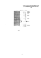
Фиг. 1. Электрофорез препаратов белка G на различных этапах выделения.FIG. 1. Electrophoresis of protein G preparations at various stages of isolation.
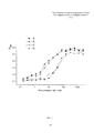
Фиг. 2. Иммуноферментный анализ препаратов белка G.FIG. 2. Enzyme-linked immunosorbent assay of protein G.
Фиг. 3. Непрямой твердофазный иммуноферментный анализ препаратов белка G.FIG. 3. Indirect enzyme-linked immunosorbent assay of protein preparations G.
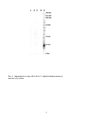
Фиг. 4. Электрофорез и иммуноблот белка G, обработанный различными иммуноглобулинами.FIG. 4. Electrophoresis and immunoblot of protein G, treated with various immunoglobulins.
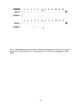
Фиг. 5. Определение иммунохимических свойств ренатурированного белка G при помощи дот-блота по схеме иммуноблота с электропереносом белков после электрофореза в ДСН-ПААГ.FIG. 5. Determination of the immunochemical properties of the renatured protein G using a dot blot according to the immunoblot scheme with protein transfer after electrophoresis in SDS-PAGE.
Фиг. 6. Определение иммунохимических свойств ренатурированного белка G при помощи дот-блота.FIG. 6. Determination of the immunochemical properties of the renatured protein G using dot blot.
Фиг. 7. Определение иммунохимических свойств ренатурированного белка G при помощи непрямого дот-блота с исключением возможного неспецифического взаимодействия конъюгатов и белка G.FIG. 7. Determination of the immunochemical properties of the renatured protein G using an indirect dot blot with the exception of the possible non-specific interaction of conjugates and protein G.
Пример 1. Условия ренатурации белка G.Example 1. Conditions for the renaturation of protein G.
Применение электрофореза позволяет решить две задачи: во-первых, разделить сложную белковую смесь с выделением зоны, содержащей гомогенный белок, и, во-вторых, создать условия, ограничивающие подвижность молекул белка и предотвратить агрегацию в процессе ренатурации. Предварительные опыты показали, что приемлемое разрешение белков электрофорезом достигается при нанесении на препаративный мини-гель 250 мкл препарата белков, растворенных в 8 М мочевине. Маркировку геля проводят при помощи предокрашенных белковых маркеров молекулярных масс, которые вносят в отдельный колодец препаративного мини-геля. После одновременного разделения маркеров и образца, находящегося в препаративной части геля, полосу с маркерами отделяют, окрашивают препаративную часть отдельно, используя раствор хлористого калия, после чего сопоставляют разделенные части геля для точной идентификации положения белка G.The use of electrophoresis allows us to solve two problems: firstly, to separate a complex protein mixture with the allocation of a zone containing a homogeneous protein, and secondly, to create conditions that limit the mobility of protein molecules and prevent aggregation during the renaturation process. Preliminary experiments showed that acceptable resolution of proteins by electrophoresis is achieved by applying 250 μl of a protein preparation dissolved in 8 M urea to a preparative mini-gel. Gel marking is carried out using pre-painted protein markers of molecular weights, which are introduced into a separate well of a preparative mini-gel. After simultaneous separation of the markers and the sample located in the preparative part of the gel, the strip with the markers is separated, the preparative part is stained separately using a solution of potassium chloride, after which the separated parts of the gel are compared to accurately identify the position of protein G.
Для экспресс-окраски и идентификации термоденатурированного ДСН белка G, находящегося в составе геля, используют обработку холодным 0,4 М раствором калия хлористого (Фиг. 1, линия 1. Числами в верхней части фигуры обозначены линии: 1 - фрагмент геля, окрашенный хлористым калием после разделения клеточного лизата, содержащего белок G; 2 - клеточный лизат; 3 - белок G, ренатурированный и выделенный из фрагмента геля; 4 - препарат как в линии 3, разведенный в 15 раз; 5 - маркеры молекулярных масс, числовые значения приведены в правой части фигуры и выражены в килодальтонах; 6 - надосадочная фракция клеточного лизата, содержащая растворимые белки; 7 - белок G, выделенный из растворимой фракции клеточного лизата при помощи аффинной хроматографии на IgG Sepharose® 6 FF с привитыми иммуноглобулинами человека из нормальной сыворотки крови).For rapid staining and identification of the thermo-denatured SDS of protein G contained in the gel, cold 0.4 M potassium chloride solution is used (Fig. 1,
Идентифицированную полосу белка G вырезают из геля и насыщают буфером, содержащим 20 мМ Трис (рН 8.0), 100 мМ NaCl, 1% Тритон Х-100, 6 М мочевину и 2 М тиомочевину в течение 1 час, при этом происходит удаление хлористого калия, замещение ДСН тритоном Х-100, а 6 М мочевина в составе буферного раствора поддерживает денатурирующие условия для молекул белка G. Гель помещают в ренатурационный буфер, содержащий 20 мМ Трис (рН 8.0), 100 мМ NaCl, 2 мМ сульфобетаин, 1 мМ ЭДТА. Ренатурацию проводят с контролем температуры в течение 3 час при 20°С при умеренном перемешивании емкости с гелем и трехкратной сменой буфера, после чего промывают деионизованной водой, замораживают, измельчают в замороженном состоянии и проводят электроэлюцию ренатурированного белка из диспергированного геля в течение 2 час и силой тока 200 мА в буферном растворе, содержащем 10 мМ калий-фосфатный буфер (рН 7,5). Результаты электроэлюции контролируют ДСН-ПААГ (Фиг. 1). Было выделено до 1,8 мг белка G после электрофореза в геле мини-формата с использованием аппарата MiniVE, GE Healthcare, ренатурации и электроэлюции. Функциональную активность очищенного и ренатурированного белка G в дальнейшем подтверждают иммуноферментным анализом и иммуноблотами.The identified strip of protein G is excised from the gel and saturated with a buffer containing 20 mM Tris (pH 8.0), 100 mM NaCl, 1% Triton X-100, 6 M urea and 2 M thiourea for 1 hour, while potassium chloride is removed, substitution of SDS with X-100 triton, and 6 M urea in the composition of the buffer solution supports denaturing conditions for protein G. The gel is placed in a renature buffer containing 20 mM Tris (pH 8.0), 100 mM NaCl, 2 mM sulfobetaine, 1 mM EDTA. Renaturation is carried out with temperature control for 3 hours at 20 ° C with moderate stirring of the container with the gel and a three-fold change of the buffer, after which it is washed with deionized water, frozen, crushed in a frozen state, and the renatured protein is eluted from the dispersed gel for 2 hours and by
Пример 2. Применение буферного раствора с увеличенной концентрацией денатурирующего компонента.Example 2. The use of a buffer solution with an increased concentration of the denaturing component.
Электрофорез проводят как в примере 1, вместе с тем, для растворения препарата белков используют буфер состава - 125 мМ трис-HCl, рН 6,8, 4% додецилсульфата натрия (SDS), 20% глицерина, 10% 2-меркаптоэтанола и 0,02% бромфенолового синего. Растворение белков достигается прогреванием проб, суспензированных в этом буфере в объемном соотношении 1:1 в течение 15 мин [Laemmli, U.K. (1970). Cleavage of structural proteins during the assembly of the head of bacteriophage T4. Nature 227 (5259): 680-685]. Определяют положение полосы белка G, вырезают ее из геля и помещают для насыщения в буферный раствор, содержащий 25 мМ Трис (рН 8.0-8.2), 1% Тритон Х-100 и 8 М мочевину в течение 1-2 час, в результате чего происходит удаление хлористого калия, введенного для обратимой окраски геля, замещение ДСН тритоном Х-100, а 8 М мочевина в составе денатурирующего буферного раствора поддерживает условия, при которых полипептидные цепи молекулы белка G находятся в «развернутом» состоянии. Гель с денатурированным белком помещают в ренатурационный буфер, содержащий 25 мМ Трис (рН 8.0), 150 мМ NaCl, 0,02% твин-80, 1 мМ ЭДТА и инкубируют при умеренном перемешивании буфера в емкости с гелем в течение 18 час при 12-14°С. Объем буфера должен не менее чем в 200 раз превосходить объем полоски геля. Полученные результаты аналогичны примеру 1.Electrophoresis is carried out as in example 1, however, to dissolve the protein preparation, a composition buffer is used - 125 mM Tris-HCl, pH 6.8, 4% sodium dodecyl sulfate (SDS), 20% glycerol, 10% 2-mercaptoethanol and 0, 02% bromophenol blue. Dissolution of proteins is achieved by heating samples suspended in this buffer in a volume ratio of 1: 1 for 15 minutes [Laemmli, U.K. (1970). Cleavage of structural proteins during the assembly of the head of bacteriophage T4. Nature 227 (5259): 680-685]. The position of the protein G band is determined, it is cut out of the gel and placed for saturation in a buffer solution containing 25 mM Tris (pH 8.0-8.2), 1% Triton X-100 and 8 M urea for 1-2 hours, resulting in removal of potassium chloride introduced for reversible coloring of the gel, substitution of SDS with triton X-100, and 8 M urea in the composition of the denaturing buffer solution supports the conditions under which the polypeptide chains of protein G are in the “expanded” state. The gel with the denatured protein is placed in a renaturation buffer containing 25 mM Tris (pH 8.0), 150 mM NaCl, 0.02% tween-80, 1 mM EDTA and incubated with moderate stirring of the buffer in a gel container for 18 hours at 12 14 ° C. The volume of the buffer should be at least 200 times the volume of the strip of gel. The results obtained are similar to example 1.
Пример 3. Использование буферных растворов с компонентами, обеспечивающими тиол-дисульфидное равновесие.Example 3. The use of buffer solutions with components that provide thiol-disulfide balance.
Электрофорез проводят как в предыдущих примерах, однако, для обработки гелевых полос, содержащих белок G, используют буферные растворы, которые на этапе денатурации обеспечивают расщепление возможных дисульфидных связей между цистеинами полипептидной цепи, а на этапе ренатурации - их восстановление. Денатурирующий буфер содержит 20 мМ Трис (рН 8.0), 8 М мочевину, 0,3% саркозил, 20 мМ CHAPS и 10 мМ 2-меркаптоэтанол. Для ренатурации белка полоску геля помещают в 200-кратный избыток (по объему) буферного раствора, содержащего 20 мМ Трис (рН 8.0), 150 мМ натрия хлористого, 20 мМ CHAPS, 5 мМ цистеина и 50 мМ цистина. Перед этим этапом температура буферного раствора должна быть обязательно доведена до 18°С. Наличие в буфере цистеин/цистиновой пары, слабощелочная среда, умеренная температура и продолжительная инкубация (не менее 12 час) способствуют восстановлению дисульфижных связей. Полученные результаты аналогичны примеру 1.Electrophoresis is carried out as in the previous examples, however, to process gel bands containing protein G, buffer solutions are used that, at the denaturation stage, provide for the cleavage of possible disulfide bonds between cysteines of the polypeptide chain, and at the renaturation stage, their restoration. The denaturing buffer contains 20 mM Tris (pH 8.0), 8 M urea, 0.3% sarcosyl, 20 mM CHAPS and 10 mM 2-mercaptoethanol. To renature the protein, a strip of gel is placed in a 200-fold excess (by volume) of a buffer solution containing 20 mM Tris (pH 8.0), 150 mM sodium chloride, 20 mM CHAPS, 5 mM cysteine, and 50 mM cystine. Before this step, the temperature of the buffer solution must be brought to 18 ° C. The presence of a cysteine / cystine pair in the buffer, a slightly alkaline medium, moderate temperature, and prolonged incubation (at least 12 hours) contribute to the restoration of disulfide bonds. The results obtained are similar to example 1.
Пример 4. Твердофазный иммуноферментный анализ.Example 4. Enzyme-linked immunosorbent assay.
Планшеты (96 лунок) сенсибилизируют последовательным внесением в каждую лунку 100 мкл раствора, содержащего 0,1 М карбонатный буфер (рН 9,6) и 800 нг белка G с инкубацией при температуре 6°С в течение 18 час, после чего проводят четырехкратную промывку 10 мМ фосфатно-солевым буферным раствором (рН 7,2), содержащим 0,05% твина-20 (ФСБТ). Неспецифическую сорбцию блокируют 1% раствором бычьего сывороточного альбумина в ФСБТ в течение 2 час при 37°С, причем на этом этапе объем наполнения лунок составлял 120 мкл. Лунки промывают, как описано ранее, но трехкратно, после чего сразу же наполняют предварительно приготовленными растворами нормальных иммуноглобулинов кролика в ФСБТ. Иммуноглобулины разводят, начиная с наибольшей концентрации, составлявшей 2500 нг/мл с кратностью разведения 2,5. Последующие стадии анализа проводят аналогично с включением промежуточных промывок ФСБТ между каждой инкубацией. Этапы включали инкубацию с раствором биотинилированных антикроличьих F(ab)2 фрагментов антител (1:3000, 2 час, 37°С) в ФСБТ, с раствором стрептавидин-пероксидазы (1:4000, 1 час, 37°С), визуализацию субстратом и остановку реакции 2 М раствором серной кислоты. Субстратом служил 0,1% раствор о-фенилендиамина и 0,03% раствор перекиси водорода в 50 мМ цитрат-фосфатном буфере (рН 5,0) (Фиг. 2. Буквами в верхней части фигуры обозначены кривые: А, В - лунки сенсибилизированы белком G, очищенным аффинной хроматографией на IgG Sepharose® 6 FF с привитыми иммуноглобулинами человека из нормальной сыворотки крови; С, D - лунки сенсибилизированы белком G, ренатурированного и выделенного электроэлюцией из полиакриламидного геля). Титрование IgG на планшетах, сенсибилизированных различными препаратами белка G, представлено S-образными кривыми, горизонтальные участки которых в области высоких концентраций соответствуют области насыщения. Восходящие участки кривых А и В расположены в области концентраций IgG от 2,5 нг/мл до 80 нг/мл, для кривых С и D - от 10 нг/мл до 300 нг/мл. Принимая во внимание одинаковые номинальные концентрации белка G в препаратах, установленное различие концентрационных интервалов показывает сниженную долю функционально-активных молекул белка G в ренатурированном препарате по сравнению с белком G, очищенным аффинной хроматографией на IgG Sepharose® 6 FF с привитыми иммуноглобулинами человека из нормальной сыворотки крови.Plates (96 wells) are sensitized by sequentially adding to each well 100 μl of a solution containing 0.1 M carbonate buffer (pH 9.6) and 800 ng of Protein G with incubation at 6 ° C for 18 hours, after which a four-fold washing is carried out 10 mM phosphate-buffered saline (pH 7.2) containing 0.05% Tween-20 (FSBT). Nonspecific sorption is blocked with a 1% solution of bovine serum albumin in FSBT for 2 hours at 37 ° C, and at this stage, the filling volume of the wells was 120 μl. Wells are washed as previously described, but three times, after which they are immediately filled with pre-prepared solutions of normal rabbit immunoglobulins in FSBT. Immunoglobulins are diluted starting from the highest concentration of 2500 ng / ml with a dilution ratio of 2.5. The subsequent stages of the analysis are carried out similarly with the inclusion of intermediate washes FSBT between each incubation. The stages included incubation with a solution of biotinylated anti-rabbit F (ab) 2 antibody fragments (1: 3000, 2 hours, 37 ° C) in FSBT, with a solution of streptavidin peroxidase (1: 4000, 1 hour, 37 ° C), visualization with a substrate and stopping the reaction with a 2 M sulfuric acid solution. The substrate was a 0.1% solution of o-phenylenediamine and a 0.03% solution of hydrogen peroxide in 50 mM citrate-phosphate buffer (pH 5.0) (Fig. 2. The letters in the upper part of the figure indicate the curves: A, B - the wells are sensitized protein G, purified by affinity chromatography on
Пример 5. Непрямой твердофазный иммуноферментный анализ.Example 5. Indirect enzyme-linked immunosorbent assay.
Для проведения непрямого твердофазного ИФА каждую лунку планшета (96 лунок) сенсибилизируют, заполняя 100 мкл раствора, содержащего 0,1 М карбонатный буфер (рН 9,6) и 500 нг иммуноглобулинов нормальной сыворотки кролика с инкубацией при температуре 6°С в течение 18 час, после чего проводят четырехкратную промывку ФСБТ. Неспецифическую сорбцию блокируют 1% раствором бычьего сывороточного альбумина в ФСБТ в течение 2 час при 37°С, причем на этом этапе объем наполнения лунок составлял 120 мкл. В последующем, этапы анализа проводят аналогично, с промежуточными промывками ФСБТ и включают инкубацию с приготовленными предварительно разведениями белка G (начиная с наибольшей концентрации 600 нг/мл), с кратностью 2,5, с раствором нормальной сыворотки мыши (1:350, 2 час, 37°С), раствором антимышиных антител, конъюгированных с пероксидазой хрена (1:4000, 2 час, 37°С), и субстратом. Раствор субстрата содержал 0,1% о-фенилендиамина и 0,03% перекиси водорода в 50 мМ цитрат-фосфатном буфере (рН 5,0). Реакцию останавливают 2 М раствором серной кислоты. (Фиг. 3 показывает, что белок G титруется и интервале концентраций от 10 нг/мл до 150 нг/мл для кривых А и В и от 20 нг/мл до 300 нг/мл для кривых С и D. Буквами в верхней части фигуры обозначены кривые титрования: А, В - белка G, очищенного аффинной хроматографией на IgG Sepharose® 6 FF с привитыми иммуноглобулинами человека из нормальной сыворотки крови; С, D - белка G, ренатурированного и выделенного электроэлюциией из полиакриламидного геля.) Различие интервалов титрования и их параллельный характер указывают на концентрационные, но не функциональные различия двух использованных препаратов белка G.To perform an indirect solid-phase ELISA, each well of a plate (96 wells) is sensitized by filling 100 μl of a solution containing 0.1 M carbonate buffer (pH 9.6) and 500 ng of normal rabbit serum immunoglobulins with an incubation at 6 ° C for 18 hours then a four-time washing of the FSBT is carried out. Nonspecific sorption is blocked with a 1% solution of bovine serum albumin in FSBT for 2 hours at 37 ° C, and at this stage, the filling volume of the wells was 120 μl. Subsequently, the analysis steps are carried out similarly with intermediate FSBT washes and include incubation with previously prepared dilutions of protein G (starting from the highest concentration of 600 ng / ml), with a multiplicity of 2.5, with a solution of normal mouse serum (1: 350, 2 hours , 37 ° C), a solution of anti-mouse antibodies conjugated to horseradish peroxidase (1: 4000, 2 hours, 37 ° C), and a substrate. The substrate solution contained 0.1% o-phenylenediamine and 0.03% hydrogen peroxide in 50 mM citrate-phosphate buffer (pH 5.0). The reaction is stopped with a 2 M sulfuric acid solution. (Fig. 3 shows that protein G is titrated in the concentration range from 10 ng / ml to 150 ng / ml for curves A and B and from 20 ng / ml to 300 ng / ml for curves C and D. The letters at the top of the figure titration curves are indicated: A, B - protein G, purified by affinity chromatography on
Пример 6. Иммуноблот.Example 6. Immunoblot.
Разделение очищенного белка G и предокрашенных маркеров проводили ДСН-электрофорезом в 12,5% полиакриламидном геле в редуцирующих условиях в режиме постоянного тока 20 мА. После завершения ДСН-ПААГ гель освобождали от стеклянных пластин и помещали на 15 мин в буферный раствор, содержащий 25 мМ трис основной, 192 мМ глицин, 0,1% SDS, 20% этанол (рН 8,3). Раствор в кювете с гелем периодически перемешивают. Гель с необходимыми прокладками помещают в устройство для переноса, накладывают на него нитроцеллюлозную мембрану со стороны анода, слои фильтровальной бумаги для уплотнения, предварительно увлажненные буфером, электроды и подключают ток. Перенос белков на нитроцеллюлозную мембрану проводят перпендикулярно первому направлению разделения при постоянном токе 400 мА и температуре 22°С, в течение 1 час, после чего мембрану осторожно извлекают и немедленно помещают в блокирующий раствор, содержащий 1% бычий сывороточный альбумин, или 5% обезжиренное молоко в ФСБТ. Подавление неспецифических взаимодействий проводят при перемешивании раствора на качалке в течение 2 час при 37°С. Для промывки мембран и разведения белков на каждом промежуточном этапе используют ФСБТ. Блокированную и промытую мембрану разрезают на части, каждую из которых инкубируют в отдельных кюветах с различными растворами нормальных сывороток в течение 2 час при 37°С, после чего промывают и инкубируют при тех же условиях с соответствующими растворами конъюгатов антивидовых антител с пероксидазой хрена. Далее промывают ФСБТ и помещают в раствор хромогенного субстрата, содержащего 3,3'-диаминобензидин (0,5 мг/мл), хлорид никеля (0,1 мг/мл), 0,03% Н2О2, 0,05 М Трис-HCl (рН 7,6). Перекись водорода добавляют после растворения всех компонентов субстрата и используют его немедленно, реакцию останавливают, промывая окрашенные фрагменты мембран дистиллированной водой (Фиг. 4. Числами в верхней части фигуры обозначены линии: 1, 2, 3, 4 - фрагменты мембран иммуноблота; 5 - фрагмент геля, окрашенный кумасси G-250; 3 - предокрашенные маркеры молекулярных масс, числовые значения приведены в правой части фигуры и выражены в килодальтонах, все остальные линии содержат очищенный белок G. Обработка мембран: линия 1 - сыворотка крови человека; линия 2 - сыворотка крови мышей; линия 4 - сыворотка крови кролика.). Анализ иммуноблотов позволяет сделать заключение о том, что белок G денатурируется в результате термообработки образцов в присутствии 0,1% ДСН буфера Лэмли перед электрофорезом, утрачивает способность связывать различные иммуноглобулины G и остается преимущественно в денатурированном состоянии после переноса на нитроцеллюлозную мембрану, что подтверждается чрезвычайно слабой окраской полос или ее отсутствием (Фиг. 4, линии 1, 2 и 4). В это же время, электрофореграмма (Фиг. 4, линия 5) указывает на значительное (не менее 1 мкг) количество белка G в линиях.Separation of purified protein G and pre-stained markers was carried out by SDS electrophoresis in 12.5% polyacrylamide gel under reducing conditions in a constant current mode of 20 mA. After completion of the SDS-PAGE, the gel was freed from the glass plates and placed for 15 min in a buffer solution containing 25 mM Tris basic, 192 mM glycine, 0.1% SDS, 20% ethanol (pH 8.3). The solution in a gel cuvette is periodically mixed. The gel with the necessary gaskets is placed in the transfer device, a nitrocellulose membrane is applied to it from the anode side, layers of filter paper for sealing, pre-moistened with buffer, electrodes and a current are connected. Transfer of proteins to the nitrocellulose membrane is carried out perpendicular to the first separation direction at a constant current of 400 mA and a temperature of 22 ° C, for 1 hour, after which the membrane is carefully removed and immediately placed in a blocking solution containing 1% bovine serum albumin, or 5% skim milk in the FSBT. The suppression of nonspecific interactions is carried out while stirring the solution on a rocking chair for 2 hours at 37 ° C. For washing membranes and diluting proteins at each intermediate stage, FSBT is used. The blocked and washed membranes are cut into pieces, each of which is incubated in separate cuvettes with different solutions of normal sera for 2 hours at 37 ° C, then washed and incubated under the same conditions with the corresponding solutions of conjugates of antiviral antibodies with horseradish peroxidase. Then washed with FSBT and placed in a solution of a chromogenic substrate containing 3,3'-diaminobenzidine (0.5 mg / ml), nickel chloride (0.1 mg / ml), 0.03% H 2 O 2 , 0.05 M Tris-HCl (pH 7.6). Hydrogen peroxide is added after all components of the substrate are dissolved and used immediately, the reaction is stopped by washing the colored membrane fragments with distilled water (Fig. 4. The numbers in the upper part of the figure indicate the lines: 1, 2, 3, 4 - fragments of the immunoblot membranes; 5 - fragment gel, Coomassie stained G-250; 3 - pre-stained molecular weight markers, numerical values are shown on the right side of the figure and are expressed in kilodaltons, all other lines contain purified protein G. Membrane processing: line 1 - human serum ESA; lane 2 - serum mouse; lane 4 - rabbit serum).. An analysis of immunoblots allows us to conclude that protein G is denatured as a result of heat treatment of samples in the presence of 0.1% SDS Lamley buffer before electrophoresis, loses the ability to bind various immunoglobulins G and remains predominantly in the denatured state after transfer to the nitrocellulose membrane, which is confirmed by an extremely weak coloring of strips or its absence (Fig. 4,
Пример 7. Дот-блот.Example 7. Dot blot.
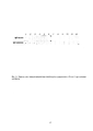
Для подготовки различных вариантов рабочей нитроцеллюлозной мембраны готовят ряд последовательных двукратных разведений белков «первого слоя», в качестве которых выступают белок G, или иммуноглобулины G нормальной сыворотки крови различных животных, в частности кролика и мыши. Растворы белков переносят в объеме 1 мкл каждого разведения рядами на поверхность размеченной мембраны. Мембрану сушат в течение 1 час при 23°С, после чего погружают в блокирующий раствор, содержащий 1% бычий сывороточный альбумин или 5% обезжиренное молоко в ФСБТ. Блокировку проводят на качалке в течение 2 час при 37°С. Для промывок мембран и разведения белков на каждом промежуточном этапе используют ФСБТ. Перечисленные этапы являются общими для всех проводок. На последующих этапах, в зависимости от схемы эксперимента, применяют различные последовательности инкубации мембран в растворах нормальных сывороток животных, белка G, конъюгатов различных антивидовых антител с пероксидазой хрена, как правило, в течение 2 час при 37°С с необходимыми промежуточными промывками ФСБТ. При длительных экспериментах, выходящих за пределы одного рабочего дня, этапы инкубации проводят в течение ночного времени, но при 6°С. Этап обработки субстратом является общим и завершающим для всех схем проводки: промытую мембрану помещают в раствор субстрата, содержащего 3,3'-диаминобензидин (0,5 мг/мл), хлорид никеля (0,1 мг/мл), 0,03% Н2О2, 0,05 М Трис-HCl (рН 7,6). После добавления перекиси водорода субстрат используют немедленно, реакцию останавливают, промывая мембрану дистиллированной водой. Описания всех использованных схем дот-блотов приведены на Фиг. 5. Числами в верхней части фигуры обозначены секторы нитроцеллюлозной мембраны с сорбированными белками в последовательных двукратных разведениях, начиная с наибольшей концентрации, составляющей 1000 мкг/мл (секторы 1 всех полос). Полосы с сорбированным белком G обозначены слева надписью «Белок G», полосы с сорбированным бычьим сывороточным альбумином обозначены слева надписью «Альбумин». Полосы, обозначенные справа как «А» и «В» вместе с прилегающими полосами «Альбумин», отличаются проводкой мембран.To prepare various options for the working nitrocellulose membrane, a series of serial two-fold dilutions of the “first layer” proteins are prepared, which are protein G, or immunoglobulins G of normal blood serum of various animals, in particular, rabbit and mouse. Protein solutions are transferred in a volume of 1 μl of each dilution in rows on the surface of the labeled membrane. The membrane is dried for 1 hour at 23 ° C, and then immersed in a blocking solution containing 1% bovine serum albumin or 5% skim milk in FSBT. The lock is carried out on a rocking chair for 2 hours at 37 ° C. For membrane washing and protein dilution at each intermediate stage, FSBT is used. The steps listed are common to all postings. At the subsequent stages, depending on the experimental design, various incubation sequences of membranes in solutions of normal animal sera, protein G, conjugates of various antispecies antibodies with horseradish peroxidase are used, usually for 2 hours at 37 ° C with the necessary intermediate washes with FSBT. With long-term experiments that go beyond one working day, the incubation steps are carried out during the night, but at 6 ° C. The substrate treatment step is general and final for all wiring schemes: the washed membrane is placed in a substrate solution containing 3,3'-diaminobenzidine (0.5 mg / ml), nickel chloride (0.1 mg / ml), 0.03% H 2 O 2 , 0.05 M Tris-HCl (pH 7.6). After adding hydrogen peroxide, the substrate is used immediately, the reaction is stopped by washing the membrane with distilled water. Descriptions of all used dot blot patterns are shown in FIG. 5. The numbers in the upper part of the figure indicate the sectors of the nitrocellulose membrane with sorbed proteins in successive two-fold dilutions, starting with the highest concentration of 1000 μg / ml (
Проводка А: этап 1 - сорбция белка G и альбумина, 1 час, 24°С; этап 2 - промывка ФСБТ; этап 3 - инкубация с блокирующим агентом, 2 час, 37°С; этап 4 - промывка ФСБТ; этап 5 - инкубация с раствором IgG мыши, 1 мкг/мл, 2 час, 37°С; этап 6 - промывка ФСБТ; этап 7 - инкубация с конъюгатами антимышиных антител с пероксидазой хрена, 2 час, 37°С; этап 8 - промывка ФСБТ; этап 9 - хромогенный субстрат.Posting A: stage 1 - sorption of protein G and albumin, 1 hour, 24 ° C; stage 2 - washing FSBT; stage 3 - incubation with a blocking agent, 2 hours, 37 ° C; stage 4 - washing FSBT; stage 5 - incubation with a solution of mouse IgG, 1 μg / ml, 2 hours, 37 ° C; stage 6 - washing FSBT; stage 7 - incubation with conjugates of anti-mouse antibodies with horseradish peroxidase, 2 hours, 37 ° C; stage 8 - washing FSBT; stage 9 - chromogenic substrate.
Проводка В: этап 1 - сорбция белка G и альбумина, 1 час, 24°С; этап 2 - промывка ФСБТ; этап 3 - инкубация с блокирующим агентом, 2 час, 37°С; этап 4 - промывка ФСБТ; этап 5 - инкубация с раствором IgG кролика, 1 мкг/мл, 2 час, 37°С; этап 6 - промывка ФСБТ; этап 7 - инкубация с конъюгатами антикроличьих антител с пероксидазой хрена, 2 час, 37°С; этап 8 - промывка ФСБТ; этап 9 - хромогенный субстрат. Числами в верхней части фигуры обозначены участки нитроцеллюлозной мембраны с сорбированными иммуноглобулинами G (IgG) в последовательных двукратных разведениях, начиная с наибольшей концентрации IgG, составляющей 1000 мкг/мл (участки 1). Виды сорбированных на нитроцеллюлозную мембрану IgG обозначены в левой части фигуры.Posting B: stage 1 - sorption of protein G and albumin, 1 hour, 24 ° C; stage 2 - washing FSBT; stage 3 - incubation with a blocking agent, 2 hours, 37 ° C; stage 4 - washing FSBT; stage 5 - incubation with a solution of rabbit IgG, 1 μg / ml, 2 hours, 37 ° C; stage 6 - washing FSBT; stage 7 - incubation with conjugates of anti-rabbit antibodies with horseradish peroxidase, 2 hours, 37 ° C; stage 8 - washing FSBT; stage 9 - chromogenic substrate. The numbers in the upper part of the figure indicate the sections of the nitrocellulose membrane with adsorbed immunoglobulins G (IgG) in successive double dilutions, starting with the highest IgG concentration of 1000 μg / ml (sections 1). Types of IgG adsorbed onto the nitrocellulose membrane are indicated on the left side of the figure.
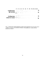
Проводку мембран осуществляли следующим образом: этап 1 - сорбция IgG мыши или кролика, 1 час, 24°С; этап 2 - промывка ФСБТ; этап 3 - инкубация с блокирующим агентом, 2 час, 37°С; этап 4 - инкубация с раствором ренатурированного белка G, 1 мкг/мл, 2 час, 37°С; этап 5 - промывка ФСБТ; этап 6 - инкубация с растворами IgG: верхний ряд - IgG кролика, нижний ряд - IgG мыши, 1 мкг/мл, 18 час, 6°С; этап 7 - промывка ФСБТ; этап 8 - инкубация с конъюгированными с пероксидазой хрена антивидовыми антителами против IgG, использованных на этапе 6, 2 час, 37°С; этап 9 - промывка ФСБТ; этап 10 - хромогенный субстрат. Числами в верхней части фигуры обозначены секторы нитроцеллюлозной мембраны с сорбированными белками в последовательных двукратных разведениях, начиная с наибольшей концентрации, составляющей 1000 мкг/мл (секторы 1 всех полос). Полосы с сорбированными IgG мыши в составе сыворотки крови мышей также обозначены надписью слева. Полосы, обозначенные справа как «А», используют в проводке белок G, очищенный из растворимой фракции лизатов клеток Е. coli аффинной хроматографией при помощи IgG Sepharose 6В. Полосы «В» используют в проводке белок G, ренатурированный и выделенный электроэлюцией в настоящем исследовании.The membrane was carried out as follows:
Проводка полос «А» и «В» с сорбированными IgG кролика: этап 1 - сорбция IgG кролика, 1 час, 24°С; этап 2 - промывка ФСБТ; этап 3 - инкубация с блокирующим агентом, 2 час, 37°С; этап 4 - промывка ФСБТ; этап 5 - инкубация с растворами белка G: линия «А» - очищенного аффинной хроматографией, линия «В» - ренатурированного и выделенного электроэлюцией, 1 мкг/мл, 2 час, 37°С; этап 6 - промывка ФСБТ; этап 7 - инкубация с раствором нормальной сыворотки мышей (1:100), 2 час, 37°С; этап 8 - промывка ФСБТ; этап 9 - инкубация с конъюгатами антимышиных антител с пероксидазой хрена, 2 час, 37°С; этап 10 - промывка ФСБТ; этап 11 - хромогенный субстрат.Posting bands “A” and “B” with adsorbed rabbit IgG: stage 1 - sorption of rabbit IgG, 1 hour, 24 ° C; stage 2 - washing FSBT; stage 3 - incubation with a blocking agent, 2 hours, 37 ° C; stage 4 - washing FSBT; stage 5 - incubation with solutions of protein G: line "A" - purified by affinity chromatography, line "B" - renatured and isolated by electroelution, 1 μg / ml, 2 hours, 37 ° C; stage 6 - washing FSBT; stage 7 - incubation with a solution of normal mouse serum (1: 100), 2 hours, 37 ° C; stage 8 - washing FSBT; stage 9 - incubation with conjugates of anti-mouse antibodies with horseradish peroxidase, 2 hours, 37 ° C; stage 10 - washing FSBT; stage 11 - chromogenic substrate.
Проводка полос «А» и «В» с сорбированной нормальной сывороткой мышей (1:30): этап 1 - сорбция белков сыворотки, включая IgG мыши, 1 час, 24°С; этап 2 - промывка ФСБТ; этап 3 - инкубация с блокирующим агентом, 2 час, 37°С; этап 4 - промывка ФСБТ; этап 5 - инкубация с растворами белка G: линия «А» - очищенного аффинной хроматографией, линия «В» - ренатурированного и выделенного электроэлюцией, 1 мкг/мл, 2 час, 37°С; этап 6 - промывка ФСБТ; этап 7 - инкубация с раствором IgG кролика (1 мкг/мл), 2 час, 37°С; этап 8 - промывка ФСБТ; этап 9 - инкубация с биотинилированными F(ab)2 фрагментами антикроличьих антител, 2 час, 37°С; этап 10 - промывка ФСБТ; этап 11 - стрептавидин-пероксидаза (1:3000), 2 час, 37°С; этап 12 - промывка ФСБТ; этап 13 - хромогенный субстрат.Posting bands “A” and “B” with sorbed normal mouse serum (1:30): stage 1 - sorption of serum proteins, including mouse IgG, 1 hour, 24 ° C; stage 2 - washing FSBT; stage 3 - incubation with a blocking agent, 2 hours, 37 ° C; stage 4 - washing FSBT; stage 5 - incubation with solutions of protein G: line "A" - purified by affinity chromatography, line "B" - renatured and isolated by electroelution, 1 μg / ml, 2 hours, 37 ° C; stage 6 - washing FSBT; stage 7 - incubation with a solution of rabbit IgG (1 μg / ml), 2 hours, 37 ° C; stage 8 - washing FSBT; stage 9 - incubation with biotinylated F (ab) 2 fragments of anti-rabbit antibodies, 2 hours, 37 ° C; stage 10 - washing FSBT; stage 11 - streptavidin peroxidase (1: 3000), 2 hours, 37 ° C; stage 12 - washing FSBT; stage 13 - chromogenic substrate.
Для подтверждения свойств ренатурированного белка используют различные схемы последовательных иммунохимических взаимодействий (различные проводки). При схеме прямого дот-блота, как и в иммуноблоте (Фиг. 4), на нитроцеллюлозную мембрану первым слоем сорбируют очищенный, функционально активный ренатурированный белок G и после блокировки проводят через растворы различных антител, соответствующих антивидовых антител, конъюгированных с пероксидазой хрена и субстрата (Фиг. 5). Появление окрашенных пятен в 11 секторе для кроличьих антител и в 12 секторе для антител мыши соответствуют количествам белка G 0,98 нг и 0,49 нг в 1 мкл раствора, сорбированного на мембраны. Альбумин, использованный в качестве контроля в проводках А и В, не показывает окраски пятен даже при сорбции на мембрану 1000 нг белка (Фиг. 5). По сравнению с иммуноблотом (Фиг. 4) ренатурированный белок G в прямом варианте дот-блота проявляет способность связывать IgG кролика и мыши в концентрациях на три порядка меньше, чем белок G, термоденатурированный перед ДСН-электрофорезом.To confirm the properties of the renatured protein, various sequential immunochemical interaction schemes (various wires) are used. In the direct dot blot scheme, as in the immunoblot (Fig. 4), a purified, functionally active renatured protein G is sorbed onto the nitrocellulose membrane with the first layer and, after blocking, it is passed through solutions of various antibodies, the corresponding antispecies antibodies conjugated to horseradish peroxidase and substrate ( Fig. 5). The appearance of stained spots in the 11th sector for rabbit antibodies and in the 12th sector for mouse antibodies corresponds to the amounts of protein G 0.98 ng and 0.49 ng in 1 μl of the solution adsorbed on the membrane. Albumin, used as a control in wires A and B, does not show stain color even when 1000 ng of protein is adsorbed onto the membrane (Fig. 5). Compared with the immunoblot (Fig. 4), the renatured protein G in the direct version of the dot blot shows the ability to bind rabbit and mouse IgG in concentrations three orders of magnitude lower than protein G thermo-denatured before SDS electrophoresis.
В отличие от прямого дот-блота, новый вариант использует белок G в качестве «мостика», связывающего иммуноглобулины различных видов животных (Фиг. 6). В этом случае макромолекула белка G должна нести по крайней мере два центра связывания константных областей IgG, и проявление такой способности доказывает наличие функциональной активности белка G, подвергнутого ренатурации. Для подтверждения, в эксперименте на нитроцеллюлозную мембрану сорбируют различные виды иммуноглобулинов G, соответственно, кролика и мыши в последовательных двукратных разведениях от нулевого до 1:2048, что соответствует в последней, 12-й ступени концентрации 0,488 мкг/мл. Далее, IgG первого слоя взаимодействуют с белком G, к которому перекрестно присоединяют IgG мыши и кролика. Обработка соответствующими конъюгированными антивидовыми антителами и субстратом приводит к появлению окрашенных пятен, отчетливо различимых в шестом и седьмом разведениях, что доказывает наличие у ренатурированного белка G способности связывать IgG мыши и кролика. Расчеты показывают, что для связывания белка G необходимо сорбировать на мембрану в одном пятне не менее 30 нг антител (Фиг. 6, участки 6 и 7). Однако в этой схеме мы не можем исключить вероятность прямого взаимодействия антивидовых конъюгатов с сорбированным белком G первого слоя за счет оставшихся свободных мест связывания. Схема дот-блота, исключающая такую возможность, и его результаты приведены на Фиг. 7. Проводка отличается от предыдущего варианта тем, что на завершающих этапах вместо конъюгатов антивидовых антител используют биотинилированные F(ab)2 фрагменты антител, утратившие способность к взаимодействию с белком G вследствие удаления константной Fc области молекулы IgG. Окраска пятен на полосах А и В с сорбированной сывороткой мыши доказывает отсутствие неспецифического связывания конъюгата с белком G.In contrast to the direct dot blot, the new variant uses protein G as a “bridge” connecting immunoglobulins of various animal species (Fig. 6). In this case, the protein G macromolecule must carry at least two binding sites for IgG constant regions, and the manifestation of this ability proves the presence of functional activity of protein G renatured. To confirm, in the experiment on the nitrocellulose membrane, various types of immunoglobulins G, respectively, of the rabbit and mouse are adsorbed in successive two-fold dilutions from zero to 1: 2048, which corresponds to a concentration of 0.488 μg / ml in the last, 12th stage. Next, the first layer IgG interacts with protein G, to which mouse and rabbit IgG are cross-linked. Treatment with appropriate conjugated anti-virus antibodies and the substrate leads to the appearance of colored spots, clearly distinguishable in the sixth and seventh dilutions, which proves the presence of the renatured protein G ability to bind mouse and rabbit IgG. Calculations show that for protein G binding, at least 30 ng of antibodies must be adsorbed onto the membrane in one spot (Fig. 6,
Claims (1)
Priority Applications (1)
| Application Number | Priority Date | Filing Date | Title |
|---|---|---|---|
| RU2016111874A RU2646103C2 (en) | 2016-03-30 | 2016-03-30 | Method for renatured protein g isolation from marked polyacrylamide gel |
Applications Claiming Priority (1)
| Application Number | Priority Date | Filing Date | Title |
|---|---|---|---|
| RU2016111874A RU2646103C2 (en) | 2016-03-30 | 2016-03-30 | Method for renatured protein g isolation from marked polyacrylamide gel |
Publications (2)
| Publication Number | Publication Date |
|---|---|
| RU2016111874A RU2016111874A (en) | 2017-10-05 |
| RU2646103C2 true RU2646103C2 (en) | 2018-03-01 |
Family
ID=60047516
Family Applications (1)
| Application Number | Title | Priority Date | Filing Date |
|---|---|---|---|
| RU2016111874A RU2646103C2 (en) | 2016-03-30 | 2016-03-30 | Method for renatured protein g isolation from marked polyacrylamide gel |
Country Status (1)
| Country | Link |
|---|---|
| RU (1) | RU2646103C2 (en) |
Cited By (1)
| Publication number | Priority date | Publication date | Assignee | Title |
|---|---|---|---|---|
| EP4560307A3 (en) * | 2018-11-14 | 2025-08-27 | The Francis Crick Institute Limited | Method, solution and kit for preparing a tissue sample for 3d imaging |
Citations (6)
| Publication number | Priority date | Publication date | Assignee | Title |
|---|---|---|---|---|
| WO1990000200A1 (en) * | 1988-06-27 | 1990-01-11 | Genex Corporation | Thermal release of recombinant protein into culture media |
| RU2070888C1 (en) * | 1990-11-22 | 1996-12-27 | Берингер Маннхайм ГмбХ | Method of reactivation of recombinant protein |
| WO2002057296A1 (en) * | 2001-01-16 | 2002-07-25 | Københavns Universitet | A method for refolding of proteins |
| RU2208445C1 (en) * | 2002-02-20 | 2003-07-20 | Волгоградский научно-исследовательский противочумный институт | Method for isolation of protein 39 kda from meliodosis pathogen external membrane |
| RU2278870C2 (en) * | 2004-08-30 | 2006-06-27 | Закрытое Акционерное Общество "Биокад" | Method for preparing, isolating, purifying and stabilizing human recombinant granulocytic colony-stimulating factor useful for medicinal using and immunobiological agent based on thereof |
| RU2448116C2 (en) * | 2010-02-01 | 2012-04-20 | Автономная некоммерческая организация "Институт молекулярной диагностики" (АНО "ИнМоДи") | Method for producing active fragment of human alpha-fetoprotein |
-
2016
- 2016-03-30 RU RU2016111874A patent/RU2646103C2/en active
Patent Citations (6)
| Publication number | Priority date | Publication date | Assignee | Title |
|---|---|---|---|---|
| WO1990000200A1 (en) * | 1988-06-27 | 1990-01-11 | Genex Corporation | Thermal release of recombinant protein into culture media |
| RU2070888C1 (en) * | 1990-11-22 | 1996-12-27 | Берингер Маннхайм ГмбХ | Method of reactivation of recombinant protein |
| WO2002057296A1 (en) * | 2001-01-16 | 2002-07-25 | Københavns Universitet | A method for refolding of proteins |
| RU2208445C1 (en) * | 2002-02-20 | 2003-07-20 | Волгоградский научно-исследовательский противочумный институт | Method for isolation of protein 39 kda from meliodosis pathogen external membrane |
| RU2278870C2 (en) * | 2004-08-30 | 2006-06-27 | Закрытое Акционерное Общество "Биокад" | Method for preparing, isolating, purifying and stabilizing human recombinant granulocytic colony-stimulating factor useful for medicinal using and immunobiological agent based on thereof |
| RU2448116C2 (en) * | 2010-02-01 | 2012-04-20 | Автономная некоммерческая организация "Институт молекулярной диагностики" (АНО "ИнМоДи") | Method for producing active fragment of human alpha-fetoprotein |
Cited By (1)
| Publication number | Priority date | Publication date | Assignee | Title |
|---|---|---|---|---|
| EP4560307A3 (en) * | 2018-11-14 | 2025-08-27 | The Francis Crick Institute Limited | Method, solution and kit for preparing a tissue sample for 3d imaging |
Also Published As
| Publication number | Publication date |
|---|---|
| RU2016111874A (en) | 2017-10-05 |
Similar Documents
| Publication | Publication Date | Title |
|---|---|---|
| Calcott et al. | Clq protein of human complement | |
| CN111751544B (en) | A kit for detecting soluble growth hormone expression gene 2 protein | |
| Butkowski et al. | Properties of the globular domain of type IV collagen and its relationship to the Goodpasture antigen. | |
| Krakow et al. | Cyclic adenosine monophosphate receptor: loss of cAMP-dependent DNA binding activity after proteolysis in the presence of cyclic adenosine monophosphate | |
| Peter et al. | Biochemical composition and organization of higher plant photosystem II light-harvesting pigment-proteins. | |
| Fullmer et al. | Bovine intestinal calcium-binding proteins: Purification and some properties | |
| Klekamp et al. | Partial purification and characterization of the Saccharomyces cerevisiae transcription factor TFIIIB. | |
| Giordano et al. | Dynamically-coated capillaries allow for capillary electrophoretic resolution of transferrin sialoforms via direct analysis of human serum | |
| Plumley et al. | Rocket and crossed immunoelectrophoresis of proteins solubilized with sodium dodecyl sulfate | |
| Patterson et al. | High-yield recovery of electroblotted proteins and cleavage fragments from a cationic polyvinylidene fluoride-based membrane | |
| US7026453B2 (en) | Method of protein purification | |
| CN104597256B (en) | Article one, the peptide sequence and the application thereof that combine with bovine viral diarrhea virus E2 albumen | |
| Leick et al. | Insulin/FGF-binding ciliary membrane glycoprotein from Tetrahymena | |
| Mappouras et al. | Purification and characterization of L-dopa decarboxylase from human kidney | |
| RU2646103C2 (en) | Method for renatured protein g isolation from marked polyacrylamide gel | |
| KR910700463A (en) | Heptoglobin associated with cancer | |
| NO773267L (en) | IMMUNOLOGICAL DETERMINATION PROCEDURES | |
| WO2001016182A3 (en) | POLYPEPTIDES THAT BIND HIV gp120 AND RELATED NUCLEIC ACIDS, ANTIBODIES, COMPOSITIONS, AND METHODS OF USE | |
| Bass et al. | Dodecyl maltoside-sodium dodecyl sulfate two-dimensional polyacrylamide gel electrophoresis of chloroplast thylakoid membrane proteins | |
| CN106146627A (en) | Fc Specific binding proteins, IgG affinity chromatography medium and preparation method and application | |
| Burgess | Elution of proteins from gels | |
| Lee et al. | Expression and functional reconstitution of a recombinant antibody (Fab′) specific for human apolipoprotein B-100 | |
| US8586315B2 (en) | Fluorescent protein particles | |
| Di Trapani et al. | Isolation from human placental extracts of a preparation possessing ‘early pregnancy factor’activity and identification of the polypeptide components | |
| Ramesh Kumar et al. | Sukumaran (2014) Anti-human IgG-horseradish peroxidase conjugate preparation and its use in ELISA and western blotting experiments |
Legal Events
| Date | Code | Title | Description |
|---|---|---|---|
| FZ9A | Application not withdrawn (correction of the notice of withdrawal) |
Effective date: 20171110 |