RU2736070C1 - Test model for investigating action of drugs which prevent demyelination, method for preparing and using thereof - Google Patents
Test model for investigating action of drugs which prevent demyelination, method for preparing and using thereof Download PDFInfo
- Publication number
- RU2736070C1 RU2736070C1 RU2019130776A RU2019130776A RU2736070C1 RU 2736070 C1 RU2736070 C1 RU 2736070C1 RU 2019130776 A RU2019130776 A RU 2019130776A RU 2019130776 A RU2019130776 A RU 2019130776A RU 2736070 C1 RU2736070 C1 RU 2736070C1
- Authority
- RU
- Russia
- Prior art keywords
- culture
- cells
- cultivation
- test model
- oligodendrocytes
- Prior art date
Links
Images


Classifications
-
- G—PHYSICS
- G01—MEASURING; TESTING
- G01N—INVESTIGATING OR ANALYSING MATERIALS BY DETERMINING THEIR CHEMICAL OR PHYSICAL PROPERTIES
- G01N33/00—Investigating or analysing materials by specific methods not covered by groups G01N1/00 - G01N31/00
- G01N33/48—Biological material, e.g. blood, urine; Haemocytometers
- G01N33/483—Physical analysis of biological material
- G01N33/4833—Physical analysis of biological material of solid biological material, e.g. tissue samples, cell cultures
Landscapes
- Health & Medical Sciences (AREA)
- Life Sciences & Earth Sciences (AREA)
- Engineering & Computer Science (AREA)
- Biomedical Technology (AREA)
- Physics & Mathematics (AREA)
- Chemical & Material Sciences (AREA)
- Medicinal Chemistry (AREA)
- General Physics & Mathematics (AREA)
- Molecular Biology (AREA)
- Urology & Nephrology (AREA)
- Optics & Photonics (AREA)
- Food Science & Technology (AREA)
- Hematology (AREA)
- Biochemistry (AREA)
- Biophysics (AREA)
- General Health & Medical Sciences (AREA)
- Analytical Chemistry (AREA)
- Immunology (AREA)
- Pathology (AREA)
- Measuring Or Testing Involving Enzymes Or Micro-Organisms (AREA)
- Investigating Or Analysing Biological Materials (AREA)
Abstract
Description
Область техники, к которой относится изобретениеThe technical field to which the invention relates
Изобретение относится к области медицины, а именно к неврологии, и биотехнологии и предназначено для исследования in vitro действия лекарственных препаратов, препятствующих демиелинизации при заболевании рассеянным склерозом, с целью скрининга лекарственных препаратов, пригодных для терапевтических испытаний.The invention relates to medicine, namely to neurology, and biotechnology, and is intended for in vitro study of the action of drugs that prevent demyelination in multiple sclerosis, with the aim of screening drugs suitable for therapeutic trials.
Уровень техникиState of the art
Проблема рассеянного склероза (PC) остается одной из самых актуальных в неврологии ввиду распространенности заболевания среди лиц молодого дееспособного возраста (от 15 до 40 лет) и неизбежным развитием инвалидности на заключительной стадии болезни. Основной мишенью PC являются миелиновые оболочки аксонов головного и спинного мозга. За миелинизацию аксонов отвечают специализированные глиальные клетки - олигодендроциты, которые, обматывая своими длинными отростками аксоны, образуют многослойные миелиновые оболочки вокруг них. Заболевание приводит к патологической демиелинизации аксонов в белом веществе мозга с последующей демиелинизацией серого вещества, что препятствует нормальной передаче нервных импульсов и сопровождается прогрессирующей нейродегенерацией у пациентов. Патогенез заболевания до конца не выяснен. Долгое время его связывали с воспалительными и аутоиммунными процессами (Waksman В. Н., Reynolds W. Е. Multiple sclerosis as a disease of immune regulation // Exp. Biol. Med. - 1984. - Vol. 175(3). - P. 282-294; Hafler D. A. and Weine H. L. T cells in multiple sclerosis and inflammatory central nervous system diseases // Immunol. Rev. - 1987. - Vol. 100(1). - P. 307-332; Ransohoff R. M., Hafler D. A. and Lucchinetti C. F. Multiple sclerosis - a quiet revolution // Nat. Rev. Neurol. - 2015. -Vol. 11(3). - P. 134-142). Для лечения заболевания применяется иммунотерапия, однако во многих случаях и особенно при прогрессирующем развитии PC она не дает положительного результата (Friese М. A., Schattling В., Fugger L. Mechanisms of neurodegeneration and axonal dysfunction in multiple sclerosis // Nat. Rev. Neurol. - 2014. -Vol. 10(4). - P. 225-238). В некоторых случаях иммунотерапия неприменима, поскольку лекарственные препараты (например, широко используемые натализумаб и финголимод) обладают иммуноподавляющим действием, что у ряда пациентов может приводить к развитию вторичных вирусных и бактериальных инфекций (Grebenciucova Е. and Pruitt А. Infections in patients receiving multiple sclerosis disease-modifying therapies // Curr. Neurol. Neurosci. Rep. - 2017. - Vol. 17(11). - Art. 88; Khouy R. A., Karampoor S., Keyvani H. et al. The frequency of varicella-zoster virus infection in patients with multiple sclerosis receiving fingolimod // J. Neuroimmunol. - 2019. - Vol. 328. - P. 94-97). Поэтому остро стоит вопрос о совершенствовании и создании новых лекарственных средств, препятствующих демиелинизации и способствующих лечению заболевания. Работы последнего десятилетия показывают, что на фоне первичного воспаления в нервных тканях при PC возникают митохондриальные нарушения, приводящие к избыточной генерации активных форм кислорода (АФК) в митохондриях, что в конечном счете усугубляет окислительный стресс и во многом предопределяет патогенез заболевания (Witte М. Е., Geurts J. J., de Vries H. E. et al. Mitochondrial dysfunction: a potential link between neuroinflammation and neurodegeneration? // Mitochondrion - 2010. - Vol. 10(5). - P. 411-418; Sadeghian M., Mastrolia V., Haddad A. R. et al. Mitochondrial dysfunction is an important cause of neurological deficits in an inflammatory model of multiple sclerosis // Sci. Rep.- 2016. - Vol. 6. - Art. 33249; Fetisova E., Chernyak В., Korshunova G. et al. Mitochondria-targeted antioxidants as a prospective therapeutic strategy for multiple sclerosis // Curr. Med. Chem. - 2017. Vol. 24(19). -P. 2086-2114; Varhaug K. N., Vedeler C. A., Myhr К. M. et al. Increased levels of cell-free mitochondrial DNA in the cerebrospinal fluid of patients with multiple sclerosis // Mitochondrion - 2017. - Vol. 34. - P. 32-35). Ввиду этого, одним из возможных терапевтических подходов для повышения эффективности терапии PC могло бы стать использование антиоксидантов, в том числе митохондриально-направленных антиоксидантов, отличающихся одновременным сочетанием антивоспалительной и нейропротекторной активностей и высокой результативностью в чрезвычайно малых дозах. Поиск новых потенциальных лекарственных средств мог бы ускориться при наличии удобной тест-модели. Задача создания такой тест-модели складывается из 1) выбора подходящего объекта для тестирования, позволяющего проводить количественную оценку действия лекарственных средств, и 2) подбора техники приготовления биологического объекта, которая должна быть простой и малоэтапной.The problem of multiple sclerosis (MS) remains one of the most urgent in neurology due to the prevalence of the disease among young people of active age (from 15 to 40 years) and the inevitable development of disability at the final stage of the disease. The main target of PC is the myelin sheaths of the axons of the brain and spinal cord. Specialized glial cells, oligodendrocytes, are responsible for myelination of axons, which, wrapping axons with their long processes, form multilayer myelin sheaths around them. The disease leads to pathological demyelination of axons in the white matter of the brain, followed by demyelination of the gray matter, which interferes with the normal transmission of nerve impulses and is accompanied by progressive neurodegeneration in patients. The pathogenesis of the disease is not fully understood. For a long time it was associated with inflammatory and autoimmune processes (Waksman V. N., Reynolds W. E. Multiple sclerosis as a disease of immune regulation // Exp. Biol. Med. - 1984. - Vol. 175 (3). - P 282-294; Hafler DA and Weine HL T cells in multiple sclerosis and inflammatory central nervous system diseases // Immunol. Rev. - 1987. - Vol. 100 (1). - P. 307-332; Ransohoff RM, Hafler DA and Lucchinetti CF Multiple sclerosis - a quiet revolution // Nat. Rev. Neurol. - 2015. -Vol. 11 (3). - P. 134-142). Immunotherapy is used to treat the disease, but in many cases, and especially with the progressive development of MS, it does not give a positive result (Friese M. A., Schattling B., Fugger L. Mechanisms of neurodegeneration and axonal dysfunction in multiple sclerosis // Nat. Rev. Neurol. - 2014. -Vol. 10 (4). - P. 225-238). In some cases, immunotherapy is not applicable, because drugs (for example, the widely used natalizumab and fingolimod) have an immunosuppressive effect, which in some patients can lead to the development of secondary viral and bacterial infections (Grebenciucova E. and Pruitt A. Infections in patients receiving multiple sclerosis disease -modifying therapies // Curr. Neurol. Neurosci. Rep. - 2017. - Vol. 17 (11). - Art. 88; Khouy RA, Karampoor S., Keyvani H. et al. The frequency of varicella-zoster virus infection in patients with multiple sclerosis receiving fingolimod // J. Neuroimmunol. - 2019. - Vol. 328. - P. 94-97). Therefore, there is an acute issue of improving and creating new drugs that prevent demyelination and contribute to the treatment of the disease. The works of the last decade show that against the background of primary inflammation in nerve tissues in MS, mitochondrial disorders occur, leading to excessive generation of reactive oxygen species (ROS) in mitochondria, which ultimately exacerbates oxidative stress and largely determines the pathogenesis of the disease (Witte M. E ., Geurts JJ, de Vries HE et al. Mitochondrial dysfunction: a potential link between neuroinflammation and neurodegeneration? // Mitochondrion - 2010. - Vol. 10 (5). - P. 411-418; Sadeghian M., Mastrolia V. , Haddad AR et al. Mitochondrial dysfunction is an important cause of neurological deficits in an inflammatory model of multiple sclerosis // Sci. Rep. - 2016. - Vol. 6. - Art. 33249; Fetisova E., Chernyak V., Korshunova G. et al. Mitochondria-targeted antioxidants as a prospective therapeutic strategy for multiple sclerosis // Curr. Med. Chem. - 2017. Vol. 24 (19). -P. 2086-2114; Varhaug KN, Vedeler CA, Myhr K M. et al. Increased levels of cell- free mitochondrial DNA in the cerebrospinal fluid of patients with multiple sclerosis // Mitochondrion - 2017. - Vol. 34. - P. 32-35). In view of this, one of the possible therapeutic approaches to increase the effectiveness of MS therapy could be the use of antioxidants, including mitochondria-targeted antioxidants, characterized by a simultaneous combination of anti-inflammatory and neuroprotective activities and high efficiency in extremely low doses. The search for new potential drugs could be accelerated with a convenient test model. The task of creating such a test model consists of 1) selection of a suitable object for testing, which allows for a quantitative assessment of the effect of drugs, and 2) selection of a technique for preparing a biological object, which should be simple and low-stage.
Известно, что в качестве экспериментальной модели PC, как правило, используют лабораторных животных (чаще всего крысы, мыши) с индуцированным аутоиммунным энцефаломиелитом (ЕАЕ) для исследований in vivo. Исследования на животных многоэтапны и всегда сопряжены с большим объемом финансовых и технических трудностей. Этих этапов можно было бы избежать при использовании культуры клеток в качестве экспериментальной модели in vitro, позволяющей напрямую следить за мишенями заболевания вне живого организма. Такой мишенью при PC является миелинобразующая олигодендроглия, поэтому задача создания культуральной тест-модели PC с целью исследований in vitro требует, чтобы культура, образующая модель, содержала олигодендроциты. В свою очередь, задача культивирования олигодендроцитов, пригодных для тестирования, включает еще одно неотъемлемое требование, а именно, необходимость получить культуру, устойчиво, эффективно и репрезентативно синтезирующую миелин.It is known that laboratory animals (most often rats, mice) with induced autoimmune encephalomyelitis (EAE) are usually used as an experimental model of PC for in vivo studies. Animal studies are multi-stage and always involve a lot of financial and technical difficulties. These steps could be avoided by using cell culture as an in vitro experimental model that allows direct monitoring of disease targets outside of a living organism. Such a target in MS is myelin-forming oligodendroglia, therefore, the task of creating a culture test model of PC for in vitro studies requires that the culture forming the model contains oligodendrocytes. In turn, the task of cultivating oligodendrocytes suitable for testing includes another inherent requirement, namely, the need to obtain a culture that stably, efficiently and representatively synthesizes myelin.
Начиная с первых работ, в которых удалось показать принципиальную возможность образования миелина in vitro на тканевых культурах млекопитающих, была отмечена высокая зависимость способности культур к миелинизации от многих факторов, объясняющая трудности достижения результата (Bornstein М. В. and Murray М. R. Serial observations on patterns of growth, myelin formation, maintenance and degeneration in cultures of new-born rat and kitten cerebellum // J. Cell Biol. - 1958. - Vol. 4(5). - P. 499-504). Кроме того, при последовательном переходе от тканевой культуры к смешанной клеточной и далее к чистой клеточной культуре достижение миелинизации прогрессивно затрудняется. Однако, проводить количественную оценку действия лекарственных средств на тканевой культуре затруднительно ввиду многослойности препаратов и связанного с этим неравномерного доступа действующих веществ к клеткам, а также трудностей учета числа клеток. В противоположность моделям in vivo на животных, модели PC in vitro на основе смешанных культур глиальных клеток, в состав которых входят олигодендроциты, не получили широкого развития ввиду малой разработанности методов (Kaneko S., Wang J., Kaneko M. et al. Protecting axonal degeneration by increasing nicotinamide adenine dinucleotide levels in experimental autoimmune encephalomyelitis models // J. Neurosci. - 2006. - Vol. 26(38). - P. 9794-9804).Since the first works, in which it was possible to show the fundamental possibility of the formation of myelin in vitro on tissue cultures of mammals, a high dependence of the ability of cultures for myelination on many factors was noted, explaining the difficulties in achieving the result (Bornstein M.V. and Murray M.R. Serial observations on patterns of growth, myelin formation, maintenance and degeneration in cultures of new-born rat and kitten cerebellum // J. Cell Biol. - 1958. - Vol. 4 (5). - P. 499-504). In addition, with the sequential transition from tissue culture to mixed cell culture and then to pure cell culture, the achievement of myelination is progressively more difficult. However, it is difficult to quantify the effect of drugs on tissue culture due to the multilayerness of drugs and the associated uneven access of active substances to cells, as well as difficulties in counting the number of cells. In contrast to in vivo animal models, in vitro PC models based on mixed cultures of glial cells, which include oligodendrocytes, have not been widely developed due to the low development of methods (Kaneko S., Wang J., Kaneko M. et al. Protecting axonal degeneration by increasing nicotinamide adenine dinucleotide levels in experimental autoimmune encephalomyelitis models // J. Neurosci. - 2006. - Vol. 26 (38). - P. 9794-9804).
В отличие от указанной выше работы Канеко и др., в которой исследовали опосредованное взаимодействие микроглии и нейронов, объектом рассмотрения в нашей задаче являются олигодендроциты - клетки, непосредственно отвечающие за производство миелина, - которые и должны составлять разрабатываемую модель. Ранее описанные методы получения таких моделей из глиальных тканей крысы (McCarthy К. D. and De Vellis J. Preparation of separate astroglial and oligodendroglial cell cultures from rat cerebral tissue // J. Cell Biol. - 1980. - Vol. 85(3). - P. 890-902; Lehnardt S., Lachance C, Patrizi S. et al. The toll-like receptor TLR4 is necessary for lipopolysaccharide-induced oligodendrocyte injury in the CNS // J. Neurosci. - 2002. - Vol. 22(7). - P. 2478-2486; Elliott C, Lindner M., Arthur A. et al. Functional identification of pathogenic autoantibody responses in patients with multiple sclerosis // Brain - 2012. - Vol. 135(6). - P. 1819-1833) и человека (Pitt D., Nagelmeier I. E., Wilson H. C. and Raine C. S. Glutamate uptake by oligodendrocytes. Implications for excitotoxicity in multiple sclerosis // Neurology - 2003. - Vol. 61(8). - P. 1113-1120) достаточно трудоемки. Они требуют дезинтеграции тканей мозга путем механического растирания и последующего трипсинолиза или обработки другими ферментами, предусматривают проведение многих промежуточных этапов, таких как многократное центрифугирование, пропускание через фильтры, обработку антителами и серию пассажей в целях обогащения культур по определенному виду глиальных клеток. Сообщалось о нескольких системах для изучения механизма PC in vitro с использованием олигодендроцитов или смешанных клеточных культур нейронов совместно с клетками микроглии (Mitrovic В., Parkinson J. and Merrill J. E. An in vitro model of oligodendrocyte destruction by nitric oxide and its relevance to multiple sclerosis // Methods - 1996. - Vol. 10(3). - P. 501-513; Lehnardt S., Massillon L., Follett P. et al. Activation of innate immunity in the CNS triggers neurodegeneration through a Toll-like receptor 4-dependent pathway // Proc. Natl. Acad. Sci. USA - 2003. - Vol. 100(14). - P. 8514-8519). Однако, описанные модельные системы имеют ограничения ввиду сложности их приготовления.In contrast to the above work by Kaneko et al., In which the mediated interaction of microglia and neurons was investigated, the object of consideration in our problem is oligodendrocytes - the cells directly responsible for the production of myelin - which should constitute the developed model. The previously described methods for obtaining such models from rat glial tissues (McCarthy K. D. and De Vellis J. Preparation of separate astroglial and oligodendroglial cell cultures from rat cerebral tissue // J. Cell Biol. - 1980. - Vol. 85 (3) - P. 890-902; Lehnardt S., Lachance C, Patrizi S. et al. The toll-like receptor TLR4 is necessary for lipopolysaccharide-induced oligodendrocyte injury in the CNS // J. Neurosci. - 2002. - Vol. 22 (7). - P. 2478-2486; Elliott C, Lindner M., Arthur A. et al. Functional identification of pathogenic autoantibody responses in patients with multiple sclerosis // Brain - 2012. - Vol. 135 (6). - P. 1819-1833) and humans (Pitt D., Nagelmeier IE, Wilson HC and Raine CS Glutamate uptake by oligodendrocytes. Implications for excitotoxicity in multiple sclerosis // Neurology - 2003. - Vol. 61 (8). - P. 1113-1120) are quite laborious. They require the disintegration of brain tissue by mechanical rubbing and subsequent trypsinolysis or treatment with other enzymes; they involve many intermediate steps, such as repeated centrifugation, passing through filters, treatment with antibodies and a series of passages in order to enrich the cultures for a certain type of glial cells. Several systems have been reported for studying the mechanism of PC in vitro using oligodendrocytes or mixed cell cultures of neurons together with microglial cells (Mitrovic B., Parkinson J. and Merrill JE An in vitro model of oligodendrocyte destruction by nitric oxide and its relevance to multiple sclerosis / / Methods - 1996. - Vol. 10 (3). - P. 501-513; Lehnardt S., Massillon L., Follett P. et al. Activation of innate immunity in the CNS triggers neurodegeneration through a Toll-like receptor 4 -dependent pathway // Proc. Natl. Acad. Sci. USA - 2003 - Vol. 100 (14) - P. 8514-8519). However, the described model systems have limitations due to the complexity of their preparation.
Известен метод культивирования нервных тканей и клеток в камерах Максимова (Bornstein М. В. and Murray М. R. Serial observations on patterns of growth, myelin formation, maintenance and degeneration in cultures of new-born rat and kitten cerebellum // J. Cell Biol. -1958. - Vol. 4(5). - P. 499-504). В таких камерах малое покровное стекло с эксплантатом в капле питательной среды помещается между двух других стекол - второго покровного большего размера и предметного с лункой посередине, - где вся конструкция герметично закупоривается парафином. В таких камерах авторам впервые удалось получить миелинизацию in vitro на тканях млекопитающих, для чего ими была использована органотипическая культура эксплантов мозжечка. Метод позволил при помощи микроскопа проследить прижизненную миелинизацию волокон ткани во времени. Однако, применительно к созданию обсуждаемой нами тест-системы данный метод имеет ряд ограничений, а именно, требует специальных стекол для камер Максимова, в таких камерах затруднен газообмен, в них необходимо проводить трудоемкую смену питательной среды, что в свою очередь обуславливает необходимость в дополнительном обслуживающем персонале. По этим же причинам в такие культуральные камеры затруднительно вводить добавки, которые предназначены для испытания в тест-системе.A known method of culturing nerve tissues and cells in Maximov chambers (Bornstein M.V. and Murray M.R. Serial observations on patterns of growth, myelin formation, maintenance and degeneration in cultures of new-born rat and kitten cerebellum // J. Cell Biol. -1958. - Vol. 4 (5). - P. 499-504). In such chambers, a small cover slip with an explant in a drop of nutrient medium is placed between two other slides - the second larger cover slip and an object slide with a hole in the middle - where the entire structure is hermetically sealed with paraffin. In such chambers, the authors were the first to succeed in obtaining myelination in vitro on mammalian tissues, for which they used an organotypic culture of cerebellar explants. The method allowed using a microscope to trace the intravital myelination of tissue fibers in time. However, in relation to the creation of the test system we are discussing, this method has a number of limitations, namely, it requires special glasses for Maksimov's chambers, gas exchange is difficult in such chambers, it is necessary to carry out a laborious change of the nutrient medium, which in turn necessitates an additional service staff. For the same reasons, it is difficult to introduce additives into such culture chambers that are intended for testing in a test system.
Наиболее близким к заявляемому является способ культивирования нервной ткани на стеклах в чашках Петри (чашечный метод) для получения органотипических эксплантатов нервной ткани, пригодных для гистологического и электронно-микроскопического исследования (Viktorov I. V. and Kenarskaya Е. М. A device for nerve tissue cultivation // Bull. Exp. Biol. Med. - 1975. - Vol. 80. - no. 2. - P. 991-992). В методике используется культивирование эксплантатов тканей мозга в одной чашке Петри диаметром 90 мм одновременно на 8 покровных стеклах, каждое из которых помещено на дополнительные подставки, приклеенные на дно чашки Петри. Такая конструкция не позволяет сохранить независимость условий как для культивирования образцов, так и для тестирования лекарственных соединений. Вариант этой методики, где используются чашки Петри диаметром 40 и 35 мм, использован для технически сложного получения диссоциированной путем трипсинолиза клеточной культуры из эксплантатов гиппокампа, перегородки и неокортекса эмбрионов мыши (Khaspekov L. G., Viktorov I. V. Action of quinolinic acid on neurons in dissociated cell cultures from various brain structures // Bull. Exp. Biol. Med. - 1988. - Vol. 106(1). - P. 1036-1039; Khaspekov L. G., Lyzhin A. A., Victorov I. V. et al. Hypoxic and posthypoxic neuronal injury in hippocampal cell culture: attenuation by lipophylic antioxidant U-18 and superoxide dismutase // Int. J. Neurosci. - 1995. -Vol. 82(1-2). -P. 33-45). В описанных работах исследовали защитный эффект искусственного антиоксиданта U-18 на нейронах в условиях гипоксии и реоксигенации. Однако, все вышеприведенные варианты чашечного метода не позволяют исключить возможное влияние дополнительных элементов конструкции (клеящих материалов) на результаты эксперимента, оценка действия которых требует дополнительных контрольных экспериментов, осложняющих трактовку результатов. Следует отметить, что разработка чашечного метода для получения монослойной первичной культуры олигодендроцитов с устойчивой миелинизацией и в силу этого пригодной для разработки тест-модели заболевания PC, также как и описание простого метода количественной оценки демиелинизации в доступных источниках не были описаны до настоящего времени.Closest to the claimed is a method of cultivating nervous tissue on glasses in Petri dishes (plate method) to obtain organotypic explants of nervous tissue suitable for histological and electron microscopic examination (Viktorov IV and Kenarskaya E.M. A device for nerve tissue cultivation // Bull Exp Biol Med 1975 Vol 80 no 2 P. 991-992). The technique uses the cultivation of brain tissue explants in one Petri dish with a diameter of 90 mm simultaneously on 8 cover glasses, each of which is placed on additional supports glued to the bottom of the Petri dish. This design does not allow maintaining the independence of conditions both for the cultivation of samples and for testing medicinal compounds. A variant of this technique, where Petri dishes with a diameter of 40 and 35 mm are used, was used for the technically difficult production of a cell culture dissociated by trypsinolysis from explants of the hippocampus, septum, and neocortex of mouse embryos (Khaspekov LG, Viktorov IV Action of quinolinic acid on neurons in dissociated cell cultures from various brain structures // Bull. Exp. Biol. Med. - 1988. - Vol. 106 (1). - P. 1036-1039; Khaspekov LG, Lyzhin AA, Victorov IV et al. Hypoxic and posthypoxic neuronal injury in hippocampal cell culture: attenuation by lipophylic antioxidant U-18 and superoxide dismutase // Int. J. Neurosci. - 1995. -Vol. 82 (1-2). -P. 33-45). The described works investigated the protective effect of the artificial antioxidant U-18 on neurons under conditions of hypoxia and reoxygenation. However, all the above variants of the cup method do not allow to exclude the possible influence of additional structural elements (adhesives) on the experimental results, the evaluation of the effect of which requires additional control experiments that complicate the interpretation of the results. It should be noted that the development of a plate method for obtaining a monolayer primary culture of oligodendrocytes with persistent myelination and therefore suitable for the development of a test model of MS disease, as well as the description of a simple method for quantifying demyelination in the available sources have not been described to date.
Таким образом, существующие методы приготовления нейроглиальных клеточных культур являются технически непростыми и в полной мере не подходят для создания тест-модели с целью скрининга лекарственных препаратов на основе миелинобразующей культуры олигодендроцитов.Thus, the existing methods for the preparation of neuroglial cell cultures are technically difficult and are not fully suitable for creating a test model for screening drugs based on myelin-forming oligodendrocyte culture.
Задача скрининга лекарственных препаратов, препятствующих демиелинизации при заболевании PC и пригодных для терапевтических испытаний, требует создания простой в исполнении тест-модели, обеспечивающей быструю количественную оценку содержания миелина в олигодендроцитах с высокой степенью достоверности (р ≤ 0,05).The task of screening drugs that prevent demyelination in MS and are suitable for therapeutic trials requires the creation of a simple-to-use test model that provides a quick quantitative assessment of the myelin content in oligodendrocytes with a high degree of reliability (p ≤ 0.05).
Технической проблемой являются технически сложные способы выращивания и ведения нейроглиальных клеточных культур, что не позволяет использовать их в качестве тест-моделей для скрининга лекарственных препаратов.A technical problem is technically complex methods of growing and maintaining neuroglial cell cultures, which does not allow their use as test models for drug screening.
Техническая проблема решается созданием культуральной модели PC in vitro с возможностью быстрого изучения и скрининга лекарственных препаратов, препятствующих демиелинизации в ходе развития заболевания.The technical problem is solved by creating a culture model of PC in vitro with the possibility of rapid study and screening of drugs that prevent demyelination during the development of the disease.
Раскрытие изобретенияDisclosure of invention
Техническим результатом предлагаемого изобретения является создание простой тест-модели, позволяющей проводить тестирование лекарственных средств на большом количестве образцов миелинобразующей культуры олигодендроцитов со статистической достоверностью р < 0,05.The technical result of the proposed invention is the creation of a simple test model that allows testing drugs on a large number of samples of myelin-forming oligodendrocyte culture with a statistical significance of p <0.05.
Технический результат достигается тест-моделью для исследования действия лекарственных препаратов на заболевание рассеянным склерозом in vitro, представляющей собой первичную культуру олигодендроцитов на стеклянных покровных стеклах в чашках Петри. При этом, первичную культуру олигодендроцитов получают путем размещения свежеиссеченных фрагментов мозжечка новорожденных крысят на стеклянных покровных стеклах в чашках Петри с диаметром основания 35 мм. Покровные стекла предварительно покрывают подложкой, обеспечивающей прикрепление, распластывание и движение культивируемых клеток, а сверху размещают слой питательной среды, обеспечивающий рост культивируемых клеток.The technical result is achieved by a test model for studying the effect of drugs on multiple sclerosis disease in vitro, which is a primary culture of oligodendrocytes on glass cover slips in Petri dishes. In this case, the primary culture of oligodendrocytes is obtained by placing freshly dissected fragments of the cerebellum of newborn rat pups on glass coverslips in Petri dishes with a base diameter of 35 mm. Cover slips are preliminarily covered with a substrate that ensures attachment, spreading and movement of cultured cells, and a layer of nutrient medium is placed on top to ensure the growth of cultured cells.
В качестве питательной среды используют среду следующего состава (мл/л):A medium of the following composition (ml / l) is used as a nutrient medium:
DMEM - 335DMEM - 335
F12 - 335;F12 - 335;
эмбриональная телячья сыворотка - 240;fetal calf serum - 240;
экстракт куриных эмбрионов - 90 с добавлением следующих компонентов (г/л):chicken embryo extract - 90 with the addition of the following components (g / l):
глютамин - 0,73;glutamine - 0.73;
ацетилсалициловая кислота - 0,0018;acetylsalicylic acid - 0.0018;
инсулин - 0,005;insulin - 0.005;
а также антибиотики - стрептомицин - 100 ед/мл и пенициллин - 100 ед/мл с допустимой величиной отклонения от указанного значения не более 1%.as well as antibiotics - streptomycin - 100 units / ml and penicillin - 100 units / ml with a permissible deviation from the specified value of no more than 1%.
Технический результат достигается способом получения тест-модели для исследования действия лекарственных препаратов на заболевание рассеянным склерозом in vitro, заключающимся в том, что свежеиссеченные фрагменты мозжечка новорожденных крысят выкладывают на покровные стекла, покрытые подложкой, обеспечивающей прикрепление, распластывание и движение клеток, и покровные стекла помещают в чашки Петри с диаметром основания 35 мм, наносят 100-200 мкл питательной среды, и переносят на 10-12 часов в термостат при 37±0,4°С, затем объем питательной среды доводят до 1±0,01 мл и продолжают культивирование 14±0,1 день. Для размещения используют фрагменты мозжечка размером 1×2 мм, при этом культивирование клеток проводят в термостате «с поддувом» при 37±0,4°С в воздушной атмосфере с 5%-ным содержанием СО2. Подложка, обеспечивающая прикрепление, распластывание и движение клеток, образована водным раствором полилизина, в качестве которого используют поли-L-лизин или поли-D-лизин. Для образования подложки используют водный раствор поли-D-лизин с концентрацией 0,1-5 мкг/мл. Перед размещением фрагментов мозжечка покровные стекла смачивают опусканием в раствор PBS или раствор Хенкса.The technical result is achieved by a method of obtaining a test model for studying the effect of drugs on multiple sclerosis disease in vitro, which consists in the fact that freshly dissected fragments of the cerebellum of newborn rat pups are laid out on cover slips covered with a substrate that provides attachment, spreading and movement of cells, and cover slips are placed in Petri dishes with a base diameter of 35 mm, apply 100-200 μl of culture medium, and transfer for 10-12 hours to a thermostat at 37 ± 0.4 ° С, then the volume of culture medium is brought to 1 ± 0.01 ml and cultivation is continued 14 ± 0.1 days. To accommodate the use of cerebellum fragments of size 1 × 2 mm, the cell culture is carried out in an oven with "blowing" at 37 ± 0,4 ° C in an air atmosphere with 5% CO 2. The substrate providing attachment, spreading and movement of cells is formed by an aqueous solution of polylysine, which is used as poly-L-lysine or poly-D-lysine. An aqueous solution of poly-D-lysine with a concentration of 0.1-5 μg / ml is used to form a substrate. Before placing the fragments of the cerebellum, the coverslips are moistened by immersion in PBS solution or Hanks solution.
Техническая проблема также решается способом исследования действия лекарственных препаратов на заболевание рассеянным склерозом in vitro с использованием тест-системы, полученную заявляемым способом, заключающимся в том, что тест-систему обрабатывают раствором исследуемого лекарственного средства в течение всего периода культивирования, начиная с первого дня и с третьего дня культивирования, к тест-системе с тестируемым лекарственным средством добавляют токсическую обработку водным раствором липолисахарида, которую проводят в течение 10 дней, затем продолжают культивирование еще 2 дня и оценку действия лекарственных препаратов на заболевание рассеянным склерозом проводят, анализируя синтеза миелина олигодендроцитами путем зондирования культур кроличьими антителами к МВР. Для токсической обработки используют раствор ЛПС в концентрации 5 мкг/мл. Способ оценки синтеза миелина олигодендроцитами путем зондирования культур кроличьими антителами к МВР, проводят после завершения культивирования клеток, выращенных на стеклянных покровных стеклах, фиксируют 1%-ным формальдегидом в растворе PBS в течение времени, необходимого для тщательной фиксации и предотвращения отслоения фрагментов культуры, затем промывают PBS и окрашивают сначала антителами к МВР, а затем антителами, конъюгированными с флуоресцентным носителем с последующим проведением конфокальной микроскопии при использовании оптического фильтра "488 нм" и с параллельной фотосъемкой видимых полей, а оценку синтеза миелина проводят путем количественной оценки флуоресценции из данных, полученных в виде фотоизображений. Фиксацию образцов формальдегидом проводят в течение 1,5 часов. Для количественной оценки флуоресценции определяют суммарную площадь фигур ярко-зеленого цвета на полученных фотоизображениях, при этом на каждом фотоизображении определяют количество клеток, которое делят на значение площади фигур ярко-зеленого цвета.The technical problem is also solved by a method of studying the effect of drugs on multiple sclerosis in vitro using a test system obtained by the claimed method, which consists in the fact that the test system is treated with a solution of the investigated drug throughout the entire cultivation period, starting from the first day and from on the third day of cultivation, toxic treatment with an aqueous solution of lipolysaccharide is added to the test system with a test drug, which is carried out for 10 days, then the cultivation is continued for 2 more days and the assessment of the effect of drugs on multiple sclerosis is carried out by analyzing the synthesis of myelin by oligodendrocytes by probing cultures rabbit antibodies to MBP. For toxic treatment, an LPS solution is used at a concentration of 5 μg / ml. A method for assessing the synthesis of myelin by oligodendrocytes by probing cultures with rabbit antibodies to MBP, carried out after the completion of the cultivation of cells grown on glass cover slips, fixed with 1% formaldehyde in a PBS solution for a time necessary for careful fixation and prevention of detachment of culture fragments, then washed PBS and stained first with antibodies to MBP, and then with antibodies conjugated to a fluorescent carrier, followed by confocal microscopy using a 488 nm optical filter and parallel photographing of visible fields, and the assessment of myelin synthesis is carried out by quantifying the fluorescence from the data obtained in the form of photographic images. Fixation of samples with formaldehyde is carried out for 1.5 hours. For a quantitative assessment of fluorescence, the total area of bright green figures on the obtained photographic images is determined, while the number of cells is determined on each photographic image, which is divided by the area value of the bright green figures.
Таким образом, технический результат достигается в силу использования предлагаемой методики культивирования, которая обеспечивает следующие возможности:Thus, the technical result is achieved due to the use of the proposed cultivation technique, which provides the following possibilities:
1) позволяет одновременно использовать столько стекол для посадки эксплантатов с целью получения культуры олигодендроцитов, сколько необходимо для получения достаточной выборки для статистического анализа,1) allows you to simultaneously use as many glasses for planting explants in order to obtain a culture of oligodendrocytes, as necessary to obtain a sufficient sample for statistical analysis,
2) предусматривает упрощение процедур за счет отсутствия так называемых «дополнительных элементов конструкции», а именно, отсутствия этапа изготовления и приклеивания дополнительных элементов и необходимости учитывать воздействие клеевого состава на культуру,2) provides for the simplification of procedures due to the absence of so-called "additional structural elements", namely, the absence of the stage of manufacturing and gluing additional elements and the need to take into account the effect of the adhesive composition on the culture,
3) дает возможность статистической обработки результатов за счет однородности и монослойности первичной культуры олигодендроцитов, полученной в указанном временном интервале, что достигается миграцией олигодендроцитов из эксплантатов в первом эшелоне мигрирующих клеток (Berg О. and Kallen В. Studies on rat neuroglia cells in tissue culture // J. Neuropathol. Exp. Neurol. - 1959. - Vol. 18(3). - P. 458-467),3) makes it possible to statistically process the results due to the homogeneity and monolayer of the primary culture of oligodendrocytes obtained in the specified time interval, which is achieved by the migration of oligodendrocytes from explants in the first echelon of migrating cells (Berg O. and Kallen B. Studies on rat neuroglia cells in tissue culture / / J. Neuropathol Exp Neurol 1959 Vol 18 (3) P. 458-467)
4) несмотря на однородность первичной культуры мигрировавших олигодендроцитов и условную чистоту по клеточному составу, в пределах поверхности одного и того же стекла сохраняется связь олигодендроцитов с остальными типами клеток, имеющимися в эксплантатах мозжечка: нейронами, микроглией и астроглией. Связь обеспечивается за счет общего покрывающего слоя питательной среды, что обеспечивает обмен стрессовыми факторами, необходимыми для выработки ответа модели на токсические воздействия,4) despite the homogeneity of the primary culture of migrated oligodendrocytes and the relative purity of the cellular composition, within the surface of the same glass, the connection of oligodendrocytes with other types of cells present in the cerebellar explants: neurons, microglia, and astroglia is preserved. The connection is provided by the common covering layer of the nutrient medium, which ensures the exchange of stress factors necessary to develop a model's response to toxic effects,
5) элиминирование стадии сбора клеток культуры и приготовления образцов для микроскопирования, т.к. стекла с выросшей на них культурой уже готовы для проведения анализа,5) elimination of the stage of collection of culture cells and preparation of samples for microscopy, since the glasses with the culture grown on them are already ready for analysis,
6) элиминирование стадии дезинтеграции ткани для получения суспензии клеток, используемых в других методах, что зачастую травматично для крайне чувствительных клеток мозга и отрицательно сказывается на способности к синтезу миелина in vitro.6) elimination of the stage of tissue disintegration to obtain a suspension of cells used in other methods, which is often traumatic for highly sensitive brain cells and negatively affects the ability to synthesize myelin in vitro.
В предлагаемом способе скорость в проведении скрининга достигается за счет того, что лекарственные препараты можно добавлять к культивируемым клеткам и непосредственно в культуре последовательно изучать их воздействие во времени.In the proposed method, the speed in screening is achieved due to the fact that drugs can be added to cultured cells and directly in culture to consistently study their effect in time.
В целом, технический результат достигается за счет 1) проведения работы на культивируемых клетках олигодендроцитов с исключением стадии дезинтеграции ткани, используемой в других методах при подготовке к культивированию: последнее зачастую травматично для клеток мозга и отрицательно сказывается на способности к синтезу миелина in vitro, и 2) применения метода быстрой количественной оценки содержания миелина в олигодендроцитах в первичной культуре для расчета воздействия различных препаратов.In general, the technical result is achieved due to 1) carrying out work on cultured cells of oligodendrocytes with the exclusion of the stage of tissue disintegration used in other methods in preparation for cultivation: the latter is often traumatic for brain cells and negatively affects the ability to synthesize myelin in vitro, and 2 ) application of the method of rapid quantitative assessment of the content of myelin in oligodendrocytes in primary culture to calculate the effects of various drugs.
(1) Первичная культура олигодендроцитов в течение 14 дней образует на покровном стекле монослой клеток, радиально мигрирующих из эксплантатов мозжечка новорожденных крысят, что делает доступным обработку лекарственным препаратом практически всего массива клеток олигодендроцитов и регистрации результатов при помощи микроскопа. Миграция олигодендроцитов обеспечивается подвижностью самих клеток при наличии контакта с поверхностью стекла (прикрепление к стеклу). Особенность этой культуральной модели в том, что в ней преимущественно задействованы олигодендроциты, которые образуют миелиновую оболочку аксонов, т.е. модель базируется на клетках, являющихся мишенью заболевания PC. Последнее обуславливает возможность направленного изучения лекарственных препаратов. Моделирование заболевания PC на первичной культуре олигодендроцитов проводят по известной методике токсической обработкой клеток липополисахаридом (ЛПС), получаемым из E.coli (Felts P. A., Woolston А. М., Fernando Н. В. et al. Inflammation and primary demyelination induced by the intraspinal injection of lipopolysaccharide // Brain - 2005. - Vol. 128(7). - P. 1649-1666). При этом содержание миелина, который синтезируется непосредственно в клетках олигодендроцитов, снижается или прекращается в этих клетках подобно тому, как это имеет место в ходе заболевания PC. Известно, что токсическое действие ЛПС инициируется путем взаимодействия этого токсина со специфическими Toll-рецепторами на клетках микроглии, но не на олигодендроцитах. При этом в клетках микроглии запускается каскад реакций, ведущих к окислительному стрессу. В использованной культуре клетки микроглии располагаются в эксплантатах. Наличие общего слоя питательной среды, покрывающего эксплантат и монослой мигрировавших из него клеток, позволяет всем клеткам такой культуры обмениваться стрессовыми факторами, возникающими при окислительном стрессе. Среди факторов, приводящих к нарушению продукции миелина, могут быть как воспалительные процессы, так и окислительный стресс, приводящий к опасному повышению уровня АФК.(1) The primary culture of oligodendrocytes for 14 days forms a monolayer of cells on the coverslip, radially migrating from the cerebellum explants of newborn rat pups, which makes it possible to treat the entire array of oligodendrocyte cells with a drug and record the results using a microscope. The migration of oligodendrocytes is provided by the mobility of the cells themselves in the presence of contact with the glass surface (attachment to the glass). The peculiarity of this culture model is that it mainly involves oligodendrocytes, which form the myelin sheath of axons, i.e. the model is based on cells that are the target of MS disease. The latter makes it possible to study drugs in a targeted manner. Modeling of MS disease on a primary culture of oligodendrocytes is carried out according to the well-known technique of toxic treatment of cells with lipopolysaccharide (LPS) obtained from E. coli (Felts PA, Woolston A. M., Fernando N. V. et al. Inflammation and primary demyelination induced by the intraspinal injection of lipopolysaccharide // Brain - 2005. - Vol. 128 (7). - P. 1649-1666). At the same time, the content of myelin, which is synthesized directly in the cells of oligodendrocytes, decreases or stops in these cells, just as it occurs in the course of MS disease. It is known that the toxic effect of LPS is initiated by the interaction of this toxin with specific Toll receptors on microglial cells, but not on oligodendrocytes. In this case, a cascade of reactions leading to oxidative stress is triggered in microglial cells. In the culture used, microglial cells are located in explants. The presence of a common nutrient medium layer covering the explant and a monolayer of cells migrated from it allows all cells of such a culture to exchange stress factors arising from oxidative stress. Among the factors leading to the disruption of myelin production, there can be both inflammatory processes and oxidative stress, leading to a dangerous increase in the level of ROS.
(2) В основе проведения скрининга результатов лежит количественная оценка миелина в монослое олигодендроцитов на покровных стеклах, которые одновременно служат для получения культуры клеток, приготовления тест-модели из растущей культуры, и оценки результатов. Количественная оценка миелина непосредственно на покровных стеклах с выросшей культурой проводится при помощи специфической иммунофлуоресцентной окраски на основной белок миелина (МВР) и последующего цифрового учета флуоресценции на микрофотографиях, полученных при флуоресцентном микроскопировании. Модель позволяет нарабатывать достаточную выборку для проведения статистической обработки результатов. МВР в образцах определяли количественно, измеряя окрашивание на МВР в 7-60 полях зрения на каждый тип обработки культуры в опыте и выражая степень окрашивания, как процент от окрашенной площади необработанных контрольных культур. Образцы были приготовлены из мозга восьми новорожденных крысят в четырех независимых экспериментах.(2) The screening of results is based on the quantitative assessment of myelin in a monolayer of oligodendrocytes on cover slips, which simultaneously serve to obtain a cell culture, prepare a test model from a growing culture, and evaluate the results. The quantitative assessment of myelin directly on the coverslips with the grown culture is carried out using a specific immunofluorescence staining for myelin basic protein (MBP) and subsequent digital registration of fluorescence on micrographs obtained by fluorescence microscopy. The model allows to generate a sufficient sample for statistical processing of the results. The MBP in the samples was quantified by measuring the MBP staining in 7-60 fields of view for each type of culture treatment in the experiment and expressing the degree of staining as a percentage of the stained area of untreated control cultures. Samples were prepared from the brains of eight newborn rat pups in four independent experiments.
Краткое описание чертежейBrief Description of Drawings
Изобретение иллюстрируется следующими чертежами.The invention is illustrated in the following drawings.
Фиг. 1 - фотоизображения видимых полей первичной культуры олигодендроцитов при органотипическом культивировании эксплантатов мозжечка крысят на покровных стеклах после иммуноокрашивания: (А) контрольная культура; культура, обработанная (Б) антиоксидантным средством Trolox (водорастворимый аналог витамина Е), либо (В) бактериальным токсином ЛПС, либо (Г) антиоксидантным средством Trolox на фоне токсической обработки ЛПС. Изображения получены при помощи конфокального микроскопа LSM 510 (Carl Zeiss, Германия).FIG. 1 - photographic images of the visible fields of the primary culture of oligodendrocytes during organotypic cultivation of rat cerebellar explants on coverslips after immunostaining: (A) control culture; a culture treated (B) with the antioxidant agent Trolox (a water-soluble analogue of vitamin E), or (C) with the bacterial toxin LPS, or (D) with the antioxidant agent Trolox against the background of toxic treatment with LPS. Images were obtained using an LSM 510 confocal microscope (Carl Zeiss, Germany).
Фиг. 2 - снимки с экрана персонального компьютера, полученные при анализе фотоизображений первичной культуры олигодендроцитов с помощью программного продукта "Corel PHOTO-PAINT" (CorelDraw(R) Graphics Suite Х3). Снимки демонстрируют способ оценки уровня миелинизации в культуре олигодендроцитов на примере образцов (А, В) контрольной культуры и (Б, Г) культуры после токсической обработки ЛПС.FIG. 2 - screenshots from a personal computer screen, obtained by analyzing photographic images of a primary culture of oligodendrocytes using the "Corel PHOTO-PAINT" software product (CorelDraw (R) Graphics Suite X3). The pictures demonstrate a method for assessing the level of myelination in a culture of oligodendrocytes using the example of samples (A, C) of the control culture and (B, D) culture after toxic treatment with LPS.
Фиг. 3 - данные о влиянии ЛПС и лекарственного антиоксидантного препарата Trolox (А) на число клеток и (Б) на продукцию миелина в первичной культуре олигодендроцитов при органотипическом культивировании эксплантатов мозжечка крысят на покровных стеклах.FIG. 3 - data on the effect of LPS and the antioxidant drug Trolox (A) on the number of cells and (B) on the production of myelin in the primary culture of oligodendrocytes during organotypic cultivation of rat cerebellar explants on coverslips.
Осуществление изобретенияImplementation of the invention
Ниже представлено более детальное описание заявляемого способа, которое не ограничивает объем притязаний заявляемого изобретения, а демонстрирует возможность осуществления изобретения с достижением заявляемого технического результата.Below is a more detailed description of the proposed method, which does not limit the scope of the claimed invention, but demonstrates the possibility of implementing the invention with the achievement of the claimed technical result.
Приготовление первичной клеточной культуры олигодендроцитовPreparation of primary cell culture of oligodendrocytes
Первичная культура олигодендроцитов формируется клетками, радиально мигрирующими из длительно поддерживающихся (в предлагаемом способе - 14 дней) культур эксплантатов мозжечка крысят в пределах одного и того же покровного стекла. Культивирование эксплантатов мозжечка крысят проводят следующим образом.The primary culture of oligodendrocytes is formed by cells that migrate radially from long-term maintained (in the proposed method - 14 days) cultures of rat cerebellar explants within the same cover glass. Cultivation of rat cerebellar explants is carried out as follows.
Вся работа по культивированию выполняется в строго асептических условиях с использованием стерильных принадлежностей: посуды, инструментов и сред.All cultivation work is carried out under strictly aseptic conditions using sterile accessories: dishes, instruments and media.
Мозжечок новорожденных крысят (1-2-дневного возраста) извлекают по методике Борнштайна и Мюррей и из мозжечка получают эксплантаты (Bornstein М. В. and Murray М. R. Serial observations on patterns of growth, myelin formation, maintenance and degeneration in cultures of new-born rat and kitten cerebellum // J. Cell Biol. - 1958. - V. 4(5). -P. 499-504). Также можно использовать методику, описанную для извлечения мозжечка новорожденных мышат, в которой приводится схема структур отделов головного мозга (Wolf М. К. Differentiation of neuronal types and synapses in myelinating cultures of mouse cerebellum // J. Cell Biol. - 1964. - V. 22(1). - P. 259-279). В общем случае инструменты и материалы, необходимые для препаративной работы, а также техника препарирования для взятия биоматериала приведены в руководстве (Культура нервной ткани // Жаботинский Ю. М. (ред.) - Москва: Медицина. - 1977. - 184 с).The cerebellum of newborn rat pups (1-2 days of age) is removed according to the method of Bornstein and Murray and explants are obtained from the cerebellum (Bornstein M.V. and Murray M.R. Serial observations on patterns of growth, myelin formation, maintenance and degeneration in cultures of new-born rat and kitten cerebellum // J. Cell Biol. - 1958. - V. 4 (5). -P. 499-504). You can also use the technique described for the extraction of the cerebellum of newborn mice, which shows a diagram of the structures of the brain departments (Wolf M.K. Differentiation of neuronal types and synapses in myelinating cultures of mouse cerebellum // J. Cell Biol. - 1964. - V . 22 (1). - P. 259-279). In general, the tools and materials necessary for preparative work, as well as the preparation technique for taking biomaterial are given in the manual (Culture of nervous tissue // Zhabotinsky Yu.M. (ed.) - Moscow: Medicine. - 1977. - 184 p.).
Для получения клеточной культуры используют стерильные малые пластиковые чашки Петри с диаметром основания 35 мм; при этом в каждую чашку Петри помещают по одному покровному стеклу размером 24×24 мм, диагональ которого практически равна внутреннему диаметру основания чашки Петри; покровное стекло выкладывается непосредственно на дно каждой чашки без дополнительных элементов и без клеевых составов. Такой способ размещения покровных стекол для культивирования позволяет получить в каждой чашке Петри независимую изолированную тест-культуру пригодную для проведения испытания лекарственных средств. Для культивирования используют покровные стекла, покрытые слоем поли-D-лизина, выполняющим роль подложки. Для его нанесения стекла опускают в раствор поли-D-лизина (Sigma, США) с концентрацией 5±0,1 мкг/мл и затем высушивают на воздухе. Подготовленные таким образом стекла можно хранить при +4°С в течение ≥12 месяцев. В качестве подложки можно использовать и другие покрытия. С этой целью могут быть использованы положительно заряженные полиаминокислоты, которые благодаря электростатическому притяжению отрицательно заряженных частиц на мембранах клеток обеспечивают прикрепление и распластывание клеток, что в свою очередь ускоряет рост, дифференцировку и движение многих типов культивируемых клеток. Для формирования подложки подходят оба вида полилизинов (примеры дистрибьюторов - Merck, Германия; Sigma-Aldrich, США), как поли-L-лизин, так и поли-D-лизин (Sitterley G. Poly-L-lysine cell attachment protocol // BioFiles - 2008. - Vol. 3(8). - P. 12), но предпочтительнее использовать полимер из искусственного предшественника (D-лизина), в силу его устойчивости к энзиматическим воздействиям и длительной сохранности. В зависимости от способа нанесения покрытия и от типа культур, концентрации растворов полилизинов могут варьировать в широких пределах от 5 до 5000 мкг/мл. Также в качестве подложки можно использовать поли-L-орнитин (10-100 мкг/мл) (примеры дистрибьюторов - Merck, Германия; Advanced BioMatrix, США) и коллаген (пример дистрибьютора - Advanced BioMatrix, США), применяя эти соединения в соответствии с протоколами производителей.To obtain a cell culture, sterile small plastic Petri dishes with a base diameter of 35 mm are used; at the same time, one cover glass with a size of 24 × 24 mm is placed in each Petri dish, the diagonal of which is practically equal to the inner diameter of the base of the Petri dish; The cover slip is placed directly on the bottom of each dish without additional elements or adhesives. This method of placing coverslips for cultivation makes it possible to obtain in each Petri dish an independent isolated test culture suitable for testing drugs. For cultivation, cover glasses coated with a poly-D-lysine layer serving as a support are used. For its application, the glasses are immersed in a poly-D-lysine solution (Sigma, USA) with a concentration of 5 ± 0.1 μg / ml and then dried in air. Glasses prepared in this way can be stored at + 4 ° C for ≥12 months. Other coatings can be used as a substrate. For this purpose, positively charged polyamino acids can be used, which, due to the electrostatic attraction of negatively charged particles on cell membranes, ensure the attachment and spreading of cells, which in turn accelerates the growth, differentiation and movement of many types of cultured cells. Both types of polylysines are suitable for forming a substrate (examples of distributors are Merck, Germany; Sigma-Aldrich, USA), both poly-L-lysine and poly-D-lysine (Sitterley G. Poly-L-lysine cell attachment protocol // BioFiles - 2008. - Vol. 3 (8). - P. 12), but it is preferable to use a polymer from an artificial precursor (D-lysine), due to its resistance to enzymatic effects and long-term storage. Depending on the coating method and on the type of culture, the concentration of polylysine solutions can vary widely from 5 to 5000 μg / ml. Poly-L-ornithine (10-100 μg / ml) (examples of distributors - Merck, Germany; Advanced BioMatrix, USA) and collagen (example of distributors - Advanced BioMatrix, USA) can also be used as a substrate, using these compounds in accordance with manufacturer protocols.
Перед взятием биопроб покровное стекло, покрытое поли-D-лизином и предназначенное для выкладывания получаемого биоматериала, смачивают опусканием в раствор PBS или раствор Хенкса, чтобы на следующем этапе не допустить пересыхания отобранного биоматериала. Смоченное покровное стекло размещают в малой чашке Петри. Свежеиссеченные фрагменты мозжечка (эксплантаты) размером 1×2 мм выкладывают на подготовленные покровные стекла, помещенные в малые чашки Петри. На эксплантаты наносят небольшое количество питательной среды (приблизительно 100-200 мкл), которое способно закрыть эксплантаты. Через 10-12 часов (интервал времени подобран экспериментально так, чтобы исключить высыхание среды и позволить клеткам прикрепиться к стеклу) объем питательной среды на покровном стекле с эксплантатом доводят до 1 мл.Before taking biological samples, a cover glass coated with poly-D-lysine and intended for laying out the resulting biomaterial is moistened by immersion in PBS solution or Hanks solution in order to prevent drying out of the selected biomaterial at the next stage. The moistened cover glass is placed in a small Petri dish. Freshly dissected fragments of the cerebellum (explants) 1 × 2 mm in size are laid out on prepared cover slips placed in small Petri dishes. A small amount of culture medium (approximately 100-200 μl) is applied to the explants, which is able to cover the explants. After 10-12 hours (the time interval was selected experimentally so as to exclude the drying out of the medium and allow the cells to attach to the glass), the volume of the nutrient medium on the cover glass with the explant is adjusted to 1 ml.
Для культивирования используют базовую питательную среду, приготовленную из готовых коммерческих компонентов (ПанЭко, Россия) в следующих соотношениях компонентов (мл/л):For cultivation, a basic nutrient medium prepared from ready-made commercial components (PanEco, Russia) in the following component ratios (ml / l) is used:
DMEM – 335DMEM - 335
F12 - 335;F12 - 335;
эмбриональная телячья сыворотка - 240;fetal calf serum - 240;
экстракт куриных эмбрионов - 90; к базовой среде добавляют остальные компоненты (г/л):extract of chicken embryos - 90; the remaining components are added to the base medium (g / l):
глютамин - 0,73;glutamine - 0.73;
ацетилсалициловая кислота - 0,0018;acetylsalicylic acid - 0.0018;
инсулин - 0,0005;insulin - 0.0005;
антибиотики, как указано ниже, с допустимой величиной отклонения от указанного значения не более 1%.antibiotics, as indicated below, with a permissible deviation from the specified value not more than 1%.
Питательную среду готовят из DMEM, содержащей 4,5 г/л глюкозы (модифицированная по Дульбекко среда Игла) и F12 (среда Хэма) в соотношении 1:1 (по объему), добавляют остальные компоненты так, чтобы полученный раствор содержал 24 об. % эмбриональной телячьей сыворотки, 9 об. % экстракта куриных эмбрионов и 5 мМ глютамина (ПанЭко, Россия). Кроме того, в питательную среду добавляют 10 мкМ ацетилсалициловую кислоту (Sigma-Aldrich, США), 0,5 мкг/мл инсулина (Sigma, США), стрептомицин (100 ед/мл) и пенициллин (100 ед/мл). Каждые 2-3 дня питательную среду заменяют на свежую, как описано ранее (Пол Дж. Культура клеток и тканей // Навашина С.М. (пер. с англ.); Якобсон Л. М. (ред.). - Москва: Медгиз. - 1963. -346 с).The nutrient medium is prepared from DMEM containing 4.5 g / l glucose (Dulbecco's modified Eagle's medium) and F12 (Ham's medium) in a 1: 1 ratio (by volume), the remaining components are added so that the resulting solution contains 24 vol. % fetal calf serum, 9 vol. % chicken embryo extract and 5 mM glutamine (PanEko, Russia). In addition, 10 μM acetylsalicylic acid (Sigma-Aldrich, USA), 0.5 μg / ml insulin (Sigma, USA), streptomycin (100 U / ml), and penicillin (100 U / ml) are added to the nutrient medium. Every 2-3 days, the nutrient medium is replaced with a fresh one, as described earlier (Paul J. Culture of cells and tissues // Navashina S.M. (translated from English); Yakobson L.M. (ed.) - Moscow: Medgiz. - 1963.-346 s).
Известно, что скорость и степень миелинизации культуры, которые напрямую зависят от синтеза миелина олигодендроцитами, сильно зависят от температуры и состава питательной среды, использующихся при культивировании. К трудностям культивирования олигодендроглии относится чувствительность синтеза миелина в культуре и к другим изменениям внешних условий, таким как, например, яркая засветка. При этом процесс миелинизации может спонтанно обрываться и не возобновляется в данном микроокружении. Поэтому необходимо учитывать, что для достижения воспроизводимости результатов крайне важно соблюдать постоянство условий культивирования.It is known that the rate and degree of culture myelination, which directly depend on the synthesis of myelin by oligodendrocytes, strongly depend on the temperature and composition of the nutrient medium used in cultivation. Difficulties in the cultivation of oligodendroglia include the sensitivity of myelin synthesis in culture and to other changes in external conditions, such as, for example, bright light. In this case, the process of myelination can spontaneously break off and does not resume in this microenvironment. Therefore, it must be borne in mind that to achieve reproducibility of results, it is extremely important to maintain the constancy of cultivation conditions.
Культивирование проводят в термостате «с поддувом» при 37±0,4°С в воздушной атмосфере с 5%-ным содержанием СО2. Культуру считают выросшей на 14-ый ±0,1 день.The cultivation is carried out in a "blower" thermostat at 37 ± 0.4 ° C in an air atmosphere with 5% CO 2 . The culture is considered grown on the 14th ± 0.1 day.
Тест-модель рассеянного склерозаMultiple sclerosis test model
Для моделирования заболевания PC in vitro на культуре олигодендроцитов, полученных при органотипическом культивировании эксплантатов мозжечка, применяют токсическую обработку культуры раствором ЛПС (конечная концентрация 5±0,1 мкг/мл). ЛПС добавляют в сменную питательную среду в течение 10 дней, начиная с третьего дня культивирования. В течение последних 2 дней культивирования ЛПС исключают из среды культивирования.To simulate MS disease in vitro on a culture of oligodendrocytes obtained by organotypic cultivation of cerebellar explants, toxic treatment of the culture with an LPS solution (final concentration 5 ± 0.1 μg / ml) is used. LPS is added to the replacement nutrient medium within 10 days, starting from the third day of cultivation. During the last 2 days of cultivation, LPS is excluded from the cultivation medium.
Для тестирования лекарственного средства опытные образцы культур обрабатывают водным раствором лекарственного средства в течение всего периода культивирования, начиная с первого дня. Для этого перед началом культивирования лекарственное средство вносят в питательную среду до требуемой концентрации. При этом, с третьего по двенадцатый день культивирования, в сменную среду, кроме тестируемого лекарственного средства, добавляется токсический компонент ЛПС.To test the drug, the test samples of the cultures are treated with an aqueous solution of the drug during the entire cultivation period, starting from the first day. For this, before the start of cultivation, the drug is introduced into the nutrient medium to the required concentration. At the same time, from the third to the twelfth day of cultivation, the toxic component of LPS is added to the replacement medium, in addition to the tested drug.
В качестве лекарственных средств, препятствующих демиелинизации, наряду с традиционно применяемыми антиоксидантами могут быть использованы и перспективные антиоксиданты нового поколения - митохондриально-направленные соединения (Fetisova Е. К., Muntyan М. S., Lyamzaev К. G. and Chernyak В. V. Therapeutic effect of the mitochondria-targeted antioxidant SkQ1 on the culture model of multiple sclerosis // Oxid. Med. Cell. Longev. - 2019. - Vol. 2019. - Art. 2082561). При подборе действующей концентрации различных антиоксидантов в качестве ориентира можно использовать ранее полученные сведения. Так, в сравнительных исследованиях на клеточных моделях, где окислительный стресс индуцировался прооксидантами или с помощью рецептор-опосредованной передачи сигналов, митохондриально-направленные антиоксиданты, как правило, проявляют эффекты сходные с действием обычных антиоксидантов (N-ацетилцистеина, α-токоферола - водорастворимого аналога витамина Е (Trolox) и др.), но в дозах меньших на три и более порядков (Severina I. I., Severin F. F., Korshunova G. A. et al. In search of novel highly active mitochondria-targeted antioxidants: thymoquinone and its cationic derivatives // FEBS Lett. - 2013. - V. 587(13). - P. 2018-2024). Однако, для каждого нового объекта действующую концентрацию применяемого антиоксиданта необходимо подбирать экспериментально. При этом нужно учитывать, что радикалобразующие антиоксиданты (например, хинолсодержащие) могут обладать в низких концентрациях антиоксидантными, а в высоких концентрациях прооксидантными свойствами, что определяет для каждого такого антиоксиданта собственное эффективное концентрационное окно (Skulachev V. P., Anisimov V. N., Antonenko Y. N. et al. An attempt to prevent senescence: a mitochondrial approach. Biochim. Biophys. Acta - 2009. - V. 1787(5). - P. 437-461).As drugs that prevent demyelination, along with traditionally used antioxidants, promising new generation antioxidants - mitochondria-targeted compounds can be used (Fetisova E.K., Muntyan M.S., Lyamzaev K.G. and Chernyak V.V. Therapeutic effect of the mitochondria-targeted antioxidant SkQ1 on the culture model of multiple sclerosis // Oxid. Med. Cell. Longev. - 2019. - Vol. 2019. - Art. 2082561). When selecting the effective concentration of various antioxidants, previously obtained information can be used as a guideline. Thus, in comparative studies on cell models, where oxidative stress was induced by prooxidants or via receptor-mediated signaling, mitochondria-targeted antioxidants, as a rule, exhibit effects similar to those of conventional antioxidants (N-acetylcysteine, α-tocopherol, a water-soluble vitamin analog E (Trolox) and others), but in doses smaller by three or more orders of magnitude (Severina II, Severin FF, Korshunova GA et al. In search of novel highly active mitochondria-targeted antioxidants: thymoquinone and its cationic derivatives // FEBS Lett . - 2013. - V. 587 (13). - P. 2018-2024). However, for each new object, the effective concentration of the antioxidant used must be selected experimentally. It should be borne in mind that radical-forming antioxidants (for example, quinol-containing ones) can have antioxidant properties at low concentrations, and prooxidant properties at high concentrations, which determines its own effective concentration window for each such antioxidant (Skulachev VP, Anisimov VN, Antonenko YN et al. An attempt to prevent senescence: a mitochondrial approach Biochim Biophys Acta 2009 V. 1787 (5) P. 437-461)
Исследование первичной культуры олигодендроцитовStudy of primary culture of oligodendrocytes
В клеточной культуре, приготовленной по предложенной методике, олигодендроциты (в отличие от нейронов) мигрируют из фрагментов мозжечка в течение 14 дней и образуют монослой на покровном стекле. По истечении этого периода проводят исследование демиелинизации/ремиелинизации олигодендроцитов. Синтез миелина олигодендроцитами анализируют путем зондирования культур кроличьими антителами к МВР.In a cell culture prepared according to the proposed method, oligodendrocytes (unlike neurons) migrate from fragments of the cerebellum within 14 days and form a monolayer on a cover slip. After this period, a demyelination / remyelination study of oligodendrocytes is carried out. Myelin synthesis by oligodendrocytes is analyzed by probing the cultures with rabbit antibodies to MBP.
ИммуноокрашиваниеImmunostaining
Перед иммуноокрашиванием клетки, выращенные на стеклянных покровных стеклах, фиксируют 1%-ным формальдегидом в физиологическом растворе с фосфатным буфером (PBS) в течение 1,5±0,05 часов при комнатной температуре для обеспечения тщательной фиксации, необходимой для предотвращения отслоения фрагментов культуры. После этого препараты промывают PBS и первоначально окрашивают антителами против МВР. В качестве антител против МВР могут быть использованы коммерческие препараты (# М3821, Sigma-Aldrich, США). Затем проводят обработку вторичными антителами, конъюгированными с флуоресцентным носителем, в качестве которых могут быть использованы Alexa-Fluor-488-conjugated anti-IgG (Invitrogen, США). Иммунофлуоресценцию наблюдают и анализируют с помощью конфокального микроскопа LSM 510 (Carl Zeiss, Германия) с использованием соответствующего оптического фильтра.Prior to immunostaining, cells grown on glass coverslips are fixed with 1% formaldehyde in phosphate buffered saline (PBS) for 1.5 ± 0.05 hours at room temperature to ensure the thorough fixation necessary to prevent detachment of culture fragments. The preparations are then washed with PBS and initially stained with anti-MBP antibodies. Commercial preparations (# М3821, Sigma-Aldrich, USA) can be used as antibodies against MBP. Then, the processing is carried out with secondary antibodies conjugated to a fluorescent carrier, which can be used as Alexa-Fluor-488-conjugated anti-IgG (Invitrogen, USA). Immunofluorescence is observed and analyzed with an LSM 510 confocal microscope (Carl Zeiss, Germany) using an appropriate optical filter.
Оценка содержания миелинаMyelin content assessment
Оценка содержания миелина включает следующие этапы: окрашивание олигодендроцитов флуоресцентными антителами по протоколам производителей, конфокальную микроскопию с параллельной фотосъемкой видимых полей, количественную оценку флуоресценции олигодендроцитов из данных, полученных в виде фотоизображений, и статистическую обработку результатов. Для количественной оценки флуоресценции все изображения для анализа сохраняют в одном масштабе с одинаковым разрешением; при необходимости фотоизображения стандартизуют по яркости фона. Далее количественную оценку флуоресценции проводят путем обработки данных на персональном компьютере с помощью программного продукта "Corel PHOTO-PAINT". Для этого используют функцию определения суммарной площади фигур определенного цвета. Площадь фигур в поле определяют следующим образом: 1) открывают файл с фотоизображением препарата в указанной программе; 2) в редакционной строке в опции «Image» выставляют режим «RGB-24»; 3) затем в опции «Image» открывают опцию «Histogram»; в появившемся окне в опции «Channel» выставляют цвет, приобретаемый объектом после окраски флуоресцентным красителем; после этого линейка под гистограммой покажет плавное распределение интенсивности этого цвета. Гистограмма отображает в пикселях распределение цвета по яркости на всей площади изображения. Под гистограммой в строке «Pixels» высвечивается значение показателя для искомого цвета (заданной интенсивности) в пикселях, а в строке «Percent» - в процентах; оценку всех имеющихся изображений по выбранной интенсивности заданного цвета проводят либо в пикселях, либо в процентах и значения записывают. Полученные значения соответствуют площади, занятой цветом определенной интенсивности для исследуемого объекта на фотоизображении, и эквивалентны суммарной площади занимаемой объектом, окрашенные таким образом (например, олигодендроцитами с нормальной миелинизацией). Далее на каждом фотоизображении определяют количество клеток (подсчет выполняют при микроскопировании в режиме проходящего света) и делят эквивалент значения площади для выбранной интенсивности заданного цвета на количество клеток, определенное для каждого фотоизображения. Получают величину миелинизации в расчете на клетку в относительных единицах. Подобным образом оценивают величины миелинизации в полях всех анализируемых образцов и составляют таблицу значений миелинизации олигодендроцитов в контрольных и опытных образцах.Assessment of myelin content includes the following steps: staining oligodendrocytes with fluorescent antibodies according to manufacturer's protocols, confocal microscopy with parallel photographing of visible fields, quantitative assessment of oligodendrocyte fluorescence from data obtained in the form of photographs, and statistical processing of the results. For quantitative assessment of fluorescence, all images for analysis are saved at the same scale with the same resolution; if necessary, photographic images are standardized in terms of background brightness. Further quantitative assessment of fluorescence is carried out by processing the data on a personal computer using the software product "Corel PHOTO-PAINT". To do this, use the function of determining the total area of figures of a certain color. The area of the figures in the field is determined as follows: 1) open the file with the photographic image of the preparation in the specified program; 2) in the editorial line in the "Image" option set the "RGB-24" mode; 3) then in the "Image" option open the "Histogram" option; in the window that appears in the "Channel" option, set the color acquired by the object after staining with a fluorescent dye; after that, the ruler under the histogram will show the smooth intensity distribution of that color. The histogram displays the distribution of color in terms of brightness in pixels over the entire area of the image. Under the histogram in the “Pixels” line, the indicator value for the desired color (specified intensity) is displayed in pixels, and in the “Percent” line - in percent; the evaluation of all available images for the selected intensity of a given color is carried out either in pixels or in percent and the values are recorded. The obtained values correspond to the area occupied by a color of a certain intensity for the object under study in the photographic image, and are equivalent to the total area occupied by the object, colored in this way (for example, oligodendrocytes with normal myelination). Next, on each photographic image, the number of cells is determined (counting is performed during microscopy in transmitted light mode) and the equivalent of the area value for the selected intensity of the given color is divided by the number of cells determined for each photographic image. Get the amount of myelination per cell in relative units. Similarly, the values of myelination in the fields of all analyzed samples are estimated and a table of the values of myelination of oligodendrocytes in the control and experimental samples is made.
Статистический анализStatistical analysis
Статистическую обработку данных выполняют общепринятыми стандартными методами, например, по алгоритму ANOVA в одном направлении с последующим тестом Бонферрони, для чего используют доступный программный продукт для статистической обработки, например, GraphPad Prism версии 6.01 для Windows (GraphPad Software, США, www.graphpad.com). Статистически значимым считают значение р < 0,05.Statistical data processing is performed by generally accepted standard methods, for example, according to the ANOVA algorithm in one direction followed by the Bonferroni test, for which an available software product for statistical processing is used, for example, GraphPad Prism version 6.01 for Windows (GraphPad Software, USA, www.graphpad.com ). A p value <0.05 is considered statistically significant.
Пример конкретного выполнения.An example of a specific implementation.
Эксплантаты мозжечка получали из мозга новорожденных крысят (1-2-дневного возраста). Для получения клеточной культуры в каждом варианте опыта (варианты: контроль, опыт + Trolox, опыт + ЛПС, опыт + Trolox + ЛПС) использовали по 4-8 стерильных малых пластиковых чашек Петри с диаметром основания 35 мм (Aptaca, Италия); при этом в каждую чашку Петри помещали по одному покровному стеклу размером 24×24 мм, покрытому раствором поли-D-лизина (Sigma, США) с концентрацией 5 мкг/мл. Перед размещением в чашке Петри покровное стекло смачивали раствором PBS. Свежеиссеченные фрагменты мозжечка (эксплантаты) размером 1×2 мм выкладывали на покровные стекла и наносили 100-200 мкл питательной среды. Через 12 часов объем питательной среды на покровном стекле с эксплантатом доводили до 1 мл.Cerebellar explants were obtained from the brains of newborn rat pups (1-2 days of age). To obtain a cell culture in each variant of the experiment (options: control, experiment + Trolox, experiment + LPS, experiment + Trolox + LPS), 4-8 sterile small plastic Petri dishes with a base diameter of 35 mm (Aptaca, Italy) were used; at the same time, one 24 × 24 mm cover slip coated with a solution of poly-D-lysine (Sigma, USA) with a concentration of 5 μg / ml was placed in each Petri dish. Before placing in a Petri dish, the coverslip was moistened with PBS solution. Freshly dissected fragments of the cerebellum (explants) 1 × 2 mm in size were laid out on coverslips and 100-200 μl of culture medium was applied. After 12 hours, the volume of the culture medium on the coverslip with the explant was brought to 1 ml.
Для культивирования использовали питательную среду, приготовленную из готовых коммерческих компонентов (ПанЭко, Россия) в следующих соотношениях (мл/л):For cultivation, a nutrient medium prepared from ready-made commercial components (PanEco, Russia) was used in the following ratios (ml / l):
DMEM – 335DMEM - 335
F12 - 335;F12 - 335;
эмбриональная телячья сыворотка - 240;fetal calf serum - 240;
экстракт куриных эмбрионов - 90; к базовой среде добавляли остальные компоненты (г/л):extract of chicken embryos - 90; the rest of the components (g / l) were added to the basic medium:
глютамин - 0,73;glutamine - 0.73;
ацетилсалициловая кислота - 0,0018;acetylsalicylic acid - 0.0018;
инсулин - 0,0005;insulin - 0.0005;
антибиотики, как указано ниже.antibiotics as indicated below.
Питательную среду готовили из DMEM, содержащей 4,5 г/л глюкозы (модифицированная по Дульбекко среда Игла) и F12 (среда Хэма) в соотношении 1:1 (по объему), добавляют остальные компоненты так, чтобы полученный раствор содержал 24% эмбриональной телячьей сыворотки, 9% экстракта куриных эмбрионов и 5 мМ глютамина. Кроме того, в питательную среду добавляли 10 мкМ ацетилсалициловую кислоту, 0,5 мкг/мл инсулина (Sigma, США), стрептомицин (100 ед/мл) и пенициллин (100 ед/мл). Каждые 2-3 дня питательную среду заменяли на свежую.The culture medium was prepared from DMEM containing 4.5 g / L glucose (Dulbecco's modified Eagle's medium) and F12 (Ham's medium) in a 1: 1 ratio (by volume), the remaining components were added so that the resulting solution contained 24% fetal calf serum, 9% chicken embryo extract and 5 mM glutamine. In addition, 10 μM acetylsalicylic acid, 0.5 μg / ml insulin (Sigma, USA), streptomycin (100 U / ml), and penicillin (100 U / ml) were added to the nutrient medium. Every 2-3 days the culture medium was replaced with a fresh one.
Моделирование заболевания PC in vitro осуществляли путем токсического воздействия ЛПС на культуру олигодендроцитов, приводящего к их демиелинизации. Для этого культуру олигодендроцитов, полученных при органотипическом культивировании эксплантатов мозжечка, обрабатывали раствором ЛПС в конечной концентрации 5 мкг/мл (E.coli lipopolysaccharide, # 055:В5, Sigma-Aldrich, США). ЛПС добавляли в течение 10 дней, начиная с третьего дня культивирования. В течение последних 2 дней культивирования ЛПС исключали из среды культивирования.In vitro modeling of MS disease was carried out by the toxic effect of LPS on the culture of oligodendrocytes, leading to their demyelination. For this, the culture of oligodendrocytes obtained by organotypic cultivation of cerebellar explants was treated with LPS solution at a final concentration of 5 μg / ml (E. coli lipopolysaccharide, # 055: B5, Sigma-Aldrich, USA). LPS was added over 10 days starting on the third day of culture. During the last 2 days of cultivation, LPS was excluded from the cultivation medium.
Для тестирования лекарственного средства опытные образцы культур обрабатывали раствором лекарственного средства в течение всего периода культивирования, начиная с первого дня. В качестве лекарственного средства использовали препарат антиоксиданта Trolox, стоковый раствор которого имел концентрацию 0,1 М. При этом культуры обрабатывали 100 мкМ раствором Trolox в течение первых 12 дней и 200 мкМ раствором Trolox в течение последних 2 дней культивирования (в обоих случаях указана конечная концентрация Trolox в культуральной среде). Начиная с третьего дня культивирования, к опытному образцу культуры с тестируемым лекарственным средством добавляли токсическую обработку культуры раствором ЛПС. В качестве контроля служили образцы первичной клеточной культуры олигодендроцитов, полученные при органотипическом культивировании, как подвергшиеся, так и не подвергавшиеся токсическому воздействию ЛПС, но без внесения лекарственного препарата.To test the drug, the test samples of the cultures were treated with the drug solution during the entire cultivation period, starting from the first day. An antioxidant preparation Trolox was used as a drug, the stock solution of which had a concentration of 0.1 M. In this case, the cultures were treated with 100 μM Trolox solution during the first 12 days and 200 μM Trolox solution during the last 2 days of cultivation (in both cases, the final concentration is indicated Trolox in culture medium). Starting from the third day of cultivation, a toxic treatment of the culture with an LPS solution was added to the experimental culture sample with the tested drug. The samples of primary cell culture of oligodendrocytes obtained during organotypic cultivation, both exposed and not exposed to the toxic effect of LPS, but without the addition of a drug, served as a control.
Культивирование проводили в термостате «с поддувом» при 37°С в воздушной атмосфере с 5%-ным содержанием СО2. Культуру считали выросшей на 14-ый день.The cultivation was carried out in a "blower" thermostat at 37 ° C in an air atmosphere with a 5% CO 2 content. The culture was considered grown on the 14th day.
Исследование первичной культуры олигодендроцитовStudy of primary culture of oligodendrocytes
По истечении этого периода проводили исследование демиелинизации/ремиелинизации олигодендроцитов. Синтез миелина олигодендроцитами анализировали путем зондирования культур кроличьими антителами к МВР.After this period, a demyelination / remyelination study of oligodendrocytes was carried out. Myelin synthesis by oligodendrocytes was analyzed by probing the cultures with rabbit antibodies to MBP.
ИммуноокрашиваниеImmunostaining
Перед иммуноокрашиванием клетки, выращенные на стеклянных покровных стеклах, фиксировали 1%-ным формальдегидом в растворе PBS в течение 1,5 часов при комнатной температуре для обеспечения тщательной фиксации, необходимой для предотвращения отслоения фрагментов культуры. После этого препараты промывали PBS и первоначально окрашивали антителами против МВР (# М3821, Sigma-Aldrich, США). Вторичные антитела, конъюгированные с флуоресцентным носителем Alexa-Fluor-488 (Invitrogen, США), использовали после обработки первичными антителами. Иммунофлуоресценцию наблюдали и анализировали с помощью конфокального микроскопа LSM 510 (Carl Zeiss, Германия) с использованием оптического фильтра "488 нм" (зеленый).Before immunostaining, cells grown on glass coverslips were fixed with 1% formaldehyde in PBS solution for 1.5 hours at room temperature to ensure thorough fixation necessary to prevent detachment of culture fragments. After that, the preparations were washed with PBS and initially stained with antibodies against MBP (# M3821, Sigma-Aldrich, USA). Secondary antibodies conjugated to the fluorescent carrier Alexa-Fluor-488 (Invitrogen, USA) were used after treatment with primary antibodies. Immunofluorescence was observed and analyzed with an LSM 510 confocal microscope (Carl Zeiss, Germany) using an optical filter "488 nm" (green).
Оценка содержания миелинаMyelin content assessment
Оценка содержания миелина включала следующие этапы: окрашивание олигодендроцитов флуоресцентными антителами по протоколам производителей, конфокальную микроскопию с параллельной фотосъемкой видимых полей (Фиг. 1) с использованием интерфейсного программного продукта "ImageJ" (http://rsb.info.nih.gov/ij/), количественную оценку флуоресценции олигодендроцитов из данных, полученных в виде фотоизображений (Фиг. 2), и статистическую обработку результатов. Для количественной оценки флуоресценции все изображения для анализа сохраняли в одном масштабе с одинаковым разрешением. Далее количественную оценку флуоресценции проводили путем обработки данных на персональном компьютере с помощью программного продукта "Corel PHOTO-PAINT" (версия CorelDraw(R) Graphics Suite Х3). Для этого использовали функцию определения суммарной площади фигур зеленого цвета, поскольку олигодендроциты окрашивались антителами на МВР в зеленый цвет. Площадь фигур зеленого цвета в поле определяли следующим образом: 1) открывали файл с фотоизображением препарата в указанной программе; 2) в редакционной строке в опции «Image» выставляли режим «RGB-24»; 3) затем в опции «Image» открывали опцию «Histogram»; в появившемся окне в опции «Channel» выставляли «Green channel», после этого линейка под гистограммой покажет плавное распределение цвета от черного до зеленого (Фиг. 2). Гистограмма отображает в пикселях распределение зеленого цвета по яркости в диапазоне от 0 до 255 на всей площади изображения. Олигодендроциты без патологии после иммуноокрашивания имеют ярко-зеленый цвет, который на гистограмме соответствует уровню [255]. В окне гистограммы курсором находили положение, когда в окошке "уровень" появлялся показатель [255] (на Фиг. 2А и 2 В крайнее правое положение на гистограмме). После этого под гистограммой в строке «Pixels» высвечивалось значение показателя для уровня [255] в пикселях, а в строке «Percent» - в процентах (Фиг. 2Б, Г); оценку всех имеющихся изображений проводили либо в пикселях, либо в процентах и значения записывали. Полученные значения соответствуют площади, занятой ярко-зеленым цветом на фотоизображении, и эквивалентны суммарной площади занимаемой олигодендроцитами с нормальной миелинизацией. Далее на каждом фотоизображении определяли количество клеток (подсчет выполняли при микроскопировании в режиме проходящего света) и делили эквивалент значения площади для ярко-зеленого цвета на количество клеток, определенное для каждого фотоизображения. Получали величину миелинизации в расчете на клетку в относительных единицах. Подобным образом оценивали величины миелинизации в полях всех анализируемых образцов и составляли таблицу значений миелинизации олигодендроцитов в контрольных и опытных образцах в 4 независимых экспериментах (таблица 1). 
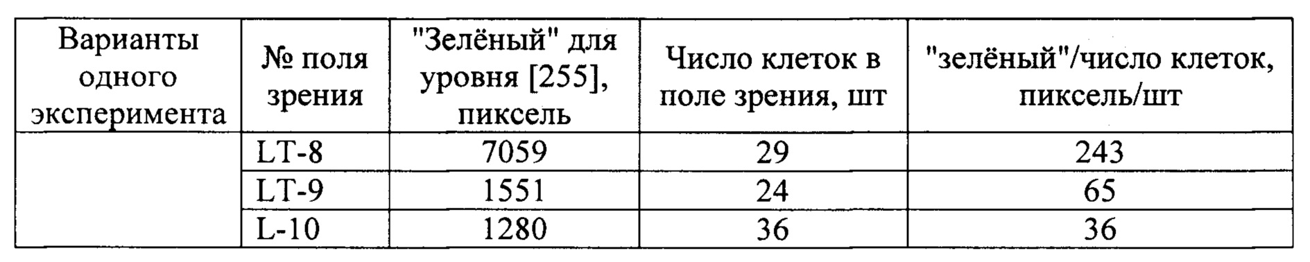
Статистический анализStatistical analysis
Статистическую обработку данных выполняли по алгоритму ANOVA в одном направлении с последующим тестом Бонферрони, для чего использовали программный продукт GraphPad Prism версии 6.01 для Windows (GraphPad Software, La Jolla California США, www.graphpad.com). Статистически значимым считали значение р < 0,05.Statistical data processing was performed according to the ANOVA algorithm in one direction followed by the Bonferroni test using the GraphPad Prism software version 6.01 for Windows (GraphPad Software, La Jolla California USA, www.graphpad.com). A p-value <0.05 was considered statistically significant.
Проведенный анализ показывает:The analysis shows:
1) культура в контроле демонстрирует устойчивый и репрезентативный уровень синтеза миелина (Фиг. 1А, таблица 1 и Фиг. 3Б), что свидетельствует о сбалансированности условий культивирования, и позволяет использовать культуру для создания модели PC;1) the culture in the control demonstrates a stable and representative level of myelin synthesis (Fig. 1A, Table 1 and Fig. 3B), which indicates a balanced culture conditions, and allows the culture to be used to create a PC model;
2) обработка культуры по отдельности - посредством ЛПС или лекарственным средством, - равно как и совместная обработка ЛПС и лекарственным средством, не приводит к значительному изменению числа клеток в культуре (Фиг. 3А), что исключает объяснение эффектов использованных реагентов - токсина и лекарственного средства - за счет гибели или избыточной пролиферации клеток в культуре; это позволяет проводить оценку влияния реагентов на синтез миелина;2) treatment of the culture separately - with LPS or a drug, as well as joint treatment with LPS and a drug, does not lead to a significant change in the number of cells in the culture (Fig.3A), which excludes an explanation of the effects of the used reagents - toxin and drug - due to the death or excessive proliferation of cells in culture; this allows assessing the effect of reagents on myelin synthesis;
3) токсическая обработка культуры посредством ЛПС снижает уровень продукции миелина до 20%, что подтверждает работоспособность модели;3) toxic treatment of the culture with LPS reduces the level of myelin production up to 20%, which confirms the performance of the model;
4) антиоксидантное лекарственное средство Trolox обеспечивает защиту клеток от токсического воздействия ЛПС на 60% (Фиг. 3Б).4) the antioxidant drug Trolox protects cells from the toxic effects of LPS by 60% (Fig. 3B).
Таким образом, предлагаемый способ может быть использован для быстрого изучения и скрининга лекарственных препаратов.Thus, the proposed method can be used for rapid study and screening of drugs.
Метод органотипического культивирования эксплантатов мозжечка на стеклах в чашках Петри технически прост, исключает стадию дезинтеграции ткани и не требует большого количества персонала и оборудования. Метод позволяет за короткий срок получить значительное количество миелинизирующих клеток (олигодендроцитов), пригодных для тестирования лекарственных препаратов, и наработать любое число образцов, обуславливающих статистическую достоверность получаемых результатов. Метод не требует изготовления дополнительных конструкций и их приклеивания к чашкам Петри для культивирования клеток. Это исключает присутствие дополнительных воздействий на изучаемые клетки (в виде клеевого слоя), что упрощает трактовку получаемых результатов. Количественная оценка результатов технически проста и занимает короткое время.The method of organotypic cultivation of cerebellar explants on glass slides in Petri dishes is technically simple, excludes the stage of tissue disintegration and does not require a large number of personnel and equipment. The method allows for a short time to obtain a significant number of myelinating cells (oligodendrocytes), suitable for testing drugs, and to produce any number of samples that determine the statistical reliability of the results. The method does not require the manufacture of additional structures and their gluing to Petri dishes for cell cultivation. This excludes the presence of additional influences on the cells under study (in the form of an adhesive layer), which simplifies the interpretation of the results obtained. Quantifying the results is technically simple and takes a short time.
Claims (14)
Priority Applications (1)
| Application Number | Priority Date | Filing Date | Title |
|---|---|---|---|
| RU2019130776A RU2736070C1 (en) | 2019-09-30 | 2019-09-30 | Test model for investigating action of drugs which prevent demyelination, method for preparing and using thereof |
Applications Claiming Priority (1)
| Application Number | Priority Date | Filing Date | Title |
|---|---|---|---|
| RU2019130776A RU2736070C1 (en) | 2019-09-30 | 2019-09-30 | Test model for investigating action of drugs which prevent demyelination, method for preparing and using thereof |
Publications (1)
| Publication Number | Publication Date |
|---|---|
| RU2736070C1 true RU2736070C1 (en) | 2020-11-11 |
Family
ID=73460952
Family Applications (1)
| Application Number | Title | Priority Date | Filing Date |
|---|---|---|---|
| RU2019130776A RU2736070C1 (en) | 2019-09-30 | 2019-09-30 | Test model for investigating action of drugs which prevent demyelination, method for preparing and using thereof |
Country Status (1)
| Country | Link |
|---|---|
| RU (1) | RU2736070C1 (en) |
Citations (1)
| Publication number | Priority date | Publication date | Assignee | Title |
|---|---|---|---|---|
| EP1294770B1 (en) * | 2000-05-10 | 2011-12-07 | Mayo Foundation For Medical Education And Research | Human igm antibodies with the capability of inducing remyelination, and diagnostic and therapeutic uses thereof particularly in the central nervous system |
-
2019
- 2019-09-30 RU RU2019130776A patent/RU2736070C1/en active
Patent Citations (1)
| Publication number | Priority date | Publication date | Assignee | Title |
|---|---|---|---|---|
| EP1294770B1 (en) * | 2000-05-10 | 2011-12-07 | Mayo Foundation For Medical Education And Research | Human igm antibodies with the capability of inducing remyelination, and diagnostic and therapeutic uses thereof particularly in the central nervous system |
Non-Patent Citations (2)
| Title |
|---|
| THOMPSON C. E. et al. Myelinated, synapsing cultures of murine spinal cord - validation as an in vitro model central nervous system // European Journal of Neuroscience, 2008, V.28, pp.1518-1535. D’SOUZA S. D. et al. Multiple Sclerosis: Fas Signaling in Oligodendrocyte Cell Death // J. Exp. Med., 1996, V.184, pp.2361-2370. * |
| СЕМЕНОВА В.М. и др. Пролиферативный и дифференцировочный потенциал нейроклеток эмбрионального мозга человека в условия х культивирования // УКРАIНСЬКИЙ НЕЙРОХIРУРГIЧНИЙ ЖУРНАЛ, 2007, N.1, стр.68-71. * |
Similar Documents
| Publication | Publication Date | Title |
|---|---|---|
| Sabate‐Soler et al. | Microglia integration into human midbrain organoids leads to increased neuronal maturation and functionality | |
| Nascimento et al. | Human cerebral organoids and fetal brain tissue share proteomic similarities | |
| Shim et al. | Generation of functional dopamine neurons from neural precursor cells isolated from the subventricular zone and white matter of the adult rat brain using Nurr1 overexpression | |
| Martin et al. | A novel method for generating glutamatergic SH-SY5Y neuron-like cells utilizing B-27 supplement | |
| Garcia et al. | Emerging role of miR-21-5p in neuron–glia dysregulation and exosome transfer using multiple models of Alzheimer’s disease | |
| Dezonne et al. | Derivation of functional human astrocytes from cerebral organoids | |
| Zhang | Defining glial cells during CNS development | |
| Lu et al. | A microdevice platform for visualizing mitochondrial transport in aligned dopaminergic axons | |
| Ray et al. | Human primary mixed brain cultures: preparation, differentiation, characterization and application to neuroscience research | |
| CN103827295B (en) | adult stem cells | |
| Kleiderman et al. | Conversion of nonproliferating astrocytes into neurogenic neural stem cells: control by FGF2 and interferon-γ | |
| Khan et al. | Neurosphere development from hippocampal and cortical embryonic mixed primary neuron culture: a potential platform for screening neurochemical modulator | |
| Kamande et al. | Multi-compartment microfluidic device geometry and covalently bound Poly-D-Lysine influence neuronal maturation | |
| EP3458119B1 (en) | Method for forming a functional network of human neuronal and glial cells | |
| Antonov et al. | Current state-of-the-art and unresolved problems in using human induced pluripotent stem cell-derived dopamine neurons for Parkinson’s disease drug development | |
| Ortega et al. | Live imaging of adult neural stem cells in rodents | |
| Feng et al. | Fibroblast growth factor 4 is required but not sufficient for the astrocyte dedifferentiation | |
| Tanti et al. | Isolation, culture and functional characterization of glia and endothelial cells from adult pig brain | |
| Chuye et al. | Brain organoids: expanding our understanding of human development and disease | |
| Yang et al. | Pleiotrophin is involved in the amniotic epithelial cell-induced differentiation of human umbilical cord blood-derived mesenchymal stem cells into dopaminergic neuron-like cells | |
| Gong et al. | SCF/SCFR signaling plays an important role in the early morphogenesis and neurogenesis of human embryonic neural retina | |
| Wang et al. | GABA inhibits proliferation and self-renewal of mouse retinal progenitor cell | |
| Arias-Carrión et al. | Neurogenesis in substantia nigra of parkinsonian brains? | |
| Yu et al. | PGC-1α mediates hypoxia-preconditioned olfactory mucosa mesenchymal stem cells improved neuroinflammatory response via inhibiting microglial ferroptosis in ischemic stroke | |
| Gil et al. | Analysis of axonal growth and cell migration in 3D hydrogel cultures of embryonic mouse CNS tissue |