TW202020165A - Nucleic acid rearrangement and integration analysis - Google Patents
Nucleic acid rearrangement and integration analysis Download PDFInfo
- Publication number
- TW202020165A TW202020165A TW108122976A TW108122976A TW202020165A TW 202020165 A TW202020165 A TW 202020165A TW 108122976 A TW108122976 A TW 108122976A TW 108122976 A TW108122976 A TW 108122976A TW 202020165 A TW202020165 A TW 202020165A
- Authority
- TW
- Taiwan
- Prior art keywords
- pathogen
- nucleic acid
- organism
- sequence
- cell
- Prior art date
Links
- 150000007523 nucleic acids Chemical class 0.000 title claims abstract description 416
- 230000010354 integration Effects 0.000 title claims abstract description 351
- 102000039446 nucleic acids Human genes 0.000 title claims abstract description 292
- 108020004707 nucleic acids Proteins 0.000 title claims abstract description 292
- 238000004458 analytical method Methods 0.000 title claims description 95
- 230000008707 rearrangement Effects 0.000 title 1
- 238000000034 method Methods 0.000 claims abstract description 333
- 244000052769 pathogen Species 0.000 claims abstract description 283
- 230000001717 pathogenic effect Effects 0.000 claims abstract description 267
- 239000012472 biological sample Substances 0.000 claims abstract description 117
- 230000007170 pathology Effects 0.000 claims abstract description 22
- 206010028980 Neoplasm Diseases 0.000 claims description 163
- 241000701806 Human papillomavirus Species 0.000 claims description 128
- 201000011510 cancer Diseases 0.000 claims description 127
- 239000012634 fragment Substances 0.000 claims description 127
- 238000012163 sequencing technique Methods 0.000 claims description 121
- 241000700605 Viruses Species 0.000 claims description 95
- 230000006798 recombination Effects 0.000 claims description 79
- 238000005215 recombination Methods 0.000 claims description 79
- 210000000349 chromosome Anatomy 0.000 claims description 75
- 230000002759 chromosomal effect Effects 0.000 claims description 68
- 239000002773 nucleotide Substances 0.000 claims description 60
- 125000003729 nucleotide group Chemical group 0.000 claims description 60
- 210000002381 plasma Anatomy 0.000 claims description 57
- 230000003321 amplification Effects 0.000 claims description 56
- 238000003199 nucleic acid amplification method Methods 0.000 claims description 56
- 206010008342 Cervix carcinoma Diseases 0.000 claims description 50
- 208000006105 Uterine Cervical Neoplasms Diseases 0.000 claims description 49
- 201000010881 cervical cancer Diseases 0.000 claims description 49
- 238000006243 chemical reaction Methods 0.000 claims description 47
- 208000000102 Squamous Cell Carcinoma of Head and Neck Diseases 0.000 claims description 46
- 201000000459 head and neck squamous cell carcinoma Diseases 0.000 claims description 46
- 238000003752 polymerase chain reaction Methods 0.000 claims description 35
- 241000700721 Hepatitis B virus Species 0.000 claims description 30
- 108020004414 DNA Proteins 0.000 claims description 27
- 210000004027 cell Anatomy 0.000 claims description 27
- 230000000295 complement effect Effects 0.000 claims description 27
- 241001465754 Metazoa Species 0.000 claims description 23
- 238000001514 detection method Methods 0.000 claims description 23
- 238000001914 filtration Methods 0.000 claims description 20
- 230000001575 pathological effect Effects 0.000 claims description 19
- 206010061306 Nasopharyngeal cancer Diseases 0.000 claims description 18
- 241000124008 Mammalia Species 0.000 claims description 16
- 102000053602 DNA Human genes 0.000 claims description 15
- 108091093088 Amplicon Proteins 0.000 claims description 12
- 210000002700 urine Anatomy 0.000 claims description 11
- 208000032839 leukemia Diseases 0.000 claims description 10
- 210000002966 serum Anatomy 0.000 claims description 10
- 241000711549 Hepacivirus C Species 0.000 claims description 9
- 206010025323 Lymphomas Diseases 0.000 claims description 9
- 206010057444 Oropharyngeal neoplasm Diseases 0.000 claims description 8
- 208000037516 chromosome inversion disease Diseases 0.000 claims description 8
- 206010009944 Colon cancer Diseases 0.000 claims description 7
- 208000001894 Nasopharyngeal Neoplasms Diseases 0.000 claims description 7
- 206010031096 Oropharyngeal cancer Diseases 0.000 claims description 7
- 201000007270 liver cancer Diseases 0.000 claims description 7
- 208000014018 liver neoplasm Diseases 0.000 claims description 7
- 201000006958 oropharynx cancer Diseases 0.000 claims description 7
- 230000008859 change Effects 0.000 claims description 6
- 206010005949 Bone cancer Diseases 0.000 claims description 5
- 208000018084 Bone neoplasm Diseases 0.000 claims description 5
- 208000003174 Brain Neoplasms Diseases 0.000 claims description 5
- 206010006187 Breast cancer Diseases 0.000 claims description 5
- 208000026310 Breast neoplasm Diseases 0.000 claims description 5
- 208000016718 Chromosome Inversion Diseases 0.000 claims description 5
- 208000003445 Mouth Neoplasms Diseases 0.000 claims description 5
- 206010035226 Plasma cell myeloma Diseases 0.000 claims description 5
- 206010060862 Prostate cancer Diseases 0.000 claims description 5
- 208000000236 Prostatic Neoplasms Diseases 0.000 claims description 5
- 206010039491 Sarcoma Diseases 0.000 claims description 5
- 208000012987 lip and oral cavity carcinoma Diseases 0.000 claims description 5
- 201000000050 myeloid neoplasm Diseases 0.000 claims description 5
- 206010005003 Bladder cancer Diseases 0.000 claims description 4
- 206010061764 Chromosomal deletion Diseases 0.000 claims description 4
- 208000001333 Colorectal Neoplasms Diseases 0.000 claims description 4
- 208000000461 Esophageal Neoplasms Diseases 0.000 claims description 4
- 206010017993 Gastrointestinal neoplasms Diseases 0.000 claims description 4
- 208000034951 Genetic Translocation Diseases 0.000 claims description 4
- 206010058467 Lung neoplasm malignant Diseases 0.000 claims description 4
- 206010030155 Oesophageal carcinoma Diseases 0.000 claims description 4
- 206010033128 Ovarian cancer Diseases 0.000 claims description 4
- 206010061535 Ovarian neoplasm Diseases 0.000 claims description 4
- 208000005718 Stomach Neoplasms Diseases 0.000 claims description 4
- 208000024770 Thyroid neoplasm Diseases 0.000 claims description 4
- 208000007097 Urinary Bladder Neoplasms Diseases 0.000 claims description 4
- 238000004364 calculation method Methods 0.000 claims description 4
- 201000004101 esophageal cancer Diseases 0.000 claims description 4
- 206010017758 gastric cancer Diseases 0.000 claims description 4
- 201000005202 lung cancer Diseases 0.000 claims description 4
- 208000020816 lung neoplasm Diseases 0.000 claims description 4
- 201000011549 stomach cancer Diseases 0.000 claims description 4
- 201000002510 thyroid cancer Diseases 0.000 claims description 4
- 201000005112 urinary bladder cancer Diseases 0.000 claims description 4
- 241000701044 Human gammaherpesvirus 4 Species 0.000 claims description 3
- 208000010505 Nose Neoplasms Diseases 0.000 claims description 3
- 208000037830 nasal cancer Diseases 0.000 claims description 3
- 208000002250 Hematologic Neoplasms Diseases 0.000 claims description 2
- 230000009033 hematopoietic malignancy Effects 0.000 claims description 2
- 206010041823 squamous cell carcinoma Diseases 0.000 claims 1
- 208000017572 squamous cell neoplasm Diseases 0.000 claims 1
- 230000008711 chromosomal rearrangement Effects 0.000 abstract description 4
- 208000022361 Human papillomavirus infectious disease Diseases 0.000 description 33
- 239000000523 sample Substances 0.000 description 33
- 230000003612 virological effect Effects 0.000 description 31
- 210000001519 tissue Anatomy 0.000 description 29
- 238000004422 calculation algorithm Methods 0.000 description 28
- 238000003860 storage Methods 0.000 description 24
- 230000015654 memory Effects 0.000 description 23
- 208000037265 diseases, disorders, signs and symptoms Diseases 0.000 description 18
- 201000010099 disease Diseases 0.000 description 17
- 241001430294 unidentified retrovirus Species 0.000 description 17
- 108090000623 proteins and genes Proteins 0.000 description 16
- 241001529453 unidentified herpesvirus Species 0.000 description 16
- 108091028043 Nucleic acid sequence Proteins 0.000 description 15
- 238000013507 mapping Methods 0.000 description 15
- 208000011580 syndromic disease Diseases 0.000 description 15
- 238000012217 deletion Methods 0.000 description 14
- 230000037430 deletion Effects 0.000 description 14
- 208000002454 Nasopharyngeal Carcinoma Diseases 0.000 description 11
- 108020005202 Viral DNA Proteins 0.000 description 11
- 201000011216 nasopharynx carcinoma Diseases 0.000 description 11
- 208000011691 Burkitt lymphomas Diseases 0.000 description 10
- 241000579048 Merkel cell polyomavirus Species 0.000 description 10
- 230000004927 fusion Effects 0.000 description 10
- 230000005945 translocation Effects 0.000 description 10
- 238000010586 diagram Methods 0.000 description 9
- 206010073071 hepatocellular carcinoma Diseases 0.000 description 9
- 231100000844 hepatocellular carcinoma Toxicity 0.000 description 9
- 208000015181 infectious disease Diseases 0.000 description 9
- 108091032973 (ribonucleotides)n+m Proteins 0.000 description 8
- 241000341655 Human papillomavirus type 16 Species 0.000 description 8
- 238000004891 communication Methods 0.000 description 8
- 230000006378 damage Effects 0.000 description 8
- 238000011161 development Methods 0.000 description 8
- 230000018109 developmental process Effects 0.000 description 8
- 241001493065 dsRNA viruses Species 0.000 description 8
- 241000282412 Homo Species 0.000 description 7
- 230000001413 cellular effect Effects 0.000 description 7
- 230000003287 optical effect Effects 0.000 description 7
- 230000008569 process Effects 0.000 description 7
- 238000012545 processing Methods 0.000 description 7
- 238000011282 treatment Methods 0.000 description 7
- 206010008263 Cervical dysplasia Diseases 0.000 description 6
- 230000005540 biological transmission Effects 0.000 description 6
- 208000007951 cervical intraepithelial neoplasia Diseases 0.000 description 6
- 238000005516 engineering process Methods 0.000 description 6
- 230000010076 replication Effects 0.000 description 6
- 230000035945 sensitivity Effects 0.000 description 6
- 238000004088 simulation Methods 0.000 description 6
- 238000012360 testing method Methods 0.000 description 6
- 208000002030 Merkel cell carcinoma Diseases 0.000 description 5
- 206010029266 Neuroendocrine carcinoma of the skin Diseases 0.000 description 5
- 241001631646 Papillomaviridae Species 0.000 description 5
- 208000029742 colonic neoplasm Diseases 0.000 description 5
- 208000017763 cutaneous neuroendocrine carcinoma Diseases 0.000 description 5
- 238000009826 distribution Methods 0.000 description 5
- 239000012530 fluid Substances 0.000 description 5
- 230000006870 function Effects 0.000 description 5
- 238000009396 hybridization Methods 0.000 description 5
- 230000003211 malignant effect Effects 0.000 description 5
- 208000030507 AIDS Diseases 0.000 description 4
- -1 DNA) molecules Chemical class 0.000 description 4
- 230000009471 action Effects 0.000 description 4
- 230000008901 benefit Effects 0.000 description 4
- 210000004369 blood Anatomy 0.000 description 4
- 239000008280 blood Substances 0.000 description 4
- 238000013500 data storage Methods 0.000 description 4
- 230000003902 lesion Effects 0.000 description 4
- 238000002493 microarray Methods 0.000 description 4
- 238000010208 microarray analysis Methods 0.000 description 4
- 230000002093 peripheral effect Effects 0.000 description 4
- 230000002441 reversible effect Effects 0.000 description 4
- 238000007480 sanger sequencing Methods 0.000 description 4
- 208000009746 Adult T-Cell Leukemia-Lymphoma Diseases 0.000 description 3
- 208000016683 Adult T-cell leukemia/lymphoma Diseases 0.000 description 3
- 208000003950 B-cell lymphoma Diseases 0.000 description 3
- 241000701022 Cytomegalovirus Species 0.000 description 3
- 241000702421 Dependoparvovirus Species 0.000 description 3
- 108020004437 Endogenous Retroviruses Proteins 0.000 description 3
- 241000941423 Grom virus Species 0.000 description 3
- 241001502974 Human gammaherpesvirus 8 Species 0.000 description 3
- 208000007766 Kaposi sarcoma Diseases 0.000 description 3
- 241000701043 Lymphocryptovirus Species 0.000 description 3
- 206010027476 Metastases Diseases 0.000 description 3
- 238000010222 PCR analysis Methods 0.000 description 3
- 241000700584 Simplexvirus Species 0.000 description 3
- 208000026487 Triploidy Diseases 0.000 description 3
- 201000006966 adult T-cell leukemia Diseases 0.000 description 3
- 238000013459 approach Methods 0.000 description 3
- 230000001684 chronic effect Effects 0.000 description 3
- 238000013467 fragmentation Methods 0.000 description 3
- 238000006062 fragmentation reaction Methods 0.000 description 3
- 230000012010 growth Effects 0.000 description 3
- 238000003780 insertion Methods 0.000 description 3
- 230000037431 insertion Effects 0.000 description 3
- 208000024312 invasive carcinoma Diseases 0.000 description 3
- 230000009401 metastasis Effects 0.000 description 3
- 230000035772 mutation Effects 0.000 description 3
- 210000004940 nucleus Anatomy 0.000 description 3
- 238000003753 real-time PCR Methods 0.000 description 3
- 230000001105 regulatory effect Effects 0.000 description 3
- 210000003296 saliva Anatomy 0.000 description 3
- 210000001082 somatic cell Anatomy 0.000 description 3
- 241000894007 species Species 0.000 description 3
- 210000004881 tumor cell Anatomy 0.000 description 3
- 238000012795 verification Methods 0.000 description 3
- 102000040650 (ribonucleotides)n+m Human genes 0.000 description 2
- 206010061424 Anal cancer Diseases 0.000 description 2
- 208000007860 Anus Neoplasms Diseases 0.000 description 2
- 208000010839 B-cell chronic lymphocytic leukemia Diseases 0.000 description 2
- 101150070189 CIN3 gene Proteins 0.000 description 2
- 201000009030 Carcinoma Diseases 0.000 description 2
- 108020004638 Circular DNA Proteins 0.000 description 2
- 108020004635 Complementary DNA Proteins 0.000 description 2
- 206010015108 Epstein-Barr virus infection Diseases 0.000 description 2
- 241000700588 Human alphaherpesvirus 1 Species 0.000 description 2
- 241000701074 Human alphaherpesvirus 2 Species 0.000 description 2
- 241000701085 Human alphaherpesvirus 3 Species 0.000 description 2
- 241000701027 Human herpesvirus 6 Species 0.000 description 2
- XQFRJNBWHJMXHO-RRKCRQDMSA-N IDUR Chemical compound C1[C@H](O)[C@@H](CO)O[C@H]1N1C(=O)NC(=O)C(I)=C1 XQFRJNBWHJMXHO-RRKCRQDMSA-N 0.000 description 2
- 206010061598 Immunodeficiency Diseases 0.000 description 2
- 208000029462 Immunodeficiency disease Diseases 0.000 description 2
- 208000031422 Lymphocytic Chronic B-Cell Leukemia Diseases 0.000 description 2
- 241000712899 Lymphocytic choriomeningitis mammarenavirus Species 0.000 description 2
- 208000032271 Malignant tumor of penis Diseases 0.000 description 2
- 206010027406 Mesothelioma Diseases 0.000 description 2
- 241001467058 Murid gammaherpesvirus 4 Species 0.000 description 2
- 201000003793 Myelodysplastic syndrome Diseases 0.000 description 2
- 206010028729 Nasal cavity cancer Diseases 0.000 description 2
- 208000009608 Papillomavirus Infections Diseases 0.000 description 2
- 208000002471 Penile Neoplasms Diseases 0.000 description 2
- 206010034299 Penile cancer Diseases 0.000 description 2
- 241001505332 Polyomavirus sp. Species 0.000 description 2
- 238000012300 Sequence Analysis Methods 0.000 description 2
- 206010047741 Vulval cancer Diseases 0.000 description 2
- 208000004354 Vulvar Neoplasms Diseases 0.000 description 2
- 230000002159 abnormal effect Effects 0.000 description 2
- 239000000427 antigen Substances 0.000 description 2
- 108091007433 antigens Proteins 0.000 description 2
- 102000036639 antigens Human genes 0.000 description 2
- 201000011165 anus cancer Diseases 0.000 description 2
- 238000003766 bioinformatics method Methods 0.000 description 2
- 210000001124 body fluid Anatomy 0.000 description 2
- 239000010839 body fluid Substances 0.000 description 2
- 238000010804 cDNA synthesis Methods 0.000 description 2
- 231100000504 carcinogenesis Toxicity 0.000 description 2
- 230000003915 cell function Effects 0.000 description 2
- 230000004663 cell proliferation Effects 0.000 description 2
- 108091092356 cellular DNA Proteins 0.000 description 2
- 238000005119 centrifugation Methods 0.000 description 2
- 101150005988 cin2 gene Proteins 0.000 description 2
- 239000002299 complementary DNA Substances 0.000 description 2
- 238000013480 data collection Methods 0.000 description 2
- 230000034994 death Effects 0.000 description 2
- 238000012350 deep sequencing Methods 0.000 description 2
- 230000005782 double-strand break Effects 0.000 description 2
- 238000007672 fourth generation sequencing Methods 0.000 description 2
- 230000003394 haemopoietic effect Effects 0.000 description 2
- 230000036541 health Effects 0.000 description 2
- 208000002672 hepatitis B Diseases 0.000 description 2
- 230000007813 immunodeficiency Effects 0.000 description 2
- 238000000338 in vitro Methods 0.000 description 2
- 230000009545 invasion Effects 0.000 description 2
- 210000005229 liver cell Anatomy 0.000 description 2
- 230000036210 malignancy Effects 0.000 description 2
- 238000004519 manufacturing process Methods 0.000 description 2
- 230000001404 mediated effect Effects 0.000 description 2
- 239000000203 mixture Substances 0.000 description 2
- 238000012544 monitoring process Methods 0.000 description 2
- 238000007481 next generation sequencing Methods 0.000 description 2
- 210000004180 plasmocyte Anatomy 0.000 description 2
- 238000002360 preparation method Methods 0.000 description 2
- 238000004393 prognosis Methods 0.000 description 2
- 230000008521 reorganization Effects 0.000 description 2
- 238000012216 screening Methods 0.000 description 2
- 239000004065 semiconductor Substances 0.000 description 2
- 239000007787 solid Substances 0.000 description 2
- 238000006467 substitution reaction Methods 0.000 description 2
- 239000000758 substrate Substances 0.000 description 2
- 238000002560 therapeutic procedure Methods 0.000 description 2
- 238000013518 transcription Methods 0.000 description 2
- 230000035897 transcription Effects 0.000 description 2
- 230000002103 transcriptional effect Effects 0.000 description 2
- 230000014616 translation Effects 0.000 description 2
- 208000026485 trisomy X Diseases 0.000 description 2
- 206010046885 vaginal cancer Diseases 0.000 description 2
- 208000013139 vaginal neoplasm Diseases 0.000 description 2
- 230000029812 viral genome replication Effects 0.000 description 2
- 201000005102 vulva cancer Diseases 0.000 description 2
- 238000012070 whole genome sequencing analysis Methods 0.000 description 2
- 208000033881 15q13.3 microdeletion syndrome Diseases 0.000 description 1
- 208000037215 17q23.1q23.2 microdeletion syndrome Diseases 0.000 description 1
- 208000037049 1q21.1 microdeletion syndrome Diseases 0.000 description 1
- 208000025304 2q23.1 microdeletion syndrome Diseases 0.000 description 1
- 208000026817 47,XYY syndrome Diseases 0.000 description 1
- 241000251468 Actinopterygii Species 0.000 description 1
- 206010000830 Acute leukaemia Diseases 0.000 description 1
- 208000024893 Acute lymphoblastic leukemia Diseases 0.000 description 1
- 208000014697 Acute lymphocytic leukaemia Diseases 0.000 description 1
- 208000031261 Acute myeloid leukaemia Diseases 0.000 description 1
- 241000702423 Adeno-associated virus - 2 Species 0.000 description 1
- 241001664176 Alpharetrovirus Species 0.000 description 1
- 241000143060 Americamysis bahia Species 0.000 description 1
- 208000009575 Angelman syndrome Diseases 0.000 description 1
- 241000272517 Anseriformes Species 0.000 description 1
- 241000269350 Anura Species 0.000 description 1
- 101100421761 Arabidopsis thaliana GSNAP gene Proteins 0.000 description 1
- 206010003445 Ascites Diseases 0.000 description 1
- 206010003805 Autism Diseases 0.000 description 1
- 208000020706 Autistic disease Diseases 0.000 description 1
- 235000000832 Ayote Nutrition 0.000 description 1
- 208000036170 B-Cell Marginal Zone Lymphoma Diseases 0.000 description 1
- 208000032791 BCR-ABL1 positive chronic myelogenous leukemia Diseases 0.000 description 1
- 206010004146 Basal cell carcinoma Diseases 0.000 description 1
- 241001231757 Betaretrovirus Species 0.000 description 1
- 241000283690 Bos taurus Species 0.000 description 1
- 206010006223 Breast discharge Diseases 0.000 description 1
- 101150061050 CIN1 gene Proteins 0.000 description 1
- 241000282472 Canis lupus familiaris Species 0.000 description 1
- 241000283707 Capra Species 0.000 description 1
- 208000014392 Cat-eye syndrome Diseases 0.000 description 1
- 241000700198 Cavia Species 0.000 description 1
- 206010007953 Central nervous system lymphoma Diseases 0.000 description 1
- 241000282693 Cercopithecidae Species 0.000 description 1
- 201000009009 Charcot-Marie-Tooth disease type 1A Diseases 0.000 description 1
- 206010068051 Chimerism Diseases 0.000 description 1
- 108010077544 Chromatin Proteins 0.000 description 1
- 208000010833 Chronic myeloid leukaemia Diseases 0.000 description 1
- RYGMFSIKBFXOCR-UHFFFAOYSA-N Copper Chemical compound [Cu] RYGMFSIKBFXOCR-UHFFFAOYSA-N 0.000 description 1
- 206010011385 Cri-du-chat syndrome Diseases 0.000 description 1
- 241000699800 Cricetinae Species 0.000 description 1
- 235000003949 Cucurbita mixta Nutrition 0.000 description 1
- 240000004244 Cucurbita moschata Species 0.000 description 1
- 235000009854 Cucurbita moschata Nutrition 0.000 description 1
- 108050006400 Cyclin Proteins 0.000 description 1
- 102000016736 Cyclin Human genes 0.000 description 1
- 238000001712 DNA sequencing Methods 0.000 description 1
- 241000450599 DNA viruses Species 0.000 description 1
- 241001663879 Deltaretrovirus Species 0.000 description 1
- 208000002699 Digestive System Neoplasms Diseases 0.000 description 1
- 208000004254 Emanuel syndrome Diseases 0.000 description 1
- 206010014733 Endometrial cancer Diseases 0.000 description 1
- 206010014759 Endometrial neoplasm Diseases 0.000 description 1
- 108090000790 Enzymes Proteins 0.000 description 1
- 102000004190 Enzymes Human genes 0.000 description 1
- 241000283086 Equidae Species 0.000 description 1
- 241000230501 Equine herpesvirus sp. Species 0.000 description 1
- 201000005866 Exanthema Subitum Diseases 0.000 description 1
- 102100039111 FAD-linked sulfhydryl oxidase ALR Human genes 0.000 description 1
- 102100037858 G1/S-specific cyclin-E1 Human genes 0.000 description 1
- 101800000863 Galanin message-associated peptide Proteins 0.000 description 1
- 102100028501 Galanin peptides Human genes 0.000 description 1
- 208000022072 Gallbladder Neoplasms Diseases 0.000 description 1
- 241000287828 Gallus gallus Species 0.000 description 1
- 241001663880 Gammaretrovirus Species 0.000 description 1
- 208000031448 Genomic Instability Diseases 0.000 description 1
- 241000282575 Gorilla Species 0.000 description 1
- 241001272567 Hominoidea Species 0.000 description 1
- 101000959079 Homo sapiens FAD-linked sulfhydryl oxidase ALR Proteins 0.000 description 1
- 101000738568 Homo sapiens G1/S-specific cyclin-E1 Proteins 0.000 description 1
- 101000848922 Homo sapiens Protein FAM72A Proteins 0.000 description 1
- 101000904499 Homo sapiens Transcription regulator protein BACH2 Proteins 0.000 description 1
- 241000598436 Human T-cell lymphotropic virus Species 0.000 description 1
- 241000714192 Human spumaretrovirus Species 0.000 description 1
- 241001051759 Iltovirus Species 0.000 description 1
- 208000026350 Inborn Genetic disease Diseases 0.000 description 1
- 208000004706 Jacobsen Distal 11q Deletion Syndrome Diseases 0.000 description 1
- 208000029279 Jacobsen Syndrome Diseases 0.000 description 1
- 208000008839 Kidney Neoplasms Diseases 0.000 description 1
- 208000004252 Kleefstra syndrome Diseases 0.000 description 1
- 201000003395 Koolen de Vries syndrome Diseases 0.000 description 1
- 208000031671 Large B-Cell Diffuse Lymphoma Diseases 0.000 description 1
- 206010023825 Laryngeal cancer Diseases 0.000 description 1
- 241000288903 Lemuridae Species 0.000 description 1
- 206010067125 Liver injury Diseases 0.000 description 1
- 208000030289 Lymphoproliferative disease Diseases 0.000 description 1
- 241000175216 Macavirus Species 0.000 description 1
- 206010064912 Malignant transformation Diseases 0.000 description 1
- 208000025205 Mantle-Cell Lymphoma Diseases 0.000 description 1
- 241001051756 Mardivirus Species 0.000 description 1
- 241000283923 Marmota monax Species 0.000 description 1
- 208000001804 Monosomy 5p Diseases 0.000 description 1
- 108010085220 Multiprotein Complexes Proteins 0.000 description 1
- 102000007474 Multiprotein Complexes Human genes 0.000 description 1
- 241000701029 Murid betaherpesvirus 1 Species 0.000 description 1
- 241000701034 Muromegalovirus Species 0.000 description 1
- 241000699666 Mus <mouse, genus> Species 0.000 description 1
- 241000699670 Mus sp. Species 0.000 description 1
- 208000033761 Myelogenous Chronic BCR-ABL Positive Leukemia Diseases 0.000 description 1
- 208000033776 Myeloid Acute Leukemia Diseases 0.000 description 1
- 108091034117 Oligonucleotide Proteins 0.000 description 1
- 238000012408 PCR amplification Methods 0.000 description 1
- 206010061902 Pancreatic neoplasm Diseases 0.000 description 1
- 241000282520 Papio Species 0.000 description 1
- 241000701945 Parvoviridae Species 0.000 description 1
- 241001494479 Pecora Species 0.000 description 1
- 241000175217 Percavirus Species 0.000 description 1
- 208000027190 Peripheral T-cell lymphomas Diseases 0.000 description 1
- 208000007913 Pituitary Neoplasms Diseases 0.000 description 1
- 241000282405 Pongo abelii Species 0.000 description 1
- 208000006994 Precancerous Conditions Diseases 0.000 description 1
- 208000006664 Precursor Cell Lymphoblastic Leukemia-Lymphoma Diseases 0.000 description 1
- 241000175215 Proboscivirus Species 0.000 description 1
- 206010036790 Productive cough Diseases 0.000 description 1
- 102100034514 Protein FAM72A Human genes 0.000 description 1
- 241001112090 Pseudovirus Species 0.000 description 1
- 241000700159 Rattus Species 0.000 description 1
- 208000015634 Rectal Neoplasms Diseases 0.000 description 1
- 206010038389 Renal cancer Diseases 0.000 description 1
- 241000701037 Rhadinovirus Species 0.000 description 1
- 208000036485 Roseola Diseases 0.000 description 1
- 241000122129 Roseolovirus Species 0.000 description 1
- 208000004337 Salivary Gland Neoplasms Diseases 0.000 description 1
- 206010061934 Salivary gland cancer Diseases 0.000 description 1
- 241000555745 Sciuridae Species 0.000 description 1
- 241000351085 Scutavirus Species 0.000 description 1
- 208000000453 Skin Neoplasms Diseases 0.000 description 1
- 201000001388 Smith-Magenis syndrome Diseases 0.000 description 1
- 241001223864 Sphyraena barracuda Species 0.000 description 1
- 241000282887 Suidae Species 0.000 description 1
- 208000031672 T-Cell Peripheral Lymphoma Diseases 0.000 description 1
- 208000000389 T-cell leukemia Diseases 0.000 description 1
- 208000028530 T-cell lymphoblastic leukemia/lymphoma Diseases 0.000 description 1
- 206010042971 T-cell lymphoma Diseases 0.000 description 1
- 208000027585 T-cell non-Hodgkin lymphoma Diseases 0.000 description 1
- 208000020982 T-lymphoblastic lymphoma Diseases 0.000 description 1
- 210000001744 T-lymphocyte Anatomy 0.000 description 1
- 208000024313 Testicular Neoplasms Diseases 0.000 description 1
- 206010057644 Testis cancer Diseases 0.000 description 1
- 208000007254 Tetrasomy X Diseases 0.000 description 1
- 206010043515 Throat cancer Diseases 0.000 description 1
- 208000000728 Thymus Neoplasms Diseases 0.000 description 1
- 241000283907 Tragelaphus oryx Species 0.000 description 1
- 102100023998 Transcription regulator protein BACH2 Human genes 0.000 description 1
- 102000044209 Tumor Suppressor Genes Human genes 0.000 description 1
- 108700025716 Tumor Suppressor Genes Proteins 0.000 description 1
- 108010040002 Tumor Suppressor Proteins Proteins 0.000 description 1
- 102000001742 Tumor Suppressor Proteins Human genes 0.000 description 1
- 208000026928 Turner syndrome Diseases 0.000 description 1
- 208000002495 Uterine Neoplasms Diseases 0.000 description 1
- 241000701067 Varicellovirus Species 0.000 description 1
- 108700005077 Viral Genes Proteins 0.000 description 1
- 108020000999 Viral RNA Proteins 0.000 description 1
- 208000036142 Viral infection Diseases 0.000 description 1
- 206010049644 Williams syndrome Diseases 0.000 description 1
- 208000006254 Wolf-Hirschhorn Syndrome Diseases 0.000 description 1
- 239000002253 acid Substances 0.000 description 1
- 230000001154 acute effect Effects 0.000 description 1
- 208000036676 acute undifferentiated leukemia Diseases 0.000 description 1
- 201000005188 adrenal gland cancer Diseases 0.000 description 1
- 208000024447 adrenal gland neoplasm Diseases 0.000 description 1
- 239000012491 analyte Substances 0.000 description 1
- 238000010171 animal model Methods 0.000 description 1
- 238000011319 anticancer therapy Methods 0.000 description 1
- 238000003491 array Methods 0.000 description 1
- 238000007845 assembly PCR Methods 0.000 description 1
- 238000007846 asymmetric PCR Methods 0.000 description 1
- 238000001574 biopsy Methods 0.000 description 1
- 230000015572 biosynthetic process Effects 0.000 description 1
- 210000000988 bone and bone Anatomy 0.000 description 1
- 210000000481 breast Anatomy 0.000 description 1
- 239000000872 buffer Substances 0.000 description 1
- 230000000711 cancerogenic effect Effects 0.000 description 1
- 231100000315 carcinogenic Toxicity 0.000 description 1
- 201000011529 cardiovascular cancer Diseases 0.000 description 1
- 230000015556 catabolic process Effects 0.000 description 1
- 230000030833 cell death Effects 0.000 description 1
- 230000024245 cell differentiation Effects 0.000 description 1
- 210000001175 cerebrospinal fluid Anatomy 0.000 description 1
- 239000003795 chemical substances by application Substances 0.000 description 1
- 235000013330 chicken meat Nutrition 0.000 description 1
- 208000006990 cholangiocarcinoma Diseases 0.000 description 1
- 210000003483 chromatin Anatomy 0.000 description 1
- 239000013611 chromosomal DNA Substances 0.000 description 1
- 201000003794 chromosome 15q13.3 microdeletion syndrome Diseases 0.000 description 1
- 201000004692 chromosome 17q23.1-q23.2 deletion syndrome Diseases 0.000 description 1
- 201000004694 chromosome 18q deletion syndrome Diseases 0.000 description 1
- 201000004727 chromosome 1q21.1 deletion syndrome Diseases 0.000 description 1
- 208000024207 chronic leukemia Diseases 0.000 description 1
- 208000032852 chronic lymphocytic leukemia Diseases 0.000 description 1
- 210000001072 colon Anatomy 0.000 description 1
- 239000003086 colorant Substances 0.000 description 1
- 230000000052 comparative effect Effects 0.000 description 1
- 238000012790 confirmation Methods 0.000 description 1
- 230000021615 conjugation Effects 0.000 description 1
- 230000001276 controlling effect Effects 0.000 description 1
- 229910052802 copper Inorganic materials 0.000 description 1
- 239000010949 copper Substances 0.000 description 1
- 230000008878 coupling Effects 0.000 description 1
- 238000010168 coupling process Methods 0.000 description 1
- 238000005859 coupling reaction Methods 0.000 description 1
- 208000031513 cyst Diseases 0.000 description 1
- 238000007405 data analysis Methods 0.000 description 1
- 238000013523 data management Methods 0.000 description 1
- 230000003247 decreasing effect Effects 0.000 description 1
- 230000007812 deficiency Effects 0.000 description 1
- 230000003412 degenerative effect Effects 0.000 description 1
- 238000004925 denaturation Methods 0.000 description 1
- 230000036425 denaturation Effects 0.000 description 1
- 238000009795 derivation Methods 0.000 description 1
- 238000013461 design Methods 0.000 description 1
- 239000003599 detergent Substances 0.000 description 1
- 238000003745 diagnosis Methods 0.000 description 1
- 230000004069 differentiation Effects 0.000 description 1
- 206010012818 diffuse large B-cell lymphoma Diseases 0.000 description 1
- 208000024558 digestive system cancer Diseases 0.000 description 1
- 238000007847 digital PCR Methods 0.000 description 1
- 208000035475 disorder Diseases 0.000 description 1
- 230000008482 dysregulation Effects 0.000 description 1
- 210000000750 endocrine system Anatomy 0.000 description 1
- 238000001839 endoscopy Methods 0.000 description 1
- 239000003623 enhancer Substances 0.000 description 1
- 230000007613 environmental effect Effects 0.000 description 1
- 230000001973 epigenetic effect Effects 0.000 description 1
- 230000004049 epigenetic modification Effects 0.000 description 1
- 210000002919 epithelial cell Anatomy 0.000 description 1
- 238000011156 evaluation Methods 0.000 description 1
- 208000024519 eye neoplasm Diseases 0.000 description 1
- 230000002550 fecal effect Effects 0.000 description 1
- 238000011010 flushing procedure Methods 0.000 description 1
- 201000003444 follicular lymphoma Diseases 0.000 description 1
- 201000010175 gallbladder cancer Diseases 0.000 description 1
- 201000010231 gastrointestinal system cancer Diseases 0.000 description 1
- 208000016361 genetic disease Diseases 0.000 description 1
- 210000004602 germ cell Anatomy 0.000 description 1
- 229930182470 glycoside Natural products 0.000 description 1
- 150000002338 glycosides Chemical class 0.000 description 1
- 201000009277 hairy cell leukemia Diseases 0.000 description 1
- 201000010536 head and neck cancer Diseases 0.000 description 1
- 208000014829 head and neck neoplasm Diseases 0.000 description 1
- 230000003862 health status Effects 0.000 description 1
- 201000005787 hematologic cancer Diseases 0.000 description 1
- 208000024200 hematopoietic and lymphoid system neoplasm Diseases 0.000 description 1
- 210000003494 hepatocyte Anatomy 0.000 description 1
- 238000007849 hot-start PCR Methods 0.000 description 1
- 210000003917 human chromosome Anatomy 0.000 description 1
- 230000008696 hypoxemic pulmonary vasoconstriction Effects 0.000 description 1
- 238000011065 in-situ storage Methods 0.000 description 1
- 238000010348 incorporation Methods 0.000 description 1
- 230000002458 infectious effect Effects 0.000 description 1
- 230000008595 infiltration Effects 0.000 description 1
- 238000001764 infiltration Methods 0.000 description 1
- 230000003834 intracellular effect Effects 0.000 description 1
- 230000009320 intrachromosomal translocation Effects 0.000 description 1
- 238000005304 joining Methods 0.000 description 1
- 201000010982 kidney cancer Diseases 0.000 description 1
- 238000002372 labelling Methods 0.000 description 1
- 206010023841 laryngeal neoplasm Diseases 0.000 description 1
- 210000000265 leukocyte Anatomy 0.000 description 1
- 239000007788 liquid Substances 0.000 description 1
- 210000004185 liver Anatomy 0.000 description 1
- 238000002865 local sequence alignment Methods 0.000 description 1
- 235000019689 luncheon sausage Nutrition 0.000 description 1
- 201000007275 lymphatic system cancer Diseases 0.000 description 1
- 201000007919 lymphoplasmacytic lymphoma Diseases 0.000 description 1
- 238000007403 mPCR Methods 0.000 description 1
- 230000036212 malign transformation Effects 0.000 description 1
- 208000015486 malignant pancreatic neoplasm Diseases 0.000 description 1
- 208000020984 malignant renal pelvis neoplasm Diseases 0.000 description 1
- 208000016847 malignant urinary system neoplasm Diseases 0.000 description 1
- 238000007726 management method Methods 0.000 description 1
- 201000007924 marginal zone B-cell lymphoma Diseases 0.000 description 1
- 208000021937 marginal zone lymphoma Diseases 0.000 description 1
- 239000003550 marker Substances 0.000 description 1
- 208000020968 mature T-cell and NK-cell non-Hodgkin lymphoma Diseases 0.000 description 1
- 230000007246 mechanism Effects 0.000 description 1
- 201000001441 melanoma Diseases 0.000 description 1
- DRLFMBDRBRZALE-UHFFFAOYSA-N melatonin Chemical compound COC1=CC=C2NC=C(CCNC(C)=O)C2=C1 DRLFMBDRBRZALE-UHFFFAOYSA-N 0.000 description 1
- 208000037819 metastatic cancer Diseases 0.000 description 1
- 208000011575 metastatic malignant neoplasm Diseases 0.000 description 1
- 238000007855 methylation-specific PCR Methods 0.000 description 1
- 210000003205 muscle Anatomy 0.000 description 1
- 230000009826 neoplastic cell growth Effects 0.000 description 1
- 201000011682 nervous system cancer Diseases 0.000 description 1
- 238000007857 nested PCR Methods 0.000 description 1
- 239000002777 nucleoside Substances 0.000 description 1
- 150000003833 nucleoside derivatives Chemical class 0.000 description 1
- 201000008106 ocular cancer Diseases 0.000 description 1
- 231100000590 oncogenic Toxicity 0.000 description 1
- 230000002246 oncogenic effect Effects 0.000 description 1
- 239000013307 optical fiber Substances 0.000 description 1
- 230000008520 organization Effects 0.000 description 1
- 201000008968 osteosarcoma Diseases 0.000 description 1
- 238000004806 packaging method and process Methods 0.000 description 1
- 201000002528 pancreatic cancer Diseases 0.000 description 1
- 208000008443 pancreatic carcinoma Diseases 0.000 description 1
- 230000003950 pathogenic mechanism Effects 0.000 description 1
- 230000037361 pathway Effects 0.000 description 1
- 239000013610 patient sample Substances 0.000 description 1
- 208000021596 pentasomy X Diseases 0.000 description 1
- 230000002085 persistent effect Effects 0.000 description 1
- 208000010916 pituitary tumor Diseases 0.000 description 1
- 210000004910 pleural fluid Anatomy 0.000 description 1
- 208000016800 primary central nervous system lymphoma Diseases 0.000 description 1
- 230000000750 progressive effect Effects 0.000 description 1
- 230000035755 proliferation Effects 0.000 description 1
- 230000000069 prophylactic effect Effects 0.000 description 1
- 102000004169 proteins and genes Human genes 0.000 description 1
- 238000012175 pyrosequencing Methods 0.000 description 1
- 238000007859 qualitative PCR Methods 0.000 description 1
- 206010038038 rectal cancer Diseases 0.000 description 1
- 201000001275 rectum cancer Diseases 0.000 description 1
- 201000007444 renal pelvis carcinoma Diseases 0.000 description 1
- 208000015608 reproductive system cancer Diseases 0.000 description 1
- 201000007048 respiratory system cancer Diseases 0.000 description 1
- 230000004044 response Effects 0.000 description 1
- 230000028617 response to DNA damage stimulus Effects 0.000 description 1
- 150000003839 salts Chemical class 0.000 description 1
- 238000012106 screening analysis Methods 0.000 description 1
- 208000011571 secondary malignant neoplasm Diseases 0.000 description 1
- 210000000582 semen Anatomy 0.000 description 1
- 238000002864 sequence alignment Methods 0.000 description 1
- 201000000849 skin cancer Diseases 0.000 description 1
- 239000010454 slate Substances 0.000 description 1
- 201000002314 small intestine cancer Diseases 0.000 description 1
- 230000000392 somatic effect Effects 0.000 description 1
- 210000003802 sputum Anatomy 0.000 description 1
- 208000024794 sputum Diseases 0.000 description 1
- 239000000126 substance Substances 0.000 description 1
- 238000001356 surgical procedure Methods 0.000 description 1
- 230000004083 survival effect Effects 0.000 description 1
- 210000004243 sweat Anatomy 0.000 description 1
- 238000003786 synthesis reaction Methods 0.000 description 1
- 210000001138 tear Anatomy 0.000 description 1
- 201000003120 testicular cancer Diseases 0.000 description 1
- 210000001550 testis Anatomy 0.000 description 1
- 208000028103 tetrasomy 9p Diseases 0.000 description 1
- 230000001225 therapeutic effect Effects 0.000 description 1
- 201000009377 thymus cancer Diseases 0.000 description 1
- 210000001685 thyroid gland Anatomy 0.000 description 1
- 210000001578 tight junction Anatomy 0.000 description 1
- 238000007862 touchdown PCR Methods 0.000 description 1
- 238000012549 training Methods 0.000 description 1
- 238000013519 translation Methods 0.000 description 1
- 208000006961 tropical spastic paraparesis Diseases 0.000 description 1
- 230000004614 tumor growth Effects 0.000 description 1
- 239000000225 tumor suppressor protein Substances 0.000 description 1
- 241000701161 unidentified adenovirus Species 0.000 description 1
- 238000011144 upstream manufacturing Methods 0.000 description 1
- 201000004435 urinary system cancer Diseases 0.000 description 1
- 206010046766 uterine cancer Diseases 0.000 description 1
- 230000009385 viral infection Effects 0.000 description 1
- 230000006490 viral transcription Effects 0.000 description 1
- 210000002845 virion Anatomy 0.000 description 1
- 230000000007 visual effect Effects 0.000 description 1
- XLYOFNOQVPJJNP-UHFFFAOYSA-N water Substances O XLYOFNOQVPJJNP-UHFFFAOYSA-N 0.000 description 1
Images


Classifications
-
- C—CHEMISTRY; METALLURGY
- C12—BIOCHEMISTRY; BEER; SPIRITS; WINE; VINEGAR; MICROBIOLOGY; ENZYMOLOGY; MUTATION OR GENETIC ENGINEERING
- C12Q—MEASURING OR TESTING PROCESSES INVOLVING ENZYMES, NUCLEIC ACIDS OR MICROORGANISMS; COMPOSITIONS OR TEST PAPERS THEREFOR; PROCESSES OF PREPARING SUCH COMPOSITIONS; CONDITION-RESPONSIVE CONTROL IN MICROBIOLOGICAL OR ENZYMOLOGICAL PROCESSES
- C12Q1/00—Measuring or testing processes involving enzymes, nucleic acids or microorganisms; Compositions therefor; Processes of preparing such compositions
- C12Q1/68—Measuring or testing processes involving enzymes, nucleic acids or microorganisms; Compositions therefor; Processes of preparing such compositions involving nucleic acids
- C12Q1/6876—Nucleic acid products used in the analysis of nucleic acids, e.g. primers or probes
- C12Q1/6883—Nucleic acid products used in the analysis of nucleic acids, e.g. primers or probes for diseases caused by alterations of genetic material
- C12Q1/6886—Nucleic acid products used in the analysis of nucleic acids, e.g. primers or probes for diseases caused by alterations of genetic material for cancer
-
- C—CHEMISTRY; METALLURGY
- C12—BIOCHEMISTRY; BEER; SPIRITS; WINE; VINEGAR; MICROBIOLOGY; ENZYMOLOGY; MUTATION OR GENETIC ENGINEERING
- C12Q—MEASURING OR TESTING PROCESSES INVOLVING ENZYMES, NUCLEIC ACIDS OR MICROORGANISMS; COMPOSITIONS OR TEST PAPERS THEREFOR; PROCESSES OF PREPARING SUCH COMPOSITIONS; CONDITION-RESPONSIVE CONTROL IN MICROBIOLOGICAL OR ENZYMOLOGICAL PROCESSES
- C12Q1/00—Measuring or testing processes involving enzymes, nucleic acids or microorganisms; Compositions therefor; Processes of preparing such compositions
- C12Q1/70—Measuring or testing processes involving enzymes, nucleic acids or microorganisms; Compositions therefor; Processes of preparing such compositions involving virus or bacteriophage
- C12Q1/701—Specific hybridization probes
-
- C—CHEMISTRY; METALLURGY
- C12—BIOCHEMISTRY; BEER; SPIRITS; WINE; VINEGAR; MICROBIOLOGY; ENZYMOLOGY; MUTATION OR GENETIC ENGINEERING
- C12Q—MEASURING OR TESTING PROCESSES INVOLVING ENZYMES, NUCLEIC ACIDS OR MICROORGANISMS; COMPOSITIONS OR TEST PAPERS THEREFOR; PROCESSES OF PREPARING SUCH COMPOSITIONS; CONDITION-RESPONSIVE CONTROL IN MICROBIOLOGICAL OR ENZYMOLOGICAL PROCESSES
- C12Q1/00—Measuring or testing processes involving enzymes, nucleic acids or microorganisms; Compositions therefor; Processes of preparing such compositions
- C12Q1/70—Measuring or testing processes involving enzymes, nucleic acids or microorganisms; Compositions therefor; Processes of preparing such compositions involving virus or bacteriophage
- C12Q1/701—Specific hybridization probes
- C12Q1/706—Specific hybridization probes for hepatitis
-
- C—CHEMISTRY; METALLURGY
- C12—BIOCHEMISTRY; BEER; SPIRITS; WINE; VINEGAR; MICROBIOLOGY; ENZYMOLOGY; MUTATION OR GENETIC ENGINEERING
- C12Q—MEASURING OR TESTING PROCESSES INVOLVING ENZYMES, NUCLEIC ACIDS OR MICROORGANISMS; COMPOSITIONS OR TEST PAPERS THEREFOR; PROCESSES OF PREPARING SUCH COMPOSITIONS; CONDITION-RESPONSIVE CONTROL IN MICROBIOLOGICAL OR ENZYMOLOGICAL PROCESSES
- C12Q1/00—Measuring or testing processes involving enzymes, nucleic acids or microorganisms; Compositions therefor; Processes of preparing such compositions
- C12Q1/70—Measuring or testing processes involving enzymes, nucleic acids or microorganisms; Compositions therefor; Processes of preparing such compositions involving virus or bacteriophage
- C12Q1/701—Specific hybridization probes
- C12Q1/706—Specific hybridization probes for hepatitis
- C12Q1/707—Specific hybridization probes for hepatitis non-A, non-B Hepatitis, excluding hepatitis D
-
- C—CHEMISTRY; METALLURGY
- C12—BIOCHEMISTRY; BEER; SPIRITS; WINE; VINEGAR; MICROBIOLOGY; ENZYMOLOGY; MUTATION OR GENETIC ENGINEERING
- C12Q—MEASURING OR TESTING PROCESSES INVOLVING ENZYMES, NUCLEIC ACIDS OR MICROORGANISMS; COMPOSITIONS OR TEST PAPERS THEREFOR; PROCESSES OF PREPARING SUCH COMPOSITIONS; CONDITION-RESPONSIVE CONTROL IN MICROBIOLOGICAL OR ENZYMOLOGICAL PROCESSES
- C12Q1/00—Measuring or testing processes involving enzymes, nucleic acids or microorganisms; Compositions therefor; Processes of preparing such compositions
- C12Q1/70—Measuring or testing processes involving enzymes, nucleic acids or microorganisms; Compositions therefor; Processes of preparing such compositions involving virus or bacteriophage
- C12Q1/701—Specific hybridization probes
- C12Q1/708—Specific hybridization probes for papilloma
-
- G—PHYSICS
- G16—INFORMATION AND COMMUNICATION TECHNOLOGY [ICT] SPECIALLY ADAPTED FOR SPECIFIC APPLICATION FIELDS
- G16B—BIOINFORMATICS, i.e. INFORMATION AND COMMUNICATION TECHNOLOGY [ICT] SPECIALLY ADAPTED FOR GENETIC OR PROTEIN-RELATED DATA PROCESSING IN COMPUTATIONAL MOLECULAR BIOLOGY
- G16B20/00—ICT specially adapted for functional genomics or proteomics, e.g. genotype-phenotype associations
-
- G—PHYSICS
- G16—INFORMATION AND COMMUNICATION TECHNOLOGY [ICT] SPECIALLY ADAPTED FOR SPECIFIC APPLICATION FIELDS
- G16B—BIOINFORMATICS, i.e. INFORMATION AND COMMUNICATION TECHNOLOGY [ICT] SPECIALLY ADAPTED FOR GENETIC OR PROTEIN-RELATED DATA PROCESSING IN COMPUTATIONAL MOLECULAR BIOLOGY
- G16B40/00—ICT specially adapted for biostatistics; ICT specially adapted for bioinformatics-related machine learning or data mining, e.g. knowledge discovery or pattern finding
- G16B40/20—Supervised data analysis
-
- C—CHEMISTRY; METALLURGY
- C12—BIOCHEMISTRY; BEER; SPIRITS; WINE; VINEGAR; MICROBIOLOGY; ENZYMOLOGY; MUTATION OR GENETIC ENGINEERING
- C12Q—MEASURING OR TESTING PROCESSES INVOLVING ENZYMES, NUCLEIC ACIDS OR MICROORGANISMS; COMPOSITIONS OR TEST PAPERS THEREFOR; PROCESSES OF PREPARING SUCH COMPOSITIONS; CONDITION-RESPONSIVE CONTROL IN MICROBIOLOGICAL OR ENZYMOLOGICAL PROCESSES
- C12Q2600/00—Oligonucleotides characterized by their use
- C12Q2600/156—Polymorphic or mutational markers
-
- C—CHEMISTRY; METALLURGY
- C12—BIOCHEMISTRY; BEER; SPIRITS; WINE; VINEGAR; MICROBIOLOGY; ENZYMOLOGY; MUTATION OR GENETIC ENGINEERING
- C12Q—MEASURING OR TESTING PROCESSES INVOLVING ENZYMES, NUCLEIC ACIDS OR MICROORGANISMS; COMPOSITIONS OR TEST PAPERS THEREFOR; PROCESSES OF PREPARING SUCH COMPOSITIONS; CONDITION-RESPONSIVE CONTROL IN MICROBIOLOGICAL OR ENZYMOLOGICAL PROCESSES
- C12Q2600/00—Oligonucleotides characterized by their use
- C12Q2600/158—Expression markers
-
- G—PHYSICS
- G16—INFORMATION AND COMMUNICATION TECHNOLOGY [ICT] SPECIALLY ADAPTED FOR SPECIFIC APPLICATION FIELDS
- G16B—BIOINFORMATICS, i.e. INFORMATION AND COMMUNICATION TECHNOLOGY [ICT] SPECIALLY ADAPTED FOR GENETIC OR PROTEIN-RELATED DATA PROCESSING IN COMPUTATIONAL MOLECULAR BIOLOGY
- G16B30/00—ICT specially adapted for sequence analysis involving nucleotides or amino acids
- G16B30/10—Sequence alignment; Homology search
Landscapes
- Life Sciences & Earth Sciences (AREA)
- Chemical & Material Sciences (AREA)
- Health & Medical Sciences (AREA)
- Organic Chemistry (AREA)
- Engineering & Computer Science (AREA)
- Proteomics, Peptides & Aminoacids (AREA)
- Immunology (AREA)
- Zoology (AREA)
- Wood Science & Technology (AREA)
- Physics & Mathematics (AREA)
- Analytical Chemistry (AREA)
- Bioinformatics & Cheminformatics (AREA)
- Biotechnology (AREA)
- Genetics & Genomics (AREA)
- Biophysics (AREA)
- General Health & Medical Sciences (AREA)
- Molecular Biology (AREA)
- Microbiology (AREA)
- Biochemistry (AREA)
- General Engineering & Computer Science (AREA)
- Virology (AREA)
- Pathology (AREA)
- Medical Informatics (AREA)
- Evolutionary Biology (AREA)
- Bioinformatics & Computational Biology (AREA)
- Spectroscopy & Molecular Physics (AREA)
- Theoretical Computer Science (AREA)
- Communicable Diseases (AREA)
- Hospice & Palliative Care (AREA)
- Oncology (AREA)
- Data Mining & Analysis (AREA)
- Bioethics (AREA)
- Artificial Intelligence (AREA)
- Computer Vision & Pattern Recognition (AREA)
- Databases & Information Systems (AREA)
- Epidemiology (AREA)
- Evolutionary Computation (AREA)
- Public Health (AREA)
- Software Systems (AREA)
- Measuring Or Testing Involving Enzymes Or Micro-Organisms (AREA)
Abstract
Description
多種疾病及病況可能與病原體(例如病毒)核酸染色體重組或整合至宿主生物體(例如人類)基因組中相關。舉例而言,染色體間及染色體內重組,諸如易位可能與癌症相關。此外,病毒可能與約20%人類癌症病例相關,且約70%子宮頸癌症及癌症前期子宮頸損傷可能起因於兩種人類乳頭狀瘤病毒(HPV)類型(16及18)感染。目前,對於大部分實體腫瘤,主要使用經由組織切片獲得之腫瘤組織樣本偵測染色體重組或病原體整合,其可能涉及大針、內窺鏡或開放手術-且可為侵襲性、有風險、成本高且疼痛的。需要使用例如來自生物樣本之無細胞核酸分子鑑別染色體重排及核酸整合事件之改良方法、系統及電腦可讀取媒體。Various diseases and conditions may be related to the reorganization or integration of the nucleic acid chromosome of the pathogen (eg, virus) into the genome of the host organism (eg, human). For example, inter- and intra-chromosomal recombination, such as translocations, may be associated with cancer. In addition, the virus may be associated with approximately 20% of human cancer cases, and approximately 70% of cervical cancer and pre-cancerous cervical damage may result from infection with two types of human papillomavirus (HPV) (16 and 18). At present, for most solid tumors, tumor tissue samples obtained from tissue sections are mainly used to detect chromosomal recombination or pathogen integration, which may involve large needles, endoscopy, or open surgery-and can be invasive, risky, and costly And painful. There is a need for improved methods, systems, and computer-readable media that use, for example, cell-free nucleic acid molecules from biological samples to identify chromosomal rearrangement and nucleic acid integration events.
在一個態樣中,本文描述一種分析生物體之生物樣本以決定病理學分類之方法,該方法包含:(a)分析複數個來自生物樣本之無細胞核酸分子以鑑別生物體-病原體嵌合核酸片段,其中分析複數個無細胞核酸分子中之每一者包含:鑑別各別無細胞核酸分子之第一末端來自第一基因組,鑑別各別無細胞核酸分子之第二末端來自第二基因組,且當第一基因組為病原體之基因組且第二基因組為生物體之基因組時,鑑別生物體-病原體嵌合核酸片段,其中生物體及病原體不同;及(b)至少部分地基於生物體-病原體嵌合核酸片段來決定病理學分類。In one aspect, this article describes a method for analyzing a biological sample of an organism to determine a pathological classification. The method includes: (a) analyzing a plurality of cell-free nucleic acid molecules from the biological sample to identify the organism-pathogen chimeric nucleic acid Fragment, wherein analyzing each of the plurality of cell-free nucleic acid molecules comprises: identifying the first end of each cell-free nucleic acid molecule from the first genome, and identifying the second end of each cell-free nucleic acid molecule from the second genome, and When the first genome is the genome of the pathogen and the second genome is the genome of the organism, identify the organism-pathogen chimeric nucleic acid fragment, where the organism and the pathogen are different; and (b) based at least in part on the organism-pathogen chimerism Nucleic acid fragments to determine the pathology classification.
在一些情況下,鑑別第一末端來自第一基因組包含獲得各別無細胞核酸分子之序列讀段且將序列讀段之第一末端之至少一部分與病原體之參考基因組進行比對。在一些情況下,鑑別第二末端來自第二基因組包含獲得各別無細胞核酸分子之序列讀段且將序列讀段之第二末端之至少一部分與生物體之參考基因組進行比對。在一些情況下,分析複數個無細胞核酸分子中之每一者包含獲得各別無細胞核酸分子之序列讀段,且當序列讀段之第一末端之至少一部分與病原體之參考基因組比對且序列讀段之第二末端之至少一部分與生物體之參考基因組比對時,鑑別生物體-病原體嵌合核酸片段。在一些實施例中,該方法進一步包含藉由配對末端測序獲得複數個無細胞核酸分子之序列讀段,且其中配對末端測序產生複數個無細胞核酸分子中之每一者之成對序列讀段。在一些情況下,成對序列讀段包含各別無細胞核酸分子之第一末端之第一序列讀段及各別無細胞核酸分子之第二末端之第二序列讀段。在一些情況下,鑑別生物體-病原體嵌合核酸片段包含將第一序列讀段或其部分與病原體之參考基因組進行比對及將第二序列讀段或其部分與生物體之參考基因組進行比對。在一些情況下,鑑別生物體-病原體嵌合核酸片段包含將第一序列讀段之至少20、25、30、35、40、45、50、55、60、65、70、75、80、85、90、95或100個連續核苷酸與病原體之參考基因組進行比對且將第二序列讀段之至少20、25、30、35、40、45、50、55、60、65、70、75、80、85、90、95或100個連續核苷酸與生物體之參考基因組進行比對。在一些情況下,鑑別生物體-病原體嵌合核酸片段包含將第一序列讀段之至少50個連續核苷酸與病原體之參考基因組進行比對且將第二序列讀段之至少50個連續核苷酸與生物體之參考基因組進行比對。在一些情況下,該方法進一步包含基於來自生物樣本之生物體-病原體嵌合核酸片段之量測定病原體整合指數;及至少部分地基於病原體整合指數來決定病理學分類。在一些情況下,測定病原體整合指數包含測定包含來自病原體之基因組之第一末端及來自病原體之基因組之第二末端的複數個無細胞核酸分子的量。在一些情況下,測定病原體整合指數包含將生物體-病原體嵌合核酸片段之量與包含來自病原體之基因組之第一末端及來自病原體之基因組之第二末端的複數個無細胞核酸分子之量進行比較。在一些情況下,比較包含測定生物體-病原體嵌合核酸片段之量與包含來自病原體之基因組之第一末端及來自病原體之基因組之第二末端的複數個無細胞核酸分子之量的比率。在一些情況下,鑑別生物體-病原體嵌合核酸片段進一步包含分析複數個來自生物樣本之無細胞核酸分子之擴增反應。在一些情況下,擴增反應包含與病原體之基因組中之第一靶序列互補的第一引子及與生物體之基因組中之第二靶序列互補的第二引子。在一些情況下,擴增反應包含聚合酶鏈反應(PCR)。在一些情況下,該方法進一步包含分析擴增反應產生之擴增子之序列。在一些情況下,病原體包含病毒。在一些情況下,病毒包含艾伯斯坦-巴爾病毒(Epstein-Barr Virus) DNA、人類乳頭狀瘤病毒DNA、B型肝炎病毒DNA、C型肝炎病毒核酸或其片段。在一些情況下,病毒為人類乳頭狀瘤病毒。在一些情況下,病理學分類包含癌症之存在。在一些情況下,癌症選自由以下組成之群:膀胱癌、骨癌、腦瘤、乳癌、子宮頸癌、結腸直腸癌、食道癌、胃腸癌、造血惡性病、頭頸部鱗狀細胞癌、白血病、肝癌、肺癌、淋巴瘤、骨髓瘤、鼻癌、鼻咽癌、口腔癌、口咽癌、卵巢癌、前列腺癌、肉瘤、胃癌及甲狀腺癌。在一些情況下,病理學分類包含癌症類型。在一些情況下,癌症類型包含子宮頸癌或頭頸部鱗狀細胞癌。在一些情況下,複數個無細胞核酸分子包含去氧核糖核酸分子。在一些情況下,生物體為動物。在一些情況下,動物為哺乳動物。在一些情況下,哺乳動物為人類。在一些情況下,生物樣本為血漿、血清或尿液。在一些情況下,生物樣本為血漿。在一些情況下,分析複數個來自生物樣本之無細胞核酸分子藉由電腦系統進行。In some cases, identifying the first end from the first genome includes obtaining sequence reads of each cell-free nucleic acid molecule and aligning at least a portion of the first end of the sequence reads with the pathogen's reference genome. In some cases, identifying the second end from the second genome includes obtaining sequence reads of each cell-free nucleic acid molecule and aligning at least a portion of the second end of the sequence reads with the organism's reference genome. In some cases, analyzing each of the plurality of cell-free nucleic acid molecules includes obtaining a sequence read of each cell-free nucleic acid molecule, and when at least a portion of the first end of the sequence read is aligned with the reference genome of the pathogen and When at least a part of the second end of the sequence read is aligned with the reference genome of the organism, the organism-pathogen chimeric nucleic acid fragment is identified. In some embodiments, the method further includes obtaining sequence reads of the plurality of cell-free nucleic acid molecules by paired-end sequencing, and wherein paired end sequencing generates paired sequence reads for each of the plurality of cell-free nucleic acid molecules . In some cases, the paired sequence reads include a first sequence read at the first end of each cell-free nucleic acid molecule and a second sequence read at the second end of each cell-free nucleic acid molecule. In some cases, identifying the organism-pathogen chimeric nucleic acid fragment includes comparing the first sequence read or part thereof with the pathogen's reference genome and comparing the second sequence read or part thereof with the organism's reference genome Correct. In some cases, identifying the organism-pathogen chimeric nucleic acid fragment comprises at least 20, 25, 30, 35, 40, 45, 50, 55, 60, 65, 70, 75, 80, 85 of the first sequence read , 90, 95 or 100 consecutive nucleotides are compared with the reference genome of the pathogen and at least 20, 25, 30, 35, 40, 45, 50, 55, 60, 65, 70 of the second sequence read 75, 80, 85, 90, 95 or 100 consecutive nucleotides are compared with the organism's reference genome. In some cases, identifying the organism-pathogen chimeric nucleic acid fragment comprises aligning at least 50 consecutive nucleotides of the first sequence read with the pathogen's reference genome and aligning at least 50 consecutive cores of the second sequence read The nucleotides are compared with the reference genome of the organism. In some cases, the method further includes determining a pathogen integration index based on the amount of organism-pathogen chimeric nucleic acid fragments from the biological sample; and determining the pathology classification based at least in part on the pathogen integration index. In some cases, determining the pathogen integration index includes determining the amount of a plurality of cell-free nucleic acid molecules that include the first end of the genome from the pathogen and the second end of the genome from the pathogen. In some cases, measuring the pathogen integration index includes the amount of the organism-pathogen chimeric nucleic acid fragment and the amount of a plurality of cell-free nucleic acid molecules including the first end of the genome from the pathogen and the second end of the genome from the pathogen Compare. In some cases, comparing the ratio comprising measuring the amount of the organism-pathogen chimeric nucleic acid fragment to the amount of a plurality of cell-free nucleic acid molecules comprising the first end of the genome from the pathogen and the second end of the genome from the pathogen. In some cases, the identification of an organism-pathogen chimeric nucleic acid fragment further includes analysis of amplification reactions of a plurality of cell-free nucleic acid molecules from a biological sample. In some cases, the amplification reaction includes a first primer complementary to the first target sequence in the genome of the pathogen and a second primer complementary to the second target sequence in the genome of the organism. In some cases, the amplification reaction involves polymerase chain reaction (PCR). In some cases, the method further includes analyzing the sequence of the amplicon produced by the amplification reaction. In some cases, the pathogen contains a virus. In some cases, the virus comprises Epstein-Barr Virus DNA, human papilloma virus DNA, hepatitis B virus DNA, hepatitis C virus nucleic acid or fragments thereof. In some cases, the virus is human papillomavirus. In some cases, the pathological classification includes the presence of cancer. In some cases, the cancer is selected from the group consisting of bladder cancer, bone cancer, brain tumor, breast cancer, cervical cancer, colorectal cancer, esophageal cancer, gastrointestinal cancer, hematopoietic malignant disease, head and neck squamous cell carcinoma, leukemia , Liver cancer, lung cancer, lymphoma, myeloma, nasal cancer, nasopharyngeal cancer, oral cancer, oropharyngeal cancer, ovarian cancer, prostate cancer, sarcoma, gastric cancer and thyroid cancer. In some cases, the pathology classification includes cancer types. In some cases, the cancer type includes cervical cancer or squamous cell carcinoma of the head and neck. In some cases, the plurality of cell-free nucleic acid molecules comprise deoxyribonucleic acid molecules. In some cases, the organism is an animal. In some cases, the animal is a mammal. In some cases, the mammal is a human. In some cases, the biological sample is plasma, serum, or urine. In some cases, the biological sample is plasma. In some cases, analysis of multiple cell-free nucleic acid molecules from a biological sample is performed by a computer system.
在一個態樣中,本文描述一種鑑別來自生物體之生物樣本之生物體-病原體嵌合無細胞核片段的方法,該方法包含:(a)測定無細胞核酸分子之第一末端來自第一基因組且無細胞核酸分子之第二末端來自第二基因組;及(b)當第一基因組為病原體之基因組且第二基因組為生物體之基因組時,鑑別生物體-病原體無細胞核酸片段,其中生物體及病原體不同。In one aspect, this article describes a method for identifying an organism-pathogen chimeric cell-free nuclear fragment of a biological sample from an organism, the method comprising: (a) determining that the first end of the cell-free nucleic acid molecule is from the first genome and The second end of the cell-free nucleic acid molecule comes from the second genome; and (b) When the first genome is the genome of the pathogen and the second genome is the genome of the organism, identify the organism-pathogen cell-free nucleic acid fragment, in which the organism and Pathogens are different.
在一些情況下,測定第一末端來自第一基因組包含獲得無細胞核酸分子之序列讀段且將序列讀段之第一末端之至少一部分與病原體之參考基因組進行比對。在一些情況下,測定第二末端來自第二基因組包含獲得無細胞核酸分子之序列讀段且將序列讀段之第二末端之至少一部分與生物體之參考基因組進行比對。在一些情況下,該方法進一步包含獲得無細胞核酸分子之序列讀段,且當序列讀段之第一末端之至少一部分與病原體之參考基因組配對且序列讀段之第二末端之至少一部分與生物體之參考基因組比對時,鑑別生物體-病原體嵌合核酸片段。在一些情況下,該方法進一步包含藉由配對末端測序獲得無細胞核酸分子之序列讀段,且其中配對末端測序產生無細胞核酸分子之成對序列讀段。在一些情況下,成對序列讀段包含無細胞核酸分子之第一末端之第一序列讀段及無細胞核酸分子之第二末端之第二序列讀段。在一些情況下,鑑別生物體-病原體嵌合核酸片段包含將第一序列讀段或其部分與病原體之參考基因組進行比對且將第二序列讀段或其部分與生物體之參考基因組進行比對。在一些情況下,鑑別生物體-病原體嵌合核酸片段包含將第一序列讀段之至少20、25、30、35、40、45、50、55、60、65、70、75、80、85、90、95或100個連續核苷酸與病原體之參考基因組進行比對且將第二序列讀段之至少20、25、30、35、40、45、50、55、60、65、70、75、80、85、90、95或100個連續核苷酸與生物體之參考基因組進行比對。在一些情況下,鑑別生物體-病原體嵌合核酸片段包含將第一序列讀段之至少50個連續核苷酸與病原體之參考基因組進行比對且將第二序列讀段之至少50個連續核苷酸與生物體之參考基因組進行比對。在一些情況下,鑑別生物體-病原體嵌合核酸片段進一步包含分析來自生物樣本之無細胞核酸分子之擴增反應。在一些情況下,擴增反應包含與病原體之基因組中之第一靶序列互補的第一引子及與生物體之基因組中之第二靶序列互補的第二引子。在一些情況下,擴增反應包含聚合酶鏈反應(PCR)。在一些情況下,該方法進一步包含分析擴增反應產生之擴增子之序列。在一些情況下,病原體包含病毒。在一些情況下,病毒包含艾伯斯坦-巴爾病毒DNA、人類乳頭狀瘤病毒DNA、B型肝炎病毒DNA、C型肝炎病毒核酸或其片段。在一些情況下,病毒為人類乳頭狀瘤病毒。在一些情況下,無細胞核酸分子為去氧核糖核酸。在一些情況下,生物體為動物。在一些情況下,動物為哺乳動物。在一些情況下,哺乳動物為人類。在一些情況下,生物樣本為血漿、血清或尿液。在一些情況下,生物樣本為血漿。在一些情況下,測定無細胞核酸分子之第一末端來自第一基因組且無細胞核酸分子之第二末端來自第二基因組藉由電腦系統進行。In some cases, determining that the first end is from the first genome includes obtaining a sequence read of the cell-free nucleic acid molecule and aligning at least a portion of the first end of the sequence read with the pathogen's reference genome. In some cases, determining that the second end is from the second genome includes obtaining a sequence read of the cell-free nucleic acid molecule and aligning at least a portion of the second end of the sequence read with the organism's reference genome. In some cases, the method further includes obtaining a sequence read of the cell-free nucleic acid molecule, and when at least a portion of the first end of the sequence read is paired with a pathogen's reference genome and at least a portion of the second end of the sequence read is Identification of organism-pathogen chimeric nucleic acid fragments when comparing the reference genomes of organisms. In some cases, the method further includes obtaining sequence reads of the cell-free nucleic acid molecule by paired-end sequencing, and wherein paired-end sequencing produces paired sequence reads of the cell-free nucleic acid molecule. In some cases, the paired sequence reads include a first sequence read at the first end of the cell-free nucleic acid molecule and a second sequence read at the second end of the cell-free nucleic acid molecule. In some cases, identifying the organism-pathogen chimeric nucleic acid fragment includes comparing the first sequence read or part thereof with the pathogen's reference genome and comparing the second sequence read or part thereof with the organism's reference genome Correct. In some cases, identifying the organism-pathogen chimeric nucleic acid fragment comprises at least 20, 25, 30, 35, 40, 45, 50, 55, 60, 65, 70, 75, 80, 85 of the first sequence read , 90, 95 or 100 consecutive nucleotides are compared with the reference genome of the pathogen and at least 20, 25, 30, 35, 40, 45, 50, 55, 60, 65, 70 of the second sequence read 75, 80, 85, 90, 95 or 100 consecutive nucleotides are compared with the organism's reference genome. In some cases, identifying the organism-pathogen chimeric nucleic acid fragment comprises aligning at least 50 consecutive nucleotides of the first sequence read with the pathogen's reference genome and aligning at least 50 consecutive cores of the second sequence read The nucleotides are compared with the reference genome of the organism. In some cases, identifying the organism-pathogen chimeric nucleic acid fragment further includes analyzing the amplification reaction of the cell-free nucleic acid molecule from the biological sample. In some cases, the amplification reaction includes a first primer complementary to the first target sequence in the genome of the pathogen and a second primer complementary to the second target sequence in the genome of the organism. In some cases, the amplification reaction involves polymerase chain reaction (PCR). In some cases, the method further includes analyzing the sequence of the amplicon produced by the amplification reaction. In some cases, the pathogen contains a virus. In some cases, the virus comprises Ebstein-Barr virus DNA, human papilloma virus DNA, hepatitis B virus DNA, hepatitis C virus nucleic acid or fragments thereof. In some cases, the virus is human papillomavirus. In some cases, the cell-free nucleic acid molecule is deoxyribonucleic acid. In some cases, the organism is an animal. In some cases, the animal is a mammal. In some cases, the mammal is a human. In some cases, the biological sample is plasma, serum, or urine. In some cases, the biological sample is plasma. In some cases, determining that the first end of the cell-free nucleic acid molecule is from the first genome and the second end of the cell-free nucleic acid molecule is from the second genome is performed by a computer system.
在一個態樣中,本文描述一種分析來自生物體之生物樣本之無細胞核酸分子以測定病理學之類型的方法,該方法包含:(a)分析來自生物樣本之無細胞核酸分子以測定病原體整合型態,該病原體整合型態包含整合在生物體之基因組中之病原體之基因組中之斷點位置;及(b)基於病原體整合型態測定病理學之類型。In one aspect, this article describes a method of analyzing cell-free nucleic acid molecules from a biological sample of an organism to determine the type of pathology, the method comprising: (a) analyzing the cell-free nucleic acid molecules from a biological sample to determine pathogen integration Pattern, the pathogen integration pattern includes the position of the breakpoint in the genome of the pathogen integrated in the genome of the organism; and (b) determining the type of pathology based on the pathogen integration pattern.
在一些情況下,當無細胞核酸分子包含來自病原體之基因組序列及來自生物體之基因組序列時,藉由偵測來自生物樣本之無細胞核酸分子中之生物體-病原體嵌合核酸片段來測定病原體整合型態。在一些情況下,偵測包含鑑別無細胞核酸分子之第一末端來自病原體之基因組,且鑑別無細胞核酸分子之第二末端來自生物體之基因組。在一些情況下,偵測包含自來自生物樣本之無細胞核酸分子獲得序列讀段,且分析序列讀段以偵測生物體-病原體嵌合核酸片段。在一些情況下,藉由無細胞核酸分子之配對末端測序獲得序列讀段,且其中配對末端測序產生無細胞核酸分子中之每一者之成對序列讀段。在一些情況下,成對序列讀段包含各別無細胞核酸分子之第一末端之第一序列讀段及各別無細胞核酸分子之第二末端之第二序列讀段。在一些情況下,偵測生物體-病原體嵌合核酸片段包含將第一序列讀段之至少20、25、30、35、40、45、50、55、60、65、70、75、80、85、90、95或100個連續核苷酸與病原體之參考基因組進行比對且將第二序列讀段之至少20、25、30、35、40、45、50、55、60、65、70、75、80、85、90、95或100個連續核苷酸與生物體之參考基因組進行比對。在一些情況下,測定病原體整合型態進一步包含:基於生物體-病原體嵌合核酸片段之量測定病原體整合指數。在一些情況下,偵測生物體-病原體嵌合核酸片段包含分析來自生物樣本之無細胞核酸分子之擴增反應。在一些情況下,擴增反應包含與病原體之基因組中之第一靶序列互補的第一引子及與生物體之基因組中之第二靶序列互補的第二引子。在一些情況下,擴增反應包含聚合酶鏈反應(PCR)。在一些情況下,該方法進一步包含分析擴增反應產生之擴增子之序列。在一些情況下,病原體包含病毒。在一些情況下,病毒包含艾伯斯坦-巴爾病毒DNA、人類乳頭狀瘤病毒DNA、B型肝炎病毒DNA、C型肝炎病毒核酸或其片段。在一些情況下,病毒為人類乳頭狀瘤病毒。在一些情況下,病理學分類包含癌症類型。在一些情況下,癌症類型包含子宮頸癌或頭頸部鱗狀細胞癌。在一些情況下,無細胞核酸分子包含去氧核糖核酸分子。在一些情況下,生物體為動物。在一些情況下,動物為哺乳動物。在一些情況下,哺乳動物為人類。在一些情況下,生物樣本為血漿、血清或尿液。在一些情況下,生物樣本為血漿。在一些情況下,分析無細胞核酸分子藉由電腦系統進行。In some cases, when the cell-free nucleic acid molecule includes the genomic sequence from the pathogen and the genomic sequence from the organism, the pathogen is determined by detecting the organism-pathogen chimeric nucleic acid fragment in the cell-free nucleic acid molecule from the biological sample Integration type. In some cases, the detection includes identifying that the first end of the cell-free nucleic acid molecule is from the genome of the pathogen, and that the second end of the cell-free nucleic acid molecule is from the genome of the organism. In some cases, detection involves obtaining sequence reads from cell-free nucleic acid molecules from a biological sample, and analyzing the sequence reads to detect organism-pathogen chimeric nucleic acid fragments. In some cases, sequence reads are obtained by paired-end sequencing of cell-free nucleic acid molecules, and wherein paired-end sequencing produces paired sequence reads for each of the cell-free nucleic acid molecules. In some cases, the paired sequence reads include a first sequence read at the first end of each cell-free nucleic acid molecule and a second sequence read at the second end of each cell-free nucleic acid molecule. In some cases, detecting the organism-pathogen chimeric nucleic acid fragment comprises at least 20, 25, 30, 35, 40, 45, 50, 55, 60, 65, 70, 75, 80, 85, 90, 95 or 100 consecutive nucleotides are aligned with the pathogen's reference genome and at least 20, 25, 30, 35, 40, 45, 50, 55, 60, 65, 70 of the second sequence read , 75, 80, 85, 90, 95 or 100 consecutive nucleotides are compared with the reference genome of the organism. In some cases, determining the pathogen integration pattern further comprises: determining the pathogen integration index based on the amount of the organism-pathogen chimeric nucleic acid fragment. In some cases, detecting the organism-pathogen chimeric nucleic acid fragment includes analyzing the amplification reaction of the cell-free nucleic acid molecule from the biological sample. In some cases, the amplification reaction includes a first primer complementary to the first target sequence in the genome of the pathogen and a second primer complementary to the second target sequence in the genome of the organism. In some cases, the amplification reaction involves polymerase chain reaction (PCR). In some cases, the method further includes analyzing the sequence of the amplicon produced by the amplification reaction. In some cases, the pathogen contains a virus. In some cases, the virus comprises Ebstein-Barr virus DNA, human papilloma virus DNA, hepatitis B virus DNA, hepatitis C virus nucleic acid or fragments thereof. In some cases, the virus is human papillomavirus. In some cases, the pathology classification includes cancer types. In some cases, the cancer type includes cervical cancer or squamous cell carcinoma of the head and neck. In some cases, the cell-free nucleic acid molecule comprises a DNA molecule. In some cases, the organism is an animal. In some cases, the animal is a mammal. In some cases, the mammal is a human. In some cases, the biological sample is plasma, serum, or urine. In some cases, the biological sample is plasma. In some cases, the analysis of cell-free nucleic acid molecules is performed by a computer system.
在一個態樣中,本文描述一種分析來自生物體之生物樣本之無細胞核酸分子以測定病原體整合型態的方法,該方法包含分析來自生物樣本之無細胞核酸分子以測定病原體整合型態,該病原體整合型態包含整合在生物體之基因組中之病原體之基因組中之整合斷點位置。In one aspect, this document describes a method for analyzing a cell-free nucleic acid molecule from a biological sample of an organism to determine a pathogen integration pattern, the method includes analyzing a cell-free nucleic acid molecule from a biological sample to determine a pathogen integration pattern, the Pathogen integration patterns include the location of integration breakpoints in the genome of pathogens integrated in the genome of the organism.
在一些情況下,當無細胞核酸分子包含來自病原體之基因組序列及來自生物體之基因組序列時,藉由偵測來自生物樣本之無細胞核酸分子中之生物體-病原體嵌合核酸片段來測定病原體整合型態。在一些情況下,偵測包含鑑別無細胞核酸分子之第一末端來自病原體之基因組,且鑑別無細胞核酸分子之第二末端來自生物體之基因組。在一些情況下,偵測包含自來自生物樣本之無細胞核酸分子獲得序列讀段,且分析序列讀段以偵測生物體-病原體嵌合核酸片段。在一些情況下,藉由無細胞核酸分子之配對末端測序獲得序列讀段,且其中配對末端測序產生無細胞核酸分子中之每一者之成對序列讀段。在一些情況下,成對序列讀段包含各別無細胞核酸分子之第一末端之第一序列讀段及各別無細胞核酸分子之第二末端之第二序列讀段。在一些情況下,偵測生物體-病原體嵌合核酸片段包含將第一序列讀段之至少20、25、30、35、40、45、50、55、60、65、70、75、80、85、90、95或100個連續核苷酸與病原體之參考基因組進行比對且將第二序列讀段之至少20、25、30、35、40、45、50、55、60、65、70、75、80、85、90、95或100個連續核苷酸與生物體之參考基因組進行比對。在一些情況下,測定病原體整合型態進一步包含:基於生物體-病原體嵌合核酸片段之量測定病原體整合指數。在一些情況下,偵測生物體-病原體嵌合核酸片段包含分析來自生物樣本之無細胞核酸分子之擴增反應。在一些情況下,擴增反應包含與病原體之基因組中之第一靶序列互補的第一引子及與生物體之基因組中之第二靶序列互補的第二引子。在一些情況下,擴增反應包含聚合酶鏈反應(PCR)。在一些情況下,該方法進一步包含分析擴增反應產生之擴增子之序列。在一些情況下,病原體包含病毒。在一些情況下,病毒包含艾伯斯坦-巴爾病毒DNA、人類乳頭狀瘤病毒DNA、B型肝炎病毒DNA、C型肝炎病毒核酸或其片段。在一些情況下,病毒為人類乳頭狀瘤病毒。在一些情況下,無細胞核酸分子包含去氧核糖核酸分子。在一些情況下,生物體為動物。在一些情況下,動物為哺乳動物。在一些情況下,哺乳動物為人類。在一些情況下,生物樣本為血漿、血清或尿液。在一些情況下,生物樣本為血漿。在一些情況下,分析無細胞核酸分子藉由電腦系統進行。在一些情況下,分析包含對無細胞核酸分子進行測序。在一些情況下,分析包含:鑑別來自包含相同潛在整合斷點之生物樣本之無細胞核酸分子之序列讀段;及基於序列讀段偵測整合斷點。在一些情況下,基於序列讀段偵測整合斷點包含:評定與側接潛在整合斷點之基因組區域比對之序列讀段中之每一者之序列長度變化;及基於評定,偵測生物體中之整合斷點。在一些情況下,分析包含:鑑別來自包含相同潛在整合斷點之生物樣本之無細胞核酸分子之成對序列讀段;及基於成對序列讀段偵測整合斷點。在一些情況下,基於成對序列讀段偵測整合斷點包含:測定成對序列讀段中之每一者之第一序列讀段及第二序列讀段之股取向;濾除包含與大部分成對序列讀段之第一序列讀段及第二序列讀段之股取向不一致的第一序列讀段及第二序列讀段之股取向的成對序列讀段;及在濾除之後,基於成對序列讀段偵測整合斷點。In some cases, when the cell-free nucleic acid molecule includes the genomic sequence from the pathogen and the genomic sequence from the organism, the pathogen is determined by detecting the organism-pathogen chimeric nucleic acid fragment in the cell-free nucleic acid molecule from the biological sample Integration type. In some cases, the detection includes identifying that the first end of the cell-free nucleic acid molecule is from the genome of the pathogen, and that the second end of the cell-free nucleic acid molecule is from the genome of the organism. In some cases, detection involves obtaining sequence reads from cell-free nucleic acid molecules from a biological sample, and analyzing the sequence reads to detect organism-pathogen chimeric nucleic acid fragments. In some cases, sequence reads are obtained by paired-end sequencing of cell-free nucleic acid molecules, and wherein paired-end sequencing produces paired sequence reads for each of the cell-free nucleic acid molecules. In some cases, the paired sequence reads include a first sequence read at the first end of each cell-free nucleic acid molecule and a second sequence read at the second end of each cell-free nucleic acid molecule. In some cases, detecting the organism-pathogen chimeric nucleic acid fragment comprises at least 20, 25, 30, 35, 40, 45, 50, 55, 60, 65, 70, 75, 80, 85, 90, 95 or 100 consecutive nucleotides are aligned with the pathogen's reference genome and at least 20, 25, 30, 35, 40, 45, 50, 55, 60, 65, 70 of the second sequence read , 75, 80, 85, 90, 95 or 100 consecutive nucleotides are compared with the reference genome of the organism. In some cases, determining the pathogen integration pattern further comprises: determining the pathogen integration index based on the amount of the organism-pathogen chimeric nucleic acid fragment. In some cases, detecting the organism-pathogen chimeric nucleic acid fragment includes analyzing the amplification reaction of the cell-free nucleic acid molecule from the biological sample. In some cases, the amplification reaction includes a first primer complementary to the first target sequence in the genome of the pathogen and a second primer complementary to the second target sequence in the genome of the organism. In some cases, the amplification reaction involves polymerase chain reaction (PCR). In some cases, the method further includes analyzing the sequence of the amplicon produced by the amplification reaction. In some cases, the pathogen contains a virus. In some cases, the virus comprises Ebstein-Barr virus DNA, human papilloma virus DNA, hepatitis B virus DNA, hepatitis C virus nucleic acid or fragments thereof. In some cases, the virus is human papillomavirus. In some cases, the cell-free nucleic acid molecule comprises a DNA molecule. In some cases, the organism is an animal. In some cases, the animal is a mammal. In some cases, the mammal is a human. In some cases, the biological sample is plasma, serum, or urine. In some cases, the biological sample is plasma. In some cases, the analysis of cell-free nucleic acid molecules is performed by a computer system. In some cases, the analysis involves sequencing cell-free nucleic acid molecules. In some cases, the analysis includes: identifying sequence reads of cell-free nucleic acid molecules from biological samples containing the same potential integration breakpoints; and detecting integration breakpoints based on the sequence reads. In some cases, detecting integration breakpoints based on sequence reads includes: assessing the sequence length change of each of the sequence reads aligned with the genomic region flanking the potential integration breakpoint; and based on the assessment, detecting organisms Integration breakpoint in the body. In some cases, the analysis includes: identifying paired sequence reads of cell-free nucleic acid molecules from biological samples containing the same potential integration breakpoints; and detecting integration breakpoints based on the pairwise sequence reads. In some cases, detecting integration breakpoints based on paired sequence reads includes: determining the strand orientation of the first and second sequence reads of each of the paired sequence reads; filtering out inclusions and large Partial paired sequence reads of the first sequence read and the second sequence read have inconsistent strand orientations. The first sequence read and the second sequence read have the strand orientation paired sequence reads; and after filtering, Integrated breakpoints are detected based on paired sequence reads.
在一些情況下,基於成對序列讀段偵測整合斷點包含:評定與側接潛在整合斷點之基因組區域比對之成對序列讀段之序列讀段之序列長度變化;及基於評定,偵測整合斷點。在一些情況下,鑑別成對序列讀段包含:鑑別由來自包含與生物體之參考基因組比對之第一序列讀段及與病原體之參考基因組比對之第二序列讀段的生物樣本之無細胞核酸分子之配對末端測序產生的生物體-病原體嵌合成對序列讀段,由此鑑別A型生物體-病原體嵌合成對序列讀段;自A型生物體-病原體嵌合成對序列讀段分出一組A型生物體-病原體嵌合成對序列讀段,其包含在生物體之參考基因組中之預定距離內重疊或間隔開之第一序列讀段及在病原體之參考基因組中之預定距離內重疊或間隔開之第二序列讀段,由此鑑別生物體及病原體之參考基因組中之生物體-病原體候選整合區域;及鑑別由來自包含與生物體-病原體候選整合區域比對之第一序列讀段及包含與生物體之參考基因組比對之第一序列及與病原體之參考基因組比對之第二序列的第二序列讀段之生物樣本之無細胞核酸分子之配對末端測序產生的生物體-病原體嵌合成對序列讀段,由此鑑別B型生物體-病原體嵌合成對序列讀段。在一些情況下,預定距離為至多10、至多50、至多75、至多100、至多120、至多150、至多175、至多200、至多225、至多250、至多275、至多300、至多325、至多350、至多375、至多400、至多425、至多450、至多475或至多500個鹼基。在一些情況下,在生物體-病原體候選整合區域中,存在至少3、4、5、6、7、8、9、10、11、12、15、20、50、100、200、1000、10,000、50,000、100,000或更多個A型生物體-病原體嵌合成對序列讀段。在一些情況下,偵測整合斷點包含:測定A型生物體-病原體嵌合成對序列讀段及B型生物體-病原體嵌合成對序列讀段中之每一者之第一序列讀段及第二序列讀段的股取向;自B型生物體-病原體嵌合成對序列讀段濾除具有與生物體-病原體候選整合區域內大部分A型生物體-病原體嵌合成對序列讀段之第一序列讀段及第二序列讀段之股取向不一致的第一序列讀段及第二序列讀段股取向之B型生物體-病原體嵌合成對序列讀段;及在濾除之後,基於B型生物體-病原體嵌合成對序列讀段偵測整合斷點。在一些情況下,偵測整合斷點包含:測定B型生物體-病原體嵌合成對序列讀段之多樣性評分;其中多樣性評分之計算法為
在一個態樣中,本文描述一種分析生物體之生物樣本以偵測染色體重組之方法,該方法包含:鑑別來自包含相同潛在染色體重組序列之生物樣本之無細胞核酸分子之序列讀段;評定與側接潛在染色體重組之基因組區域比對之序列讀段中之每一者之序列長度變化;及基於評定,偵測生物體中之染色體重組。In one aspect, this article describes a method for analyzing biological samples of an organism to detect chromosomal recombination. The method includes: identifying sequence reads of cell-free nucleic acid molecules from biological samples containing the same potential chromosomal recombination sequence; The sequence length change of each of the sequence reads of the genomic region alignment flanking the potential chromosomal recombination; and detection of chromosomal recombination in the organism based on the assessment.
在一些情況下,生物體為人類。在一些情況下,染色體重組包含染色體易位、染色體缺失、染色體反轉或染色體擴增。在一些情況下,鑑別序列讀段包含:鑑別由來自生物樣本且包含序列讀段(其包含潛在染色體重組)之無細胞核酸分子之配對末端測序產生之染色體嵌合成對序列讀段。在一些情況下,鑑別染色體嵌合成對序列讀段包含:鑑別由包含與生物體之參考基因組之第一基因組區域比對之第一序列讀段及與參考基因組之第二基因組區域比對之第二序列讀段的配對末端測序產生之染色體嵌合成對序列讀段,由此鑑別A型染色體嵌合成對序列讀段,其中參考基因組中第一基因組區域及第二基因組區域之相對位置,指示潛在染色體重組;自A型染色體嵌合成對序列讀段分出一組A型染色體嵌合成對序列讀段,其包含在第一基因組區域中之預定距離內重疊或間隔開之第一序列讀段且包含在第二基因組區域中之預定距離內重疊或間隔開之第二序列讀段,由此鑑別第一基因組區域及第二基因組區域中之候選重組區域;及鑑別由包含與候選整合區域比對之第一序列讀段及包含與第一基因組區域比對之第一序列及與第二基因組區域比對之第二序列的第二序列讀段之配對末端測序產生的染色體嵌合成對序列讀段,由此鑑別B型染色體嵌合成對序列讀段。在一些情況下,評定變化包含:測定B型染色體嵌合成對序列讀段之多樣性評分,其中多樣性評分之計算法為
在一個態樣中,本文描述一種分析生物體之生物樣本以偵測染色體重組之方法,該方法包含:鑑別來自包含相同潛在染色體重組之生物樣本之無細胞核酸分子之成對序列讀段;測定成對序列讀段中之每一者之第一序列讀段及第二序列讀段之股取向;濾除包含與大部分成對序列讀段之第一序列讀段及第二序列讀段之股取向不一致的第一序列讀段及第二序列讀段之股取向的成對序列讀段;及在濾除之後,基於成對序列讀段偵測生物體中之染色體重組。In one aspect, this article describes a method for analyzing biological samples of organisms to detect chromosomal recombination, the method comprising: identifying paired sequence reads of cell-free nucleic acid molecules from biological samples containing the same potential chromosomal recombination; determination The strand orientation of the first and second sequence reads of each of the paired sequence reads; filtering out the first and second sequence reads that include most of the paired sequence reads The pair-oriented sequence reads of the first sequence read and the second sequence read with inconsistent strand orientations; and after filtering, the chromosomal recombination in the organism is detected based on the paired sequence reads.
在一些情況下,生物體為人類。在一些情況下,染色體重組包含染色體易位、染色體缺失、染色體反轉或染色體擴增。在一些情況下,鑑別成對序列讀段包含:鑑別由來自包含與生物體之參考基因組之第一基因組區域比對之第一序列讀段及與參考基因組之第二基因組區域比對之第二序列讀段的生物樣本之無細胞核酸分子之配對末端測序產生的染色體嵌合成對序列讀段,由此鑑別A型染色體嵌合成對序列讀段,其中參考基因組中之第一基因組區域及第二基因組區域之相對位置,指示潛在染色體重組;自A型染色體嵌合成對序列讀段分出一組A型染色體嵌合成對序列讀段,其包含在第一基因組區域中之預定距離內重疊或間隔開之第一序列讀段及在第二基因組區域中之預定距離內重疊或間隔開之第二序列讀段,由此鑑別第一基因組區域及第二基因組區域中之候選重組區域;及鑑別由來自包含與候選重組區域比對之第一序列讀段及包含與第一基因組區域比對之第一序列及與第二基因組區域比對之第二序列的第二序列讀段之生物樣本之無細胞核酸分子之配對末端測序產生的染色體嵌合成對序列讀段,由此鑑別B型染色體嵌合成對序列讀段。在一些情況下,濾除包含:濾除具有與候選重組區域內大部分A型染色體嵌合成對序列讀段之第一序列讀段及第二序列讀段之股取向不一致的第一序列讀段及第二序列讀段之股取向的B型染色體嵌合成對序列讀段。在一些情況下,第一基因組區域與第二基因組區域之間的距離為至少140個鹼基、至少180個鹼基、至少250個鹼基、至少350個鹼基、至少450個鹼基、至少550個鹼基、至少750個鹼基、至少900個鹼基、至少1100個鹼基、至少1250個鹼基、至少1800個鹼基、至少2500個鹼基、至少3500個鹼基、至少5500個鹼基、至少7500個鹼基、至少9000個鹼基或至少104 個鹼基。在一些情況下,參考基因組中之第一基因組區域及第二基因組區域之相對5'至3'關係與A型染色體嵌合成對序列讀段中之每一者之各別無細胞核酸分子中之第一序列讀段及第二序列讀段之相對5'至3'關係相反。In some cases, the organism is human. In some cases, chromosomal recombination involves chromosomal translocation, chromosomal deletion, chromosomal inversion, or chromosomal amplification. In some cases, identifying paired sequence reads includes: identifying the first sequence read from the first genomic region containing the reference genome of the organism and the second aligning the second genome region of the reference genome Sequence reads The paired end sequence of chromosomes generated by paired end sequencing of cell-free nucleic acid molecules in biological samples of sequence reads, thereby identifying type A chromosome paired sequence reads, in which the first genomic region and the second in the reference genome The relative position of the genomic regions indicates potential chromosomal recombination; a set of type A chromosome chimeric paired sequence reads is divided from the type A chromosome chimeric paired sequence reads, which include overlaps or gaps within a predetermined distance in the first genomic region Open first sequence reads and second sequence reads that overlap or are spaced apart within a predetermined distance in the second genomic region, thereby identifying candidate recombination regions in the first genomic region and the second genomic region; and None of the biological samples from the first sequence reads including the alignment with the candidate recombination region and the second sequence reads including the first sequence with the first genomic region and the second sequence with the second genomic region Paired sequence reads of chromosomes generated by sequencing of paired ends of cellular nucleic acid molecules, thereby identifying paired sequence reads of type B chromosomes. In some cases, the filtering includes: filtering out the first sequence reads that have inconsistent strand orientations of the first sequence reads and the second sequence reads that are paired with most type A chromosomes in the candidate recombination region And the strand-oriented type B chromosome of the second sequence read is chimeric to pair sequence reads. In some cases, the distance between the first genomic region and the second genomic region is at least 140 bases, at least 180 bases, at least 250 bases, at least 350 bases, at least 450 bases, at least 550 bases, at least 750 bases, at least 900 bases, at least 1100 bases, at least 1250 bases, at least 1800 bases, at least 2500 bases, at least 3500 bases, at least 5500 bases bases, at least 7,500 bases, 9,000 bases or at least at least 104 bases. In some cases, the relative 5'to 3'relationship between the first genomic region and the second genomic region in the reference genome is in each cell-free nucleic acid molecule of each of the paired sequence reads that are chimeric with the type A chromosome The relative 5′ to 3′ relationship between the first sequence read and the second sequence read is opposite.
在一個態樣中,本文描述一種包含至少部分地基於藉由本文所描述之任何方法測定之染色體重組來決定病理學分類的方法。In one aspect, described herein is a method that includes determining a pathological classification based at least in part on chromosomal recombination determined by any of the methods described herein.
在一個態樣中,本文描述一種包含一或多個處理器及非暫時性電腦可讀取媒體之電腦系統,該非暫時性電腦可讀取媒體包含可操作的指令,當由一或多個電腦處理器執行時,可以使得電腦系統執行本文所描述之任何方法。In one aspect, this article describes a computer system that includes one or more processors and non-transitory computer-readable media that includes operable instructions when When the processor executes, it can cause the computer system to execute any method described herein.
在一個態樣中,本文描述一種非暫時性電腦可讀取媒體,其包含可操作的指令,當由電腦系統之一或多個處理器執行時,可以使得電腦系統執行本文所描述之任何方法。In one aspect, this article describes a non-transitory computer readable medium that contains operable instructions that, when executed by one or more processors of a computer system, can cause the computer system to perform any of the methods described herein .
交叉引用cross reference
本申請案主張2018年6月29日申請之美國臨時申請案第62/691,890號之權益,該申請案以全文引用之方式併入本文中。 引用併入This application claims the rights and interests of US Provisional Application No. 62/691,890 filed on June 29, 2018, which is incorporated by reference in its entirety. Incorporation of references
本說明書中所提及之所有公開案、專利及專利申請案均以引用的方式併入本文中,其引用的程度如各單獨的公開案、專利或專利申請經特定及單獨地指示以引用的方式併入一般。 I. 概述All publications, patents and patent applications mentioned in this specification are incorporated herein by reference, to the extent that they are cited as if each individual publication, patent or patent application was specifically and individually indicated to be cited The way is incorporated into the general. I. Overview
本文提供用於分析生物體之生物樣本以鑑別包含來自病原體之核酸序列及來自生物體之核酸序列(生物體-病原體嵌合核酸片段)之嵌合核酸片段的方法及系統。該等方法及系統可包含例如藉由電腦系統分析複數個核酸分子,例如來自例如血漿之生物樣本之無細胞核酸(例如DNA)分子以偵測包含來自病原體之核酸序列及來自生物體之核酸序列的嵌合核酸片段。分析複數個核酸分子,例如無細胞核酸(例如DNA)分子中之每一者可包含鑑別各別核酸分子,例如無細胞核酸分子之第一末端來自第一基因組且鑑別各別核酸分子,例如無細胞核酸分子之第二末端來自第二基因組。當第一基因組為病原體(例如病毒,例如人類乳頭狀瘤病毒(HPV))之基因組且第二基因組為生物體(例如人類)之基因組時,可偵測包含來自病原體之核酸序列及來自生物體(例如宿主生物體)之核酸序列之嵌合核酸片段。該等方法及系統可進一步包含至少部分地基於包含來自病原體(例如病毒)之核酸序列及來自生物體(例如宿主生物體,例如人類)之核酸序列的嵌合核酸片段來決定病理學分類(例如癌症)。癌症分類可為癌症類型,例如子宮頸癌或頭頸部鱗狀細胞癌。Provided herein are methods and systems for analyzing biological samples of organisms to identify chimeric nucleic acid fragments comprising nucleic acid sequences from pathogens and nucleic acid sequences from organisms (organism-pathogen chimeric nucleic acid fragments). The methods and systems may include, for example, analyzing a plurality of nucleic acid molecules by a computer system, such as cell-free nucleic acid (eg, DNA) molecules from biological samples such as plasma to detect nucleic acid sequences including pathogens and nucleic acids from organisms Chimeric nucleic acid fragments. Analysis of a plurality of nucleic acid molecules, for example, each of the cell-free nucleic acid (e.g., DNA) molecules can include identifying individual nucleic acid molecules, for example, the first end of the cell-free nucleic acid molecule is from the first genome and identifying each nucleic acid molecule, for example, no The second end of the cell nucleic acid molecule comes from the second genome. When the first genome is the genome of a pathogen (such as a virus, such as human papilloma virus (HPV)) and the second genome is the genome of an organism (such as human), the nucleic acid sequence containing the pathogen and the organism can be detected Chimeric nucleic acid fragments of nucleic acid sequences (e.g. host organisms). The methods and systems may further include determining a pathology classification (e.g., based at least in part on chimeric nucleic acid fragments comprising nucleic acid sequences from pathogens (e.g., viruses) and nucleic acid sequences from organisms (e.g., host organisms, such as humans) cancer). Cancer can be classified as a type of cancer, such as cervical cancer or squamous cell carcinoma of the head and neck.
本文亦提供例如藉由電腦系統分析來自例如血漿之生物樣本之核酸分子,例如無細胞核酸分子以測定病原體整合型態的方法及系統。病原體整合型態可包含整合在生物體(例如宿主生物體,例如人類)之基因組中之病原體(例如病毒)之基因組中之斷點位置。該等方法及系統可進一步包含基於病原體整合型態測定病理學之類型。病理學之類型可為例如癌症類型,例如子宮頸癌或頭頸部鱗狀細胞癌(HNSCC),或與癌症風險提高相關之個體狀態,例如子宮頸原位贅瘤或子宮頸上皮內贅瘤。Also provided herein are methods and systems for analyzing nucleic acid molecules, such as cell-free nucleic acid molecules, from biological samples such as plasma by computer systems to determine pathogen integration patterns. Pathogen integration patterns may include the location of breakpoints in the genome of a pathogen (eg, virus) integrated in the genome of an organism (eg, host organism, eg, human). The methods and systems may further include determining the type of pathology based on the integrated pattern of pathogens. The type of pathology can be, for example, a type of cancer, such as cervical cancer or squamous cell carcinoma of the head and neck (HNSCC), or an individual condition associated with an increased risk of cancer, such as cervical neoplasia in situ or cervical intraepithelial neoplasia.
本文亦提供用於偵測染色體重組之方法及系統。在一些情況下,該等方法包含分析來自例如血漿之生物樣本之核酸分子,例如無細胞核酸分子以鑑別包含染色體重組之嵌合核酸片段。該等方法及系統可包含例如藉由電腦系統分析複數個核酸分子,例如來自例如血漿之生物樣本之無細胞核酸(例如DNA)分子以偵測包含染色體重組之嵌合核酸片段。分析複數個核酸分子,例如無細胞核酸(例如DNA)分子中之每一者可包含鑑別各別核酸分子,例如無細胞核酸分子之第一末端來自生物體之參考基因組之第一基因組區域且鑑別各別核酸分子,例如無細胞核酸分子之第二末端來自生物體之參考基因組之第二基因組區域。當生物體之參考基因組中之第一及第二基因組區域之相對位置與各別無細胞核酸分子中之第一末端及第二末端之相對位置不一致時,可偵測包含染色體重組之嵌合核酸片段。在一些情況下,如本文所描述之方法及系統包含分析包含染色體重組之嵌合核酸片段以偵測染色體重組。在一些情況下,該等方法及系統包含至少部分地基於染色體重組來決定病理學分類。This article also provides methods and systems for detecting chromosome recombination. In some cases, the methods include analyzing nucleic acid molecules, such as cell-free nucleic acid molecules, from biological samples such as plasma to identify chimeric nucleic acid fragments that include chromosomal recombination. The methods and systems may include, for example, analyzing a plurality of nucleic acid molecules by a computer system, such as cell-free nucleic acid (eg, DNA) molecules from biological samples such as plasma, to detect chimeric nucleic acid fragments that include chromosomal recombination. Analysis of a plurality of nucleic acid molecules, such as cell-free nucleic acid (e.g. DNA) molecules, each of which may include identification of individual nucleic acid molecules, such as the first end of the cell-free nucleic acid molecule is derived from the first genomic region of the reference genome of the organism and identified The second end of each nucleic acid molecule, such as a cell-free nucleic acid molecule, comes from the second genomic region of the reference genome of the organism. When the relative positions of the first and second genomic regions in the reference genome of the organism are inconsistent with the relative positions of the first and second ends in the respective cell-free nucleic acid molecule, a chimeric nucleic acid containing chromosomal recombination can be detected Fragment. In some cases, methods and systems as described herein include analyzing chimeric nucleic acid fragments that include chromosomal recombination to detect chromosomal recombination. In some cases, the methods and systems include determining pathological classification based at least in part on chromosomal recombination.
本文所提供之方法可包括提高自包含病原體,例如病毒序列之無細胞樣本採集核酸片段之可能性的步驟。舉例而言,本文所提供之方法可利用用於靶向採集之雜交探針,其涵蓋完整病毒(諸如人類乳頭狀瘤病毒,例如HPV16、HPV18、HPV33;艾伯斯坦-巴爾病毒(EBV);或B型肝炎病毒(HBV))之病原體(例如病毒)基因組,或完整病原體(例如病毒)基因組的至少99%、至少95%、至少90%或至少85%。使用此類雜交探針來靶向採集可增濃病毒核酸且提高獲得含有宿主(例如人類)及病毒核酸兩者之嵌合核酸片段之機會以便於病毒整合分析。The methods provided herein may include steps to increase the likelihood of collecting nucleic acid fragments from cell-free samples containing pathogens, such as viral sequences. For example, the methods provided herein can utilize hybridization probes for targeted collection, which encompass whole viruses (such as human papillomaviruses, such as HPV16, HPV18, HPV33; Ebstein-Barr virus (EBV); Or at least 99%, at least 95%, at least 90%, or at least 85% of the genome of the pathogen (eg, virus) of hepatitis B virus (HBV), or the genome of the entire pathogen (eg, virus). The use of such hybridization probes for targeted collection can enrich viral nucleic acids and increase the chance of obtaining chimeric nucleic acid fragments containing both host (eg, human) and viral nucleic acids to facilitate viral integration analysis.
本文所提供之方法可提供增強的敏感性。舉例而言,本文所提供之方法及系統可缺少用於濾除基因組區域(例如人類基因組區域),諸如重複區域,例如短連續重複序列、短散佈核元件(SINE/Alu)或長末端重複序列/內源性反轉錄病毒(LTR/ERV1)的步驟。消除此類過濾可提高本文所提供之方法之敏感性。The methods provided herein can provide enhanced sensitivity. For example, the methods and systems provided herein may lack the ability to filter out genomic regions (eg, human genomic regions), such as repeat regions, such as short continuous repeats, short interspersed nuclear elements (SINE/Alu), or long terminal repeats /Steps of endogenous retrovirus (LTR/ERV1). Eliminating such filtering can increase the sensitivity of the methods provided herein.
本文所提供之方法可提供增強的特異性。舉例而言,跨越宿主(例如人類)及病原體(例如病毒基因組)之成對末端讀段可用於構築可含有病原體(例如病毒)整合斷點之局部參考序列。彼等部分映射成對末端讀段之局部再比對可用於測定局部參考序列中之斷點。具有可相容映射定向或股資訊之成對末端讀段可用於病毒整合之進一步下游分析。可移除與比對(由於例如不相容的映射定向或股資訊)不相容的讀段以降低起因於映射誤差或其他誤差之偽影。此類分析亦可排除來源於核酸,例如DNA庫製備(參見例如圖 4 )期間不同分子之交叉接合的彼等嵌合片段。本文所提供之方法可參考所關注區域內大部分讀段之比對來測試生物體-病原體嵌合讀段(亦即,在病原體(例如病毒)核酸序列與宿主(例如人類)核酸之間具有斷點之序列讀段)之比對相容性。舉例而言,若嵌合讀段中之病原體(例如病毒)整合斷點與大部分讀段中存在之股資訊不相容,則可較不確信地稱作此類病原體(例如病毒)整合斷點。The methods provided herein can provide enhanced specificity. For example, paired end reads that span hosts (eg, humans) and pathogens (eg, viral genomes) can be used to construct local reference sequences that can contain integration breakpoints for pathogens (eg, viruses). Their partial mapping to local realignment of the end reads can be used to determine breakpoints in the local reference sequence. Paired end reads with compatible mapping orientation or strand information can be used for further downstream analysis of virus integration. Readings that are incompatible with alignment (due to, for example, incompatible mapping orientation or stock information) can be removed to reduce artifacts due to mapping errors or other errors. Such analysis can also exclude chimeric fragments that originate from nucleic acids, such as cross-joining of different molecules during DNA library preparation (see, eg, Figure 4 ). The method provided herein can refer to the alignment of most reads in the region of interest to test the organism-pathogen chimeric reads (that is, between the nucleic acid sequence of the pathogen (e.g. virus) and the nucleic acid of the host (e.g. human) Compatibility of alignment of sequence reads of breakpoints). For example, if the integration breakpoint of a pathogen (such as a virus) in a chimeric read is incompatible with the stock information present in most reads, it may be less confidently called the integration break of such a pathogen (such as a virus) point.
本文所提供之方法可利用圍繞在片段長度、片段末端位置及嵌合核酸片段中之斷點相對位置方面具有足夠多樣性之整合斷點分佈之嵌合片段(參見例如圖 5 )。在一些情況下,對於特定整合斷點,片段長度、片段末端位置及嵌合片段中之斷點相對位置多樣性愈大,整合置信度愈高。The methods provided herein can utilize chimeric fragments that have a sufficient diversity of integrated breakpoint distributions around fragment length, fragment end positions, and relative position of breakpoints in chimeric nucleic acid fragments (see, eg, Figure 5 ). In some cases, for a specific integration breakpoint, the greater the diversity in fragment length, fragment end position, and relative position of the breakpoint in the chimeric fragment, the higher the integration confidence.
在一些情況下,因為病毒游離型核酸(例如DNA)可比宿主(例如人類)核酸(例如DNA)短,由於病毒核酸(例如DNA)整合至宿主(例如人類)基因組中產生之嵌合核酸片段可預期比來源於游離基因體之病毒核酸(例如DNA)長。因此,包括病原體(例如病毒)整合斷點之嵌合核酸片段之平均或中值長度可大於來自游離基因體之其他病原體(例如病毒)核酸(例如DNA)。In some cases, because the viral free nucleic acid (e.g. DNA) may be shorter than the host (e.g. human) nucleic acid (e.g. DNA), the chimeric nucleic acid fragment produced by the integration of the viral nucleic acid (e.g. DNA) into the host (e.g. human) genome may It is expected to be longer than viral nucleic acids (eg DNA) derived from episomal bodies. Therefore, the average or median length of the chimeric nucleic acid fragments including the integration breakpoint of the pathogen (eg, virus) may be greater than the nucleic acid (eg, DNA) of other pathogens (eg, viruses) from epigenetic bodies.
此外,在一些情況下,本文所提供之方法不包括涉及用於構築可用病毒基因組之種系發生學特徵之集合的數學建模(例如隱式馬爾可夫模型(Hidden Markov Model))之訓練步驟。在一些情況下,本文所提供之方法不包括來自組織樣本之病原體(例如病毒)核酸之分析。本文所提供之方法可利用無細胞生物樣本,例如血漿。核酸,例如血漿中之DNA可經天然片段化,且可根據血漿核酸來源(例如DNA,例如肝臟DNA、病毒DNA及腫瘤DNA)改變片段化模式。在一些情況下,本文所提供之方法不包括在短讀段測序之前剪切來自無細胞生物樣本之核酸。 II. 工作流In addition, in some cases, the methods provided herein do not include training steps involving mathematical modeling (eg Hidden Markov Model) for constructing a collection of phylogenetic features of available viral genomes . In some cases, the methods provided herein do not include the analysis of pathogen (eg, viral) nucleic acids from tissue samples. The methods provided herein can utilize cell-free biological samples, such as plasma. Nucleic acids, such as DNA in plasma, can be fragmented naturally, and the fragmentation pattern can be changed according to the source of plasma nucleic acids (eg, DNA, such as liver DNA, viral DNA, and tumor DNA). In some cases, the methods provided herein do not include cleaving nucleic acids from cell-free biological samples prior to short read sequencing. II. Workflow
本文所提供之系統及方法可用於分析包含來自生物體(例如宿主生物體,例如人類)之基因組之序列及來自病原體(例如病毒,例如HPV)之基因組之序列的無細胞核酸分子。在人類個體,例如具有病毒相關惡性病之患者之血漿中,一些無細胞核酸分子可含有病毒基因組之序列及人類基因組之序列。血漿中之此等無細胞核酸分子可稱為嵌合核酸片段。The systems and methods provided herein can be used to analyze cell-free nucleic acid molecules that include sequences from the genome of an organism (eg, host organisms, such as humans) and genomes from pathogens (eg, viruses, such as HPV). In human individuals, such as the plasma of patients with virus-related malignancies, some cell-free nucleic acid molecules may contain the sequence of the viral genome and the sequence of the human genome. Such cell-free nucleic acid molecules in plasma may be referred to as chimeric nucleic acid fragments.
圖 1 展示用於測定包含來自病原體之序列及來自生物體(其來自生物體之生物樣本)之序列的嵌合核酸片段之例示性工作流之示意圖。如所描繪,用於測定嵌合核酸片段之本文所提供之方法可包含鑑別各別無細胞核酸分子之第一末端來自第一基因組(110)。該等方法可進一步包含鑑別各別無細胞核酸分子之第二末端來自第二基因組(120)。該等方法可進一步包含當第一基因組為病原體之基因組且第二基因組為生物體之基因組時,偵測嵌合核酸片段,其中生物體及病原體不同(130)。核酸分子可為無細胞核酸分子。核酸分子可來自生物樣本。核酸分子之第一末端可為核酸分子之5'或3'端,而核酸分子之第二末端可為核酸分子之3'或5'端。生物體可為病原體,例如人類之宿主生物體。病原體可為病毒,例如HPV、EBV及HBV。 FIG. 1 shows a schematic diagram of an exemplary workflow for determining a chimeric nucleic acid fragment comprising a sequence from a pathogen and a sequence from an organism whose biological sample is from the organism. As depicted, the methods provided herein for determining chimeric nucleic acid fragments can include identifying that the first end of each cell-free nucleic acid molecule is from the first genome (110). The methods may further include identifying that the second end of each cell-free nucleic acid molecule is from the second genome (120). The methods may further include detecting the chimeric nucleic acid fragment when the first genome is the genome of the pathogen and the second genome is the genome of the organism, where the organism and the pathogen are different (130). The nucleic acid molecule may be a cell-free nucleic acid molecule. The nucleic acid molecule can be from a biological sample. The first end of the nucleic acid molecule may be the 5'or 3'end of the nucleic acid molecule, and the second end of the nucleic acid molecule may be the 3'or 5'end of the nucleic acid molecule. The organism may be a pathogen, such as a human host organism. The pathogen can be a virus, such as HPV, EBV, and HBV.
本文亦提供藉由分析來自生物樣本之核酸分子測定病原體整合型態,例如宿主生物體之基因組或病原體之基因組中之一或多個病原體整合斷點之位置的方法及系統。如圖 2 中所描繪,在配對末端測序中,可偵測宿主-生物體-病原體(例如人類-病毒)嵌合片段且呈現兩種不同形式之成對序列讀段(獲自相同核酸分子,例如無細胞DNA分子之兩端的成對序列讀段)。配對末端測序資料中之單一核酸片段之讀段對之兩個讀段可稱作配對讀段。一種類型(A型)可指具有可映射至宿主生物體參考基因組(例如人類參考基因組)之第一末端處之一個序列讀段及可映射至病原體參考基因組(例如病毒參考基因組)之第二末端處之另一讀段且反之亦然的無細胞核酸分子(例如無細胞DNA)。This article also provides a method and system for determining pathogen integration patterns by analyzing nucleic acid molecules from biological samples, such as the location of one or more pathogen integration breakpoints in the genome of the host organism or the genome of the pathogen. As depicted in FIG. 2, the paired-end sequencing, the host can detect - organism - pathogen (e.g., a human - Virus) fragments and chimeric forms of presentation of two different pairs of sequence reads (obtained from the same nucleic acid molecule, (For example, paired sequence reads at both ends of cell-free DNA molecules). The two reads of a pair of reads of a single nucleic acid fragment in the paired-end sequencing data can be called paired reads. A type (type A) may refer to having a sequence read at the first end of the host organism's reference genome (e.g., human reference genome) and the second end of the pathogen's reference genome (e.g., viral reference genome) Cell-free nucleic acid molecule (eg cell-free DNA) at another read and vice versa.
在一些情況下,當在完整序列讀段內序列讀段與參考基因組,例如人類參考基因組之特定區域具有至少80%、至少85%、至少90%、至少95%、至少98%、至少99%或100%序列一致性或互補性時,序列讀段映射至,可映射至,或比對至參考基因組。在一些情況下,當在完整序列讀段內序列讀段與參考基因組,例如人類參考基因組之特定區域具有至少80%序列一致性或互補性時,序列讀段映射至,可映射至,或比對至參考基因組。在一些情況下,當序列讀段與參考基因組,例如人類參考基因組之特定區域以不超過20、15、10、9、8、7、6、5、4、3、2或1個錯配或零個錯配一致或互補時,序列讀段映射至,可映射至,或比對至參考基因組。視本文所提供之方法及系統之應用之目的及上下文而定,最大錯配數目或百分比或最小類似性數目或百分比可隨著選擇準則改變。在一些情況下,當序列讀段與參考基因組,例如人類參考基因組之特定區域以不超過2個錯配一致或互補時,序列讀段映射至,可映射至,或比對至參考基因組。其他片段類型(B型)可指具有可映射至宿主生物體(例如人類)基因組之第一末端處之一個序列讀段之一部分及可映射至病原體(例如病毒)基因組之序列讀段之其他部分(「嵌合讀段」)且具有可映射至宿主生物體基因組或病原體基因組之第二末端處之另一序列讀段的無細胞核酸分子(例如無細胞DNA)。 如圖 2 中所描繪,B型宿主生物體-病原體(例如人類-病毒)嵌合片段成對序列讀段可具有可映射至宿主生物體基因組(例如人類基因組或病原體基因組(例如病毒基因組)的一個序列讀段及具有可映射至宿主生物體(例如人類)基因組之其一部分及可映射至病原體(例如病毒)基因組之其其他部分的另一序列讀段。在B型成對序列讀段中,精確整合斷點可直接自嵌合讀段鑑別。在一個實例中,對於A型片段之鑑別,可使用具有兩個之最大錯配數目之SOAP算法作為選擇準則,將序列讀段與參考基因組,例如人類參考基因組及病毒參考基因組(例如HPV參考基因組)進行比對。在一個實例中,對於B型片段之鑑別,可使用具有三個之最大錯配數目之Bowtie2及局部比對程序作為選擇準則,將序列讀段與參考基因組,例如人類參考基因組及病毒參考基因組(例如HPV參考基因組)進行比對。In some cases, when the sequence read and the reference genome are within the complete sequence read, for example, a specific region of the human reference genome has at least 80%, at least 85%, at least 90%, at least 95%, at least 98%, at least 99% Or 100% sequence identity or complementarity, the sequence reads are mapped to, can be mapped to, or aligned to the reference genome. In some cases, when the sequence read and the reference genome, such as a specific region of the human reference genome, have at least 80% sequence identity or complementarity within the complete sequence read, the sequence read is mapped to, may be mapped to, or compared To the reference genome. In some cases, when the sequence reads and the reference genome, such as a specific region of the human reference genome, do not exceed 20, 15, 10, 9, 8, 7, 6, 6, 5, 4, 3, 2 or 1 mismatch or When zero mismatches are consistent or complementary, sequence reads are mapped to, can be mapped to, or aligned to the reference genome. Depending on the purpose and context of application of the methods and systems provided herein, the maximum number or percentage of mismatches or the minimum number or percentage of similarities may vary with selection criteria. In some cases, when a sequence read matches a reference genome, such as a specific region of a human reference genome, with no more than 2 mismatches that are identical or complementary, the sequence read is mapped to, can be mapped to, or aligned to the reference genome. Other fragment types (type B) may refer to a portion of a sequence read that can be mapped to the first end of the genome of a host organism (e.g. human) and another portion of a sequence read that can be mapped to the genome of a pathogen (e.g. virus) ("Chimeric read") and a cell-free nucleic acid molecule (eg, cell-free DNA) that has another sequence read that can be mapped to the second end of the host organism genome or pathogen genome. As as depicted in FIG, B type host organism - pathogen (e.g., a human - Virus) chimeric fragment pairwise sequence reads may have may be mapped into the host genome of an organism (e.g. human genome or the genome of a pathogen (e.g. virus genome) One sequence read and another sequence read that has a portion that can be mapped to the genome of the host organism (such as a human) and another portion that can be mapped to the genome of a pathogen (such as a virus). In paired sequence reads of type B , The precise integration breakpoint can be directly identified from the chimeric reads. In one example, for the identification of type A fragments, the SOAP algorithm with the maximum number of two mismatches can be used as the selection criterion, the sequence reads and the reference genome For example, human reference genomes and viral reference genomes (such as HPV reference genomes) are compared. In one example, for the identification of type B fragments, Bowtie2 with a maximum number of three mismatches and a local alignment program can be used as an option As a guideline, compare sequence reads with reference genomes, such as human reference genomes and viral reference genomes (such as HPV reference genomes).
圖 3A 展示使用來自血漿之無細胞DNA片段分析整合至宿主生物體(例如人類)基因組之病原體(例如HPV)病毒DNA之例示性工作流之示意圖(310)。可藉由無細胞DNA片段之配對末端測序獲得來自血漿樣本之無細胞DNA片段之序列讀段(320)。在獲得來自血漿樣本之無細胞DNA片段之兩端之成對序列讀段之後,可藉由將一個序列讀段與人類之參考基因組且將其配對讀段與HPV之參考基因組進行比對來鑑別A型片段(330)。可藉由分別以人類及HPV參考基因組中之相鄰座標分組嵌合片段序列讀段(340)來偵測人類及HPV參考基因組中之候選整合區域。B型嵌合片段讀段與涵蓋潛在斷點之一個序列讀段配對且可隨後搜尋完全映射至相同候選區域之另一序列讀段(350)。可進一步藉由測定A型及B型片段序列讀段之股型且藉由測定各候選整合斷點之B型片段(360)之嵌合讀段之多樣性評分過濾候選整合斷點以便測定整合斷點之可能性。 A. 鑑別生物體-病原體候選整合區域 FIG. 3A shows a schematic workflow diagram (310) of an exemplary workflow for analyzing viral DNA of a pathogen (eg, HPV) integrated into the genome of a host organism (eg, human) using cell-free DNA fragments from plasma. Sequence reads of cell-free DNA fragments from plasma samples can be obtained by paired-end sequencing of cell-free DNA fragments (320). After obtaining paired sequence reads at both ends of a cell-free DNA fragment from a plasma sample, it can be identified by aligning a sequence read with a human reference genome and pairing the read with the HPV reference genome Type A fragment (330). Candidate integration regions in human and HPV reference genomes can be detected by grouping chimeric fragment sequence reads (340) with adjacent coordinates in human and HPV reference genomes, respectively. Type B chimeric fragment reads are paired with one sequence read covering a potential breakpoint and can then be searched for another sequence read completely mapped to the same candidate region (350). The candidate integration breakpoints can be further filtered by determining the strand type of the sequence reads of the type A and type B fragments and by measuring the diversity score of the chimeric reads of the type B fragments (360) of each candidate integration breakpoint to determine integration The possibility of a breakpoint. A. Identify candidate regions for organism-pathogen integration
本文所提供之方法及系統可用於測定病原體(例如病毒,例如HPV)之基因組及宿主生物體(例如人類)之基因組中之生物體-病原體候選整合區域。The methods and systems provided herein can be used to determine the organism-pathogen candidate integration region in the genome of a pathogen (eg, virus, such as HPV) and the genome of a host organism (eg, human).
在一些實例中,可藉由搜尋具有來自映射至人類基因組之片段之第一末端之一個序列讀段及來自映射至病原體(例如病毒,例如HPV)基因組之片段之第二末端之另一序列讀段的成對序列讀段來鑑別A型生物體-病原體嵌合片段。A型生物體-病原體嵌合片段序列讀段可分別用於測定宿主生物體及病原體之參考基因組中之候選整合區域。在一些實例中,當嵌合片段讀段重疊或分別在宿主及病原體之參考基因組中彼此鄰接時,可將所有A型嵌合片段讀段分組在一起以鑑別宿主及病原體兩者之參考基因組中之候選整合區域。在一些實例中,具有相同起點及末尾外部座標之嵌合片段可移除,因為其可能疑似為PCR複製品。在一些實例中,在移除推定PCR複製品之後,所有剩餘的A型嵌合片段讀段可隨後一起混合以分別將宿主及病原體之參考基因組中重疊或彼此鄰接之核苷酸座標分組,從而鑑別宿主及病原體兩者之參考基因組中之候選整合區域。在如圖 3A 中所描繪之實例中,人類基因組上各候選整合區域將具有一或多個HPV基因組上之對應整合區域,且反之亦然。序列讀段之間的距離可為參考基因組上兩個相鄰讀段上之兩個最近核苷酸之間的距離。分別在宿主生物體及病原體參考基因組中之預定距離內相鄰「宿主生物體讀段」(與宿主生物體之參考基因組比對之序列讀段)或其對應相鄰「病原體讀段」(與病原體之參考基因組比對之序列讀段)可隨後視為屬於一個候選整合區域。預定距離之截斷值可為300個鹼基,如圖 3A 中所展現。在一個實例中,在一個候選整合區域中,任何兩個最近宿主序列讀段之間或任何兩個最近病原體序列讀段之間的距離不超過300個鹼基。在一個實例中,與其最近讀段之距離相隔超過300個鹼基之宿主序列讀段不包括於相同候選整合區域中。在一個實例中,與其最近讀段之距離相隔超過300個鹼基之病原體序列讀段不包括於相同候選整合區域中。在一些實例中,當測定候選整合區域時,在將A型嵌合片段讀段分組之後,候選斷點側上最外A型片段讀段之邊界可進一步朝向候選斷點擴增,例如擴增100個鹼基、200個鹼基、300個鹼基、400個鹼基、500個鹼基或600個鹼基,以使得在一些情況下,宿主生物體參考基因組、病原體參考基因組或兩者上之候選整合區域可覆蓋候選整合斷點。如上文所描述之擴增可為任何適當數目的鹼基。圖 3B 說明藉由合併宿主生物體候選整合區域及病原體候選整合區域推導生物體-病原體候選整合區域(例如人類-HPV候選整合區域)之例示性程序。如圖中所示,可分別藉由根據其在人類及HPV之參考基因組中之座標將A型嵌合片段讀段分組來推導人類候選整合區域及HPV候選整合區域。在此實例中,在相同候選整合區域內,任何相鄰人類讀段或HPV讀段之間的預定距離之截斷值為300個鹼基。此外,對於人類及HPV候選整合區域兩者,在此實例中朝向候選整合斷點側使邊界擴增300個鹼基。如圖中所示,在此情況下,300個鹼基擴增使得人類及HPV候選整合區域覆蓋候選斷點。隨後,可藉由沿著候選斷點側邊界合併人類及HPV候選整合區域來推導人類-HPV候選整合區域。在一些實例中,生物體-病原體候選整合區域內A型嵌合片段成對序列讀段之數目亦可用以評估及測定候選整合區域。 1. 比對至參考基因組之序列讀段In some examples, it is possible to search for a sequence read with the first end from the segment mapped to the human genome and another sequence read from the second end of the segment mapped to the pathogen (eg, virus, such as HPV) genome Paired sequence reads of segments to identify type A organism-pathogen chimeric fragments. Type A organism-pathogen chimeric fragment sequence reads can be used to determine candidate integration regions in the reference genome of the host organism and pathogen, respectively. In some examples, when the chimeric fragment reads overlap or are adjacent to each other in the reference genome of the host and pathogen, respectively, all type A chimeric fragment reads can be grouped together to identify the reference genome of both the host and pathogen Candidate integration area. In some examples, chimeric fragments with the same starting and ending external coordinates can be removed because they may be suspected of being PCR replicas. In some examples, after the putative PCR copy is removed, all remaining type A chimeric fragment reads can then be mixed together to group nucleotide coordinates that overlap or are adjacent to each other in the reference genome of the host and pathogen, respectively, thereby Identify candidate integration regions in the reference genome of both the host and the pathogen. In the example depicted in Figure 3A of the respective candidate integration region of the human genome will have on one or more corresponding regions of HPV genomic integration, and vice versa. The distance between sequence reads may be the distance between the two nearest nucleotides on two adjacent reads on the reference genome. The adjacent "host organism reads" (sequence reads aligned with the host organism's reference genome) or their corresponding adjacent "pathogen reads" (with Sequence reads of pathogen reference genome alignments) can then be considered to belong to a candidate integration region. A predetermined cutoff distance may be 300 bases, as show in FIG. 3A. In one example, in a candidate integration region, the distance between any two nearest host sequence reads or between any two nearest pathogen sequence reads does not exceed 300 bases. In one example, host sequence reads that are more than 300 bases apart from their closest read are not included in the same candidate integration region. In one example, pathogen sequence reads that are more than 300 bases apart from their closest read are not included in the same candidate integration region. In some examples, when determining candidate integration regions, after grouping the type A chimeric fragment reads, the boundary of the outermost type A fragment read on the candidate breakpoint side may be further amplified toward the candidate breakpoint, such as 100 bases, 200 bases, 300 bases, 400 bases, 500 bases, or 600 bases, so that in some cases, the host organism reference genome, pathogen reference genome, or both The candidate integration area can cover candidate integration breakpoints. The amplification as described above can be any suitable number of bases. FIG. 3B illustrates an exemplary procedure for deriving an organism-pathogen candidate integration region (eg, a human-HPV candidate integration region) by merging a host organism candidate integration region and a pathogen candidate integration region. As shown in the figure, human candidate integration regions and HPV candidate integration regions can be derived by grouping type A chimeric fragment reads according to their coordinates in the human and HPV reference genomes, respectively. In this example, within the same candidate integration area, the cut-off value of the predetermined distance between any adjacent human reads or HPV reads is 300 bases. In addition, for both human and HPV candidate integration regions, the border is amplified by 300 bases toward the candidate integration breakpoint side in this example. As shown in the figure, in this case, the 300 base amplification causes the human and HPV candidate integration regions to cover candidate breakpoints. Subsequently, the human-HPV candidate integration region can be derived by merging the human and HPV candidate integration regions along the candidate breakpoint side boundary. In some examples, the number of paired sequence reads of the type A chimeric fragment in the candidate integration region of the organism-pathogen can also be used to evaluate and determine the candidate integration region. 1. Align sequence reads to the reference genome
可藉由任何適當的生物資訊算法、程序、工具套組或封裝進行來自個體之樣本之無細胞核酸分子之序列讀段比對。舉例而言,吾人可使用短寡核苷酸分析封裝(SOAP)作為應用如本文所提供之方法及系統之比對工具。可用於本文所提供之方法及系統中之短序列讀段分析工具之實例包括Arioc、BarraCUDA、BBMap、BFAST、BigBWA、BLASTN、BLAT、Bowtie、Bowtie2、BWA、BWA-PSSM、CASHX、Cloudburst、CUDA-EC、CUSHAW、CUSHAW2、CUSHAW2- GPU、CUSHAW3、drFAST、ELAND、ERNE、GASSST、GEM、Genalice MAP、Geneious Assembler、GensearchNGS、GMAP及GSNAP、GNUMAP、HIVE-hexagon、Isaac、LAST、MAQ、mrFAST、mrsFAST、MOM、MOSAIK 、MPscan、Novoalign & NovoalignCS、NextGENe、NextGenMap、Omixon Variant Toolkit、PALMapper、Partek Flow、PASS、PerM、PRIMEX、QPalma、RazerS、REAL、cREAL、RMAP、rNA、RTG Investigator、Segemehl、SeqMap、Shrec、SHRiMP、SLIDER、SOAP、SOAP2、SOAP3、SOAP3-dp、SOCS、SparkBWA、SSAHA、SSAHA2、Stampy、SToRM、Subread、Subjunc、Taipan、UGENE 、VelociMapper、XpressAlign及ZOOM。The sequence read alignment of cell-free nucleic acid molecules from individual samples can be performed by any suitable bioinformatics algorithm, program, tool set or package. For example, we can use short oligonucleotide analysis packaging (SOAP) as a comparison tool for applying methods and systems as provided herein. Examples of short sequence read analysis tools that can be used in the methods and systems provided herein include Arioc, BarraCUDA, BBMap, BFAST, BigBWA, BLASTN, BLAT, Bowtie, Bowtie2, BWA, BWA-PSSM, CASHX, Cloudburst, CUDA- EC, CUSHAW, CUSHAW2, CUSHAW2-GPU, CUSHAW3, drFAST, ELAND, ERNE, GASSST, GEM, Genalice MAP, Geneious Assembler, GensearchNGS, GMAP and GSNAP, GNUMAP, HIVE-hexagon, Isaac, LAST, MAQ, mrFAST, mrsFAST, MOM, MOSAIK, MPscan, Novoalign & NovoalignCS, NextGENe, NextGenMap, Omixon Variant Toolkit, PALMapper, Partek Flow, PASS, PerM, PRIMEX, QPalma, RazerS, REAL, cREAL, RMAP, rNA, RTG Investigator, Segemehl, SeqMap, Shrec, SHRiMP, SLIDER, SOAP, SOAP2, SOAP3, SOAP3-dp, SOCS, SparkBWA, SSAHA, SSAHA2, Stampy, SToRM, Subread, Subjunc, Taipan, UGENE, VelociMapper, XpressAlign and ZOOM.
序列讀段中之多個連續核苷酸(「序列伸長段」)可用於比對至參考基因組以關於比對進行判讀。舉例而言,比對可包含將序列讀段之至少4、至少6、至少8、至少10、至少12、至少14、至少16、至少18、至少20、至少22、至少24、至少25、至少26、至少28、至少30、至少32、至少34、至少35、至少36、至少38、至少40、至少42、至少44、至少45、至少46、至少48、至少50、至少52、至少54、至少55、至少56、至少58、至少60、至少62、至少64、至少65、至少66、至少67、至少68、至少69、至少70、至少71、至少72、至少73、至少74、至少75、至少76、至少78、至少80、至少82、至少84、至少85、至少86、至少88、至少90、至少92、至少94、至少95、至少96、至少98、至少100、至少102、至少104、至少106、至少108、至少110、至少112、至少114、至少116、至少118、至少120、至少122、至少124、至少126、至少128、至少130、至少132、至少134、至少136、至少138、至少140、至少142、至少145、至少146、至少148或至少150個連續核苷酸與參考基因組,例如病原體之參考基因組或宿主生物體之參考基因組進行比對。在一些情況下,如本文所提及之比對可包含將序列讀段之至多5、至多7、至多9、至多11、至多13、至多15、至多17、至多19、至多21、至多23、至多25、至多27、至多29、至多31、至多33、至多35、至多37、至多39、至多41、至多43、至多45、至多47、至多49、至多51、至多53、至多55、至多57、至多59、至多61、至多63、至多65、至多67、至多68、至多69、至多70、至多71、至多72、至多73、至多74、至多75、至多76、至多78、至多80、至多81、至多83、至多85、至多87、至多89、至多91、至多93、至多95、至多97、至多99、至多101、至多103、至多105、至多107、至多109、至多111、至多113、至多115、至多117、至多119、至多121、至多123、至多125、至多127、至多129、至多131、至多133、至多135、至多137、至多139、至多141、至多143、至多145、至多147、至多149或至多151個連續核苷酸與參考基因組,例如病原體之參考基因組或宿主生物體之參考基因組進行比對。在一些情況下,如本文所提及之比對包含將序列讀段之約20、約22、約24、約25、約26、約28、約30、約32、約34、約35、約36、約38、約40、約42、約44、約45、約46、約48、約50、約52、約54、約55、約56、約58、約60、約62、約64、約65、約66、約67、約68、約69、約70、約71、約72、約73、約74、約75、約76、約78、約80、約82、約84、約85、約86、約88、約90、約92、約94、約95、約96、約98、約100、約102、約104、約106、約108、約110、約112、約114、約116、約118、約120、約122、約124、約126、約128、約130、約132、約134、約136、約138、約140、約142、約145、約146、約148、約150、約152、約154、約155、約156、約158、約160、約162、約164、約165、約166、約168、約170、約172、約174、約175、約176、約178、約180、約185、約190、約195或約200個連續核苷酸與參考基因組,例如病原體之參考基因組,或宿主生物體之參考基因組進行比對。Multiple consecutive nucleotides in a sequence read ("sequence stretch") can be used for alignment to the reference genome to interpret the alignment. For example, the alignment may include at least 4, at least 6, at least 8, at least 10, at least 12, at least 14, at least 16, at least 18, at least 20, at least 22, at least 24, at least 25, at least 25 26, at least 28, at least 30, at least 32, at least 34, at least 35, at least 36, at least 38, at least 40, at least 42, at least 44, at least 45, at least 46, at least 48, at least 50, at least 52, at least 54, At least 55, at least 56, at least 58, at least 60, at least 62, at least 64, at least 65, at least 66, at least 67, at least 68, at least 69, at least 70, at least 71, at least 72, at least 73, at least 74, at least 75 , At least 76, at least 78, at least 80, at least 82, at least 84, at least 85, at least 86, at least 88, at least 90, at least 92, at least 94, at least 95, at least 96, at least 98, at least 100, at least 102, at least 104, at least 106, at least 108, at least 110, at least 112, at least 114, at least 116, at least 118, at least 120, at least 122, at least 124, at least 126, at least 128, at least 130, at least 132, at least 134, at least 136, At least 138, at least 140, at least 142, at least 145, at least 146, at least 148 or at least 150 contiguous nucleotides are compared with a reference genome, such as a pathogen's reference genome or a host organism's reference genome. In some cases, the alignment as mentioned herein may include up to 5, up to 7, up to 9, up to 11, up to 13, up to 15, up to 17, up to 19, up to 21, up to 23, At most 25, at most 27, at most 29, at most 31, at most 33, at most 35, at most 37, at most 39, at most 41, at most 43, at most 45, at most 47, at most 49, at most 51, at most 53, at most 55, at most 57 , At most 59, at most 61, at most 63, at most 65, at most 67, at most 68, at most 69, at most 70, at most 71, at most 72, at most 73, at most 74, at most 75, at most 76, at most 78, at most 80, at most 81, at most 83, at most 85, at most 87, at most 89, at most 91, at most 93, at most 95, at most 97, at most 99, at most 101, at most 103, at most 105, at most 107, at most 109, at most 111, at most 113, At most 115, at most 117, at most 119, at most 121, at most 123, at most 125, at most 127, at most 129, at most 131, at most 133, at most 135, at most 137, at most 139, at most 141, at most 143, at most 145, at most 147 , At most 149 or at most 151 consecutive nucleotides are compared with a reference genome, such as the reference genome of a pathogen or the reference genome of a host organism. In some cases, the alignment as referred to herein includes about 20, about 22, about 24, about 25, about 26, about 28, about 30, about 32, about 34, about 35, about 35, about 36, about 38, about 40, about 42, about 44, about 45, about 46, about 48, about 50, about 52, about 54, about 55, about 56, about 58, about 60, about 62, about 64, About 65, about 66, about 67, about 68, about 69, about 70, about 71, about 72, about 73, about 74, about 75, about 76, about 78, about 80, about 82, about 84, about 85 , About 86, about 88, about 90, about 92, about 94, about 95, about 96, about 98, about 100, about 102, about 104, about 106, about 108, about 110, about 112, about 114, about 114 116, about 118, about 120, about 122, about 124, about 126, about 128, about 130, about 132, about 134, about 136, about 138, about 140, about 142, about 145, about 146, about 148, About 150, about 152, about 154, about 155, about 156, about 158, about 160, about 162, about 164, about 165, about 166, about 168, about 170, about 172, about 174, about 175, about 176 , About 178, about 180, about 185, about 190, about 195, or about 200 consecutive nucleotides are compared with a reference genome, such as a pathogen's reference genome, or a host organism's reference genome.
在一些情況下,當序列伸長段之完整序列讀段與參考基因組(例如人類參考基因組)之特定區域具有至少80%、至少85%、至少90%、至少95%、至少98%、至少99%或100%序列一致性或互補性時,則進行比對判讀。在一些情況下,當序列伸長段之完整序列讀段與參考基因組(例如人類參考基因組)之特定區域具有至少80%序列一致性或互補性時,則進行比對判讀。在一些情況下,當序列伸長段與參考基因組(例如人類參考基因組)之特定區域一致或以不超過20、15、10、9、8、7、6、5、4、3、2或1個錯配或零個錯配互補時,則進行比對判讀。在一些情況下,當序列伸長段與參考基因組(例如人類參考基因組)之特定區域一致或以不超過2個錯配互補時,則進行比對判讀。視本文所提供之方法及系統之應用之目的及上下文而定,最大錯配數目或百分比或最小類似數目或百分比可隨著選擇準則改變。 2. 參考基因組之類型In some cases, when the complete sequence read of the sequence stretch and the specific region of the reference genome (eg, human reference genome) have at least 80%, at least 85%, at least 90%, at least 95%, at least 98%, at least 99% Or 100% sequence identity or complementarity, then the comparison is interpreted. In some cases, when the complete sequence read of the stretched sequence has at least 80% sequence identity or complementarity with a specific region of the reference genome (for example, the human reference genome), a comparison is performed. In some cases, when the sequence stretch is consistent with a specific region of the reference genome (eg, the human reference genome) or does not exceed 20, 15, 10, 9, 8, 7, 6, 5, 4, 4, 3, 2 or 1 When mismatches or zero mismatches are complementary, the comparison is interpreted. In some cases, when the sequence stretch is consistent with a specific region of a reference genome (eg, a human reference genome) or complementary with no more than 2 mismatches, an alignment interpretation is performed. Depending on the purpose and context of application of the methods and systems provided herein, the maximum number or percentage of mismatches or the minimum similar number or percentage may vary with selection criteria. 2. Type of reference genome
可將使用本文所提供之方法及系統產生之序列讀段與一或多個參考基因組(諸如病原體之參考基因組及宿主生物體之參考基因組)進行比對。在一些情況下,病原體之參考基因組可為病毒基因組,例如HPV病毒基因組。參考基因組可為共同基因組或特定病毒菌株之參考基因組。在一些情況下,病原體基因組包含RNA,與病原體之參考基因組之比對可包含將序列讀段之序列資訊轉化成RNA序列或將RNA基因組序列轉化成其互補DNA序列。宿主生物體可為人類或非人類動物。人類參考基因組之實例包括GRChg37、GRChg37、NCBI Build 34、NCBI Build 35及NCBI Build 36.1。人類參考基因組可為個別人類之基因組。人類基因組可為共同序列。
3. 整合區域之截斷值Sequence reads generated using the methods and systems provided herein can be aligned with one or more reference genomes (such as the reference genome of a pathogen and the reference genome of a host organism). In some cases, the reference genome of the pathogen may be a viral genome, such as the HPV viral genome. The reference genome may be a common genome or a reference genome of a specific virus strain. In some cases, the genome of the pathogen contains RNA, and the alignment with the reference genome of the pathogen may include the conversion of sequence information of sequence reads into RNA sequences or the conversion of RNA genome sequences into their complementary DNA sequences. The host organism can be a human or non-human animal. Examples of human reference genomes include GRChg37, GRChg37,
在界定候選整合區域時,宿主生物體讀段與宿主生物體讀段之間或病原體讀段與病原體讀段之間的預定距離之截斷值實例包括約10、約15、約20、約25、約30、約35、約40、約45、約50、約55、約60、約65、約70、約75、約80、約85、約90、約95、約100、約105、約110、約120、約125、約130、約135、約140、約145、約150、約155、約160、約165、約170、約175、約180、約185、約190、約195、約200、約205、約210、約215、約220、約225、約230、約235、約240、約245、約250、約255、約260、約265、約270、約275、約280、約285、約290、約295、約300、約305、約310、約315、約320、約325、約330、約335、約340、約345、約350、約355、約360、約365、約370、約375、約380、約385、約390、約395、約400、約405、約410、約415、約420、約425、約430、約435、約440、約445、約450、約460、約470、約480、約490或約500個鹼基。在界定候選整合區域時,宿主生物體讀段與宿主生物體讀段之間或病原體讀段與病原體讀段之間的預定距離之截斷值可為至少10、至少15、至少20、至少25、至少30、至少35、至少40、至少45、至少50、至少55、至少60、至少65、至少70、至少75、至少80、至少85、至少90、至少95、至少100、至少105、至少110、至少120、至少125、至少130、至少135、至少140、至少145、至少150、至少155、至少160、至少165、至少170、至少175、至少180、至少185、至少190、至少195、至少200、至少205、至少210、至少215、至少220、至少225、至少230、至少235、至少240、至少245、至少250、至少255、至少260、至少265、至少270、至少275、至少280、至少285、至少290、至少295、至少300、至少305、至少310、至少315、至少320、至少325、至少330、至少335、至少340、至少345、至少350、至少355、至少360、至少365、至少370、至少375、至少380、至少385、至少390、至少395、至少400、至少405、至少410、至少415、至少420、至少425、至少430、至少435、至少440、至少445、至少450、至少460、至少470、至少480、至少490或至少500個鹼基。在一些情況下,在界定候選整合區域時,宿主生物體讀段與宿主生物體讀段之間或病原體讀段與病原體讀段之間的預定距離可為至多10、至多15、至多20、至多25、至多30、至多35、至多40、至多45、至多50、至多55、至多60、至多65、至多70、至多75、至多80、至多85、至多90、至多95、至多100、至多105、至多110、至多120、至多125、至多130、至多135、至多140、至多145、至多150、至多155、至多160、至多165、至多170、至多175、至多180、至多185、至多190、至多195、至多200、至多205、至多210、至多215、至多220、至多225、至多230、至多235、至多240、至多245、至多250、至多255、至多260、至多265、至多270、至多275、至多280、至多285、至多290、至多295、至多300、至多305、至多310、至多315、至多320、至多325、至多330、至多335、至多340、至多345、至多350、至多355、至多360、至多365、至多370、至多375、至多380、至多385、至多390、至多395、至多400、至多405、至多410、至多415、至多420、至多425、至多430、至多435、至多440、至多445、至多450、至多460、至多470、至多480、至多490或至多500個鹼基。When defining candidate integration regions, examples of cut-off values for the predetermined distance between host organism reads and host organism reads or between pathogen reads and pathogen reads include about 10, about 15, about 20, about 25, About 30, about 35, about 40, about 45, about 50, about 55, about 60, about 65, about 70, about 75, about 80, about 85, about 90, about 95, about 100, about 105, about 110 , About 120, about 125, about 130, about 135, about 140, about 145, about 150, about 155, about 160, about 165, about 170, about 175, about 180, about 185, about 190, about 195, about 200, about 205, about 210, about 215, about 220, about 225, about 230, about 235, about 240, about 245, about 250, about 255, about 260, about 265, about 270, about 275, about 280, About 285, about 290, about 295, about 300, about 305, about 310, about 315, about 320, about 325, about 330, about 335, about 340, about 345, about 350, about 355, about 360, about 365 , About 370, about 375, about 380, about 385, about 390, about 395, about 400, about 405, about 410, about 415, about 420, about 425, about 430, about 435, about 440, about 445, about 450, about 460, about 470, about 480, about 490 or about 500 bases. When defining candidate integration regions, the cut-off value of the predetermined distance between the host organism reading and the host organism reading or between the pathogen reading and the pathogen reading may be at least 10, at least 15, at least 20, at least 25, At least 30, at least 35, at least 40, at least 45, at least 50, at least 55, at least 60, at least 65, at least 70, at least 75, at least 80, at least 85, at least 90, at least 95, at least 100, at least 105, at least 110 , At least 120, at least 125, at least 130, at least 135, at least 140, at least 145, at least 150, at least 155, at least 160, at least 165, at least 170, at least 175, at least 180, at least 185, at least 190, at least 195, at least 200, at least 205, at least 210, at least 215, at least 220, at least 225, at least 230, at least 235, at least 240, at least 245, at least 250, at least 255, at least 260, at least 265, at least 270, at least 275, at least 280, At least 285, at least 290, at least 295, at least 300, at least 305, at least 310, at least 315, at least 320, at least 325, at least 330, at least 335, at least 340, at least 345, at least 350, at least 355, at least 360, at least 365 , At least 370, at least 375, at least 380, at least 385, at least 390, at least 395, at least 400, at least 405, at least 410, at least 415, at least 420, at least 425, at least 430, at least 435, at least 440, at least 445, at least 450, at least 460, at least 470, at least 480, at least 490, or at least 500 bases. In some cases, when defining candidate integration regions, the predetermined distance between the host organism reading and the host organism reading or between the pathogen reading and the pathogen reading may be at most 10, at most 15, at most 20, at most 25, at most 30, at most 35, at most 40, at most 45, at most 50, at most 55, at most 60, at most 65, at most 70, at most 75, at most 80, at most 85, at most 90, at most 95, at most 100, at most 105, At most 110, at most 120, at most 125, at most 130, at most 135, at most 140, at most 145, at most 150, at most 155, at most 160, at most 165, at most 170, at most 175, at most 180, at most 185, at most 190, at most 195 , At most 200, at most 205, at most 210, at most 215, at most 220, at most 225, at most 230, at most 235, at most 240, at most 245, at most 250, at most 255, at most 260, at most 265, at most 270, at most 275, at most 280, at most 285, at most 290, at most 295, at most 300, at most 305, at most 310, at most 315, at most 320, at most 325, at most 330, at most 335, at most 340, at most 345, at most 350, at most 355, at most 360, At most 365, at most 370, at most 375, at most 380, at most 385, at most 390, at most 395, at most 400, at most 405, at most 410, at most 415, at most 420, at most 425, at most 430, at most 435, at most 440, at most 445 , At most 450, at most 460, at most 470, at most 480, at most 490 or at most 500 bases.
生物體-病原體候選整合區域內嵌合片段成對序列讀段之數目可用於評估及篩選候選整合區域。舉例而言,各生物體-病原體候選整合區域可具有至少兩種生物體-病原體(例如人類-HPV)嵌合片段成對序列讀段,其不為PCR複製品,例如具有相同起點及末尾外部座標之讀段。在一些情況下,各生物體-病原體候選整合區域可具有至少3、4、5、6、7、8、9、10、11、12、15、20、50、100、200、1000、10,000、50,000、100,000或更多個生物體-病原體(例如人類-HPV)嵌合片段成對序列讀段,其不為PCR複製品,例如具有相同起點及末尾外部座標之讀段。 B. 股取向及推導整合斷點The number of paired sequence reads of the chimeric fragments within the organism-pathogen candidate integration region can be used to evaluate and screen candidate integration regions. For example, each organism-pathogen candidate integration region may have at least two organism-pathogen (eg, human-HPV) chimeric fragment paired sequence reads, which are not PCR replicas, eg, have the same starting point and external end Coordinate reading. In some cases, each organism-pathogen candidate integration region may have at least 3, 4, 5, 6, 7, 8, 9, 10, 11, 12, 15, 20, 50, 100, 200, 1000, 10,000, 50,000, 100,000 or more organism-pathogen (eg, human-HPV) chimeric fragments are paired sequence reads that are not PCR replicas, such as reads with the same origin and external coordinates at the end. B. Stock orientation and derivation integration breakpoints
生物體-病原體候選整合區域內嵌合片段成對序列讀段之數目可用於評估及篩選候選整合區域。舉例而言,各生物體-病原體候選整合區域可具有至少兩種生物體-病原體(例如人類-HPV)嵌合片段成對序列讀段,其不為PCR複製品,例如具有相同起點及末尾外部座標之讀段。在一些情況下,各生物體-病原體候選整合區域可具有至少3、4、5、6、7、8、9、10、11、12、15、20、50、100、200、1000、10,000、50,000、100,000或更多個生物體-病原體(例如人類-HPV)嵌合片段成對序列讀段,其不為PCR複製品,例如具有相同起點及末尾外部座標之讀段。The number of paired sequence reads of the chimeric fragments within the organism-pathogen candidate integration region can be used to evaluate and screen candidate integration regions. For example, each organism-pathogen candidate integration region may have at least two organism-pathogen (eg, human-HPV) chimeric fragment paired sequence reads, which are not PCR replicas, eg, have the same starting point and external end Coordinate reading. In some cases, each organism-pathogen candidate integration region may have at least 3, 4, 5, 6, 7, 8, 9, 10, 11, 12, 15, 20, 50, 100, 200, 1000, 10,000, 50,000, 100,000 or more organism-pathogen (eg, human-HPV) chimeric fragments are paired sequence reads that are not PCR replicas, such as reads with the same origin and external coordinates at the end.
藉由配對末端測序產生之序列讀段之股型可用於測定整合斷點之可能性。在鑑別潛在候選整合區域之後,生物體-病原體嵌合片段成對序列讀段中之宿主生物體序列讀段及病原體序列讀段之股取向可用於消除活體外(例如藉由活體外接合)產生之生物體-病原體嵌合片段。可基於宿主生物體及病原體序列讀段上游或下游之宿主生物體序列讀段及病原體序列讀段之股取向推導宿主生物體及病原體基因組中之整合區域。可濾出具有相比於用於推導候選整合區域之A型嵌合片段成對序列讀段不一致之定向的嵌合讀段之B型嵌合片段成對序列讀段。當將A型嵌合片段成對序列讀段分組用於測定候選整合區域時,可存在分組在一起之具有不同股取向(或股型模式)之A型成對序列讀段。在一些情況下,給定樣本中可存在多達四種可能的不同股取向:「+/+」、「-/-」、「+/-」、「-/+」。其中當序列讀段匹配參考基因組中之序列時,+表示正股,且當序列讀段與參考基因組中之序列互補時,-表示反股;及與宿主生物體基因組比對之序列讀段在與病原體基因組比對序列讀段之前,表現術語如「+/+」、「-/-」、「+/-」及「-/+」。在一些情況下,計數具有四種可能的不同股取向中之每一者之A型嵌合片段成對序列讀段之數目。在一些情況下,具有群組中之大部分股取向之A型嵌合片段成對序列讀段用於推導候選整合區域。在一些情況下,具有大部分股取向之A型嵌合片段成對序列讀段之數目不超過群組中之全部A型嵌合片段成對序列讀段的50%。在一些情況下,具有大部分股取向之A型嵌合片段成對序列讀段之數目超過任何其他股取向。圖 4 展示無細胞DNA片段之成對序列讀段之股取向之例示性分析。在此實例中,分析人類-HPV嵌合成對序列讀段。如圖 4 示意圖中所示,當大部分A型片段成對序列讀段(3/4)具有人類序列讀段之+股及HPV序列讀段之-股時,剩餘的A型片段成對序列讀段(其具有人類序列讀段之+股及HPV序列讀段之+股)可測定為以「假」讀段對形式濾出。可因此基於其股取向篩選B型嵌合片段成對序列讀段。在此實例中,具有與大部分A型片段成對序列讀段之股型不一致的股取向之嵌合讀段之B型成對序列讀段可作為「假」讀段濾出。圖 4 中之表列出候選整合區域中之大部分A型人類-HPV片段成對序列讀段之股取向及相同候選整合區域中之人類-HPV嵌合讀段之對應預期股取向的四種不同情境。 C. 鑑別整合斷點The strands of sequence reads generated by paired-end sequencing can be used to determine the possibility of integrating breakpoints. After identifying potential candidate integration regions, the strand orientation of the host organism sequence read and pathogen sequence read in the paired sequence reads of the organism-pathogen chimeric fragment can be used to eliminate in vitro (e.g., by in vitro conjugation) production Organism-pathogen chimeric fragment. The integration region in the genome of the host organism and pathogen can be derived based on the strand orientation of the host organism sequence read and the pathogen sequence read upstream or downstream of the host organism and pathogen sequence read. It is possible to filter out type B chimeric fragment paired sequence reads with orientation chimeric reads that are inconsistent compared to the type A chimeric fragment pair sequence reads used to derive candidate integration regions. When paired sequence reads of type A chimeric fragments are used to determine candidate integration regions, there may be grouped paired sequence reads of different strand orientations (or strand patterns) grouped together. In some cases, there may be up to four possible different stock orientations in a given sample: "+/+", "-/-", "+/-", "-/+". Where the sequence read matches the sequence in the reference genome, + means positive strand, and when the sequence read is complementary to the sequence in the reference genome,-means reverse strand; and the sequence read aligned with the genome of the host organism is at Before reading the sequence alignment with the pathogen genome, performance terms such as "+/+", "-/-", "+/-" and "-/+". In some cases, the number of paired sequence reads of a type A chimeric fragment with each of the four possible different strand orientations is counted. In some cases, paired sequence reads of type A chimeric fragments with most strand orientations in the group are used to derive candidate integration regions. In some cases, the number of paired sequence reads of type A chimeric fragments with most strand orientations does not exceed 50% of the paired sequence reads of all type A chimeric fragments in the group. In some cases, type A chimeric fragments with most strand orientations have more sequence reads than any other strand orientation. Figure 4 shows an exemplary analysis of the strand orientation of paired sequence reads of cell-free DNA fragments. In this example, human-HPV chimeric paired sequence reads were analyzed. As shown in schematic in FIG. 4, the A-type segment when most sequence reads the pair (3/4) having a sequence of human reads + Shares and reads the HPV sequences - femoral remaining fragments of A-type sequence pairs The reads (which have + shares of human sequence reads and + shares of HPV sequence reads) can be determined to be filtered out in the form of "false" read pairs. Pair-wise sequence reads of type B chimeric fragments can therefore be screened based on their strand orientation. In this example, B-type paired sequence reads that have strand-oriented chimeric reads that are inconsistent with the strand type of most A-type fragment paired sequence reads can be filtered out as "false" reads. The table in Figure 4 lists the four strand orientations of the paired sequence reads of most of the type A human-HPV fragments in the candidate integration region and the corresponding expected strand orientations of the human-HPV chimeric reads in the same candidate integration region Different situations. C. Identify integration breakpoints
如圖 2 中所例示之B型嵌合片段讀段對或B型嵌合片段讀段對中之嵌合讀段可藉由使用補充有局部比對功能之短序列讀段分析工具偵測。舉例而言,Bowtie2軟體可用於該分析,且局部比對功能可適用於將短核苷酸伸長段(「分段」)與宿主生物體參考基因組或病原體參考基因組進行比對。短核苷酸伸長段可含有至少4、至少5、至少6、至少7、至少8、至少9、至少10、至少12或至少15個鹼基。在一些情況下,短核苷酸伸長段可含有至多50、至多30、至多25、至多24、至多23、至多22、至多21、至多20、至多19、至多18、至多17、至多16、至多14、至多13、至多11、至多9、至多8、至多7、至多6、至多5或至多4個鹼基。短核苷酸伸長段可含有約4、約5、約6、約7、約8、約9、約10、約11、約12、約15、約18、約20、約22、約24、約25、約30或約50個鹼基。在一些情況下,最小映射伸長段可為4 bp。在一些情況下,可分步進行比對。舉例而言,比對可以相對更長伸長長度,例如約30個鹼基或約22個鹼基起始以使讀段局部映射至人類或HPV之參考基因組,以便鑑別嵌合讀段,其中之每一者具有與人類參考基因組比對之一個部分及與HPV參考基因組比對之其他部分。在鑑別出嵌合讀段後,嵌合讀段之較短「部分」,例如可映射至人類基因組或HPV基因組且比嵌合讀段之另一部分短之嵌合讀段之部分可隨後使用相對較短伸長長度,例如約4個鹼基與人類或HPV之參考基因組進一步比對,以便獲得與參考基因組更精細的比對,由此推導候選整合斷點。短核苷酸伸長長度可視各種參數而定進行調節,該等參數包括(但不限於)病原體及宿主生物體之物種、如擴增、銜接子接合、合成測序、測序深度及序列讀段長度之測序分析之各種步驟之變量以及測定病原體整合斷點之預期敏感性及特異性。在一些情況下,可在一個步驟中使用適當的局部序列比對算法進行比對。舉例而言,可藉由使用基於史密斯-沃特曼(Smith-Waterman)之動態算法進行之其序列之部分與宿主生物體基因組或另一部分與病原體基因組之比對來偵測嵌合讀段。As illustrated in Figure 2. The chimeric B or B fragment reads of chimeric fragment reads pair of reads can be fitted by using a local alignment supplemented with functions at a short sequence reads analysis tool detection. For example, Bowtie2 software can be used for this analysis, and the local alignment function can be used to align a short nucleotide stretch ("segment") with a host organism reference genome or pathogen reference genome. The short nucleotide stretch can contain at least 4, at least 5, at least 6, at least 7, at least 8, at least 9, at least 10, at least 12, or at least 15 bases. In some cases, the short nucleotide stretch may contain at most 50, at most 30, at most 25, at most 24, at most 23, at most 22, at most 21, at most 20, at most 19, at most 18, at most 17, at most 16, at most 16, 14. At most 13, at most 11, at most 9, at most 8, at most 7, at most 6, at most 5 or at most 4 bases. The short nucleotide stretch may contain about 4, about 5, about 6, about 7, about 8, about 9, about 10, about 11, about 12, about 15, about 18, about 20, about 22, about 24, About 25, about 30, or about 50 bases. In some cases, the minimum mapping extension can be 4 bp. In some cases, the comparison can be done step by step. For example, the alignment can be of relatively longer elongation length, such as about 30 bases or about 22 bases to allow the reads to be locally mapped to the human or HPV reference genome in order to identify chimeric reads, where Each has one part aligned with the human reference genome and the other part aligned with the HPV reference genome. After the chimeric read is identified, the shorter "portion" of the chimeric read, such as the portion of the chimeric read that can be mapped to the human genome or HPV genome and shorter than another part of the chimeric read, can then be used relative A shorter elongation length, for example, about 4 bases, is further aligned with the reference genome of human or HPV in order to obtain a finer alignment with the reference genome, thereby deducing candidate integration breakpoints. The length of short nucleotide elongation can be adjusted according to various parameters, including (but not limited to) the species of pathogens and host organisms, such as amplification, adaptor splicing, synthetic sequencing, sequencing depth and sequence read length Variables in various steps of sequencing analysis and determination of the expected sensitivity and specificity of pathogen integration breakpoints. In some cases, an appropriate local sequence alignment algorithm may be used for alignment in one step. For example, chimeric reads can be detected by aligning a part of its sequence with the genome of the host organism or another part with the genome of the pathogen using a dynamic algorithm based on Smith-Waterman.
可在候選整合區域內鑑別人類-HPV嵌合讀段以測定潛在整合斷點。在此實例中,整合斷點可定義為HPV DNA針對人類DNA併接之邊界。如上文所論述,用於構築候選區域之嵌合讀段及A型嵌合片段讀段對之股取向可一致。利用此資訊,可進一步藉由組合候選區域及嵌合讀段之股資訊來推導潛在斷點之相對位置。圖 4 中之表進一步展示在何處且如何嵌合片段之定向與具有人類-HPV嵌合讀段之嵌合片段具有一致股取向之例示性情境。亦針對各例示性情形給出斷點對人類或HPV基因組序列之預期相對位置。Human-HPV chimeric reads can be identified within candidate integration regions to determine potential integration breakpoints. In this example, the integration breakpoint can be defined as the boundary where HPV DNA is connected to human DNA. As discussed above, the pair of chimeric reads used to construct candidate regions and the A-type chimeric fragment read pairs can have the same strand orientation. Using this information, the relative position of potential breakpoints can be further deduced by combining candidate region and stock information of chimeric reads. The table in FIG. 4 further shows where and how the orientation of the chimeric fragments and the chimeric fragments with human-HPV chimeric reads have an exemplary context of consistent strand orientation. The expected relative position of the breakpoint to the human or HPV genome sequence is also given for each exemplary case.
當鑑別及分析B型嵌合讀段時,若潛在人類-HPV嵌合讀段(配對末端測序中之讀段)之較長部分可映射於候選區域之人類側上,則可在嵌合讀段之較短部分與候選區域之HPV側之間測試比對。類似地,若潛在人類-HPV嵌合讀段(配對末端測序中之讀段)之較長部分可映射於候選區域之HPV側上,則可在嵌合讀段之較短部分與候選區域之人類側之間測試比對。因為嵌合讀段之較短部分之最小映射長度可較短,短映射伸長段偶然可能可映射至人類或HPV基因組上之多個區域。因此,當短序列錨之映射位置在潛在斷點之較短預定距離內時,映射位置可視為推定擊點,且因此,對應讀段可視為涵蓋整合斷點之嵌合讀段。 D. 高度確信整合斷點之篩選When identifying and analyzing type B chimeric reads, if the longer part of the potential human-HPV chimeric read (read in paired-end sequencing) can be mapped on the human side of the candidate region, the chimeric read Test comparison between the shorter part of the segment and the HPV side of the candidate area. Similarly, if the longer part of the potential human-HPV chimeric read (read in paired-end sequencing) can be mapped on the HPV side of the candidate region, the shorter part of the chimeric read and the candidate region Test comparison between human sides. Because the minimum mapping length of the shorter part of the chimeric read can be shorter, the short mapping extension can occasionally be mapped to multiple regions on the human or HPV genome. Therefore, when the mapped position of the short sequence anchor is within a short predetermined distance of the potential breakpoint, the mapped position can be regarded as the presumed strike point, and therefore, the corresponding read segment can be regarded as the chimeric read segment covering the integrated breakpoint. D. High confidence in the selection of integration breakpoints
對於多個嵌合讀段所覆蓋之斷點,多樣性測試可用於評估在如上文所提及之協定之後鑑別出之整合斷點且濾出潛在假陽性斷點。真斷點可被具有比對至宿主生物體或病原體參考基因組之不同序列長度之多個嵌合片段覆蓋,其因此具有更大多樣性。可藉由檢驗涵蓋整合斷點之嵌合讀段之多樣性,例如比對至宿主生物體或病原體參考基因組之嵌合讀段之序列長度之多樣性來評估潛在整合斷點。用於檢驗嵌合讀段之多樣性之多樣性評分可計算如下: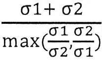
整合斷點可隨後藉由多樣性評分分選。在一些情況下,多樣性評分愈高,所鑑別之整合斷點可為真之可能性愈高(參見例如圖 5 )。在一些實例中,截止多樣性評分可設定為4,以使得排除具有低於4之多樣性評分之整合斷點,而具有至少4之多樣性評分之剩餘整合斷點可視為來自實際HPV整合事件。在一些情況下,截止多樣性評分設定為約0.1、約0.2、約0.4、約0.6、約0.8、約1、約1.2、約1.4、約1.6、約1.8、約2.0、約2.2、約2.4、約2.5、約2.6、約2.8、約3.0、約3.1、約3.2、約3.3、約3.4、約3.5、約3.6、約3.7、約3.8、約3.9、約4.0、約4.1、約4.2、約4.3、約4.4、約4.5、約4.6、約4.7、約4.8、約4.9、約5.0、約5.2、約5.4、約5.6、約5.8、約6.0、約6.5、約7.0、約7.5、約8、約8.5、約9、約9.5、約10、約20、約30、約40、約50、約60、約80或約100。在一些情況下,截止多樣性評分可為至多0.1、至多0.2、至多0.4、至多0.6、至多0.8、至多1、至多1.2、至多1.4、至多1.6、至多1.8、至多2.0、至多2.2、至多2.4、至多2.5、至多2.6、至多2.8、至多3.0、至多3.1、至多3.2、至多3.3、至多3.4、至多3.5、至多3.6、至多3.7、至多3.8、至多3.9、至多4.0、至多4.1、至多4.2、至多4.3、至多4.4、至多4.5、至多4.6、至多4.7、至多4.8、至多4.9、至多5.0、至多5.2、至多5.4、至多5.6、至多5.8、至多6.0、至多6.5、至多7.0、至多7.5、至多8、至多8.5、至多9、至多9.5或至多10。在其他情況下,截止多樣性評分可為至少1、至少1.2、至少1.4、至少1.6、至少1.8、至少2.0、至少2.2、至少2.4、至少2.5、至少2.6、至少2.8、至少3.0、至少3.1、至少3.2、至少3.3、至少3.4、至少3.5、至少3.6、至少3.7、至少3.8、至少3.9、至少4.0、至少4.1、至少4.2、至少4.3、至少4.4、至少4.5、至少4.6、至少4.7、至少4.8、至少4.9、至少5.0、至少5.2、至少5.4、至少5.6、至少5.8、至少6.0、至少6.5、至少7.0、至少7.5、至少8、至少8.5、至少9、至少9.5、至少10、至少20、至少30、至少40、至少50、至少60、至少80或至少100。Integration breakpoints can then be sorted by diversity score. In some cases, the higher the diversity score, the higher the probability that the identified integration breakpoint may be true (see, eg, Figure 5 ). In some examples, the cut-off diversity score can be set to 4, so that integration breakpoints with a diversity score lower than 4 are excluded, and the remaining integration breakpoints with a diversity score of at least 4 can be considered as coming from actual HPV integration events . In some cases, the cut-off diversity score is set to about 0.1, about 0.2, about 0.4, about 0.6, about 0.8, about 1, about 1.2, about 1.4, about 1.6, about 1.8, about 2.0, about 2.2, about 2.4, About 2.5, about 2.6, about 2.8, about 3.0, about 3.1, about 3.2, about 3.3, about 3.4, about 3.5, about 3.6, about 3.7, about 3.8, about 3.9, about 4.0, about 4.1, about 4.2, about 4.3 , About 4.4, about 4.5, about 4.6, about 4.7, about 4.8, about 4.9, about 5.0, about 5.2, about 5.4, about 5.6, about 5.8, about 6.0, about 6.5, about 7.0, about 7.5, about 8, about 8, 8.5, about 9, about 9.5, about 10, about 20, about 30, about 40, about 50, about 60, about 80 or about 100. In some cases, the cut-off diversity score may be at most 0.1, at most 0.2, at most 0.4, at most 0.6, at most 0.8, at most 1, at most 1.2, at most 1.4, at most 1.6, at most 1.8, at most 2.0, at most 2.2, at most 2.4, At most 2.5, at most 2.6, at most 2.8, at most 3.0, at most 3.1, at most 3.2, at most 3.3, at most 3.4, at most 3.5, at most 3.6, at most 3.7, at most 3.8, at most 3.9, at most 4.0, at most 4.1, at most 4.2, at most 4.3 , At most 4.4, at most 4.5, at most 4.6, at most 4.7, at most 4.8, at most 4.9, at most 5.0, at most 5.2, at most 5.4, at most 5.6, at most 5.8, at most 6.0, at most 6.5, at most 7.0, at most 7.5, at most 8, at most 8.5, at most 9, at most 9.5 or at most 10. In other cases, the cut-off diversity score may be at least 1, at least 1.2, at least 1.4, at least 1.6, at least 1.8, at least 2.0, at least 2.2, at least 2.4, at least 2.5, at least 2.6, at least 2.8, at least 3.0, at least 3.1, At least 3.2, at least 3.3, at least 3.4, at least 3.5, at least 3.6, at least 3.7, at least 3.8, at least 3.9, at least 4.0, at least 4.1, at least 4.2, at least 4.3, at least 4.4, at least 4.5, at least 4.6, at least 4.7, at least 4.8 , At least 4.9, at least 5.0, at least 5.2, at least 5.4, at least 5.6, at least 5.8, at least 6.0, at least 6.5, at least 7.0, at least 7.5, at least 8, at least 8.5, at least 9, at least 9.5, at least 10, at least 20, at least 30. At least 40, at least 50, at least 60, at least 80 or at least 100.
如本文所提及之各種截斷值中之一或多者可視多種參數而定進行調節。可調節此等變量以便獲得所需評定結果,例如測定病原體整合斷點之敏感性及特異性。可根據本文所提供之方法調節之各種截斷值包括用於搜尋及鑑別A型嵌合片段成對序列讀段之比對長度、界定整合區域時讀段之間的距離之截斷值、搜尋及鑑別B型嵌合片段序列讀段期間用於比對之短核苷酸分段之長度及截止多樣性評分。此等變量中之一或多者可視多種參數而定進行調節,該等參數包括樣本分析之情境,例如病原體及宿主生物體之物種、待分析癌症類型、宿主生物體之年齡、性別及健康狀況、如擴增、銜接子接合、合成測序、測序深度及序列讀段長度之測序分析之各種步驟之變量以及樣本分析之預期結果,例如測定病原體整合斷點之敏感性及特異性。在一些情況下,可藉由其他分析評估及/或確認可變調節。舉例而言,在患有某種類型之癌症,例如子宮頸癌之患者中,可藉由根據本文所提供之方法及系統分析血漿樣本中之無細胞DNA來測定HPV整合斷點。候選HPV整合斷點之不同列表可經由調節如本文所論述之變量中之一或多者推導出。候選HPV整合斷點之此等列表可隨後與藉由分析來自同一患者之腫瘤組織樣本測定之HPV整合斷點進行比較。在一些情況下,全基因組測序或其他下一代測序技術可用於此類分析。在一些情況下,可經由比較使變量最佳化以便獲得關於個體中之病原體整合型態的準確資訊。 E. 病原體整合指數One or more of the various cutoff values as mentioned herein can be adjusted depending on various parameters. These variables can be adjusted in order to obtain the desired assessment results, for example to determine the sensitivity and specificity of pathogen integration breakpoints. The various cutoff values that can be adjusted according to the methods provided herein include the length of the alignment of paired sequence reads for searching and identifying type A chimeric fragments, the cutoff value of the distance between reads when defining the integration region, search and identification The length and cut-off diversity score of the short nucleotide segments used for alignment during the read of the B-type chimeric fragment sequence. One or more of these variables can be adjusted depending on a variety of parameters, including the context of the sample analysis, such as the species of the pathogen and host organism, the type of cancer to be analyzed, the age, gender, and health status of the host organism , Variables in various steps of sequencing analysis such as amplification, adaptor ligation, synthetic sequencing, sequencing depth and sequence read length, and expected results of sample analysis, such as measuring the sensitivity and specificity of pathogen integration breakpoints. In some cases, variable adjustments can be evaluated and/or confirmed by other analyses. For example, in patients with certain types of cancer, such as cervical cancer, HPV integration breakpoints can be determined by analyzing cell-free DNA in plasma samples according to the methods and systems provided herein. Different lists of candidate HPV integration breakpoints can be derived by adjusting one or more of the variables as discussed herein. These lists of candidate HPV integration breakpoints can then be compared to HPV integration breakpoints determined by analyzing tumor tissue samples from the same patient. In some cases, whole genome sequencing or other next-generation sequencing technologies can be used for such analysis. In some cases, the variables can be optimized through comparison in order to obtain accurate information about the type of pathogen integration in the individual. E. Pathogen integration index
如本文所描述之病原體整合型態可包含病原體整合指數。本發明之一些態樣亦提供藉由分析來自生物樣本之核酸分子來測定病原體整合指數之方法及系統。整合指數可為生物樣本中之核酸分子總數目內生物體-病原體嵌合核酸分子之幾率比。在一些情況下,基於如上文所論述之多種嵌合片段成對序列讀段而測定整合指數。舉例而言,可在樣本分析,例如測序分析期間測定嵌合讀段對(例如A型片段讀段對、B型片段讀段對或兩者)之數目。在一些實例中,藉由生物體-病原體嵌合讀段對(例如人類-HPV嵌合讀段對)之數目除以病原體片段讀段對(例如HPV片段讀段對)之數目計算病原體整合指數。HPV片段讀段對可為成對末端成對序列讀段,其兩者映射至病原體(例如HPV)之基因組。Pathogen integration patterns as described herein may include a pathogen integration index. Some aspects of the present invention also provide a method and system for determining the pathogen integration index by analyzing nucleic acid molecules from a biological sample. The integration index may be the probability ratio of the organism-pathogen chimeric nucleic acid molecule in the total number of nucleic acid molecules in the biological sample. In some cases, the integration index is determined based on the paired sequence reads of various chimeric fragments as discussed above. For example, the number of chimeric read pairs (eg, type A fragment read pairs, type B fragment read pairs, or both) can be determined during sample analysis, such as sequencing analysis. In some examples, the pathogen integration index is calculated by dividing the number of organism-pathogen chimeric read pairs (eg, human-HPV chimeric read pairs) by the number of pathogen fragment read pairs (eg, HPV fragment read pairs) . HPV fragment read pairs may be paired end paired sequence reads, both of which map to the genome of the pathogen (eg HPV).
在一些實例中,整合指數可包含位置資訊。舉例而言,整合指數可運算病原體之基因組、宿主生物體之基因組或兩者中之位置中之每一者。在一些情況下,對於病原體之基因組中之位置(例如核苷酸座標),可測定在該位置處終止之生物體-病原體嵌合片段讀段對之數目,且亦可測定在該位置處終止之病原體片段讀段對之數目。可因此藉由在該位置處終止之生物體-病原體嵌合片段讀段對之數目除以在該位置處終止之病原體片段讀段對之數目來計算該位置之整合指數。其他計算方法亦可用於測定整合指數,只要其反映生物樣本中之生物體-病原體嵌合片段之豐度即可。In some examples, the integration index may include location information. For example, the integration index can be calculated for each of the pathogen's genome, the host organism's genome, or both. In some cases, for a position in the genome of a pathogen (e.g., nucleotide coordinates), the number of read pairs of organism-pathogen chimeric fragments terminating at that position can also be determined, and termination at that position can also be determined The number of read segments of pathogen fragments. The integration index at this position can thus be calculated by dividing the number of pairs of organism-pathogen chimeric fragment reads terminated at that position by the number of pairs of pathogen fragment reads terminated at this position. Other calculation methods can also be used to determine the integration index, as long as it reflects the abundance of the organism-pathogen chimeric fragments in the biological sample.
可在基因組譜或基因座特定水準下分析整合指數。在基因座特定水準下,可相對於沿著尺寸至少1 bp之病毒基因組之任何位點計算指數。在一個實例中,整合指數可用於預測癌症產生之風險或可能性。舉例而言,在一些情況下,整合指數愈高,提供樣本之個體愈可能罹患癌症。在另一實例中,病原體整合指數沿著病毒基因組(分析之基因座特定水準)之分佈可在整個不同腫瘤類型中不同。舉例而言,HPV整合指數展示為患有子宮頸癌及HNSCC 之患者之間的差異(圖 7 ),因此告知腫瘤來源。整合指數之計算可基於例如A型病毒-人類嵌合片段與全部病毒(僅DNA)之比率。在計算整合指數時可或可不應用用於鑑別整合斷點之上述過濾步驟。舉例而言,一端與病毒基因組比對且另一端與人類基因組比對之任何成對末端讀段,例如A型嵌合片段可用於在無任何過濾之情況下計算整合指數。在另一實例中,A型及B型嵌合片段之總數目比例可用於計算整合指數。The integration index can be analyzed at a specific level of genomic profile or locus. At a specific locus level, the index can be calculated relative to any position along the viral genome of at least 1 bp in size. In one example, the integration index can be used to predict the risk or likelihood of cancer development. For example, in some cases, the higher the integration index, the more likely the individual who provided the sample to develop cancer. In another example, the distribution of the pathogen integration index along the viral genome (a specific level of locus analyzed) can be different across different tumor types. For example, the HPV integration index is shown as the difference between patients with cervical cancer and HNSCC ( Figure 7 ), so the source of the tumor is informed. The calculation of the integration index may be based on, for example, the ratio of virus A-human chimeric fragments to total virus (DNA only). The above filtering step for identifying integration breakpoints may or may not be used when calculating the integration index. For example, any pair of end reads that are aligned with the viral genome at one end and the human genome at the other end, such as type A chimeric fragments, can be used to calculate the integration index without any filtering. In another example, the ratio of the total number of type A and type B chimeric fragments can be used to calculate the integration index.
應理解,當上文所描述之詳細例示性工作流集中於分析來自生物體-病原體嵌合無細胞核酸片段,尤其人類-HPV嵌合無細胞DNA片段之配對末端測序的序列讀段時,類似概念及方法可應用於使用其他測序技術及不一定僅獲得末端序列資訊之情境,例如當使用如離子激流測序及奈米孔測序之測序技術以便測定整合區域及/或斷點時;類似概念及方法亦可應用於病原體及宿主生物體及/或其他核酸分子(非細胞或細胞)之其他組合有意義之情境。 III. 病原體整合型態It should be understood that when the detailed exemplary workflow described above focuses on the analysis of sequence reads from paired-end sequencing of organism-pathogen chimeric cell-free nucleic acid fragments, especially human-HPV chimeric cell-free DNA fragments, similar The concepts and methods can be applied to scenarios where other sequencing technologies are used and not necessarily only end-of-line sequence information is obtained, for example when using sequencing technologies such as ion torrent sequencing and nanopore sequencing to determine integration regions and/or breakpoints; similar concepts and The method can also be applied to situations where pathogens and host organisms and/or other combinations of nucleic acid molecules (non-cells or cells) are meaningful. III. Pathogen integration patterns
病原體整合型態可包含病原體基因組、宿主生物體基因組或兩者中一或多個病原體整合斷點之位置。病原體整合型態可包含病原體基因組或宿主生物體基因組中一或多個整合斷點之位置。病原體整合型態可用於決定病理學分類,例如癌症或癌前病變之存在或不存在、癌症類型及癌症分期。Pathogen integration patterns may include the location of one or more pathogen integration breakpoints in the pathogen genome, the host organism genome, or both. Pathogen integration patterns can include the location of one or more integration breakpoints in the pathogen genome or the host organism genome. Pathogen integration patterns can be used to determine pathological classification, such as the presence or absence of cancer or precancerous lesions, cancer type, and cancer stage.
病原體整合型態可包含一或多個「整合斷點」。整合斷點可為病原體核酸分子(例如病毒DNA)整合至經病原體感染之宿主細胞之基因組(例如人類基因組)中的位點。Pathogen integration patterns can include one or more "integration breakpoints." The integration breakpoint may be a site where the pathogen nucleic acid molecule (eg, viral DNA) integrates into the genome (eg, human genome) of the host cell infected with the pathogen.
病原體核酸之整合會破壞宿主生物體基因組之完整性,且在一些情況下,導致宿主細胞中之嚴重下游後果。宿主生物體基因組中之整合斷點可為可轉譯成蛋白質之表現序列。病原體整合會破壞經感染細胞中之蛋白質生產。在一些其他情況下宿主生物體基因組中之整合斷點可在轉錄調節區,例如強化子或啟動子中。病原體核酸序列之整合會導致通常藉由受影響之轉錄調節區調節之基因轉錄失調。在一些情況下,病原體整合可導致轉錄/轉譯橫向、細胞功能變化或正常細胞惡性轉化成癌細胞。病原體核酸分子可為病原體基因組DNA分子。當病原體基因組包含RNA分子時,例如當病原體為RNA病毒,例如反轉錄病毒時,病原體核酸分子可為病原體基因組RNA分子之互補DNA。在經HPV感染之個體之血液中,可存在在具有HPV DNA序列及人類DNA序列之血漿中循環之人類-HPV嵌合DNA片段。在經EBV感染之個體之血液中,可存在具有EBV DNA序列及人類DNA序列之血漿中循環之人類-EBV嵌合DNA片段。The integration of pathogen nucleic acids can destroy the integrity of the host organism's genome and, in some cases, lead to serious downstream consequences in the host cell. Integration breakpoints in the genome of the host organism can be expression sequences that can be translated into proteins. Pathogen integration disrupts protein production in infected cells. In some other cases, the integration breakpoint in the genome of the host organism may be in a transcriptional regulatory region, such as an enhancer or promoter. The integration of pathogen nucleic acid sequences can lead to dysregulation of gene transcription normally regulated by the affected transcriptional regulatory region. In some cases, pathogen integration can result in transversal transcription/translation, changes in cell function, or malignant transformation of normal cells into cancer cells. The pathogen nucleic acid molecule may be a pathogen genomic DNA molecule. When the pathogen genome contains RNA molecules, for example when the pathogen is an RNA virus, such as a retrovirus, the pathogen nucleic acid molecule may be the complementary DNA of the pathogen genome RNA molecule. In the blood of an HPV-infected individual, there may be human-HPV chimeric DNA fragments circulating in plasma having HPV DNA sequences and human DNA sequences. Human-EBV chimeric DNA fragments circulating in plasma with EBV DNA sequence and human DNA sequence may be present in the blood of individuals infected with EBV.
病原體整合型態可指受關注之核酸分子中存在或不存在來自病原體基因組之核苷酸序列與來自宿主生物體基因組之核苷酸序列之組合、受關注之核酸分子中來自病原體基因組之核苷酸序列之尺寸或長度、受關注之核酸分子中來自病原體基因組之核苷酸序列之部分貢獻、受關注之核酸分子中來自病原體基因組之核苷酸序列之序列模式、受關注之核酸分子中病原體-宿主生物體接合點之特性、整合至宿主生物體之基因組中之病原體之基因組中之斷點位置或病原體核酸分子插入於宿主生物體基因組中之宿主生物體之基因組之斷點位置。在一些情況下,病原體整合型態包含整合在宿主生物體之基因組中之病原體之基因組中之斷點位置。Pathogen integration pattern may refer to the presence or absence of a combination of a nucleotide sequence from the pathogen genome and a nucleotide sequence from the host organism genome in the nucleic acid molecule of interest, a nucleoside from the pathogen genome in the nucleic acid molecule of interest The size or length of the acid sequence, the partial contribution of the nucleotide sequence from the pathogen genome in the nucleic acid molecule of interest, the sequence pattern of the nucleotide sequence from the pathogen genome in the nucleic acid molecule of interest, the pathogen in the nucleic acid molecule of interest -The characteristics of the junction point of the host organism, the position of the breakpoint in the genome of the pathogen integrated into the genome of the host organism, or the position of the breakpoint of the genome of the host organism in which the pathogen nucleic acid molecule is inserted into the genome of the host organism. In some cases, the pathogen integration pattern includes the location of the breakpoint in the genome of the pathogen integrated in the genome of the host organism.
在一些實例中,本文所提供之方法係關於分析生物體之生物樣本以測定病原體整合型態。在一些情況下,該等方法可包含分析來自生物樣本之無細胞核酸分子以測定病原體整合型態。病原體整合型態可包含整合在宿主生物體之基因組中之病原體之基因組中之整合斷點位置、宿主生物體之基因組中之整合斷點位置或兩者。一般而言,當來自第一生物體之核酸序列整合至第二生物體之基因組中時,斷點可指來自第一生物體之核酸序列併接至來自第二生物體之核酸序列的位置。在一些情況下,如本文所論述之斷點可為針對來自宿主生物體之基因組之核酸序列併接來自病原體之基因組之核酸序列的位置。舉例而言,當病原體基因組整合至宿主基因組中時,病原體之基因組中之斷點可指病原體基因組DNA斷裂且連接宿主生物體基因組DNA之病原體基因組中之位置。在一些情況下,整合過程可涉及病原體基因組中之一個斷點。在其他情況下,整合過程可涉及病原體基因組中之多個斷點,例如病原體基因組DNA破碎成多個片段,例如2、3、4、5、6、7、8、9、10、15、20、100或更多個片段,其分別插入宿主生物體基因組中。在一些情況下,本文所提供之方法及系統實現鑑別來自生物樣本之核酸分子中之一或多個整合斷點。在一些情況下,本文所提供之方法及系統實現鑑別來自生物樣本之核酸分子中之2、3、4、5、6、7、8、9或甚至更多個整合斷點。 IV. 染色體重組 A. 偵測染色體重組In some examples, the methods provided herein relate to analyzing biological samples of organisms to determine pathogen integration patterns. In some cases, these methods may include analyzing cell-free nucleic acid molecules from a biological sample to determine the pathogen integration pattern. Pathogen integration patterns may include integration breakpoint positions in the pathogen's genome integrated into the host organism's genome, integration breakpoint positions in the host organism's genome, or both. Generally speaking, when the nucleic acid sequence from the first organism is integrated into the genome of the second organism, the breakpoint can refer to the position where the nucleic acid sequence from the first organism is joined to the nucleic acid sequence from the second organism. In some cases, the breakpoint as discussed herein may be the location for the nucleic acid sequence from the genome of the host organism and the nucleic acid sequence from the genome of the pathogen. For example, when the pathogen genome is integrated into the host genome, the breakpoint in the pathogen's genome may refer to the location in the pathogen's genome where the pathogen's genomic DNA is broken and connected to the host organism's genomic DNA. In some cases, the integration process may involve a breakpoint in the pathogen genome. In other cases, the integration process may involve multiple breakpoints in the pathogen's genome, such as fragmentation of the pathogen's genomic DNA into multiple fragments, such as 2, 3, 4, 5, 6, 7, 8, 9, 10, 15, 20 , 100 or more fragments, which are inserted into the genome of the host organism, respectively. In some cases, the methods and systems provided herein enable identification of one or more integration breakpoints in nucleic acid molecules from biological samples. In some cases, the methods and systems provided herein enable identification of 2, 3, 4, 5, 6, 7, 8, 9, or even more integration breakpoints in nucleic acid molecules from biological samples. IV. Chromosome reorganization A. Detection of chromosome recombination
在一些態樣中,本發明提供偵測染色體重組之方法。該等方法及系統可包含藉由電腦系統分析複數個核酸分子,例如來自例如血漿之生物樣本之無細胞核酸(例如DNA)分子以偵測包含染色體重組之嵌合核酸片段(染色體重組嵌合核酸片段)。分析複數個核酸分子,例如無細胞核酸(例如DNA)分子中之每一者可包含鑑別各別核酸分子,例如無細胞核酸分子之第一末端來自生物體之參考基因組之第一基因組區域且鑑別各別核酸分子,例如無細胞核酸分子之第二末端來自生物體之參考基因組之第二基因組區域。當生物體之參考基因組中之第一及第二基因組區域之相對位置與各別無細胞核酸分子中之第一末端及第二末端之相對位置不一致時,可偵測包含染色體重組之嵌合核酸片段。In some aspects, the present invention provides methods for detecting chromosomal recombination. The methods and systems may include analyzing a plurality of nucleic acid molecules by a computer system, such as cell-free nucleic acid (eg, DNA) molecules from biological samples such as plasma, to detect chimeric nucleic acid fragments containing chromosomal recombination (chromosomal recombination chimeric nucleic acids) Fragment). Analysis of a plurality of nucleic acid molecules, such as cell-free nucleic acid (e.g. DNA) molecules, each of which may include identification of individual nucleic acid molecules, such as the first end of the cell-free nucleic acid molecule is derived from the first genomic region of the reference genome of the organism and identified The second end of each nucleic acid molecule, such as a cell-free nucleic acid molecule, comes from the second genomic region of the reference genome of the organism. When the relative positions of the first and second genomic regions in the reference genome of the organism are inconsistent with the relative positions of the first and second ends in the respective cell-free nucleic acid molecule, a chimeric nucleic acid containing chromosomal recombination can be detected Fragment.
相對位置可指參考基因組或無細胞核酸片段中之相對距離或相對5'至3'(或上游-下游)關係。相對距離可指精確相對距離或相對距離範圍。舉例而言,若參考基因組中之第一及第二基因組區域之間的相對距離及無細胞核酸片段中之第一末端及第二末端之間的相對距離在一些情況下恰好相同或在一些其他情況下在相同範圍(例如100-180個鹼基或無細胞核酸片段之典型長度範圍)內,則相對位置可一致。若參考基因組中之第一及第二基因組區域之間的相對距離及無細胞核酸片段中之第一末端及第二末端之間的相對距離在一些情況下不恰好相同或在一些其他情況下不在相同範圍(例如100-180個鹼基或無細胞核酸片段之典型長度範圍)內,則相對位置可不一致。不一致性可視生物體之參考基因組中之第一及第二基因組區域之相對位置之間的不同類型的染色體重組與各別無細胞核酸分子中之第一末端及第二末端之相對位置不一致而變化。染色體重組可為易位、擴增、缺失、反轉、染色體臂缺失及染色體臂獲得。The relative position may refer to the relative distance or relative 5′ to 3′ (or upstream-downstream) relationship in the reference genome or cell-free nucleic acid fragment. Relative distance may refer to precise relative distance or relative distance range. For example, if the relative distance between the first and second genomic regions in the reference genome and the relative distance between the first and second ends in the cell-free nucleic acid fragment are in some cases exactly the same or in some other In the same range (for example, 100-180 bases or a typical length range of cell-free nucleic acid fragments), the relative positions can be consistent. If the relative distance between the first and second genomic regions in the reference genome and the relative distance between the first and second ends in the cell-free nucleic acid fragment are not exactly the same in some cases or not the same in some other cases Within the range (for example, 100-180 bases or the typical length range of cell-free nucleic acid fragments), the relative positions may not be consistent. The inconsistency can be changed by the different types of chromosomal recombination between the relative positions of the first and second genomic regions in the reference genome of the organism and the relative positions of the first and second ends in the respective cell-free nucleic acid molecules. . Chromosomal recombination can be obtained by translocation, amplification, deletion, reversal, chromosome arm deletion, and chromosome arm.
易位可涉及分別在不同染色體或在相同染色體上兩個先前各別基因組區域之核酸序列之染色體間或染色體內重組。若無細胞核酸片段起因於包涵易位事件之基因組區域,則無細胞核酸片段之兩端可來自生物體中之兩個先前各別基因組區域。在一些情況下,若無細胞核酸片段之第一末端與生物體之參考基因組中之第一基因組區域比對且無細胞核酸片段之第二末端與生物體之參考基因組中之第二基因組區域比對,則參考基因組中之第一及第二基因組區域可具有與無細胞核酸片段中之第一末端及第二末端之間的距離不一致的距離。舉例而言,若當無細胞核酸片段之兩端橫跨兩個原先不同染色體之部分時,參考基因組中之第一及第二基因組區域在不同染色體上,則參考基因組中之第一及第二基因組區域之間的距離可為不可測定的。在一些情況下,若無細胞核酸片段跨越發生染色體之染色體內易位之位置,則參考基因組中第一末端與第二基因組區域之間的距離可比無細胞核酸片段中第一末端及第二末端之間的距離長。在一些情況下,將參考基因組中之第一及第二基因組區域之相對距離與任意截斷值進行比較以便測定無細胞核酸是否跨越發生易位之位置。舉例而言,截斷值可為比無細胞核酸片段之預期長度更長的距離。在一些情況下,截斷值可為比無細胞核酸片段之預期長度減第一及第二序列讀段之長度更長的距離。截斷值可為約100個鹼基、約200個鹼基、約300個鹼基、約400個鹼基、約500個鹼基、約600個鹼基、約800個鹼基、約1000個鹼基、約1200個鹼基、約1500個鹼基、約2000個鹼基、約300個鹼基、約4000個鹼基、約5000個鹼基、約6000個鹼基、約8000個鹼基或約104 個鹼基。截斷值可為至少80個鹼基、至少140個鹼基、至少180個鹼基、至少250個鹼基、至少350個鹼基、至少450個鹼基、至少550個鹼基、至少750個鹼基、至少900個鹼基、至少1100個鹼基、至少1250個鹼基、至少1800個鹼基、至少2500個鹼基、至少3500個鹼基、至少5500個鹼基、至少7500個鹼基、至少9000個鹼基或至少104 個鹼基。Translocation can involve inter- or intra-chromosomal recombination of nucleic acid sequences of two previously separate genomic regions on different chromosomes or on the same chromosome, respectively. If the cell-free nucleic acid fragment results from a genomic region that contains a translocation event, the two ends of the cell-free nucleic acid fragment can come from two previously separate genomic regions in the organism. In some cases, if the first end of the cell-free nucleic acid fragment is aligned with the first genomic region in the reference genome of the organism and the second end of the cell-free nucleic acid fragment is compared with the second genomic region in the reference genome of the organism Yes, the first and second genomic regions in the reference genome may have a distance that is not consistent with the distance between the first and second ends in the cell-free nucleic acid fragment. For example, if both ends of a cell-free nucleic acid fragment span two parts of a previously different chromosome, the first and second genomic regions in the reference genome are on different chromosomes, then the first and second in the reference genome The distance between genomic regions may not be measurable. In some cases, if the cell-free nucleic acid fragment spans the location of the intrachromosomal translocation that occurs on the chromosome, the distance between the first end and the second genomic region in the reference genome may be comparable to the first and second ends in the cell-free nucleic acid fragment The distance between them is long. In some cases, the relative distances of the first and second genomic regions in the reference genome are compared with arbitrary cutoff values to determine whether the cell-free nucleic acid crosses the location where the translocation occurs. For example, the cutoff value may be a distance longer than the expected length of the cell-free nucleic acid fragment. In some cases, the cutoff value may be a distance longer than the expected length of the cell-free nucleic acid fragment minus the length of the first and second sequence reads. The cutoff value can be about 100 bases, about 200 bases, about 300 bases, about 400 bases, about 500 bases, about 600 bases, about 800 bases, about 1000 bases Base, about 1200 bases, about 1500 bases, about 2000 bases, about 300 bases, about 4000 bases, about 5000 bases, about 6000 bases, about 8000 bases or about 104 bases. The cutoff value can be at least 80 bases, at least 140 bases, at least 180 bases, at least 250 bases, at least 350 bases, at least 450 bases, at least 550 bases, at least 750 bases Base, at least 900 bases, at least 1100 bases, at least 1250 bases, at least 1800 bases, at least 2500 bases, at least 3500 bases, at least 5500 bases, at least 7500 bases, at least 9000 bases, or at least 104 bases.
反轉可為染色體鏈段端至端倒轉之染色體重組。當單一染色體在自身內經受斷裂及重組時,可出現反轉。若無細胞核酸片段起因於包涵反轉事件之基因組區域,則無細胞核酸片段之兩端可來自原先具有相反5'至3'關係之兩個基因組區域。在此等情況下,若無細胞核酸片段之第一末端與生物體之參考基因組中之第一基因組區域比對且無細胞核酸片段之第二末端與生物體之參考基因組中之第二基因組區域比對,則參考基因組中之第一及第二基因組區域可具有相比於無細胞核酸片段之第一末端及第二末端反向的5'至3'關係。Reversal can be the recombination of chromosomes with end-to-end reversal of chromosome segments. When a single chromosome undergoes a break and recombination within itself, a reversal may occur. If the cell-free nucleic acid fragment is caused by a genomic region containing an inversion event, both ends of the cell-free nucleic acid fragment may be from two genomic regions that originally had opposite 5'to 3'relationships. In these cases, if the first end of the cell-free nucleic acid fragment matches the first genomic region in the organism's reference genome and the second end of the cell-free nucleic acid fragment matches the second genomic region in the organism's reference genome For comparison, the first and second genomic regions in the reference genome may have a reverse 5'to 3'relationship compared to the first and second ends of the cell-free nucleic acid fragment.
擴增可為局部複製染色體鏈段之染色體重組。由包涵擴增之基因組區域產生之無細胞核酸片段可具有包含相同複製序列之兩端。缺失可為染色體鏈段缺失之染色體重組。由包涵缺失之基因組區域產生之無細胞核酸片段可具有來自先前藉由缺失鏈段間隔開之兩個基因組區域的兩端。Amplification may be chromosomal recombination that locally replicates chromosome segments. The cell-free nucleic acid fragment generated from the genomic region containing the amplification may have both ends containing the same replication sequence. The deletion can be a chromosomal recombination where the chromosome segment is missing. The cell-free nucleic acid fragment generated from the genomic region containing the deletion may have two ends from the two genomic regions previously separated by the deleted segment.
該等方法可進一步包含分析染色體重組嵌合核酸片段以偵測染色體重組。染色體重組之偵測可包括測定染色體重組之類型,例如易位、擴增、缺失、反轉、染色體臂缺失或染色體臂獲得。染色體重組之偵測亦可包括測定染色體重組斷點。The methods may further include analyzing chromosomal recombination chimeric nucleic acid fragments to detect chromosomal recombination. Detection of chromosomal recombination can include determining the type of chromosomal recombination, such as translocation, amplification, deletion, inversion, chromosome arm deletion, or chromosome arm acquisition. Detection of chromosomal recombination may also include determination of chromosomal recombination breakpoints.
在一些情況下,該等方法包含鑑別來自複數個由來自生物樣本之無細胞核酸分子之配對末端測序產生之成對序列讀段的A型染色體嵌合成對序列讀段。A型染色體嵌合成對序列讀段可具有與生物體之參考基因組之第一基因組區域比對的第一序列讀段及與生物體之參考基因組之第二基因組區域比對的第二序列讀段;且對於A型染色體嵌合成對序列讀段中之每一者,生物體之參考基因組中之第一及第二基因組區域之相對位置可與各別無細胞核酸分子中之第一末端及第二末端之相對位置不一致。該等方法可進一步包含將在生物體之參考基因組中之預定距離內重疊或間隔開之A型染色體嵌合成對序列讀段之第一序列讀段分組,且將在生物體之參考基因組中之預定距離內重疊或間隔開之A型染色體嵌合成對序列讀段之第二序列讀段分組,由此測定分別包含生物體之參考基因組之第一及第二基因組區域中之候選區域的候選重組區域。可基於A型染色體嵌合成對序列讀段推導染色體重組類型(例如易位或反轉)。In some cases, the methods include identifying paired sequence reads from type A chromosomes derived from a plurality of paired sequence reads resulting from paired end sequencing of cell-free nucleic acid molecules from a biological sample. The type A chromosome paired sequence reads may have a first sequence read aligned with the first genomic region of the organism's reference genome and a second sequence read aligned with the second genomic region of the organism's reference genome ; And for each of the paired sequence reads of the type A chromosome chimera, the relative position of the first and second genomic regions in the reference genome of the organism can be the same as the first end and the The relative positions of the two ends are inconsistent. The methods may further include grouping the first sequence reads of paired sequence reads that are overlapped or spaced apart within a predetermined distance in the reference genome of the organism, and grouping the first sequence reads in the organism's reference genome Grouping of second sequence reads of paired sequence reads of overlapping type A chromosomes that are overlapping or spaced apart within a predetermined distance, thereby determining candidate recombination of candidate regions in the first and second genomic regions that respectively include the reference genome of the organism area. The type of chromosomal recombination (eg, translocation or inversion) can be derived based on paired sequence reads of type A chromosome chimeras.
該等方法可進一步包含鑑別及分析B型染色體嵌合成對序列讀段。B型染色體嵌合成對序列讀段可具有與候選重組區域比對之第一序列讀段及為嵌合讀段之第二序列讀段,且其中與第一基因組區域比對之嵌合讀段之一部分及與第二基因組區域比對之嵌合讀段之其餘部分。基於B型染色體嵌合成對序列讀段,可測定染色體重組。舉例而言,染色體重組斷點可藉由將嵌合讀段之序列與參考基因組進行局部比對來測定。至少部分地基於染色體重組斷點,可進一步測定染色體重組之類型。Such methods may further include identifying and analyzing paired sequence reads of type B chromosome chimeras. Type B chromosome chimeric paired sequence reads may have a first sequence read aligned with the candidate recombination region and a second sequence read that is a chimeric read, and wherein the chimeric read aligned with the first genomic region One part and the rest of the chimeric read aligned with the second genomic region. Based on paired sequence reads of chimeric B-type chromosomes, chromosome recombination can be determined. For example, the chromosomal recombination breakpoint can be determined by locally aligning the sequence of the chimeric read with the reference genome. Based at least in part on chromosomal recombination breakpoints, the type of chromosomal recombination can be further determined.
在一些情況下,該等方法及系統包含測定B型染色體嵌合成對序列讀段之嵌合讀段之多樣性,由此基於多樣性篩選染色體重組。篩選可包含測定B型染色體嵌合成對序列讀段之多樣性評分。多樣性評分可計算為: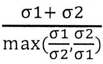
該等方法可包括將多樣性評分與截斷值進行比較。在一些情況下,若多樣性評分等於或高於截斷值,則可基於嵌合讀段測定染色體重組。在一些情況下,多樣性評分低於截斷值,可排除候選染色體重組區域作為假陽性擊點。多樣性評分之截斷值可為約0.1、約0.2、約0.4、約0.6、約0.8、約1、約1.2、約1.4、約1.6、約1.8、約2.0、約2.2、約2.4、約2.5、約2.6、約2.8、約3.0、約3.1、約3.2、約3.3、約3.4、約3.5、約3.6、約3.7、約3.8、約3.9、約4.0、約4.1、約4.2、約4.3、約4.4、約4.5、約4.6、約4.7、約4.8、約4.9、約5.0、約5.2、約5.4、約5.6、約5.8、約6.0、約6.5、約7.0、約7.5、約8、約8.5、約9、約9.5、約10、約20、約30、約40、約50、約60、約80或約100。在一些情況下,截止多樣性評分可為至多0.1、至多0.2、至多0.4、至多0.6、至多0.8、至多1、至多1.2、至多1.4、至多1.6、至多1.8、至多2.0、至多2.2、至多2.4、至多2.5、至多2.6、至多2.8、至多3.0、至多3.1、至多3.2、至多3.3、至多3.4、至多3.5、至多3.6、至多3.7、至多3.8、至多3.9、至多4.0、至多4.1、至多4.2、至多4.3、至多4.4、至多4.5、至多4.6、至多4.7、至多4.8、至多4.9、至多5.0、至多5.2、至多5.4、至多5.6、至多5.8、至多6.0、至多6.5、至多7.0、至多7.5、至多8、至多8.5、至多9、至多9.5或至多10。在其他情況下,截止多樣性評分可為至少1、至少1.2、至少1.4、至少1.6、至少1.8、至少2.0、至少2.2、至少2.4、至少2.5、至少2.6、至少2.8、至少3.0、至少3.1、至少3.2、至少3.3、至少3.4、至少3.5、至少3.6、至少3.7、至少3.8、至少3.9、至少4.0、至少4.1、至少4.2、至少4.3、至少4.4、至少4.5、至少4.6、至少4.7、至少4.8、至少4.9、至少5.0、至少5.2、至少5.4、至少5.6、至少5.8、至少6.0、至少6.5、至少7.0、至少7.5、至少8、至少8.5、至少9、至少9.5、至少10、至少20、至少30、至少40、至少50、至少60、至少80或至少100。Such methods may include comparing the diversity score to the cutoff value. In some cases, if the diversity score is equal to or higher than the cutoff value, chromosomal recombination can be determined based on chimeric reads. In some cases, the diversity score is lower than the cutoff value, which can exclude candidate chromosome recombination regions as false positive hit points. The cutoff value of the diversity score may be about 0.1, about 0.2, about 0.4, about 0.6, about 0.8, about 1, about 1.2, about 1.4, about 1.6, about 1.8, about 2.0, about 2.2, about 2.4, about 2.5, About 2.6, about 2.8, about 3.0, about 3.1, about 3.2, about 3.3, about 3.4, about 3.5, about 3.6, about 3.7, about 3.8, about 3.9, about 4.0, about 4.1, about 4.2, about 4.3, about 4.4 , About 4.5, about 4.6, about 4.7, about 4.8, about 4.9, about 5.0, about 5.2, about 5.4, about 5.6, about 5.8, about 6.0, about 6.5, about 7.0, about 7.5, about 8, about 8.5, about 9. About 9.5, about 10, about 20, about 30, about 40, about 50, about 60, about 80 or about 100. In some cases, the cut-off diversity score may be at most 0.1, at most 0.2, at most 0.4, at most 0.6, at most 0.8, at most 1, at most 1.2, at most 1.4, at most 1.6, at most 1.8, at most 2.0, at most 2.2, at most 2.4, At most 2.5, at most 2.6, at most 2.8, at most 3.0, at most 3.1, at most 3.2, at most 3.3, at most 3.4, at most 3.5, at most 3.6, at most 3.7, at most 3.8, at most 3.9, at most 4.0, at most 4.1, at most 4.2, at most 4.3 , At most 4.4, at most 4.5, at most 4.6, at most 4.7, at most 4.8, at most 4.9, at most 5.0, at most 5.2, at most 5.4, at most 5.6, at most 5.8, at most 6.0, at most 6.5, at most 7.0, at most 7.5, at most 8, at most 8.5, at most 9, at most 9.5 or at most 10. In other cases, the cut-off diversity score may be at least 1, at least 1.2, at least 1.4, at least 1.6, at least 1.8, at least 2.0, at least 2.2, at least 2.4, at least 2.5, at least 2.6, at least 2.8, at least 3.0, at least 3.1, At least 3.2, at least 3.3, at least 3.4, at least 3.5, at least 3.6, at least 3.7, at least 3.8, at least 3.9, at least 4.0, at least 4.1, at least 4.2, at least 4.3, at least 4.4, at least 4.5, at least 4.6, at least 4.7, at least 4.8 , At least 4.9, at least 5.0, at least 5.2, at least 5.4, at least 5.6, at least 5.8, at least 6.0, at least 6.5, at least 7.0, at least 7.5, at least 8, at least 8.5, at least 9, at least 9.5, at least 10, at least 20, at least 30. At least 40, at least 50, at least 60, at least 80 or at least 100.
該等方法可包括藉由與偵測病原體整合斷點之方法類似的股型濾除「假陽性」成對序列讀段。該等方法可包含濾除具有與自測定染色體重組排除之大部分股型模式不一致的股型模式之B型染色體嵌合成對序列讀段。 B. 鑑別癌症基因組中之基因融合物Such methods may include filtering out "false positive" paired sequence reads by a strand type similar to the method for detecting pathogen integration breakpoints. Such methods may include filtering out paired sequence reads of type B chromosome chimeras that have strand patterns that are inconsistent with most of the pattern patterns excluded from assaying chromosomal recombination. B. Identify gene fusions in the cancer genome
在一些態樣中,如本文所提供之方法及系統可應用於偵測人類癌症基因組中之基因融合物。基因融合物可包括分別不同或相同染色體上兩個先前各別基因組區域之核酸序列之染色體間或染色體內重組。基因融合事件會導致與自病原體整合所得之混雜DNA分子共有類似性的混雜基因組片段(「融合基因」)。基因融合可自易位、間質缺失或染色體反轉出現。基因融合可在癌症產生中起作用。鑑別基因融合可為診斷工具且融合基因可為抗癌治療中之治療標靶。如本文所論述之方法及系統係關於偵測來自腫瘤組織基因組釋放之無細胞核酸之融合基因。本文所提供之方法可賦予此類偵測無痛及非侵襲性,其可輔助精確癌症治療及實時監測腫瘤DNA之治療反應。 C. 鑑別體細胞結構變化In some aspects, the methods and systems as provided herein can be applied to detect gene fusions in the human cancer genome. Gene fusions can include inter- or intra-chromosomal recombination of nucleic acid sequences of two previously separate genomic regions on different or the same chromosomes. Gene fusion events can lead to hybrid genomic fragments ("fusion genes") that share similarities with hybrid DNA molecules resulting from pathogen integration. Gene fusion can occur from translocations, interstitial deletions, or chromosome inversion. Gene fusion can play a role in cancer development. The differential gene fusion can be a diagnostic tool and the fusion gene can be a therapeutic target in anti-cancer therapy. The methods and systems as discussed herein are related to the detection of fusion genes of cell-free nucleic acids released from the tumor tissue genome. The methods provided herein can impart such painless and non-invasive detection, which can assist in precise cancer treatment and real-time monitoring of tumor DNA treatment response. C. Identify changes in somatic cell structure
在一些態樣中,本文所提供之方法可應用於偵測基因組中之體細胞結構變化。結構變化可包括缺失、複製、複本數目變型、插入、反轉及易位,其可促進複雜病症,包括自閉症及癌症。體細胞結構變化會導致釋放嵌合DNA分子至血漿中且可潛在地藉由如本文所描述之整合偵測方法來偵測。本文所提供之方法可允許精確鑑別各種結構變化且可促進探索病原性機制及與基因組結構變化相關之疾病之治療方法。可非侵襲性地基於本文所提供之方法偵測體細胞結構變化。 V. 測序方法In some aspects, the methods provided herein can be applied to detect somatic structural changes in the genome. Structural changes can include deletions, duplications, copy number variants, insertions, reversals, and translocations, which can promote complex disorders, including autism and cancer. Somatic cell structural changes can result in the release of chimeric DNA molecules into plasma and can be potentially detected by the integrated detection method as described herein. The methods provided herein can allow accurate identification of various structural changes and can facilitate the exploration of pathogenic mechanisms and treatment methods for diseases associated with changes in genome structure. Somatic cell structural changes can be detected noninvasively based on the methods provided herein. V. Sequencing methods
可進行如本文所描述之生物樣本之測序分析以便測定病原體整合型態。本文所提供之方法可包含測序核酸分子,例如來自生物樣本之無細胞核酸分子、細胞核酸分子或兩者。在一些情況下,本文所提供之方法包含分析測序結果,例如來自生物樣本之核酸分子之測序讀段。本文所提供之方法及系統可涉及或不涉及測序之活性步驟。方法及系統可包含或提供用於自測序儀接受及處理測序資料之手段。方法及系統亦可包含或提供用於為測序儀提供命令以調節測序過程之參數,例如基於測序結果分析之命令的手段。Sequencing analysis of biological samples as described herein can be performed to determine pathogen integration patterns. The methods provided herein can include sequencing nucleic acid molecules, such as cell-free nucleic acid molecules, cellular nucleic acid molecules, or both from biological samples. In some cases, the methods provided herein include analyzing sequencing results, such as sequencing reads of nucleic acid molecules from biological samples. The methods and systems provided herein may or may not involve the active steps of sequencing. The method and system may include or provide means for receiving and processing sequencing data from the sequencer. The method and system may also include or provide means for providing commands to the sequencer to adjust the parameters of the sequencing process, such as commands based on analysis of sequencing results.
可商購的測序設備可用於本發明中所提供之方法,諸如伊路米那(Illumina)測序平台及454/Roche平台。可使用此項技術中已知之任何方法進行測序核酸。舉例而言,測序可包括下一代測序。在一些情況下,可使用鏈終止測序、雜交測序、伊路米那測序(例如使用可逆終止物)、離子激流半導體測序、質量分光光度法測序、大規模平行標誌測序(MPSS)、Maxam-Gilbert測序、奈米孔測序、波羅泥(polony)測序、焦磷酸測序、鳥槍測序、單分子即時(SMRT)測序、SOLiD測序(使用四種經螢光標記之二-鹼基探針之雜交)、通用測序或其任何組合來進行核酸測序。Commercially available sequencing equipment can be used in the methods provided in the present invention, such as the Illumina sequencing platform and the 454/Roche platform. The nucleic acid can be sequenced using any method known in the art. For example, sequencing may include next-generation sequencing. In some cases, chain termination sequencing, hybridization sequencing, illumina sequencing (for example, using reversible terminators), ion torrent semiconductor sequencing, mass spectrophotometric sequencing, massively parallel marker sequencing (MPSS), Maxam-Gilbert Sequencing, nanopore sequencing, polony sequencing, pyrosequencing, shotgun sequencing, single molecule real-time (SMRT) sequencing, SOLiD sequencing (hybridization using four fluorescently labeled two-base probes) , Universal sequencing or any combination thereof to perform nucleic acid sequencing.
可用於如本文所提供之方法中之一個測序方法可涉及配對末端測序,例如使用具有其基因組分析器之伊路米那「成對末端模組」。使用此模組,在基因組分析器已完成第一次測序讀段之後,成對末端模組可引導初始模板之再合成及第二輪簇生成。藉由使用本文所提供之方法中之成對末端讀段,吾人可自核酸分子之兩端獲得序列資訊且將兩端映射至參考基因組,例如病原體之基因組或宿主生物體之基因組。在映射兩端之後,吾人可根據如本文所提供之方法之一些實施例測定病原體整合型態。One sequencing method that can be used in the methods as provided herein may involve paired-end sequencing, for example using an Illumina "paired-end module" with its genome analyzer. Using this module, after the genome analyzer has completed the first sequencing read, the paired end modules can guide the re-synthesis of the initial template and the second round of cluster generation. By using the paired end reads in the method provided herein, one can obtain sequence information from both ends of the nucleic acid molecule and map the two ends to a reference genome, such as the genome of a pathogen or the genome of a host organism. After mapping the two ends, we can determine the pathogen integration pattern according to some embodiments of the method as provided herein.
在配對末端測序期間,來自核酸分子之第一末端之序列讀段可包含至少20、至少25、至少30、至少35、至少40、至少45、至少50、至少55、至少60、至少65、至少70、至少75、至少80、至少85、至少90、至少95、至少100、至少105、至少110、至少105、至少120、至少125、至少130、至少135、至少140、至少145、至少150、至少155、至少160、至少165、至少170、至少175或至少180個連續核苷酸。來自核酸分子之第一末端之序列讀段可包含至多24、至多28、至多32、至多38、至多42、至多48、至多52、至多58、至多62、至多68、至多72、至多78、至多82、至多88、至多92、至多98、至多102、至多108、至多122、至多128、至多132、至多138、至多142、至多148、至多152、至多158、至多162、至多168、至多172或至多180個連續核苷酸。來自核酸分子之第一末端之序列讀段可包含約20、約25、約30、約35、約40、約45、約50、約55、約60、約65、約70、約75、約80、約85、約90、約95、約100、約105、約110、約105、約120、約125、約130、約135、約140、約145、約150、約155、約160、約165、約170、約175或約180個連續核苷酸。來自核酸分子之第二末端之序列讀段可包含至少20、至少25、至少30、至少35、至少40、至少45、至少50、至少55、至少60、至少65、至少70、至少75、至少80、至少85、至少90、至少95、至少100、至少105、至少110、至少105、至少120、至少125、至少130、至少135、至少140、至少145、至少150、至少155、至少160、至少165、至少170、至少175或至少180個連續核苷酸。來自核酸分子之第二末端之序列讀段可包含至多24、至多28、至多32、至多38、至多42、至多48、至多52、至多58、至多62、至多68、至多72、至多78、至多82、至多88、至多92、至多98、至多102、至多108、至多122、至多128、至多132、至多138、至多142、至多148、至多152、至多158、至多162、至多168、至多172或至多180個連續核苷酸。來自核酸分子之第二末端之序列讀段可包含約20、約25、約30、約35、約40、約45、約50、約55、約60、約65、約70、約75、約80、約85、約90、約95、約100、約105、約110、約105、約120、約125、約130、約135、約140、約145、約150、約155、約160、約165、約170、約175或約180個連續核苷酸。在一些情況下,來自核酸分子之第一末端之序列讀段可包含至少75個連續核苷酸。在一些情況下,來自核酸分子之第二末端之序列讀段可包含至少75個連續核苷酸。來自核酸分子之第一末端及第二末端之序列讀段可具有相同長度或不同長度。來自複數個來自生物樣本之核酸分子之序列讀段可具有相同長度或不同長度。During paired-end sequencing, the sequence reads from the first end of the nucleic acid molecule may include at least 20, at least 25, at least 30, at least 35, at least 40, at least 45, at least 50, at least 55, at least 60, at least 65, at least 70, at least 75, at least 80, at least 85, at least 90, at least 95, at least 100, at least 105, at least 110, at least 105, at least 120, at least 125, at least 130, at least 135, at least 140, at least 145, at least 150, At least 155, at least 160, at least 165, at least 170, at least 175, or at least 180 consecutive nucleotides. The sequence reads from the first end of the nucleic acid molecule can include at most 24, at most 28, at most 32, at most 38, at most 42, at most 48, at most 52, at most 58, at most 62, at most 68, at most 72, at most 78, at most 82, at most 88, at most 92, at most 98, at most 102, at most 108, at most 122, at most 128, at most 132, at most 138, at most 142, at most 148, at most 152, at most 158, at most 162, at most 168, at most 172 or Up to 180 consecutive nucleotides. The sequence read from the first end of the nucleic acid molecule may comprise about 20, about 25, about 30, about 35, about 40, about 45, about 50, about 55, about 60, about 65, about 70, about 75, about 80, about 85, about 90, about 95, about 100, about 105, about 110, about 105, about 120, about 125, about 130, about 135, about 140, about 145, about 150, about 155, about 160, About 165, about 170, about 175, or about 180 consecutive nucleotides. The sequence read from the second end of the nucleic acid molecule may comprise at least 20, at least 25, at least 30, at least 35, at least 40, at least 45, at least 50, at least 55, at least 60, at least 65, at least 70, at least 75, at least 80, at least 85, at least 90, at least 95, at least 100, at least 105, at least 110, at least 105, at least 120, at least 125, at least 130, at least 135, at least 140, at least 145, at least 150, at least 155, at least 160, At least 165, at least 170, at least 175, or at least 180 consecutive nucleotides. The sequence reads from the second end of the nucleic acid molecule can include at most 24, at most 28, at most 32, at most 38, at most 42, at most 48, at most 52, at most 58, at most 62, at most 68, at most 72, at most 78, at most 82, at most 88, at most 92, at most 98, at most 102, at most 108, at most 122, at most 128, at most 132, at most 138, at most 142, at most 148, at most 152, at most 158, at most 162, at most 168, at most 172 or Up to 180 consecutive nucleotides. The sequence read from the second end of the nucleic acid molecule may comprise about 20, about 25, about 30, about 35, about 40, about 45, about 50, about 55, about 60, about 65, about 70, about 75, about 80, about 85, about 90, about 95, about 100, about 105, about 110, about 105, about 120, about 125, about 130, about 135, about 140, about 145, about 150, about 155, about 160, About 165, about 170, about 175, or about 180 consecutive nucleotides. In some cases, the sequence read from the first end of the nucleic acid molecule may contain at least 75 consecutive nucleotides. In some cases, the sequence read from the second end of the nucleic acid molecule may contain at least 75 consecutive nucleotides. The sequence reads from the first end and the second end of the nucleic acid molecule may have the same length or different lengths. The sequence reads from the plurality of nucleic acid molecules from the biological sample may have the same length or different lengths.
可在各種測序深度下進行本文所提供之方法中之測序。測序深度可指基因座被與基因座比對之序列讀段覆蓋的次數。基因座可小至一個核苷酸,或大至一個染色體臂,或大至整個基因組。本文所提供之方法中之測序深度可為1x、2x、5x、10x、20x、25x、30x、40x、50x、75x或100x等,其中「x」之前的數字係指基因座被序列讀段覆蓋的次數。測序深度亦可應用於多個基因座或全基因組,在此情況下,x可指基因座或單倍體基因組或全基因組分別定序之平均次數。在一些情況下,在本文所描述之方法中進行超深度測序,其可指進行至少100x測序深度。Sequencing in the methods provided herein can be performed at various sequencing depths. Sequencing depth can refer to the number of times a locus is covered by sequence reads aligned with a locus. The locus can be as small as one nucleotide, or as large as a chromosomal arm, or as large as the entire genome. The sequencing depth in the method provided herein can be 1x, 2x, 5x, 10x, 20x, 25x, 30x, 40x, 50x, 75x or 100x, etc., where the number before "x" refers to the locus covered by sequence reads Times. Sequencing depth can also be applied to multiple loci or whole genomes. In this case, x can refer to the average number of times a locus or haploid genome or whole genome is sequenced, respectively. In some cases, performing ultra-deep sequencing in the methods described herein may refer to performing at least 100x sequencing depth.
在測序過程(例如測序深度)期間讀段特定核苷酸在核酸內之次數或平均次數可比所測序之核酸之長度大多倍。在一些情況下,當測序深度足夠比核酸之長度大(例如至少5倍)時,測序可稱作『深度測序』。在一些實例中,測序深度可平均比所測序之核酸之長度大至少約5倍、大至少約10倍、大至少約20倍、大至少約30倍、大至少約40倍、大至少約50倍、大至少約60倍、大至少約70倍、大至少約80倍、大至少約90倍、大至少約100倍。在一些情況下,樣本可富集特定分析物(例如核酸片段或癌症特定核酸片段)。The number or average number of reads of a specific nucleotide in a nucleic acid during a sequencing process (eg, sequencing depth) can be many times longer than the length of the nucleic acid sequenced. In some cases, when the sequencing depth is sufficiently greater than the length of the nucleic acid (for example, at least 5 times), sequencing may be referred to as "deep sequencing." In some examples, the sequencing depth may be, on average, at least about 5 times larger, at least about 10 times larger, at least about 20 times larger, at least about 30 times larger, at least about 40 times larger, or at least about 50 times larger than the length of the nucleic acid sequenced Times, at least about 60 times larger, at least about 70 times larger, at least about 80 times larger, at least about 90 times larger, at least about 100 times larger. In some cases, the sample may be enriched for a specific analyte (eg, nucleic acid fragments or cancer specific nucleic acid fragments).
本文所提供之方法中產生之序列讀段(或定序讀段)可指自核酸分子之任何部分或所有測序之核苷酸串。舉例而言,序列讀段可為與核酸片段互補之核苷酸短串(例如20-150)、與核酸片段之末端互補之核苷酸串或與生物樣本中存在之整個核酸片段互補之核苷酸串。可在多種方法中,例如使用測序技術獲得序列讀段。 VI. 其他分析The sequence reads (or sequenced reads) generated in the methods provided herein may refer to any part or all of the sequenced nucleotide strings from the nucleic acid molecule. For example, the sequence read may be a short string of nucleotides complementary to the nucleic acid fragment (eg 20-150), a nucleotide string complementary to the end of the nucleic acid fragment or a core complementary to the entire nucleic acid fragment present in the biological sample Glycosides. Sequence reads can be obtained in a variety of methods, for example using sequencing technology. VI. Other analysis
在一些情況下,本發明之系統及方法係關於使用測序技術,繼而序列讀段之生物信息學分析以分析生物樣本中之病原體整合型態,例如分析整合斷點。可收集來自生物樣本之無細胞核酸且經受測序,例如配對末端測序。可藉由本文所提供之方法及系統分析無細胞核酸之序列讀段以便測定病原體之基因組、宿主生物體之基因組或兩者中之整合斷點位置。其他技術亦可用作整合型態分析之替代或額外途徑,包括(但不限於)擴增分析及微陣列分析。In some cases, the system and method of the present invention are related to the use of sequencing technology, followed by bioinformatics analysis of sequence reads to analyze pathogen integration patterns in biological samples, such as analysis of integration breakpoints. Cell-free nucleic acids from biological samples can be collected and subjected to sequencing, such as paired-end sequencing. The sequence reads of cell-free nucleic acids can be analyzed by the methods and systems provided herein to determine the location of integration breakpoints in the genome of the pathogen, the genome of the host organism, or both. Other techniques can also be used as an alternative or additional approach to integrated pattern analysis, including (but not limited to) amplification analysis and microarray analysis.
在一些實例中,本文所提供之方法亦可包含分析來自生物樣本之核酸分子之擴增反應。擴增反應可包含與病原體之基因組中之第一靶序列互補的第一引子及與生物體之基因組中之第二靶序列互補的第二引子。擴增反應可經設計以擴增病原體整合區域,其中來自病原體之核酸序列連接來自宿主生物體之核酸序列。具有癌症之某些分類之整合斷點特性之知識可幫助設計此類擴增反應。在一些情況下,擴增反應包含聚合酶鏈反應(PCR)。PCR反應可以至少一對引子起始,其中之每一者與分別側接來自病原體基因組或宿主生物體基因組之整合斷點的序列互補。替代地,PCR反應可以隨機結合至模板序列之隨機探針起始。在一些情況下,擴增反應可提供關於整合斷點之有成本效益且快速的結果。PCR反應可為定量的,其可提供關於不同類型的病原體整合事件之部分貢獻之定量洞察。在一些情況下,亦可進行擴增反應所產生之擴增子序列之分析以便確認整合斷點。替代地或另外,可進行擴增反應及擴增子之序列分析作為靶向靶標特異性富集之步驟,因此提供潛在更穩固的讀數。可用於本文所描述之系統及方法之PCR反應之不同變型包括多重PCR、不對稱PCR、基於克列諾之PCR、巢式PCR、定量PCR、即時PCR、熱起始PCR、觸地PCR、組裝PCR、COLD-PCR(在較低變性溫度下共擴增-PCR)、雙尾PCR、接合介導PCR及甲基化特異性PCR。儘管本文已提供實例及實施例,與例如數位PCR及隨機測序相關之額外技術及實施例可見於2010年10月28日申請之美國專利8,722,334、2007年7月23日申請之美國臨時申請案60/951,438及2013年7月8日申請之美國專利9,121,069,其中之每一者全部以引用之方式併入本文中。In some examples, the methods provided herein may also include analyzing amplification reactions of nucleic acid molecules from biological samples. The amplification reaction may include a first primer complementary to the first target sequence in the genome of the pathogen and a second primer complementary to the second target sequence in the genome of the organism. The amplification reaction can be designed to amplify the pathogen integration region, where the nucleic acid sequence from the pathogen is linked to the nucleic acid sequence from the host organism. Knowledge of the integrated breakpoint characteristics of certain classifications of cancer can help design such amplification reactions. In some cases, the amplification reaction involves polymerase chain reaction (PCR). The PCR reaction can be initiated with at least one pair of primers, each of which is complementary to a sequence flanking the integration breakpoint from the pathogen genome or the host organism genome, respectively. Alternatively, the PCR reaction can be initiated with a random probe that binds randomly to the template sequence. In some cases, amplification reactions can provide cost-effective and rapid results regarding integration breakpoints. The PCR reaction can be quantitative, which can provide quantitative insight into the partial contribution of different types of pathogen integration events. In some cases, analysis of the amplicon sequence generated by the amplification reaction can also be performed to confirm the integration breakpoint. Alternatively or additionally, amplification reactions and sequence analysis of amplicons can be performed as a step for targeted target-specific enrichment, thus providing potentially more robust readings. Different variants of PCR reactions that can be used in the systems and methods described herein include multiplex PCR, asymmetric PCR, Klenow-based PCR, nested PCR, quantitative PCR, real-time PCR, hot start PCR, touchdown PCR, assembly PCR, COLD-PCR (co-amplification-PCR at lower denaturation temperature), double-tailed PCR, junction-mediated PCR, and methylation-specific PCR. Although examples and examples have been provided herein, additional techniques and examples related to, for example, digital PCR and random sequencing can be found in US Patent 8,722,334 filed on October 28, 2010, and U.S. Provisional Application 60 filed on July 23, 2007 /951,438 and US Patent 9,121,069 filed on July 8, 2013, each of which is incorporated herein by reference.
在一些其他情況下諸如微陣列分析之任何其他序列分析可用於獲得核酸分子,例如來自生物樣本之無細胞核酸之序列資訊。舉例而言,微陣列可包含靶向宿主基因組序列及病原體基因組序列兩者之探針以便偵測如本文所描述之嵌合片段。在其他實例中,微陣列可經設計以偵測施加至微陣列之核酸分子中之病原體整合斷點之位置。In some other cases, any other sequence analysis such as microarray analysis can be used to obtain sequence information of nucleic acid molecules, such as cell-free nucleic acids from biological samples. For example, the microarray may include probes that target both the host genomic sequence and the pathogen genomic sequence in order to detect chimeric fragments as described herein. In other examples, the microarray may be designed to detect the location of pathogen integration breakpoints applied to the nucleic acid molecules of the microarray.
在一些情況下,如本文所描述之樣本分析可藉由利用不同原理及/或途徑之分析補充及/或確認。舉例而言,血漿樣本中之無細胞DNA之分析可產生展示與癌症之特定類型相關之較佳病原體整合斷點的一系列結果。在一些情況下,由無細胞DNA樣本產生之此等之敏感性及特異性可藉由諸如來自活檢體腫瘤樣本之特定類型之轉化癌細胞之測序,例如完整基因組測序評定。替代地,亦可應用相對更簡單且有成本效益的途徑,例如定性或定量PCR反應可藉由使用在鑑別候選斷點期間獲得之序列資訊經設計以評定斷點。In some cases, sample analysis as described herein may be supplemented and/or confirmed by analysis using different principles and/or approaches. For example, analysis of cell-free DNA in plasma samples can produce a series of results that show better pathogen integration breakpoints associated with specific types of cancer. In some cases, these sensitivities and specificities resulting from cell-free DNA samples can be assessed by sequencing of specific types of transformed cancer cells, such as from biopsy tumor samples, such as complete genome sequencing. Alternatively, a relatively simpler and cost-effective approach can also be applied. For example, qualitative or quantitative PCR reactions can be designed to assess breakpoints by using sequence information obtained during the identification of candidate breakpoints.
在一些情況下,本文所提供之系統及方法係關於用於決定病理學分類之第一分析及第二分析。第一分析可包含測序分析,例如來自生物樣本之核酸分子之測序,繼而如本文所描述之序列讀段之生物資訊分析。第二分析可包含擴增分析、微陣列分析或其他類型之分析。在一些實例中,第二分析包含測序分析,而第一分析包含擴增分析、微陣列分析或其他類型之分析。在一些情況下,第一分析,例如測序分析用於測定病原體整合型態,例如病原體之基因組或宿主生物體之基因組中之一或多個病原體整合斷點之位置。來自第一分析之病原體整合型態可提供第二分析之引導,例如一或多個病原體整合斷點之位置可提供設計第二分析之PCR探針或微陣列探針之引導。在一些實例中,人類個體,例如疑似經病原體,例如HPV感染之患者可藉由用於測定病原體整合型態之第一分析測試。病原體整合型態可用於決定病理學分類。至少部分地基於第一分析,患者可診斷患有疾病,例如癌症,例如子宮頸癌。患者可因此經受第二分析,其可用於例如在向患者投與療法之後或在對個體進行手術之後繼續監測來自患者之生物樣本中之病原體整合型態。病原體整合型態之持續監測可用於監測患者已經診斷患有之疾病,例如用於監測疾病之進展或消退。在一些實例中,第一分析可用作一或多種疾病/病況之篩選分析,而第二分析可用作一或多種疾病/病況之確認分析。 VII. 病理學分類In some cases, the systems and methods provided herein are related to the first analysis and the second analysis used to determine the pathology classification. The first analysis may include sequencing analysis, such as sequencing of nucleic acid molecules from biological samples, followed by bioinformatics analysis of sequence reads as described herein. The second analysis may include amplification analysis, microarray analysis, or other types of analysis. In some examples, the second analysis includes sequencing analysis, and the first analysis includes amplification analysis, microarray analysis, or other types of analysis. In some cases, a first analysis, such as sequencing analysis, is used to determine the pathogen integration pattern, such as the location of one or more pathogen integration breakpoints in the genome of the pathogen or the genome of the host organism. Pathogen integration patterns from the first analysis can provide guidance for the second analysis. For example, the location of one or more pathogen integration breakpoints can provide guidance for designing PCR probes or microarray probes for the second analysis. In some examples, a human individual, such as a patient suspected of being infected with a pathogen, such as HPV, may be tested by a first analysis used to determine the pathogen integration pattern. Pathogen integration patterns can be used to determine pathological classification. Based at least in part on the first analysis, the patient may be diagnosed with a disease, such as cancer, such as cervical cancer. The patient can therefore undergo a second analysis, which can be used to continue to monitor the integrated pattern of pathogens in the biological sample from the patient, for example, after administration of therapy to the patient or after surgery on the individual. Continuous monitoring of pathogen integration patterns can be used to monitor the disease that the patient has been diagnosed with, for example to monitor the progression or regression of the disease. In some examples, the first analysis can be used as a screening analysis for one or more diseases/conditions, and the second analysis can be used as a confirmation analysis for one or more diseases/conditions. VII. Pathology classification
整合在生物體之基因組中之病原體之基因組中之整合斷點之分析可用於決定病理學分類。舉例而言,本文所提供之方法可包含病毒DNA整合型態之分析,例如在人類宿主生物體基因組中整合之HPV病毒基因組中之斷點之分析,且可進一步包含基於病毒DNA整合型態測定癌症類型。病理學可一般涉及偏離健康情況之人類病況或疾病。與病原體整合相關且適用於本文所提供之方法及系統之病理學之非限制性實例可包括成人T細胞白血病、熱帶痙攣性截癱、子宮頸癌、頭頸癌、肛殖癌、卡堡氏肉瘤、伯基特氏淋巴瘤、肝細胞癌、梅克爾細胞癌、AIDS、腦癌、骨癌、間皮瘤、前列腺癌及B細胞淋巴瘤。The analysis of integration breakpoints in the genome of pathogens integrated into the genome of an organism can be used to determine pathological classification. For example, the methods provided herein may include analysis of viral DNA integration patterns, such as breakpoint analysis in the HPV viral genome integrated in the genome of the human host organism, and may further include viral DNA integration pattern determination Types of cancer. Pathology may generally involve human conditions or diseases that deviate from health. Non-limiting examples of pathologies related to pathogen integration and suitable for the methods and systems provided herein may include adult T-cell leukemia, tropical spastic paraplegia, cervical cancer, head and neck cancer, anal colon cancer, Kaborg's sarcoma, Burkitt's lymphoma, hepatocellular carcinoma, Merkel cell carcinoma, AIDS, brain cancer, bone cancer, mesothelioma, prostate cancer, and B-cell lymphoma.
癌症或腫瘤可指組織之異常質量,其中質量生長超越且與正常組織之生長不協調。癌症或腫瘤可取決於以下特徵而定義為「良性」或「惡性」:細胞分化程度,包含形態及功能;生長速率;局部侵襲;及轉移。「良性」腫瘤可為分化良好,生長典型地比惡性腫瘤慢,且保持侷限於原發部位。另外,良性腫瘤沒有浸潤、侵襲或轉移至遠端之能力。「惡性」腫瘤可以為分化不良(退行發育),典型地快速生長,伴隨進行性浸潤、侵襲及破壞周圍組織。此外,惡性腫瘤具有轉移至遠端之能力。Cancer or tumor can refer to the abnormal quality of tissues, in which the quality growth exceeds and does not coordinate with the growth of normal tissues. Cancer or tumor can be defined as "benign" or "malignant" depending on the following characteristics: degree of cell differentiation, including morphology and function; growth rate; local invasion; and metastasis. "Benign" tumors can be well differentiated, typically grow slower than malignant tumors, and remain confined to the primary site. In addition, benign tumors have no ability to infiltrate, invade, or metastasize to the distal end. "Malignant" tumors can be poorly differentiated (degenerative development), typically rapidly growing, with progressive infiltration, invasion and destruction of surrounding tissues. In addition, malignant tumors have the ability to metastasize to the far end.
在一些態樣中,本文所提供之方法可包含染色體重組之分析,例如複本數變化、缺失、複製、反轉及易位,且可進一步包含測定與染色體重組相關之病理學。可採用本文所提供方法及系統之與染色體重組相關之病理學可包括幾乎所有類型之癌症及各種類型的遺傳疾病,例如沃夫-赫希霍恩(Wolf-Hirschhorn)症候群、雅各布森(Jacobsen)症候群、恰克-馬利-杜斯氏症(Charcot-Marie-Tooth disease)類型1A、安格爾曼氏症候群(Angelman syndrome)、特納症候群(Turner Syndrome)、22q11.2缺失症候群、三倍X症候群、威廉姆斯症候群(Williams Syndrome)、貓叫症候群(Cri du Chat Syndrome)、貓眼症候群(Cat Eye Syndrome)、1q21.1複製症候群、15q13.3微缺失症候群、16p11.2缺失症候群、17q23.1q23.2微缺失症候群、1q複製症候群、1q21.1微缺失症候群、22ql 1.2缺失症候群、22q11.2複製症候群、2q23.1微缺失症候群、2q37缺失症候群、47 XXX症候群、47,XYY症候群、49,XXXXX症候群、二倍體-三倍體鑲嵌、末梢染色體18q缺失症候群、伊曼紐爾症候群(Emanuel syndrome)、克里斯塔(Kleefstra Syndrome)、庫冷德弗里斯(Koolen de Vries Syndrome)、鑲嵌型單染色體18、納布盧斯(Nablus)面具臉症候群、帕里斯特-基利安鑲嵌型症候群(Pallister-Killian Mosaic Syndrome)、史密斯-馬吉利氏症候群(Smith-Magenis Syndrome)、四體性(Tetrasomy) 9p症候群、四體性X症候群、三倍體性症候群(Triploidy Syndrome)、三染色體13症候群、三染色體17鑲嵌及三染色體2鑲嵌。
VIII. 癌症分類In some aspects, the methods provided herein can include analysis of chromosomal recombination, such as copy number changes, deletions, duplications, reversals, and translocations, and can further include measuring pathology associated with chromosomal recombination. Pathologies related to chromosomal recombination that can use the methods and systems provided herein can include almost all types of cancer and various types of genetic diseases, such as Wolf-Hirschhorn syndrome, Jacobson ( Jacobsen syndrome, Charcot-Marie-Tooth disease type 1A, Angelman syndrome, Turner Syndrome, 22q11.2 deficiency syndrome, Triple X syndrome, Williams Syndrome, Cri du Chat Syndrome, Cat Eye Syndrome, 1q21.1 replication syndrome, 15q13.3 microdeletion syndrome, 16p11.2 deletion syndrome , 17q23.1q23.2 microdeletion syndrome, 1q replication syndrome, 1q21.1 microdeletion syndrome, 22ql 1.2 deletion syndrome, 22q11.2 replication syndrome, 2q23.1 microdeletion syndrome, 2q37 deletion syndrome, 47 XXX syndrome, 47, XYY Syndrome, 49,XXXXX syndrome, diploid-triploid mosaic, terminal chromosome 18q deletion syndrome, Emanuel syndrome, Kleefstra Syndrome, Koolen de Vries Syndrome ), mosaic monochromosome 18, Nablus mask syndrome, Pallister-Killian mosaic syndrome (Pallister-Killian Mosaic Syndrome), Smith-Magenis Syndrome, four Tetrasomy 9p syndrome, tetrasomy X syndrome, triploid syndrome (Triploidy Syndrome),
病毒DNA整合斷點可用作根據本文所提及之方法分類癌症之基礎。在一些情況下,其他病理學分類,例如癌症亦可利用本發明中所提供之方法。Viral DNA integration breakpoints can be used as a basis for classifying cancer according to the methods mentioned herein. In some cases, other pathological classifications, such as cancer, can also utilize the methods provided in the present invention.
癌症分類可包含癌症之存在或不存在。本文所提供之方法可用於測定任何類型的癌症之分類,諸如(但不限於)膀胱癌、骨癌、腦瘤、乳癌、子宮頸癌、結腸直腸癌、食道癌、胃腸癌、造血惡性病、頭頸部鱗狀細胞癌、白血病、肝癌、肺癌、淋巴瘤、骨髓瘤、鼻癌、鼻咽癌、口腔癌、口咽癌、卵巢癌、前列腺癌、肉瘤、胃癌或甲狀腺癌。The cancer classification may include the presence or absence of cancer. The methods provided herein can be used to determine the classification of any type of cancer, such as (but not limited to) bladder cancer, bone cancer, brain tumor, breast cancer, cervical cancer, colorectal cancer, esophageal cancer, gastrointestinal cancer, hematopoietic malignancies, Head and neck squamous cell carcinoma, leukemia, liver cancer, lung cancer, lymphoma, myeloma, nasal cancer, nasopharyngeal cancer, oral cancer, oropharyngeal cancer, ovarian cancer, prostate cancer, sarcoma, gastric cancer or thyroid cancer.
癌症分類可包含癌症程度或癌症分期。癌症程度或癌症分期(例如I期、II期、III期或IV期)可指腫瘤之尺寸、癌轉移之存在或不存在、身體之全部腫瘤負荷、及/或癌症嚴重程度之其他量度(例如癌症復發)。癌症程度可為數字或其他標誌,諸如符號、字母及顏色。程度可為零。癌症程度亦可包括與突變或突變數量有關的惡變前或癌變前病況(狀態)。The cancer classification may include the degree of cancer or the stage of the cancer. The degree of cancer or cancer stage (e.g., stage I, stage II, stage III, or stage IV) may refer to the size of the tumor, the presence or absence of cancer metastasis, the total tumor burden of the body, and/or other measures of cancer severity (e.g. Cancer recurrence). The degree of cancer can be numbers or other signs, such as symbols, letters, and colors. The degree can be zero. The degree of cancer may also include pre-malignant or pre-malignant conditions (states) related to mutations or the number of mutations.
癌症分類亦可用於各種方法中。在一些情況下,癌症分類包含癌症進展或癌症消退。在一些情況下,癌症分類包含在具有或不具有癌症治療之情況下癌症進展或消退之預言或預測。舉例而言,如本文所描述之方法及系統可用於判定癌症是否存在於先前尚未知曉患有癌症之某人中。評定可調查已經診斷患有癌症之某人以隨時間推移監測癌症之進展,研究療法有效性或確定預後。在一些情況下,預後可用患者死於癌症之機率或特定期限或時間之後癌症進展之機率或癌症轉移之機率表示。Cancer classification can also be used in various methods. In some cases, the cancer classification includes cancer progression or cancer regression. In some cases, the cancer classification includes predictions or predictions of cancer progression or regression with or without cancer treatment. For example, methods and systems as described herein can be used to determine whether cancer is present in someone who has not previously been known to have cancer. Assessment can investigate someone who has been diagnosed with cancer to monitor the progress of the cancer over time, study the effectiveness of the therapy or determine the prognosis. In some cases, the prognosis can be expressed as the probability of the patient dying of cancer or the probability of cancer progression or the probability of cancer metastasis after a certain period or time.
在一些情況下,癌症分類包含癌症類型,例如子宮頸癌(CC)或頭頸部鱗狀細胞癌(HNSCC)。圖 6 展示基於來自分別患有子宮頸癌及HPV陽性頭頸部鱗狀細胞癌之患者之血漿樣本之無細胞DNA之分析,HPV16基因組中之病毒整合斷點之分佈。如圖中所示,在子宮頸癌與HPV-陽性頭頸部鱗狀細胞癌(區域A)之間可存在共有的較佳整合斷點。另一方面,亦可存在某一類型之癌症之較佳整合斷點特性,例如區域B展示HNSCC較佳整合斷點。In some cases, the cancer classification includes cancer types, such as cervical cancer (CC) or squamous cell carcinoma of the head and neck (HNSCC). Figure 6 shows the distribution of viral integration breakpoints in the HPV16 genome based on the analysis of cell-free DNA from plasma samples from patients with cervical cancer and HPV-positive head and neck squamous cell carcinoma, respectively. As shown in the figure, there may be a common better integration breakpoint between cervical cancer and HPV-positive head and neck squamous cell carcinoma (region A). On the other hand, there may be a better integration breakpoint characteristic for a certain type of cancer, for example, region B shows that HNSCC has a better integration breakpoint.
某些癌症分類,例如癌症類型之病毒整合斷點特性可使用本文所提供之方法鑑別;在一些情況下,為了測定癌症分類,可應用類似方法以測定病毒整合斷點,其中分析來自生物樣本之核酸分子之序列讀段。 IX. 病原體整合:病毒整合Certain cancer classifications, such as the characteristics of the virus integration breakpoints of cancer types, can be identified using the methods provided herein; in some cases, in order to determine the cancer classification, similar methods can be applied to determine the virus integration breakpoints, in which Sequence reads of nucleic acid molecules. IX. Pathogen integration: viral integration
本文所提供之方法及系統可用於偵測宿主生物體基因組中可能存在之不同類型的病毒整合,諸如必選整合。在一些病毒,諸如反轉錄病毒屬、假病毒科、轉座病毒科、一些肌尾噬菌體科及長尾病毒科之病毒複製期間,必選整合至宿主生物體基因組中可為必然事件。病毒DNA整合會導致病毒基因組永久性插入至宿主生物體染色體DNA中,該宿主生物體染色體DNA在反轉錄病毒之情況下稱為原病毒或在原核病毒之情況下稱為原噬菌體。本文所提供之方法及系統可用於偵測偶然整合。在一些病毒之病毒複製期間,偶然整合可能不為必需,但可賦予宿主/病毒偶合一些優點。本文所提供之方法及系統可用於偵測內源性病毒元件(EVE)。一些內源性病毒可經由罕見且有時意外的過程整合及「僵化」至宿主生物體基因組中,包括內源性反轉錄病毒及罕見的RNA病毒整合。EVE之一個實例包括整合於生殖系中之內源性反轉錄病毒。內源性逆病毒之整合基因組可保持潛伏且與宿主生物體基因組一起被動複製且傳遞至細胞後代。然而,宿主生物體之環境條件變化可使病毒再活化,導致病毒轉錄及產生新感染性病毒(噬菌體感染)。The methods and systems provided herein can be used to detect different types of viral integration that may be present in the genome of the host organism, such as mandatory integration. During the replication of some viruses, such as retroviruses, pseudoviruses, transposable viruses, some sarcophage families, and long-tailed virus families, integration into the genome of the host organism may be an inevitable event. The integration of viral DNA results in the permanent insertion of the viral genome into the chromosomal DNA of the host organism, which is called a provirus in the case of a retrovirus or a prophage in the case of a prokaryotic virus. The methods and systems provided herein can be used to detect accidental integration. During viral replication of some viruses, accidental integration may not be necessary, but it can confer some advantages on the host/virus coupling. The methods and systems provided herein can be used to detect endogenous viral elements (EVE). Some endogenous viruses can integrate and "stiffen" into the host organism's genome through rare and sometimes unexpected processes, including endogenous retroviruses and rare RNA virus integration. An example of EVE includes endogenous retroviruses integrated into the germ line. The integrated genome of the endogenous retrovirus can remain latent and passively replicate with the genome of the host organism and be transmitted to the progeny of the cell. However, changes in the environmental conditions of the host organism can reactivate the virus, leading to viral transcription and the production of new infectious viruses (phage infection).
本文所提供之方法及系統可用於分析DNA病毒及RNA病毒。The methods and systems provided herein can be used to analyze DNA viruses and RNA viruses.
本文所提供之方法及系統可用於分析DNA病毒整合。DNA病毒之基因組可為宿主生物體基因組整合之潛在受質,而不需要先前處理。在細胞感染後,DNA病毒之基因組可易位至細胞核,其中其可保持為游離基因體以確保病毒續存或整合至宿主細胞之基因組中。The methods and systems provided herein can be used to analyze DNA virus integration. The genome of the DNA virus can be a potential substrate for the integration of the host organism's genome without prior processing. After a cell infection, the genome of the DNA virus can be translocated to the nucleus, where it can be maintained as an episomal body to ensure the surviving or integration of the virus into the genome of the host cell.
本文所提供之方法及系統可用於研究疱疹病毒整合。疱疹病毒可為DNA包膜病毒,且可基於其序列種系發生分類於三個家族中:α、β及γ疱疹病毒。其可含有在病毒進入及環化後細胞核中遞送之線形雙股DNA。疱疹病毒可保持游離,例如呈染色體外環形DNA形式。一些疱疹病毒可將其基因組整合至宿主染色體中。疱疹病毒之實例包括艾伯斯坦-巴爾病毒(EBV)及人類疱疹病毒-6 (HHV-6)。The methods and systems provided herein can be used to study herpes virus integration. Herpes viruses can be DNA enveloped viruses, and can be classified into three families based on their sequence phylogeny: alpha, beta, and gamma herpes viruses. It may contain linear double-stranded DNA delivered in the nucleus after virus entry and circularization. Herpes viruses can remain free, for example in the form of extrachromosomal circular DNA. Some herpes viruses can integrate their genomes into the host chromosome. Examples of herpes viruses include Albertstein-Barr virus (EBV) and human herpes virus-6 (HHV-6).
本文所提供之方法及系統可用於分析人類乳突狀瘤病毒(HPV)整合。HPV DNA可整合至宿主細胞之基因組中。DNA破壞及可誘導DNA破壞(例如雙股斷裂(DSB))之藥劑可在HPV整合中起作用。The methods and systems provided herein can be used to analyze human papillomavirus (HPV) integration. HPV DNA can be integrated into the genome of the host cell. DNA destruction and agents that can induce DNA destruction (such as double strand breaks (DSB)) can play a role in HPV integration.
本文所提供之方法及系統可用於分析B型肝炎病毒(HBV)之整合。HBV可導致肝細胞癌發展或在肝細胞癌發展中起作用。在急性感染期間,HBV可將其基因組整合至宿主染色體中且與反轉錄病毒整合存在若干類似性。The methods and systems provided herein can be used to analyze the integration of hepatitis B virus (HBV). HBV can cause or play a role in the development of hepatocellular carcinoma. During an acute infection, HBV can integrate its genome into the host chromosome and there are several similarities with retrovirus integration.
本文所提供之方法及系統可用於分析2型腺相關病毒(AAV-2)之整合。腺相關病毒AAV可為分類於細小病毒科家族中之普遍病毒。AAV之複製可嚴格地藉由輔助病毒,諸如腺病毒、人類乳突狀瘤病毒(HPV)或單純疱疹病毒(HSV)之相同經感染細胞之存在改良。在輔助病毒不存在下,AAV可以位點特異性方式整合其基因組。The methods and systems provided herein can be used to analyze the integration of adeno-associated virus type 2 (AAV-2). Adeno-associated virus AAV can be a widespread virus classified in the Parvoviridae family. The replication of AAV can be strictly improved by the presence of helper viruses such as adenovirus, human papilloma virus (HPV) or herpes simplex virus (HSV) in the same infected cells. In the absence of helper viruses, AAV can integrate its genome in a site-specific manner.
本文所提供之方法及系統可用於分析RNA病毒整合。反轉錄病毒之病毒RNA基因組可反轉錄至線形雙股DNA分子(病毒DNA中間物)中,其可因此為後續病毒基因組整合至宿主基因組中之受質。對於反轉錄病毒,整合可為噬菌體感染之必選步驟。其他RNA病毒之基因組亦可在宿主基因組中鑑別出。在此等情況下,整合可附帶出現,諸如對於淋巴球性脈絡叢腦膜炎病毒(LCMV)而言。本文所提供之方法及系統亦可幫助闡明病毒生命週期中RNA病毒整合之作用以及宿主生物體中之病理學發展。 X. 病毒整合及癌症The methods and systems provided herein can be used to analyze RNA virus integration. The viral RNA genome of a retrovirus can be reverse transcribed into a linear double-stranded DNA molecule (viral DNA intermediate), which can therefore be the substrate for subsequent integration of the viral genome into the host genome. For retroviruses, integration can be a necessary step for phage infection. The genomes of other RNA viruses can also be identified in the host genome. In these cases, integration can occur incidentally, such as for lymphocytic choriomeningitis virus (LCMV). The methods and systems provided herein can also help elucidate the role of RNA virus integration in the virus life cycle and the pathological development in the host organism. X. Viral integration and cancer
在一些實例中,本文所提供之方法及系統可用於藉由測定病毒整合來測定癌症分類。In some examples, the methods and systems provided herein can be used to determine cancer classification by measuring viral integration.
在一些情況下,本文所提供之方法及系統可用於分析腫瘤病毒(或致癌病毒),其可直接導致實驗動物或人類中之癌症。存在若干腫瘤病毒家族,包括B型肝炎病毒(肝癌)、乳突狀瘤病毒(子宮頸癌及其他肛殖癌)、艾伯斯坦-巴爾病毒(伯基特氏淋巴瘤及鼻咽癌)、卡波西氏肉瘤相關疱疹病毒(卡波西氏肉瘤)、多瘤病毒(梅克爾細胞癌)及人類T細胞嗜淋巴細胞病毒(成人T細胞白血病)。六個非限制性家族中之五個具有DNA基因組且稱為DNA腫瘤病毒,且另一家族為反轉錄病毒,其在病毒粒子中具有RNA基因組,但可經由經感染細胞中之DNA原病毒合成來複製。另外,由於免疫缺陷,HIV可間接造成AIDS患者中之發生之癌症,且C型肝炎病毒(RNA病毒)可為由慢性組織破壞產生之肝癌之間接病因。In some cases, the methods and systems provided herein can be used to analyze tumor viruses (or oncogenic viruses), which can directly cause cancer in laboratory animals or humans. There are several families of tumor viruses, including hepatitis B virus (liver cancer), papilloma virus (cervical cancer and other anal colon cancers), Ebstein-Barr virus (Burkitt's lymphoma and nasopharyngeal cancer), Kaposi's sarcoma-associated herpes virus (Kaposi's sarcoma), polyoma virus (Meckel's cell carcinoma), and human T-cell lymphotropic virus (adult T-cell leukemia). Five of the six non-limiting families have DNA genomes and are called DNA tumor viruses, and the other family are retroviruses, which have RNA genomes in virions, but can be synthesized by DNA proviruses in infected cells To copy. In addition, due to immunodeficiency, HIV can indirectly cause cancer in AIDS patients, and hepatitis C virus (RNA virus) can be an indirect cause of liver cancer caused by chronic tissue destruction.
本文所提供之方法及系統可用於分析HBV整合以便測定HBV相關病況/疾病,如肝細胞癌(HCC)。HBV可感染若干物種,包括鴨、土撥鼠、松鼠及人類之肝細胞。B型肝炎病毒感染會導致急性肝損傷。B型肝炎病毒感染可常見於亞洲及非洲部分地區,其中其可與每年高達一百萬肝癌病例相關(約全世界癌症發病率之10%)。B型肝炎病毒之細胞變形可藉由影響驅動異常細胞增殖及存活之多種細胞基因之表現的病毒基因(稱作X基因)介導。另外,藉由B型肝炎病毒誘發之癌症發展可由慢性組織破壞所導致的肝細胞連續增殖驅動。本文所提供之方法及系統可用於診斷、監測或預言與B型肝炎病毒之任何基因型或血清型,諸如基因型F、E、A、B、C及D及其各種次型相關之病理學。The methods and systems provided herein can be used to analyze HBV integration in order to determine HBV-related conditions/disease, such as hepatocellular carcinoma (HCC). HBV can infect several species of liver cells, including ducks, woodchucks, squirrels, and humans. Hepatitis B virus infection can cause acute liver damage. Hepatitis B virus infection can be common in parts of Asia and Africa, where it can be associated with up to one million cases of liver cancer each year (about 10% of cancer incidence worldwide). Hepatitis B virus cell deformation can be mediated by viral genes (called X genes) that affect the performance of various cellular genes that drive abnormal cell proliferation and survival. In addition, cancer development induced by hepatitis B virus can be driven by continuous proliferation of liver cells caused by chronic tissue destruction. The methods and systems provided herein can be used to diagnose, monitor, or predict any genotype or serotype of hepatitis B virus, such as genotypes F, E, A, B, C, and D and their various subtypes. .
HBV可與HCC相關。HBV為具有反轉錄病毒特徵之部分雙股DNA嗜肝病毒。HBV基因組整合至細胞基因組中可存在於超過85%-90% HBV相關HCC中。然而,HBV之整合形式亦可存在於具有慢性HBV感染之患者之非腫瘤組織中。HBV基因組整合至肝細胞中可在持續性HBV感染期間出現且在HCC發展之前。HBV整合可導致提若干細胞癌症相關基因,諸如TERT、混合譜系白血病4 (MLL4)及CCNE1 (編碼週期素El)之高的表現。HBV整合亦可與HCC提前發作及不佳結果有關。HBV can be related to HCC. HBV is a partial double-stranded DNA hepadnavirus with retrovirus characteristics. The integration of the HBV genome into the cell genome can be present in more than 85%-90% of HBV-associated HCC. However, the integrated form of HBV can also be present in non-tumor tissues of patients with chronic HBV infection. Integration of the HBV genome into hepatocytes can occur during persistent HBV infection and before the development of HCC. HBV integration can lead to high performance of several cell cancer-related genes, such as TERT, mixed lineage leukemia 4 (MLL4) and CCNE1 (encoding cyclin El). HBV integration can also be related to early onset of HCC and poor results.
本文所提供之方法及系統可用於測定乳突狀瘤病毒整合型態以評定乳突狀瘤病毒相關病況/疾病,如子宮頸癌。乳突狀瘤病毒可為較小DNA病毒(約8 kb之基因組),其可在人類及多種其他動物物種中誘發良性及惡性腫瘤兩者。已鑑別出約60種不同類型的人類乳突狀瘤病毒,其可感染若干組織之上皮細胞。此等病毒中之一些可僅導致良性腫瘤(諸如疣),而其他可為惡性癌瘤,尤其子宮頸癌、頭頸部鱗狀細胞癌(HNSCC)及其他肛殖癌之病原體。本文所提供之方法及系統可用於使不同類型的病理學分化,例如與HPV相關之癌症。本文所提供之方法及系統亦可用以分析任何類型的乳突狀瘤病毒,包括HPV類型6、11、16、18、31、33、35、39、45、51、52、56、58、59、68、73及82。The methods and systems provided herein can be used to determine the papillomavirus integration pattern to assess papillomavirus-related conditions/disease, such as cervical cancer. Papilloma virus can be a smaller DNA virus (about 8 kb of genome), which can induce both benign and malignant tumors in humans and various other animal species. About 60 different types of human papillomaviruses have been identified, which can infect several tissue epithelial cells. Some of these viruses can only cause benign tumors (such as warts), while others can be malignant carcinomas, especially cervical cancer, head and neck squamous cell carcinoma (HNSCC), and other pathogens of anal colon cancer. The methods and systems provided herein can be used to differentiate different types of pathology, such as cancers associated with HPV. The methods and systems provided herein can also be used to analyze any type of papillomavirus, including
HPV之子集可感染肛殖區域,且在此子集內,個別類型可歸類為高風險或低風險。高風險HPV可導致癌性損傷,而低風險HPV不會。高風險及低風險HPV之間的差異可為高風險HPV展示更大的整合至宿主基因組中之傾向,由此造成高級損傷及癌症,而低風險類型可優先維持染色體外環形游離基因體。HPV可導致幾乎所有子宮頸癌病例,其可為全世界女性中第二常見之癌症及全世界女性中第四常見之癌症死亡病因。HPV16、HPV18、HPV31及HPV33可佔所有子宮頸癌病例之90%。在此等高風險HPV中,HPV類型16為最普遍類型且本身可佔超過所有子宮頸癌病例之50%。高風險HPV感染亦可能與若干其他肛殖癌及口咽癌有關。舉例而言,認為造成超過90%肛門癌、70%陰道癌及外陰癌、60%陰莖癌及63%口咽癌。95% 具有子宮頸癌前病變之患者中可攜帶HPV,其中僅小部分可最終進展至侵襲性癌瘤。三個癌前階段(子宮頸上皮內贅瘤(CIN) 1、CIN2及CIN3)可在產生侵襲性癌瘤之前。CIN1損傷可自發消退,其中僅幾個損傷發展為CIN2/CIN3且最終發展為侵襲性癌瘤。經HPV感染之女性中之子宮頸癌進展可與整合狀態緊密相關,且發現整合於子宮頸癌症中之HPV之頻率可始終較高。舉例而言,100% HPV18、80% HPV16及81% HPV31陽性癌症可展示病毒整合。本文所提供之方法及系統可適用於藉由測定個體中之HPV整合型態來預言及監測癌症,例如子宮頸癌。A subset of HPV can infect the anal colon area, and within this subset, individual types can be classified as high risk or low risk. High-risk HPV can cause cancerous damage, while low-risk HPV does not. The difference between high-risk and low-risk HPV can show high-risk HPV's tendency to integrate more into the host genome, thereby causing higher-level damage and cancer, while low-risk types can preferentially maintain extrachromosomal circular episomes. HPV can cause almost all cases of cervical cancer and it can be the second most common cause of cancer death among women worldwide and the fourth most common cause of cancer death among women worldwide. HPV16, HPV18, HPV31 and HPV33 can account for 90% of all cervical cancer cases. Among these high-risk HPVs, HPV type 16 is the most common type and can account for more than 50% of all cervical cancer cases. High-risk HPV infection may also be related to several other anal colon cancers and oropharyngeal cancers. For example, it is believed to cause more than 90% of anal cancer, 70% of vaginal cancer and vulvar cancer, 60% of penile cancer and 63% of oropharyngeal cancer. 95% of patients with cervical precancerous lesions can carry HPV, and only a small proportion of them can eventually progress to invasive carcinoma. Three precancerous stages (cervical intraepithelial neoplasia (CIN) 1, CIN2, and CIN3) can occur before invasive carcinoma. CIN1 lesions resolve spontaneously, of which only a few lesions develop into CIN2/CIN3 and eventually develop into invasive carcinoma. The progression of cervical cancer in HPV-infected women can be closely related to the integration status, and the frequency of HPV integration in cervical cancer can always be found to be higher. For example, 100% HPV18, 80% HPV16, and 81% HPV31 positive cancers can display viral integration. The methods and systems provided herein can be adapted to predict and monitor cancer, such as cervical cancer, by determining the HPV integration pattern in an individual.
本文所提供之方法及系統可用於測定疱疹病毒整合型態以評定疱疹病毒相關病況/疾病,如鼻咽癌。疱疹病毒可在具有100至200 kb基因組之大部分複雜動物病毒中。若干疱疹病毒可誘發動物物種,包括青蛙、雞及猴中之腫瘤。另外,疱疹病毒家族之兩個成員卡波西氏肉瘤相關疱疹病毒及艾伯斯坦-巴爾病毒可與人類癌症相關。卡波西氏肉瘤相關疱疹病毒可在發展卡堡氏肉瘤中起關鍵作用,且艾伯斯坦-巴爾病毒可涉及若干人類惡性病,包括一些非洲地區的伯基特氏淋巴瘤、AIDS患者及其他免疫抑制個體中之B細胞淋巴瘤及中國的鼻咽癌。本文所提供之方法及系統可用於分析不同疱疹病毒屬之整合,如傳喉炎病毒(Iltovirus)、長鼻病毒屬(Proboscivirus)、細胞巨大病毒屬(Cytomegalovirus)、馬立克病毒屬(Mardivirus)、細長病毒屬(Rhadinovirus)、瑪卡病毒屬(Macavirus)、玫瑰疹病毒屬(Roseolovirus)、單純疱疹病毒屬(Simplexvirus)、壞血病病毒屬(Scutavirus)、水痘病毒屬(Varicellovirus)、馬疱疹病毒屬(Percavirus)、淋巴濾泡病毒屬(Lymphocryptovirus)及鼠巨細胞病毒屬(Muromegalovirus);人類疱疹病毒,如單純疱疹病毒-1 (HSV-1)、單純疱疹病毒-2 (HSV-2)、水痘帶狀疱疹病毒(VZV)、艾伯斯坦-巴爾病毒(EB V)、淋巴濾泡病毒(lymphocryptovirus)、細胞巨大病毒(CMV)、玫瑰疹病毒、疱疹嗜淋巴細胞病毒及卡波西氏肉瘤相關疱疹病毒(KSHV);及動物源性疱疹病毒,如獼猴疱疹病毒-1及 鼠科動物疱疹病毒68 (MHV-68)。The methods and systems provided herein can be used to determine herpesvirus integration patterns to assess herpesvirus-related conditions/disease, such as nasopharyngeal carcinoma. Herpes viruses can be found in most complex animal viruses with a genome of 100 to 200 kb. Several herpes viruses can induce tumors in animal species, including frogs, chickens, and monkeys. In addition, two members of the herpes virus family, Kaposi's sarcoma-associated herpes virus and Albertstein-Barr virus, can be associated with human cancer. Kaposi's sarcoma-associated herpes virus can play a key role in the development of Kaposi's sarcoma, and Ebstein-Barr virus can be involved in several human malignancies, including Burkitt's lymphoma, AIDS patients and others in some African regions B-cell lymphoma in immunosuppressed individuals and nasopharyngeal carcinoma in China. The method and system provided in this article can be used to analyze the integration of different herpesvirus genera, such as Iltovirus, Proboscivirus, Cytomegalovirus, Mardivirus, Rhadinovirus, Macavirus, Roseolovirus, Simplexvirus, Scutavirus, Varicellovirus, equine herpes virus Genus (Percavirus), lymphofollicular virus (Lymphocryptovirus) and murine cytomegalovirus (Muromegalovirus); human herpes viruses, such as herpes simplex virus-1 (HSV-1), herpes simplex virus-2 (HSV-2), Varicella-zoster virus (VZV), Ebstein-Barr virus (EB V), lymphocryptovirus (lymphocryptovirus), cytomegalovirus (CMV), roseola virus, herpes lymphotropic virus and Kaposi's sarcoma Related herpes viruses (KSHV); and animal-derived herpes viruses, such as rhesus herpes virus-1 and murine herpesvirus 68 (MHV-68).
EBV為可主要與伯基特氏淋巴瘤、鼻咽癌及若干淋巴增生病症相關之雙股DNA疱疹病毒。伯基特氏淋巴瘤可以三個主要臨床變型出現-地方性、偶發性及免疫缺陷相關變型。可在96%涉及頜部之地方性變型伯基特氏淋巴瘤病例中偵測到EBV,其為某些中非地區兒童之最常見惡性病。相比之下,EBV可能很少與伯基特氏淋巴瘤之偶發性變型有關,且較不常涉及頜部。EBV相關伯基特氏淋巴瘤可常見於不具有有效T細胞功能之個體,諸如AIDS患者或移植受體中。EBV is a double-stranded DNA herpes virus that can be primarily associated with Burkitt's lymphoma, nasopharyngeal carcinoma, and several lymphoproliferative disorders. Burkitt's lymphoma can occur in three major clinical variants-endemic, sporadic, and immunodeficiency-related variants. EBV can be detected in 96% of cases of endemic Burkitt's lymphoma involving the jaws, which is the most common malignant disease in children in certain Central African regions. In contrast, EBV may rarely be associated with incidental variants of Burkitt's lymphoma, and it involves the jaw less frequently. EBV-associated Burkitt's lymphoma can be common in individuals who do not have effective T cell function, such as AIDS patients or transplant recipients.
EBV可持續呈具有多個環形DNA複本之游離型狀態,但EBV整合至宿主染色體之脆性位點中可能與病毒基因組之部分缺失相關且可產生在宿主細胞基因組中增強的染色質不穩定性區域。此基因組不穩定性可能誘發宿主基因缺失,諸如BACH2,其可為推定腫瘤抑制基因,且此可促進淋巴瘤生成。EBV can continue to be in a free state with multiple circular DNA copies, but the integration of EBV into the fragile site of the host chromosome may be related to the partial deletion of the viral genome and may produce enhanced chromatin instability regions in the host cell genome . This genomic instability may induce deletion of host genes, such as BACH2, which may be a putative tumor suppressor gene, and this may promote lymphoma formation.
除了伯基特氏淋巴瘤之外,EBV亦可能與鼻咽癌(NPC)有關。WHO分類為III型之NPC之未分化形式可展示與全世界EBV,例如尤其中國及東南亞地區一致相關。In addition to Burkitt's lymphoma, EBV may also be associated with nasopharyngeal carcinoma (NPC). The undifferentiated form of NPC classified by WHO as type III can be shown to be consistent with EBV worldwide, such as in particular China and Southeast Asia.
本文所提供之方法及系統可用於測定梅克爾細胞多瘤病毒(MCV)整合型態以評定MCV相關病況/疾病,如梅克爾細胞癌(MCC)。MCV可為雙股DNA多瘤病毒,其可能與MCC相關。此等腫瘤可在純系模式下腫瘤細胞基因組內呈現整合形式之MCV DNA。MCV感染及整合可促進腫瘤細胞之純系擴增。MCV基因組可編碼靶向若干腫瘤抑制蛋白,諸如pRB及p53之腫瘤(T)抗原蛋白複合物之多個剪接變異體。此等剪接變異體中之一者(較大腫瘤抗原)可在MCV陽性MCC腫瘤細胞中突變,且此選擇性突變可能影響細胞DNA損傷反應以阻止整合病毒複製之自身活化,破壞宿主基因組完整性且抑制細胞增殖,MCV可常常且選擇性地與MCV結合。The methods and systems provided herein can be used to determine Merkel cell polyoma virus (MCV) integration patterns to assess MCV-related conditions/disease, such as Merkel cell carcinoma (MCC). MCV may be a double-stranded DNA polyoma virus, which may be related to MCC. These tumors can present integrated forms of MCV DNA in the tumor cell genome in a pure line model. MCV infection and integration can promote the pure line expansion of tumor cells. The MCV genome can encode multiple splice variants of tumor (T) antigen protein complexes that target several tumor suppressor proteins, such as pRB and p53. One of these splice variants (larger tumor antigens) can be mutated in MCV-positive MCC tumor cells, and this selective mutation may affect the cellular DNA damage response to prevent self-activation of integrated virus replication and destroy the integrity of the host genome And inhibit cell proliferation, MCV can often and selectively bind to MCV.
RNA病毒之一個家族之成員(反轉錄病毒)可在多種動物物種,包括人類中導致癌症。舉例而言,如人類T細胞白血病病毒之反轉錄病毒可導致成人T細胞白血病,其可常見於日本、加勒比及非洲部分地區。不同反轉錄病毒在其致癌可能性方面可實質上不同。本文所提供之方法及系統可用於分化病理學,例如與反轉錄病毒相關之癌症。本文所提供之方法及系統可用於分析任何類型的反轉錄病毒,包括外源性反轉錄病毒,如α反轉錄病毒屬、β反轉錄病毒屬、γ反轉錄病毒屬、δ反轉錄病毒屬、ε反轉錄病毒屬、慢反轉錄病毒屬(Lentiretrovirus)及泡沫病毒屬及各種內源性反轉錄病毒屬之成員。 XI. 生物樣本Members of a family of RNA viruses (retroviruses) can cause cancer in various animal species, including humans. For example, retroviruses such as human T-cell leukemia virus can cause adult T-cell leukemia, which can be common in parts of Japan, the Caribbean, and Africa. Different retroviruses can differ substantially in their carcinogenic potential. The methods and systems provided herein can be used in differentiation pathology, such as cancer associated with retroviruses. The methods and systems provided herein can be used to analyze any type of retroviruses, including exogenous retroviruses, such as alpha retroviruses, beta retroviruses, gamma retroviruses, delta retroviruses, Members of the ε retrovirus, Lentiretrovirus, foamy virus and various endogenous retroviruses. XI. Biological samples
用於如本文所提供之方法中之生物樣本可包括源自活個體或死個體之任何組織或物質。生物樣本可為無細胞樣本。生物樣本可包含核酸(例如DNA或RNA)或其片段。樣本中之核酸可為無細胞核酸。樣本可為液體樣本或固體樣本(例如細胞或組織樣本)。生物樣本可為體液,諸如血液、血漿、血清、尿液、陰道液、來自水囊腫(例如睪丸)之液體、陰道沖洗液、胸膜液、腹水、腦脊髓液、唾液、汗液、淚液、痰、支氣管肺泡灌洗液、乳頭溢液、來自身體不同部分(例如甲狀腺、乳房)之抽吸液等。亦可使用糞便樣本。在各種實施例中,無細胞DNA已富集之生物樣本(例如經由離心方案獲得之血漿樣本)中之大部分DNA可為無細胞的(例如大於50%、60%、70%、80%、90%、95%或99%之DNA可為無細胞的)。可處理生物樣本以便以物理方式使組織或細胞結構破裂(例如離心及/或細胞分解),由此將胞內組分釋放至可另外含有酶、緩衝液、鹽、清潔劑及其用於製備樣本以供分析之類似者的溶液中。The biological sample used in the method as provided herein may include any tissue or substance derived from a living or dead individual. The biological sample may be a cell-free sample. The biological sample may contain nucleic acids (eg DNA or RNA) or fragments thereof. The nucleic acid in the sample may be cell-free nucleic acid. The sample may be a liquid sample or a solid sample (eg, cell or tissue sample). Biological samples can be body fluids such as blood, plasma, serum, urine, vaginal fluids, fluids from water cysts (eg testes), vaginal flushing fluid, pleural fluid, ascites, cerebrospinal fluid, saliva, sweat, tears, sputum, Bronchoalveolar lavage fluid, nipple discharge, aspiration fluid from different parts of the body (eg thyroid, breast), etc. Fecal samples can also be used. In various embodiments, most of the DNA in biological samples that have been enriched in cell-free DNA (eg, plasma samples obtained via a centrifugation protocol) may be cell-free (eg, greater than 50%, 60%, 70%, 80%, 90%, 95% or 99% of the DNA may be cell-free). Biological samples can be processed to physically disrupt tissues or cell structures (eg centrifugation and/or cell breakdown), thereby releasing intracellular components until they can additionally contain enzymes, buffers, salts, detergents and their preparation The sample is in a solution of similar for analysis.
本文所提供之方法及系統可用於分析生物樣本中之核酸分子。核酸分子可為細胞核酸分子、無細胞核酸分子或兩者。由如本文所提供之方法使用之無細胞核酸可為超出生物樣本中之細胞範圍外的核酸分子。無細胞核酸分子可存在於各種體液中,例如血液、唾液、精液及尿液。無細胞DNA分子可由於可起因於健康狀況及/或疾病之各種組織之細胞死亡(例如病毒感染及腫瘤生長)產生。無細胞核酸分子可包含由於病原體整合事件產生之序列。The methods and systems provided herein can be used to analyze nucleic acid molecules in biological samples. The nucleic acid molecule can be a cellular nucleic acid molecule, a cell-free nucleic acid molecule, or both. The cell-free nucleic acid used by the method as provided herein may be a nucleic acid molecule that exceeds the range of cells in a biological sample. Cell-free nucleic acid molecules can be present in various body fluids, such as blood, saliva, semen, and urine. Cell-free DNA molecules can be produced due to cell death (eg, viral infection and tumor growth) of various tissues that can result from health conditions and/or diseases. The cell-free nucleic acid molecule may contain sequences resulting from pathogen integration events.
用於如本文所提供之方法中之無細胞核酸分子(例如無細胞DNA)可存在於血漿、尿液、唾液或血清中。無細胞DNA可天然地以短片段形式存在。不含細胞之DNA片段化可指藉以使高分子量DNA (諸如細胞核中之DNA)裂解、斷裂或消化成短片段(此時產生或釋放不含細胞之DNA分子)的過程。本文所提供之方法及系統可用於在一些情況下分析細胞核酸分子,例如在患者具有白血病、淋巴瘤或骨髓瘤時分析來自腫瘤組織之細胞DNA或來自白血球之細胞DNA。根據本發明之一些實例,可對獲自腫瘤組織之樣本進行檢定及分析。 XII. 個體Cell-free nucleic acid molecules (eg, cell-free DNA) used in the methods as provided herein may be present in plasma, urine, saliva, or serum. Cell-free DNA can naturally exist in the form of short fragments. Cell-free DNA fragmentation may refer to the process by which high-molecular-weight DNA (such as DNA in the nucleus) is cleaved, broken, or digested into short fragments (at this time, cell-free DNA molecules are produced or released). The methods and systems provided herein can be used to analyze cellular nucleic acid molecules under certain circumstances, such as cellular DNA from tumor tissue or cellular DNA from leukocytes when the patient has leukemia, lymphoma, or myeloma. According to some examples of the present invention, samples obtained from tumor tissue can be tested and analyzed. XII. Individual
本文所提供之方法及系統可用於分析來自個體(例如生物體,例如宿主生物體)之樣本。該個體可為任何人類患者,諸如癌症患者、有患癌風險之患者或具有家族或個人癌症病史之患者。在一些情況下,個體處於癌症治療之特定階段。在一些情況下,個體可患有癌症或可被懷疑患有癌症。在一些情況下,個體是否患有癌症係未知的。The methods and systems provided herein can be used to analyze samples from individuals (eg, organisms, such as host organisms). The individual may be any human patient, such as a cancer patient, a patient at risk of cancer, or a patient with a family or personal history of cancer. In some cases, the individual is at a specific stage of cancer treatment. In some cases, the individual may have cancer or may be suspected of having cancer. In some cases, it is unknown whether the individual has cancer.
個體可患有任何類型之癌症或腫瘤。在一個實例中,個體可能患有鼻咽癌或鼻腔癌。在另一個實例中,個體可能患有口咽癌或口腔癌。癌症之非限制性實例可包括(但不限於)腎上腺癌、肛門癌、基底細胞癌、膽管癌、膀胱癌、血癌、骨癌、腦腫瘤、乳癌、支氣管癌、心血管系統癌、子宮頸癌、結腸癌、結腸直腸癌、消化系統癌、內分泌系統癌、子宮內膜癌、食道癌、眼癌、膽囊癌、胃腸腫瘤、肝細胞癌、腎癌、造血惡性腫瘤、喉癌、白血病、肝癌、肺癌、淋巴瘤、黑素瘤、間皮瘤、肌肉系統癌、骨髓發育不良症候群(MDS)、骨髓瘤、鼻腔癌、鼻咽癌、神經系統癌、淋巴系統癌、口腔癌、口咽癌、骨肉瘤、卵巢癌、胰臟癌、陰莖癌、垂體瘤、前列腺癌、直腸癌、腎盂癌、生殖系統癌、呼吸系統癌、肉瘤、唾液腺癌、骨骼系統癌、皮膚癌、小腸癌、胃癌、睪丸癌、咽喉癌、胸腺癌、甲狀腺癌、腫瘤、泌尿系統癌、子宮癌、陰道癌或外陰癌。淋巴瘤可為任何類型之淋巴瘤,包括B細胞淋巴瘤(例如瀰漫性大B細胞淋巴瘤、濾泡性淋巴瘤、小淋巴球性淋巴瘤、套細胞淋巴瘤、邊緣區B細胞淋巴瘤、伯基特淋巴瘤、淋巴漿細胞性淋巴瘤、毛細胞白血病或原發性中樞神經系統淋巴瘤)或T細胞淋巴瘤(例如前體T-淋巴母細胞性淋巴瘤或外周T細胞淋巴瘤)。白血病可為任何類型之白血病,包括急性白血病或慢性白血病。白血病之類型包括急性骨髓性白血病、慢性骨髓性白血病、急性淋巴球性白血病、急性未分化白血病或慢性淋巴球性白血病。在一些情況下,癌症患者未患特定類型之癌症。舉例而言,在一些情況下,患者可能患有不是乳癌的癌症。The individual may have any type of cancer or tumor. In one example, the individual may have nasopharyngeal cancer or nasal cavity cancer. In another example, the individual may have oropharyngeal cancer or oral cancer. Non-limiting examples of cancer may include, but are not limited to, adrenal cancer, anal cancer, basal cell carcinoma, cholangiocarcinoma, bladder cancer, blood cancer, bone cancer, brain tumor, breast cancer, bronchial cancer, cardiovascular system cancer, cervical cancer , Colon cancer, colorectal cancer, digestive system cancer, endocrine system cancer, endometrial cancer, esophageal cancer, eye cancer, gallbladder cancer, gastrointestinal cancer, hepatocellular carcinoma, renal cancer, hematopoietic malignant tumor, laryngeal cancer, leukemia, liver cancer , Lung cancer, lymphoma, melanoma, mesothelioma, muscle system cancer, myelodysplastic syndrome (MDS), myeloma, nasal cavity cancer, nasopharyngeal cancer, nervous system cancer, lymphatic system cancer, oral cancer, oropharyngeal cancer , Osteosarcoma, ovarian cancer, pancreatic cancer, penile cancer, pituitary tumor, prostate cancer, rectal cancer, renal pelvis cancer, reproductive system cancer, respiratory system cancer, sarcoma, salivary gland cancer, bone system cancer, skin cancer, small intestine cancer, stomach cancer , Testicular cancer, throat cancer, thymus cancer, thyroid cancer, tumor, urinary system cancer, uterine cancer, vaginal cancer or vulvar cancer. Lymphoma can be any type of lymphoma, including B-cell lymphoma (eg, diffuse large B-cell lymphoma, follicular lymphoma, small lymphocytic lymphoma, mantle cell lymphoma, marginal zone B-cell lymphoma, Burkitt lymphoma, lymphoplasmacytic lymphoma, hairy cell leukemia or primary central nervous system lymphoma) or T-cell lymphoma (e.g. precursor T-lymphoblastic lymphoma or peripheral T-cell lymphoma) . Leukemia can be any type of leukemia, including acute leukemia or chronic leukemia. Types of leukemia include acute myelogenous leukemia, chronic myelogenous leukemia, acute lymphocytic leukemia, acute undifferentiated leukemia, or chronic lymphocytic leukemia. In some cases, cancer patients do not have a specific type of cancer. For example, in some cases, the patient may have cancer that is not breast cancer.
癌症之實例包括導致實體腫瘤之癌症以及不導致實體腫瘤之癌症。此外,本文所提及之任何癌症可為原發癌(例如以其首先開始生長之身體部位命名的癌症)或繼發癌或轉移癌(例如起源於身體之另一部位的癌症)。Examples of cancer include cancer that causes solid tumors and cancer that does not cause solid tumors. In addition, any cancer mentioned herein may be a primary cancer (eg, a cancer named after the body part where it first started to grow) or a secondary cancer or a metastatic cancer (eg, a cancer that originated in another part of the body).
藉由本文所描述之任一方法診斷之個體可為任何年齡且可為成人、嬰兒或兒童。在一些情況下,個體為0、1、2、3、4、5、6、7、8、9、10、11、12、13、14、15、16、17、18、19、20、21、22、23、24、25、26、27、28、29、30、31、32、33、34、35、36、37、38、39、40、41、42、43、44、45、46、47、48、49、50、51、52、53、54、55、56、57、58、59、60、61、62、63、64、65、66、67、68、69、70、71、72、73、74、75、76、77、78、79、80、81、82、83、84、85、86、87、88、89、90、91、92、93、94、95、96、97、98或99歲,或在其範圍內(例如在2至20歲之間、在20至40歲之間或在40至90歲之間)。可能受益之特定類別的患者為40歲以上之患者。可能受益之另一特定類別的患者可為兒科患者。此外,藉由本文所描述之任一方法或組合物診斷之個體可為男性或女性。An individual diagnosed by any of the methods described herein can be of any age and can be an adult, infant, or child. In some cases, the individual is 0, 1, 2, 3, 4, 5, 6, 7, 8, 9, 10, 11, 12, 13, 14, 15, 16, 17, 18, 19, 20, 21 , 22, 23, 24, 25, 26, 27, 28, 29, 30, 31, 32, 33, 34, 35, 36, 37, 38, 39, 40, 41, 42, 43, 44, 45, 46 , 47, 48, 49, 50, 51, 52, 53, 54, 55, 56, 57, 58, 59, 60, 61, 62, 63, 64, 65, 66, 67, 68, 69, 70, 71 , 72, 73, 74, 75, 76, 77, 78, 79, 80, 81, 82, 83, 84, 85, 86, 87, 88, 89, 90, 91, 92, 93, 94, 95, 96 , 97, 98, or 99 years old, or within its range (eg, between 2 and 20 years old, between 20 and 40 years old, or between 40 and 90 years old). The specific categories of patients that may benefit are those over 40 years of age. Another specific category of patients that may benefit may be pediatric patients. In addition, an individual diagnosed by any of the methods or compositions described herein can be male or female.
在一些實施例中,本發明之方法可偵測個體之腫瘤或癌症,其中該腫瘤或癌症具有疾病之地理模式。在一個實例中,個體可能患有在華南(例如香港特區)盛行之EBV相關癌症(例如鼻咽癌)。在另一實例中,個體可能患有在美國及西歐盛行之HPV相關癌症(例如口咽癌)。在另一實例中,個體可能患有在日本南部、加勒比、中非、南美洲部分地區及美國東南部一些移民團體中盛行之人類T淋巴病毒-1 (HTLV-1)相關癌症(例如成人T細胞白血病/淋巴瘤)。In some embodiments, the method of the present invention can detect a tumor or cancer in an individual, where the tumor or cancer has a geographic pattern of disease. In one example, the individual may have EBV-related cancer (eg, nasopharyngeal cancer) that is prevalent in southern China (eg, Hong Kong Special Administrative Region). In another example, the individual may have HPV-associated cancer (eg oropharyngeal cancer) prevalent in the United States and Western Europe. In another example, an individual may have a human T lymphovirus-1 (HTLV-1)-associated cancer (such as adult T Cellular leukemia/lymphoma).
亦可對非人類個體,諸如實驗室或農場動物,或源自本文所揭示之生物體之細胞樣本執行本文所揭示之方法中之任一種。非人類個體之非限制性實例包括犬、山羊、天竺鼠、倉鼠、小鼠、豬、非人類靈長類(例如大猩猩、猿、猩猩、狐猴或狒狒)、大鼠、綿羊、牛或斑馬魚。 XIII. 電腦系統Any of the methods disclosed herein can also be performed on non-human individuals, such as laboratory or farm animals, or cell samples derived from the organisms disclosed herein. Non-limiting examples of non-human individuals include dogs, goats, guinea pigs, hamsters, mice, pigs, non-human primates (such as gorillas, apes, orangutans, lemurs, or baboons), rats, sheep, cattle, or zebras fish. XIII. Computer System
本文所揭示之方法中之任一者可藉由一或多個電腦系統執行及/或控制。在一些實例中,本文所揭示之方法之任何步驟可全部地、單獨地或連續地由一或多個電腦系統執行及/或控制。本文所提及之電腦系統中之任一種可利用任何適合數目之子系統。在一些實施例中,電腦系統包括單個電腦設備,其中子系統可為電腦設備之組件。在其他實施例中,電腦系統可包括具有內部組件之多個電腦設備,其各自為子系統。電腦系統可包括桌上型及膝上型電腦、平板電腦、移動電話及其他移動裝置。Any of the methods disclosed herein can be performed and/or controlled by one or more computer systems. In some examples, any steps of the methods disclosed herein may be performed and/or controlled by one or more computer systems in whole, individually, or continuously. Any of the computer systems mentioned in this article can utilize any suitable number of subsystems. In some embodiments, the computer system includes a single computer device, where the subsystem may be a component of the computer device. In other embodiments, the computer system may include multiple computer devices with internal components, each of which is a subsystem. Computer systems can include desktop and laptop computers, tablet computers, mobile phones, and other mobile devices.
子系統可經由系統匯流排互連。額外之子系統包括印表機、鍵盤、儲存裝置及耦接至顯示配接器之監視器。周圍裝置及耦接至I/O控制器之輸入/輸出(I/O)裝置可藉由此項技術中已知之任何數目之連接(諸如,輸入/輸出(I/O)埠(例如USB、FireWire® ))連接至電腦系統。舉例而言,I/O埠或外部界面(例如乙太網路(Ethernet)、Wi-Fi等)可用於連接電腦系統與諸如網際網路之廣域網路、鼠標輸入器件或掃描儀。經由系統匯流排之互連允許中央處理器與各子系統通信並控制來自系統記憶體或儲存裝置(例如固定磁碟,諸如硬碟機,或光碟)之複數個指令的執行,以及資訊在子系統之間的交換。系統記憶體及/或儲存裝置可體現電腦可讀取媒體。另一子系統為資料收集裝置,諸如攝影機、麥克風、加速計及其類似者。本文所提及之任何資料可自一個組件向另一個組件輸出且可向使用者輸出。The subsystems can be interconnected via the system bus. Additional subsystems include printers, keyboards, storage devices, and monitors coupled to the display adapter. Peripheral devices and input/output (I/O) devices coupled to the I/O controller can be connected by any number known in the art (such as input/output (I/O) ports (such as USB, FireWire ® )) connected to the computer system. For example, I/O ports or external interfaces (such as Ethernet, Wi-Fi, etc.) can be used to connect computer systems to wide area networks such as the Internet, mouse input devices, or scanners. The interconnection via the system bus allows the central processor to communicate with each subsystem and control the execution of multiple instructions from the system memory or storage devices (such as fixed disks, such as hard drives, or optical disks) Exchange between systems. The system memory and/or storage device may embody computer-readable media. Another subsystem is data collection devices, such as cameras, microphones, accelerometers and the like. Any data mentioned in this article can be output from one component to another and can be output to the user.
電腦系統可包括例如藉由外部界面或藉由內部界面連接在一起的複數個相同組件或子系統。在一些實施例中,電腦系統、子系統或設備可經網路通信。在此類情況下,可將一台電腦視為用戶端且另一台電腦視為伺服器,其中每一者可為同一電腦系統之一部分。用戶端及伺服器各自可包括多個系統、子系統或組件。The computer system may include, for example, a plurality of identical components or subsystems connected together by an external interface or by an internal interface. In some embodiments, computer systems, subsystems, or devices can communicate via a network. In such cases, one computer can be regarded as a client and the other computer as a server, each of which can be part of the same computer system. Each client and server may include multiple systems, subsystems, or components.
本發明提供經程式化以實施本發明之方法的電腦控制系統。圖 8
展示經程式化或以其他方式經配置以偵測嵌合核酸片段或以測定核酸分子中之病原體整合型態或以測定如本文所描述之病理學分類的電腦系統1101。電腦系統1101可實施及/或調節本文所提供之方法之各種態樣,諸如控制來自生物樣本之核酸分子之測序,執行如本文所描述之測序資料之生物資訊分析的各種步驟,整合資料收集、分析及結果報告,及資料管理。電腦系統1101可為使用者之電子裝置或相對於電子裝置遠距離定位之電腦系統。電子裝置可為行動電子裝置。The invention provides a computer control system programmed to implement the method of the invention. FIG. 8 shows a
電腦系統1101包括中央處理單元(CPU,本文中亦為「處理器」及「電腦處理器」) 1105,其可為單核或多核處理器,或用於並行處理之複數個處理器。電腦系統1101亦包括記憶體或記憶體位置1110 (例如,隨機存取記憶體、唯讀記憶體、快閃記憶體)電子儲存單元1115 (例如,硬碟)、用於與一或多個其他系統連通之通信接口1120 (例如,網路適配器),及周邊裝置1125,諸如緩存、其他記憶體、資料儲存器及/或電子顯示適配器。記憶體1110、儲存單元1115、接口1120及周邊裝置1125經由通信匯流排(實線),諸如母板與CPU 1105通信。儲存單元1115可為用於儲存資料之資料儲存單元(或資料儲存庫)。電腦系統1101可藉助於通信接口1120可操作地耦接至電腦網路(「網路」) 1130。網路1130可為網際網路,亦即網際網路及/或商際網路,或與網際網路通信之企業內部網路及/或商際網路。在一些情況下,該網路1130係電信及/或數據網路。網路1130可包括一或多個電腦伺服器,其可實現分佈式計算,諸如雲端計算。網路1130在一些情況下藉助於電腦系統1101可實施點對點網路,其可實現耦接至電腦系統1101之裝置表現為用戶端或伺服器。The
CPU 1105可執行可體現於程式或軟體中之一連串機器可讀取指令。指令可儲存於記憶體位置,諸如記憶體1110中。指令可針對CPU 1105,其可隨後使CPU 1105程式化或以其他方式組態以實施本發明之方法。藉由CPU 1105執行之操作的實例可包括擷取、解碼、執行及寫回。The
CPU 1105可為諸如積體電路之電路之部分。系統1101之一或多個其他組件可包括於該電路中。在一些情況下,電路為特殊應用積體電路(ASIC)。The
儲存單元1115可儲存文件,諸如驅動程式、資料庫及保存的程式。儲存單元1115可儲存使用者資料,例如使用者偏好及使用者程式。電腦系統1101在一些情況下可包括在電腦系統1101外部,諸如位於經由企業內部網路或網際網路與電腦系統1101通信之遠端伺服器上之一或多個額外資料儲存單元。The
電腦系統1101可經由網路1130與一或多個遠端電腦系統通信。舉例而言,電腦系統1101可與使用者之遠端電腦系統(例如,安裝有接收或顯示自電腦系統1101發送之樣本分析之結果的應用程式的智慧型手機)通信。遠端電腦系統之實例包括個人電腦(例如便攜式PC)、平板(slate/tablet) PC (例如Apple® iPad、Samsung® Galaxy Tab)、電話、智慧型電話(例如Apple® iPhone、Android功能裝置、Blackberry®
)或個人數位助理。使用者可經由網路1130獲取電腦系統1101。The
如本文所描述之方法可藉助於儲存於電腦系統1101之電子儲存器位置上,諸如記憶體1110或電子儲存單元1115上之機器(例如,電腦處理器)可執行碼來實施。機器可執行或機器可讀取代碼可以軟體形式來提供。在使用期間,程式碼可藉由處理器1105執行。在一些情況下,程式碼可自儲存單元1115檢索且儲存於記憶體1110上以備由處理器1105獲取。在一些情形下,可排除電子儲存單元1115,且將機器可執行指令儲存於記憶體1110上。The method as described herein may be implemented by machine (eg, computer processor) executable code stored on an electronic storage location of the
程式碼可進行預編譯且經組態用於與具有經調適以執行該程式碼之處理器之機器一起使用,或可在運行時間期間編譯。該程式碼可以可經選擇以使得程式碼能夠以預編譯或剛編譯方式來執行之程式設計語言形式來供應。The code may be pre-compiled and configured for use with a machine having a processor adapted to execute the code, or may be compiled during runtime. The code can be supplied in the form of a programming language that can be selected so that the code can be executed in a pre-compiled or just-compiled manner.
本文所提供之系統及方法之態樣(諸如電腦系統1101)可體現於程式設計中。技術之各種態樣可被認為係通常呈機器(或處理器)可執行碼及/或承載於或體現在一種類型之機器可讀取媒體中的相關資料形式的「產物」或「製品」。機器可執行碼可儲存於電子儲存單元(諸如,記憶體(例如唯讀記憶體、隨機存取存儲器、快閃記憶體)或硬碟)上。「儲存」型媒體可包括電腦之有形記憶體、處理器或其類似物或其相關模組諸如各種半導體記憶體、磁帶機、磁碟機及其類似物中之任一者或全部,其可提供用於軟體程式設計之任何時間之非瞬時性儲存。軟體之全部或部分可時常經由網際網路或各種其他電信網路來通信。該等通信例如可致能軟體自一個電腦或處理器負載至另一者中,例如自管理伺服器或主電腦負載至應用程式伺服器之電腦平台中。因此,可攜帶軟體元件之另一類型之媒體包括諸如跨越本地器件之間之物理界面、經由有線及光學陸上通訊網路及越過各種空中鏈路來使用之光波、電波及電磁波。攜載此類波之物理元件,諸如有線或無線鏈路、光學鏈路或類似物亦可視為攜帶軟體之媒體。如本文所使用,除非限於非暫時性有形「儲存」媒體,否則諸如電腦或機器「可讀取媒體」之術語係指參與向處理器提供指令以用於執行之任何媒體。The aspects of the system and method provided herein (such as computer system 1101) can be embodied in programming. Various aspects of technology may be considered to be "products" or "articles" usually in the form of machine (or processor) executable code and/or related data carried or embodied in a type of machine-readable medium. The machine executable code may be stored on an electronic storage unit (such as a memory (eg, read only memory, random access memory, flash memory) or hard disk). "Storage" type media may include any or all of the tangible memory of a computer, a processor or the like, or its related modules such as various semiconductor memories, tape drives, disk drives, and the like, which may Provide non-transitory storage for software programming at any time. All or part of the software can often communicate via the Internet or various other telecommunications networks. Such communications can, for example, enable software to be loaded from one computer or processor to another, such as from a management server or host computer to a computer platform of an application server. Therefore, another type of media that can carry software components includes, for example, light waves, radio waves, and electromagnetic waves used across physical interfaces between local devices, via wired and optical land communication networks, and across various air links. Physical components that carry such waves, such as wired or wireless links, optical links, or the like can also be regarded as media that carries software. As used herein, unless limited to non-transitory tangible "storage" media, terms such as computer or machine "readable media" refer to any media that participates in providing instructions to a processor for execution.
因此,諸如電腦可執行代碼之機器可讀取媒體可採用多種形式,包括(但不限於)有形儲存媒體、載波媒體或物理傳輸媒體。非揮發性儲存媒體包括例如光碟或磁碟,諸如任何電腦或類似物中之存儲裝置中之任一者,諸如可用於實施附圖中所展示之資料庫等。揮發性儲存媒體包括動態記憶體,諸如此類電腦平台之主記憶體。有形傳輸媒體包括同軸電纜;銅線及光纖,包括包含電腦系統內之匯流排之電線。載波傳輸媒體可採用以下形式:電信號或電磁信號,或聲波或光波,諸如在射頻(RF)及紅外線(IR)資料通信期間所產生之聲波或光波。因此,電腦可讀取媒體之常見形式包括例如:軟碟、可撓性磁碟、硬碟、磁帶、任何其他磁性媒體、CD-ROM、DVD或DVD-ROM、任何其他光學媒體、打孔卡紙帶、任何其他具有洞圖案之實體儲存媒體、RAM、ROM、PROM及EPROM、FLASH-EPROM、任何其他記憶體晶片或盒、傳輸資料或指令之載波、傳輸此類載波之電纜或鏈路或電腦可自其中讀取程式碼及/或資料之任何其他媒體。此等形式之電腦可讀取媒體中之多者可涉及將一或多個序列之一或多個指令攜載至處理器以用於執行。Therefore, machine-readable media, such as computer-executable code, can take many forms, including (but not limited to) tangible storage media, carrier media, or physical transmission media. Non-volatile storage media include, for example, optical disks or magnetic disks, such as any of the storage devices in any computer or the like, such as a database that can be used to implement the databases shown in the drawings. Volatile storage media include dynamic memory, such as the main memory of computer platforms. Tangible transmission media include coaxial cables; copper wires and optical fibers, including wires containing bus bars in computer systems. Carrier wave transmission media may take the form of electrical signals or electromagnetic signals, or acoustic or light waves, such as those generated during radio frequency (RF) and infrared (IR) data communications. Therefore, common forms of computer readable media include, for example: floppy disks, flexible magnetic disks, hard disks, magnetic tapes, any other magnetic media, CD-ROM, DVD or DVD-ROM, any other optical media, punch cards Paper tape, any other physical storage medium with a hole pattern, RAM, ROM, PROM and EPROM, FLASH-EPROM, any other memory chip or box, carrier for transmitting data or instructions, cable or link for transmitting such carrier or Any other media from which the computer can read the code and/or data. Many of these forms of computer readable media may involve carrying one or more instructions of one or more sequences to a processor for execution.
電腦系統1101可包括或與包含使用者界面(UI) 1140之電子顯示器1135通信,以便提供例如樣本分析結果,諸如(但不限於)病原體整合型態之圖解顯示、病原體整合斷點之基因組位置、病理學分類(例如疾病或癌症之類型及癌症程度)及基於病理學分類之預防性步驟之治療暗示或建議。UI之實例包括(但不限於)圖形使用者界面(GUI)及基於網站之使用者界面。The
本發明之方法及系統可藉助於一或多個演算法來實施。算法在由中央處理單元1105執行時可藉助於軟體來實施。演算法可例如控制來自樣本之核酸分子之測序、導入測序資料之收集、分析測序資料或基於測序資料之分析決定病理學分類。The method and system of the present invention can be implemented by means of one or more algorithms. The algorithm can be implemented by software when executed by the
在一些情況下,如圖 9
中所示,樣本902可獲自個體901,諸如人類個體。樣本902可經受如本文所描述之一或多種方法,諸如進行分析。在一些情況下,分析可包含雜交、擴增、測序、標記、表觀遺傳修飾鹼基或其任何組合。可將方法產生的一或多個結果輸入至處理器904中。可將諸如樣本鑑別、個體鑑別、樣本類型、參考物或其他資訊之一或多個輸入參數輸入至處理器904中。來自分析之一或多個量度可輸入至處理器904中,使得該處理器可產生結果,諸如病理學分類(例如診斷)或治療建議。處理器可將結果、輸入參數、量度、參考或其任何組合發送至顯示器905,諸如視覺顯示器或圖形使用者界面。處理器904可(i)將結果、輸入參數、量度或其任何組合發送至服務器907中,(ii)自服務器907接收結果、輸入參數、量度或其任何組合,(iii)或其組合。In some cases, as shown in Figure 9, the sample obtained from the individual 902 901, such as a human subject. The
本發明之態樣可以控制邏輯之形式使用硬體(例如,特殊應用積體電路或場可程式化閘陣列)及/或使用電腦軟體,利用大體可程式化處理器以模組化或整合方式實施。如本文所使用,處理器包括位於同一積體晶片上之單核處理器、多核處理器,或位於單一電路板上或網路化之多個處理單元。基於本發明及本文所提供之教示內容,一般熟習此項技術者應知道及瞭解使用硬體及硬體與軟體之組合來實施本文所描述之實施例的其他方式及/或方法。The aspect of the present invention may use hardware (eg, special application integrated circuits or field programmable gate arrays) in the form of control logic and/or use computer software, using a generally programmable processor in a modular or integrated manner Implementation. As used herein, processors include single-core processors, multi-core processors on the same integrated chip, or multiple processing units on a single circuit board or networked. Based on the present invention and the teaching content provided herein, those skilled in the art should generally know and understand other ways and/or methods of using hardware and a combination of hardware and software to implement the embodiments described herein.
本申請中所描述之軟體組件或功能中之任一種可以待使用例如習知或面向對象之技術使用任何合適的電腦語言(諸如(例如) Java、C、C++、C#、Objective-C、Swift)或腳本語言(諸如Perl或Python)由處理器執行之軟體代碼形式來實施。軟體程式碼可儲存為電腦可讀取媒體上之一系列指令或命令以用於儲存及/或傳輸。適合的非暫時性電腦可讀取媒體可包括隨機存取記憶體(RAM)、唯讀記憶體(ROM)、磁性媒體(諸如硬碟機或軟碟機)或光學媒體,諸如光盤(CD)或DVD (數位化通用光碟)、快閃記憶體及其類似者。電腦可讀取媒體可為此類儲存或傳輸裝置之任何組合。Any of the software components or functions described in this application can be used, for example, by conventional or object-oriented techniques using any suitable computer language (such as (for example) Java, C, C++, C#, Objective-C, Swift) Or a scripting language (such as Perl or Python) is implemented in the form of software code executed by the processor. The software program code can be stored as a series of commands or commands on a computer-readable medium for storage and/or transmission. Suitable non-transitory computer readable media may include random access memory (RAM), read only memory (ROM), magnetic media (such as a hard drive or floppy drive), or optical media such as a compact disc (CD) Or DVD (Digital Versatile Disc), flash memory and the like. Computer readable media can be any combination of such storage or transmission devices.
此類程式亦可使用適用於經由符合多種協定之有線、光學及/或無線網路(包括網際網路)傳輸的載波信號來編碼及傳輸。因此,電腦可讀取媒體可使用以此類程式編碼的資料信號建立。以程式碼編碼之電腦可讀取媒體可與相容裝置一起封裝或與其他裝置分開提供(例如藉助於網際網路下載)。任何此類電腦可讀取媒體可駐存於單個電腦產品(例如硬碟機、CD或整個電腦系統)上或其內部,且可存在於系統或網路內之不同電腦產品上或其內部。電腦系統可包括用於向使用者提供本文所提及之任何結果的監測器、印表機或其他合適之顯示器。Such programs can also be encoded and transmitted using carrier signals suitable for transmission over wired, optical, and/or wireless networks (including the Internet) that conform to multiple protocols. Therefore, computer-readable media can be created using data signals encoded with such programs. Computer readable media encoded with program code can be packaged with compatible devices or provided separately from other devices (for example by downloading via the Internet). Any such computer readable media may reside on or within a single computer product (such as a hard drive, CD, or entire computer system), and may exist on or within different computer products within the system or network. The computer system may include a monitor, printer, or other suitable display for providing users with any results mentioned herein.
本文所描述之任何方法可完全或部分地使用電腦系統來執行,該電腦系統包括一或多個處理器,該等處理器可經組態以執行該等步驟。因此,實施例可係關於經組態以執行本文所描述之方法中之任一者之步驟的電腦系統,其使用不同組件執行各別步驟或各別步驟組。儘管本文中方法之步驟以經編號步驟呈現,但其可同時或以不同次序執行。另外,此等步驟之部分可與其他方法之其他步驟之部分一起使用。此外,所有或部分步驟可為視情況選用的。另外,任何方法中之任何步驟可使用用於執行此等步驟的模組、單元、電路或其他途徑來執行。 XIV. 其他實施例Any of the methods described herein can be performed in whole or in part using a computer system that includes one or more processors that can be configured to perform these steps. Accordingly, embodiments may relate to computer systems configured to perform the steps of any of the methods described herein, which use different components to perform individual steps or groups of individual steps. Although the steps of the method herein are presented as numbered steps, they can be performed simultaneously or in a different order. In addition, parts of these steps can be used together with parts of other steps of other methods. In addition, all or some of the steps may be optional. In addition, any steps in any method can be performed using modules, units, circuits, or other means for performing these steps. XIV. Other embodiments
本文所使用之部分標題僅出於組織目的且不應理解為限制所描述之主題。Some of the headings used herein are for organizational purposes only and should not be construed as limiting the subject matter described.
應理解,本文所描述之方法不限於本文所描述之特定方法、協定、主題及測序技術且因此可變化。亦應理解,本文所使用之術語僅出於描述特定實施例之目的,而並不意欲限制本文所描述之方法及組合物之範疇,該範疇將僅受隨附申請專利範圍限制。雖然本文已顯示及描述本發明之一些實施例,但對於熟習此項技術者應顯而易見的是,此類實施例僅藉助於實例提供。熟習此項技術者現將在不背離本發明之情況下想到許多變化、改變及取代。應理解,本文所描述之本發明之實施例之各種替代方案可在實踐本發明時使用。預期以下申請專利範圍界定本揭示之範疇,且因此涵蓋此等申請專利範圍及其等效者之範疇內的方法及結構。It should be understood that the methods described herein are not limited to the specific methods, protocols, themes, and sequencing techniques described herein and may therefore vary. It should also be understood that the terminology used herein is for the purpose of describing specific embodiments only, and is not intended to limit the scope of the methods and compositions described herein, which will be limited only by the scope of the accompanying patent applications. Although some embodiments of the present invention have been shown and described herein, it should be apparent to those skilled in the art that such embodiments are provided by way of example only. Those skilled in the art will now think of many changes, changes, and substitutions without departing from the invention. It should be understood that various alternatives to the embodiments of the invention described herein may be used in practicing the invention. The following patent application scope is expected to define the scope of the present disclosure, and therefore covers methods and structures within the scope of these patent application scopes and their equivalents.
參考用於說明之實例應用來描述若干態樣。除非另外指明,否則任何實施例可與任何其他實施例組合。應理解,闡述許多具體詳情、關係及方法以提供對本文所描述之特徵的充分理解。然而,熟習此項技術者應容易認識到,可在沒有一或多個具體詳情之情況下或使用其他方法來實踐本文所描述之特徵。本文所描述之特徵不受所示行為或事件之順序限制,因為一些行為可以不同的順序發生及/或與其他行為或事件同時發生。此外,實施根據本文所描述之特徵的方法不需要所有所示動作或事件。 XV. 實例Several aspects are described with reference to example applications for illustration. Unless otherwise specified, any embodiment can be combined with any other embodiment. It should be understood that many specific details, relationships, and methods are set forth to provide a thorough understanding of the features described herein. However, those skilled in the art should readily recognize that the features described herein can be implemented without one or more specific details or using other methods. The features described herein are not limited by the order of actions or events shown, as some actions may occur in a different order and/or simultaneously with other actions or events. Furthermore, not all illustrated actions or events are required to implement a method according to the features described herein. XV. Examples
以下實例係以說明而非限制之方式提供。The following examples are provided by way of illustration and not limitation.
實例1. HPV DNA整合之分析Example 1. Analysis of HPV DNA integration
此實例藉由分析來自血漿樣本之無細胞DNA之序列讀段來測試用於測定HPV整合斷點之例示性途徑。實例亦檢驗患有子宮頸癌及HPV相關頭頸部鱗狀細胞癌之患者之樣本中之不同HPV整合圖譜,以及具有EBV感染之患者之樣本中之EBV整合型態。This example tests an exemplary pathway for determining HPV integration breakpoints by analyzing sequence reads of cell-free DNA from plasma samples. The example also examines different HPV integration profiles in samples from patients with cervical cancer and HPV-related head and neck squamous cell carcinoma, as well as EBV integration patterns in samples from patients with EBV infection.
收集且處理來自具有HPV感染之患者之血漿樣本。隨後收集血漿樣本中之無細胞DNA分子且經受配對末端測序。在配對末端測序資料中,選擇來自如圖 2 中所說明之A型及B型人類-HPV嵌合片段之序列讀段。Collect and process plasma samples from patients with HPV infection. The cell-free DNA molecules in the plasma sample are then collected and subjected to paired-end sequencing. In the paired end sequencing data, select the sequence reads in the two described type A and type B human chimeric fragment from -HPV as FIG.
為了鑑別無細胞DNA樣本中之人類-HPV嵌合DNA,使用SOAP比對工具將所獲得之序列讀段與參考人類基因組及參考HPV基因組進行比對。藉由搜尋具有映射至參考人類基因組之一個讀段及映射至參考HPV基因組之另一讀段的讀段對(獲自相同漿細胞-游離DNA分子之兩端之成對序列讀段)鑑別A型人類-HPV嵌合片段讀段對。移除具有相同起點及末尾外部座標之A型嵌合片段作為疑似PCR複製品。所有其餘的A型嵌合讀段隨後覆疊以將人類及HPV序列中重疊或彼此鄰接之核苷酸座標分組以鑑別候選整合區域。人類基因組上之各候選整合區域將具有HPV基因組上之一或多個對應候選整合區域,且反之亦然。在300個鹼基之距離內之相鄰人類讀段分組至一個候選整合區域中,且在300個鹼基之距離內之相鄰HPV讀段分組至一個候選整合區域中(例如如圖 3A 中所說明)。與其最近相鄰序列讀段距離超過300個鹼基之任何人類序列讀段不包括於相同候選整合區域中,且與其最近相鄰序列讀段距離相隔超過300個鹼基之任何HPV人類序列讀段不包括於相同候選整合區域中。在此實例中,在各人類-HPV候選整合區域中,必須存在不為PCR複製品之至少兩個人類-HPV嵌合片段。In order to identify human-HPV chimeric DNA in cell-free DNA samples, the obtained sequence reads were compared with the reference human genome and the reference HPV genome using the SOAP alignment tool. Identify A by searching for a pair of reads (paired sequence reads obtained from both ends of the same plasma cell-free DNA molecule) that have one read mapped to the reference human genome and another read mapped to the reference HPV genome Human-HPV chimeric fragment read pair. The type A chimeric fragment with the same starting point and external coordinates at the end was removed as a suspected PCR replica. All remaining type A chimeric reads are subsequently overlaid to group nucleotide coordinates in human and HPV sequences that overlap or are adjacent to each other to identify candidate integration regions. Each candidate integration region on the human genome will have one or more corresponding candidate integration regions on the HPV genome, and vice versa. Human adjacent reads 300 bases from within the packet to the integration of a candidate region, and adjacent within a distance of 300 base reads the packet to a HPV integration candidate region (e.g., as in FIG. 3A Described). Any human sequence reads that are more than 300 bases away from their nearest neighbor sequence reads are not included in the same candidate integration region, and any HPV human sequence reads that are more than 300 bases away from their nearest neighbor sequence reads Not included in the same candidate integration area. In this example, in each human-HPV candidate integration region, there must be at least two human-HPV chimeric fragments that are not PCR replicas.
來自第一比對之未映射讀段隨後使用具有22個鹼基之默認最小映射長度之局部比對功能之Bowtie2將人類及HPV基因組重新比對。隨後在A型嵌合片段所表明之候選整合區域中搜尋其可能的映射位置。隨後選擇4個鹼基之最小映射長度以將部分映射讀段之未映射部分映射至具有4 bp之最小映射長度之對應整合候選區域。因此,在候選整合區域內鑑別B型片段讀段對中之人類-HPV嵌合讀段以測定潛在整合斷點。在候選整合區域內,藉由用於構築候選整合區域之A型嵌合片段讀段對之股資訊推斷人類-HPV嵌合讀段之預期股取向。僅在潛在斷點之100 bp距離內之短序列錨定物之映射位置視為整合斷點之推定擊點。此等讀段視為涵蓋整合斷點之嵌合讀段。The unmapped reads from the first alignment are then used to realign the human and HPV genomes using Bowtie2, which has a local base mapping function of a default minimum mapping length of 22 bases. The candidate integration regions indicated by the type A chimeric fragments were then searched for possible mapping positions. Then a minimum mapping length of 4 bases is selected to map the unmapped portion of the partially mapped read to the corresponding integration candidate region with a minimum mapping length of 4 bp. Therefore, human-HPV chimeric reads in pairs of type B fragment reads were identified within candidate integration regions to determine potential integration breakpoints. Within the candidate integration region, the expected strand orientation of the human-HPV chimeric read is inferred from the strand information of the pair of A-type chimeric fragments used to construct the candidate integration region. Only the mapping position of the short sequence anchor within 100 bp of the potential breakpoint is regarded as the putative strike point of the integrated breakpoint. These reads are considered to be chimeric reads that include integrated breakpoints.
進一步排除配對讀段不與庫插入尺寸及映射定向相容之人類-HPV嵌合讀段。隨後,排除多樣性評分低於4之候選整合位點。其餘整合斷點視為來自實際HPV整合事件。表1概述在具有或不具有多樣性評分過濾之情況下用來自患有子宮頸癌及HNSCC之患者之樣本來分析HPV整合斷點。Further exclude human-HPV chimeric reads where the paired reads are not compatible with the library insertion size and mapping orientation. Subsequently, candidate integration sites with diversity scores below 4 were excluded. The remaining integration breakpoints are considered to be from actual HPV integration events. Table 1 summarizes the analysis of HPV integration breakpoints with samples from patients with cervical cancer and HNSCC with or without diversity score filtering.
用來自具有EBV感染之鼻咽癌(NPC)患者之血漿樣本進行EBV整合斷點之類似分析。在具有或不具有多樣性評分過濾之情況下EBV整合斷點之分析概述於表2中。A similar analysis of EBV integration breakpoints was performed with plasma samples from patients with nasopharyngeal carcinoma (NPC) with EBV infection. The analysis of EBV integration breakpoints with or without diversity score filtering is summarized in Table 2.
圖 6
展示基於來自分別患有子宮頸癌及HPV陽性頭頸部鱗狀細胞癌之患者之血漿無細胞DNA之分析,HPV16基因組中之病毒整合斷點之分佈。如圖中所示,在子宮頸癌與HPV-陽性頭頸部鱗狀細胞癌(區域A)之間存在共有的較佳整合斷點。另一方面,亦存在某一類型之癌症之較佳整合斷點特性,例如區域B展示HNSCC較佳整合斷點。
表1. HPV整合分析之概述

實例2. HPV DNA整合指數之分析Example 2. Analysis of HPV DNA Integration Index
此實例檢驗來自患有子宮頸癌及HNSCC之患者之血漿樣本之無細胞DNA中之HPV DNA整合指數。This example examines the HPV DNA integration index in cell-free DNA from plasma samples from patients with cervical cancer and HNSCC.
為了分析展示HPV整合至癌症基因組內人類染色體中之血漿中之無細胞DNA片段,用此等無細胞DNA片段進行配對末端測序。使用讀取來自無細胞DNA片段之各端之75個核苷酸的配對末端測序方案。具有映射至HPV基因組之一個讀段序列及映射至人類基因組之另一讀段序列的所有75個核苷酸之無細胞DNA片段視為人類-HPV嵌合片段。具有映射至HPV基因組之配對末端測序中之兩種讀段之無細胞DNA片段視為血漿HPV讀段。分析來自11名患有子宮頸癌之患者之樣本。
表3. CC樣本中之無漿細胞人類-HPV片段DNA
使用用於界定無漿細胞人類-HPV嵌合片段之同一協定,在來自一個患有子宮頸癌之患者之樣本(CaCx3485)及來自一個患有HPV陽性頭頸部鱗狀細胞癌(HPV+ve HNSCC)之患者之樣本(TBR1989)中分析無漿細胞人類-HPV嵌合片段之基因組位置。在圖 7 中,各點代表具有HPV整合之無漿細胞DNA片段之精確末端位置。整合指數係指具有HPV整合之無漿細胞DNA片段之末端位置相比於血漿HPV片段之幾率比(映射至HPV基因組之讀段1及2兩者)。整合指數愈高,彼等DNA片段之位置愈可能以HPV整合末端讀取。在患有子宮頸癌及HPV陽性HNSCC (HPV+ve HNSCC)之患者之間的整個HPV基因組中具有HPV整合之無漿細胞DNA讀段之較佳末端位置中觀測到差異。Using the same protocol used to define the plasmaless human-HPV chimeric fragment, in a sample from a patient with cervical cancer (CaCx3485) and from a HPV-positive head and neck squamous cell carcinoma (HPV+ve HNSCC ) Patient samples (TBR1989) were analyzed for the genomic location of the plasmaless human-HPV chimeric fragment. In FIG. 7 , each dot represents the precise end position of the plasma cell DNA fragment with HPV integration. The integration index refers to the odds ratio of the end positions of plasma cell DNA fragments with HPV integration compared to plasma HPV fragments (mapped to both reads 1 and 2 of the HPV genome). The higher the integration index, the more likely the position of their DNA fragments is read with the HPV integration end. Differences were observed in better end positions of plasmaless cell DNA reads with HPV integration in the entire HPV genome between patients with cervical cancer and HPV positive HNSCC (HPV+ve HNSCC).
實例3.利用公開軟體ViFi之比較Example 3. Comparison of using public software ViFi
在此實例中,將根據本發明之HPV整合偵測中之例示性算法之效能與ViFi (Nguyen等人, Nucleic Acids Res. 2018 Apr 20;46(7):3309-3325. doi: 10.1093/nar/gkyl80)進行比較,其為經由譜系學方法及基於參考物之讀段比對之組合偵測病毒整合位點之軟體。具有人工構築整合位點之模擬數據集以及來自26名子宮頸上皮內贅瘤(CIN)、26名子宮頸癌(CaCx)及7名頭頸部鱗狀細胞癌(HNSCC)患者之組織樣本及成對血漿樣本之生物學資料用於評估例示性算法及ViFi之偵測效能。藉由例示性算法鑑別出之整合位點進一步藉由PCR擴增及桑格測序(Sanger sequencing)檢驗。In this example, the performance of an exemplary algorithm in HPV integration detection according to the present invention and ViFi (Nguyen et al., Nucleic Acids Res. 2018
方法method
公開軟體:ViFiPublic software: ViFi
ViFi自github.com/namphuon/ViFi下載且在其默認設置及來自fastq型式之子宮頸癌及HNSCC患者之組織及血漿樣本之模擬資料及真實測序資料之默認參數下執行。ViFi is downloaded from github.com/namphuon/ViFi and executed under its default settings and the default parameters of simulation data and real sequencing data of tissue and plasma samples from cervical cancer and HNSCC patients of fastq type.
模擬數據集Simulation dataset
為了評估例示性算法,經由計算機構築包括A型及B型片段兩者之32,500個嵌合片段以人工產生500個HPV整合位點。此等經由計算機構築之嵌合片段隨後與一個人類子宮頸癌血漿樣本之真實測序讀段混合。人類子宮頸癌血漿樣本之測序讀段不含有HPV整合位點。用ViFi (Nguyen等人, Nucleic Acids Res. 2018 Apr 20;46(7):3309-3325. doi: 10.1093/nar/gky 180)比較例示性算法之偵測效能。To evaluate the exemplary algorithm, 32,500 chimeric fragments including both type A and type B fragments were constructed by a computing mechanism to artificially generate 500 HPV integration sites. These chimeric fragments constructed by the computing organization are then mixed with a real sequencing read of a human cervical cancer plasma sample. Sequencing reads of human cervical cancer plasma samples do not contain HPV integration sites. ViFi (Nguyen et al., Nucleic Acids Res. 2018
來自子宮頸癌、HNSCC患者及NPC患者之組織及血漿DNA之測序資料Sequencing data of tissue and plasma DNA from cervical cancer, HNSCC patients and NPC patients
為了探究HPV相關腫瘤中之HPV16整合橫向,對26個來自子宮頸癌之血漿樣本及7個組織樣本及來自HNSCC患者之成對血漿樣本進行測序。應用例示性算法及ViFi兩者以便偵測此等樣本中之HPV整合位點。亦對6個來自鼻咽癌(NPC)患者之血漿樣本進行測序以便偵測人類基因組中之艾伯斯坦-巴爾病毒(EBV)整合事件。To explore the horizontal integration of HPV16 in HPV-related tumors, 26 plasma samples from cervical cancer and 7 tissue samples and paired plasma samples from HNSCC patients were sequenced. Both the exemplary algorithm and ViFi are applied in order to detect HPV integration sites in these samples. Six plasma samples from patients with nasopharyngeal carcinoma (NPC) were also sequenced to detect Epstein-Barr virus (EBV) integration events in the human genome.
結果result
模擬資料Simulation data
為了直接評估例示性算法之偵測效能且將其與ViFi進行比較,如對應嵌合片段所支持人工構築500個整合位點。例示性算法及ViFi兩者均在此模擬數據集上執行且比較偵測比率(圖 10
)。如圖 11
中所示,ViFi以僅62.8%之偵測比率偵測全部500位點中之314個模擬整合區域。然而,使用例示性算法,以96%之顯著較高偵測比率,精確鑑別480個模擬整合位點,不僅包括整合區域且亦包括精確斷點。此等結果展示,相比於ViFi,例示性算法在偵測模擬數據集中之整合位點方面具有更高準確度。In order to directly evaluate the detection performance of the exemplary algorithm and compare it with ViFi, 500 integration sites were artificially constructed as supported by the corresponding chimeric fragments. Both the exemplary algorithm and ViFi are executed on this simulated data set and the detection ratios are compared ( Figure 10 ). As shown in FIG. 11, ViFi to detect a ratio of only 62.8% of the
自血漿DNA鑑別之斷點Breakpoint from plasma DNA identification
對26個CIN、26個子宮頸癌及7個HNSCC患者之血漿DNA測序資料執行例示性算法,其中6個子宮頸癌及5個HNSCC血漿樣本展示具有至少1個候選整合區域(圖 12 )。在用4之截止多樣性評分過濾之後,1個子宮頸癌(CaCx3485)及1個HNSCC (TBR1989)病例分別保持6個及41個藉由例示性算法鑑別之斷點(圖 12 )。在此等斷點中,選擇6個及9個斷點以分別用於子宮頸癌及HNSCC病例中之實驗驗證,其中,子宮頸癌病例中之1個斷點及HNSCC病例中之3個斷點藉由PCR分析驗證。同時,對相同血漿測序數據集執行ViFi以用例示性算法進行比較。在藉由HPV16開始之491個推定整合斷點之情況下,ViFi僅鑑別一個病例CaCx3485。然而,藉由ViFi鑑別之斷點中無一者與藉由例示性算法鑑別之斷點中之任一者(多樣性評分大於4)重疊,在此情況下包括經驗證的斷點。Exemplary algorithms were performed on the plasma DNA sequencing data of 26 CIN, 26 cervical cancer, and 7 HNSCC patients, of which 6 cervical cancer and 5 HNSCC plasma samples exhibited at least one candidate integration region ( Figure 12 ). After filtering with a cutoff diversity score of 4, 1 cervical cancer (CaCx3485) and 1 HNSCC (TBR1989) case maintained 6 and 41 breakpoints identified by an exemplary algorithm, respectively ( Figure 12 ). Among these breakpoints, 6 and 9 breakpoints were selected for experimental verification in cervical cancer and HNSCC cases, of which, 1 breakpoint in cervical cancer cases and 3 breakpoints in HNSCC cases Points were verified by PCR analysis. At the same time, ViFi was performed on the same plasma sequencing data set to compare with an exemplary algorithm. In the case of 491 putative integration breakpoints starting with HPV16, ViFi only identified one case, CaCx3485. However, none of the breakpoints identified by ViFi overlap with any of the breakpoints identified by the exemplary algorithm (diversity score greater than 4), in this case including verified breakpoints.
自腫瘤組織DNA鑑別之斷點Breakpoints for DNA identification from tumor tissue
另外,對前述HNSCC患者之7個組織樣本進行測序。4之多樣性評分用作例示性算法中之截斷值。圖 13 展示4/5樣本含有在應用截止評分之後保持至少一個鑑別整合斷點之至少一個候選整合區域。此等斷點進一步以實驗方式藉由PCR及桑格測序驗證。In addition, 7 tissue samples of the aforementioned HNSCC patients were sequenced. The diversity score of 4 is used as the cutoff value in the illustrative algorithm. Figure 13 shows that 4/5 samples contain at least one candidate integration region that maintains at least one discriminant integration breakpoint after applying the cutoff score. These breakpoints were further experimentally verified by PCR and Sanger sequencing.
總共存在56個自多樣性評分≥4之4個HNSCC樣本鑑別之整合斷點(圖 13 )。在與緊密連接位點(1-5 bp)合併26個斷點之後,斷點之總數目降低至43。針對側接尺寸小於200 bp之擴增子中之整合位點的32個斷點設計PCR分析。23個斷點已成功地自對應樣本之組織DNA擴增,展示預期擴增子尺寸且其中,進一步藉由桑格測序驗證22個斷點。因此,基於吾等算法,多樣性評分≥4之斷點總體驗證比率為69.0% (22/32)。In total, there were 56 integration breakpoints identified from 4 HNSCC samples with a diversity score ≥ 4 ( Figure 13 ). After merging 26 breakpoints with tight junctions (1-5 bp), the total number of breakpoints decreased to 43. PCR analysis was designed for 32 breakpoints flanking the integration sites in amplicons with a size less than 200 bp. 23 breakpoints have been successfully amplified from the tissue DNA of the corresponding samples, showing the expected amplicon size and among them, 22 breakpoints were further verified by Sanger sequencing. Therefore, based on our algorithm, the overall verification rate of breakpoints with diversity score ≥ 4 is 69.0% (22/32).
若多樣性評分截斷值設定為9,則藉由經設計之PCR分析靶向24個斷點,其中22個已成功地藉由桑格測序擴增及驗證。因此,在截止多樣性評分自4升高至9之後,總體驗證比率顯著提高至91.7% (22/24)。If the diversity score cutoff is set to 9, then 24 breakpoints are targeted by the designed PCR analysis, 22 of which have been successfully amplified and verified by Sanger sequencing. Therefore, after the cut-off diversity score increased from 4 to 9, the overall verification rate increased significantly to 91.7% (22/24).
總之,使用組織測序資料將例示性算法之效能與ViFi進行比較。藉由ViFi任何組織樣本中均未鑑別出斷點。In summary, tissue sequencing data was used to compare the performance of the exemplary algorithm with ViFi. No breakpoints were identified in any tissue samples with ViFi.
雖然已在本文中顯示並描述本發明之較佳實施例,但熟習此項技術者應顯而易見,此等實施例僅以舉例方式提供。熟習此項技術者現將在不背離本發明之情況下想到許多變化、改變及取代。應理解,本文所描述之實施例之各種替代方案可在實踐本發明時使用。預期以下申請專利範圍界定本揭示之範疇,且因此涵蓋此等申請專利範圍及其等效者之範疇內的方法及結構。Although the preferred embodiments of the present invention have been shown and described herein, it should be apparent to those skilled in the art that these embodiments are provided by way of example only. Those skilled in the art will now think of many changes, changes, and substitutions without departing from the invention. It should be understood that various alternatives to the embodiments described herein may be used in practicing the present invention. The following patent application scope is expected to define the scope of the present disclosure, and therefore covers methods and structures within the scope of these patent application scopes and their equivalents.
110:步驟 120:步驟 130:步驟 901:個體 902:樣本 904:處理器 905:顯示器 907:服務器 1101:電腦系統 1105:中央處理單元/CPU/處理器/電腦處理器 1110:記憶體 1115:儲存單元 1120:通信接口 1125:周邊裝置 1130:網路 1135:電子顯示器 1140:使用者界面110: Step 120: Step 130: Step 901: Individual 902: sample 904: processor 905: display 907: Server 1101: Computer system 1105: central processing unit/CPU/processor/computer processor 1110: Memory 1115: Storage unit 1120: Communication interface 1125: peripheral devices 1130: Internet 1135: Electronic display 1140: User interface
本文所描述之新穎型態將在隨附申請專利範圍中細緻闡述。將參考闡述說明性實施例之以下具體實施方式獲得本文所描述之型態及優點之較佳理解,其中利用本文所描述之原理及其附圖:The novel forms described herein will be elaborated in the scope of the accompanying patent application. A better understanding of the types and advantages described herein will be obtained with reference to the following detailed description of illustrative embodiments, in which the principles described herein and their drawings are utilized:
圖 1 為用於測定包含來自病原體(例如病毒)之核酸序列及來自宿主生物體(例如人類)之核酸序列之嵌合核酸片段的工作流之示意圖。 FIG. 1 is a schematic diagram of a workflow for determining a chimeric nucleic acid fragment containing a nucleic acid sequence from a pathogen (eg, a virus) and a nucleic acid sequence from a host organism (eg, a human).
圖 2 為說明藉由來自兩種類型之核酸片段A型及B型之配對末端測序產生之成對序列讀段的示意圖。 FIG 2 is a schematic arising by paired-end sequencing nucleic acid fragments from two types of A-type and B-type paired sequence reads of FIG.
圖 3A 為人類乳頭狀瘤病毒(HPV)病毒DNA整合分析之例示性工作流的示意圖。 FIG. 3A is a schematic diagram of an exemplary workflow of human papillomavirus (HPV) viral DNA integration analysis.
圖 3B 為用於推導人類-HPV候選整合區域之例示性工作流的示意圖。 FIG. 3B is a schematic diagram of an exemplary workflow for deriving human-HPV candidate integration regions.
圖 4 展示表明基於例示性股型之過濾法之示意圖及圖表。 Fig. 4 shows a schematic diagram and a chart showing an exemplary stock-based filtering method.
圖 5 為表明具有較低(<4)及較高(>=4)多樣性評分之候選整合斷點之示意圖。 FIG. 5 is a schematic diagram illustrating candidate integration breakpoints with lower (<4) and higher (>=4) diversity scores.
圖 6 展示自來自患有子宮頸癌(Ca Cer)及HPV陽性頭頸部鱗狀細胞癌(HNSCC)之患者之樣本的人類-HPV嵌合片段推導之HPV16基因組中之整合斷點之分佈圖案。 Figure 6 shows the distribution pattern of integration breakpoints in the HPV16 genome deduced from human-HPV chimeric fragments of samples from patients with cervical cancer (Ca Cer) and HPV positive head and neck squamous cell carcinoma (HNSCC).
圖 7 展示整個自來自患有子宮頸癌(Ca子宮頸-)及HPV陽性HNSCC (HPV+veSCC)之患者之樣本之無細胞DNA片段測定之HPV基因組中之整合指數。 Figure 7 shows the integration index in the entire HPV genome measured from cell-free DNA fragments of samples from patients with cervical cancer (Ca cervix-) and HPV-positive HNSCC (HPV+veSCC).
圖 8 展示可經程式化或以其他方式經配置以實施本文所提供之方法的電腦控制系統。 Figure 8 shows a computer control system that can be programmed or otherwise configured to implement the methods provided herein.
圖 9 展示如本文所揭示之方法及系統之圖式。 9 shows a diagram of a method and system as disclosed herein.
圖 10 為說明用於比較例示性算法與模擬數據集上之VIFI之間的偵測比率之工作流的圖式。 10 is a diagram illustrating a workflow type detection of the ratio between VIFI Comparative Example illustrates the algorithm with the simulated data sets.
圖 11 為概述例示性算法與模擬數據集上之VIFI之間的偵測比率之比較的圖表。 FIG. 11 is a graph outlining the comparison of detection ratios between the exemplary algorithm and VIFI on the simulation data set.
圖 12 展示概述藉由例示性算法對來自患有子宮頸癌(CaCx)、頭頸部鱗狀細胞癌(HNSCC)之患者之血漿樣本進行的病毒DNA整合分析之表格。 Figure 12 shows a table summarizing the analysis of viral DNA integration analysis performed on plasma samples from patients with cervical cancer (CaCx), head and neck squamous cell carcinoma (HNSCC) by an exemplary algorithm.
圖 13 展示概述藉由例示性算法對來自患有頭頸部鱗狀細胞癌(HNSCC)之患者之腫瘤組織樣本進行的病毒DNA整合分析之表格。 Figure 13 shows a table summarizing the analysis of viral DNA integration analysis performed by an exemplary algorithm on tumor tissue samples from patients with head and neck squamous cell carcinoma (HNSCC).
110:步驟 110: Step
120:步驟 120: Step
130:步驟 130: Step
Claims (134)
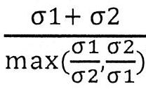
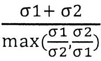
Applications Claiming Priority (2)
| Application Number | Priority Date | Filing Date | Title |
|---|---|---|---|
| US201862691890P | 2018-06-29 | 2018-06-29 | |
| US62/691,890 | 2018-06-29 |
Publications (2)
| Publication Number | Publication Date |
|---|---|
| TW202020165A true TW202020165A (en) | 2020-06-01 |
| TWI852936B TWI852936B (en) | 2024-08-21 |
Family
ID=68987585
Family Applications (2)
| Application Number | Title | Priority Date | Filing Date |
|---|---|---|---|
| TW113127597A TW202511496A (en) | 2018-06-29 | 2019-06-28 | Nucleic acid rearrangement and integration analysis |
| TW108122976A TWI852936B (en) | 2018-06-29 | 2019-06-28 | Nucleic acid rearrangement and integration analysis |
Family Applications Before (1)
| Application Number | Title | Priority Date | Filing Date |
|---|---|---|---|
| TW113127597A TW202511496A (en) | 2018-06-29 | 2019-06-28 | Nucleic acid rearrangement and integration analysis |
Country Status (7)
| Country | Link |
|---|---|
| US (2) | US11447829B2 (en) |
| EP (1) | EP3815094A4 (en) |
| CN (1) | CN112639987B (en) |
| AU (1) | AU2019291907A1 (en) |
| CA (1) | CA3105207A1 (en) |
| TW (2) | TW202511496A (en) |
| WO (1) | WO2020006370A1 (en) |
Families Citing this family (6)
| Publication number | Priority date | Publication date | Assignee | Title |
|---|---|---|---|---|
| TW202511496A (en) | 2018-06-29 | 2025-03-16 | 美商格瑞爾有限責任公司 | Nucleic acid rearrangement and integration analysis |
| EP4087942A4 (en) * | 2020-01-08 | 2024-01-24 | The Chinese University Of Hong Kong | BITERMINAL DNA FRAGMENT TYPES IN CELL-FREE SAMPLES AND THEIR USE |
| US20230197269A1 (en) * | 2020-02-18 | 2023-06-22 | Tempus Labs, Inc. | Systems and methods for detecting viral dna from sequencing |
| AU2021292311A1 (en) * | 2020-06-20 | 2023-02-16 | Grail, Llc | Detection and classification of human papillomavirus associated cancers |
| EP4413151A4 (en) * | 2021-10-04 | 2025-08-13 | Univ Hong Kong Chinese | SEQUENCE OF VIRAL DNA TO PREDICT DISEASE RELAPSE |
| WO2024086499A1 (en) * | 2022-10-17 | 2024-04-25 | University Of Washington | Systems and methods for detecting fusion genes from sequencing data |
Family Cites Families (9)
| Publication number | Priority date | Publication date | Assignee | Title |
|---|---|---|---|---|
| PT2121977T (en) * | 2007-01-11 | 2017-08-18 | Erasmus Univ Medical Center | Circular chromosome conformation capture (4c) |
| US12180549B2 (en) * | 2007-07-23 | 2024-12-31 | The Chinese University Of Hong Kong | Diagnosing fetal chromosomal aneuploidy using genomic sequencing |
| US20090053719A1 (en) | 2007-08-03 | 2009-02-26 | The Chinese University Of Hong Kong | Analysis of nucleic acids by digital pcr |
| JP2013526852A (en) | 2010-04-06 | 2013-06-27 | カリス ライフ サイエンシズ ルクセンブルク ホールディングス | Circulating biomarkers for disease |
| EP3228715B1 (en) | 2012-02-09 | 2019-08-14 | Life Technologies Corporation | Conjugated polymeric particle and method of making same |
| WO2014005329A1 (en) * | 2012-07-06 | 2014-01-09 | 深圳华大基因科技有限公司 | Method and system for determining integration manner of foreign gene in human genome |
| CN105874082B (en) * | 2013-10-07 | 2020-06-02 | 塞昆纳姆股份有限公司 | Methods and processes for non-invasive assessment of chromosomal changes |
| EP3978627A1 (en) | 2016-03-25 | 2022-04-06 | Karius, Inc. | Methods using synthetic nucleic acid spike-ins |
| TW202511496A (en) | 2018-06-29 | 2025-03-16 | 美商格瑞爾有限責任公司 | Nucleic acid rearrangement and integration analysis |
-
2019
- 2019-06-28 TW TW113127597A patent/TW202511496A/en unknown
- 2019-06-28 CA CA3105207A patent/CA3105207A1/en active Pending
- 2019-06-28 AU AU2019291907A patent/AU2019291907A1/en not_active Abandoned
- 2019-06-28 WO PCT/US2019/039750 patent/WO2020006370A1/en not_active Ceased
- 2019-06-28 CN CN201980056897.0A patent/CN112639987B/en active Active
- 2019-06-28 US US16/456,354 patent/US11447829B2/en active Active
- 2019-06-28 EP EP19825350.2A patent/EP3815094A4/en active Pending
- 2019-06-28 TW TW108122976A patent/TWI852936B/en active
-
2022
- 2022-07-29 US US17/816,272 patent/US20220380855A1/en not_active Abandoned
Also Published As
| Publication number | Publication date |
|---|---|
| AU2019291907A1 (en) | 2021-02-18 |
| TW202511496A (en) | 2025-03-16 |
| CN112639987A (en) | 2021-04-09 |
| US20220380855A1 (en) | 2022-12-01 |
| EP3815094A1 (en) | 2021-05-05 |
| WO2020006370A1 (en) | 2020-01-02 |
| CN112639987B (en) | 2024-12-10 |
| CA3105207A1 (en) | 2020-01-02 |
| EP3815094A4 (en) | 2022-04-13 |
| US11447829B2 (en) | 2022-09-20 |
| TWI852936B (en) | 2024-08-21 |
| US20200002770A1 (en) | 2020-01-02 |
Similar Documents
| Publication | Publication Date | Title |
|---|---|---|
| TWI852936B (en) | Nucleic acid rearrangement and integration analysis | |
| US20250346961A1 (en) | Methods and systems for tumor detection | |
| Holmes et al. | Mechanistic signatures of HPV insertions in cervical carcinomas | |
| TWI873615B (en) | Diagnostic applications using nucleic acid fragments | |
| ES2959360T3 (en) | Improving cancer screening using acellular viral nucleic acids | |
| AU2019261597B2 (en) | Systems and methods for using pathogen nucleic acid load to determine whether a subject has a cancer condition | |
| JP2025084804A (en) | Risk stratification for virus-associated cancers | |
| HK40045642B (en) | Nucleic acid rearrangement and integration analysis | |
| HK40045642A (en) | Nucleic acid rearrangement and integration analysis | |
| HK40063165A (en) | Stratification of risk of virus associated cancers | |
| WO2025239952A1 (en) | Viral cancer detection tests | |
| Gregson | Characterisation of Copy Number Changes in the Progression of Barrett’s Oesophagus | |
| HK40042779A (en) | Tissue-specific methylation marker | |
| Royse | Molecular Profiling in Cervical Carcinogenesis and Progression |