US10353461B2 - Evaluating clinician - Google Patents
Evaluating clinician Download PDFInfo
- Publication number
- US10353461B2 US10353461B2 US15/316,288 US201515316288A US10353461B2 US 10353461 B2 US10353461 B2 US 10353461B2 US 201515316288 A US201515316288 A US 201515316288A US 10353461 B2 US10353461 B2 US 10353461B2
- Authority
- US
- United States
- Prior art keywords
- attention
- clinician
- images
- detection module
- lapse
- Prior art date
- Legal status (The legal status is an assumption and is not a legal conclusion. Google has not performed a legal analysis and makes no representation as to the accuracy of the status listed.)
- Active, expires
Links
Images



Classifications
-
- G—PHYSICS
- G06—COMPUTING OR CALCULATING; COUNTING
- G06F—ELECTRIC DIGITAL DATA PROCESSING
- G06F3/00—Input arrangements for transferring data to be processed into a form capable of being handled by the computer; Output arrangements for transferring data from processing unit to output unit, e.g. interface arrangements
- G06F3/01—Input arrangements or combined input and output arrangements for interaction between user and computer
- G06F3/011—Arrangements for interaction with the human body, e.g. for user immersion in virtual reality
- G06F3/013—Eye tracking input arrangements
-
- A—HUMAN NECESSITIES
- A61—MEDICAL OR VETERINARY SCIENCE; HYGIENE
- A61B—DIAGNOSIS; SURGERY; IDENTIFICATION
- A61B3/00—Apparatus for testing the eyes; Instruments for examining the eyes
- A61B3/18—Arrangement of plural eye-testing or -examining apparatus
-
- A—HUMAN NECESSITIES
- A61—MEDICAL OR VETERINARY SCIENCE; HYGIENE
- A61B—DIAGNOSIS; SURGERY; IDENTIFICATION
- A61B5/00—Measuring for diagnostic purposes; Identification of persons
- A61B5/16—Devices for psychotechnics; Testing reaction times ; Devices for evaluating the psychological state
- A61B5/161—Flicker fusion testing
-
- A—HUMAN NECESSITIES
- A61—MEDICAL OR VETERINARY SCIENCE; HYGIENE
- A61B—DIAGNOSIS; SURGERY; IDENTIFICATION
- A61B5/00—Measuring for diagnostic purposes; Identification of persons
- A61B5/16—Devices for psychotechnics; Testing reaction times ; Devices for evaluating the psychological state
- A61B5/163—Devices for psychotechnics; Testing reaction times ; Devices for evaluating the psychological state by tracking eye movement, gaze, or pupil change
-
- A—HUMAN NECESSITIES
- A61—MEDICAL OR VETERINARY SCIENCE; HYGIENE
- A61B—DIAGNOSIS; SURGERY; IDENTIFICATION
- A61B5/00—Measuring for diagnostic purposes; Identification of persons
- A61B5/16—Devices for psychotechnics; Testing reaction times ; Devices for evaluating the psychological state
- A61B5/165—Evaluating the state of mind, e.g. depression, anxiety
-
- A—HUMAN NECESSITIES
- A61—MEDICAL OR VETERINARY SCIENCE; HYGIENE
- A61B—DIAGNOSIS; SURGERY; IDENTIFICATION
- A61B5/00—Measuring for diagnostic purposes; Identification of persons
- A61B5/16—Devices for psychotechnics; Testing reaction times ; Devices for evaluating the psychological state
- A61B5/168—Evaluating attention deficit, hyperactivity
-
- G—PHYSICS
- G06—COMPUTING OR CALCULATING; COUNTING
- G06F—ELECTRIC DIGITAL DATA PROCESSING
- G06F3/00—Input arrangements for transferring data to be processed into a form capable of being handled by the computer; Output arrangements for transferring data from processing unit to output unit, e.g. interface arrangements
- G06F3/01—Input arrangements or combined input and output arrangements for interaction between user and computer
- G06F3/011—Arrangements for interaction with the human body, e.g. for user immersion in virtual reality
- G06F3/012—Head tracking input arrangements
-
- G—PHYSICS
- G06—COMPUTING OR CALCULATING; COUNTING
- G06F—ELECTRIC DIGITAL DATA PROCESSING
- G06F3/00—Input arrangements for transferring data to be processed into a form capable of being handled by the computer; Output arrangements for transferring data from processing unit to output unit, e.g. interface arrangements
- G06F3/01—Input arrangements or combined input and output arrangements for interaction between user and computer
- G06F3/03—Arrangements for converting the position or the displacement of a member into a coded form
- G06F3/0304—Detection arrangements using opto-electronic means
-
- G—PHYSICS
- G11—INFORMATION STORAGE
- G11B—INFORMATION STORAGE BASED ON RELATIVE MOVEMENT BETWEEN RECORD CARRIER AND TRANSDUCER
- G11B27/00—Editing; Indexing; Addressing; Timing or synchronising; Monitoring; Measuring tape travel
-
- G—PHYSICS
- G11—INFORMATION STORAGE
- G11B—INFORMATION STORAGE BASED ON RELATIVE MOVEMENT BETWEEN RECORD CARRIER AND TRANSDUCER
- G11B27/00—Editing; Indexing; Addressing; Timing or synchronising; Monitoring; Measuring tape travel
- G11B27/005—Reproducing at a different information rate from the information rate of recording
- G11B27/007—Reproducing at a different information rate from the information rate of recording reproducing continuously a part of the information, i.e. repeating
-
- A—HUMAN NECESSITIES
- A61—MEDICAL OR VETERINARY SCIENCE; HYGIENE
- A61B—DIAGNOSIS; SURGERY; IDENTIFICATION
- A61B3/00—Apparatus for testing the eyes; Instruments for examining the eyes
- A61B3/10—Objective types, i.e. instruments for examining the eyes independent of the patients' perceptions or reactions
- A61B3/113—Objective types, i.e. instruments for examining the eyes independent of the patients' perceptions or reactions for determining or recording eye movement
-
- A—HUMAN NECESSITIES
- A61—MEDICAL OR VETERINARY SCIENCE; HYGIENE
- A61B—DIAGNOSIS; SURGERY; IDENTIFICATION
- A61B5/00—Measuring for diagnostic purposes; Identification of persons
- A61B5/16—Devices for psychotechnics; Testing reaction times ; Devices for evaluating the psychological state
-
- G—PHYSICS
- G06—COMPUTING OR CALCULATING; COUNTING
- G06F—ELECTRIC DIGITAL DATA PROCESSING
- G06F3/00—Input arrangements for transferring data to be processed into a form capable of being handled by the computer; Output arrangements for transferring data from processing unit to output unit, e.g. interface arrangements
- G06F3/01—Input arrangements or combined input and output arrangements for interaction between user and computer
- G06F3/011—Arrangements for interaction with the human body, e.g. for user immersion in virtual reality
-
- G—PHYSICS
- G06—COMPUTING OR CALCULATING; COUNTING
- G06F—ELECTRIC DIGITAL DATA PROCESSING
- G06F3/00—Input arrangements for transferring data to be processed into a form capable of being handled by the computer; Output arrangements for transferring data from processing unit to output unit, e.g. interface arrangements
- G06F3/01—Input arrangements or combined input and output arrangements for interaction between user and computer
- G06F3/048—Interaction techniques based on graphical user interfaces [GUI]
- G06F3/0484—Interaction techniques based on graphical user interfaces [GUI] for the control of specific functions or operations, e.g. selecting or manipulating an object, an image or a displayed text element, setting a parameter value or selecting a range
- G06F3/0485—Scrolling or panning
Definitions
- the following generally relates to evaluating clinician attention during display of imaging data sets.
- Medical imaging provides useful information about the interior characteristics (e.g., anatomical tissue, material flow, etc.) of a subject under examination.
- clinicians view sequences of hundreds of images corresponding to multiple patients while analyzing patient data.
- Such images have been viewed in a continuous cinematic loop in which each image is sequentially visually displayed based on a predetermined rate for a predetermined amount of time. With such display, any image may provide crucial visual information regarding a particular disease and/or state of a patient.
- a system includes a display that sequentially displays one or more images from an image data set at a predetermined rate, an optical attention monitoring device that senses a characteristic indicative of a clinician's attention to the one or more images of the image data set; and a processor that executes an attention detection module ( 118 ) that detects a lapse in attention with respect to the one or more of the images based on the sensed characteristic indicative of the clinician's attention and generates a signal indicating a relationship between the lapse in attention and the one or more images.
- an attention detection module 118
- a method in another aspect, includes sequentially displaying images from an image data set at a predetermined rate, monitoring a characteristic indicative of a clinician's attention to a displayed image of the image data set; detecting a lapse in attention of the clinician to at least one of the displayed images based on the characteristic indicative of the clinician's attention; and re-displaying the at least one of the displayed images in response to the detecting of the lapse in attention.
- a computer readable storage medium is encoded with computer readable instructions, which, when executed by a processor, causes the processor to: sequentially display images from a volumetric image data set at a predetermined rate, monitor a characteristic indicative of a clinician's attention to the displayed image of the volumetric image data set; detect a lapse in attention to at least one of the displayed images based on the characteristic indicative of the clinician's attention; and identify the at least one of the displayed images to re-display in response to detecting of the lapse in attention.
- the invention may take form in various components and arrangements of components, and in various steps and arrangements of steps.
- the drawings are only for purposes of illustrating the preferred embodiments and are not to be construed as limiting the invention.
- FIG. 1 schematically illustrates a computing system with an attention detection module in connection with an imaging system(s) and a data repository.
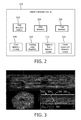
- FIG. 2 schematically illustrates an example of the attention detection module.
- FIG. 3 schematically illustrates displayed data including an attention status bar.
- FIG. 4 illustrates a method for detecting a lapse of attention and replaying images presented during the lapse of attention.
- FIG. 1 schematically illustrates a system 100 in connection with an imaging system(s) 102 and/or an image data repository 104 .
- Imaging data may be stored by and/or transferred between the computing system 101 , the imaging system(s) 102 , and/or the image data repository 104 in standard formats such as Digital Imaging and Communications in Medicine (DICOM), Health Level 7 (HL7), and/or other standard formats, and/or non-standard, proprietary, and/or other format.
- DICOM Digital Imaging and Communications in Medicine
- HL7 Health Level 7
- the imaging system(s) 102 can include one or more of an ultrasound (US), a computed tomography (CT), a magnetic resonance (MR), a positron emission tomography (PET), a single-photon emission computed tomography (SPECT), and/or other imaging system.
- the image data repository 104 can include one or more of a picture archiving and communication system (PACS), a radiology information system (RIS), a hospital information system (HIS), and/or other data repository.
- PPS picture archiving and communication system
- RIS radiology information system
- HIS hospital information system
- the computing system 101 includes one or more processors 106 (e.g. microprocessor, central processing unit, controller, etc.) and computer readable storage medium (“memory”) 108 (i.e., physical memory and other non-transitory storage medium, and excluding transitory medium).
- processors 106 e.g. microprocessor, central processing unit, controller, etc.
- memory computer readable storage medium
- the computing system 101 interfaces with an input device(s) 110 such as a keyboard, a mouse, a microphone, a touchscreen, etc. and an output device such as a display 112 , a filmer, a printer, etc.
- the computer readable storage medium 108 is encoded with computer readable instructions, which, when executed by the one or more processors 106 cause the computing system 101 to carry out the various functions described herein.
- the one or more processors 106 can additionally or alternatively execute instructions carried by a carrier wave, a signal and other transitory storage medium.
- the computer readable storage medium 108 includes several software modules, including a data retriever 114 , an image display module 116 and an attention detection module 118 .
- the data retriever 114 receives a signal, as an input, via the input device(s) 110 and/or processor 106 , identifying an imaging data set to retrieve.
- the computing system 101 first visually presents through the display 112 a list of stored patient studies, from which a clinician can select an imaging data set to load.
- the selected imaging data set may be an initial imaging data set of a patient to be read or a follow-up imaging data set of the patient for reading and comparison with a previously generated data set of the patient.
- the imaging data set comprises a plurality of images that can be displayed in a sequence.
- the display 112 sequentially displays images from the imaging data set.
- the images are received from the image display module 116 .
- a rate at which each image is displayed is predetermined and interpreted by the image display module 116 .
- a rate of image display can vary based on conditions such as user input, etc.
- An example of a predetermined rate of display is one image per second. However, other rates are also contemplated herein. For example, an image can be presented at a faster or slower rate.
- the image display module 116 additionally determines a subset of already displayed images to display again (as described in more detail below) based on an observed attention characteristic of the clinician.
- the optical attention monitoring device 120 monitors a clinician's visual attention to each displayed image.
- the optical attention monitoring device 120 provides information regarding eye-related activity such as gaze measurements, eye movements, events of saccades and fixations, pupil and iris tracking, and center/size estimation of pupils.
- the optical attention monitoring device 120 is a remote eye tracker placed in front of the display 112 facing a clinician, remotely tracking eye features.
- the optical attention monitoring device 120 can be a head-mounted/mobile eye tracker that records a scene as viewed by the clinician.
- the optical attention monitoring device 120 can be a camera that obtains data related to facial orientation and facial features.
- An optical attention monitoring device 120 is a physical device such as an optical sensor that senses a characteristic indicative of the clinician's visual attention to each displayed image.
- the attention detection module 118 evaluates the signal from the optical attention monitoring device 120 and detects a lapse in attention for an image, if one exists, based on the signal.
- the signal for example, includes information such as attention related to eye contact along a line of sight to the displayed images, direction of eye gaze at the displayed images, the capture of facial characteristics in relation to the displayed images, and/or other information.
- the optical attention monitoring device can compensate for a clinician wearing glasses, contact lenses, and any other type of vision correction device in the detection of the lapse of attention.
- the optical attention monitoring device 120 and the display 112 are separate from the computing system 101 and the imaging system 102 .
- the optical attention monitoring device 120 and/or the display can be integrated within the computing system 101 or the imaging system 102 .
- FIG. 2 an example of the attention detection module 118 is illustrated.
- the attention detection module 118 includes one or more of a blink detection module 202 , a gaze detection module 208 , a vigilance loss detection module 212 or a facial movement detection module 214 .
- the attention detection module 118 may include more or less modules, including a same module and/or a different module.
- the blink detection module 202 detects blinks of an eye, measures a time period of blink duration, and compares the time period of blink duration to a threshold.
- the time period of blink duration is a time period from an eyelid beginning to close with a first saccade, and ending with the eyelid fully opening after a second saccade. Based on eyelid movement data provided by the attention monitoring device, the event detection module determines the time period of blink duration irrespective of whether the clinician is wearing glasses, contacts and/or another vision correction device.
- the blink detection module 202 compares the time period to a predetermined blink threshold 204 . If the blink duration exceeds the predetermined blink threshold 204 (e.g. the eye has been closed for a sufficient amount of time), the blink detection module 202 transmits a signal, which indicates the eye has been closed for a sufficient amount of time, to the image display module 116 . The signal indicates the blink caused a lapse of attention and includes time data indicative of the lapse.
- a predetermined blink threshold 204 e.g. the eye has been closed for a sufficient amount of time
- the blink detection module 202 analyzes a visual representation of a pupil captured by the optical attention monitoring device 120 .
- the optical attention monitoring device 120 may track a clinician's eyeball while the attention detection module analyzes pupil data sent by the attention monitoring device.
- the eyelid may cover a pupil, resulting in the optical attention monitoring device 120 losing pupil data due to a lack of eyeball exposure.
- a pupil threshold 206 can be retrieved (i.e. 30-80 milliseconds (ms), such as 45 ms, 50 ms, 52 ms, etc.) that causes the attention detection module 118 to ignore loss of pupil data for a length of time up to the pupil threshold 206 , before sending a signal indicating a lapse in attention.
- the gaze detection module 208 analyzes a gaze of a clinician with respect to the displayed images. If the gaze of the clinician is focused on the displayed images, the clinician is determined to be adequately focused with no lapse in attention. However, if the gaze is not focused on the displayed images, the clinician is not providing adequate attention.
- the attention detection module 118 transmits a signal to the image display module to adjust image display and/or repeat at least a segment of the displayed images.
- the gaze detection module 208 tracks a clinician's eye at a frequency from thirty (30) to hundreds of eye tracking points per second.
- a clinician's gaze location may drift even though the clinician has not blinked.
- the clinician's gaze location may not be fixated on the individual frame of the consecutive images, but instead fixated on a location outside of the image frame or in a non-pertinent part of the image frame.
- the attention detection module 118 may send a signal indicating an errant gaze to the image display module 116 .
- the attention detection module 118 indicates a sub-set of images to re-display.
- the image display module 116 re-displays the sub-set.
- the gaze detection module 208 additionally analyzes gazes detected by the optical attention monitoring device 120 to determine whether an unsatisfactory number of eye tracking points were directed to the displayed images. Some tracking points may directed toward the displayed images while others may be directed toward a background of the displayed images or the controls of the input device 110 . The gaze detection module 208 compares the number of tracking points directed at the displayed images to a tracking point threshold 210 .
- the gaze detection module 208 does not determine that a lapse in attention has occurred and does not send a signal to the image display module to replay images. However, if a number of tracked locations less than the threshold are focused on the displayed images, the attention detection module 118 determines that a lapse in attention has occurred and sends a signal to the image display module to replay images.
- the gaze detection module 208 can analyze image data before presentation to determine likely regions of abnormalities on the images.
- the clinician's viewing location can be tracked to make sure the clinician has viewed these likely regions. For example, image regions within the displayed images may be pre-identified using artificial intelligence techniques or algorithms based on expert radiologists' interpretation techniques.
- the gaze detection module 208 analyzes viewing locations obtained by the optical attention monitoring device 120 . The gaze detection module 208 checks to see that the viewing locations match the pre-identified likely regions of abnormalities.
- the gaze detection module 208 compares a clinician's viewing location with known abnormalities on the displayed images.
- the displayed images can represent a follow-up case, and the clinician may be viewing previously-annotated images.
- the annotations provide specific locations of abnormalities within the displayed images that are known to the attention detection module 118 .
- the gaze detection module 208 analyzes the gaze of the clinician in relationship to the displayed images and the annotations of the displayed images to determine if gaze points are directed toward the areas of the displayed images identified by the annotations.
- CAD computer-aided detection
- a vigilance loss detection module 212 detects loss of vigilance of a clinician.
- a loss of vigilance occurs when a clinician has too high of a mental workload is inadequately rested or is distracted.
- the vigilance loss detection module 212 establishes a baseline measurement at a first period of time. The period of time can be in the beginning of a clinician's work day. The baseline measurement can be computed by obtaining a distribution of blink durations over the course of the baseline period measurement. During other times of the work day, a clinician's attention can be analyzed and compared to the baseline measurement. If a discrepancy exists beyond a predetermined threshold, a loss of vigilance can be determined and the attention detection module 118 can adjust display of the images.
- the vigilance loss detection module 212 can detect a loss of vigilance using a measure of proficiency based on the clinician scanning images for suspicious regions.
- the vigilance loss detection module 212 measures the scanning path during the first period of time at the beginning of a clinician's work day. When a clinician is losing vigilance, the scan path may become erratic and/or drift from a usual gaze path. A significant departure from the originally established path during the first time period could indicate a loss of vigilance. The loss of vigilance could indicate a sub-optimal review of the study.
- the vigilance loss detection module 212 can indicate to a clinician that rest is required for a given period of time, and present the images to the image display module after the rest period of time has ended.
- a facial movement detection module 214 determines if a face of the clinician is focused on the display. For example the facial movement detection module 214 may employ a facial recognition detection algorithm to determine if the face is focused along a line of sight to the image. If no facial features are detected as focused on the line of sight, the facial movement detection module 214 may send a signal to the image display module 116 to pause or stop the image displayed on the display 112 .
- the image display module 116 re-displays images via the display 112 based on signals received from the attention detection module 118 .
- the image display module 116 presents, in one instance, a graphical attention status bar 302 .
- the graphical attention status bar 302 comprises a first bar 304 and one or more segments 306 (three shown in the illustrated example).
- the first bar 304 represents the displayed images, and the one or more segments 306 indicate images where the clinician's attention had lapsed and that images were not viewed thoroughly.
- the image display module 116 allows a clinician to interact with the graphical attention status bar 302 via a cursor, stylus, finger or other selection device. During or after a complete sequence of image data has been displayed, the image display module 116 analyzes signals sent from the event detection module. The image display module 116 presents missed images based on clinician interactions with the graphical attention status bar 302 . For example, a clinician can select a portion of the graphical attention status bar 302 that indicates missed images. The image display module 116 retrieves missed image data corresponding to a selected segment of the graphical attention status bar 302 , and replays the selected images for the clinician.
- the image display module 116 may actively adjust playback as images are displayed. If any signal is received from the event detection module indicating that a clinician's attention is not focused on the display, the image display module 116 may replay the missed image data as soon as the clinician's attention is refocused. The replay may be accompanied by audio feedback indicating that missed frames are being replayed.
- FIG. 4 schematically illustrates a method for detecting a lapse of attention and replaying images presented during the lapse of attention.
- images from a data set are sequentially displayed at a predetermined rate.
- a characteristic indicative of a clinician's attention to each displayed image of the volumetric image data set is monitored.
- a lapse in attention for one or more of the images is detected based on the monitored characteristic.
- the one or more of the images are re-displayed in response to the detection of the lapse in attention.
- the above methods may be implemented by way of computer readable instructions, encoded or embedded on computer readable storage medium, which, when executed by a computer processor(s), cause the processor(s) to carry out the described acts. Additionally or alternatively, at least one of the computer readable instructions is carried by a signal, carrier wave or other transitory medium.
Landscapes
- Health & Medical Sciences (AREA)
- Engineering & Computer Science (AREA)
- Life Sciences & Earth Sciences (AREA)
- Physics & Mathematics (AREA)
- Veterinary Medicine (AREA)
- Surgery (AREA)
- Public Health (AREA)
- General Health & Medical Sciences (AREA)
- Animal Behavior & Ethology (AREA)
- Molecular Biology (AREA)
- Medical Informatics (AREA)
- Biophysics (AREA)
- Heart & Thoracic Surgery (AREA)
- Biomedical Technology (AREA)
- Theoretical Computer Science (AREA)
- General Engineering & Computer Science (AREA)
- Developmental Disabilities (AREA)
- Psychiatry (AREA)
- Social Psychology (AREA)
- Psychology (AREA)
- Pathology (AREA)
- Child & Adolescent Psychology (AREA)
- Hospice & Palliative Care (AREA)
- Educational Technology (AREA)
- Human Computer Interaction (AREA)
- General Physics & Mathematics (AREA)
- Ophthalmology & Optometry (AREA)
- Eye Examination Apparatus (AREA)
- Measuring And Recording Apparatus For Diagnosis (AREA)
- Image Processing (AREA)
- User Interface Of Digital Computer (AREA)
Priority Applications (1)
| Application Number | Priority Date | Filing Date | Title |
|---|---|---|---|
| US15/316,288 US10353461B2 (en) | 2014-06-17 | 2015-06-16 | Evaluating clinician |
Applications Claiming Priority (3)
| Application Number | Priority Date | Filing Date | Title |
|---|---|---|---|
| US201462013050P | 2014-06-17 | 2014-06-17 | |
| US15/316,288 US10353461B2 (en) | 2014-06-17 | 2015-06-16 | Evaluating clinician |
| PCT/IB2015/054538 WO2015193806A1 (en) | 2014-06-17 | 2015-06-16 | Evaluating clinician attention |
Publications (2)
| Publication Number | Publication Date |
|---|---|
| US20170192502A1 US20170192502A1 (en) | 2017-07-06 |
| US10353461B2 true US10353461B2 (en) | 2019-07-16 |
Family
ID=53879726
Family Applications (1)
| Application Number | Title | Priority Date | Filing Date |
|---|---|---|---|
| US15/316,288 Active 2036-01-15 US10353461B2 (en) | 2014-06-17 | 2015-06-16 | Evaluating clinician |
Country Status (6)
| Country | Link |
|---|---|
| US (1) | US10353461B2 (pl) |
| EP (1) | EP3157432B1 (pl) |
| CN (1) | CN106659441B (pl) |
| PL (1) | PL3157432T3 (pl) |
| TR (1) | TR201904055T4 (pl) |
| WO (1) | WO2015193806A1 (pl) |
Families Citing this family (16)
| Publication number | Priority date | Publication date | Assignee | Title |
|---|---|---|---|---|
| US10281743B2 (en) | 2015-10-01 | 2019-05-07 | Verily Life Sciences Llc | Eye convergence detection with contact lenses |
| US10222632B2 (en) * | 2015-10-01 | 2019-03-05 | Verily Life Sciences Llc | Lens-to-lens communication for contact lenses |
| EP3264222B1 (en) * | 2016-06-27 | 2019-04-10 | Nokia Technologies Oy | An apparatus and associated methods |
| US10839520B2 (en) * | 2017-03-03 | 2020-11-17 | The United States Of America, As Represented By The Secretary, Department Of Health & Human Services | Eye tracking applications in computer aided diagnosis and image processing in radiology |
| US10810773B2 (en) * | 2017-06-14 | 2020-10-20 | Dell Products, L.P. | Headset display control based upon a user's pupil state |
| CN108877362A (zh) * | 2018-06-28 | 2018-11-23 | 深圳市尚高科技发展有限公司 | 一种聚集注意力方法及其终端 |
| JP7067502B2 (ja) * | 2018-08-08 | 2022-05-16 | 株式会社Jvcケンウッド | 評価装置、評価方法、及び評価プログラム |
| CN113474789A (zh) | 2019-02-25 | 2021-10-01 | 皇家飞利浦有限公司 | 确定对象的相对认知能力 |
| CN110162185A (zh) * | 2019-06-10 | 2019-08-23 | 京东方科技集团股份有限公司 | 一种智能显示方法及装置 |
| DE102019214784A1 (de) | 2019-09-26 | 2021-04-01 | Carl Zeiss Meditec Ag | Verfahren zur Ermittlung physiologisch korrekter, biometrischer Daten eines Auges |
| JP7380365B2 (ja) * | 2020-03-19 | 2023-11-15 | マツダ株式会社 | 状態推定装置 |
| EP3889970A1 (en) * | 2020-04-03 | 2021-10-06 | Koninklijke Philips N.V. | Diagnosis support system |
| EP3893091A1 (en) * | 2020-04-09 | 2021-10-13 | Koninklijke Philips N.V. | Controlling a data inspection by a user |
| CN113126762B (zh) * | 2021-04-21 | 2022-08-16 | 惠东县人民医院 | 一种用于监测医疗行为的医疗数据核查装置及方法 |
| US11983316B2 (en) * | 2022-01-19 | 2024-05-14 | Apple Inc. | Method and device for managing attention accumulators |
| US20250314888A1 (en) * | 2022-07-14 | 2025-10-09 | Qualcomm Incorporated | Reducing eye strain and improving performance in headset device |
Citations (10)
| Publication number | Priority date | Publication date | Assignee | Title |
|---|---|---|---|---|
| US6111580A (en) * | 1995-09-13 | 2000-08-29 | Kabushiki Kaisha Toshiba | Apparatus and method for controlling an electronic device with user action |
| US20070173699A1 (en) | 2006-01-21 | 2007-07-26 | Honeywell International Inc. | Method and system for user sensitive pacing during rapid serial visual presentation |
| US20070265507A1 (en) | 2006-03-13 | 2007-11-15 | Imotions Emotion Technology Aps | Visual attention and emotional response detection and display system |
| US20080048931A1 (en) | 2003-11-26 | 2008-02-28 | Rafael - Armament Development Authority Ltd. | Helmet System for Information or Weapon Systems |
| JP2008219286A (ja) | 2007-03-01 | 2008-09-18 | Sharp Corp | スライドショー装置 |
| US20090150821A1 (en) | 2007-12-11 | 2009-06-11 | Honeywell International, Inc. | Hierarchichal rapid serial visual presentation for robust target identification |
| US20110273731A1 (en) | 2010-05-10 | 2011-11-10 | Canon Kabushiki Kaisha | Printer with attention based image customization |
| US20130340006A1 (en) | 2012-06-14 | 2013-12-19 | Mobitv, Inc. | Eye-tracking navigation |
| US20150043033A1 (en) | 2013-08-06 | 2015-02-12 | Konica Minolta, Inc. | Display device, non-transitory computer-readable recording medium and image processing apparatus |
| US20150192992A1 (en) | 2014-01-03 | 2015-07-09 | Harman International Industries, Inc. | Eye vergence detection on a display |
Family Cites Families (2)
| Publication number | Priority date | Publication date | Assignee | Title |
|---|---|---|---|---|
| CN2441437Y (zh) * | 2000-08-01 | 2001-08-08 | 上海仪器仪表研究所 | 儿童注意力缺陷测试仪器 |
| CN103839046B (zh) * | 2013-12-26 | 2017-02-01 | 苏州清研微视电子科技有限公司 | 驾驶人注意力自动识别系统及其识别方法 |
-
2015
- 2015-06-16 TR TR2019/04055T patent/TR201904055T4/tr unknown
- 2015-06-16 EP EP15751072.8A patent/EP3157432B1/en active Active
- 2015-06-16 CN CN201580032373.XA patent/CN106659441B/zh active Active
- 2015-06-16 US US15/316,288 patent/US10353461B2/en active Active
- 2015-06-16 PL PL15751072T patent/PL3157432T3/pl unknown
- 2015-06-16 WO PCT/IB2015/054538 patent/WO2015193806A1/en not_active Ceased
Patent Citations (10)
| Publication number | Priority date | Publication date | Assignee | Title |
|---|---|---|---|---|
| US6111580A (en) * | 1995-09-13 | 2000-08-29 | Kabushiki Kaisha Toshiba | Apparatus and method for controlling an electronic device with user action |
| US20080048931A1 (en) | 2003-11-26 | 2008-02-28 | Rafael - Armament Development Authority Ltd. | Helmet System for Information or Weapon Systems |
| US20070173699A1 (en) | 2006-01-21 | 2007-07-26 | Honeywell International Inc. | Method and system for user sensitive pacing during rapid serial visual presentation |
| US20070265507A1 (en) | 2006-03-13 | 2007-11-15 | Imotions Emotion Technology Aps | Visual attention and emotional response detection and display system |
| JP2008219286A (ja) | 2007-03-01 | 2008-09-18 | Sharp Corp | スライドショー装置 |
| US20090150821A1 (en) | 2007-12-11 | 2009-06-11 | Honeywell International, Inc. | Hierarchichal rapid serial visual presentation for robust target identification |
| US20110273731A1 (en) | 2010-05-10 | 2011-11-10 | Canon Kabushiki Kaisha | Printer with attention based image customization |
| US20130340006A1 (en) | 2012-06-14 | 2013-12-19 | Mobitv, Inc. | Eye-tracking navigation |
| US20150043033A1 (en) | 2013-08-06 | 2015-02-12 | Konica Minolta, Inc. | Display device, non-transitory computer-readable recording medium and image processing apparatus |
| US20150192992A1 (en) | 2014-01-03 | 2015-07-09 | Harman International Industries, Inc. | Eye vergence detection on a display |
Non-Patent Citations (1)
| Title |
|---|
| Holmqvist, K. et al., "Eye Tracking: A comprehensive guide to methods and measures", Oxford University Press, 2011, ISBN: 9780199697083. |
Also Published As
| Publication number | Publication date |
|---|---|
| EP3157432A1 (en) | 2017-04-26 |
| EP3157432B1 (en) | 2019-01-02 |
| CN106659441A (zh) | 2017-05-10 |
| CN106659441B (zh) | 2019-11-26 |
| WO2015193806A1 (en) | 2015-12-23 |
| TR201904055T4 (tr) | 2019-04-22 |
| US20170192502A1 (en) | 2017-07-06 |
| PL3157432T3 (pl) | 2019-06-28 |
Similar Documents
| Publication | Publication Date | Title |
|---|---|---|
| EP3157432B1 (en) | Evaluating clinician attention | |
| US20240257981A1 (en) | Method and apparatus for determining health status | |
| US11699529B2 (en) | Systems and methods for diagnosing a stroke condition | |
| US10755817B2 (en) | Systems, apparatuses and methods for predicting medical events and conditions reflected in gait | |
| US9841811B2 (en) | Visually directed human-computer interaction for medical applications | |
| JP6230708B2 (ja) | 撮像データセットの間の所見のマッチング | |
| US20150213725A1 (en) | Method, apparatus, and system for viewing multiple-slice medical images | |
| US20230059015A1 (en) | Apparatus for monitoring of a patient undergoing a magnetic resonance image scan | |
| US20240115213A1 (en) | Diagnosing and tracking stroke with sensor-based assessments of neurological deficits | |
| JPWO2017033516A1 (ja) | 読影支援装置及び方法 | |
| US20190148015A1 (en) | Medical information processing device and program | |
| Bernini et al. | Ambulatory seizure detection | |
| US20170071468A1 (en) | Motion tracking method for sonographer | |
| US20230284962A1 (en) | Systems and methods for diagnosing, assessing, and quantifying brain trauma | |
| US20250281046A1 (en) | Infrared thermography for intraoperative functional mapping | |
| CN113633257B (zh) | 基于虚拟现实的前庭功能检查方法、系统、设备及介质 | |
| EP4348670A1 (en) | Autonomous image acquisition start-stop managing system | |
| US20240382125A1 (en) | Information processing system, information processing method and computer program product | |
| WO2017184215A1 (en) | Automated abdominojugular reflux testing | |
| Kazancigil | Innovations and convergence in mobile medical applications and cloud-based hospital information systems for the real-time monitoring of patients and early warning of diseases | |
| Alreja et al. | A new paradigm for investigating real-world social behavior and its neural underpinnings | |
| Newman et al. | Reconstructing animated eye movements from electrooculography data to aid the diagnosis of vestibular disorders | |
| CN118044777B (zh) | 基于被测者的眼动监测所述被测者的意识的方法、装置、系统、电子设备、计算机可读存储介质及计算机程序产品 | |
| US20250157601A1 (en) | Medical record generation for virtual nursing | |
| Culemann et al. | Blink parameters are confounded by vertical eye orientation in video-based eye tracking: Comparing pupil-and eyelid-based methods |
Legal Events
| Date | Code | Title | Description |
|---|---|---|---|
| AS | Assignment |
Owner name: KONINKLIJKE PHILIPS N.V., NETHERLANDS Free format text: ASSIGNMENT OF ASSIGNORS INTEREST;ASSIGNORS:DMITRIEVA, JULIA;MANKOVICH, GABRIEL RYAN;QIAN, YUECHAN;SIGNING DATES FROM 20161130 TO 20161201;REEL/FRAME:040519/0695 |
|
| STPP | Information on status: patent application and granting procedure in general |
Free format text: NOTICE OF ALLOWANCE MAILED -- APPLICATION RECEIVED IN OFFICE OF PUBLICATIONS |
|
| STPP | Information on status: patent application and granting procedure in general |
Free format text: AWAITING TC RESP, ISSUE FEE PAYMENT VERIFIED Free format text: PUBLICATIONS -- ISSUE FEE PAYMENT VERIFIED |
|
| STCF | Information on status: patent grant |
Free format text: PATENTED CASE |
|
| MAFP | Maintenance fee payment |
Free format text: PAYMENT OF MAINTENANCE FEE, 4TH YEAR, LARGE ENTITY (ORIGINAL EVENT CODE: M1551); ENTITY STATUS OF PATENT OWNER: LARGE ENTITY Year of fee payment: 4 |