US10582949B2 - Sheath assembly and multihole catheter for different fields of endoscopic surgery involving suction, irrigation and material removal - Google Patents
Sheath assembly and multihole catheter for different fields of endoscopic surgery involving suction, irrigation and material removal Download PDFInfo
- Publication number
- US10582949B2 US10582949B2 US15/325,878 US201415325878A US10582949B2 US 10582949 B2 US10582949 B2 US 10582949B2 US 201415325878 A US201415325878 A US 201415325878A US 10582949 B2 US10582949 B2 US 10582949B2
- Authority
- US
- United States
- Prior art keywords
- suction
- reservoir
- sheath
- assembly
- surgery
- Prior art date
- Legal status (The legal status is an assumption and is not a legal conclusion. Google has not performed a legal analysis and makes no representation as to the accuracy of the status listed.)
- Active, expires
Links
- 238000002674 endoscopic surgery Methods 0.000 title claims abstract description 44
- 230000002262 irrigation Effects 0.000 title claims abstract description 19
- 238000003973 irrigation Methods 0.000 title claims abstract description 19
- 239000000463 material Substances 0.000 title claims abstract description 18
- 238000001356 surgical procedure Methods 0.000 claims abstract description 81
- 238000007789 sealing Methods 0.000 claims abstract description 43
- 210000000746 body region Anatomy 0.000 claims abstract description 35
- XLYOFNOQVPJJNP-UHFFFAOYSA-N water Substances O XLYOFNOQVPJJNP-UHFFFAOYSA-N 0.000 claims abstract description 9
- 239000012530 fluid Substances 0.000 claims description 67
- 239000012634 fragment Substances 0.000 claims description 66
- 238000000034 method Methods 0.000 claims description 38
- 230000000694 effects Effects 0.000 claims description 14
- 230000008569 process Effects 0.000 claims description 8
- 239000002245 particle Substances 0.000 claims description 6
- 230000005484 gravity Effects 0.000 claims description 2
- 210000000244 kidney pelvis Anatomy 0.000 description 92
- 239000004575 stone Substances 0.000 description 62
- 210000000626 ureter Anatomy 0.000 description 51
- 230000002620 ureteric effect Effects 0.000 description 41
- FAPWRFPIFSIZLT-UHFFFAOYSA-M Sodium chloride Chemical compound [Na+].[Cl-] FAPWRFPIFSIZLT-UHFFFAOYSA-M 0.000 description 25
- 230000008901 benefit Effects 0.000 description 15
- 230000001276 controlling effect Effects 0.000 description 15
- 230000000740 bleeding effect Effects 0.000 description 14
- 230000007246 mechanism Effects 0.000 description 14
- XUIMIQQOPSSXEZ-UHFFFAOYSA-N Silicon Chemical compound [Si] XUIMIQQOPSSXEZ-UHFFFAOYSA-N 0.000 description 12
- 229910052710 silicon Inorganic materials 0.000 description 12
- 239000010703 silicon Substances 0.000 description 12
- 239000000835 fiber Substances 0.000 description 11
- 239000000428 dust Substances 0.000 description 10
- 238000011084 recovery Methods 0.000 description 10
- 238000012273 nephrostomy Methods 0.000 description 9
- 210000002700 urine Anatomy 0.000 description 9
- 241001164374 Calyx Species 0.000 description 8
- 230000006378 damage Effects 0.000 description 8
- 239000000523 sample Substances 0.000 description 8
- 210000000056 organ Anatomy 0.000 description 7
- 208000027418 Wounds and injury Diseases 0.000 description 6
- 238000010276 construction Methods 0.000 description 6
- 208000014674 injury Diseases 0.000 description 6
- 239000011780 sodium chloride Substances 0.000 description 6
- 208000002193 Pain Diseases 0.000 description 5
- 208000028659 discharge Diseases 0.000 description 5
- 230000036407 pain Effects 0.000 description 5
- 210000004197 pelvis Anatomy 0.000 description 5
- 210000003932 urinary bladder Anatomy 0.000 description 5
- 201000010099 disease Diseases 0.000 description 4
- 208000037265 diseases, disorders, signs and symptoms Diseases 0.000 description 4
- 210000004877 mucosa Anatomy 0.000 description 4
- 206010007027 Calculus urinary Diseases 0.000 description 3
- 238000003780 insertion Methods 0.000 description 3
- 230000037431 insertion Effects 0.000 description 3
- 239000002184 metal Substances 0.000 description 3
- 229910052751 metal Inorganic materials 0.000 description 3
- 229910001220 stainless steel Inorganic materials 0.000 description 3
- 239000010935 stainless steel Substances 0.000 description 3
- 238000009211 ultrasonic lithotripsy Methods 0.000 description 3
- 210000001635 urinary tract Anatomy 0.000 description 3
- 208000008281 urolithiasis Diseases 0.000 description 3
- 230000000007 visual effect Effects 0.000 description 3
- 229910001369 Brass Inorganic materials 0.000 description 2
- 208000000913 Kidney Calculi Diseases 0.000 description 2
- 206010029148 Nephrolithiasis Diseases 0.000 description 2
- 208000004550 Postoperative Pain Diseases 0.000 description 2
- 239000010951 brass Substances 0.000 description 2
- 238000004891 communication Methods 0.000 description 2
- 230000001934 delay Effects 0.000 description 2
- 238000011161 development Methods 0.000 description 2
- 238000009434 installation Methods 0.000 description 2
- 210000003734 kidney Anatomy 0.000 description 2
- 238000002324 minimally invasive surgery Methods 0.000 description 2
- 230000037361 pathway Effects 0.000 description 2
- 230000002035 prolonged effect Effects 0.000 description 2
- 230000001105 regulatory effect Effects 0.000 description 2
- 206010002091 Anaesthesia Diseases 0.000 description 1
- 229910052689 Holmium Inorganic materials 0.000 description 1
- 208000032984 Intraoperative Complications Diseases 0.000 description 1
- 206010033372 Pain and discomfort Diseases 0.000 description 1
- 208000035965 Postoperative Complications Diseases 0.000 description 1
- 206010057765 Procedural complication Diseases 0.000 description 1
- 229910000831 Steel Inorganic materials 0.000 description 1
- 230000009471 action Effects 0.000 description 1
- 230000037005 anaesthesia Effects 0.000 description 1
- 238000001949 anaesthesia Methods 0.000 description 1
- 230000009286 beneficial effect Effects 0.000 description 1
- 230000000903 blocking effect Effects 0.000 description 1
- 239000002872 contrast media Substances 0.000 description 1
- 239000013078 crystal Substances 0.000 description 1
- 238000002574 cystoscopy Methods 0.000 description 1
- 238000013461 design Methods 0.000 description 1
- 238000001839 endoscopy Methods 0.000 description 1
- 238000002594 fluoroscopy Methods 0.000 description 1
- KJZYNXUDTRRSPN-UHFFFAOYSA-N holmium atom Chemical compound [Ho] KJZYNXUDTRRSPN-UHFFFAOYSA-N 0.000 description 1
- 238000007689 inspection Methods 0.000 description 1
- 238000013508 migration Methods 0.000 description 1
- 230000005012 migration Effects 0.000 description 1
- 230000008855 peristalsis Effects 0.000 description 1
- 230000002980 postoperative effect Effects 0.000 description 1
- 239000010959 steel Substances 0.000 description 1
- 230000001954 sterilising effect Effects 0.000 description 1
- 238000004659 sterilization and disinfection Methods 0.000 description 1
- 230000002195 synergetic effect Effects 0.000 description 1
- 230000000472 traumatic effect Effects 0.000 description 1
- 201000011086 ureterolithiasis Diseases 0.000 description 1
- 210000003708 urethra Anatomy 0.000 description 1
- 238000012800 visualization Methods 0.000 description 1
Images




Classifications
-
- A—HUMAN NECESSITIES
- A61—MEDICAL OR VETERINARY SCIENCE; HYGIENE
- A61B—DIAGNOSIS; SURGERY; IDENTIFICATION
- A61B17/00—Surgical instruments, devices or methods
- A61B17/34—Trocars; Puncturing needles
- A61B17/3417—Details of tips or shafts, e.g. grooves, expandable, bendable; Multiple coaxial sliding cannulas, e.g. for dilating
- A61B17/3421—Cannulas
- A61B17/3423—Access ports, e.g. toroid shape introducers for instruments or hands
-
- A—HUMAN NECESSITIES
- A61—MEDICAL OR VETERINARY SCIENCE; HYGIENE
- A61B—DIAGNOSIS; SURGERY; IDENTIFICATION
- A61B1/00—Instruments for performing medical examinations of the interior of cavities or tubes of the body by visual or photographical inspection, e.g. endoscopes; Illuminating arrangements therefor
- A61B1/00131—Accessories for endoscopes
- A61B1/00135—Oversleeves mounted on the endoscope prior to insertion
-
- A—HUMAN NECESSITIES
- A61—MEDICAL OR VETERINARY SCIENCE; HYGIENE
- A61B—DIAGNOSIS; SURGERY; IDENTIFICATION
- A61B1/00—Instruments for performing medical examinations of the interior of cavities or tubes of the body by visual or photographical inspection, e.g. endoscopes; Illuminating arrangements therefor
- A61B1/00142—Instruments for performing medical examinations of the interior of cavities or tubes of the body by visual or photographical inspection, e.g. endoscopes; Illuminating arrangements therefor with means for preventing contamination, e.g. by using a sanitary sheath
-
- A—HUMAN NECESSITIES
- A61—MEDICAL OR VETERINARY SCIENCE; HYGIENE
- A61B—DIAGNOSIS; SURGERY; IDENTIFICATION
- A61B1/00—Instruments for performing medical examinations of the interior of cavities or tubes of the body by visual or photographical inspection, e.g. endoscopes; Illuminating arrangements therefor
- A61B1/00147—Holding or positioning arrangements
- A61B1/00154—Holding or positioning arrangements using guiding arrangements for insertion
-
- A—HUMAN NECESSITIES
- A61—MEDICAL OR VETERINARY SCIENCE; HYGIENE
- A61B—DIAGNOSIS; SURGERY; IDENTIFICATION
- A61B1/00—Instruments for performing medical examinations of the interior of cavities or tubes of the body by visual or photographical inspection, e.g. endoscopes; Illuminating arrangements therefor
- A61B1/012—Instruments for performing medical examinations of the interior of cavities or tubes of the body by visual or photographical inspection, e.g. endoscopes; Illuminating arrangements therefor characterised by internal passages or accessories therefor
- A61B1/015—Control of fluid supply or evacuation
-
- A—HUMAN NECESSITIES
- A61—MEDICAL OR VETERINARY SCIENCE; HYGIENE
- A61B—DIAGNOSIS; SURGERY; IDENTIFICATION
- A61B1/00—Instruments for performing medical examinations of the interior of cavities or tubes of the body by visual or photographical inspection, e.g. endoscopes; Illuminating arrangements therefor
- A61B1/012—Instruments for performing medical examinations of the interior of cavities or tubes of the body by visual or photographical inspection, e.g. endoscopes; Illuminating arrangements therefor characterised by internal passages or accessories therefor
- A61B1/018—Instruments for performing medical examinations of the interior of cavities or tubes of the body by visual or photographical inspection, e.g. endoscopes; Illuminating arrangements therefor characterised by internal passages or accessories therefor for receiving instruments
-
- A—HUMAN NECESSITIES
- A61—MEDICAL OR VETERINARY SCIENCE; HYGIENE
- A61B—DIAGNOSIS; SURGERY; IDENTIFICATION
- A61B1/00—Instruments for performing medical examinations of the interior of cavities or tubes of the body by visual or photographical inspection, e.g. endoscopes; Illuminating arrangements therefor
- A61B1/313—Instruments for performing medical examinations of the interior of cavities or tubes of the body by visual or photographical inspection, e.g. endoscopes; Illuminating arrangements therefor for introducing through surgical openings, e.g. laparoscopes
-
- A—HUMAN NECESSITIES
- A61—MEDICAL OR VETERINARY SCIENCE; HYGIENE
- A61B—DIAGNOSIS; SURGERY; IDENTIFICATION
- A61B17/00—Surgical instruments, devices or methods
- A61B17/32—Surgical cutting instruments
- A61B17/320016—Endoscopic cutting instruments, e.g. arthroscopes, resectoscopes
-
- A—HUMAN NECESSITIES
- A61—MEDICAL OR VETERINARY SCIENCE; HYGIENE
- A61B—DIAGNOSIS; SURGERY; IDENTIFICATION
- A61B17/00—Surgical instruments, devices or methods
- A61B17/34—Trocars; Puncturing needles
- A61B17/3498—Valves therefor, e.g. flapper valves, slide valves
-
- A61M1/0031—
-
- A61M1/0058—
-
- A—HUMAN NECESSITIES
- A61—MEDICAL OR VETERINARY SCIENCE; HYGIENE
- A61M—DEVICES FOR INTRODUCING MEDIA INTO, OR ONTO, THE BODY; DEVICES FOR TRANSDUCING BODY MEDIA OR FOR TAKING MEDIA FROM THE BODY; DEVICES FOR PRODUCING OR ENDING SLEEP OR STUPOR
- A61M1/00—Suction or pumping devices for medical purposes; Devices for carrying-off, for treatment of, or for carrying-over, body-liquids; Drainage systems
- A61M1/71—Suction drainage systems
- A61M1/74—Suction control
-
- A—HUMAN NECESSITIES
- A61—MEDICAL OR VETERINARY SCIENCE; HYGIENE
- A61M—DEVICES FOR INTRODUCING MEDIA INTO, OR ONTO, THE BODY; DEVICES FOR TRANSDUCING BODY MEDIA OR FOR TAKING MEDIA FROM THE BODY; DEVICES FOR PRODUCING OR ENDING SLEEP OR STUPOR
- A61M1/00—Suction or pumping devices for medical purposes; Devices for carrying-off, for treatment of, or for carrying-over, body-liquids; Drainage systems
- A61M1/71—Suction drainage systems
- A61M1/77—Suction-irrigation systems
-
- A—HUMAN NECESSITIES
- A61—MEDICAL OR VETERINARY SCIENCE; HYGIENE
- A61M—DEVICES FOR INTRODUCING MEDIA INTO, OR ONTO, THE BODY; DEVICES FOR TRANSDUCING BODY MEDIA OR FOR TAKING MEDIA FROM THE BODY; DEVICES FOR PRODUCING OR ENDING SLEEP OR STUPOR
- A61M29/00—Dilators with or without means for introducing media, e.g. remedies
-
- A—HUMAN NECESSITIES
- A61—MEDICAL OR VETERINARY SCIENCE; HYGIENE
- A61M—DEVICES FOR INTRODUCING MEDIA INTO, OR ONTO, THE BODY; DEVICES FOR TRANSDUCING BODY MEDIA OR FOR TAKING MEDIA FROM THE BODY; DEVICES FOR PRODUCING OR ENDING SLEEP OR STUPOR
- A61M39/00—Tubes, tube connectors, tube couplings, valves, access sites or the like, specially adapted for medical use
- A61M39/20—Closure caps or plugs for connectors or open ends of tubes
-
- A—HUMAN NECESSITIES
- A61—MEDICAL OR VETERINARY SCIENCE; HYGIENE
- A61B—DIAGNOSIS; SURGERY; IDENTIFICATION
- A61B17/00—Surgical instruments, devices or methods
- A61B17/32—Surgical cutting instruments
- A61B2017/320044—Blunt dissectors
-
- A—HUMAN NECESSITIES
- A61—MEDICAL OR VETERINARY SCIENCE; HYGIENE
- A61B—DIAGNOSIS; SURGERY; IDENTIFICATION
- A61B2217/00—General characteristics of surgical instruments
- A61B2217/002—Auxiliary appliance
- A61B2217/005—Auxiliary appliance with suction drainage system
-
- A—HUMAN NECESSITIES
- A61—MEDICAL OR VETERINARY SCIENCE; HYGIENE
- A61B—DIAGNOSIS; SURGERY; IDENTIFICATION
- A61B2217/00—General characteristics of surgical instruments
- A61B2217/002—Auxiliary appliance
- A61B2217/007—Auxiliary appliance with irrigation system
-
- A—HUMAN NECESSITIES
- A61—MEDICAL OR VETERINARY SCIENCE; HYGIENE
- A61M—DEVICES FOR INTRODUCING MEDIA INTO, OR ONTO, THE BODY; DEVICES FOR TRANSDUCING BODY MEDIA OR FOR TAKING MEDIA FROM THE BODY; DEVICES FOR PRODUCING OR ENDING SLEEP OR STUPOR
- A61M2210/00—Anatomical parts of the body
- A61M2210/10—Trunk
- A61M2210/1078—Urinary tract
- A61M2210/1082—Kidney
-
- A—HUMAN NECESSITIES
- A61—MEDICAL OR VETERINARY SCIENCE; HYGIENE
- A61M—DEVICES FOR INTRODUCING MEDIA INTO, OR ONTO, THE BODY; DEVICES FOR TRANSDUCING BODY MEDIA OR FOR TAKING MEDIA FROM THE BODY; DEVICES FOR PRODUCING OR ENDING SLEEP OR STUPOR
- A61M25/00—Catheters; Hollow probes
- A61M25/0067—Catheters; Hollow probes characterised by the distal end, e.g. tips
- A61M25/0068—Static characteristics of the catheter tip, e.g. shape, atraumatic tip, curved tip or tip structure
- A61M25/007—Side holes, e.g. their profiles or arrangements; Provisions to keep side holes unblocked
-
- A—HUMAN NECESSITIES
- A61—MEDICAL OR VETERINARY SCIENCE; HYGIENE
- A61M—DEVICES FOR INTRODUCING MEDIA INTO, OR ONTO, THE BODY; DEVICES FOR TRANSDUCING BODY MEDIA OR FOR TAKING MEDIA FROM THE BODY; DEVICES FOR PRODUCING OR ENDING SLEEP OR STUPOR
- A61M25/00—Catheters; Hollow probes
- A61M25/01—Introducing, guiding, advancing, emplacing or holding catheters
- A61M25/06—Body-piercing guide needles or the like
- A61M25/0662—Guide tubes
Definitions
- the present invention relates to a sheath assembly for different fields of endoscopic surgery involving suction, irrigation and material removal. More specifically, the present invention relates to a sheath assembly for different fields of endoscopic surgery which would on one hand enable reducing the size of a keyhole for endoscopic surgery. The reduced keyhole size makes the endoscopic surgery minimally invasive by reducing damage to surrounding organ, reduced bleeding, faster recovery and early discharge. While on the other hand providing the advantages of big hole surgery.
- the sheath assembly for different fields of endoscopic surgery of the invention is directed to make vision clear making the procedure faster, arrest stone from flying away from visual field and enable complete clearance of stone easier and faster.
- the sheath assembly of the invention would enable using variety of sheath sizes of different length and width of working sheath and thus facilitate applying to neonates as well as morbidly obese patients.
- the sheath assembly is directed to achieve endoscopic surgery such as percutaneous renal surgery with even about 3.0 mm inner diameter tubular sheath for large varieties of stones which previously essentially required about 5 to 8 mm size sheaths to achieve like results.
- the sheath assembly of the invention is directed to add comfort and extra safety both for the patient as well as the surgeons.
- Endoscopic surgery is very popular surgery procedure due to its less complex and faster recovery procedure. Endoscopic surgery is always preferred than conventional surgery. The, endoscopic surgery does have importance such as percutaneous renal surgery which has a big role to play in upper tract diseases and urolithiasis.
- PCS pelvicalyceal system
- renal sheath is a hollow tube of uniform diameter, made of plastic or metal.
- the inner end can be straight or obliquely cut.
- Outer end is straight. Both ends are open to allow free passage of fluid and endoscope.
- renal sheath allows repeated entry of operating endoscope from outside body to PCS.
- Operating endoscope may have a channel for normal saline irrigation, to pass different forceps, suction cannula and energy probes. Renal sheath allows fluid to come out by the side of endoscope.
- suction cannula is passed via instrument channel of the operating endoscope to suck out fluid and fragments. Hence size of suction cannula is small leading to aspiration of only small fragments.
- the sheath cannot be used for suction. If the sheath size is only marginally bigger than the diameter of the endoscope, all fluid cannot come out easily and intra renal pressure can rise and it may lead to different medical complications. In this scenario, stone fragments may not come out easily especially in dilated flabby pelvicalyceal system.
- endoscope In order to use suction cannula via endoscope, diameter of endoscope has to be bigger. To use bigger endoscope, sheath size has to be bigger leading to large size of opening in the body. To put large sheath, the surgeon need to dilate the tract with associated risk of losing the tract, bleeding and clots in pelvicalyceal system. Large hole in the body leads to more chances of bleeding, leakage of fluid/urine and more chances of injury to surrounding organs. It leads to use of nephrostomy, stents and catheters at the end of procedure. It increases the postoperative pain and delays recovery. It leads to prolonged hospital stay and increased medical cost. It is thus not possible to use smaller keyhole for percutaneous renal surgery and other surgery.
- the main object of the present invention is to provide for a sheath assembly for endoscopic surgery such as percutaneous renal surgery which would enable reducing keyhole size to do the endoscopic surgery to make the endoscopic surgery minimally invasive, and ensure faster recovery and early discharge.
- Another object of the present invention is directed to a sheath assembly for different fields of endoscopic surgery such as percutaneous renal surgery which would make vision clear making the procedure faster, arrest stone from flying away from visual field and enable complete clearance of stone easier and faster.
- Yet another object of the present invention is directed to a sheath assembly for different fields of endoscopic surgery such as percutaneous renal surgery which would enable using variety of sheath sizes of different length and width of working sheath and thus facilitate applying to neonates as well as morbidly obese patients.
- a further object of the present invention is directed to the development of a sheath assembly which would facilitate carrying out endoscopic surgery such as percutaneous renal surgery with even about 3.0 mm inner diameter tubular sheath for large varieties of stones which previously essentially required about 5 to 8 mm size sheaths to achieve like results.
- Another object of the present invention is directed to a sheath assembly which would add desired comfort and extra safety both for the patient as well as the surgeons in carrying out endoscopic surgery such as percutaneous renal surgery.
- a further object of the present invention is to provide for advancement in sheath assembly which would serve to reduce the size of keyhole to do endoscopic surgery so that we can avoid all above mentioned complication related to tract size making it minimally invasive surgery and yet at the same time achieve advantages of big hole surgery.
- Another object of the present invention is directed to provide for sheath assembly for different fields of endoscopic surgery such as percutaneous renal surgery which would be a close system and there would be minimal chance of spillage of fluid in operative field and all the material drained out can be completely collected in suction bottle with no extra effort.
- Yet further object of the present invention is to advancements in working sheath which would enable its use as suction cannula so that larger fragments can be sucked out, fluid can be sucked out leading to controlled intra renal pressure during surgery.
- Another object is to provide for a sheath assembly whereby suction during laser lithotripsy and pneumatic lithotripsy would help to remove dust and small fragments and make vision clear making the procedure faster and does not allow stone to fly away from the visual field and achieve easier and faster complete clearance of stone.
- a further object of the present invention is directed to sheath assembly which would involve smaller diameter sheath and also does not need to dilate the tract thereby leading to minimal damage to surrounding organ and practically no bleeding, faster recovery and early discharge.
- Another object of the present invention is directed to the development of an ureteric catheter which would benefit surgeons in effectively carrying out endoscopic surgery such as percutaneous renal surgery such as the removal of stones from the renal pelvic with more precision and accuracy and at the same time causing lesser pain and discomfort to the patient.
- a further object of the present invention is directed to a kit suitable for carrying our endoscopic surgery involving suction, irrigation and material removal involving a sheath assembly and a ureteric catheter.
- Yet further object of the present invention is directed to a method for carrying out endoscopic surgery involving suction, irrigation and material removal involving the sheath assembly and/or the ureteric catheter in accordance with the present invention.
- a sheath assembly for different fields of endoscopic surgery involving suction, irrigation and material removal comprising:
- tubular sheath having an inner open end adapted for introducing into the body region requiring surgery and an outer other open end;
- a suction unit with controllable suction provision operatively connected to said outer other open end of said tubular sheath enabling selective suction effect inside the tubular sheath in said body region requiring surgery and transmitting of suction pressure to said body region requiring surgery such that during operative process when there is no suction effect inside the tubular sheath only fluid and particles can release by overflowing from the body region requiring surgery for continuous release from said suction unit and when suction pressure is transmitted to said body region requiring surgery via sheath operative fragments can be easily suctioned out from said body region requiring surgery.
- a sheath assembly for different fields of endoscopic surgery involving suction, irrigation and material removal comprising:
- tubular sheath having an inner open end adapted for introducing into the body region requiring surgery and an outer externally accessible other rear open end;
- a suction unit with controllable suction provision operatively and releasably connectable to said outer externally accessible other open end of said tubular sheath enabling selective suction effect inside the tubular sheath in said body region requiring surgery and transmitting of suction pressure to said body region requiring surgery such that during operative process when there is no suction effect inside the tubular sheath only fluid and particles can release by overflowing from the body region requiring surgery for continuous release from said suction unit and when suction pressure is transmitted to said body region requiring surgery via sheath operative fragments can be easily suctioned out from said body region requiring surgery.
- said suction unit comprises a reservoir which can be of any suitable shape_with a reservoir inlet which is operatively in line with and releasably connected to said outer other open end of said tubular sheath and a reservoir outlet facing downwards to facilitate the gravitational release of contents entering the reservoir through said reservoir outlet, said reservoir outlet being connected to a suction machine with a suction controlling opening in the reservoir exactly opposite to the said reservoir outlet and an outlet tube connecting said reservoir outlet to said suction machine.
- the said suction controlling opening can be disposed at any other suitable place other than exactly opposite to the said reservoir outlet.
- said suction unit comprises a sealing assembly releasably attachable to said suction unit to make the assembly water and air tight for desired functioning of the suction unit and also allow passage of operating endoscope/instruments for surgery there through to said body region requiring surgery.
- said sealing assembly comprises a sealing flap adjacent the front end disposed in line with the inlet opening of said reservoir which is in turn in line with the tubular sheath passage and a sealing cap on the outside.
- said suction controlling opening enables switching on and off the suction effect from inside the sheath such that when said suction controlling opening is open to air, there is no suction effect inside the sheath and only water and dust can overflow from the said body region requiring surgery but there is continuous suction from reservoir and when the said suction controlling opening is closed, suction pressure is transmitted to said body region requiring surgery vis sheath and operative fragments sucked out easily through said reservoir.
- said sealing assembly in the sheath assembly as above said sealing assembly can be releasably connected to facilitate the operative process involving any releasable connection means including thread and screw mechanism with sealing ring and said sealing assembly is releasably connected to said suction unit involving any releasable connection means including thread and screw mechanism with sealing ring.
- said suction unit with said reservoir cooperating with said sheath enable selectively involving sheath with reduced dimension favouring minimally invasive surgery and also achieve advantages of big hole surgery.
- the sheath assembly as discussed above comprises means for controlling intra renal pressure during surgery with clear vision and selective length and width of the said working renal sheath depending upon the patient and purpose of surgery.
- the reservoir enables collection of operative fragments in the suction passage preventing its going back to said body region requiring surgery free of involvement of any mechanical valve.
- a sheath assembly wherein the said sheath and inlet diameter of suction unit is smaller than the outlet diameter whereby everything that is sucked into the reservoir can be suctioned out preferably involving gravity force as well by positioning the said reservoir outlet at the lower end of said reservoir facing downwards and preferably disposed substantially perpendicular to said reservoir inlet.
- a sheath assembly as above which is a renal sheath assembly for percutaneous renal surgery comprising:
- said sheath comprising a tubular renal sheath having a front open end adapted for introducing into the pelvicalyceal system and an externally accessible other rear open end;
- said suction unit operatively and releasably connectable to said externally accessible other rear open end of said renal sheath enabling selective suction effect inside the renal sheath and transmitting of suction pressure to said pelvicalyceal system such that during operative process when there is no suction effect inside the sheath only water and dust can release by overflowing from the pelvicalyceal system for continuous release from said suction unit and when suction pressure is transmitted to said pelvicalyceal system via sheath operative fragments can be easily suctioned out from said pelvicalyceal system.
- a sheath assembly as above wherein said sealing flap is a silicon flap valve.
- a sheath assembly as above wherein said tubular sheath is adapted to house a dilator cum obturator with one end tapered for a short distance and inside diameter allowing passage of a guide wire.
- a sheath assembly as above wherein said sheath allows entry of the operating endoscope via sealing cap to silicon flap valve to reservoir to inlet and when the sheath is placed in said body region requiring surgery/said pelvicalyceal system, the operating endoscope can be introduced and taken out including for creating space for suction of big fragments easily without losing tract.
- a sheath assembly as above wherein the sheath is obtained of any suitable biocompatible transparent or opaque material including stainless steel and of different diameter and length, said suction unit is obtained of brass but it can be made of stainless steel or any material which can be used in making surgical instrument.
- a sheath assembly as above comprising ureteric catheter with holes at a distance of 1 cm in first 10 cm for retrograde flow of fluid in said pelvicalyceal system and stop passage of fragments going down the ureter.
- a sheath assembly as above wherein the reservoir comprise a cylindrical reservoir and at inlet is in form of perpendicular wall such that anything that comes at the end of the sheath falls in the reservoir and has no chance of going back into the sheath.
- a ureteric catheter suitable for flow of saline/fluids into the renal pelvis during surgery such as required in endoscopic surgery involving suction, irrigation and material removal
- a ureteric catheter suitable for flow of saline/fluids into the renal pelvis during surgery such as required in endoscopic surgery involving suction, irrigation and material removal
- an open ended tubular catheter along with multiple side openings/holes spaced at least towards the front portion thereof adapted for insertion in the ureter and the renal pelvis region for facilitating controlled flow through the catheter during surgery enabling controlling pressure flow kinetics of renal pelvis during surgery.
- the ureteric catheter as above includes said multiple side openings are suitably spaced and disposed such that upon insertion in the ureter and the renal pelvis region the side openings/holes are disposed such that some are in the region of the ureter and some in the renal pelvis to ensure controlled and selective retrograde flow of fluid/contrast/air introduced via the ureteric catheter uniformly and/or selectively towards a direction of renal pelvis to suit the surgery.
- the ureteric catheter as above comprises a terminal open end with markers at equidistance to facilitate catheter insertion and having of uniform diameter to facilitate saline/fluid flow there through.
- the method as above is carried out involving said multi hole ureteric catheter fluid is allowed to flow into the ureteric catheter from open outer end of ureteric catheter, and the arrangement of multiple holes is involved to ensure free flow of fluid/saline from ureter to renal pelvis in all areas of pelvis where there is hole and towards the inner end of the sheath assembly in the body.
- the endoscope is taken out from the sheath gradually.
- FIG. 1 is an illustration of a sheath conventionally used in endoscopic surgery
- FIGS. 2 ( a ), ( b ) and ( c ) are illustrations of tubular sheath part of varying sized and diameter of the sheath assembly for endoscopic surgery in accordance with the present invention
- FIG. 2 ( d ) is an illustration of the suction unit used in the sheath assembly in accordance with the present invention
- FIG. 2 ( e ) is an illustration of the sealing assembly being a reliably connected part of the suction unit shown in FIG. 2 ( d ) above;
- FIG. 2 ( f ) is an illustration of dilator cum obturator used in the sheath assembly of the present invention.
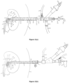
- FIG. 3( a ) is an illustration of an existing renal sheath based operative gadgets and its assembly with respect to the renal sheath involved in the operative procedure;
- FIG. 3( b ) is another illustration of an existing renal sheath based operative gadgets and its assembly with respect to the renal sheath involved in the operative procedure;
- FIG. 4 is an illustration of a sheath assembly sheath based operative gadgets and its assembly with respect to the sheath assembly in accordance with the present invention for different fields of endoscopic surgery involving suction, irrigation and material removal involved in the operative procedure.
- FIG. 5 illustrates the flow of fluid & pressure kinetics in renal pelvis during normal circumstances
- FIG. 6 is an illustration of open ended multiple side holes based ureteric catheter in accordance with another aspect of the present invention.
- FIG. 1 illustrates a conventional renal sheath which is basically a hollow tube of uniform diameter. Usually, the same is made of plastic or metal. The inner end can be straight or obliquely cut while the outer end is straight.
- Such a conventional renal sheath is known to be used in percutaneous renal surgery which has a big role to play in upper tract diseases and urolithiasis.
- a ureteric catheter is placed retrograde in the pelvis.
- a puncture is made in pelvicalyceal system (PCS).
- PCS pelvicalyceal system
- serial dilatation has problems of bleeding from the tract, loss of tract, slippage of the guide wire outside during dilatation and dilatation of the wrong tract.
- the renal sheath in form of hollow tube of uniform diameter as shown in accompanying FIGS. 1 a and 1 b is placed extending from skin to PCS.
- the renal sheath is a hollow tube of uniform diameter, made of plastic or metal.
- the inner end can be straight or obliquely cut.
- Outer end is straight. Both ends are open to allow free passage of fluid and endoscope.
- the renal sheath allows repeated entry of operating endoscope from outside body to PCS.
- Operating endoscope may have a channel for normal saline irrigation, to pass different forceps, suction cannula and energy probes. Renal sheath allows fluid to come out by the side of endoscope.
- suction cannula is passed via instrument channel of the operating endoscope to suck out fluid and fragments.
- size of suction cannula is small leading to aspiration of only small fragments.
- the sheath cannot be used for suction. If the sheath size is only marginally bigger than the diameter of the endoscope, all fluid cannot come out easily and intra renal pressure can rise and it may lead to different medical complications. In this scenario, stone fragments may not come out easily especially in dilated flabby pelvicalyceal system.
- FIGS. 2 a , 2 b and 2 c illustrate various sizes of sheaths suitable for use as a part of the sheath assembly in accordance with the present invention.
- the sheath comprises a tubular sheath ( 11 ) of uniform diameter of different length & width having an inner end and an outer end and provided with mechanism at the outer end to attach suction unit shown in FIGS. 2 ( d ) and 2 ( e ).
- the suction unit basically comprises of reservoir ( 1 ) with inlet ( 2 ), outlet tube ( 3 ), opening in reservoir to control the suction( 10 ) and sealing assembly ( FIG. 2 e ).
- the sealing assembly of FIG. 2( e ) attaches to reservoir ( 1 ). It has sealing silicon flap valve ( 4 ) on inner side and sealing cap on outside ( 5 ). Its main function is to allow passage of telescope without air or water leak. This makes the sheath assembly water and air tight for suction to work.
- FIG. 2 ( f ) illustrates a dilator cum obturator. It is a hollow tube of suitable diameter to fit inside sheath ( FIG. 2 a , 2 b , 2 c ). One end is tapered for a short distance of few mm (4 to 8) Inside diameter ( 11 ) is enough to allow passage of 0.038 inch guide wire.
- the tubular sheath ( 11 ) ( FIG. 2 a, b, c ) is attached to suction unit ( FIG. 2 d - 2 ) by screw and thread with O sealing ring.
- Suction unit is thus basically made of two parts ( FIG. 2 d , 2 e ). It is attached to each other by thread and screw mechanism ( 6 , 7 ) and O sealing ring ( 8 ). Reservoir outlet is connected to suction machine with suction tube ( 9 ).
- Operating endoscope is passed into sheath ( 11 ) ( FIG. 2 b ) via sealing cap ( 5 ) to silicon flap valve ( 4 ) to reservoir ( 1 ) to inlet ( 2 ).
- sheath is placed inside PCS, it is possible to go in and come out easily without losing tract.
- suction controlling opening ( 10 ) in the reservoir ( FIG. 2 d ) exactly opposite to outlet tube ( 3 ). Silicon suction tube ( 9 ) is attached to outlet ( 3 ) of reservoir. Other end of suction tube ( 9 ) is attached to suction machine. The suction machine is always on during the procedure.
- suction controlling opening ( 10 ) When the suction controlling opening ( 10 ) is open to air, there is no suction effect inside the sheath but water and dust can come out by overflowing from PCS but there is continuous suction from reservoir.
- suction controlling opening ( 10 ) is closed by finger, suction pressure is transmitted to PCS via sheath and fragments come out easily.
- sheath assembly of the invention which includes a suction unit to take out endoscope from sheath gradually giving a space in the sheath for big fragments to come out.
- suction unit it is possible to actually see the whole path taken by fragment while coming out. Once it has fallen into reservoir ( 1 ), there is no chance for fragment to go back into PCS due to design of suction unit.
- sheath of prior art is simply a tube open at both ends with no mechanism to attach suction device.
- the sheath is a tube, which is much smaller in diameter as compared to conventional sheath and outer end has a mechanism to attach suction unit. It has multiple advantages.
- suction unit is totally new construction previously not known. It includes a sealing assembly with silicon flap valve ( 4 ) used widely to provide sealing effect to reservoir.
- the silicon suction tube ( 9 ) is widely used silicon tube for various purposes.
- the tubular sheath ( 2 a , 2 b , 2 c ) of the sheath assembly of the invention can be preferably made of stainless steel. It can also be made of any material which is biocompatible, can withstand sterilization process, stiff enough and resistant to laser energy. It can be transparent or opaque. It can be of different in diameter and length.
- the mechanism to attach to suction unit is by screw and thread but it can be in any way which is user friendly and leak proof.
- Suction unit as mentioned hereinbefore and illustrated by way of accompanying FIGS. 2( d ) and 2( e ) has two parts.
- One part includes reservoir ( FIG. 2 d ) with inlet, opening to control suction power, outlet and mechanism to attach to sealing assembly.
- Inlet has to be at least equal to or more than the diameter of sheath.
- Outlet tube diameter has to be bigger than the inlet diameter.
- Mechanism to attach to sealing assembly is by screw and thread but it can be anything which is simple to operate and disassemble when needed.
- the whole piece can be preferably made of brass but it can also be of steel or any other material which can be used in surgical instrument.
- Sealing assembly houses a silicon flap valve which opens up when some instrument passes without damaging the instrument and prevents leakage of air and fluid with or without instrument in place. It can be replaced by any other mechanism which is effective and non-traumatic to optics of endoscope.
- Ureteric catheter with holes at a distance of one cm in first 10 cm is very useful when one injects air/contrast from below via ureteric catheter, contrast/air fill up whole upper ureter and renal pelvis simultaneously, even in presence of big/stag horn stone. Contrast/air do not have to overflow from one area to other areas of renal pelvis. This makes easy to decide which calyx to puncture and puncture the desired calyx. It is useful to provide retrograde flow of fluid in PCS and stops passage of fragments going down the ureter. As it provides continuous fluid in PCS, during suction the system is never completely collapsed preventing excessive suction on delicate mucosa. It is very essential in operation of percutaneous renal surgery.
- the sheath assembly of the present advancement can be used as detailed hereunder.
- a ureteric catheter with multiple holes in first 10 cm is passed in renal pelvicalyceal system (PCS) from urethra by doing cystoscopy.
- PCS renal pelvicalyceal system
- Suitable calyx is selected for desired route of entry in PCS and is punctured by initial puncture needle.
- a guide wire is passed through the needle in the PCS. Retrograde fluid via ureteric catheter is started. Over a guide wire, dilator cum obturator ( FIG. 2 f ) is passed up to PCS.
- sheath of 10/12 F size (3.66-4 mm diameter) of suitable length (12, 16 or 20 cm) is passed up to PCS. This way dilatation becomes a single step procedure. It prevents bleeding from tract, loss of tract & makes procedure faster and safer.
- Suction unit FIG. 2 d , 2 e
- Operating endoscope of suitable size is passed via sealing cap ( 5 ) into the suction unit to pass thorough tubular sheath ( FIG. 2 b ) to reach the PCS. Entire PCS is visualized.
- Operating endoscope has channel for flow of normal saline fluid to continuously enter the PCS.
- Same channel is used for passage of laser fiber (Holmium Yag), biprong forceps or triprong forceps or similar rigid instrument for different action inside PCS.
- Stone is located and laser energy is applied on stone by touching the stone by laser fiber (200 to 600 micron size). Laser energy is absorbed by stone and stone is broken into small pieces and dust. These dust and fragments disturbs the vision and these have to come out of body.
- the sheath assembly of the present invention has suction attached to it and it is continuously on.
- suction controlling opening ( 10 ) in reservoir. If the surgeon closes the opening with a finger, suction is applied to tubular sheath and hence inside the PCS.
- Suction activated during laser lithotripsy sucks all dust and fragments from PCS to inside of sheath ( FIG. 2 b ). Once sucked into tubular sheath ( 2 a / 2 b / 2 c ), it enters the reservoir ( 1 ) and goes out via suction tube ( 9 ) to suction machine bottle.
- the mechanism of reservoir ( 1 ) is such that once the fragments have entered the reservoir, they cannot go back in the sheath. Fragments have only one way to go and that is outlet tube.
- the suction controlling opening exactly opposite to outlet tube is open to air.
- suction When suction is on, there is negative suction in outlet tube. It will suck all fluid and fragments from reservoir preventing spillage outside but will not transmit suction pressure inside the sheath and PCS/body cavity.
- suction pressure If operating surgeon wants to have suction pressure inside the sheath and hence inside the body, it is possible to just have to close the opening by a finger.
- the opening is placed at an ergonomically convenient position. If on the other hand the operating surgeon does not want suction inside the body, the finger is lifted up from the opening allowing it to communicate with air.
- the surgeon can take the inner end of sheath near the fragments and suction can be applied to suck all fragments. This way all the fragments from the PCS can be removed under vision. Moreover, in case fragment or stone moves away from endoscope due to fluid from endoscope, suction is activated to bring it back towards endoscope. This way migration of stone and fragments in the distant calyx or ureter is also prevented. Complete inspection of PCS is done. Once it is confirmed that every possible stone is removed, the sheath is taken out without leaving any nephrostomy tube in the tract. As the tract size is very small, there is minimal chance of bleeding. There is less pain and early recovery.
- FIG. 3( a ) illustrates the manner of installation and use of the existing renal sheath in endoscopic surgery such as when surgeon wants to remove a stone/stones ( 3 ) from upper urinary tract/renal pelvis ( 22 ) by percutaneous surgery.
- the surgeon places an open ended ureteric catheter ( 1 ) in renal pelvis region ( 22 ).
- ureteric catheter has only a terminal open end ( 2 )
- all contrast/air has to go in a particular area of renal pelvis. Contrast overflows from that area to fill other parts of renal pelvis.
- a nephroscope ( 12 ) of 6 to 8 mm size is placed in renal pelvis ( 22 ) to visualize the stone ( 3 ).
- Fiber optic cable ( 16 ) is attached to light pillar ( 15 ) of nephroscope ( 12 ) to transmit light inside the renal pelvis ( 22 ).
- Endo vision camera ( 18 ) is attached to eye piece ( 17 ) of nephroscope to transmit images from inside the renal pelvis to outside body on a television monitor. Surgeon operates by seeing in the monitor.
- Normal saline/irrigation tubing ( 14 ) is attached to stop cock ( 13 ) of nephroscope to allow normal saline to enter ( 6 ) the renal pelvis ( 22 ) to keep vision clear in the renal pelvis.
- Various instruments like pneumatic probe, laser fiber, forceps & suction cannula ( 19 ) are used. Stone is broken into small pieces ( 4 ) by pneumatic probe/laser fiber.
- Suction cannula ( 19 ) is attached to a suction tubing ( 21 ) to connect to suction machine.
- Suction cannula ( 19 ) has a trumpet valve ( 20 ).
- Trumpet valve ( 20 ) controls suction applied in renal pelvis ( 22 ) to suck the fluid & stone fragments ( 8 ). Whatever normal saline is going in it has to come out ( 7 ) otherwise intra renal pressure will rise. Whatever fluid is going in comes out of renal pelvis ( 22 ) by the side of nephroscope ( 12 ) via renal sheath ( 11 ). All fluid drains out by overflowing ( 11 ) as outer end of sheath is open to air alongwith fluid, small stone fragments & dust ( 4 , 9 ) also coming out via renal sheath. Once stone is completely removed, nephrostomy (a tube from renal pelvis to outside body) is usually kept to control bleeding.
- nephrostomy a tube from renal pelvis to outside body
- a nephroscope ( 22 ) of 4 mm size is placed in renal pelvis to visualize the stone ( 3 ).
- Fiber optic cable ( 13 ) is attached to light pillar ( 12 ) of nephroscope ( 22 ) to transmit light inside the renal pelvis ( 21 ).
- Endo vision camera ( 15 ) is attached to eye piece ( 14 ) of nephroscope to transmit images from inside the renal pelvis to outside body on a television monitor. Surgeon operates by seeing in the monitor.
- Normal saline/irrigation tubing ( 20 ) is attached to stop cock ( 19 ) of nephroscope ( 22 ) to allow normal saline to enter ( 4 ) the renal pelvis to keep vision clear in the renal pelvis.
- FIG. 4 illustrates the advanced sheath based operative gadgets and its assembly in accordance with the present invention such as may be applied to remove a stone/stones ( 4 ) from upper urinary tract/renal pelvis ( 29 ) by percutaneous surgery.
- ureteric catheter 1
- first 10 cms 2
- renal pelvis 29
- ureteric catheter has multiple opening in first 10 cm ( 2 )
- contrast/air fill up whole upper ureter and entire renal pelvis ( 29 ) simultaneously. Contrast does not overflow from one area to other parts of renal pelvis. All parts of renal pelvis are simultaneously filled even in big/staghorn stone.
- dilator cum obturator also shown in FIG. 2 f , is passed to dilate the tract as a single step to 3 to 3.3 mm size.
- Such small tract size is advantageously very less likely to be associated with problem of bleeding, adjacent organ injury, possible loss of tract & others.
- tubular sheath 11 in form of hollow tube of uniform diameter of 4 mm size (outer diameter) is placed extending from skin surface ( 10 ) to renal pelvis ( 29 ).
- Tubular sheath also shown in FIGS.
- FIG. 2 a , 2 b , 2 c & 11 is attached to suction unit (of FIG. 2 d , 2 e ) at inlet ( 30 ) of reservoir ( 17 ).
- a nephroscope ( 18 ) of 2.6 mm size is passed into the renal pelvis ( 29 ) via sealing cap ( 16 ) to silicon flap valve ( 15 ) to reservoir ( 17 ) to inlet ( 30 ) of reservoir to tubular sheath ( 11 ).
- Suction tube ( 14 ) is attached to outlet ( 13 ) of reservoir ( 17 ).
- Suction tube ( 14 ) is attached to suction machine. Suction machine is always on during the procedure.
- Fiber optic cable ( 20 ) is attached to light pillar ( 19 ) of nephroscope ( 27 ) to transmit light inside the renal pelvis ( 29 ).
- Endo vision camera ( 26 ) is attached to eye piece ( 25 ) of nephroscope to transmit images from inside the renal pelvis to outside body on a television monitor. Surgeon operates by seeing in the monitor.
- Normal saline/irrigation tubing ( 22 ) is attached to stop cock ( 21 ) of nephroscope ( 18 ) to allow normal saline ( 28 ) to enter through ( 6 ) the renal pelvis ( 29 ) to keep vision clear in the renal pelvis.
- Various instruments like pneumatic probe, laser fiber & forceps etc. ( 24 ) are used.
- Stone ( 4 ) is broken into small pieces ( 5 ) by pneumatic probe/laser fiber ( 24 ). Small fragments and fluid can be sucked out through ( 7 ) from renal pelvis ( 29 ) by closing the suction control opening ( 12 ) with the finger.
- the construction of the sheath assembly suitable for endoscopic surgery in accordance with the present advancement involving the reservoir and the suction control opening located at reservoir advantageously divide the sheath assembly/unit into two distinctly separable communication zones and the opening decides whether suction communication zones should be limited to the reservoir or extend to the cannula and reservoir both.
- This is unique and benefits a lot both for the suction from the cannula and also from the reservoir alone and has contributes dramatically towards providing the operating surgeon with a user friendly and operation friendly system to clear up the particles and fluid from the operation site as well as from the operating pathway/device.
- the construction of the sheath assembly has enabled the following advantages:
- FIG. 5 illustrates what usually happens normally as far as flow of fluid & pressure kinetics in renal pelvis is concerned.
- urine produced by kidney comes in renal pelvis ( 4 ) from multiple calyces ( 1 ) like streams of water ( 2 ) forming a river ( 3 ).
- Urine enters ureter ( 6 ) via ureteropelvic junction ( 5 ) due to rise in pressure in renal pelvis due to continuous entry of urine from calyces.
- Urine is carried down by wave of peristalsis from renal pelvis ( 4 ) to ureter ( 6 ) to urinary bladder ( 7 ).
- small stone and crystals also go down the ureter & then in the urinary bladder in normal individual. If a stone blocks the lumen of ureter ( 6 ), person develops pain of stone disease.
- FIG. 3 a the same illustrates what is usually done in conventional percutaneous renal surgery.
- FIG. 3 b is again an illustration of what happens in smaller tract size.
- FIG. 6 shows an open ended multihole ureteric catheter in accordance with the present invention.
- the ureteric catheter has a terminal open end ( 1 ) with markers ( 3 ) at equidistance to know how much catheter is inserted. It is radiopaque and other open end ( 5 ) with a uniform inner diameter to allow fluid to go in & come out.
- the extra feature added is multiple holes ( 2 ) in first few centimetres of ureteric catheter ( 4 ).
- Ureteric catheter is placed in such a way that few holes are in ureter & few are in renal pelvis.
- this arrangement of hole ensures free flow of saline from ureter to renal pelvis in all areas of pelvis where there is hole.
- ureteric catheter Multiple holes in ureteric catheter in first few centimetres is all what is needed.
- the placement of ureteric catheter should be such that few holes should be in ureter & few in renal pelvis.
- the advancement of the invention is aimed for redefining the way surgeons treat kidney stones and carry out other endoscopic surgery where irrigation of fluid is required. It provides easy removal of return fluid under our control and help to control the body cavity fluid& pressure dynamics. It shifts the suction mechanism from operating surgeon holding endoscope to assistant holding the sheath. It immeasurably improves the comforts of operating surgeon. It allows us to use smaller tract size to operate in closed body cavity. It makes procedure of endoscopy much safer, faster and effective.
Landscapes
- Health & Medical Sciences (AREA)
- Life Sciences & Earth Sciences (AREA)
- Surgery (AREA)
- Heart & Thoracic Surgery (AREA)
- General Health & Medical Sciences (AREA)
- Public Health (AREA)
- Animal Behavior & Ethology (AREA)
- Engineering & Computer Science (AREA)
- Biomedical Technology (AREA)
- Veterinary Medicine (AREA)
- Molecular Biology (AREA)
- Nuclear Medicine, Radiotherapy & Molecular Imaging (AREA)
- Medical Informatics (AREA)
- Pathology (AREA)
- Radiology & Medical Imaging (AREA)
- Physics & Mathematics (AREA)
- Optics & Photonics (AREA)
- Biophysics (AREA)
- Anesthesiology (AREA)
- Hematology (AREA)
- Pulmonology (AREA)
- Vascular Medicine (AREA)
- Orthopedic Medicine & Surgery (AREA)
- Endoscopes (AREA)
- External Artificial Organs (AREA)
- Surgical Instruments (AREA)
Applications Claiming Priority (3)
| Application Number | Priority Date | Filing Date | Title |
|---|---|---|---|
| IN3119/MUM/2014 | 2014-09-30 | ||
| IN3119MU2014 | 2014-09-30 | ||
| PCT/IN2014/000785 WO2016051421A1 (fr) | 2014-09-30 | 2014-12-19 | Ensemble gaine et cathéter à trous multiples pour différents domaines de chirurgie endoscopique concernant une aspiration, une irrigation et un enlèvement de matière |
Publications (2)
| Publication Number | Publication Date |
|---|---|
| US20180289394A1 US20180289394A1 (en) | 2018-10-11 |
| US10582949B2 true US10582949B2 (en) | 2020-03-10 |
Family
ID=55629532
Family Applications (1)
| Application Number | Title | Priority Date | Filing Date |
|---|---|---|---|
| US15/325,878 Active 2035-12-31 US10582949B2 (en) | 2014-09-30 | 2014-12-19 | Sheath assembly and multihole catheter for different fields of endoscopic surgery involving suction, irrigation and material removal |
Country Status (4)
| Country | Link |
|---|---|
| US (1) | US10582949B2 (fr) |
| EP (1) | EP3200711A4 (fr) |
| CN (1) | CN106604687B (fr) |
| WO (1) | WO2016051421A1 (fr) |
Families Citing this family (45)
| Publication number | Priority date | Publication date | Assignee | Title |
|---|---|---|---|---|
| US12064567B2 (en) * | 2015-07-20 | 2024-08-20 | Roivios Limited | Percutaneous urinary catheter |
| EP3399924A4 (fr) * | 2016-01-04 | 2019-11-27 | Endovate LLC | Dispositif de surtube et procédé d'utilisation |
| EP3407771B1 (fr) * | 2016-01-29 | 2025-12-31 | Boston Scientific Scimed, Inc. | Fixation pour un urétéroscope |
| CN109069797A (zh) * | 2016-04-26 | 2018-12-21 | 波士顿科学有限公司 | 具有可调节深度止动件的经皮进入装置 |
| CN107080569A (zh) * | 2017-06-01 | 2017-08-22 | 庞广富 | 经尿道输尿管硬性对流负压通道碎石清石器 |
| EP3684438B1 (fr) * | 2017-12-08 | 2024-09-11 | Auris Health, Inc. | Systèmes de fluides dirigés |
| KR102731868B1 (ko) | 2018-02-09 | 2024-11-19 | 자이러스 에이씨엠아이, 인코포레이티드 디.비.에이. 올림푸스 써지컬 테크놀러지스 아메리카 | 의료용 레이저 장치 및 시스템 |
| JP2021166563A (ja) * | 2018-05-15 | 2021-10-21 | オリンパス株式会社 | 光学的治療装置及び光学的治療方法 |
| US12653614B2 (en) | 2018-05-24 | 2026-06-16 | The Regents Of The University Of California | Modular wireless large bore vacuum universal endoscope and vacuumscope |
| ES3000010T3 (en) | 2018-08-13 | 2025-02-27 | Inari Medical Inc | System for treating embolism and associated devices and methods |
| US10881280B2 (en) | 2018-08-24 | 2021-01-05 | Auris Health, Inc. | Manually and robotically controllable medical instruments |
| US10765487B2 (en) | 2018-09-28 | 2020-09-08 | Auris Health, Inc. | Systems and methods for docking medical instruments |
| CN119924988A (zh) | 2018-09-28 | 2025-05-06 | 奥瑞斯健康公司 | 用于伴随内窥镜和经皮医学规程的机器人系统和方法 |
| US12048595B2 (en) | 2018-10-26 | 2024-07-30 | Eureka Inventions Limited | Surgical apparatus and method |
| KR102108736B1 (ko) * | 2018-12-13 | 2020-05-08 | 대한민국 | 반추동물의 음경 흡입 장치 |
| WO2020140072A1 (fr) | 2018-12-28 | 2020-07-02 | Auris Health, Inc. | Gaine percutanée pour méthodes et systèmes médicaux robotiques |
| EP4485120A3 (fr) * | 2019-01-18 | 2025-03-26 | IPG Photonics Corporation | Poignée de direction à collecteur multicanal |
| EP3911258A4 (fr) * | 2019-01-18 | 2022-11-16 | IPG Photonics Corporation | Instrument endoscopique multifonctionnel efficace |
| KR20220050151A (ko) | 2019-08-15 | 2022-04-22 | 아우리스 헬스, 인코포레이티드 | 다수의 굽힘 섹션을 갖는 의료 장치 |
| CN112401971B (zh) * | 2019-08-23 | 2025-09-09 | 贝克顿·迪金森公司 | 为经皮肾镜取石术外科手术设计的套件 |
| WO2021137072A1 (fr) | 2019-12-31 | 2021-07-08 | Auris Health, Inc. | Identification et ciblage d'éléments anatomiques |
| WO2021137109A1 (fr) | 2019-12-31 | 2021-07-08 | Auris Health, Inc. | Techniques d'alignement pour un accès percutané |
| KR20260020213A (ko) | 2019-12-31 | 2026-02-10 | 아우리스 헬스, 인코포레이티드 | 경피 접근을 위한 정렬 인터페이스 |
| JP7537785B2 (ja) | 2020-01-29 | 2024-08-21 | ボスイーグル サージカル インコーポレイテッド | 内視鏡手術用ロボットシステム |
| CN111282136B (zh) * | 2020-02-27 | 2021-07-13 | 付强 | 肾内科导尿管系统 |
| US11737663B2 (en) | 2020-03-30 | 2023-08-29 | Auris Health, Inc. | Target anatomical feature localization |
| CN111449617B (zh) * | 2020-04-10 | 2023-07-25 | 西安交通大学医学院第二附属医院 | 带有抽吸功能的膀胱镜 |
| WO2021220107A1 (fr) * | 2020-04-27 | 2021-11-04 | Auris Health, Inc. | Gestion de fluide spécifique à un cas |
| US11883041B2 (en) | 2020-04-30 | 2024-01-30 | Michael Alesi | Catheter for thrombus removal |
| US12268363B2 (en) * | 2020-05-01 | 2025-04-08 | Intuitive Surgical Operations, Inc. | Steerable apparatus having a slideable imaging probe and associated methods of use |
| AU2021283979A1 (en) | 2020-06-05 | 2023-01-19 | Stryker Corporation | Recapturable funnel catheters, and associated systems and methods |
| CN112401826B (zh) * | 2020-09-06 | 2025-07-22 | 温州医科大学附属第二医院、温州医科大学附属育英儿童医院 | 关节镜手术用吸引套件 |
| CN116234510B (zh) * | 2020-09-30 | 2025-03-25 | 奥瑞斯健康公司 | 机器人器械对准 |
| DE102020125697A1 (de) | 2020-10-01 | 2022-04-07 | Urotech Gmbh | Amplatzhülse und Dilatationssystem |
| US20220240980A1 (en) * | 2021-02-03 | 2022-08-04 | Chin-Piao Chang | Endoscope Combined with Injection Needle |
| CN113081163B (zh) * | 2021-04-07 | 2022-04-15 | 中南大学湘雅二医院 | 一种微创型尿路结石手术综合系统 |
| CN113289217B (zh) * | 2021-07-06 | 2025-04-04 | 江苏伊凯医疗器械有限公司 | 一次性双腔输尿管导管 |
| CN113768591A (zh) * | 2021-09-09 | 2021-12-10 | 遵义医科大学附属医院 | 一种可调控肾内压的经皮肾镜穿刺鞘 |
| JP2025503965A (ja) | 2022-02-08 | 2025-02-06 | イナリ メディカル, インコーポレイテッド | 拡張器係止機構を有する凝塊治療システム、並びに関連するデバイス及び方法 |
| CN119562793A (zh) * | 2022-06-30 | 2025-03-04 | 伊纳里医疗有限公司 | 经皮治疗胆石症的设备、方法及系统 |
| CN116898389B (zh) * | 2023-09-12 | 2023-11-28 | 湖南英术生命科技有限公司 | 一种关节镜组件 |
| US12558486B2 (en) | 2023-11-16 | 2026-02-24 | Inari Medical, Inc. | Automatic locking and unlocking vacuum syringes, and associated systems and methods |
| US12465382B1 (en) | 2024-05-10 | 2025-11-11 | Inari Medical, Inc. | Mechanical thrombectomy assemblies with relief features, and associated devices, systems, and methods |
| KR20260026157A (ko) * | 2024-08-19 | 2026-02-26 | 고신대학교 산학협력단 | 말초 기관지 병변 처치를 위한 가이드 시스의 사이드 튜브 |
| CN118948177B (zh) * | 2024-10-16 | 2025-03-11 | 湖南省华芯医疗器械有限公司 | 一种前端组件、引导鞘及置入装置 |
Citations (26)
| Publication number | Priority date | Publication date | Assignee | Title |
|---|---|---|---|---|
| US4935006A (en) * | 1987-11-12 | 1990-06-19 | Hasson Harrith M | Suction and irrigation device with right angle and oblique openings |
| US4955895A (en) * | 1986-12-23 | 1990-09-11 | Terumo Kabushiki Kaisha | Vasodilating catheter |
| US5047045A (en) * | 1989-04-13 | 1991-09-10 | Scimed Life Systems, Inc. | Multi-section coaxial angioplasty catheter |
| US5207648A (en) * | 1990-12-14 | 1993-05-04 | The Kendall Company | Multilumen catheter |
| US5250059A (en) * | 1992-01-22 | 1993-10-05 | Devices For Vascular Intervention, Inc. | Atherectomy catheter having flexible nose cone |
| US5458574A (en) * | 1994-03-16 | 1995-10-17 | Heartport, Inc. | System for performing a cardiac procedure |
| US5575756A (en) * | 1993-08-16 | 1996-11-19 | Olympus Optical Co., Ltd. | Endoscope apparatus |
| US5637075A (en) | 1994-04-25 | 1997-06-10 | Hamamatsu Ent Surgicenter | Apparatus for observing inside of body cavity |
| US20010016751A1 (en) * | 1992-04-07 | 2001-08-23 | Scott O. Trerotola | Percutaneous mechanical fragmentation catheter system |
| US20020072651A1 (en) * | 1997-04-01 | 2002-06-13 | George A. Vilos | Debris aspirating resectoscope |
| US20030097133A1 (en) * | 2001-11-21 | 2003-05-22 | Green James M. | Attachable/detachable reaming head for surgical reamer |
| US20030130565A1 (en) * | 2002-01-07 | 2003-07-10 | Muller Richard P. | Outflow system for an endoscope |
| US20030144594A1 (en) * | 2002-01-31 | 2003-07-31 | Gellman Barry N. | Needle device |
| US20050020966A1 (en) * | 2002-07-22 | 2005-01-27 | Holger Soring | Medical treatment apparatus |
| US20050059931A1 (en) * | 2003-09-16 | 2005-03-17 | Venomatrix | Methods and apparatus for localized and semi-localized drug delivery |
| US20050234298A1 (en) * | 2004-01-29 | 2005-10-20 | Cannuflow Incorporated | Atraumatic arthroscopic instrument sheath |
| US20060129091A1 (en) * | 2004-12-10 | 2006-06-15 | Possis Medical, Inc. | Enhanced cross stream mechanical thrombectomy catheter with backloading manifold |
| US20060229645A1 (en) * | 2005-04-07 | 2006-10-12 | Possis Medical, Inc. | Cross stream thrombectomy catheter with flexible and expandable cage |
| US20060266423A1 (en) * | 2005-05-25 | 2006-11-30 | Fujinon Corporation | Water feeding device for endoscope |
| US20060276692A1 (en) * | 2005-06-01 | 2006-12-07 | Cannuflow Incorporated | Protective cap for arthroscopic instruments |
| US20070118072A1 (en) * | 1996-07-26 | 2007-05-24 | Kensey Nash Corporation | System and Method of Use for Revascularizing Stenotic Bypass Grafts and Other Blood Vessels |
| US20070184717A1 (en) * | 2004-08-04 | 2007-08-09 | Masanori Hamazaki | Endoscope plug body |
| US20080208004A1 (en) * | 2005-08-24 | 2008-08-28 | Tsutomu Okada | Endoscope system |
| US20090069829A1 (en) * | 2005-05-26 | 2009-03-12 | Leonid Shturman | Rotational Atherectomy Device with Distal Protection Capability and Method of Use |
| US20090275893A1 (en) * | 2006-01-05 | 2009-11-05 | Dibiasio Christopher | Instrument port for minimally invasive cardiac surgery |
| US20090312696A1 (en) * | 2005-12-28 | 2009-12-17 | Copa Vincent G | Devices, Systems, and Related Methods for Delivery of Fluid to Tissue |
Family Cites Families (6)
| Publication number | Priority date | Publication date | Assignee | Title |
|---|---|---|---|---|
| CN2284060Y (zh) * | 1996-11-07 | 1998-06-17 | 刘武 | 可控式眼内硅油吸引装置 |
| CN2640506Y (zh) * | 2003-06-13 | 2004-09-15 | 宋修军 | 套管式胸腔穿刺针 |
| US7150713B2 (en) * | 2003-10-16 | 2006-12-19 | Smith & Nephew, Inc. | Endoscopic device |
| US20050096627A1 (en) * | 2003-11-03 | 2005-05-05 | Howard Mark E. | Fluid aspiration device |
| CN202505943U (zh) * | 2012-03-27 | 2012-10-31 | 何朝辉 | 一体化肾输尿管导管 |
| CN103356151A (zh) * | 2013-06-28 | 2013-10-23 | 苏州联科盛世科技有限公司 | 多用途的一次性宫腔诊疗内窥镜 |
-
2014
- 2014-12-19 EP EP14902929.0A patent/EP3200711A4/fr not_active Withdrawn
- 2014-12-19 CN CN201480081771.6A patent/CN106604687B/zh active Active
- 2014-12-19 US US15/325,878 patent/US10582949B2/en active Active
- 2014-12-19 WO PCT/IN2014/000785 patent/WO2016051421A1/fr not_active Ceased
Patent Citations (27)
| Publication number | Priority date | Publication date | Assignee | Title |
|---|---|---|---|---|
| US4955895A (en) * | 1986-12-23 | 1990-09-11 | Terumo Kabushiki Kaisha | Vasodilating catheter |
| US4935006A (en) * | 1987-11-12 | 1990-06-19 | Hasson Harrith M | Suction and irrigation device with right angle and oblique openings |
| US5047045A (en) * | 1989-04-13 | 1991-09-10 | Scimed Life Systems, Inc. | Multi-section coaxial angioplasty catheter |
| US5207648A (en) * | 1990-12-14 | 1993-05-04 | The Kendall Company | Multilumen catheter |
| US5250059A (en) * | 1992-01-22 | 1993-10-05 | Devices For Vascular Intervention, Inc. | Atherectomy catheter having flexible nose cone |
| US20010016751A1 (en) * | 1992-04-07 | 2001-08-23 | Scott O. Trerotola | Percutaneous mechanical fragmentation catheter system |
| US5575756A (en) * | 1993-08-16 | 1996-11-19 | Olympus Optical Co., Ltd. | Endoscope apparatus |
| US5458574A (en) * | 1994-03-16 | 1995-10-17 | Heartport, Inc. | System for performing a cardiac procedure |
| US5637075A (en) | 1994-04-25 | 1997-06-10 | Hamamatsu Ent Surgicenter | Apparatus for observing inside of body cavity |
| US20070118072A1 (en) * | 1996-07-26 | 2007-05-24 | Kensey Nash Corporation | System and Method of Use for Revascularizing Stenotic Bypass Grafts and Other Blood Vessels |
| US20020072651A1 (en) * | 1997-04-01 | 2002-06-13 | George A. Vilos | Debris aspirating resectoscope |
| US20030097133A1 (en) * | 2001-11-21 | 2003-05-22 | Green James M. | Attachable/detachable reaming head for surgical reamer |
| US20030130565A1 (en) * | 2002-01-07 | 2003-07-10 | Muller Richard P. | Outflow system for an endoscope |
| US20030144594A1 (en) * | 2002-01-31 | 2003-07-31 | Gellman Barry N. | Needle device |
| US20050020966A1 (en) * | 2002-07-22 | 2005-01-27 | Holger Soring | Medical treatment apparatus |
| US20050059931A1 (en) * | 2003-09-16 | 2005-03-17 | Venomatrix | Methods and apparatus for localized and semi-localized drug delivery |
| US20050234298A1 (en) * | 2004-01-29 | 2005-10-20 | Cannuflow Incorporated | Atraumatic arthroscopic instrument sheath |
| US20120215067A1 (en) * | 2004-01-29 | 2012-08-23 | Cannuflow, Inc. | Atraumatic Arthroscopic Instrument Sheath |
| US20070184717A1 (en) * | 2004-08-04 | 2007-08-09 | Masanori Hamazaki | Endoscope plug body |
| US20060129091A1 (en) * | 2004-12-10 | 2006-06-15 | Possis Medical, Inc. | Enhanced cross stream mechanical thrombectomy catheter with backloading manifold |
| US20060229645A1 (en) * | 2005-04-07 | 2006-10-12 | Possis Medical, Inc. | Cross stream thrombectomy catheter with flexible and expandable cage |
| US20060266423A1 (en) * | 2005-05-25 | 2006-11-30 | Fujinon Corporation | Water feeding device for endoscope |
| US20090069829A1 (en) * | 2005-05-26 | 2009-03-12 | Leonid Shturman | Rotational Atherectomy Device with Distal Protection Capability and Method of Use |
| US20060276692A1 (en) * | 2005-06-01 | 2006-12-07 | Cannuflow Incorporated | Protective cap for arthroscopic instruments |
| US20080208004A1 (en) * | 2005-08-24 | 2008-08-28 | Tsutomu Okada | Endoscope system |
| US20090312696A1 (en) * | 2005-12-28 | 2009-12-17 | Copa Vincent G | Devices, Systems, and Related Methods for Delivery of Fluid to Tissue |
| US20090275893A1 (en) * | 2006-01-05 | 2009-11-05 | Dibiasio Christopher | Instrument port for minimally invasive cardiac surgery |
Non-Patent Citations (1)
| Title |
|---|
| International Search Report for related application No. PCT/IN14/000785 dated Oct. 16, 2015. |
Also Published As
| Publication number | Publication date |
|---|---|
| CN106604687B (zh) | 2019-08-16 |
| US20180289394A1 (en) | 2018-10-11 |
| EP3200711A4 (fr) | 2018-06-27 |
| WO2016051421A1 (fr) | 2016-04-07 |
| CN106604687A (zh) | 2017-04-26 |
| EP3200711A1 (fr) | 2017-08-09 |
Similar Documents
| Publication | Publication Date | Title |
|---|---|---|
| US20180289394A1 (en) | Sheath assembly and multihole catheter for different fields of endoscopic surgery involving suction, irrigation and material removal | |
| US12178459B2 (en) | Suction evacuation device | |
| EP3422964B1 (fr) | Gaine d'évacuation par aspiration | |
| US11944329B2 (en) | Suction evacuation device | |
| US10828050B2 (en) | Suction evacuation device | |
| US10071191B2 (en) | Nasal fluid management device | |
| US10391209B2 (en) | System and method for guided removal from an in vivo subject | |
| WO2017132371A1 (fr) | Dispositif médical et procédés d'utilisation | |
| US20140276625A1 (en) | Nasal fluid management device | |
| US20210022759A1 (en) | Suction evacuation device | |
| US20070051380A1 (en) | System, kit, and method of transgastric removal of visceral fat and other related methods | |
| Sathaye | Per-urethral endoscopic management of bladder stones: does size matter? | |
| US20250041503A1 (en) | Suction evacuation device | |
| Mills et al. | Minimally Invasive Management of Calculi in Augmented/Neuropathic Bladders | |
| ES2562530B1 (es) | Dispositivo de introducción, guiado y fijación de instrumental quirúrgico percutáneo | |
| TWM605102U (zh) | 負壓吸引裝置 | |
| Delvecchio | Access to the ureter: Rigid ureteroscopy | |
| Traxer | Flexible Ureterorenoscopy: Tips and Tricks |
Legal Events
| Date | Code | Title | Description |
|---|---|---|---|
| STPP | Information on status: patent application and granting procedure in general |
Free format text: RESPONSE TO NON-FINAL OFFICE ACTION ENTERED AND FORWARDED TO EXAMINER |
|
| STPP | Information on status: patent application and granting procedure in general |
Free format text: NON FINAL ACTION MAILED |
|
| STPP | Information on status: patent application and granting procedure in general |
Free format text: RESPONSE TO NON-FINAL OFFICE ACTION ENTERED AND FORWARDED TO EXAMINER |
|
| STPP | Information on status: patent application and granting procedure in general |
Free format text: NOTICE OF ALLOWANCE MAILED -- APPLICATION RECEIVED IN OFFICE OF PUBLICATIONS |
|
| STPP | Information on status: patent application and granting procedure in general |
Free format text: PUBLICATIONS -- ISSUE FEE PAYMENT RECEIVED |
|
| STCF | Information on status: patent grant |
Free format text: PATENTED CASE |
|
| MAFP | Maintenance fee payment |
Free format text: PAYMENT OF MAINTENANCE FEE, 4TH YR, SMALL ENTITY (ORIGINAL EVENT CODE: M2551); ENTITY STATUS OF PATENT OWNER: SMALL ENTITY Year of fee payment: 4 |