US11193943B2 - Endothelial cell derived exosomes and uses thereof - Google Patents
Endothelial cell derived exosomes and uses thereof Download PDFInfo
- Publication number
- US11193943B2 US11193943B2 US15/703,955 US201715703955A US11193943B2 US 11193943 B2 US11193943 B2 US 11193943B2 US 201715703955 A US201715703955 A US 201715703955A US 11193943 B2 US11193943 B2 US 11193943B2
- Authority
- US
- United States
- Prior art keywords
- endothelial cell
- vcam
- biomarkers
- biological sample
- derived
- Prior art date
- Legal status (The legal status is an assumption and is not a legal conclusion. Google has not performed a legal analysis and makes no representation as to the accuracy of the status listed.)
- Active
Links
Images


Classifications
-
- G—PHYSICS
- G01—MEASURING; TESTING
- G01N—INVESTIGATING OR ANALYSING MATERIALS BY DETERMINING THEIR CHEMICAL OR PHYSICAL PROPERTIES
- G01N33/00—Investigating or analysing materials by specific methods not covered by groups G01N1/00 - G01N31/00
- G01N33/48—Biological material, e.g. blood, urine; Haemocytometers
- G01N33/50—Chemical analysis of biological material, e.g. blood, urine; Testing involving biospecific ligand binding methods; Immunological testing
- G01N33/68—Chemical analysis of biological material, e.g. blood, urine; Testing involving biospecific ligand binding methods; Immunological testing involving proteins, peptides or amino acids
- G01N33/6893—Chemical analysis of biological material, e.g. blood, urine; Testing involving biospecific ligand binding methods; Immunological testing involving proteins, peptides or amino acids related to diseases not provided for elsewhere
-
- G—PHYSICS
- G01—MEASURING; TESTING
- G01N—INVESTIGATING OR ANALYSING MATERIALS BY DETERMINING THEIR CHEMICAL OR PHYSICAL PROPERTIES
- G01N33/00—Investigating or analysing materials by specific methods not covered by groups G01N1/00 - G01N31/00
- G01N33/48—Biological material, e.g. blood, urine; Haemocytometers
- G01N33/50—Chemical analysis of biological material, e.g. blood, urine; Testing involving biospecific ligand binding methods; Immunological testing
- G01N33/53—Immunoassay; Biospecific binding assay; Materials therefor
- G01N33/573—Immunoassay; Biospecific binding assay; Materials therefor for enzymes or isoenzymes
-
- G—PHYSICS
- G01—MEASURING; TESTING
- G01N—INVESTIGATING OR ANALYSING MATERIALS BY DETERMINING THEIR CHEMICAL OR PHYSICAL PROPERTIES
- G01N33/00—Investigating or analysing materials by specific methods not covered by groups G01N1/00 - G01N31/00
- G01N33/48—Biological material, e.g. blood, urine; Haemocytometers
- G01N33/50—Chemical analysis of biological material, e.g. blood, urine; Testing involving biospecific ligand binding methods; Immunological testing
- G01N33/86—Chemical analysis of biological material, e.g. blood, urine; Testing involving biospecific ligand binding methods; Immunological testing involving blood coagulating time or factors, or their receptors
-
- G—PHYSICS
- G01—MEASURING; TESTING
- G01N—INVESTIGATING OR ANALYSING MATERIALS BY DETERMINING THEIR CHEMICAL OR PHYSICAL PROPERTIES
- G01N2333/00—Assays involving biological materials from specific organisms or of a specific nature
- G01N2333/435—Assays involving biological materials from specific organisms or of a specific nature from animals; from humans
- G01N2333/475—Assays involving growth factors
- G01N2333/49—Platelet-derived growth factor [PDGF]
-
- G—PHYSICS
- G01—MEASURING; TESTING
- G01N—INVESTIGATING OR ANALYSING MATERIALS BY DETERMINING THEIR CHEMICAL OR PHYSICAL PROPERTIES
- G01N2333/00—Assays involving biological materials from specific organisms or of a specific nature
- G01N2333/435—Assays involving biological materials from specific organisms or of a specific nature from animals; from humans
- G01N2333/575—Hormones
- G01N2333/60—Growth-hormone releasing factors (GH-RF) (Somatoliberin)
-
- G—PHYSICS
- G01—MEASURING; TESTING
- G01N—INVESTIGATING OR ANALYSING MATERIALS BY DETERMINING THEIR CHEMICAL OR PHYSICAL PROPERTIES
- G01N2333/00—Assays involving biological materials from specific organisms or of a specific nature
- G01N2333/435—Assays involving biological materials from specific organisms or of a specific nature from animals; from humans
- G01N2333/705—Assays involving receptors, cell surface antigens or cell surface determinants
- G01N2333/70503—Immunoglobulin superfamily, e.g. VCAMs, PECAM, LFA-3
-
- G—PHYSICS
- G01—MEASURING; TESTING
- G01N—INVESTIGATING OR ANALYSING MATERIALS BY DETERMINING THEIR CHEMICAL OR PHYSICAL PROPERTIES
- G01N2333/00—Assays involving biological materials from specific organisms or of a specific nature
- G01N2333/435—Assays involving biological materials from specific organisms or of a specific nature from animals; from humans
- G01N2333/705—Assays involving receptors, cell surface antigens or cell surface determinants
- G01N2333/70503—Immunoglobulin superfamily, e.g. VCAMs, PECAM, LFA-3
- G01N2333/70542—CD106
-
- G—PHYSICS
- G01—MEASURING; TESTING
- G01N—INVESTIGATING OR ANALYSING MATERIALS BY DETERMINING THEIR CHEMICAL OR PHYSICAL PROPERTIES
- G01N2333/00—Assays involving biological materials from specific organisms or of a specific nature
- G01N2333/435—Assays involving biological materials from specific organisms or of a specific nature from animals; from humans
- G01N2333/705—Assays involving receptors, cell surface antigens or cell surface determinants
- G01N2333/70596—Molecules with a "CD"-designation not provided for elsewhere in G01N2333/705
-
- G—PHYSICS
- G01—MEASURING; TESTING
- G01N—INVESTIGATING OR ANALYSING MATERIALS BY DETERMINING THEIR CHEMICAL OR PHYSICAL PROPERTIES
- G01N2333/00—Assays involving biological materials from specific organisms or of a specific nature
- G01N2333/435—Assays involving biological materials from specific organisms or of a specific nature from animals; from humans
- G01N2333/745—Assays involving non-enzymic blood coagulation factors
- G01N2333/755—Factors VIII, e.g. factor VIII C [AHF], factor VIII Ag [VWF]
-
- G—PHYSICS
- G01—MEASURING; TESTING
- G01N—INVESTIGATING OR ANALYSING MATERIALS BY DETERMINING THEIR CHEMICAL OR PHYSICAL PROPERTIES
- G01N2333/00—Assays involving biological materials from specific organisms or of a specific nature
- G01N2333/90—Enzymes; Proenzymes
- G01N2333/902—Oxidoreductases (1.)
- G01N2333/90245—Oxidoreductases (1.) acting on paired donors with incorporation of molecular oxygen (1.14)
- G01N2333/90248—Oxidoreductases (1.) acting on paired donors with incorporation of molecular oxygen (1.14) with NADH or NADPH as one of the donors, and incorporation of one atom of oxygen 1.14.13
- G01N2333/90251—Oxidoreductases (1.) acting on paired donors with incorporation of molecular oxygen (1.14) with NADH or NADPH as one of the donors, and incorporation of one atom of oxygen 1.14.13 with a definite EC number (1.14.13.-)
- G01N2333/90254—Nitric-oxide synthase (NOS; 1.14.13.39)
-
- G—PHYSICS
- G01—MEASURING; TESTING
- G01N—INVESTIGATING OR ANALYSING MATERIALS BY DETERMINING THEIR CHEMICAL OR PHYSICAL PROPERTIES
- G01N2800/00—Detection or diagnosis of diseases
- G01N2800/28—Neurological disorders
- G01N2800/2871—Cerebrovascular disorders, e.g. stroke, cerebral infarct, cerebral haemorrhage, transient ischemic event
-
- G—PHYSICS
- G01—MEASURING; TESTING
- G01N—INVESTIGATING OR ANALYSING MATERIALS BY DETERMINING THEIR CHEMICAL OR PHYSICAL PROPERTIES
- G01N2800/00—Detection or diagnosis of diseases
- G01N2800/32—Cardiovascular disorders
-
- G—PHYSICS
- G01—MEASURING; TESTING
- G01N—INVESTIGATING OR ANALYSING MATERIALS BY DETERMINING THEIR CHEMICAL OR PHYSICAL PROPERTIES
- G01N2800/00—Detection or diagnosis of diseases
- G01N2800/52—Predicting or monitoring the response to treatment, e.g. for selection of therapy based on assay results in personalised medicine; Prognosis
Definitions
- the present invention relates to endothelial cell biomarkers and diagnostic and prognostic methods for vascular diseases, including cardiovascular and cerebrovascular diseases.
- the invention also provides compositions for detecting endothelial cell biomarkers (e.g., endothelial cell-derived exosome biomarkers) as well as compositions and methods useful for treating vascular diseases (e.g., atherosclerotic cerebrovascular disease).
- Endothelial cells in the monolayer lining all blood vessels have diverse physiological functions, including establishment of a selective barrier between blood and perivascular tissues, provision of an endovascular surface that is non-thrombogenic and minimally adhesive to blood cells, regulation of vascular tone, and regional organization of the proliferation and migration of newly-generated endothelial cells during angiogenesis (Tabas et al. J Cell Biol. 2015; 209:13-22; Boulanger C M. Arterioscler Thromb Vasc Biol. 2016; 36:e26-31; Gimbrone et al. Circ Res. 2016; 118:620-636).
- endothelial dysfunction A broad range of abnormalities of endothelial cell functions, collectively termed endothelial dysfunction, are fundamental contributors to the pathogenesis of atherosclerosis.
- the major endothelial abnormalities observed at different stages of atherosclerosis include reduced production of vasoprotective factors such as nitric oxide (NO), generation of inflammatory mediators, altered adherence of platelets and leukocytes, diverse metabolic perturbations, and decreased viability leading to apoptosis (Tabas et al. J Cell Biol. 2015; 209:13-22; Gimbrone et al. Circ Res. 2016; 118:620-636; Pircher et al. Atherosclerosis. 2016; 253:247-257).
- vasoprotective factors such as nitric oxide (NO)
- NO nitric oxide
- Isolated endothelial exosomes contain functionally relevant levels of cell-surface proteins and other proteins involved in endothelial cell activities, including angiopoietin-2 and the collagen cross-linker lysyl oxidase-2 (Haqqani et al. Fluids Barriers CNS. 2013; 10:4; Ju et al. J Biol Chem. 2014; 289:510-519; de Jong et al. J Cell Mol Med. 2016; 20:342-350).
- endothelial exosomes also retain the capacities of their endothelial cells of origin to promote monocyte adherence and transendothelial migration, as well as angiogenesis (Boulanger C M. Arterioscler Thromb Vasc Biol. 2016; 36:e26-31; Rautou et al. Circ Res. 2011; 108:335-343).
- EDEs endothelial-derived exosomes
- biomarkers and methods for detecting endothelial cell abnormalities associated with pathogenesis of vascular diseases there is a need in the art for biomarkers and methods for detecting endothelial cell abnormalities associated with pathogenesis of vascular diseases. Additionally, there is a need in the art for compositions for detecting biomarkers as well as compositions and methods useful for treating atherosclerosis and other vascular diseases.
- the present invention meets this need by providing accurate, noninvasive methods for detecting biomarkers that are diagnostic for endothelial cell abnormalities.
- the present invention further provides novel methods, assays, kits, and compositions for diagnosing, prognosing, predicting, and treating atherosclerosis and other vascular diseases.
- the present invention is based on the discovery of biomarkers from endothelial cell-derived exosomes that can be used to detect endothelial cell abnormalities associated with pathogenesis of vascular diseases, including cardiovascular and cerebrovascular diseases. These biomarkers can be used alone or in combination with one or more additional biomarkers or relevant clinical parameters in prognosis, diagnosis, or monitoring treatment of endothelial cell abnormalities associated with vascular diseases.
- Biomarkers that can be used in the practice of the invention include, but are not limited to, Tetraspanin-28 (CD81), Vascular Cell Adhesion Molecule 1 (VCAM-1), endothelial Nitric Oxide Synthase (eNOS), Von Willebrand Factor (vWF), Platelet Derived Growth Factor (PDGF), glycoprotein VI (GPVI), Yes Associated Protein (YAP), Tafazzin (TAZ), p-selectin, e-selectin, Angiotensin Converting Enzyme (ACE/CD143), C1qR1/CD93, VE-Cadherin, CC Chemokine Receptor D6, Angiopoietin-2 (Tie-2), Tumor necrosis factor receptor 1 (TNF RI/TNFRSF1A), Tumor necrosis factor receptor 2 (TNF RII/TNFRSF1B), basigin (TRA-1-85/CD147), TRAIL receptor 1 (TRAIL R1/TN
- the invention includes a biomarker panel comprising one or more biomarkers selected from the group consisting of CD81, VCAM-1, eNOS, vWF, PDGF, GPVI, YAP, TAZ, p-selectin, e-selectin, ACE/CD143, C1qR1/CD93, VE-Cadherin, CC Chemokine Receptor D6, Tie-2, TNF RI/TNFRSF1A, TNF RII/TNFRSF1B, TRA-1-85/CD147, TRAIL R1/TNFRSF10A, TRAIL R2/TNFRSF10B, VCAM-1/CD106, VE-Statin, VEGF R1/Flt-1, VEGF R2/KDR/Flk-1, VEGF R3/Flt-4, SLAM/CD150, Stabilin-1, Stabilin-2, TEM7/PLXDC1, TEM8/ANTXR1, Thrombomodulin/
- the present invention provides a method of detecting markers in a biological sample, the method comprising: a) providing a biological sample comprising endothelial-derived exosomes from a subject having a cardiovascular or cerebrovascular disease; and b) detecting the presence of one or more biomarkers selected from the group consisting of CD81, VCAM-1, eNOS, vWF, PDGF, GPVI, YAP, TAZ, p-selectin, e-selectin, ACE/CD143, C1qR1/CD93, VE-Cadherin, CC Chemokine Receptor D6, Tie-2, TNF RI/TNFRSF1A, TNF RII/TNFRSF1B, TRA-1-85/CD147, TRAIL R1/TNFRSF10A, TRAIL R2/TNFRSF10B, VCAM-1/CD106, VE-Statin, VEGF R1/Flt-1, VEGF R2/KDR
- a method comprising: a) providing a biological sample comprising endothelial-derived exosomes from a subject having a cardiovascular or cerebrovascular disease; b) enriching the sample for endothelial-derived exosomes; and c) detecting the presence of one or more biomarkers selected from the group consisting of CD81, VCAM-1, eNOS, vWF, PDGF, GPVI, YAP, TAZ, p-selectin, e-selectin, ACE/CD143, C1qR1/CD93, VE-Cadherin, CC Chemokine Receptor D6, Angiopoietin-2 (Tie-2), TNF RI/TNFRSF1A, TNF RII/TNFRSF1B, TRA-1-85/CD147, TRAIL R1/TNFRSF10A, TRAIL R2/TNFRSF10B, VCAM-1/CD106, VE-Statin, VEGF R1/Fl
- the methods of the present invention further comprise determining the level or concentration of one or more biomarkers selected from the group consisting of CD81, VCAM-1, eNOS, vWF, PDGF, GPVI, YAP, TAZ, p-selectin, e-selectin, ACE/CD143, C1qR1/CD93, VE-Cadherin, CC Chemokine Receptor D6, Tie-2, TNF RI/TNFRSF1A, TNF RII/TNFRSF1B, TRA-1-85/CD147, TRAIL R1/TNFRSF10A, TRAIL R2/TNFRSF10B, VCAM-1/CD106, VE-Statin, VEGF R1/Flt-1, VEGF R2/KDR/Flk-1, VEGF R3/Flt-4, SLAM/CD150, Stabilin-1, Stabilin-2, TEM7/PLXDC1, TEM8/ANTXR1, Thr
- the present invention provides a method of detecting markers in a biological sample, the method comprising: a) providing a biological sample comprising endothelial-derived exosomes from a subject having a cardiovascular or cerebrovascular disease; b) isolating endothelial cell-derived exosomes from the biological sample; and c) detecting the presence of one or more biomarkers selected from the group consisting of CD81, VCAM-1, eNOS, vWF, PDGF, GPVI, YAP, TAZ, p-selectin, e-selectin, ACE/CD143, C1qR1/CD93, VE-Cadherin, CC Chemokine Receptor D6, Tie-2, TNF RI/TNFRSF1A, TNF RII/TNFRSF1B, TRA-1-85/CD147, TRAIL R1/TNFRSF10A, TRAIL R2/TNFRSF10B, VCAM-1/CD106,
- the one or more biomarkers comprises VCAM-1, vWF, PDGF, Tie-2, GLUT-1, and LOXL-2. In some embodiments, the one or more biomarkers comprises GLUT-1, LAT-1, P-gp, VCAM-1, and NOSTRIN. In some embodiments, the one or more biomarkers comprises VCAM-1, Tie-2, and LOXL-2. In some embodiments, the one or more biomarkers comprises GLUT-1, LAT-1, and P-gp.
- the present invention provides a method of detecting markers in a biological sample, the method comprising: a) providing; i) a biological sample comprising endothelial-derived exosomes from a subject having a cardiovascular or cerebrovascular disease and ii) immunoassay reagents for the detection of one or more biomarkers selected from the group consisting of CD81, VCAM-1, eNOS, vWF, PDGF, GPVI, YAP, TAZ, p-selectin, e-selectin, ACE/CD143, C1qR1/CD93, VE-Cadherin, CC Chemokine Receptor D6, Tie-2, TNF RI/TNFRSF1A, TNF RII/TNFRSF1B, TRA-1-85/CD147, TRAIL R1/TNFRSF10A, TRAIL R2/TNFRSF10B, VCAM-1/CD106, VE-Statin, VEGF R1/F
- the one or more biomarkers comprises VCAM-1, vWF, PDGF, Tie-2, GLUT-1, and LOXL-2. In some embodiments, the one or more biomarkers comprises GLUT-1, LAT-1, P-gp, VCAM-1, and NOSTRIN. In some embodiments, the one or more biomarkers comprises VCAM-1, Tie-2, and LOXL-2. In some embodiments, the one or more biomarkers comprises GLUT-1, LAT-1, and P-gp.
- the present invention provides a method of detecting markers in a biological sample, the method comprising: a) providing; i) a biological sample comprising endothelial-derived exosomes from a subject having a cardiovascular or cerebrovascular disease and ii) immunoassay reagents for detection of one or more biomarkers selected from the group consisting of CD81, VCAM-1, eNOS, vWF, PDGF, GPVI, YAP, TAZ, p-selectin, e-selectin, ACE/CD143, C1qR1/CD93, VE-Cadherin, CC Chemokine Receptor D6, Tie-2, TNF RI/TNFRSF1A, TNF RII/TNFRSF1B, TRA-1-85/CD147, TRAIL R1/TNFRSF10A, TRAIL R2/TNFRSF10B, VCAM-1/CD106, VE-Statin, VEGF R1/Fl
- the one or more biomarkers comprises VCAM-1, vWF, PDGF, Tie-2, GLUT-1, and LOXL-2. In some embodiments, the one or more biomarkers comprises GLUT-1, LAT-1, P-gp, VCAM-1, and NOSTRIN. In some embodiments, the one or more biomarkers comprises VCAM-1, Tie-2, and LOXL-2. In some embodiments, the one or more biomarkers comprises GLUT-1, LAT-1, and P-gp.
- the reagents comprise antibodies for performing an immunoassay.
- the immunoassay is selected from the group consisting of an ELISA, radio-immunoassay, automated immunoassay, cytometric bead assay, and immunoprecipitation assay.
- the biological sample can be any bodily fluid comprising endothelial cell-derived exosomes, including, but not limited to, whole blood, plasma, serum, lymph, amniotic fluid, urine, saliva, and umbilical cord blood.
- the marker is a full size marker. In other embodiments said marker is a fragment of the full size marker.
- the detecting the presence of the marker in the biological sample comprises detecting the amount of the marker in the biological sample.
- the method further comprises the step of determining a treatment course of action based on the diagnosis of a cardiovascular or cerebrovascular disease.
- the subject has been diagnosed with a cardiovascular or cerebrovascular disease or suspected of having a cardiovascular and cerebrovascular disease, which may include, but not limited to, atherosclerosis, coronary artery disease, thrombosis, thrombophlebitis, embolism, infarction, stroke, transient ischemic attack (TIA), vascular dementia, senile dementia, and Alzheimer's disease.
- a cardiovascular or cerebrovascular disease which may include, but not limited to, atherosclerosis, coronary artery disease, thrombosis, thrombophlebitis, embolism, infarction, stroke, transient ischemic attack (TIA), vascular dementia, senile dementia, and Alzheimer's disease.
- the subject has an infection (e.g., severe, chronic or systemic infection), inflammation, other severe disease, or other condition putting the subject at risk of developing a cardiovascular and cerebrovascular disease.
- the subject is at-risk of developing a cardiovascular or cerebrovascular disease, which may include, but not limited to, atherosclerosis, coronary artery disease, thrombosis, thrombophlebitis, embolism, infarction, stroke, transient ischemic attack, vascular dementia, senile dementia, and Alzheimer's disease.
- a cardiovascular or cerebrovascular disease which may include, but not limited to, atherosclerosis, coronary artery disease, thrombosis, thrombophlebitis, embolism, infarction, stroke, transient ischemic attack, vascular dementia, senile dementia, and Alzheimer's disease.
- isolating endothelial cell-derived exosomes from the biological sample comprises: contacting the biological sample with an agent under conditions wherein an endothelial cell-derived exosome present in the biological sample binds to the agent to form an endothelial cell-derived exosome-agent complex; and isolating the endothelial cell-derived exosome from the endothelial cell-derived exosome-agent complex to obtain a sample containing the endothelial cell-derived exosome, wherein the purity of the endothelial cell-derived exosomes present in said sample is greater than the purity of the endothelial cell-derived exosomes present in said biological sample.
- the agent may be an antibody that specifically binds to an endothelial cell-derived exosome surface marker (e.g., CD105, CD31, or CD146).
- an endothelial cell-derived exosome surface marker e.g., CD105, CD31, or CD146.
- Example 1 describes isolation of endothelial cell-derived exosomes from a biological sample, for example, by immunoabsorption using an anti-human CD31 antibody and an anti-human CD146 antibody specific for these constitutively expressed surface protein.
- endothelial cell-derived exosomes are isolated by immunoabsorption using an anti-human CD105 antibody specific for this constitutively expressed surface protein.
- Biomarker proteins can be measured, for example, by performing immunohistochemistry, immunocytochemistry, immunofluorescence, immunoprecipitation, Western blotting, or an enzyme-linked immunosorbent assay (ELISA).
- the level of a biomarker is measured with an immunoassay.
- the level of the biomarker can be measured by contacting an antibody with the biomarker, wherein the antibody specifically binds to the biomarker, or a fragment thereof containing an antigenic determinant of the biomarker.
- Antibodies that can be used in the practice of the invention include, but are not limited to, monoclonal antibodies, polyclonal antibodies, chimeric antibodies, recombinant fragments of antibodies, Fab fragments, Fab′ fragments, F(ab′)2 fragments, F v fragments, or scF v fragments.
- the method comprises measuring amounts of an in vitro complex comprising a labeled antibody bound to an endothelial cell-derived exosome biomarker.
- the endothelial cell-derived exosome biomarker is selected from the group consisting of CD81, VCAM-1, eNOS, vWF, PDGF, GPVI, YAP, TAZ, p-selectin, e-selectin, ACE/CD143, C1qR1/CD93, VE-Cadherin, CC Chemokine Receptor D6, Tie-2, TNF RI/TNFRSF1A, TNF RII/TNFRSF1B, TRA-1-85/CD147, TRAIL R1/TNFRSF10A, TRAIL R2/TNFRSF10B, VCAM-1/CD106, VE-Statin, VEGF R1/Flt-1, VEGF R2/KDR/Flk-1, VEGF R3/Flt-4, SLAM/CD150, Stabilin-1, Stabilin-2, TEM7/PLXDC1, TEM8/ANTXR1, Thrombomodulin/BD
- the control subject is a subject without a cardiovascular or cerebrovascular disease.
- the endothelial cell-derived exosome biomarker comprises VCAM-1, vWF, PDGF, Tie-2, GLUT-1, and LOXL-2.
- the endothelial cell-derived exosome biomarker comprises GLUT-1, LAT-1, P-gp, VCAM-1, and NOSTRIN.
- the one or more biomarkers comprises VCAM-1, Tie-2, and LOXL-2.
- the one or more biomarkers comprises GLUT-1, LAT-1, and P-gp.
- the levels of the biomarkers from endothelial cell-derived exosomes from a subject can be compared to reference value ranges for the biomarkers found in one or more samples of endothelial cell-derived exosomes from one or more subjects without a cardiovascular or cerebrovascular disease (e.g., control sample, healthy subject without vascular disease).
- the levels of the biomarkers from endothelial cell-derived exosomes from a subject can be compared to reference values ranges for the biomarkers found in one or more samples of endothelial cell-derived exosomes from one or more subjects with a cardiovascular or cerebrovascular disease.
- the invention provides a method for monitoring the efficacy of a therapy for treating a cardiovascular or cerebrovascular disease in a patient, the method comprising: a) providing a first biological sample comprising endothelial cell-derived exosomes from the patient before the patient undergoes the therapy and a second biological sample comprising endothelial cell-derived exosomes after the patient undergoes the therapy; b) isolating endothelial cell-derived exosomes from the first biological sample and the second biological sample; and c) detecting one or more biomarkers selected from the group consisting of CD81, VCAM-1, eNOS, vWF, PDGF, GPVI, YAP, TAZ, p-selectin, e-selectin, ACE/CD143, C1qR1/CD93, VE-Cadherin, CC Chemokine Receptor D6, Tie-2, TNF RI/TNFRSF1A, TNF RII
- the one or more biomarkers comprises VCAM-1, vWF, PDGF, Tie-2, GLUT-1, and LOXL-2. In some embodiments, the one or more biomarkers comprises GLUT-1, LAT-1, P-gp, VCAM-1, and NOSTRIN. In some embodiments, the one or more biomarkers comprises VCAM-1, Tie-2, and LOXL-2. In some embodiments, the one or more biomarkers comprises GLUT-1, LAT-1, and P-gp.
- the invention provides a method for monitoring a cardiovascular or cerebrovascular disease in a subject, the method comprising: a) measuring levels of one or more biomarkers selected from the group consisting of CD81, VCAM-1, eNOS, vWF, PDGF, GPVI, YAP, TAZ, p-selectin, e-selectin, ACE/CD143, C1qR1/CD93, VE-Cadherin, CC Chemokine Receptor D6, Tie-2, TNF RI/TNFRSF1A, TNF RII/TNFRSF1B, TRA-1-85/CD147, TRAIL R1/TNFRSF10A, TRAIL R2/TNFRSF10B, VCAM-1/CD106, VE-Statin, VEGF R1/Flt-1, VEGF R2/KDR/Flk-1, VEGF R3/Flt-4, SLAM/CD150, Stabilin-1, Stabilin-2,
- the one or more biomarkers comprises VCAM-1, vWF, PDGF, Tie-2, GLUT-1, and LOXL-2. In some embodiments, the one or more biomarkers comprises GLUT-1, LAT-1, P-gp, VCAM-1, and NOSTRIN. In some embodiments, the one or more biomarkers comprises VCAM-1, Tie-2, and LOXL-2. In some embodiments, the one or more biomarkers comprises GLUT-1, LAT-1, and P-gp.
- the invention provides a method of treating a patient suspected of having a cardiovascular or cerebrovascular disease, the method comprising: a) detecting endothelial cell abnormalities in the patient or receiving information regarding the endothelial cell abnormalities status of the patient, as determined according to a method described herein; and b) administering a therapeutically effective amount of at least one drug that inhibits endothelial cell abnormalities to the subject if endothelial cell abnormalities are detected in the subject.
- the method may further comprise monitoring the response of the patient to treatment.
- the invention provides a method comprising: providing a biological sample from a subject suspected of having a cardiovascular or cerebrovascular disease; detecting the presence or level of at least one or more biomarkers selected from the group consisting of CD81, VCAM-1, eNOS, vWF, PDGF, GPVI, YAP, TAZ, p-selectin, e-selectin, ACE/CD143, C1qR1/CD93, VE-Cadherin, CC Chemokine Receptor D6, Tie-2, TNF RI/TNFRSF1A, TNF RII/TNFRSF1B, TRA-1-85/CD147, TRAIL R1/TNFRSF10A, TRAIL R2/TNFRSF10B, VCAM-1/CD106, VE-Statin, VEGF R1/Flt-1, VEGF R2/KDR/Flk-1, VEGF R3/Flt-4, SLAM/CD150, Stabilin-1
- the method further comprises administering a therapeutically effective amount of at least one drug that treats a cardiovascular or cerebrovascular disease to the subject if increased levels of the one or more biomarkers are detected in the subject.
- the method may further comprise monitoring the response of the subject to treatment.
- the one or more biomarkers comprises VCAM-1, vWF, PDGF, Tie-2, GLUT-1, and LOXL-2.
- the one or more biomarkers comprises GLUT-1, LAT-1, P-gp, VCAM-1, and NOSTRIN.
- the one or more biomarkers comprises VCAM-1, Tie-2, and LOXL-2.
- the one or more biomarkers comprises GLUT-1, LAT-1, and P-gp.
- the present invention provides a method of treating a subject with a cardiovascular or cerebrovascular disease, comprising: providing a biological sample from the subject; determining the level of at least one or more biomarkers selected from the list consisting of CD81, VCAM-1, eNOS, vWF, PDGF, GPVI, YAP, TAZ, p-selectin, e-selectin, ACE/CD143, C1qR1/CD93, VE-Cadherin, CC Chemokine Receptor D6, Tie-2, TNF RI/TNFRSF1A, TNF RII/TNFRSF1B, TRA-1-85/CD147, TRAIL R1/TNFRSF10A, TRAIL R2/TNFRSF10B, VCAM-1/CD106, VE-Statin, VEGF R1/Flt-1, VEGF R2/KDR/Flk-1, VEGF R3/Flt-4, SLAM/CD150, Stabil
- the method further comprises isolating endothelial cell-derived exosomes from the biological sample.
- the vascular disease is selected from the group consisting of atherosclerosis, coronary artery disease, thrombosis, thrombophlebitis, embolism, infarction, stroke, transient ischemic attack (TIA), vascular dementia, senile dementia, and Alzheimer's disease.
- the one or more biomarkers comprises VCAM-1, vWF, PDGF, Tie-2, GLUT-1, and LOXL-2.
- the one or more biomarkers comprises GLUT-1, LAT-1, P-gp, VCAM-1, and NOSTRIN.
- the one or more biomarkers comprises VCAM-1, Tie-2, and LOXL-2.
- the one or more biomarkers comprises GLUT-1, LAT-1, and P-gp.
- the invention provides a set of biomarkers for assessing cardiovascular or cerebrovascular disease status of a subject, the set comprising one or more biomarkers selected from the group consisting of CD81, VCAM-1, eNOS, vWF, PDGF, GPVI, YAP, TAZ, p-selectin, e-selectin, ACE/CD143, C1qR1/CD93, VE-Cadherin, CC Chemokine Receptor D6, Tie-2, TNF RI/TNFRSF1A, TNF RII/TNFRSF1B, TRA-1-85/CD147, TRAIL R1/TNFRSF10A, TRAIL R2/TNFRSF10B, VCAM-1/CD106, VE-Statin, VEGF R1/Flt-1, VEGF R2/KDR/Flk-1, VEGF R3/Flt-4, SLAM/CD150, Stabilin-1, Stabilin-2, TEM7/
- the set of biomarkers determine the cardiovascular or cerebrovascular disease status of the subject with at least 40%, 50%, 60%, 70%, 75%, 80%, 85%, 90%, 95%, 99%, or 100% sensitivity. In yet other aspects, the set of biomarkers determine the cardiovascular or cerebrovascular disease status of the subject with at least 40%, 50%, 60%, 70%, 75%, 80%, 85%, 90%, 95%, 99%, or 100% accuracy.
- the one or more biomarkers comprises VCAM-1, vWF, PDGF, Tie-2, GLUT-1, and LOXL-2. In some embodiments, the one or more biomarkers comprises GLUT-1, LAT-1, P-gp, VCAM-1, and NOSTRIN. In some embodiments, the one or more biomarkers comprises VCAM-1, Tie-2, and LOXL-2. In some embodiments, the one or more biomarkers comprises GLUT-1, LAT-1, and P-gp.
- the invention provides a composition comprising at least one in vitro complex comprising a labeled antibody bound to a biomarker protein selected from the group consisting of CD81, VCAM-1, eNOS, vWF, PDGF, GPVI, YAP, TAZ, p-selectin, e-selectin, ACE/CD143, C1qR1/CD93, VE-Cadherin, CC Chemokine Receptor D6, Tie-2, TNF RI/TNFRSF1A, TNF RII/TNFRSF1B, TRA-1-85/CD147, TRAIL R1/TNFRSF10A, TRAIL R2/TNFRSF10B, VCAM-1/CD106, VE-Statin, VEGF R1/Flt-1, VEGF R2/KDR/Flk-1, VEGF R3/Flt-4, SLAM/CD150, Stabilin-1, Stabilin-2, TEM7/PLXDC1,
- the antibody may be detectably labeled with any type of label, including, but not limited to, a fluorescent label, an enzyme label, a chemiluminescent label, or an isotopic label.
- the composition is in a detection device (i.e., device capable of detecting labeled antibody).
- the subject has endothelial cell abnormalities.
- the biomarker protein comprises VCAM-1, vWF, PDGF, Tie-2, GLUT-1, and LOXL-2.
- the biomarker protein comprises GLUT-1, LAT-1, P-gp, VCAM-1, and NOSTRIN.
- the one or more biomarkers comprises VCAM-1, Tie-2, and LOXL-2.
- the one or more biomarkers comprises GLUT-1, LAT-1, and P-gp.
- the invention provides a kit for detecting or monitoring a cardiovascular or cerebrovascular disease in a subject.
- the kit may include a container for holding a biological sample isolated from a subject who has been diagnosed or suspected of having a cardiovascular or cerebrovascular disease or at risk of developing a cardiovascular or cerebrovascular disease, at least one agent that specifically detects a biomarker of the present invention; and printed instructions for reacting the agent with endothelial cell-derived exosomes from the biological sample or a portion of the biological sample to detect the presence or amount of at least one biomarker.
- the kit may also comprise one or more agents that specifically bind endothelial cell-derived exosomes for use in isolating endothelial cell-derived exosomes from a biological sample.
- the kit may further comprise one or more control reference samples and reagents for performing an immunoassay.
- the agents may be packaged in separate containers.
- the kit comprises agents for measuring the levels of CD81, VCAM-1, eNOS, vWF, PDGF, GPVI, YAP, TAZ, p-selectin, e-selectin, ACE/CD143, C1qR1/CD93, VE-Cadherin, CC Chemokine Receptor D6, Tie-2, TNF RI/TNFRSF1A, TNF RII/TNFRSF1B, TRA-1-85/CD147, TRAIL R1/TNFRSF10A, TRAIL R2/TNFRSF10B, VCAM-1/CD106, VE-Statin, VEGF R1/Flt-1, VEGF R2/KDR/Flk-1, VEGF R3/Flt-4, SLAM/CD150, Stabilin-1, Stabilin-2, TEM7/PLXDC1, TEM8/ANTXR1, Thrombomodulin/BDCA-3, THSD1, THSD7A,
- the invention provides a method for treating a cardiovascular or cerebrovascular disease, the method comprising the steps of: providing a biological sample from a subject suspected of having a cardiovascular or cerebrovascular disease, wherein the sample comprises endothelial cell-derived exosomes; measuring the level of one or more biomarkers selected from the group consisting of CD81, VCAM-1, eNOS, vWF, PDGF, GPVI, YAP, TAZ, p-selectin, e-selectin, ACE/CD143, C1qR1/CD93, VE-Cadherin, CC Chemokine Receptor D6, Tie-2, TNF RI/TNFRSF1A, TNF RII/TNFRSF1B, TRA-1-85/CD147, TRAIL R1/TNFRSF10A, TRAIL R2/TNFRSF10B, VCAM-1/CD106, VE-Statin, VEGF R1/Flt-1, VEGF R2/
- FIGS. 1A-1F set forth data showing levels of endothelial cell biomarkers and proteins implicated in atherosclerosis in plasma EDEs of patients with cerebrovascular disease (CeVD) relative to those of matched controls without cerebrovascular disease (C). Each point represents the value for one of the patients or controls and horizontal lines depict the respective mean levels. The significance of differences between levels for CeVD patients and controls was calculated by an unpaired t test; *, p ⁇ 0.001 and **, p ⁇ 0.0001.
- FIGS. 2A-2E set forth data showing higher levels of cerebrovascular (CV)-selective endothelial cell biomarkers (GLUT-1, LAT-1, P-gp) in plasma endothelial-derived exosomes (EDEs) of patients with cerebrovascular disease (CeVD) than patients with coronary artery disease (CoAD).
- CV cerebrovascular
- EDEs plasma endothelial-derived exosomes
- the horizontal line in each point cluster represents the mean for the group.
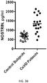
- FIGS. 3A-3B set forth data showing levels of eNOS and NOSTRIN in plasma endothelial-derived exosomes (EDEs) of patients with cerebrovascular disease (CeVD) and matched controls without cerebrovascular disease. The mean for each group is depicted by the horizontal line in that cluster.
- the present invention relates, in part, to the discovery that endothelial cell-derived exosomal biomarkers can be used to detect endothelial cell abnormalities associated with pathogenesis of vascular diseases, including cardiovascular and cerebrovascular diseases.
- EEE endothelial cell-derived exosome
- the inventor has demonstrated that endothelial cell-derived exosome (EDE) levels of CD81, VCAM-1, eNOS, vWF, PDGF, GPVI, YAP, TAZ, p-selectin, e-selectin, ACE/CD143, C1qR1/CD93, VE-Cadherin, CC Chemokine Receptor D6, Tie-2, TNF RI/TNFRSF1A, TNF RII/TNFRSF1B, TRA-1-85/CD147, TRAIL R1/TNFRSF10A, TRAIL R2/TNFRSF10B, VCAM-1/CD106, VE-Statin, VE
- compositions for use in the methods described herein may include small molecule compounds; peptides and proteins including antibodies or functionally active fragments thereof.
- kits for detecting endothelial cell abnormalities in a subject identifying a subject at risk of a vascular disorder associated with endothelial cell abnormalities, or prescribing a therapeutic regimen or predicting benefit from therapy in a subject having a vascular disorder or at risk of developing a vascular disorder.
- the kits comprise one or more antibodies which specifically bind endothelial cell-derived exosomes, one or more antibodies which specifically bind an endothelial cell-derived exosomal biomarker of the present invention, one or more containers for collecting and or holding the biological sample, and instructions for the kits use.
- Biomarker are detected from endothelial cell-derived exosomes from a biological sample obtained from a subject.
- Biological samples can include any bodily fluid comprising exosomes, including, but not limited to, whole blood, plasma, serum, lymph, amniotic fluid, and umbilical cord blood.
- the biological sample of the invention can be obtained from blood.
- about 1-10 mL of blood is drawn from a subject.
- about 10-50 mL of blood is drawn from a subject.
- Blood can be drawn from any suitable area of the body, including an arm, a leg, or blood accessible through a central venous catheter.
- blood is collected following a treatment or activity.
- blood can be collected following a medical exam.
- the timing of collection can also be coordinated to increase the number and/or composition of endothelial cell-derived exosomes present in the sample.
- blood can be collected following exercise or a treatment that induces vascular dilation.
- Blood may be combined with various components following collection to preserve or prepare samples for subsequent techniques.
- blood is treated with an anticoagulant, a cell fixative, a protease inhibitor, a phosphatase inhibitor, or preservative(s) for protein or DNA or RNA following collection.
- blood is collected via venipuncture using a needle and a syringe that is emptied into collection tubes containing an anticoagulant such as EDTA, heparin, or acid citrate dextrose (ACD). Blood can also be collected using a heparin-coated syringe and hypodermic needle. Blood can also be combined with components that will be useful for cell culture.
- blood is combined with cell culture media or supplemented cell culture media (e.g., cytokines).
- cell culture media e.g., cytokines
- platelet-rich plasma PRP
- PBS platelet-rich plasma
- Samples can be enriched for endothelial cell-derived exosomes through positive selection, negative selection, or a combination of positive and negative selection.
- exosomes are directly captured.
- blood cells are captured and exosomes are collected from the remaining biological sample.
- Samples can also be enriched for exosomes based on the biochemical properties of exosomes.
- the first step is physical isolation entailing polymer precipitation with centrifugation in one or two cycles.
- samples can be enriched for exosomes based on differences in antigens.
- antibody-conjugated magnetic or paramagnetic beads in magnetic field gradients or fluorescently labeled antibodies with flow cytometry are used.
- dye uptake/exclusion measured by flow cytometry or another sorting technology is used.
- Samples can also be enriched for exosomes based on other biochemical properties known in the art. For example, samples can be enriched for exosomes using ligands or soluble receptors.
- surface markers are used to positively enrich endothelial cell-derived exosomes in the sample.
- cell surface markers that are not found on exosomes are used to negatively enrich exosomes by depleting cell populations.
- Modified versions of flow cytometry sorting may also be used to further enrich for endothelial cell-derived exosomes using surface markers or intracellular or extracellular markers conjugated to fluorescent labels.
- Intracellular and extracellular markers may include nuclear stains or antibodies against intracellular or extracellular proteins preferentially expressed in exosomes.
- Cell surface markers may include cell surface antigens that are preferentially expressed on endothelial cell-derived exosomes.
- the cell surface marker is an endothelial cell-derived exosome surface marker, including, for example, CD105, CD31, and CD146.
- a monoclonal antibody that specifically binds to CD105, CD31, or CD146 is used to enrich or isolate endothelial cell-derived exosomes from the sample.
- the antibody against CD105, CD31, or CD146 is biotinylated.
- the biotinylated antibody can form an antibody-exosome complex that can be subsequently isolated using streptavidin-agarose resin or beads.
- the antibody is a monoclonal anti-human CD105, CD31, or CD146 antibody.
- endothelial cell-derived exosomes are isolated or enriched from a biological sample comprising: contacting a biological sample with an agent under conditions wherein an endothelial cell-derived exosome present in said biological sample binds to said agent to form an exosome-agent complex; and isolating said exosome from said exosome-agent complex to obtain a sample containing said exosome, wherein the purity of the exosomes present in the sample is greater than the purity of exosomes present in the biological sample.
- the exosomes are endothelial cell-derived exosomes.
- the agent is an antibody or a lectin.
- Lectins useful for forming an exosome-lectin complex are described in U.S. Patent Application Publication No. 2012/0077263.
- multiple isolating or enriching steps are performed.
- a first isolating step is performed to isolate exosomes from a blood sample freed of plasma membrane-derived membrane vesicles and a second isolating step is performed to isolate endothelial cell-derived exosomes from other exosomes.
- the exosome portion of the exosome-agent complex is lysed using a lysis reagent and the protein levels of the lysed exosome are assayed.
- the antibody-exosome complex is created on a solid phase.
- the methods further comprise releasing the exosome from the antibody-exosome complex.
- the solid phase is non-magnetic beads, magnetic beads, agarose, or sepharose.
- the vesicle is released by exposing the antibody-exosome complex to low pH between 3.5 and 1.5.
- the released exosome is neutralized by adding a high pH solution.
- the released exosomes are lysed by incubating the released exosomes with a lysis solution.
- the lysis solution contains inhibitors for proteases and phosphatases.
- the present invention provides methods for detecting endothelial cell abnormalities associated with a vascular disorder in a subject and identifying a subject at risk of developing a vascular disorder due to endothelial cell abnormalities, or prescribing a therapeutic regimen or predicting benefit from therapy.
- High levels of endothelial cell abnormalities in vivo result in elevated endothelial-derived exosomal cargo levels of cytoadhesive, thrombogenic, and inflammatory factors, which can accelerate formation of vascular plaques, clots, and strictures.
- endothelial cell abnormalities are associated with development or worsening of a vascular disorder. Accordingly, detection of endothelial cell abnormalities can be used to identify individuals who will benefit from therapy.
- the vascular disorder is a cardiovascular disorder or a cerebrovascular disorder such as, but not limited to, atherosclerosis, coronary artery disease, thrombosis (e.g., venous thrombosis and arterial thrombosis), thrombocytosis, thrombophlebitis, embolism (e.g., pulmonary embolism and venous thromboembolism), infarction (e.g., myocardial infarction and cerebral infarction), stroke, transient ischemic attack, myeloproliferative diseases, vascular dementia, senile dementia, and Alzheimer's disease.
- thrombosis e.g., venous thrombosis and arterial thrombosis
- thrombocytosis e.g., thrombocytosis
- thrombophlebitis e.g., embolism (e.g., pulmonary embolism and venous thromboembolism), infarction (e.
- Endothelial cell abnormalities may also be associated with an infection (e.g., severe, chronic or systemic infection), inflammation, or severe disease, which increases the risk of developing a vascular disorder.
- an infection e.g., severe, chronic or systemic infection
- endothelial cell abnormalities associated with sepsis, acute respiratory distress syndrome, hepatitis B virus (HBV) infection, pneumonia, or an opportunistic infection in an immunocompromised or immunodeficient patient e.g., infected with human immunodeficiency virus (HIV)
- HIV human immunodeficiency virus
- risk factors for developing a vascular disorder include diabetes mellitus, cigarette smoking, high blood pressure, abnormal lipid panel, increased fasting blood glucose, and a family history of premature death due to cardiovascular causes. Patients with such risk factors may benefit from testing for endothelial cell abnormalities by the methods described herein.
- the present invention enables a medical practitioner to diagnose or prognose one or more vascular disorders in a subject. In yet other embodiments, the present invention enables a medical practitioner to identify a subject at risk of developing a vascular disorder associated with endothelial cell abnormalities. In other embodiments, the present invention enables a medical practitioner to predict whether a subject will later develop a vascular disorder. In further embodiments the present invention enables a medical practitioner to prescribe a therapeutic regimen or predict benefit from therapy in a subject having a vascular disorder or at risk of developing a vascular disorder.
- the subject is a mammalian subject, including, e.g., a cat, a dog, a rodent, etc. In preferred embodiments, the subject is a human subject.
- Endothelial-derived exosomal cargo levels of biomarker proteins are assayed for a subject having or at-risk of having a vascular disorder.
- one or more biomarkers selected from the group consisting of CD81, VCAM-1, eNOS, vWF, PDGF, GPVI, YAP, TAZ, p-selectin, e-selectin, ACE/CD143, C1qR1/CD93, VE-Cadherin, CC Chemokine Receptor D6, Tie-2, TNF RI/TNFRSF1A, TNF RII/TNFRSF1B, TRA-1-85/CD147, TRAIL R1/TNFRSF10A, TRAIL R2/TNFRSF10B, VCAM-1/CD106, VE-Statin, VEGF R1/Flt-1, VEGF R2/KDR/Flk-1, VEGF R3/Flt-4, SLAM/CD150, Stabilin
- immunoassay devices and methods are often used. These devices and methods can utilize labeled molecules in various sandwich, competitive, or non-competitive assay formats, to generate a signal that is related to the presence or amount of an analyte of interest. Additionally, certain methods and devices, such as biosensors and optical immunoassays, may be employed to determine the presence or amount of analytes without the need for a labeled molecule.
- the markers are analyzed using an immunoassay, although other methods are well known to those skilled in the art (for example, the measurement of marker RNA levels).
- the presence or amount of a marker is generally determined using antibodies specific for each marker and detecting specific binding.
- Any suitable immunoassay may be utilized, for example, enzyme-linked immunoassays (ELISA), radioimmunoassay (RIAs), competitive binding assays, planar waveguide technology, and the like.
- ELISA enzyme-linked immunoassays
- RIAs radioimmunoassay
- Specific immunological binding of the antibody to the marker can be detected directly or indirectly.
- Direct labels include fluorescent or luminescent tags, metals, dyes, radionuclides, and the like, attached to the antibody.
- Indirect labels include various enzymes well known in the art, such as alkaline phosphatase, horseradish peroxidase and the like.
- immobilized antibodies specific for the biomarkers is also contemplated by the present invention.
- the antibodies could be immobilized onto a variety of solid supports, such as magnetic or chromatographic matrix particles, the surface of an assay place (such as microtiter wells), pieces of a solid substrate material (such as plastic, nylon, paper), and the like.
- An assay strip could be prepared by coating the antibody or a plurality of antibodies in an array on solid support. This strip could then be dipped into the test sample and then processed quickly through washes and detection steps to generate a measurable signal, such as a colored spot.
- biomarkers may be carried out separately or simultaneously with one test sample. Several biomarkers may be combined into one test for efficient processing of a multiple of samples. In addition, one skilled in the art would recognize the value of testing multiple samples (for example, at successive time points) from the same individual. Such testing of serial samples will allow the identification of changes in marker levels over time. Increases or decreases in biomarker levels, as well as the absence of change in biomarker levels, would provide useful information about disease status that includes, but is not limited to the appropriateness of drug therapies, the effectiveness of various therapies, identification of the severity of endothelial cell abnormalities, susceptibility to atherosclerosis, and prognosis of the patient's outcome, including risk of a future vascular event.
- An assay consisting of a combination of the biomarkers referenced in the instant invention may be constructed to provide relevant information related to differential diagnosis.
- a panel may be constructed using 1, 2, 3, 4, 5, 6, 7, 8, 9, 10, 15, 20, or more individual markers.
- the analysis of a single biomarker or subsets of biomarkers comprising a larger panel of biomarkers could be carried out using methods described within the instant invention to optimize clinical sensitivity or specificity in various clinical settings.
- markers could be carried out in a variety of physical formats as well.
- the use of microtiter plates or automation could be used to facilitate the processing of large numbers of test samples.
- single sample formats could be developed to facilitate immediate treatment and diagnosis in a timely fashion, for example, in ambulatory transport or emergency room settings.
- Particularly useful physical formats comprise surfaces having a plurality of discrete, addressable locations for the detection of a plurality of different analytes.
- Such formats include protein microarrays, or “protein chips” and capillary devices.
- Biomarkers of the present invention serve an important role in the early detection and monitoring of endothelial cell abnormalities associated with vascular disorders (e.g., atherosclerosis).
- Biomarkers are typically substances found in a bodily sample that can be measured.
- the measured amount can correlate with underlying disorder or disease pathophysiology (e.g., presence or absence of endothelial cell abnormalities) and probability of developing a vascular disorder in the future. In patients receiving treatment for their condition, the measured amount will also correlate with responsiveness to therapy.
- the biomarker is measured by a method selected from the group consisting of immunohistochemistry, immunocytochemistry, immunofluorescence, immunoprecipitation, western blotting, and ELISA.
- the methods of the present invention for detecting endothelial cell abnormalities may be used in clinical assays to diagnose or prognose a vascular disorder in a subject, identify a subject at risk of a vascular disorder (e.g., atherosclerosis), and/or for prescribing a therapeutic regimen or predicting benefit from therapy in a subject having a vascular disorder.
- Clinical assay performance can be assessed by determining the assay's sensitivity, specificity, area under the ROC curve (AUC), accuracy, positive predictive value (PPV), and negative predictive value (NPV).
- the clinical performance of the assay may be based on sensitivity.
- the sensitivity of an assay of the present invention may be at least about 40%, 45%, 50%, 55%, 60%, 65%, 70%, 75%, 80%, 85%, 90%, 95%, 99%, or 100%.
- the clinical performance of the assay may be based on specificity.
- the specificity of an assay of the present invention may be at least about 40%, 45%, 50%, 55%, 60%, 65%, 70%, 75%, 80%, 85%, 90%, 95%, 99%, or 100%.
- the clinical performance of the assay may be based on area under the ROC curve (AUC).
- the AUC of an assay of the present invention may be at least about 0.5, 0.55, 0.6, 0.65, 0.7, 0.75, 0.8, 0.85, 0.9, or 0.95.
- the clinical performance of the assay may be based on accuracy.
- the accuracy of an assay of the present invention may be at least about 40%, 45%, 50%, 55%, 60%, 65%, 70%, 75%, 80%, 85%, 90%, 95%, 99%, or 100%.
- compositions useful in the methods of the present invention include compositions that specifically recognize one or more biomarkers associated with endothelial cell abnormalities, including CD81, VCAM-1, eNOS, vWF, PDGF, GPVI, YAP, TAZ, p-selectin, e-selectin, ACE/CD143, C1qR1/CD93, VE-Cadherin, CC Chemokine Receptor D6, Tie-2, TNF RI/TNFRSF1A, TNF RII/TNFRSF1B, TRA-1-85/CD147, TRAIL R1/TNFRSF10A, TRAIL R2/TNFRSF10B, VCAM-1/CD106, VE-Statin, VEGF R1/Flt-1, VEGF R2/KDR/Flk-1, VEGF R3/Flt-4, SLAM/CD150, Stabilin-1, Stabilin-2, TEM7/PLXDC1, TEM8/ANTXR1,
- the composition enhances the activity of at least one biomarker. In other embodiments, the composition decreases the activity of at least one biomarker. In yet other embodiments, the composition comprises a peptide, a nucleic acid, an antibody, or a small molecule.
- the present invention relates to compositions that specifically detect a biomarker associated with endothelial cell abnormalities.
- the present invention is based upon the finding that endothelial-derived exosomal CD81, VCAM-1, eNOS, vWF, PDGF, GPVI, YAP, TAZ, p-selectin, e-selectin, ACE/CD143, C1qR1/CD93, VE-Cadherin, CC Chemokine Receptor D6, Tie-2, TNF RI/TNFRSF1A, TNF RII/TNFRSF1B, TRA-1-85/CD147, TRAIL R1/TNFRSF10A, TRAIL R2/TNFRSF10B, VCAM-1/CD106, VE-Statin, VEGF R1/Flt-1, VEGF R2/KDR/Flk-1, VEGF R3/Flt-4, SLAM/CD150, Stabil
- compositions of the invention specifically bind to and detect one or more of the biomarkers CD81, VCAM-1, eNOS, vWF, PDGF, GPVI, YAP, TAZ, p-selectin, e-selectin, ACE/CD143, C1qR1/CD93, VE-Cadherin, CC Chemokine Receptor D6, Tie-2, TNF RI/TNFRSF1A, TNF RII/TNFRSF1B, TRA-1-85/CD147, TRAIL R1/TNFRSF10A, TRAIL R2/TNFRSF10B, VCAM-1/CD106, VE-Statin, VEGF R1/Flt-1, VEGF R2/KDR/Flk-1, VEGF R3/Flt-4, SLAM/CD150, Stabilin-1, Stabilin-2, TEM7/PLXDC1, TEM8/ANTXR1, Thrombomodulin/BDCA-3,
- the composition comprises an antibody, wherein the antibody specifically binds to a biomarker or endothelial cell-derived exosomes.
- antibody as used herein and further discussed below is intended to include fragments thereof which are also specifically reactive with a biomarker or vesicle (e.g., exosome).
- Antibodies can be fragmented using conventional techniques and the fragments screened for utility in the same manner as described above for whole antibodies. For example, F(ab) 2 fragments can be generated by treating antibody with pepsin. The resulting F(ab) 2 fragment can be treated to reduce disulfide bridges to produce Fab fragments.
- Antigen-binding portions may also be produced by recombinant DNA techniques or by enzymatic or chemical cleavage of intact antibodies.
- Antigen-binding portions include, inter alia, Fab, Fab′, F(ab′) 2 , Fv, dAb, and complementarity determining region (CDR) fragments, single-chain antibodies (scFv), single domain antibodies, bispecific antibodies, chimeric antibodies, humanized antibodies, diabodies and polypeptides that contain at least a portion of an immunoglobulin that is sufficient to confer specific antigen binding to the polypeptide.
- the antibody further comprises a label attached thereto and able to be detected (e.g., the label can be a radioisotope, fluorescent compound, enzyme or enzyme co-factor).
- an antibody of the invention is a monoclonal antibody
- the invention makes available methods for generating novel antibodies that specifically bind the biomarker or the exosome of the invention.
- a method for generating a monoclonal antibody that specifically binds a biomarker or exosome may comprise administering to a mouse an amount of an immunogenic composition comprising the biomarker or exosome, or fragment thereof, effective to stimulate a detectable immune response, obtaining antibody-producing cells (e.g., cells from the spleen) from the mouse and fusing the antibody-producing cells with myeloma cells to obtain antibody-producing hybridomas, and testing the antibody-producing hybridomas to identify a hybridoma that produces a monocolonal antibody that binds specifically to the biomarker or exosome.
- antibody-producing cells e.g., cells from the spleen
- a hybridoma can be propagated in a cell culture, optionally in culture conditions where the hybridoma-derived cells produce the monoclonal antibody that binds specifically to the biomarker or exosome.
- the monoclonal antibody may be purified from the cell culture.
- the term “specifically reactive with” or “specifically binds” as used in reference to an antibody is intended to mean, as is generally understood in the art, that the antibody is sufficiently selective between the antigen of interest (e.g., a biomarker or exosome) and other antigens that are not of interest. In certain methods employing the antibody, such as therapeutic applications, a higher degree of specificity in binding may be desirable. Monoclonal antibodies generally have a greater tendency (as compared to polyclonal antibodies) to discriminate effectively between the desired antigens and cross-reacting polypeptides. One characteristic that influences the specificity of an antibody:antigen interaction is the affinity of the antibody for the antigen. Although the desired specificity may be reached with a range of different affinities, generally preferred antibodies will have an affinity (a dissociation constant) of about 10 ⁇ 6 , 10 ⁇ 7 , 10 ⁇ 8 , 10 ⁇ 9 or less.
- Antibodies can be generated to bind specifically to an epitope of an endothelial cell-derived exosome or a biomarker of the present invention, including, for example, endothelial cell-derived exosome surface markers, such as CD105, CD31, and CD146.
- the techniques used to screen antibodies in order to identify a desirable antibody may influence the properties of the antibody obtained.
- a variety of different techniques are available for testing interaction between antibodies and antigens to identify particularly desirable antibodies. Such techniques include ELISAs, surface plasmon resonance binding assays (e.g., the Biacore binding assay, Biacore AB, Uppsala, Sweden), sandwich assays (e.g., the paramagnetic bead system of IGEN International, Inc., Gaithersburg, Md.), western blots, immunoprecipitation assays, immunocytochemistry, and immunohistochemistry.
- the present invention relates to compositions used for treating or preventing a vascular disorder.
- endothelial cell abnormalities are implicated in the pathology of a variety of vascular disorders, including cardiovascular and cerebrovascular diseases (e.g., atherosclerosis). Therefore, in one embodiment, the present invention provides compositions that inhibit endothelial cell abnormalities.
- Compositions useful for preventing and/or reducing endothelial cell abnormalities may include proteins, peptides, nucleic acids, small molecules, and the like.
- the present invention provides methods of treating a vascular disorder associated with endothelial cell abnormalities in a subject, comprising administering to the subject an effective amount of a composition, wherein the composition inhibits endothelial cell abnormalities.
- the methods of the invention can be used for monitoring the efficacy of therapy in a patient.
- the method comprises: analyzing the levels of one or more biomarkers selected from the group consisting of CD81, VCAM-1, eNOS, vWF, PDGF, GPVI, YAP, TAZ, p-selectin, e-selectin, ACE/CD143, C1qR1/CD93, VE-Cadherin, CC Chemokine Receptor D6, Tie-2, TNF RI/TNFRSF1A, TNF RII/TNFRSF1B, TRA-1-85/CD147, TRAIL R1/TNFRSF10A, TRAIL R2/TNFRSF10B, VCAM-1/CD106, VE-Statin, VEGF R1/Flt-1, VEGF R2/KDR/Flk-1, VEGF R3/Flt-4, SLAM/CD150, Stabilin-1, Stabilin-2, TEM7/PL
- the methods of the invention provide a method for treating a vascular disorder the method comprising the steps of: obtaining a biological sample from a subject suspected of having a vascular disorder, wherein the sample comprises endothelial cell-derived exosomes; measuring the level of one or more biomarkers selected from the group consisting of CD81, VCAM-1, eNOS, vWF, PDGF, GPVI, YAP, TAZ, p-selectin, e-selectin, ACE/CD143, C1qR1/CD93, VE-Cadherin, CC Chemokine Receptor D6, Tie-2, TNF RI/TNFRSF1A, TNF RII/TNFRSF1B, TRA-1-85/CD147, TRAIL R1/TNFRSF10A, TRAIL R2/TNFRSF10B, VCAM-1/CD106, VE-Statin, VEGF R1/Flt-1, VEGF R2/KDR/
- kits for detecting or monitoring endothelial cell abnormalities in a subject A variety of kits having different components are contemplated by the current invention. Generally speaking, the kit will include the means for quantifying one or more biomarkers in a subject. In another embodiment, the kit will include means for collecting a biological sample, means for quantifying one or more biomarkers in the biological sample, and instructions for use of the kit contents. In certain embodiments, the kit comprises a means for enriching or isolating endothelial cell-derived exosomes in a biological sample.
- the means for enriching or isolating endothelial cell-derived exosomes comprises reagents necessary to enrich or isolate endothelial cell-derived exosomes from a biological sample.
- the kit comprises a means for quantifying the amount of a biomarker.
- the means for quantifying the amount of a biomarker comprises reagents necessary to detect the amount of a biomarker.
- Example 1 Biomarker Detection and Levels in Endothelial-Derived Exosomes from Patients with Cerebrovascular Disease
- Biomarkers were detected and measured in endothelial-derived exosomes from patients with cerebrovascular disease as follows. Blood samples were obtained from eighteen patients with definite neurologically-relevant cerebrovascular disease (CeVD) and eighteen control subjects. Each patient had CeVD proven by MRI and 14 had experienced an ischemic cerebrovascular event with tissue damage in the 10 years prior to donating blood. Strokes had occurred one year (1 patient), two (2), three (2), four (1), five (1), six (2), eight (1), nine (2) and ten years (2 patients) prior to obtaining the study blood sample. All patients had clinical evidence of continued evolution of CeVD. Each of the eighteen control subjects matched a patient by age and gender, and donated blood concurrently with patients.
- CeVD cerebrovascular disease
- Exosome suspensions each were incubated and mixed sequentially with 2.0 ⁇ g of mouse IgG1 anti-human CD31 (PECAM1, platelet and endothelial cell adhesion molecule 1) biotinylated antibody (clone MEM-05, ThermoFisher Scientific [Life Technologies], Carlsbad, Calif.) in 50 ⁇ L of 3% BSA for 90 min followed by incubation with 10 ⁇ L of Streptavidin-Plus ULTRALINK resin (Pierce-Thermo Scientific, Inc.) in 40 ⁇ L of 3% BSA for 60 min with continuous mixing at room temperature.
- PECAM1 anti-human CD31 PECAM1, platelet and endothelial cell adhesion molecule 1
- biotinylated antibody clone MEM-05, ThermoFisher Scientific [Life Technologies], Carlsbad, Calif.
- each pellet was resuspended in 100 ⁇ l of cold 0.05 M acetic acid, incubated at 4° C. for 10 min and centrifuged at 4° C. for 10 min at 4,000 ⁇ g. These supernatants were transferred to new pre-chilled EPPENDORF tubes containing 265 ⁇ L of DBS, 10 ⁇ L of 1 M Tris-HCl (pH 8.0) and 25 ⁇ L of 10% BSA, and mixed.
- each of these exosome suspensions was incubated and mixed sequentially with 2.0 ⁇ g of goat anti-human CD146/MCAM (cell surface glycoprotein MUC18/melanoma cell adhesion molecule) biotinylated antibody (Novus Biologicals, Littleton, Colo.) in 50 ⁇ L of 3% BSA for 90 min followed by incubation with 10 ⁇ l of Streptavidin-Plus ULTRALINK resin (Pierce-Thermo Scientific, Inc.) in 40 ⁇ L of 3% BSA for 60 min all at room temperature. After centrifugation at 600 ⁇ g and removal of the supernatant, each pellet was resuspended in 100 ⁇ l of cold 0.05 M acetic acid, incubated at 4° C.
- CD146/MCAM cell surface glycoprotein MUC18/melanoma cell adhesion molecule
- each suspension was diluted 1:50 in PBS.
- the mean diameter (nm) and concentration (particles/ml) of exosomes in each suspension were determined using the NANOSIGHT NS500 system with a G532 nm laser module and NTA 3.1 nanoparticle tracking software (Malvern Instruments, Malvern, UK). Camera settings were: gain 366; shutter 31.48; frame rate 24.9825 fps/s; Brownian motion was captured by five repeated 20 sec video recordings.
- EDE proteins were quantified by ELISA kits for human endothelial nitric oxide synthase (eNOS), glucose transporter 1 (GLUT1), lysyl-oxidase homolog-2 (LOXL2), platelet glycoprotein VI (GPVI) and the tetraspanning exosome marker CD81 (American Research Products, Inc.-Cusabio, Waltham, Mass.), angiopoietin-2 and von Willebrand factor (vWF) (Life Technologies-ThermoFisher Scientific), vascular cell adhesion molecule-1 (VCAM-1) and platelet-derived growth factor (PDGF-AB) (RayBiotech, Norcross, Ga.) according to suppliers' directions.
- the mean value for all determinations of CD81 in each assay group was set at 1.00 and the relative values of CD81 for each sample used to normalize their recovery.
- the mean ( ⁇ SEM) ages of the patients with atherosclerotic CeVD and controls were 75.3 ⁇ 2.71 years and 74.6 ⁇ 2.94 years, respectively, and each group had nine females and nine males.
- Clinically-apparent non-cerebral arterial disease was found in six of the patients (two coronary, one peripheral and three both coronary and peripheral), but none of the controls.
- Medication programs were heterogeneous with seven patients on low-dose aspirin and three on an oral anticoagulant. None of the controls had any evidence of atherosclerotic arterial disease.
- Four controls were on low-dose aspirin, but none was on an anticoagulant.
- the mean ( ⁇ SEM) diameters of the EDEs were 230 ⁇ 3.00 nm for the controls and 246 ⁇ 6.18 nm for the patients, that were not significantly different.
- Endothelial cell marker proteins in plasma endothelial-derived exosomes (EDEs) and platelet-derived exosomes (PDEs) isolated separately from the same controls and patients were quantified and the results are shown in Table 1 below.
- CD81 levels in PDEs from both groups were significantly higher than those of EDEs. All other values were normalized for their levels of CD81 (see Table 1).
- the levels of the endothelial cell protein markers VCAM-1 and eNOS were significantly more than 10-fold higher for EDEs than PDEs.
- levels of the platelet marker GPVI was significantly more than six-fold higher for PDEs than EDEs of controls and patients.
- CD81-normalized EDE levels of proteins characteristic of endothelial cells and implicated in atherosclerosis were quantified for all patients and controls. The levels of all six proteins were significantly higher for CeVD patients than matched controls (see FIG. 1 ).
- Example 2 Biomarker Detection and Levels in Endothelial-Derived Exosomes from Patients with Cerebrovascular Disease and Cardiovascular Disease
- EDE endothelial-derived exosomes
- EDE proteins were quantified using ELISA kits for GLUT-1, LAT-1, P-gp, VCAM-1, and NOSTRIN. The mean value for all determinations of CD81 in each assay group was set at 1.00 and the relative values of CD81 for each sample used to normalize their recovery.
- the mean levels of all five biomarker proteins in plasma EDEs for CoAD, CeVD, and both CoAD and CeVD were significantly higher than the respective means for control plasma samples.
- mean levels for patients with CeVD were significantly higher than those for patients with CoAD alone and there was no overlap between levels for patients with CeVD and those for control samples.
- Example 3 NOSTRIN Detection and Levels in Endothelial-Derived Exosomes from Patients with Cerebrovascular Disease
- NOSTRIN was detected and measured in endothelial-derived exosomes from patients with cerebrovascular disease as follows. Blood samples were obtained from eighteen patients with cerebrovascular disease (CeVD) and eighteen control subjects.
- the eNOS mean ⁇ SEM values were 25.1 ⁇ 2.02 IU/ml for control subjects and 25.5 ⁇ 2.13 IU/ml for CeVD patients, and the NOSTRIN mean ⁇ SEM values were 688 ⁇ 37.3 pg/ml for control subjects and 1343 ⁇ 113 pg/ml for CeVD patients.
- the difference between NOSTRIN levels for the two groups is significant with p ⁇ 0.0001 by an unpaired t test, whereas there was no significant difference between eNOS levels of the two groups.
Landscapes
- Health & Medical Sciences (AREA)
- Life Sciences & Earth Sciences (AREA)
- Engineering & Computer Science (AREA)
- Hematology (AREA)
- Immunology (AREA)
- Molecular Biology (AREA)
- Biomedical Technology (AREA)
- Chemical & Material Sciences (AREA)
- Urology & Nephrology (AREA)
- Biotechnology (AREA)
- Biochemistry (AREA)
- Cell Biology (AREA)
- Food Science & Technology (AREA)
- Medicinal Chemistry (AREA)
- Physics & Mathematics (AREA)
- Analytical Chemistry (AREA)
- Microbiology (AREA)
- General Health & Medical Sciences (AREA)
- General Physics & Mathematics (AREA)
- Pathology (AREA)
- Proteomics, Peptides & Aminoacids (AREA)
- Investigating Or Analysing Biological Materials (AREA)
- Peptides Or Proteins (AREA)
Priority Applications (1)
| Application Number | Priority Date | Filing Date | Title |
|---|---|---|---|
| US15/703,955 US11193943B2 (en) | 2017-02-06 | 2017-09-13 | Endothelial cell derived exosomes and uses thereof |
Applications Claiming Priority (2)
| Application Number | Priority Date | Filing Date | Title |
|---|---|---|---|
| US201762455582P | 2017-02-06 | 2017-02-06 | |
| US15/703,955 US11193943B2 (en) | 2017-02-06 | 2017-09-13 | Endothelial cell derived exosomes and uses thereof |
Publications (2)
| Publication Number | Publication Date |
|---|---|
| US20180224465A1 US20180224465A1 (en) | 2018-08-09 |
| US11193943B2 true US11193943B2 (en) | 2021-12-07 |
Family
ID=63037628
Family Applications (1)
| Application Number | Title | Priority Date | Filing Date |
|---|---|---|---|
| US15/703,955 Active US11193943B2 (en) | 2017-02-06 | 2017-09-13 | Endothelial cell derived exosomes and uses thereof |
Country Status (3)
| Country | Link |
|---|---|
| US (1) | US11193943B2 (de) |
| EP (1) | EP3577214A4 (de) |
| WO (1) | WO2018144070A1 (de) |
Families Citing this family (6)
| Publication number | Priority date | Publication date | Assignee | Title |
|---|---|---|---|---|
| US11345957B2 (en) * | 2017-07-18 | 2022-05-31 | Exosome Diagnostics, Inc. | Methods of treating glioblastoma in a subject informed by exosomal RNA signatures |
| WO2019038660A1 (en) | 2017-08-21 | 2019-02-28 | Seattle Children's Hospital (dba Seattle Children's Research Institute) | EXOSOMAL PROFILING FOR THE DIAGNOSIS AND MONITORING OF VASCULITE AND VASCULOPATHIES, INCLUDING KAWASAKI'S DISEASE |
| CN110389165A (zh) * | 2018-04-23 | 2019-10-29 | 江成鸿 | Cd93在制备预警婴幼儿血管瘤脐血检测试剂盒及治疗药物中的应用 |
| CN109738625A (zh) * | 2019-01-04 | 2019-05-10 | 湖南师范大学 | 一种捕获纳米磁珠、捕获外泌体的方法和应用 |
| WO2025179281A1 (en) * | 2024-02-23 | 2025-08-28 | The United States Of America, As Represented By The Secretary, Department Of Health And Human Services | Treatment of cardiovascular disease with antxr1 antibodies |
| CN118150843B (zh) * | 2024-04-01 | 2025-02-25 | 北京热景生物技术股份有限公司 | 一种阿尔兹海默症检测标志物糖链外泌体P-Tau-217及其应用 |
Citations (3)
| Publication number | Priority date | Publication date | Assignee | Title |
|---|---|---|---|---|
| WO2009015357A1 (en) | 2007-07-25 | 2009-01-29 | University Of Louisville Research Foundation, Inc. | Exosome-associated microrna as a diagnostic marker |
| WO2012115885A1 (en) | 2011-02-22 | 2012-08-30 | Caris Life Sciences Luxembourg Holdings, S.A.R.L. | Circulating biomarkers |
| US20120295286A1 (en) | 2011-04-21 | 2012-11-22 | The George Washington University | Methods for the Diagnosis, Prognosis and Monitoring of Cancer Therapy Using BP1 |
Family Cites Families (4)
| Publication number | Priority date | Publication date | Assignee | Title |
|---|---|---|---|---|
| WO2012110253A2 (en) * | 2011-02-18 | 2012-08-23 | Cavadis B.V. | Exosomal biomarkers for cardiovascular events |
| WO2012126531A1 (en) * | 2011-03-24 | 2012-09-27 | Cavadis B.V. | Method for diagnosing acute coronary syndrome (acs) |
| US20140024046A1 (en) * | 2011-02-18 | 2014-01-23 | Cavadis B.V. | Exosomal Biomarkers for Cardiovasular Events |
| WO2014004424A1 (en) * | 2012-06-26 | 2014-01-03 | Temple University - Of The Commonwealth System Of Higher Education | Method for detecting injury to the brian |
-
2017
- 2017-09-13 EP EP17895281.8A patent/EP3577214A4/de not_active Withdrawn
- 2017-09-13 US US15/703,955 patent/US11193943B2/en active Active
- 2017-09-13 WO PCT/US2017/051425 patent/WO2018144070A1/en not_active Ceased
Patent Citations (3)
| Publication number | Priority date | Publication date | Assignee | Title |
|---|---|---|---|---|
| WO2009015357A1 (en) | 2007-07-25 | 2009-01-29 | University Of Louisville Research Foundation, Inc. | Exosome-associated microrna as a diagnostic marker |
| WO2012115885A1 (en) | 2011-02-22 | 2012-08-30 | Caris Life Sciences Luxembourg Holdings, S.A.R.L. | Circulating biomarkers |
| US20120295286A1 (en) | 2011-04-21 | 2012-11-22 | The George Washington University | Methods for the Diagnosis, Prognosis and Monitoring of Cancer Therapy Using BP1 |
Non-Patent Citations (10)
| Title |
|---|
| De Jong et al. Cellular stress conditions are reflected in the protein and RNA content of endothelial cell derived exosomes. Journal of Extracellular Vesicles 2012, 1:1, 18396 (Year: 2012). * |
| De Jong et al. Exosomes from hypoxic endothelial cells have increased collagen crosslinking activity through up-regulation of lysyl oxidase-like 2. J. Cell. Mol. Med. Vol 20, No. 2, 2016 pp. 342-350 (Year: 2015). * |
| Dignat-George and Boulanger. Arteriosclerosis, Thrombosis, and Vascular Biology. 2011;31:27-33 (Year: 2011). * |
| Ge and Wang. Identifying novel biomarkers for cardiovascular disease risk predication. J Intern Med 2012; 272: 430-439 (Year: 2012). * |
| Husseini and Laskowitz. Clinical application of blood biomarkers in cerebrovascular disease. Expert Review of Neurotherapeutics, 2010; 10(2):189-203 (Year: 2010). * |
| International Search Report and the Written Opinion of the International Searching Authority re PCT/US2017/051425. |
| Kernagis et al. Evolving Role of Biomarkers in Acute Cerebrovascular Disease. Ann Neurol 2012;71:289-303 (Year: 2012). * |
| Osada-Oka M. et al. "Macrophage-derived exosomes induce inflammatory factors in endothelial cells under hypertensive conditions." Hypertension Research. (2017) 40, 353-360. |
| Suzuki E. et al. "Stem cell-derived exosomes as a therapeutic tool for cadiovascular disease". World J Stem Cells , 2016; 8(9):1297-395. |
| Wang J. et al. "The novel methods for analysis of exosomes released from endothelial cells and endothelial progenitor cells." Stem Cells Int. Vol 16, 12 pages. |
Also Published As
| Publication number | Publication date |
|---|---|
| EP3577214A4 (de) | 2021-03-24 |
| WO2018144070A1 (en) | 2018-08-09 |
| EP3577214A1 (de) | 2019-12-11 |
| US20180224465A1 (en) | 2018-08-09 |
Similar Documents
| Publication | Publication Date | Title |
|---|---|---|
| US11193943B2 (en) | Endothelial cell derived exosomes and uses thereof | |
| US10203342B2 (en) | Biomarkers and differential diagnosis of alzheimer's disease and other neurodegenerative disorders | |
| US11619637B2 (en) | Biomarkers and diagnostic methods for Alzheimer's disease and other neurodegenerative disorders | |
| EP3452828B1 (de) | Synaptische proteinbiomarker und differenzielle diagnose von morbus alzheimer und anderen neurodegenerativen erkrankungen | |
| US11852635B2 (en) | Quantification of subpopulations of exosomes and diagnosis of neurogenerative disorders | |
| US20200341011A1 (en) | Astrocyte exosome complement-based assay for neuroinflammation in alzheimer's disease and uses thereof | |
| US11360097B2 (en) | Platelet biomarkers and diagnostic methods for vascular diseases | |
| US20180080945A1 (en) | Biomarkers and diagnostic methods for alzheimer's disease and other neurodegenerative disorders | |
| JP2014070942A (ja) | 可溶型clec−2に基づく血小板活性化測定方法 | |
| US20220057501A1 (en) | Endothelial Cell Derived Exosomes and Uses Thereof | |
| US20240272178A1 (en) | A method for predicting sepsis and septic shock | |
| WO2012140148A2 (en) | Junction plakoglobin for diagnosis of cardiovascular diseases | |
| JP2008526937A (ja) | ファクターXIIaのフォーム | |
| JP6923212B2 (ja) | 動脈硬化性疾患の発症の予測因子および検査方法 | |
| EP2015075A1 (de) | Verfahren zur Vorhersage der Wahrscheinlichkeit einer Herzkranz-Erkrankung bei Patienten mit akutem Koronarsyndrom |
Legal Events
| Date | Code | Title | Description |
|---|---|---|---|
| FEPP | Fee payment procedure |
Free format text: ENTITY STATUS SET TO UNDISCOUNTED (ORIGINAL EVENT CODE: BIG.); ENTITY STATUS OF PATENT OWNER: SMALL ENTITY |
|
| FEPP | Fee payment procedure |
Free format text: ENTITY STATUS SET TO SMALL (ORIGINAL EVENT CODE: SMAL); ENTITY STATUS OF PATENT OWNER: SMALL ENTITY |
|
| STCB | Information on status: application discontinuation |
Free format text: ABANDONED -- FAILURE TO RESPOND TO AN OFFICE ACTION |
|
| STPP | Information on status: patent application and granting procedure in general |
Free format text: RESPONSE TO NON-FINAL OFFICE ACTION ENTERED AND FORWARDED TO EXAMINER |
|
| STPP | Information on status: patent application and granting procedure in general |
Free format text: NON FINAL ACTION MAILED |
|
| STPP | Information on status: patent application and granting procedure in general |
Free format text: RESPONSE TO NON-FINAL OFFICE ACTION ENTERED AND FORWARDED TO EXAMINER |
|
| STPP | Information on status: patent application and granting procedure in general |
Free format text: FINAL REJECTION MAILED |
|
| STPP | Information on status: patent application and granting procedure in general |
Free format text: NOTICE OF ALLOWANCE MAILED -- APPLICATION RECEIVED IN OFFICE OF PUBLICATIONS |
|
| STPP | Information on status: patent application and granting procedure in general |
Free format text: PUBLICATIONS -- ISSUE FEE PAYMENT RECEIVED |
|
| STPP | Information on status: patent application and granting procedure in general |
Free format text: PUBLICATIONS -- ISSUE FEE PAYMENT VERIFIED |
|
| STCF | Information on status: patent grant |
Free format text: PATENTED CASE |
|
| MAFP | Maintenance fee payment |
Free format text: PAYMENT OF MAINTENANCE FEE, 4TH YR, SMALL ENTITY (ORIGINAL EVENT CODE: M2551); ENTITY STATUS OF PATENT OWNER: SMALL ENTITY Year of fee payment: 4 |