US11674130B2 - Method for producing therapeutic exosomes from nanoelectroporation and other non-endocytic cell transfection - Google Patents
Method for producing therapeutic exosomes from nanoelectroporation and other non-endocytic cell transfection Download PDFInfo
- Publication number
- US11674130B2 US11674130B2 US16/635,471 US201816635471A US11674130B2 US 11674130 B2 US11674130 B2 US 11674130B2 US 201816635471 A US201816635471 A US 201816635471A US 11674130 B2 US11674130 B2 US 11674130B2
- Authority
- US
- United States
- Prior art keywords
- evs
- dna
- donor cells
- extracellular vesicles
- biochip
- Prior art date
- Legal status (The legal status is an assumption and is not a legal conclusion. Google has not performed a legal analysis and makes no representation as to the accuracy of the status listed.)
- Active
Links
Images


Classifications
-
- C—CHEMISTRY; METALLURGY
- C12—BIOCHEMISTRY; BEER; SPIRITS; WINE; VINEGAR; MICROBIOLOGY; ENZYMOLOGY; MUTATION OR GENETIC ENGINEERING
- C12N—MICROORGANISMS OR ENZYMES; COMPOSITIONS THEREOF; PROPAGATING, PRESERVING, OR MAINTAINING MICROORGANISMS; MUTATION OR GENETIC ENGINEERING; CULTURE MEDIA
- C12N15/00—Mutation or genetic engineering; DNA or RNA concerning genetic engineering, vectors, e.g. plasmids, or their isolation, preparation or purification; Use of hosts therefor
-
- C—CHEMISTRY; METALLURGY
- C12—BIOCHEMISTRY; BEER; SPIRITS; WINE; VINEGAR; MICROBIOLOGY; ENZYMOLOGY; MUTATION OR GENETIC ENGINEERING
- C12N—MICROORGANISMS OR ENZYMES; COMPOSITIONS THEREOF; PROPAGATING, PRESERVING, OR MAINTAINING MICROORGANISMS; MUTATION OR GENETIC ENGINEERING; CULTURE MEDIA
- C12N13/00—Treatment of microorganisms or enzymes with electrical or wave energy, e.g. magnetism, sonic waves
-
- A—HUMAN NECESSITIES
- A61—MEDICAL OR VETERINARY SCIENCE; HYGIENE
- A61K—PREPARATIONS FOR MEDICAL, DENTAL OR TOILETRY PURPOSES
- A61K35/00—Medicinal preparations containing materials or reaction products thereof with undetermined constitution
- A61K35/12—Materials from mammals; Compositions comprising non-specified tissues or cells; Compositions comprising non-embryonic stem cells; Genetically modified cells
-
- A—HUMAN NECESSITIES
- A61—MEDICAL OR VETERINARY SCIENCE; HYGIENE
- A61K—PREPARATIONS FOR MEDICAL, DENTAL OR TOILETRY PURPOSES
- A61K9/00—Medicinal preparations characterised by special physical form
- A61K9/48—Preparations in capsules, e.g. of gelatin, of chocolate
- A61K9/50—Microcapsules having a gas, liquid or semi-solid filling; Solid microparticles or pellets surrounded by a distinct coating layer, e.g. coated microspheres, coated drug crystals
- A61K9/5005—Wall or coating material
- A61K9/5063—Compounds of unknown constitution, e.g. material from plants or animals
- A61K9/5068—Cell membranes or bacterial membranes enclosing drugs
-
- C—CHEMISTRY; METALLURGY
- C07—ORGANIC CHEMISTRY
- C07K—PEPTIDES
- C07K14/00—Peptides having more than 20 amino acids; Gastrins; Somatostatins; Melanotropins; Derivatives thereof
- C07K14/435—Peptides having more than 20 amino acids; Gastrins; Somatostatins; Melanotropins; Derivatives thereof from animals; from humans
- C07K14/705—Receptors; Cell surface antigens; Cell surface determinants
- C07K14/70503—Immunoglobulin superfamily
-
- C—CHEMISTRY; METALLURGY
- C07—ORGANIC CHEMISTRY
- C07K—PEPTIDES
- C07K14/00—Peptides having more than 20 amino acids; Gastrins; Somatostatins; Melanotropins; Derivatives thereof
- C07K14/435—Peptides having more than 20 amino acids; Gastrins; Somatostatins; Melanotropins; Derivatives thereof from animals; from humans
- C07K14/705—Receptors; Cell surface antigens; Cell surface determinants
- C07K14/70596—Molecules with a "CD"-designation not provided for elsewhere
-
- C—CHEMISTRY; METALLURGY
- C12—BIOCHEMISTRY; BEER; SPIRITS; WINE; VINEGAR; MICROBIOLOGY; ENZYMOLOGY; MUTATION OR GENETIC ENGINEERING
- C12M—APPARATUS FOR ENZYMOLOGY OR MICROBIOLOGY; APPARATUS FOR CULTURING MICROORGANISMS FOR PRODUCING BIOMASS, FOR GROWING CELLS OR FOR OBTAINING FERMENTATION OR METABOLIC PRODUCTS, i.e. BIOREACTORS OR FERMENTERS
- C12M23/00—Constructional details, e.g. recesses, hinges
- C12M23/02—Form or structure of the vessel
- C12M23/16—Microfluidic devices; Capillary tubes
-
- C—CHEMISTRY; METALLURGY
- C12—BIOCHEMISTRY; BEER; SPIRITS; WINE; VINEGAR; MICROBIOLOGY; ENZYMOLOGY; MUTATION OR GENETIC ENGINEERING
- C12M—APPARATUS FOR ENZYMOLOGY OR MICROBIOLOGY; APPARATUS FOR CULTURING MICROORGANISMS FOR PRODUCING BIOMASS, FOR GROWING CELLS OR FOR OBTAINING FERMENTATION OR METABOLIC PRODUCTS, i.e. BIOREACTORS OR FERMENTERS
- C12M33/00—Means for introduction, transport, positioning, extraction, harvesting, peeling or sampling of biological material in or from the apparatus
- C12M33/10—Means for introduction, transport, positioning, extraction, harvesting, peeling or sampling of biological material in or from the apparatus by centrifugation ; Cyclones
-
- C—CHEMISTRY; METALLURGY
- C12—BIOCHEMISTRY; BEER; SPIRITS; WINE; VINEGAR; MICROBIOLOGY; ENZYMOLOGY; MUTATION OR GENETIC ENGINEERING
- C12M—APPARATUS FOR ENZYMOLOGY OR MICROBIOLOGY; APPARATUS FOR CULTURING MICROORGANISMS FOR PRODUCING BIOMASS, FOR GROWING CELLS OR FOR OBTAINING FERMENTATION OR METABOLIC PRODUCTS, i.e. BIOREACTORS OR FERMENTERS
- C12M35/00—Means for application of stress for stimulating the growth of microorganisms or the generation of fermentation or metabolic products; Means for electroporation or cell fusion
- C12M35/02—Electrical or electromagnetic means, e.g. for electroporation or for cell fusion
-
- C—CHEMISTRY; METALLURGY
- C12—BIOCHEMISTRY; BEER; SPIRITS; WINE; VINEGAR; MICROBIOLOGY; ENZYMOLOGY; MUTATION OR GENETIC ENGINEERING
- C12N—MICROORGANISMS OR ENZYMES; COMPOSITIONS THEREOF; PROPAGATING, PRESERVING, OR MAINTAINING MICROORGANISMS; MUTATION OR GENETIC ENGINEERING; CULTURE MEDIA
- C12N15/00—Mutation or genetic engineering; DNA or RNA concerning genetic engineering, vectors, e.g. plasmids, or their isolation, preparation or purification; Use of hosts therefor
- C12N15/09—Recombinant DNA-technology
- C12N15/11—DNA or RNA fragments; Modified forms thereof; Non-coding nucleic acids having a biological activity
- C12N15/113—Non-coding nucleic acids modulating the expression of genes, e.g. antisense oligonucleotides; Antisense DNA or RNA; Triplex- forming oligonucleotides; Catalytic nucleic acids, e.g. ribozymes; Nucleic acids used in co-suppression or gene silencing
-
- C—CHEMISTRY; METALLURGY
- C12—BIOCHEMISTRY; BEER; SPIRITS; WINE; VINEGAR; MICROBIOLOGY; ENZYMOLOGY; MUTATION OR GENETIC ENGINEERING
- C12N—MICROORGANISMS OR ENZYMES; COMPOSITIONS THEREOF; PROPAGATING, PRESERVING, OR MAINTAINING MICROORGANISMS; MUTATION OR GENETIC ENGINEERING; CULTURE MEDIA
- C12N15/00—Mutation or genetic engineering; DNA or RNA concerning genetic engineering, vectors, e.g. plasmids, or their isolation, preparation or purification; Use of hosts therefor
- C12N15/09—Recombinant DNA-technology
- C12N15/63—Introduction of foreign genetic material using vectors; Vectors; Use of hosts therefor; Regulation of expression
- C12N15/79—Vectors or expression systems specially adapted for eukaryotic hosts
- C12N15/85—Vectors or expression systems specially adapted for eukaryotic hosts for animal cells
-
- C—CHEMISTRY; METALLURGY
- C12—BIOCHEMISTRY; BEER; SPIRITS; WINE; VINEGAR; MICROBIOLOGY; ENZYMOLOGY; MUTATION OR GENETIC ENGINEERING
- C12N—MICROORGANISMS OR ENZYMES; COMPOSITIONS THEREOF; PROPAGATING, PRESERVING, OR MAINTAINING MICROORGANISMS; MUTATION OR GENETIC ENGINEERING; CULTURE MEDIA
- C12N15/00—Mutation or genetic engineering; DNA or RNA concerning genetic engineering, vectors, e.g. plasmids, or their isolation, preparation or purification; Use of hosts therefor
- C12N15/09—Recombinant DNA-technology
- C12N15/87—Introduction of foreign genetic material using processes not otherwise provided for, e.g. co-transformation
- C12N15/88—Introduction of foreign genetic material using processes not otherwise provided for, e.g. co-transformation using microencapsulation, e.g. using amphiphile liposome vesicle
-
- C—CHEMISTRY; METALLURGY
- C12—BIOCHEMISTRY; BEER; SPIRITS; WINE; VINEGAR; MICROBIOLOGY; ENZYMOLOGY; MUTATION OR GENETIC ENGINEERING
- C12N—MICROORGANISMS OR ENZYMES; COMPOSITIONS THEREOF; PROPAGATING, PRESERVING, OR MAINTAINING MICROORGANISMS; MUTATION OR GENETIC ENGINEERING; CULTURE MEDIA
- C12N5/00—Undifferentiated human, animal or plant cells, e.g. cell lines; Tissues; Cultivation or maintenance thereof; Culture media therefor
- C12N5/06—Animal cells or tissues; Human cells or tissues
- C12N5/0602—Vertebrate cells
- C12N5/0652—Cells of skeletal and connective tissues; Mesenchyme
- C12N5/0656—Adult fibroblasts
-
- A—HUMAN NECESSITIES
- A61—MEDICAL OR VETERINARY SCIENCE; HYGIENE
- A61M—DEVICES FOR INTRODUCING MEDIA INTO, OR ONTO, THE BODY; DEVICES FOR TRANSDUCING BODY MEDIA OR FOR TAKING MEDIA FROM THE BODY; DEVICES FOR PRODUCING OR ENDING SLEEP OR STUPOR
- A61M37/00—Other apparatus for introducing media into the body; Percutany, i.e. introducing medicines into the body by diffusion through the skin
-
- C—CHEMISTRY; METALLURGY
- C12—BIOCHEMISTRY; BEER; SPIRITS; WINE; VINEGAR; MICROBIOLOGY; ENZYMOLOGY; MUTATION OR GENETIC ENGINEERING
- C12N—MICROORGANISMS OR ENZYMES; COMPOSITIONS THEREOF; PROPAGATING, PRESERVING, OR MAINTAINING MICROORGANISMS; MUTATION OR GENETIC ENGINEERING; CULTURE MEDIA
- C12N2310/00—Structure or type of the nucleic acid
- C12N2310/10—Type of nucleic acid
- C12N2310/14—Type of nucleic acid interfering nucleic acids [NA]
- C12N2310/141—MicroRNAs, miRNAs
-
- C—CHEMISTRY; METALLURGY
- C12—BIOCHEMISTRY; BEER; SPIRITS; WINE; VINEGAR; MICROBIOLOGY; ENZYMOLOGY; MUTATION OR GENETIC ENGINEERING
- C12N—MICROORGANISMS OR ENZYMES; COMPOSITIONS THEREOF; PROPAGATING, PRESERVING, OR MAINTAINING MICROORGANISMS; MUTATION OR GENETIC ENGINEERING; CULTURE MEDIA
- C12N2510/00—Genetically modified cells
-
- C—CHEMISTRY; METALLURGY
- C12—BIOCHEMISTRY; BEER; SPIRITS; WINE; VINEGAR; MICROBIOLOGY; ENZYMOLOGY; MUTATION OR GENETIC ENGINEERING
- C12N—MICROORGANISMS OR ENZYMES; COMPOSITIONS THEREOF; PROPAGATING, PRESERVING, OR MAINTAINING MICROORGANISMS; MUTATION OR GENETIC ENGINEERING; CULTURE MEDIA
- C12N2800/00—Nucleic acids vectors
- C12N2800/10—Plasmid DNA
- C12N2800/106—Plasmid DNA for vertebrates
- C12N2800/107—Plasmid DNA for vertebrates for mammalian
Definitions
- the present invention relates to methods for producing therapeutic extracellular vesicles (EVs), exosomes in particular, that contain functional messenger RNAs (mRNAs), microRNAs (miRs), short hairpin RNAs (shRNAs), proteins, and other biomolecules by non-endocytic delivery of DNA plasmids and other vectors into donor cells in a way that the strong stimulation caused by delivery triggers donor cells to generate a large number of vesicles within the cell while the non-endocytic delivery of DNA plasmids/vectors leads to fast transcription of RNAs and translation of proteins within cytoplasm, allowing those functional biomolecules to be encapsulated in the vesicles endogenously before they are secreted out from donor cells as EVs.
- mRNAs functional messenger RNAs
- miRs microRNAs
- shRNAs short hairpin RNAs
- Extracellular vesicles including exosomes, microvesicles and other vesicles, are secreted by numerous cell types.
- EVs Extracellular vesicles
- Exosomes are nano-vesicles (40-150 nm), while microvesicles have sizes varied from ⁇ 100 nm to >1 micron. They contain both coding and non-coding RNAs and their fragments, DNA fragments, proteins, and other cell related biomolecules.
- EVs and their biomolecule contents have been proposed as biomarkers for disease diagnosis. In addition, they play major roles in cell-cell communications in tumor microenvironment and circulation.
- EVs loaded with functional RNAs and proteins have also been suggested as drugs and drug carriers for therapeutic applications.
- To deliver specific nucleic acids and/or proteins to target tissues or cell types in vitro and in vivo requires methods that can produce EVs with either endogenous or exogenous therapeutic cargos.
- NEP nanochannel electroporation
- the present invention is related to the development of new concept and methods that DNA plasmids and other vectors can be non-endocytically delivered into donor cells with strong cellular stimulation such that a large number of vesicles and transcribed RNAs as well as translated proteins are formed within the transfected cells.
- Cells would secret many extracellular vesicles (EVs) containing specific RNA and protein targets with therapeutic functions.
- EVs extracellular vesicles
- a three-dimensional (3D) NEP biochip is fabricated, that can transfect many donor cells with pre-specified DNA plasmids to secret 10 ⁇ 100 folds more EVs, including exosomes, containing high copies of intact mRNA and miR targets up to many thousands folds more than those in EVs secreted from the non-transfected donor cells.
- Some aspects of the invention are achieved by a method of producing a large number of therapeutic extracellular vesicles (EVs) containing high copies of functional nucleic acids and other biomolecules.
- a method of producing a large number of therapeutic extracellular vesicles (EVs) containing high copies of functional nucleic acids and other biomolecules comprises the steps of:
- the diameter of nanochannels is between 50-900 nm.
- the plasmids and vectors transcribe mRNA, microRNA, shRNA, and other RNAs, and lead to translation of proteins and other biomolecules in the transfected cells.
- the EVs secreted by the transfected cells contain the transcribed mRNA, microRNA, shRNA, and other RNAs, and the translated proteins and other biomolecules.
- means to increase the expression of heat shock proteins and other proteins that can promote vesicle formation and exocytosis in the transfected cells are added to the system, wherein the means includes a thermal shock treatment of the cells, or addition of heat shock proteins in cell culture.
- means to increase the expression of proteins that promote exosome formation in the transfected cells are added to the system, wherein the means includes co-transfection of CD63, CD9 and other DNA plasmid.
- multiple DNA plasmids and other vectors are delivered to the transfected cells sequentially to promote co-localization of RNA/protein targets and EV secretion.
- exogenous biomolecules such as DNA plasmids, other transfection vectors, RNAs, proteins/peptides, small molecule drugs are encapsulated within vesicles in cells and secreted out as therapeutic EVs by sequential transfection of donor cells by NEP.
- NEP non-endocytic plasmid/vector delivery for fast RNA transcription and protein translation are used to produce therapeutic EVs with similar efficacy.
- the other cell transfection methods include, gene gun, and micro- or nanoinjection.
- the plasmids and/or other vectors are tethered on nano- or micron-sized gold or other solid particles, and those particles are injected into donor cells under a pneumatic force using a gene gun to cause strong cell stimulation and non-endocytic plasmids/vector delivery.
- the plasmids and/or other vectors are tethered on a nano- or micron-sized tip array, and donor cells are pultruded by those tips to cause strong cell stimulation and non-endocytic plasmids/vector delivery into donor cells.
- a device for producing a large number of therapeutic extracellular vesicles (EVs) containing high copies of functional nucleic acids and other biomolecules comprising: a chip having a three-dimensional (3D) nanochannel electroporation (NEP) biochip and a buffer for receiving formed thereon, the buffer adapted for receiving plasmids and other transfection vectors.
- 3D three-dimensional nanochannel electroporation
- FIG. 1 is a schematic of a 3D Nanochannel Electroporation (NEP) biochip for donor cell transfection
- FIG. 2 A shows a comparison of cell images of BEP and NEP based cell transfection at 1 hr post-transfection using Yoyo-1 fluorescence labelled Achaete-Scute Complex Like-1 (Ascl1) DNA plasmid, a neuronal related gene;
- FIG. 2 B shows a graphical comparison of the fluorescence intensity in the images of FIG. 2 A ;
- FIG. 3 shows that NEP cell transfection with or without DNA plasmids significantly stimulates the EV secretion from transfected mouse embryonic fibroblast (MEF) cells, with performance much better than lipofectamine (Lipo) and BEP based cell transfection.
- Ctrl stands for non-transfected MEF cells
- NEP stands for NEP cell transfection with DNA plasmids
- NEP-PBS stands for NEP cell transfection with PBS buffer only.
- the DNA plasmids used are Achaete-Scute Complex Like-1 (Ascl1), Pou Domain Class 3 Transcription factor 2 (Pou3f2 or Brn2 ) and Myelin Transcription Factor 1 Like (Mytl1) at a weight ratio of 2/1/1.
- a mixture of those DNA plasmids is known to reprogram donor cells into induced neurons (iNs).
- iNs induced neurons
- the same number of MEF cells were transfected with DNA plasmids by various techniques, and cell culture mediums were collected 24 h post-transfection. The EV numbers were detected by NanoSightTM.
- the transfection voltage was 1250 volts
- NEP the transfection voltage was 220 volts with five 10 ms pulses;
- FIG. 4 shows the effect of heat shock protein 70 (HSP70) and heat shock protein 90 (HSP90 ) inhibitors on EV secretion from NEP transfected MEF cells.
- HSP70 heat shock protein 70
- HSP90 heat shock protein 90
- FIG. 5 shows the effect of NEP transfection of CD63 DNA plasmid on EV secretion from MEF cells.
- Cells were transfected with or without CD63 plasmid by NEP.
- the cell culture medium was collected and replaced with fresh medium every 4 hr.
- the EV numbers were detected by DLS goniometry;
- FIG. 6 A shows a size distribution measured by DLS goniometery of EVs without NEP harvested at 24 h post-cell transfection
- FIG. 6 B shows a size distribution measured by DLS goniometery of EVs with NEP harvested at 24 h post-cell transfection, where the cell culture medium was collected 24 h post transfection, where cell debris was removed by centrifugation at 1500 g for 10 min, and the EVs in supernatant were measured by DLS;
- FIGS. 7 A and 7 B demonstrate EV Ascl1 mRNA expression determined by qRT-PCR from MEF cells transfected by Ascl1/Brn2/Myt1l DNA plasmids at a ratio of 2/1/1 using various techniques at 24 h post-transfection, with total RNAs obtained and reverse transcript, according to manufacturer's instructions, where the same amount of total RNA (20 ng) used for Ascl1 detection by qRT-PCR;
- FIG. 8 shows FIGS. 8 A and 8 B show EV Brn2 mRNA expression determined by qRT-PCR from MEF cells transfected by Ascl1/Brn2/Myt1l DNA plasmids at a ratio of 2/1/1 using various techniques at 24 h post-transfection, with total RNAs obtained and reverse transcript, according to manufacturer's instructions, where the same amount of total RNA (20) used for Ascl1 detection by qRT-PCR;
- FIGS. 9 A and 9 B show EV Myt1l mRNA expression determined by qRT-PCR from MEF cells transfected by Ascl1/Brn2/Myt1l DNA plasmids at a ratio of 2/1/1 using various techniques at 24 h post-transfection, with total RNAs obtained and reverse transcript, according to manufacturer's instructions, where the same amount of total RNA (20) used for Ascl1 detection by qRT-PCR;
- FIG. 10 shows that only EVs obtained by NEP contain functional mRNA determined by in vitro translation, where the same amount of total RNAs (1 ⁇ g) from each transfection group was used for in vitro protein translation according to manufacturer's instruction, and where the samples were separated by SDS-PAGE, and the proteins were detected with antibodies;
- FIG. 11 shows that EV-mRNAs from NEP are found in exosomes (Exo), and not in microvesicles (MV).
- EVs were collected from cell culture medium by simple centrifugation at 1500 g for 10 min. Microvesicles were harvested by ultracentrifugation at 10,000 g for 30 min. The supernatant was further centrifugated at 100,000 g for 2 h to collect the exosomes. Total RNAs were collected from these two parts and the total mRNA concentration was measured by NanodropTM. The EV mRNA expressions were measured by qRT-PCR.
- FIG. 12 shows that exosome-mRNAs, not microvesicle-RNAs, from NEP cell transfection can translate proteins.
- EVs were collected from cell culture medium by simple centrifugation at 1500 g for 10 min. Microvesicles were harvested by ultracentrifugationat 10,000 g for 30 min. The supernatant was further centrifugated at 100,000 g for 2 h to collect the exosomes. Total RNAs were collected from these two parts and 1 pg of total RNA was used for in vitro translation. The samples were separated by SDS-PAGE, and the proteins, exosomes and micrvesicle markers were detected by Western blotting.
- FIG. 13 shows EV-mRNAs secretion profiles from NEP transfected MEF cells, where MEF cels were transfected with DNA plasmids by NEP, where the cell culture medium was collected at indicated time points, and replaced with fresh medium.
- the EV numbers were detected by DLD goniometry and the mRNA expressions were detected by qRT-PCR;
- FIG. 14 depicts action potential detection by patch clamp shows that MEF cells transfected every other day with Ascl1/Brn2/Myt1l mRNA containing EVs obtained from NEP could be reprogrammed into functional induced neurons (iNs) after 24 days. NEP-transfected MEF cells were reprogrammed into iNs after 21 days.
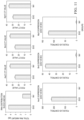
- FIGS. 15 A and 15 B show EV miR-128 expression determined by qRT-PCR from MEF cells transfected by miR-128 DNA plasmid using various techniques at 24 h post-transfection, where EVs were harvested from cell culture medium at 24 h post tranfection (miR-128 plasmid) by various techniques, total RNAs were obtained according to manufacturer's instructions, and the same amount of total RNA (30 ng) was used for miR-128 detection by qRT-PCR;
- FIGS. 16 A to 16 E compare secreted EVs containing miR-128 by NEP transfection of DNA plasmid to MEF cells vs. existing EVs loaded with pre-collected miR-128 by BEP post-insertion;
- FIGS. 17 A to 17 C compare secreted EVs containing Brn2 mRNA by NEP transfection of DNA plasmid to MEF cells vs. existing EVs loaded with pre-collected Brn2 mRNA by BEP post-insertion;
- FIG. 18 shows increased mRNA co-localization in the same EV by sequential-NEP.
- Ascl1, Brn2 and Myt1I plasmids were transfected at the same time as described before.
- the Myt1l plasmid was transfected first, Brn2 plasmid was transfected 4 h later, while Ascl1 plasmid was transfected 4 h after Brn2 transfection.
- culture medium was collected for TLN assay.
- Equal amount of FAM-Ascl1, Cy3-Brn2, and Cy5-Myt1l MBs were encapsulated in tethered lipoplex nanoparticles for EV-mRNA detection.
- Yellow arrow EVs containing 3 mRNAs
- Blue arrow EVs containing 2 mRNAs
- Pink arrow EVs containing 1 mRNA.
- FIG. 1 shows the schematic of a 3D NEP biochip with a single layer of donor cells laid on the chip surface.
- the DNA plasmids pre-loaded in PBS buffer were injected into individual donor cells via nanochannels using a 220 volts electric field across the nanochannels.
- electroporation conditions such as voltage level, pulse number and pulse length can be chosen.
- FIGS. 2 A and 2 B show transfected cells imaged using fluorescence microscopy 1 h after transfection by either BEP or NEP under a wavelength of 488 nm.
- the fluorescence intensity was calculated by NIS software. Comparison of fluorescence intensity in these two groups is given as bar charts. The results show that BEP at the manufacturer recommended best conditions could deliver nearly 3 folds more plasmids than NEP at 220 volts with five 10-ms pulses to the MEF cells.
- FIG. 3 compares EV numbers secreted from the same number of MEF cells (5E6 cells) transfected with the same Ascl1, Brn2 and Myt1l DNA plasmids at a weight ratio of 2/1/1 by either lipofectamine (Lipo), BEP or NEP. All EVs were collected from cell culture medium at 24 h post-transfection and the total EV number was determined by NanoSightTM.
- the transfection voltage was 1250 v with one 30-ms pulse.
- NEP the transfection voltage was 220 with five 10 ms pulses.
- the EVs were collected from cell culture medium by simply centrifugation at 1500 g for 10 mins.
- the results show that lipofectamine (Lipo) based cell transfection did not change the EV secretion.
- the EV concentration was around 2E9/ml with or without transfection.
- a slow plasmid endocytosis process by nanoparticle carriers would not stimulate the transfected cells much and, consequently, there was almost no change on EV secretion.
- BEP based cell transfection led to more EV secretion to ⁇ 6E9/ml.
- thermal shocking may increase cell secretion of EVs due to chaperone mediated autophage caused by the increase of heat shock proteins in cells (8-10). Indeed, we found that NEP could substantially increase the expression of both heat shock protein 70 (HSP70) by 13.8 folds and heat shock protein 90 (HSP90) by 4.2 folds in the transfected MEF cells vs. the non-transfected MEF cells (Ctrl). When HSP inhibitors were added in cell culture medium after electroporation, EV secretion could be suppressed.
- HSP70 heat shock protein 70
- HSP90 heat shock protein 90
- HSP 70 inhibitor (VER 155008, 50 ⁇ M) HSP90 inhibitor (NVP-HSP990, 1 ⁇ M)
- HSP90 inhibitor (NVP-HSP990, 1 ⁇ M)
- the cell culture was replaced with fresh medium containing HSP70 inhibitor (VER 155008), HSP90 inhibitor (NVP-HSP990), or their mixture right after NEP transfection.
- Medium was collected at 24 h post-transfection and EV numbers were detected by dynamic light scattering (DLS) goniometry.
- FIG. 5 shows the effect of NEP transfection of CD63 DNA plasmid on EV secretion from MEF cells.
- Cells were transfected with or without CD63 DNA plasmid by NEP. The cell culture medium was collected and replaced with fresh medium every 4 h. The EV numbers were detected by DLS goniometry. The results show a similar EV secretion profile during the first 16 h after NEP transfection in both cases. However, more EVs were secreted between 16 to 44 h after NEP transfection with CD63 DNA plasmid.
- CD63 protein is essential for the reorganization of endosomal membrane into tetraspanin enriched microdomains, a precursor of exosome secretion.
- FIGS. 6 A and 6 B show the EV size distribution measured by DLS goniometry for MEFs (ctrl) and NEP transfected MEFs. NEP stimulation did not change the larger EV (mostly microvesicles) distribution much, but substantially increased the secretion of exosomes with sizes ranging from 40 to 110 nm.
- FIGS. 8 A, 8 B, 9 A and 9 B show that the secreted EVs from NEP cell transfection of Ascl1, Brn2 and Myt1l DNA plasmids contain a large amount of corresponding Ascl1, Brn2 and Myt1l mRNAs or their fragments as determined using quantitative-Reverse Transcription Polymerase Chain Reaction (qRT-PCR).
- qRT-PCR quantitative-Reverse Transcription Polymerase Chain Reaction
- lipofectamine (Lipo) based cell transfection did not change the mRNA expression much, while the BEP based cell transfection could increase the mRNA expression several folds.
- the NEP based cell transfection resulted in thousands folds increase of target mRNAs.
- the same amount of total RNAs were obtained and reverse transcription was conducted by qRT-PCR according to manufacturer's instruction.
- FIG. 10 shows that some of the EV mRNAs were intact and functional because they were able to translate Ascl1, Brn2 and Myt1l proteins.
- a same amount of total RNA (1 ⁇ g) from each transfection method was applied for in vitro protein translation using Rabbit Reticulocyte Lysate System (Promega) according to manufacturer's instruction. Samples were separated by SDS-PAGE and the proteins were detected with various antibodies as shown in the Western blotting plot.
- RNAs were collected from these two parts as described above. The total mRNA concentration was measured by NanodropTM, while the ABM expressions of Ascl1, Brn2 and Myt1l mRNAs were measured by qRT-PCR.
- FIG. 11 shows that there was more than twice RNA in exosomes than in microvesicles, but most Ascl1, Brn2 and Myt1l mRNAs were presented only in exosomes.
- FIG. 13 shows the EV secretion and content profiles as a function of time after NEP transfection with Ascl1, Brn2 and Myt1l DNA plasmids.
- the Ascl1 plasmid is the smallest one (7 k bp) among the three, while the Myt1l plasmid is the largest (9 k bp) with the Brn2 plasmid in between (8 k bp).
- EVs in the cell culture medium was collected at the indicated time points, and the culture medium was replaced with fresh medium.
- the EV numbers were detected by DLS goniometry, while the EV mRNA expressions were detected by qRT-PCR as described before.
- NEP-produced-EVs containing endogenous mRNAs have therapeutic functions.
- the NEP-transfected MEF cells also showed a similar electrophysiological activity on Day 21. Cells displayed the necessary voltage-gated currents to fire action potentials. Both transient inward currents and sustained outward currents were observed in response to depolarizing voltage simulations. A typical response to a 20 pA current injection is illustrated in FIG. 14 and indicates that cells fired action potentials in response to depolarizing current.
- Glass electrodes (3-4 M ⁇ ) were filled with a pipette solution containing 115 mM K-gluconate, 10 mM N-2-Hydroxyethylpiperazine-N′-2-Ethanesulfonic Acid (HEPES), 4 mM NaCl, 0.5 mM ethylene glycol tetraacetic acid (EGTA), 1.5 mM MgCl 2 , (pH 7.3).
- Cells had a patch resistance of >100 MOhm after whole-cell access was gained, and series resistance was compensated 40-50%. Data were collected using an Axopatch 200B amplifier, Digidata 1322A digitizer, and Clampex 9 software (Molecular Devices, Sunnyvale, Calif.).
- the basal holding potential was ⁇ 70 mV and cells were stepped for 400 ms in 10 mV increments from ⁇ 120 mV to 80 mV.
- Transient inward currents due to activity of voltage-gated sodium channels, were isolated from measuring the peak amplitude.
- Sustained plateau currents reflective of voltage-gated potassium currents, were measured as the average of the last 50 ms of the voltage step in the plateau phase of the current.
- Action potential induction was measured using current clamp. Current was held at 0 pA and then stepped in 20 pA intervals for 1 sec.
- FIGS. 15 A and 15 B show the EV miR- 128 expression for EVs harvested from cell culture medium at 24 h post-transfection (miR-128 plasmid) by various techniques. Total RNAs were obtained according to manufacturer's instruction. The same amount of total RNA (30 ng) was used for miR-128 detection by qRT-PCR using the aforementioned procedures. Again, NEP based transfection was able to produce EVs containing a large amount of miR-128 (more than 4,500 folds increase), not achievable by BEP or lipofectamine based cell transfection.
- Example 3 Comparison of EVs Containing Endogenous RNAs by NEP Transfection of DNA Plasmid to MEF Cells Vs. Existing EVs Loaded with Pre-Collected RNAs by BEP Post-Insertion
- the miR-128 plasmid was co-transfected with CD63-GFP plasmid to MEF cells by NEP to generate EVs containing miR-128 according to aforementioned procedures.
- blank EVs were first harvested from MEF cells transfected with CD63-GFP plasmid 24 h after NEP.
- miR-128 was collected from MEF cells transfected with miR-128 plasmid 24 h post-transfection by NEP.
- FIG. 16 A shows the TLN-TIRF assay schematic (2, 11).
- a molecular beacon (MB) for the RNA target is designed and encapsulated in cationic liposomal nanoparticles.
- cationic lipoplex nanoparticles are tethered on a glass slide, which are able to capture negatively charged EVs by electrical static interactions to form a larger nanoscale complex.
- This lipoplex-EV fusion leads to mixing of RNAs and MBs within the nanoscale confinement near the biochip interface.
- TIRF microscopy is capable of detecting a single biomolecule and it measures signals ⁇ 300 nm near the interface, which is where the tethered liposomal nanoparticles locate.
- FIG. 16 B shows the representative TLN-TIRF images of the captured EVs.
- the green fluorescence is from EVs containing CD63-GFP, while the red fluorescence is from hybridization of miR-128 molecules and the Cy5-miR128 MBs in the captured EVs. It is clear that our NEP approach is able to produce more EVs containing higher copies of miR-128 than the BEP post-insertion approach.
- FIGS. 16 C-E show a quantitative comparison of those two approaches.
- FIGS. 17 A to 17 C show that our NEP approach could produce >70% EVs containing Brn2 mRNA, while only very few existing EVs could be loaded with the same mRNA by BEP post-insertion approach.
- the concentration of Brn2 mRNA in NEP produced EVs is high, while that in BEP post-insertion is very poor.
- FIG. 13 implies that different mRNA targets could be transcribed at different times and rates in the transfected cells, even though multiple DNA plasmids were delivered to the cells at the same time, due to the size difference of plasmids or other reasons. This may lead to individual EVs containing only one or few mRNA targets. For better therapeutic efficacy, it would be valuable if more or all mRNA targets can be encapsulated in the same secreted EVs.
- FIG. 18 shows that we could substantially increase the secreted EVs containing all three mRNAs, Ascl1, Brn2 and Myt1l (>50% vs.
Landscapes
- Health & Medical Sciences (AREA)
- Life Sciences & Earth Sciences (AREA)
- Engineering & Computer Science (AREA)
- Genetics & Genomics (AREA)
- Chemical & Material Sciences (AREA)
- Zoology (AREA)
- Bioinformatics & Cheminformatics (AREA)
- Organic Chemistry (AREA)
- Wood Science & Technology (AREA)
- Biotechnology (AREA)
- Biomedical Technology (AREA)
- General Engineering & Computer Science (AREA)
- General Health & Medical Sciences (AREA)
- Biochemistry (AREA)
- Microbiology (AREA)
- Molecular Biology (AREA)
- Biophysics (AREA)
- Physics & Mathematics (AREA)
- Cell Biology (AREA)
- Plant Pathology (AREA)
- Sustainable Development (AREA)
- Medicinal Chemistry (AREA)
- Immunology (AREA)
- Electromagnetism (AREA)
- Animal Behavior & Ethology (AREA)
- Veterinary Medicine (AREA)
- Public Health (AREA)
- Virology (AREA)
- Pharmacology & Pharmacy (AREA)
- Epidemiology (AREA)
- Clinical Laboratory Science (AREA)
- Dispersion Chemistry (AREA)
- Botany (AREA)
- Toxicology (AREA)
- Gastroenterology & Hepatology (AREA)
- Proteomics, Peptides & Aminoacids (AREA)
- Developmental Biology & Embryology (AREA)
- Rheumatology (AREA)
- Micro-Organisms Or Cultivation Processes Thereof (AREA)
- Medicines That Contain Protein Lipid Enzymes And Other Medicines (AREA)
Priority Applications (1)
| Application Number | Priority Date | Filing Date | Title |
|---|---|---|---|
| US16/635,471 US11674130B2 (en) | 2017-08-04 | 2018-08-06 | Method for producing therapeutic exosomes from nanoelectroporation and other non-endocytic cell transfection |
Applications Claiming Priority (3)
| Application Number | Priority Date | Filing Date | Title |
|---|---|---|---|
| US201762541157P | 2017-08-04 | 2017-08-04 | |
| PCT/US2018/045333 WO2019028450A1 (fr) | 2017-08-04 | 2018-08-06 | Procédé de production d'exosomes thérapeutiques à partir de nanoélectroporation et autre transfection de cellule non endocytique |
| US16/635,471 US11674130B2 (en) | 2017-08-04 | 2018-08-06 | Method for producing therapeutic exosomes from nanoelectroporation and other non-endocytic cell transfection |
Related Parent Applications (1)
| Application Number | Title | Priority Date | Filing Date |
|---|---|---|---|
| PCT/US2018/045333 A-371-Of-International WO2019028450A1 (fr) | 2017-08-04 | 2018-08-06 | Procédé de production d'exosomes thérapeutiques à partir de nanoélectroporation et autre transfection de cellule non endocytique |
Related Child Applications (1)
| Application Number | Title | Priority Date | Filing Date |
|---|---|---|---|
| US18/199,442 Continuation US12359188B2 (en) | 2017-08-04 | 2023-05-19 | Method for producing therapeutic exosomes from nanoelectroporation and other non-endocytic cell transfection |
Publications (2)
| Publication Number | Publication Date |
|---|---|
| US20210054359A1 US20210054359A1 (en) | 2021-02-25 |
| US11674130B2 true US11674130B2 (en) | 2023-06-13 |
Family
ID=65233122
Family Applications (3)
| Application Number | Title | Priority Date | Filing Date |
|---|---|---|---|
| US16/635,471 Active US11674130B2 (en) | 2017-08-04 | 2018-08-06 | Method for producing therapeutic exosomes from nanoelectroporation and other non-endocytic cell transfection |
| US18/199,442 Active US12359188B2 (en) | 2017-08-04 | 2023-05-19 | Method for producing therapeutic exosomes from nanoelectroporation and other non-endocytic cell transfection |
| US19/223,920 Pending US20250290061A1 (en) | 2017-08-04 | 2025-05-30 | Method for producing therapeutic exosomes from nanoelectroporation and other non-endocytic cell transfection |
Family Applications After (2)
| Application Number | Title | Priority Date | Filing Date |
|---|---|---|---|
| US18/199,442 Active US12359188B2 (en) | 2017-08-04 | 2023-05-19 | Method for producing therapeutic exosomes from nanoelectroporation and other non-endocytic cell transfection |
| US19/223,920 Pending US20250290061A1 (en) | 2017-08-04 | 2025-05-30 | Method for producing therapeutic exosomes from nanoelectroporation and other non-endocytic cell transfection |
Country Status (10)
| Country | Link |
|---|---|
| US (3) | US11674130B2 (fr) |
| EP (1) | EP3661485A4 (fr) |
| JP (3) | JP7420708B2 (fr) |
| KR (3) | KR102888092B1 (fr) |
| CN (2) | CN117987467A (fr) |
| AU (2) | AU2018312099C1 (fr) |
| CA (1) | CA3071553A1 (fr) |
| IL (1) | IL272401A (fr) |
| SG (1) | SG11202000851YA (fr) |
| WO (1) | WO2019028450A1 (fr) |
Cited By (2)
| Publication number | Priority date | Publication date | Assignee | Title |
|---|---|---|---|---|
| US20210093567A1 (en) * | 2019-08-06 | 2021-04-01 | Ohio State Innovation Foundation | Therapeutic extracellular vesicles |
| WO2025083467A2 (fr) | 2023-10-17 | 2025-04-24 | Spot Biosystems Ltd. | Dispositifs et procédés d'électroporation multi-puits |
Families Citing this family (7)
| Publication number | Priority date | Publication date | Assignee | Title |
|---|---|---|---|---|
| US11491483B2 (en) | 2018-02-15 | 2022-11-08 | Ohio State Innovation Foundation | Microfluidic devices and methods for high throughput electroporation |
| CN111378685B (zh) * | 2020-02-15 | 2023-10-31 | 深圳承启生物科技有限公司 | 一种制备载药外泌体的方法及载药外泌体 |
| JP2023524397A (ja) * | 2020-05-01 | 2023-06-12 | ザ・トラスティーズ・オブ・インディアナ・ユニバーシティー | 糖尿病性皮膚多発ニューロパシーの管理における神経性組織ナノトランスフェクション |
| CN112226365A (zh) * | 2020-10-13 | 2021-01-15 | 北京航空航天大学 | 基于单细胞阵列的纳米电穿孔装置及其应用 |
| KR102568745B1 (ko) * | 2021-05-18 | 2023-08-24 | 주식회사 바이오솔루션 | 세포외소포체 분비능이 증진된 면역세포 및 이를 활용한 면역 항암요법 |
| US20240226133A1 (en) * | 2023-01-09 | 2024-07-11 | Claudia Chimisso Dos Santos | Microrna-based particle for the treatment of dysregulated immune response |
| CN118988431B (zh) * | 2024-08-23 | 2025-09-16 | 北京航空航天大学 | 工程化细胞外囊泡量产与富集的集成微流控芯片系统与方法 |
Citations (5)
| Publication number | Priority date | Publication date | Assignee | Title |
|---|---|---|---|---|
| US7338796B1 (en) * | 2003-08-13 | 2008-03-04 | Sandia Corporation | Vesicle-based method and apparatus for collecting, manipulating, and chemically processing trace macromolecular species |
| CN102596177A (zh) | 2009-07-01 | 2012-07-18 | 阿昂梅迪克斯公司 | 来源于有核哺乳动物细胞的微囊泡及其应用 |
| US8524679B2 (en) * | 2006-11-08 | 2013-09-03 | Veritas Bio, Llc | In vivo delivery of double stranded RNA to a target cell |
| US20140256047A1 (en) * | 2010-07-06 | 2014-09-11 | The Ohio State University | Dose and location controlled drug/gene/particle delivery to individual cells by nanoelectroporation |
| WO2017054086A1 (fr) | 2015-10-01 | 2017-04-06 | Exerkine Corporation | Traitement de myopathies génétiques au moyen d'exosomes mis au point par génie biologique |
Family Cites Families (3)
| Publication number | Priority date | Publication date | Assignee | Title |
|---|---|---|---|---|
| ATE512982T1 (de) * | 2001-08-17 | 2011-07-15 | Exothera L L C | Verfahren und zusammensetzung zum gezielten einbringen in exosome |
| EP3569254B1 (fr) * | 2009-04-17 | 2022-07-20 | Oxford University Innovation Limited | Composition d'administration de matériau génétique |
| EP3037515A1 (fr) * | 2014-12-28 | 2016-06-29 | Femtofab Co., Ltd. | Cellule modifiée préparée par placement de matériau dans la cellule sans utiliser de véhicule d'administration |
-
2018
- 2018-08-06 WO PCT/US2018/045333 patent/WO2019028450A1/fr not_active Ceased
- 2018-08-06 AU AU2018312099A patent/AU2018312099C1/en active Active
- 2018-08-06 JP JP2020505790A patent/JP7420708B2/ja active Active
- 2018-08-06 CA CA3071553A patent/CA3071553A1/fr active Pending
- 2018-08-06 CN CN202410102849.3A patent/CN117987467A/zh active Pending
- 2018-08-06 KR KR1020247015455A patent/KR102888092B1/ko active Active
- 2018-08-06 SG SG11202000851YA patent/SG11202000851YA/en unknown
- 2018-08-06 KR KR1020207006189A patent/KR102667894B1/ko active Active
- 2018-08-06 KR KR1020257038045A patent/KR20250164879A/ko active Pending
- 2018-08-06 US US16/635,471 patent/US11674130B2/en active Active
- 2018-08-06 CN CN201880065004.4A patent/CN111194210B/zh active Active
- 2018-08-06 EP EP18841065.8A patent/EP3661485A4/fr active Pending
-
2020
- 2020-02-02 IL IL272401A patent/IL272401A/en unknown
-
2023
- 2023-05-19 US US18/199,442 patent/US12359188B2/en active Active
- 2023-09-20 JP JP2023152497A patent/JP7712333B2/ja active Active
-
2024
- 2024-10-09 AU AU2024227190A patent/AU2024227190A1/en active Pending
-
2025
- 2025-05-30 US US19/223,920 patent/US20250290061A1/en active Pending
- 2025-07-09 JP JP2025115485A patent/JP2025137558A/ja active Pending
Patent Citations (5)
| Publication number | Priority date | Publication date | Assignee | Title |
|---|---|---|---|---|
| US7338796B1 (en) * | 2003-08-13 | 2008-03-04 | Sandia Corporation | Vesicle-based method and apparatus for collecting, manipulating, and chemically processing trace macromolecular species |
| US8524679B2 (en) * | 2006-11-08 | 2013-09-03 | Veritas Bio, Llc | In vivo delivery of double stranded RNA to a target cell |
| CN102596177A (zh) | 2009-07-01 | 2012-07-18 | 阿昂梅迪克斯公司 | 来源于有核哺乳动物细胞的微囊泡及其应用 |
| US20140256047A1 (en) * | 2010-07-06 | 2014-09-11 | The Ohio State University | Dose and location controlled drug/gene/particle delivery to individual cells by nanoelectroporation |
| WO2017054086A1 (fr) | 2015-10-01 | 2017-04-06 | Exerkine Corporation | Traitement de myopathies génétiques au moyen d'exosomes mis au point par génie biologique |
Non-Patent Citations (12)
| Title |
|---|
| CFC, Chinese Oncology Clinical Yearbook 2015, China Cancer Foundation et al., Sep. 2016, pp. 36-37. |
| Chang, Lingqian et al., 3D Nanochannel Electroporation for High-Throughput Cell Transfection with High Uniformity and Dosage Control, Figures 1-3, 2016. |
| Chang, Lingqian et al., 3D Nanochannel Electroporation for High-Throughput Cell Transfection with High Uniformity and Dosage Control, Nanoscale, 2016, vol. 8, pp. 243-252. |
| Kanuma, T. et al. May 15, 2017. CD63-mediated antigen delivery into extracellular vesicles via DNA vaccination results in robust CD8+ T cell responses. The Journal of Immunology 198: 1-9; specif, pp. 1, 2, 6. * |
| Lamichhane, T.N. et al. 2015. Exogenous DNA loading into extracellular vesicles via electroporation is size-dependent and enables limited gene delivery. Molecular Pharmaceutics 12: 3650-3657; specif. p. 3650. * |
| Mcknight, T.E. et al. 2004. Tracking gene expression after DNA delivery using spatially indexed nanofiber arrays. Nano Letters 4(7) : 1213-1219; specif, pp. 1213, 1214, 1217, 1218, 1219. * |
| Mizrak, A. et al., Genetically Engineered Microvesicles Carrying Suicide mRNA/Protein Inhibit Schwannoma Tumor Growth, The American Society of Gene & Cell Therapy, Molecular Therapy, Jan. 2013, pp. 101-108, vol. 21, No. 1. |
| Obregon, C. et al. 2006. Exovesicles from human activated dendritic cells fuse with resting dendritic cells, allowing them to present alloantigens. American Journal of Pathology 169(6): 2127-2136; specif. pp. 2127, 2129, 2131. * |
| Pegtel, D.M. et al., Functional delivery of viral miRNAs via exosomes, The Proceedings of the National Academy of Sciences, Apr. 6, 2010, pp. 6328-6333, vol. 107, No. 14. |
| Raposo, G. et al. 2013. Extracellular vesicles: exosomes, microvesicles, and friends. Journal of Cell Biology 200(4): 373-383; specif. pp. 373, 377. * |
| Shi, Junfeng, Development of Nanoelectroporation-based Biochips for Living Cell Interrogation and Extracellular Vesicle Engineering, Thesis, May 10, 2017, pp. 130-136. |
| Tabar, M.S. et al. 2015. Evaluating electroporation and lipofectamine approaches for transient and stable transgene expressions in human fibroblasts and embryonic stem cells. Cell Journal 17(3): 438-450; specif. pp. 438, 445, 446. * |
Cited By (2)
| Publication number | Priority date | Publication date | Assignee | Title |
|---|---|---|---|---|
| US20210093567A1 (en) * | 2019-08-06 | 2021-04-01 | Ohio State Innovation Foundation | Therapeutic extracellular vesicles |
| WO2025083467A2 (fr) | 2023-10-17 | 2025-04-24 | Spot Biosystems Ltd. | Dispositifs et procédés d'électroporation multi-puits |
Also Published As
| Publication number | Publication date |
|---|---|
| US12359188B2 (en) | 2025-07-15 |
| EP3661485A4 (fr) | 2021-05-12 |
| JP2025137558A (ja) | 2025-09-19 |
| KR102667894B1 (ko) | 2024-05-22 |
| WO2019028450A1 (fr) | 2019-02-07 |
| CN117987467A (zh) | 2024-05-07 |
| SG11202000851YA (en) | 2020-02-27 |
| IL272401A (en) | 2020-03-31 |
| EP3661485A1 (fr) | 2020-06-10 |
| KR20240093552A (ko) | 2024-06-24 |
| AU2018312099C1 (en) | 2024-10-24 |
| US20250290061A1 (en) | 2025-09-18 |
| US20210054359A1 (en) | 2021-02-25 |
| KR102888092B1 (ko) | 2025-11-18 |
| JP7712333B2 (ja) | 2025-07-23 |
| AU2024227190A1 (en) | 2024-10-31 |
| CN111194210A (zh) | 2020-05-22 |
| AU2018312099B2 (en) | 2024-07-11 |
| KR20200035107A (ko) | 2020-04-01 |
| JP2023164661A (ja) | 2023-11-10 |
| CN111194210B (zh) | 2024-02-20 |
| US20230313170A1 (en) | 2023-10-05 |
| AU2018312099A1 (en) | 2020-02-27 |
| JP7420708B2 (ja) | 2024-01-23 |
| KR20250164879A (ko) | 2025-11-25 |
| CA3071553A1 (fr) | 2019-02-07 |
| JP2020529210A (ja) | 2020-10-08 |
Similar Documents
| Publication | Publication Date | Title |
|---|---|---|
| US20230313170A1 (en) | Method for producing therapeutic exosomes from nanoelectroporation and other non-endocytic cell transfection | |
| JP7591265B2 (ja) | 破壊及び場による細胞への化合物及び組成物の送達 | |
| US12241063B2 (en) | Modified messenger RNA comprising functional RNA elements | |
| Golzio et al. | In vitro and in vivo electric field-mediated permeabilization, gene transfer, and expression | |
| CN107058101B (zh) | 细胞内传递 | |
| Guo et al. | The application of mRNA-based gene transfer in mesenchymal stem cell-mediated cytotoxicity of glioma cells | |
| Liu et al. | Nanochannel electro‐injection as a versatile platform for efficient RNA/DNA programming on dendritic cells | |
| Haberl et al. | Effect of Mg ions on efficiency of gene electrotransfer and on cell electropermeabilization | |
| Faurie et al. | Electric Field‐Induced Cell Membrane Permeabilization and Gene Transfer: Theory and Experiments | |
| HK40112139A (zh) | 一种利用纳米电穿孔和其他非内吞的细胞转染制备治疗性外泌体的方法 | |
| KR20230100212A (ko) | 비바이러스 기반 고효율 핵산 형질주입용 조성물 및 그 용도 | |
| JP2019511566A (ja) | 核酸凝縮ペプチド、核酸凝縮ペプチドセット、核酸送達キャリア、核酸送達方法、細胞作製方法、細胞検出方法及びキット | |
| Golzio et al. | siRNA delivery via electropulsation: a review of the basic processes | |
| KR20250165238A (ko) | 원형질막 수포 기반 양극성 막단백질 발현 나노 소포와 이의 제조 방법 및 이의 용도 | |
| Liu | Coupling Micro/Nanoscale Electroporation to Enhance Genetic Probe Delivery for Cell Therapy | |
| Pachamuthu et al. | Electroporation induced membrane permeability changes at the ionic level | |
| VIVO | Sergei I. Sukharev, Alexander V. Titomirov, and Vadim A. Klenchin |
Legal Events
| Date | Code | Title | Description |
|---|---|---|---|
| FEPP | Fee payment procedure |
Free format text: ENTITY STATUS SET TO UNDISCOUNTED (ORIGINAL EVENT CODE: BIG.); ENTITY STATUS OF PATENT OWNER: SMALL ENTITY |
|
| STPP | Information on status: patent application and granting procedure in general |
Free format text: APPLICATION UNDERGOING PREEXAM PROCESSING |
|
| FEPP | Fee payment procedure |
Free format text: ENTITY STATUS SET TO SMALL (ORIGINAL EVENT CODE: SMAL); ENTITY STATUS OF PATENT OWNER: SMALL ENTITY |
|
| AS | Assignment |
Owner name: OHIO STATE INNOVATION FOUNDATION, OHIO Free format text: ASSIGNMENT OF ASSIGNORS INTEREST;ASSIGNORS:SHI, JUNFENG;YANG, ZHAOGANG;LEE, LY JAMES;SIGNING DATES FROM 20200522 TO 20200717;REEL/FRAME:053248/0588 Owner name: OHIO STATE INNOVATION FOUNDATION, OHIO Free format text: ASSIGNMENT OF ASSIGNORS INTEREST;ASSIGNORS:SHI, JUNFENG;YANG, ZHAOGANG;LEE, LY JAMES;SIGNING DATES FROM 20200522 TO 20200717;REEL/FRAME:053248/0628 |
|
| STPP | Information on status: patent application and granting procedure in general |
Free format text: APPLICATION DISPATCHED FROM PREEXAM, NOT YET DOCKETED |
|
| STPP | Information on status: patent application and granting procedure in general |
Free format text: DOCKETED NEW CASE - READY FOR EXAMINATION |
|
| STPP | Information on status: patent application and granting procedure in general |
Free format text: NON FINAL ACTION MAILED |
|
| STPP | Information on status: patent application and granting procedure in general |
Free format text: RESPONSE TO NON-FINAL OFFICE ACTION ENTERED AND FORWARDED TO EXAMINER |
|
| STPP | Information on status: patent application and granting procedure in general |
Free format text: FINAL REJECTION MAILED |
|
| STCF | Information on status: patent grant |
Free format text: PATENTED CASE |
|
| FEPP | Fee payment procedure |
Free format text: ENTITY STATUS SET TO UNDISCOUNTED (ORIGINAL EVENT CODE: BIG.); ENTITY STATUS OF PATENT OWNER: LARGE ENTITY |