US12071663B2 - Semi-permeable arrays for analyzing biological systems and methods of using same - Google Patents
Semi-permeable arrays for analyzing biological systems and methods of using same Download PDFInfo
- Publication number
- US12071663B2 US12071663B2 US16/070,505 US201716070505A US12071663B2 US 12071663 B2 US12071663 B2 US 12071663B2 US 201716070505 A US201716070505 A US 201716070505A US 12071663 B2 US12071663 B2 US 12071663B2
- Authority
- US
- United States
- Prior art keywords
- array
- wells
- cells
- cell
- container
- Prior art date
- Legal status (The legal status is an assumption and is not a legal conclusion. Google has not performed a legal analysis and makes no representation as to the accuracy of the status listed.)
- Active, expires
Links
- 238000000034 method Methods 0.000 title claims abstract description 165
- 238000003491 array Methods 0.000 title description 71
- 239000012528 membrane Substances 0.000 claims abstract description 237
- 238000012742 biochemical analysis Methods 0.000 claims abstract description 23
- 210000004027 cell Anatomy 0.000 claims description 419
- 108090000623 proteins and genes Proteins 0.000 claims description 133
- 239000011324 bead Substances 0.000 claims description 119
- -1 polydimethylsiloxane Polymers 0.000 claims description 77
- 150000007523 nucleic acids Chemical class 0.000 claims description 53
- 102000039446 nucleic acids Human genes 0.000 claims description 50
- 108020004707 nucleic acids Proteins 0.000 claims description 50
- 239000004205 dimethyl polysiloxane Substances 0.000 claims description 42
- 102000004169 proteins and genes Human genes 0.000 claims description 42
- 229920000435 poly(dimethylsiloxane) Polymers 0.000 claims description 41
- 108020004999 messenger RNA Proteins 0.000 claims description 38
- 125000000524 functional group Chemical group 0.000 claims description 36
- 150000001412 amines Chemical class 0.000 claims description 33
- 108091032973 (ribonucleotides)n+m Proteins 0.000 claims description 27
- 229920001661 Chitosan Polymers 0.000 claims description 27
- 239000011347 resin Substances 0.000 claims description 27
- 229920005989 resin Polymers 0.000 claims description 27
- 239000000463 material Substances 0.000 claims description 24
- 238000006243 chemical reaction Methods 0.000 claims description 20
- 238000002474 experimental method Methods 0.000 claims description 19
- 238000007789 sealing Methods 0.000 claims description 19
- 229920002521 macromolecule Polymers 0.000 claims description 17
- 239000004417 polycarbonate Substances 0.000 claims description 17
- 229920000515 polycarbonate Polymers 0.000 claims description 17
- 238000000108 ultra-filtration Methods 0.000 claims description 17
- 108020004414 DNA Proteins 0.000 claims description 16
- 239000007864 aqueous solution Substances 0.000 claims description 16
- 239000011148 porous material Substances 0.000 claims description 16
- 239000011521 glass Substances 0.000 claims description 15
- 230000006037 cell lysis Effects 0.000 claims description 13
- 229920003229 poly(methyl methacrylate) Polymers 0.000 claims description 13
- 239000004926 polymethyl methacrylate Substances 0.000 claims description 13
- 230000002441 reversible effect Effects 0.000 claims description 12
- 229920000936 Agarose Polymers 0.000 claims description 11
- 238000002955 isolation Methods 0.000 claims description 11
- 239000002105 nanoparticle Substances 0.000 claims description 11
- 229910000077 silane Inorganic materials 0.000 claims description 11
- 229920000089 Cyclic olefin copolymer Polymers 0.000 claims description 10
- 239000004793 Polystyrene Substances 0.000 claims description 10
- BLRPTPMANUNPDV-UHFFFAOYSA-N Silane Chemical compound [SiH4] BLRPTPMANUNPDV-UHFFFAOYSA-N 0.000 claims description 10
- 238000000576 coating method Methods 0.000 claims description 10
- 239000004743 Polypropylene Substances 0.000 claims description 9
- 239000011248 coating agent Substances 0.000 claims description 9
- 239000007788 liquid Substances 0.000 claims description 9
- 229920001223 polyethylene glycol Polymers 0.000 claims description 9
- 229920001155 polypropylene Polymers 0.000 claims description 9
- 238000004113 cell culture Methods 0.000 claims description 8
- 229920000656 polylysine Polymers 0.000 claims description 8
- 238000004163 cytometry Methods 0.000 claims description 7
- 150000004676 glycans Chemical class 0.000 claims description 7
- 230000004048 modification Effects 0.000 claims description 7
- 238000012986 modification Methods 0.000 claims description 7
- 229920001282 polysaccharide Polymers 0.000 claims description 7
- 239000005017 polysaccharide Substances 0.000 claims description 7
- 229920002223 polystyrene Polymers 0.000 claims description 7
- 229920002981 polyvinylidene fluoride Polymers 0.000 claims description 7
- 238000011282 treatment Methods 0.000 claims description 7
- 150000001298 alcohols Chemical class 0.000 claims description 6
- 125000002887 hydroxy group Chemical group [H]O* 0.000 claims description 6
- FZHAPNGMFPVSLP-UHFFFAOYSA-N silanamine Chemical compound [SiH3]N FZHAPNGMFPVSLP-UHFFFAOYSA-N 0.000 claims description 6
- 238000010186 staining Methods 0.000 claims description 6
- 230000001580 bacterial effect Effects 0.000 claims description 5
- 150000001282 organosilanes Chemical class 0.000 claims description 5
- 125000005375 organosiloxane group Chemical group 0.000 claims description 5
- 239000004800 polyvinyl chloride Substances 0.000 claims description 5
- 239000007787 solid Substances 0.000 claims description 5
- CNHYKKNIIGEXAY-UHFFFAOYSA-N thiolan-2-imine Chemical compound N=C1CCCS1 CNHYKKNIIGEXAY-UHFFFAOYSA-N 0.000 claims description 5
- 229920002125 Sokalan® Polymers 0.000 claims description 4
- AHGFXGSMYLFWEC-UHFFFAOYSA-N [SiH4].CC(=C)C(O)=O Chemical compound [SiH4].CC(=C)C(O)=O AHGFXGSMYLFWEC-UHFFFAOYSA-N 0.000 claims description 4
- 102000025171 antigen binding proteins Human genes 0.000 claims description 4
- 108091000831 antigen binding proteins Proteins 0.000 claims description 4
- 239000001257 hydrogen Substances 0.000 claims description 4
- 229910052739 hydrogen Inorganic materials 0.000 claims description 4
- 229920000083 poly(allylamine) Polymers 0.000 claims description 4
- 239000004584 polyacrylic acid Substances 0.000 claims description 4
- 239000010703 silicon Substances 0.000 claims description 4
- 229910052710 silicon Inorganic materials 0.000 claims description 4
- ATGUDZODTABURZ-UHFFFAOYSA-N thiolan-2-ylideneazanium;chloride Chemical compound Cl.N=C1CCCS1 ATGUDZODTABURZ-UHFFFAOYSA-N 0.000 claims description 4
- 229920002101 Chitin Polymers 0.000 claims description 3
- 125000002091 cationic group Chemical group 0.000 claims description 3
- 239000001913 cellulose Substances 0.000 claims description 3
- 229920002678 cellulose Polymers 0.000 claims description 3
- 230000002209 hydrophobic effect Effects 0.000 claims description 3
- 210000004962 mammalian cell Anatomy 0.000 claims description 3
- 229920002307 Dextran Polymers 0.000 claims description 2
- PEEHTFAAVSWFBL-UHFFFAOYSA-N Maleimide Chemical compound O=C1NC(=O)C=C1 PEEHTFAAVSWFBL-UHFFFAOYSA-N 0.000 claims description 2
- 239000012954 diazonium Substances 0.000 claims description 2
- 150000002118 epoxides Chemical class 0.000 claims description 2
- PCHJSUWPFVWCPO-UHFFFAOYSA-N gold Chemical compound [Au] PCHJSUWPFVWCPO-UHFFFAOYSA-N 0.000 claims description 2
- 239000010931 gold Substances 0.000 claims description 2
- 229910052737 gold Inorganic materials 0.000 claims description 2
- 239000003014 ion exchange membrane Substances 0.000 claims description 2
- 229920000915 polyvinyl chloride Polymers 0.000 claims description 2
- 229920002239 polyacrylonitrile Polymers 0.000 claims 2
- 239000000020 Nitrocellulose Substances 0.000 claims 1
- 239000004695 Polyether sulfone Substances 0.000 claims 1
- 125000003178 carboxy group Chemical group [H]OC(*)=O 0.000 claims 1
- 229920001220 nitrocellulos Polymers 0.000 claims 1
- 229920006393 polyether sulfone Polymers 0.000 claims 1
- 239000000872 buffer Substances 0.000 description 49
- 230000014509 gene expression Effects 0.000 description 43
- 238000012163 sequencing technique Methods 0.000 description 39
- 230000001413 cellular effect Effects 0.000 description 38
- 235000013870 dimethyl polysiloxane Nutrition 0.000 description 38
- 235000018102 proteins Nutrition 0.000 description 38
- 239000000243 solution Substances 0.000 description 35
- 238000011068 loading method Methods 0.000 description 34
- 238000004458 analytical method Methods 0.000 description 31
- 210000003819 peripheral blood mononuclear cell Anatomy 0.000 description 31
- 239000000523 sample Substances 0.000 description 28
- 230000003321 amplification Effects 0.000 description 23
- 238000005516 engineering process Methods 0.000 description 23
- 238000003199 nucleic acid amplification method Methods 0.000 description 23
- 108091003079 Bovine Serum Albumin Proteins 0.000 description 21
- 239000012530 fluid Substances 0.000 description 21
- 238000007306 functionalization reaction Methods 0.000 description 21
- 239000013615 primer Substances 0.000 description 21
- 238000001514 detection method Methods 0.000 description 20
- 239000012139 lysis buffer Substances 0.000 description 20
- 229920000642 polymer Polymers 0.000 description 20
- 238000003752 polymerase chain reaction Methods 0.000 description 20
- 239000000047 product Substances 0.000 description 19
- 210000001519 tissue Anatomy 0.000 description 18
- 239000003153 chemical reaction reagent Substances 0.000 description 17
- 210000002540 macrophage Anatomy 0.000 description 17
- YBJHBAHKTGYVGT-ZKWXMUAHSA-N (+)-Biotin Chemical compound N1C(=O)N[C@@H]2[C@H](CCCCC(=O)O)SC[C@@H]21 YBJHBAHKTGYVGT-ZKWXMUAHSA-N 0.000 description 16
- LOKCTEFSRHRXRJ-UHFFFAOYSA-I dipotassium trisodium dihydrogen phosphate hydrogen phosphate dichloride Chemical compound P(=O)(O)(O)[O-].[K+].P(=O)(O)([O-])[O-].[Na+].[Na+].[Cl-].[K+].[Cl-].[Na+] LOKCTEFSRHRXRJ-UHFFFAOYSA-I 0.000 description 16
- 239000000203 mixture Substances 0.000 description 16
- 239000002953 phosphate buffered saline Substances 0.000 description 16
- 238000003559 RNA-seq method Methods 0.000 description 15
- 238000003556 assay Methods 0.000 description 15
- 239000012091 fetal bovine serum Substances 0.000 description 15
- 238000009396 hybridization Methods 0.000 description 15
- 210000001744 T-lymphocyte Anatomy 0.000 description 13
- 238000005259 measurement Methods 0.000 description 13
- 230000008569 process Effects 0.000 description 13
- 108091034117 Oligonucleotide Proteins 0.000 description 12
- 239000000017 hydrogel Substances 0.000 description 12
- 238000002372 labelling Methods 0.000 description 12
- 238000002156 mixing Methods 0.000 description 12
- 210000001616 monocyte Anatomy 0.000 description 12
- 241000894007 species Species 0.000 description 12
- 238000009792 diffusion process Methods 0.000 description 11
- 230000001965 increasing effect Effects 0.000 description 11
- 239000003599 detergent Substances 0.000 description 10
- 238000010195 expression analysis Methods 0.000 description 10
- 229920000370 gamma-poly(glutamate) polymer Polymers 0.000 description 10
- 238000003384 imaging method Methods 0.000 description 10
- 238000011160 research Methods 0.000 description 10
- 239000000126 substance Substances 0.000 description 10
- 239000000758 substrate Substances 0.000 description 10
- 238000012546 transfer Methods 0.000 description 10
- 102000004190 Enzymes Human genes 0.000 description 9
- 108090000790 Enzymes Proteins 0.000 description 9
- 210000003719 b-lymphocyte Anatomy 0.000 description 9
- 239000000470 constituent Substances 0.000 description 9
- 229940088598 enzyme Drugs 0.000 description 9
- 239000012634 fragment Substances 0.000 description 9
- 150000002500 ions Chemical class 0.000 description 9
- 201000008827 tuberculosis Diseases 0.000 description 9
- 239000006146 Roswell Park Memorial Institute medium Substances 0.000 description 8
- 239000000427 antigen Substances 0.000 description 8
- 108091007433 antigens Proteins 0.000 description 8
- 102000036639 antigens Human genes 0.000 description 8
- 239000012472 biological sample Substances 0.000 description 8
- 150000007942 carboxylates Chemical class 0.000 description 8
- 239000006285 cell suspension Substances 0.000 description 8
- 239000002299 complementary DNA Substances 0.000 description 8
- 150000001875 compounds Chemical class 0.000 description 8
- 208000037265 diseases, disorders, signs and symptoms Diseases 0.000 description 8
- 230000000694 effects Effects 0.000 description 8
- 230000006870 function Effects 0.000 description 8
- 238000013507 mapping Methods 0.000 description 8
- 238000003786 synthesis reaction Methods 0.000 description 8
- 108020005196 Mitochondrial DNA Proteins 0.000 description 7
- 238000013459 approach Methods 0.000 description 7
- 230000015572 biosynthetic process Effects 0.000 description 7
- 229960002685 biotin Drugs 0.000 description 7
- 235000020958 biotin Nutrition 0.000 description 7
- 239000011616 biotin Substances 0.000 description 7
- 239000007795 chemical reaction product Substances 0.000 description 7
- 230000009089 cytolysis Effects 0.000 description 7
- 150000002540 isothiocyanates Chemical class 0.000 description 7
- 238000010839 reverse transcription Methods 0.000 description 7
- 238000000926 separation method Methods 0.000 description 7
- 238000005406 washing Methods 0.000 description 7
- RPFLVLIPBDQGAQ-UHFFFAOYSA-N 1,2-diisothiocyanatobenzene Chemical compound S=C=NC1=CC=CC=C1N=C=S RPFLVLIPBDQGAQ-UHFFFAOYSA-N 0.000 description 6
- KCXVZYZYPLLWCC-UHFFFAOYSA-N EDTA Chemical compound OC(=O)CN(CC(O)=O)CCN(CC(O)=O)CC(O)=O KCXVZYZYPLLWCC-UHFFFAOYSA-N 0.000 description 6
- JLCPHMBAVCMARE-UHFFFAOYSA-N [3-[[3-[[3-[[3-[[3-[[3-[[3-[[3-[[3-[[3-[[3-[[5-(2-amino-6-oxo-1H-purin-9-yl)-3-[[3-[[3-[[3-[[3-[[3-[[5-(2-amino-6-oxo-1H-purin-9-yl)-3-[[5-(2-amino-6-oxo-1H-purin-9-yl)-3-hydroxyoxolan-2-yl]methoxy-hydroxyphosphoryl]oxyoxolan-2-yl]methoxy-hydroxyphosphoryl]oxy-5-(5-methyl-2,4-dioxopyrimidin-1-yl)oxolan-2-yl]methoxy-hydroxyphosphoryl]oxy-5-(6-aminopurin-9-yl)oxolan-2-yl]methoxy-hydroxyphosphoryl]oxy-5-(6-aminopurin-9-yl)oxolan-2-yl]methoxy-hydroxyphosphoryl]oxy-5-(6-aminopurin-9-yl)oxolan-2-yl]methoxy-hydroxyphosphoryl]oxy-5-(6-aminopurin-9-yl)oxolan-2-yl]methoxy-hydroxyphosphoryl]oxyoxolan-2-yl]methoxy-hydroxyphosphoryl]oxy-5-(5-methyl-2,4-dioxopyrimidin-1-yl)oxolan-2-yl]methoxy-hydroxyphosphoryl]oxy-5-(4-amino-2-oxopyrimidin-1-yl)oxolan-2-yl]methoxy-hydroxyphosphoryl]oxy-5-(5-methyl-2,4-dioxopyrimidin-1-yl)oxolan-2-yl]methoxy-hydroxyphosphoryl]oxy-5-(5-methyl-2,4-dioxopyrimidin-1-yl)oxolan-2-yl]methoxy-hydroxyphosphoryl]oxy-5-(6-aminopurin-9-yl)oxolan-2-yl]methoxy-hydroxyphosphoryl]oxy-5-(6-aminopurin-9-yl)oxolan-2-yl]methoxy-hydroxyphosphoryl]oxy-5-(4-amino-2-oxopyrimidin-1-yl)oxolan-2-yl]methoxy-hydroxyphosphoryl]oxy-5-(4-amino-2-oxopyrimidin-1-yl)oxolan-2-yl]methoxy-hydroxyphosphoryl]oxy-5-(4-amino-2-oxopyrimidin-1-yl)oxolan-2-yl]methoxy-hydroxyphosphoryl]oxy-5-(6-aminopurin-9-yl)oxolan-2-yl]methoxy-hydroxyphosphoryl]oxy-5-(4-amino-2-oxopyrimidin-1-yl)oxolan-2-yl]methyl [5-(6-aminopurin-9-yl)-2-(hydroxymethyl)oxolan-3-yl] hydrogen phosphate Polymers Cc1cn(C2CC(OP(O)(=O)OCC3OC(CC3OP(O)(=O)OCC3OC(CC3O)n3cnc4c3nc(N)[nH]c4=O)n3cnc4c3nc(N)[nH]c4=O)C(COP(O)(=O)OC3CC(OC3COP(O)(=O)OC3CC(OC3COP(O)(=O)OC3CC(OC3COP(O)(=O)OC3CC(OC3COP(O)(=O)OC3CC(OC3COP(O)(=O)OC3CC(OC3COP(O)(=O)OC3CC(OC3COP(O)(=O)OC3CC(OC3COP(O)(=O)OC3CC(OC3COP(O)(=O)OC3CC(OC3COP(O)(=O)OC3CC(OC3COP(O)(=O)OC3CC(OC3COP(O)(=O)OC3CC(OC3COP(O)(=O)OC3CC(OC3COP(O)(=O)OC3CC(OC3COP(O)(=O)OC3CC(OC3COP(O)(=O)OC3CC(OC3CO)n3cnc4c(N)ncnc34)n3ccc(N)nc3=O)n3cnc4c(N)ncnc34)n3ccc(N)nc3=O)n3ccc(N)nc3=O)n3ccc(N)nc3=O)n3cnc4c(N)ncnc34)n3cnc4c(N)ncnc34)n3cc(C)c(=O)[nH]c3=O)n3cc(C)c(=O)[nH]c3=O)n3ccc(N)nc3=O)n3cc(C)c(=O)[nH]c3=O)n3cnc4c3nc(N)[nH]c4=O)n3cnc4c(N)ncnc34)n3cnc4c(N)ncnc34)n3cnc4c(N)ncnc34)n3cnc4c(N)ncnc34)O2)c(=O)[nH]c1=O JLCPHMBAVCMARE-UHFFFAOYSA-N 0.000 description 6
- 238000000137 annealing Methods 0.000 description 6
- QVGXLLKOCUKJST-UHFFFAOYSA-N atomic oxygen Chemical compound [O] QVGXLLKOCUKJST-UHFFFAOYSA-N 0.000 description 6
- 238000004422 calculation algorithm Methods 0.000 description 6
- 230000000295 complement effect Effects 0.000 description 6
- 238000012864 cross contamination Methods 0.000 description 6
- 201000010099 disease Diseases 0.000 description 6
- 239000006185 dispersion Substances 0.000 description 6
- 239000012894 fetal calf serum Substances 0.000 description 6
- 239000007850 fluorescent dye Substances 0.000 description 6
- LEQAOMBKQFMDFZ-UHFFFAOYSA-N glyoxal Chemical compound O=CC=O LEQAOMBKQFMDFZ-UHFFFAOYSA-N 0.000 description 6
- 238000000126 in silico method Methods 0.000 description 6
- 239000003550 marker Substances 0.000 description 6
- 239000011159 matrix material Substances 0.000 description 6
- 238000002844 melting Methods 0.000 description 6
- 230000008018 melting Effects 0.000 description 6
- 210000000822 natural killer cell Anatomy 0.000 description 6
- 239000003921 oil Substances 0.000 description 6
- 230000003287 optical effect Effects 0.000 description 6
- 239000001301 oxygen Substances 0.000 description 6
- 229910052760 oxygen Inorganic materials 0.000 description 6
- 239000011435 rock Substances 0.000 description 6
- 238000012174 single-cell RNA sequencing Methods 0.000 description 6
- BACYUWVYYTXETD-UHFFFAOYSA-N N-Lauroylsarcosine Chemical compound CCCCCCCCCCCC(=O)N(C)CC(O)=O BACYUWVYYTXETD-UHFFFAOYSA-N 0.000 description 5
- 238000004132 cross linking Methods 0.000 description 5
- 210000004443 dendritic cell Anatomy 0.000 description 5
- 238000009826 distribution Methods 0.000 description 5
- 208000015181 infectious disease Diseases 0.000 description 5
- 230000003993 interaction Effects 0.000 description 5
- 230000003834 intracellular effect Effects 0.000 description 5
- 150000002632 lipids Chemical class 0.000 description 5
- 239000002122 magnetic nanoparticle Substances 0.000 description 5
- 239000002245 particle Substances 0.000 description 5
- 229920003023 plastic Polymers 0.000 description 5
- 239000004033 plastic Substances 0.000 description 5
- 229920000768 polyamine Polymers 0.000 description 5
- 238000002360 preparation method Methods 0.000 description 5
- 238000012545 processing Methods 0.000 description 5
- 230000004044 response Effects 0.000 description 5
- 238000003757 reverse transcription PCR Methods 0.000 description 5
- PYWVYCXTNDRMGF-UHFFFAOYSA-N rhodamine B Chemical compound [Cl-].C=12C=CC(=[N+](CC)CC)C=C2OC2=CC(N(CC)CC)=CC=C2C=1C1=CC=CC=C1C(O)=O PYWVYCXTNDRMGF-UHFFFAOYSA-N 0.000 description 5
- 108700004121 sarkosyl Proteins 0.000 description 5
- 150000003384 small molecules Chemical class 0.000 description 5
- WYTZZXDRDKSJID-UHFFFAOYSA-N (3-aminopropyl)triethoxysilane Chemical compound CCO[Si](OCC)(OCC)CCCN WYTZZXDRDKSJID-UHFFFAOYSA-N 0.000 description 4
- 238000010146 3D printing Methods 0.000 description 4
- 208000031261 Acute myeloid leukaemia Diseases 0.000 description 4
- LFQSCWFLJHTTHZ-UHFFFAOYSA-N Ethanol Chemical compound CCO LFQSCWFLJHTTHZ-UHFFFAOYSA-N 0.000 description 4
- WHUUTDBJXJRKMK-VKHMYHEASA-N L-glutamic acid Chemical compound OC(=O)[C@@H](N)CCC(O)=O WHUUTDBJXJRKMK-VKHMYHEASA-N 0.000 description 4
- 206010028980 Neoplasm Diseases 0.000 description 4
- 108091028043 Nucleic acid sequence Proteins 0.000 description 4
- 239000002202 Polyethylene glycol Substances 0.000 description 4
- 238000011529 RT qPCR Methods 0.000 description 4
- DZBUGLKDJFMEHC-UHFFFAOYSA-N acridine Chemical compound C1=CC=CC2=CC3=CC=CC=C3N=C21 DZBUGLKDJFMEHC-UHFFFAOYSA-N 0.000 description 4
- 238000000205 computational method Methods 0.000 description 4
- 239000000839 emulsion Substances 0.000 description 4
- 238000000684 flow cytometry Methods 0.000 description 4
- 229930195712 glutamate Natural products 0.000 description 4
- 210000005260 human cell Anatomy 0.000 description 4
- 238000004519 manufacturing process Methods 0.000 description 4
- 238000009832 plasma treatment Methods 0.000 description 4
- 238000000746 purification Methods 0.000 description 4
- BBEAQIROQSPTKN-UHFFFAOYSA-N pyrene Chemical compound C1=CC=C2C=CC3=CC=CC4=CC=C1C2=C43 BBEAQIROQSPTKN-UHFFFAOYSA-N 0.000 description 4
- 230000002829 reductive effect Effects 0.000 description 4
- 230000000717 retained effect Effects 0.000 description 4
- 229940016590 sarkosyl Drugs 0.000 description 4
- 239000002904 solvent Substances 0.000 description 4
- 238000001179 sorption measurement Methods 0.000 description 4
- 239000004094 surface-active agent Substances 0.000 description 4
- 238000012360 testing method Methods 0.000 description 4
- 230000000007 visual effect Effects 0.000 description 4
- 239000011534 wash buffer Substances 0.000 description 4
- DGVVWUTYPXICAM-UHFFFAOYSA-N β‐Mercaptoethanol Chemical compound OCCS DGVVWUTYPXICAM-UHFFFAOYSA-N 0.000 description 4
- 102000040650 (ribonucleotides)n+m Human genes 0.000 description 3
- 108050005493 CD3 protein, epsilon/gamma/delta subunit Proteins 0.000 description 3
- 102000017420 CD3 protein, epsilon/gamma/delta subunit Human genes 0.000 description 3
- XPDXVDYUQZHFPV-UHFFFAOYSA-N Dansyl Chloride Chemical compound C1=CC=C2C(N(C)C)=CC=CC2=C1S(Cl)(=O)=O XPDXVDYUQZHFPV-UHFFFAOYSA-N 0.000 description 3
- IAZDPXIOMUYVGZ-UHFFFAOYSA-N Dimethylsulphoxide Chemical compound CS(C)=O IAZDPXIOMUYVGZ-UHFFFAOYSA-N 0.000 description 3
- 239000006144 Dulbecco’s modified Eagle's medium Substances 0.000 description 3
- 238000002965 ELISA Methods 0.000 description 3
- PEDCQBHIVMGVHV-UHFFFAOYSA-N Glycerine Chemical compound OCC(O)CO PEDCQBHIVMGVHV-UHFFFAOYSA-N 0.000 description 3
- 102000006354 HLA-DR Antigens Human genes 0.000 description 3
- 108010058597 HLA-DR Antigens Proteins 0.000 description 3
- 108060003951 Immunoglobulin Proteins 0.000 description 3
- 108090000364 Ligases Proteins 0.000 description 3
- 102000003960 Ligases Human genes 0.000 description 3
- 239000007987 MES buffer Substances 0.000 description 3
- 108010052285 Membrane Proteins Proteins 0.000 description 3
- 102000018697 Membrane Proteins Human genes 0.000 description 3
- 239000004698 Polyethylene Substances 0.000 description 3
- 108010090804 Streptavidin Proteins 0.000 description 3
- 230000004913 activation Effects 0.000 description 3
- 230000009056 active transport Effects 0.000 description 3
- 150000001299 aldehydes Chemical class 0.000 description 3
- 230000005875 antibody response Effects 0.000 description 3
- 239000012062 aqueous buffer Substances 0.000 description 3
- 230000008901 benefit Effects 0.000 description 3
- 230000027455 binding Effects 0.000 description 3
- 210000004369 blood Anatomy 0.000 description 3
- 239000008280 blood Substances 0.000 description 3
- DEGAKNSWVGKMLS-UHFFFAOYSA-N calcein Chemical compound O1C(=O)C2=CC=CC=C2C21C1=CC(CN(CC(O)=O)CC(O)=O)=C(O)C=C1OC1=C2C=C(CN(CC(O)=O)CC(=O)O)C(O)=C1 DEGAKNSWVGKMLS-UHFFFAOYSA-N 0.000 description 3
- 201000011510 cancer Diseases 0.000 description 3
- 150000001732 carboxylic acid derivatives Chemical class 0.000 description 3
- 238000005119 centrifugation Methods 0.000 description 3
- 230000008859 change Effects 0.000 description 3
- 239000003795 chemical substances by application Substances 0.000 description 3
- 238000010276 construction Methods 0.000 description 3
- 230000001276 controlling effect Effects 0.000 description 3
- GLNDAGDHSLMOKX-UHFFFAOYSA-N coumarin 120 Chemical compound C1=C(N)C=CC2=C1OC(=O)C=C2C GLNDAGDHSLMOKX-UHFFFAOYSA-N 0.000 description 3
- 230000001186 cumulative effect Effects 0.000 description 3
- 230000003247 decreasing effect Effects 0.000 description 3
- 238000004925 denaturation Methods 0.000 description 3
- 230000036425 denaturation Effects 0.000 description 3
- 238000013461 design Methods 0.000 description 3
- 239000000975 dye Substances 0.000 description 3
- 238000004049 embossing Methods 0.000 description 3
- IINNWAYUJNWZRM-UHFFFAOYSA-L erythrosin B Chemical compound [Na+].[Na+].[O-]C(=O)C1=CC=CC=C1C1=C2C=C(I)C(=O)C(I)=C2OC2=C(I)C([O-])=C(I)C=C21 IINNWAYUJNWZRM-UHFFFAOYSA-L 0.000 description 3
- GNBHRKFJIUUOQI-UHFFFAOYSA-N fluorescein Chemical compound O1C(=O)C2=CC=CC=C2C21C1=CC=C(O)C=C1OC1=CC(O)=CC=C21 GNBHRKFJIUUOQI-UHFFFAOYSA-N 0.000 description 3
- 238000001943 fluorescence-activated cell sorting Methods 0.000 description 3
- 229940015043 glyoxal Drugs 0.000 description 3
- 239000005090 green fluorescent protein Substances 0.000 description 3
- 238000012165 high-throughput sequencing Methods 0.000 description 3
- 102000018358 immunoglobulin Human genes 0.000 description 3
- 238000001802 infusion Methods 0.000 description 3
- 210000000265 leukocyte Anatomy 0.000 description 3
- 239000003446 ligand Substances 0.000 description 3
- 239000006166 lysate Substances 0.000 description 3
- 230000014759 maintenance of location Effects 0.000 description 3
- 230000001404 mediated effect Effects 0.000 description 3
- 238000002493 microarray Methods 0.000 description 3
- 239000011325 microbead Substances 0.000 description 3
- 230000035772 mutation Effects 0.000 description 3
- 229960002378 oftasceine Drugs 0.000 description 3
- 239000008188 pellet Substances 0.000 description 3
- 239000010702 perfluoropolyether Substances 0.000 description 3
- 238000003762 quantitative reverse transcription PCR Methods 0.000 description 3
- 238000005096 rolling process Methods 0.000 description 3
- 238000005070 sampling Methods 0.000 description 3
- 239000012453 solvate Substances 0.000 description 3
- 210000000130 stem cell Anatomy 0.000 description 3
- 230000000638 stimulation Effects 0.000 description 3
- 238000006557 surface reaction Methods 0.000 description 3
- 238000002054 transplantation Methods 0.000 description 3
- 230000003612 virological effect Effects 0.000 description 3
- 238000012800 visualization Methods 0.000 description 3
- YMXHPSHLTSZXKH-RVBZMBCESA-N (2,5-dioxopyrrolidin-1-yl) 5-[(3as,4s,6ar)-2-oxo-1,3,3a,4,6,6a-hexahydrothieno[3,4-d]imidazol-4-yl]pentanoate Chemical compound C([C@H]1[C@H]2NC(=O)N[C@H]2CS1)CCCC(=O)ON1C(=O)CCC1=O YMXHPSHLTSZXKH-RVBZMBCESA-N 0.000 description 2
- CMCBDXRRFKYBDG-UHFFFAOYSA-N 1-dodecoxydodecane Chemical compound CCCCCCCCCCCCOCCCCCCCCCCCC CMCBDXRRFKYBDG-UHFFFAOYSA-N 0.000 description 2
- HBEDSQVIWPRPAY-UHFFFAOYSA-N 2,3-dihydrobenzofuran Chemical compound C1=CC=C2OCCC2=C1 HBEDSQVIWPRPAY-UHFFFAOYSA-N 0.000 description 2
- PXBFMLJZNCDSMP-UHFFFAOYSA-N 2-Aminobenzamide Chemical compound NC(=O)C1=CC=CC=C1N PXBFMLJZNCDSMP-UHFFFAOYSA-N 0.000 description 2
- OBYNJKLOYWCXEP-UHFFFAOYSA-N 2-[3-(dimethylamino)-6-dimethylazaniumylidenexanthen-9-yl]-4-isothiocyanatobenzoate Chemical compound C=12C=CC(=[N+](C)C)C=C2OC2=CC(N(C)C)=CC=C2C=1C1=CC(N=C=S)=CC=C1C([O-])=O OBYNJKLOYWCXEP-UHFFFAOYSA-N 0.000 description 2
- MSWZFWKMSRAUBD-IVMDWMLBSA-N 2-amino-2-deoxy-D-glucopyranose Chemical compound N[C@H]1C(O)O[C@H](CO)[C@@H](O)[C@@H]1O MSWZFWKMSRAUBD-IVMDWMLBSA-N 0.000 description 2
- HATKUFQZJPLPGN-UHFFFAOYSA-N 2-phosphanylethane-1,1,1-tricarboxylic acid Chemical compound OC(=O)C(CP)(C(O)=O)C(O)=O HATKUFQZJPLPGN-UHFFFAOYSA-N 0.000 description 2
- CMOAVXMJUDBIST-UHFFFAOYSA-N 3,6,9,12,15,18-Hexaoxadotriacontan-1-ol Chemical compound CCCCCCCCCCCCCCOCCOCCOCCOCCOCCOCCO CMOAVXMJUDBIST-UHFFFAOYSA-N 0.000 description 2
- 239000012109 Alexa Fluor 568 Substances 0.000 description 2
- 101100272788 Arabidopsis thaliana BSL3 gene Proteins 0.000 description 2
- 102100024222 B-lymphocyte antigen CD19 Human genes 0.000 description 2
- 241000894006 Bacteria Species 0.000 description 2
- 102000004225 Cathepsin B Human genes 0.000 description 2
- 108090000712 Cathepsin B Proteins 0.000 description 2
- LZZYPRNAOMGNLH-UHFFFAOYSA-M Cetrimonium bromide Chemical compound [Br-].CCCCCCCCCCCCCCCC[N+](C)(C)C LZZYPRNAOMGNLH-UHFFFAOYSA-M 0.000 description 2
- 108020004635 Complementary DNA Proteins 0.000 description 2
- 239000004971 Cross linker Substances 0.000 description 2
- 230000004544 DNA amplification Effects 0.000 description 2
- 108010053770 Deoxyribonucleases Proteins 0.000 description 2
- 102000016911 Deoxyribonucleases Human genes 0.000 description 2
- 238000012286 ELISA Assay Methods 0.000 description 2
- 229920001917 Ficoll Polymers 0.000 description 2
- 101000980825 Homo sapiens B-lymphocyte antigen CD19 Proteins 0.000 description 2
- 101000938351 Homo sapiens Ephrin type-A receptor 3 Proteins 0.000 description 2
- 101000987581 Homo sapiens Perforin-1 Proteins 0.000 description 2
- 101000979599 Homo sapiens Protein NKG7 Proteins 0.000 description 2
- 208000026350 Inborn Genetic disease Diseases 0.000 description 2
- CKLJMWTZIZZHCS-REOHCLBHSA-N L-aspartic acid Chemical compound OC(=O)[C@@H](N)CC(O)=O CKLJMWTZIZZHCS-REOHCLBHSA-N 0.000 description 2
- 238000003657 Likelihood-ratio test Methods 0.000 description 2
- 108700011259 MicroRNAs Proteins 0.000 description 2
- 208000033776 Myeloid Acute Leukemia Diseases 0.000 description 2
- 102100028467 Perforin-1 Human genes 0.000 description 2
- 102000003992 Peroxidases Human genes 0.000 description 2
- 229920003171 Poly (ethylene oxide) Polymers 0.000 description 2
- 229920002873 Polyethylenimine Polymers 0.000 description 2
- 108010020346 Polyglutamic Acid Proteins 0.000 description 2
- 206010036790 Productive cough Diseases 0.000 description 2
- 108010026552 Proteome Proteins 0.000 description 2
- JUJWROOIHBZHMG-UHFFFAOYSA-N Pyridine Chemical compound C1=CC=NC=C1 JUJWROOIHBZHMG-UHFFFAOYSA-N 0.000 description 2
- AUNGANRZJHBGPY-SCRDCRAPSA-N Riboflavin Chemical compound OC[C@@H](O)[C@@H](O)[C@@H](O)CN1C=2C=C(C)C(C)=CC=2N=C2C1=NC(=O)NC2=O AUNGANRZJHBGPY-SCRDCRAPSA-N 0.000 description 2
- 240000004808 Saccharomyces cerevisiae Species 0.000 description 2
- CDBYLPFSWZWCQE-UHFFFAOYSA-L Sodium Carbonate Chemical compound [Na+].[Na+].[O-]C([O-])=O CDBYLPFSWZWCQE-UHFFFAOYSA-L 0.000 description 2
- DBMJMQXJHONAFJ-UHFFFAOYSA-M Sodium laurylsulphate Chemical compound [Na+].CCCCCCCCCCCCOS([O-])(=O)=O DBMJMQXJHONAFJ-UHFFFAOYSA-M 0.000 description 2
- 108091008874 T cell receptors Proteins 0.000 description 2
- 102000016266 T-Cell Antigen Receptors Human genes 0.000 description 2
- 229920006362 Teflon® Polymers 0.000 description 2
- XSQUKJJJFZCRTK-UHFFFAOYSA-N Urea Chemical compound NC(N)=O XSQUKJJJFZCRTK-UHFFFAOYSA-N 0.000 description 2
- 230000002776 aggregation Effects 0.000 description 2
- 238000004220 aggregation Methods 0.000 description 2
- 125000003277 amino group Chemical group 0.000 description 2
- 239000012491 analyte Substances 0.000 description 2
- 239000003957 anion exchange resin Substances 0.000 description 2
- 125000000129 anionic group Chemical group 0.000 description 2
- 230000000840 anti-viral effect Effects 0.000 description 2
- 229940009098 aspartate Drugs 0.000 description 2
- 230000006399 behavior Effects 0.000 description 2
- MSWZFWKMSRAUBD-UHFFFAOYSA-N beta-D-galactosamine Natural products NC1C(O)OC(CO)C(O)C1O MSWZFWKMSRAUBD-UHFFFAOYSA-N 0.000 description 2
- 230000001588 bifunctional effect Effects 0.000 description 2
- 238000004166 bioassay Methods 0.000 description 2
- 238000001574 biopsy Methods 0.000 description 2
- 210000001124 body fluid Anatomy 0.000 description 2
- 239000010839 body fluid Substances 0.000 description 2
- 239000002775 capsule Substances 0.000 description 2
- 230000006727 cell loss Effects 0.000 description 2
- 239000002458 cell surface marker Substances 0.000 description 2
- 230000003833 cell viability Effects 0.000 description 2
- 239000003638 chemical reducing agent Substances 0.000 description 2
- 238000000546 chi-square test Methods 0.000 description 2
- 238000010367 cloning Methods 0.000 description 2
- 238000011109 contamination Methods 0.000 description 2
- ZYGHJZDHTFUPRJ-UHFFFAOYSA-N coumarin Chemical compound C1=CC=C2OC(=O)C=CC2=C1 ZYGHJZDHTFUPRJ-UHFFFAOYSA-N 0.000 description 2
- 230000008878 coupling Effects 0.000 description 2
- 238000010168 coupling process Methods 0.000 description 2
- 238000005859 coupling reaction Methods 0.000 description 2
- 230000005574 cross-species transmission Effects 0.000 description 2
- 210000004748 cultured cell Anatomy 0.000 description 2
- 230000001461 cytolytic effect Effects 0.000 description 2
- 231100000433 cytotoxic Toxicity 0.000 description 2
- 230000001472 cytotoxic effect Effects 0.000 description 2
- 230000007423 decrease Effects 0.000 description 2
- 238000000151 deposition Methods 0.000 description 2
- 230000008021 deposition Effects 0.000 description 2
- 238000010790 dilution Methods 0.000 description 2
- 239000012895 dilution Substances 0.000 description 2
- 208000035475 disorder Diseases 0.000 description 2
- 238000006073 displacement reaction Methods 0.000 description 2
- 238000002224 dissection Methods 0.000 description 2
- VHJLVAABSRFDPM-QWWZWVQMSA-N dithiothreitol Chemical compound SC[C@@H](O)[C@H](O)CS VHJLVAABSRFDPM-QWWZWVQMSA-N 0.000 description 2
- NLEBIOOXCVAHBD-QKMCSOCLSA-N dodecyl beta-D-maltoside Chemical compound O[C@@H]1[C@@H](O)[C@H](OCCCCCCCCCCCC)O[C@H](CO)[C@H]1O[C@@H]1[C@H](O)[C@@H](O)[C@H](O)[C@@H](CO)O1 NLEBIOOXCVAHBD-QKMCSOCLSA-N 0.000 description 2
- 229920001971 elastomer Polymers 0.000 description 2
- 239000000806 elastomer Substances 0.000 description 2
- 238000005538 encapsulation Methods 0.000 description 2
- YQGOJNYOYNNSMM-UHFFFAOYSA-N eosin Chemical compound [Na+].OC(=O)C1=CC=CC=C1C1=C2C=C(Br)C(=O)C(Br)=C2OC2=C(Br)C(O)=C(Br)C=C21 YQGOJNYOYNNSMM-UHFFFAOYSA-N 0.000 description 2
- VYXSBFYARXAAKO-UHFFFAOYSA-N ethyl 2-[3-(ethylamino)-6-ethylimino-2,7-dimethylxanthen-9-yl]benzoate;hydron;chloride Chemical compound [Cl-].C1=2C=C(C)C(NCC)=CC=2OC2=CC(=[NH+]CC)C(C)=CC2=C1C1=CC=CC=C1C(=O)OCC VYXSBFYARXAAKO-UHFFFAOYSA-N 0.000 description 2
- 238000001914 filtration Methods 0.000 description 2
- GVEPBJHOBDJJJI-UHFFFAOYSA-N fluoranthrene Natural products C1=CC(C2=CC=CC=C22)=C3C2=CC=CC3=C1 GVEPBJHOBDJJJI-UHFFFAOYSA-N 0.000 description 2
- 108091006047 fluorescent proteins Proteins 0.000 description 2
- 102000034287 fluorescent proteins Human genes 0.000 description 2
- NBVXSUQYWXRMNV-UHFFFAOYSA-N fluoromethane Chemical compound FC NBVXSUQYWXRMNV-UHFFFAOYSA-N 0.000 description 2
- 230000004907 flux Effects 0.000 description 2
- 238000009472 formulation Methods 0.000 description 2
- 238000013467 fragmentation Methods 0.000 description 2
- 238000006062 fragmentation reaction Methods 0.000 description 2
- 208000016361 genetic disease Diseases 0.000 description 2
- 229960002442 glucosamine Drugs 0.000 description 2
- 238000010559 graft polymerization reaction Methods 0.000 description 2
- ZJYYHGLJYGJLLN-UHFFFAOYSA-N guanidinium thiocyanate Chemical compound SC#N.NC(N)=N ZJYYHGLJYGJLLN-UHFFFAOYSA-N 0.000 description 2
- 238000010438 heat treatment Methods 0.000 description 2
- 238000013537 high throughput screening Methods 0.000 description 2
- 102000057382 human EPHA3 Human genes 0.000 description 2
- 230000000887 hydrating effect Effects 0.000 description 2
- 238000010191 image analysis Methods 0.000 description 2
- 210000002865 immune cell Anatomy 0.000 description 2
- 238000010348 incorporation Methods 0.000 description 2
- 230000002757 inflammatory effect Effects 0.000 description 2
- 230000010354 integration Effects 0.000 description 2
- 238000007834 ligase chain reaction Methods 0.000 description 2
- 230000002934 lysing effect Effects 0.000 description 2
- 230000004060 metabolic process Effects 0.000 description 2
- 239000002679 microRNA Substances 0.000 description 2
- 238000010369 molecular cloning Methods 0.000 description 2
- 210000004980 monocyte derived macrophage Anatomy 0.000 description 2
- 210000000066 myeloid cell Anatomy 0.000 description 2
- 210000003098 myoblast Anatomy 0.000 description 2
- 230000009871 nonspecific binding Effects 0.000 description 2
- 238000010606 normalization Methods 0.000 description 2
- 239000002773 nucleotide Substances 0.000 description 2
- 125000003729 nucleotide group Chemical group 0.000 description 2
- HEGSGKPQLMEBJL-RKQHYHRCSA-N octyl beta-D-glucopyranoside Chemical compound CCCCCCCCO[C@@H]1O[C@H](CO)[C@@H](O)[C@H](O)[C@H]1O HEGSGKPQLMEBJL-RKQHYHRCSA-N 0.000 description 2
- 210000003463 organelle Anatomy 0.000 description 2
- 239000003960 organic solvent Substances 0.000 description 2
- 230000003076 paracrine Effects 0.000 description 2
- 238000005192 partition Methods 0.000 description 2
- 230000009057 passive transport Effects 0.000 description 2
- 239000013610 patient sample Substances 0.000 description 2
- 230000035699 permeability Effects 0.000 description 2
- 108040007629 peroxidase activity proteins Proteins 0.000 description 2
- 150000008300 phosphoramidites Chemical class 0.000 description 2
- 239000005518 polymer electrolyte Substances 0.000 description 2
- 238000007639 printing Methods 0.000 description 2
- 108090000765 processed proteins & peptides Proteins 0.000 description 2
- 238000004393 prognosis Methods 0.000 description 2
- 238000010926 purge Methods 0.000 description 2
- 239000002096 quantum dot Substances 0.000 description 2
- 238000010791 quenching Methods 0.000 description 2
- 230000000171 quenching effect Effects 0.000 description 2
- 230000009467 reduction Effects 0.000 description 2
- 230000008844 regulatory mechanism Effects 0.000 description 2
- 210000003296 saliva Anatomy 0.000 description 2
- 238000012216 screening Methods 0.000 description 2
- 210000000582 semen Anatomy 0.000 description 2
- 230000035945 sensitivity Effects 0.000 description 2
- 238000002864 sequence alignment Methods 0.000 description 2
- 229920005573 silicon-containing polymer Polymers 0.000 description 2
- DAEPDZWVDSPTHF-UHFFFAOYSA-M sodium pyruvate Chemical compound [Na+].CC(=O)C([O-])=O DAEPDZWVDSPTHF-UHFFFAOYSA-M 0.000 description 2
- 238000007614 solvation Methods 0.000 description 2
- 238000000527 sonication Methods 0.000 description 2
- 230000003595 spectral effect Effects 0.000 description 2
- 210000003802 sputum Anatomy 0.000 description 2
- 208000024794 sputum Diseases 0.000 description 2
- 238000003860 storage Methods 0.000 description 2
- 210000004881 tumor cell Anatomy 0.000 description 2
- 210000002700 urine Anatomy 0.000 description 2
- 229960005486 vaccine Drugs 0.000 description 2
- 238000010200 validation analysis Methods 0.000 description 2
- 238000007740 vapor deposition Methods 0.000 description 2
- GIANIJCPTPUNBA-QMMMGPOBSA-N (2s)-3-(4-hydroxyphenyl)-2-nitramidopropanoic acid Chemical compound [O-][N+](=O)N[C@H](C(=O)O)CC1=CC=C(O)C=C1 GIANIJCPTPUNBA-QMMMGPOBSA-N 0.000 description 1
- QGKMIGUHVLGJBR-UHFFFAOYSA-M (4z)-1-(3-methylbutyl)-4-[[1-(3-methylbutyl)quinolin-1-ium-4-yl]methylidene]quinoline;iodide Chemical compound [I-].C12=CC=CC=C2N(CCC(C)C)C=CC1=CC1=CC=[N+](CCC(C)C)C2=CC=CC=C12 QGKMIGUHVLGJBR-UHFFFAOYSA-M 0.000 description 1
- ZORQXIQZAOLNGE-UHFFFAOYSA-N 1,1-difluorocyclohexane Chemical compound FC1(F)CCCCC1 ZORQXIQZAOLNGE-UHFFFAOYSA-N 0.000 description 1
- 125000001140 1,4-phenylene group Chemical group [H]C1=C([H])C([*:2])=C([H])C([H])=C1[*:1] 0.000 description 1
- DUFUXAHBRPMOFG-UHFFFAOYSA-N 1-(4-anilinonaphthalen-1-yl)pyrrole-2,5-dione Chemical compound O=C1C=CC(=O)N1C(C1=CC=CC=C11)=CC=C1NC1=CC=CC=C1 DUFUXAHBRPMOFG-UHFFFAOYSA-N 0.000 description 1
- ZTTARJIAPRWUHH-UHFFFAOYSA-N 1-isothiocyanatoacridine Chemical compound C1=CC=C2C=C3C(N=C=S)=CC=CC3=NC2=C1 ZTTARJIAPRWUHH-UHFFFAOYSA-N 0.000 description 1
- RUDINRUXCKIXAJ-UHFFFAOYSA-N 2,2,3,3,4,4,5,5,6,6,7,7,8,8,9,9,10,10,11,11,12,12,13,13,14,14,14-heptacosafluorotetradecanoic acid Chemical compound OC(=O)C(F)(F)C(F)(F)C(F)(F)C(F)(F)C(F)(F)C(F)(F)C(F)(F)C(F)(F)C(F)(F)C(F)(F)C(F)(F)C(F)(F)C(F)(F)F RUDINRUXCKIXAJ-UHFFFAOYSA-N 0.000 description 1
- JKMHFZQWWAIEOD-UHFFFAOYSA-N 2-[4-(2-hydroxyethyl)piperazin-1-yl]ethanesulfonic acid Chemical compound OCC[NH+]1CCN(CCS([O-])(=O)=O)CC1 JKMHFZQWWAIEOD-UHFFFAOYSA-N 0.000 description 1
- IOOMXAQUNPWDLL-UHFFFAOYSA-N 2-[6-(diethylamino)-3-(diethyliminiumyl)-3h-xanthen-9-yl]-5-sulfobenzene-1-sulfonate Chemical compound C=12C=CC(=[N+](CC)CC)C=C2OC2=CC(N(CC)CC)=CC=C2C=1C1=CC=C(S(O)(=O)=O)C=C1S([O-])(=O)=O IOOMXAQUNPWDLL-UHFFFAOYSA-N 0.000 description 1
- LAXVMANLDGWYJP-UHFFFAOYSA-N 2-amino-5-(2-aminoethyl)naphthalene-1-sulfonic acid Chemical compound NC1=CC=C2C(CCN)=CC=CC2=C1S(O)(=O)=O LAXVMANLDGWYJP-UHFFFAOYSA-N 0.000 description 1
- CPBJMKMKNCRKQB-UHFFFAOYSA-N 3,3-bis(4-hydroxy-3-methylphenyl)-2-benzofuran-1-one Chemical compound C1=C(O)C(C)=CC(C2(C3=CC=CC=C3C(=O)O2)C=2C=C(C)C(O)=CC=2)=C1 CPBJMKMKNCRKQB-UHFFFAOYSA-N 0.000 description 1
- GOLORTLGFDVFDW-UHFFFAOYSA-N 3-(1h-benzimidazol-2-yl)-7-(diethylamino)chromen-2-one Chemical compound C1=CC=C2NC(C3=CC4=CC=C(C=C4OC3=O)N(CC)CC)=NC2=C1 GOLORTLGFDVFDW-UHFFFAOYSA-N 0.000 description 1
- UMCMPZBLKLEWAF-BCTGSCMUSA-N 3-[(3-cholamidopropyl)dimethylammonio]propane-1-sulfonate Chemical compound C([C@H]1C[C@H]2O)[C@H](O)CC[C@]1(C)[C@@H]1[C@@H]2[C@@H]2CC[C@H]([C@@H](CCC(=O)NCCC[N+](C)(C)CCCS([O-])(=O)=O)C)[C@@]2(C)[C@@H](O)C1 UMCMPZBLKLEWAF-BCTGSCMUSA-N 0.000 description 1
- FWBHETKCLVMNFS-UHFFFAOYSA-N 4',6-Diamino-2-phenylindol Chemical compound C1=CC(C(=N)N)=CC=C1C1=CC2=CC=C(C(N)=N)C=C2N1 FWBHETKCLVMNFS-UHFFFAOYSA-N 0.000 description 1
- YSCNMFDFYJUPEF-OWOJBTEDSA-N 4,4'-diisothiocyano-trans-stilbene-2,2'-disulfonic acid Chemical compound OS(=O)(=O)C1=CC(N=C=S)=CC=C1\C=C\C1=CC=C(N=C=S)C=C1S(O)(=O)=O YSCNMFDFYJUPEF-OWOJBTEDSA-N 0.000 description 1
- YJCCSLGGODRWKK-NSCUHMNNSA-N 4-Acetamido-4'-isothiocyanostilbene-2,2'-disulphonic acid Chemical compound OS(=O)(=O)C1=CC(NC(=O)C)=CC=C1\C=C\C1=CC=C(N=C=S)C=C1S(O)(=O)=O YJCCSLGGODRWKK-NSCUHMNNSA-N 0.000 description 1
- OSWZKAVBSQAVFI-UHFFFAOYSA-N 4-[(4-isothiocyanatophenyl)diazenyl]-n,n-dimethylaniline Chemical compound C1=CC(N(C)C)=CC=C1N=NC1=CC=C(N=C=S)C=C1 OSWZKAVBSQAVFI-UHFFFAOYSA-N 0.000 description 1
- SJQRQOKXQKVJGJ-UHFFFAOYSA-N 5-(2-aminoethylamino)naphthalene-1-sulfonic acid Chemical compound C1=CC=C2C(NCCN)=CC=CC2=C1S(O)(=O)=O SJQRQOKXQKVJGJ-UHFFFAOYSA-N 0.000 description 1
- ZWONWYNZSWOYQC-UHFFFAOYSA-N 5-benzamido-3-[[5-[[4-chloro-6-(4-sulfoanilino)-1,3,5-triazin-2-yl]amino]-2-sulfophenyl]diazenyl]-4-hydroxynaphthalene-2,7-disulfonic acid Chemical compound OC1=C(N=NC2=CC(NC3=NC(NC4=CC=C(C=C4)S(O)(=O)=O)=NC(Cl)=N3)=CC=C2S(O)(=O)=O)C(=CC2=C1C(NC(=O)C1=CC=CC=C1)=CC(=C2)S(O)(=O)=O)S(O)(=O)=O ZWONWYNZSWOYQC-UHFFFAOYSA-N 0.000 description 1
- WOVKYSAHUYNSMH-RRKCRQDMSA-N 5-bromodeoxyuridine Chemical compound C1[C@H](O)[C@@H](CO)O[C@H]1N1C(=O)NC(=O)C(Br)=C1 WOVKYSAHUYNSMH-RRKCRQDMSA-N 0.000 description 1
- NJYVEMPWNAYQQN-UHFFFAOYSA-N 5-carboxyfluorescein Chemical compound C12=CC=C(O)C=C2OC2=CC(O)=CC=C2C21OC(=O)C1=CC(C(=O)O)=CC=C21 NJYVEMPWNAYQQN-UHFFFAOYSA-N 0.000 description 1
- YERWMQJEYUIJBO-UHFFFAOYSA-N 5-chlorosulfonyl-2-[3-(diethylamino)-6-diethylazaniumylidenexanthen-9-yl]benzenesulfonate Chemical compound C=12C=CC(=[N+](CC)CC)C=C2OC2=CC(N(CC)CC)=CC=C2C=1C1=CC=C(S(Cl)(=O)=O)C=C1S([O-])(=O)=O YERWMQJEYUIJBO-UHFFFAOYSA-N 0.000 description 1
- AXGKYURDYTXCAG-UHFFFAOYSA-N 5-isothiocyanato-2-[2-(4-isothiocyanato-2-sulfophenyl)ethyl]benzenesulfonic acid Chemical compound OS(=O)(=O)C1=CC(N=C=S)=CC=C1CCC1=CC=C(N=C=S)C=C1S(O)(=O)=O AXGKYURDYTXCAG-UHFFFAOYSA-N 0.000 description 1
- HWQQCFPHXPNXHC-UHFFFAOYSA-N 6-[(4,6-dichloro-1,3,5-triazin-2-yl)amino]-3',6'-dihydroxyspiro[2-benzofuran-3,9'-xanthene]-1-one Chemical compound C=1C(O)=CC=C2C=1OC1=CC(O)=CC=C1C2(C1=CC=2)OC(=O)C1=CC=2NC1=NC(Cl)=NC(Cl)=N1 HWQQCFPHXPNXHC-UHFFFAOYSA-N 0.000 description 1
- WQZIDRAQTRIQDX-UHFFFAOYSA-N 6-carboxy-x-rhodamine Chemical compound OC(=O)C1=CC=C(C([O-])=O)C=C1C(C1=CC=2CCCN3CCCC(C=23)=C1O1)=C2C1=C(CCC1)C3=[N+]1CCCC3=C2 WQZIDRAQTRIQDX-UHFFFAOYSA-N 0.000 description 1
- XZIIFPSPUDAGJM-UHFFFAOYSA-N 6-chloro-2-n,2-n-diethylpyrimidine-2,4-diamine Chemical compound CCN(CC)C1=NC(N)=CC(Cl)=N1 XZIIFPSPUDAGJM-UHFFFAOYSA-N 0.000 description 1
- YALJZNKPECPZAS-UHFFFAOYSA-N 7-(diethylamino)-3-(4-isothiocyanatophenyl)-4-methylchromen-2-one Chemical compound O=C1OC2=CC(N(CC)CC)=CC=C2C(C)=C1C1=CC=C(N=C=S)C=C1 YALJZNKPECPZAS-UHFFFAOYSA-N 0.000 description 1
- CJIJXIFQYOPWTF-UHFFFAOYSA-N 7-hydroxycoumarin Natural products O1C(=O)C=CC2=CC(O)=CC=C21 CJIJXIFQYOPWTF-UHFFFAOYSA-N 0.000 description 1
- SGAOZXGJGQEBHA-UHFFFAOYSA-N 82344-98-7 Chemical compound C1CCN2CCCC(C=C3C4(OC(C5=CC(=CC=C54)N=C=S)=O)C4=C5)=C2C1=C3OC4=C1CCCN2CCCC5=C12 SGAOZXGJGQEBHA-UHFFFAOYSA-N 0.000 description 1
- 102000002260 Alkaline Phosphatase Human genes 0.000 description 1
- 108020004774 Alkaline Phosphatase Proteins 0.000 description 1
- FYEHYMARPSSOBO-UHFFFAOYSA-N Aurin Chemical compound C1=CC(O)=CC=C1C(C=1C=CC(O)=CC=1)=C1C=CC(=O)C=C1 FYEHYMARPSSOBO-UHFFFAOYSA-N 0.000 description 1
- 102100022005 B-lymphocyte antigen CD20 Human genes 0.000 description 1
- 108700003785 Baculoviral IAP Repeat-Containing 3 Proteins 0.000 description 1
- 102100021662 Baculoviral IAP repeat-containing protein 3 Human genes 0.000 description 1
- 102100026189 Beta-galactosidase Human genes 0.000 description 1
- 101150104237 Birc3 gene Proteins 0.000 description 1
- 210000001266 CD8-positive T-lymphocyte Anatomy 0.000 description 1
- 108091033409 CRISPR Proteins 0.000 description 1
- 238000010354 CRISPR gene editing Methods 0.000 description 1
- BVKZGUZCCUSVTD-UHFFFAOYSA-L Carbonate Chemical compound [O-]C([O-])=O BVKZGUZCCUSVTD-UHFFFAOYSA-L 0.000 description 1
- 201000009030 Carcinoma Diseases 0.000 description 1
- 208000005443 Circulating Neoplastic Cells Diseases 0.000 description 1
- 108050006400 Cyclin Proteins 0.000 description 1
- 108090000695 Cytokines Proteins 0.000 description 1
- 102000004127 Cytokines Human genes 0.000 description 1
- AUNGANRZJHBGPY-UHFFFAOYSA-N D-Lyxoflavin Natural products OCC(O)C(O)C(O)CN1C=2C=C(C)C(C)=CC=2N=C2C1=NC(=O)NC2=O AUNGANRZJHBGPY-UHFFFAOYSA-N 0.000 description 1
- SHZGCJCMOBCMKK-UHFFFAOYSA-N D-mannomethylose Natural products CC1OC(O)C(O)C(O)C1O SHZGCJCMOBCMKK-UHFFFAOYSA-N 0.000 description 1
- 239000003155 DNA primer Substances 0.000 description 1
- 108010014303 DNA-directed DNA polymerase Proteins 0.000 description 1
- 102000016928 DNA-directed DNA polymerase Human genes 0.000 description 1
- QRLVDLBMBULFAL-UHFFFAOYSA-N Digitonin Natural products CC1CCC2(OC1)OC3C(O)C4C5CCC6CC(OC7OC(CO)C(OC8OC(CO)C(O)C(OC9OCC(O)C(O)C9OC%10OC(CO)C(O)C(OC%11OC(CO)C(O)C(O)C%11O)C%10O)C8O)C(O)C7O)C(O)CC6(C)C5CCC4(C)C3C2C QRLVDLBMBULFAL-UHFFFAOYSA-N 0.000 description 1
- SHIBSTMRCDJXLN-UHFFFAOYSA-N Digoxigenin Natural products C1CC(C2C(C3(C)CCC(O)CC3CC2)CC2O)(O)C2(C)C1C1=CC(=O)OC1 SHIBSTMRCDJXLN-UHFFFAOYSA-N 0.000 description 1
- 206010013710 Drug interaction Diseases 0.000 description 1
- 241000196324 Embryophyta Species 0.000 description 1
- QTANTQQOYSUMLC-UHFFFAOYSA-O Ethidium cation Chemical compound C12=CC(N)=CC=C2C2=CC=C(N)C=C2[N+](CC)=C1C1=CC=CC=C1 QTANTQQOYSUMLC-UHFFFAOYSA-O 0.000 description 1
- 229920002449 FKM Polymers 0.000 description 1
- PNNNRSAQSRJVSB-SLPGGIOYSA-N Fucose Natural products C[C@H](O)[C@@H](O)[C@H](O)[C@H](O)C=O PNNNRSAQSRJVSB-SLPGGIOYSA-N 0.000 description 1
- 241000233866 Fungi Species 0.000 description 1
- 108010073178 Glucan 1,4-alpha-Glucosidase Proteins 0.000 description 1
- 102100022624 Glucoamylase Human genes 0.000 description 1
- SXRSQZLOMIGNAQ-UHFFFAOYSA-N Glutaraldehyde Chemical compound O=CCCCC=O SXRSQZLOMIGNAQ-UHFFFAOYSA-N 0.000 description 1
- 102100031181 Glyceraldehyde-3-phosphate dehydrogenase Human genes 0.000 description 1
- 108010043121 Green Fluorescent Proteins Proteins 0.000 description 1
- 102000004144 Green Fluorescent Proteins Human genes 0.000 description 1
- 108020005004 Guide RNA Proteins 0.000 description 1
- 239000007995 HEPES buffer Substances 0.000 description 1
- 239000012981 Hank's balanced salt solution Substances 0.000 description 1
- 101000897405 Homo sapiens B-lymphocyte antigen CD20 Proteins 0.000 description 1
- 101001046677 Homo sapiens Integrin alpha-V Proteins 0.000 description 1
- 101000917858 Homo sapiens Low affinity immunoglobulin gamma Fc region receptor III-A Proteins 0.000 description 1
- 101000917839 Homo sapiens Low affinity immunoglobulin gamma Fc region receptor III-B Proteins 0.000 description 1
- 101000946889 Homo sapiens Monocyte differentiation antigen CD14 Proteins 0.000 description 1
- 101000581981 Homo sapiens Neural cell adhesion molecule 1 Proteins 0.000 description 1
- 101000797623 Homo sapiens Protein AMBP Proteins 0.000 description 1
- 101000738771 Homo sapiens Receptor-type tyrosine-protein phosphatase C Proteins 0.000 description 1
- 101000946863 Homo sapiens T-cell surface glycoprotein CD3 delta chain Proteins 0.000 description 1
- 101000669402 Homo sapiens Toll-like receptor 7 Proteins 0.000 description 1
- 108010001336 Horseradish Peroxidase Proteins 0.000 description 1
- GRRNUXAQVGOGFE-UHFFFAOYSA-N Hygromycin-B Natural products OC1C(NC)CC(N)C(O)C1OC1C2OC3(C(C(O)C(O)C(C(N)CO)O3)O)OC2C(O)C(CO)O1 GRRNUXAQVGOGFE-UHFFFAOYSA-N 0.000 description 1
- 206010020751 Hypersensitivity Diseases 0.000 description 1
- 206010021143 Hypoxia Diseases 0.000 description 1
- 102100034343 Integrase Human genes 0.000 description 1
- 102100022337 Integrin alpha-V Human genes 0.000 description 1
- 229920004459 Kel-F® PCTFE Polymers 0.000 description 1
- XUJNEKJLAYXESH-REOHCLBHSA-N L-Cysteine Chemical compound SC[C@H](N)C(O)=O XUJNEKJLAYXESH-REOHCLBHSA-N 0.000 description 1
- SHZGCJCMOBCMKK-DHVFOXMCSA-N L-fucopyranose Chemical compound C[C@@H]1OC(O)[C@@H](O)[C@H](O)[C@@H]1O SHZGCJCMOBCMKK-DHVFOXMCSA-N 0.000 description 1
- 102100029185 Low affinity immunoglobulin gamma Fc region receptor III-B Human genes 0.000 description 1
- 108060001084 Luciferase Proteins 0.000 description 1
- 239000005089 Luciferase Substances 0.000 description 1
- 208000019693 Lung disease Diseases 0.000 description 1
- 206010025323 Lymphomas Diseases 0.000 description 1
- FYYHWMGAXLPEAU-UHFFFAOYSA-N Magnesium Chemical compound [Mg] FYYHWMGAXLPEAU-UHFFFAOYSA-N 0.000 description 1
- 241001465754 Metazoa Species 0.000 description 1
- 102100035877 Monocyte differentiation antigen CD14 Human genes 0.000 description 1
- 108010085220 Multiprotein Complexes Proteins 0.000 description 1
- 102000007474 Multiprotein Complexes Human genes 0.000 description 1
- 102000016943 Muramidase Human genes 0.000 description 1
- 108010014251 Muramidase Proteins 0.000 description 1
- 241001529936 Murinae Species 0.000 description 1
- 101100412856 Mus musculus Rhod gene Proteins 0.000 description 1
- 241000187479 Mycobacterium tuberculosis Species 0.000 description 1
- 241001646725 Mycobacterium tuberculosis H37Rv Species 0.000 description 1
- 108700035964 Mycobacterium tuberculosis HsaD Proteins 0.000 description 1
- KWYHDKDOAIKMQN-UHFFFAOYSA-N N,N,N',N'-tetramethylethylenediamine Chemical compound CN(C)CCN(C)C KWYHDKDOAIKMQN-UHFFFAOYSA-N 0.000 description 1
- 108010062010 N-Acetylmuramoyl-L-alanine Amidase Proteins 0.000 description 1
- 102100027347 Neural cell adhesion molecule 1 Human genes 0.000 description 1
- 229920000459 Nitrile rubber Polymers 0.000 description 1
- 101710163270 Nuclease Proteins 0.000 description 1
- 108020005187 Oligonucleotide Probes Proteins 0.000 description 1
- 108090000854 Oxidoreductases Proteins 0.000 description 1
- 102000004316 Oxidoreductases Human genes 0.000 description 1
- 239000002033 PVDF binder Substances 0.000 description 1
- 229930040373 Paraformaldehyde Natural products 0.000 description 1
- BELBBZDIHDAJOR-UHFFFAOYSA-N Phenolsulfonephthalein Chemical compound C1=CC(O)=CC=C1C1(C=2C=CC(O)=CC=2)C2=CC=CC=C2S(=O)(=O)O1 BELBBZDIHDAJOR-UHFFFAOYSA-N 0.000 description 1
- 229920001363 Polidocanol Polymers 0.000 description 1
- 229920002367 Polyisobutene Polymers 0.000 description 1
- 102100036691 Proliferating cell nuclear antigen Human genes 0.000 description 1
- 102100032859 Protein AMBP Human genes 0.000 description 1
- 102100023370 Protein NKG7 Human genes 0.000 description 1
- 230000021839 RNA stabilization Effects 0.000 description 1
- 108010092799 RNA-directed DNA polymerase Proteins 0.000 description 1
- 239000012980 RPMI-1640 medium Substances 0.000 description 1
- 102100037422 Receptor-type tyrosine-protein phosphatase C Human genes 0.000 description 1
- 102000007056 Recombinant Fusion Proteins Human genes 0.000 description 1
- 108010008281 Recombinant Fusion Proteins Proteins 0.000 description 1
- 102000006382 Ribonucleases Human genes 0.000 description 1
- 108010083644 Ribonucleases Proteins 0.000 description 1
- KJTLSVCANCCWHF-UHFFFAOYSA-N Ruthenium Chemical compound [Ru] KJTLSVCANCCWHF-UHFFFAOYSA-N 0.000 description 1
- 101100379220 Saccharomyces cerevisiae (strain ATCC 204508 / S288c) API2 gene Proteins 0.000 description 1
- 206010039491 Sarcoma Diseases 0.000 description 1
- 108020004459 Small interfering RNA Proteins 0.000 description 1
- LSNNMFCWUKXFEE-UHFFFAOYSA-N Sulfurous acid Chemical class OS(O)=O LSNNMFCWUKXFEE-UHFFFAOYSA-N 0.000 description 1
- 102100032891 Superoxide dismutase [Mn], mitochondrial Human genes 0.000 description 1
- 102100035891 T-cell surface glycoprotein CD3 delta chain Human genes 0.000 description 1
- 239000004809 Teflon Substances 0.000 description 1
- 229910052771 Terbium Inorganic materials 0.000 description 1
- 229920004933 Terylene® Polymers 0.000 description 1
- 101100242191 Tetraodon nigroviridis rho gene Proteins 0.000 description 1
- 102100039390 Toll-like receptor 7 Human genes 0.000 description 1
- 108020004566 Transfer RNA Proteins 0.000 description 1
- 102000008579 Transposases Human genes 0.000 description 1
- 108010020764 Transposases Proteins 0.000 description 1
- 239000007983 Tris buffer Substances 0.000 description 1
- 102000004142 Trypsin Human genes 0.000 description 1
- 108090000631 Trypsin Proteins 0.000 description 1
- 229940127174 UCHT1 Drugs 0.000 description 1
- 238000005411 Van der Waals force Methods 0.000 description 1
- 241000700605 Viruses Species 0.000 description 1
- BLQCQNFLEGAHPA-RRKCRQDMSA-N [[(2r,3s,5r)-5-(5-bromo-2,4-dioxopyrimidin-1-yl)-3-hydroxyoxolan-2-yl]methoxy-hydroxyphosphoryl] phosphono hydrogen phosphate Chemical compound O1[C@H](COP(O)(=O)OP(O)(=O)OP(O)(O)=O)[C@@H](O)C[C@@H]1N1C(=O)NC(=O)C(Br)=C1 BLQCQNFLEGAHPA-RRKCRQDMSA-N 0.000 description 1
- 238000010521 absorption reaction Methods 0.000 description 1
- 238000013019 agitation Methods 0.000 description 1
- 125000003545 alkoxy group Chemical group 0.000 description 1
- 208000026935 allergic disease Diseases 0.000 description 1
- 230000000735 allogeneic effect Effects 0.000 description 1
- 230000004075 alteration Effects 0.000 description 1
- 230000003373 anti-fouling effect Effects 0.000 description 1
- 210000000612 antigen-presenting cell Anatomy 0.000 description 1
- 238000002617 apheresis Methods 0.000 description 1
- 238000000149 argon plasma sintering Methods 0.000 description 1
- 238000013476 bayesian approach Methods 0.000 description 1
- 108010005774 beta-Galactosidase Proteins 0.000 description 1
- 108010047754 beta-Glucosidase Proteins 0.000 description 1
- 102000006995 beta-Glucosidase Human genes 0.000 description 1
- 239000002551 biofuel Substances 0.000 description 1
- 230000033228 biological regulation Effects 0.000 description 1
- 229960000074 biopharmaceutical Drugs 0.000 description 1
- 229920001222 biopolymer Polymers 0.000 description 1
- 238000001369 bisulfite sequencing Methods 0.000 description 1
- 239000007844 bleaching agent Substances 0.000 description 1
- 108091005948 blue fluorescent proteins Proteins 0.000 description 1
- 238000006664 bond formation reaction Methods 0.000 description 1
- 229910021538 borax Inorganic materials 0.000 description 1
- 210000005013 brain tissue Anatomy 0.000 description 1
- 239000008364 bulk solution Substances 0.000 description 1
- 150000004648 butanoic acid derivatives Chemical class 0.000 description 1
- BQRGNLJZBFXNCZ-UHFFFAOYSA-N calcein am Chemical compound O1C(=O)C2=CC=CC=C2C21C1=CC(CN(CC(=O)OCOC(C)=O)CC(=O)OCOC(C)=O)=C(OC(C)=O)C=C1OC1=C2C=C(CN(CC(=O)OCOC(C)=O)CC(=O)OCOC(=O)C)C(OC(C)=O)=C1 BQRGNLJZBFXNCZ-UHFFFAOYSA-N 0.000 description 1
- 239000004202 carbamide Substances 0.000 description 1
- 229910052799 carbon Inorganic materials 0.000 description 1
- 229920003237 carborane-containing polymer Polymers 0.000 description 1
- 230000021523 carboxylation Effects 0.000 description 1
- 238000006473 carboxylation reaction Methods 0.000 description 1
- 230000032823 cell division Effects 0.000 description 1
- 230000003915 cell function Effects 0.000 description 1
- 230000010261 cell growth Effects 0.000 description 1
- 238000003320 cell separation method Methods 0.000 description 1
- 241000902900 cellular organisms Species 0.000 description 1
- 210000003710 cerebral cortex Anatomy 0.000 description 1
- 210000001175 cerebrospinal fluid Anatomy 0.000 description 1
- 238000012512 characterization method Methods 0.000 description 1
- 239000013522 chelant Substances 0.000 description 1
- UUAGAQFQZIEFAH-UHFFFAOYSA-N chlorotrifluoroethylene Chemical compound FC(F)=C(F)Cl UUAGAQFQZIEFAH-UHFFFAOYSA-N 0.000 description 1
- 239000013611 chromosomal DNA Substances 0.000 description 1
- 239000005515 coenzyme Substances 0.000 description 1
- 230000002860 competitive effect Effects 0.000 description 1
- 238000010205 computational analysis Methods 0.000 description 1
- 238000004590 computer program Methods 0.000 description 1
- 238000002508 contact lithography Methods 0.000 description 1
- 229920001577 copolymer Polymers 0.000 description 1
- 229960000956 coumarin Drugs 0.000 description 1
- 235000001671 coumarin Nutrition 0.000 description 1
- 108010082025 cyan fluorescent protein Proteins 0.000 description 1
- UFULAYFCSOUIOV-UHFFFAOYSA-N cysteamine Chemical compound NCCS UFULAYFCSOUIOV-UHFFFAOYSA-N 0.000 description 1
- 235000018417 cysteine Nutrition 0.000 description 1
- XUJNEKJLAYXESH-UHFFFAOYSA-N cysteine Natural products SCC(N)C(O)=O XUJNEKJLAYXESH-UHFFFAOYSA-N 0.000 description 1
- 210000001151 cytotoxic T lymphocyte Anatomy 0.000 description 1
- 238000000354 decomposition reaction Methods 0.000 description 1
- 238000012217 deletion Methods 0.000 description 1
- 230000037430 deletion Effects 0.000 description 1
- 229940009976 deoxycholate Drugs 0.000 description 1
- KXGVEGMKQFWNSR-LLQZFEROSA-N deoxycholic acid Chemical compound C([C@H]1CC2)[C@H](O)CC[C@]1(C)[C@@H]1[C@@H]2[C@@H]2CC[C@H]([C@@H](CCC(O)=O)C)[C@@]2(C)[C@@H](O)C1 KXGVEGMKQFWNSR-LLQZFEROSA-N 0.000 description 1
- 238000003745 diagnosis Methods 0.000 description 1
- 238000002405 diagnostic procedure Methods 0.000 description 1
- 238000000502 dialysis Methods 0.000 description 1
- 230000004069 differentiation Effects 0.000 description 1
- 238000007847 digital PCR Methods 0.000 description 1
- UVYVLBIGDKGWPX-KUAJCENISA-N digitonin Chemical compound O([C@@H]1[C@@H]([C@]2(CC[C@@H]3[C@@]4(C)C[C@@H](O)[C@H](O[C@H]5[C@@H]([C@@H](O)[C@@H](O[C@H]6[C@@H]([C@@H](O[C@H]7[C@@H]([C@@H](O)[C@H](O)CO7)O)[C@H](O)[C@@H](CO)O6)O[C@H]6[C@@H]([C@@H](O[C@H]7[C@@H]([C@@H](O)[C@H](O)[C@@H](CO)O7)O)[C@@H](O)[C@@H](CO)O6)O)[C@@H](CO)O5)O)C[C@@H]4CC[C@H]3[C@@H]2[C@@H]1O)C)[C@@H]1C)[C@]11CC[C@@H](C)CO1 UVYVLBIGDKGWPX-KUAJCENISA-N 0.000 description 1
- UVYVLBIGDKGWPX-UHFFFAOYSA-N digitonine Natural products CC1C(C2(CCC3C4(C)CC(O)C(OC5C(C(O)C(OC6C(C(OC7C(C(O)C(O)CO7)O)C(O)C(CO)O6)OC6C(C(OC7C(C(O)C(O)C(CO)O7)O)C(O)C(CO)O6)O)C(CO)O5)O)CC4CCC3C2C2O)C)C2OC11CCC(C)CO1 UVYVLBIGDKGWPX-UHFFFAOYSA-N 0.000 description 1
- QONQRTHLHBTMGP-UHFFFAOYSA-N digitoxigenin Natural products CC12CCC(C3(CCC(O)CC3CC3)C)C3C11OC1CC2C1=CC(=O)OC1 QONQRTHLHBTMGP-UHFFFAOYSA-N 0.000 description 1
- SHIBSTMRCDJXLN-KCZCNTNESA-N digoxigenin Chemical compound C1([C@@H]2[C@@]3([C@@](CC2)(O)[C@H]2[C@@H]([C@@]4(C)CC[C@H](O)C[C@H]4CC2)C[C@H]3O)C)=CC(=O)OC1 SHIBSTMRCDJXLN-KCZCNTNESA-N 0.000 description 1
- OOYIOIOOWUGAHD-UHFFFAOYSA-L disodium;2',4',5',7'-tetrabromo-4,5,6,7-tetrachloro-3-oxospiro[2-benzofuran-1,9'-xanthene]-3',6'-diolate Chemical compound [Na+].[Na+].O1C(=O)C(C(=C(Cl)C(Cl)=C2Cl)Cl)=C2C21C1=CC(Br)=C([O-])C(Br)=C1OC1=C(Br)C([O-])=C(Br)C=C21 OOYIOIOOWUGAHD-UHFFFAOYSA-L 0.000 description 1
- VHJLVAABSRFDPM-ZXZARUISSA-N dithioerythritol Chemical compound SC[C@H](O)[C@H](O)CS VHJLVAABSRFDPM-ZXZARUISSA-N 0.000 description 1
- 229940079593 drug Drugs 0.000 description 1
- 239000003814 drug Substances 0.000 description 1
- 238000007877 drug screening Methods 0.000 description 1
- 230000009977 dual effect Effects 0.000 description 1
- 230000002900 effect on cell Effects 0.000 description 1
- 239000012636 effector Substances 0.000 description 1
- 230000003028 elevating effect Effects 0.000 description 1
- 238000006911 enzymatic reaction Methods 0.000 description 1
- XHXYXYGSUXANME-UHFFFAOYSA-N eosin 5-isothiocyanate Chemical compound O1C(=O)C2=CC(N=C=S)=CC=C2C21C1=CC(Br)=C(O)C(Br)=C1OC1=C(Br)C(O)=C(Br)C=C21 XHXYXYGSUXANME-UHFFFAOYSA-N 0.000 description 1
- 239000003797 essential amino acid Substances 0.000 description 1
- 235000020776 essential amino acid Nutrition 0.000 description 1
- 125000004185 ester group Chemical group 0.000 description 1
- 230000007717 exclusion Effects 0.000 description 1
- 238000000605 extraction Methods 0.000 description 1
- 210000003608 fece Anatomy 0.000 description 1
- 230000001605 fetal effect Effects 0.000 description 1
- 239000000835 fiber Substances 0.000 description 1
- ZFKJVJIDPQDDFY-UHFFFAOYSA-N fluorescamine Chemical compound C12=CC=CC=C2C(=O)OC1(C1=O)OC=C1C1=CC=CC=C1 ZFKJVJIDPQDDFY-UHFFFAOYSA-N 0.000 description 1
- MHMNJMPURVTYEJ-UHFFFAOYSA-N fluorescein-5-isothiocyanate Chemical compound O1C(=O)C2=CC(N=C=S)=CC=C2C21C1=CC=C(O)C=C1OC1=CC(O)=CC=C21 MHMNJMPURVTYEJ-UHFFFAOYSA-N 0.000 description 1
- 238000000799 fluorescence microscopy Methods 0.000 description 1
- 238000012632 fluorescent imaging Methods 0.000 description 1
- 238000010230 functional analysis Methods 0.000 description 1
- 238000010199 gene set enrichment analysis Methods 0.000 description 1
- 238000012252 genetic analysis Methods 0.000 description 1
- 230000007614 genetic variation Effects 0.000 description 1
- 238000013412 genome amplification Methods 0.000 description 1
- 238000011331 genomic analysis Methods 0.000 description 1
- 238000003205 genotyping method Methods 0.000 description 1
- 230000009477 glass transition Effects 0.000 description 1
- RWSXRVCMGQZWBV-WDSKDSINSA-N glutathione Chemical compound OC(=O)[C@@H](N)CCC(=O)N[C@@H](CS)C(=O)NCC(O)=O RWSXRVCMGQZWBV-WDSKDSINSA-N 0.000 description 1
- 108020004445 glyceraldehyde-3-phosphate dehydrogenase Proteins 0.000 description 1
- 230000005484 gravity Effects 0.000 description 1
- 230000012010 growth Effects 0.000 description 1
- 229960000789 guanidine hydrochloride Drugs 0.000 description 1
- ZRALSGWEFCBTJO-UHFFFAOYSA-O guanidinium Chemical compound NC(N)=[NH2+] ZRALSGWEFCBTJO-UHFFFAOYSA-O 0.000 description 1
- PJJJBBJSCAKJQF-UHFFFAOYSA-N guanidinium chloride Chemical compound [Cl-].NC(N)=[NH2+] PJJJBBJSCAKJQF-UHFFFAOYSA-N 0.000 description 1
- 230000036541 health Effects 0.000 description 1
- 210000003958 hematopoietic stem cell Anatomy 0.000 description 1
- 239000008241 heterogeneous mixture Substances 0.000 description 1
- 238000000265 homogenisation Methods 0.000 description 1
- 230000008348 humoral response Effects 0.000 description 1
- 210000004408 hybridoma Anatomy 0.000 description 1
- 238000009652 hydrodynamic focusing Methods 0.000 description 1
- GRRNUXAQVGOGFE-NZSRVPFOSA-N hygromycin B Chemical compound O[C@@H]1[C@@H](NC)C[C@@H](N)[C@H](O)[C@H]1O[C@H]1[C@H]2O[C@@]3([C@@H]([C@@H](O)[C@@H](O)[C@@H](C(N)CO)O3)O)O[C@H]2[C@@H](O)[C@@H](CO)O1 GRRNUXAQVGOGFE-NZSRVPFOSA-N 0.000 description 1
- 229940097277 hygromycin b Drugs 0.000 description 1
- 230000009610 hypersensitivity Effects 0.000 description 1
- 230000007954 hypoxia Effects 0.000 description 1
- 210000000987 immune system Anatomy 0.000 description 1
- 208000026278 immune system disease Diseases 0.000 description 1
- 238000010166 immunofluorescence Methods 0.000 description 1
- 238000013394 immunophenotyping Methods 0.000 description 1
- 238000009169 immunotherapy Methods 0.000 description 1
- 238000007901 in situ hybridization Methods 0.000 description 1
- 238000011065 in-situ storage Methods 0.000 description 1
- 238000011534 incubation Methods 0.000 description 1
- 238000001746 injection moulding Methods 0.000 description 1
- 238000003780 insertion Methods 0.000 description 1
- 230000037431 insertion Effects 0.000 description 1
- 239000004816 latex Substances 0.000 description 1
- 229920000126 latex Polymers 0.000 description 1
- 238000000707 layer-by-layer assembly Methods 0.000 description 1
- 208000032839 leukemia Diseases 0.000 description 1
- 230000000670 limiting effect Effects 0.000 description 1
- 239000012160 loading buffer Substances 0.000 description 1
- 230000007774 longterm Effects 0.000 description 1
- 210000004072 lung Anatomy 0.000 description 1
- 210000003563 lymphoid tissue Anatomy 0.000 description 1
- 229960000274 lysozyme Drugs 0.000 description 1
- 235000010335 lysozyme Nutrition 0.000 description 1
- 239000004325 lysozyme Substances 0.000 description 1
- 239000011777 magnesium Substances 0.000 description 1
- 229910052749 magnesium Inorganic materials 0.000 description 1
- 229940107698 malachite green Drugs 0.000 description 1
- 230000003211 malignant effect Effects 0.000 description 1
- 238000012083 mass cytometry Methods 0.000 description 1
- 238000004949 mass spectrometry Methods 0.000 description 1
- 238000005374 membrane filtration Methods 0.000 description 1
- 238000009285 membrane fouling Methods 0.000 description 1
- 229960003151 mercaptamine Drugs 0.000 description 1
- 239000002207 metabolite Substances 0.000 description 1
- WSFSSNUMVMOOMR-NJFSPNSNSA-N methanone Chemical compound O=[14CH2] WSFSSNUMVMOOMR-NJFSPNSNSA-N 0.000 description 1
- 238000001471 micro-filtration Methods 0.000 description 1
- 238000010208 microarray analysis Methods 0.000 description 1
- 239000011859 microparticle Substances 0.000 description 1
- 108010029942 microperoxidase Proteins 0.000 description 1
- 230000000116 mitigating effect Effects 0.000 description 1
- 230000009149 molecular binding Effects 0.000 description 1
- 239000000178 monomer Substances 0.000 description 1
- 230000000877 morphologic effect Effects 0.000 description 1
- 230000004899 motility Effects 0.000 description 1
- GKTNLYAAZKKMTQ-UHFFFAOYSA-N n-[bis(dimethylamino)phosphinimyl]-n-methylmethanamine Chemical compound CN(C)P(=N)(N(C)C)N(C)C GKTNLYAAZKKMTQ-UHFFFAOYSA-N 0.000 description 1
- HEGSGKPQLMEBJL-UHFFFAOYSA-N n-octyl beta-D-glucopyranoside Natural products CCCCCCCCOC1OC(CO)C(O)C(O)C1O HEGSGKPQLMEBJL-UHFFFAOYSA-N 0.000 description 1
- CGVLVOOFCGWBCS-RGDJUOJXSA-N n-octyl β-d-thioglucopyranoside Chemical compound CCCCCCCCS[C@@H]1O[C@H](CO)[C@@H](O)[C@H](O)[C@H]1O CGVLVOOFCGWBCS-RGDJUOJXSA-N 0.000 description 1
- 238000001728 nano-filtration Methods 0.000 description 1
- LKKPNUDVOYAOBB-UHFFFAOYSA-N naphthalocyanine Chemical compound N1C(N=C2C3=CC4=CC=CC=C4C=C3C(N=C3C4=CC5=CC=CC=C5C=C4C(=N4)N3)=N2)=C(C=C2C(C=CC=C2)=C2)C2=C1N=C1C2=CC3=CC=CC=C3C=C2C4=N1 LKKPNUDVOYAOBB-UHFFFAOYSA-N 0.000 description 1
- 230000001613 neoplastic effect Effects 0.000 description 1
- 230000007935 neutral effect Effects 0.000 description 1
- 108091027963 non-coding RNA Proteins 0.000 description 1
- 102000042567 non-coding RNA Human genes 0.000 description 1
- 239000002751 oligonucleotide probe Substances 0.000 description 1
- 238000005457 optimization Methods 0.000 description 1
- 230000003204 osmotic effect Effects 0.000 description 1
- 239000006174 pH buffer Substances 0.000 description 1
- 229920002866 paraformaldehyde Polymers 0.000 description 1
- AFAIELJLZYUNPW-UHFFFAOYSA-N pararosaniline free base Chemical compound C1=CC(N)=CC=C1C(C=1C=CC(N)=CC=1)=C1C=CC(=N)C=C1 AFAIELJLZYUNPW-UHFFFAOYSA-N 0.000 description 1
- 244000052769 pathogen Species 0.000 description 1
- 230000001717 pathogenic effect Effects 0.000 description 1
- 230000037361 pathway Effects 0.000 description 1
- 238000000059 patterning Methods 0.000 description 1
- QPCDCPDFJACHGM-UHFFFAOYSA-K pentetate(3-) Chemical compound OC(=O)CN(CC([O-])=O)CCN(CC([O-])=O)CCN(CC(O)=O)CC([O-])=O QPCDCPDFJACHGM-UHFFFAOYSA-K 0.000 description 1
- 125000002080 perylenyl group Chemical group C1(=CC=C2C=CC=C3C4=CC=CC5=CC=CC(C1=C23)=C45)* 0.000 description 1
- CSHWQDPOILHKBI-UHFFFAOYSA-N peryrene Natural products C1=CC(C2=CC=CC=3C2=C2C=CC=3)=C3C2=CC=CC3=C1 CSHWQDPOILHKBI-UHFFFAOYSA-N 0.000 description 1
- 229960003531 phenolsulfonphthalein Drugs 0.000 description 1
- 125000001997 phenyl group Chemical group [H]C1=C([H])C([H])=C(*)C([H])=C1[H] 0.000 description 1
- ZWLUXSQADUDCSB-UHFFFAOYSA-N phthalaldehyde Chemical compound O=CC1=CC=CC=C1C=O ZWLUXSQADUDCSB-UHFFFAOYSA-N 0.000 description 1
- IEQIEDJGQAUEQZ-UHFFFAOYSA-N phthalocyanine Chemical compound N1C(N=C2C3=CC=CC=C3C(N=C3C4=CC=CC=C4C(=N4)N3)=N2)=C(C=CC=C2)C2=C1N=C1C2=CC=CC=C2C4=N1 IEQIEDJGQAUEQZ-UHFFFAOYSA-N 0.000 description 1
- ONJQDTZCDSESIW-UHFFFAOYSA-N polidocanol Chemical compound CCCCCCCCCCCCOCCOCCOCCOCCOCCOCCOCCOCCOCCO ONJQDTZCDSESIW-UHFFFAOYSA-N 0.000 description 1
- 229960002226 polidocanol Drugs 0.000 description 1
- 229920001084 poly(chloroprene) Polymers 0.000 description 1
- 229920002492 poly(sulfone) Polymers 0.000 description 1
- 229920002857 polybutadiene Polymers 0.000 description 1
- 229920001748 polybutylene Polymers 0.000 description 1
- 239000005020 polyethylene terephthalate Substances 0.000 description 1
- 229920002643 polyglutamic acid Polymers 0.000 description 1
- 229920001195 polyisoprene Polymers 0.000 description 1
- 238000006116 polymerization reaction Methods 0.000 description 1
- 235000010482 polyoxyethylene sorbitan monooleate Nutrition 0.000 description 1
- 229920000053 polysorbate 80 Polymers 0.000 description 1
- 229920001343 polytetrafluoroethylene Polymers 0.000 description 1
- 239000004810 polytetrafluoroethylene Substances 0.000 description 1
- 229920002635 polyurethane Polymers 0.000 description 1
- 239000004814 polyurethane Substances 0.000 description 1
- 230000001124 posttranscriptional effect Effects 0.000 description 1
- 230000003389 potentiating effect Effects 0.000 description 1
- 230000000135 prohibitive effect Effects 0.000 description 1
- 230000002062 proliferating effect Effects 0.000 description 1
- 230000001681 protective effect Effects 0.000 description 1
- 238000000159 protein binding assay Methods 0.000 description 1
- 238000000164 protein isolation Methods 0.000 description 1
- 239000012460 protein solution Substances 0.000 description 1
- AJMSJNPWXJCWOK-UHFFFAOYSA-N pyren-1-yl butanoate Chemical compound C1=C2C(OC(=O)CCC)=CC=C(C=C3)C2=C2C3=CC=CC2=C1 AJMSJNPWXJCWOK-UHFFFAOYSA-N 0.000 description 1
- UMJSCPRVCHMLSP-UHFFFAOYSA-N pyridine Natural products COC1=CC=CN=C1 UMJSCPRVCHMLSP-UHFFFAOYSA-N 0.000 description 1
- 238000013442 quality metrics Methods 0.000 description 1
- 239000000941 radioactive substance Substances 0.000 description 1
- 239000011541 reaction mixture Substances 0.000 description 1
- 238000003753 real-time PCR Methods 0.000 description 1
- 108010054624 red fluorescent protein Proteins 0.000 description 1
- 230000022532 regulation of transcription, DNA-dependent Effects 0.000 description 1
- 230000001105 regulatory effect Effects 0.000 description 1
- 230000003252 repetitive effect Effects 0.000 description 1
- 238000012827 research and development Methods 0.000 description 1
- 230000027756 respiratory electron transport chain Effects 0.000 description 1
- 108091008146 restriction endonucleases Proteins 0.000 description 1
- 238000012552 review Methods 0.000 description 1
- MYFATKRONKHHQL-UHFFFAOYSA-N rhodamine 123 Chemical compound [Cl-].COC(=O)C1=CC=CC=C1C1=C2C=CC(=[NH2+])C=C2OC2=CC(N)=CC=C21 MYFATKRONKHHQL-UHFFFAOYSA-N 0.000 description 1
- 229940043267 rhodamine b Drugs 0.000 description 1
- 229960002477 riboflavin Drugs 0.000 description 1
- 235000019192 riboflavin Nutrition 0.000 description 1
- 239000002151 riboflavin Substances 0.000 description 1
- 210000003705 ribosome Anatomy 0.000 description 1
- 229910052707 ruthenium Inorganic materials 0.000 description 1
- 150000003839 salts Chemical class 0.000 description 1
- 238000007790 scraping Methods 0.000 description 1
- 230000028327 secretion Effects 0.000 description 1
- 238000013207 serial dilution Methods 0.000 description 1
- 230000000405 serological effect Effects 0.000 description 1
- 238000010008 shearing Methods 0.000 description 1
- 230000019491 signal transduction Effects 0.000 description 1
- 238000002444 silanisation Methods 0.000 description 1
- 229920002379 silicone rubber Polymers 0.000 description 1
- 229910000029 sodium carbonate Inorganic materials 0.000 description 1
- 229940054269 sodium pyruvate Drugs 0.000 description 1
- 235000010339 sodium tetraborate Nutrition 0.000 description 1
- 238000002174 soft lithography Methods 0.000 description 1
- 229940035044 sorbitan monolaurate Drugs 0.000 description 1
- 239000001593 sorbitan monooleate Substances 0.000 description 1
- 235000011069 sorbitan monooleate Nutrition 0.000 description 1
- 229940035049 sorbitan monooleate Drugs 0.000 description 1
- 238000009987 spinning Methods 0.000 description 1
- 239000007858 starting material Substances 0.000 description 1
- 239000012536 storage buffer Substances 0.000 description 1
- 238000006467 substitution reaction Methods 0.000 description 1
- CCEKAJIANROZEO-UHFFFAOYSA-N sulfluramid Chemical group CCNS(=O)(=O)C(F)(F)C(F)(F)C(F)(F)C(F)(F)C(F)(F)C(F)(F)C(F)(F)C(F)(F)F CCEKAJIANROZEO-UHFFFAOYSA-N 0.000 description 1
- COIVODZMVVUETJ-UHFFFAOYSA-N sulforhodamine 101 Chemical compound OS(=O)(=O)C1=CC(S([O-])(=O)=O)=CC=C1C1=C(C=C2C3=C4CCCN3CCC2)C4=[O+]C2=C1C=C1CCCN3CCCC2=C13 COIVODZMVVUETJ-UHFFFAOYSA-N 0.000 description 1
- YBBRCQOCSYXUOC-UHFFFAOYSA-N sulfuryl dichloride Chemical class ClS(Cl)(=O)=O YBBRCQOCSYXUOC-UHFFFAOYSA-N 0.000 description 1
- 108010045815 superoxide dismutase 2 Proteins 0.000 description 1
- 238000004381 surface treatment Methods 0.000 description 1
- GZCRRIHWUXGPOV-UHFFFAOYSA-N terbium atom Chemical compound [Tb] GZCRRIHWUXGPOV-UHFFFAOYSA-N 0.000 description 1
- WGTODYJZXSJIAG-UHFFFAOYSA-N tetramethylrhodamine chloride Chemical compound [Cl-].C=12C=CC(N(C)C)=CC2=[O+]C2=CC(N(C)C)=CC=C2C=1C1=CC=CC=C1C(O)=O WGTODYJZXSJIAG-UHFFFAOYSA-N 0.000 description 1
- MPLHNVLQVRSVEE-UHFFFAOYSA-N texas red Chemical compound [O-]S(=O)(=O)C1=CC(S(Cl)(=O)=O)=CC=C1C(C1=CC=2CCCN3CCCC(C=23)=C1O1)=C2C1=C(CCC1)C3=[N+]1CCCC3=C2 MPLHNVLQVRSVEE-UHFFFAOYSA-N 0.000 description 1
- 230000001225 therapeutic effect Effects 0.000 description 1
- 238000002560 therapeutic procedure Methods 0.000 description 1
- 238000005382 thermal cycling Methods 0.000 description 1
- 239000010409 thin film Substances 0.000 description 1
- 239000003104 tissue culture media Substances 0.000 description 1
- 231100000331 toxic Toxicity 0.000 description 1
- 230000002588 toxic effect Effects 0.000 description 1
- 239000002341 toxic gas Substances 0.000 description 1
- 238000013518 transcription Methods 0.000 description 1
- 230000035897 transcription Effects 0.000 description 1
- 230000002103 transcriptional effect Effects 0.000 description 1
- 238000011222 transcriptome analysis Methods 0.000 description 1
- 230000032258 transport Effects 0.000 description 1
- LENZDBCJOHFCAS-UHFFFAOYSA-N tris Chemical compound OCC(N)(CO)CO LENZDBCJOHFCAS-UHFFFAOYSA-N 0.000 description 1
- BSVBQGMMJUBVOD-UHFFFAOYSA-N trisodium borate Chemical compound [Na+].[Na+].[Na+].[O-]B([O-])[O-] BSVBQGMMJUBVOD-UHFFFAOYSA-N 0.000 description 1
- GPRLSGONYQIRFK-MNYXATJNSA-N triton Chemical compound [3H+] GPRLSGONYQIRFK-MNYXATJNSA-N 0.000 description 1
- 239000012588 trypsin Substances 0.000 description 1
- ORHBXUUXSCNDEV-UHFFFAOYSA-N umbelliferone Chemical compound C1=CC(=O)OC2=CC(O)=CC=C21 ORHBXUUXSCNDEV-UHFFFAOYSA-N 0.000 description 1
- HFTAFOQKODTIJY-UHFFFAOYSA-N umbelliferone Natural products Cc1cc2C=CC(=O)Oc2cc1OCC=CC(C)(C)O HFTAFOQKODTIJY-UHFFFAOYSA-N 0.000 description 1
- 238000002255 vaccination Methods 0.000 description 1
- 229920002554 vinyl polymer Polymers 0.000 description 1
- UKRDPEFKFJNXQM-UHFFFAOYSA-N vinylsilane Chemical compound [SiH3]C=C UKRDPEFKFJNXQM-UHFFFAOYSA-N 0.000 description 1
- 239000002699 waste material Substances 0.000 description 1
- 108091005957 yellow fluorescent proteins Proteins 0.000 description 1
Images


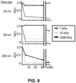


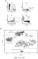


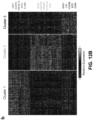

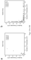

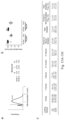
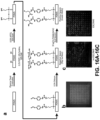


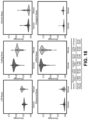
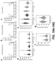


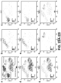

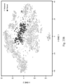


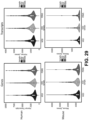
Classifications
-
- C—CHEMISTRY; METALLURGY
- C12—BIOCHEMISTRY; BEER; SPIRITS; WINE; VINEGAR; MICROBIOLOGY; ENZYMOLOGY; MUTATION OR GENETIC ENGINEERING
- C12Q—MEASURING OR TESTING PROCESSES INVOLVING ENZYMES, NUCLEIC ACIDS OR MICROORGANISMS; COMPOSITIONS OR TEST PAPERS THEREFOR; PROCESSES OF PREPARING SUCH COMPOSITIONS; CONDITION-RESPONSIVE CONTROL IN MICROBIOLOGICAL OR ENZYMOLOGICAL PROCESSES
- C12Q1/00—Measuring or testing processes involving enzymes, nucleic acids or microorganisms; Compositions therefor; Processes of preparing such compositions
- C12Q1/68—Measuring or testing processes involving enzymes, nucleic acids or microorganisms; Compositions therefor; Processes of preparing such compositions involving nucleic acids
- C12Q1/6869—Methods for sequencing
- C12Q1/6874—Methods for sequencing involving nucleic acid arrays, e.g. sequencing by hybridisation
-
- B—PERFORMING OPERATIONS; TRANSPORTING
- B01—PHYSICAL OR CHEMICAL PROCESSES OR APPARATUS IN GENERAL
- B01J—CHEMICAL OR PHYSICAL PROCESSES, e.g. CATALYSIS OR COLLOID CHEMISTRY; THEIR RELEVANT APPARATUS
- B01J19/00—Chemical, physical or physico-chemical processes in general; Their relevant apparatus
- B01J19/0046—Sequential or parallel reactions, e.g. for the synthesis of polypeptides or polynucleotides; Apparatus and devices for combinatorial chemistry or for making molecular arrays
-
- C—CHEMISTRY; METALLURGY
- C12—BIOCHEMISTRY; BEER; SPIRITS; WINE; VINEGAR; MICROBIOLOGY; ENZYMOLOGY; MUTATION OR GENETIC ENGINEERING
- C12Q—MEASURING OR TESTING PROCESSES INVOLVING ENZYMES, NUCLEIC ACIDS OR MICROORGANISMS; COMPOSITIONS OR TEST PAPERS THEREFOR; PROCESSES OF PREPARING SUCH COMPOSITIONS; CONDITION-RESPONSIVE CONTROL IN MICROBIOLOGICAL OR ENZYMOLOGICAL PROCESSES
- C12Q2537/00—Reactions characterised by the reaction format or use of a specific feature
- C12Q2537/10—Reactions characterised by the reaction format or use of a specific feature the purpose or use of
- C12Q2537/143—Multiplexing, i.e. use of multiple primers or probes in a single reaction, usually for simultaneously analyse of multiple analysis
-
- C—CHEMISTRY; METALLURGY
- C12—BIOCHEMISTRY; BEER; SPIRITS; WINE; VINEGAR; MICROBIOLOGY; ENZYMOLOGY; MUTATION OR GENETIC ENGINEERING
- C12Q—MEASURING OR TESTING PROCESSES INVOLVING ENZYMES, NUCLEIC ACIDS OR MICROORGANISMS; COMPOSITIONS OR TEST PAPERS THEREFOR; PROCESSES OF PREPARING SUCH COMPOSITIONS; CONDITION-RESPONSIVE CONTROL IN MICROBIOLOGICAL OR ENZYMOLOGICAL PROCESSES
- C12Q2563/00—Nucleic acid detection characterized by the use of physical, structural and functional properties
- C12Q2563/149—Particles, e.g. beads
-
- C—CHEMISTRY; METALLURGY
- C12—BIOCHEMISTRY; BEER; SPIRITS; WINE; VINEGAR; MICROBIOLOGY; ENZYMOLOGY; MUTATION OR GENETIC ENGINEERING
- C12Q—MEASURING OR TESTING PROCESSES INVOLVING ENZYMES, NUCLEIC ACIDS OR MICROORGANISMS; COMPOSITIONS OR TEST PAPERS THEREFOR; PROCESSES OF PREPARING SUCH COMPOSITIONS; CONDITION-RESPONSIVE CONTROL IN MICROBIOLOGICAL OR ENZYMOLOGICAL PROCESSES
- C12Q2563/00—Nucleic acid detection characterized by the use of physical, structural and functional properties
- C12Q2563/179—Nucleic acid detection characterized by the use of physical, structural and functional properties the label being a nucleic acid
-
- C—CHEMISTRY; METALLURGY
- C12—BIOCHEMISTRY; BEER; SPIRITS; WINE; VINEGAR; MICROBIOLOGY; ENZYMOLOGY; MUTATION OR GENETIC ENGINEERING
- C12Q—MEASURING OR TESTING PROCESSES INVOLVING ENZYMES, NUCLEIC ACIDS OR MICROORGANISMS; COMPOSITIONS OR TEST PAPERS THEREFOR; PROCESSES OF PREPARING SUCH COMPOSITIONS; CONDITION-RESPONSIVE CONTROL IN MICROBIOLOGICAL OR ENZYMOLOGICAL PROCESSES
- C12Q2565/00—Nucleic acid analysis characterised by mode or means of detection
- C12Q2565/50—Detection characterised by immobilisation to a surface
- C12Q2565/514—Detection characterised by immobilisation to a surface characterised by the use of the arrayed oligonucleotides as identifier tags, e.g. universal addressable array, anti-tag or tag complement array
-
- B—PERFORMING OPERATIONS; TRANSPORTING
- B01—PHYSICAL OR CHEMICAL PROCESSES OR APPARATUS IN GENERAL
- B01J—CHEMICAL OR PHYSICAL PROCESSES, e.g. CATALYSIS OR COLLOID CHEMISTRY; THEIR RELEVANT APPARATUS
- B01J2219/00—Chemical, physical or physico-chemical processes in general; Their relevant apparatus
- B01J2219/00274—Sequential or parallel reactions; Apparatus and devices for combinatorial chemistry or for making arrays; Chemical library technology
- B01J2219/00277—Apparatus
- B01J2219/0054—Means for coding or tagging the apparatus or the reagents
- B01J2219/00572—Chemical means
Definitions
- the present application provides a method of assembling a container for one or multiple parallel steps of biochemical analysis on one or more cells comprising performing molecular bonding of a porous membrane on an apical surface of an array having a plurality of wells, wherein the molecular bonding substantially isolates each well from adjacent wells.
- the well-based biological analysis platform also referred to as Seq-well, facilitates the creation of barcoded single-cell sequencing libraries from thousands of single cells using a device that contains 86,000 40-micron wells.
- the device contains wells comprising 45 ⁇ m ⁇ 45 ⁇ m ⁇ 60 ⁇ m.
- single beads can be loaded into each microwell with a low frequency of duplicates due to size exclusion (average bead diameter 35 ⁇ m).
- loading efficiency is greatly increased compared to drop-seq, which requires poisson loading of beads to avoid duplication at the expense of increased cell input requirements.
- Seq-well is capable of capturing nearly 100% of cells applied to the surface of the device.
- the Seq-well platform Due to its low input requirements and efficient capture of beads and cells, the Seq-well platform has broad application in numerous research and clinical settings.
- the device provides Biologic Safety Level 3 (BL3) facilities to study the dynamics of tuberculosis infection or in the field.
- BL3 Biologic Safety Level 3
- membrane sealing enables efficient exchange of buffers, allowing for more efficient capture of biological molecules of interest and limits cross-contamination of molecules from adjacent wells by spatial diffusion.
- the microwell technology platform enables easy addition and removal of materials from the container or well, a need currently not met by other modalities. Addition of materials allows for the introduction of necessary reagents, such as lysis buffers and affinity probes; removal or subtraction of materials allows for purification and other processes not previously well-suited on a massively parallel platform.
- the present application provides a high-throughput parallel single cell biochemical analysis in an array of wells or containers comprising or characterized by providing a first functionalized surface of each well or container, wherein the functionalized surface comprises an affinity resin; and providing a second functionalized surface of an array material to a top surface, wherein the functionalized surface provides accessible ionic functional groups.
- the method further comprises at least one or more additional functionalized surface of each well or container, wherein the additional functionalized surfaces provides for multiplexing reactions.
- the array material comprises polydimethylsiloxane, polycarbonate, polystyrene, polymethyl-methacrylate, polyvinylidene difluoride, polyvinylchloride, polypropylene, cyclic olefin co-polymer, a glass, or silicon.
- the array material is doped with an ionic functional group anywhere from 1% to 30% (by molar basis).
- the array material is doped with a functional group which can undergo further molecular bonding or modification via covalent bonding anywhere from 1% to 30% (by molar basis).
- the ionic functional group is a negatively charged functional group.
- the negatively charged functional group is a carboxylic acid.
- the top surface comprises an array functionalized to an organosilane.
- the organosilane functionalized array surface provides free alcohols upon treatment.
- the treatment comprises air plasma under mild vacuum.
- the free alcohols are reacted with an amino-silane providing an amine.
- the amine is activated with an isothiocyanate.
- the isothiocynate is conjugated to a polysaccharide.
- the ionic functional group is a positively charged functional group.
- the positively charged functional group is an amine.
- the amine is a polyamine.
- the amine is selected from a group consisting of chitosan, poly(lysine), polyglucosamine and poly(acetyl)glucosamine.
- the affinity resin comprises a negatively charged surface, wherein the negatively charged surface repels mRNA, and wherein the negatively charged surface enables mRNA to be captured by poly(dT) beads.
- the affinity resin comprises a carboxylate.
- the carboxylate is poly(glutamate) glutamate or aspartate.
- the wells or containers comprise micro-sized wells, nano-sized wells, or pico-sized wells.
- the wells or containers comprise 250 ⁇ m, 100 ⁇ m, 50 ⁇ m, 30 ⁇ m, 15 ⁇ m, 5 ⁇ m, or 1 ⁇ m wells.
- the wells or containers are sealed with a membrane.
- the membrane In an embodiment, the membrane.
- the membrane is an ultrafiltration membrane.
- the membrane comprises a weak cationic exchange surface.
- the ultrafiltration membrane comprises a polymeric surface functionalized with chitosan.
- the present application provides a microwell comprising or characterized by a first functionalized surface of each well or container, wherein the functionalized surface comprises an affinity resin; and a second functionalized surface, wherein the functionalized surface provides accessible ionic functional groups.
- the second functionalized surface is configured for attachment of an ion exchange membrane.
- the top surface comprises an array functionalized to an organosilane.
- the organosilane functionalized array surface provides free alcohols upon treatment.
- the treatment comprises air plasma under mild vacuum.
- the free alcohols are reacted with an amino-silane providing an amine.
- the amine is activated with an isothiocyanate.
- the isothiocynate is conjugated to a polysaccharide.
- the ionic functional group is a positively charged functional group.
- the positively charged functional group is an amine.
- the negatively charged functional group is a polyamine.
- the amine is selected from a group consisting of chitosan, poly(lysine), polyglucosamine and poly(acetyl)glucosamine.
- the affinity resin comprises a negatively charged surface, wherein the negatively charged surface repels mRNA, and wherein the negatively charged surface enables mRNA to be captured by poly(dT) beads.
- the affinity resin comprises a carboxylate.
- the carboxylate is poly(glutamate)glutamate or aspartate.
- the wells or containers comprise micro-sized wells, nano-sized wells, or pico-sized wells.
- the wells or containers comprise 250 ⁇ m, 100 ⁇ m, 50 ⁇ m, 30 ⁇ m, 15 ⁇ m, 5 ⁇ m, or 1 ⁇ m wells.
- the wells are from 250 ⁇ m to 100 ⁇ m, or from 100 ⁇ m to 50 ⁇ m, or from 50 ⁇ m to 30 ⁇ m, or from 30 ⁇ m to 15 ⁇ m, of from 15 ⁇ m to 5 ⁇ m, or from 5 ⁇ m to 1 ⁇ m.
- the first functionalized surface comprises a nucleic acid. In an embodiment, the first functionalized surface comprises an antigen binding protein. In an embodiment, the microwell is configured to contain a barcoded bead. The present application also provides an array comprising two or more microwells of the aforementioned microwells.
- the present application provides method for high-throughput parallel single cell biochemical analysis in an array of wells or containers comprising or characterized by (a) a first functionalized surface of an open well or container, wherein the functionalized surface comprises an affinity resin or antigen binding protein; (b) loading each functionalized surface well or container with anywhere from 1 to 5 cells; (c) further loading each functionalized surface well or container with a barcoded poly (dT) bead, wherein the barcoded bead; (d) sealing the first functionalized surface well or container with a second functionalized surface; wherein the second functionalized surface is an ultrafiltration membrane; wherein the second functionalized surface is transfixed to the edges of the well or container providing a closed vessel; wherein the functionalized surface provides accessible ionic functional groups; (e) performing successive buffer exchanges to lyse the cells in solution, wherein the lyse cells provide protein and nucleic acids; and, (f) removing the ultrafiltration membrane.
- a first functionalized surface of an open well or container wherein the functionalized
- the present application provides a method for high-throughput parallel single cell biochemical analysis in an array of wells or containers comprising: (a) delivering a first reagent to a container comprising a functionalized inner surface; (b) attaching a membrane to a functionalized top surface; and (c) delivering a second reagent to the container by transfer through the membrane.
- the method further comprises analyzing the barcoded beads, protein, and nucleic acids.
- the method further comprises analyzing the reaction products.
- the present application provides a research method comprising or characterized by obtaining the reaction product of any one of the foregoing methods and transmitting over a network or connection for receipt by an electronic data system data relating to the obtained reaction product.
- the research method further comprises receiving by an electronic system the data relating to the obtained reaction product.
- the present application provides an improved high-throughput parallel single cell biochemical analysis in an array of wells or containers comprising or characterized by providing a first functionalized surface of each well or container, wherein the functionalized surface comprises an affinity resin; and providing a second functionalized surface of an array material to a top surface, wherein the functionalized surface provides accessible ionic functional groups.
- the present application provides an improved research method comprising or characterized by obtaining the reaction product of any one of the foregoing methods and transmitting over a network or connection for receipt by an electronic data system data relating to the obtained reaction product.
- the research method further comprises receiving by an electronic system the data relating to the obtained reaction product.
- the present application provides wells or containers wherein the wells or containers are open vessels.
- the open vessels can be configured to affix a membrane or additional surface to provide a closed vessel.
- the wells or containers may be any number of shapes such as conical, flat-bottomed, hemispherical-bottomed.
- the wells or containers comprise volume to accommodate several types of starting material, including, but not limited to a barcoded bead, solution, and approximately 1 to 5 cells.
- Well dimensions are designed to accommodate only one bead enabling single-bead loading efficiencies of ⁇ 95%.
- Well dimensions may range anywhere from 15 microns to 250 microns. In an embodiment, the dimensions may range anywhere from 50 microns to 90 microns. In an embodiment, the dimensions may range anywhere from 75 microns to 100 microns. In an embodiment, the dimensions may range anywhere from 40 microns to 75 microns.
- the present application provides a method of assembling a container for biochemical analysis, multiple steps of biochemical analysis, or multiple parallel steps of biochemical analysis on one or more cells comprising performing molecular bonding of a porous membrane on an apical or basal surface of an array having a plurality of wells, wherein the molecular bonding substantially isolates each well from adjacent wells.
- the molecular bonding of the method of assembling a container for one or multiple parallel steps of biochemical analysis comprises one or more of covalent bonding, ion-ion bonding, dipole-dipole interaction, dipole-dipole interaction, ion-dipole interaction, hydrogen bonding, van der Waals forces.
- the bonding is effective when the container is immersed in an aqueous solution.
- the plurality of wells comprises about at least 1,000 or at least 10,000 or at least 100,000 or at least 1,000,000.
- the wells are micro-sized wells, nano-sized wells, or pico-sized wells (e.g., microliter, nanoliter, picoliter).
- the well volume is equal or less than a microliter, preferably equal or less than a nanoliter.
- the isolation includes confining cells (eukaryotic or prokaryotic) or cellular components inside the wells, while allowing flow of liquid through the porous membrane.
- the isolation includes confining macromolecules inside the wells, while allowing flow of liquid through the porous membrane. In another embodiment, the isolation includes confining macromolecules inside the wells, while allowing flow of small molecules through the porous membrane. In an even further embodiment of the method, the isolation includes confining RNA, DNA, proteins, or combination thereof inside the wells, while allowing flow of liquid through the porous membrane. In another embodiment of the method, the isolation includes confining RNA, DNA, proteins, or combinations thereof inside the wells, while allowing flow of small molecules through the porous membrane.
- the isolation includes confining molecules having a size of 1, kDa, 10 kDa, 50 kDa, 100 kDa; 1,000 kDa, 10,000 kDa, 100,000 kDa, or of 1,000,000 kDa.
- the method of assembling a container for biochemical analysis, multiple steps of biochemical analysis, or multiple parallel steps of biochemical analysis on one or more cells wherein the bonding between the porous membrane and the top surface of the array is reversible.
- the method of bonding comprises providing an array, wherein the exposed surface of the array bears charge of a first polarity; providing a porous membrane, wherein the porous membrane bears a charge of a second polarity, the second polarity being opposite the first polarity; hydrating the charged porous membrane; and positioning the charged porous membrane onto the charged top surface of the array.
- the method comprises a first and second polarity, wherein the first polarity is positive and the second polarity is negative.
- the array top surface comprises organosiloxane functional groups conjugated to PDMS.
- the array is a polymer poly-dimethylsiloxane (PDMS), polycarbonate (PC), polystyrene (PS), polymethyl-methacrylate (PMMA), PVDF, polyvinylchloride (PVC), polypropylene (PP), cyclic olefin co-polymer (COC), a glass, or is silicon.
- the method comprises a top surface wherein the top surface comprises functional groups conjugated to cyclic olefin co-polymer using aryl diazonium salts.
- the functionalization of the top surface is achieved through UV-mediated graft polymerization.
- the method comprises differential functionalization wherein the differential functionalization of well and surface for membrane attachment using UV-mediated graft polymerization.
- the adsorption of biomolecules is reduced through UV mediated graft.
- the polypropylene devices are modified with chitosan following oxygen plasma treatment.
- the method comprises PMMA devices, wherein the devices are modified with chitosan following oxygen plasma treatment.
- the method comprises polystyrene devices, wherein the polystyrene devices are modified with dextran.
- the invention comprises PDMS devices, wherein the PDMS devices are modified with biological antifouling reagents.
- the method comprises a charge wherein the charge is induced on the top or bottom surface of the array by plasma treating the array; coating the exposed surface with an organosiloxane functional group and optionally treating the exposed surface of the array with a polysaccharide.
- the polysaccharides is chitosan, chitin, or cellulose.
- the method comprises covalent linking of the membrane.
- the porous membrane has been functionalized.
- the porous membrane has been functionalized by a reactive functional group.
- the method comprises a porous membrane which has been functionalized with an amine, an aminosilane, a thiosilane, a methacrylate silane, or a poly(allylamine), and optionally in addition with one or more of maleimide, 2-iminothiolane (Traut's reagent), polyacrylic acid, bisepoxy-PEG.
- the method comprises a functionalized surface, wherein the functionalization includes treating with an air or oxygen plasma, optionally followed by vapor or solution phase deposition of an amine silane or a thiosilane.
- the method comprises treating the porous membrane with a reactive biotin, such as a biotin functionalized with a reactive group, such as a biotin linked to a leaving group, such as NHS-biotin.
- the method further comprises treating the membrane with NHS-maleimide.
- the method comprises an exposed surface of the array, wherein the exposed surface has been functionalized.
- the method comprises an exposed surface, wherein the exposed surface of the array has been functionalized by a reactive functional group.
- the method comprises an exposed surface, wherein the exposed surface of the array has been functionalized with an amine, an amine silane, a thiosilane, a methacrylate silane, a poly(allylamine), poly(lysine), BSA, epoxide silane, chitosan and optionally in addition with one or more of 2-iminothiolane (Traut's reagent), polyacrylic acid, epoxide-PEG, oxidized agarose.
- the method comprises functionalization of an exposed surface, wherein the functionalization includes treating with an air or oxygen plasma followed by submersion in an aqueous solution followed by submersion in aqueous solution followed by baking.
- the aqueous solution includes a polyamine such as polyethylenimine (PEI) or poly(lysine).
- the baking is performed at 80° C.
- the functionalization includes treating with an air or oxygen plasma followed by vapor deposition of an amine silane.
- the functionalization results in a complementary crosslinking group on the exposed surface of the array.
- the method comprises part or all of an inside surface wherein the part or all of the inside surface of the wells of the array is functionalized with a different molecule than the exposed surface of the array. In another embodiment, the method comprises part or all of an inside surface wherein the part or all of the inside surface of the wells of the array is functionalized to activate loaded cells. In a further embodiment, the method comprises part or all of an inside surface wherein the part or all of the inside surface of the wells of the array is functionalized to capture secreted products, such as proteins.
- the method comprises part or all of an inside surface wherein the part or all of the inside surface of the wells of the array is functionalized to make the wells hospitable to a living cell, for example with a hydrophilic coating, such as an alcohol, amine or a carboxylic acid functionality or any combination thereof.
- the method comprises submerging the array in an aqueous solution.
- the aqueous solution contains a functional group which will react with an amine to place a complementary crosslinking moiety on the exposed surface of the array.
- the aqueous solution contains a functional group which will react with an amine to place a complementary crosslinking moiety on the porous membrane.
- the method comprises an aqueous solution and a molecule, wherein the aqueous solution and molecule comprises streptavidin in a carbonate buffer, or any aqueous buffer solution which facilitates peptide bond formation.
- the aqueous solution and molecule comprises 2-iminothiolane and streptavidin.
- the method comprises washing the array and heating the array.
- the heating is performed in an aqueous buffer to a temperature of approximately 50° C. for approximately 10 minutes.
- the method comprises moving the array to a buffer including a molecule for inclusion on the inside surface of the well.
- the present application provides a method for massively parallel single cell biochemical analysis, comprising providing a porous membrane, providing an array of wells, loading the wells of the array with cells, for example by pouring or pipetting a cell suspension onto the exposed surface of the array; and, assembling the porous membrane on the array by molecular bonding.
- the method provides biochemical analysis, wherein the biochemical analysis comprises cell lysis performed in the well, and subsequently one or more of RT-PCR, RNA-seq, PCR, qPCR, DNA-seq, mass spectroscopy, ATAC-seq, bisulfite sequencing, immuno-PCR, in-situ sequencing, rolling circle amplification, in-situ hybridization, proximity extension assays, immunofluorescence, ELISA, reverse ELISA, multiple displacement reaction, DNase hypersensitivity, chip-seq, or any other genomic assay.
- the porosity of the porous membrane is selected from porosities suitable for one or more of the following applications: protein capture after cell lysis, RNA capture after cell lysis, transcript capture after cell lysis, mammalian cell culture, protein capture trough micro-engraving, antibody staining for cytometry, bacterial cell culture.
- the invention provides a kit for massively parallel single cell biochemical analysis, comprising: one or more porous membrane(s), one or more arrays of wells, optionally, a set of micro-beads, wherein each micro-bead bears a unique molecular identifier (UMI), optionally, a set of instructions for assembling the porous membrane on the array by bonding.
- UMI unique molecular identifier
- the invention provides a kit wherein the method of bonding comprises: providing an array, wherein the exposed surface of the array bears charge of a first polarity; providing a porous membrane, wherein the porous membrane bears a charge of a second polarity, the second polarity being opposite the first polarity; hydrating the charged porous membrane; and positioning the charged porous membrane onto the charged exposed surface of the array.
- the present application also provides a container for multiple parallel single-cell biochemical analysis, comprising an array having a plurality of wells, wherein said array has an exposed surface in a planar configuration with the openings of the wells, and a porous membrane configured to be positioned on the exposed surface of the array, wherein the porous membrane and the exposed surface of the array can cooperate by establishing molecular bonding so as to substantially isolate each well, or a subset of wells, from adjacent wells.
- the invention provides a container for selective manipulation of cells, comprising: at least one compartments having an opening, and a reversibly-sealed, porous membrane removably positioned over the opening of the compartment, wherein the porous membrane substantially isolates the compartment from adjacent compartments.
- the present application generally provides methods of diagnosis and treatment in a subject in need thereof.
- the present application provides a method of identifying cellular heterogeneity in a subject.
- the cellular heterogeneity is associated with or a characteristic of a disease or disorder, or a state of a multicellular organism.
- Diseases or disorders comprising or characterized by cellular heterogeneity include, without limitation, neoplastic diseases, benign tumors, malignant tumors, sarcomas, carcinomas, and lymphomas.
- the cellular heterogeneity characterizes a lung disease.
- the cellular heterogeneity characterizes an immune disease or immune system state.
- the method comprises sequencing a nucleic acid sample from cells or tissues of a subject for example, but not limited to bronchial alveolar lavages and lung biopsies of the subject, thereby providing single cell sequences; analyzing cellular microenvironments using categorical spatial information; and, optionally assembling single cell sequences into a visual representation, wherein the relationship between amplified shared spatial barcodes of single cell sequences is obtained by a computational method.
- the present application provides a method of identifying differentiation of myoblasts in a subject undergoing IGF therapy comprising sequencing a nucleic acid sample from myoblasts of a patient, thereby providing single cell sequences; analyzing cellular microenvironments using categorical spatial information; and, optionally assembling single cell sequences into a visual representation, wherein the relationship between amplified shared spatial barcodes of single cell sequences is obtained by a computational method.
- the present application provides a method of identifying transcriptional regulation in macrophages comprising sequencing a nucleic acid sample from macrophages of a subject infected with M. tuberculosis , thereby providing single cell sequences of an infected subject; sequencing a nucleic acid sample from macrophages of a healthy subject uninfected with M. tuberculosis , thereby providing single cell sequences of an uninfected subject; measuring the differential nucleic acid levels between an infected subject and an uninfected subject; analyzing cellular microenvironments using categorical spatial information; and, optionally assembling single cell sequences into a visual representation, wherein the relationship between amplified shared spatial barcodes of single cell sequences is obtained by a computational method.
- the present application provides a method of identifying cellular heterogeneity in acute myeloid leukemia cells in a subject having leukemia comprising sequencing a nucleic acid sample from acute myeloid leukemia cells of a subject, thereby providing single cell sequences; analyzing cellular microenvironments using categorical spatial information; and optionally assembling single cell sequences into a visual representation, wherein the relationship between amplified shared spatial barcodes of single cell sequences is obtained by a computational method.
- FIGS. 1 A- 1 B illustrate a schematic of the technique for massively parallel capture of matched transcriptome, proteome and genome of single cells.
- A) Treated arrays of nanoliter-scale wells are pre-loaded with affinity resins specific for mRNA (barcoded-poly(dT) beads), protein (NHS-activated or aldehyde-activated agarose beads) and genome (weak anionic exchange resin). Cells are then loaded and the array is sealed with an ultrafiltration membrane. Successive buffer exchanges are then performed to lyse the cells and then activate separately each affinity resin to capture its intended target, primarily through changes in the ionic strength and pH of the buffer. Once captured, the membrane is removed and the mRNA reverse transcribed to cDNA.
- the protein can be queried through iterative fluorescent staining using antibodies or stained with DNA-barcoded antibodies which are subsequently transferred to the barcoded poly(dT) bead to add a cell barcode to the antibody barcode tag. Both cDNA and antibody tags are then sequenced in bulk. The genome can be recovered by micromanipulation or amplified in the nanowell using PCR or WGA.
- FIGS. 2 A- 2 B illustrate massively parallel capture and reverse transcription of mRNA from cells in nanoliter-scale wells.
- FIG. 3 illustrates massively parallel capture of protein from cells in nanowell.
- A) Glyoxal functionalized agarose beads and PBMC labeled with ⁇ CD45-AF647 antibody were co-loaded into functionalized nanoliter scale wells, sealed in with ultrafiltration membrane and imaged for AF647 signal.
- B) Cells were lysed by buffer exchanging with GuTCN+sarkosyl lysis buffer and then imaged for AF647 signal.
- FIG. 4 illustrates massively parallel capture of genomes from cells in nanoliter scale wells.
- PBMC peripheral blood mononuclear cells
- A is an exemplar image of Array A before or after membrane removal—genome remains bound to surface in punctate form where cell originally sat.
- B is an exemplar image Arrays B or C while still sealed with membrane—genome diffuse through well indicating free diffusion.
- C is an exemplar images of Arrays B and C after membrane removal. Genome remains bound in array buffer exchanged with pH 7.4 buffer while genomes are lost from arrays kept in pH 9 buffer.
- FIG. 5 illustrates agarose coating of arrays improves transcript capture.
- Top surfaces of 3 nanowell arrays were functionalized with chitosan and wells were functionalized with poly(glutamate) using typical procedure. Two of the arrays were then further functionalized with a thin film agarose hydrogel only on the inner well surfaces. Nanowell arrays were then used to capture and reverse transcribe mRNA from single cells. Beads were collected from array and technical replicates of 1000 beads were used in a qPCR reaction measuring the number of GAPDH cDNA molecules. Amplification curves for untreated (blue) and the two agarose treated arrays (red and green) are displayed.
- FIG. 6 illustrates WGA in membrane-covered nanoliter scale wells.
- FIG. 7 illustrates microengraving through the membrane.
- FIG. 8 illustrates the effect of pore size on analyte diffusion from cells.
- FIG. 9 illustrates the Seq-Well clearly separates a mixture of mouse and human cells.
- Each unique cell barcode only aligns to one of the two species, proving that each barcode distinguishes a single cell.
- Reverse transcription or other molecular processing can be performed on the array (‘array) or off of the array (‘live cells’) after cellular isolation, lyse, and affinity capture. Fixed cells can also be run on the array and cells can be fixed on the array prior to running (‘RNAlater’).
- FIGS. 10 A- 10 D illustrate an embodiment of the invention.
- A Photograph of equipment and array used to capture and lyse cells, respectively.
- B Transcripts captured from a mix of human (HEK293) and mouse (NIH/3T3) cells reveal distinct transcript mapping and single-cell resolution.
- Human (mouse) cells (>2,000 human (mouse) transcripts and ⁇ 1,000 mouse (human) transcripts) are shown in blue (red).
- Among the 254 cells identified 1.6% (shown in purple) had a mixed phenotype.
- C,D Violin plots of the number of transcripts (C) and genes (D) detected in human or mouse single-cell libraries generated by Seq-Well or Drop-Seq (Macosko, E.
- FIGS. 11 A- 11 B illustrate combined image cytometry and scRNA-Seq of human PBMCs.
- A The hierarchical gating scheme (with the frequencies of major cell subpopulations) used to analyze PBMCs that had been labeled with a panel of fluorescent antibodies, loaded onto three replicate arrays and imaged prior to bead loading and transcript capture (Methods).
- Myeloid cells (green) were identified as the population of hCD3( ⁇ ) HLA-DR(+) CD19( ⁇ ) cells; B cells (orange) as the subset of hCD3( ⁇ ) HLA-DR(+) CD19(+) cells; CD4 T cells (blue) as the subset of CD3(+) CD4(+) cells; CD8 T cells (yellow) as the CD3(+) CD8(+) subset of cells; and, NK cells (red) as the subset of CD3( ⁇ ) HLA-DR( ⁇ ) CD56(+) CD16(+) cells.
- FIGS. 22 - 24 t-SNE visualization of single-cell clusters identified among 3,694 human Seq-Well PBMCs single-cell transcriptomes recovered from the imaged array and the two additional ones ( FIGS. 22 - 24 ). Clusters (subpopulations) are labeled based on annotated marker gene ( FIG. 22 ).
- D A heatmap showing the relative expression level of a set of inflammatory and antiviral genes among cells identified as monocytes.
- FIGS. 12 A- 12 C illustrates sequencing of TB-exposed macrophages in a BSL3 facility using Seq-Well.
- A t-SNE visualization of single-cell clusters identified among 2,560 macrophages (1,686 exposed, solid circles; 874 unexposed, open circles) generated using 5 principal components across 377 variable genes.
- B Marker genes for the 3 phenotypic clusters of macrophages highlighted in (A).
- C Volcano plots of differential expression between exposed and unexposed macrophages within each cluster showing genes enriched in cells exposed to M. tuberculosis .
- a p-value threshold of 5.0 ⁇ 10 ⁇ 16 based on a likelihood ratio test was used to establish statistical significance, while a log 2-fold change threshold of 0.4 was used to determine differential expression.
- Genes with p-values less than 5.0 ⁇ 10 ⁇ 6 are shown in cyan and absolute log 2-fold changes greater than 0.4; In magenta are genes with p-values less than 5.0 ⁇ 10 ⁇ 6 but absolute log 2-fold changes less than 0.4; and, in black, are genes with p-values greater than 5.0 ⁇ 10 ⁇ 6 and absolute log 2-fold changes less than 0.4.
- FIGS. 13 A- 13 B illustrates open array gene and transcript capture.
- A An open array format results in decreased gene and transcript capture, and increased cross-contamination, relative to the membrane sealing implemented in Seq-Well.
- B Species mixing experiments with reversible membrane sealing using Seq-Well provides increased gene/transcript capture and improved single-cell resolution.
- FIG. 14 illustrates Seq-Well experimental workflow.
- Cells are obtained from complex tissues or clinical biopsies, and digested to form a single-cell suspension.
- Barcoded mRNA capture beads are added to the surface of the microwell device, settling into wells by gravity, and then a single-cell suspension is applied.
- the device is sealed using a semi-permeable membrane that, upon addition of a chemical lysis buffer, confines cellular mRNAs within wells while allowing efficient buffer exchange.
- Liberated cellular transcripts hybridize to the bead-bound barcoded poly(dT) primers that contain a cell barcode (shared by all probes on the same bead but different between beads) and a unique molecular identifier (UMI) for each transcript molecule.
- UMI unique molecular identifier
- the beads are removed from the array and bulk reverse transcription is performed to generate single-cell cDNAs attached to beads. Libraries are then made by a combination of PCR and tagmentation, and sequenced. After, Single-cell transcriptomes are assembled in silico using cell barcodes and UMIs.
- FIGS. 15 A- 15 C illustrates bead and cell loading efficiency.
- A Two arrays were loaded with barcoded beads through intermittent rocking. After washing, arrays were imaged in transmitted light and AF488 channel to capture bead autofluorescence. A plot of the frequency of the 75 th percentile AF488 well intensity across the array (Panel 1) and the frequency of wells containing zero, one and multiple beads is displayed (Panel 2).
- the number of cells loaded into each array and well as enumerated by fluorescent imaging is plotted, normalized to the average number of cells/well in the 96 well plate. Mean and standard error are denoted by line and error bars respectively.
- (C) 2 ⁇ 10 2 , 2 ⁇ 10 3 , and 2 ⁇ 10 4 total cells of a 1:1 mix of fluorescently labeled HEK 293T and 3T3 cells were loaded onto three functionalized arrays each. All arrays were fluorescently imaged to enumerate the number of each cell line in each array microwell. The mean ⁇ standard deviation of the number of empty, single and multiple occupancy wells across the three replicate arrays for each loading density is displayed along with the mean ⁇ standard deviation of the percentage of occupied wells containing a cell from each species.
- FIGS. 16 A- 16 C illustrate PDMS surface chemistry functionalization protocol and differential functionalization of microwell arrays.
- the surface of the PDMS device is initially treated with an air plasma under mild vacuum, terminating the surface in hydroxyls.
- This PDMS surface is aminated using (3-Aminopropyl)triethoxysilane (APTES).
- APTES (3-Aminopropyl)triethoxysilane
- the amine surface is then activated with PDITC to create an isothiocyanate surface.
- the isothiocyanate on the top surface of the array (negative space) is covalently linked to chitosan polymers through their amine group.
- the hydrophobicity of the isothiocyanate surface prevents solvation of the microwells with the aqueous chitosan solution, preventing chitosan from reacting with the inner well surfaces (positive space). These surfaces are subsequently reacted with the free amine of poly(glutamic) acid polymers under vacuum to drive the solvation of the wells.
- the top surface of a PDITC-activated array was coated with streptavidin-PE (red) and the inner well surfaces were coated with streptavidin-AF488 (green) using same method used to functionalize with chitosan and poly(glutamate).
- FIGS. 17 A- 17 C illustrate microwell sealing with semipermeable membrane.
- PBMCs labeled with ⁇ CD45-AF647 were loaded into two BSA-blocked arrays and one array functionalized with chitosan and poly(glutamate).
- a semipermeable membrane was attached to one of the BSA-blocked arrays and the chitosan:polyglutamate functionalized array prior to addition of lysis buffer.
- FIG. 18 illustrates read mapping quality.
- Read mapping quality matrices were generated for each sample for human (blue) and mouse (red) cells, aligned to hg19 and mm10, respectively.
- High quality samples had relatively higher percentages of annotated genomic (genic) and exonic transcripts and low percentages of annotated intergenic and ribosomal transcripts (Center-line: Median; Limits: 1 st and 3 rd Quartile; Whiskers: +/ ⁇ 1.5 IQR; Points: Values >1.5 IQR).
- FIGS. 19 A- 19 G illustrate a comparison of gene and transcript capture and percent contamination among massively-parallel scRNA-Seq methods using mouse and human cell lines. Histograms of the percent cross-species contamination in (A) Seq-Well, (B) Drop-Seq (Ref 12), and (C) Yuan and Sims (Ref. 15). In each plot, cells with greater than 90% of human transcripts are displayed in blue and cells with less than 10% human transcripts are displayed in red.
- FIGS. 20 A- 20 B illustrate transcript cutoff for species-mixing validation.
- Two arrays (a & b) were sequenced to confirm single-cell resolution and minimal cross-contamination between mouse and human cells. Applicants called cells by plotting the cumulative distribution of transcripts and making a cutoff at the elbow in the curve.
- A which was used to validate the single-cell resolution
- B where Applicants sequenced the array deeply to allow a competitive comparison to Drop-Seq, Applicants made the cutoff at 10,000 transcripts.
- FIGS. 22 A- 22 I illustrate mapping lineage defining transcripts to PBMC clusters.
- A Clusters identified through graph-based clustering correspond to major immune cell populations.
- B, E CD4 T cells are characterized by expression of CD3D and T-cell receptor expression without pronounced expression of cytotoxic genes NKG7 and PRF1.
- C,F CD8 T cells are defined by expression of NKG7 and PRF1.
- D,G Monocytes are defined by expression of cathepsin B (CTSB) and SOD2.
- CTSB cathepsin B
- SOD2 cathepsin B
- Natural killer cells are characterized by expression of cytotoxic genes in the absence of T cell receptor expression.
- H B cells are marked by elevated expression of MS4A1 (CD20) transcripts.
- I Dendritic cells are enriched for expression of BIRC3.
- FIGS. 23 A- 23 B illustrate a heatmap of PBMCs
- A Genes enriched in each cluster were identified using an “ROC” test in Seurat, comparing cells assigned to each cluster to all other cells. A heatmap was constructed using enriched genes found to define each cluster. One cluster of 602 cells that demonstrated exclusive enrichment of mitochondrial genes was removed as these likely represent low-quality or dying cells.
- B We generated a t-SNE projection of 4,296 cells with greater than 10,000 reads, 1,000 transcripts, 500 genes, and 65% transcript mapping. We removed a total of 602 cells from the final analysis found to be strongly enriched for expression of mitochondrial genes. The remaining 3,694 cells form distinct clusters enriched for lineage-defining that distinguish cells types from one another.
- FIGS. 24 A- 24 D illustrate read mapping quality in PBMCs.
- A-C Violin plots depicting reads (A), transcripts (B), and genes (C) per cell, separated by cell type.
- D Percent mRNA bases per cell, separated by cell type.
- FIGS. 25 A- 25 B illustrate a comparison of human PBMC gene and transcript capture with other massively-parallel scRNA-Seq methods.
- B cells (orange), an average of 1,315 genes and 3,632 transcripts were detected using Seq-Well and an average of 710 genes and 1,910 transcripts were detected in 10 ⁇ Genomics data.
- CD4 T cells blue
- an average of 861 genes and 2,444 transcripts were detected using Seq-Well and an average of 815 genes and 2,370 transcripts were detected in 10 ⁇ Genomics data.
- CD8 T cells yellow
- an average of 885 genes and 2,574 transcripts were detected using Seq-Well and an average of 809 genes and 2,029 transcripts were detected in 10 ⁇ Genomics data.
- Monocytes green
- an average of 1,288 genes and 3,568 transcripts were detected using Seq-Well and an average of 974 genes and 2,835 transcripts were detected in 10 ⁇ Genomics data.
- FIGS. 26 A- 26 D illustrate t-SNE visualization of exposed and unexposed macrophages using a 5,000 transcript cutoff.
- A Using a threshold of 5,000 detected transcripts identified 4,638 macrophages.
- B Among these 4,638 cells, 5 distinct clusters of macrophages were identified by performing graph-based clustering over 5 principal components (377 variable genes).
- C Clusters 1-3 are defined by unique gene expression signatures, while Clusters 4 and 5 are defined by expression of mitochondrial genes, suggesting low-quality cells
- D Following removal of cells within Clusters 4 and 5, there remain a total of 2,560 cells in Clusters 1-3.
- FIGS. 27 A- 27 D illustrate quality by cluster among tb macrophages.
- A-C Violin plots depicting reads (A), transcripts (B), and genes (C) per cell, separated by cluster.
- D Percent mRNA bases per cell, separated by cluster.
- FIGS. 28 A- 28 C illustrate the effect of doping of ionic functional group with carboxylic acid functional groups (see FIG. 16 ).
- A MicroTEC species-mixing experiment, 10% carboxylate doping.
- B MicroTEC species-mixing experiment, 20% carboxylate doping.
- C PDMS species-mixing experiment, no doping.
- FIG. 29 illustrates gene, transcripts and cell yields.
- M10 MicroTEC, 10% carboxylate doping.
- M20 MicroTEC, 20% carboxylate doping.
- PDMS PDMS, no doping.
- the present application provides a method of assembling a container for biochemical analysis, multiple steps of biochemical analysis, or multiple parallel steps of biochemical analysis on one or more cells comprising performing molecular bonding of a porous membrane on an apical or basal surface of an array having a plurality of wells, wherein the molecular bonding substantially isolates each well from adjacent wells or a subset of wells.
- Applicants provide a methodology which allows attachment of a porous membrane to a container in conditions which are benign to living cells.
- the invention provides the creation of hundreds of thousands of isolated dialysis chambers which can be used for many different applications.
- Applicants provide single cell lysis procedures for single cell RNA-seq, whole genome amplification or proteome capture; highly multiplexed single cell nucleic acid preparation ( ⁇ 100 ⁇ increase over current approaches); highly parallel growth of clonal bacterial populations thus providing synthetic biology applications as well as basic recombinant protein expression; selection of bacterial that have increased secretion of a recombinant product possible product could also be small molecule metabolite which could have considerable utility in chemical industry and biofuels; retention of cells during multiple microengraving events'; long term capture of secreted products from single cells; and screening of cellular events. Principles of the present methodology allow for addition and subtraction of materials from the containers, which has not previously been available on the present scale in other modalities.
- Applicants have established chemistries that enable the stable attachment of porous membranes to PDMS nanowell devices in conditions that do not affect cells. Applicants have established multiple chemistries to attach the membranes. In a preferred embodiment, Applicants have selected is to functionalize the PDMS device with an amino-silane and oxidize the membrane with plasma. Applicants have found that the selected functional group provides facile pH modification. With regard to general cell culture uses, the PDMS is amine functionalized by air plasma treatment followed by submersion in an aqueous solution of poly(lysine) followed by baking at 80° C. For processes that require robust denaturing conditions, the amine must be covalently linked to the surface.
- membrane can be amine-silanized using vapor deposition and then treated in solution with NHS-biotin or NHS-maleimide to turn the amine groups into the crosslinking species.
- the devices are loaded with cells (bacterial, mammalian or yeast) in compatible buffers.
- the cell laden device is then brought in contact with the functionalized membrane using a clamping device.
- a plain glass slide is placed on top of the membrane in the clamp to provide force for bringing the two surfaces together.
- the clamp is opened and the glass slide is removed.
- the device can then be submerged in any aqueous buffer for days without the membrane detaching, enabling repetitive measurements of the cells without any cell loss.
- the covalently-linked membrane is stable in many harsh buffers including guanidine hydrochloride which can be used to robustly lyse cells.
- the pore size of the membrane is small, the products from the lysed cells will be retained in each well.
- the lysing buffer can be washed out and replaced with a different buffer which allows binding of biomolecules to probes preloaded in the wells.
- the membrane can then be removed, enabling addition of enzymes to reverse transcribe or amplify nucleic acids captured in the wells after lysis.
- the chemistry enables removal of one membrane and replacement with a membrane with a different pore size to enable integration of multiple activities on the same array.
- secreted products e.g., proteins
- a printed microarray see, e.g., U.S. Pat. Nos. 7,776,553; 8,835,187; 8,772,049; 8,865,479; JP 571822; EP 2 297 333; U.S. application Ser. No. 13/132,858).
- RNA profiling is in principle particularly informative, as cells express thousands of different RNAs. Approaches that measure for example the level of every type of RNA have until recently been applied to “homogenized” samples—in which the contents of all the cells are mixed together. Methods to profile the RNA content of tens and hundreds of thousands of individual human cells have been recently developed, including from brain tissues, quickly and inexpensively.
- Profiling large numbers of cells may also be important for distinguishing noise from biologically meaningful patterns (sometimes involving small numbers of genes) that recur in many cells (Grun, D., et al., “Validation of noise models for single-cell transcriptomics” (2014) Nature Methods 11, 637-640; Kharchenko, P., et al., “Bayesian approach to single-cell differential expression analysis” (2014) Nature Methods 11, 740-742).
- Single-cell profiling is a technique that exposes inherent responses which otherwise are unable to be studied in the context of a complex, and non-uniform environment. Biological samples, such as tissue, are broken down in order to study cell types and reveal pertinent cell expression profiles.
- techniques used for single-cell profiling entail quantitative reverse transcription PCR (RT-qPCR) and single-cell RNA-Seq, as well as other single-cell genomic techniques.
- RT-qPCR provides a highly sensitive, high-throughput single-cell profiling technique with multiplexing developed to target mRNA, microRNA, non-coding RNA, and proteins.
- Microfluidics involves micro-scale devices that handle small volumes of fluids. Because microfluidics may accurately and reproducibly control and dispense small fluid volumes, in particular volumes less than 1 ⁇ l, application of microfluidics provides significant cost-savings.
- the use of microfluidics technology reduces cycle times, shortens time-to-results, and increases throughput.
- incorporation of microfluidics technology enhances system integration and automation.
- Microfluidic reactions are generally conducted in microdroplets. The ability to conduct reactions in microdroplets depends on being able to merge different sample fluids and different microdroplets. See, e.g., US Patent Publication No. 20120219947.
- Droplet microfluidics offers significant advantages for performing high-throughput screens and sensitive assays. Droplets allow sample volumes to be significantly reduced, leading to concomitant reductions in cost. Manipulation and measurement at kilohertz speeds enable up to 10 8 discrete biological entities (including, but not limited to, individual cells or organelles) to be screened in a single day. Compartmentalization in droplets increases assay sensitivity by increasing the effective concentration of rare species and decreasing the time required to reach detection thresholds. Droplet microfluidics combines these powerful features to enable currently inaccessible high-throughput screening applications, including single-cell and single-molecule assays. See, e.g., Guo et al., Lab Chip, 2012, 12, 2146-2155.
- Drop-Sequence methods and apparatus provides a high-throughput single-cell RNA-Seq and/or targeted nucleic acid profiling (for example, sequencing, quantitative reverse transcription polymerase chain reaction, and the like) where the RNAs from different cells are tagged individually, allowing a single library to be created while retaining the cell identity of each read.
- a combination of molecular barcoding and emulsion-based microfluidics to isolate, lyse, barcode, and prepare nucleic acids from individual cells in high-throughput is used.
- Microfluidic devices for example, fabricated in polydimethylsiloxane), sub-nanoliter reverse emulsion droplets.
- nucleic acids are used to co-encapsulate nucleic acids with a barcoded capture bead.
- Each bead for example, is uniquely barcoded so that each drop and its contents are distinguishable.
- the nucleic acids may come from any source known in the art, such as for example, those which come from a single cell, a pair of cells, a cellular lysate, or a solution.
- the cell is lysed as it is encapsulated in the droplet.
- Poisson statistics 100,000 to 10 million such beads are needed to barcode ⁇ 10,000-100,000 cells.
- the present application enables one to capture mRNA transcripts from single cells using the methods described. Mention is made of mRNA transcript capture from single cells using microfluidic devices.
- the disclosure relates to a microfluidic device for automatic hydrodynamic capture of single mammalian cells and subsequent immobilization and digital counting of polyadenylated mRNA molecules released from individual cells (see Abstract).
- Single HeLA cells are captured by hybridization to oligonucleotides attached on the glass surface in the device, which is visually monitored using single-molecule fluorescence imaging.
- OPENARRAY® technology uses a microscope slide-sized plate with 3,072 through-holes. Each plate contains 48 subarrays, each with 64 through-holes where each through-hole is 300 ⁇ m in diameter and 300 ⁇ m deep and is treated with hydrophilic and hydrophobic coatings. Reagents are retained in the through-holes via surface tension.
- the technology allows for real-time PCR based solution for high-throughput gene expression analysis, genotyping, microRNA analysis, and digital PCR applications.
- semi-permeable membranes refer to thin layers or coverings which allow certain compounds, molecules, ions, or proteins to pass through via diffusion.
- the diffusion requires energy to pass through the membrane and is sometimes referred to as active transport.
- the invention is practiced with a semi-permeable membrane including pores which may be used to separate a material with a specific size. As a result, such a semi-permeable or separation membrane may be applied to biological analysis by using this property to remove any undesired components.
- a semi-permeable membrane is also termed a selectively permeable membrane, a partially permeable membrane or a differentially permeable membrane, and is a type of membrane, e.g., biological membrane, that may allow certain molecules or ions to pass through it, e.g., by diffusion or facilitated diffusion or passive transport or active transport; accordingly, a membrane as in the present invention or as meeting criteria as herein discussed may be considered permeable, or semi-permeable, or selectively permeable or partially permeable or differentially permeable, or a biological membrane, or a membrane that allows certain molecules or ions to pass through it, or a membrane that allows certain molecules or ions to pass through it by diffusion, or a membrane that allows certain molecules or ions to pass through it by facilitated diffusion, or a membrane that allows certain molecules or ions to pass through it by passive transport, or a membrane that allows certain molecules or ions to pass through it by active transport.
- a membrane as in the present invention or as meeting criteria as herein discussed may be
- the layer-by-layer assembly may form a functional multi-layered thin membrane by alternatively combining a polymer electrolyte, nanoparticles, particles, proteins, large organic molecules, and the like having a positive ion charge with another polymer electrolyte, nanoparticles, nanoparticles, particles, proteins, large organic molecules, and the like having a negative ion charge through molecular attraction including, but not limited to, electrostatic attraction, hydrogen bonding, and electron transfer.
- Membrane filtration comprises ultrafiltration, where pressure and/or concentration gradients enable separation of analytes through a semipermeable membrane.
- Ultrafiltration is applicable for the purification of protein solutions, where membranes are defined by a molecular weight cut off in order to provide the separation of the desired proteins.
- Membrane fouling is a process wherein a solution or a particle is deposited on a membrane surface and/or the membrane pores such that the deposition the membrane performance is degraded.
- the label is a fluorescent label.
- fluorescent labels include, but are not limited to, Atto dyes, 4-acetamido-4′-isothiocyanatostilbene-2,2′disulfonic acid; acridine and derivatives: acridine, acridine isothiocyanate; 5-(2′-aminoethyl)aminonaphthalene-1-sulfonic acid (EDANS); 4-amino-N-[3-vinylsulfonyl)phenyl]naphthalimide-3,5 disulfonate; N-(4-anilino-1-naphthyl)maleimide; anthranilamide; BODIPY; Brilliant Yellow; coumarin and derivatives; coumarin, 7-amino-4-methylcoumarin (AMC, Coumarin 120), 7-amino-4-trifluoromethylcouluarin (Coumaran 151); cyanine dyes; cyanosine
- the fluorescent label may be a fluorescent protein, such as blue fluorescent protein, cyan fluorescent protein, green fluorescent protein, red fluorescent protein, yellow fluorescent protein or any photoconvertible protein. Colormetric labeling, bioluminescent labeling and/or chemiluminescent labeling may further accomplish labeling. Labeling further may include energy transfer between molecules in the hybridization complex by perturbation analysis, quenching, or electron transport between donor and acceptor molecules, the latter of which may be facilitated by double stranded match hybridization complexes.
- the fluorescent label may be a perylene or a terylene. In the alternative, the fluorescent label may be a fluorescent bar code.
- the oligonucleotide tags may be detectable by virtue of their nucleotide sequence, or by virtue of a non-nucleic acid detectable moiety that is attached to the oligonucleotide such as but not limited to a fluorophore, or by virtue of a combination of their nucleotide sequence and the non-nucleic acid detectable moiety.
- a detectable oligonucleotide tag may comprise one or more nonoligonucleotide detectable moieties.
- detectable moieties may include, but are not limited to, fluorophores, microparticles including quantum dots (Empodocles, et al., Nature 399:126-130, 1999), gold nanoparticles (Reichert et al., Anal. Chem. 72:6025-6029, 2000), microbeads (Lacoste et al., Proc. Natl. Acad. Sci.
- the detectable moieties may be quantum dots. Methods for detecting such moieties are described herein and/or are known in the art.
- detectable oligonucleotide tags may be, but are not limited to, oligonucleotides which may comprise unique nucleotide sequences, oligonucleotides which may comprise detectable moieties, and oligonucleotides which may comprise both unique nucleotide sequences and detectable moieties.
- a unique label may be produced by sequentially attaching two or more detectable oligonucleotide tags to each other.
- the detectable tags may be present or provided in a plurality of detectable tags.
- the same or a different plurality of tags may be used as the source of each detectable tag may be part of a unique label.
- a plurality of tags may be subdivided into subsets and single subsets may be used as the source for each tag.
- one or more other species may be associated with the tags.
- nucleic acids released by a lysed cell may be ligated to one or more tags. These may include, for example, chromosomal DNA, RNA transcripts, tRNA, mRNA, mitochondrial DNA, or the like. Such nucleic acids may be sequenced, in addition to sequencing the tags themselves, which may yield information about the nucleic acid profile of the cells, which can be associated with the tags, or the conditions that the corresponding droplet or cell was exposed to.
- single cells or single organelles or single molecules are encapsulated into containers, chambers, or wells from an aqueous solution/dispersion.
- multiple cells or multiple molecules may take the place of single cells or single molecules.
- the aqueous droplets of volume ranging from 1 pL to 10 nL work as individual reactors.
- One method is to generate droplets using hydrodynamic focusing of a dispersed phase fluid and immiscible carrier fluid, such as disclosed in U.S. Publication No. US 2005/0172476 and International Publication No. WO 2004/002627.
- one of the species introduced at the confluence is a pre-made library of droplets where the library contains a plurality of reaction conditions, e.g., a library may contain plurality of different compounds at a range of concentrations encapsulated as separate library elements for screening their effect on cells or enzymes, alternatively a library could be composed of a plurality of different primer pairs encapsulated as different library elements for targeted amplification of a collection of loci, alternatively a library could contain a plurality of different antibody species encapsulated as different library elements to perform a plurality of binding assays. Principles of the present invention allow for library construction.
- the introduction of a library of reaction conditions onto a substrate is achieved by pushing a premade collection of library droplets out of a vial with a drive fluid.
- the drive fluid is a continuous fluid.
- the drive fluid may comprise the same substance as the carrier fluid (e.g., a fluorocarbon oil).
- the carrier fluid e.g., a fluorocarbon oil.
- a 10,000 picoliter/second infusion rate will nominally produce a range in frequencies from 900 to 1,100 droplet per second.
- sample to sample variation in the composition of dispersed phase for droplets made on chip a tendency for the number density of library droplets to increase over time and library-to-library variations in mean droplet volume severely limit the extent to which frequencies of droplets may be reliably matched at a confluence by simply using fixed infusion rates.
- these limitations also have an impact on the extent to which volumes may be reproducibly combined.
- a cell library element may include, but is not limited to, hybridomas, B-cells, primary cells, cultured cell lines, cancer cells, stem cells, cells obtained from tissue, or any other cell type.
- Cellular library elements are prepared by encapsulating a number of cells from one to hundreds of thousands in individual droplets. The number of cells encapsulated is usually given by Poisson statistics from the number density of cells and volume of the droplet. However, in some cases the number deviates from Poisson statistics as described in Edd et al., “Controlled encapsulation of single-cells into monodisperse picolitre drops.” Lab Chip, 8(8): 1262-1264, 2008.
- the discrete nature of cells allows for libraries to be prepared in mass with a plurality of cellular variants all present in a single starting media and then that media is broken up into individual droplet capsules that contain at most one cell. These individual droplets capsules are then combined or pooled to form a library consisting of unique library elements. Cell division subsequent to, or in some embodiments following, encapsulation produces a clonal library element.
- a bead based library element may contain one or more beads, of a given type and may also contain other reagents, such as antibodies, enzymes or other proteins.
- the library elements may all be prepared from a single starting fluid or have a variety of starting fluids.
- the library elements will be prepared from a variety of starting fluids.
- Elastomers in general are polymers existing at a temperature between their glass transition temperature and liquefaction temperature. For illustration, a brief description of the most common classes of elastomers is presented below.
- Silicone polymers have great structural variety, and a large number of commercially available formulations.
- the present systems are fabricated from an elastomeric polymer such as GE® RTV 615 (formulation), a vinyl-silane crosslinked (type) silicone elastomer (family).
- the silicone polymer is polydimethylsiloxane (PDMS).
- PFPE Functionalized photocurable perfluoropolyether
- PFPE is particularly useful as a material for fabricating solvent-resistant microfluidic devices for use with certain organic solvents.
- These PFPEs have material properties and fabrication capabilities similar to PDMS but with compatibility with a broader range of solvents.
- Suitable PFPE compounds are described, for example, in International Publication Nos. WO 2005/030822 and WO 2005/084191 and Rolland et al., “Solvent-resistant photocurable ‘liquid Teflon’ for microfluidic device fabrication,” 126 J. Amer. Chem. Soc. 2322-23 (2004).
- suitable materials include polyisoprenes, polybutadienes, polychloroprenes, polyisobutylenes, poly(styrene-butadiene-styrene)s, polyurethanes, poly(bis(fluoroalkoxy)phosphazene) (PNF, Eypel-F), poly(carborane-siloxanes) (e.g., Dexsil), poly(acrylonitrile-butadiene) (nitrile rubber), poly(1-butene), poly(chlorotrifluoroethylene-vinylidene fluoride) copolymers (Kel-F), poly(ethyl vinyl ether), poly(vinylidene fluoride), poly(vinylidene fluoride-hexafluoropropylene) copolymer (available, for example, under the VITON® trademark), elastomeric compositions of polyvinylchloride (PVC), polysulfone, polycarbonate, poly
- micro-sized arrays refer to (sub) nanoliter-scale volumes.
- the volumes are on the order of 1 pL to 1 nL, generally. Therefore, linear dimensions and volumetric dimensions are referred to discretely.
- the sample may include nucleic acid target molecules.
- Nucleic acid molecules may be synthetic or derived from naturally occurring sources.
- nucleic acid molecules may be isolated from a biological sample containing a variety of other components, such as proteins, lipids and non-template nucleic acids.
- Nucleic acid target molecules may be obtained from any cellular material, obtained from an animal, plant, bacterium, fungus, or any other cellular organism.
- the nucleic acid target molecules may be obtained from a single cell.
- Biological samples for use in the present invention may include viral particles or preparations.
- Nucleic acid target molecules may be obtained directly from an organism or from a biological sample obtained from an organism, e.g., from blood, urine, cerebrospinal fluid, seminal fluid, saliva, sputum, stool and tissue. Any tissue or body fluid specimen may be used as a source for nucleic acid for use in the invention.
- Nucleic acid target molecules may also be isolated from cultured cells, such as a primary cell culture or a cell line. The cells or tissues from which target nucleic acids are obtained may be infected with a virus or other intracellular pathogen.
- a sample may also be total RNA extracted from a biological specimen, a cDNA library, viral, or genomic DNA.
- nucleic acid may be extracted from a biological sample by a variety of techniques such as those described by Maniatis, et al., Molecular Cloning: A Laboratory Manual, Cold Spring Harbor, N.Y., pp. 280-281 (1982). Nucleic acid molecules may be single-stranded, double-stranded, or double-stranded with single-stranded regions (for example, stem- and loop-structures).
- Nucleic acid obtained from biological samples typically may be fragmented to produce suitable fragments for analysis.
- Target nucleic acids may be fragmented or sheared to desired length, using a variety of mechanical, chemical and/or enzymatic methods.
- DNA may be randomly sheared via sonication, e.g. Covaris method, brief exposure to a DNase, or using a mixture of one or more restriction enzymes, or a transposase or nicking enzyme.
- RNA may be fragmented by brief exposure to an RNase, heat plus magnesium, or by shearing. The RNA may be converted to cDNA. If fragmentation is employed, the RNA may be converted to cDNA before or after fragmentation.
- nucleic acid from a biological sample is fragmented by sonication. In another embodiment, nucleic acid is fragmented by a hydroshear instrument.
- individual nucleic acid target molecules may be from about 40 bases to about 40 kb. Nucleic acid molecules may be single-stranded, double-stranded, or double-stranded with single-stranded regions (for example, stem- and loop-structures).
- a biological sample as described herein may be homogenized or fractionated in the presence of a detergent or surfactant.
- concentration of the detergent in the buffer may be about 0.05% to about 10.0%.
- concentration of the detergent may be up to an amount where the detergent remains soluble in the solution. In one embodiment, the concentration of the detergent is between 0.1% to about 2%.
- the detergent particularly a mild one that is nondenaturing, may act to solubilize the sample.
- Detergents may be ionic or nonionic.
- ionic detergents examples include deoxycholate, sodium dodecyl sulfate (SDS), N-lauroylsarcosine, and cetyltrimethylammoniumbromide (CTAB).
- a zwitterionic reagent may also be used in the purification schemes of the present invention, such as Chaps, zwitterion 3-14, and 3-[(3-cholamidopropyl)dimethylammonio]-1-propanesulfonate. It is contemplated also that urea may be added with or without another detergent or surfactant.
- Lysis or homogenization solutions may further contain other agents, such as reducing agents.
- reducing agents include dithiothreitol (DTT), ⁇ -mercaptoethanol, DTE, GSH, cysteine, cysteamine, tricarboxyethyl phosphine (TCEP), or salts of sulfurous acid.
- Size selection of the nucleic acids may be performed to remove very short fragments or very long fragments.
- the nucleic acid fragments may be partitioned into fractions which may comprise a desired number of fragments using any suitable method known in the art. Suitable methods to limit the fragment size in each fragment are known in the art. In various embodiments of the invention, the fragment size is limited to between about 10 and about 100 Kb or longer.
- Applications of the disclosed device may include use for the dynamic generation of molecular barcodes (e.g., DNA oligonucleotides, fluorophores, etc.) either independent from or in concert with the controlled delivery of various compounds of interest (drugs, small molecules, siRNA, CRISPR guide RNAs, reagents, etc.).
- molecular barcodes e.g., DNA oligonucleotides, fluorophores, etc.
- compounds of interest drugs, small molecules, siRNA, CRISPR guide RNAs, reagents, etc.
- unique molecular barcodes can be created in one array of nozzles while individual compounds or combinations of compounds can be generated by another nozzle array. Barcodes/compounds of interest can then be merged with cell-containing droplets.
- An electronic record in the form of a computer log file is kept to associate the barcode delivered with the downstream reagent(s) delivered.
- This methodology makes it possible to efficiently screen a large population of cells for applications such as single-cell drug screening, controlled perturbation of regulatory pathways, etc.
- the device and techniques of the disclosed invention facilitate efforts to perform studies that require data resolution at the single cell (or single molecule) level and in a cost effective manner.
- a plurality of biological assays as well as biological synthesis are contemplated for the present invention.
- PCR polymerase chain reactions
- Methods of the invention may be used for merging sample fluids for conducting any type of chemical reaction or any type of biological assay.
- methods of the invention are used for merging sample fluids for conducting an amplification reaction in a droplet.
- Amplification refers to production of additional copies of a nucleic acid sequence and is generally carried out using polymerase chain reaction or other technologies well known in the art (e.g., Dieffenbach and Dveksler, PCR Primer, a Laboratory Manual, Cold Spring Harbor Press, Plainview, N.Y. [1995]).
- the amplification reaction may be any amplification reaction known in the art that amplifies nucleic acid molecules, such as polymerase chain reaction, nested polymerase chain reaction, polymerase chain reaction-single strand conformation polymorphism, ligase chain reaction (Barany F. “Genetic disease detection and DNA amplification using cloned thermostable ligase” (1991) PNAS 88:189-193; Barany F. “The ligase chain reaction in a PCR world.” (1991) PCR Methods and Applications 1:5-16), ligase detection reaction (Barany F.
- the amplification reaction is the polymerase chain reaction.
- Polymerase chain reaction refers to methods by K. B. Mullis (U.S. Pat. Nos. 4,683,195 and 4,683,202, hereby incorporated by reference) for increasing concentration of a segment of a target sequence in a mixture of genomic DNA without cloning or purification.
- the process for amplifying the target sequence includes introducing an excess of oligonucleotide primers to a DNA mixture containing a desired target sequence, followed by a precise sequence of thermal cycling in the presence of a DNA polymerase.
- the primers are complementary to their respective strands of the double stranded target sequence.
- primers are annealed to their complementary sequence within the target molecule. Following annealing, the primers are extended with a polymerase so as to form a new pair of complementary strands.
- the steps of denaturation, primer annealing and polymerase extension may be repeated many times (i.e., denaturation, annealing and extension constitute one cycle; there may be numerous cycles) to obtain a high concentration of an amplified segment of a desired target sequence.
- the length of the amplified segment of the desired target sequence is determined by relative positions of the primers with respect to each other, and therefore, this length is a controllable parameter.
- Primers may be prepared by a variety of methods including but not limited to cloning of appropriate sequences and direct chemical synthesis using methods well known in the art (Narang et al., Methods Enzymol., 68:90 (1979); Brown et al., Methods Enzymol., 68:109 (1979)). Primers may also be obtained from commercial sources such as Operon Technologies, Amersham Pharmacia Biotech, Sigma, and Life Technologies. The primers may have an identical melting temperature. The lengths of the primers may be extended or shortened at the 5′ end or the 3′ end to produce primers with desired melting temperatures. Also, the annealing position of each primer pair may be designed such that the sequence and, length of the primer pairs yield the desired melting temperature.
- Computer programs may also be used to design primers, including but not limited to Array Designer Software (Arrayit Inc.), Oligonucleotide Probe Sequence Design Software for Genetic Analysis (Olympus Optical Co.), NetPrimer, and DNAsis from Hitachi Software Engineering.
- the TM (melting or annealing temperature) of each primer is calculated using software programs such as Oligo Design, available from Invitrogen Corp.
- the three temperature zones are used for the amplification reaction.
- the three temperature zones are controlled to result in denaturation of double stranded nucleic acid (high temperature zone), annealing of primers (low temperature zones), and amplification of single stranded nucleic acid to produce double stranded nucleic acids (intermediate temperature zones).
- the temperatures within these zones fall within ranges well known in the art for conducting PCR reactions. See for example, Sambrook et al. (Molecular Cloning, A Laboratory Manual, 3rd edition, Cold Spring Harbor Laboratory Press, Cold Spring Harbor, N.Y., 2001).
- examples of assays are ELISA assays (see, e.g., US Patent Publication No. 20100022414).
- Prior emulsion libraries may comprise a plurality of aqueous droplets within an immiscible fluorocarbon oil which may comprise at least one fluorosurfactant, wherein each droplet is uniform in size and may comprise at least a first antibody, and a single element linked to at least a second antibody, wherein said first and second antibodies are different.
- each library element may comprise a different bead, wherein each bead is attached to a number of antibodies and the bead is encapsulated within a droplet that contains a different antibody in solution.
- ELISA sandwiches which may be washed and prepared for a ELISA assay. Further, these contents of the droplets may be altered to be specific for the antibody contained therein to maximize the results of the assay.
- the present methodology allows for sample preparation without the use of oil, which facilitates addition and removal of materials during various sample processing steps.
- single-cell assays are also contemplated as part of the present invention (see, e.g., Ryan et al., Biomicrofluidics 5, 021501 (2011) for an overview of applications of microfluidics to assay individual cells).
- a single-cell assay may be contemplated as an experiment that quantifies a function or property of an individual cell when the interactions of that cell with its environment may be controlled precisely or may be isolated from the function or property under examination.
- the research and development of single-cell assays is largely predicated on the notion that genetic variation causes disease and that small subpopulations of cells represent the origin of the disease.
- Methods of assaying compounds secreted from cells, subcellular components, cell-cell or cell-drug interactions as well as methods of patterning individual cells are also contemplated within the present invention.
- Isolating specific cell types is often desirable for clinical diagnostic and therapeutic applications.
- clinical diagnostics field there is a need, for example, for morphological analysis of tumor cells, fetal karyotyping, and tissue typing procedures.
- therapeutically there is a need, for example, for purging cells or tissues intended for use in autologous cellular or tissue transfusions or transplantations, e.g. purging tissues of viral antigens and tumor cells.
- purging or isolating desirable cells for use in transplantations e.g.
- hematopoietic cells intended for allogeneic and autologous transplantation, and for the use in adoptive immunotherapy of potent antigen presenting cells (dendritic cells), cytotoxic T lymphocytes, natural killer (NK) cells and natural suppressor cells.
- dendritic cells potent antigen presenting cells
- cytotoxic T lymphocytes cytotoxic T lymphocytes
- NK natural killer cells
- separating desirable cells from body fluids include separating cells based upon buoyant density in a cell separation composition (U.S. Pat. No. 4,927,750), separating serological factors on density gradients using latex beads coated with antiserological factor (U.S. Pat. No. 3,862,303), separating cells through the use of a magnetic field (U.S. Pat. No. 4,777,145), and separating T and B cells on density gradients (U.S. Pat. No. 4,511,662).
- Cell separation methods known in the art may have the disadvantage of cell loss due to the sticking of cells to tubes and pipettes.
- Fluorescence-activated cell sorting is a type of flow cytometry that allows a researcher to separate samples expressing a fluorescence marker from those not expressing the marker.
- Cells are suspended in a stream of fluid and passing them by an electronic detection apparatus.
- a heterogeneous mixture of cells can be separated one cell at a time based on the light scattering and the fluorescent characteristics of each cell.
- the cells are suspended in a narrow, rapidly flowing stream of liquid with a large separation between cells.
- the stream of cells is formed into individual droplets, preferably with one cell per droplet. Just before the stream breaks into droplets, the flow passes through a fluorescence measuring station where the fluorescent character of interest of each cell is measured.
- An electrical charging ring is placed just at the point where the stream breaks into droplets.
- a charge is placed on the ring based on the immediately prior fluorescence intensity measurement, and the opposite charge is trapped on the droplet as it breaks from the stream.
- the charged droplets then fall through an electrostatic deflection system that diverts droplets into containers based upon their charge.
- Magnet-activated cell sorting uses superparamagnetic nanoparticles and microfluidic columns to assist in separating and isolating specific cell types and in areas like immunology, cancer research, neuroscience, and stem cell research.
- Cells are incubated with magnetic nanoparticles coated with antibodies against a particular surface antigen.
- Cells can be directly labeled or attached to the magnetic nanoparticles if the cells express the particular surface antigen and thus attach to the magnetic nanoparticles.
- Cells can be indirectly labeled by incubating with a primary antibody directed against a cell surface marker, with magnetic nanoparticles then binding to the primary antibody or to a molecule that is conjugated to the primary antibody.
- the labeled cells in solution are then placed in a column and then a strong magnetic field is applied. During separation, the magnetically labeled cells are retained within a column. Unlabeled cells flow through. After a washing step, the column is removed from the magnetic field of the separator, and the target cells are eluted from the column.
- This is referred to as positive and can be performed by direct or indirect magnetic labeling.
- the type of nanoparticle can be varied for a specific antigen/molecule binding to allow for capture of different types of cells. Negative selection could alternatively be performed such that the antibody used is against surface antigen(s) which are known to be present on cells that are not of interest. After administration of the cells/magnetic nanoparticles solution onto the column the cells expressing these antigens bind to the column and fraction that goes through is collected, as it contains almost no cells with desired antigens.
- the present invention provides a diagnostic method for early detection and tracking of a proliferative disease, such as but not limited to cancer progression, by determining the presence of at least one peptide or nucleic acid of the present invention in a patient sample.
- the patient sample may comprise or be derived from one or more cells or tissues of, without limitation, blood, sputum, saliva, urine, tumor tissue, lymphatic fluid, semen or feces.
- the invention provides a diagnostic and method of use, comprising a first functionalized surface of each well or container, wherein the functionalized surface comprises an affinity resin; and a second functionalized surface, such as a top surface of an array material, wherein the functionalized surface provides accessible ionic functional groups.
- the second surface is configured to be sealed, for example with a permeable membrane, as described herein.
- the well or container is loaded with one or more cells, such as 1, 2, 3, 4, 5 or more cells of a cell or tissue sample, together with a detection agent, and sealed, for example with a permeable membrane as described herein.
- the detection agent is attached to a barcoded bead.
- the detection reagent is selected to bind to a peptide or nucleic acid.
- additional reagents may be preloaded into the well or container before sealing.
- Such reagents can include, without limitation, DNA and/or RNA amplification reagents, polymerases, reverse transcriptase, nucleases, enzymes, antigen binding proteins, labeling reagents, and the like.
- the diagnostic is configured to detect one or more mutated nucleic acids, for example by amplification based methods and/or sequencing.
- amplification based methods and/or sequencing For example, reverse transcription PCR (RT-PCR) can be used to detect mutations in transcribed genes. Additionally, any sequencing technique can be used to determine the presence of a mutation.
- RT-PCR reverse transcription PCR
- any sequencing technique can be used to determine the presence of a mutation.
- the present invention also provides for a kit that includes primers that are specific to sequences encompassing the mutations.
- the PDMS arrays are first plasma treated to coat the surface with hydroxyls, giving them a negative charge.
- the arrays then undergo silanization with APTES, which is the process of covering a surface with organofunctional alkoxysilane molecules—this occurs when the surface hydroxyls attack and displace the silane's alkoxy groups, forming —Si—O—Si— bonds.
- APTES APTES
- PDITC-coating step the array is treated with chitosan, a linear polysaccharide that is derived from chitin, giving the surface an overall positive charge and mitigating the adsorption properties of PDMS.
- the porous polycarbonate membranes are plasma treated to coat the surface with hydroxyls, giving them a negative charge. These arrays are immediately hydrated, then attached to the nanowell at the appropriate time. Electrostatic bonding or electrostatic attraction of polycarbonate membranes is necessary to maintain attachment and nanowell sealing during the application of harsh lysis buffer. Adding to the complexity of membrane attachment is the need to maintain cell viability during membrane attachment.
- PDMS poly(methyl methacrylate)
- PMMA poly(methyl methacrylate)
- polystyrene polystyrene
- polypropylene the chemistry used to covalently attach the polycarbonate membrane would be modified according to which substrate is being used.
- other chemistries suitable for application include: poly(methyl methylacrylate) which enables one to forgo plasma treatment, however it swells and dissolves in many organic solvents, and has poor resistance due to hydrolyzed ester groups.
- Variation in the pore size of membranes attached to nano-well devices can be used for numerous applications. By controlling pore size, it allows for efficient exchange of buffer solutes while retaining user-selected bulky macromolecules (e.g. cellular proteins, cytokines, nucleic acids, etc.) following cell lysis, limiting loss of molecules of interest and increasing the capture efficiency.
- user-selected bulky macromolecules e.g. cellular proteins, cytokines, nucleic acids, etc.
- membranes used in the current implementation is 10-15 nm, however, increasing membrane thickness could be used to vary the diffusion rate of buffers.
- An additional modification to membrane design includes membranes that contain pH-sensitive polymers or hydrogels that are capable of altering permeability in response to changes in buffer pH. Importantly, the pH range available is not limited by requirements for cell viability once cells have been lysed.
- lysing the cell can be employed (e.g., with or without elevating the temperature), the reagents can be added sequentially or together, the cell and the capture substrate can be held in different chambers (e.g., so that the activation chemical is added directly to the capture substrate and does not contact the cell prior to lysis), and the various steps can be conducted in any suitable order, provided that the cellular components are released for capture by the activated capture substrate.
- the enzyme can be neutralized and/or diluted in the mixing circuit.
- any suitable method of controlling the temperature of the device can be used.
- the device can be placed on a thermocycler to elevate the temperature of the device and maintain a specified temperature of the device.
- the thermocycler can be set to vary between a lower temperature and a higher temperature over a predetermined period of time. The predetermined period of time can correspond to a minimum amount of time for a cell lysis reaction to occur in the mixing circuit.
- the device can be subjected to a lower temperature to cool the device, chemicals, enzymes, fluid, and/or components of the cell. This microfluidic device is well suited for various temperature control solutions through standard lab equipment and without extensive modifications to the equipment.
- the present disclosure relates to porosity.
- the step of admixing the isolated aggregation of cellular constituents with monomers may be carried out in an aqueous solution, or in an aqueous aliquot or droplet present in an oil emulsion.
- the polymer matrix may be a hydrogel.
- the polymer matrix may be any hydrogel capable of polymerization to create a solid matrix that fixes the cellular constituents and provides a porosity capable of allowing labeling ligands to freely diffuse through the network of pores.
- the cellular constituents may be further fixed by treating with an aldehyde.
- the aldehyde may be formaldehyde, paraformaldehyde, or glutaraldehyde.
- the fixation in a solid matrix prevents the mixing of the cellular constituents between the isolated aggregations of cellular constituents.
- capturing cellular constituents in a solid polymer mesh ensures that they are physical units that can be ligand and/or antibody stained as a pool and isolated as single cells or isolated aggregates of cellular constituents subsequently.
- the fixing of cellular constituents in the polymer matrix allows access to the labeling ligands to intracellular constituents.
- hydrogel droplets to ensure proper staining of intracellular and cell surface proteins with, for instance, DNA-tagged antibodies.
- the hydrogel mesh provides a physical framework, chemically incorporates biomolecules and is permeable to macromolecules such as antibodies (Chung, K., Wallace, J., et al., “Structural and Molecular Interrogation of Intact Biological Systems” (2013) Nature, 497, 332-337).
- lipids are cleared (Chung, K., Wallace, J., et al., “Structural and Molecular Interrogation of Intact Biological Systems” (2013) Nature, 497, 332-337). Not being bound by a theory, the clearance of the lipids and the porosity of the hydrogel allow for more efficient washing and removal of unspecific antibodies. This higher accuracy of measurement is important for the high multiplex measurements and computational inference of regulatory mechanisms.
- the hydrogel mesh provides a physical framework, chemically incorporates biomolecules and is permeable to macromolecules such as antibodies (Chung, K., Wallace, J., et al., “Structural and Molecular Interrogation of Intact Biological Systems” (2013) Nature, 497, 332-337). Lipids are cleared as described (Chung, K., Wallace, J., et al., “Structural and Molecular Interrogation of Intact Biological Systems” (2013) Nature, 497, 332-337).
- FIGS. 2 A and B show hydrogel embedded cells that have been fluorescently stained for genomic DNA, the intracellular protein PCNA, and surface marker CD51.
- Device construction will not be limited to soft lithography in PDMS or other elastomeric polymers.
- Construction of nanowell devices in optical plastic including poly(methyl methacrylate) (PMMA), cyclic olefin co-polymer (COC), polyvinyl, polystyrene, and polypropylene is possible using hot embossing or 3D printing technologies.
- Consistent membrane attachment via covalent linkage relies on close and uniform contact between reactive moieties on the surface of the device and membrane. Minor deformability of elastomeric materials facilitates efficient attachment of polycarbonate membranes to the surface of PDMS arrays to form a seal.
- Minimal elasticity and minor imperfections in the surface of optical plastic devices might interfere with efficient attachment of polycarbonate membranes to the surface of hot embossed devices.
- Membrane attachment and nanowell sealing for optical plastic devices will, therefore, rely on materials with a relatively high degree of elasticity capable of forming a uniform seal.
- Applicants can achieve the desired range of size selectivity while maintaining efficient sealing.
- Options include the use of PDMS membranes and woven fiber membranes.
- 3D printing could be used to selectively print layered devices using different plastics with different surface functionalizations, namely, to reduce surface adsorption of biomolecules of interest within nanowells while optimizing membrane attachment to the surface of the device.
- 3D printing with low-retention surfactant-coated polymers mention is made of U.S. Pat. No. 6,319,664
- Other methods for synthesis of low-retention polymers involve doping surfactants into polymers through melting and mixing that are then suitable for injection molding or 3D printing (mention is made of European Patent 1464677).
- Procedure for massively parallel capture from single cells of transcript, protein or genomic content or any combination thereof begins by functionalizing a surface of an array material, for example, the top PDMS surface of the arrays with a poly-amine to enable robust membrane attachment in aqueous solutions.
- Chitosan is the preferred polyamine as its charge can be more easily manipulated by pH but poly(lysine) also works. This can be done by nonspecific absorption of the polymers to the surface but if robust denaturing lysis buffers are to be used, covalent linking of the polymer to the surface is required to maintain sealing.
- a homo-bifunctional amine crosslinker preferably phenylene diisothiocyanate
- the wells can be functionalized with any number of molecules containing an amine, preferably Tris (creates more hydrophilic, neutral surface) or poly(glutamate) to create a negative surface to repel mRNA from the PDMS surface, enabling more to be specifically bound by the poly(dT) bead.
- Tris creates more hydrophilic, neutral surface
- poly(glutamate) to create a negative surface to repel mRNA from the PDMS surface
- covering of the PDMS surface within the well with an agarose hydrogel further decreased nonspecific binding of mRNA to the PDMS surface. ( FIG. 5 ). This required developing special protocols to achieve because it is critical that the agarose does not coat the top surface as this prevents efficient binding of the membrane to the surface.
- Array functionalization can be done in batches of 25 or more and once functionalized, the arrays have been demonstrated to be stabile for at least one week stored at 4° C.
- Macromolecular capture is accomplished by co-loading cells of interest and affinity resin for the molecule of interest into functionalized arrays.
- the preferred resin for mRNA is barcoded, poly(dT) beads, for protein is base-activated resins such as NHS or glyoxal activated agarose beads and for genome is weak anionic exchange resins.
- the resins are specific to their intended target which is accomplished either by the specificity of the resin or the sequence in which the resins are activated for binding—i.e, anionic exchange resin when active will bind both DNA and RNA and some protein when active at low pH and low ionic strength, but can be held in the well in an inactive form by high pH buffer while RNA and protein bind their respective resins.
- anionic exchange resin when active will bind both DNA and RNA and some protein when active at low pH and low ionic strength, but can be held in the well in an inactive form by high pH buffer while RNA and protein bind their respective resins.
- the affinity resins are loaded into the nanowell array first due to buffer requirements for efficient resin loading (low pH for protein resin and high pH for poly(dT) beads) that are toxic to cells. Once the desired combination of resins is loaded, buffer is switched to tissue culture media and cells are loaded.
- the nanoliter scale wells are then sealed using a track-etched polycarbonate ultrafiltration membrane using a clamp. After 30 min the clamp is removed. In normal tissue media, wells can remain sealed for >24 hours retaining any macromolecule within the volume of the nanowell while allowing exchange of small molecules from bulk solutions in which the sealed array is submerged.
- the buffers can be easily changed, enabling control over the reaction conditions within the sealed nanoliter scale wells.
- the sealed array goes through a series of buffer exchanges and hybridizations depending on the identity and number of macromolecules that are to be captured. Once all molecules are secured to a surface, the membrane is peeled off the array. Typically, reverse transcription is the first step performed after membrane removal as mRNA is the most labile of the macromolecules.
- Protein is the next to be analyzed which can be accomplished directly with fluorescent antibodies.
- the protein DNA barcodes will be released from the antibody, captured on the barcoded poly(dT) bead and reading out protein content with sequencing using the bead barcode to match it to the transcript but this has not been fully implemented yet. Captured genomic content can be queried through on array PCR, amplified on array use WGA or recovered through micromanipulation for bulk processes.
- the barcoded beads can be recovered from the array through centrifugation or scraping them off the surface.
- the barcoded cDNA can undergo whole transcriptome amplification and then be sequenced in bulk.
- Each sequencing read can be traced back to a single cell using the bead barcode attached to each transcript during the RT reaching identical to published protocols such as DropSeq ( FIG. 9 , “live cells”).
- reverse transcription can be performed on the array ( FIG. 9 , “array”).
- Barcoded oligo-dT beads (Macosko, E. Z., Basu, A., Satija, R., Nemesh, J., Shekar, K., Goldman, M., Tirosh, I., Bialas, A. R., Kamitaki, N., Martersteck, E. M., Trombetta, J. J., Weitz, D. A., Sanes, J. A., Shalek, A. K., Regev, A., McCarroll, S. A. “Highly Parallel Genome-wide Expression Profiling of Individual Cells Using Nanoliter Droplets” Cell, 161, 1202-1214) were purchased from Chemgenes (Wilmington, MA, USA; Cat.
- PBMCs were labeled with ⁇ CD45-AF647.
- Cells were washed and loaded onto two arrays previously blocked with 1% BSA solution for 30 min and one array functionalized with chitosan as described above.
- a polycarbonate membrane was attached to the chitosan-functionalized array as described above.
- the array was submerged in PBS and imaged for AF647 fluorescence to identify wells containing cells.
- the BSA-blocked arrays were imaged prior to membrane attachment because the membrane would detach when submerged in media. After imaging, a plasma-treated polycarbonate membrane was attached to one of the BSA-blocked arrays as described (Dekoskey, B.
- Bead loading efficiencies were determined by loading two functionalized arrays with beads as outlined above ( FIG. 13 ). Arrays were imaged in transmitted light and AF488 channel to capture bead autofluorescence. Automated image analysis was used to identify well locations and extract the 75 th percentile fluorescence intensity in each well. Histogram analysis of fluorescence intensities was used to identify empty wells and wells containing beads. Finally, manual review of 50 randomly selected image positions, each containing 121 nanowells, of a total of 690 positions was used to calculate the frequency of wells containing two beads.
- HEK293 and 3T3 cells were labeled with Calcein AM (Life Technologies) and Calcein Violet (Life Technologies), respectively, per the manufacturer's recommendations.
- 200 ⁇ L of serial dilutions of a 1:1 mix of the cells at an estimated concentration of 1,000, 10,000 and 100,000 cells/mL were loaded in functionalized arrays in triplicate using the standard protocol.
- the same volume of each solution was added to 12 wells each of a 96 well plate. 690 array positions on each array were imaged in the transmitted light, AF488 and AF405 spectral channels.
- Overlapping images of each well of the 96-well plate were acquired in the same channels. Automated image analysis was used to identify well and cell locations in the array images. The overlapping images of the 96-well plate were stitched together based on x-y location of each image and analyzed in a similar manner to identify cell locations. All three dilutions were used to determine the distribution of well occupancy as a function of the number of cells loaded. The 10,000 cells/mL dilution were used to calculate cell loading efficiency.
- Murine NIH/3T3 cells (ATCC, CRL-1658) were cultured in Dulbecco's modified eagle medium (DMEM) with glutamate and supplemented with 10% fetal calf serum (FCS) at 37° C. and 5% CO2.
- DMEM Dulbecco's modified eagle medium
- FCS fetal calf serum
- Human 293T cells (ATCC, CRL-11268) were cultured at 37° C. and 5% CO 2 in DMEM with glutamate supplemented with 10% fetal bovine serum (FBS). The media was removed from the culture flasks, which were then rinsed with 5 mL of 1 ⁇ PBS. Cells were detached from the surface of the culture flasks by applying 3.5 mL of Trypsin-LE (Life Technologies) and incubating at room temperature for 5 minutes.
- Trypsin-LE Trypsin-LE
- HEK293 cells were cultured in RPMI supplemented with 10% FBS. A total of 10,000 HEK293 cells were applied to a Seq-Well device and scRNA-Seq libraries were generated from 24,000 beads and sequenced on a NextSeq 500.
- RNA-Seq sample cellular lysate from 40,000 HEK293 cells in 200 ⁇ L of lysis buffer (5M GTCN, 1% 2-mercaptoethanol, 1 mM EDTA, and 0.1% Sarkosyl in 1 ⁇ PBS, pH 6.0) was combined with 40,000 mRNA capture beads in a PCR tube and rotated end-over-end for 1 hour.
- lysis buffer 5M GTCN, 1% 2-mercaptoethanol, 1 mM EDTA, and 0.1% Sarkosyl in 1 ⁇ PBS, pH 6.0
- In-silico populations were created by randomly sampling 1, 10, 100 or 1000 HEK cells from a total of 1,453 cells with greater than 3,000 transcripts obtained from a Seq-Well array. Average Pearson correlation coefficients, and their standard deviation, were calculated between 100 randomly generated in-silico populations for each number of cells and the bead population ( FIG. 21 ).
- Leukocytes isolated from a leukocyte reduction filter used during platelet apheresis were purchased from Key Biologics (Memphis, TN). The cells were shipped overnight at room temperature. PBMC were isolated from the sample using a Ficoll-Hypaque (GE) gradient, washed two times with HBSS buffer, and frozen in 90% FBS/10% DMSO in aliquots of 10 7 cells. The day before the experiment, an aliquot was thawed and rested overnight in RPMI-1640 supplemented with 10% FBS, Pen/Strep, non-essential amino acids, sodium pyruvate, and HEPES buffer (RP10) at 10 6 cells/mL in 50 mL conical tube. Cells were counted the next day, and 5 ⁇ 10 5 cells were pelleted, resuspended in 1 mL of CellCover solution, and processed as described above.
- GE Ficoll-Hypaque
- PBMCs Array Loading for Imaging
- PBMCs were loaded first and imaged prior to bead addition due to bead autofluorescence.
- cells were resuspended in cold CellCover (Anacyte), an RNA stabilization reagent, and placed at 4 C for 1 hour.
- ⁇ CD45-AF647 Biolegend; HI30
- ⁇ CD3-PerCP Biolegend; UCHT1
- ⁇ CD4-PECy5.5 eBioscience; SK3
- ⁇ CD56-PECy5 BD Biosciences; B159
- ⁇ CD8-APCCy7 Biolegend; RPA-T8
- ⁇ HLA-DR-PECy7 BD Biosciences; L243
- ⁇ CD19-PE Biolegend; HIB19
- Cells were washed twice with PBS and resuspended in CellCover10 buffer (CellCover supplemented with 10% FBS and 100 mM sodium carbonate (pH 10) buffer). Functionalized arrays were washed with 5 mL of CellCover10 buffer. 2.0 ⁇ 10 4 cells were loaded onto the array and washed twice with CellCover10 buffer and finally the array was placed in 5 mL CellCover. Arrays were imaged with a Zeiss AxioVision microscope with Lumencor light source and EMCCD camera using the settings described herein. Automated imaging software was used to identify cell locations within the images and extract signal intensities in each spectral channel.
- ⁇ -mouse beads (Bangs Labs) were stained individually with each antibody using the same protocol as the cells. Images of the singly stained beads were used to generate spillover coefficients for each fluorophore that were then used to calculate the amount of each fluorophore on each cell as previously described (Roederer, M. “Compensation in flow cytometry” (2002) Current protocols in cytometry. Chapter 1, Unit 114). After imaging, arrays were washed with 5 mL CellCover10 media. Barcoded beads suspended in CellCover10 media were loaded into the array through gentle agitation. Arrays were washed 3 ⁇ with CellCover10 without FBS and finally washed with CellCover. Arrays were then moved on to membrane attachment.
- Mycobacterium tuberculosis H37Rv expressing the E2-Crimson fluorescent protein was grown in Difco Middlebrook 7H9 media supplemented with 10% OADC, 0.2% glycerol, 0.05% Tween-80 and Hygromycin B (50 ⁇ g/mL).
- Mtb culture was pelleted by centrifugation and washed once with RPMI+10% FCS, sonicated briefly, and filtered through a 5 ⁇ m syringe filter.
- Monocyte-derived macrophages (MDM) were infected at an MOI of 10 for four hours, and then washed 3 ⁇ with RPMI+10% FCS. 24 hours after infection, cells were washed briefly with 1 ⁇ PBS. 10 ⁇ Trypsin (Life Technologies) was added and cells were incubated briefly at 37° C. to allow for cell detachment.
- Detached cells were spun down and resuspended in 1 ⁇ PBS supplemented with 2% FCS and 1 mM EDTA, and then passed through a mesh filter to eliminate clumps. Uninfected and infected cells were sorted by flow cytometry on an Aria IIu flow cytometer. Mtb-infected cells were identified by the presence of an E2-Crimson signal above the background autofluorescence of uninfected cells.
- Digital gene expression matrices were obtained by collapsing filtered and mapped reads by 8-bp unique molecular identifier sequence within each cell barcode. From each cell, Applicants performed library-size normalization UMI-collapsed gene expression values for each cell barcode were scaled by the total number of transcripts and multiplied by 10,000. Scaled expression data were then natural-log transformed prior to analysis using using Seurat (Satija, R., et al. “Spatial reconstruction of single-cell expression data” Nature Biotechnology (2015), 33, 495-502).
- HEK cells were identified as those barcodes with greater than 2,000 human transcripts and less than 1,000 mouse transcripts, while barcodes with greater than 2,000 mouse transcripts and less than 1,000 human transcripts were identified as 3T3 cells. Cells with fewer than 2,000 total transcripts were considered indeterminate, while any cell with greater than 5,000 total transcripts and more than 1,000 non-mouse or non-human transcripts was considered a multiplet ( FIG. 10 d ).
- HEK cells were identified as those barcodes with greater than 10,000 human transcripts, greater than 2,000 human genes, and greater than 90% human transcript alignment, while barcodes with greater than 10,000 mouse transcripts, greater than 2,000 mouse genes, and greater than 90% mouse transcript alignment were identified as 3T3 cells. Cells with fewer than 10,000 total transcripts were considered indeterminate, while any cell with greater than 10,000 total transcripts and more than 1,000 non-mouse or non-human transcripts were considered multiples ( FIG. 10 c , FIG. 20 ).
- Applicants analyzed a total of 4,296 cells in which at least 10,000 reads, 1000 transcripts and 500 genes were detected with mRNA alignment rate greater than 65% ( FIG. 11 b - d ), which resulted in filtering of 1,670 cells with greater than 1,000 transcripts.
- Clusters corresponding to CD4+ T cells, CD8+ T cells, B cells, NK cells, Monocytes and Dendritic Cells were all identified on the basis of significant enrichment using a ROC test implemented in Seurat (also see FIGS. 22 & 23 ).
- Applicants used a recently developed method for modularity optimization which aims to optimize a function describing the density of connections within a cluster versus connections between clusters, essentially to identify highly interconnected nodes within the SNN graph.
- Applicants applied the smart local moving algorithm, which is similar to the widely used ‘Louvain’ algorithm for community detection, but implements a local moving heuristic that enables communities to be split up and iteratively re-organized in an attempt to improve the overall partition modularity. This grants the SLM algorithm additional freedom in identifying an optimal clustering solution, and Applicants empirically observed increased sensitivity and consistency applying this approach to single cell data.
- single cells and barcoded poly(dT) mRNA capture beads are confined in a PDMS array of ⁇ 86,000 subnanoliter wells.
- Well dimensions are designed to accommodate only one bead enabling single-bead loading efficiencies of ⁇ 95% ( FIG. 10 a , FIG. 15 a ).
- a simplified cell-loading scheme in turn, enables capture efficiencies around 80% ( FIG. 15 b ), with a rate of dual occupancy that can be tuned by adjusting the number of cells loaded and visualized prior to processing ( FIG. 15 c ).
- the array's unique three-layer surface functionalization comprises an amino-silane base (Steinberg, G., et al., “Strategies for covalent attachment of DNA to beads” (2004) Biopolymers 73, 597-605) crosslinked to bifunctional poly(glutamate)/chitosan top via a p-Phenylene diisothiocyante intermediate ( FIG.
- cellular mRNAs are captured by bead-bound poly(dT) oligonucleotides that also contain a universal primer sequence, a cell barcode, and a unique molecular identifier (UMI).
- UMI unique molecular identifier
- Seq-Well only requires a pipette, a manual clamp, an oven, and a tube rotator to achieve stable, barcoded single-cell cDNAs ( FIG. 10 a ), enabling it to be performed almost anywhere.
- Deeper sequencing of a fraction of a second array detected an average of 37,878 mRNA transcripts from 6,927 genes in HEK cells and 33,586 mRNA transcripts from 6,113 genes in 3T3 cells, comparable to a droplet-based approach using the same mRNA capture beads Macosko, E. Z., Basu, A., Satija, R., Nemesh, J., Shekar, K., Goldman, M., Tirosh, I., Bialas, A. R., Kamitaki, N., Martersteck, E. M., Trombetta, J. J., Weitz, D. A., Sanes, J. A., Shalek, A.
- FIG. 10 c,d FIGS. 19 , 20
- transcript and gene detection levels were observed to be consistent with other massively-parallel bead-based scRNA-Seq methods ( FIG. 19 d - g ).
- PBMCs peripheral blood mononuclear cells
- Clusters 1 and 3 did not present strong stimulation-independent enrichments
- Cluster 2 uniquely expressed several genes associated with metabolism. Within each cluster, shifts in gene expression were observed pronounced in response to M. tuberculosis ( FIG. 2 c ), with common enrichments for gene sets previously observed in response to intracellular infection, LPS stimulation, and activation of TLR7/8.
- Cluster 1 uniquely displayed stimulation-induced shifts in several genes associated with cell growth
- Cluster 3 in transcripts associated with hypoxia
- Cluster 2 again, in genes linked to metabolism.
- Membrane Preparation (1.) Place a pre-cut (22 ⁇ 66 mm) polycarbonate membrane onto a glass slide, carefully using a gloved finger and tweezers to separate the membrane and paper (Note 1: orientation of polycarbonate membranes not important; discard any membranes that have creases or large-scale imperfections). (2.) Place membranes onto a shelf in the plasma cleaner (Note: Place membranes on bottom shelf to reduce risk of them flying after vacuum is removed. (3.) Close the plasma cleaner door, and then turn on the main power and pump switch. To form a vacuum, ensure that the 3-way valve lever is at the 9:00 position as shown below. (4.) Allow vacuum to form for 2 minutes.
- membranes solvated with 1 ⁇ PBS should be used same day; Note 2: if transporting solvated membranes (e.g. between buildings), remove all by ⁇ 1 mL of PBS to prevent membranes from flipping within the dish; note 3: Alternatively, membranes can be solvated in a 2% Ficoll solution and stored dry for 2 weeks at room temperature.) When ready to use membranes, can be rehydrated with 1 ⁇ PBS.
- Bead Loading (1.) Aspirate storage solution and solvate arrays with 5 mL of bead loading buffer (BLB). (2.) Place arrays under vacuum with rotation (50 rpm) for 10-15 minutes to remove air bubbles in wells. (3.) Aliquot ⁇ 110,000 beads from stock into a 1.5 mL tube and spin on a tabletop centrifuge for 10-15 seconds to form a pellet. (4.) Aspirate storage buffer and wash beads once in 500 ⁇ L of BLB. (5.) Pellet beads, aspirate BLB, and resuspend beads in 200 ⁇ L of BLB.
- BLB bead loading buffer
- Cell Loading (without imaging): (1) Arrays should be loaded with beads and immersed in BLB. (2) Obtain a cell or tissue sample and prepare a single cell suspension using your preferred protocol (3) While preparing your single cell suspension, aspirate the BLB from array and soak it in 5 mL of RPMI+10% FBS for 5 minutes (4) After obtaining a single cell suspension, count cells using a hemocytometer and make a new solution of 10,000 cells in 200 ⁇ L of RPMI+10% FBS (Cell Loading Solution) (5) Aspirate the RPMI+10% FBS solution, center the array in well, then load the cell loading solution in a dropwise fashion onto the surface of the array (6) Intermittently rock the array in the x & y direction for 5 minutes (to visualize membrane sealing or cell loading, pre-label cells with AF647-anti CD45 if leukocytes or another surface marker in AF647) (7) Wash arrays 4 ⁇ with 5 mL of PBS to remove FBS in media—this is
- Cell Loading (with imaging): (1) When pre-imaging cells, cells should be loaded first as beads will obstruct view of many cells and bead autofluorescence can interfere with the signal (2) Obtain a cell or tissue sample and prepare a single cell suspension using your preferred protocol (3) Count cells using a hemocytometer and resuspend 10,000 cells in 200 ⁇ L of cold CellCover (Anacyte). (4) Incubate cells at 4° C. for 1 hour (5) After the cells have been fixed, perform antibody staining at 4° C.
- Membrane Sealing (1) Use wafer forceps to transfer the array(s) from media to the lid of a 4-well dish, being careful to keep the array as close to horizontal as possible (2) Use wafer forceps to remove a pre-treated membrane from the 4-well dish.
Landscapes
- Chemical & Material Sciences (AREA)
- Life Sciences & Earth Sciences (AREA)
- Organic Chemistry (AREA)
- Proteomics, Peptides & Aminoacids (AREA)
- Health & Medical Sciences (AREA)
- Zoology (AREA)
- Wood Science & Technology (AREA)
- Engineering & Computer Science (AREA)
- Molecular Biology (AREA)
- Biochemistry (AREA)
- Genetics & Genomics (AREA)
- Immunology (AREA)
- Biotechnology (AREA)
- Microbiology (AREA)
- General Health & Medical Sciences (AREA)
- General Engineering & Computer Science (AREA)
- Bioinformatics & Cheminformatics (AREA)
- Analytical Chemistry (AREA)
- Physics & Mathematics (AREA)
- Biophysics (AREA)
- Chemical Kinetics & Catalysis (AREA)
- Apparatus Associated With Microorganisms And Enzymes (AREA)
- Measuring Or Testing Involving Enzymes Or Micro-Organisms (AREA)
Priority Applications (1)
| Application Number | Priority Date | Filing Date | Title |
|---|---|---|---|
| US16/070,505 US12071663B2 (en) | 2016-01-15 | 2017-01-17 | Semi-permeable arrays for analyzing biological systems and methods of using same |
Applications Claiming Priority (3)
| Application Number | Priority Date | Filing Date | Title |
|---|---|---|---|
| US201662279500P | 2016-01-15 | 2016-01-15 | |
| PCT/US2017/013791 WO2017124101A2 (fr) | 2016-01-15 | 2017-01-17 | Réseaux semi-perméables pour analyser des systèmes biologiques et procédé pour les utiliser |
| US16/070,505 US12071663B2 (en) | 2016-01-15 | 2017-01-17 | Semi-permeable arrays for analyzing biological systems and methods of using same |
Publications (2)
| Publication Number | Publication Date |
|---|---|
| US20190144936A1 US20190144936A1 (en) | 2019-05-16 |
| US12071663B2 true US12071663B2 (en) | 2024-08-27 |
Family
ID=58044142
Family Applications (1)
| Application Number | Title | Priority Date | Filing Date |
|---|---|---|---|
| US16/070,505 Active 2039-10-24 US12071663B2 (en) | 2016-01-15 | 2017-01-17 | Semi-permeable arrays for analyzing biological systems and methods of using same |
Country Status (3)
| Country | Link |
|---|---|
| US (1) | US12071663B2 (fr) |
| EP (1) | EP3402902B1 (fr) |
| WO (1) | WO2017124101A2 (fr) |
Families Citing this family (44)
| Publication number | Priority date | Publication date | Assignee | Title |
|---|---|---|---|---|
| WO2015175398A1 (fr) | 2014-05-12 | 2015-11-19 | President And Fellows Of Harvard College | Systèmes et procédés de fabrication et d'utilisation de capteurs, de sondes et autres dispositifs |
| US10584366B2 (en) | 2014-12-03 | 2020-03-10 | IsoPlexis Corporation | Analysis and screening of cell secretion profiles |
| EP3901282B1 (fr) | 2015-04-10 | 2023-06-28 | Spatial Transcriptomics AB | Analyse de plusieurs acides nucléiques spatialement différenciés de spécimens biologiques |
| US12071663B2 (en) | 2016-01-15 | 2024-08-27 | Massachusetts Institute Of Technology | Semi-permeable arrays for analyzing biological systems and methods of using same |
| WO2018049418A1 (fr) | 2016-09-12 | 2018-03-15 | IsoPlexis Corporation | Système et procédés d'analyse multiplexée d'agents immunothérapeutiques cellulaires et autres |
| EP3538891B1 (fr) | 2016-11-11 | 2022-01-05 | Isoplexis Corporation | Compositions et procédés pour l'analyse génomique, intradermique et protéomique simultanée de cellules uniques |
| US11525783B2 (en) | 2016-11-22 | 2022-12-13 | IsoPlexis Corporation | Systems, devices and methods for cell capture and methods of manufacture thereof |
| KR20190133711A (ko) * | 2017-03-24 | 2019-12-03 | 싱가포르국립대학교 | 분자의 다중 검출 방법 |
| TWI832820B (zh) | 2017-07-21 | 2024-02-21 | 美商伯克利之光生命科技公司 | 抗原呈現合成表面、共價功能化表面、經活化之t細胞及其用途 |
| EP4427841A3 (fr) * | 2017-09-08 | 2024-11-13 | President And Fellows Of Harvard College | Procédés et appareils de nanocapteur pour la détermination d'analytes |
| US11680296B2 (en) | 2017-10-16 | 2023-06-20 | Massachusetts Institute Of Technology | Mycobacterium tuberculosis host-pathogen interaction |
| US20200256862A1 (en) * | 2017-10-23 | 2020-08-13 | Massachusetts Institute Of Technology | Functionalized solid support |
| EP3717661A1 (fr) * | 2017-11-27 | 2020-10-07 | The Trustees of Columbia University in the City of New York | Dispositifs, méthodes et systèmes d'impression et de sequençage d'arn |
| GB2569561A (en) * | 2017-12-19 | 2019-06-26 | Sphere Fluidics Ltd | Methods for performing biological reactions |
| US11352714B1 (en) | 2018-03-06 | 2022-06-07 | Verily Life Sciences Llc | Xseq |
| KR20210078526A (ko) | 2018-10-18 | 2021-06-28 | 버클리 라잇츠, 인크. | 원 항원 제시 합성 표면, 활성화된 t 세포, 및 이들의 용도 |
| WO2020123316A2 (fr) | 2018-12-10 | 2020-06-18 | 10X Genomics, Inc. | Procédés de détermination d'un emplacement d'un analyte biologique dans un échantillon biologique |
| CN111378728A (zh) * | 2018-12-31 | 2020-07-07 | 新格元(南京)生物科技有限公司 | 一种带分子标签引物序列的磁珠及其制备方法 |
| CN113826011A (zh) | 2019-02-27 | 2021-12-21 | 纳米马赛克公司 | 纳米传感器及其用途 |
| CN109991152B (zh) * | 2019-04-15 | 2022-04-12 | 英诺维尔智能科技(苏州)有限公司 | 一种高通量全自动细胞光学检测系统 |
| AU2020289362A1 (en) * | 2019-06-07 | 2022-01-06 | Honeycomb Biotechnologies, Inc. | Cellular cassettes for the collection, storage, and analysis of biological samples |
| WO2021067650A1 (fr) * | 2019-10-02 | 2021-04-08 | The Regents Of The University Of California | Analyse de libération synchronisée de cellules incubées dans des nœuds de microenvironnement |
| US12529102B2 (en) | 2019-11-14 | 2026-01-20 | The Trustees Of Columbia University In The City Of New York | Systems, methods, and compositions for generating multi-omic information from single cells |
| RU197467U1 (ru) * | 2019-12-18 | 2020-04-29 | Общество с ограниченной ответственностью "ЭНЕРДЖИН" | Биочип для мультиплексного анализа |
| CN111077298A (zh) * | 2019-12-21 | 2020-04-28 | 东南大学 | 一种免疫荧光共定位成像平台的制备方法及应用 |
| CN115135984B (zh) | 2019-12-23 | 2025-12-23 | 10X基因组学有限公司 | 可逆固定试剂及其使用方法 |
| EP4081649A1 (fr) * | 2019-12-23 | 2022-11-02 | Resistell AG | Fixation d'objets biologiques et non biologiques, par exemple des cellules bactériennes, à des surfaces, par exemple à des éléments en porte-à-faux |
| EP4081656A1 (fr) | 2019-12-23 | 2022-11-02 | 10X Genomics, Inc. | Compositions et méthodes d'utilisation d'échantillons biologiques fixés dans des dosages basés sur des compartiments |
| US20210230681A1 (en) | 2020-01-24 | 2021-07-29 | 10X Genomics, Inc. | Methods for spatial analysis using proximity ligation |
| US12076701B2 (en) | 2020-01-31 | 2024-09-03 | 10X Genomics, Inc. | Capturing oligonucleotides in spatial transcriptomics |
| US12110541B2 (en) | 2020-02-03 | 2024-10-08 | 10X Genomics, Inc. | Methods for preparing high-resolution spatial arrays |
| US11926863B1 (en) * | 2020-02-27 | 2024-03-12 | 10X Genomics, Inc. | Solid state single cell method for analyzing fixed biological cells |
| JP2023518274A (ja) * | 2020-03-18 | 2023-04-28 | チャン ザッカーバーグ バイオハブ, インコーポレイテッド | 単一細胞コンビナトリアルインデクシング式サイトメトリーシークエンシング(Single-Cell Combinatorial Indexed Cytometry Sequencing) |
| US20230193355A1 (en) * | 2020-04-16 | 2023-06-22 | Singleron (Nanjing) Biotechnologies, Ltd. | Methods and compositions for high-throughput target sequencing in single cells |
| EP4414459B1 (fr) | 2020-05-22 | 2025-09-03 | 10X Genomics, Inc. | Mesure spatio-temporelle simultanée de l'expression génique et de l'activité cellulaire |
| US12265079B1 (en) | 2020-06-02 | 2025-04-01 | 10X Genomics, Inc. | Systems and methods for detecting analytes from captured single biological particles |
| US12031177B1 (en) | 2020-06-04 | 2024-07-09 | 10X Genomics, Inc. | Methods of enhancing spatial resolution of transcripts |
| EP4165207B1 (fr) | 2020-06-10 | 2024-09-25 | 10X Genomics, Inc. | Methode pour determiner la position d'un analyte dans un echantillon biologique |
| ES2994976T3 (en) | 2020-06-25 | 2025-02-05 | 10X Genomics Inc | Spatial analysis of dna methylation |
| CN113171762B (zh) * | 2021-04-27 | 2023-02-24 | 重庆科技学院 | 一种纤维基吸附材料及其制备方法和回收钯的应用 |
| WO2023028954A1 (fr) * | 2021-09-02 | 2023-03-09 | 新格元(南京)生物科技有限公司 | Réactif et procédé pour le séquençage ciblé à haut débit de cellules uniques |
| CN114515515B (zh) * | 2022-01-25 | 2023-06-23 | 浙江工业大学 | 一种超疏水中空MOFs改性中空纤维复合膜及其应用 |
| CN114778836A (zh) * | 2022-05-07 | 2022-07-22 | 国科温州研究院(温州生物材料与工程研究所) | 一种基于亲和素和生物素结合作用的纳米颗粒非特异性捕获外泌体的方法 |
| WO2024124055A1 (fr) * | 2022-12-07 | 2024-06-13 | Georgia Tech Research Corporation | Manipulation en ligne de cellules et de groupes de cellules |
Citations (58)
| Publication number | Priority date | Publication date | Assignee | Title |
|---|---|---|---|---|
| JPS571822A (en) | 1980-06-04 | 1982-01-07 | Hitachi Ltd | Damping bearing |
| US4683202A (en) | 1985-03-28 | 1987-07-28 | Cetus Corporation | Process for amplifying nucleic acid sequences |
| US4683195A (en) | 1986-01-30 | 1987-07-28 | Cetus Corporation | Process for amplifying, detecting, and/or-cloning nucleic acid sequences |
| US5972694A (en) * | 1997-02-11 | 1999-10-26 | Mathus; Gregory | Multi-well plate |
| US6033880A (en) | 1993-07-28 | 2000-03-07 | The Perkin-Elmer Corporation | Nucleic acid amplification reaction apparatus and method |
| US20020172965A1 (en) | 1996-12-13 | 2002-11-21 | Arcaris, Inc. | Methods for measuring relative amounts of nucleic acids in a complex mixture and retrieval of specific sequences therefrom |
| WO2002099078A2 (fr) | 2001-06-05 | 2002-12-12 | Irm Llc | Profilage proteomique fonctionnel |
| US6524456B1 (en) | 1999-08-12 | 2003-02-25 | Ut-Battelle, Llc | Microfluidic devices for the controlled manipulation of small volumes |
| US6617145B2 (en) | 1999-10-01 | 2003-09-09 | Amgen Inc | DNA molecules encoding fibrinolytically active polypeptide |
| WO2004002627A2 (fr) | 2002-06-28 | 2004-01-08 | President And Fellows Of Harvard College | Procede et appareil pour la dispersion de fluides |
| WO2004016767A2 (fr) | 2002-08-19 | 2004-02-26 | The President And Fellows Of Harvard College | Developpement d'une nouvelle fonction moleculaire |
| WO2005003291A2 (fr) | 2002-10-16 | 2005-01-13 | Board Of Regents Of The University Of Texas System | Banques combinatoires d'aptameres a groupes phosphorothioate et phosphorodithioate oligonucleotidiques lies a des billes |
| US20050260135A1 (en) * | 2004-01-15 | 2005-11-24 | Washington University | High throughput pharmaceutical screening using drosophila |
| US7041481B2 (en) | 2003-03-14 | 2006-05-09 | The Regents Of The University Of California | Chemical amplification based on fluid partitioning |
| US7129091B2 (en) | 2002-05-09 | 2006-10-31 | University Of Chicago | Device and method for pressure-driven plug transport and reaction |
| WO2007089541A2 (fr) | 2006-01-27 | 2007-08-09 | President And Fellows Of Harvard College | Coalescence de gouttelettes fluidiques |
| US20080003142A1 (en) | 2006-05-11 | 2008-01-03 | Link Darren R | Microfluidic devices |
| US20090042737A1 (en) | 2007-08-09 | 2009-02-12 | Katz Andrew S | Methods and Devices for Correlated, Multi-Parameter Single Cell Measurements and Recovery of Remnant Biological Material |
| WO2009036379A2 (fr) | 2007-09-14 | 2009-03-19 | Adimab, Inc. | Bibliothèques d'anticorps synthétiques rationnelles et leurs utilisations |
| US20100002241A1 (en) | 2008-07-07 | 2010-01-07 | Canon Kabushiki Kaisha | Optical coherence tomographic imaging apparatus and optical coherence tomographic imaging method |
| US20100022414A1 (en) | 2008-07-18 | 2010-01-28 | Raindance Technologies, Inc. | Droplet Libraries |
| US20100137163A1 (en) | 2006-01-11 | 2010-06-03 | Link Darren R | Microfluidic Devices and Methods of Use in The Formation and Control of Nanoreactors |
| US7776553B2 (en) | 2005-09-16 | 2010-08-17 | Presidents And Fellows Of Harvard College | Screening assays and methods |
| EP2297333A1 (fr) | 2008-05-30 | 2011-03-23 | Massachusetts Institute of Technology | Compositions et procédés pour une séparation spatiale et un criblage de cellules |
| US20110111981A1 (en) | 2008-12-04 | 2011-05-12 | Masschusetts Institute Of Technology | Method for detecting active and latent virally infected cells |
| US20110116992A1 (en) | 2009-11-17 | 2011-05-19 | The Government Of The United States Of America, As Represented By The Secretary Of The Navy | Processing microtitre plates for covalent immobilization chemistries |
| US20110319298A1 (en) | 2009-04-21 | 2011-12-29 | Benner Steven A | Differential detection of single nucleotide polymorphisms |
| US20120015824A1 (en) | 2008-12-04 | 2012-01-19 | Massachusetts Institute Of Technology | Method For Diagnosing Allergic Reactions |
| US20120122714A1 (en) | 2010-09-30 | 2012-05-17 | Raindance Technologies, Inc. | Sandwich assays in droplets |
| US20120141562A1 (en) * | 2009-08-12 | 2012-06-07 | Achneck Hardean E | Cell coated implantable device |
| US20120149592A1 (en) | 2009-01-21 | 2012-06-14 | The General Hospital Corporation D/B/A Massachusetts General Hospital | Compositions And Methods For Assessing Cytotoxicity Of Single Cells |
| US20120219947A1 (en) | 2011-02-11 | 2012-08-30 | Raindance Technologies, Inc. | Methods for forming mixed droplets |
| US20120220494A1 (en) | 2011-02-18 | 2012-08-30 | Raindance Technolgies, Inc. | Compositions and methods for molecular labeling |
| US8273573B2 (en) | 2002-05-09 | 2012-09-25 | The University Of Chicago | Method for obtaining a collection of plugs comprising biological molecules |
| US20130274117A1 (en) | 2010-10-08 | 2013-10-17 | President And Fellows Of Harvard College | High-Throughput Single Cell Barcoding |
| WO2013188872A1 (fr) | 2012-06-15 | 2013-12-19 | The Board Of Regents Of The University Of Texas System | Séquençage à haut débit de multiples transcrits d'une cellule unique |
| WO2014026032A2 (fr) | 2012-08-08 | 2014-02-13 | Apprise Bio, Inc. | Accroissement de la plage dynamique pour identifier de multiples épitopes dans des cellules |
| US8658430B2 (en) | 2011-07-20 | 2014-02-25 | Raindance Technologies, Inc. | Manipulating droplet size |
| WO2014047561A1 (fr) | 2012-09-21 | 2014-03-27 | The Broad Institute Inc. | Compositions et procédés permettant de marquer des agents |
| US20140093738A1 (en) * | 2012-09-28 | 2014-04-03 | Ppg Industries Ohio, Inc. | Coating composition for coated substrates and methods of making the same |
| US20140155295A1 (en) | 2012-08-14 | 2014-06-05 | 10X Technologies, Inc. | Capsule array devices and methods of use |
| US20140235506A1 (en) | 2013-02-08 | 2014-08-21 | 10X Technologies, Inc. | Polynucleotide barcode generation |
| US8835358B2 (en) | 2009-12-15 | 2014-09-16 | Cellular Research, Inc. | Digital counting of individual molecules by stochastic attachment of diverse labels |
| US20140357500A1 (en) | 2013-03-15 | 2014-12-04 | Abvitro, Inc. | Single cell bar-coding for antibody discovery |
| US20150005199A1 (en) | 2012-08-14 | 2015-01-01 | 10X Technologies, Inc. | Compositions and methods for sample processing |
| US20150011430A1 (en) | 2011-04-25 | 2015-01-08 | Bio-Rad Laboratories, Inc. | Methods and compositions for nucleic acid analysis |
| WO2015031691A1 (fr) | 2013-08-28 | 2015-03-05 | Cellular Research, Inc. | Analyse massivement parallèle de cellules uniques |
| US9089844B2 (en) | 2010-11-01 | 2015-07-28 | Bio-Rad Laboratories, Inc. | System for forming emulsions |
| US9126160B2 (en) | 2008-09-23 | 2015-09-08 | Bio-Rad Laboratories, Inc. | System for forming an array of emulsions |
| WO2016040476A1 (fr) | 2014-09-09 | 2016-03-17 | The Broad Institute, Inc. | Procédé à base de gouttelettes et appareil pour l'analyse composite d'acide nucléique de cellules uniques |
| US9315857B2 (en) | 2009-12-15 | 2016-04-19 | Cellular Research, Inc. | Digital counting of individual molecules by stochastic attachment of diverse label-tags |
| US9500664B2 (en) | 2010-03-25 | 2016-11-22 | Bio-Rad Laboratories, Inc. | Droplet generation for droplet-based assays |
| US9567631B2 (en) | 2012-12-14 | 2017-02-14 | 10X Genomics, Inc. | Methods and systems for processing polynucleotides |
| US9636682B2 (en) | 2008-09-23 | 2017-05-02 | Bio-Rad Laboratories, Inc. | System for generating droplets—instruments and cassette |
| WO2017124101A2 (fr) | 2016-01-15 | 2017-07-20 | The Broad Institute Inc. | Réseaux semi-perméables pour analyser des systèmes biologiques et procédé pour les utiliser |
| US9816121B2 (en) | 2007-03-07 | 2017-11-14 | President And Fellows Of Harvard College | Assays and other reactions involving droplets |
| US9826137B2 (en) | 2013-11-12 | 2017-11-21 | Canon Kabushiki Kaisha | Imaging apparatus, external apparatus, imaging system, control method for imaging apparatus, control method for external apparatus, control method for imaging system, and program |
| US9856530B2 (en) | 2012-12-14 | 2018-01-02 | 10X Genomics, Inc. | Methods and systems for processing polynucleotides |
Family Cites Families (9)
| Publication number | Priority date | Publication date | Assignee | Title |
|---|---|---|---|---|
| US3862303A (en) | 1972-02-02 | 1975-01-21 | Atomic Energy Commission | Method for rapid detection and identification of serological factors |
| US4511662A (en) | 1982-06-18 | 1985-04-16 | Bio-Rad Laboratories, Inc. | Simultaneous assay for T and B lymphocyte populations and subpopulations |
| FI844027A7 (fi) | 1984-10-12 | 1986-04-13 | Labsystems Oy | Immunologiskt bestaemningsfoerfarande. |
| US4927750A (en) | 1986-04-09 | 1990-05-22 | Jeanette Simpson | Cell separation process |
| JPH0571822A (ja) | 1991-09-10 | 1993-03-23 | Toshiba Corp | 空気調和機 |
| EP1464677B2 (fr) | 1996-10-24 | 2016-10-12 | Corning Incorporated | Procédé de production de surfaces à faible liaison |
| US6093559A (en) | 1996-10-24 | 2000-07-25 | Corning Incorporated | Producing low binding hydrophobic surfaces by treating with a low HLB number non-ionic surfactant |
| EP1694731B1 (fr) | 2003-09-23 | 2012-03-28 | University Of North Carolina At Chapel Hill | Perfluoropolyethers photopolymerisables destines a etre utilises comme nouveaux materiaux dans des dispositifs microfluidiques |
| AU2005220150A1 (en) | 2004-02-13 | 2005-09-15 | The University Of North Carolina At Chapel Hill | Functional materials and novel methods for the fabrication of microfluidic devices |
-
2017
- 2017-01-17 US US16/070,505 patent/US12071663B2/en active Active
- 2017-01-17 EP EP17705198.4A patent/EP3402902B1/fr active Active
- 2017-01-17 WO PCT/US2017/013791 patent/WO2017124101A2/fr not_active Ceased
Patent Citations (95)
| Publication number | Priority date | Publication date | Assignee | Title |
|---|---|---|---|---|
| JPS571822A (en) | 1980-06-04 | 1982-01-07 | Hitachi Ltd | Damping bearing |
| US4683202A (en) | 1985-03-28 | 1987-07-28 | Cetus Corporation | Process for amplifying nucleic acid sequences |
| US4683202B1 (fr) | 1985-03-28 | 1990-11-27 | Cetus Corp | |
| US4683195A (en) | 1986-01-30 | 1987-07-28 | Cetus Corporation | Process for amplifying, detecting, and/or-cloning nucleic acid sequences |
| US4683195B1 (fr) | 1986-01-30 | 1990-11-27 | Cetus Corp | |
| US6033880A (en) | 1993-07-28 | 2000-03-07 | The Perkin-Elmer Corporation | Nucleic acid amplification reaction apparatus and method |
| US20020172965A1 (en) | 1996-12-13 | 2002-11-21 | Arcaris, Inc. | Methods for measuring relative amounts of nucleic acids in a complex mixture and retrieval of specific sequences therefrom |
| US5972694A (en) * | 1997-02-11 | 1999-10-26 | Mathus; Gregory | Multi-well plate |
| US6524456B1 (en) | 1999-08-12 | 2003-02-25 | Ut-Battelle, Llc | Microfluidic devices for the controlled manipulation of small volumes |
| US6617145B2 (en) | 1999-10-01 | 2003-09-09 | Amgen Inc | DNA molecules encoding fibrinolytically active polypeptide |
| WO2002099078A2 (fr) | 2001-06-05 | 2002-12-12 | Irm Llc | Profilage proteomique fonctionnel |
| US8304193B2 (en) | 2002-05-09 | 2012-11-06 | The University Of Chicago | Method for conducting an autocatalytic reaction in plugs in a microfluidic system |
| US8889083B2 (en) | 2002-05-09 | 2014-11-18 | The University Of Chicago | Device and method for pressure-driven plug transport and reaction |
| US8273573B2 (en) | 2002-05-09 | 2012-09-25 | The University Of Chicago | Method for obtaining a collection of plugs comprising biological molecules |
| US8329407B2 (en) | 2002-05-09 | 2012-12-11 | The University Of Chicago | Method for conducting reactions involving biological molecules in plugs in a microfluidic system |
| US8822148B2 (en) | 2002-05-09 | 2014-09-02 | The University Of Chicago | Method of performing PCR reaction in continuously flowing microfluidic plugs |
| US7129091B2 (en) | 2002-05-09 | 2006-10-31 | University Of Chicago | Device and method for pressure-driven plug transport and reaction |
| US20050172476A1 (en) | 2002-06-28 | 2005-08-11 | President And Fellows Of Havard College | Method and apparatus for fluid dispersion |
| WO2004002627A2 (fr) | 2002-06-28 | 2004-01-08 | President And Fellows Of Harvard College | Procede et appareil pour la dispersion de fluides |
| US20100172803A1 (en) | 2002-06-28 | 2010-07-08 | President And Fellows Of Harvard College | Method and apparatus for fluid dispersion |
| US7708949B2 (en) | 2002-06-28 | 2010-05-04 | President And Fellows Of Harvard College | Method and apparatus for fluid dispersion |
| WO2004016767A2 (fr) | 2002-08-19 | 2004-02-26 | The President And Fellows Of Harvard College | Developpement d'une nouvelle fonction moleculaire |
| WO2005003291A2 (fr) | 2002-10-16 | 2005-01-13 | Board Of Regents Of The University Of Texas System | Banques combinatoires d'aptameres a groupes phosphorothioate et phosphorodithioate oligonucleotidiques lies a des billes |
| US7041481B2 (en) | 2003-03-14 | 2006-05-09 | The Regents Of The University Of California | Chemical amplification based on fluid partitioning |
| USRE41780E1 (en) | 2003-03-14 | 2010-09-28 | Lawrence Livermore National Security, Llc | Chemical amplification based on fluid partitioning in an immiscible liquid |
| US20050260135A1 (en) * | 2004-01-15 | 2005-11-24 | Washington University | High throughput pharmaceutical screening using drosophila |
| US7776553B2 (en) | 2005-09-16 | 2010-08-17 | Presidents And Fellows Of Harvard College | Screening assays and methods |
| US8865479B2 (en) | 2005-09-16 | 2014-10-21 | President And Fellows Of Harvard College | Screening assays and methods |
| US8772049B2 (en) | 2005-09-16 | 2014-07-08 | President And Fellows Of Harvard College | Screening assays and methods |
| US20110281745A1 (en) * | 2005-09-16 | 2011-11-17 | President And Fellows Of Harvard College | Screening assays and methods |
| US8835187B2 (en) | 2005-09-16 | 2014-09-16 | Presidents And Fellows Of Harvard College | Screening assays and methods |
| US20100137163A1 (en) | 2006-01-11 | 2010-06-03 | Link Darren R | Microfluidic Devices and Methods of Use in The Formation and Control of Nanoreactors |
| WO2007089541A2 (fr) | 2006-01-27 | 2007-08-09 | President And Fellows Of Harvard College | Coalescence de gouttelettes fluidiques |
| US20070195127A1 (en) | 2006-01-27 | 2007-08-23 | President And Fellows Of Harvard College | Fluidic droplet coalescence |
| US20080014589A1 (en) | 2006-05-11 | 2008-01-17 | Link Darren R | Microfluidic devices and methods of use thereof |
| US20080003142A1 (en) | 2006-05-11 | 2008-01-03 | Link Darren R | Microfluidic devices |
| EP2047910A2 (fr) | 2006-05-11 | 2009-04-15 | Raindance Technologies, Inc. | Dispositifs microfluidiques |
| US9816121B2 (en) | 2007-03-07 | 2017-11-14 | President And Fellows Of Harvard College | Assays and other reactions involving droplets |
| US20090042737A1 (en) | 2007-08-09 | 2009-02-12 | Katz Andrew S | Methods and Devices for Correlated, Multi-Parameter Single Cell Measurements and Recovery of Remnant Biological Material |
| WO2009036379A2 (fr) | 2007-09-14 | 2009-03-19 | Adimab, Inc. | Bibliothèques d'anticorps synthétiques rationnelles et leurs utilisations |
| EP2297333A1 (fr) | 2008-05-30 | 2011-03-23 | Massachusetts Institute of Technology | Compositions et procédés pour une séparation spatiale et un criblage de cellules |
| US20100002241A1 (en) | 2008-07-07 | 2010-01-07 | Canon Kabushiki Kaisha | Optical coherence tomographic imaging apparatus and optical coherence tomographic imaging method |
| US20100022414A1 (en) | 2008-07-18 | 2010-01-28 | Raindance Technologies, Inc. | Droplet Libraries |
| US9636682B2 (en) | 2008-09-23 | 2017-05-02 | Bio-Rad Laboratories, Inc. | System for generating droplets—instruments and cassette |
| US9649635B2 (en) | 2008-09-23 | 2017-05-16 | Bio-Rad Laboratories, Inc. | System for generating droplets with push-back to remove oil |
| US9216392B2 (en) | 2008-09-23 | 2015-12-22 | Bio-Rad Laboratories, Inc. | System for forming an array of emulsions |
| US9126160B2 (en) | 2008-09-23 | 2015-09-08 | Bio-Rad Laboratories, Inc. | System for forming an array of emulsions |
| US20120015824A1 (en) | 2008-12-04 | 2012-01-19 | Massachusetts Institute Of Technology | Method For Diagnosing Allergic Reactions |
| US20110111981A1 (en) | 2008-12-04 | 2011-05-12 | Masschusetts Institute Of Technology | Method for detecting active and latent virally infected cells |
| US20120149592A1 (en) | 2009-01-21 | 2012-06-14 | The General Hospital Corporation D/B/A Massachusetts General Hospital | Compositions And Methods For Assessing Cytotoxicity Of Single Cells |
| US20110319298A1 (en) | 2009-04-21 | 2011-12-29 | Benner Steven A | Differential detection of single nucleotide polymorphisms |
| US20120141562A1 (en) * | 2009-08-12 | 2012-06-07 | Achneck Hardean E | Cell coated implantable device |
| US20110116992A1 (en) | 2009-11-17 | 2011-05-19 | The Government Of The United States Of America, As Represented By The Secretary Of The Navy | Processing microtitre plates for covalent immobilization chemistries |
| US9290808B2 (en) | 2009-12-15 | 2016-03-22 | Cellular Research, Inc. | Digital counting of individual molecules by stochastic attachment of diverse labels |
| US9290809B2 (en) | 2009-12-15 | 2016-03-22 | Cellular Research, Inc. | Digital counting of individual molecules by stochastic attachment of diverse labels |
| US9845502B2 (en) | 2009-12-15 | 2017-12-19 | Cellular Research, Inc. | Digital counting of individual molecules by stochastic attachment of diverse labels |
| US8835358B2 (en) | 2009-12-15 | 2014-09-16 | Cellular Research, Inc. | Digital counting of individual molecules by stochastic attachment of diverse labels |
| US9816137B2 (en) | 2009-12-15 | 2017-11-14 | Cellular Research, Inc. | Digital counting of individual molecules by stochastic attachment of diverse labels |
| US9708659B2 (en) | 2009-12-15 | 2017-07-18 | Cellular Research, Inc. | Digital counting of individual molecules by stochastic attachment of diverse labels |
| US9315857B2 (en) | 2009-12-15 | 2016-04-19 | Cellular Research, Inc. | Digital counting of individual molecules by stochastic attachment of diverse label-tags |
| US9500664B2 (en) | 2010-03-25 | 2016-11-22 | Bio-Rad Laboratories, Inc. | Droplet generation for droplet-based assays |
| US20120122714A1 (en) | 2010-09-30 | 2012-05-17 | Raindance Technologies, Inc. | Sandwich assays in droplets |
| US20130274117A1 (en) | 2010-10-08 | 2013-10-17 | President And Fellows Of Harvard College | High-Throughput Single Cell Barcoding |
| US9089844B2 (en) | 2010-11-01 | 2015-07-28 | Bio-Rad Laboratories, Inc. | System for forming emulsions |
| US20120219947A1 (en) | 2011-02-11 | 2012-08-30 | Raindance Technologies, Inc. | Methods for forming mixed droplets |
| US20120220494A1 (en) | 2011-02-18 | 2012-08-30 | Raindance Technolgies, Inc. | Compositions and methods for molecular labeling |
| US9885034B2 (en) | 2011-04-25 | 2018-02-06 | Bio-Rad Laboratories, Inc. | Methods and compositions for nucleic acid analysis |
| US20150011430A1 (en) | 2011-04-25 | 2015-01-08 | Bio-Rad Laboratories, Inc. | Methods and compositions for nucleic acid analysis |
| US9347059B2 (en) | 2011-04-25 | 2016-05-24 | Bio-Rad Laboratories, Inc. | Methods and compositions for nucleic acid analysis |
| US8658430B2 (en) | 2011-07-20 | 2014-02-25 | Raindance Technologies, Inc. | Manipulating droplet size |
| WO2013188872A1 (fr) | 2012-06-15 | 2013-12-19 | The Board Of Regents Of The University Of Texas System | Séquençage à haut débit de multiples transcrits d'une cellule unique |
| US9708654B2 (en) | 2012-06-15 | 2017-07-18 | Board Of Regents, The University Of Texas System | High throughput sequencing of multiple transcripts |
| WO2014026032A2 (fr) | 2012-08-08 | 2014-02-13 | Apprise Bio, Inc. | Accroissement de la plage dynamique pour identifier de multiples épitopes dans des cellules |
| US20150005199A1 (en) | 2012-08-14 | 2015-01-01 | 10X Technologies, Inc. | Compositions and methods for sample processing |
| US9689024B2 (en) | 2012-08-14 | 2017-06-27 | 10X Genomics, Inc. | Methods for droplet-based sample preparation |
| US20140155295A1 (en) | 2012-08-14 | 2014-06-05 | 10X Technologies, Inc. | Capsule array devices and methods of use |
| US9695468B2 (en) | 2012-08-14 | 2017-07-04 | 10X Genomics, Inc. | Methods for droplet-based sample preparation |
| WO2014047561A1 (fr) | 2012-09-21 | 2014-03-27 | The Broad Institute Inc. | Compositions et procédés permettant de marquer des agents |
| US20140093738A1 (en) * | 2012-09-28 | 2014-04-03 | Ppg Industries Ohio, Inc. | Coating composition for coated substrates and methods of making the same |
| US9856530B2 (en) | 2012-12-14 | 2018-01-02 | 10X Genomics, Inc. | Methods and systems for processing polynucleotides |
| US9567631B2 (en) | 2012-12-14 | 2017-02-14 | 10X Genomics, Inc. | Methods and systems for processing polynucleotides |
| US9388465B2 (en) | 2013-02-08 | 2016-07-12 | 10X Genomics, Inc. | Polynucleotide barcode generation |
| US9644204B2 (en) | 2013-02-08 | 2017-05-09 | 10X Genomics, Inc. | Partitioning and processing of analytes and other species |
| US20140235506A1 (en) | 2013-02-08 | 2014-08-21 | 10X Technologies, Inc. | Polynucleotide barcode generation |
| US20140357500A1 (en) | 2013-03-15 | 2014-12-04 | Abvitro, Inc. | Single cell bar-coding for antibody discovery |
| US9598736B2 (en) | 2013-08-28 | 2017-03-21 | Cellular Research, Inc. | Massively parallel single cell analysis |
| US9567646B2 (en) | 2013-08-28 | 2017-02-14 | Cellular Research, Inc. | Massively parallel single cell analysis |
| WO2015031691A1 (fr) | 2013-08-28 | 2015-03-05 | Cellular Research, Inc. | Analyse massivement parallèle de cellules uniques |
| US9567645B2 (en) | 2013-08-28 | 2017-02-14 | Cellular Research, Inc. | Massively parallel single cell analysis |
| US9637799B2 (en) | 2013-08-28 | 2017-05-02 | Cellular Research, Inc. | Massively parallel single cell analysis |
| US20150299784A1 (en) * | 2013-08-28 | 2015-10-22 | Cellular Research, Inc. | Massively parallel single cell analysis |
| US9826137B2 (en) | 2013-11-12 | 2017-11-21 | Canon Kabushiki Kaisha | Imaging apparatus, external apparatus, imaging system, control method for imaging apparatus, control method for external apparatus, control method for imaging system, and program |
| WO2016040476A1 (fr) | 2014-09-09 | 2016-03-17 | The Broad Institute, Inc. | Procédé à base de gouttelettes et appareil pour l'analyse composite d'acide nucléique de cellules uniques |
| US20180030515A1 (en) | 2014-09-09 | 2018-02-01 | The Broad Institute Inc. | Droplet-Based Method And Apparatus For Composite Single-Cell Nucleic Acid Analysis |
| WO2017124101A2 (fr) | 2016-01-15 | 2017-07-20 | The Broad Institute Inc. | Réseaux semi-perméables pour analyser des systèmes biologiques et procédé pour les utiliser |
Non-Patent Citations (185)
| Title |
|---|
| "2017 Top 10 Innovations", 2017 Top 10 Innovations, The Scientist, pp. 1-11, Dec. 1, 2017. |
| "Acrylamide Product Information Sheet", Sigma Aldrich 1996 Product Information Sheet, A8887, pp. 1-2, 1996. |
| "American Cell Biology Meeting Program 2017", The 2017 ASCB EMBO Meeting, pp. 1-198, Dec. 2017. |
| "An Introduction to Linked-Read Technology for a More Comprehensive Genome and Exome Analysis", 10X Genomics Technical Note, pp. 1-5, 2016. |
| "Bio-Rad ddSEQ Single-Cell Isolator Instruction Manual", Bio-Rad, Catalog #12004336, pp. 1-24, 2017. |
| "Bio-Rad Laboratories, Inc. Form 10-K for the year ended Dec. 31, 2016", pp. 1-92. |
| "Bio-Rad Life Science Research Product Catalog", Bio-Rad Life Science Research 2017 Product Catalog, pp. 1-500, 2017. |
| "Boston Medical Center/ Boston University School of Medicine Department of Medicine Newsletter", pp. 1-20, 2017. |
| "Cancer Moonshot", National Cancer Institute, pp. 1-4, Jan. 8, 2019. |
| "Chromium Single Cell 3′ Reagent Kits v2 Safety Data Sheets," Chromium Single Cell A Chip Kit, 48 runs, 10X Genomics, PN-120236, Oct. 6, 2016. |
| "Chromium Single Cell 3′ Reagent Kits v2 User Guide," Chromium Single Cell 3′ Library & Gel Bead Kit v2, 16 rxns PN-120237, 10X Genomics, pp. 1-74, 2018. |
| "Chromium Single Cell 3′ Reagent Kits v2 User Guide", Chromium Single Cell A Chip Kit, 16 rxns PN-1000009, 10X Genomics, pp. 1-74, CG00052 | Rev E, 2018. |
| "Chromium Single Cell 3′ Reagent Kits v3 with Feature Barcoding technology for CRISPR Screening", Chromium Single Cell 3′ GEM, Library & Gel Bead Kit v3, 4 rxns PN-1000092, 10X Genomics, pp. 1-70, CG000184 | Rev A, 2018. |
| "Chromium Single Cell ATAC Reagent Kits," Chromium Single Cell ATAC Library & Gel Bead Kit, 16 rxns PN-1000110, 10X Genomics, CG000168 | Rev A, pp. 1-47, 2018. |
| "Chromium Single Cell DNA Reagent Kits", Chromium Single Cell DNA Library & Gel Bead Kit, 16 rxns PN-1000040, 10X Genomics, CG000153 | Rev B, pp. 1-65, 2018. |
| "ChromiumTM Chromium Single Cell 3′ Reagent Kits Safety Data Sheets", ChromiumTM Single Cell 3′ Gel Bead Kit PN-120231, 10X Genomics, pp. 1-10, Jul. 11, 2016. |
| "ChromiumTM Chromium Single Cell 3′ Reagent Kits v2 Safety Data Sheets," Chromium Single Cell 3′ Gel Bead Kit v2, 16 runs, PN-120235, 10X Genomics, pp. 1-10, Oct. 7, 2016. |
| "ChromiumTM Controller Training Kit User Guide", 10X Genomics, CG00021 | Rev B, pp. 1-27, (Product ID 120244), 2016. |
| "ChromiumTM Genome Reagent Kits v2 User Guide," Multiplex Kit, 96 rxns, PN-120262, 10X Genomics, pp. 1-71, 2016. |
| "ChromiumTM Single Cell 3′ Reagent Kits Quick Reference Cards", ChromiumTM Single Cell 3′ Chip Kit PN-120232, 10X Genomics, pp. 1-10, 2016. |
| "ChromiumTM Single Cell 3′ Reagent Kits Safety Data Sheets," ChromiumTM Single Cell 3′ Library Kit, 10X Genomics, PN-120230, pp. 1-139, May 25, 2016. |
| "ChromiumTM Single Cell 3′ Reagent Kits v2 Quick Reference Cards," ChromiumTM Single Cell 3′ Library & Gel Bead Kit, 4 rxns PN-120267, 10X Genomics, CG000075 | Rev C, pp. 1-10, 2017. |
| "ChromiumTM Single Cell 3′ Reagent Kits v2 Safety Data Sheets," ChromiumTM Single Cell 3′ Library Kit v2 16 rxns, PN-120234, 10X Genomics, pp. 1-121, Oct. 7, 2016. |
| "ChromiumTM Single Cell V(D)J Reagent Kits User Guide," ChromiumTM Single Cell 5′ Library & Gel Bead Kit, 16 rxns PN-1000006, 10X Genomics, pp. 1-73, 2017. |
| "ChromiumTM Training Kits Safety Data Sheets", ChromiumTM Training Reagents and Gel Bead Kit, 10X Genomics, PN-120238, Rev A, pp. 1-33, May 24, 2016. |
| "ddSEQ™ Cartridge Holder", Bio-Rad ddSEQ™ Cartridge Holder #12004739, 2016. |
| "ddSEQ™ Single-Cell Isolator by Bio-Rad", Bio-Rad, pp. 1-8, Select Science, 2019. |
| "ddSEQ™ Single-Cell Isolator by Bio-Rad", ddSEQ™ Single-Cell Isolator, Bio-Rad, pp. 1-2, 2016. |
| "ddSEQ™ Single-Cell Isolator—Accessories", ddSEQ™ Single-Cell Isolator—Accessories—Bio-Rad, pp. 1-2, 2016. |
| "ddSEQ™ Single-Cell Isolator—Ordering", ddSEQ™ Single-Cell Isolator Bio-Rad, 2016. |
| "ddSEQ™ Test Cartridges", Bio-Rad ddSEQ™ Test Cartridges #12003862, 2016. |
| "Deoxyribonuclease | from bovine pancreas", Sigma-Aldrich Deoxyribonuclease I from bovine pancreas, CAS No. 9003-98-9, 2018. |
| "DNase I (RNase-free)", New England Biolabs, Inc. (NEB), pp. 1-6, 2018. |
| "DTT 1,4-Dithiothreitol", Sigma-Aldrich, CAS No. 3483-12-3, pp. 1-4, 2015. |
| "Generation of Human Tumor Atlases-Cancer Moonshot Recommendation", National Cancer Institute, pp. 1-4, Jan. 8, 2019. |
| "Genome Analysis Core", pp. 1-2, Georgia Institute of Technology, 2019. |
| "Genomics Resources Core Facility", Weill Cornell Medicine, pp. 1-5, 2018. |
| "Georgia Tech—Shared User Management System", pp. 1-12, Georgia Institute of Technology, 2015. |
| "Hydrophobic Interaction Chromatography", Amersham Pharmacia Biotech 2000, Edition AB, pp. 1-104, ISBN 91-970490-4-2, 2000. |
| "Illumina and Bio-Rad Launch Solution for Single-Cell Genomic Sequencing to Enable Robust Study of Complex Diseases", 69th AACC Annual Scientific Meeting Press Program, Article ID: 678428, pp. 1-6, Jul. 25, 2017. |
| "Illumina and Bio-Rad Launch Solution for Single-Cell Genomic Sequencing to Enable Robust Study of Complex Diseases", Bio-Rad, pp. 1-2, Jan. 9, 2017. |
| "Illumina Bio-Rad SureCell WTA 3′ Library Prep Reference Guide", Illumina, Document # 1000000021452 v01, pp. 1-53, Jun. 2017. |
| "Illumina SureCell WTA 3′ Checklist", Illumina, Document # 1000000021454 v00, pp. 1-6, Feb. 2017. |
| "Illumina® | Bio-Rad® Single Cell Sequencing", illumina | Bio-Rad, pp. 1-37, 2015. |
| "Illumina® Bio-Rad® SureCellTM WTA 3′ Library Prep Kit for the ddSEQTM System", illumina | Bio-Rad, pp. 1-4, 2015. |
| "Infoporte—Cores", Infoporte | Version: 7.1.1 | © 2019 The University of North Carolina at Chapel Hill. |
| "Markman Order in re Certain Microfluidic Systems and Components Thereof and Products Containing Same", Docket Alarm, pp. 1-6, Oct. 31, 2018. |
| "Molecular and Genomics Core Facility Equipment", Molecular and Genomics Core Facility, pp. 1-7, 2018. |
| "N,N′-Methylenebis(acrylamide)", 146072 Sigma-Aldrich, CAS No. 110-26-9, 2018. |
| "Neuroscience 2017 Program", Society for Neuroscience, pp. 1-2, 2017. |
| "Notice of Intent to Certify Sole Source", Sole Source Certification No. SS5098 for Bio-Rad ddSeq Single Cell Isolation System and associated accessories, pp. 1-5, Jun. 5, 2017. |
| "Nucleic Acid Sample Preparation for Downstream Analyses", GE Healthcare Life Sciences Manual, pp. 1-168, 2009. |
| "Omniscript Reverse Transcription Handbook", Qiagen, pp. 1-32, Oct. 2010. |
| "Phosphate-buffered saline (PBS)", pdb.rec8247-, Cold Spring Harbor Protocols (2006). |
| "Powerful New Tool for Genome Analysis", Georgia Tech Bioinformatics, pp. 1-3, Nov. 14, 2017. |
| "Q Sepharose High Performance SP Sepharose High Performance", GE Healthcare, Data File 18-1172-88 AB, pp. 1-8, Apr. 2006. |
| "Research Highlights: Human Cell Atlas", Human Cell Atlas | Broad Institute, pp. 1-4, Jan. 8, 2019. |
| "Restriction Endonucleases Technical Guide", BioLabs Inc., pp. 1-24, Aug. 2015. |
| "Reverse Transcription Reaction Setup—Seven Important Considerations", ThermoFisher Scientific, pp. 1-15, 2018. |
| "Sequencing Power for Every Scale Systems for every application. For every lab.", Illumina, pp. 1-70, 2016. |
| "Single-Cell RNA Data Analysis Workflow RNA analysis from single cells using the Illumina Bio-Rad Single-Cell Sequencing Solution with the BaseSpace® SureCell™ RNA Single-Cell App.", illumina | Bio-Rad, pp. 1-4, 2017. |
| "Single-cell RNAseq (Biorad/Illumina ddSEQ)", UNC School of Medicine, pp. 1-3, 2018. |
| "SITC 2017 Scientific Highlights—Nov. 11", The Sentinel—The Official Blog of the Society for Immunotherapy of Cancer (SITC)., pp. 1-4, Nov. 12, 2017. |
| "SureCell WTA 3′ Library Prep Kit for the ddSEQ System", Ilumina, pp. 1-6, 2019. |
| "SureCell WTA 3′ Library Prep Kit Support, Questions & Answers", Illumina, pp. 1-4, 2019. |
| "The Illumina Bio-Rad Single Cell Sequencing Solution", illumina | Bio-Rad, pp. 1-3, 2018. |
| "The Illumina Bio-Rad Single-Cell Sequencing Solution Robust and scalable single-cell sequencing", illumina | Bio-Rad, pp. 1-4, 2016. |
| "The Instrument—Chromium Controller Compatible Solutions", 10X Genomics, pp. 1-7, 2019. |
| "Top 10 Innovations 2015", The Scientist, pp. 1-12, Dec. 1, 2015. |
| "Transcriptor Reverse Transcriptase", Roche, Ver. 13, pp. 1-13, Jun. 2017. |
| "Types of Restriction Endonucleases", pp. 1-2, 2018. |
| "University of Mississippi Medical Center, Molecular and Genomics Core Facility, Service Home", pp. 1-2, 2018. |
| Abate, et al., "Beating Poisson encapsulation statistics using close-packed ordering", Lab Chip, vol. 9, pp. 2628-2631, Accepted: Jul. 24, 2009. |
| Adamson, et al., "A multiplexed single-cell CRISPR screening platform enables systematic dissection of the unfolded protein response", Cell., vol. 167, Issue 7, pp. 1867-18822, Dec. 15, 2016. |
| AeraSeal™ (Excel Scientific webpage II rendered Mar. 27, 2022). * |
| AeraSeal™ (Excel Scientific webpage rendered Jul. 26, 2021). * |
| Andersen, et al., "A Quantitative Study of the Human Cerebellum with Unbiased Stereological Techniques", The Journal of comparative neurology, vol. 326, Issue 4, pp. 549-560, Dec. 22, 1992. |
| Ascoli, et al., "Petilla Terminology: Nomenclature of Features of GABAergic Interneurons of the Cerebral Cortex", Nature reviews Neuroscience, vol. 9, pp. 557-568, Jul. 2008. |
| Banga, J.P., "SDS-Polyacrylamide Gel Electrophoresis (SDS-PAGE)", Encyclopedia of Immunology ISBN:0-12-226765-6, pp. 2143-2144, 1998. |
| Barany, Francis, "Genetic Disease Detection and DNA Amplification Using Cloned Thermostable Ligase", PNAS, vol. 88, Issue 1, pp. 189-193, Jan. 1991. |
| Barany, Francis, "The Ligase Chain Reaction in a PCR World", PCR Methods and Applications, vol. 1, pp. 5-16, 1991. |
| Bar-Joseph, et al., "Genome-Wide Transcriptional Analysis of the Human Cell Cycle Identifies Genes Differentially Regulated in Normal and Cancer Cells", PNAS, vol. 105, Issue 3, pp. 955-960, Jan. 22, 2008. |
| Barres, et al., "Immunological, Morphological, and Electrophysiological Variation Among Retinal Ganglion Cells Purified by Panning", Neuron, vol. 1, Issue 9, pp. 791-803, Nov. 1988. |
| Beer, et al., "On-Chip Single-Copy Real-Time Reverse-Transcription PCR in Isolated Picoliter Droplets", Analytical Chemistry, vol. 80, Issue 6, pp. 1854-1858, Mar. 15, 2008. |
| Bentley, et al., "Accurate whole human genome sequencing using reversible terminator chemistry.", Nature, 456 (7218), pp. 53-59, Nov. 6, 2008. |
| Berman, et al., "Mapping the Stereotyped Behaviour of Free Moving Fruit Flies", Journal of the Royal Society Interface, vol. 11, Issue 99, 20140672, pp. 1-12, Aug. 20, 2014. |
| Binladen, et al., "The Use of Coded PCR Primers Enables High-Throughput Sequencing of Multiple Homolog Amplification Products by 454 Parallel Sequencing", PLoS One; vol. 2, Issue 2: e197, pp. 1-9, Feb. 14, 2007. |
| Bio-Rad and Illumina to Co-Develop Comprehensive Solution for Single-Cell Genomics, "Scalable, High-Throughput Platform to Offer Unprecedented Insight into Gene Expression of Individual Cells," Bio-Rad Newsroom, pp. 1-2, Jan. 11, 2016. |
| Bitinaite, et al., "User™ friendly DNA engineering and cloning method by uracil excision", Nucleic Acids Res. , vol. 35, No. 6, pp. 1992-2002, Publised online Mar. 6, 2007. |
| Black, Chris, "The ChromiumTM System: Linked Read and Single Cell RNA-Seq Applications Powered by GemCode Technology", 10X Genomics, pp. 1-57, Jul. 17, 2017. |
| Bochet, Christian G., "Photolabile protecting groups and linkers", J. Chem. Soc., Perkin Trans. 1, 2002,0, pp. 125-142, First published as an Advance Article on the Web: Dec. 13, 2001. |
| Brennecke, et al., "Accounting for Technical Noise in Single-Cell RNA-seq Experiments", Nature methods, vol. 10, Issue 11, 1093-1095, Sep. 22, 2013. |
| Bringer, et al., "Microfluidic Systems for Chemical Kinetics that Rely on Chaotic Mixing in Droplets", Philosophical Transactions of the Royal Society a Mathematical Physical and Engineering Sciences, vol. 362, Issue 1818, pp. 1087-1104, Jun. 2004. |
| Britten, et al., "Repeated Sequences in DNA. Hundreds of Thousands of Copies of DNA Sequences have been Incorporated into the Genomes of Higher Organisms", Science, vol. 161, Issue 3841, pp. 529-540, Aug. 9, 1968. |
| Brouzes, et al., "Droplet Microfluidic Technology for Single-Cell High-Throughput Screening", Proceedings of the National Academy of Sciences, vol. 106, No. 34, pp. 14195-14200, Aug. 25, 2009. |
| Brown, et al., "Chemical Synthesis and Cloning of a Tyrosine tRNA Gene", Methods in Enzymology, vol. 68, pp. 109-151, 1979. |
| Buettner, et al., "Computational Analysis of Cell-to-Cell Heterogeneity in Single-Cell RNA-Sequencing Data Reveals Hidden Subpopulations of Cells", Nature Biotechnology, vol. 33, Issue 2, pp. 155-160, Feb. 2015. |
| Chung, et al., "Statistical Significance of Variables Driving Systematic Variation in High-Dimensional Data", Bioinformatics, vol. 31, No. 4, pp. 545-554, Advance Access publication: Oct. 21, 2014. |
| Collins, "Biomedical Research Highlighted in Science's 2018 Breakthroughs", NIH Director's Blog, pp. 1-9, Jan. 8, 2019. |
| Corbo, et al., "A Typology of Photoreceptor Gene Expression Patterns in the Mouse", PNAS, vol. 104, Issue 29, pp. 12069-12074, Jul. 17, 2007. |
| Cuatrecasas, Pedro, "Protein Purification by Affinity Chromatography", J Biol Chem, vol. 245, Issue 12, pp. 3059-3065, Jun. 25, 1970. |
| Damha, et al., "An improved procedure for derivatization of controlled-pore glass beads for solid-phase oligonucleotide synthesis", Nucleic Acids Res., vol. 18, No. 13, pp. 3813-3821, Accepted: May 17, 1990. |
| Dekoskey, et al., "High-throughput sequencing of the paired human immunoglobulin heavy and light chain repertoire", Nature Biotechnology, 31(2), Feb. 2013, pp. 166-169. |
| Dekosky, et al., "High-Throughput Sequencing of the Paired Human Immunoglobulin Heavy and Light Chain Repertoire", Nature Biology, vol. 31, No. 2, Feb. 2013, pp. 166-169. |
| Descamps, et al., "Gelatinase B/matrix Metalloproteinase-9 Pprovokes Cataract by Cleaving Lens BetaB 1 Crystallin", The FASEB Journal, vol. 19, Issue 1, pp. 29-35, Jan. 2005. |
| Ding, et al., "Progress Towards a Systematic Comparison of Single Cell RNA-Seq Methods", Broad Institute, Feb. 12, 2019. |
| Dixit, et al., "Perturb-Seq: Dissecting Molecular Circuits with Scalable Single-Cell RNA Profiling of Pooled Genetic Screens", Cell, vol. 167, Issue 7, pp. 1853-1866, Dec. 15, 2016. |
| Dobin, et al., "Star: Ultrafast Universal RNA-seq Aligner", Bioinformatics, vol. 29, Issue 1, pp. 15-21, Advance Access publication: Oct. 25, 2012. |
| Dressman, et al., "Transforming single DNA molecules into fluorescent magnetic particles for detection and enumeration of genetic variations", PNAS, vol. 100, No. 15, pp. 8817-8822, Jul. 22, 2003. |
| Droege, et al., "The Genome Sequencer FLX System—longer reads, more applications, straight forward bioinformatics and more complete data sets.", J Biotechnol., vol. 136, Issues 1-2, pp. 3-10, Accepted: Mar. 31, 2008. |
| Edd, et al., "Controlled Encapsulation of Single Cells into Monodisperse Picoliter Drops", Lab Chip, vol. 8, Issue 8, pp. 1262-1264, Aug. 2008. |
| Ester, et al., "A Density-Based Algorithm for Discovering Clusters in Large Spatial Databases with Noise", pp. 226-231, KDD-96, 1996. |
| Farmer, et al., "Defining epithelial cell dynamics and lineage relationships in the developing lacrimal gland", Development, The Company of Biologists, vol. 144, Issue 13, pp. 2517-2528, Accepted: May 31, 2017. |
| Feigenspan, et al., "Expression of Neuronal Connexin36 in All Amacrine Cells of the Mammalian Retina", The Journal of Neuroscience, vol. 21, Issue 1, pp. 230-239, Jan. 1, 2001. |
| Final Office Action for U.S. Appl. No. 15/453,405, issued by the U.S. Patent Office on Mar. 27, 2019, 17 pages. |
| Gao, et al., "Secondary structure effects on DNA hybridization kinetics: a solution versus surface comparison", Nucleic Acids Research, 2006, vol. 34, No. 11, pp. 3370-3377, Accepted: May 27, 2006. |
| Glatthar, et al., "A New Photocleavable Linker in Solid-Phase Chemistry for Ether Cleavage", Org. Lett. 2000, vol. 2, No. 15, pp. 2315-2317, Received: May 18, 2000. |
| Greenfieldboyce, "Biological cartographers seek to map the trillions of cells in the human body", NPR, pp. 1-5, Jan. 5, 2019. |
| Greer, et al., "Linked read sequencing resolves complex genomic rearrangements in gastric cancer metastases", Genome Medicine, vol. 9, No. 57, pates 1-17, 2017. |
| Gueroult, et al., "How Cations Can Assist DNase I in DNA Binding and Hydrolysis", PLoS Comput Biol., vol. 6, Issue 11:e1001000, pp. 1-11, Nov. 18, 2010. |
| Haber, et al., "A single-cell survey of the small intestinal epithelium", Nature, vol. 551, No. 7680, pp. 333-339, Nov. 16, 2017. |
| Hamady, et al., "Error-correcting barcoded primers for pyrosequencing hundreds of samples in multiplex", Nature Methods, vol. 5, No. 3, pp. 235-237, Mar. 2008. |
| Hamady, et al., "Microbial community profiling for human microbiome projects: Tools, techniques, and challenges", Genome Res., vol. 19, No. 7, pp. 1141-1152, ISSN 1088-9051/09, Jul. 2009. |
| He, et al., "High-resolution crystal structures reveal plasticity in the metal binding site of apurinic/apyrimidinic endonuclease I.", Biochemistry, vol. 53, No. 41, pp. 6520-6529, Published: Sep. 24, 2014. |
| Hoffmann, et al., "DNA bar coding and pyrosequencing to identify rare HIV drug resistance mutations", Nucleic Acids Res., vol. 35, No. 13, e91, pp. 1-8, Published online: Jun. 18, 2007. |
| Holmberg, et al., "The biotin-streptavidin interaction can be reversibly broken using water at elevated temperatures.", Electrophoresis, vol. 26, No. 3, pp. 501-510, Feb. 2005. |
| Ilicic, et al., "Classification of Low Quality Cells from Single-Cell RNA-Seq Data", Genome Biology, vol. 17, No. 1, Feb. 17, 2016, 15 pages. |
| International Preliminary Report on Patentability issues in International Application No. PCT/US2015/049178, mailed on Mar. 23, 2017, 12 pages. |
| International Search Report and Written Opinion issued in International Application No. PCT/US2015/049178, mailed on Feb. 22, 2016, 18 pages. |
| Islam, et al., "Quantitative single-cell RNA-seq with unique molecular identifiers", Nature Methods, , vol. 11, No. 2, pp. 163-166, Feb. 2014. |
| Kaiser, et al., "Huge trove of British biodata is unlocking secrets of depression, sexual orientation, and more", Science | AAAS, pp. 1-12, Jan. 3, 2019. |
| Kovall, et al., "Structural, functional, and evolutionary relationships between exonuclease and the type II restriction endonucleases", Proc Natl Acad Sci U S A., vol. 95, No. 14, pp. 7893-7897, Jul. 1998. |
| Kumaresan, et al., "High-Throughput Single Copy DNA Amplification and Cell Analysis in Engineered Nanoliter Droplets", Anal. Chem., vol. 80, No. 10, pp. 3522-3529, May 15, 2008. |
| Kutnjak, et al., "Calorimetric study of octylcyanobiphenyl liquid crystal confined to a controlled-pore glass.", Physical Review E, The American Physical Society, pp. 021705-1-021705-12, Published: Aug. 22, 2003. |
| Litosh, et al., "Improved nucleotide selectivity and termination of 3′-OH unblocked reversible terminators by molecular tuning of 2-nitrobenzyl alkylated HOMedU triphosphates.", Nucleic Acids Res., vol. 39, No. 6, pp. 1-13, Published online: Jan. 11, 2011. |
| Lu et al (Biomedical Materials 4 044103 (5pp)) (Year: 2009). * |
| Macosko, et al., "Highly Parallel Genome-wide Expression Profiling of Individual Cells Using Nanoliter Droplets" Cell, 161, May 21, 2015, pp. 1202-1214. |
| Macosko, et al., "Highly Parallel Genome-Wide Expression Profiling of Individual Cells Using Nanoliter Droplets", Cell, vol. 161, No. 5, pp. 1202-1214, May 21, 2015. |
| Malone, et al., "Bringing Renal Biopsy Interpretation Into the Molecular Age With Single-Cell RNA Sequencing", Seminars in Nephrology, vol. 38, Issue 1, pp. 1-17, Author Manuscript; available in PMC: Jan. 1, 2019. |
| Margulies, et al., "Genome Sequencing in Open Microfabricated High Density Picoliter Reactors", Nature, vol. 437, No. 7057, pp. 376-380, Sep. 15, 2005. |
| Massachusetts Institute of Technology, "Communication Pursuant to Article 94(3) EPC for EP 17705198.4", Jul. 9, 2020, 5 pages. |
| Massachusetts Institute of Technology, "Communication Pursuant to Rule 164(2)(b) and Article 94(3) EPC for EP 17705198.4", Oct. 11, 2019, 9 pages. |
| McKenna, et al., "The Macaque Gut Microbiome in Health, Lentiviral Infection, and Chronic Enterocolitis", PLoS Pathog., vol. 4, Issue 2, e20, pp. 0001-0012, Feb. 8, 2008. |
| Metzker, "Emerging technologies in DNA sequencing.", Genome Res., vol. 15, No. 12, pp. 1767-1776, Dec. 2005. |
| Metzker, "Sequencing technologies—the next generation", Nature Reviews, Genetics, vol. 11, pp. 31-46, Published online: Dec. 8, 2009. |
| Miller, et al., "Basic Concepts of Microarrays and Potential Applications in Clinical Microbiology", Clinical Microbiology Reviews, vol. 22, No. 4, pp. 611-633, Oct. 2009. |
| Mol, et al., "DNA-bound structures and mutants reveal abasic DNA binding by APE1 and DNA repair coordination.", Nature, vol. 403, No. 6768, pp. 451-456, Jan. 27, 2000. |
| Narasimhan, et al., "Health and population effects of rare gene knockouts in adult humans with related parents", Science, vol. 352, No. 6284, pp. 474-477, Apr. 22, 2016. |
| Nguyen, "Optical detection for droplet size control in microfluidic droplet-based analysis systems", Nguyen et al., Optical detection for droplet size control in microfluidic droplet-based analysis systems, 117 Sensors and Actuators B 117, pp. 431-436, Available online: Jan. 18, 2006. |
| Novak, et al., "Single Cell Multiplex Gene Detection and Sequencing with Microfluidically Generated Agarose Emulsions", Agnew. Chem. Int. Ed., pp. 390-395, Jan. 10, 2011. |
| Novak, et al., "Single cell multiplex gene detection and sequencing with microfluidically generated agarose emulsions", Angew. Chem. Int. Ed., pp. 1-11, 2010. |
| Office Action issued by the European Patent Office in Application No. 15767655.2 on Apr. 17, 2018, 4 pages. |
| Office Action issued by the European Patent Office in Application No. 15767655.2 on Jul. 11, 2018, 12 pages. |
| Ogunniyi, et al., "Profiling Human Antibody Responses by Integrated Single-Cell Analysis", Vaccine, vol. 32, No. 24, May 19, 2014, 17 pages. |
| Pal, et al., "Construction of developmental lineage relationships in the mouse mammary gland by single-cell RNA profiling", Nature Communications, vol. 8, Article No. 1627, pp. 1-14, Nov. 20, 2017. |
| Parameswaran, et al., "A pyrosequencing-tailored nucleotide barcode design unveils opportunities for large-scale sample multiplexing", Nucleic Acids Res., vol. 35, No. 19, e130, pp. 1-9, Published online: Oct. 11, 2007. |
| Pennisi, "Development Cell by Cell", Science, vol. 362, Issue 6421, pp. 1344-1345, Dec. 21, 2018. |
| Perona, "Type II restriction endonucleases.", Methods, vol. 28, No. 3, pp. 353-364, Accepted: Jul. 30, 2002. |
| Peterson, et al., "The effect of surface probe density on DNA hybridization", Nucleic Acids Res., vol. 29, No. 24, pp. 5163-5168, Dec. 15, 2001. |
| Qi, et al., "Digital analysis of the expression levels of multiple colorectal cancer-related genes by multiplexed digital-PCR coupled with hydrogel bead-array.", Analyst, vol. 136, No. 11, pp. 2252-2259, Accepted: Mar. 11, 2011. |
| Rothberg, et al., "An integrated semiconductor device enabling non-optical genome sequencing", Nature, vol. 475, pp. 348-352, Jul. 21, 2011. |
| SECO (Scientific Equipment Company web pages rendered: Sep. 14, 2022) (Year: 2022). * |
| Sheng et al., "Different Strategies of Covalent Attachment of Oligonucleotide Probe onto Glass Beads and the Hybridization Properties", Applied Biochemistry and Biotechnology, 2008, pp. 54-65, vol. 152, No. 1. |
| Shimkus, et al., "A chemically cleavable biotinylated nucleotide: usefulness in the recovery of protein-DNA complexes from avidin affinity columns.", Proc Natl Acad Sci U S A., vol. 82, No. 9, pp. 2593-2597, May 1985. |
| Song, et al., "A Microfluidic System for Controlling Reaction Networks in Time", Angew. Chem. Int. Ed. 2003, vol. 42, No. 7, pp. 767-772, ,Received: Sep. 6, 2002. |
| Soumillon, et al., "Characterization of Directed Differentiation by High-Throughput Single-Cell RNA-Seq", BioRxiv, pp. 1-13, Preprint: Mar. 5, 2014. |
| Spies, et al., "Genome-wide reconstruction of complex structural variants using read clouds", Nat Methods, vol. 14, No. 9, pp. 915-920, Sep. 2017. |
| Steinberg, et al., "Strategies for Covalent Attachment of DNA to Beads", Biopolymers, vol. 73, No. 5, Feb. 17, 2004, pp. 597-605. |
| Stoeckius, et al., "Large-scale simultaneous measurement of epitopes and transcriptomes in single cells", Nature Methods, vol. 14, No. 9, pp. 865-868, Sep. 2017. |
| Taylor, et al., "A scalable high-throughput method for RNA-Seq analysis of thousands of single cells", illumina | Bio-Rad, 2016. |
| Tewhey, et al., "Microdroplet-based PCR enrichment for large-scale targeted sequencing", Nature Biotechnology, vol. 27, No. 11, pp. 1025-1031, Nov. 1, 2009. |
| The Broad Institute, Inc., "Communication Pursuant to Rule 164(2)(b) and Article 94(3) EPC", Jul. 11, 2018, 12 pages. |
| The Broad Institute, Inc., "International Preliminary Report on Patentability issued in International Application No. PCT/US2017/013791", mailed on Feb. 12, 2018, 7 pages. |
| The Broad Institute, Inc., "International Search Report and Written Opinion issued in International Application No. PCT/US2017/013791", mailed on Jul. 12, 2017, 15 pages. |
| Tice, et al., "Formation of Droplets and Mixing in Multiphase Microfluidics at Low Values of the Reynolds and the Capillary Numbers", Langmuir, vol. 19, No. 22, pp. 9127-9132, Published on Web: Aug. 12, 2003. |
| U.S. Office Action issued in copending U.S. Appl. No. 15/453,405, filed Aug. 28, 2018, Aug. 28, 2018, 16 pages. |
| Wilson, "Ape1 abasic endonuclease activity is regulated by magnesium and potassium concentrations and is robust on alternative DNA structures.", J Mol Biol., vol. 345, No. 5, pp. 1003-1014, Feb. 4, 2005. |
| Wu, et al., "Termination of DNA synthesis by N6-alkylated, not 3′-O-alkylated, photocleavable 2′-deoxyadenosine triphosphates", Nucleic Acids Res., vol. 35, No. 19, pp. 6339-6349, Sep. 18, 2007. |
| Xu et al., "Microengineering Methods for Cell-based Microarrays and High-Throughput Drug-Screening Applications", Biofabrication, 2011, pp. 1-14, vol. 3, No. 3. |
| Yamamoto, "In situ Modification of Cell-Culture Scaffolds by Photocatalytic Decomposition of Organosilane Monolayers", Biofabrication, 2014, pp. 1-9, vol. 6, No. 3. |
| Yan, et al., "Intestinal enteroendocrine lineage cells possess homeostatic and injury-inducible stem cell activity", Cell Stem Cell, vol. 21, No. 1, pp. 78-90, Jul. 6, 2017. |
| Yan, et al., "Non-equivalence of Wnt and R-spondin ligands during Lgr5+ intestinal stem cell self-renewal", Nature, vol. 545, No. 7653, pp. 238-242, May 11, 2017. |
| Yao, et al., "Functional Analysis of Single Cells Identifies a Rare Subset of Circulating Tumor Cells with Malignant Traits", Integrative Biology, vol. 6, No. 4, Apr. 2014, 20 pages. |
| Zhang, et al., "Massively Parallel Single-Molecule and Single-Cell Emulsion Reverse Transcription Polymerase Chain Reaction Using Agarose Droplet Microfluidics", Anal. Chem., vol. 84, No. 8, pp. 3599-3606, Published: Mar. 27, 2012. |
| Zheng, et al., "Massively parallel digital transcriptional profiling of single cells", Nature Communications, vol. 8, Article No. 14049, pp. 1-12, Published: Jan. 16, 2017. |
Also Published As
| Publication number | Publication date |
|---|---|
| WO2017124101A2 (fr) | 2017-07-20 |
| US20190144936A1 (en) | 2019-05-16 |
| EP3402902A2 (fr) | 2018-11-21 |
| EP3402902B1 (fr) | 2021-10-27 |
| WO2017124101A3 (fr) | 2017-08-24 |
Similar Documents
| Publication | Publication Date | Title |
|---|---|---|
| US12071663B2 (en) | Semi-permeable arrays for analyzing biological systems and methods of using same | |
| US12551860B2 (en) | Systems and methods for barcoding nucleic acids | |
| US11312990B2 (en) | PCR-activated sorting (PAS) | |
| EP3248018B1 (fr) | Dispositifs et systèmes permettant le codage moléculaire à barres d'acides nucléiques cibles dans des cellules individuelles | |
| Myung et al. | Microfluidic devices to enrich and isolate circulating tumor cells | |
| JP6894480B2 (ja) | 単一細胞の迅速かつ正確な分注、視覚化及び解析のための方法 | |
| EP3134536B1 (fr) | Systèmes et procédés permettant de marquer des acides nucléiques avec un code à barres | |
| CN107208157B (zh) | 用于条形编码核酸以用于测序的方法和组合物 | |
| US11426729B2 (en) | Systems and methods for whole cell analysis | |
| US20220034869A1 (en) | Multi-stage, multiplexed target isolation and processing from heterogeneous populations | |
| JP2012529643A (ja) | 単一細胞または他の粒子の分析用ピコウェル捕捉装置 | |
| CN110964796A (zh) | 大规模平行单细胞分析 | |
| JP2021534956A (ja) | 安定なジェットからの単分散粒子に誘発される液滴の形成 | |
| JP2009531037A (ja) | 混合試料の特性決定法 | |
| Udani | Engineered Hydrogel Microparticles for Single-Cell Multiomics and Droplet Generation | |
| Azimzadeh et al. | CRISPR-Powered Microfluidics in Diagnostics: A Review of Main Applications. Chemosensors 2022, 10, 3 | |
| Thompson | Targeted Virus Detection and Enrichment Using Droplet Microfluidics |
Legal Events
| Date | Code | Title | Description |
|---|---|---|---|
| FEPP | Fee payment procedure |
Free format text: ENTITY STATUS SET TO UNDISCOUNTED (ORIGINAL EVENT CODE: BIG.); ENTITY STATUS OF PATENT OWNER: SMALL ENTITY |
|
| AS | Assignment |
Owner name: MASSACHUSETTS INSTITUTE OF TECHNOLOGY, MASSACHUSETTS Free format text: ASSIGNMENT OF ASSIGNORS INTEREST;ASSIGNOR:CARROLL, SHAINA;REEL/FRAME:046490/0948 Effective date: 20170122 Owner name: MASSACHUSETTS INSTITUTE OF TECHNOLOGY, MASSACHUSETTS Free format text: ASSIGNMENT OF ASSIGNORS INTEREST;ASSIGNOR:SHALEK, ALEXANDER K.;REEL/FRAME:046490/0944 Effective date: 20170123 Owner name: MASSACHUSETTS INSTITUTE OF TECHNOLOGY, MASSACHUSETTS Free format text: ASSIGNMENT OF ASSIGNORS INTEREST;ASSIGNOR:WADSWORTH II, MARC H.;REEL/FRAME:046490/0919 Effective date: 20170113 Owner name: MASSACHUSETTS INSTITUTE OF TECHNOLOGY, MASSACHUSETTS Free format text: ASSIGNMENT OF ASSIGNORS INTEREST;ASSIGNOR:HUGHES, TRAVIS K.;REEL/FRAME:046490/0915 Effective date: 20170123 Owner name: MASSACHUSETTS INSTITUTE OF TECHNOLOGY, MASSACHUSETTS Free format text: ASSIGNMENT OF ASSIGNORS INTEREST;ASSIGNOR:LOVE, JOHN CHRISTOPHER;REEL/FRAME:046490/0905 Effective date: 20170118 Owner name: MASSACHUSETTS INSTITUTE OF TECHNOLOGY, MASSACHUSETTS Free format text: ASSIGNMENT OF ASSIGNORS INTEREST;ASSIGNOR:GIERAHN, TODD M.;REEL/FRAME:046490/0897 Effective date: 20170118 Owner name: MASSACHUSETTS INSTITUTE OF TECHNOLOGY, MASSACHUSET Free format text: ASSIGNMENT OF ASSIGNORS INTEREST;ASSIGNOR:LOVE, JOHN CHRISTOPHER;REEL/FRAME:046490/0905 Effective date: 20170118 Owner name: MASSACHUSETTS INSTITUTE OF TECHNOLOGY, MASSACHUSET Free format text: ASSIGNMENT OF ASSIGNORS INTEREST;ASSIGNOR:SHALEK, ALEXANDER K.;REEL/FRAME:046490/0944 Effective date: 20170123 Owner name: MASSACHUSETTS INSTITUTE OF TECHNOLOGY, MASSACHUSET Free format text: ASSIGNMENT OF ASSIGNORS INTEREST;ASSIGNOR:HUGHES, TRAVIS K.;REEL/FRAME:046490/0915 Effective date: 20170123 Owner name: MASSACHUSETTS INSTITUTE OF TECHNOLOGY, MASSACHUSET Free format text: ASSIGNMENT OF ASSIGNORS INTEREST;ASSIGNOR:CARROLL, SHAINA;REEL/FRAME:046490/0948 Effective date: 20170122 Owner name: MASSACHUSETTS INSTITUTE OF TECHNOLOGY, MASSACHUSET Free format text: ASSIGNMENT OF ASSIGNORS INTEREST;ASSIGNOR:WADSWORTH II, MARC H.;REEL/FRAME:046490/0919 Effective date: 20170113 Owner name: MASSACHUSETTS INSTITUTE OF TECHNOLOGY, MASSACHUSET Free format text: ASSIGNMENT OF ASSIGNORS INTEREST;ASSIGNOR:GIERAHN, TODD M.;REEL/FRAME:046490/0897 Effective date: 20170118 |
|
| FEPP | Fee payment procedure |
Free format text: ENTITY STATUS SET TO SMALL (ORIGINAL EVENT CODE: SMAL); ENTITY STATUS OF PATENT OWNER: SMALL ENTITY |
|
| STPP | Information on status: patent application and granting procedure in general |
Free format text: APPLICATION DISPATCHED FROM PREEXAM, NOT YET DOCKETED |
|
| STPP | Information on status: patent application and granting procedure in general |
Free format text: DOCKETED NEW CASE - READY FOR EXAMINATION |
|
| STPP | Information on status: patent application and granting procedure in general |
Free format text: NON FINAL ACTION MAILED |
|
| STPP | Information on status: patent application and granting procedure in general |
Free format text: RESPONSE TO NON-FINAL OFFICE ACTION ENTERED AND FORWARDED TO EXAMINER |
|
| STPP | Information on status: patent application and granting procedure in general |
Free format text: RESPONSE TO NON-FINAL OFFICE ACTION ENTERED AND FORWARDED TO EXAMINER |
|
| STPP | Information on status: patent application and granting procedure in general |
Free format text: NON FINAL ACTION MAILED |
|
| STPP | Information on status: patent application and granting procedure in general |
Free format text: RESPONSE TO NON-FINAL OFFICE ACTION ENTERED AND FORWARDED TO EXAMINER |
|
| STPP | Information on status: patent application and granting procedure in general |
Free format text: FINAL REJECTION MAILED |
|
| STPP | Information on status: patent application and granting procedure in general |
Free format text: DOCKETED NEW CASE - READY FOR EXAMINATION |
|
| STPP | Information on status: patent application and granting procedure in general |
Free format text: NON FINAL ACTION MAILED |
|
| STPP | Information on status: patent application and granting procedure in general |
Free format text: RESPONSE TO NON-FINAL OFFICE ACTION ENTERED AND FORWARDED TO EXAMINER |
|
| STPP | Information on status: patent application and granting procedure in general |
Free format text: NOTICE OF ALLOWANCE MAILED -- APPLICATION RECEIVED IN OFFICE OF PUBLICATIONS |
|
| ZAAB | Notice of allowance mailed |
Free format text: ORIGINAL CODE: MN/=. |
|
| STPP | Information on status: patent application and granting procedure in general |
Free format text: PUBLICATIONS -- ISSUE FEE PAYMENT VERIFIED |
|
| STCF | Information on status: patent grant |
Free format text: PATENTED CASE |