US20120184827A1 - Miniature disease optical spectroscopy diagnostic system - Google Patents
Miniature disease optical spectroscopy diagnostic system Download PDFInfo
- Publication number
- US20120184827A1 US20120184827A1 US13/378,916 US201013378916A US2012184827A1 US 20120184827 A1 US20120184827 A1 US 20120184827A1 US 201013378916 A US201013378916 A US 201013378916A US 2012184827 A1 US2012184827 A1 US 2012184827A1
- Authority
- US
- United States
- Prior art keywords
- spectrometer
- light
- biological subject
- tissue
- canceled
- Prior art date
- Legal status (The legal status is an assumption and is not a legal conclusion. Google has not performed a legal analysis and makes no representation as to the accuracy of the status listed.)
- Abandoned
Links
- 230000003287 optical effect Effects 0.000 title claims description 57
- 238000004611 spectroscopical analysis Methods 0.000 title description 12
- 201000010099 disease Diseases 0.000 title description 2
- 208000037265 diseases, disorders, signs and symptoms Diseases 0.000 title description 2
- 238000001228 spectrum Methods 0.000 claims abstract description 24
- 239000007787 solid Substances 0.000 claims abstract description 11
- 210000001519 tissue Anatomy 0.000 claims description 86
- 239000000523 sample Substances 0.000 claims description 48
- 238000001574 biopsy Methods 0.000 claims description 32
- 238000000034 method Methods 0.000 claims description 21
- 210000004027 cell Anatomy 0.000 claims description 16
- 230000003595 spectral effect Effects 0.000 claims description 15
- WQZGKKKJIJFFOK-GASJEMHNSA-N Glucose Natural products OC[C@H]1OC(O)[C@H](O)[C@@H](O)[C@@H]1O WQZGKKKJIJFFOK-GASJEMHNSA-N 0.000 claims description 13
- 239000008103 glucose Substances 0.000 claims description 13
- 239000006187 pill Substances 0.000 claims description 9
- 239000012780 transparent material Substances 0.000 claims description 7
- 239000008280 blood Substances 0.000 claims description 6
- 210000004369 blood Anatomy 0.000 claims description 6
- 230000001079 digestive effect Effects 0.000 claims description 6
- 239000007788 liquid Substances 0.000 claims description 5
- 238000013188 needle biopsy Methods 0.000 claims description 5
- 239000000126 substance Substances 0.000 claims description 5
- 230000002792 vascular Effects 0.000 claims description 5
- 238000001727 in vivo Methods 0.000 claims description 4
- 210000004204 blood vessel Anatomy 0.000 claims description 3
- 150000002632 lipids Chemical class 0.000 claims description 3
- WQZGKKKJIJFFOK-VFUOTHLCSA-N beta-D-glucose Chemical compound OC[C@H]1O[C@@H](O)[C@H](O)[C@@H](O)[C@@H]1O WQZGKKKJIJFFOK-VFUOTHLCSA-N 0.000 claims description 2
- 210000000805 cytoplasm Anatomy 0.000 claims description 2
- 229940088597 hormone Drugs 0.000 claims description 2
- 239000005556 hormone Substances 0.000 claims description 2
- 210000004940 nucleus Anatomy 0.000 claims description 2
- 206010028980 Neoplasm Diseases 0.000 description 16
- 238000001356 surgical procedure Methods 0.000 description 14
- 238000004891 communication Methods 0.000 description 10
- NOESYZHRGYRDHS-UHFFFAOYSA-N insulin Chemical compound N1C(=O)C(NC(=O)C(CCC(N)=O)NC(=O)C(CCC(O)=O)NC(=O)C(C(C)C)NC(=O)C(NC(=O)CN)C(C)CC)CSSCC(C(NC(CO)C(=O)NC(CC(C)C)C(=O)NC(CC=2C=CC(O)=CC=2)C(=O)NC(CCC(N)=O)C(=O)NC(CC(C)C)C(=O)NC(CCC(O)=O)C(=O)NC(CC(N)=O)C(=O)NC(CC=2C=CC(O)=CC=2)C(=O)NC(CSSCC(NC(=O)C(C(C)C)NC(=O)C(CC(C)C)NC(=O)C(CC=2C=CC(O)=CC=2)NC(=O)C(CC(C)C)NC(=O)C(C)NC(=O)C(CCC(O)=O)NC(=O)C(C(C)C)NC(=O)C(CC(C)C)NC(=O)C(CC=2NC=NC=2)NC(=O)C(CO)NC(=O)CNC2=O)C(=O)NCC(=O)NC(CCC(O)=O)C(=O)NC(CCCNC(N)=N)C(=O)NCC(=O)NC(CC=3C=CC=CC=3)C(=O)NC(CC=3C=CC=CC=3)C(=O)NC(CC=3C=CC(O)=CC=3)C(=O)NC(C(C)O)C(=O)N3C(CCC3)C(=O)NC(CCCCN)C(=O)NC(C)C(O)=O)C(=O)NC(CC(N)=O)C(O)=O)=O)NC(=O)C(C(C)CC)NC(=O)C(CO)NC(=O)C(C(C)O)NC(=O)C1CSSCC2NC(=O)C(CC(C)C)NC(=O)C(NC(=O)C(CCC(N)=O)NC(=O)C(CC(N)=O)NC(=O)C(NC(=O)C(N)CC=1C=CC=CC=1)C(C)C)CC1=CN=CN1 NOESYZHRGYRDHS-UHFFFAOYSA-N 0.000 description 8
- 239000000463 material Substances 0.000 description 7
- 201000011510 cancer Diseases 0.000 description 6
- 238000003745 diagnosis Methods 0.000 description 5
- 239000012530 fluid Substances 0.000 description 5
- 238000012545 processing Methods 0.000 description 5
- 239000004065 semiconductor Substances 0.000 description 5
- 102000004877 Insulin Human genes 0.000 description 4
- 108090001061 Insulin Proteins 0.000 description 4
- 239000011248 coating agent Substances 0.000 description 4
- 238000000576 coating method Methods 0.000 description 4
- 230000001276 controlling effect Effects 0.000 description 4
- 238000005286 illumination Methods 0.000 description 4
- 229940125396 insulin Drugs 0.000 description 4
- 230000003211 malignant effect Effects 0.000 description 4
- 239000000203 mixture Substances 0.000 description 4
- 230000007480 spreading Effects 0.000 description 4
- 238000003892 spreading Methods 0.000 description 4
- 238000012360 testing method Methods 0.000 description 4
- 210000000707 wrist Anatomy 0.000 description 4
- 238000004458 analytical method Methods 0.000 description 3
- 239000002775 capsule Substances 0.000 description 3
- 210000003679 cervix uteri Anatomy 0.000 description 3
- 150000001875 compounds Chemical class 0.000 description 3
- 238000001816 cooling Methods 0.000 description 3
- 238000005530 etching Methods 0.000 description 3
- 210000001035 gastrointestinal tract Anatomy 0.000 description 3
- 238000003780 insertion Methods 0.000 description 3
- 230000037431 insertion Effects 0.000 description 3
- 230000003902 lesion Effects 0.000 description 3
- 238000005259 measurement Methods 0.000 description 3
- 230000007170 pathology Effects 0.000 description 3
- GACDQMDRPRGCTN-KQYNXXCUSA-N 3'-phospho-5'-adenylyl sulfate Chemical compound C1=NC=2C(N)=NC=NC=2N1[C@@H]1O[C@H](COP(O)(=O)OS(O)(=O)=O)[C@@H](OP(O)(O)=O)[C@H]1O GACDQMDRPRGCTN-KQYNXXCUSA-N 0.000 description 2
- 238000005033 Fourier transform infrared spectroscopy Methods 0.000 description 2
- 230000002596 correlated effect Effects 0.000 description 2
- 230000008878 coupling Effects 0.000 description 2
- 238000010168 coupling process Methods 0.000 description 2
- 238000005859 coupling reaction Methods 0.000 description 2
- 238000001514 detection method Methods 0.000 description 2
- 206010012601 diabetes mellitus Diseases 0.000 description 2
- 238000005538 encapsulation Methods 0.000 description 2
- 238000001914 filtration Methods 0.000 description 2
- 238000003384 imaging method Methods 0.000 description 2
- 239000004615 ingredient Substances 0.000 description 2
- 238000007689 inspection Methods 0.000 description 2
- 210000002429 large intestine Anatomy 0.000 description 2
- 238000002406 microsurgery Methods 0.000 description 2
- 238000002324 minimally invasive surgery Methods 0.000 description 2
- 238000012986 modification Methods 0.000 description 2
- 230000004048 modification Effects 0.000 description 2
- 238000012806 monitoring device Methods 0.000 description 2
- 238000012544 monitoring process Methods 0.000 description 2
- 238000009595 pap smear Methods 0.000 description 2
- 230000000149 penetrating effect Effects 0.000 description 2
- 238000012216 screening Methods 0.000 description 2
- 230000035945 sensitivity Effects 0.000 description 2
- 230000001225 therapeutic effect Effects 0.000 description 2
- 230000000007 visual effect Effects 0.000 description 2
- 208000004476 Acute Coronary Syndrome Diseases 0.000 description 1
- 206010008342 Cervix carcinoma Diseases 0.000 description 1
- 208000000461 Esophageal Neoplasms Diseases 0.000 description 1
- 206010058467 Lung neoplasm malignant Diseases 0.000 description 1
- 206010061902 Pancreatic neoplasm Diseases 0.000 description 1
- 208000000453 Skin Neoplasms Diseases 0.000 description 1
- 208000006105 Uterine Cervical Neoplasms Diseases 0.000 description 1
- 210000001015 abdomen Anatomy 0.000 description 1
- 230000002159 abnormal effect Effects 0.000 description 1
- 239000000853 adhesive Substances 0.000 description 1
- 230000001070 adhesive effect Effects 0.000 description 1
- 229910052782 aluminium Inorganic materials 0.000 description 1
- XAGFODPZIPBFFR-UHFFFAOYSA-N aluminium Chemical compound [Al] XAGFODPZIPBFFR-UHFFFAOYSA-N 0.000 description 1
- 210000001367 artery Anatomy 0.000 description 1
- 239000000560 biocompatible material Substances 0.000 description 1
- 210000004556 brain Anatomy 0.000 description 1
- 201000010881 cervical cancer Diseases 0.000 description 1
- 238000006243 chemical reaction Methods 0.000 description 1
- 238000004140 cleaning Methods 0.000 description 1
- 238000002052 colonoscopy Methods 0.000 description 1
- 230000003750 conditioning effect Effects 0.000 description 1
- 238000010276 construction Methods 0.000 description 1
- 239000012809 cooling fluid Substances 0.000 description 1
- 238000001839 endoscopy Methods 0.000 description 1
- 201000004101 esophageal cancer Diseases 0.000 description 1
- 210000003238 esophagus Anatomy 0.000 description 1
- 238000000605 extraction Methods 0.000 description 1
- 238000002594 fluoroscopy Methods 0.000 description 1
- 239000000499 gel Substances 0.000 description 1
- 239000003292 glue Substances 0.000 description 1
- PCHJSUWPFVWCPO-UHFFFAOYSA-N gold Chemical compound [Au] PCHJSUWPFVWCPO-UHFFFAOYSA-N 0.000 description 1
- 229910052737 gold Inorganic materials 0.000 description 1
- 239000010931 gold Substances 0.000 description 1
- 238000009499 grossing Methods 0.000 description 1
- 231100001261 hazardous Toxicity 0.000 description 1
- 230000036541 health Effects 0.000 description 1
- 230000003054 hormonal effect Effects 0.000 description 1
- WPYVAWXEWQSOGY-UHFFFAOYSA-N indium antimonide Chemical compound [Sb]#[In] WPYVAWXEWQSOGY-UHFFFAOYSA-N 0.000 description 1
- 230000036512 infertility Effects 0.000 description 1
- 238000002329 infrared spectrum Methods 0.000 description 1
- 210000000936 intestine Anatomy 0.000 description 1
- 230000002262 irrigation Effects 0.000 description 1
- 238000003973 irrigation Methods 0.000 description 1
- 238000010329 laser etching Methods 0.000 description 1
- 238000001459 lithography Methods 0.000 description 1
- 230000004807 localization Effects 0.000 description 1
- 201000005202 lung cancer Diseases 0.000 description 1
- 208000020816 lung neoplasm Diseases 0.000 description 1
- 210000001165 lymph node Anatomy 0.000 description 1
- 230000036210 malignancy Effects 0.000 description 1
- 208000015486 malignant pancreatic neoplasm Diseases 0.000 description 1
- 238000004519 manufacturing process Methods 0.000 description 1
- 201000001441 melanoma Diseases 0.000 description 1
- 208000010125 myocardial infarction Diseases 0.000 description 1
- 238000013059 nephrectomy Methods 0.000 description 1
- 230000000771 oncological effect Effects 0.000 description 1
- 239000013307 optical fiber Substances 0.000 description 1
- 238000006213 oxygenation reaction Methods 0.000 description 1
- 201000002528 pancreatic cancer Diseases 0.000 description 1
- 208000008443 pancreatic carcinoma Diseases 0.000 description 1
- 238000011471 prostatectomy Methods 0.000 description 1
- 230000000979 retarding effect Effects 0.000 description 1
- SBIBMFFZSBJNJF-UHFFFAOYSA-N selenium;zinc Chemical compound [Se]=[Zn] SBIBMFFZSBJNJF-UHFFFAOYSA-N 0.000 description 1
- 238000000926 separation method Methods 0.000 description 1
- 201000000849 skin cancer Diseases 0.000 description 1
- 231100000444 skin lesion Toxicity 0.000 description 1
- 206010040882 skin lesion Diseases 0.000 description 1
- 210000000813 small intestine Anatomy 0.000 description 1
- 230000001954 sterilising effect Effects 0.000 description 1
- 238000004659 sterilization and disinfection Methods 0.000 description 1
- 230000001360 synchronised effect Effects 0.000 description 1
- 239000013077 target material Substances 0.000 description 1
- 230000001960 triggered effect Effects 0.000 description 1
- 210000004291 uterus Anatomy 0.000 description 1
- 210000001835 viscera Anatomy 0.000 description 1
- 239000002699 waste material Substances 0.000 description 1
Images

Classifications
-
- A—HUMAN NECESSITIES
- A61—MEDICAL OR VETERINARY SCIENCE; HYGIENE
- A61B—DIAGNOSIS; SURGERY; IDENTIFICATION
- A61B5/00—Measuring for diagnostic purposes; Identification of persons
- A61B5/41—Detecting, measuring or recording for evaluating the immune or lymphatic systems
- A61B5/414—Evaluating particular organs or parts of the immune or lymphatic systems
- A61B5/415—Evaluating particular organs or parts of the immune or lymphatic systems the glands, e.g. tonsils, adenoids or thymus
-
- A—HUMAN NECESSITIES
- A61—MEDICAL OR VETERINARY SCIENCE; HYGIENE
- A61B—DIAGNOSIS; SURGERY; IDENTIFICATION
- A61B5/00—Measuring for diagnostic purposes; Identification of persons
- A61B5/0059—Measuring for diagnostic purposes; Identification of persons using light, e.g. diagnosis by transillumination, diascopy, fluorescence
- A61B5/0075—Measuring for diagnostic purposes; Identification of persons using light, e.g. diagnosis by transillumination, diascopy, fluorescence by spectroscopy, i.e. measuring spectra, e.g. Raman spectroscopy, infrared absorption spectroscopy
-
- A—HUMAN NECESSITIES
- A61—MEDICAL OR VETERINARY SCIENCE; HYGIENE
- A61B—DIAGNOSIS; SURGERY; IDENTIFICATION
- A61B5/00—Measuring for diagnostic purposes; Identification of persons
- A61B5/0059—Measuring for diagnostic purposes; Identification of persons using light, e.g. diagnosis by transillumination, diascopy, fluorescence
- A61B5/0082—Measuring for diagnostic purposes; Identification of persons using light, e.g. diagnosis by transillumination, diascopy, fluorescence adapted for particular medical purposes
- A61B5/0084—Measuring for diagnostic purposes; Identification of persons using light, e.g. diagnosis by transillumination, diascopy, fluorescence adapted for particular medical purposes for introduction into the body, e.g. by catheters
-
- A—HUMAN NECESSITIES
- A61—MEDICAL OR VETERINARY SCIENCE; HYGIENE
- A61B—DIAGNOSIS; SURGERY; IDENTIFICATION
- A61B5/00—Measuring for diagnostic purposes; Identification of persons
- A61B5/02—Detecting, measuring or recording for evaluating the cardiovascular system, e.g. pulse, heart rate, blood pressure or blood flow
- A61B5/02007—Evaluating blood vessel condition, e.g. elasticity, compliance
-
- A—HUMAN NECESSITIES
- A61—MEDICAL OR VETERINARY SCIENCE; HYGIENE
- A61B—DIAGNOSIS; SURGERY; IDENTIFICATION
- A61B5/00—Measuring for diagnostic purposes; Identification of persons
- A61B5/145—Measuring characteristics of blood in vivo, e.g. gas concentration or pH-value ; Measuring characteristics of body fluids or tissues, e.g. interstitial fluid or cerebral tissue
- A61B5/1455—Measuring characteristics of blood in vivo, e.g. gas concentration or pH-value ; Measuring characteristics of body fluids or tissues, e.g. interstitial fluid or cerebral tissue using optical sensors, e.g. spectral photometrical oximeters
-
- A—HUMAN NECESSITIES
- A61—MEDICAL OR VETERINARY SCIENCE; HYGIENE
- A61B—DIAGNOSIS; SURGERY; IDENTIFICATION
- A61B5/00—Measuring for diagnostic purposes; Identification of persons
- A61B5/41—Detecting, measuring or recording for evaluating the immune or lymphatic systems
- A61B5/414—Evaluating particular organs or parts of the immune or lymphatic systems
- A61B5/418—Evaluating particular organs or parts of the immune or lymphatic systems lymph vessels, ducts or nodes
-
- A—HUMAN NECESSITIES
- A61—MEDICAL OR VETERINARY SCIENCE; HYGIENE
- A61B—DIAGNOSIS; SURGERY; IDENTIFICATION
- A61B5/00—Measuring for diagnostic purposes; Identification of persons
- A61B5/43—Detecting, measuring or recording for evaluating the reproductive systems
- A61B5/4306—Detecting, measuring or recording for evaluating the reproductive systems for evaluating the female reproductive systems, e.g. gynaecological evaluations
- A61B5/4318—Evaluation of the lower reproductive system
- A61B5/4331—Evaluation of the lower reproductive system of the cervix
-
- A—HUMAN NECESSITIES
- A61—MEDICAL OR VETERINARY SCIENCE; HYGIENE
- A61B—DIAGNOSIS; SURGERY; IDENTIFICATION
- A61B5/00—Measuring for diagnostic purposes; Identification of persons
- A61B5/44—Detecting, measuring or recording for evaluating the integumentary system, e.g. skin, hair or nails
- A61B5/441—Skin evaluation, e.g. for skin disorder diagnosis
- A61B5/444—Evaluating skin marks, e.g. mole, nevi, tumour, scar
-
- A—HUMAN NECESSITIES
- A61—MEDICAL OR VETERINARY SCIENCE; HYGIENE
- A61B—DIAGNOSIS; SURGERY; IDENTIFICATION
- A61B5/00—Measuring for diagnostic purposes; Identification of persons
- A61B5/44—Detecting, measuring or recording for evaluating the integumentary system, e.g. skin, hair or nails
- A61B5/441—Skin evaluation, e.g. for skin disorder diagnosis
- A61B5/445—Evaluating skin irritation or skin trauma, e.g. rash, eczema, wound, bed sore
-
- A—HUMAN NECESSITIES
- A61—MEDICAL OR VETERINARY SCIENCE; HYGIENE
- A61B—DIAGNOSIS; SURGERY; IDENTIFICATION
- A61B5/00—Measuring for diagnostic purposes; Identification of persons
- A61B5/68—Arrangements of detecting, measuring or recording means, e.g. sensors, in relation to patient
- A61B5/6801—Arrangements of detecting, measuring or recording means, e.g. sensors, in relation to patient specially adapted to be attached to or worn on the body surface
- A61B5/6802—Sensor mounted on worn items
- A61B5/681—Wristwatch-type devices
-
- A—HUMAN NECESSITIES
- A61—MEDICAL OR VETERINARY SCIENCE; HYGIENE
- A61B—DIAGNOSIS; SURGERY; IDENTIFICATION
- A61B5/00—Measuring for diagnostic purposes; Identification of persons
- A61B5/68—Arrangements of detecting, measuring or recording means, e.g. sensors, in relation to patient
- A61B5/6846—Arrangements of detecting, measuring or recording means, e.g. sensors, in relation to patient specially adapted to be brought in contact with an internal body part, i.e. invasive
- A61B5/6847—Arrangements of detecting, measuring or recording means, e.g. sensors, in relation to patient specially adapted to be brought in contact with an internal body part, i.e. invasive mounted on an invasive device
- A61B5/6848—Needles
- A61B5/6849—Needles in combination with a needle set
-
- A—HUMAN NECESSITIES
- A61—MEDICAL OR VETERINARY SCIENCE; HYGIENE
- A61B—DIAGNOSIS; SURGERY; IDENTIFICATION
- A61B5/00—Measuring for diagnostic purposes; Identification of persons
- A61B5/68—Arrangements of detecting, measuring or recording means, e.g. sensors, in relation to patient
- A61B5/6846—Arrangements of detecting, measuring or recording means, e.g. sensors, in relation to patient specially adapted to be brought in contact with an internal body part, i.e. invasive
- A61B5/6847—Arrangements of detecting, measuring or recording means, e.g. sensors, in relation to patient specially adapted to be brought in contact with an internal body part, i.e. invasive mounted on an invasive device
- A61B5/6861—Capsules, e.g. for swallowing or implanting
-
- G—PHYSICS
- G01—MEASURING; TESTING
- G01N—INVESTIGATING OR ANALYSING MATERIALS BY DETERMINING THEIR CHEMICAL OR PHYSICAL PROPERTIES
- G01N21/00—Investigating or analysing materials by the use of optical means, i.e. using sub-millimetre waves, infrared, visible or ultraviolet light
- G01N21/17—Systems in which incident light is modified in accordance with the properties of the material investigated
- G01N21/25—Colour; Spectral properties, i.e. comparison of effect of material on the light at two or more different wavelengths or wavelength bands
- G01N21/31—Investigating relative effect of material at wavelengths characteristic of specific elements or molecules, e.g. atomic absorption spectrometry
- G01N21/35—Investigating relative effect of material at wavelengths characteristic of specific elements or molecules, e.g. atomic absorption spectrometry using infrared light
-
- A—HUMAN NECESSITIES
- A61—MEDICAL OR VETERINARY SCIENCE; HYGIENE
- A61B—DIAGNOSIS; SURGERY; IDENTIFICATION
- A61B10/00—Instruments for taking body samples for diagnostic purposes; Other methods or instruments for diagnosis, e.g. for vaccination diagnosis, sex determination or ovulation-period determination; Throat striking implements
- A61B10/02—Instruments for taking cell samples or for biopsy
- A61B10/0233—Pointed or sharp biopsy instruments
- A61B10/0283—Pointed or sharp biopsy instruments with vacuum aspiration, e.g. caused by retractable plunger or by connected syringe
-
- A—HUMAN NECESSITIES
- A61—MEDICAL OR VETERINARY SCIENCE; HYGIENE
- A61B—DIAGNOSIS; SURGERY; IDENTIFICATION
- A61B2562/00—Details of sensors; Constructional details of sensor housings or probes; Accessories for sensors
- A61B2562/02—Details of sensors specially adapted for in-vivo measurements
- A61B2562/028—Microscale sensors, e.g. electromechanical sensors [MEMS]
-
- A—HUMAN NECESSITIES
- A61—MEDICAL OR VETERINARY SCIENCE; HYGIENE
- A61B—DIAGNOSIS; SURGERY; IDENTIFICATION
- A61B5/00—Measuring for diagnostic purposes; Identification of persons
- A61B5/0059—Measuring for diagnostic purposes; Identification of persons using light, e.g. diagnosis by transillumination, diascopy, fluorescence
- A61B5/0082—Measuring for diagnostic purposes; Identification of persons using light, e.g. diagnosis by transillumination, diascopy, fluorescence adapted for particular medical purposes
- A61B5/0084—Measuring for diagnostic purposes; Identification of persons using light, e.g. diagnosis by transillumination, diascopy, fluorescence adapted for particular medical purposes for introduction into the body, e.g. by catheters
- A61B5/0086—Measuring for diagnostic purposes; Identification of persons using light, e.g. diagnosis by transillumination, diascopy, fluorescence adapted for particular medical purposes for introduction into the body, e.g. by catheters using infrared radiation
-
- A—HUMAN NECESSITIES
- A61—MEDICAL OR VETERINARY SCIENCE; HYGIENE
- A61B—DIAGNOSIS; SURGERY; IDENTIFICATION
- A61B5/00—Measuring for diagnostic purposes; Identification of persons
- A61B5/145—Measuring characteristics of blood in vivo, e.g. gas concentration or pH-value ; Measuring characteristics of body fluids or tissues, e.g. interstitial fluid or cerebral tissue
- A61B5/14532—Measuring characteristics of blood in vivo, e.g. gas concentration or pH-value ; Measuring characteristics of body fluids or tissues, e.g. interstitial fluid or cerebral tissue for measuring glucose, e.g. by tissue impedance measurement
-
- G—PHYSICS
- G01—MEASURING; TESTING
- G01N—INVESTIGATING OR ANALYSING MATERIALS BY DETERMINING THEIR CHEMICAL OR PHYSICAL PROPERTIES
- G01N2201/00—Features of devices classified in G01N21/00
- G01N2201/02—Mechanical
- G01N2201/022—Casings
- G01N2201/0221—Portable; cableless; compact; hand-held
-
- G—PHYSICS
- G01—MEASURING; TESTING
- G01N—INVESTIGATING OR ANALYSING MATERIALS BY DETERMINING THEIR CHEMICAL OR PHYSICAL PROPERTIES
- G01N2201/00—Features of devices classified in G01N21/00
- G01N2201/06—Illumination; Optics
- G01N2201/061—Sources
- G01N2201/06113—Coherent sources; lasers
- G01N2201/0612—Laser diodes
Definitions
- the present invention generally relates to disease diagnostic system.
- the invention relates to “optical pathology” using a miniature to semiconductor based devices for infrared spectroscopic diagnostics of cancer and other maladies.
- One of the problems that the present invention is solving is the identification of human cancer cells in real time, in-vivo or ex-vivo.
- US application 20100069720A1 titled “spectroscopically enhanced imaging”; Fulghum, Stephen; et. al.; discloses systems and methods for the spectroscopic determination of the physical characteristics of the tissue under observation by an autofluorescence or other endoscope without the requirement of contacting the tissue directly.
- the optical probe contained in the endoscope itself is passive and may be either built into the endoscope or positioned in a biopsy channel of same.
- the spectroscopic information combined with other information provided by the endoscope such as total fluorescence, improves the sensitivity and specificity of the identification of to precancerous or cancerous lesions.
- US application 20090135870A1 titled “Light source based on simultaneous Two-Photon emission”; to Hayat, Alex, et. al.; discloses a semiconductor device for, e.g. target material analyzing system, produces at least 1 W/m2 two photon emission power per area, when operating at one or more temperatures greater than 20 K.
- U.S. Pat. No. 5,522,870 titled “Fast changing heating-cooling device and method”; to Ben-Zion Maytal, and references therein; discloses a miniature cryogenic cooler for surgical instrument treating human skin, brain or eye—uses gas which liquefies when expanded.
- a system according to the present invention is based on a semiconductor infrared emission source (e.g. based on 2 -photons as is disclosed in U.S. patent application Ser. No. 11/987,071 Hayat et al. titled “Light Source Based on Simultaneous Two-Photon Emission” filed on Nov. 27, 2007).
- a hand held device or an integrated device with the semiconductor emission source and detector(s) integrated inside it is placed in proximity or in contact with the tissue and illuminates the examined site (in the examples above: excised tumor margin, margins in the excised area, target tissue in endoscopy, the target tissue of the core needle biopsy, cervix, blood vessels, internal tissues such as intestines, skin etc).
- the device may operate at room temperature and in the environment of surgery room.
- the light reflected from the tissue is collected, detected and analyzed by an adjoint processing unit, using special algorithms to determine the “finger-prints” and their classification, with the potential of accurate, possibly non-invasive, in-vivo or ex-vivo and immediate diagnosis of the presence or absence of malignant tissue or of other maladies.
- the device is designed for easy use in the surgery room, the Ob/Gyn practitioner, dermatologist office etc.
- the present invention may be used in numerous applications, including but not limited to:
- tissue inside the body e.g., identify cancerous cells.
- the current practice in oncological micro-surgery is to inspect a specimen ex-vivo to assess whether the specimen has cancer cells or is clear of them.
- Core-needle biopsy a needle is inserted into the body to take biopsy of tissue suspected of having some malignancy.
- the needle is guided to the target tissue by previously taken or “on-line” images of the area (such as ultrasonic images, X-Ray and others). On the average 6 insertions are performed in a single biopsy, as reported in the literature.
- vulnerable plaque a hazardous type of plaque which is characterized by its lipid-rich chemical composition. These plaques are currently undetectable by the common imaging methods.
- Some of the diagnostics of lesions in the digestive tract, specifically in the upper part, are done using gastro-endoscope.
- gastro-endoscope For the large intestine, a colonoscopy is utilized.
- Another method is by a swallowable capsule containing a camera which travels in the digestive tract.
- the system according to the present invention can be used for numerous healthcare related diagnosis applications, not limited to the applications listed below:
- the system according to the present invention can be used by the surgeon during lumpectomy, nephrectomy, prostatectomy, esophagus cancer surgery, pancreatic cancer surgery, lung cancer etc. to examine the margins of the tumor and get an immediate result. This result is the indication whether all cancerous cells were removed, whether the clear margins are within the required values, or there is a need to shave additional layers in order to ensure area clear of malignant cells
- the system according to the present invention can be integrated in an endoscope to be used during endoscopic and minimally invasive procedures
- the system according to the present invention can be further miniaturized and integrated within the needle which is used to perform the biopsy, guiding the needle to the target tissue and possibly identify the malignant tissue
- the system according to the present invention can be used by the Ob/Gyn to perform PAP smear tests in the office and obtain immediate results without the need to take a sample from the cervix and send it to the lab for analysis. (Taking a sample may be optional in case it is positive because it may be required to keep a sample when it is positive for the records and future use)
- the system according to the present invention can be used by the dermatologist to assess presence or absence of suspected skin cancer.
- the system according to the present invention can be integrated at the tip of an intravascular catheter; it can be used to identify vulnerable plaque.
- the system according to the present invention can be embedded in a capsule, for scanning internal organs such as the esophagus, small and large intestines for suspected lesions.
- the system according to the present invention can be used externally to assess glucose level in diabetic patients
- an infrared source which emits one or two photons, operates in room temperature, emits a wide spectrum of infrared light (e.g., near infrared, mid-infrared), can provide immediate diagnostic results.
- the source is made of available, inexpensive materials.
- Another novelty is by making the spectroscopy device small enough, enabling its insertion into the body such as in endoscopic procedures, core needle biopsy, swallow-able capsule, intravascular catheter etc.
- the current invention provides a small footprint, low cost and low power consumption inspection device that allows for a variety of possible configurations, for example a hand held spectral inspection tool.
- the monitor is preferably shaped as a wrist watch.
- a miniature spectrometer in the monitor directs laser light to the skin of the user and determine non-invasively the glucose level and/or level of other substances in the tissue in front of its optical window.
- It is yet another aspect of the invention to provide a swallowable pill comprising a power source, a communication unit, a camera and a light source and a miniature spectrometer.
- the pill is swallowed and travels through the digestive track of a patient.
- the pill transmits to a unit outside the patient's body correlated images and at least one spectra of suspected tissue in at least one location along the digestive track.
- a medical spectrometer comprises: a solid state light source generating high intensity broad wavelength; a light projection optics, projecting said generated light on biological subject; a light collection optics, collecting reflected light from said biological subject; a wavelength selector spectrally analyzing said collected light;
- a detector detecting said analyzed light
- a housing holding said light source, said optics, said wavelength selector and said detector, wherein said housing is sized to be hand held.
- the solid state light source is a two photon emission source.
- the two photon emission source is a room m temperature two photon emission laser.
- the solid state light source is capable of emitting light in the range of 2 to 5 micrometers wavelength.
- the solid state light source is electrically excited.
- the wavelength selector is a grating.
- the detector is a solid state detector array.
- the detector is a room temperature detector.
- the spectrometer further comprises at least one waveguide for delivering light from said light source to said biological subject, wherein length of said waveguide is less than 100 cm.
- the spectrometer further comprises at least one waveguide for delivering light from said light source to said biological subject, wherein length of said waveguide is less than 10 cm.
- At least one of: a light projection optics; light collection optics; or wavelength selector are constructed from a sheet of IR transparent material.
- the spectrometer is integrated into a hand held probe.
- the spectrometer is integrated into an endoscope.
- the spectrometer is integrated into the distal end of an endoscope.
- the spectrometer is integrated into a vascular catheter.
- the spectrometer is capable of determining presence of plaque on walls of blood vessels.
- the spectrometer is integrated into a biopsy apparatus, wherein spectral data collected from said spectrometer is used for guiding the biopsy apparatus.
- the biopsy apparatus is capable of obtaining biopsy samples, and said spectrometer is capable of real time acquisition of spectra from tissue adjacent to the biopsy-obtaining-tip of said biopsy apparatus.
- the biopsy apparatus comprises an aspiration needle, and said spectrometer is capable of real time acquisition of spectra from aspired liquid.
- the spectrometer is integrated into a swallowable pill.
- the spectrometer in said pill is capable of real time acquisition of spectra from walls of the digestive track.
- the spectrometer further comprises a controller unit, wherein said controller unit is capable of receiving data from said detector, analyzing said data and calculating result indicative of medical state of said biological subject.
- the biological subject is human tissue, and said result is indicative of the probability of said tissue being cancerous.
- the biological subject is human tissue and said result is indicative of the glucose level in said tissue.
- the biological subject is human blood and said result is indicative of levels of substances such as glucose, lipids and hormones in said blood.
- the spectral acquisition is performed in vivo.
- the spectral acquisition is performed on extracted blood sample.
- the spectrometer is a disposable spectrometer.
- the light projection optics is less than 10 mm from said biological subject during acquisition of spectral data.
- the light projection optics is in contact with said biological subject during acquisition of spectral data.
- a method for acquiring spectra from biological tissue comprises: generating high intensity, broad wavelength light in the 2 to 5 micrometer wavelength range by a room temperature, electrically excited, two photon light source; projecting said generated light onto biological subject; collecting light reflected from said biological subject; spectrally analyzing said collected light; detecting said spectrally analyzed light and generating signal indicative of optical spectrum of said detected light.
- a method for diagnosing tissue comprising: generating high intensity, broad wavelength light in the 2 to 5 micrometer wavelength range by a room temperature, electrically excited, two photon light source; projecting said generated light onto biological subject; collecting light reflected from said biological subject; spectrally analyzing said collected light; detecting said spectrally analyzed light and generating signal indicative of optical spectrum of said detected light; and calculating result indicative of medical state of said biological subject based of said signal indicative of optical spectrum of said detected light.
- the biological subject comprises tissue cells, and calculating result indicative of medical state of said biological subject is based on differences in light reflection between cell's nucleuses and cell's cytoplasm.
- calculating result indicative of medical state of said biological subject is based on differences in light reflection caused by difference in fat content of said tissue.
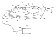
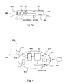
- FIG. 1 illustrates a miniature spectrometer-on-a-chip device in accordance with a preferred embodiment of the present invention.
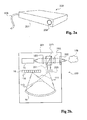
- FIG. 2 a schematically depicts a miniature spectrometer device according to another exemplary embodiment of the invention.
- FIG. 2 b schematically depicts the main components of device according to an exemplary embodiment of the current invention.
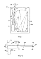
- FIG. 3 schematically depicts a miniature spectrometer device having its optical system primarily etched from a thin sheet of transparent material.
- FIG. 4 a schematically depicts a core biopsy device capable of removing a biopsy sample from tissue.
- FIG. 4 b schematically depicts a guided aspiration biopsy apparatus 420 according to another embodiment of the current invention.
- FIG. 4 c schematically depicts an optical spectroscopy probe according to an exemplary embodiment of the current invention.
- FIG. 4 d schematically depicts a diagnostic endoscope having a miniature spectrometer at its distal end according to an exemplary embodiment of the current invention.
- FIG. 4 e schematically a diagnostic endoscope according to an exemplary embodiment o the current invention.
- FIG. 4 f schematically depicts a vascular diagnostic catheter according to an exemplary embodiment of the current invention.
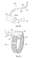
- FIG. 5 a shows an external view of glucose monitor according to an exemplary embodiment of the current invention.
- FIG. 5 b shows a cross section view of glucose monitor according to an exemplary embodiment of the current invention.
- FIG. 6 schematically depicts a swallow-able spectroscopy pill according to yet another exemplary embodiment of the current invention.
- compositions, method or structure may include additional ingredients, steps and/or parts, but only if the additional ingredients, steps and/or parts do not materially alter the basic and novel characteristics of the claimed composition, method or structure.
- a compound or “at least one compound” may include a plurality of compounds, including mixtures thereof.
- the system according to the claimed invention provides results in real is time easy to use, operates at point-of-care, operates at room temperature, provides full infrared spectrum, emits streams of simultaneous photon groups, provides higher signal to noise ratio, gives more accurate results, can be very small, can provide results in noisy environment, is cheap to manufacture, can be sterile and can include disposable parts, such as the emission source.
- FIG. 1 schematically illustrating a miniature spectrometer on a chip device 100 in accordance with a preferred embodiment of the present invention.
- Miniature spectrometer device 100 comprises a carrier 101 on which electro-optical components are integrated.
- Broad band light is produced by a Miniature solid-state Infrared light source 11 which emits broad spectral light.
- light source 11 is a two photon laser 11 capable of producing Infra Red (IR) light.
- IR Infra Red
- light produced by laser 11 is in the wavelength range of 2 to 5 micrometer, however different range of wavelengths may be used. For example, wavelength range of 3 to 5 micrometer may be preferred as it may probe rich spectral features of biological molecules. Optionally, wavelength range may extend to shorter wavelength such as to 1 or even 0.5 micrometer using a solid state two photon laser.
- source 11 produces high light intensity per area, and collimated light capable of being efficiently coupled into input waveguide 107 .
- Source 11 may emit a single stream of photons, or two streams of correlated photons. For example, two streams may be chosen for improved signal to noise ratio by correlations measurements.
- the source may be composed of a single or a few electrically excited solid state sources.
- Input light in input waveguide 107 arrives at coupler 12 and continues towards the tissue to be examined via waveguide 13 that directs the light towards the tissue, and collect the reflected light back to coupler 12 .
- Optical output probe tip 14 is a tip, delivering the light to and from the tested tissue 109 .
- Tip 14 may be in contact with the tested tissue or in proximity to said tissue 109 .
- tip 14 is a one-use consumable element designed to provide sterility, while miniature spectrometer on a chip device 100 is capable of being reused. In other application the entire miniature spectrometer on a chip device 100 is a one-use device.
- Coupler 12 is designed to separate the source light from the back reflected light and directs the reflected light to its output port 110 .
- light is spread 111 on grating 15 by a dispersing optical element such as mirror 112 .
- Mirror 112 may be a flat or convex mirror, a lens, a lens system or combination of some of these elements. In some embodiments, spreading of light is due to the small aperture of the output port 110 of coupler 12 .
- optical element 112 is astigmatic to provide spreading of the light on grating 15 , while preventing the spread of light in the direction normal to carrier 101 .
- Grating 15 is preferably a curved grating acting to disperse the different wavelengths of light, and focusing each range of wavelength of the wavelength dispersed light 113 on a separate element 114 of detector array 16 .
- grating 15 may be replaced with other spectral dispersing of filtering elements such as prisms, filter array or tunable spectral filter.
- Detector array 16 is preferably a One Dimensional ( 1 D) array of detectors. Typically, each element 114 detects the reflected light in a specific range within the spectrum of interest. Detector array 16 may be composed for example of 64 elements, optionally with some separation between their detected spectrums. Alternatively, detector array 16 comprises a plurality of discrete detector elements, positioned at locations where relevant wavelengths are focused, with gaps where wavelengths irrelevant to the desired diagnosis are focused. This reduces the number of detection elements and reduces cost and power consumption. Detector array 16 preferably does not require to cryogenic temperature for its operation. Detector array may comprise indium antimonide (InSb) semiconductor detector array or a miniature bolometer array.
- InSb indium antimonide
- Electronics module 102 is preferably an integrated circuit (IC) such as an Application Specific Integrated Circuit (ASIC) comprising amplifiers for the signals from elements 114 of detector 16 .
- IC integrated circuit
- ASIC Application Specific Integrated Circuit
- Electronics module 102 may provide other signal conditioning and signal processing functions such as filtering, smoothing, lock-in-amplifier function, and Amplitude to Digital Conversion (ADC) function.
- ADC Amplitude to Digital Conversion
- electronics module 102 comprises a plurality of ICs and/or discrete electronic elements. Electronics module 102 may also be used to control the light source 11 , for example providing intensity modulation of light source 11 . Modulation of light source 11 may be achieved by modulating the current powering source 11 . Modulation of light source 11 may be in the form of pulses or sinusoidal. Modulation of light source 11 , in combination with synchronous detection improves the Signal to Noise Ration (SNR) and allows operation of the miniature spectrometer device 100 in the presence of background light. This allows operation of the device, for example, during surgery without diming or turning off the lights.
- SNR Signal to Noise Ration
- Heat removal 121 may be a liquid or gas cooling unit, for example a miniature Joule-Thompson cooler.
- heat removal unit 121 comprises a Thermo Electric Cooler (TEC) 122 coupled with heat sink 103 .
- TEC Thermo Electric Cooler
- Optional heat removal unit 121 removes heat generated by light source 11 and optionally maintain it at proper operational temperature.
- heat removal unit 121 cools detector array 16 for example to maintain it at proper operational temperature.
- heat removal unit 121 removes heat generated by electrical module 102 .
- Same heat removal device may be used for cooling more than one unit of the miniature spectrometer such as electronics, light source and detector, or separate heat removal devices may be used, or some or all the units are not cooled.
- Miniature spectrometer device 100 is connected to a controller unit 120 , preferably using cable 105 .
- cable 105 is connected to miniature spectrometer device 100 via connector 104 , which allows replacing miniature spectrometer device 100 and reusing controller unit 120 .
- cable 105 is connected to controller unit 120 at connector 123 .
- Controller unit 120 may be powered by a battery or a rechargeable battery or may be connected to main power outlet.
- Controller 120 may comprise input and output devices and connectors for programming, controlling and interfacing with the controller such as display, mouse, keyboard and communication devices such as wired or wireless communication such as LAN, USB, Wi-Fi, and other public or proprietary communication protocols. Controller 120 may optionally be split to several sub units.
- controller 120 may comprise a front end unit supplying power to, receiving data from, and controlling the spectrometer; and data processing unit for analyzing the collected data and determine the type of data.
- the data processing unit may be a laptop computer, a PC, a notebook computer, a PDA, a smart phone or other computing device known in the art.
- miniature spectrometer device 100 and controller unit 120 are integrated into one apparatus.
- cable 105 allows inserting miniature spectrometer device 100 into natural or made cavity in the human body, for example using a catheter or an endoscope or a probe such as vascular catheter, urological catheter, vaginal probe or a colonoscope.
- FIG. 1 Not seen in FIG. 1 is a cover, a housing or encapsulation of device 100 .
- said encapsulation is made of bio-compatible material and optionally can be sterilized.
- FIG. 2 a schematically depicts a miniature spectrometer device 200 according to another exemplary embodiment of the invention.
- Devices 100 and 200 primarily differ by their optical system used for interfacing with tissue 109 . Similar components and structures are marked with like numbers and their function already disclosed.
- Device 200 is seen enclosed in enclosure 201 , having an optical port 202 .
- Light reflected from the tissue or sample enters the housing through the optical port 202 , where it is analyzed and detected.
- Cable 105 provides electrical power to device 200 , transmits data indicative of the detected light, provides control signals, and optionally carry cooling fluids.
- FIG. 2 b schematically depicts the main components of device 200 according to an exemplary embodiment of the current invention.
- Light from source 11 is optionally collimated by optional light collimator 222 and enters coupler 220 .
- Collimator 222 may be a lens or other optical element such as concave mirror, etc.
- collimator 222 in integrated into source 11 , for example as convex front face.
- Coupler 220 is preferably a beam splitter directing at least some of the source light to a focusing optical element such as lens 255 .
- coupler 220 comprises a polarizer and phase retarding wave plate (not seen for drawing clarity) for efficient coupling of collimated polarized source light from source 11 to tissue 109 , and efficient coupling of reflected light to output port 251 of coupler 220 .
- Output light 111 is spreads over grating 15 by defocusing optical element 205 .
- Defocusing element 205 may be for example a concave lens. Alternatively, a system of lenses, or mirrors or combination thereof may be used.
- Optical port 202 may be a flat optical window, transparent at the relevant wavelength.
- optical port 202 is integrated with other optical components such as focusing lens 255 .
- At least some of the optical path is confined to a two dimensional (2D) waveguide in the form of thin sheet of transparent material.
- spreading light 111 , and wavelength dispersed light 113 may travel in a 2D waveguide having grating 15 etched onto it, and detector array 16 coupled to it, preferably using index matching or antireflection coating, interface or gel.
- defocusing elements 205 may also be parts of the 2D waveguide.
- 1D waveguides 107 and 13 may also be etched in the transparent sheet.
- Other elements such as couplers may also be integrated or created as part of a wave-guiding sheet.
- FIG. 3 schematically depicts a miniature spectrometer device 300 having its optical system primarily etched from a thin sheet of transparent material 310 .
- the optical components of device 300 are integrated into one or few thin sheet of optically transparent material 300 .
- the thickness of sheet 310 is preferably such that it confines the light in the plane of the sheet creating a 2D optical system. Confinement into 2D surface may be achieved by total internal reflection, or by reflective coating.
- thickness of sheet 310 is such that the confined light is limited essentially to first optical mode in the direction normal to sheet 310 .
- Shape of sheet 310 , and optical components shaped into it may be created by etching, for example lithography or laser etching or other etching methods known in the art.
- sheet 310 is made of material transparent to the wavelength range used. For the preferred range of 2 to 5 micrometers, such material may be ZnSe or Si.
- Sheet 310 is preferably carried on mechanical support 330 .
- Input waveguide 307 may be etched as 1D waveguide, leading to coupler 312 and splitting to sample waveguide 313 that directs the light towards the tissue, and collect the reflected light back to coupler 312 .
- a second port 302 of coupler 312 may be terminated by a monitoring detector 301 used for monitoring and adjusting the output power of source 11 for calibration
- grating 315 From output port 316 of coupler 312 , light is spread 311 on grating 315 by a dispersing optical element such as mirror 333 .
- Mirror 333 is preferably a convex etch in sheet 310 .
- grating 315 may also be created by etching a pattern in sheet 310 .
- surface of mirror 333 and/or grating 315 are coated with highly reflecting material such as gold, Aluminum or other reflecting structure such as dielectric coating.
- Sheet 310 is preferably thin enough to provide light confinement in the direction normal to its plane.
- sheet 310 may have a thickness compatible to the light wavelength.
- coupler 312 is a three terminals (“Y”) coupler having no port 302 .
- detector array 16 is abutted to sheet 310 , optionally using some index matching means such as gel, glue or coating.
- coupler 12 , 220 or 312 may be missing and replaced with an illumination optical path for illuminating the sample 109 with light from laser 11 and reflection optical path for collecting reflected light from sample 109 and directing it to the light spreading element such as 112 205 or 333 which spreads it on the grating.
- the construction of spectrometers 100 , 200 or 300 is three dimensional such that optical paths may be in different layers or heights respect to carrier 101 or 330 , and grating 15 may be concave or spherical.
- a single detector is used in combination with a tunable filter.
- the filter may be situated on the optical path between laser 11 and tissue, or on the path between the tissue and said single detector.
- light from light source is projected onto the tissue by light projection optics, and reflected light is collected by a light collection optics that is used as an input to the spectrometer, wherein light projection optics collection optics uses separate optical paths.
- the inventive miniature spectrometer may be to small enough to be integrated into a hand held probe or to an endoscope.
- spectrometer 100 may be as small as 50 mm by 30 mm by 15 mm.
- spectrometer 100 may be as small as 20 mm by 15 mm by 5 mm or smaller.
- FIGS. 4 a to 4 e schematically depicts medical applications for miniature spectroscopy devices such as device 100 , 200 or 300 .
- FIG. 4 a schematically depicts a core biopsy device 400 capable of removing a biopsy sample from tissue.
- Core biopsy device 400 comprises a body 401 equipped with handle 405 and trigger 402 to trigger sample extraction from opening 404 by biopsy needle 403 having a sharp penetrating end 416 and connector 415 to connect to body 401 .
- Guided core biopsy device 400 further comprises a miniature spectroscopy device such as device 100 , 200 or 300 .
- the miniature spectroscopy device is attached to or integrated within body 401 of guided core biopsy apparatus 400 .
- the miniature spectrometer is optically interfaced with the tissue to be sampled using a needle waveguide 410 ending with optical tip 414 for illuminating the tissue and collecting reflected light.
- Needle waveguide 410 interfaces with miniature spectrometer 100 , 200 or 300 using interface 418 which interfaces the spectrometer optical port 14 (or 202 ) with waveguide 410 .
- needle 403 and waveguide 410 with their parts are integrated into one consumable guided biopsy needle configured as one-use device.
- the one-use device is preferably configured for fast connection to body 401 of guided core biopsy 400 .
- Waveguide 410 may be a hollow optical fiber or a waveguide made of IR transparent material. It should be noted that the short length of waveguide 410 reduces optical loss. Short waveguide is possible by placing spectrometer 100 , 200 or 300 near the proximal end of needle 403 . This is impossible to do if large size spectrometer is used, for example a Fourier Transform InfraRed spectrometer (FTIR) or large size light source such as a hot black body are used.
- FTIR Fourier Transform InfraRed spectrometer
- Spectrometer 100 , 200 or 300 interfaces with controller 120 which may be integrated into body 401 or may be remotely situated, connected by cable 105 .
- user advances the needle 403 into the tissue while the miniature spectrometer analyzes in real time the light reflected from the tissue in location of or proximate to the location where biopsy sample may be taken.
- Controller 120 report to the user about the probability of the tissue to be sampled being a target tissue.
- controller 120 may emit an acoustic signal having volume or pitch indicative of the location of tip 414 in contact to target tissue such as cancerous tumor.
- controller 120 may comprise a display or other visual indicator for informing the user of properties of tissue near opening 404 of needle 403 .
- opening 404 When the user discovers that opening 404 is near or at target tissue type, he may release the trigger 402 to obtain a tissue sample.
- the guided biopsy apparatus increases the probability of obtaining the correct sample, reducing the number of needle insertions to collect specimen, reducing false negative sample, reducing cost and patient discomfort.
- Needle waveguide 410 is preferably as short as needed for reducing light loss from the source to the tissue and back to the spectrometer.
- needle waveguide 410 is replaced with two waveguides: one for delivering laser light, and the other for returning reflected light.
- FIG. 4 b schematically depicts a guided aspiration biopsy apparatus 420 according to another embodiment of the current invention.
- Guided aspiration biopsy 420 comprises a hollow aspiration needle 421 ending in a sharp penetrating tip 427 and connecting to the apparatus in connector 422 .
- Needle 421 has a channel opened at orifice 429 at or near sharp 427 and leading to measurement chamber 435 in the body of guided aspiration biopsy apparatus 420 .
- a valve 439 connected to a suction device 423 allows the user to suck a fluid sample from tissue near orifice 429 into chamber 435 .
- Sample in chamber 435 is optically probed by miniature spectroscope device 200 having optical port 202 in optical communication with fluid sample in chamber 435 .
- spectrometer 200 is replaced with spectrometer 100 or 300 having its tip 14 in optical communication with fluid sample in chamber 435 .
- Valve 439 allows transferring the collected fluid into sample bottle 429 , preferably having fast connection 430 allowing collecting a plurality of samples from plurality of locations in the tissue. Additionally and optionally, valve 439 may be set to discard the collected sample to waste 431 .
- Spectrometer 200 analyzes the fluid in chamber 435 in real time.
- valve 439 is activated manually or automatically to collect a sample for further analysis, for example at a pathology lab.
- FIG. 4 c schematically depicts an optical spectroscopy probe 440 according to an exemplary embodiment of the current invention.
- Probe 440 may be used for example during surgery to examine excision site after removal of a tumor for identification of cancerous tissue due to incomplete removal. Similarly, margins of removed tumor may be examined to ensure that a safety layer of healthy tissue exists around the removed tumor. Probe may be used for example for testing lymph nodes for presence of cancerous cells. The probe may also be used non-invasively for identification and classification of exposed tissue, for example testing skin lesions for cancer such as melanoma.
- Probe 440 comprises a housing 445 , for housing miniature spectrometer 100 , 200 or 300 . Tip 14 extends beyond housing 445 , or window 202 on surface of housing 445 is brought to contact with or to proximity of the tissue 109 to be examined.
- Control unit 120 may be integrated into housing 445 or situated remotely and connected by cable 105 to housing 445 .
- probe 440 is a self contained probe having controller 120 integrated into its body 445 .
- housing 445 may also comprise a power source such as a battery or a rechargeable battery, controlling buttons and other user inputs 441 for example such as on/off switch, activate measurement button and tissue type selector.
- Probe 440 may also comprise outputs such as a display 443 , visual and/or audio indicator 442 and the likes.
- probe 440 is sized to be hand held.
- probe 440 may be sterilized or covered with a sterile cover having a light transparent window or tip such that it may be used in an operation room. However, for external use, or to be used on removed tissue, sterilization may not be needed.
- optical path to the tissue and back is short, thus optical loss may be minimized.
- Hand held probe may be made as small as few cm in size so it can be manipulated in the operation room.
- the probe may be sized as a mobile phone.
- FIG. 4 d schematically depicts a diagnostic endoscope 460 having a miniature spectrometer 100 , 200 or 300 at its distal end.
- Endoscope 460 may be shaped and function as an endoscope of the art with an addition of optical tip 14 extending from, or window 202 exposed on its surface near its distal end.
- distal end of shaft 460 of diagnostic endoscope 460 shown with working channel 463 , illumination light source 464 , camera 462 and spectroscopy tip or window 14 or 202 .
- endoscope 460 may further comprise other channels and means such as irrigation channel, optical surfaces cleaning jets, and tissue manipulation or treatment means.
- Endoscope 460 may be flexible or rigid and may comprise means for navigation and positioning.
- spectroscopic tip 14 or window 202 is so situates such that the part of tissue that is spectroscopically examined is within the field of view of camera 462 .
- tip 14 or window 202 is situated on the side wall of shaft 461 .
- endoscope 460 comprises a plurality of tips 14 or windows 202 connected to one or to plurality of spectrometers 100 , 200 or 300 .
- controller 120 is integrated into endoscope 460 . In other embodiments, parts or the entire controller 120 is remotely positioned, optionally wirelessly communicating with the endoscope.
- Having miniature spectrometer 100 , 200 or 300 in proximity to distal end of endoscope 460 is advantageous as it reduces loss of light in transferring light to the tissue and transferring reflected light from the tissue to the spectrometer.
- FIG. 4 e schematically depicts a diagnostic endoscope 480 according to an exemplary embodiment of the current invention.
- miniature spectrometer 100 or 200 is located in the base 481 of endoscope 480 , and connected to tip 14 with elongated waveguide 483 .
- This embodiment is advantageous for endoscopes having thin shaft 484 such as endoscopes used in the uterus.
- FIG. 4 f schematically depicts a vascular diagnostic catheter 490 according to an exemplary embodiment of the current invention.
- Catheter 490 comprises a flexible shaft 493 having an exposed optical port 494 , preferably located on the side of the shaft close to the distal end of shaft 493 .
- Flexible waveguide 495 communicates illumination light to port 494 and returns scattered light to spectrometer 100 or 300 in base 491 of catheter 490 .
- radio-opaque mark enables locating the position, and optionally the orientation of distal end of catheter 499 using standard X-Ray fluoroscopy equipment. Other localization and orientation finding methods may be used.
- shaft of catheter 490 is advanced for example in an artery to a desired location where existence or type of plaque in front of optical port 494 is determined using spectrometer 100 or 300 .
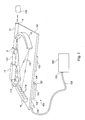
- FIGS. 5 a and 5 b schematically depict personal spectroscopic glucose monitoring device 500 according to an exemplary embodiment of the current invention.
- FIG. 5 a shows an external view of monitor 500
- FIG. 5 b shows a cross section of said monitor.
- Monitor 500 is preferably shaped like a wrist watch having a body 501 and a wrist band 502 .
- Body 501 of monitor 500 comprises an optical port (not seen in this figure) on its bottom side that faces and in contact with the skin of the monitored person.
- a display 503 On the upper face of body 501 is a display 503 and control switches 504 for controlling the operation of monitor 500 .
- Optional audible alarm 505 may provide acoustic alarm to alert the user of abnormal readings.
- vibration may be used as alarm.
- monitor 500 may act as a normal watch showing time, date, etc.
- monitor 500 is attached to the body at location other than the wrist, for example on the abdomen or the arm or leg.
- strap is missing and adhesive is used.
- monitor 500 comprises a wireless communication unit 508 for wirelessly communicating 509 with external unit 550 .
- monitor 500 may not comprise display or controls except for example an optional on/off switch and/or optional indicator 506 such as on/off indicator and/or low battery indicator.
- external unit 550 may comprise an insulin pump, for example external or implanted insulin pump. Using an insulin pump with an optical glucose monitor according to the current invention enables for example a continues and optionally fully automatic control of glucose levels in diabetic patients.
- External unit 550 may be an insulin pump, internally implanted or external to the user.
- External unit 550 may provide some of the functions of controller 120 , for example analyzing spectra obtained by spectrometer 100 , 200 or 300 to determine and log glucose level. External unit 550 may be in constant communication with monitor 500 or communicate periodically or on demand. External unit 550 may be a smart phone, a computer or a specifically made device made for communicating with monitor 500 .
- Monitor 500 is powered by a replaceable or rechargeable battery 510 .
- Monitor 500 may optionally, additionally or alternatively monitor other bodily functions and levels of chemicals other than glucose, for example oxygenation level, hormonal levels.
- FIG. 6 schematically depicts a swallow-able spectroscopy pill 606 according to yet another exemplary embodiment of the current invention.
- Pill 606 comprises a body 615 sized to be swallowed by a human patient and pass through his digestive track.
- Body 615 comprises a transparent dome 614 .
- Camera 613 and illumination light source 617 are positioned behind dome 614 for providing images of digestive track tissue 655 in front of said dome.
- Light source 617 illuminates the tissue 655 and camera 613 images the light reflected from the tissue 655 .
- Controller 620 receives image data from camera 613 and transmits said image data using wireless communication unit 630 to control unit 640 over RF link 609 .
- Control unit 640 preferably comprises a computer such as a PC, laptop, or a notebook computer or other computer having a display and user interface such as a keyboard or a mouse.
- control unit 640 uses user input on control unit 640 to communicate via optional link 619 and wireless communication unit 630 a command, commanding controller 620 to activate miniature spectrometer 100 , 200 or 300 within body 615 to acquire a spectra of said suspected tissue 655 .
- spectroscopic data is acquired and transmitted automatically or periodically.
- Optical port 14 or 202 of miniature spectrometer 100 , 200 or 300 is preferably oriented such that IR source light is directed towards tissue 655 and reflected IR light is reflected at least in part to said optical port 14 or 202 .
- acquiring spectra may be triggered automatically, for example by an image processing unit, identifying that suspected tissue is in front of the spectrometer.
- Acquired spectra from tissue 655 may be analyzed by unit 640 to determine the probability of said tissue being cancerous or inflamed.
Landscapes
- Health & Medical Sciences (AREA)
- Life Sciences & Earth Sciences (AREA)
- Physics & Mathematics (AREA)
- Pathology (AREA)
- General Health & Medical Sciences (AREA)
- Engineering & Computer Science (AREA)
- Surgery (AREA)
- Biophysics (AREA)
- Biomedical Technology (AREA)
- Heart & Thoracic Surgery (AREA)
- Medical Informatics (AREA)
- Molecular Biology (AREA)
- Veterinary Medicine (AREA)
- Animal Behavior & Ethology (AREA)
- Public Health (AREA)
- Spectroscopy & Molecular Physics (AREA)
- Vascular Medicine (AREA)
- Immunology (AREA)
- Dermatology (AREA)
- Optics & Photonics (AREA)
- Gynecology & Obstetrics (AREA)
- Cardiology (AREA)
- Physiology (AREA)
- Reproductive Health (AREA)
- Chemical & Material Sciences (AREA)
- Analytical Chemistry (AREA)
- Biochemistry (AREA)
- General Physics & Mathematics (AREA)
- Endocrinology (AREA)
- Investigating Or Analysing Materials By Optical Means (AREA)
- Measuring Temperature Or Quantity Of Heat (AREA)
Priority Applications (1)
| Application Number | Priority Date | Filing Date | Title |
|---|---|---|---|
| US13/378,916 US20120184827A1 (en) | 2009-06-16 | 2010-06-16 | Miniature disease optical spectroscopy diagnostic system |
Applications Claiming Priority (3)
| Application Number | Priority Date | Filing Date | Title |
|---|---|---|---|
| US18728009P | 2009-06-16 | 2009-06-16 | |
| US13/378,916 US20120184827A1 (en) | 2009-06-16 | 2010-06-16 | Miniature disease optical spectroscopy diagnostic system |
| PCT/IL2010/000477 WO2010146588A2 (fr) | 2009-06-16 | 2010-06-16 | Système miniature permettant de diagnostiquer une maladie |
Publications (1)
| Publication Number | Publication Date |
|---|---|
| US20120184827A1 true US20120184827A1 (en) | 2012-07-19 |
Family
ID=42964241
Family Applications (1)
| Application Number | Title | Priority Date | Filing Date |
|---|---|---|---|
| US13/378,916 Abandoned US20120184827A1 (en) | 2009-06-16 | 2010-06-16 | Miniature disease optical spectroscopy diagnostic system |
Country Status (2)
| Country | Link |
|---|---|
| US (1) | US20120184827A1 (fr) |
| WO (1) | WO2010146588A2 (fr) |
Cited By (56)
| Publication number | Priority date | Publication date | Assignee | Title |
|---|---|---|---|---|
| US20120049744A1 (en) * | 2010-08-25 | 2012-03-01 | Hon Hai Precision Industry Co., Ltd. | Measuring instrument |
| WO2014153200A1 (fr) | 2013-03-14 | 2014-09-25 | Echo Labs, Inc. | Systèmes et méthodes d'analyse multispectrale du sang |
| EP2813174A1 (fr) * | 2013-06-11 | 2014-12-17 | Spiess Media Systems Asc, Corp. | Système portable de capteurs modulaires |
| US20150004641A1 (en) * | 2013-06-26 | 2015-01-01 | General Electric Company | System and method for holding a biopsy collecting device |
| US20150185140A1 (en) * | 2013-06-26 | 2015-07-02 | General Electric Company | System and method for attaching a biopsy collecting device to a spectroscopy system |
| US20150182204A1 (en) * | 2013-06-26 | 2015-07-02 | General Electric Company | System and method for attaching a biopsy collecting device to a spectroscopy system |
| US20160139051A1 (en) * | 2013-07-11 | 2016-05-19 | Henry Ford Health System | Apparatus and method for distinguishing between different tissue types using specific raman spectral regions |
| WO2017156182A1 (fr) * | 2016-03-08 | 2017-09-14 | Zebra Medical Technologies, Inc. | Détection non invasive d'une maladie de peau |
| WO2017191236A2 (fr) | 2016-05-04 | 2017-11-09 | Centre Technologique Alphanov | Procede d'etalonnage d'un spectre obtenu a l'aide d'un appareil electronique equipé d'un accessoire amovible |
| US9885655B2 (en) * | 2012-11-13 | 2018-02-06 | Viavi Solutions Inc. | Spectrometer with a relay lightpipe |
| US9883854B2 (en) * | 2015-04-08 | 2018-02-06 | Synaptive Medical (Barbados) Inc. | Systems, devices and methods for tissue removal and analysis |
| US10222325B2 (en) | 2012-11-13 | 2019-03-05 | Viavi Solutions Inc. | Portable spectrometer |
| US10799172B2 (en) | 2017-10-18 | 2020-10-13 | Ladysense (2017) Ltd. | Multifunctional measuring device for automatic examination of female genital organs |
| CN112229830A (zh) * | 2020-09-14 | 2021-01-15 | 中国科学院上海光学精密机械研究所 | 内窥式激光诱导击穿光谱系统 |
| WO2021026145A1 (fr) * | 2019-08-05 | 2021-02-11 | Gyrus Acmi, Inc. D/B/A Olympus Surgical Technologies America | Identification de cible avec diviseur de signal de rétroaction optique |
| DE102020108957A1 (de) | 2020-03-31 | 2021-09-30 | Otto-Von-Guericke-Universität Magdeburg | Vorrichtung, Verfahren und Computerprogramm zur Fluoreszenzmessung |
| WO2022130022A1 (fr) * | 2020-12-14 | 2022-06-23 | Rockley Photonics Limited | Dispositif minimalement invasif pourvu d'un spectrophotomètre |
| US11375897B2 (en) | 2017-04-20 | 2022-07-05 | Henry Ford Health System | System and method for characterization of a brain tissue sample using Raman marker regions |
| CN114903418A (zh) * | 2021-02-08 | 2022-08-16 | 苏州佳世达光电有限公司 | 口腔扫描仪 |
| US11474310B2 (en) | 2020-02-28 | 2022-10-18 | Bard Access Systems, Inc. | Optical connection systems and methods thereof |
| US11525670B2 (en) | 2019-11-25 | 2022-12-13 | Bard Access Systems, Inc. | Shape-sensing systems with filters and methods thereof |
| US11624677B2 (en) | 2020-07-10 | 2023-04-11 | Bard Access Systems, Inc. | Continuous fiber optic functionality monitoring and self-diagnostic reporting system |
| US11622816B2 (en) | 2020-06-26 | 2023-04-11 | Bard Access Systems, Inc. | Malposition detection system |
| US11630009B2 (en) | 2020-08-03 | 2023-04-18 | Bard Access Systems, Inc. | Bragg grated fiber optic fluctuation sensing and monitoring system |
| US11633149B2 (en) | 2017-04-28 | 2023-04-25 | Enspectra Health, Inc. | Systems and methods for imaging and measurement of sarcomeres |
| DE102014107342B4 (de) | 2014-05-24 | 2023-05-04 | Frank Braun | Vorrichtung und Verfahren zur Erkennung von Krebstumoren und anderen Gewebeveränderungen |
| US11850338B2 (en) | 2019-11-25 | 2023-12-26 | Bard Access Systems, Inc. | Optical tip-tracking systems and methods thereof |
| US11883609B2 (en) | 2020-06-29 | 2024-01-30 | Bard Access Systems, Inc. | Automatic dimensional frame reference for fiber optic |
| US11899249B2 (en) | 2020-10-13 | 2024-02-13 | Bard Access Systems, Inc. | Disinfecting covers for functional connectors of medical devices and methods thereof |
| US11931179B2 (en) | 2020-03-30 | 2024-03-19 | Bard Access Systems, Inc. | Optical and electrical diagnostic systems and methods thereof |
| US11931112B2 (en) | 2019-08-12 | 2024-03-19 | Bard Access Systems, Inc. | Shape-sensing system and methods for medical devices |
| EP4359762A1 (fr) * | 2021-06-24 | 2024-05-01 | Clearstream Technologies Limited | Système et procédé d'identification d'un échantillon biologique dans un porte-échantillon, et dispositif de biopsie permettant d'obtenir un échantillon biologique de tissu |
| US12064569B2 (en) | 2020-09-25 | 2024-08-20 | Bard Access Systems, Inc. | Fiber optics oximetry system for detection and confirmation |
| US12089815B2 (en) | 2022-03-17 | 2024-09-17 | Bard Access Systems, Inc. | Fiber optic medical systems and devices with atraumatic tip |
| US12140487B2 (en) | 2017-04-07 | 2024-11-12 | Bard Access Systems, Inc. | Optical fiber-based medical device tracking and monitoring system |
| US12220219B2 (en) | 2020-11-24 | 2025-02-11 | Bard Access Systems, Inc. | Steerable fiber optic shape sensing enabled elongated medical instrument |
| US12232818B2 (en) | 2020-03-03 | 2025-02-25 | Bard Access Systems, Inc. | System and method for optic shape sensing and electrical signal conduction |
| US12232821B2 (en) | 2021-01-06 | 2025-02-25 | Bard Access Systems, Inc. | Needle guidance using fiber optic shape sensing |
| US12246139B2 (en) | 2020-02-28 | 2025-03-11 | Bard Access Systems, Inc. | Catheter with optic shape sensing capabilities |
| US12285572B2 (en) | 2020-11-18 | 2025-04-29 | Bard Access Systems, Inc. | Optical-fiber stylet holders and methods thereof |
| US12285236B2 (en) | 2018-11-13 | 2025-04-29 | Enspectra Health, Inc. | Methods and systems for generating depth profiles with improved optical resolution |
| US12318149B2 (en) | 2022-03-08 | 2025-06-03 | Bard Access Systems, Inc. | Medical shape sensing devices and systems |
| US12343117B2 (en) | 2022-06-28 | 2025-07-01 | Bard Access Systems, Inc. | Fiber optic medical systems and methods for identifying blood vessels |
| US12349984B2 (en) | 2022-06-29 | 2025-07-08 | Bard Access Systems, Inc. | System, method, and apparatus for improved confirm of an anatomical position of a medical instrument |
| US12419694B2 (en) | 2021-10-25 | 2025-09-23 | Bard Access Systems, Inc. | Reference plane for medical device placement |
| US12426956B2 (en) | 2022-03-16 | 2025-09-30 | Bard Access Systems, Inc. | Medical system and method for monitoring medical device insertion and illumination patterns |
| US12426954B2 (en) | 2021-01-26 | 2025-09-30 | Bard Access Systems, Inc. | Fiber optic shape sensing system associated with port placement |
| US12446988B2 (en) | 2021-09-16 | 2025-10-21 | Bard Access Systems, Inc. | Swappable high mating cycle fiber connection interface |
| US12490937B2 (en) | 2021-05-18 | 2025-12-09 | Bard Access Systems, Inc. | Anatomical oscillation and fluctuation sensing and confirmation system |
| US12551288B2 (en) | 2020-10-13 | 2026-02-17 | Bard Access Systems, Inc. | Fiber optic enabled deployable medical devices for monitoring, assessment and capture of deployment information |
| US12564340B2 (en) | 2022-07-12 | 2026-03-03 | Bard Access Systems, Inc. | Eccentric single-core fiber-optic enabled medical device |
| US12569221B2 (en) | 2018-08-22 | 2026-03-10 | Bard Access Systems, Inc. | Systems and methods for infrared-enhanced ultrasound visualization |
| US12575893B2 (en) | 2022-06-24 | 2026-03-17 | Bard Access Systems, Inc. | Shape sensing fiber optic tip protection systems and devices |
| US12605116B2 (en) | 2022-04-11 | 2026-04-21 | Bard Access Systems, Inc. | Fiber optic medical systems and devices with electrical tip |
| US12667311B2 (en) | 2021-04-15 | 2026-06-30 | Bard Access Systems, Inc. | Extended fiber optic sensing system |
| US12685595B2 (en) | 2022-04-27 | 2026-07-21 | Bard Access Systems, Inc. | Conductor incorporated fiber enabled medical systems |
Families Citing this family (8)
| Publication number | Priority date | Publication date | Assignee | Title |
|---|---|---|---|---|
| US20120283563A1 (en) * | 2011-05-03 | 2012-11-08 | Moore Kyle P | Biopsy device with manifold alignment feature and tissue sensor |
| US8953160B2 (en) | 2012-05-14 | 2015-02-10 | Redox Biomedical, Inc. | Systems having a reflected light sensor and methods of use |
| AU2015319850B2 (en) | 2014-09-25 | 2019-09-26 | Bt Bidco, Inc. | Electromechanical pill device with localization capabilities |
| DE102014115502A1 (de) * | 2014-10-24 | 2016-04-28 | Pyreos Ltd. | Hautmessgerät und Armbanduhr |
| EP4467182A3 (fr) | 2016-09-09 | 2025-01-15 | Biora Therapeutics, Inc. | Dispositif électromécanique ingérable pour l'administration d'une substance distribuable |
| CA3045944A1 (fr) | 2016-12-07 | 2018-06-14 | Progenity, Inc. | Procedes, dispositifs et systemes de detection du tractus gastro-intestinal |
| AU2018243312B2 (en) | 2017-03-31 | 2023-02-02 | Bt Bidco, Inc. | Localization systems and methods for an ingestible device |
| CN113348011B (zh) | 2018-11-19 | 2023-04-18 | 比奥拉治疗股份有限公司 | 用生物治疗剂治疗疾病的方法和装置 |
Citations (8)
| Publication number | Priority date | Publication date | Assignee | Title |
|---|---|---|---|---|
| US5995867A (en) * | 1997-03-19 | 1999-11-30 | Lucid Inc | Cellular surgery utilizing confocal microscopy |
| US6324418B1 (en) * | 1997-09-29 | 2001-11-27 | Boston Scientific Corporation | Portable tissue spectroscopy apparatus and method |
| US20020091324A1 (en) * | 1998-04-06 | 2002-07-11 | Nikiforos Kollias | Non-invasive tissue glucose level monitoring |
| US20020133065A1 (en) * | 2001-01-18 | 2002-09-19 | Lucassen Gerhardus Wilhelmus | Analysis of a composition |
| US20060189860A1 (en) * | 2005-02-10 | 2006-08-24 | Terumo Cardiovascular Systems Corporation | Near infrared spectroscopy device with reusable portion |
| US20080262324A1 (en) * | 2004-05-11 | 2008-10-23 | Koninklijke Philips Electronics, N.V. | Measurement Head For Non-Invasive Blood Analysis |
| US20090051898A1 (en) * | 2007-08-24 | 2009-02-26 | Samsung Electronics Co., Ltd. | Non-invasive probe for measuring body components and a non-invasive body component measurement system including the non-invasive probe |
| US20090203991A1 (en) * | 2006-04-21 | 2009-08-13 | Cedars-Sinai Medical Center | Multiple imaging and/or spectroscopic modality probe |
Family Cites Families (8)
| Publication number | Priority date | Publication date | Assignee | Title |
|---|---|---|---|---|
| IL104506A (en) | 1993-01-25 | 1997-11-20 | Israel State | Fast changing heating- cooling device and method, particularly for cryogenic and/or surgical use |
| US6485703B1 (en) * | 1998-07-31 | 2002-11-26 | The Texas A&M University System | Compositions and methods for analyte detection |
| US6816241B2 (en) * | 2000-09-26 | 2004-11-09 | Sensys Medical, Inc. | LED light source-based instrument for non-invasive blood analyte determination |
| US9295391B1 (en) * | 2000-11-10 | 2016-03-29 | The General Hospital Corporation | Spectrally encoded miniature endoscopic imaging probe |
| AU2002314969A1 (en) * | 2001-06-06 | 2002-12-16 | University Of Kentucky Research Foundation | Multimode structure for current measurement, ultrasonic and near-ir spectrum collection and processing |
| WO2008066911A2 (fr) | 2006-11-30 | 2008-06-05 | Newton Laboratories, Inc. | Imagerie amplifiée spectroscopiquement |
| US20080221411A1 (en) * | 2007-03-09 | 2008-09-11 | Nellcor Puritan Bennett Llc | System and method for tissue hydration estimation |
| US8921826B2 (en) | 2007-11-27 | 2014-12-30 | Technion Research & Development Foundation Limited | Light source based on simultaneous two-photon emission |
-
2010
- 2010-06-16 WO PCT/IL2010/000477 patent/WO2010146588A2/fr not_active Ceased
- 2010-06-16 US US13/378,916 patent/US20120184827A1/en not_active Abandoned
Patent Citations (8)
| Publication number | Priority date | Publication date | Assignee | Title |
|---|---|---|---|---|
| US5995867A (en) * | 1997-03-19 | 1999-11-30 | Lucid Inc | Cellular surgery utilizing confocal microscopy |
| US6324418B1 (en) * | 1997-09-29 | 2001-11-27 | Boston Scientific Corporation | Portable tissue spectroscopy apparatus and method |
| US20020091324A1 (en) * | 1998-04-06 | 2002-07-11 | Nikiforos Kollias | Non-invasive tissue glucose level monitoring |
| US20020133065A1 (en) * | 2001-01-18 | 2002-09-19 | Lucassen Gerhardus Wilhelmus | Analysis of a composition |
| US20080262324A1 (en) * | 2004-05-11 | 2008-10-23 | Koninklijke Philips Electronics, N.V. | Measurement Head For Non-Invasive Blood Analysis |
| US20060189860A1 (en) * | 2005-02-10 | 2006-08-24 | Terumo Cardiovascular Systems Corporation | Near infrared spectroscopy device with reusable portion |
| US20090203991A1 (en) * | 2006-04-21 | 2009-08-13 | Cedars-Sinai Medical Center | Multiple imaging and/or spectroscopic modality probe |
| US20090051898A1 (en) * | 2007-08-24 | 2009-02-26 | Samsung Electronics Co., Ltd. | Non-invasive probe for measuring body components and a non-invasive body component measurement system including the non-invasive probe |
Cited By (96)
| Publication number | Priority date | Publication date | Assignee | Title |
|---|---|---|---|---|
| CN102374835A (zh) * | 2010-08-25 | 2012-03-14 | 鸿富锦精密工业(深圳)有限公司 | 量测仪 |
| US8314389B2 (en) * | 2010-08-25 | 2012-11-20 | Hon Hai Precision Industry Co., Ltd. | Measuring instrument |
| US20120049744A1 (en) * | 2010-08-25 | 2012-03-01 | Hon Hai Precision Industry Co., Ltd. | Measuring instrument |
| US10422743B2 (en) | 2012-11-13 | 2019-09-24 | Viavi Solutions Inc. | Spectrometer with a relay lightpipe |
| US10222325B2 (en) | 2012-11-13 | 2019-03-05 | Viavi Solutions Inc. | Portable spectrometer |
| US9885655B2 (en) * | 2012-11-13 | 2018-02-06 | Viavi Solutions Inc. | Spectrometer with a relay lightpipe |
| EP4032469A1 (fr) * | 2013-03-14 | 2022-07-27 | Spry Health, Inc. | Systèmes et procédés de mesure de sang multispectrale |
| WO2014153200A1 (fr) | 2013-03-14 | 2014-09-25 | Echo Labs, Inc. | Systèmes et méthodes d'analyse multispectrale du sang |
| US10463283B2 (en) | 2013-03-14 | 2019-11-05 | Spry Health, Inc. | Systems and methods of multispectral blood measurement |
| EP2967413A4 (fr) * | 2013-03-14 | 2016-11-23 | Echo Labs Inc | Systèmes et méthodes d'analyse multispectrale du sang |
| EP2813174A1 (fr) * | 2013-06-11 | 2014-12-17 | Spiess Media Systems Asc, Corp. | Système portable de capteurs modulaires |
| US9913628B2 (en) * | 2013-06-26 | 2018-03-13 | General Electric Company | System and method for attaching a biopsy collecting device to a spectroscopy system |
| US20150182204A1 (en) * | 2013-06-26 | 2015-07-02 | General Electric Company | System and method for attaching a biopsy collecting device to a spectroscopy system |
| US20150185140A1 (en) * | 2013-06-26 | 2015-07-02 | General Electric Company | System and method for attaching a biopsy collecting device to a spectroscopy system |
| US20150004641A1 (en) * | 2013-06-26 | 2015-01-01 | General Electric Company | System and method for holding a biopsy collecting device |
| US10531862B2 (en) * | 2013-06-26 | 2020-01-14 | General Electric Company | System and method for attaching a biopsy collecting device to a spectroscopy system |
| US20160139051A1 (en) * | 2013-07-11 | 2016-05-19 | Henry Ford Health System | Apparatus and method for distinguishing between different tissue types using specific raman spectral regions |
| DE102014107342B4 (de) | 2014-05-24 | 2023-05-04 | Frank Braun | Vorrichtung und Verfahren zur Erkennung von Krebstumoren und anderen Gewebeveränderungen |
| US9883854B2 (en) * | 2015-04-08 | 2018-02-06 | Synaptive Medical (Barbados) Inc. | Systems, devices and methods for tissue removal and analysis |
| US12514453B2 (en) | 2016-03-08 | 2026-01-06 | Enspectra Health, Inc. | Non-invasive detection of skin disease |
| CN109069007A (zh) * | 2016-03-08 | 2018-12-21 | 泽博拉医疗科技公司 | 皮肤疾病的非侵入式检测 |
| US11877826B2 (en) | 2016-03-08 | 2024-01-23 | Enspectra Health, Inc. | Non-invasive detection of skin disease |
| US11172826B2 (en) | 2016-03-08 | 2021-11-16 | Enspectra Health, Inc. | Non-invasive detection of skin disease |
| WO2017156182A1 (fr) * | 2016-03-08 | 2017-09-14 | Zebra Medical Technologies, Inc. | Détection non invasive d'une maladie de peau |
| WO2017191236A2 (fr) | 2016-05-04 | 2017-11-09 | Centre Technologique Alphanov | Procede d'etalonnage d'un spectre obtenu a l'aide d'un appareil electronique equipé d'un accessoire amovible |
| US12140487B2 (en) | 2017-04-07 | 2024-11-12 | Bard Access Systems, Inc. | Optical fiber-based medical device tracking and monitoring system |
| US11375897B2 (en) | 2017-04-20 | 2022-07-05 | Henry Ford Health System | System and method for characterization of a brain tissue sample using Raman marker regions |
| US11633149B2 (en) | 2017-04-28 | 2023-04-25 | Enspectra Health, Inc. | Systems and methods for imaging and measurement of sarcomeres |
| US10799172B2 (en) | 2017-10-18 | 2020-10-13 | Ladysense (2017) Ltd. | Multifunctional measuring device for automatic examination of female genital organs |
| US12569221B2 (en) | 2018-08-22 | 2026-03-10 | Bard Access Systems, Inc. | Systems and methods for infrared-enhanced ultrasound visualization |
| US12285236B2 (en) | 2018-11-13 | 2025-04-29 | Enspectra Health, Inc. | Methods and systems for generating depth profiles with improved optical resolution |
| US20230075714A1 (en) * | 2019-08-05 | 2023-03-09 | Gyrus Acm, Inc. D/B/A Olympus Surgical Technologies America | Target identification with optical feedback signal splitter |
| US20250072964A1 (en) * | 2019-08-05 | 2025-03-06 | Gyrus Acmi, Inc. D/B/A Olympus Surgical Technologies America | Target identification with optical feedback signal splitter |
| WO2021026145A1 (fr) * | 2019-08-05 | 2021-02-11 | Gyrus Acmi, Inc. D/B/A Olympus Surgical Technologies America | Identification de cible avec diviseur de signal de rétroaction optique |
| US11523865B2 (en) * | 2019-08-05 | 2022-12-13 | Gyrus Acmi, Inc. | Target identification with optical feedback signal splitter |
| US12207869B2 (en) * | 2019-08-05 | 2025-01-28 | Gyrus Acmi, Inc. | Target identification with optical feedback signal splitter |
| US12220166B2 (en) * | 2019-08-05 | 2025-02-11 | Gyrus Acmi, Inc. | Target identification with optical feedback signal splitter |
| JP2022543289A (ja) * | 2019-08-05 | 2022-10-11 | ジャイラス エーシーエムアイ インク ディー/ビー/エー オリンパス サージカル テクノロジーズ アメリカ | 光学フィードバック信号スプリッタを用いたターゲット識別 |
| US20230310076A1 (en) * | 2019-08-05 | 2023-10-05 | Gyrus Acmi, Inc. D/B/A Olympus Surgical Technologies America | Target identification with optical feedback signal splitter |
| US11406447B2 (en) * | 2019-08-05 | 2022-08-09 | Gyrus Acmi, Inc. | Target identification with optical feedback signal splitter |
| JP7551732B2 (ja) | 2019-08-05 | 2024-09-17 | ジャイラス エーシーエムアイ インク ディー/ビー/エー オリンパス サージカル テクノロジーズ アメリカ | 光学フィードバック信号スプリッタを用いたターゲット識別 |
| CN114340536A (zh) * | 2019-08-05 | 2022-04-12 | 捷锐士阿希迈公司(以奥林巴斯美国外科技术名义) | 利用光反馈信号分离器进行目标识别 |
| US12514652B2 (en) | 2019-08-12 | 2026-01-06 | Bard Access Systems, Inc. | Shape-sensing systems and methods for medical devices |
| US11931112B2 (en) | 2019-08-12 | 2024-03-19 | Bard Access Systems, Inc. | Shape-sensing system and methods for medical devices |
| US12403288B2 (en) | 2019-11-25 | 2025-09-02 | Bard Access Systems, Inc. | Optical tip-tracking systems and methods thereof |
| US11850338B2 (en) | 2019-11-25 | 2023-12-26 | Bard Access Systems, Inc. | Optical tip-tracking systems and methods thereof |
| US11525670B2 (en) | 2019-11-25 | 2022-12-13 | Bard Access Systems, Inc. | Shape-sensing systems with filters and methods thereof |
| US12130127B2 (en) | 2019-11-25 | 2024-10-29 | Bard Access Systems, Inc. | Shape-sensing systems with filters and methods thereof |
| US12246139B2 (en) | 2020-02-28 | 2025-03-11 | Bard Access Systems, Inc. | Catheter with optic shape sensing capabilities |
| US12287520B2 (en) | 2020-02-28 | 2025-04-29 | Bard Access Systems, Inc. | Optical connection systems and methods thereof |
| US11638536B1 (en) | 2020-02-28 | 2023-05-02 | Bard Access Systems, Inc. | Optical connection systems and methods thereof |
| US11474310B2 (en) | 2020-02-28 | 2022-10-18 | Bard Access Systems, Inc. | Optical connection systems and methods thereof |
| US12232818B2 (en) | 2020-03-03 | 2025-02-25 | Bard Access Systems, Inc. | System and method for optic shape sensing and electrical signal conduction |
| US12376794B2 (en) | 2020-03-30 | 2025-08-05 | Bard Access Systems, Inc. | Optical and electrical diagnostic systems and methods thereof |
| US11931179B2 (en) | 2020-03-30 | 2024-03-19 | Bard Access Systems, Inc. | Optical and electrical diagnostic systems and methods thereof |
| DE102020108957A1 (de) | 2020-03-31 | 2021-09-30 | Otto-Von-Guericke-Universität Magdeburg | Vorrichtung, Verfahren und Computerprogramm zur Fluoreszenzmessung |
| DE102020108957B4 (de) | 2020-03-31 | 2021-10-07 | Otto-Von-Guericke-Universität Magdeburg | Vorrichtung, Verfahren und Computerprogramm zur Fluoreszenzmessung |
| US11622816B2 (en) | 2020-06-26 | 2023-04-11 | Bard Access Systems, Inc. | Malposition detection system |
| US12390283B2 (en) | 2020-06-26 | 2025-08-19 | Bard Access Systems, Inc. | Malposition detection system |
| US12397131B2 (en) | 2020-06-29 | 2025-08-26 | Bard Access Systems, Inc. | Automatic dimensional frame reference for fiber optic |
| US11883609B2 (en) | 2020-06-29 | 2024-01-30 | Bard Access Systems, Inc. | Automatic dimensional frame reference for fiber optic |
| US11624677B2 (en) | 2020-07-10 | 2023-04-11 | Bard Access Systems, Inc. | Continuous fiber optic functionality monitoring and self-diagnostic reporting system |
| US12264996B2 (en) | 2020-07-10 | 2025-04-01 | Bard Access Systems, Inc. | Continuous fiber optic functionality monitoring and self-diagnostic reporting system |
| US12584807B2 (en) | 2020-08-03 | 2026-03-24 | Bard Access Systems, Inc. | Bragg grated fiber optic fluctuation sensing and monitoring system |
| US12038338B2 (en) | 2020-08-03 | 2024-07-16 | Bard Access Systems, Inc. | Bragg grated fiber optic fluctuation sensing and monitoring system |
| US11630009B2 (en) | 2020-08-03 | 2023-04-18 | Bard Access Systems, Inc. | Bragg grated fiber optic fluctuation sensing and monitoring system |
| CN112229830A (zh) * | 2020-09-14 | 2021-01-15 | 中国科学院上海光学精密机械研究所 | 内窥式激光诱导击穿光谱系统 |
| US12623055B2 (en) | 2020-09-25 | 2026-05-12 | Bard Access Systems, Inc. | Fiber optics oximetry system for detection and confirmation |
| US12064569B2 (en) | 2020-09-25 | 2024-08-20 | Bard Access Systems, Inc. | Fiber optics oximetry system for detection and confirmation |
| US12181720B2 (en) | 2020-10-13 | 2024-12-31 | Bard Access Systems, Inc. | Disinfecting covers for functional connectors of medical devices and methods thereof |
| US11899249B2 (en) | 2020-10-13 | 2024-02-13 | Bard Access Systems, Inc. | Disinfecting covers for functional connectors of medical devices and methods thereof |
| US12551288B2 (en) | 2020-10-13 | 2026-02-17 | Bard Access Systems, Inc. | Fiber optic enabled deployable medical devices for monitoring, assessment and capture of deployment information |
| US12285572B2 (en) | 2020-11-18 | 2025-04-29 | Bard Access Systems, Inc. | Optical-fiber stylet holders and methods thereof |
| US12220219B2 (en) | 2020-11-24 | 2025-02-11 | Bard Access Systems, Inc. | Steerable fiber optic shape sensing enabled elongated medical instrument |
| WO2022130022A1 (fr) * | 2020-12-14 | 2022-06-23 | Rockley Photonics Limited | Dispositif minimalement invasif pourvu d'un spectrophotomètre |
| US12629031B2 (en) | 2020-12-14 | 2026-05-19 | Rockley Photonics Limited | Minimally invasive device with spectrophotometer |
| US12232821B2 (en) | 2021-01-06 | 2025-02-25 | Bard Access Systems, Inc. | Needle guidance using fiber optic shape sensing |
| US12426954B2 (en) | 2021-01-26 | 2025-09-30 | Bard Access Systems, Inc. | Fiber optic shape sensing system associated with port placement |
| CN114903418A (zh) * | 2021-02-08 | 2022-08-16 | 苏州佳世达光电有限公司 | 口腔扫描仪 |
| US12667311B2 (en) | 2021-04-15 | 2026-06-30 | Bard Access Systems, Inc. | Extended fiber optic sensing system |
| US12490937B2 (en) | 2021-05-18 | 2025-12-09 | Bard Access Systems, Inc. | Anatomical oscillation and fluctuation sensing and confirmation system |
| JP2024524988A (ja) * | 2021-06-24 | 2024-07-09 | クリアストリーム・テクノロジーズ・リミテッド | 生体試料を識別するためのシステムおよび生体試料を識別するための方法 |
| EP4359762A1 (fr) * | 2021-06-24 | 2024-05-01 | Clearstream Technologies Limited | Système et procédé d'identification d'un échantillon biologique dans un porte-échantillon, et dispositif de biopsie permettant d'obtenir un échantillon biologique de tissu |
| EP4359762B1 (fr) * | 2021-06-24 | 2026-08-05 | Clearstream Technologies Limited | Systeme et procede d'identification d'un echantillon biologique dans un porte-echantillon, et dispositif de biopsie pour obtenir un echantillon biologique de tissu |
| US20240302270A1 (en) * | 2021-06-24 | 2024-09-12 | Clearstream Technologies Limited | A system and method for identifying a biological sample in a sample holder, and a biopsy device for obtaining a biological sample of tissue |
| US12446988B2 (en) | 2021-09-16 | 2025-10-21 | Bard Access Systems, Inc. | Swappable high mating cycle fiber connection interface |
| US12419694B2 (en) | 2021-10-25 | 2025-09-23 | Bard Access Systems, Inc. | Reference plane for medical device placement |
| US12318149B2 (en) | 2022-03-08 | 2025-06-03 | Bard Access Systems, Inc. | Medical shape sensing devices and systems |
| US12426956B2 (en) | 2022-03-16 | 2025-09-30 | Bard Access Systems, Inc. | Medical system and method for monitoring medical device insertion and illumination patterns |
| US12089815B2 (en) | 2022-03-17 | 2024-09-17 | Bard Access Systems, Inc. | Fiber optic medical systems and devices with atraumatic tip |
| US12605116B2 (en) | 2022-04-11 | 2026-04-21 | Bard Access Systems, Inc. | Fiber optic medical systems and devices with electrical tip |
| US12685595B2 (en) | 2022-04-27 | 2026-07-21 | Bard Access Systems, Inc. | Conductor incorporated fiber enabled medical systems |
| US12575893B2 (en) | 2022-06-24 | 2026-03-17 | Bard Access Systems, Inc. | Shape sensing fiber optic tip protection systems and devices |
| US12343117B2 (en) | 2022-06-28 | 2025-07-01 | Bard Access Systems, Inc. | Fiber optic medical systems and methods for identifying blood vessels |
| US12349984B2 (en) | 2022-06-29 | 2025-07-08 | Bard Access Systems, Inc. | System, method, and apparatus for improved confirm of an anatomical position of a medical instrument |
| US12564340B2 (en) | 2022-07-12 | 2026-03-03 | Bard Access Systems, Inc. | Eccentric single-core fiber-optic enabled medical device |
Also Published As
| Publication number | Publication date |
|---|---|
| WO2010146588A2 (fr) | 2010-12-23 |
| WO2010146588A3 (fr) | 2011-03-10 |
Similar Documents
| Publication | Publication Date | Title |
|---|---|---|
| WO2010146588A2 (fr) | Système miniature permettant de diagnostiquer une maladie | |
| Mourant et al. | Elastic scattering spectroscopy as a diagnostic tool for differentiating pathologies in the gastrointestinal tract: preliminary testing | |
| CN105997000B (zh) | 一种基于纤维内窥镜的拉曼光谱检测装置及其实现方法 | |
| RU2288636C2 (ru) | Способ и системы для определения параметров и картографирования поражений ткани | |
| US6741884B1 (en) | Infrared endoscopic balloon probes | |
| US8538507B2 (en) | Infrared endoscopic balloon probes | |
| CA2658811C (fr) | Spectroscopie multimodale | |
| US20060217594A1 (en) | Endoscopy device with removable tip | |
| US6826422B1 (en) | Spectral volume microprobe arrays | |
| US20060293556A1 (en) | Endoscope with remote control module or camera | |
| US20060184040A1 (en) | Apparatus, system and method for optically analyzing a substrate | |
| JP2002505900A (ja) | 光学生検装置および組織診断方法 | |
| JP2002534201A (ja) | スペクトル体積マイクロプローブアレイ | |
| Liu | Role of optical spectroscopy using endogenous contrasts in clinical cancer diagnosis | |
| JP2010537771A (ja) | テザー付きカプセル内視鏡の食道内配置の監視 | |
| Barik et al. | In vivo spectroscopy: optical fiber probes for clinical applications | |
| EP1620001B1 (fr) | Tete de catheter | |
| Qiu et al. | Multispectral endoscopy with light gating for early cancer detection | |
| RU2526929C2 (ru) | Оптическое исследовательское устройство, выполненное с возможностью, по меньшей мере, частичного помещения в мутную среду | |
| WO2014063257A1 (fr) | Procédés et appareils de détection de néoplasie du côlon à spectres raman de haute fréquence | |
| CN206138087U (zh) | 一种基于纤维内窥镜的拉曼光谱检测装置 | |
| US9867557B2 (en) | Probe | |
| WO2000013578A1 (fr) | Sondes a ballonnet pour endoscope a infrarouge | |
| WO2022130022A1 (fr) | Dispositif minimalement invasif pourvu d'un spectrophotomètre | |
| EP1182963A1 (fr) | Systeme de biopsie optique |
Legal Events
| Date | Code | Title | Description |
|---|---|---|---|
| AS | Assignment |
Owner name: TECHNION RESEARCH AND DEVELOPMENT FOUNDATION LTD., Free format text: ASSIGNMENT OF ASSIGNORS INTEREST;ASSIGNORS:SHWARTZ, SHOULAMIT COHEN;ORENSTEIN, MEIR;REEL/FRAME:029858/0279 Effective date: 20121030 |
|
| STCB | Information on status: application discontinuation |
Free format text: ABANDONED -- FAILURE TO RESPOND TO AN OFFICE ACTION |