US20230061116A1 - Targeting par1 and par2 to regulate lipid and cholesterol abundance - Google Patents
Targeting par1 and par2 to regulate lipid and cholesterol abundance Download PDFInfo
- Publication number
- US20230061116A1 US20230061116A1 US17/792,591 US202117792591A US2023061116A1 US 20230061116 A1 US20230061116 A1 US 20230061116A1 US 202117792591 A US202117792591 A US 202117792591A US 2023061116 A1 US2023061116 A1 US 2023061116A1
- Authority
- US
- United States
- Prior art keywords
- par1
- cholesterol
- production
- mammal
- par2
- Prior art date
- Legal status (The legal status is an assumption and is not a legal conclusion. Google has not performed a legal analysis and makes no representation as to the accuracy of the status listed.)
- Abandoned
Links
Images




Classifications
-
- A—HUMAN NECESSITIES
- A61—MEDICAL OR VETERINARY SCIENCE; HYGIENE
- A61K—PREPARATIONS FOR MEDICAL, DENTAL OR TOILETRY PURPOSES
- A61K38/00—Medicinal preparations containing peptides
- A61K38/04—Peptides having up to 20 amino acids in a fully defined sequence; Derivatives thereof
- A61K38/08—Peptides having 5 to 11 amino acids
-
- A—HUMAN NECESSITIES
- A61—MEDICAL OR VETERINARY SCIENCE; HYGIENE
- A61K—PREPARATIONS FOR MEDICAL, DENTAL OR TOILETRY PURPOSES
- A61K31/00—Medicinal preparations containing organic active ingredients
- A61K31/33—Heterocyclic compounds
- A61K31/395—Heterocyclic compounds having nitrogen as a ring hetero atom, e.g. guanethidine or rifamycins
- A61K31/435—Heterocyclic compounds having nitrogen as a ring hetero atom, e.g. guanethidine or rifamycins having six-membered rings with one nitrogen as the only ring hetero atom
- A61K31/44—Non condensed pyridines; Hydrogenated derivatives thereof
- A61K31/4427—Non condensed pyridines; Hydrogenated derivatives thereof containing further heterocyclic ring systems
- A61K31/443—Non condensed pyridines; Hydrogenated derivatives thereof containing further heterocyclic ring systems containing a five-membered ring with oxygen as a ring hetero atom
-
- A—HUMAN NECESSITIES
- A61—MEDICAL OR VETERINARY SCIENCE; HYGIENE
- A61K—PREPARATIONS FOR MEDICAL, DENTAL OR TOILETRY PURPOSES
- A61K31/00—Medicinal preparations containing organic active ingredients
- A61K31/13—Amines
- A61K31/135—Amines having aromatic rings, e.g. ketamine, nortriptyline
- A61K31/136—Amines having aromatic rings, e.g. ketamine, nortriptyline having the amino group directly attached to the aromatic ring, e.g. benzeneamine
-
- A—HUMAN NECESSITIES
- A61—MEDICAL OR VETERINARY SCIENCE; HYGIENE
- A61K—PREPARATIONS FOR MEDICAL, DENTAL OR TOILETRY PURPOSES
- A61K31/00—Medicinal preparations containing organic active ingredients
- A61K31/16—Amides, e.g. hydroxamic acids
- A61K31/165—Amides, e.g. hydroxamic acids having aromatic rings, e.g. colchicine, atenolol, progabide
- A61K31/167—Amides, e.g. hydroxamic acids having aromatic rings, e.g. colchicine, atenolol, progabide having the nitrogen of a carboxamide group directly attached to the aromatic ring, e.g. lidocaine, paracetamol
-
- A—HUMAN NECESSITIES
- A61—MEDICAL OR VETERINARY SCIENCE; HYGIENE
- A61K—PREPARATIONS FOR MEDICAL, DENTAL OR TOILETRY PURPOSES
- A61K31/00—Medicinal preparations containing organic active ingredients
- A61K31/33—Heterocyclic compounds
- A61K31/395—Heterocyclic compounds having nitrogen as a ring hetero atom, e.g. guanethidine or rifamycins
- A61K31/435—Heterocyclic compounds having nitrogen as a ring hetero atom, e.g. guanethidine or rifamycins having six-membered rings with one nitrogen as the only ring hetero atom
- A61K31/438—The ring being spiro-condensed with carbocyclic or heterocyclic ring systems
-
- A—HUMAN NECESSITIES
- A61—MEDICAL OR VETERINARY SCIENCE; HYGIENE
- A61K—PREPARATIONS FOR MEDICAL, DENTAL OR TOILETRY PURPOSES
- A61K45/00—Medicinal preparations containing active ingredients not provided for in groups A61K31/00 - A61K41/00
- A61K45/06—Mixtures of active ingredients without chemical characterisation, e.g. antiphlogistics and cardiaca
-
- A—HUMAN NECESSITIES
- A61—MEDICAL OR VETERINARY SCIENCE; HYGIENE
- A61P—SPECIFIC THERAPEUTIC ACTIVITY OF CHEMICAL COMPOUNDS OR MEDICINAL PREPARATIONS
- A61P3/00—Drugs for disorders of the metabolism
- A61P3/06—Antihyperlipidemics
-
- A—HUMAN NECESSITIES
- A61—MEDICAL OR VETERINARY SCIENCE; HYGIENE
- A61P—SPECIFIC THERAPEUTIC ACTIVITY OF CHEMICAL COMPOUNDS OR MEDICINAL PREPARATIONS
- A61P35/00—Antineoplastic agents
-
- A—HUMAN NECESSITIES
- A61—MEDICAL OR VETERINARY SCIENCE; HYGIENE
- A61P—SPECIFIC THERAPEUTIC ACTIVITY OF CHEMICAL COMPOUNDS OR MEDICINAL PREPARATIONS
- A61P43/00—Drugs for specific purposes, not provided for in groups A61P1/00-A61P41/00
-
- A—HUMAN NECESSITIES
- A01—AGRICULTURE; FORESTRY; ANIMAL HUSBANDRY; HUNTING; TRAPPING; FISHING
- A01K—ANIMAL HUSBANDRY; AVICULTURE; APICULTURE; PISCICULTURE; FISHING; REARING OR BREEDING ANIMALS, NOT OTHERWISE PROVIDED FOR; NEW BREEDS OF ANIMALS
- A01K2217/00—Genetically modified animals
- A01K2217/07—Animals genetically altered by homologous recombination
- A01K2217/075—Animals genetically altered by homologous recombination inducing loss of function, i.e. knock out
-
- A—HUMAN NECESSITIES
- A01—AGRICULTURE; FORESTRY; ANIMAL HUSBANDRY; HUNTING; TRAPPING; FISHING
- A01K—ANIMAL HUSBANDRY; AVICULTURE; APICULTURE; PISCICULTURE; FISHING; REARING OR BREEDING ANIMALS, NOT OTHERWISE PROVIDED FOR; NEW BREEDS OF ANIMALS
- A01K2227/00—Animals characterised by species
- A01K2227/10—Mammal
- A01K2227/105—Murine
-
- A—HUMAN NECESSITIES
- A01—AGRICULTURE; FORESTRY; ANIMAL HUSBANDRY; HUNTING; TRAPPING; FISHING
- A01K—ANIMAL HUSBANDRY; AVICULTURE; APICULTURE; PISCICULTURE; FISHING; REARING OR BREEDING ANIMALS, NOT OTHERWISE PROVIDED FOR; NEW BREEDS OF ANIMALS
- A01K2267/00—Animals characterised by purpose
- A01K2267/03—Animal model, e.g. for test or diseases
- A01K2267/035—Animal model for multifactorial diseases
- A01K2267/0362—Animal model for lipid/glucose metabolism, e.g. obesity, type-2 diabetes
Definitions
- This document relates to materials and methods for regulating lipid abundance by modulating Protease Activated Receptor 1 (PAR1) and Protease Activated Receptor 2 (PAR2) levels.
- PAR1 Protease Activated Receptor 1
- PAR2 Protease Activated Receptor 2
- the central nervous system is especially cholesterol rich (containing ⁇ 20% of the body's cholesterol) and is particularly vulnerable to disorders of lipid and cholesterol synthesis. Identification of factors regulating lipid and cholesterol metabolism therefore can be seen as essential to understanding human physiology/pathophysiology, and to the identification of new therapies targeting cholesterol and lipid production that will ultimately have wide clinical utility.
- blocking the thrombin receptor also known as PAR1
- PAR2 can increase lipid and cholesterol biosynthesis.
- blocking PAR1 and/or PAR2 provides a therapeutic strategy for increasing lipid production
- activating PAR1 and/or PAR2 provides a strategy for inhibiting lipid production and cholesterol biosynthesis.
- this document features a method that includes (a) identifying a mammal as having a condition characterized at least by impaired lipid production, impaired cholesterol production, or both impaired lipid production and impaired cholesterol production; and (b) administering to the mammal an inhibitor of PAR1 and/or PAR2 in an amount effective to increase lipid production and/or cholesterol production in the mammal.
- the mammal can be a human.
- the condition can be selected from the group consisting of Alexander disease, metachromatic leukodystrophy, Krabbe disease, adrenoleukodystrophy, Canavan disease, Pelizaeus-Merzbacher disease, cerebrotenineous xanthomatosis, hypomyelinating leukodystrophy type 7, Refsum disease, and congenital lipidoses.
- the inhibitor of PAR1 and/or PAR2 can be a small molecule (e.g., Vorapaxar, GB88, or a parmodulin).
- this document features a method that includes (a) identifying a mammal as having a condition characterized at least by excess lipid production, excess cholesterol production, or both excess lipid production and excess cholesterol production; and (b) administering to the mammal an activator of PAR1 and/or PAR2 in an amount effective to reduce lipid production and/or cholesterol production in the mammal.
- the mammal can be a human.
- the condition can be selected from the group consisting of Smith-Lemli-Opitz Syndrome, Gaucher, Niemann-Pick, Tay-Sachs, fatty liver disease, familial hypercholesteroletnia, adrenoleukodystrophy, Fabry, Farber, Hunter, Hurler, Krabbe, Tangier, acquired dyslipidemia, and non-alcoholic fatty liver disease (NAFLD).
- Smith-Lemli-Opitz Syndrome Gaucher, Niemann-Pick, Tay-Sachs
- fatty liver disease familial hypercholesteroletnia, adrenoleukodystrophy, Fabry, Farber, Hunter, Hurler, Krabbe, Tangier, acquired dyslipidemia, and non-alcoholic fatty liver disease (NAFLD).
- the activator of PAR1 and/or PAR2 can be a peptide ligand (e.g., Thr-Phe-Leu-Leu-Arg (SEQ ID NO:3), Ser-Leu-Ile-Gly-Lys-Val (SEQ ID NO:4), or 2-Furoyl-Leu-Ile-Gly-Arg-Leu-Orn (SEQ ID NO:5)).
- a peptide ligand e.g., Thr-Phe-Leu-Leu-Arg (SEQ ID NO:3), Ser-Leu-Ile-Gly-Lys-Val (SEQ ID NO:4), or 2-Furoyl-Leu-Ile-Gly-Arg-Leu-Orn (SEQ ID NO:5).
- this document feature a method that includes administering an inhibitor of PAR1 and/or PAR2 to a mammal identified as having a condition characterized at least by impaired lipid production, impaired cholesterol production, or both impaired lipid production and impaired cholesterol production, where the inhibitor is administered in an amount effective to increase lipid and/or cholesterol production in the mammal.
- the mammal can be a human.
- the condition can be selected from the group consisting of Alexander disease, metachromatic leukodystrophy, Krabbe disease, adrenoleukodystrophy, Canavan disease, Pelizaeus-Merzbacher disease, cerebrotenineous xanthomatosis, hypomyelinating leukodystrophy type 7, Refsum disease, and congenital lipidoses.
- the inhibitor of PAR1 and/or PAR2 can be a small molecule (e.g., Vorapaxar, GB88, or a parmoldulin).
- this document features a method that includes administering an activator of PAR1 and/or PAR2 to a mammal identified as having a condition characterized at least by excess lipid production, excess cholesterol production, or both excess lipid production and excess cholesterol production, where the activator is administered in an amount effective to reduce lipid production and/or cholesterol production in the mammal.
- the mammal can be a human.
- the condition can be selected from the group consisting of Smith-Lemli-Opitz Syndrome, Gaucher, Niemann-Pick, Tay-Sachs, fatty liver disease, familial hypercholesterolemia, adrenoleukodystrophy, Fabry, Farber, Hunter, Hurler, Krabbe, Tangier, acquired dyslipidemia, and NAFLD.
- the activator of PAR1 and/or PAR2 can be a peptide ligand (e.g., Thr-Phe-Leu-Leu-Arg (SEQ ID NO:3), Ser-Leu-Ile-Gly-Lys-Val (SEQ ID NO:4), or 2-Furoyl-Leu-lle-Gly-Arg-Leu-Orn (SEQ ID NO:5)).
- a peptide ligand e.g., Thr-Phe-Leu-Leu-Arg (SEQ ID NO:3), Ser-Leu-Ile-Gly-Lys-Val (SEQ ID NO:4), or 2-Furoyl-Leu-lle-Gly-Arg-Leu-Orn (SEQ ID NO:5).
- FIGS. 1 A and 1 B provide results from whole genome RNA sequencing of the spinal cord of adult PAR1+/+ and PAR1 ⁇ / ⁇ mice. These studies revealed that PAR1 loss-of-function increased expression of genes essential for cholesterol and lipid production and neural cell differentiation ( FIG. 1 A ).
- PANTHER Gene Ontology pointed to cholesterol biosynthesis as one of the top pathways affected, along with axon ensheathment and myelination ( FIG. 1 B ; see, also, FIG. 2 for Liquid Chromatography-Mass Spectrometry (LC-MS) quantification of lipids).
- Hmgcsl codes for an enzyme involved in the production of HMGCoA, a rate-limiting step in cholesterol biosynthesis.
- DHCR7 codes for an enzyme involved in the conversion of 7-dehydrocholesterol to cholesterol with mutations disrupting cholesterol synthesis manifesting in Smith-Lemli-Opitz Syndrome (Saher et al., Biochim Biophys Acta. 2015, 1851(8):1083-1094; and Berghoff et al., Nature Commun 2017, 8:14241).
- Ugt8 galactosyltransferase
- Fa2h fatty acid 2-hydroxylase
- FIG. 2 is a series of graphs plotting levels of cholesterol, sphingomyelin, and sphingolipid in the spinal cord of PAR1+/+ and PAR1 ⁇ / ⁇ mice at postnatal days 21 (P21) and 60 (P60), as determined by LC-MS. These studies showed increased cholesterol and sphingomyelin in the spinal cord of PAR1 ⁇ / ⁇ mice at P21, and increased sphingolipid at P60. Brain and liver also were assessed using tissues collected from the same mice.
- FIG. 3 is a graph plotting HMGCS1 expression in cultures of primary cortical neurons treated with the PAR1 small molecule inhibitor Vorapaxar (100 nM) or vehicle control for 72 hours. These studies demonstrated that Vorapaxar promoted increases in HMGCS1 expression, compared to vehicle-treated controls (*P ⁇ 0.05, Student's t-test).
- Genes that were upregulated in PAR1 ⁇ / ⁇ mice included those encoding the major myelin proteins PLP1 and MBP ( FIG. 4 A ), Hmgcs1( FIG. 4 C ), which codes for an enzyme involved in production of HMG-CoA (a rate-limiting step in cholesterol biosynthesis), and DHCR7 ( FIG.
- FIG. 4 C which encodes an enzyme involved in the conversion of 7-dehydrocholesterol to cholesterol, as well as Ugt8 (galactosyltransferase) and Fa2h (fatty acid 2-hydroxylase) ( FIG. 4 A ), which are essential components of lipid synthesis pathways critical for membrane formation.
- PANTHER GO Pathways highlighted key myelination events, including axon ensheathment, lipid biosynthesis, and oligodendrocyte differentiation (uninjured (UI) animals shown, FIG. 4 B ).
- Ingenuity Pathways analysis highlighted signaling to intermediates in the ERK and AKT pathways ( FIG. 4 D ) that was confirmed by Western analysis.
- FIG. 5 is a series of graphs plotting levels of cholesterol, sphingomyelin, and sphingolipid in the spinal cord of PAR1 and PAR2 knockout mice, showing that PAR1 and PAR2 knockout mice exhibit higher levels of cholesterol and/or lipid synthesis.
- cholesterol and sphingomyelin were increased in the spinal cord of PAR ⁇ / ⁇ mice at P21, and sphingolipid was increased at P60.
- the spinal cord of PAR2 ⁇ / ⁇ mice showed increases in sphingomyelin at P21 and P60 relative to PAR+/+ (*P ⁇ 0.05, **P ⁇ 0.01, ***P ⁇ 0.001, NK). All lipids were quantified by LC-MS.
- FIGS. 6 A- 6 G show that PART knockout mice exhibited improved myelin regeneration in a lysolecithin model of focal demyelination.
- FIG. 6 A is a table providing the mean number of remyelinated axons after focal lysolecithin-mediated demyelination of the ventral spinal cord white matter ( FIG. 6 B ) in PAR+/+ and PAR1 ⁇ / ⁇ mice.
- dpi day post injury
- FIG. 6 C is a series of images showing representative paraphenylenediamine-stained thin sections from which the counts of remyelinated axons were made (left panel), and a graph plotting the numbers of remyelinated axons (right panel).
- Asterisks in FIGS. 6 D- 6 G represent significant differences with *P ⁇ 0.05; **P ⁇ 0.01; ***P ⁇ 0.001, Student's t-test. Scale bars indicate 20 ⁇ m ( FIG. 6 C ) and 50 ⁇ m ( FIGS. 6 D- 6 G ).
- FIG. 7 shows that PAR1 knockout improves the cholesterol synthesis pathway in spinal cord oligodendrocytes after focal demyelinating injury.
- Hmges1 is an essential regulatory enzyme in cholesterol production.
- FIG. 8 is a pair of graphs demonstrating that PAR1 knockout mice have increased expression of SREBP1 (left panel) and SREBP2 (right panel) in astrocytes during myelin repair following lysolecithin-induced demyelination in the spinal cord.
- SREBP1 and SREBP2 are master transcriptional regulators of cholesterol and lipid biosynthesis.
- Spinal cords were stained for GFAP (astrocyte marker) and SREBP1 and 2 at 14 or 28 days following lysolecithin-mediated focal demyelination.
- SREBP positivity was defined by measuring the percent of GFAP+ area colocalized with SREBP within the demyelinated lesion.
- FIGS. 9 A- 9 F show that PAR1 knockout improves remyelination after cuprizone (CPZ) withdrawal.
- FIG. 9 A is a schematic depicting the phases of demyelination and remyelination during and after CPZ feeding, with arrows indicating time points for rotarod assessment ( FIG. 913 ) and immunohistochemistry (IHC) for markers of myelin injury and repair ( FIGS. 9 C- 9 F ).
- FIG. 9 B is a graph plotting motor function as assessed by angular speed at fall on an accelerating rotarod test, expressed as a percent of maximum at baseline for each genotype.
- FIG. 9 C- 9 F provide representative images through the corpus callosum of mice after 3 or 6 weeks of remyelination (6+3 or 6+6).
- Corresponding quantification of markers of remyelination showed that PAR1 ⁇ / ⁇ mice exhibited greater increases in the number of oligodendrocyte lineage cells (Olig2+) ( FIG. 9 C ) after 3 weeks on regular chow and in the number of mature oligodendrocytes (CC-1+) ( FIG. 9 D ) at 3 and 6 weeks, compared to wild type (P ⁇ 0.05).
- the area stained for MBP or neurofilament (NF) after CPZ, treatment was not altered by PAR1 knockout ( FIG. 9 E ).
- FIGS. 10 A- 10 D show that PAR1 inhibition increases oligodendrocyte expression of cholesterol synthesis machinery during CNS myelin regeneration, and that enhanced remyelination after cuprizone-mediated demyelination in PAR1 knockout mice was accompanied by increases in the percentage of HMGCS1+ oligodendrocytes after acute lysolecithin- or chronic cuprizone-mediated demyelination.
- FIG. 10 A includes a series of representative images of the ventrolateral spinal cord at 14 or 28 days following lysolecithin injection, demonstrating co-labelling of Olig2 for oligodendrocyte lineage cells and the cholesterol synthesis enzyme HMGCS1.
- the area of demyelination is outlined with a white dashed line.
- the percentage of HMGSC1+ cells is plotted in the histogram shown in FIG. 10 B , with values representing the mean ⁇ S.E.M.
- 10 C includes representative images taken in the corpus callosum after 6 weeks of cuprizone feeding to induce demyelination, followed by 3 weeks of feeding regular chow to elicit remyelination.
- Cells were co-labeled with Olig2 and HMGCS1.
- Oligodendrocyte expression of HMGCS1 was increased in both wild type (p ⁇ 0.001) and PAR1 ⁇ / ⁇ (p ⁇ 0.001) after 6 weeks of cuprizone feeding plus 3 weeks on a regular diet to induce repair (6+3), with PAR1 ⁇ / ⁇ showing greater increases in the number of HMGCS1+Olig2+ cells compared to wild type (p ⁇ 0.001).
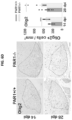
- FIG. 11 contains a series of images showing that Hmgcs1 is expressed at higher levels by NeuN + neurons in the cingulate cortex in PAR1 knockout mice prior to 6 weeks of CPZ mediated injury (Ctrl) and after 3 weeks on a regular diet to elicit repair (6+3). These results suggested that blocking PAR1 increases cholesterol synthesis in neurons in the intact CNS and in response to CPZ mediated injury. Results are plotted in the graph in the right panel (*P ⁇ 0.05 SNK).
- FIG. 12 contains a series of images showing that inhibition of PAR1 signaling accelerates neurite outgrowth in primary mouse cortical neurons.
- Murine neurons were cultured for 24 hours before addition of the PAR1 inhibitor Vorapaxar, subtherapeutic growth factor BDNF, or both.
- Inhibition of PAR1 potentiated the effect of BDNF, significantly increasing production of neurites at both 24 and 72 hours of treatment.
- FIG. 13 contains images showing that inhibition of PAR1 signaling accelerates synaptogenesis in primary mouse cortical neurons.
- Murine neurons were cultured in vitro for 24 hours before addition of the PAR1 inhibitor Vorapaxar, subtherapeutic growth factor BDNF, Or both.
- Inhibition of PAR1 potentiated the effect of BDNF, significantly increasing production of synaptic densities at both 24 and 72 hours of treatment.
- Synapse density was quantified by staining for Homer, a scaffold protein concentrated at the post-synaptic densities by counting puncta in ImageJ.
- FIG. 14 contains a series of images showing that inhibition of PAR1 signaling accelerates growth of new neurites following transection injury in vitro.
- Murine neurons were cultured in vitro to confluence, and then a large wound was created by mechanically scratching the center of each field.
- the PAR1 inhibitor Vorapaxar, subtherapeutic growth factor BDNF, or both were added following injury.
- FIG. 15 is a graph plotting expression of expression of HMGCS1, and demonstrating that inhibition of PAR1 by Vorapaxar increases expression of cholesterol synthesis enzymes in primary murine cortical neurons.
- Expression of HMGCS1 an enzyme responsible for production of HMGCoA for the rate-limiting step in cholesterol biosynthesis, was quantified by real-time quantitative PCR.
- Inhibition of PAR1 (with or without co-treatment with BDNF) increased the expression of HMGCS1 in cortical neurons.
- FIG. 16 contains a series of images showing that inhibition of cholesterol production with statins (reversible inhibitors of HMG-CoA Reductase) diminishes neurite outgrowth of primary mouse cortical neurons, demonstrating the essential role of cholesterol production in neuron growth and development.
- Inhibition of PAR1 (by Vorapaxar) or growth factor addition (by BDNF) alone did not result in recovery of neurite production and still produced a statistically significant decrease in neurite production compared to controls.
- co-treatment with both subtherapeutic BDNF with inhibition of PAR1 partially rescued neurite production from statin treatment, and cotreated cultures were no longer statistically different from untreated controls.
- FIG. 17 is a graph plotting neurite area for primary mouse cortical neurons following mechanical injury, showing that inhibition of cholesterol production with statins diminished neurite outgrowth, and demonstrating the essential role of cholesterol production in neuron repair as well as development.
- Co-treatment with both subtherapeutic BDNF and inhibition of PAR1 rescued neurite repair from statin treatment and additionally increased repair even beyond untreated control cultures.
- FIGS. 18 A- 18 D show that PAR1 knockout promotes increased expression of cholesterol synthesis intermediates by neurons in the injured spinal cord.
- FIG. 18 A contains a series of immunofluorescent images showing co-labeling for NeuN (neuron marker) and HMGCS1 in spinal segments above the injury epicenter 30 days after 0.25 mm lateral compression (LC) and FEJOTA clip contusion-compression SCI in wild type (PAR1+/+) and PAR1 ⁇ / ⁇ mice.
- FIG. 18 A contains a series of immunofluorescent images showing co-labeling for NeuN (neuron marker) and HMGCS1 in spinal segments above the injury epicenter 30 days after 0.25 mm lateral compression (LC) and FEJOTA clip contusion-
- FIG. 18 C includes histograms plotting the total number of neurons in the spinal cord, which did not significantly differ by genotype in either injury.
- FIG. 18 D includes images showing expression of HMGCS1 by ventral horn motoneurons in intact human spinal cord and at subacute and chronic time points after traumatic SCI.
- FIG. 19 is a graph showing that a higher percentage of neurons in PAR1 ⁇ / ⁇ uninjured spinal cord express HMGCS1 than wild type controls at baseline.
- FIGS. 20 A- 20 D show that enhanced remyelination after lysolecithin demyelination in PAR1 ⁇ / ⁇ knockout mice was accompanied by increases in the number of SREBP1+ and SREPB2+ oligodendrocyte lineage cells.
- FIG. 20 A includes a series of representative images of remyelinating lesions at 14 or 28 days after lysolecithin microinjection into the ventrolateral spinal cord white matter of wild type or PAR1 knockout mice, co-labeled with Olig2 for oligodendrocyte lineage cells and SREB1. Lesion borders are outlined by a dashed line.
- FIG. 20 A includes a series of representative images of remyelinating lesions at 14 or 28 days after lysolecithin microinjection into the ventrolateral spinal cord white matter of wild type or PAR1 knockout mice, co-labeled with Olig2 for oligodendrocyte lineage cells and SREB1. Lesion borders are
- FIG. 20 D is a histogram plotting the data (mean ⁇ SEM) from FIG.
- FIGS. 21 A- 21 D show that enhanced remyelination after cuprizone-mediated demyelination in PAR1 ⁇ / ⁇ mice was accompanied by increases in the number of SREBP1+ and SREBP2+ oligodendrocyte lineage cells.
- FIG. 22 is a graph plotting human neuronal (SH-SY5Y) cells grown in the presence of the small molecule PAR1 inhibitor, Vorapaxar (“Vora,” at 100 nM) or control for 72 hours. Cells treated with the inhibitor contained more than twice the amount of cellular cholesterol than untreated cells. Cholesterol content was quantified by the amplex red colorimetric assay.
- PAR1 and PAR2 are G protein coupled receptors that are specifically activated by select serine proteases. Under physiological conditions, these receptors communicate changes in the proteolytic microenvironment to cells, activating or inhibiting intracellular signaling cascades that modulate cellular physiology. Depending on the cell type, PAR1 activation engages several Ga subunits, G ⁇ , G12/13, Gq/1.1. or Gi resulting in modulation of signaling through Rho.GEF, P1-PLC, MAPK, PI3-kinase or adenylate cyclase pathways. PARs therefore can serve as biosensors that translate dynamic changes in the proteolytic microenvironment into adaptive (or maladaptive) cellular responses.
- PAR1 and PAR2 knockout mice also show superior recovery of function after traumatic spinal cord injury (Ra.dulovic et al., Neurobial Dis 2015, 83:75-89; and Radulovic et al., Neurohiol Dis 2016, 93:226-242), including improved recovery of myelin (Yoon et al. 2017, supra) and synaptic elements.
- lipid content in the central nervous system (CNS) of wild type and PAR1 knockout mice revealed increases in lipid availability in the knockout mice, including availability of hound cholesterol, galactosylceramides, sphingolipids, and sphingomyelins.
- Proteomic analysis demonstrated that blocking PAR1 increased ApoA1, the primary apolipoprotein of high-density lipoprotein (HDL).
- PANTHER GO analysis of RNA sequencing data obtained from the CNS of wild type compared to PAR1 knockout mice demonstrated that mice with knockout of the PAR1 gene had increases in lipid and cholesterol biosynthetic processes.
- Blocking PAR1 increased expression of genes involved in cholesterol biosynthesis pathways, including the Hmges1, Sqle, Mvd, Lss. Dher7, Fdft1, and Hmger genes.
- Blocking PAR2 increased the expression of genes involved in cholesterol biosynthesis, including the Hmgcs1, Sqle, Mvd, Lss, Dhcr7, Fdft1, and Hmgcr genes. Blocking PAR2 therefore can increase the availability of lipids, including the fatty acid DHA, sphingolipids, and sphingomyelin.
- this document provides methods and materials for targeting PAR1 and/or PAR2, which can have clinical utility in the treatment of disorders of lipid synthesis.
- Disorders that result from impaired production of lipids necessary for normal brain development include, without limitation, Alexander disease, metachromatic leukodystrophy, Krabbe disease, adrenoleukodystrophy, Canavan disease, Pelizaeus-Merzbacher disease, cerebrotenineous xanthomatosis, hypomyelinating leukodystrophy type 7, and Refsum disease.
- a small molecule inhibitor of PAR1 and/or PAR2 can be used.
- Small molecule inhibitors can be orally bioavailable and can have moderate penetrance through the blood-brain barrier, providing a strong rationale for successful treatment with long-term oral administration of these inhibitors. Since disorders of lipid synthesis typically are congenital disorders, the treatment paradigm may necessitate life-long administration, with early diagnosis and therapy initiation most likely to produce positive results. Reducing the activity of PAR1 or PAR2 to increase lipid and cholesterol levels also may provide benefit for conditions in which increases in lipids and cholesterol will enhance tissue regeneration (e.g., demyelinating lesions, neurotrauma, neurodegeneration, and congenital lipidoses).
- activation of PAR1 and/or PAR2 signaling also can have clinical benefits.
- Smith Lemli Opitz is a severe disorder caused by mutation in cholesterol synthesis enzymes. The mutation causes not only reduced production of cholesterol (which may be supplemented exogenously), but also toxic buildup of cholesterol production byproducts that accumulate over time, which manifests with multiple system abnormalities across the brain and peripheral organs.
- Activation of PAR1 and/or PAR2 with, for example, targeted short peptide ligands can diminish cholesterol to production in vivo, reducing the buildup of toxins and ameliorating the disease phenotype. Disturbances in lipid and cholesterol metabolism also contribute to a number of other health conditions.
- excess lipid accumulates in the brain and/or in peripheral organs, including the liver, heart, lungs and kidneys. Individual organs can also be subject to excess lipid accumulation, such as in fatty liver disease. High circulating cholesterol levels also is a major risk factor for heart disease and stroke, the leading causes of death in the United States.
- Increasing the expression of activity of PAR1 or PAR2 to reduce lipid and cholesterol levels may provide a new treatment target for disorders such as those listed above, as well as other clinical conditions [e.g., familial hypercholesterolemia, adrenoleukodystrophy, Fabry, Farber, Hunter, Hurler, Krabbe, Tangier, and non-alcoholic fatty liver disease (NAFLD)].
- familial hypercholesterolemia adrenoleukodystrophy
- Fabry Fabry
- Farber Hunter
- Hurler Krabbe
- Tangier and non-alcoholic fatty liver disease
- RNA sequencing genome wide analysis
- LC-MS-MS LC-MS-MS revealed substantial increases in cholesterol and lipid abundance in the spinal cord of PAR1 and PAR2 knockout mice.
- cholesterol and lipids are significant components of myelin and neural membranes, including synapses, these data collectively support a new biological model in which PAR1 and/or PAR2 activation serves as a negative regulator of cholesterol and lipid. production in the CNS and, most likely, in all organs and tissues of the body. in this model, blocking the activity of PAR1 or PAR2 increases cholesterol and lipid synthesis. Therefore, PAR1 and PAR2 represent new targets for modulating cholesterol and lipid production, with receptor activation serving to promote reductions in lipid and cholesterol levels, and receptor inhibiting serving to increase levels of lipids and cholesterol.
- This document therefore provides methods that can include administering, to a mammal identified as having a condition characterized by impaired lipid production, impaired cholesterol production, or both, an inhibitor of PAR1 and/or PAR2.
- the methods can further include identifying a mammal as having a condition characterized by impaired lipid production, impaired cholesterol production, or both.
- Any suitable mammal can be treated using the methods provided herein.
- the mammal can be, for example, a human, a non-human primate, a cow, a horse, a pig, a sheep, a goat, a cat, to a dog, a mouse, or a rat.
- the mammal to be treated with a PAR1 and/or PAR2 inhibitor can be identified as having a condition such as Alexander disease, metachromatic leukodystrophy, Krabbe disease, adrenoleukodystrophy, Canavan disease, Pelizaeus-Merzbacher disease, cerebrotenineous xanthomatosis, hypomyelinating leukodystrophy type 7, Refsum disease, or a congenital lipidosis.
- Alexander disease metachromatic leukodystrophy
- Krabbe disease adrenoleukodystrophy
- Canavan disease Pelizaeus-Merzbacher disease
- cerebrotenineous xanthomatosis cerebrotenineous xanthomatosis
- hypomyelinating leukodystrophy type 7, Refsum disease or a congenital lipidosis.
- any suitable PAR1 and/or PAR2 inhibitor can be used.
- a small molecule such as Vorapaxar (also known as SCH530348) can be administered to block PAR1.
- Other non-limiting examples of small molecule PAR1 inhibitors include SCH79797, parmodulins (e.g., ML161 and NRD21), and atopaxar (E5555).
- Examples of small molecule PAR2 inhibitors include, without limitation, GB88, I-191, AZ3451, AZ2623, AZ0107, and AZ8838.
- PAR1 and/or PAR2 blocking molecules include function-blocking antibodies or antibody fragments (e.g., Fab′ fragments, F(ab′) 2 fragments, or scFv fragments), antisense molecules, interfering RNA [RNAi, including short interfering RNA (siRNA) and short hairpin RNA (shRNA)], and pepducins, any of which can be used to increase lipid and cholesterol production as described herein.
- RNAi including short interfering RNA (siRNA) and short hairpin RNA (shRNA)
- pepducins any of which can be used to increase lipid and cholesterol production as described herein.
- Chimeric antibodies and humanized antibodies made from non-human (e.g., mouse, rat, gerbil, or hamster) antibodies also can be useful. Chimeric and humanized monoclonal antibodies can be produced by recombinant DNA techniques known in the art, for example, using methods described in U.S. Pat. Nos. 4,816,567;
- Antisense oligonucleotides typically are at least 8 nucleotides in length (e.g., about 8, 9, 10, 11, 12, 13, 14, 15, 16, 17, 18, 19, 20, 10 to 10, 15 to 20, 18 to 25, or 20 to 50 nucleotides in length) and can hybridize to a PAR1 or PAR2 transcript. In some cases, an antisense molecule greater than 50 nucleotides in length can be used, including a full-length PAR1 or PAR2 mRNA.
- An “oligonucleotide” is an oligomer or polymer of ribonucleic acid (RNA) or deoxyribonucleic acid (DNA) or analogs thereof.
- Nucleic acid analogs can be modified at the base moiety, sugar moiety, or phosphate backbone to improve, for example, stability, hybridization, or solubility of a nucleic acid.
- Methods for synthesizing antisense oligonucleotides include, for example, solid phase synthesis techniques. Equipment for such synthesis is commercially available from several vendors including, for example, Applied Biosystems (Foster City, Calif.).
- expression vectors that contain a regulatory element that directs production of an antisense transcript can be used to produce antisense molecules.
- Antisense oligonucleotides can bind to a nucleic acid encoding PAR1, including DNA encoding PAR1 RNA (including pre-mRNA and mRNA) transcribed from such DNA, and also cDNA derived from such RNA, under physiological conditions (i.e., physiological pH and ionic strength).
- the sequence of an antisense oligonucleotide need not be 100% complementary to that of its target nucleic acid in order to be hybridizable under physiological conditions.
- Antisense oligonucleotides can hybridize under physiological conditions when binding of the oligonucleotide to the PAR1 or PAR2 nucleic acid interferes with the normal function of the PAR1 or PAR2 nucleic acid, and non-specific binding to non-target sequences is minimal.
- Target sites for PAR1 or PAR2 antisense oligonucleotides can include the regions encompassing the translation initiation or termination codon of the open reading frame (ORF) of the gene.
- ORF open reading frame
- the ORF can be targeted effectively in antisense technology, as can the 5′ and 3′ untranslated regions.
- antisense oligonucleotides can be directed at intron regions or intron-exon junction regions.
- antisense oligonucleotides include, for example, the lack of predicted secondary structure of a potential antisense oligonucleotide, an appropriate G and C nucleotide content (e.g., about 50%), and the absence of sequence motifs such as single nucleotide repeats (e.g., GGGG runs).
- the effectiveness of antisense oligonucleotides at modulating expression of a PAR1 or PAR2 nucleic acid can he evaluated by measuring levels of the PAR1 mRNA or polypeptide (e.g., by Northern blotting, RT-PCR, Western blotting, ELISA, or immunohistochemical staining).
- a representative human PAR1 mRNA sequence is set forth in SEQ ID NO:1, with the coding sequence underlined:
- a representative human PAR2 mRNA sequence is set forth in SEQ ID NO:2, with the coding sequence underlined:
- RNAi Single and double-stranded interfering RNA (RNAi, such as siRNA and shRNA) homologous to PAR1 or PAR2 DNA also can be used to reduce expression of PAR1 or PAR2 and consequently, activity of PAR1 or PAR2.
- RNAi such as siRNA and shRNA
- Single and double-stranded interfering RNA (RNAi, such as siRNA and shRNA) homologous to PAR1 or PAR2 DNA also can be used to reduce expression of PAR1 or PAR2 and consequently, activity of PAR1 or PAR2.
- RNAi such as siRNA and shRNA
- the sense and anti-sense RNA strands of RNAi can be individually constructed using chemical synthesis and enzymatic ligation reactions using procedures known in the art. For example, each strand can be chemically synthesized using naturally occurring nucleotides or nucleic acid analogs.
- the sense or anti-sense strand also can be produced biologically using an expression vector into which a target PAR1 or PAR2 sequence (full-length or a fragment) has been subcloned in a sense or anti-sense orientation.
- the sense and anti-sense RNA strands can he annealed in vitro before delivery of the dsRNA to cells. Alternatively, annealing can occur in vivo after the sense and anti-sense strands are sequentially delivered to the tumor vasculature or to tumor cells.
- a genetic approach can he used to knock down PAR1 or PAR2 gene function.
- CRISPR/Cas-mediated genome editing, adeno-associated virus- (AAV-) mediated delivery of a knockdown vector (e.g., shRNAi), or other suitable means can be used.
- AAV- adeno-associated virus-
- shRNAi knockdown vector
- These approaches may be applied to a population of stem cells ex vivo, or can he used in a mammal per se.
- a population of stem cells that have been modified to have reduced PAR1 and/or PAR2 expression, as compared to corresponding wild type neural stem cells can be used.
- stem cells can be modified in vitro to contain a mutation in the PAR1 and/or PAR2 gene, such that PAR1 and/or PAR2 expression is reduced or even knocked out.
- Suitable types of stern cells include, without limitation, embryonic stem cells, induced pluripotent stein cells, bone marrow derived stem cells, mesenchymal stem cells, and neural stem cells. After delivery to a mammal, the stern cells can differentiate into neuronal cells and, due to their reduced level of PAR1 and/or PAR2 expression, can lead to increased lipid or cholesterol production.
- An effective amount of a PAR1 and/or PAR2 inhibitor can be an amount sufficient to increase the level of one or more lipids and/or cholesterol in a mammal after to treatment by at least 5% (e.g., at least 10%, at least 20%, at least 25%, at least 50%, at least 75%, or at least 100%), as compared to the level of the one or more lipids or the level of cholesterol prior to treatment. Any appropriate method can be used to measure the level of one or more lipids or cholesterol in a mammal, or in a biological sample from a mammal to be treated as described herein.
- LC-MS can be used to determine the level of one or more lipids or the level of cholesterol in a sample from a mammal (e.g., a blood sample, a sample of cerebrospinal fluid, a spinal cord sample, or a solid tissue sample).
- a mammal e.g., a blood sample, a sample of cerebrospinal fluid, a spinal cord sample, or a solid tissue sample.
- an effective amount of a PAR1 and/or PAR2 inhibitor can be an amount sufficient to reduce one or more symptoms of a disorder resulting from impaired production of lipids necessary for normal brain development (e.g., Alexander disease, metachromatic leukodystrophy, Krabbe disease, adrenoleukodystrophy, Canavan disease, Pelizaeus-Merzbacher disease, cerebrotenineous xanthomatosis, hypomyelinating leukodystrophy type 7, or Refsum disease).
- a disorder resulting from impaired production of lipids necessary for normal brain development e.g., Alexander disease, metachromatic leukodystrophy, Krabbe disease, adrenoleukodystrophy, Canavan disease, Pelizaeus-Merzbacher disease, cerebrotenineous xanthomatosis, hypomyelinating leukodystrophy type 7, or Refsum disease.
- this document provides methods that can include administering, to a mammal identified as having a condition characterized by excess lipid production, excess cholesterol production, or both, an activator of PAR1 and/or PAR2.
- the method can further include identifying a mammal as having a condition characterized by excess lipid production, excess cholesterol production, or both.
- any suitable mammal can be treated using the methods provided herein.
- the mammal can be, for example, a human, a non-human primate, a cow, a horse, a pig, a sheep, a goat, a cat, a dog, a mouse, or a rat.
- the mammal to he treated with a PAR1 and/or PAR2 activator can be identified as having a condition such as, without limitation, Smith-Lemli-Opitz Syndrome, Gaudier, Niemann-Pick, Tay-Sachs, fatty liver disease, familial hypercholesterolemia, adrenoleukodystrophy, Fabry, Farber, Hunter, Hurler, Krabbe, Tangier, or NAFLD.
- a condition such as, without limitation, Smith-Lemli-Opitz Syndrome, Gaudier, Niemann-Pick, Tay-Sachs, fatty liver disease, familial hypercholesterolemia, adrenoleukodystrophy, Fabry, Farber, Hunter, Hurler, Krabbe, Tangier, or NAFLD.
- any suitable PAR1 and/or PAR2 activator can be used.
- a peptide ligand that mimics a natural PAR1 or PAR2 ligand can be used.
- peptide ligands include Thr-Phe-Leu-Leu-Arg (SEQ ID NO:3) for PAR1, and Ser-Leu-Ile-Gly-Lys-Val (SEQ ID NO:4) and 2-Furoyl-Leu-ile-Gly-Arg-Leu-Orn (SEQ ID NO:5) for PAR2.
- Other non-limiting examples of PAR1/PAR2 activators include enzymatic activators such as thrombin, plasmin, trypsin, kallikrein 6, and activating antibodies.
- An effective amount of a PAR1 and/or PAR2 activator can be an amount sufficient to reduce the level of one or more lipids and/or cholesterol in a mammal after treatment by at least 5% (e.g., at least 10%, at least 20%, at least 25%, at least 50%, or at least 75%), as compared to the level of the one or more lipids and/or the level of cholesterol prior to treatment.
- an effective amount of a PAR1 and/or PAR2 activator can be an amount that results in a reduction of one or more symptoms of a condition characterized by excess lipid production, excess cholesterol production, or both (e.g., Smith-Lemli-Opitz Syndrome, Gaucher, Niemann-Pick, Tay-Sachs, fatty liver disease, familial hypercholesterolemia, adrenoleukodystrophy, Fabry, Farber, Hunter, Hurler, Krabbe, Tangier, or NAFLD).
- a condition characterized by excess lipid production, excess cholesterol production, or both e.g., Smith-Lemli-Opitz Syndrome, Gaucher, Niemann-Pick, Tay-Sachs, fatty liver disease, familial hypercholesterolemia, adrenoleukodystrophy, Fabry, Farber, Hunter, Hurler, Krabbe, Tangier, or NAFLD).
- a pharmaceutical composition containing an agent that inhibits or activates PAR1 and/or PAR2 in a mammal can be administered locally (e.g., to the brain or the CNS) or systemically.
- Administration can be, for example, oral, parenteral (e.g., by subcutaneous, intrathecal, intraventricular, intramuscular, or intraperitoneal injection, or by intravenous drip), or topical (e.g., transdermal, sublingual, ophthalmic, or intranasal), or can occur by a combination of such methods.
- Administration can be rapid (e.g., by injection) or can occur over a period of time (e.g., by slow infusion or administration of a slow release formulation).
- administration of an agent that inhibits or activates PAR1 and/or PAR2 in a mammal having a condition as described herein can increase or reduce the level of one or more lipids and/or cholesterol in the mammal, and can reduce at least one symptom of the condition.
- the mammal can be treated with a composition containing a PAR1 and/or PAR2 inhibitor or activator.
- the composition can be administered to the mammal in any amount, at any frequency, and for any duration effective to achieve a desired outcome (e.g., to increase or decrease lipid and/or cholesterol levels) in the mammal.
- a composition can be administered to a mammal repeatedly (e.g., once or more than once a day, once or more than once a week, or once or more than once a month).
- the frequency of administration can remain constant or can be variable during the duration of treatment.
- Various factors can influence the frequency of administration. For example, the effective amount, duration of treatment, route of administration, and severity of the condition may require an increase or decrease in administration frequency.
- PAR2 inhibitor or activator can be for example, from about 0.1 mg/kg to about 100 mg/kg (e.g., from about 0.01 mg/kg to about 0.05 mg/kg, from about 0.05 mg/kg to about 0.1 mg/kg, from about 0.1 mg/kg to about 0.5 mg.kg, from about 0.3 mg/kg to about 11 mg/kg, from about 1 mg/kg to about 10 mg/kg, from about 2 mg/kg to about 10 mg/kg, from about 5 mg/kg to about 10 mg/kg, from about 6 mg/kg to about 10 mg/kg, from about 6 mg/kg to about 8 mg/kg, or from about 7 mg/kg to about 9 mg/kg).
- from about 100 ⁇ g to about 100 mg e.g., from about 100 ⁇ g to about 1 mg, from about 1 mg to about 100 mg, from about 100 mg to about 250 mg, from about 250 mg to about 1000 mg, from about 300 mg to about 1000 mg, from about 400 mg to about 1000 mg, from about 100 mg to about 900 mg, from about 100 mg to about 800 mg, from about 400 mg to about 800 mg, or from about 500 mg to about 700 mg
- a PAR1 and/or PAR2 modulating agent an activator or an inhibitor
- an average sized human e.g., about 75-85 kg human
- per administration e.g., per daily or weekly administration
- the amount of the administered PAR1 and/or PAR2 inhibitor or activator can be increased by, for example, two fold. After receiving this higher amount, the mammal can be monitored for both responsiveness to the treatment and toxicity symptoms, and further adjustments can be made accordingly.
- the effective amount can remain constant or can be adjusted as a sliding scale or variable dose depending on the mammal's response to treatment. Various factors can influence the actual effective amount used for a particular application. For example, the frequency of administration, duration of treatment, use of multiple treatment agents, route of administration, and severity of the condition (e.g., cancer) may require an increase or decrease in the actual effective amount administered.
- the frequency of administration of a PAR1 and/or PAR2 activator or inhibitor can be any frequency that alters the production and/or levels of lipid and/or cholesterol in the mammal, without producing significant toxicity to the mammal.
- the frequency of administration can be from about once a day to about once a month (e.g., from about once a week to about once every other week).
- the frequency of administration can remain constant or can be variable during the duration of treatment.
- a course of treatment with a composition containing a PAR1 and/or PAR2 activator or inhibitor can include rest periods.
- a composition containing a PAR1 inhibitor can be administered daily over a two-week period followed by a two-week rest period, and such a regimen can be repeated multiple times.
- a PAR1 inhibitor e.g., Vorapaxar
- various factors can influence the actual frequency of administration used for a particular application.
- the effective amount, duration of treatment, use of multiple treatment agents, route of administration, and severity of the condition may require an increase or decrease in administration frequency.
- An effective duration for administering a composition containing a PAR1 and/or PAR2 activator or inhibitor can be any duration that alters the levels and/or production of lipid and/or cholesterol in the mammal without producing significant toxicity to the mammal.
- the effective duration can vary from several days to several months. In general, the effective duration can range from about six weeks to about six months or longer, even for years or for life. Multiple factors can influence the actual effective duration used for a particular treatment. For example, an effective duration can vary with the frequency of administration, effective amount, use of multiple treatment agents, route of administration, and severity of the condition being treated.
- a course of treatment and/or the severity of one or more symptoms related to the condition being treated can be monitored. Any appropriate method can be used to determine whether or not a mammal's lipid and/or cholesterol levels are altered.
- a biological sample e.g., a blood or tissue sample
- a PAR1 and/or PAR2 inhibitor or activator can be assessed following administration of a PAR1 and/or PAR2 inhibitor or activator to determine if the treatment increased or reduced the level of one or more lipids or cholesterol in the sample, as compared to the level measured in a sample obtained from a control mammal not having the condition, or as compared to the level measured in a sample obtained from the mammal prior to treatment.
- Any appropriate method e.g., LC-MS
- the spinal cord of PAR1 knockout mice contains higher levels of total cholesterol, and increases in expression of cholesterol and lipid synthesis intermediates ( FIGS. 1 A, 1 B, and 2 ).
- GC-MS dynamic 13 C-labeling gas chromatography-mass spectrometry
- cholesterol and lipid abundance are quantified in the brain and liver of wild type and PAR1 knockout mice using LC-MS and tissue samples collected from the same P60 mice used for the studies with results presented in ( FIGS. 1 A, 1 B, and 2 ). Free and bound cholesterol, free fatty acids, sphingolipids, sphingomyelin, and galactocerebroside are specifically quantified. Quantitative PCR is used to determine whether expression of genes in the cholesterol and lipid synthesis pathways (e.g., DHCR7, Ugt8, and Fa2h) are elevated in brain and liver RNA as they are in the spinal cord.
- genes in the cholesterol and lipid synthesis pathways e.g., DHCR7, Ugt8, and Fa2h
- tissue regeneration e.g., demyelinating lesions, neurotrauma, neurodegeneration, and congenital lipidoses.
- a GC-MS platform is utilized to quantify 13 C-acetate incorporation into newly synthesized cholesterol in primary cultures of murine cortical neurons or in the murine hepatocyte cell line AML12.
- PAR1 loss-of-function is modeled using Vorapaxar, an FDA approved PAR1 small molecule inhibitor ( FIG. 3 ) (Correa et al., J Thrombosis Thrombolysis 2019, 47(3):353-360; and Tsigkou et al., Curr Opin Pharmacol 2018, 39:43-52).
- PAR1 gain-of-function is modeled using a PAR1-activating peptide that mimics the tethered ligand of the receptor (Choi et al., Sci Rep 2018, 8(1):9360).
- the GC-MS assay for cholesterol biosynthesis quantification established in the Mayo Metabolomics Core, includes standards for cholesterol and its synthesis intermediates (lansterol, zymosterol, desmosterol, and 7-dehydrocholesterol). The results demonstrated that PAR1 activity can regulate cholesterol biosynthesis, and suggesting strategies for therapeutic modulation.
- FIGS. 1 A and 1 B Given the increases in cholesterol synthesis genes observed in the spinal cord of PAR1 knockout mice ( FIGS. 1 A and 1 B ), and in neurons treated with a PAR1 small molecule inhibitor ( FIG. 3 ), PAR1 inhibition is likely to increase cholesterol biosynthesis while PAR1 activation promotes decreases. Alternatively, changes in cholesterol biosynthesis gene expression and overall abundance ( FIGS. 1 A, 1 B, and 2 ) may occur by PAR1 effects on cholesterol efflux or uptake, and this is assessed as an alternative using cell-based assay kits. Additional studies apply similar dynamic labeling approaches to quantify the effect of PAR1 on lipid biosynthesis in vitro and on lipid and cholesterol biosynthesis in vivo.
- PAR gene KO was associated with increased expression of genes critical for myelination, cholesterol and lipid biosynthesis.
- major myelin proteins such as PLP1 and MBP were increased by PAR knockout ( FIG. 4 A ).
- the Ugt8 (galactosyltransferase) and Fa2h (fatty acid 2-hydroxylase) genes also were upregulated ( FIG. 4 A ); these are essential components of lipid synthesis pathways critical for membrane formation.
- Hmgcs1 which codes for an enzyme involved in production of HMG-CoA (a rate-limiting step in cholesterol biosynthesis) also was increased in PAR1 ⁇ / ⁇ mice ( FIG. 4 C ).
- DHCR7 encodes an enzyme involved in conversion of 7-dehydrocholesterol to cholesterol, and mutations in DHCR7 can disrupt cholesterol synthesis and manifest in Smith-Lemli-Opitz Syndrome.
- PANTHER GO Pathways highlighted key myelination events, including axon ensheathment, lipid biosynthesis, and oligodendrocyte differentiation (uninjured (UI) animals shown, FIG. 4 B ).
- Ingenuity Pathways analysis highlighted signaling intermediates in the ERK and AKT pathways ( FIG. 4 D ).
- Lipid quantification by LC-MS demonstrated that PAR1 and PAR2 knockout mice also exhibited higher levels of cholesterol and/or lipid synthesis in the spinal cord ( FIG. 5 ).
- cholesterol and sphingomyelin were increased in the spinal cord of PAR ⁇ / ⁇ mice at P21, and sphingolipid was increased at P60.
- the spinal cord of PAR2 ⁇ / ⁇ mice showed increases in sphingomyelin at P21 and P60 relative to PAR+/+ (*P ⁇ 0.05, **P ⁇ 0.01, ***P ⁇ 0.001, NK).
- FIGS. 6 A- 6 G The mean numbers of remyelinated axons after focal lysolecithin-mediated demyelination of the ventral spinal cord white matter ( FIG. 6 B ) in PAR+/+ and PAR1 ⁇ / ⁇ mice are provided in FIG. 6 A .
- PAR1 knockout improved the cholesterol synthesis pathway in spinal cord oligodendrocytes after focal demyelinating injury ( FIG. 7 ).
- Hmgcs1 an essential regulatory enzyme in cholesterol production
- dpi lysolecithin injury
- PAR1 knockout mice also showed increased expression of the master transcriptional regulators of cholesterol and lipid biosynthesis, SREBP1 ( FIG. 8 , left panel) and SREBP2 ( FIG. 8 , right panel) in astrocytes during myelin repair following lysolecithin-induced demyelination in the spinal cord.
- Spinal cords were stained for GFAP (an astrocyte marker), SREBP1, and SREBP2 at 14 or 28 days following lysolecithin-mediated focal demyelination.
- SREBP positivity was defined by measuring the percent of GFAP+ area colocalized with SREBP within the demyelinated lesion.
- SREBP2 expression in astrocytes was significantly increased in PAR1 knockout compared to wild-type controls at 14 days. Both SREBPs trended toward higher astrocytic expression at all time points in the context of PAR1 knockout (P ⁇ 0.05).
- FIG. 9 A The phases of demyelination and remyelination during and after CPZ feeding are depicted in FIG. 9 A , with arrows indicating time points at which rotarod assessment ( FIG. 9 B ) and immunohistochemistry (IHC) for markers of myelin injury and repair ( FIGS. 9 C -9F) were conducted.
- the impact of PAR1 knockout on remyelination was evaluated by feeding mice CPZ-laden chow for 6 weeks, followed by a period of “induced remyelination” upon CPZ withdrawal and feeding regular chow for an additional 3 (6+3) or 6 (6+6) week period.
- FIG. 9 B Motor function as assessed by angular speed at fall on an accelerating rotarod test, expressed as a percent of maximum at baseline for each genotype ( FIG. 9 B ).
- Representative images through the corpus callosum of mice after 3 or 6 weeks of remyelination (6+3 or 6+6) are shown in FIGS. 9 C- 9 F . Quantification of markers of remyelination revealed that PAR1 ⁇ / ⁇ mice exhibited greater increases in the number of oligodendrocyte lineage cells (Olig2+) ( FIG.
- FIG. 9 C after 3 weeks on regular chow and in the number of mature oligodendrocytes (CC-1+) ( FIG. 9 B ) at 3 and 6 weeks, compared to wild type (P ⁇ 0.05).
- the area stained for MBP ( FIG. 9 E ) or neurofilament (NF) ( FIG. 9 F ) after CPZ treatment was not altered by PAR1 knockout.
- PAR1 ⁇ / ⁇ mice consuming regular chow for the full period of the experiment showed higher counts of CC-1+ and higher levels of MBP and NF immunoreactivity in the corpus callosum (P ⁇ 0.04) relative to regular chow WT counterparts ( FIGS. 9 D, 9 E, and 9 F ).
- HMGCS1 is an essential regulatory enzyme in cholesterol production. Enhanced remyelination after cuprizone-mediated demyelination in PAR1 knockout mice was accompanied by increases in the percentage of HMGCS1+ oligodendrocytes after acute lysolecithin- or chronic cuprizone-mediated demyelination. Images taken of the ventrolateral spinal cord 14 or 28 days after lysolecithin injection revealed co-labelling of Olig2 for oligodendrocyte lineage cells and the cholesterol synthesis enzyme HMGCS1 ( FIG. 10 A ), as well as areas of demyelination (outlined with the white dashed line in FIG. 10 A ).
- Hmgcs1 also was expressed at higher levels by NeuN+neurons in the cingulate cortex in PAR1 knockout mice prior to 6 weeks of CPZ mediated injury (Ctrl) and after 3 weeks on a regular diet to elicit repair (6+3) ( FIG. 11 ). This suggested that blocking PAR1 increases cholesterol synthesis in neurons in the intact CNS and in response to CPZ mediated injury.
- Inhibition of PAR1 signaling was found to accelerates neurite outgrowth in primary mouse cortical neurons ( FIG. 12 ).
- Murine neurons were cultured for 24 hours before addition of the PAR1 inhibitor Vorapaxar, subtherapeutic growth factor BDNF, or both.
- Inhibition of PAR1 potentiated the effect of BDNF, significantly increasing production of neurites at both 24 and 72 hours of treatment.
- Neurite outgrowth was quantified by staining for TUJ1 (a cytoskeletal protein present in both axons and dendrites) and measuring TUJ1+ area (the average of 5 randomly selected fields per well, 6 wells per treatment). Production of lipids and cholesterol, major constituents of neuronal membranes, was required to grow both axons and dendrites.
- HMGCS1 is responsible for production of HMGCoA for the rate-limiting step in cholesterol biosynthesis.
- Inhibition of PAR1 (with or without co-treatment with BDNF) increased the expression of HMGCS1 in cortical neurons.
- PAR1 knockout increased neuronal expression of cholesterol synthesis enzymes in mice following spinal cord injury by lateral compression or FEJOTA clip. After injury, cords were divided regionally by removing the epicenter of injury as well as tissue immediately above and below the injured region in wild type controls and PAR1 knockout mice. Slices from each region were stained for NeuN, a neuronal marker, and HMGCS1, a cholesterol synthesis enzyme. The number of HMGCS1+ neurons was counted for each slide. PAR1 knockout mice demonstrated a significantly increased number of neurons positive for HMGCS1 both above ( FIGS. 18 A and 18 B ) and below ( FIG. 18 B ) the injured spinal cord, possibly indicative of enhanced ability for cholesterol production and therefore extension of new axons.
- PAR1 inhibition also was found to increase oligodendrocyte expression of master regulators of lipid synthesis during remyelination in an acute model of myelin injury.
- PAR1 inhibition increased oligodendrocyte expression of master regulators of lipid synthesis during remyelination in a chronic model of myelin injury.
- Enhanced remyelination after cuprizone-mediated demyelination in PAR1 ⁇ / ⁇ mice was accompanied by increases in the number of SREBPI oligodendrocyte lineage cells in the corpus callosum of mice fed regular chow (untreated) or after 6 weeks of CPZ feeding followed by 3 weeks of regular chow consumption to induce myelin regeneration.
- Increases in SREBP1+ oligodendrocytes were observed during remyelination in both wild type and PAR1 ⁇ / ⁇ mice ( FIGS.
Landscapes
- Health & Medical Sciences (AREA)
- Life Sciences & Earth Sciences (AREA)
- General Health & Medical Sciences (AREA)
- Chemical & Material Sciences (AREA)
- Veterinary Medicine (AREA)
- Medicinal Chemistry (AREA)
- Pharmacology & Pharmacy (AREA)
- Public Health (AREA)
- Animal Behavior & Ethology (AREA)
- Epidemiology (AREA)
- Engineering & Computer Science (AREA)
- Bioinformatics & Cheminformatics (AREA)
- Chemical Kinetics & Catalysis (AREA)
- General Chemical & Material Sciences (AREA)
- Nuclear Medicine, Radiotherapy & Molecular Imaging (AREA)
- Organic Chemistry (AREA)
- Proteomics, Peptides & Aminoacids (AREA)
- Immunology (AREA)
- Diabetes (AREA)
- Hematology (AREA)
- Obesity (AREA)
- Gastroenterology & Hepatology (AREA)
- Pain & Pain Management (AREA)
- Medicines That Contain Protein Lipid Enzymes And Other Medicines (AREA)
Priority Applications (1)
| Application Number | Priority Date | Filing Date | Title |
|---|---|---|---|
| US17/792,591 US20230061116A1 (en) | 2020-01-14 | 2021-01-13 | Targeting par1 and par2 to regulate lipid and cholesterol abundance |
Applications Claiming Priority (3)
| Application Number | Priority Date | Filing Date | Title |
|---|---|---|---|
| US202062960881P | 2020-01-14 | 2020-01-14 | |
| US17/792,591 US20230061116A1 (en) | 2020-01-14 | 2021-01-13 | Targeting par1 and par2 to regulate lipid and cholesterol abundance |
| PCT/US2021/013251 WO2021146292A1 (fr) | 2020-01-14 | 2021-01-13 | Ciblage de par1 et de par2 pour réguler l'abondance de lipides et de cholestérol |
Publications (1)
| Publication Number | Publication Date |
|---|---|
| US20230061116A1 true US20230061116A1 (en) | 2023-03-02 |
Family
ID=76863270
Family Applications (1)
| Application Number | Title | Priority Date | Filing Date |
|---|---|---|---|
| US17/792,591 Abandoned US20230061116A1 (en) | 2020-01-14 | 2021-01-13 | Targeting par1 and par2 to regulate lipid and cholesterol abundance |
Country Status (3)
| Country | Link |
|---|---|
| US (1) | US20230061116A1 (fr) |
| EP (1) | EP4090372A4 (fr) |
| WO (1) | WO2021146292A1 (fr) |
Cited By (2)
| Publication number | Priority date | Publication date | Assignee | Title |
|---|---|---|---|---|
| US20230000928A1 (en) * | 2016-06-23 | 2023-01-05 | Mayo Foundation For Medical Education And Research | Par2 modulation to alter myelination |
| US11813264B2 (en) | 2014-07-07 | 2023-11-14 | Mayo Foundation For Medical Education And Research | PAR1 modulation to alter myelination |
Family Cites Families (7)
| Publication number | Priority date | Publication date | Assignee | Title |
|---|---|---|---|---|
| EP1603947A1 (fr) * | 2003-03-11 | 2005-12-14 | Bayer HealthCare AG | Diagnostique et therapeutique destinees aux maladies associees au recepteur active par protease 1 (par1) couple a la proteine g |
| WO2004080373A2 (fr) * | 2003-03-11 | 2004-09-23 | Bayer Healthcare Ag | Agents diagnostiques et therapeutiques destines a des maladies associees au recepteur 2 active par la proteinase (par2) couple aux proteines g |
| WO2012033518A1 (fr) * | 2010-09-09 | 2012-03-15 | The Scripps Research Institute | Procédés et compositions pour le traitement de troubles métaboliques |
| US20160000791A1 (en) * | 2014-07-07 | 2016-01-07 | Mayo Foundation For Medical Education And Research | Par1 modulation to alter myelination |
| WO2017083618A1 (fr) * | 2015-11-13 | 2017-05-18 | Oasis Pharmaceuticals, LLC | Modulateurs du récepteur activé par protéase de type 2 |
| US20190201454A1 (en) * | 2016-06-23 | 2019-07-04 | Mayo Foundation For Medical Education And Research | Par2 modulation to alter myelination |
| WO2019172969A1 (fr) * | 2018-03-08 | 2019-09-12 | The Scripps Research Institute | Méthodes pour favoriser la myélinisation et pour traiter des maladies démyélinisantes |
-
2021
- 2021-01-13 EP EP21741025.7A patent/EP4090372A4/fr active Pending
- 2021-01-13 WO PCT/US2021/013251 patent/WO2021146292A1/fr not_active Ceased
- 2021-01-13 US US17/792,591 patent/US20230061116A1/en not_active Abandoned
Cited By (2)
| Publication number | Priority date | Publication date | Assignee | Title |
|---|---|---|---|---|
| US11813264B2 (en) | 2014-07-07 | 2023-11-14 | Mayo Foundation For Medical Education And Research | PAR1 modulation to alter myelination |
| US20230000928A1 (en) * | 2016-06-23 | 2023-01-05 | Mayo Foundation For Medical Education And Research | Par2 modulation to alter myelination |
Also Published As
| Publication number | Publication date |
|---|---|
| EP4090372A1 (fr) | 2022-11-23 |
| EP4090372A4 (fr) | 2023-10-18 |
| WO2021146292A1 (fr) | 2021-07-22 |
Similar Documents
| Publication | Publication Date | Title |
|---|---|---|
| Yan et al. | LncRNA NEAT1 promotes autophagy in MPTP-induced Parkinson's disease through stabilizing PINK1 protein | |
| Pusic et al. | Youth and environmental enrichment generate serum exosomes containing miR‐219 that promote CNS myelination | |
| Han et al. | Mechanism of microRNA‐22 in regulating neuroinflammation in Alzheimer’s disease | |
| JP4316373B2 (ja) | ヒトのアセチルコリンエステラーゼ(ache)に対するアンチセンスオリゴヌクレオチド及びその使用 | |
| Toth et al. | Locally synthesized calcitonin gene-related peptide has a critical role in peripheral nerve regeneration | |
| EP3524679B1 (fr) | Micro arn mir-19 et compositions les comprenant pour le traitement d'états médicaux dans lesquels le niveau d'adrénaline faible ou de noradrénaline est bénéfique thérapeutiquement | |
| Xiang et al. | The lncRNA Ftx/miR-382-5p/Nrg1 axis improves the inflammation response of microglia and spinal cord injury repair | |
| CN111733231A (zh) | 用于治疗和诊断的微rna和包含所述微rna的组合物 | |
| US20240374693A1 (en) | Treatment and detection of inherited neuropathies and associated disorders | |
| Beltran et al. | Expression of PTPRO during mouse development suggests involvement in axonogenesis and differentiation of NT‐3 and NGF‐dependent neurons | |
| US20230061116A1 (en) | Targeting par1 and par2 to regulate lipid and cholesterol abundance | |
| US20190216891A1 (en) | Method for modulating myelination | |
| LaCroix-Fralish et al. | The β3 subunit of the Na+, K+-ATPase mediates variable nociceptive sensitivity in the formalin test | |
| Czapiga et al. | Microglial function in human APOE3 and APOE4 transgenic mice: altered arginine transport | |
| US20140105919A1 (en) | Synergistic inhibition of erbb2/erbb3 signal pathways in the treatment of cancer | |
| EP2162137B1 (fr) | Procédés de traitement de troubles cognitifs par inhibition du gpr12 | |
| EP2733205B1 (fr) | Neurones moteurs supérieurs cortico-spinaux, procédés et compositions permettant de différencier des cellules souches neurales par modulation de la signalisation des récepteurs cannabinoïdes CB1 et leurs utilisations | |
| JP2007530029A (ja) | アネキシンii及びその使用 | |
| Ahrendsen et al. | The protein tyrosine phosphatase Shp2 regulates oligodendrocyte differentiation and early myelination and contributes to timely remyelination | |
| WO2007011702A2 (fr) | Utilisation d'inhibiteurs de recepteur egf pour prevenir ou traiter l'obesite | |
| Xu et al. | The transcription factor Stat-1 is essential for Schwann cell differentiation, myelination and myelin sheath regeneration | |
| JP4427639B2 (ja) | 合成アンチセンスオリゴデオキシヌクレオチド類およびそれらを含む医薬組成物 | |
| Li et al. | Focal cortical dysplasia II caused by brain somatic mutation of IRS-1 is associated with ERK signaling pathway activation | |
| US20260103714A1 (en) | Antisense oligonucleotides for the treatment of canavan disease | |
| WO2025068557A1 (fr) | Oligonucléotides antisens pour le traitement de la maladie de canavan |
Legal Events
| Date | Code | Title | Description |
|---|---|---|---|
| AS | Assignment |
Owner name: MAYO FOUNDATION FOR MEDICAL EDUCATION AND RESEARCH, MINNESOTA Free format text: ASSIGNMENT OF ASSIGNORS INTEREST;ASSIGNORS:SCARISBRICK, ISOBEL A.;TRIPLET, ERIN M.;SIGNING DATES FROM 20210223 TO 20210317;REEL/FRAME:060679/0284 |
|
| STPP | Information on status: patent application and granting procedure in general |
Free format text: DOCKETED NEW CASE - READY FOR EXAMINATION |
|
| STPP | Information on status: patent application and granting procedure in general |
Free format text: NON FINAL ACTION MAILED |
|
| STCB | Information on status: application discontinuation |
Free format text: ABANDONED -- FAILURE TO RESPOND TO AN OFFICE ACTION |