US4211237A - Method and apparatus for identifying recurring signal patterns - Google Patents
Method and apparatus for identifying recurring signal patterns Download PDFInfo
- Publication number
- US4211237A US4211237A US05/896,771 US89677178A US4211237A US 4211237 A US4211237 A US 4211237A US 89677178 A US89677178 A US 89677178A US 4211237 A US4211237 A US 4211237A
- Authority
- US
- United States
- Prior art keywords
- signal
- amplitude
- pattern
- criterion
- sample
- Prior art date
- Legal status (The legal status is an assumption and is not a legal conclusion. Google has not performed a legal analysis and makes no representation as to the accuracy of the status listed.)
- Expired - Lifetime
Links
- 238000000034 method Methods 0.000 title claims description 135
- 239000000203 mixture Substances 0.000 claims abstract description 63
- 238000000718 qrs complex Methods 0.000 claims abstract description 55
- 230000015654 memory Effects 0.000 claims abstract description 52
- 210000002458 fetal heart Anatomy 0.000 claims abstract description 38
- 230000008774 maternal effect Effects 0.000 claims description 85
- 230000001605 fetal effect Effects 0.000 claims description 61
- 238000005311 autocorrelation function Methods 0.000 claims description 44
- 238000005314 correlation function Methods 0.000 claims description 39
- 238000001514 detection method Methods 0.000 claims description 34
- 238000012935 Averaging Methods 0.000 claims description 26
- 230000006870 function Effects 0.000 claims description 20
- 238000012545 processing Methods 0.000 claims description 16
- 238000004364 calculation method Methods 0.000 claims description 15
- 238000011156 evaluation Methods 0.000 claims description 12
- 238000001914 filtration Methods 0.000 claims description 5
- 230000008859 change Effects 0.000 claims description 3
- 230000001976 improved effect Effects 0.000 claims description 2
- 230000007774 longterm Effects 0.000 claims 4
- 238000002565 electrocardiography Methods 0.000 description 88
- 230000008569 process Effects 0.000 description 35
- 230000015572 biosynthetic process Effects 0.000 description 20
- 230000003187 abdominal effect Effects 0.000 description 19
- 238000010586 diagram Methods 0.000 description 18
- 238000004458 analytical method Methods 0.000 description 11
- 230000008901 benefit Effects 0.000 description 11
- 230000000694 effects Effects 0.000 description 7
- 230000006872 improvement Effects 0.000 description 7
- 230000006978 adaptation Effects 0.000 description 6
- 238000010219 correlation analysis Methods 0.000 description 6
- 238000005259 measurement Methods 0.000 description 6
- 210000003754 fetus Anatomy 0.000 description 5
- 230000002452 interceptive effect Effects 0.000 description 4
- 230000000717 retained effect Effects 0.000 description 4
- 238000005070 sampling Methods 0.000 description 4
- 238000004590 computer program Methods 0.000 description 3
- 238000011161 development Methods 0.000 description 3
- 230000010354 integration Effects 0.000 description 3
- 230000002093 peripheral effect Effects 0.000 description 3
- 239000004065 semiconductor Substances 0.000 description 3
- 230000035945 sensitivity Effects 0.000 description 3
- 238000001228 spectrum Methods 0.000 description 3
- 238000012546 transfer Methods 0.000 description 3
- 230000033228 biological regulation Effects 0.000 description 2
- 238000004422 calculation algorithm Methods 0.000 description 2
- 238000004891 communication Methods 0.000 description 2
- 238000012937 correction Methods 0.000 description 2
- 238000013461 design Methods 0.000 description 2
- 238000012854 evaluation process Methods 0.000 description 2
- 238000012544 monitoring process Methods 0.000 description 2
- 210000003205 muscle Anatomy 0.000 description 2
- 230000000737 periodic effect Effects 0.000 description 2
- 238000011084 recovery Methods 0.000 description 2
- 230000009467 reduction Effects 0.000 description 2
- 230000002829 reductive effect Effects 0.000 description 2
- 238000000926 separation method Methods 0.000 description 2
- 230000008093 supporting effect Effects 0.000 description 2
- 230000001629 suppression Effects 0.000 description 2
- 230000036962 time dependent Effects 0.000 description 2
- 210000001015 abdomen Anatomy 0.000 description 1
- 230000036982 action potential Effects 0.000 description 1
- 230000004913 activation Effects 0.000 description 1
- 230000003044 adaptive effect Effects 0.000 description 1
- 230000003321 amplification Effects 0.000 description 1
- 230000002238 attenuated effect Effects 0.000 description 1
- 230000005540 biological transmission Effects 0.000 description 1
- 230000000747 cardiac effect Effects 0.000 description 1
- 230000000295 complement effect Effects 0.000 description 1
- 239000013256 coordination polymer Substances 0.000 description 1
- 230000007423 decrease Effects 0.000 description 1
- 238000009795 derivation Methods 0.000 description 1
- 230000004069 differentiation Effects 0.000 description 1
- 230000008030 elimination Effects 0.000 description 1
- 238000003379 elimination reaction Methods 0.000 description 1
- 230000002349 favourable effect Effects 0.000 description 1
- 230000000977 initiatory effect Effects 0.000 description 1
- 238000012986 modification Methods 0.000 description 1
- 230000004048 modification Effects 0.000 description 1
- 238000003199 nucleic acid amplification method Methods 0.000 description 1
- 230000010363 phase shift Effects 0.000 description 1
- 230000035935 pregnancy Effects 0.000 description 1
- 210000004761 scalp Anatomy 0.000 description 1
- 230000001360 synchronised effect Effects 0.000 description 1
- 238000013519 translation Methods 0.000 description 1
- 230000001960 triggered effect Effects 0.000 description 1
- 238000011144 upstream manufacturing Methods 0.000 description 1
Images




Classifications
-
- A—HUMAN NECESSITIES
- A61—MEDICAL OR VETERINARY SCIENCE; HYGIENE
- A61B—DIAGNOSIS; SURGERY; IDENTIFICATION
- A61B5/00—Measuring for diagnostic purposes; Identification of persons
- A61B5/24—Detecting, measuring or recording bioelectric or biomagnetic signals of the body or parts thereof
- A61B5/316—Modalities, i.e. specific diagnostic methods
- A61B5/318—Heart-related electrical modalities, e.g. electrocardiography [ECG]
- A61B5/346—Analysis of electrocardiograms
- A61B5/349—Detecting specific parameters of the electrocardiograph cycle
- A61B5/35—Detecting specific parameters of the electrocardiograph cycle by template matching
-
- A—HUMAN NECESSITIES
- A61—MEDICAL OR VETERINARY SCIENCE; HYGIENE
- A61B—DIAGNOSIS; SURGERY; IDENTIFICATION
- A61B5/00—Measuring for diagnostic purposes; Identification of persons
- A61B5/24—Detecting, measuring or recording bioelectric or biomagnetic signals of the body or parts thereof
- A61B5/316—Modalities, i.e. specific diagnostic methods
- A61B5/318—Heart-related electrical modalities, e.g. electrocardiography [ECG]
- A61B5/344—Foetal cardiography
-
- A—HUMAN NECESSITIES
- A61—MEDICAL OR VETERINARY SCIENCE; HYGIENE
- A61B—DIAGNOSIS; SURGERY; IDENTIFICATION
- A61B5/00—Measuring for diagnostic purposes; Identification of persons
- A61B5/43—Detecting, measuring or recording for evaluating the reproductive systems
- A61B5/4306—Detecting, measuring or recording for evaluating the reproductive systems for evaluating the female reproductive systems, e.g. gynaecological evaluations
- A61B5/4343—Pregnancy and labour monitoring, e.g. for labour onset detection
- A61B5/4362—Assessing foetal parameters
Definitions
- the present invention relates to a method for identifying periodically occurring like signals that are part of a signal mixture including interference components, particularly for detecting QRS complexes of fetal heart signals in an abdominally derived fetomaternal electrocardiogram, as well as to an apparatus for practicing the method.
- the detection and identification of periodically occurring like signals in a signal mixture containing interference signals represents a goal which has not yet been satisfactorily reached in, for example, perinatalogy where it is desired to detect and process fetal electrocardiograms (EKGs).
- EKGs fetal electrocardiograms
- a fetal EKG cannot be derived directly from the fetus with the aid of scalp electrodes until immediately preceding birth, after the amnionic sac has broken. Since, therefore, this cannot be done during the entire period of pregnancy, methods of indirectly deriving the fetal EKG from the mother must be used if effective monitoring of the fetus is to be achieved.
- a fetal QRS complex is a characteristic component of the heart signal, specifically a signal occurrence containing P, Q and R peaks. Since this is sufficient, at most, to indicate the average fetal heart frequency, the physician has available to him neither sufficient data about microfluctuations, i.e., temporary fluctuations in the heart frequency, nor about the wave shape of the fetal EKG.
- the maternal EKG Interfering with the fetal EKG are the maternal EKG, the maternal electromyogram signals due to muscle movements and noise signals. Their amplitudes are much higher than that of the fetal electrocardiogram whose maximum amplitude when recorded by means of abdominal electrodes is 10 to 50 ⁇ V. The amplitude of the fetal EKG changes considerably, however, depending on the position of the fetus with respect to the electrodes.
- a maternal abdominal signal has the following composition:
- the wave shapes of the individual components are not known in advance and are often even subject to fluctuations within the time interval in which the signals are being processed so that evaluation is made substantially more difficult.
- the maternal QRS complex has its greatest power density in the frequency range between 10 and 30 Hz.
- the maximum of the fetal QRS complex in the frequency spectrum is a bit higher, i.e., between 15 and 40 Hz.
- the noise components contained in the abdominal signal extends over the entire frequency range. Very low frequency noise probably originates mainly from movement of the electrodes, while the higher frequency portion must be attributed to the electromyogram.
- the maternal EKG controls an electronic switch which blanks out the abdominal EKG as soon as a maternal QRS complex occurs. With this method it is impossible to detect fetal QRS complexes which wholly or partly coincide with the maternal complexes.
- the abdominal EKG controls two trigger circuits set to different threshold values.
- the upper threshold value is exceeded, this indicates the occurrence of a maternal QRS complex, while the exceeding of the lower threshold indicates a fetal QRS complex.
- This method has the same drawbacks as the blanking method.
- the greatest accuracy in detecting interference with signals according to the prior art is offered by selective filters.
- the best solution for locating a signal of known waveform is the optimum search filter, or matched filter.
- the selective filters previously employed to effect a "beat-to-beat" recognition of the fetal QRS complex do not have the structure of an optimally matched filter and therefore do not operate reliably.
- the output of the matched filter furnishes the autocorrelation function (ACF) of the useful signal.
- ACF autocorrelation function
- German Offenlegungsschrift (Laid Open application) No. 1,807,147 discloses a wave shape detector which makes it possible to detect a known wave shape in a signal mixture by means of a correlation process. However, this detector is unable to detect a wave shape whose curve is not known beforehand.
- the method according to the invention can be practiced by an appropriate measuring device and requires a small number of calculating operations so that measurements can also be made quickly and reliably even by technically untrained personnel. After detecting the signal it is moreover possible to reproduce its wave shape.
- a method for detecting similar periodically recurring signal segments contained in a signal mixture which also contains interference signal components, particularly for detecting the QRS complexes of fetal heart signals contained in an abdominally derived fetomaternal electrocardiogram which method includes detecting the amplitude-time pattern, or waveform, of a signal according to a first criterion, storing that signal as a sample in a memory, and then utilizing the stored sample to identify subsequently occurring signal segments according to a second criterion by determining the degree of coincidence of each such signal segment with the pattern of the stored sample.
- a particular advantage of the process according to the invention is that it permits signal evaluation in real time so that the results are available immediately once a short starting phase has been completed. Each individual signal train or segment is recognized directly.
- the evaluation process automatically adapts itself to a change in the time of the input signals so that it is always possible to optimally detect the interfered with signals according to the circumstances at hand.
- the evaluation results correspond, in a good approximation, to those that can be realized with a matched filter.
- a medical examination device operating accoridng to the method of the invention is particularly suited for routine examinations due to its simple operability as a result of the automatic evaluation process and reliable detection of the signals to be monitored. It is therefore suitable for a broad field of applications, for example, also for recording myosignals. With a few additional means it is possible to display the wave shape of the fetal electrocardiogram at any time.
- One of the possible solutions according to the invention is based on a signal analysis by means of correlation.
- the calculation of the ACF of the signal is independent of the shape of the signal and can therefore also be effected if the signal to be expected is not known beforehand.
- the present invention is based on the realization that in a process for detecting signals whose wave shape or pattern is unknown and which occur moreover at variable intervals, there must exist a way to adapt the process to the differing states of the signal.
- the wave shape or pattern of the signal which has initially been detected with some certainty by one method and basing the later recovery, or detection of that signal or a like signal on this signal shape, the drawbacks of the known methods are avoided.
- the desired signal has been detected in a starting phase of the measurement, for example during a period of time when an interference signal that is repeated at intervals in time happens to be absent, the sensitivity of the process is increased in such a manner that useful signals on which interference signals are superposed are detected with regularity. This characteristic is of particular importance if it is required, as is the case for an electrocardiogram to precisely determine the repetition frequency of the signal and the fluctuations occurring therein.
- E is the fetal EKG
- N is the superposition including the maternal electromyogram, noise components and remainders of the suppressed maternal EKG
- R EE ( ⁇ ) is the autocorrelation function component for E f
- R NN ( ⁇ ) is the autocorrelation function component for N
- R EN ( ⁇ ) and R NE ( ⁇ ) are the cross correlation function components for E f ( ⁇ ) and N(t- ⁇ ), and N(t) and E f (t- ⁇ ), respectively.
- the maximum of the ACF is determined for a given period of time.
- this advantageously extends over the duration of a plurality of maternal heart signal cycles.
- the reliability of the signal detection is sufficient to use the rise in the level of the signal obtained by autocorrelation as the triggering pulse for initiating the recording process of the signal detected in the signal mixture as a sample for further signal detection.
- the accuracy of this sample is improved during the course of the process by average formation with later recorded signals and the sample is thus brought up to date.
- a CCF can be derived from the abdominal signal S(t) and the sample EKG M(t).
- M(t) is the group average (average waveform) of the fetal EKG E f (t).
- This CCF has the following composition: ##EQU2## where
- the CCF has a substantially better signal to noise ratio than the ACF.
- a digital calculation is then made according to the formula ##EQU3## where ⁇ t is the time interval between two successive scanning points.
- P is the number of supporting points used to calculate the CCF.
- the accuracy of the analysis depends on the number of supporting points used to calculate the CCF.
- the signal evaluation in the process according to the invention is effected, if a time sequence is of importance, by evaluation or storage, respectively, of amplitude values occurring at different points in time. In this case, a compromise must be made under consideration of the desired accuracy as well as the circuitry involved.
- a conclusion is made at a point in time t i , at which the CCF which is normalized to 1 exceeds a given threshold value, that there exists a fetal QRS complex in the abdominal EKG. Once the CCF has reached its maximum the sample EKG and the fetal EKG contained in the abdominal signal are of identical phase.
- a trigger signal derived therefrom is able to control adaptation of the sample to the actual EKG by the above-mentioned average formation which can then likewise be made exponentially.
- the amplitude of the fetal EKG is subject to time fluctuations, it is of advantage to provide for variation of the triggering threshold, the exceeding of which by the correlation function (CF) indicates the presence of a fetal QRS pulse and, on the other hand, initiates the recording of the heart signal as a sample or its processing in the form of an average formation, respectively, so that the signal detection process adapts itself to fluctuations in the level of the input signal.
- CF correlation function
- the interfered with signal mixture contains other signals which are similar to one another, like the useful signal, and which occur at intervals but which are unwanted since they interfere with the detection of the useful signals.
- the maternal heart signal E m (t) constitutes such an interference. Because this signal is of a type very much like the fetal heart signal E f (t) to be detected and because the amplitude of E m (t) exceeds that of E f (t) by a multiple, the E M (t) signal constitutes a considerable impediment to the detection of the fetal heart signal, E f (t).
- this difficulty is overcome in that like signal components such as the E m (t) segments, which recur at intervals and exceed a certain amplitude level are detected and eliminated from the signal mixture by subtraction. It is important that upon the occurrence of such an interference signal the actual signal form is not substracted since then a zero level would appear at the output. Instead a signal is subtracted which has been obtained by average formation of past detected signals, this being advisably again effected by exponential averaging.
- the maternal EKG is subtracted from the abdominal signal before the correlation analysis so that sufficient assurance is given that the fetal EKG can be detected even when analyzing electrocardiograms with heavy interference.
- a preferred embodiment of the invention provides that the interfered signal is prefiltered before the detection process is employed to permit passage of only those frequency components which are contained in the signal to be found.
- the original signal pattern can be reconstructed by suitable inverse filtering.
- An improvement in interference suppression can be realized if the amplitude of the stored sample is adapted, for subtraction, not to the amplitude of the signal mixture but to the amplitude of the interference signal which is contained in the mixture and is to be suppressed.
- This is of particular advantage if the amplitude of the useful signal is not much less than that of the interference signal.
- the fact that the interference signal and signal mixture maxima deviate considerably from one another would otherwise produce errors in the subtraction process. Since the amplitude of the interference signal contained in the signal mixture cannot be measured directly before its separation, such adaptation initially seems to be impossible. However, it has been found to be possible to solve this problem by utilizing signal statistics.
- a particularly advantageous method provides for the calculation of a scaling factor K for amplitude matching from the ratio of the cross correlation function between sample and signal of the ACF of the sample, or to the cross power between sample and signal and the power of the sample. This results in a scale which depends only on the interference signal itself and thus is theoretically free of errors.
- the cross power spectrum and the cross correlation function are Fourier transformed pairs such as the power spectrum and the autocorrelation function.
- ACF and CCF in connection with the entire process is that components included in the circuit in any case (e.g. the correlator) can be used for this purpose as well.
- N(t) the useful signal (fetal EKG)
- M(t) the sample of the interference signals.
- the scaling factor K is thus determined as the quotient of the CCF between signal and sample and the ACF of the sample. This corresponds to the quotient of the cross power between signal and sample and the power of the sample.
- the threshold value is advisably above the sensitivity threshold that will be realized later on in the process.
- These detected signals are used as "sample” for detecting signals that are hidden in the signal mixture and cannot be easily recognized.
- use can be made of either the original criterion or of a control signal which has been obtained from a comparison of the interfered with signal mixture with the sample signal with respect to similarities.
- Either the averaged signal can then be evaluated as the desired signal found in the interfered with signal mixture, in which case the last detected signal is also included in the averaging, or the averaged signal can be used for the subtractive removal of the signal forming an interference from the signal mixture before a final evaluation is made.
- the last detected signal should not be included in the average formation before subtraction since, if it constitutes a time segment of the signal mixture, it also contains the desired signal in a superposed signal component which, in order to produce freedom from interference by subtraction, should not be included in the subtrahend.
- the process according to one invention permits the detection of, on the one hand, a desired signal and, on the other hand, an interference signal, both signal shapes being initially unknown and the desired signal being evaluated after the detected interference signal is removed
- the invention opens the possibility of separating the desired signal from such an interference signal even in a signal mixture where both signals are relatively similar to one another. It is only necessary to provide for separate recognition of the two signals by way of a first criterion in the starting phase of the measurement and to store them, so as to be able to use them as a sample signal for later recognition of the signal or the interference, respectively.
- the desired signal may, for example, have a substantially lower amplitude than the interference signal.
- An apparatus for practicing the method according to the invention is realized with advantage by using one or a plurality of microprocessors.
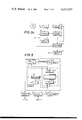
- FIG. 1 is a basic block diagram illustrating the signal processing sequence of the present invention which is common to all embodiments.
- FIG. 2 is a simplified pictorial view illustrating the way in which signals are obtained.
- FIG. 3a is a block circuit diagram of a first advantageous embodiment of the apparatus according to the invention.
- FIG. 3b is a block circuit diagram of a second embodiment of the invention utilizing a microprocessor.
- FIG. 3c is a block circuit diagram of a variation of the embodiment of FIG. 3a which can also be used together with the embodiment of FIG. 3b.
- FIG. 4 is a flow diagram of the basic sequence of the method according to the present invention, providing a basis for a computer program which may serve, for example, to control a microprocessor.
- FIG. 5 is a block circuit diagram of a subtractor for use in apparatus for practicing the method according to the invention.
- FIG. 6 is a block circuit diagram of an average former for use in apparatus for practicing the invention.
- FIG. 7a is a circuit diagram of a maximum detector in digital design suitable for use in apparatus for practicing the method of the present invention.
- FIG. 7b is a circuit diagram of an analog version of the maximum detector of FIG. 7a.
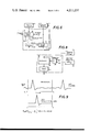
- FIG. 8 is a block circuit diagram of a correlator suitable for use in apparatus for practicing the method of the present invention.
- FIG. 9 is a schematic waveform representation of the formation of a cross correlation function.
- FIG. 1 is a basic illustration of the operating sequence of the method according to the invention which sequence is performed in the embodiments described hereinbelow. The sequence is thus shown in general terms applicable to all embodiments and variations thereof described hereinafter.
- the individual illustrated stages may be either circuits that are constituted by electronic components or by routines of a computer program, which is effected in real time by a suitable computer.
- Point 201 represents either the input of the circuit or the starting point of the computer program.
- Stage 202 performs all operations connected with recording the EKG.
- FIG. 2 shows the recording of a fetomaternal EKG.
- Various electrodes, 301, 302 and 303 are fastened to the body of a female patient in order to receive signals to determine the electrocardiogram of a fetus 305.
- the signal curve 306 recorded by means of pickups 301 to 303 the signal from the heart 307 of the fetus is superposed on the signal from the heart 308 of the mother together with various interference components.
- the fetal and the maternal heart signals are identified several times in the signal curve 306 as "f" and "m,” respectively.
- a filter stage 203 the received fetomaternal heart signal is freed of interfering signal components and in the subsequent stage 204 the maternal heart signal is removed from the filtered input signal by subtraction.
- What is subtracted in stage 204 is a signal shape which corresponds to the average of the maternal EKG in that the maternal EKG is detected by a maximum value detector and in the course of formation of the average value, that value is continuously corrected or brought up to date.
- the maximum value criterion is retained over the total period or the maximum value criterion is used in a starting time period, for the autocorrelation function of the input signal, while at a later point in time, when the maternal electrocardiogram has been recognized with sufficient certainty, it is based on the detection of the maximum of the cross correlation function between the stored signal shape and the input signal so that only a signal similar to the stored signal shape is utilized for the average formation and thus for a correction of the signal shape.
- the average of the maternal EKG is retained, or stored, in stage 205.
- the amplitude of the signal to be subtracted is matched with the amplitude of the input signal and the scaling factor K, which determines the amplitude of the signal to be subtracted, is formed from the ratio of the amplitude of the stored maternal EKG to the amplitude of the input signal, i.e. the maximum of the actual maternal QRS complex.
- the scaling factor K can be formed from the ratio of the cross correlation function between the stored sample EKG and the input signal to the autocorrelation function of the sample signal.
- the fetal EKG signal is actually determined and, according to the invention, the presence of a fetal heart signal is first detected by means of a maximum criterion while during the later course of the process the second criterion is used to determine the presence of a fetal heart signal from the degree of coincidence between the shape of the input signal and the shape of the stored average value, i.e. the sample signal.
- the average of the fetal EKG is stored in stage 206, while in stage 207 the fetal heart frequency is calculated from the time periods of the fetal heart signals and is stored there.
- FIGS. 3a and 3c indicate how an apparatus for practicing the method of the invention can be constructed in principle.
- the person skilled in the art is familiar with the manner in which the individual stages, which are also shown in the subsequent drawing figures, can be realized by electronic circuits, since it is possible, for example, by means of instructions issued by component manufacturers, to arrive at a possible concrete embodiment for every circuit module named in FIG. 3. For example, this can be done according to the TTL, the CMOS or comparable techniques.
- Suitable embodiments of individual circuit groups are illustrated in detail in FIGS. 5 through 8 to which reference will be made in the detailed description of an embodiment.
- FIG. 3b is a block circuit diagram of a further embodiment of an apparatus operating according to the present invention.
- the signals are here processed by microprocessors.
- microprocessors For reasons of clarity two microprocessors are provided.
- a single microprocessor can also be employed which then effects signal processing according to a time sharing method. The decision as to which one of the possibilities to choose depends on the further technical development, the number of units required, and on the requirements for operating speed, accuracy and reliability.
- the abdominally derived fetomaternal heart signal is fed to a preamplifier 1 which raises the low level of the input signal to a value that is sufficient for processing in subsequent stages.
- a first improvement of the signal to noise ratio is effected by filtering in a bandpass filter 2.
- the power of the maternal QRS complexes exceeds that of the fetal complexes to a considerable degree.
- the frequency components of the expected action potential of the maternal heart signal also lie in this range.
- both signal powers are in approximately the same order of magnitude.
- Bandpass filtering with a pass band in this frequency range thus permits selective amplification of the fetal heart signal.
- the lower and upper frequency limits of filter 2 are selected so that those frequency components of the received signal which do not contribute to the detection of the useful signal are suppressed.
- there is a second criterion for determining the limits of the frequency band i.e. that the maternal heart signal is attenuated but has a greater amplitude than the fetal signal so that it is possible to detect the maternal QRS complex by means of threshold value detectors.
- the frequency limits for the bandpass filter 2 in an advantageous embodiment of the invention are composed of a lower limit frequency of 15 Hz and an upper limit frequency of 40 Hz. Good results have been realized with the use of a nonrecursive digital filter of linear phase.
- a feedback connection from the output of the bandpass filter 2 to a control input of preamplifier 1 it is possible to effect an amplitude regulation of the preamplifier 1, to keep the triggering threshold for recognizing the maternal EKG at a constant value so that no further external amplitude setting is required.
- a rectifier (not shown) is provided in the signal input so as to effect full wave rectification of the input signal without integration in a conventional circuit arrangement.
- This solution is of advantage during recording of myographic signals, i.e. signals derived from muscle movements which are a function of cardiac movements, if the input signal to be detected has no fixed polarity.
- FIG. 4 is a flow diagram of the course of the further signal processing in the individual blocks of the systems of FIGS. 3a and 3b.
- This flow diagram may constitute the basis for the development of the circuitry for a device employing discrete or integrated components and is only an example which can be modified according to individual requirements.
- the operational sequence can be permanently wired in, can be stored in ROM's or in programmed logic circuits and controlled accordingly.
- FIGS. 3a, 3b, 3c and 4 together and, insofar as details are concerned, also to FIGS. 5 through 9.
- the maternal heart signal which in this case constitutes an interference signal
- the bandwidth of the preliminary filter is set so that the low frequency interference components and the high frequency interference components of the signals are sufficiently suppressed, the maternal electrocardiogram remains easily detectable and the fetal heart signal has its amplitude raised in addition.
- the sample QRS complex required for the subtraction is determined by a weighted average formation in which the calculation of the maternal QRS sample and the subtraction are actuated by a trigger signal which indicates the occurrence of a QRS complex and is generated by a threshold value detector.
- This threshold value detector is provided in FIG. 3a in the form of a maximum or peak detector 3 or 3'.
- the maternal QRS complex is detected by the peak detector in a digital manner by unit 3 or in an analog manner by unit 3'. Due to the smaller amount of circuitry required, the analog solution is more advantageous, and is shown in FIG. 3a in dashed lines as an alternative.
- the signal for the maximum detector 3 or 3' is obtained downstream or upstream, respectively, of a conventional analog-digital converter 4.
- the analog-digital converter 4 operates in a known manner at a sampling frequency of 1 kHz.
- the filtered and digitalized fetomaternal heart signal provided by converter 4 is stored in a memory 5 and is fed from there to an average former 6.
- the average former 6 forms a new average, as soon as a new maternal QRS complex has been detected, from this new complex and from the sample of the maternal QRS complex already stored in an average memory 7, and the new average is subsequently fed into memory 7, where it is stored.
- the stored value may equal zero or any desired sample signal may be initially stored.
- the maternal sample signal stored in memory 7 is processed in an amplitude matching device 8', one embodiment of which is shown in FIG. 5 together with the units with which it directly cooperates.
- the amplitude of the maternal sample signal stored in average memory 7 is adapted to the actual amplitude shape of the input signal by means of division by a scaling factor K in order to achieve as complete a separation as possible of the fetal heart signal from the maternal component.
- the embodiment shown in FIG. 5 includes a conventional digitally operating divider 10 which receives the signals to be divided via multiplexers 11 and 12 in time alternation. The division result is provided via a demultiplexer 13. In addition, there is provided a maximum detector 14 for storing the amplitude of the actual maternal QRS complex and a memory 14a for storing the present value of the scaling factor K.
- the broken lines in FIG. 5 signify the simultaneous switching of the multiplexers 11 and 12 and the demultiplexer 13. The inputs and outputs connected by the dashed lines are activated at the same time.
- the maximum value EKG mat is transferred from memory 7 to detector 14 after each averging cycle, which in this configuration only functions as a memory for the value of the maximum detected.
- the relative (centered) position of the maximum within the sample remains unchanged in the memory 7.
- the maximum detector 3 When the maximum detector 3 signals the occurrence of a maximum of the maternal QRS-complex, this maximal value is loaded into the dividing device 10 via multiplexer 12. At the same time the maximum of the sample of the QRS-complex is loaded into the dividing device 10 via multiplexer 11. The resulting quotient is transferred into memory 14a. After that the configuration of the devices 11, 12 an 13 is switched into its second state. Subsequently each value of the points of the sample is divided by the constant registered in the memory 14a and subtracted from the value of the corresponding point of the abdominal EKG that is to be read from memory 5. After the values of all the points have been substracted, the amplitude matching device remains inoperative until the next maternal QRS-complex is detected.
- the values of the sample points of the abdominal EKG are loaded successively into memory 5. After a maximum has been detected by detector 3, the address of the last entry into memory 5 is used for computing the address of the point that corresponds to the first point in memory 7.
- the sample is represented by the values of 256 points.
- the value of the maximum is numbered 128.
- synchronisation during subtraction is simplified by starting the subtractions wiith the value of point one of memory 7 and point "n minus 127" of memory 5, if the maximum has been detected for the value of the n-th point.
- the two last points are numbered 256 in memory 7 and "n+128" in memory 5. It has to be mentioned that the values of the points "n+1" until "n+128" are not immediately loaded into memory 5 after a maximum has been detected. They are transferred in a sampling mode in synchronism with the subtraction of the values of the first 128 points.
- the signals supplied to multiplexers 11 and 12 from detectors 14 and 3 are maximum values.
- the single value pulses shown in FIG. 7a are representations of these values, which are used as trigger pulses to control the transfer of the data to the processor (not shown) connected to the circuit displayed.
- the amplitude matching device 8 together with the digital dividing device 10 included therein can be constructed from commercially available semiconductor devices, such as, for example integrated TTL circuits, according to the instructions provided by the semiconductor manufacturers.
- the division result is supplied from demultiplexer 13 to subtraction device 9 together with the current abdominal EKG signal in memory 5 and there appears at the output of the subtraction device 9 the difference D representing the signal EKG fet , which is formed as follows:
- M the sample of the maternal EKG
- K the scaling factor for amplitude matching during subtraction, the value for K being recalculated for each detected QRS complex from
- E max is the amplitude of the actual maternal QRS complex
- M max is the amplitude of the maternal average value.
- the average formation required for matching the stored signals to the actual signal shape at various points in the process, so as to "refresh" the contents of the memories in question, is shown in FIG. 6 according to its functional sequence.
- the illustrated blocks show the principle of how such a circuit can be realized.
- the average former 6 is shown in FIG. 6 together with those blocks of the system of FIG. 3a that surround it.
- the interior circuitry of block 6 shown there may just as well be used for the average former 15 of FIG. 3a, which will be described in detail below.
- M n-1 the average value at time t n-1 ;
- E n the EKG at time t n ;
- the averaging constant determines the speed of matching of the stored values to the present signal shape.
- the value U should here be selected so that an optimum compromise is made between the improvement of the signal to noise ratio and the time constant for matching the EKG's to the actual wave shape.
- a power of two is selected for U because this makes the algorithm particularly easy to set up.
- FIG. 6 is a schematic representation of an electronic circuit for calculating the signal average according to equation (5).
- an parallel adder 16 the difference between E n and M n-1 is formed according to the 2's complement method, the signal from memory 7 being supplied via an inverter.
- a new average is formed each time.
- the selected embodiment affords high processing speeds at low cost.
- the illustrated method of exponential averaging results in an improvement of the signal to noise ratio by the factor ⁇ 2U-1.
- integrated circuits of Type 7483 can be used as the parallel adders 16 and 17, Type 7404 for the inverter and Types 74,100, 74,199 etc. for the (EKG) memory 5 and the average value memory 7, depending on the length of the signal words.
- FIG. 3a there are the maximum detectors 3, and 3' and 18, in FIG. 3b the maximum detector 3', and in FIG. 5, there is a maximum detector 14.
- FIG. 7a An embodiment of the digital form, as represented, for example, by the maximum detector 3 in FIG. 3a, is shown in FIG. 7a.
- the signal identified as "MAX” appears at the output when three voltage values of the input signal EKG measured at different times have exceeded the value of a triggering threshold T1, the measurement times being determined by an externally applied sampling pulse train "T" having a repetition rate of 1 kHz.
- the detector includes a counting unit which is reset to zero if another three measured values fall below the triggering threshold T1.
- Threshold T1 constitutes a variable parameter that can be adapted to the amplitude of the input signal of the circuit.
- the respective EKG signal is fed in digitalized form to the "EKG" input of a digital comparator 19 and is compared with the value of triggering threshold T1. If the EKG signal exceeds this triggering threshold, a signal appears at output ">", while if the signal stays below the triggering threshold a signal " ⁇ " is emitted.
- These signals are fed to respective AND gates 20 and 21.
- the output of each gate has a logic L if in addition to a 1 kHz clock pulse, a signal is also present at its associated comparator output.
- the output of maximum detector 3 in the configuration according to the embodiment shown in FIG. 7a signals the surpassing of trigger level T1 by the peak of the maternal QRS-complex.
- the width of the positive pulse covers a certain time interval around the exact time position of the maximum, the matter is found by means of differentiating the analog input signal, where minimal differences between neighbouring values indicate the location of the maximum.
- the differentiation is carried out by the processor in the usual way.
- FIG. 7b shows a maximum detector for analog operation, which includes two operational amplifiers 28 and 29 as well as an AND gate 30.
- the input signal "EKG” is fed to the positive input of operational amplifier 28 via a lowpass filter 31 and to the inverting input of amplifier 29 via a differentiating member 32.
- the triggering threshold value T1 is applied to the negative input of the operational amplifier 28 as a reference level while the noninverting input of operational amplifier 29 is at ground potential.
- the logic “L” level appears at the output of the AND gate 30 when the signal “EKG” exceeds the amplitude value of T1 for a sufficiently long period of time and has a slope of zero value.
- T1 must have been exceeded at several points in time during the duration of the input signal "EKG,” it being a prerequisite that the latter has taken on an extreme value.
- a microprocessor 33 In the circuit for practicing the method as shown in FIG. 3b several of the evaluation functions described above are preferred by a microprocessor 33.
- the modules corresponding to blocks 1 through 4 are identical with those of the circuit shown in FIG. 3a.
- the peripheral components of the microprocessor 33 include the average memory 7 and the amplitude matching device 8 which also take over the same functions as in the circuit according to FIGS. 3a and 3c, respectively, but with respect to the processing of data are adapted to the special microprocessor employed in the circuit.
- the fetal heart signal which has been separated from the maternal component.
- the microprocessor 33 is programmed according to the flow diagram shown in FIG. 4 which represents a generally preferred embodiment of a program for the process according to the invention and which can also be used for practicing the process in the same or a similar manner in devices designed according to other techniques, such as discrete, TTL, CMOS, etc.
- the program for operation of the system starts, as shown in FIG. 4, at "Start" 101.
- a branch instruction 104 then causes the value of a flag E to be checked. If E equals zero, which is equivalent to the fact that no maternal QRS complex has been recognized and is being processed, it is determined in a further branch 105 whether the input "EKG" exceeds the triggering threshold T1, this corresponding to the function of the previously described maximum detector. If T1 has not been exceeded, it is determined in a branch 111, whether the value K1, corresponding to the number of detected maternal complexes, is greater than three. If this is not the case, the program sequence jumps back to block 103.
- a program block 106 is entered from branch 105. Since exceeding of the triggering threshold T1 is synonymous with the recognition of a maternal QRS complex, the flag E is set to one and the value K1, the number of detected maternal complexes, is raised by one, i.e. from zero to one. If E equals one, the subsequent EKG values need no longer exceed the triggering threshold T1, and the program sequence proceeds directly from block 104 to the now following block 107 in that the averaging of the maternal EKG is effected as described above in connection with the average former. The averaging is effected individually for each scanned EKG value.
- the averaged EKG max value is subtracted from the currently measured value, the subtraction being made with amplitude matching as described in connection with FIG. 5.
- a check is made as to whether the described averaging and subtraction has already been effected for the entire expected duration of a maternal QRS complex. For this purpose the number of measured values that have been read in since the flag E was changed from zero to one is counted. If 256 values have been measured, the detection of a maternal QRS complex is considered to be completed and the program proceeds from branch 109 to block 110 at which the flag E is reset to zero so that for the renewed activation of the subtraction process it will again be necessary that the input signal exceeds the triggering threshold T1.
- the number of averages of the maternal complex EKG mat which must be exceeded so that a reliable average of the maternal complex is available for separating the abdominally derived heart signal from the maternal component, will advisably be selected to be higher than three.
- the sample signal that has been formed by averaging in the preceding cycle is used as a basis so that the actual section of the signal curve does not influence the suppression of the maternal heart signal.
- Matching the amplitude of the average value with the amplitude of the input signal significantly contributes to an improvement of the signal evaluation since, particularly at the onset of the averaging procedure, the sample and the momentarily present QRS complex may differ significantly in amplitude. Due to the fact that only one measured value is subtracted and averaged from the signal section per cycle, the process can be realized with particularly economical equipment since the required computing speed has been reduced.
- the abdominally recorded heart signal portion, containing predominantly the fetal EKG, which has been separated from the maternal QRS complex is now fed to a memory 34 from which the further processing branches out.
- the signal is fed to a correlator 35 which is able to perform an autocorrelation as well as a cross correlation calculation in dependence on the type of process involved, as will be explained in detail below.
- the average former 15 produces a sample of the fetal EKG in a manner similar to that in which the average former 6 produced a sample of the maternal QRS complex.
- the resulting calculated average of the fetal EKG is retained in an average memory 36.
- the values contained therein are used by the correlator 35 to form the cross correlation function with the input signal.
- the maximum detector 18, which is connected to the output of correlator 35, senses the maxima of the correlation function which is equivalent to the occurrence of a fetal QRS complex and thus triggers the formation of a revised average of the fetal electrocardiogram in the average former 15.
- the formation of the average itself is here again effected exponentially.
- correlator 35 In the starting phase, when the average of the fetal EKG cannot as yet be utilized, correlator 35 forms an autocorrelation function and when there is a maximum in this function, the average former 15 is put into operation. Upon completion of this starting phase, when the calculated average EKG fet approximates the actual fetal EKG with sufficient accuracy to be used as a sample, the correlator 35 forms the cross correlation function of the current EKG with the averaged sample that has been obtained by exponential determination as the reference signal. From the time interval between the maxima of the autocorrelation function and of the cross correlation function the fetal heart frequency is determined in function block 37.
- the detection of the fetal QRS complex can also be effected without correlation.
- the preprocessed signal is fed directly to the maximum detector 18, i.e. there exists a direct connection between blocks 34 and 18 with elimination of blocks 15, 35 and 36.
- FIG. 8 shows a block circuit diagram of an embodiment of the correlator 35 for use in the process of the present invention.
- the signal EKG fet which has been separated from the maternal component simultaneously enters a multiplexer 38 and a rapid multiplier 39, that has been designed according to the TTL technique, in order to form the autocorrelation function.
- the other input of the multiplexer 38 receives the average of the fetal heart signal EKG fet from average memory 36.
- the autocorrelation and cross correlation products are thus formed alternatingly.
- the respective correlation integral itself is calculated by an adder 40 downstream of multiplier 39 and is fed to maximum detector 18 of FIG. 3a.
- FIG. 9 is a schematic representation of the formation of the cross correlation function between a stored sample signal and the currently recorded heart signal for the example of the fetal EKG.
- the time period covered by the integration is identified as the "data window” and in successive observations the signal curve s(t) moves in the direction of the arrow with respect to the data window.
- the correlation functions may be formed according to known methods such as those employed, for example, in the Model 3721A correlator made by Hewlett Packard.
- a microprocessor 41 receives input signals from block 34 and is in communication with the average former 15, the average memory 36 for EKG fet and the correlator 35. These units are of course also adapted to the microprocessor employed. The latter, for example, may perform the calculations required for the various stages in a time sharing process and obtain the required signal values. From the available data it computes the fetal heart frequency according to a given program.
- Program blocks 112 to 123 of FIG. 4 show the course of a correlation analysis which is suitable for operation of a microprocessor or generally of an electronic data processing system.
- a conclusion as to the presence of a fetal QRS complex is made if the value of the correlation function produced by correlator 35 exceeds a triggering threshold T2, determined by detector 18. It is advisable for this triggering threshold T2 to adapt itself to the changing signal amplitudes since only in this way will dependable detection be assured. Since the shape and amplitude of the fetal electrocardiogram is not known beforehand, an amplitude value cannot be set beforehand for the triggering threshold T2.
- a certain period of time is used to search in the signal mixture that has been separated from the maternal complex to locate the fetal complex, and its maximum is determined.
- the period of time between three successive maternal QRS complexes is used for this purpose.
- the program moves from branch 111 to a branch 112 if the number (K1) of determined maternal complexes is greater than three. In branch 112 it is determined whether the number (K2) of detected fetal complexes is greater than six.
- the program portion taking place between points A and B serves, inter alia, to fix and influence the triggering threshold T2.
- K1 the fifth maternal complex has been determined, i.e. K1 equals five
- the program continues over a branch 124, where this value of K1 is sensed, and block 125 where the amplitude value of triggering threshold T2 is set to half the value of the greatest maximum of the autocorrelation function occurring in correlator 35 during the time between the third and fifth maternal complexes.
- a branch 117 determines whether the maximum of the formed correlation function (CF max ) exceeds the value of the triggering threshold T2--which had been set to half the value of the previously formed autocorrelation function. If this is not the case, the program jumps back to block 103. If it did exceed it, the triggering threshold is brought up to the amplitude shape of the signal by means of exponential averaging. T2 is thus set so that it always takes on half the valiue of the average correlation maximum. This likewise optimally adapts the sensitivity of the system to the actually prevailing signal conditions. From the correlation function, the fetal heart frequency (FHF) and the maternal heart frequency (MHF) as well are calculated in block 119.
- FHF fetal heart frequency
- MHF maternal heart frequency
- the fetal heart signal is averaged exponentially and stored in an average memory. This sample serves for the later calculation of the cross correlation function with the actual signal in block 113. Thus a fetal heart signal has been determined and the value of K2 which indicates the number of detected fetal complexes, is upped by one according to block 121.
- the sample of the fetal heart signal as calculated by average formation is accurate enough to serve as a reference signal for the calculation of the cross correlation function of the fetal electrocardiogram.
- the occurrence of seven fetal QRS complexes is considered to be sufficient. If K2 is greater than six, branch 112 directs the program to block 113 and the cross correlation function is now determined in the described manner.
- the amplitude of the fetal heart signal decreases so rapidly during the analysis that the average formation cannot follow the triggering threshold T2 in block 118, or if the fetal heart signal changes in shape, the amplitude of the correlation function will no longer exceed the triggering threshold T2. In that case the value of T2 is reset to 0.75 of its original value if no fetal QRS complex was detected for 900, 1,800 and 2,700 ms. If the triggering threshold was not exceeded for 3.6 seconds, all parameters and samples are erased and the algorithm is started anew. The program sequence required for this is contained in function blocks 126 to 132 between points A and B in FIG. 2.
- Resetting of the triggering threshold T2 to 0.75 of its original or previous value is effected in blocks 127, 129, and 131 after the respective periods have expired without the triggering threshold having been exceeded and this in dependence on the results in branches 126, 128, 130 which are associated with the respective time periods as shown in FIG. 4.
- the operations carried out by the elements 119 and 124 to 132 are performed by means of circuits that are set up according to examples given in the applications published by leading semiconductor manufactures.
- examples may be chosen from the publication "Designing with TTL Integrated Circuits.”
- the functions of the individual elements that may be seen from the description of the flow chart counters (7493 e.g.), comparators (7485 e.g.), shift registers and one or more arithmetic logic units (SN 74181) may be used.
- FIG. 3c shows a variation of the two described embodiments of the method according to the invention, which variation differs from the previously described embodiments in the manner of determining the average of the maternal EKG and in the determination of the scaling factor K during subtraction of the stored average from the actual signal shape.
- the stages shown in FIG. 3c replace the stages 3 to 9 of FIG. 3a.
- the basic function of the illustrated modules corresponds to that of modules 15, 18 and 34 to 36 of FIG. 3a with the difference that it is not the fetal electrocardiogram that is determined and stored for the purpose of calculating the fetal heart frequency, but rather the maternal electrocardiogram which is subtracted from the input signal for the purpose of separating it from interference. No differences exist in the basic signal processing with respect to initial detection of the heart signal and the continuous correction or adaptation by way of average formation.
- Each stage whose function coincides with that of a corresponding stage of previously described embodiments is identified with the same reference numeral.
- the input signal again passes through an analog-digital converter 4 into memory 5 in which the pattern or waveform of a segment of the actual input signal is stored.
- a correlator 35' is provided which corresponds to that shown in FIG. 8, with the exception that the input signals to correlator 35' are derived from stages 5 and 7 instead of stages 34 and 36.
- the maximum of the autocorrelation function is determined and the associated heart signal is stored in average memory 7.
- the averaged signal triggered by occurrence of the maximum of the cross correlation function, is adjusted by a newly detected maternal heart signal by way of averaging, the function of average memory 7 corresponding to that of stage 7 of FIG. 3a.
- amplitude matching device 8' which serves to adapt the amplitude of the average maternal electrocardiogram, which is to be subtracted, to the actual amplitude of the then occurring input signal so that this signal component is removed from the signal mixture.
- the circuit employed differs from the amplitude matching device 8 shown in FIG. 5 only in that instead of the values M max (stage 14) and E max (stage 39 the autocorrelation function and the cross correlation function are processed, the quotient K of the two values being formed alternatingly and, on the other hand, the average value of the maternal heart signal stored in stage 7 is divided by this value K.
- the subtraction device 9 thus precisely corresponds in its operation to that shown in FIG. 3a.
- the mathematical derivation to obtain the scaling factor K for this variation has been explained above.
- the advantage is here that the available correlator 35' can be utilized for two different functions, its time control in the sense of a multiplex operation being known.
- the triggering threshold T2 is set, once a maximum has been detected, to the value of this maximum until one or more further maxima have been found. At the end of a time of 100 ms after detection of the highest maximum, T2 is set back to its normal value.
- the time-dependent regulations are not shown in the block circuit diagrams of FIGS. 3a to 3c and 5 to 8. However, the necessary switching means can be realized by way of known logic circuits.
- the formed and stored sample function and the actual signal shape are not offset in time during point-by-point scanning. For that reason, the sample and actual signal are continuously scanned as to their maximum or peak value during average formation for the maternal heart signal. Averaging is effected only if both signals are in the same phase, i.e. the maxima coincide in time.
- the correlation analysis triggers the average formation of the fetal heart signal when there is a maximum. While phase equality is assured during calculation of the cross correlation function, a phase shift may occur during calculation of the autocorrelation function. In order to avoid faulty triggering in this case, sample and signal are additionally scanned for their maximum values during analysis by means of autocorrelation. The two maxima are then synchronized in order to form the average.
- the preceding is a description of the process according to the invention and of apparatus for practicing the process, using as an example the detection of fetal heart signals in an abdominally derived signal mixture of maternal and fetal heart signals in which the maternal signals far exceed the fetal signals in amplitude.
- the described process also has significant advantages in the detection of many other kinds of masked signals in which similar problems exist, such as, for example, myographically derived signals. Adaptation and selection of individual advantageous embodiments of the method suitable for the particular problem at hand which, in particular, can be adapted to the particular type of fluctuation in fluctuating signal amplitudes, are within the competence of the person skilled in the art.
- the particular fields of application and advantages can be derived from the above description of the example of the detection of fetal heart signals since the respective problems and the solutions found therefor can be generalized without difficulty.
- the two appended programs "KORA 1 and 2" have been used with the method described in connection with two cooperating microprocessors of the Type CP 1600 (manufactured by GI and ITT repectively).
- the program has been printed with the source program in assembler form, while the object program is listed on the left, according to the General Instrument developing system "GIMINI".
- GIMINI General Instrument developing system
- the inputs and outputs addressed by the program belong to the peripherals connected to the microprocessors. These addresses also serve for the transfer of data between the two processors and are listed seperately in the top of the programs.
- the peripheral units are digital-analog and analog-digital converters serving for the transmission and the reception of data in connection with the method for detecting signals described.
- the communication between the two processors is carried out by means of transfer registers.
- a version of the time dependent threshold variation described in connection with the flow chart of FIG. 4 in microprocessor program form is listed in program "KORA 2" under #129-address 204 of the object program-(setting the trigger level on the maximal value) and #206 to 212 - addresses 365 to 375 of the object program-(adaptive following of the trigger level as performed by block 118 in FIG. 4).
Landscapes
- Health & Medical Sciences (AREA)
- Life Sciences & Earth Sciences (AREA)
- Cardiology (AREA)
- Molecular Biology (AREA)
- Animal Behavior & Ethology (AREA)
- Pathology (AREA)
- Engineering & Computer Science (AREA)
- Biomedical Technology (AREA)
- Heart & Thoracic Surgery (AREA)
- Medical Informatics (AREA)
- Physics & Mathematics (AREA)
- Surgery (AREA)
- Biophysics (AREA)
- General Health & Medical Sciences (AREA)
- Public Health (AREA)
- Veterinary Medicine (AREA)
- Pediatric Medicine (AREA)
- Pregnancy & Childbirth (AREA)
- Gynecology & Obstetrics (AREA)
- Reproductive Health (AREA)
- Measurement And Recording Of Electrical Phenomena And Electrical Characteristics Of The Living Body (AREA)
- Measurement Of Current Or Voltage (AREA)
Applications Claiming Priority (2)
| Application Number | Priority Date | Filing Date | Title |
|---|---|---|---|
| DE2716739A DE2716739C3 (de) | 1977-04-14 | 1977-04-14 | Verfahren zur Detektion von Signalen |
| DE2716739 | 1977-04-14 |
Publications (1)
| Publication Number | Publication Date |
|---|---|
| US4211237A true US4211237A (en) | 1980-07-08 |
Family
ID=6006388
Family Applications (1)
| Application Number | Title | Priority Date | Filing Date |
|---|---|---|---|
| US05/896,771 Expired - Lifetime US4211237A (en) | 1977-04-14 | 1978-04-14 | Method and apparatus for identifying recurring signal patterns |
Country Status (7)
| Country | Link |
|---|---|
| US (1) | US4211237A (de) |
| JP (1) | JPS53128373A (de) |
| CH (1) | CH632848A5 (de) |
| DE (1) | DE2716739C3 (de) |
| GB (1) | GB1605051A (de) |
| NL (1) | NL7803531A (de) |
| SE (1) | SE437762B (de) |
Cited By (47)
| Publication number | Priority date | Publication date | Assignee | Title |
|---|---|---|---|---|
| US4338950A (en) * | 1980-09-22 | 1982-07-13 | Texas Instruments Incorporated | System and method for sensing and measuring heart beat |
| US4456959A (en) * | 1980-07-17 | 1984-06-26 | Terumo Corporation | Period measurement system |
| US4573479A (en) * | 1984-06-22 | 1986-03-04 | American Home Products Corporation | Systems and methods for monitoring the fetal heart |
| US4616333A (en) * | 1984-06-05 | 1986-10-07 | Elscint, Ltd. | Data correlation system |
| WO1986006604A1 (en) | 1985-05-06 | 1986-11-20 | American Telephone & Telegraph Company | Heart rate detection utilizing autoregressive analysis |
| US4627441A (en) * | 1984-04-27 | 1986-12-09 | Medtronic, Inc. | B medical monitoring circuit |
| US4680708A (en) * | 1984-03-20 | 1987-07-14 | Washington University | Method and apparatus for analyzing electrocardiographic signals |
| US4704681A (en) * | 1984-04-19 | 1987-11-03 | Nec Corporation | Electrocardiogram signal processing apparatus for determining the existence of the Wolff-Parkinson-White syndrome |
| US4721114A (en) * | 1986-02-21 | 1988-01-26 | Cardiac Pacemakers, Inc. | Method of detecting P-waves in ECG recordings |
| US4736322A (en) * | 1985-07-12 | 1988-04-05 | Clifford Ralph D | Cardiological simulator |
| US4781200A (en) * | 1985-10-04 | 1988-11-01 | Baker Donald A | Ambulatory non-invasive automatic fetal monitoring system |
| US4782904A (en) * | 1986-11-07 | 1988-11-08 | Ohaus Scale Corporation | Electronic balance |
| WO1990000368A1 (en) * | 1988-07-07 | 1990-01-25 | National Research Development Corporation | Apparatus for monitoring fetal heart rate |
| US4951680A (en) * | 1987-09-30 | 1990-08-28 | National Research Development Corporation | Fetal monitoring during labor |
| US5025787A (en) * | 1986-10-08 | 1991-06-25 | The Imperial College Of Science, Technology & Medicine | Intrauterine probe |
| US5042499A (en) * | 1988-09-30 | 1991-08-27 | Frank Thomas H | Noninvasive electrocardiographic method of real time signal processing for obtaining and displaying instantaneous fetal heart rate and fetal heart rate beat-to-beat variability |
| WO1991015995A1 (en) * | 1990-04-12 | 1991-10-31 | Felix Rosenthal | Noise cancellation arrangement |
| US5170791A (en) * | 1991-03-28 | 1992-12-15 | Hewlett-Packard Company | Method and apparatus for calculating the fetal heart rate |
| US5209237A (en) * | 1990-04-12 | 1993-05-11 | Felix Rosenthal | Method and apparatus for detecting a signal from a noisy environment and fetal heartbeat obtaining method |
| US5243993A (en) * | 1991-06-28 | 1993-09-14 | Life Fitness | Apparatus and method for measuring heart rate |
| US5666959A (en) * | 1995-08-30 | 1997-09-16 | British Technology Group Limited | Fetal heart rate monitoring |
| US5704365A (en) * | 1994-11-14 | 1998-01-06 | Cambridge Heart, Inc. | Using related signals to reduce ECG noise |
| EP0825723A1 (de) * | 1996-08-16 | 1998-02-25 | Dr.-Ing. P. Osypka GmbH | Verfahren zur Abschwächung einer Störung bei der Auswertung eines bioelektrischen Messsignals |
| US5749831A (en) * | 1997-06-23 | 1998-05-12 | Baker; Donald A. | Fetal cardiac monitoring utilizing umbilical blood flow parameters and heartbeat information |
| US5891047A (en) * | 1997-03-14 | 1999-04-06 | Cambridge Heart, Inc. | Detecting abnormal activation of heart |
| WO2000036975A1 (en) * | 1998-12-22 | 2000-06-29 | Neoventa Medical Ab | Device for reducing signal noise in a fetal ecg signal |
| US6115624A (en) * | 1997-07-30 | 2000-09-05 | Genesis Technologies, Inc. | Multiparameter fetal monitoring device |
| US20030122677A1 (en) * | 1997-03-07 | 2003-07-03 | Cardionet, Inc. | Reprogrammable remote sensor monitoring system |
| US20030172940A1 (en) * | 2002-03-13 | 2003-09-18 | Cardionet, Inc. | Method and apparatus for monitoring and communicating with an implanted medical device |
| US6662043B1 (en) | 2000-08-03 | 2003-12-09 | Ge Marquette Medical Systems, Inc. | Heart beat coincidence detection |
| US6664893B1 (en) | 2001-04-23 | 2003-12-16 | Cardionet, Inc. | Method for controlling access to medical monitoring device service |
| US6665385B2 (en) | 2001-04-23 | 2003-12-16 | Cardionet, Inc. | Medical monitoring system having multipath communications capability |
| US6694177B2 (en) | 2001-04-23 | 2004-02-17 | Cardionet, Inc. | Control of data transmission between a remote monitoring unit and a central unit |
| US6801137B2 (en) | 2001-04-23 | 2004-10-05 | Cardionet, Inc. | Bidirectional communication between a sensor unit and a monitor unit in patient monitoring |
| US20050119580A1 (en) * | 2001-04-23 | 2005-06-02 | Eveland Doug C. | Controlling access to a medical monitoring system |
| US20070027396A1 (en) * | 2005-07-26 | 2007-02-01 | American University of Sharajah | Separating mixed signals |
| US20070213627A1 (en) * | 2003-10-14 | 2007-09-13 | James David K | Fetal surveillance |
| EP1987767A1 (de) * | 2007-05-02 | 2008-11-05 | Biosense Webster, Inc. | Kohärente Signalunterdrückung in EKGs |
| US20090182242A1 (en) * | 2008-01-10 | 2009-07-16 | Moses Edward J | Apparatus and Method for Non-Invasive, Passive Fetal Heart Monitoring |
| US20090259133A1 (en) * | 2008-04-15 | 2009-10-15 | Wolfberg Adam J | Fetal ecg monitoring |
| US8892181B2 (en) | 2011-10-21 | 2014-11-18 | Mindchild Medical, Inc. | Non-invasive fetal monitoring |
| CN104305992A (zh) * | 2014-07-02 | 2015-01-28 | 中山大学 | 一种交互式胎儿心电快速自动提取方法 |
| WO2015136567A1 (en) * | 2014-03-12 | 2015-09-17 | Universita' Politecnica Delle Marche | Method for the filtering of electrocardiographic signals or the like, usable in particular for the monitoring of fetal heart function |
| CN105310688A (zh) * | 2015-11-02 | 2016-02-10 | 广东工业大学 | 一种基于非负盲分离胎儿心电特征信号提取方法 |
| CN107910065A (zh) * | 2017-10-25 | 2018-04-13 | 厦门传芯健康科技有限公司 | 一种基于心电图的健康预警方法 |
| CN112869724A (zh) * | 2021-01-19 | 2021-06-01 | 西安交通大学 | 一种基于多通道被动式采集信号的胎儿健康监测仪 |
| US20220257952A1 (en) * | 2018-06-05 | 2022-08-18 | Medtronic, Inc. | Cardiac signal t-wave detection |
Families Citing this family (6)
| Publication number | Priority date | Publication date | Assignee | Title |
|---|---|---|---|---|
| US4289142A (en) * | 1978-11-24 | 1981-09-15 | Kearns Kenneth L | Physiological occurrence, such as apnea, monitor and X-ray triggering device |
| DE3209850C2 (de) * | 1982-03-18 | 1985-03-28 | Dornier System Gmbh, 7990 Friedrichshafen | Selbsttätiges Auswerteverfahren für ein EKG |
| US4893632A (en) * | 1988-04-13 | 1990-01-16 | Siemens Aktiengesellschaft | Method and apparatus for comparing waveform shapes of time-varying signals |
| US4979110A (en) * | 1988-09-22 | 1990-12-18 | Massachusetts Institute Of Technology | Characterizing the statistical properties of a biological signal |
| US4938228A (en) * | 1989-02-15 | 1990-07-03 | Righter William H | Wrist worn heart rate monitor |
| JP4027746B2 (ja) * | 2002-08-07 | 2007-12-26 | 株式会社日立ハイテクノロジーズ | 生体磁場計測装置 |
Citations (7)
| Publication number | Priority date | Publication date | Assignee | Title |
|---|---|---|---|---|
| US3755783A (en) * | 1970-08-04 | 1973-08-28 | Inst Technicheska Kib Pri Ban | Arrangement for analyzing irregularities in repetitive biological processes |
| US3811428A (en) * | 1971-12-30 | 1974-05-21 | Brattle Instr Corp | Biological signals monitor |
| US3861387A (en) * | 1972-09-11 | 1975-01-21 | Cardiodynamics | Cardiac arrhythmia detector |
| US3878833A (en) * | 1973-10-09 | 1975-04-22 | Gen Electric | Physiological waveform detector |
| US3940692A (en) * | 1972-12-15 | 1976-02-24 | The University Of Edinburgh | Apparatus for monitoring recurrent waveforms |
| US4023564A (en) * | 1976-01-26 | 1977-05-17 | Spacelabs, Inc. | Arrhythmia detector |
| US4086652A (en) * | 1977-01-19 | 1978-04-25 | Block Engineering, Inc. | Method and apparatus for analyzing a time-dependent phenomenon |
-
1977
- 1977-04-14 DE DE2716739A patent/DE2716739C3/de not_active Expired
-
1978
- 1978-04-03 NL NL7803531A patent/NL7803531A/xx not_active Application Discontinuation
- 1978-04-12 SE SE7804136A patent/SE437762B/sv unknown
- 1978-04-13 JP JP4377578A patent/JPS53128373A/ja active Pending
- 1978-04-13 CH CH396378A patent/CH632848A5/de not_active IP Right Cessation
- 1978-04-13 GB GB14665/78A patent/GB1605051A/en not_active Expired
- 1978-04-14 US US05/896,771 patent/US4211237A/en not_active Expired - Lifetime
Patent Citations (7)
| Publication number | Priority date | Publication date | Assignee | Title |
|---|---|---|---|---|
| US3755783A (en) * | 1970-08-04 | 1973-08-28 | Inst Technicheska Kib Pri Ban | Arrangement for analyzing irregularities in repetitive biological processes |
| US3811428A (en) * | 1971-12-30 | 1974-05-21 | Brattle Instr Corp | Biological signals monitor |
| US3861387A (en) * | 1972-09-11 | 1975-01-21 | Cardiodynamics | Cardiac arrhythmia detector |
| US3940692A (en) * | 1972-12-15 | 1976-02-24 | The University Of Edinburgh | Apparatus for monitoring recurrent waveforms |
| US3878833A (en) * | 1973-10-09 | 1975-04-22 | Gen Electric | Physiological waveform detector |
| US4023564A (en) * | 1976-01-26 | 1977-05-17 | Spacelabs, Inc. | Arrhythmia detector |
| US4086652A (en) * | 1977-01-19 | 1978-04-25 | Block Engineering, Inc. | Method and apparatus for analyzing a time-dependent phenomenon |
Cited By (75)
| Publication number | Priority date | Publication date | Assignee | Title |
|---|---|---|---|---|
| US4456959A (en) * | 1980-07-17 | 1984-06-26 | Terumo Corporation | Period measurement system |
| US4338950A (en) * | 1980-09-22 | 1982-07-13 | Texas Instruments Incorporated | System and method for sensing and measuring heart beat |
| US4680708A (en) * | 1984-03-20 | 1987-07-14 | Washington University | Method and apparatus for analyzing electrocardiographic signals |
| US4704681A (en) * | 1984-04-19 | 1987-11-03 | Nec Corporation | Electrocardiogram signal processing apparatus for determining the existence of the Wolff-Parkinson-White syndrome |
| US4627441A (en) * | 1984-04-27 | 1986-12-09 | Medtronic, Inc. | B medical monitoring circuit |
| US4616333A (en) * | 1984-06-05 | 1986-10-07 | Elscint, Ltd. | Data correlation system |
| US4573479A (en) * | 1984-06-22 | 1986-03-04 | American Home Products Corporation | Systems and methods for monitoring the fetal heart |
| WO1986006604A1 (en) | 1985-05-06 | 1986-11-20 | American Telephone & Telegraph Company | Heart rate detection utilizing autoregressive analysis |
| US4736322A (en) * | 1985-07-12 | 1988-04-05 | Clifford Ralph D | Cardiological simulator |
| US4781200A (en) * | 1985-10-04 | 1988-11-01 | Baker Donald A | Ambulatory non-invasive automatic fetal monitoring system |
| US4721114A (en) * | 1986-02-21 | 1988-01-26 | Cardiac Pacemakers, Inc. | Method of detecting P-waves in ECG recordings |
| US5025787A (en) * | 1986-10-08 | 1991-06-25 | The Imperial College Of Science, Technology & Medicine | Intrauterine probe |
| US4782904A (en) * | 1986-11-07 | 1988-11-08 | Ohaus Scale Corporation | Electronic balance |
| US4951680A (en) * | 1987-09-30 | 1990-08-28 | National Research Development Corporation | Fetal monitoring during labor |
| WO1990000368A1 (en) * | 1988-07-07 | 1990-01-25 | National Research Development Corporation | Apparatus for monitoring fetal heart rate |
| US5042499A (en) * | 1988-09-30 | 1991-08-27 | Frank Thomas H | Noninvasive electrocardiographic method of real time signal processing for obtaining and displaying instantaneous fetal heart rate and fetal heart rate beat-to-beat variability |
| WO1991015995A1 (en) * | 1990-04-12 | 1991-10-31 | Felix Rosenthal | Noise cancellation arrangement |
| US5209237A (en) * | 1990-04-12 | 1993-05-11 | Felix Rosenthal | Method and apparatus for detecting a signal from a noisy environment and fetal heartbeat obtaining method |
| US5170791A (en) * | 1991-03-28 | 1992-12-15 | Hewlett-Packard Company | Method and apparatus for calculating the fetal heart rate |
| US5243993A (en) * | 1991-06-28 | 1993-09-14 | Life Fitness | Apparatus and method for measuring heart rate |
| US5704365A (en) * | 1994-11-14 | 1998-01-06 | Cambridge Heart, Inc. | Using related signals to reduce ECG noise |
| US5666959A (en) * | 1995-08-30 | 1997-09-16 | British Technology Group Limited | Fetal heart rate monitoring |
| EP0825723A1 (de) * | 1996-08-16 | 1998-02-25 | Dr.-Ing. P. Osypka GmbH | Verfahren zur Abschwächung einer Störung bei der Auswertung eines bioelektrischen Messsignals |
| US20030122677A1 (en) * | 1997-03-07 | 2003-07-03 | Cardionet, Inc. | Reprogrammable remote sensor monitoring system |
| US6940403B2 (en) | 1997-03-07 | 2005-09-06 | Cardionet, Inc. | Reprogrammable remote sensor monitoring system |
| US5891047A (en) * | 1997-03-14 | 1999-04-06 | Cambridge Heart, Inc. | Detecting abnormal activation of heart |
| US5749831A (en) * | 1997-06-23 | 1998-05-12 | Baker; Donald A. | Fetal cardiac monitoring utilizing umbilical blood flow parameters and heartbeat information |
| US6115624A (en) * | 1997-07-30 | 2000-09-05 | Genesis Technologies, Inc. | Multiparameter fetal monitoring device |
| WO2000036975A1 (en) * | 1998-12-22 | 2000-06-29 | Neoventa Medical Ab | Device for reducing signal noise in a fetal ecg signal |
| US6658284B1 (en) | 1998-12-22 | 2003-12-02 | Neoventa Medical Ab | Device for reducing signal noise in a fetal ECG signal |
| US6662043B1 (en) | 2000-08-03 | 2003-12-09 | Ge Marquette Medical Systems, Inc. | Heart beat coincidence detection |
| US20040260189A1 (en) * | 2001-04-23 | 2004-12-23 | Cardionet, Inc., A California Corporation. | Control of data transmission between a remote monitoring unit and a central unit |
| US6665385B2 (en) | 2001-04-23 | 2003-12-16 | Cardionet, Inc. | Medical monitoring system having multipath communications capability |
| US6694177B2 (en) | 2001-04-23 | 2004-02-17 | Cardionet, Inc. | Control of data transmission between a remote monitoring unit and a central unit |
| US20040085186A1 (en) * | 2001-04-23 | 2004-05-06 | Cardionet, Inc., A California Corporation | Controlling access to a medical monitoring system |
| US6801137B2 (en) | 2001-04-23 | 2004-10-05 | Cardionet, Inc. | Bidirectional communication between a sensor unit and a monitor unit in patient monitoring |
| US6664893B1 (en) | 2001-04-23 | 2003-12-16 | Cardionet, Inc. | Method for controlling access to medical monitoring device service |
| US20050119580A1 (en) * | 2001-04-23 | 2005-06-02 | Eveland Doug C. | Controlling access to a medical monitoring system |
| US8290129B2 (en) | 2001-04-23 | 2012-10-16 | Cardionet, Inc. | Medical monitoring system having multiple communications channels |
| USRE43767E1 (en) | 2001-04-23 | 2012-10-23 | Cardionet, Inc. | Control of data transmission between a remote monitoring unit and a central unit |
| US7002468B2 (en) | 2001-04-23 | 2006-02-21 | Cardionet, Inc. | Controlling access to a medical monitoring system |
| US7130396B2 (en) | 2001-04-23 | 2006-10-31 | Cardionet, Inc. | Medical monitoring system having multiple communications channels |
| US8425414B2 (en) | 2001-04-23 | 2013-04-23 | Braemar Manufacturing, Llc | Controlling access to a medical monitoring system |
| US20070130657A1 (en) * | 2001-04-23 | 2007-06-07 | Cardionet, Inc., A California Corporation | Medical monitoring system having multiple communications channels |
| US9474445B2 (en) | 2001-04-23 | 2016-10-25 | Braemar Manufacturing, Llc | Controlling access to medical monitoring system |
| US20070288067A1 (en) * | 2001-04-23 | 2007-12-13 | Cardionet, Inc. | Controlling Access to a Medical Monitoring System |
| US6957107B2 (en) | 2002-03-13 | 2005-10-18 | Cardionet, Inc. | Method and apparatus for monitoring and communicating with an implanted medical device |
| US20030172940A1 (en) * | 2002-03-13 | 2003-09-18 | Cardionet, Inc. | Method and apparatus for monitoring and communicating with an implanted medical device |
| US20070213627A1 (en) * | 2003-10-14 | 2007-09-13 | James David K | Fetal surveillance |
| US8116855B2 (en) * | 2003-10-14 | 2012-02-14 | Monica Healthcare Limited | Fetal surveillance |
| EP1680018B1 (de) * | 2003-10-14 | 2008-11-12 | Monica Healthcare Limited | Fötus-überwachung |
| US20090036787A1 (en) * | 2003-10-14 | 2009-02-05 | Monica Healthcare Limited | Fetal surveillance |
| US8229550B2 (en) * | 2003-10-14 | 2012-07-24 | Monica Healthcare Limited | Fetal surveillance |
| US20070027396A1 (en) * | 2005-07-26 | 2007-02-01 | American University of Sharajah | Separating mixed signals |
| US7474915B2 (en) * | 2005-07-26 | 2009-01-06 | American University Of Sharjah And Arab Science And Technology Foundation | Separating mixed signals containing a distorted signal |
| EP1987767A1 (de) * | 2007-05-02 | 2008-11-05 | Biosense Webster, Inc. | Kohärente Signalunterdrückung in EKGs |
| US7894885B2 (en) | 2007-05-02 | 2011-02-22 | Biosense Webster, Inc. | Coherent signal rejection in ECG |
| JP2008284352A (ja) * | 2007-05-02 | 2008-11-27 | Biosense Webster Inc | Ecgにおけるコヒーレント信号の除去 |
| US20080275353A1 (en) * | 2007-05-02 | 2008-11-06 | Meir Bartal | Coherent signal rejection in ecg |
| US7869863B2 (en) | 2008-01-10 | 2011-01-11 | The Johns Hopkins University | Apparatus and method for non-invasive, passive fetal heart monitoring |
| US20090182242A1 (en) * | 2008-01-10 | 2009-07-16 | Moses Edward J | Apparatus and Method for Non-Invasive, Passive Fetal Heart Monitoring |
| US7949389B2 (en) * | 2008-04-15 | 2011-05-24 | Tufts Medical Center, Inc. | Fetal ECG monitoring |
| US20090259133A1 (en) * | 2008-04-15 | 2009-10-15 | Wolfberg Adam J | Fetal ecg monitoring |
| EP2276401A4 (de) * | 2008-04-15 | 2014-02-05 | Tufts Medical Ct | Ekg-überwachung eines fötus |
| WO2009146181A1 (en) | 2008-04-15 | 2009-12-03 | Tufts Medical Center | Fetal ecg monitoring |
| US8892181B2 (en) | 2011-10-21 | 2014-11-18 | Mindchild Medical, Inc. | Non-invasive fetal monitoring |
| WO2015136567A1 (en) * | 2014-03-12 | 2015-09-17 | Universita' Politecnica Delle Marche | Method for the filtering of electrocardiographic signals or the like, usable in particular for the monitoring of fetal heart function |
| CN104305992A (zh) * | 2014-07-02 | 2015-01-28 | 中山大学 | 一种交互式胎儿心电快速自动提取方法 |
| CN105310688A (zh) * | 2015-11-02 | 2016-02-10 | 广东工业大学 | 一种基于非负盲分离胎儿心电特征信号提取方法 |
| CN105310688B (zh) * | 2015-11-02 | 2018-07-10 | 广东工业大学 | 一种基于非负盲分离胎儿心电特征信号提取方法 |
| CN107910065A (zh) * | 2017-10-25 | 2018-04-13 | 厦门传芯健康科技有限公司 | 一种基于心电图的健康预警方法 |
| US20220257952A1 (en) * | 2018-06-05 | 2022-08-18 | Medtronic, Inc. | Cardiac signal t-wave detection |
| US11918815B2 (en) * | 2018-06-05 | 2024-03-05 | Medtronic, Inc. | Cardiac signal T-wave detection |
| CN112869724A (zh) * | 2021-01-19 | 2021-06-01 | 西安交通大学 | 一种基于多通道被动式采集信号的胎儿健康监测仪 |
| CN112869724B (zh) * | 2021-01-19 | 2022-04-22 | 西安交通大学 | 一种基于多通道被动式采集信号的胎儿健康监测仪 |
Also Published As
| Publication number | Publication date |
|---|---|
| DE2716739B2 (de) | 1979-10-04 |
| CH632848A5 (de) | 1982-10-29 |
| DE2716739C3 (de) | 1980-06-26 |
| DE2716739A1 (de) | 1978-10-26 |
| SE7804136L (sv) | 1978-10-15 |
| JPS53128373A (en) | 1978-11-09 |
| NL7803531A (nl) | 1978-10-17 |
| SE437762B (sv) | 1985-03-18 |
| GB1605051A (en) | 1981-12-16 |
Similar Documents
| Publication | Publication Date | Title |
|---|---|---|
| EP0310349B1 (de) | Fetale Überwachung während der Arbeit | |
| US4181135A (en) | Method and apparatus for monitoring electrocardiographic waveforms | |
| US5372139A (en) | Method for suppressing a maternal electrocardiogram signal from a fetal electrocardiogram signal obtained with invasive and non-invasive techniques using an almost pure maternal electrocardiogram signal as a trigger | |
| US4170992A (en) | Fiducial point location | |
| US4945917A (en) | Method and apparatus for indicating repetition intervals of a specified component of a composite electrical signal, particularly useful for displaying fetal r-waves | |
| US5033472A (en) | Method of and apparatus for analyzing propagation of arterial pulse waves through the circulatory system | |
| US4240442A (en) | Variable threshold R-wave detector | |
| US8064991B2 (en) | Method of fetal and maternal ECG identification across multiple EPOCHS | |
| GB1605051A (en) | Method of detecting signals | |
| JP3224552B2 (ja) | 呼吸信号およびまたはアーティファクト信号の導出方法 | |
| US4832041A (en) | Pace pulse eliminator | |
| JP3319140B2 (ja) | 心拍変動波形解析方法及び装置 | |
| AU1877400A (en) | Device for reducing signal noise in a fetal ecg signal | |
| EP3970615B1 (de) | Verfahren zur detektion eines qrs-komplexes eines elektrokardiogrammsignals | |
| US3581735A (en) | Heartbeat frequency determining apparatus and method | |
| Azad | Fetal QRS complex detection from abdominal ECG: a fuzzy approach | |
| De Chazal et al. | Automatic measurement of the QRS onset and offset in individual ECG leads | |
| Emrich et al. | Physiology-Informed ECG Delineation Based on Peak Prominence | |
| US4716907A (en) | Apparatus for detecting electroencephalogram and evoked response with monopolar derivation method | |
| RU2294139C1 (ru) | Способ выделения начала кардиоцикла и устройство для его осуществления | |
| Budin et al. | Real-time multichannel abdominal fetal ECG monitor using digital signal coprocessor | |
| Azad et al. | Development of a fuzzy rule-based QRS detection algorithm for fetal and maternal heart rate monitoring | |
| Xia et al. | Influence of recognition errors of computerised analysis of 24-hour electrocardiograms on the measurement of spectral components of heart rate variability | |
| Nadila et al. | Fetal heart rate detection algorithm from noninvasive fetal electrocardiogram | |
| EP3970616A1 (de) | Vorrichtung zur detektion des qrs-komplexes eines elektrokardiogrammsignals |