US5445936A - Method for non-competitive binding assays - Google Patents
Method for non-competitive binding assays Download PDFInfo
- Publication number
- US5445936A US5445936A US08/121,806 US12180693A US5445936A US 5445936 A US5445936 A US 5445936A US 12180693 A US12180693 A US 12180693A US 5445936 A US5445936 A US 5445936A
- Authority
- US
- United States
- Prior art keywords
- analyte
- assay
- solid phase
- specific binder
- labeled specific
- Prior art date
- Legal status (The legal status is an assumption and is not a legal conclusion. Google has not performed a legal analysis and makes no representation as to the accuracy of the status listed.)
- Expired - Lifetime
Links
Images


Classifications
-
- G—PHYSICS
- G01—MEASURING; TESTING
- G01N—INVESTIGATING OR ANALYSING MATERIALS BY DETERMINING THEIR CHEMICAL OR PHYSICAL PROPERTIES
- G01N33/00—Investigating or analysing materials by specific methods not covered by groups G01N1/00 - G01N31/00
-
- C—CHEMISTRY; METALLURGY
- C12—BIOCHEMISTRY; BEER; SPIRITS; WINE; VINEGAR; MICROBIOLOGY; ENZYMOLOGY; MUTATION OR GENETIC ENGINEERING
- C12Q—MEASURING OR TESTING PROCESSES INVOLVING ENZYMES, NUCLEIC ACIDS OR MICROORGANISMS; COMPOSITIONS OR TEST PAPERS THEREFOR; PROCESSES OF PREPARING SUCH COMPOSITIONS; CONDITION-RESPONSIVE CONTROL IN MICROBIOLOGICAL OR ENZYMOLOGICAL PROCESSES
- C12Q1/00—Measuring or testing processes involving enzymes, nucleic acids or microorganisms; Compositions therefor; Processes of preparing such compositions
- C12Q1/68—Measuring or testing processes involving enzymes, nucleic acids or microorganisms; Compositions therefor; Processes of preparing such compositions involving nucleic acids
- C12Q1/6813—Hybridisation assays
-
- C—CHEMISTRY; METALLURGY
- C12—BIOCHEMISTRY; BEER; SPIRITS; WINE; VINEGAR; MICROBIOLOGY; ENZYMOLOGY; MUTATION OR GENETIC ENGINEERING
- C12Q—MEASURING OR TESTING PROCESSES INVOLVING ENZYMES, NUCLEIC ACIDS OR MICROORGANISMS; COMPOSITIONS OR TEST PAPERS THEREFOR; PROCESSES OF PREPARING SUCH COMPOSITIONS; CONDITION-RESPONSIVE CONTROL IN MICROBIOLOGICAL OR ENZYMOLOGICAL PROCESSES
- C12Q1/00—Measuring or testing processes involving enzymes, nucleic acids or microorganisms; Compositions therefor; Processes of preparing such compositions
- C12Q1/68—Measuring or testing processes involving enzymes, nucleic acids or microorganisms; Compositions therefor; Processes of preparing such compositions involving nucleic acids
- C12Q1/6813—Hybridisation assays
- C12Q1/6834—Enzymatic or biochemical coupling of nucleic acids to a solid phase
-
- G—PHYSICS
- G01—MEASURING; TESTING
- G01N—INVESTIGATING OR ANALYSING MATERIALS BY DETERMINING THEIR CHEMICAL OR PHYSICAL PROPERTIES
- G01N33/00—Investigating or analysing materials by specific methods not covered by groups G01N1/00 - G01N31/00
- G01N33/0078—Testing material properties on manufactured objects
- G01N33/008—Sport articles, e.g. balls, skis or rackets
-
- G—PHYSICS
- G01—MEASURING; TESTING
- G01N—INVESTIGATING OR ANALYSING MATERIALS BY DETERMINING THEIR CHEMICAL OR PHYSICAL PROPERTIES
- G01N33/00—Investigating or analysing materials by specific methods not covered by groups G01N1/00 - G01N31/00
- G01N33/48—Biological material, e.g. blood, urine; Haemocytometers
- G01N33/50—Chemical analysis of biological material, e.g. blood, urine; Testing involving biospecific ligand binding methods; Immunological testing
- G01N33/53—Immunoassay; Biospecific binding assay; Materials therefor
- G01N33/543—Immunoassay; Biospecific binding assay; Materials therefor with an insoluble carrier for immobilising immunochemicals
- G01N33/54306—Solid-phase reaction mechanisms
Definitions
- Sensitivity means the minimal detectable dose, namely the smallest mass of analyte that generates a statistically significant change in the signal generated by the assay vs. that obtained in the absence of analyte.
- binding assays i.e., detect smaller amounts of analyte
- high sensitivity allows the use of small sample size, which can help to reduce "sample matrix" interferences.
- higher sensitivity allows measuring low analyte concentrations with a higher precision.
- the signal which is measured is that emanating from the specific binder that does not bind analyte.
- the labeled antibody is incubated with a sample containing analyte and a solid phase-immobilized analyte derivative. The labeled antibody that did not bind analyte binds the solid phase, and the signal emanating from the solid phase-bound labeled antibody is measured.
- unlabeled antibody is incubated with a sample containing an analyte and a labeled analyte derivative (or analyte mimic).
- the labeled analyte derivative binds those antibody binding sites that did not bind analyte.
- the assayist By measuring the signal coming from the labeled analyte derivative that bound the antibody, the assayist actually obtains an estimate of the concentration of antibody sites that did not bind analyte.
- the signal generated from a competitive assay decreases as the analyte concentration increases. Since small levels of analyte correspond to large signals, small changes in low concentrations of analyte lead to small differences between large numbers, which are hard to measure accurately.
- Type A of the non-competitive assay has the potential for the highest sensitivity.
- Jackson and Ekins T. M. Jackson and Ekins, R. P., Journal of Immunological Methods, 87:13, 1986
- sensitivity of type A is higher than that of the competitive assay.
- Empirical data supports the conclusion that type A of immunoassays is more sensitive than the competitive type of immunoassays: several immunoassays, such as thyroid stimulating hormone, have sensitivity of several million molecules per assay cuvette; in contrast, the most sensitive competitive immunoassays, such as those of digoxin and triiodothyronine, have sensitivities of several billion molecules per assay cuvette.
- type A assays are the most sensitive type, there is a need to improve their sensitivities even further.
- the large gap in potential sensitivity (a number of orders of magnitudes, depending on the value for fractional non-specific binding) between the competitive type and type A of the non-competitive is the main reason for the wide use of type A.
- the former is used either when high sensitivity is not required or when type A is not possible due to the existence of only one epitope on the analyte, as is the case for analytes that are haptens or short peptides.
- the non-competitive assay should be more sensitive.
- the signal increases as the concentration of analyte increases, and low concentrations of analyte can be detected more easily since small differences between small numbers are relatively easy to distinguish, and the signal due to the presence of analyte is distinguished from a small, rather than a large background.
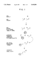
- Baier et al. described a non-competitive immunoassay format of type B with an added separation step. (See FIG. 1 for Baier analytical procedure.) After incubating the sample containing analyte with the labeled antibody, a solid phase with immobilized analyte derivative is added to bind the unreacted labeled antibody. The solid phase is separated and an aliquot of the remaining liquid phase is pipetted off to a new reaction cuvette containing a second solid phase. The second solid phase has an immobilized antibody against the labeled antibody or some part of the labeled antibody-analyte complex. The signal associated with the complex captured on the second solid phase is measured.
- this additional step was to allow a wash step in order to remove sample matrix interfering factors. It is not clear whether a substantial improvement of sensitivity was achieved by this additional step, because comparison to the competitive version of the same assay is not presented by Baier et al. Since competitive solid phase assays include one separation step, the added inconvenience of two separation steps in the Baier et al. patent is a disadvantage. This may be the main reason for which the method has not been commercialized or widely used in academic settings.
- Baier avoids mixing of the two solid phases because he could not separate them later, and even if he used separable solid phases, it was believed that the two solid phases would clump together, since they carry immunochemical binding partners (anti-label on one particle is a binding partner with a labeled antibody bound to the other particle). Clumping of particulate solid phases coupled to immunochemical binding partners is used often in particle agglutination assays. (See K. E. Hechemy and E. E. Michaelson, Laboratory Management, June, 1984, pp. 27-40.)
- the sample to be analyzed in an immunoassay is delivered in an environment that includes interfering substances.
- a serum sample not only contains the analyte of interest, but also many components that could interfere with the immunoassay.
- Immunochemical assay techniques include steps that easily isolate the analyte from the interfering substances. For example, the analyte can be reacted with an antibody which is connected to a solid phase. The solid phase can then be separated from the other components in its environment and analyzed.
- the separation step referred to above can be accomplished in one of many ways.
- an assayist can use nonmagnetic particles as the solid phase using either centrifugation filtration as the method of separation, or magnetic particles as the solid phase, in which case the separation is accomplished by the application of a magnetic field.
- Other effective means of separation involve various chromatographies, electrophoreses, and the use of extended surfaces, such as microtiter plates, large beads, fibers and others.
- the separation step can be done manually or by an automated or non-automated instrument; in either case, however, the solid phase is separated and washed, the liquid phases are discarded, and the solid phase-associated signal is the one being measured.
- cross-reactants share structural similarities with the analyte and also bind the labeled or unlabeled specific binder.
- a cross-reactant binds the labeled specific binder the assay result is falsely elevated.
- sufficiently high concentration of a cross-reactant binds the unlabeled specific binder and saturate it, a falsely low result is obtained.
- Heterophilic antibodies and rheumatoid factors bind antibodies and can either form a bridge between the labeled and unlabeled antibodies or inhibit their desired binding activity, leading to false results.
- the separation step is one of the most technically demanding operations in the assay. It needs to be done rapidly, so that the analyte-binder complex of interest does not dissociate. It also needs to be efficient, so that the unbound labeled binder and interfering substances are nearly completely removed. In addition, it needs to be reproducible, in order to maintain overall high assay precision.
- an assay should not require additional steps beyond what is currently used on a given instrument (i.e., there should not be a need to retrofit the instrument). Furthermore, the assay should preferably be capable of being run in one tube.
- a novel non-competitive immunoassay technique has been developed which not only improves sensitivity, but also is convenient and less susceptible to interfering factors. It is compatible with existing instruments and is an assay that can be run in one test tube.
- the analyte is reacted with labeled specific binder, after which the mixture is reacted with (1) an insoluble material attached to an analyte derivative and (2) a solid phase carrying a binder.
- the solid phase is then separated, and the label attached to the solid phase is measured.
- FIG. 1 illustrates a scheme for the assay technique of Baier et al.
- FIGS. 2-4 illustrate schemes for assay formats according to the present invention, with FIG. 2 illustrating a monoepitopic example, FIG. 3 showing a sandwich type with at least 2 epitopes, and FIG. 4 illustrating a gene probe assay.
- FIG. 5A illustrates a non-competitive T3 assay utilizing the present invention
- FIG. 5B illustrates a competitive T3 assay, both run on an automated immunoassay instrument.
- FIGS. 6-8 illustrate the results from the assays for monoepitopic analytes as described in the Examples.
- FIG. 9 shows a comparison to the results of using the novel assay technique vs. the technique of Baier et al.
- FIG. 10A shows a scheme of the non-competitive assay of the present invention as applied to a protein analyte (TSH) possessing more than one epitope using a solid phase immobilized anti-label.
- FIG. 10B shows use of a solid phase immobilized anti-analyte in an assay that is otherwise the same as FIG. 10A.
- FIG. 11 shows the results obtained in the assay for TSH using the present invention which utilizing either a capture with an immobilized anti-label or an immobilized anti-analyte.
- a convenient binding assay technique that has improved sensitivity and is less susceptible to interfering substances is described herein.
- One of its primary applications is in the field of immunochemistry, and, thus, most of the discussion of the technique is related to the immunochemistry field.
- This assay technique is equally applicable to other binding assays, such as gene probe assays and receptor assays.
- This assay technique is suitable for a wide diversity of analytes, including those with one or more epitopes.
- Analytes may include proteins, peptides, drugs, hormones, environmental pollutants, nucleic acids, lipids, carbohydrates and various conjugates of these.
- the technique is suitable for both determination of the total amount of analyte in the sample or the free fraction (e.g. free hormones and free drugs in biological fluids).
- Reagent 1 which comprises a labeled specific binder that binds at least a portion of the analyte.
- Reagent 1 comprises a labeled specific binder that binds at least a portion of the analyte.
- the label is a chemiluminescent label (e.g., an acridinium ester) or an enzymatic label.
- the label can be attached directly to the specific binder by a covalent bond. Alternatively it can be attached indirectly using a binding pair such as biotin/avidin, DNP/anti-DNP or any other binding pair. All are similarly suitable for use in the assays described herein.
- Reagent 2 contains a component comprising an analyte or analyte derivative or analyte mimic attached to an insoluble material that retards or reduces the binding of the previously unreacted labeled specific binder to reagent 3. If it did not reduce this binding, the binder attached to reagent 3 might bind not only to the labeled binder-analyte complex, as desired, but also to the labeled binder which is now bound to the insoluble material, thereby increasing the non-specific signal.
- reagent 2 retards binding due to its steric hindrance
- this insoluble material include particles made of controlled-pore glass, polymer particles, latex, colloidal metal or metal oxide particles, immiscible liquid phase, extended surface, porous paper, porous gel, liposome, emulsion, a system of very small particles that do not settle readily by standing or centrifugation, paramagnetic particles, cellulose beads, cross-linked dextran or any other particle.
- Extended surface is meant to include relatively flat surfaces, such as the surface of a cuvette or a microtiter plate, and the surface of a relatively large bead, such as one with diameter of greater than 1 mm.
- Preferred insoluble materials are controlled pore glass, polymer particles, latex particles, cross-linked dextran and extended surfaces.
- Particle size can vary from 10 nm to several microns in diameter, and smaller materials may include large molecular polymers such as dextran or protein aggregates. Larger beads of any size, flat surfaces, testtube wall, dipstick surface, fibers, membranes, rods and discs, or any extended or particulate surface capable of carrying an immobilized binder can also be used. Other mechanisms aside from steric hindrance (for example, porosity) also tend to retard the binding to reagent 3.
- reagent 2 can be in the form of particles or an extended surface, it does not function as usual solid phases function in binding assays, because there is no need to separate reagent 2 from the liquid phase containing the sample and other components of the assay. However, reagent 2 should not substantially adhere to the solid phase or be co-separated with it. Therefore, if the solid phase is separated from the liquid phase containing the sample plus other assay components prior to signal measurement, reagent 2 should remain substantially with the liquid phase and be removed together with it. For these reasons and to distinguish it from a true solid phase, the material used in reagent 2 is referred to as insoluble material.
- the component attached to the insoluble material in reagent 2 can be an analyte or a derivative of the analyte.
- the affinity of the labeled specific binder to this component may be similar (within the same order of magnitude) to its affinity toward the intact analyte.
- the component may be an analog of the analyte, the affinity of which to the labeled specific binder being much lower (by more than one order of magnitude), in this case the binding of the labeled specific binder to reagent 2 is facilitated by avidity (cooperativity between two or more binding site of the binder. See Piran U. et al. Clinical Chemistry Vol. 39, pp. 879-883, 1993).
- the component may be a synthetic molecule (such as an organic molecule, a synthetic peptide or an oligonucleotide) or a biologically derived molecule (such as a protein, a peptide, an antibody, an antiidiotypic antibody, receptor, antigen, nucleic acid etc.).
- a synthetic molecule such as an organic molecule, a synthetic peptide or an oligonucleotide
- a biologically derived molecule such as a protein, a peptide, an antibody, an antiidiotypic antibody, receptor, antigen, nucleic acid etc.
- Reagent 3 is a solid phase containing an immobilized binder that binds the labeled specific binder-analyte complex.
- This immobilized binder can be an antibody against (1) the specific binder, (2) the label, or (3) the complex.
- the immobilized binder can be, in the case when bi- or multi- valent labeled specific binder is used, an analyte or analyte mimic.
- the immobilized binder can be an anti-analyte, a specific receptor, or a complementary nucleic acid sequence. In other words, it can bind with any portion of the labeled specific binder-analyte complex.
- Solid phase materials may include: paramagnetic particles, particles made of controlled-pore glass, polymer particles, latex, colloidal metal or metal oxide particles, immiscible liquid phase, extended surface, porous paper, porous gel, lipos cellulose beads, cross-linked dextran or any other particle.
- Particle size can vary from 10 nm to several microns in diameter, larger beads of any size, flat surfaces, testtube wall, dipstick surface, fibers, membranes, rods and discs, or any extended or particulate surface capable of carrying an immobilized binder.
- the solid phase material is either a paramagnetic particle or an extended surface.
- Various techniques can be used for separation of the solid phase from the liquid phase, including centrifugation, filtration, settling by gravity, magnetic attraction, electrophoresis, various column chromatographies, capillary forces, etc.
- the present invention is also compatible with the sensor format, which does not requires removal of the liquid phase, since the sensor can read the signal that is situated near the sensor surface, with only small amount of liquid phase signal being read by the sensor. Also compatible are both batch systems such as those used in automated laboratory instruments, and continuous flow systems Assay formats involved in "near patient testing," such as dipsticks, immunochromatography and immunoconcentration devices are compatible with the present invention.
- This novel procedure can be utilized with analytes having one epitope as well as analytes with two or more epitopes, and examples demonstrating variations in the scheme are shown below.
- this analytical technique can be used for procedures aside from immunoassays, such as gene probe and receptor assays.
- FIG. 2 illustrates the novel procedure as it applies to a monoepitopic analyte (triiodothyronine, T3). Because there is only one epitope on the analyte, the binder attached to the second solid phase cannot be an anti-analyte, but a binder of any part of the labeled anti-T3; in this case it is an anti-label.
- This solid phase is a universal reagent that can be used for assays of a wide variety of analytes. (See Example 1.)
- FIG. 3 illustrates the procedure for an analyte which has at least 2 epitopes (thyroid stimulating hormone or TSH).
- TSH thyroid stimulating hormone
- FIG. 4 illustrates a similar reaction mechanism with a gene probe assay.
- nucleic acids are polymers of high molecular weight
- the solid phase binder can usually be a sequence complementary to the target analyte, which will be referred to as an epitope, as shown in FIG. 4.
- an anti-label as in the case of immunoassays for multi-epitopic analytes, in some cases it may be advantageous to use an anti-label.
- Sample interferences in the instant invention can be reduced by using a smaller sample volume and eliminated by avoiding the use of a binder to a second epitope.
- increased interference is encountered when the sample contains (1) an excess of analyte, which may cause a "hook effect", or (2) a molecule that cross reacts with the binder to the second epitope.
- materials that are similar to the analyte in their chemical structure e.g., cross-reactants
- This procedure is compatible with currently existing automated immunoassay instruments and, thus, can be utilized on them without the need to redesign the instruments.
- the procedure is also convenient for using manually or on non-automated instruments.
- Monoclonal anti-T3 and anti-DMAE antibodies were produced in mice (A/J) by immunizations and subsequent fusions of the splenocytes with Sp2/0-Ag 14 myeloma cells by the procedure described by Kohler and Milstein in Nature (London) Vol. 256, pp. 494-497 (1975).
- the immunogen for producing anti-T3 was bovine-serum albumin (BSA-T3), prepared as described by Burke and Shakespear in J. Endocrinol. Vol. 65, p. 133 (1975).
- the immunogen for producing anti-DMAE was Keyhole Limpet Haemocyanin KLH-DMAE, prepared with an input ratio of 500:1 DMAE per protein as described by Law et al. in J. Biolumin. Chemilumin. Vol. 4, pp. 88-98 (1989). Mice were immunized 3 times with about 0.1 mg immunogen. The first injection was in Complete Freund's Adjuvant and subsequent ones in Incomplete Freund's Adjuvant. Four days prior to the fusion, mice were immunized with 0.01 mg of antigen intraveneously. Spleen cells from immunized mice were fused with myeloma cells at a ratio of 5:1.
- Cell culture supernatants were screened for antibody activity production 7-21 days post fusion, when macroscopic colonies were observed.
- the tracers used for screening for anti-T3 and anti-DMAE antibodies were I-125-T3 and DMAE, respectively, and the solid phase was PMP-goat-anti-mouse-IgG.
- Hybridoma cells secreting the desired antibodies were injected intraperitoneally into pristane-primed mice (CAF).Ascitic fluids from these mice were collected after 3-5 weeks.
- the antibodies were purified from the ascitic fluid by Protein A column chromatography using Affi-gel Protein A MAPS II kit (Bio-Rad Laboratories, Richmond, Calif. 94901) according to the protocol provided with the kit.
- Bovine gamma globulin (BGG) was coupled to N-hydroxysuccinimide activated N-hemisuccinate methyl esters of L-T3 and L-3,5-diiodothyronine (T2) as described for L-thyroxine by Law et al in J. Biolumin. chemilumin. Vol. 4, pp. 88-98 (1989).
- BGG-T2 was coupled to CNBr-activated Sepharose 6B (Pharmacia, Piscataway, N.J.) and used for affinity purification of anti-T3 according to the manufacturer's instructions.
- Paramagnetic particles (PMP) coupled to BGG-T2 or BGG-T3 or anti-DMAE were prepared as described by Groman et al. BioTechniques, Vol. 3, pp. 156-160 (1985).
- BGG-T2 was coupled to controlled pore glass (CPG) particles by essentially the same method described therein.
- CPG controlled pore glass
- the CPG itself (same material used in Ciba Corning's Immophase products; 1 micron diameter; has aminosilane groups on its surface) was prepared essentially as described by H. H. Weetall. (See Science, 166:615, 1969; Nature, 223:959, 1969; Biochim and Biophys Acta 185:464, 1969.)
- the protein A purified anti-T3 and anti-DMAE and affinity purified anti-T3 were labeled with DMAE as described by Law et al in J. Biolumin. Chemilumin. Vol. 4, pp. 88-98 (1989).
- the non-competitive assay of T3 was performed on the ACS:180 instrument (Ciba Corning Diagnostics Corp.).
- the sample probe delivered 0.01 ml sample or standard and 0.05 ml 0.15N NaOH to the reaction cuvette.
- Reagent probe 1 delivered 0.1 ml affinity purified, DMAE-labeled anti-T3, 2X10e6 relative light units (RLU) in Buffer A containing 140 mM sodium phosphate, 20 mM sodium barbital, 4 mM sodium chloride, 1 mM ethylenediamine-tetraacetic acid (EDTA), 0.15 g/L 8-anilino-1-naphtalene-sulfonic acid (ANS), 1 g/L sodium azide, 0.02 g/L bovine gamma globulin (BGG), and 2.5 g/L bovine serum albumin (BSA), pH 6.6.
- EDTA ethylenediamine-tetraacetic acid
- ANS 0.
- reagent probe 2 delivered 0.1 ml of CPG-BGG-T2 in Buffer B containing 50 mM sodium phosphate, 150 mM sodium chloride, 1 mM EDTA, 0.2 g/L sodium azide, and 1 g/L BSA, pH 7.4.
- reagent probe 3 delivered 0.05 mg of PMP-anti-DMAE in 0.5 ml Buffer B. Following another 2.5 min incubation the instrument attracted the PMP to the cuvette wall and performed two washes with 1 ml dionized water.
- the instrument then added 0.3 ml of 5%(v/v) H2O2 in 0.1N HNO3 and 0.3 ml of 0.1N NaOH, 0.5% (w/V) cationic surfactant Arquad, and the light was collected in the instrument's photomultiplier tube and was expressed as RLU's. (See FIG. 5A for a scheme of the reaction).
- the competitive assay of T3 was performed on the same instrument under similar conditions.
- the sample probe delivered 0.01 ml sample or standard and 0.05 ml of 0.1N NaOH to the reaction cuvette.
- Reagent probe 1 delivered 0.1 ml of the same DMAE-labeled anti-T3 as in the case of the non-competitive assay.
- After 2.5 min incubation reagent probe 2 added 0.1 ml of buffer, and after another 2.5 min reagent probe 3 delivered 0.005 mg of PMP-BGG-T2 in 0.5 ml of Buffer B (See FIG. 5B for a scheme of the reaction). Separation and washes of the PMP and light measurement was done by the instrument under the same conditions as for the non-competitive assay. Both types of assays were run in triplicates.
- the standard curves obtained in the two types of assays is shown in FIG. 6.
- the change in signal caused by the lowest standard (0.25 ng/ml) as a fraction of the signal at zero T3 is about 9-fold larger in the non-competitive than the competitive assays.
- the signal precision was indeed found to be identical by testing 50 repetitions of a serum standard containing 0.15 ng/ml.
- the competitive assay gave signal coefficient of variation (CV) of 2.1% and the non-competitive assay gave CV of 1.9%.
- nearly identical signal CV's were found in both assays at all levels of T3.
- the free T3 assay was performed on the ACS:180 using the same reagents and protocol as used in Example 1, except that Buffer B was used in all reagents.
- a monoclonal anti-digoxin antibody was obtained from Chemicon International, Inc., Temecula, Calif. Digitoxin was coupled to BGG by the periodate method described by Butler and Tse-eng in Methods in Enzymology, Vol. 84, pp. 558-577 (1982). The methods described in Example 1 were used to affinity purify the antibody on Sepharose-BGG-digitoxin, immobilize BGG-digitoxin on CPG and PMP and to label the purified antibody with DMAE.
- PMP-goat-anti-mouse-IgG Advanced Magnetic Corp., Boston Mass.
- Buffer B Buffer B
- the tubes were then placed in a magnetic separators, the PMP was washed twice with deionized water and the chemiluminescence associated with the PMP was measured in a luminometer (Magic Lite Analyzer II, Ciba-Corning Diagnostics Corp.) as described by Piran et al. in Clinical Chemistry Vol. 33, pp. 1517-1520 (1987).
- the standard curves for the non-competitive assay of the present invention and the competitive assay are shown in FIG. 8. It can be seen that the non-competitive assay is about 8-fold more sensitive than the competitive one, based on the change of signal as a fraction of the zero-dose signal. Serial dilutions of a digitoxin solution showed the non-competitive assay to give 10-fold less cross-reactivity with digitoxin than the competitive one (0.05% vs. 0.5%).
- a third type of digoxin assay was performed essentially as described by Baier et al.. After incubating the 0.025 ml of the standards and 0.05 ml of the labeled antibody for 1 hour at room temperature (RT), 0.25 mg CPG-digitoxin in 0.1 ml was added and the mixture incubated for 5 min. The test tubes were spun for 10 min in a clinical centrifuge at 2000 revolutions per minutes and 0.1 ml of the clear supernatant was aspirated with a pipet and transferred to a new test tube. To the second tube was added 0.05 mg of PMP-anti-DMAE in 0.5 ml and the mixture incubated for 30 min at RT. All reagents were in Buffer B.
- Monoclonal anti-TSH antibodies (7A10 and 11A8) were prepared by immunizations of mice (Balb/c) with human TSH essentially by the methods described in Example 1.
- An antiidiotypic anti-anti-TSH (7A10) was produced by immunizing mice (A.SW) with Fab2 fragments of anti-TSH (7A10) coupled to maleimide-activated keyhole limphet heamocyanine (Pierce Chemical Co.).
- the Fab2 fragments were prepared by digestion with pepsin as described in "Antibodies: a laboratory manual" E. D. Harlow and D. Lane Eds. Cold Spring Harbor Pub. 1988, pp. 630-1.
- the TSH assay was performed on the ACS:180 instrument.
- the sample probe added 0.1 ml standard to the reaction cuvette.
- Reagent probe 1 added 0.1 ml DMAE-anti-TSH (7A10) 2X10e7 RLU's in Buffer B. After 2.5 min incubation at 37C reagent probe 2 added 0.1 ml 0.2 mg CPG-antiidiotype (anti-11A10) in Buffer B. After 2.5 min incubation at 37C reagent probe 3 added 0.25 ml of 0.05 mg PMP-anti-DMAE or PMP-anti-TSH (11A8) in Buffer B.
Landscapes
- Chemical & Material Sciences (AREA)
- Health & Medical Sciences (AREA)
- Life Sciences & Earth Sciences (AREA)
- Engineering & Computer Science (AREA)
- Immunology (AREA)
- Organic Chemistry (AREA)
- General Health & Medical Sciences (AREA)
- Physics & Mathematics (AREA)
- Proteomics, Peptides & Aminoacids (AREA)
- Molecular Biology (AREA)
- Analytical Chemistry (AREA)
- Wood Science & Technology (AREA)
- Zoology (AREA)
- Biochemistry (AREA)
- Biotechnology (AREA)
- Microbiology (AREA)
- Biomedical Technology (AREA)
- Food Science & Technology (AREA)
- Bioinformatics & Cheminformatics (AREA)
- Genetics & Genomics (AREA)
- Pathology (AREA)
- Biophysics (AREA)
- General Physics & Mathematics (AREA)
- Hematology (AREA)
- Urology & Nephrology (AREA)
- General Engineering & Computer Science (AREA)
- Medicinal Chemistry (AREA)
- Cell Biology (AREA)
- Chemical Kinetics & Catalysis (AREA)
- Investigating Or Analysing Biological Materials (AREA)
- Steroid Compounds (AREA)
- Drying Of Semiconductors (AREA)
- Investigating Or Analysing Materials By The Use Of Chemical Reactions (AREA)
- Measuring Or Testing Involving Enzymes Or Micro-Organisms (AREA)
- Testing Of Optical Devices Or Fibers (AREA)
- Bridges Or Land Bridges (AREA)
Priority Applications (10)
| Application Number | Priority Date | Filing Date | Title |
|---|---|---|---|
| US08/121,806 US5445936A (en) | 1993-09-15 | 1993-09-15 | Method for non-competitive binding assays |
| CA002117597A CA2117597A1 (en) | 1993-09-15 | 1994-08-29 | Method for non-competitive binding assays |
| AU71576/94A AU680270B2 (en) | 1993-09-15 | 1994-08-31 | Method for non-competitive binding assays |
| PL94304988A PL304988A1 (en) | 1993-09-15 | 1994-09-09 | Method of testing non-competition ski bindings |
| AT94306702T ATE186599T1 (de) | 1993-09-15 | 1994-09-13 | Verfahren für nicht-kompetitive bindungsassays |
| EP94306702A EP0643306B1 (de) | 1993-09-15 | 1994-09-13 | Verfahren für nicht-kompetitive Bindungsassays |
| DE69421582T DE69421582T2 (de) | 1993-09-15 | 1994-09-13 | Verfahren für nicht-kompetitive Bindungsassays |
| JP22018694A JP3364537B2 (ja) | 1993-09-15 | 1994-09-14 | 非競合結合検定方法 |
| KR1019940023263A KR950009259A (ko) | 1993-09-15 | 1994-09-14 | 비경쟁적 결합 검정 방법 |
| US08/434,743 US5705338A (en) | 1993-09-15 | 1995-05-04 | Reduction of background in noncompetitive binding assays |
Applications Claiming Priority (1)
| Application Number | Priority Date | Filing Date | Title |
|---|---|---|---|
| US08/121,806 US5445936A (en) | 1993-09-15 | 1993-09-15 | Method for non-competitive binding assays |
Related Child Applications (1)
| Application Number | Title | Priority Date | Filing Date |
|---|---|---|---|
| US08/434,743 Continuation-In-Part US5705338A (en) | 1993-09-15 | 1995-05-04 | Reduction of background in noncompetitive binding assays |
Publications (1)
| Publication Number | Publication Date |
|---|---|
| US5445936A true US5445936A (en) | 1995-08-29 |
Family
ID=22398918
Family Applications (2)
| Application Number | Title | Priority Date | Filing Date |
|---|---|---|---|
| US08/121,806 Expired - Lifetime US5445936A (en) | 1993-09-15 | 1993-09-15 | Method for non-competitive binding assays |
| US08/434,743 Expired - Lifetime US5705338A (en) | 1993-09-15 | 1995-05-04 | Reduction of background in noncompetitive binding assays |
Family Applications After (1)
| Application Number | Title | Priority Date | Filing Date |
|---|---|---|---|
| US08/434,743 Expired - Lifetime US5705338A (en) | 1993-09-15 | 1995-05-04 | Reduction of background in noncompetitive binding assays |
Country Status (9)
| Country | Link |
|---|---|
| US (2) | US5445936A (de) |
| EP (1) | EP0643306B1 (de) |
| JP (1) | JP3364537B2 (de) |
| KR (1) | KR950009259A (de) |
| AT (1) | ATE186599T1 (de) |
| AU (1) | AU680270B2 (de) |
| CA (1) | CA2117597A1 (de) |
| DE (1) | DE69421582T2 (de) |
| PL (1) | PL304988A1 (de) |
Cited By (14)
| Publication number | Priority date | Publication date | Assignee | Title |
|---|---|---|---|---|
| US5705338A (en) * | 1993-09-15 | 1998-01-06 | Chiron Diagnostics Corporation | Reduction of background in noncompetitive binding assays |
| US5789262A (en) * | 1995-07-05 | 1998-08-04 | Behring Diagnostics Gmbh | Nephelometric and turbidimetric protein deterinations free of an excess of antigen |
| US5795784A (en) | 1996-09-19 | 1998-08-18 | Abbott Laboratories | Method of performing a process for determining an item of interest in a sample |
| US5817527A (en) * | 1995-11-06 | 1998-10-06 | Chiron Diagnostics Corporation | Conjugation of ligand to immobilized protein in organic solvent |
| US6010868A (en) * | 1994-06-24 | 2000-01-04 | Behring Diagnostics Gmbh | Method for stabilizing hydrolysis-sensitive molecules or molecular moieties |
| US6143575A (en) * | 1994-11-12 | 2000-11-07 | Dade Behring Marburg Gmbh | Heterogeneous immunoassay using a precipitable solid phase |
| US6165800A (en) * | 1997-05-30 | 2000-12-26 | Bayer Corporation | Chemiluminescent energy transfer conjugates and their uses as labels in binding assays |
| US6461874B1 (en) | 1999-01-27 | 2002-10-08 | Bayer Corporation | Stabilization of particles and reduction of sample discordance in immunoassays using casein coating of particles |
| US6562298B1 (en) | 1996-09-19 | 2003-05-13 | Abbott Laboratories | Structure for determination of item of interest in a sample |
| US20030153011A1 (en) * | 2002-02-08 | 2003-08-14 | Bell Michael L. | Methods and reagents for conducting multiplexed assays of multiple analytes |
| US20040053308A1 (en) * | 2002-06-28 | 2004-03-18 | Canon Kabushiki Kaisha | Probe immobilized substrate and method for manufacturing the same, and analytical method |
| US6723851B2 (en) | 2001-10-31 | 2004-04-20 | Quest Diagnostics Investment Incorporated | Chemiluminescent compounds and use thereof |
| US20090246795A1 (en) * | 2005-12-22 | 2009-10-01 | Rohm Co., Ltd. | Immunoassay device and method |
| US9181579B2 (en) | 2003-09-02 | 2015-11-10 | Restalyst Pte Ltd | Soluble analyte detection and amplification |
Families Citing this family (25)
| Publication number | Priority date | Publication date | Assignee | Title |
|---|---|---|---|---|
| KR970025944A (ko) * | 1995-11-24 | 1997-06-24 | 김준웅 | 옥외광고판용 원단 |
| KR970025947A (ko) * | 1995-11-30 | 1997-06-24 | 김준웅 | 옥외 광고판용 원단 |
| US6174667B1 (en) * | 1997-09-23 | 2001-01-16 | Cornell Research Foundation, Inc. | Bovine viral diarrhea virus serum antigen capture |
| US6103537A (en) * | 1997-10-02 | 2000-08-15 | Aclara Biosciences, Inc. | Capillary assays involving separation of free and bound species |
| US6127140A (en) * | 1999-06-18 | 2000-10-03 | Abbott Laboratories | Assay for quantitative measurement of analytes in biological samples |
| US6720157B2 (en) * | 2000-02-23 | 2004-04-13 | Zyomyx, Inc. | Chips having elevated sample surfaces |
| JP4860085B2 (ja) * | 2000-03-16 | 2012-01-25 | ジーイー・ヘルスケア・バイオサイエンス・アクチボラグ | 表面結合リガンドから溶出された分析物を捕捉するための方法 |
| US6797481B1 (en) * | 2000-10-17 | 2004-09-28 | Dade Behring Marburg Gmbh | Simultaneous screening of multiple analytes |
| CA2446246A1 (en) * | 2001-05-03 | 2002-11-14 | Immunetics, Inc. | Systems and methods for detection of analytes in biological fluids |
| US7262019B2 (en) * | 2001-05-03 | 2007-08-28 | Immunetics, Inc. | System and methods for detection of Bacillus anthracis related analytes in biological fluids |
| AU2002356520A1 (en) * | 2001-08-09 | 2003-03-18 | Cornell Research Foundation, Inc. | Detection of bovine viral diarrhea virus in tissue samples |
| EP1432813B1 (de) * | 2001-08-09 | 2009-03-18 | Idexx Laboratories, Inc. | Nachweis des rinderdiarrhoevirus in haarproben |
| DE10200374A1 (de) * | 2002-01-08 | 2003-07-24 | Friz Biochem Gmbh | Verfahren zur qualitativ und quantitativ vergleichenden Detektion chemischer Substanzen |
| US7108993B2 (en) * | 2002-07-19 | 2006-09-19 | Bayer Healthcare Llc | Use of dual conjugated labels in the elimination of serum interference in immunochromatographic assays |
| US20050112586A1 (en) * | 2003-11-24 | 2005-05-26 | Roland Janzen | Method and composition for stabilizing liquid reagents |
| US7329391B2 (en) * | 2003-12-08 | 2008-02-12 | Applera Corporation | Microfluidic device and material manipulating method using same |
| KR100639776B1 (ko) * | 2004-01-05 | 2006-10-27 | 바이오메드포토닉스 주식회사 | 측방 유동 정량 검정 방법 및 이를 위한 스트립과 레이저유발 표면형광 검출 장치 및 소형 스캐너 |
| KR100572173B1 (ko) * | 2004-11-12 | 2006-04-18 | 한화폴리드리머 주식회사 | 방오성 및 내후성 향상용 천막지의 제조방법 |
| US7172906B2 (en) * | 2004-11-16 | 2007-02-06 | Dade Behring Inc. | Reduction of non-specific binding in assays |
| WO2006089027A2 (en) * | 2005-02-18 | 2006-08-24 | Charm Sciences, Inc. | Lateral flow test kit and method for detecting an analyte |
| EP1960783B1 (de) * | 2005-11-17 | 2015-05-27 | Siemens Healthcare Diagnostics Inc. | Reduzierung nichtspezifischer bindung in testverfahren |
| US20110262989A1 (en) * | 2010-04-21 | 2011-10-27 | Nanomr, Inc. | Isolating a target analyte from a body fluid |
| WO2016061453A1 (en) * | 2014-10-16 | 2016-04-21 | The General Hospital Corporation Dba Massachusetts General Hospital | Devices and methods for target analyte detection in liquid samples |
| CN109642869B (zh) | 2016-04-15 | 2022-04-12 | 伯克利之光生命科技公司 | 用于坞内测定的方法、系统和试剂盒 |
| WO2019075476A2 (en) * | 2017-10-15 | 2019-04-18 | Berkeley Lights, Inc. | METHODS, SYSTEMS AND KITS FOR ENCLOSED TESTS |
Citations (6)
| Publication number | Priority date | Publication date | Assignee | Title |
|---|---|---|---|---|
| US3654090A (en) * | 1968-09-24 | 1972-04-04 | Organon | Method for the determination of antigens and antibodies |
| US4434236A (en) * | 1982-10-20 | 1984-02-28 | E. I. Du Pont De Nemours & Co. | Immunoassay wherein labeled antibody is displaced from immobilized analyte-analogue |
| EP0139489A2 (de) * | 1983-09-26 | 1985-05-02 | Ortho Diagnostic Systems Inc. | Sandwichhybridisierungsverfahren zum Nachweis von Nukleinsäuren |
| US4551426A (en) * | 1983-10-03 | 1985-11-05 | E. I. Du Pont De Nemours And Company | Heterogeneous immunoassay for digoxin using ouabain as a separation means |
| US4670383A (en) * | 1984-09-13 | 1987-06-02 | Boehringer Mannheim Gmbh | Immune-chemical measurement process for haptens and proteins |
| US4788136A (en) * | 1987-04-07 | 1988-11-29 | Abbott Laboratories | Diagnostic immunoassay by solid phase separation for digoxin |
Family Cites Families (5)
| Publication number | Priority date | Publication date | Assignee | Title |
|---|---|---|---|---|
| US5236849A (en) * | 1987-08-11 | 1993-08-17 | Eiji Ishikawa | Method of high sensitivity immunoassay |
| WO1991016116A1 (en) * | 1990-04-23 | 1991-10-31 | Cellpro Incorporated | Immunoselection device and method |
| US5451504A (en) * | 1991-07-29 | 1995-09-19 | Serex, Inc. | Method and device for detecting the presence of analyte in a sample |
| US5312730A (en) * | 1992-05-27 | 1994-05-17 | Ciba Corning Diagnostics Corp. | Immune complex transfer with lypophilic bridge |
| US5445936A (en) * | 1993-09-15 | 1995-08-29 | Ciba Corning Diagnostics Corp. | Method for non-competitive binding assays |
-
1993
- 1993-09-15 US US08/121,806 patent/US5445936A/en not_active Expired - Lifetime
-
1994
- 1994-08-29 CA CA002117597A patent/CA2117597A1/en not_active Abandoned
- 1994-08-31 AU AU71576/94A patent/AU680270B2/en not_active Ceased
- 1994-09-09 PL PL94304988A patent/PL304988A1/xx unknown
- 1994-09-13 EP EP94306702A patent/EP0643306B1/de not_active Expired - Lifetime
- 1994-09-13 DE DE69421582T patent/DE69421582T2/de not_active Expired - Fee Related
- 1994-09-13 AT AT94306702T patent/ATE186599T1/de active
- 1994-09-14 KR KR1019940023263A patent/KR950009259A/ko not_active Ceased
- 1994-09-14 JP JP22018694A patent/JP3364537B2/ja not_active Expired - Fee Related
-
1995
- 1995-05-04 US US08/434,743 patent/US5705338A/en not_active Expired - Lifetime
Patent Citations (7)
| Publication number | Priority date | Publication date | Assignee | Title |
|---|---|---|---|---|
| US3654090A (en) * | 1968-09-24 | 1972-04-04 | Organon | Method for the determination of antigens and antibodies |
| US3654090B1 (de) * | 1968-09-24 | 1982-07-20 | ||
| US4434236A (en) * | 1982-10-20 | 1984-02-28 | E. I. Du Pont De Nemours & Co. | Immunoassay wherein labeled antibody is displaced from immobilized analyte-analogue |
| EP0139489A2 (de) * | 1983-09-26 | 1985-05-02 | Ortho Diagnostic Systems Inc. | Sandwichhybridisierungsverfahren zum Nachweis von Nukleinsäuren |
| US4551426A (en) * | 1983-10-03 | 1985-11-05 | E. I. Du Pont De Nemours And Company | Heterogeneous immunoassay for digoxin using ouabain as a separation means |
| US4670383A (en) * | 1984-09-13 | 1987-06-02 | Boehringer Mannheim Gmbh | Immune-chemical measurement process for haptens and proteins |
| US4788136A (en) * | 1987-04-07 | 1988-11-29 | Abbott Laboratories | Diagnostic immunoassay by solid phase separation for digoxin |
Non-Patent Citations (18)
| Title |
|---|
| Freytag, J. W. et al, 30:3, Clin. Chem. 417 420 (1984). * |
| Freytag, J. W. et al, 30:3, Clin. Chem. 417-420 (1984). |
| Hechemy, K. E. et al, Laboratory Management, 27 (Jun. 1984). * |
| Jackson, T. M. et al., 87 J. Immuno Methods, 13 20 (1986). * |
| Jackson, T. M. et al., 87 J. Immuno Methods, 13-20 (1986). |
| Law, S. J., et al, 4, J. of Bioluminescence and Chemiluminescence, 88 98 (1989). * |
| Law, S. J., et al, 4, J. of Bioluminescence and Chemiluminescence, 88-98 (1989). |
| Leflar, C. C. et al., 30:11 Clin. Chem., 1809 (1984). * |
| Miles, L. E. M. et al, 219 Nature, 186 189 (1968). * |
| Miles, L. E. M. et al, 219 Nature, 186-189 (1968). |
| Piran, U. et al, 39/5 Clin. Chem. 879 883 (1993). * |
| Piran, U. et al, 39/5 Clin. Chem. 879-883 (1993). |
| Piran, U. et al., 33/9 Clin. Chem. 1517 1520 (1987). * |
| Piran, U. et al., 33/9 Clin. Chem. 1517-1520 (1987). |
| Weetall, H. 166 Science, 615 617, (1969). * |
| Weetall, H. 166 Science, 615-617, (1969). |
| Westall, H., et al., 185 Biochim. Biophys. Acta, 464 465 (1969). * |
| Westall, H., et al., 185 Biochim. Biophys. Acta, 464-465 (1969). |
Cited By (17)
| Publication number | Priority date | Publication date | Assignee | Title |
|---|---|---|---|---|
| US5705338A (en) * | 1993-09-15 | 1998-01-06 | Chiron Diagnostics Corporation | Reduction of background in noncompetitive binding assays |
| US6010868A (en) * | 1994-06-24 | 2000-01-04 | Behring Diagnostics Gmbh | Method for stabilizing hydrolysis-sensitive molecules or molecular moieties |
| US6143575A (en) * | 1994-11-12 | 2000-11-07 | Dade Behring Marburg Gmbh | Heterogeneous immunoassay using a precipitable solid phase |
| US5789262A (en) * | 1995-07-05 | 1998-08-04 | Behring Diagnostics Gmbh | Nephelometric and turbidimetric protein deterinations free of an excess of antigen |
| US5817527A (en) * | 1995-11-06 | 1998-10-06 | Chiron Diagnostics Corporation | Conjugation of ligand to immobilized protein in organic solvent |
| US6562298B1 (en) | 1996-09-19 | 2003-05-13 | Abbott Laboratories | Structure for determination of item of interest in a sample |
| US5795784A (en) | 1996-09-19 | 1998-08-18 | Abbott Laboratories | Method of performing a process for determining an item of interest in a sample |
| US6165800A (en) * | 1997-05-30 | 2000-12-26 | Bayer Corporation | Chemiluminescent energy transfer conjugates and their uses as labels in binding assays |
| US6461874B1 (en) | 1999-01-27 | 2002-10-08 | Bayer Corporation | Stabilization of particles and reduction of sample discordance in immunoassays using casein coating of particles |
| US6756234B2 (en) | 1999-01-27 | 2004-06-29 | Bayer Corporation | Stabilization of particles and reduction of sample discordance in immunoassays using casein coating of particles |
| US6723851B2 (en) | 2001-10-31 | 2004-04-20 | Quest Diagnostics Investment Incorporated | Chemiluminescent compounds and use thereof |
| US20040214217A1 (en) * | 2001-10-31 | 2004-10-28 | Quest Diagnostics Investments Incorporated | Chemiluminescent compounds and use thereof |
| US6994980B2 (en) | 2001-10-31 | 2006-02-07 | Quest Diagnostics Investments Inc. | Chemiluminescent compounds and use thereof |
| US20030153011A1 (en) * | 2002-02-08 | 2003-08-14 | Bell Michael L. | Methods and reagents for conducting multiplexed assays of multiple analytes |
| US20040053308A1 (en) * | 2002-06-28 | 2004-03-18 | Canon Kabushiki Kaisha | Probe immobilized substrate and method for manufacturing the same, and analytical method |
| US9181579B2 (en) | 2003-09-02 | 2015-11-10 | Restalyst Pte Ltd | Soluble analyte detection and amplification |
| US20090246795A1 (en) * | 2005-12-22 | 2009-10-01 | Rohm Co., Ltd. | Immunoassay device and method |
Also Published As
| Publication number | Publication date |
|---|---|
| US5705338A (en) | 1998-01-06 |
| DE69421582D1 (de) | 1999-12-16 |
| JPH07151757A (ja) | 1995-06-16 |
| EP0643306B1 (de) | 1999-11-10 |
| ATE186599T1 (de) | 1999-11-15 |
| CA2117597A1 (en) | 1995-03-16 |
| DE69421582T2 (de) | 2000-02-17 |
| KR950009259A (ko) | 1995-04-21 |
| AU680270B2 (en) | 1997-07-24 |
| JP3364537B2 (ja) | 2003-01-08 |
| AU7157694A (en) | 1995-03-30 |
| EP0643306A3 (de) | 1996-01-24 |
| EP0643306A2 (de) | 1995-03-15 |
| PL304988A1 (en) | 1995-03-20 |
Similar Documents
| Publication | Publication Date | Title |
|---|---|---|
| EP0643306B1 (de) | Verfahren für nicht-kompetitive Bindungsassays | |
| US5770457A (en) | Rapid oneside single targeting (ROST) immunoassay method | |
| JPS6112224B2 (de) | ||
| JPH07504498A (ja) | 改良された投与量応答曲線を有するアッセイ | |
| JP2001505663A (ja) | 抗原特異的IgG検出 | |
| JP2024178283A (ja) | 抗体コンジュゲート | |
| JPS6171361A (ja) | 抗原性物質の免疫化学的定量的測定方法及び試薬 | |
| US6087088A (en) | Binding assays using more than one label for determining analyte in the presence of interfering factors | |
| RU2132070C1 (ru) | Способ обнаружения антитела в пробе с использованием хемилюминесцентного соединения и способ измерения концентрации и/или относительного содержания специфического антитела в пробе | |
| JP3544962B2 (ja) | 改良された結合アッセイ | |
| US20030003602A1 (en) | Homogeneous immunoassay method | |
| EP0585346A1 (de) | Hla-typisierung | |
| CN101446586A (zh) | 免疫分析试剂和分析方法 | |
| JPH06501559A (ja) | 増幅不均一化学ルミネッセンスイムノアッセイ | |
| JP3502497B2 (ja) | 複合化被検物質誘導体を用いた競合イムノアッセイ | |
| CA1301646C (en) | Method for diagnostic immunoassay by solid phase separation | |
| CN116298302A (zh) | 检测免疫球蛋白g亚类的试剂盒及其制备方法和使用方法 | |
| GB2217335A (en) | Compositions for isolation and immobilisation of C-reactive protein in body liquids | |
| EP1184666A2 (de) | Haptenen und haptenylierte Agenzien als Hilfsstoffe in Verdringungs-Immunoassays | |
| Piran et al. | New noncompetitive immunoassays of small analytes | |
| KR940008091B1 (ko) | 리간드의 면역학적 검출방법 | |
| WO2001044499A9 (en) | Modified labeled complement components for immunoassays | |
| JPS63235868A (ja) | リウマチ因子定量法 | |
| JPH0552845A (ja) | 免疫測定用試薬 | |
| JP2002350442A (ja) | 免疫測定試薬及び免疫測定方法 |
Legal Events
| Date | Code | Title | Description |
|---|---|---|---|
| AS | Assignment |
Owner name: CIBA CORNING DIAGNOSTICS CORP., MASSACHUSETTS Free format text: ASSIGNMENT OF ASSIGNORS INTEREST;ASSIGNORS:PIRAN, URI;RIORDAN, WILLIAM J.;LIVSHIN, LAURIE ANN;REEL/FRAME:006708/0871 Effective date: 19930915 |
|
| STCF | Information on status: patent grant |
Free format text: PATENTED CASE |
|
| FPAY | Fee payment |
Year of fee payment: 4 |
|
| FEPP | Fee payment procedure |
Free format text: PAYOR NUMBER ASSIGNED (ORIGINAL EVENT CODE: ASPN); ENTITY STATUS OF PATENT OWNER: LARGE ENTITY |
|
| FPAY | Fee payment |
Year of fee payment: 8 |
|
| FPAY | Fee payment |
Year of fee payment: 12 |
|
| AS | Assignment |
Owner name: CHIRON DIAGNOSTICS CORPORATION,MASSACHUSETTS Free format text: CHANGE OF NAME;ASSIGNOR:CIBA CORNING DIAGNOSTICS CORP.;REEL/FRAME:024463/0443 Effective date: 19961007 |
|
| AS | Assignment |
Owner name: BAYER CORPORATION,PENNSYLVANIA Free format text: MERGER;ASSIGNOR:CHIRON DIAGNOSTICS CORPORATION;REEL/FRAME:024468/0567 Effective date: 19990305 |
|
| AS | Assignment |
Owner name: SIEMENS HEALTHCARE DIAGNOSTICS INC.,NEW YORK Free format text: ASSIGNMENT OF ASSIGNORS INTEREST;ASSIGNOR:BAYER CORPORATION;REEL/FRAME:024474/0810 Effective date: 20070101 |