US5576192A - Prostaglandin E receptors, their DNA and production - Google Patents
Prostaglandin E receptors, their DNA and production Download PDFInfo
- Publication number
- US5576192A US5576192A US08/390,162 US39016295A US5576192A US 5576192 A US5576192 A US 5576192A US 39016295 A US39016295 A US 39016295A US 5576192 A US5576192 A US 5576192A
- Authority
- US
- United States
- Prior art keywords
- pge
- receptor
- cells
- seq
- cdna
- Prior art date
- Legal status (The legal status is an assumption and is not a legal conclusion. Google has not performed a legal analysis and makes no representation as to the accuracy of the status listed.)
- Expired - Fee Related
Links
- 108010088540 Prostaglandin E receptors Proteins 0.000 title claims abstract description 41
- 102000008866 Prostaglandin E receptors Human genes 0.000 title claims abstract description 40
- 238000004519 manufacturing process Methods 0.000 title claims abstract 3
- 108020004414 DNA Proteins 0.000 claims abstract description 25
- 239000013598 vector Substances 0.000 claims abstract description 13
- 108020004511 Recombinant DNA Proteins 0.000 claims abstract description 9
- 239000001963 growth medium Substances 0.000 claims abstract 2
- 108020003175 receptors Proteins 0.000 claims description 72
- 102000005962 receptors Human genes 0.000 claims description 63
- 150000001413 amino acids Chemical class 0.000 claims description 32
- 230000014509 gene expression Effects 0.000 claims description 8
- 239000002773 nucleotide Substances 0.000 claims description 5
- 125000003729 nucleotide group Chemical group 0.000 claims description 5
- 125000003275 alpha amino acid group Chemical group 0.000 claims 1
- 108090000623 proteins and genes Proteins 0.000 abstract description 44
- 102000004169 proteins and genes Human genes 0.000 abstract description 14
- 239000000556 agonist Substances 0.000 abstract description 12
- 238000010367 cloning Methods 0.000 abstract description 7
- 239000005557 antagonist Substances 0.000 abstract description 5
- 210000004027 cell Anatomy 0.000 description 63
- 108020004635 Complementary DNA Proteins 0.000 description 46
- 239000002299 complementary DNA Substances 0.000 description 45
- 238000010804 cDNA synthesis Methods 0.000 description 44
- XEYBRNLFEZDVAW-ARSRFYASSA-N dinoprostone Chemical compound CCCCC[C@H](O)\C=C\[C@H]1[C@H](O)CC(=O)[C@@H]1C\C=C/CCCC(O)=O XEYBRNLFEZDVAW-ARSRFYASSA-N 0.000 description 41
- 238000000034 method Methods 0.000 description 40
- 230000027455 binding Effects 0.000 description 28
- 239000002585 base Substances 0.000 description 23
- 239000013612 plasmid Substances 0.000 description 23
- 108091032973 (ribonucleotides)n+m Proteins 0.000 description 14
- 241000894006 Bacteria Species 0.000 description 14
- 101100007328 Cocos nucifera COS-1 gene Proteins 0.000 description 13
- 229940024606 amino acid Drugs 0.000 description 13
- 235000001014 amino acid Nutrition 0.000 description 12
- 239000003446 ligand Substances 0.000 description 12
- 238000003752 polymerase chain reaction Methods 0.000 description 12
- 210000001519 tissue Anatomy 0.000 description 12
- 241000588722 Escherichia Species 0.000 description 10
- OHCQJHSOBUTRHG-KGGHGJDLSA-N FORSKOLIN Chemical compound O=C([C@@]12O)C[C@](C)(C=C)O[C@]1(C)[C@@H](OC(=O)C)[C@@H](O)[C@@H]1[C@]2(C)[C@@H](O)CCC1(C)C OHCQJHSOBUTRHG-KGGHGJDLSA-N 0.000 description 10
- 239000012634 fragment Substances 0.000 description 10
- 239000002609 medium Substances 0.000 description 10
- 235000018102 proteins Nutrition 0.000 description 10
- 241000193830 Bacillus <bacterium> Species 0.000 description 9
- 240000004808 Saccharomyces cerevisiae Species 0.000 description 9
- 235000014680 Saccharomyces cerevisiae Nutrition 0.000 description 9
- 210000000170 cell membrane Anatomy 0.000 description 9
- 150000003180 prostaglandins Chemical class 0.000 description 9
- 238000009396 hybridization Methods 0.000 description 8
- 230000015572 biosynthetic process Effects 0.000 description 7
- 230000000694 effects Effects 0.000 description 7
- 230000002401 inhibitory effect Effects 0.000 description 7
- 238000011160 research Methods 0.000 description 7
- 239000000126 substance Substances 0.000 description 7
- 238000003786 synthesis reaction Methods 0.000 description 7
- 238000001890 transfection Methods 0.000 description 7
- 102000040650 (ribonucleotides)n+m Human genes 0.000 description 6
- 210000004102 animal cell Anatomy 0.000 description 6
- 108020004999 messenger RNA Proteins 0.000 description 6
- 102000039446 nucleic acids Human genes 0.000 description 6
- 108020004707 nucleic acids Proteins 0.000 description 6
- 150000007523 nucleic acids Chemical class 0.000 description 6
- 108090000765 processed proteins & peptides Proteins 0.000 description 6
- SUZLHDUTVMZSEV-UHFFFAOYSA-N Deoxycoleonol Natural products C12C(=O)CC(C)(C=C)OC2(C)C(OC(=O)C)C(O)C2C1(C)C(O)CCC2(C)C SUZLHDUTVMZSEV-UHFFFAOYSA-N 0.000 description 5
- 108091028043 Nucleic acid sequence Proteins 0.000 description 5
- 229950008654 butaprost Drugs 0.000 description 5
- 210000004978 chinese hamster ovary cell Anatomy 0.000 description 5
- 210000001728 clone cell Anatomy 0.000 description 5
- OHCQJHSOBUTRHG-UHFFFAOYSA-N colforsin Natural products OC12C(=O)CC(C)(C=C)OC1(C)C(OC(=O)C)C(O)C1C2(C)C(O)CCC1(C)C OHCQJHSOBUTRHG-UHFFFAOYSA-N 0.000 description 5
- XRISENIKJUKIHD-LHQZMKCDSA-N methyl 7-[(1r,2r,3r)-3-hydroxy-2-[(e,4r)-4-hydroxy-4-(1-propylcyclobutyl)but-1-enyl]-5-oxocyclopentyl]heptanoate Chemical compound CCCC1([C@H](O)C\C=C\[C@@H]2[C@H](C(=O)C[C@H]2O)CCCCCCC(=O)OC)CCC1 XRISENIKJUKIHD-LHQZMKCDSA-N 0.000 description 5
- 239000000523 sample Substances 0.000 description 5
- IJGRMHOSHXDMSA-UHFFFAOYSA-N Atomic nitrogen Chemical compound N#N IJGRMHOSHXDMSA-UHFFFAOYSA-N 0.000 description 4
- 241000588724 Escherichia coli Species 0.000 description 4
- DBMJMQXJHONAFJ-UHFFFAOYSA-M Sodium laurylsulphate Chemical compound [Na+].CCCCCCCCCCCCOS([O-])(=O)=O DBMJMQXJHONAFJ-UHFFFAOYSA-M 0.000 description 4
- 230000009471 action Effects 0.000 description 4
- 238000005273 aeration Methods 0.000 description 4
- 238000004458 analytical method Methods 0.000 description 4
- 238000013262 cAMP assay Methods 0.000 description 4
- 239000013604 expression vector Substances 0.000 description 4
- 230000005764 inhibitory process Effects 0.000 description 4
- 210000004072 lung Anatomy 0.000 description 4
- 201000006512 mast cell neoplasm Diseases 0.000 description 4
- 208000006971 mastocytoma Diseases 0.000 description 4
- 239000012528 membrane Substances 0.000 description 4
- 229940094443 oxytocics prostaglandins Drugs 0.000 description 4
- GMVPRGQOIOIIMI-DWKJAMRDSA-N prostaglandin E1 Chemical compound CCCCC[C@H](O)\C=C\[C@H]1[C@H](O)CC(=O)[C@@H]1CCCCCCC(O)=O GMVPRGQOIOIIMI-DWKJAMRDSA-N 0.000 description 4
- 238000003756 stirring Methods 0.000 description 4
- UCSJYZPVAKXKNQ-HZYVHMACSA-N streptomycin Chemical compound CN[C@H]1[C@H](O)[C@@H](O)[C@H](CO)O[C@H]1O[C@@H]1[C@](C=O)(O)[C@H](C)O[C@H]1O[C@@H]1[C@@H](NC(N)=N)[C@H](O)[C@@H](NC(N)=N)[C@H](O)[C@H]1O UCSJYZPVAKXKNQ-HZYVHMACSA-N 0.000 description 4
- 230000009466 transformation Effects 0.000 description 4
- PXGPLTODNUVGFL-BRIYLRKRSA-N (E,Z)-(1R,2R,3R,5S)-7-(3,5-Dihydroxy-2-((3S)-(3-hydroxy-1-octenyl))cyclopentyl)-5-heptenoic acid Chemical compound CCCCC[C@H](O)C=C[C@H]1[C@H](O)C[C@H](O)[C@@H]1CC=CCCCC(O)=O PXGPLTODNUVGFL-BRIYLRKRSA-N 0.000 description 3
- UXVMQQNJUSDDNG-UHFFFAOYSA-L Calcium chloride Chemical compound [Cl-].[Cl-].[Ca+2] UXVMQQNJUSDDNG-UHFFFAOYSA-L 0.000 description 3
- 108091026890 Coding region Proteins 0.000 description 3
- 102000053602 DNA Human genes 0.000 description 3
- DHMQDGOQFOQNFH-UHFFFAOYSA-N Glycine Chemical compound NCC(O)=O DHMQDGOQFOQNFH-UHFFFAOYSA-N 0.000 description 3
- 101100278853 Mus musculus Dhfr gene Proteins 0.000 description 3
- 108700026244 Open Reading Frames Proteins 0.000 description 3
- 108091034057 RNA (poly(A)) Proteins 0.000 description 3
- 108010022394 Threonine synthase Proteins 0.000 description 3
- 239000001110 calcium chloride Substances 0.000 description 3
- 229910001628 calcium chloride Inorganic materials 0.000 description 3
- 238000012217 deletion Methods 0.000 description 3
- 230000037430 deletion Effects 0.000 description 3
- 102000004419 dihydrofolate reductase Human genes 0.000 description 3
- 201000010099 disease Diseases 0.000 description 3
- 208000037265 diseases, disorders, signs and symptoms Diseases 0.000 description 3
- 210000003527 eukaryotic cell Anatomy 0.000 description 3
- UYTPUPDQBNUYGX-UHFFFAOYSA-N guanine Chemical class O=C1NC(N)=NC2=C1N=CN2 UYTPUPDQBNUYGX-UHFFFAOYSA-N 0.000 description 3
- 210000003405 ileum Anatomy 0.000 description 3
- HIFJCPQKFCZDDL-ACWOEMLNSA-N iloprost Chemical compound C1\C(=C/CCCC(O)=O)C[C@@H]2[C@@H](/C=C/[C@@H](O)C(C)CC#CC)[C@H](O)C[C@@H]21 HIFJCPQKFCZDDL-ACWOEMLNSA-N 0.000 description 3
- 229960002240 iloprost Drugs 0.000 description 3
- 238000001727 in vivo Methods 0.000 description 3
- 210000003734 kidney Anatomy 0.000 description 3
- 239000003550 marker Substances 0.000 description 3
- 230000026731 phosphorylation Effects 0.000 description 3
- 238000006366 phosphorylation reaction Methods 0.000 description 3
- 229920001184 polypeptide Polymers 0.000 description 3
- 102000004196 processed proteins & peptides Human genes 0.000 description 3
- BHMBVRSPMRCCGG-OUTUXVNYSA-N prostaglandin D2 Chemical compound CCCCC[C@H](O)\C=C\[C@@H]1[C@@H](C\C=C/CCCC(O)=O)[C@@H](O)CC1=O BHMBVRSPMRCCGG-OUTUXVNYSA-N 0.000 description 3
- 238000000746 purification Methods 0.000 description 3
- 238000000926 separation method Methods 0.000 description 3
- 238000012163 sequencing technique Methods 0.000 description 3
- 230000009870 specific binding Effects 0.000 description 3
- 230000000638 stimulation Effects 0.000 description 3
- QQTQLHVRUZWVQZ-WYFSZTNASA-N (4-benzamidophenyl) (z)-7-[(1r,2r,3r)-3-hydroxy-2-(2-hydroxy-3-phenoxypropoxy)-5-oxocyclopentyl]hept-4-enoate Chemical compound C([C@H]1C(=O)C[C@@H](O)[C@@H]1OCC(O)COC=1C=CC=CC=1)C\C=C/CCC(=O)OC(C=C1)=CC=C1NC(=O)C1=CC=CC=C1 QQTQLHVRUZWVQZ-WYFSZTNASA-N 0.000 description 2
- ZKHQWZAMYRWXGA-UHFFFAOYSA-N Adenosine triphosphate Natural products C1=NC=2C(N)=NC=NC=2N1C1OC(COP(O)(=O)OP(O)(=O)OP(O)(O)=O)C(O)C1O ZKHQWZAMYRWXGA-UHFFFAOYSA-N 0.000 description 2
- 244000063299 Bacillus subtilis Species 0.000 description 2
- 235000014469 Bacillus subtilis Nutrition 0.000 description 2
- 241000283690 Bos taurus Species 0.000 description 2
- 108091003079 Bovine Serum Albumin Proteins 0.000 description 2
- OKTJSMMVPCPJKN-UHFFFAOYSA-N Carbon Chemical compound [C] OKTJSMMVPCPJKN-UHFFFAOYSA-N 0.000 description 2
- 108020004705 Codon Proteins 0.000 description 2
- 229920002307 Dextran Polymers 0.000 description 2
- KCXVZYZYPLLWCC-UHFFFAOYSA-N EDTA Chemical compound OC(=O)CN(CC(O)=O)CCN(CC(O)=O)CC(O)=O KCXVZYZYPLLWCC-UHFFFAOYSA-N 0.000 description 2
- 102000030782 GTP binding Human genes 0.000 description 2
- 108091000058 GTP-Binding Proteins 0.000 description 2
- WQZGKKKJIJFFOK-GASJEMHNSA-N Glucose Natural products OC[C@H]1OC(O)[C@H](O)[C@@H](O)[C@@H]1O WQZGKKKJIJFFOK-GASJEMHNSA-N 0.000 description 2
- WHUUTDBJXJRKMK-UHFFFAOYSA-N Glutamic acid Natural products OC(=O)C(N)CCC(O)=O WHUUTDBJXJRKMK-UHFFFAOYSA-N 0.000 description 2
- NYHBQMYGNKIUIF-UUOKFMHZSA-N Guanosine Chemical compound C1=NC=2C(=O)NC(N)=NC=2N1[C@@H]1O[C@H](CO)[C@@H](O)[C@H]1O NYHBQMYGNKIUIF-UUOKFMHZSA-N 0.000 description 2
- DGAQECJNVWCQMB-PUAWFVPOSA-M Ilexoside XXIX Chemical compound C[C@@H]1CC[C@@]2(CC[C@@]3(C(=CC[C@H]4[C@]3(CC[C@@H]5[C@@]4(CC[C@@H](C5(C)C)OS(=O)(=O)[O-])C)C)[C@@H]2[C@]1(C)O)C)C(=O)O[C@H]6[C@@H]([C@H]([C@@H]([C@H](O6)CO)O)O)O.[Na+] DGAQECJNVWCQMB-PUAWFVPOSA-M 0.000 description 2
- DCXYFEDJOCDNAF-REOHCLBHSA-N L-asparagine Chemical compound OC(=O)[C@@H](N)CC(N)=O DCXYFEDJOCDNAF-REOHCLBHSA-N 0.000 description 2
- CKLJMWTZIZZHCS-REOHCLBHSA-N L-aspartic acid Chemical compound OC(=O)[C@@H](N)CC(O)=O CKLJMWTZIZZHCS-REOHCLBHSA-N 0.000 description 2
- COLNVLDHVKWLRT-QMMMGPOBSA-N L-phenylalanine Chemical compound OC(=O)[C@@H](N)CC1=CC=CC=C1 COLNVLDHVKWLRT-QMMMGPOBSA-N 0.000 description 2
- OUYCCCASQSFEME-QMMMGPOBSA-N L-tyrosine Chemical compound OC(=O)[C@@H](N)CC1=CC=C(O)C=C1 OUYCCCASQSFEME-QMMMGPOBSA-N 0.000 description 2
- KDXKERNSBIXSRK-UHFFFAOYSA-N Lysine Natural products NCCCCC(N)C(O)=O KDXKERNSBIXSRK-UHFFFAOYSA-N 0.000 description 2
- TWRXJAOTZQYOKJ-UHFFFAOYSA-L Magnesium chloride Chemical compound [Mg+2].[Cl-].[Cl-] TWRXJAOTZQYOKJ-UHFFFAOYSA-L 0.000 description 2
- 241001465754 Metazoa Species 0.000 description 2
- 102000016943 Muramidase Human genes 0.000 description 2
- 108010014251 Muramidase Proteins 0.000 description 2
- 108010062010 N-Acetylmuramoyl-L-alanine Amidase Proteins 0.000 description 2
- 238000000636 Northern blotting Methods 0.000 description 2
- 239000004677 Nylon Substances 0.000 description 2
- 229930182555 Penicillin Natural products 0.000 description 2
- JGSARLDLIJGVTE-MBNYWOFBSA-N Penicillin G Chemical compound N([C@H]1[C@H]2SC([C@@H](N2C1=O)C(O)=O)(C)C)C(=O)CC1=CC=CC=C1 JGSARLDLIJGVTE-MBNYWOFBSA-N 0.000 description 2
- 102000001708 Protein Isoforms Human genes 0.000 description 2
- 108010029485 Protein Isoforms Proteins 0.000 description 2
- FAPWRFPIFSIZLT-UHFFFAOYSA-M Sodium chloride Chemical compound [Na+].[Cl-] FAPWRFPIFSIZLT-UHFFFAOYSA-M 0.000 description 2
- 239000002253 acid Substances 0.000 description 2
- 239000011543 agarose gel Substances 0.000 description 2
- 210000004899 c-terminal region Anatomy 0.000 description 2
- 239000001506 calcium phosphate Substances 0.000 description 2
- 229910000389 calcium phosphate Inorganic materials 0.000 description 2
- 235000011010 calcium phosphates Nutrition 0.000 description 2
- 229910052799 carbon Inorganic materials 0.000 description 2
- 238000004113 cell culture Methods 0.000 description 2
- 239000000470 constituent Substances 0.000 description 2
- OPTASPLRGRRNAP-UHFFFAOYSA-N cytosine Chemical compound NC=1C=CNC(=O)N=1 OPTASPLRGRRNAP-UHFFFAOYSA-N 0.000 description 2
- SUYVUBYJARFZHO-RRKCRQDMSA-N dATP Chemical compound C1=NC=2C(N)=NC=NC=2N1[C@H]1C[C@H](O)[C@@H](COP(O)(=O)OP(O)(=O)OP(O)(O)=O)O1 SUYVUBYJARFZHO-RRKCRQDMSA-N 0.000 description 2
- RGWHQCVHVJXOKC-SHYZEUOFSA-N dCTP Chemical compound O=C1N=C(N)C=CN1[C@@H]1O[C@H](CO[P@](O)(=O)O[P@](O)(=O)OP(O)(O)=O)[C@@H](O)C1 RGWHQCVHVJXOKC-SHYZEUOFSA-N 0.000 description 2
- HAAZLUGHYHWQIW-KVQBGUIXSA-N dGTP Chemical compound C1=NC=2C(=O)NC(N)=NC=2N1[C@H]1C[C@H](O)[C@@H](COP(O)(=O)OP(O)(=O)OP(O)(O)=O)O1 HAAZLUGHYHWQIW-KVQBGUIXSA-N 0.000 description 2
- NHVNXKFIZYSCEB-XLPZGREQSA-N dTTP Chemical compound O=C1NC(=O)C(C)=CN1[C@@H]1O[C@H](COP(O)(=O)OP(O)(=O)OP(O)(O)=O)[C@@H](O)C1 NHVNXKFIZYSCEB-XLPZGREQSA-N 0.000 description 2
- 239000005549 deoxyribonucleoside Substances 0.000 description 2
- 238000000502 dialysis Methods 0.000 description 2
- 239000003814 drug Substances 0.000 description 2
- 238000001962 electrophoresis Methods 0.000 description 2
- 238000005516 engineering process Methods 0.000 description 2
- 238000000855 fermentation Methods 0.000 description 2
- 230000004151 fermentation Effects 0.000 description 2
- 230000001605 fetal effect Effects 0.000 description 2
- 239000000499 gel Substances 0.000 description 2
- 239000008103 glucose Substances 0.000 description 2
- 229910052500 inorganic mineral Inorganic materials 0.000 description 2
- 238000003780 insertion Methods 0.000 description 2
- 230000037431 insertion Effects 0.000 description 2
- 230000003834 intracellular effect Effects 0.000 description 2
- 229960000274 lysozyme Drugs 0.000 description 2
- 239000004325 lysozyme Substances 0.000 description 2
- 235000010335 lysozyme Nutrition 0.000 description 2
- OJLOPKGSLYJEMD-URPKTTJQSA-N methyl 7-[(1r,2r,3r)-3-hydroxy-2-[(1e)-4-hydroxy-4-methyloct-1-en-1-yl]-5-oxocyclopentyl]heptanoate Chemical compound CCCCC(C)(O)C\C=C\[C@H]1[C@H](O)CC(=O)[C@@H]1CCCCCCC(=O)OC OJLOPKGSLYJEMD-URPKTTJQSA-N 0.000 description 2
- 244000005700 microbiome Species 0.000 description 2
- 239000011707 mineral Substances 0.000 description 2
- 229960005249 misoprostol Drugs 0.000 description 2
- 238000010369 molecular cloning Methods 0.000 description 2
- 210000003205 muscle Anatomy 0.000 description 2
- 230000003957 neurotransmitter release Effects 0.000 description 2
- 229910052757 nitrogen Inorganic materials 0.000 description 2
- 229920001778 nylon Polymers 0.000 description 2
- 229940049954 penicillin Drugs 0.000 description 2
- 230000003389 potentiating effect Effects 0.000 description 2
- 230000037452 priming Effects 0.000 description 2
- KAQKFAOMNZTLHT-OZUDYXHBSA-N prostaglandin I2 Chemical class O1\C(=C/CCCC(O)=O)C[C@@H]2[C@@H](/C=C/[C@@H](O)CCCCC)[C@H](O)C[C@@H]21 KAQKFAOMNZTLHT-OZUDYXHBSA-N 0.000 description 2
- 230000009103 reabsorption Effects 0.000 description 2
- 239000002342 ribonucleoside Substances 0.000 description 2
- 229920002477 rna polymer Polymers 0.000 description 2
- FGDZQCVHDSGLHJ-UHFFFAOYSA-M rubidium chloride Chemical compound [Cl-].[Rb+] FGDZQCVHDSGLHJ-UHFFFAOYSA-M 0.000 description 2
- 210000002966 serum Anatomy 0.000 description 2
- 239000011734 sodium Substances 0.000 description 2
- 229910052708 sodium Inorganic materials 0.000 description 2
- 239000002904 solvent Substances 0.000 description 2
- 230000010473 stable expression Effects 0.000 description 2
- 230000001300 stimulation of adenylate cyclase Effects 0.000 description 2
- 229960005322 streptomycin Drugs 0.000 description 2
- 229960003400 sulprostone Drugs 0.000 description 2
- UQZVCDCIMBLVNR-TWYODKAFSA-N sulprostone Chemical compound O[C@@H]1CC(=O)[C@H](C\C=C/CCCC(=O)NS(=O)(=O)C)[C@H]1\C=C\[C@@H](O)COC1=CC=CC=C1 UQZVCDCIMBLVNR-TWYODKAFSA-N 0.000 description 2
- 230000002194 synthesizing effect Effects 0.000 description 2
- 238000012360 testing method Methods 0.000 description 2
- RZWIIPASKMUIAC-VQTJNVASSA-N thromboxane Chemical compound CCCCCCCC[C@H]1OCCC[C@@H]1CCCCCCC RZWIIPASKMUIAC-VQTJNVASSA-N 0.000 description 2
- RWQNBRDOKXIBIV-UHFFFAOYSA-N thymine Chemical compound CC1=CNC(=O)NC1=O RWQNBRDOKXIBIV-UHFFFAOYSA-N 0.000 description 2
- QORWJWZARLRLPR-UHFFFAOYSA-H tricalcium bis(phosphate) Chemical compound [Ca+2].[Ca+2].[Ca+2].[O-]P([O-])([O-])=O.[O-]P([O-])([O-])=O QORWJWZARLRLPR-UHFFFAOYSA-H 0.000 description 2
- 238000002525 ultrasonication Methods 0.000 description 2
- 241001430294 unidentified retrovirus Species 0.000 description 2
- 210000004291 uterus Anatomy 0.000 description 2
- XLYOFNOQVPJJNP-UHFFFAOYSA-N water Substances O XLYOFNOQVPJJNP-UHFFFAOYSA-N 0.000 description 2
- ZKHQWZAMYRWXGA-KQYNXXCUSA-J ATP(4-) Chemical compound C1=NC=2C(N)=NC=NC=2N1[C@@H]1O[C@H](COP([O-])(=O)OP([O-])(=O)OP([O-])([O-])=O)[C@@H](O)[C@H]1O ZKHQWZAMYRWXGA-KQYNXXCUSA-J 0.000 description 1
- 229930024421 Adenine Natural products 0.000 description 1
- GFFGJBXGBJISGV-UHFFFAOYSA-N Adenine Chemical compound NC1=NC=NC2=C1N=CN2 GFFGJBXGBJISGV-UHFFFAOYSA-N 0.000 description 1
- 239000004475 Arginine Substances 0.000 description 1
- DCXYFEDJOCDNAF-UHFFFAOYSA-N Asparagine Natural products OC(=O)C(N)CC(N)=O DCXYFEDJOCDNAF-UHFFFAOYSA-N 0.000 description 1
- 101000862089 Clarkia lewisii Glucose-6-phosphate isomerase, cytosolic 1A Proteins 0.000 description 1
- 241000699802 Cricetulus griseus Species 0.000 description 1
- 102000008130 Cyclic AMP-Dependent Protein Kinases Human genes 0.000 description 1
- 108010049894 Cyclic AMP-Dependent Protein Kinases Proteins 0.000 description 1
- IVOMOUWHDPKRLL-KQYNXXCUSA-N Cyclic adenosine monophosphate Chemical compound C([C@H]1O2)OP(O)(=O)O[C@H]1[C@@H](O)[C@@H]2N1C(N=CN=C2N)=C2N=C1 IVOMOUWHDPKRLL-KQYNXXCUSA-N 0.000 description 1
- 108020003215 DNA Probes Proteins 0.000 description 1
- 239000003298 DNA probe Substances 0.000 description 1
- 229920001353 Dextrin Polymers 0.000 description 1
- 239000004375 Dextrin Substances 0.000 description 1
- 239000006144 Dulbecco’s modified Eagle's medium Substances 0.000 description 1
- 241000701959 Escherichia virus Lambda Species 0.000 description 1
- 229920001917 Ficoll Polymers 0.000 description 1
- 241000287828 Gallus gallus Species 0.000 description 1
- 239000004471 Glycine Substances 0.000 description 1
- 244000068988 Glycine max Species 0.000 description 1
- 235000010469 Glycine max Nutrition 0.000 description 1
- 108090000288 Glycoproteins Proteins 0.000 description 1
- 102000003886 Glycoproteins Human genes 0.000 description 1
- SQUHHTBVTRBESD-UHFFFAOYSA-N Hexa-Ac-myo-Inositol Natural products CC(=O)OC1C(OC(C)=O)C(OC(C)=O)C(OC(C)=O)C(OC(C)=O)C1OC(C)=O SQUHHTBVTRBESD-UHFFFAOYSA-N 0.000 description 1
- 206010061218 Inflammation Diseases 0.000 description 1
- 102100034343 Integrase Human genes 0.000 description 1
- 108010002386 Interleukin-3 Proteins 0.000 description 1
- AGPKZVBTJJNPAG-WHFBIAKZSA-N L-isoleucine Chemical compound CC[C@H](C)[C@H](N)C(O)=O AGPKZVBTJJNPAG-WHFBIAKZSA-N 0.000 description 1
- FFEARJCKVFRZRR-BYPYZUCNSA-N L-methionine Chemical compound CSCC[C@H](N)C(O)=O FFEARJCKVFRZRR-BYPYZUCNSA-N 0.000 description 1
- QIVBCDIJIAJPQS-VIFPVBQESA-N L-tryptophane Chemical compound C1=CC=C2C(C[C@H](N)C(O)=O)=CNC2=C1 QIVBCDIJIAJPQS-VIFPVBQESA-N 0.000 description 1
- 239000004472 Lysine Substances 0.000 description 1
- 241000713869 Moloney murine leukemia virus Species 0.000 description 1
- 101000929796 Mus musculus Acyl-CoA-binding protein Proteins 0.000 description 1
- 230000004988 N-glycosylation Effects 0.000 description 1
- 101150012394 PHO5 gene Proteins 0.000 description 1
- 229910019142 PO4 Inorganic materials 0.000 description 1
- 239000001888 Peptone Substances 0.000 description 1
- 108010080698 Peptones Proteins 0.000 description 1
- 102000007982 Phosphoproteins Human genes 0.000 description 1
- 108010089430 Phosphoproteins Proteins 0.000 description 1
- ONIBWKKTOPOVIA-UHFFFAOYSA-N Proline Natural products OC(=O)C1CCCN1 ONIBWKKTOPOVIA-UHFFFAOYSA-N 0.000 description 1
- 108010076504 Protein Sorting Signals Proteins 0.000 description 1
- 108010092799 RNA-directed DNA polymerase Proteins 0.000 description 1
- 239000012980 RPMI-1640 medium Substances 0.000 description 1
- 102000001218 Rec A Recombinases Human genes 0.000 description 1
- 108010055016 Rec A Recombinases Proteins 0.000 description 1
- BFDMCHRDSYTOLE-UHFFFAOYSA-N SC#N.NC(N)=N.ClC(Cl)Cl.OC1=CC=CC=C1 Chemical compound SC#N.NC(N)=N.ClC(Cl)Cl.OC1=CC=CC=C1 BFDMCHRDSYTOLE-UHFFFAOYSA-N 0.000 description 1
- 229910003797 SPO1 Inorganic materials 0.000 description 1
- 229910003798 SPO2 Inorganic materials 0.000 description 1
- 101100150136 Saccharomyces cerevisiae (strain ATCC 204508 / S288c) SPO1 gene Proteins 0.000 description 1
- 101100478210 Schizosaccharomyces pombe (strain 972 / ATCC 24843) spo2 gene Proteins 0.000 description 1
- 238000012300 Sequence Analysis Methods 0.000 description 1
- MTCFGRXMJLQNBG-UHFFFAOYSA-N Serine Natural products OCC(N)C(O)=O MTCFGRXMJLQNBG-UHFFFAOYSA-N 0.000 description 1
- 108010071390 Serum Albumin Proteins 0.000 description 1
- 102000007562 Serum Albumin Human genes 0.000 description 1
- 244000061456 Solanum tuberosum Species 0.000 description 1
- 235000002595 Solanum tuberosum Nutrition 0.000 description 1
- 229920002472 Starch Polymers 0.000 description 1
- 108091081024 Start codon Proteins 0.000 description 1
- 229930006000 Sucrose Natural products 0.000 description 1
- CZMRCDWAGMRECN-UGDNZRGBSA-N Sucrose Chemical compound O[C@H]1[C@H](O)[C@@H](CO)O[C@@]1(CO)O[C@@H]1[C@H](O)[C@@H](O)[C@H](O)[C@@H](CO)O1 CZMRCDWAGMRECN-UGDNZRGBSA-N 0.000 description 1
- 240000004120 Syzygium malaccense Species 0.000 description 1
- 210000001744 T-lymphocyte Anatomy 0.000 description 1
- 108020005038 Terminator Codon Proteins 0.000 description 1
- AYFVYJQAPQTCCC-UHFFFAOYSA-N Threonine Natural products CC(O)C(N)C(O)=O AYFVYJQAPQTCCC-UHFFFAOYSA-N 0.000 description 1
- 239000004473 Threonine Substances 0.000 description 1
- QIVBCDIJIAJPQS-UHFFFAOYSA-N Tryptophan Natural products C1=CC=C2C(CC(N)C(O)=O)=CNC2=C1 QIVBCDIJIAJPQS-UHFFFAOYSA-N 0.000 description 1
- 102000014384 Type C Phospholipases Human genes 0.000 description 1
- 108010079194 Type C Phospholipases Proteins 0.000 description 1
- IVOMOUWHDPKRLL-UHFFFAOYSA-N UNPD107823 Natural products O1C2COP(O)(=O)OC2C(O)C1N1C(N=CN=C2N)=C2N=C1 IVOMOUWHDPKRLL-UHFFFAOYSA-N 0.000 description 1
- 241000700618 Vaccinia virus Species 0.000 description 1
- 206010047141 Vasodilatation Diseases 0.000 description 1
- 241000700605 Viruses Species 0.000 description 1
- 240000008042 Zea mays Species 0.000 description 1
- 235000005824 Zea mays ssp. parviglumis Nutrition 0.000 description 1
- 235000002017 Zea mays subsp mays Nutrition 0.000 description 1
- 230000003213 activating effect Effects 0.000 description 1
- 230000004913 activation Effects 0.000 description 1
- 239000000654 additive Substances 0.000 description 1
- 229960000643 adenine Drugs 0.000 description 1
- 102000030621 adenylate cyclase Human genes 0.000 description 1
- 108060000200 adenylate cyclase Proteins 0.000 description 1
- 210000000577 adipose tissue Anatomy 0.000 description 1
- 238000001042 affinity chromatography Methods 0.000 description 1
- 230000001270 agonistic effect Effects 0.000 description 1
- 239000003513 alkali Substances 0.000 description 1
- 150000003863 ammonium salts Chemical class 0.000 description 1
- 230000003321 amplification Effects 0.000 description 1
- 238000000137 annealing Methods 0.000 description 1
- 230000003042 antagnostic effect Effects 0.000 description 1
- ODKSFYDXXFIFQN-UHFFFAOYSA-N arginine Natural products OC(=O)C(N)CCCNC(N)=N ODKSFYDXXFIFQN-UHFFFAOYSA-N 0.000 description 1
- 235000009582 asparagine Nutrition 0.000 description 1
- 229960001230 asparagine Drugs 0.000 description 1
- 235000003704 aspartic acid Nutrition 0.000 description 1
- 238000000376 autoradiography Methods 0.000 description 1
- 230000001580 bacterial effect Effects 0.000 description 1
- 102000012740 beta Adrenergic Receptors Human genes 0.000 description 1
- 108010079452 beta Adrenergic Receptors Proteins 0.000 description 1
- OQFSQFPPLPISGP-UHFFFAOYSA-N beta-carboxyaspartic acid Natural products OC(=O)C(N)C(C(O)=O)C(O)=O OQFSQFPPLPISGP-UHFFFAOYSA-N 0.000 description 1
- 210000004204 blood vessel Anatomy 0.000 description 1
- 229940098773 bovine serum albumin Drugs 0.000 description 1
- 210000004900 c-terminal fragment Anatomy 0.000 description 1
- 230000001593 cAMP accumulation Effects 0.000 description 1
- 239000005018 casein Substances 0.000 description 1
- BECPQYXYKAMYBN-UHFFFAOYSA-N casein, tech. Chemical compound NCCCCC(C(O)=O)N=C(O)C(CC(O)=O)N=C(O)C(CCC(O)=N)N=C(O)C(CC(C)C)N=C(O)C(CCC(O)=O)N=C(O)C(CC(O)=O)N=C(O)C(CCC(O)=O)N=C(O)C(C(C)O)N=C(O)C(CCC(O)=N)N=C(O)C(CCC(O)=N)N=C(O)C(CCC(O)=N)N=C(O)C(CCC(O)=O)N=C(O)C(CCC(O)=O)N=C(O)C(COP(O)(O)=O)N=C(O)C(CCC(O)=N)N=C(O)C(N)CC1=CC=CC=C1 BECPQYXYKAMYBN-UHFFFAOYSA-N 0.000 description 1
- 235000021240 caseins Nutrition 0.000 description 1
- 230000001413 cellular effect Effects 0.000 description 1
- 230000008859 change Effects 0.000 description 1
- 239000013043 chemical agent Substances 0.000 description 1
- 238000006243 chemical reaction Methods 0.000 description 1
- 238000004891 communication Methods 0.000 description 1
- 150000001875 compounds Chemical class 0.000 description 1
- 235000005822 corn Nutrition 0.000 description 1
- 230000008878 coupling Effects 0.000 description 1
- 238000010168 coupling process Methods 0.000 description 1
- 238000005859 coupling reaction Methods 0.000 description 1
- 230000037029 cross reaction Effects 0.000 description 1
- 229940095074 cyclic amp Drugs 0.000 description 1
- 235000018417 cysteine Nutrition 0.000 description 1
- XUJNEKJLAYXESH-UHFFFAOYSA-N cysteine Natural products SCC(N)C(O)=O XUJNEKJLAYXESH-UHFFFAOYSA-N 0.000 description 1
- 229940104302 cytosine Drugs 0.000 description 1
- SUYVUBYJARFZHO-UHFFFAOYSA-N dATP Natural products C1=NC=2C(N)=NC=NC=2N1C1CC(O)C(COP(O)(=O)OP(O)(=O)OP(O)(O)=O)O1 SUYVUBYJARFZHO-UHFFFAOYSA-N 0.000 description 1
- 230000007423 decrease Effects 0.000 description 1
- 239000003398 denaturant Substances 0.000 description 1
- 238000004925 denaturation Methods 0.000 description 1
- 230000036425 denaturation Effects 0.000 description 1
- 230000001419 dependent effect Effects 0.000 description 1
- 235000019425 dextrin Nutrition 0.000 description 1
- 210000001840 diploid cell Anatomy 0.000 description 1
- 231100000673 dose–response relationship Toxicity 0.000 description 1
- 230000003828 downregulation Effects 0.000 description 1
- 229940079593 drug Drugs 0.000 description 1
- 230000001747 exhibiting effect Effects 0.000 description 1
- 238000002474 experimental method Methods 0.000 description 1
- 239000012091 fetal bovine serum Substances 0.000 description 1
- 238000004108 freeze drying Methods 0.000 description 1
- 235000011389 fruit/vegetable juice Nutrition 0.000 description 1
- 210000004211 gastric acid Anatomy 0.000 description 1
- 230000027119 gastric acid secretion Effects 0.000 description 1
- 210000001035 gastrointestinal tract Anatomy 0.000 description 1
- 238000002523 gelfiltration Methods 0.000 description 1
- 235000013922 glutamic acid Nutrition 0.000 description 1
- 239000004220 glutamic acid Substances 0.000 description 1
- 239000007952 growth promoter Substances 0.000 description 1
- 229960000789 guanidine hydrochloride Drugs 0.000 description 1
- PJJJBBJSCAKJQF-UHFFFAOYSA-N guanidinium chloride Chemical compound [Cl-].NC(N)=[NH2+] PJJJBBJSCAKJQF-UHFFFAOYSA-N 0.000 description 1
- ZJYYHGLJYGJLLN-UHFFFAOYSA-N guanidinium thiocyanate Chemical compound SC#N.NC(N)=N ZJYYHGLJYGJLLN-UHFFFAOYSA-N 0.000 description 1
- 210000002216 heart Anatomy 0.000 description 1
- HNDVDQJCIGZPNO-UHFFFAOYSA-N histidine Natural products OC(=O)C(N)CC1=CN=CN1 HNDVDQJCIGZPNO-UHFFFAOYSA-N 0.000 description 1
- 230000002962 histologic effect Effects 0.000 description 1
- 210000000987 immune system Anatomy 0.000 description 1
- 230000002163 immunogen Effects 0.000 description 1
- 230000004054 inflammatory process Effects 0.000 description 1
- 229960000367 inositol Drugs 0.000 description 1
- -1 inositol phosphates Chemical class 0.000 description 1
- 230000000968 intestinal effect Effects 0.000 description 1
- 238000011835 investigation Methods 0.000 description 1
- 238000004255 ion exchange chromatography Methods 0.000 description 1
- 238000001155 isoelectric focusing Methods 0.000 description 1
- 238000002955 isolation Methods 0.000 description 1
- 238000000670 ligand binding assay Methods 0.000 description 1
- 150000002632 lipids Chemical class 0.000 description 1
- 238000001638 lipofection Methods 0.000 description 1
- 230000004130 lipolysis Effects 0.000 description 1
- 239000007788 liquid Substances 0.000 description 1
- 210000004185 liver Anatomy 0.000 description 1
- 229910001629 magnesium chloride Inorganic materials 0.000 description 1
- 238000005259 measurement Methods 0.000 description 1
- 235000013372 meat Nutrition 0.000 description 1
- 229930182817 methionine Natural products 0.000 description 1
- 230000000813 microbial effect Effects 0.000 description 1
- 229910000403 monosodium phosphate Inorganic materials 0.000 description 1
- 235000019799 monosodium phosphate Nutrition 0.000 description 1
- 150000002823 nitrates Chemical class 0.000 description 1
- 238000003199 nucleic acid amplification method Methods 0.000 description 1
- 230000003287 optical effect Effects 0.000 description 1
- 230000002611 ovarian Effects 0.000 description 1
- 235000019319 peptone Nutrition 0.000 description 1
- 230000000144 pharmacologic effect Effects 0.000 description 1
- 238000002205 phenol-chloroform extraction Methods 0.000 description 1
- COLNVLDHVKWLRT-UHFFFAOYSA-N phenylalanine Natural products OC(=O)C(N)CC1=CC=CC=C1 COLNVLDHVKWLRT-UHFFFAOYSA-N 0.000 description 1
- 235000021317 phosphate Nutrition 0.000 description 1
- 239000001267 polyvinylpyrrolidone Substances 0.000 description 1
- 229920000036 polyvinylpyrrolidone Polymers 0.000 description 1
- 235000013855 polyvinylpyrrolidone Nutrition 0.000 description 1
- 239000000843 powder Substances 0.000 description 1
- 238000001556 precipitation Methods 0.000 description 1
- 230000035755 proliferation Effects 0.000 description 1
- 230000002997 prostaglandinlike Effects 0.000 description 1
- 238000000159 protein binding assay Methods 0.000 description 1
- 230000004648 relaxation of smooth muscle Effects 0.000 description 1
- 108091008146 restriction endonucleases Proteins 0.000 description 1
- 238000004007 reversed phase HPLC Methods 0.000 description 1
- 229940102127 rubidium chloride Drugs 0.000 description 1
- 238000005185 salting out Methods 0.000 description 1
- 238000012216 screening Methods 0.000 description 1
- CDAISMWEOUEBRE-UHFFFAOYSA-N scyllo-inosotol Natural products OC1C(O)C(O)C(O)C(O)C1O CDAISMWEOUEBRE-UHFFFAOYSA-N 0.000 description 1
- 230000028327 secretion Effects 0.000 description 1
- 230000019491 signal transduction Effects 0.000 description 1
- 239000011780 sodium chloride Substances 0.000 description 1
- 239000001509 sodium citrate Substances 0.000 description 1
- NLJMYIDDQXHKNR-UHFFFAOYSA-K sodium citrate Chemical compound O.O.[Na+].[Na+].[Na+].[O-]C(=O)CC(O)(CC([O-])=O)C([O-])=O NLJMYIDDQXHKNR-UHFFFAOYSA-K 0.000 description 1
- AJPJDKMHJJGVTQ-UHFFFAOYSA-M sodium dihydrogen phosphate Chemical compound [Na+].OP(O)([O-])=O AJPJDKMHJJGVTQ-UHFFFAOYSA-M 0.000 description 1
- 238000002415 sodium dodecyl sulfate polyacrylamide gel electrophoresis Methods 0.000 description 1
- 238000001179 sorption measurement Methods 0.000 description 1
- 241000894007 species Species 0.000 description 1
- 210000000952 spleen Anatomy 0.000 description 1
- 239000008107 starch Substances 0.000 description 1
- 235000019698 starch Nutrition 0.000 description 1
- 210000002784 stomach Anatomy 0.000 description 1
- 239000005720 sucrose Substances 0.000 description 1
- 239000006228 supernatant Substances 0.000 description 1
- 210000001550 testis Anatomy 0.000 description 1
- 238000010257 thawing Methods 0.000 description 1
- DSNBHJFQCNUKMA-SCKDECHMSA-N thromboxane A2 Chemical compound OC(=O)CCC\C=C/C[C@@H]1[C@@H](/C=C/[C@@H](O)CCCCC)O[C@@H]2O[C@H]1C2 DSNBHJFQCNUKMA-SCKDECHMSA-N 0.000 description 1
- 229940113082 thymine Drugs 0.000 description 1
- 210000001541 thymus gland Anatomy 0.000 description 1
- 210000003437 trachea Anatomy 0.000 description 1
- 230000001131 transforming effect Effects 0.000 description 1
- 230000014621 translational initiation Effects 0.000 description 1
- OUYCCCASQSFEME-UHFFFAOYSA-N tyrosine Natural products OC(=O)C(N)CC1=CC=C(O)C=C1 OUYCCCASQSFEME-UHFFFAOYSA-N 0.000 description 1
- 238000000108 ultra-filtration Methods 0.000 description 1
- 241001515965 unidentified phage Species 0.000 description 1
- 238000011144 upstream manufacturing Methods 0.000 description 1
- 230000024883 vasodilation Effects 0.000 description 1
- 108700026220 vif Genes Proteins 0.000 description 1
- 239000011782 vitamin Substances 0.000 description 1
- 229940088594 vitamin Drugs 0.000 description 1
- 235000013343 vitamin Nutrition 0.000 description 1
- 229930003231 vitamin Natural products 0.000 description 1
- 210000005253 yeast cell Anatomy 0.000 description 1
Images


Classifications
-
- C—CHEMISTRY; METALLURGY
- C07—ORGANIC CHEMISTRY
- C07K—PEPTIDES
- C07K14/00—Peptides having more than 20 amino acids; Gastrins; Somatostatins; Melanotropins; Derivatives thereof
- C07K14/435—Peptides having more than 20 amino acids; Gastrins; Somatostatins; Melanotropins; Derivatives thereof from animals; from humans
- C07K14/705—Receptors; Cell surface antigens; Cell surface determinants
- C07K14/72—Receptors; Cell surface antigens; Cell surface determinants for hormones
Definitions
- This invention relates to a PGE (especially PGE 2 ) receptor which binds to prostaglandin (PG) E, particularly prostaglandin E 2 (PGE 2 ), which is known to be involved widely in digestive tract constriction and relaxation, gastric acid and intestinal juice secretion, smooth muscle relaxation, neurotransmitter release and other phenomena in vivo, on the cell membrane, and transmits information on PGE 2 etc. to cells, and a gene which codes therefor.
- PGE prostaglandin
- PGE 2 prostaglandin E 2
- PGE particularly PGE 2
- PGE 2 The importance of PGE, particularly PGE 2 , in vivo is widely recognized.
- Analyses of the physiological and pharmacological action of PGE 2 and action sites have suggested that there exist at least three types of PGE receptors, EP 1 , EP 2 and EP 3 and they are thought to be different in their signal transduction. These subtypes are presumed coupled to stimulation of phospholipase C, and stimulation and inhibition of adenylate cyclase, respectively.
- the nature of the PGE receptor remains largely unknown. However, if a gene which codes for at least one type thereof is successfully cloned to yield a transformant which constantly expresses the PGE receptor, cloning of the genes of other types of PGE receptor and structural determination of the PGE receptor will be feasible, but also the role of PGE, particularly PGE 2 in vivo will be clarified. It is also expected that receptor-specific antibodies will be successfully obtained by using a transformant which expresses the receptor as an immunogen, which will contribute to elucidation of the histologic distribution of receptor-expressing cells.
- prostaglandin (PG) receptors are thought to be highly homologous to each other with respect to the amino acid sequence of the ligand-binding site. It is therefore conjectured that part of the DNA sequence of the human thromboxane (TX) A 2 receptor gene [M. Hirata et al., Nature, 349, 617 (1991)] is very similar to the DNA sequence of the gene of the receptor of PGE such as PGE 2 .
- TX human thromboxane
- the present inventors succeeded in cloning from mouse cells a gene which codes for the EP 3 receptor, a subtype of mouse PGE 2 receptor, by using part of the human TXA 2 receptor gene as a probe, and further in cloning from mouse cells a gene which codes for the EP 2 receptor, a subtype of mouse PGE 2 receptor, by using part of the mouse EP 3 receptor gene as a probe.
- the present inventors constructed their recombinant DNA containing each of said genes, and then found that the transformants resulting from transformation with each of said DNAs, whether entirely or partially, is capable of binding to PGE 2 .
- the present inventors made further investigations based on these findings, and developed the present invention.
- An object of the present invention is to provide (1) a protein capable of receiving PGE, (2) a recombinant DNA containing a gene which codes for the protein of (1) above, (3) a vector containing the recombinant DNA of(2) above, (4) a transformant carrying the vector of (3) above, and (5) a method of producing the protein of (1) above wherein the transformant of (4) above is cultured.
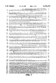
- FIG. 1 shows the base sequence of the clone MP660 containing the gene which codes for the PGE 2 receptor, and the amino acid sequence deduced therefrom.
- the Figure includes DNA sequence SEQ ID NO:1 and amino acid sequence SEQ ID NO:2.
- FIGS. 2(a) and 2(b) show the inhibitory activities of various ligands on the binding of [ 3 H]-PGE 2 to the PGE 2 receptor (EP 3 ) expressed on MP660-transfected COS-1 cell membranes.
- FIG. 3 shows the inhibitory activities of various ligands on the cAMP synthesis in CHO cells expressing the PGE 2 receptor.
- FIG. 4 shows the base sequence of the clone MP653 containing the gene which codes for the PGE 2 receptor, and the amino acid sequence deduced therefrom.
- the figure includes DNA sequence SEQ ID NO:3 and amino acid sequence SEQ ID NO:4.
- the boxed portion of FIG. 4 designated "peptide- ⁇ " is present only in MP660, and is included in FIG. 4 for comparison with MP653.
- FIG. 5 shows comparison of cDNA structures of two EP 3 receptor isoforms.
- FIG. 6 shows the base sequence of the clone MP412 containing the gene which codes for the PGE 2 receptor, and the amino acid sequence deduced therefrom.
- the figure includes DNA sequence SQ ID NO:5 and amino acid sequence SEQ ID NO:6.
- FIG. 7 and 8 show the inhibitory activities of various ligands on the binding of [ 3 H]-PGE 2 to the PGE 2 receptor (EP 2 ) expressed on MP412-transfected COS-1 cell membrances.
- FIG. 9 shows the effect of PGE 2 on cAMP level in MP412-transfected or untransfected COS-1 cells.
- PGEs for the present invention include PGE 1 and PGE 2 , with preference given to PGE 2 .
- the capability of receiving PGE 2 means that the receptor is capable of specifically binding to PGE 2 or a similar substance in the transmembrane domain, and that the structural change due to such ligand binding induces activation of related GTP-binding protein in the intracellular domain.
- the protein of the present invention which is capable of receiving PGE (hereinafter also referred to as PGE receptor), is preferably a protein which is capable of receiving PGE 2 (hereinafter also referred to as PGE 2 receptor), and may be of several types, including human, chicken and mouse, and may be a glycoprotein resulting from sugar chain binding to the sugar-binding site or a complex protein such as phosphoprotein resulting from phosphorylation at the phosphorylation site.
- PGE 2 receptors of the mouse type include a polypeptide having the amino acid sequence comprising a series of the amino acid sequence of FIG. 1 (SEQ ID NO:2), a polypeptide having the amino acid sequence comprising a series of the amino acid sequence of FIG. 4 (SEQ ID NO:4) and a polypeptide having the amino acid sequence comprising a series of the amino acid sequence of FIG. 5.
- Any PGE 2 receptor is acceptable, as long as it is capable of receiving PGE 2 and activating GTP-binding protein.
- the receptor-G protein coupling may be examined in several ways. One example is to examine modulation of the finding affinity of the receptor by guanine nucleotides (Ann. Rev. Biochem. 56, 615-649 (1987)). Specifically, it may be a mutein resulting from deletion of at least one constituent amino acid from said amino acid sequence, replacement of at least one constituent amino acid by another amino acid, or addition of at least one amino acid, and may be a functional fragment.
- the PGE receptor subtype may be of EP 1 , EP 2 or EP 3 ( ⁇ , ⁇ ), with preference given to the EP 2 receptor and the EP 3 receptor.
- the gene which codes for the PGE receptor may be any one, as long as it codes for the PGE receptor.
- the gene which codes for the EP 3 ⁇ receptor, a mouse PGE 2 receptor subtype, the gene which codes for the EP 3 ⁇ receptor, a mouse PGE 2 receptor subtype and the gene which codes for the EP 2 receptor, a mouse PGE 2 receptor subtype are exemplified by a gene having the base sequence comprising a series of the 1-1095 bases shown in FIG. 1 SEQ ID NO:1), a gene having the base sequence comprising a series of the 1-1083 bases shown in FIG. 4 (SEQ ID NO:3) and a gene having the base sequence comprising a series of the 1-1539 bases shown in FIG. 5, respectively.
- the vector according to the present invention which harbors a recombinant DNA containing a gene which codes for PGE receptor, can, for example, be produced by:
- Said cDNA can also be produced by chemical synthesis.
- RNA which codes for the PGE receptor can be obtained from various PGE-receptor-expressing cells known to those of ordinary skill in the art, such as mouse mastocytoma line P-815 cells and IL-3 dependent cell line BNu-c13 cells.
- RNA from PGE-receptor-expressing cells include the guanidine thiocyanate method [J. M. Chirgwin et al., Biochemistry, 18, 5294 (1979)]. Other suitable methods known to those skilled within this art are also within this invention.
- a cDNA is synthesized in accordance with, for example, the method of H. Okayarea et al. [Molecular and Cellular Biology, 2, 161 (1982) and 3, 280 (1983)], and the resulting cDNA is inserted to a plasmid.
- Other methods for recombinantly synthesizing cDNA known to those skilled in the art are also within the claimed invention.
- Examples of the plasmid for cDNA insertion include plasmids derived from Escherichia coli such as pBR322 [Gene, 2, 95 (1977)], pBR325 [Gene, 4, 121 (1978], pUC12 [Gene, 19, 259 (1982)], pUC13 [Gene, 19, 259 (1982)], pUC118 and pUC119 [Methods in Enzymology, 153, 3-11 (1987)] and those derived from Bacillus subtitis such as pUB110 [Biochemical and Biophysical Research Communications, 112, 678 (1983)], but any other can be used for this purpose, as long as it is replicable in the host.
- the plasmid incorporating said eDNA may be a plasmid obtained by using a cDNA library with Escherichia coli x1776 host prepared by inserting a cDNA synthesized from human normal diploid cell mRNA to the pCD vector [see Okayarea et al., Molecular Cell Biology, 3, 280 (1983)], which cDNA library is available from Dr. Okayama at the Research Institute for Microbial Diseases, Osaka University.
- the plasmid thus obtained is introduced to an appropriate host such as a bacterium of the genus Escherichia or Bacillus.
- Example bacteria of the genus Escherichia include Escherichia coli K12DH1 [Proceedings of the National Academy of Science, USA, 60, 160 (1968)], M103 [Nucleic Acids Research, 9, 309 (1981)], JA221 [Journal of Molecular Biology, 120, 517 (1978)], HB101 [Journal of Molecular Biology, 41, 459 (1969)] and C600 [Genetics, 39, 440 (1954)].
- Example bacteria of the genus Bacillus include Bacillus subtilis MI114 [Gene, 24, 255 (1983)] and 207-21 [Journal of Biochemistry, 95, 87 (1984)].
- Methods of transformation include the calcium chloride method and calcium chloride/rubidium chloride method described by T. Maniatis in Molecular Cloning, Cold Spring Harbor Laboratory, page 249 (1982).
- the desired clone is selected using a known method, such as colony hybridization [Gene, 10, 63 (1980)] or DNA base sequencing [Proceedings of the National Academy of Science, USA, 74, 560 (1977); Nucleic Acids Research, 9,309 (1981)].
- a microorganism carrying a vector having a cloned DNA containing a base sequence which codes for the PGE receptor is thus obtained.
- the plasmid is isolated from the microorganism.
- the above plasmid having a cloned recombinant DNA containing a gene which codes for the PGE receptor can be used as such or after being cleaved out with restriction enzyme as necessary.
- the cloned gene is ligated downstream of a promoter, in a vehicle (vector) suitable for its expression, to yield an expression vector.
- Example vectors include the above-mentioned plasmids derived from Escherichia coli (e.g., pBR322, pBR325, pUC12, pUC13, pUC118, pUC119), plasmids derived from Bacillus subtilis (e.g., pUB110, pTP5, pC194), yeast-derived plasmids (e.g., pSH19, pSH15), bacteriophages such as ⁇ phage, animal viruses such as retrovirus and vaccinia virus and plasmids for animal expression (e.g., pcDNAI, pdKCR-dhfr).
- Escherichia coli e.g., pBR322, pBR325, pUC12, pUC13, pUC118, pUC119
- Bacillus subtilis e.g., pUB110, pTP5, pC194
- the gene may have ATG (base sequence which codes for an appropriate signal peptide as desired) as a translational initiation codon at its 5'-terminal and TAA, TGA or TAG (preferably TGA) as a translational termination codon at its 3'-terminal.
- ATG base sequence which codes for an appropriate signal peptide as desired
- TAA base sequence which codes for an appropriate signal peptide as desired
- TGA translational termination codon at its 3'-terminal
- a promoter is ligated to the upstream thereof. Any promoter can be used for the present invention, as long as it is appropriate for the host used to express the gene.
- promoters examples include the T7 promoter, trp promoter, lac promoter, rec A promoter, ⁇ PL promoter or lpp promoter, when the transformation host is a bacterium of the genus Escherichia; the SPO1 promoter, SPO2 promoter or pen P promoter when the host is a bacterium of the genus Bacillus; and the PHO5 promoter, PGK promoter, GAP promoter or ADH promoter when the host is a yeast. Preference is given to the case in which a bacterium of the genus Escherichia is used as host in combination with the trp promoter or T7 promoter.
- preferable promoters include the SV40-derived promoter and retrovirus promoter, with preference given to the SV40-derived promoter.
- the thus-constructed vector, harboring a DNA is used to produce a transformant.
- Examples of the host include bacteria of the genus Escherichia, bacteria of the genus Bacillus, yeasts and animal cells.
- Examples of the bacteria of the genus Escherichia and of the genus Bacillus include the same as specified above.
- yeasts examples include Saccharomyces cerevisiae AH22R, NA 87-11 A and DKD-5D.
- Example animal cells include simian cells COS-7, Vero, Chinese hamster ovarian cells CHO, mouse L cells and human FL cells.
- the bacteria of the genus Escherichia can be transformed in accordance with the method described in the Proceedings of the National Academy of Science, USA, 69, 2110 (1972), Gene, 17, 107 (1982) and other publications known to those skilled in the art.
- Bacteria of the genus Bacillus can be transformed in accordance with the method described in Molecular and General Genetics, 168, 111 (1979) and other publications, for instance.
- Yeasts can be transformed in accordance with the method described in the Proceedings of the National Academy of Science, USA, 75, 1929 (1978), for instance.
- Animal cells can be transformed in accordance with the method described in Virology, 52, 456 (1973), for instance.
- a transformant resulting from transformation with a vector harboring the cDNA of PGE receptor is thus obtained.
- Example carbon sources include glucose, dextrin, soluble starch and sucrose.
- Example nitrogen sources include organic or inorganic substances such as ammonium salts, nitrates, corn steep liquor, peptone, casein, meat extract, soybean cake and potato extract.
- Example minerals include calcium chloride, sodium dihydrogen phosphate and magnesium chloride. Yeasts, vitamins, growth promoters and other additives may be added.
- the pH of the medium is preferably about 6 to 8.
- Examples of media preferably used to cultivate Escherichia bacteria include the M9 medium containing glucose and casamino acid [Miller, Journal of Experiments in Molecular Genetics, 431-433, Cold Spring Harbor Laboratory, New York (1972)]. To increase promoter efficiency as necessary, a chemical agent such as 3 ⁇ -indolyl acrylic acid may be added.
- cultivation is normally carried out at about 15° to 43° C. for about 3 to 24 hours, with aeration and/or stirring as necessary.
- cultivation is normally carried out at about 30° to 40° C. for about 6 to 24 hours, with aeration and/or stirring as necessary.
- Examples of media for cultivating a transformant whose host is a yeast include Burkholder's minimal medium [Bostian, K. L. et al., Proceedings of the National Academy of Science, USA, 77, 4505 (1980)]. It is preferable to adjust the medium to a pH of about 5 to 8. Cultivation is normally carried out at about 20° to 35° C. for 24 to 72 hours, with aeration and/or stirring as necessary.
- Examples of media for cultivating a transformant whose host is an animal cell include MEM medium [Science, 122, 501 (1952)], DMEM medium [Virology, 8, 396 (1959)], RPMI1640 medium [Journal of the American Medical Association, 199, 519 (1967)] and 199 medium [Proceedings of the Society for the Biological Medicine, 73, 1 (1950)]. These media may be supplemented with about 5 to 20% fetal bovine serum. The pH is preferably about 6 to 8. Cultivation is normally carried out at about 30° to 40° C. for 15 to 60 hours, with aeration and/or stirring as necessary.
- the cells are collected by a known method after cultivation and suspended in a buffer containing a protein denaturant, such as guanidine hydrochloride, to elute the desired PGE receptor extracellularly.
- a protein denaturant such as guanidine hydrochloride
- the cells are disrupted by ultrasonication, lysozyme treatment and/or freeze-thawing, after which they are centrifuged to separate the PGE receptor of the invention.
- the method using a combination of lysozyme and ultrasonication is preferred.
- known methods of separation and purification can be used in combination as appropriate.
- Such known methods of separation and purification include those based on solubility differences such as salting-out and solvent precipitation, those based mainly on molecular weight differences such as dialysis, ultrafiltration, gel filtration and SDS-polyacrylamide gel electrophoresis, those based on charge differences such as ion exchange chromatography, those based on specific affinity such as affinity chromatography, those based on hydrophobicity differences such as reverse-phase high performance liquid chromatography, and those based on isoelectric point differences such as isoelectric focusing.
- the thus-obtained PGE receptor of the present invention may be prepared as a dry powder by dialysis and lyophilization. It is appropriate to add serum albumin etc. as a carrier in storing the PGE receptor, since its adsorption to the container is prevented.
- the PGE receptor of the present invention substantially pure, is thus obtained.
- the substantially pure protein of the present invention has a protein content of not less than 95% (w/w), preferably not less than 98% (w/w).
- the PGE receptor thus obtained itself, or a transformant expressing it or a moiety thereof can be used to screen substances exhibiting antagonistic or agonistic activity thereon by, for example, a ligand-binding test.
- the PGE receptor as such, can also be used as a PGE-masking protein.
- the transformant obtained according to the present invention, which expressed the PGE receptor, and parts thereof can be efficiently used to obtain antibodies against said receptor.
- DNA Deoxyribonucleic acid
- RNA Ribonucleic acid
- mRNA Messenger ribonucleic acid
- Trp or W Tryptophan
- a single-stranded cDNA was synthesized from mouse lung total RNA by using random hexanucleotides as primers.
- PCR primers were designed based on the human TXA 2 receptor cDNA (HPL) sequences corresponding to the putative third and sixth transmenbrane domains of the receptor [M. Hirata et al., Nature 349, 617 (1991)]
- Mouse lung cDNA served as template in 30 cycles of PCR with 1 min of denaturation at 95° C., 0.5 min of annealing at 60° C., and 1.5 min of extension at 72° C. on a Zymoreactor (Atto Corp., Tokyo, Japan).
- a single 418-base pair cDNA fragment was amplified and subcloned into pBluescript SK(+) (Stratagene).
- a clone isolated (LT3) showed a sequence 78% homologous to the corresponding region of the human cDNA (HPL).
- Mouse lung cDNA prepared by an oligo (dT) priming method was size-selected (>1.8 kilobases) and inserted into the EcoRI site of ⁇ ZAPf[DNA (Stratagene) with EcoRI adaptors (New England Biolabs, Inc.).
- the 1.9 ⁇ 10 5 clones derived from the cDNA library were screened by hybridization with LT3 obtained in (1) above. Hybridization was carried out at 58° C.
- FIG. 1 shows the base sequence (SEQ ID NO:1) of the eDNA of MP660 and the amino acid sequence (SEQ ID NO:2) deduced therefrom.
- the overlined portions, the sites marked with ⁇ and the sites marked with ⁇ denote transmembrane domains I through VII, extracellular domain N-glycosylation sites and sites of phosphorylation by cAMP-dependent protein kinase, respectively.
- the cDNA of MP660 was cleaved out with EcoRI and inserted to pcDNAI (Invitrogen) and subcloned, followed by transfection of this plasmid DNA to COS-1 cells by the DEAE-dextran method [D. J. Sussmann and G. Milman, Mol. Cell. Biol., 4, 1641 (1984)]. After 72-hour cultivation, cells were harvested and cell membranes were separated [M. Hirata et al., Nature, 349, 617 (1991)]. Using these cell membranes, various [3H]-labeled prostaglandins were assayed for binding activity; [3H]-PGE 2 was found to show specific binding.
- FIG. 2 shows inhibitory activities of various ligands on the binding of [ 3 H]-PGE 2 to the MP660-transfected cell membrane
- panel "a” is for inhibitory activities of various prostaglandins ( ⁇ : PGE 2 ; ⁇ : PGE 1 ; ⁇ : iloprost; ⁇ : PGF 2 ⁇ ; ⁇ : PGD 2 );
- panel "b” is for inhibitory activities of prostaglandin-like substances ( ⁇ : M&B28,767; ⁇ : GR63799X; ⁇ : butaprost; ⁇ : SC-19220)]. Specificity of this binding is shown in FIG. 2a.
- cDNA transfection was conducted by the method described by Nakajima et al. [J. Biol. Chem., 267, 2437 (1992)] to establish a cell line. Specifically, the EcoRI fragment of MP660 was inserted to pdKCR-dhfr [S. Oikawa et al., Biochem. Biophys. Res. Commun., 164, 39 (1989)], a eukaryotic cell expression vector having the mouse dhfr gene as a selection marker. This plasmid was transfected to CHO-dhfr-(lacking dihydrofolate reductase activity) cells [G. Urlaub and L. A.
- the transfected CHO cells showed a dosedependent decrease to PGE 2 in forskolin-induced cellular cAMP accumulation.
- RNAs from various mouse tissues were isolated by the acid guanidinium thiocyanate-phenol-chloroform method [P. Chomczynski and N. Sacchi, Anal. Biochem., 162, 156 (1987)].
- poly(A)+ RNAs were purified using Oligotex dT 30 (Takara Shuzo, Kyoto, Japan).
- Poly (A) + RNAs (10 ⁇ g) from each tissue were separated by electrophoresis on a 1.2% agarose gel, transferred onto nylon membranes (Hybond-N, Amersham Corp.), and hybridized with a 32 P-labeled EcoRI/Bam HI fragment of MP660 clone.
- Hybridization was carried out at 68° C. in 6 ⁇ SSC, and filters were washed at 68° C. in 1 ⁇ SSC.
- a 2.3 kb strong band appeared from tissues on which PGE 2 is pharmacologically active, such as the kidney, stomach and uterus, and from P-815 cells.
- Another band appeared near 7.0 kb from these tissues and cells.
- FIG. 4 shows the base sequence (SEQ ID NO:3) of the cDNA of MP653 and the amino acid sequence (SEQ ID NO:4) deduced therefrom as compared with those of MP660.
- MP653 is identical to MP660 in the nucleotide sequence except deletion of an 89-bp sequence in the coding region of the putative C-terminal tail of the receptor in MP660-encoded receptor (FIG. 5). Deletion of this 89-bp sequence creates another reading frame downstream from this junction, which extends coding region until a new stop codon placed on 77-bp downstream from the stop codon of MP660. As a consequence, a 30-amino-acid (aa) C-terminal fragment of the MP660-encoded receptor (peptide- ⁇ ) was replaced with a new 26-an fragment (peptide- ⁇ ) in the C-terminal end of MP653-encoded receptor.
- aa 30-amino-acid
- the cDNA of MP653 was cleaved out with EcoRI and inserted to pcDNAI (Invitrogen) and subcloned, followed by transfection of this plasmid DNA to COS-1 cells by the DEAE-dextran method [D. J. Sussmann and G. Milman, Mol. Cell. Biol., 4, 1641 (1984)]. After 72-hour cultivation, cells were harvested and cell membranes were separated [M. Hirata et al., Nature, 349, 617 (1991)]. Using these cell membranes, various [ 3 H]-labeled prostaglandins were assayed for binding activity; [ 3 H]-PGE 2 was found to show specific binding.
- the PGE 2 receptor obtained was identified as the subtype EP 3 ⁇ receptor.
- the result of inhibitory activities of various ligands on the binding of [ 3 H]-PGE 2 to the MP653-transfected cell membrance was substantially the same as on the binding of [ 3 H]-PGE 2 to the MP660-transfected cell membrance obtained in Example 1.
- MP660-encoding receptor is designated as EP 3 ⁇ (containing the peptide- ⁇ ) and MP653-encoding one as EP 3 ⁇ (containing the peptide- ⁇ ).
- cDNA transfection was conducted by the method described by Nakajima et al. [J. Biol. Chem., 267, 2437 (1992)] to establish a cell line. Specifically, the EcoRI fragment of MP653 was inserted to pdKCR-dhfr [S. Oikawa et al., Biochem. Biophys. Res. Commun., 164, 39 (1989)], a eukaryotic cell expression vector having the mouse dhfr gene as a selection marker. This plasmid was transfected to CHO-dhfr-(lacking dihydrofolate reductase activity) cells [G. Urlaub and L. A.
- the cells which proliferated were cloned to have clone cells.
- EP 3 ⁇ receptor cDNA transfection was confirmed by the RNA blotting method.
- CHO cells which constantly express the EP 3 ⁇ receptor [MP653/KCR cells; IFO 50397, FERM BP-4183]
- the effect of PGE 2 on forskolin-stimulated cAMP synthesis or M&B28, 767, an EP 3 -specific agonist was assessed. It was thus found that cAMP synthesis in MP653/KCR cells is inhibited by the co-presence of these substances.
- the 5'-end 32 P-labeled PCR II (0.3 pmol; 1.0 ⁇ 10 6 c.p.m./pmol) was incubated in each PCR reaction (final 25 ⁇ l). Twenty-three cycles of PCR were performed using the following temperature profile: 94° C., 40 s; 60° C., 40 s; 72° C., 1.5 min. DNA-resolved gel was dried and subject to autoradiography, and the radioactivity of the gel corresponding to the bands was counted. Consequently, it was found that in any tissue expressing EP 3 , EP 3 ⁇ was dominantly expressed over EP 3 ⁇ .
- Mouse mastocytoma P-815 cell cDNA library carring cDNAs larger than 2.0 kb was prepared according to Example 1 (2).
- the probe DNA was prepared by PCR using mouse EPs cDNA as a template; this 482 bp fragment covers the transmembrane segments I-IV region of the EP 3 receptor.
- the 2.0 ⁇ 10 5 clones derived from the cDNA library were screened under either high (Sambrook) or low stringency condition.
- the resultant positive clones were subjected to PCR, restriction and sequence analyses and classified into two major groups; one group (six clones) belonged to EP 3 receptor eDNA, and the other (five clones) showed a sequence homologous but not identical to EP 3 cDNA.
- FIG. 6 shows the base sequence (SEQ ID NO:5) of the cDNA of MP412 and the amino acid sequence (SEQ ID NO:6) deduced therefrom.
- This cDNA was transfected into COS-1 cells according to the same manner as in Example 1(3).
- FIG. 8 shows ligand binding specificity using several ligands which show characteristic agonist or antagonist activity for PGE receptor subtypes.
- the PGE 2 binding was inhibited by misoprostol( ⁇ ), an EP 2 and EP 3 agonist, and more weakly by M&B 28,767( ⁇ ), an EP 3 agonist.
- sulprostone ( ⁇ ), an EP 1 and EP 3 agonist, SC-19220( ⁇ ), an EP 1 antagonist, and butaprost (A), an EP 2 agonist did not inhibit it.
- EP 2 receptor is coupled to stimulation of adenylate cyclase.
- the plasmid DNA was transfected into COS-1 cells by the lipofection method (P. L. Felgner et al., Proc. Natl. Acad. Sci., 84, 7413 (1987)) and cultured for 72 h in a 24-well plate. Cyclic AMP levels in the cells were determined according to the method of Nakajima et al. (J. Biol. Chem. 267, 2437 (1992)). As shown in FIG. 9, PGE 2 dose-dependently increased cAMP level in these cells ( ⁇ : MP412-transfected COS-1 cells; ⁇ : untransfected COS-1 cells).
- cDNA transfection was conducted by the method according to Example 1 (4) to establish a cell line. Specifically, the EcoRI fragment of MP412 was inserted to pdKCR-dhfr, a eukaryotic cell expression vector having the mouse dhfr gene as a selection marker. This plasmid was transfected to CHO-dhfr- (lacking dihydrofolate reductase activity) cells. The cells were subjected to selection culture, the cells which proliferated were cloned to have clone cells, and thus the CHO cells which constantly express the EP 2 receptor [CHO/EP 2 cells; IFO 50396, FERM BP-4182] were obtained.
- RNAs (10 ⁇ g) from each tissue which Were prepared by the same method as in Example 1 (5), were separated by electrophoresis on a 1.2% agarose gel, transferred onto nylon membranes (Hybond-N, Amersham Corp.), and hybridized with a 32 P-labeled EcoRI/BamHI fragment of MP412 clone. Hybridization was carried out at 68° C. in 6 ⁇ SSC, and filters were washed at 68° C. in 2 ⁇ SSC. A positive band is observed at 3.9 kilobase in most of tissues, suggesting widespread distribution of the EP 2 receptor.
- EP 2 mRNA The tissues highly expressing EP 2 mRNA were ileum and thymus in which PGE 2 induces relaxation of ileum circular muscle and inhibits proliferation of T cells by increasing intracellular cAMP levels. A significant band was also observed in lung, spleen, heart or uterus. On the other hand, EP 2 mRNA was not detectable in testis and liver.
Landscapes
- Health & Medical Sciences (AREA)
- Chemical & Material Sciences (AREA)
- Life Sciences & Earth Sciences (AREA)
- Organic Chemistry (AREA)
- Biophysics (AREA)
- Medicinal Chemistry (AREA)
- Zoology (AREA)
- Gastroenterology & Hepatology (AREA)
- Immunology (AREA)
- Biochemistry (AREA)
- Cell Biology (AREA)
- General Health & Medical Sciences (AREA)
- Genetics & Genomics (AREA)
- Toxicology (AREA)
- Molecular Biology (AREA)
- Proteomics, Peptides & Aminoacids (AREA)
- Endocrinology (AREA)
- Preparation Of Compounds By Using Micro-Organisms (AREA)
- Micro-Organisms Or Cultivation Processes Thereof (AREA)
- Pharmaceuticals Containing Other Organic And Inorganic Compounds (AREA)
- Saccharide Compounds (AREA)
- Peptides Or Proteins (AREA)
Priority Applications (2)
| Application Number | Priority Date | Filing Date | Title |
|---|---|---|---|
| US08/390,162 US5576192A (en) | 1992-02-24 | 1995-02-17 | Prostaglandin E receptors, their DNA and production |
| US08/685,945 US5804415A (en) | 1992-02-24 | 1996-07-22 | Prostaglandin E receptors, their DNA and production |
Applications Claiming Priority (6)
| Application Number | Priority Date | Filing Date | Title |
|---|---|---|---|
| JP4-036580 | 1992-02-24 | ||
| JP3658092 | 1992-02-24 | ||
| JP6488992 | 1992-03-23 | ||
| JP4-064889 | 1992-03-23 | ||
| US2417993A | 1993-02-23 | 1993-02-23 | |
| US08/390,162 US5576192A (en) | 1992-02-24 | 1995-02-17 | Prostaglandin E receptors, their DNA and production |
Related Parent Applications (1)
| Application Number | Title | Priority Date | Filing Date |
|---|---|---|---|
| US2417993A Continuation | 1992-02-24 | 1993-02-23 |
Related Child Applications (1)
| Application Number | Title | Priority Date | Filing Date |
|---|---|---|---|
| US08/685,945 Division US5804415A (en) | 1992-02-24 | 1996-07-22 | Prostaglandin E receptors, their DNA and production |
Publications (1)
| Publication Number | Publication Date |
|---|---|
| US5576192A true US5576192A (en) | 1996-11-19 |
Family
ID=26375656
Family Applications (2)
| Application Number | Title | Priority Date | Filing Date |
|---|---|---|---|
| US08/390,162 Expired - Fee Related US5576192A (en) | 1992-02-24 | 1995-02-17 | Prostaglandin E receptors, their DNA and production |
| US08/685,945 Expired - Fee Related US5804415A (en) | 1992-02-24 | 1996-07-22 | Prostaglandin E receptors, their DNA and production |
Family Applications After (1)
| Application Number | Title | Priority Date | Filing Date |
|---|---|---|---|
| US08/685,945 Expired - Fee Related US5804415A (en) | 1992-02-24 | 1996-07-22 | Prostaglandin E receptors, their DNA and production |
Country Status (6)
| Country | Link |
|---|---|
| US (2) | US5576192A (de) |
| EP (1) | EP0557966B1 (de) |
| JP (1) | JP3508865B2 (de) |
| AT (1) | ATE224913T1 (de) |
| CA (1) | CA2090278A1 (de) |
| DE (1) | DE69332321T2 (de) |
Families Citing this family (7)
| Publication number | Priority date | Publication date | Assignee | Title |
|---|---|---|---|---|
| WO1995000552A1 (en) * | 1993-06-25 | 1995-01-05 | Merck Frosst Canada Inc. | Prostaglandin receptor ep3 and dna encoding it |
| US5605814A (en) * | 1993-08-31 | 1997-02-25 | Merck Frosst Canada Inc. | DNA encoding human prostaglandin receptor EP2 |
| US6670134B1 (en) | 1993-11-19 | 2003-12-30 | Allergan, Inc. | Human EP3 prostaglandin receptor |
| US6057433A (en) | 1993-11-19 | 2000-05-02 | Allergan Sales, Inc. | Nucleic acid encoding human EP3 prostaglandin receptor |
| JP3692536B2 (ja) * | 1994-02-10 | 2005-09-07 | 小野薬品工業株式会社 | プロスタグランジンi2受容体 |
| CA2186231A1 (en) * | 1994-03-23 | 1995-09-28 | Atsushi Ichikawa | Prostaglandin e receptor |
| US20030157599A1 (en) * | 2002-01-30 | 2003-08-21 | Decode Genetics Ehf | Gene for peripheral arterial occlusive disease |
-
1993
- 1993-02-22 JP JP03225593A patent/JP3508865B2/ja not_active Expired - Fee Related
- 1993-02-24 AT AT93102873T patent/ATE224913T1/de not_active IP Right Cessation
- 1993-02-24 DE DE69332321T patent/DE69332321T2/de not_active Expired - Fee Related
- 1993-02-24 CA CA002090278A patent/CA2090278A1/en not_active Abandoned
- 1993-02-24 EP EP93102873A patent/EP0557966B1/de not_active Expired - Lifetime
-
1995
- 1995-02-17 US US08/390,162 patent/US5576192A/en not_active Expired - Fee Related
-
1996
- 1996-07-22 US US08/685,945 patent/US5804415A/en not_active Expired - Fee Related
Non-Patent Citations (22)
| Title |
|---|
| Coleman et al., Prostanoids and their Receptors, Comprehensive Medicinal Chemistry, vol. 3, p. 643, 1989. * |
| Doug et al., Br. J. Pharmac., vol. 87, 97 107, 1986. * |
| Doug et al., Br. J. Pharmac., vol. 87, 97-107, 1986. |
| Haluska et al., Annu. Rev. Pharm. Tox., vol. 10, 213 239, 1989. * |
| Haluska et al., Annu. Rev. Pharm. Tox., vol. 10, 213-239, 1989. |
| Hirata et al., Nature, vol. 349, p. 617, 1991. * |
| Jacobs et al., Nature, vol. 313, 806 810, 1985. * |
| Jacobs et al., Nature, vol. 313, 806-810, 1985. |
| Libert et al., Science, vol. 244, p. 569, 1989. * |
| Lopaschuk et al., Circulation Research, vol. 65(3), 538 545, 1989. * |
| Lopaschuk et al., Circulation Research, vol. 65(3), 538-545, 1989. |
| Masakazu Hirata et al., Cloning and expression of cDNA for a human thromboxane A 2 receptor, Nature, vol. 349, pp. 617 620 (Feb. 14, 1991). * |
| Masakazu Hirata et al., Cloning and expression of cDNA for a human thromboxane A2 receptor, Nature, vol. 349, pp. 617-620 (Feb. 14, 1991). |
| Masu et al., Nature, vol. 329, p. 836, 1987. * |
| Shigeo Suzuki et al., Prostaglandin E2 receptor and GTP binding . . . , Chem. Abstr., vol. 111, No. 21, Abstr. No. 192618n (Nov. 20, 1989). * |
| Sugimoto et al., Journal of Biol. Chem., vol. 268, 2712 2718. * |
| Sugimoto et al., Journal of Biol. Chem., vol. 268, 2712-2718. |
| Tsuyoshi Watanabe et al., Characterization of partially purified . . . , Chem. Abstr., vol. 115, No. 17, Abstr. No. 177670n (Oct. 28, 1991). * |
| Wolfgang Holter et al., Expression of GTP binding proteins . . . , Chem. Abstr., vol. 115, No. 1, Abstr. No. 6785z (Jul. 8, 1991). * |
| Wolfgang Holter et al., Expression of GTP-binding proteins . . . , Chem. Abstr., vol. 115, No. 1, Abstr. No. 6785z (Jul. 8, 1991). |
| Yukihiko Sugimoto et al., Cloning and Expression of a cDNA for Mouse Prostaglandin E Receptor EP 3 Subtype, The Journal of Biological Chemistry, vol. 267, No. 10, pp. 6463 6466 (Apr. 5, 1992). * |
| Yukihiko Sugimoto et al., Cloning and Expression of a cDNA for Mouse Prostaglandin E Receptor EP3 Subtype, The Journal of Biological Chemistry, vol. 267, No. 10, pp. 6463-6466 (Apr. 5, 1992). |
Also Published As
| Publication number | Publication date |
|---|---|
| JP3508865B2 (ja) | 2004-03-22 |
| DE69332321D1 (de) | 2002-10-31 |
| DE69332321T2 (de) | 2003-01-23 |
| EP0557966A1 (de) | 1993-09-01 |
| US5804415A (en) | 1998-09-08 |
| EP0557966B1 (de) | 2002-09-25 |
| CA2090278A1 (en) | 1993-08-25 |
| ATE224913T1 (de) | 2002-10-15 |
| JPH05320199A (ja) | 1993-12-03 |
Similar Documents
| Publication | Publication Date | Title |
|---|---|---|
| Baud et al. | EMR1, an unusual member in the family of hormone receptors with seven transmembrane segments | |
| Koller et al. | Conservation of the kinaselike regulatory domain is essential for activation of the natriuretic peptide receptor guanylyl cyclases | |
| CA2024629C (en) | Xenopus laevis bone morphogenic protein, dna encoding same and use thereof | |
| US5326860A (en) | Pituitary adenylate cyclase activating protein precursor | |
| WO1997044455A1 (en) | Hematopoietic cytokine receptor | |
| US5643749A (en) | Soluble interferon α-receptor, its preparation and use | |
| US5576192A (en) | Prostaglandin E receptors, their DNA and production | |
| US5595882A (en) | Angiotensin II type-1 receptor proteins and screening assays using such proteins or transformants expressing them | |
| JPH0678772A (ja) | ヒトインターロイキン−5受容体のα鎖又はその一部、特にshIL5RαをコードするDNA及びそれを含むベクター、このベクターで形質転換された宿主細胞、ならびにその製造及び利用方法 | |
| JP2807474B2 (ja) | Dnaおよびその用途 | |
| AU669889B2 (en) | Nucleic acid sequences coding for or complementary to nucleic acid sequences coding for interleukin 9 receptor | |
| US6218509B1 (en) | LH/CG receptor, DNA and use thereof | |
| US5716835A (en) | Nucleic acid encoding a novel human EP prostaglandin receptor | |
| AU753400C (en) | Orphan receptors | |
| JP3120856B2 (ja) | Dnaおよびその用途 | |
| EP0753528A1 (de) | Protaglandin i 2?-rezeptor | |
| EP0488196A2 (de) | HST-2, ein Mitglied der Heparin-bindenden Wachstumsfaktoren-Familie | |
| JPH06298799A (ja) | ヒトアンジオテンシンiiタイプ1レセプター、その製造法および用途 | |
| AU764484B2 (en) | Orphan cytokine receptor | |
| JP3169239B2 (ja) | 蛋白質、dnaおよびその用途 | |
| JPH06181778A (ja) | ヒトカルシニューリンAαアイソフォーム蛋白質をコードするDNAおよびその用途 | |
| US20030017530A1 (en) | Heptahelix receptor and its use as leukotriene B4 receptor | |
| JPH07258295A (ja) | プロスタグランジンd受容体、その製造方法、その受容体をコードするdna、そのdnaからなるベクターおよびそのベクターで形質転換された宿主細胞 |
Legal Events
| Date | Code | Title | Description |
|---|---|---|---|
| FEPP | Fee payment procedure |
Free format text: PAYOR NUMBER ASSIGNED (ORIGINAL EVENT CODE: ASPN); ENTITY STATUS OF PATENT OWNER: LARGE ENTITY |
|
| FPAY | Fee payment |
Year of fee payment: 4 |
|
| AS | Assignment |
Owner name: ATSUSHI ICHIKAWA, JAPAN Free format text: ASSIGNMENT OF ASSIGNORS INTEREST;ASSIGNOR:TAKEDA CHEMICAL INDUSTRIES, LTD.;REEL/FRAME:013922/0356 Effective date: 20020701 Owner name: SHUH NARUMIYA, JAPAN Free format text: ASSIGNMENT OF ASSIGNORS INTEREST;ASSIGNOR:TAKEDA CHEMICAL INDUSTRIES, LTD.;REEL/FRAME:013922/0356 Effective date: 20020701 |
|
| AS | Assignment |
Owner name: RESEARCH INSTITUTE FOR PRODUCTION DEVELOPMENT, JAP Free format text: ASSIGNMENT OF ASSIGNORS INTEREST;ASSIGNORS:ICHIKAWA, ATSUSHI;NARUMIYA, SHUH;REEL/FRAME:013922/0835 Effective date: 20020715 |
|
| FPAY | Fee payment |
Year of fee payment: 8 |
|
| AS | Assignment |
Owner name: KYOTO UNIVERSITY, JAPAN Free format text: ASSIGNMENT OF ASSIGNORS INTEREST;ASSIGNORS:ICHIKAWA, ATSUSHI;NARUMIYA, SHUH;RESEARCH INSTITUTE FOR PRODUCTION DEVELOPMENT;REEL/FRAME:016369/0977;SIGNING DATES FROM 20041227 TO 20050120 |
|
| REMI | Maintenance fee reminder mailed | ||
| LAPS | Lapse for failure to pay maintenance fees | ||
| STCH | Information on status: patent discontinuation |
Free format text: PATENT EXPIRED DUE TO NONPAYMENT OF MAINTENANCE FEES UNDER 37 CFR 1.362 |
|
| FP | Lapsed due to failure to pay maintenance fee |
Effective date: 20081119 |