US7407646B2 - Hydrophilic polymer biomaterial having a specific magnetic resonance imaging signal and process for the preparation of said biomaterial - Google Patents
Hydrophilic polymer biomaterial having a specific magnetic resonance imaging signal and process for the preparation of said biomaterial Download PDFInfo
- Publication number
- US7407646B2 US7407646B2 US10/278,256 US27825602A US7407646B2 US 7407646 B2 US7407646 B2 US 7407646B2 US 27825602 A US27825602 A US 27825602A US 7407646 B2 US7407646 B2 US 7407646B2
- Authority
- US
- United States
- Prior art keywords
- copolymers
- hydrophilic polymer
- biomaterial
- iron oxide
- polyvinyl
- Prior art date
- Legal status (The legal status is an assumption and is not a legal conclusion. Google has not performed a legal analysis and makes no representation as to the accuracy of the status listed.)
- Expired - Lifetime
Links
Images


Classifications
-
- C—CHEMISTRY; METALLURGY
- C08—ORGANIC MACROMOLECULAR COMPOUNDS; THEIR PREPARATION OR CHEMICAL WORKING-UP; COMPOSITIONS BASED THEREON
- C08K—Use of inorganic or non-macromolecular organic substances as compounding ingredients
- C08K9/00—Use of pretreated ingredients
- C08K9/08—Ingredients agglomerated by treatment with a binding agent
Definitions
- This invention pertains to the domain of biomaterials detectable in magnetic resonance imaging, more specifically, to a biomaterial charged with superparamagnetic iron oxide particles to have a signal different from that of the uncharged biomaterial and that of the biological environment in which it is placed for one or more types of MRI sequences.
- a biomaterial is a material tolerated by the human body which can be employed in the composition of a medical device.
- the contact between the biomaterial and the human body can be temporary, for the implementation of an image or images, or permanent, especially for the creation of an implant. This contact can relate to all or part of the biomaterial.
- WO 94/23782 which pertains to medical devices such as catheters comprising nonmetallic members having paramagnetic ionic particles incorporated in the interior.
- the paramagnetic ionic particles are coated with a coating agent, the role of which is to retain water or any proton-donor liquid around the ionic particles since the medical device does not contain them.
- the results obtained are of very low significance: only a slight, not significant augmentation of the spin echo signal (T 1 , T 2 ) and gradient echo signal is seen.
- Particles constituted of a polymer and iron oxide nanoparticles forming a superparamagnetic agent for the preparation of an injectable contrast agent to enable signalizing of e.g., hepatic tumors cf. Fahlvik 1993, JMRI 3: 187-194; Chambon 1993 Magn Resol Imaging, 11: 509-519 are also known.
- contrast agents do not constitute a biomaterial in the classic sense of the term nor do they enable creation by themselves of a biomaterial.
- This invention relates to a charged biomaterial including at least one first hydrophilic polymer, and superparamagnetic iron oxide particles complexed with a second hydrophilic polymer substantially identical to or different from the first hydrophilic polymer, wherein the superparamagnetic iron oxide particles complexed with the second hydrophilic polymer are distributed substantially homogeneously and substantially without aggregates in the first hydrophilic polymer.
- This invention also relates to a process for preparing a charged biomaterial including forming an aqueous solution of hydrophilic monomers and superparamagnetic iron oxide particles complexed with a hydrophilic polymer including monomers substantially identical to or different from the hydrophilic monomers, polymerizing the solution and forming a polymer hydrogel in which are distributed substantially homogeneously and substantially without aggregates the superparamagnetic iron oxide particles complexed with the hydrophilic polymer.
- FIG. 1 illustrates a comparative table of the signal reduction obtained with a biomaterial according to the invention
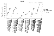
- FIG. 2 illustrates the results presented in FIG. 1 by means of a curve comprising as ordinate the percentage of signal reduction obtained
- FIG. 3 is a comparative table demonstrating that there is no major difference in signal intensity between a gel and a fragmented gel produced by the process according to the invention.
- FIG. 4 illustrates liquids in which are found the fragmented gel particles, the liquids not having signal modifications after four days, irrespective of their concentration of marker complex.
- This invention is remarkable in that superparamagnetic particles are complexed with a hydrophilic polymer substantially identical to or different from that of the biomaterial, and in that superparamagnetic iron oxide particles complexed with a hydrophilic polymer are distributed substantially homogeneously without aggregates in the hydrophilic polymer of the biomaterial.
- hydrophilic polymer is understood to mean a natural or synthetic polymer having a high affinity for water, being capable of taking up a large volume of water and becoming a hydrogel.
- a “water-soluble” polymer is a hydrophilic polymer capable of dissolving in an aqueous solution.
- complexing is understood to mean an interaction without chemical binding.
- the invention is also remarkable in that the hydrophilic polymer of the biomaterial comprises a quantity of complexed superparamagnetic iron oxide particles which is proportional to the desired signal.
- the invention is also remarkable in that the hydrophilic polymer of the biomaterial comprises on the order of about 10 ⁇ 1 % to about 10 ⁇ 6 % in dry weight of superparamagnetic iron oxide particles.
- biomaterial can constitute, among other things:
- the invention also pertains to a process for the preparation of a biomaterial according to the invention.
- the process is remarkable in that one mixes in an aqueous solution of hydrophilic monomers, superparamagnetic iron oxide particles complexed with a hydrophilic polymer constituted of monomers substantially identical with or different from the first-mentioned monomers, then one polymerizes this mixture to obtain a polymer hydrogel in which the superparamagnetic iron oxide particles complexed with a hydrophilic polymer are distributed homogeneously without aggregates.
- the hydrophilic polymer constituting the biomaterial can be a water-soluble polymer and in that the mixture is cross-linked to obtain a polymer hydrogel in which the superparamagnetic iron oxide particles complexed with a hydrophilic polymer are distributed substantially homogeneously without aggregates.
- the hydrogel obtained in this manner can then be presented in the form of a hydrogel with a high or low water content or in the form of a solid biomaterial obtained after drying or lyophilization and intended to be used as such or to be rehydrated.
- the biomaterial according to the invention thus has a significant signal extinction which is substantially homogeneous throughout the material in classic medical MRI sequences, in spin echo (T 1 , T 2 ) and in gradient echo sequences.
- the biomaterial according to the invention makes it possible to have the same signal variation direction in the principal sequences of medical MRI, in spin echo (T 1 , T 2 ) and in gradient echo.
- the process according to the invention thus advantageously makes it possible to differentiate the biomaterial from the surrounding soft tissues which have an intermediate signal (gray, when they are not necrotic) or a hypersignal when they are edematous.
- the process according to the invention also advantageously makes it possible to differentiate the biomaterial from the vascular contrast agents of the marked structures (e.g., gadolinium) because the very vascularized tissues, and more particularly, the hypervascular tumors, acquire a hypersignal after the injection.
- the vascular contrast agents of the marked structures e.g., gadolinium
- the biomaterial according to the invention also advantageously has an exactly reproducible signal in MRI as a function of the concentration of markers formed by the superparamagnetic iron oxide particles complexed with a hydrophilic polymer and as a function of the water content.
- the biomaterial according to the invention also advantageously has a stable marking when it is in contact with an aqueous medium, the marker being solidly fixed in the biomaterial which does not precipitate out.
- the biomaterial according to the invention is a biomaterial charged with superparamagnetic iron oxide particles to have a signal different from that of the uncharged biomaterial and that of the biological environment in which it is placed for one or more types of MRI sequences, the biomaterial being constituted of at least one hydrophilic polymer.
- the biomaterial according to the invention is characterized in that the superparamagnetic iron oxide particles are complexed with a hydrophilic polymer substantially identical to or different from that of the biomaterial, and in that the superparamagnetic iron oxide particles complexed with a hydrophilic polymer are distributed substantially homogeneously without aggregates in the hydrophilic polymer of the biomaterial.
- the polymer part of the biomaterial according to the invention preferably contains a quantity of superparamagnetic iron oxide particles which is proportional to the desired specific signal and especially to the desired extinction signal.
- the polymer part of the biomaterial preferably comprises on the order of about 10 ⁇ 1 % to about 10 ⁇ 6 % in dry weight of superparamagnetic iron oxide particles.
- the hydrophilic polymer of the biomaterial is preferably selected from among:
- the polymer complexing the superparamagnetic iron oxide particles is preferably selected from among:
- the biomaterial according to the invention can thus constitute:
- the biomaterial according to the invention can be in the form of a hydrogel.
- the biomaterial can be in the form:
- the invention also pertains to a process for preparation of a biomaterial charged with superparamagnetic iron oxide particles to have a signal different from that of the uncharged material and that of the biological environment in which it is placed for one or more types of MRI sequences, the biomaterial being constituted of at least one hydrophilic polymer.
- This process is characterized in that one mixes in an aqueous solution of hydrophilic monomers, superparamagnetic iron oxide particles complexed with a hydrophilic polymer constituted of a monomer which is substantially identical to or different from the first hydrophilic monomer, then one polymerizes the mixture to obtain a polymer hydrogel in which are distributed substantially homogenously without aggregates the superparamagnetic iron oxide particles complexed with a hydrophilic polymer.
- the superparamagnetic iron oxide particles are preferably stabilized prior to being complexed. They are then complexed by a hydrophilic polymer or possibly by a hydrogel.
- the polymer layer isolates each particle from the other particles and prevents their aggregation. This makes it possible to suspend the particles at different concentrations for their incorporation in a mixture of hydrophilic monomers before polymerization or a mixture of water-soluble polymers before cross-linking.
- Adjustment of the thickness of the complexation makes it possible to control the minimal spacing between the iron oxide crystals in the suspension before polymerization or cross-linking, for example.
- the hydrophilic polymer constituting the biomaterial can also be a water-soluble polymer.
- the ratio by weight of hydrophilic monomers or water-soluble polymers and superparamagnetic iron oxide particles complexed with a hydrophilic or water-soluble polymer is preferably between about 10 ⁇ 6 and about 10 ⁇ 1 %.
- the hydrophilic polymer constituting the biomaterial can be selected, for example, from among:
- the hydrophilic polymer complexing the superparamagnetic iron oxide particles is preferably more hydrophilic than the hydrophilic polymer constituting the biomaterial, this latter polymer being capable of being dispersed in a solution containing a nonaqueous solvent.
- solutions were prepared diluted in water (to 1/10 th , 1/100 th , 1/1000 th , 1/10,000 th , (vol/vol) of dextran-magnetite (commercial Endorem solution, Laboratoires Guerbet, Aulnay, France).
- the tubes of charged gel and multiple controls were examined in MRI (0.2 tesla) with three sequences: T 1 (SE 490/18 (90°), T 2 (SE 2000/80 (120°)), FE 3d (60/20 (20°)).
- the uncharged gel was spontaneously in hypersignal in MRI in relation to the water, in T 1 , in T 2 and in FE as illustrated in FIGS. 1 and 2 .
- T 1 the signal of the charged gel decreased inversely with the augmentation of the charge of complexed marker. It was very different from the signal of the oil (hypersignal) and the marker solution (hypersignal).
- the uncharged gel was different from the gels charged at more than 1/10,000 th . It was not very different from the least charged gel. The gel that was the most highly charged in marker and the water containing the same charge concentration were very different in sequences T 1 and T 2 and in FE.
- the gel enriched in marker loses its hypersignal in T 1 , in T 2 and in FE, and did so in a manner proportional to the concentration. A homogeneous distribution of the complexed marker in the gel was easily obtained.
- a hydrogel was prepared as in the example with a charge concentration identical to that of the most highly charged gel cited in Example 1.
- the gel was fragmented into particles which were suspended in physiological serum. Several days later, the signal of the particles in suspension and that of the supernatant were studied using the MRI technique of Example 1.
- a hydrogel was prepared as in example 2. It was fragmented in the mixer and dried by lyophilization. This produced a powder constituted of charged particles. It was also possible to dry the hydrogel block and then fragment it to obtain a similar result.
- Example 3 10 ml of a 10% (w/v) solution of PHEMA (Polysciences Europe, Eppelheim, Germany) in ethanol was prepared. 0.1 g of dry powder of charged gel obtained according to Example 3 was added. Multiple ml of this solution was injected via a catheter into the bloodstream of a test subject and gelled it by elimination of solvent and absorption of water. The resultant gel had a signal of the type of the charged gels of Example 1.
Landscapes
- Chemical & Material Sciences (AREA)
- Health & Medical Sciences (AREA)
- Chemical Kinetics & Catalysis (AREA)
- Medicinal Chemistry (AREA)
- Polymers & Plastics (AREA)
- Organic Chemistry (AREA)
- Medicines Containing Antibodies Or Antigens For Use As Internal Diagnostic Agents (AREA)
- Materials For Medical Uses (AREA)
- Immobilizing And Processing Of Enzymes And Microorganisms (AREA)
- Compounds Of Iron (AREA)
- Compositions Of Macromolecular Compounds (AREA)
Applications Claiming Priority (4)
| Application Number | Priority Date | Filing Date | Title |
|---|---|---|---|
| FRFR00/05400 | 2000-04-25 | ||
| FR0005400A FR2808026B1 (fr) | 2000-04-25 | 2000-04-25 | Biomateriau a base de polymere hydrophile presentant un signal specifique en imagerie par resonance magnetique et procede de preparation d'un tel biomateriau |
| FR0005400 | 2000-04-25 | ||
| PCT/FR2001/001267 WO2001081460A1 (fr) | 2000-04-25 | 2001-04-25 | Biomateriau a base de polymere hydrophile presentant un signal specifique en imagerie par resonance magnetique et procede de preparation d'un tel biomateriau |
Related Parent Applications (1)
| Application Number | Title | Priority Date | Filing Date |
|---|---|---|---|
| PCT/FR2001/001267 Continuation WO2001081460A1 (fr) | 2000-04-25 | 2001-04-25 | Biomateriau a base de polymere hydrophile presentant un signal specifique en imagerie par resonance magnetique et procede de preparation d'un tel biomateriau |
Publications (2)
| Publication Number | Publication Date |
|---|---|
| US20030077225A1 US20030077225A1 (en) | 2003-04-24 |
| US7407646B2 true US7407646B2 (en) | 2008-08-05 |
Family
ID=8849671
Family Applications (1)
| Application Number | Title | Priority Date | Filing Date |
|---|---|---|---|
| US10/278,256 Expired - Lifetime US7407646B2 (en) | 2000-04-25 | 2002-10-23 | Hydrophilic polymer biomaterial having a specific magnetic resonance imaging signal and process for the preparation of said biomaterial |
Country Status (8)
| Country | Link |
|---|---|
| US (1) | US7407646B2 (de) |
| EP (1) | EP1276808B1 (de) |
| AT (1) | ATE303416T1 (de) |
| AU (1) | AU2001256405A1 (de) |
| DE (1) | DE60113089T2 (de) |
| ES (1) | ES2248318T3 (de) |
| FR (1) | FR2808026B1 (de) |
| WO (1) | WO2001081460A1 (de) |
Cited By (10)
| Publication number | Priority date | Publication date | Assignee | Title |
|---|---|---|---|---|
| US20100215587A1 (en) * | 2009-02-23 | 2010-08-26 | Kent State University | Materials and methods for mri contrast agents and drug delivery |
| US20110082427A1 (en) * | 2009-10-06 | 2011-04-07 | Regents Of The University Of Minnesota | Bioresorbable embolization microspheres |
| US20130022548A1 (en) * | 2009-12-14 | 2013-01-24 | Bennett Kevin M | Methods and Compositions Relating to Reporter Gels for Use in MRI Techniques |
| US8795727B2 (en) | 2009-11-09 | 2014-08-05 | Spotlight Technology Partners Llc | Fragmented hydrogels |
| US8936795B2 (en) | 2012-12-19 | 2015-01-20 | Regents Of The University Of Minnesota | Liquid embolic material including carboxymethyl chitosan crosslinked with carboxymethyl cellulose |
| US9308169B2 (en) | 2000-03-24 | 2016-04-12 | Biosphere Medical, Inc. | Microspheres for active embolization |
| US9700650B2 (en) | 2009-11-09 | 2017-07-11 | Spotlight Technology Partners Llc | Polysaccharide based hydrogels |
| US10182979B2 (en) | 2016-03-22 | 2019-01-22 | Regents Of The University Of Minnesota | Biodegradable microspheres |
| US10293063B2 (en) | 2005-05-09 | 2019-05-21 | Merit Medical Systems, Inc. | Compositions and methods using microspheres and non-ionic contrast agents |
| US10695440B2 (en) | 2012-05-24 | 2020-06-30 | Biosphere Medical, Inc. | Biomaterials suitable for use as drug eluting, magnetic resonance imaging detectable implants for vascular occlusion |
Families Citing this family (33)
| Publication number | Priority date | Publication date | Assignee | Title |
|---|---|---|---|---|
| US6361759B1 (en) | 1998-05-26 | 2002-03-26 | Wisconsin Alumni Research Foundation | MR signal-emitting coatings |
| US6896874B2 (en) * | 1998-05-26 | 2005-05-24 | Wisconsin Alumni Research Foundation | MR-signal emitting coatings |
| KR100756801B1 (ko) * | 2000-09-11 | 2007-09-07 | 유니온 카바이드 케미칼즈 앤드 플라스틱스 테크날러지 코포레이션 | 자기 공명 영상법에 조영성을 갖는 친수성 윤활성 의료 장치 |
| US20030100830A1 (en) * | 2001-11-27 | 2003-05-29 | Sheng-Ping Zhong | Implantable or insertable medical devices visible under magnetic resonance imaging |
| US20030100829A1 (en) * | 2001-11-27 | 2003-05-29 | Sheng-Ping Zhong | Medical devices with magnetic resonance visibility enhancing material |
| US20090137018A1 (en) | 2003-06-30 | 2009-05-28 | University Of South Florida | Magnetic three-dimensional cell culture apparatus and method |
| FR2857268B1 (fr) * | 2003-07-08 | 2007-09-21 | Urodelia | Composite injectable pour magnetocytolyse de cellules metastatiques osseuses |
| WO2005010139A2 (en) | 2003-07-23 | 2005-02-03 | University Of South Florida | Ferromagnetic cell and tissue culture microcarriers |
| US7257151B2 (en) * | 2004-05-05 | 2007-08-14 | Texas Instruments Incorporated | (Updated) preamble for FDMA |
| US7896879B2 (en) * | 2004-07-29 | 2011-03-01 | Vertos Medical, Inc. | Spinal ligament modification |
| AU2006339072A1 (en) | 2005-03-14 | 2007-10-25 | Board Of Regents Of The University Of Texas System | Bioactive FUS1 peptides and nanoparticle-polypeptide complexes |
| US8696671B2 (en) | 2005-07-29 | 2014-04-15 | Vertos Medical Inc. | Percutaneous tissue excision devices |
| US20070123890A1 (en) * | 2005-11-04 | 2007-05-31 | X-Sten, Corp. | Tissue retrieval devices and methods |
| US8457712B2 (en) | 2005-12-30 | 2013-06-04 | Wisconsin Alumni Research Foundation | Multi-mode medical device system and methods of manufacturing and using same |
| US20070156042A1 (en) * | 2005-12-30 | 2007-07-05 | Orhan Unal | Medical device system and method for tracking and visualizing a medical device system under MR guidance |
| US7942830B2 (en) * | 2006-05-09 | 2011-05-17 | Vertos Medical, Inc. | Ipsilateral approach to minimally invasive ligament decompression procedure |
| JP5200425B2 (ja) * | 2006-06-23 | 2013-06-05 | 住友化学株式会社 | 農薬活性微生物製剤 |
| USD606654S1 (en) | 2006-07-31 | 2009-12-22 | Vertos Medical, Inc. | Tissue excision device |
| USD620593S1 (en) | 2006-07-31 | 2010-07-27 | Vertos Medical, Inc. | Tissue excision device |
| US8532742B2 (en) | 2006-11-15 | 2013-09-10 | Wisconsin Alumni Research Foundation | System and method for simultaneous 3DPR device tracking and imaging under MR-guidance for therapeutic endovascular interventions |
| US20080183070A1 (en) * | 2007-01-29 | 2008-07-31 | Wisconsin Alumni Research Foundation | Multi-mode medical device system with thermal ablation capability and methods of using same |
| EP2114268A4 (de) * | 2007-02-12 | 2010-03-03 | Vertos Medical Inc | Gewebeexzisionsvorrichtungen und -verfahren |
| US8412306B2 (en) | 2007-02-28 | 2013-04-02 | Wisconsin Alumni Research Foundation | Voltage standing wave suppression for MR-guided therapeutic interventions |
| US20090118709A1 (en) * | 2007-11-06 | 2009-05-07 | Vertos Medical, Inc. A Delaware Corporation | Tissue Excision Tool, Kits and Methods of Using the Same |
| USD598549S1 (en) | 2008-09-16 | 2009-08-18 | Vertos Medical, Inc. | Surgical trocar |
| USD619253S1 (en) | 2008-10-23 | 2010-07-06 | Vertos Medical, Inc. | Tissue modification device |
| USD610259S1 (en) | 2008-10-23 | 2010-02-16 | Vertos Medical, Inc. | Tissue modification device |
| USD621939S1 (en) | 2008-10-23 | 2010-08-17 | Vertos Medical, Inc. | Tissue modification device |
| USD619252S1 (en) | 2008-10-23 | 2010-07-06 | Vertos Medical, Inc. | Tissue modification device |
| USD611146S1 (en) | 2008-10-23 | 2010-03-02 | Vertos Medical, Inc. | Tissue modification device |
| DE102008060708A1 (de) * | 2008-12-05 | 2010-06-17 | Dianogen Gmbh | Beschichtung von Kunststoffsubstraten für den medizinischen Einsatz zur Verbesserung der bildgebenden Eigenschaften |
| US10617441B2 (en) | 2016-09-07 | 2020-04-14 | Vertos Medical, Inc. | Percutaneous lateral recess resection methods and instruments |
| EP4539756A1 (de) | 2022-06-16 | 2025-04-23 | Vertos Medical, Inc. | Integrierte instrumentenanordnung |
Citations (8)
| Publication number | Priority date | Publication date | Assignee | Title |
|---|---|---|---|---|
| US4587284A (en) * | 1985-06-14 | 1986-05-06 | Ed. Geistlich Sohne Ag Fur Chemische Industrie | Absorbent polymer material and its preparation |
| WO1994023782A1 (en) * | 1993-04-14 | 1994-10-27 | Pharmacyclics, Inc. | Medical devices and materials having enhanced magnetic images visibility |
| WO1995005669A1 (en) | 1993-08-12 | 1995-02-23 | Advanced Magnetics, Inc. | Synthesis of polysaccharide covered superparamagnetic oxide colloids |
| US5492814A (en) | 1990-07-06 | 1996-02-20 | The General Hospital Corporation | Monocrystalline iron oxide particles for studying biological tissues |
| US5684051A (en) * | 1996-04-24 | 1997-11-04 | Hercules Incorporated | Medical devices with improved elastic response |
| WO1999019000A1 (en) | 1997-10-11 | 1999-04-22 | The Research Foundation Of State University Of New York | Controlled size polymeric microspheres with superparamagnetic cores |
| US6368586B1 (en) * | 1996-01-26 | 2002-04-09 | Brown University Research Foundation | Methods and compositions for enhancing the bioadhesive properties of polymers |
| US6685963B1 (en) * | 1998-07-01 | 2004-02-03 | Corneal Industrie | Diphasic injection composition containing dispersed and continuous phases useful for reparative and plastic surgery |
-
2000
- 2000-04-25 FR FR0005400A patent/FR2808026B1/fr not_active Expired - Lifetime
-
2001
- 2001-04-25 ES ES01929702T patent/ES2248318T3/es not_active Expired - Lifetime
- 2001-04-25 EP EP01929702A patent/EP1276808B1/de not_active Expired - Lifetime
- 2001-04-25 DE DE60113089T patent/DE60113089T2/de not_active Expired - Lifetime
- 2001-04-25 WO PCT/FR2001/001267 patent/WO2001081460A1/fr not_active Ceased
- 2001-04-25 AT AT01929702T patent/ATE303416T1/de active
- 2001-04-25 AU AU2001256405A patent/AU2001256405A1/en not_active Abandoned
-
2002
- 2002-10-23 US US10/278,256 patent/US7407646B2/en not_active Expired - Lifetime
Patent Citations (8)
| Publication number | Priority date | Publication date | Assignee | Title |
|---|---|---|---|---|
| US4587284A (en) * | 1985-06-14 | 1986-05-06 | Ed. Geistlich Sohne Ag Fur Chemische Industrie | Absorbent polymer material and its preparation |
| US5492814A (en) | 1990-07-06 | 1996-02-20 | The General Hospital Corporation | Monocrystalline iron oxide particles for studying biological tissues |
| WO1994023782A1 (en) * | 1993-04-14 | 1994-10-27 | Pharmacyclics, Inc. | Medical devices and materials having enhanced magnetic images visibility |
| WO1995005669A1 (en) | 1993-08-12 | 1995-02-23 | Advanced Magnetics, Inc. | Synthesis of polysaccharide covered superparamagnetic oxide colloids |
| US6368586B1 (en) * | 1996-01-26 | 2002-04-09 | Brown University Research Foundation | Methods and compositions for enhancing the bioadhesive properties of polymers |
| US5684051A (en) * | 1996-04-24 | 1997-11-04 | Hercules Incorporated | Medical devices with improved elastic response |
| WO1999019000A1 (en) | 1997-10-11 | 1999-04-22 | The Research Foundation Of State University Of New York | Controlled size polymeric microspheres with superparamagnetic cores |
| US6685963B1 (en) * | 1998-07-01 | 2004-02-03 | Corneal Industrie | Diphasic injection composition containing dispersed and continuous phases useful for reparative and plastic surgery |
Cited By (20)
| Publication number | Priority date | Publication date | Assignee | Title |
|---|---|---|---|---|
| US10265271B2 (en) | 2000-03-24 | 2019-04-23 | Biosphere Medical, Inc. | Microspheres for the treatment of a prostate hyperplasia by active embolization |
| US9308169B2 (en) | 2000-03-24 | 2016-04-12 | Biosphere Medical, Inc. | Microspheres for active embolization |
| US10293063B2 (en) | 2005-05-09 | 2019-05-21 | Merit Medical Systems, Inc. | Compositions and methods using microspheres and non-ionic contrast agents |
| US20100215587A1 (en) * | 2009-02-23 | 2010-08-26 | Kent State University | Materials and methods for mri contrast agents and drug delivery |
| US8580230B2 (en) | 2009-02-23 | 2013-11-12 | Kent State University | Materials and methods for MRI contrast agents and drug delivery |
| US20110082427A1 (en) * | 2009-10-06 | 2011-04-07 | Regents Of The University Of Minnesota | Bioresorbable embolization microspheres |
| US11439725B2 (en) | 2009-10-06 | 2022-09-13 | Regents Of The University Of Minnesota | Bioresorbable embolization microspheres |
| US8617132B2 (en) | 2009-10-06 | 2013-12-31 | Regents Of The University Of Minnesota | Bioresorbable embolization microspheres |
| US10179187B2 (en) | 2009-10-06 | 2019-01-15 | Regents Of The University Of Minnesota | Bioresorbable embolization microspheres |
| US9700650B2 (en) | 2009-11-09 | 2017-07-11 | Spotlight Technology Partners Llc | Polysaccharide based hydrogels |
| US9592299B2 (en) | 2009-11-09 | 2017-03-14 | Spotlight Technology Partners Llc | Hydrogel compositions |
| US9861701B2 (en) | 2009-11-09 | 2018-01-09 | Spotlight Technology Partners Llc | Hydrogel compositions |
| US10159742B2 (en) | 2009-11-09 | 2018-12-25 | Spotlight Technology Partners Llc | Hydrogel compositions |
| US9289449B2 (en) | 2009-11-09 | 2016-03-22 | Spotlight Technology Partners Llc | Hydrogel compositions |
| US8795727B2 (en) | 2009-11-09 | 2014-08-05 | Spotlight Technology Partners Llc | Fragmented hydrogels |
| US9823325B2 (en) * | 2009-12-14 | 2017-11-21 | Kevin M. Bennett | Methods and compositions relating to reporter gels for use in MRI techniques |
| US20130022548A1 (en) * | 2009-12-14 | 2013-01-24 | Bennett Kevin M | Methods and Compositions Relating to Reporter Gels for Use in MRI Techniques |
| US10695440B2 (en) | 2012-05-24 | 2020-06-30 | Biosphere Medical, Inc. | Biomaterials suitable for use as drug eluting, magnetic resonance imaging detectable implants for vascular occlusion |
| US8936795B2 (en) | 2012-12-19 | 2015-01-20 | Regents Of The University Of Minnesota | Liquid embolic material including carboxymethyl chitosan crosslinked with carboxymethyl cellulose |
| US10182979B2 (en) | 2016-03-22 | 2019-01-22 | Regents Of The University Of Minnesota | Biodegradable microspheres |
Also Published As
| Publication number | Publication date |
|---|---|
| EP1276808A1 (de) | 2003-01-22 |
| FR2808026A1 (fr) | 2001-10-26 |
| EP1276808B1 (de) | 2005-08-31 |
| ES2248318T3 (es) | 2006-03-16 |
| WO2001081460A1 (fr) | 2001-11-01 |
| FR2808026B1 (fr) | 2002-06-14 |
| DE60113089D1 (de) | 2005-10-06 |
| AU2001256405A1 (en) | 2001-11-07 |
| ATE303416T1 (de) | 2005-09-15 |
| DE60113089T2 (de) | 2006-06-14 |
| US20030077225A1 (en) | 2003-04-24 |
Similar Documents
| Publication | Publication Date | Title |
|---|---|---|
| US7407646B2 (en) | Hydrophilic polymer biomaterial having a specific magnetic resonance imaging signal and process for the preparation of said biomaterial | |
| Frachini et al. | Magneto-responsive hydrogels: preparation, characterization, biotechnological and environmental applications | |
| Zhao et al. | Preparation of biodegradable magnetic microspheres with poly (lactic acid)-coated magnetite | |
| CA1301063C (en) | Biologically degradable superparamagnetic materials for use in clinicalapplications | |
| Samal et al. | Biomimetic magnetic silk scaffolds | |
| Arias et al. | Tegafur loading and release properties of magnetite/poly (alkylcyanoacrylate)(core/shell) nanoparticles | |
| US20100272649A1 (en) | Bioactive Bone Cement and Method for Its Production | |
| JP2018510011A (ja) | ポリマーインプラントに結合された強磁性粒子 | |
| Turrina et al. | Application of magnetic iron oxide nanoparticles: Thrombotic activity, imaging and cytocompatibility of silica-coated and carboxymethyl dextrane-coated particles | |
| Hagit et al. | Synthesis and characterization of dual modality (CT/MRI) core− shell microparticles for embolization purposes | |
| Saralidze et al. | New acrylic microspheres for arterial embolization: combining radiopacity for precise localization with immobilized thrombin to trigger local blood coagulation | |
| US20050131458A1 (en) | Biodegradable embolic agents | |
| CN104069516B (zh) | 一种超顺磁性纳米颗粒及其制备方法和用途 | |
| CN115845148A (zh) | 一种温敏可注射壳聚糖/丝素蛋白/水滑石复合水凝胶及其制备方法和应用 | |
| Gu et al. | Polymer/iron oxide nanocomposites as magnetic resonance imaging contrast agents: Polymer modulation and probe property control | |
| CN117982675A (zh) | 一种双靶向性金属离子网络水凝胶及其制备方法与应用 | |
| Almeida et al. | 3D (bio) printing of magnetic hydrogels: formulation and applications in tissue engineering | |
| WO2019093923A1 (en) | Method of fabrication of preparation on the base of iron oxide magnetic nanoparticles for neoplasms diagnostics by magnetic resonance imaging | |
| Lopez-Lopez et al. | Synthesis, characterization and in vivo evaluation of biocompatible ferrogels | |
| Kolouchova et al. | Cell-interactive gelatin-based 19F MRI tracers: an in vitro proof-of-concept study | |
| CN114555659B (zh) | 不可降解的不透射线栓塞微球 | |
| Gautam et al. | Nanoferrites as drug carriers in targeted drug delivery applications | |
| Namur et al. | MR imaging detection of superparamagnetic iron oxide–loaded tris-acryl embolization microspheres | |
| Liao et al. | Enhancing MRI through high loading of superparamagnetic nanogels with high sensitivity to the tumor environment | |
| CN114588281A (zh) | 一种水溶性非晶氧化铁纳米簇的制备方法 |
Legal Events
| Date | Code | Title | Description |
|---|---|---|---|
| AS | Assignment |
Owner name: X-BIO, A CORPORATION OF FRANCE, FRANCE Free format text: ASSIGNMENT OF ASSIGNORS INTEREST;ASSIGNORS:LAURENT, ALEXANDRE;LABARRE, DENIS;REEL/FRAME:013605/0113 Effective date: 20021202 |
|
| AS | Assignment |
Owner name: L'ASSISTANCE PUBLIQUE-HOPITAUX DE PARIS ETABLISSEM Free format text: ASSIGNMENT OF ASSIGNORS INTEREST;ASSIGNOR:X. BIO;REEL/FRAME:015669/0316 Effective date: 20030418 |
|
| STCF | Information on status: patent grant |
Free format text: PATENTED CASE |
|
| FPAY | Fee payment |
Year of fee payment: 4 |
|
| FPAY | Fee payment |
Year of fee payment: 8 |
|
| MAFP | Maintenance fee payment |
Free format text: PAYMENT OF MAINTENANCE FEE, 12TH YR, SMALL ENTITY (ORIGINAL EVENT CODE: M2553); ENTITY STATUS OF PATENT OWNER: SMALL ENTITY Year of fee payment: 12 |