US7822254B2 - Automatic positioning of matching multi-planar image reformatting (MPR) views of multiple 3D medical images - Google Patents
Automatic positioning of matching multi-planar image reformatting (MPR) views of multiple 3D medical images Download PDFInfo
- Publication number
- US7822254B2 US7822254B2 US11/695,096 US69509607A US7822254B2 US 7822254 B2 US7822254 B2 US 7822254B2 US 69509607 A US69509607 A US 69509607A US 7822254 B2 US7822254 B2 US 7822254B2
- Authority
- US
- United States
- Prior art keywords
- mpr
- volume
- digitized
- digitized volume
- mprs
- Prior art date
- Legal status (The legal status is an assumption and is not a legal conclusion. Google has not performed a legal analysis and makes no representation as to the accuracy of the status listed.)
- Active, expires
Links
Images


Classifications
-
- G—PHYSICS
- G06—COMPUTING OR CALCULATING; COUNTING
- G06T—IMAGE DATA PROCESSING OR GENERATION, IN GENERAL
- G06T7/00—Image analysis
- G06T7/30—Determination of transform parameters for the alignment of images, i.e. image registration
-
- G—PHYSICS
- G06—COMPUTING OR CALCULATING; COUNTING
- G06T—IMAGE DATA PROCESSING OR GENERATION, IN GENERAL
- G06T2207/00—Indexing scheme for image analysis or image enhancement
- G06T2207/30—Subject of image; Context of image processing
- G06T2207/30004—Biomedical image processing
Definitions
- This invention relates to a system and method for matching three dimensional volumes for monitoring anatomical changes in a patient over time, and more particularly to a system and method for automatically matching reference volumes of multiple medical images taken over time.
- MPR multi-planar reformatting
- MRI magnetic resonance imaging
- CT computed tomography
- PET positron emission tomography
- volumes three-dimensional arrays of data
- medical staff here denoted as user
- MPRs three-dimensional arrays of data
- This is an effective way to view and compare the 3D volumes for anatomical changes over time.
- a developing tumor may be detected, measured, and monitored over time, as can its effect on the surrounding region.
- stenosis an abnormal narrowing in a blood vessel—may require MPR comparison of two or more CT scans to diagnose.
- Positioning an MPR from a subsequent scan to match a reference MPR in the reference volume (or “baseline” volume) is a demanding task when done manually, due to the multiple orientation and shift adjustments required to obtain a particular MPR position within the volume under consideration.
- MPR 3D digital medical images
- the invention enables quick and efficient positioning of multiple MPRs of substantially the same content from one or more 3D digital medical images (here denoted as volumes).
- MPR is a technique widely used to view volumes, providing the facility for an arbitrarily positioned and oriented 2D plane to be placed in a 3D volume so that the projection of the data on that plane may be viewed.
- the MPR may be adjusted by changing its position and orientation in three dimensions.
- Those MPRs may be reconstructed from any volume such as but not limited to CT scan, MRI scan, DynaCT and the like.
- a method for automatically matching medical digital images.
- the method may comprise: providing a first digitized volume; providing a second digitized volume; obtaining a first MPR from the first digitized volume; obtaining a second MPR from the second digitized volume; and positioning the second MPR adjacent to the first image to compare the first and second MPRs.
- the method may further comprise computing a comparison score that represents a correspondence between at least a portion of each of the first and second MPRs; determining whether the comparison score is within a predetermined range; and identifying the second MPR as a matching MPR if the comparison score is within the predetermined range; or obtaining a third MPR from the second digitized volume if the comparison score is not within the predetermined range.
- a system for automatically matching medical digital images comprising means for providing a first digitized volume, means for providing a second digitized volume, means for obtaining a first MPR from the first digitized volume, means for obtaining a second MPR from the second digitized volume, and means for positioning the second MPR adjacent to the first MPR to compare the first and second MPRs.
- the system may further comprise means for computing a comparison score that represents a correspondence between at least a portion of each of the first and second MPRs; means for determining whether the comparison score is within a predetermined range; and means for identifying the second MPR as a matching MPR if the comparison score is within the predetermined range; or means for obtaining a third MPR from the second digitized volume if the comparison score is not within the predetermined range.
- a machine readable storage device tangibly embodying a series of instructions executable by the machine to perform a series of steps.
- the steps may comprise: providing a first digitized volume; providing a second digitized volume; obtaining a first MPR from the first digitized volume; obtaining a second MPR from the second digitized volume; and positioning the second MPR adjacent to the first MPR to compare the first and second MPRs.
- the steps may further comprise computing a comparison score that represents a correspondence between at least a portion of each of the first and second MPRs; determining whether the comparison score is within a predetermined range; and identifying the second MPR as a matching image slice if the comparison score is within the predetermined range; or obtaining a third MPR from the second digitized volume if the comparison score is not within the predetermined range.
- FIGS. 1A-1D are MPR's of four separate CT volumes constructed at different points in time; the MPRs have been “matched” to show the progression of stenosis of a blood vessel over time;
- FIG. 2 is a flow chart showing the workflow of one embodiment of the inventive system
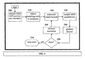
- FIG. 3 is a flow chart illustrating an exemplary algorithm for implementing the inventive system.
- FIGS. 1A-D an exemplary plurality of successive medical images (MPRs) are shown for use in diagnosing arterial stenosis.
- FIG. 1A illustrates the pulmonary vein and it's width (diameter) that is being observed for changes due to stenosis.
- the development of stenosis can be easily identified by the user when comparing these successive MPRs.
- the process starts at 10 , in which several volumes to be compared are selected. Typically these volumes will be built from scans (e.g., CT, MRI, PET) of a portion of a patient's anatomy which have been taken at different points in time (e.g., over the span of several months). The individual volumes will be stored as data files on the user's computer, server or other appropriate storage device.
- the user selects a “reference” volume, i.e., the volume from which a baseline or “reference” MPR is constructed.
- the reference MPR may be taken as the origin, or starting point, from which subsequent MPRs may be judged in order to diagnose a developing condition such as progressive stenosis or tumor.
- the “reference” volume represents a scan taken at an earlier point in time than the scans used to construct the subsequent volumes.
- the reference volume must be loaded to memory for viewing, at 30 .
- the user may then manually position (i.e., rotate, shift, etc.) the reference MPR view at 40 to center the “region of interest” on the screen.
- the region of interest is a blood vessel adjacent to the heart in which a stenosis condition is suspected.
- the user may optionally supply additional marking (e.g., mouse clicks, scribble) or other information (e.g., coordinates) to further identify the region of interest.
- additional marking e.g., mouse clicks, scribble
- other information e.g., coordinates
- the user would then be required to load each subsequent comparison volume into memory, if they are not already stored in memory.
- the user would then manually adjust the subsequent MPR slice taken from a subsequent “comparison” volume by iteratively comparing that subsequent “comparison” MPR with the reference MPR.
- This iterative process may be painstaking and is very time consuming, since the user must adjust the comparison MPR about 3 mutually-orthogonal axes of rotation within the comparison volume to obtain the most accurate positioning with respect to the reference MPR.
- the “matching” MPR is then stored for later use, or kept in memory for immediate comparison.
- the next comparison volume may be processed in the same manner until a plurality of comparison MPR's (typically one from each comparison volume) are obtained for viewing ( FIGS. 1B-1D )
- the operations associated with identifying the appropriate comparison MPRs are enhanced so that more accurate MPRs can be obtained from each volume used in the comparison.
- the system loads a comparison volume into memory and an initial “guess” is made as to a possible matching MPR position within the comparison volume. This MPR guess can be informed by a number of factors, including user input of a “region of interest,” or other information about MPR location in the reference volume.
- the “guess” MPR is then compared to the reference MPR and a comparison score is obtained at 70 . If the comparison score of the “guess” MPR is outside of a predetermined range, then the “guess” MPR is adjusted at 80 .
- This adjustment step may comprise slightly altering the position of the MPR slice within the comparison volume, and this adjusted guess MPR is compared 70 with the reference MPR to obtain a new comparison score. This iteration is performed until the comparison score falls within the predetermined range, whereupon the MPR is deemed to be “matching” and the matching MPR image and/or locational information is stored at 90 .
- This process is repeated for as may comparison volumes as are selected by the user. Thus, at 100 , a determination is made at 100 as to whether additional comparison volumes exist, and if so, the next comparison volume is loaded into more 60 and the process repeats to obtain another “matching” MPR from the new comparison volume. The process continues until there are no more comparison volumes 110 .
- One substantial advantage to the inventive system is that the loading of the comparison volumes to memory can be done more efficiently, since only a portion of the volume needs to be loaded.
- the user is required to load the entire volume so that it can be manually scanned for an MPR that matches the reference MPR. Due to the substantial amount of data associated with a typical volume, a large amount of memory is required and overall processing speeds can be slow.
- each of the comparison volumes needs to be loaded to memory. This is because once the reference MPR is identified, the general location of the comparison MPRs within their respective volumes can be estimated (within a certain range), so that only the portion of each comparison volume that lies within that range (which can be thought of as a “thick slice” of the comparison volume) need be loaded to memory. This technique reduces the total amount of memory required to perform the desired comparisons, and facilitates faster processing, resulting in an overall more efficient and faster operation.
- the user can then view the matching MPRs side by side, and against the reference MPR. It is contemplated that the user may wish to slightly adjust one or more of the matching MPRs as a “fine tuning” operation. This fine tuning could be performed after each matching MPR is identified, or it could be performed at the end, once all of the matching MPRs are identified.
- the algorithm is flexible, and may identify matching MPRs using only the volume information (i.e., the list of volumes) provided by the user. It will be appreciated that this may not be the most efficient approach from a pure processing standpoint.
- the user may provide the system with information aimed to guide the algorithm to a more quick identification of a matching MPR for a given comparison volume. As previously noted, this could include providing the system with the position coordinates of the reference MPR within the reference volume. By and large, the patient's anatomy and position on the scanning bed or table will not change significantly between scans, and thus this may provide a good initial guess.
- the user in addition to the list of volumes, the user must specify the reference MPR or the reference MPR location parameters, or a reference image as input.
- the user may also specify particular MPR locational parameters, such as orientation, translation, location of center, zoom or voxel spacing (i.e., resolution), or patient positioning information (e.g., whether the patient's position on the scanning table or bed has changed between scans).
- the user may also provide acquisition parameters (e.g., intensity of image, quality of image) and the like.
- acquisition parameters e.g., intensity of image, quality of image
- These particular MPR parameters may be estimated using the corresponding parameters from the reference MPR.
- the particular MPR parameters will be provided in a range, to ensure that slight differences between the reference and comparison volumes are taken into account when performing the comparison.
- Additional MPR locational criteria may also include identifying criteria supplied by the user who has marked a region of interest in the reference MPR. These markings may be made by the user on the reference MPR, and would then be used as reference when searching for matching MPRs in the subsequent comparison volumes. In one example, clicking on a region of interest in the reference MPR may result in the comparison algorithm according more “weight” to that area, thus ensuring that a higher degree of correspondence is required for the identified portion in the subsequent MPR's. Anther way of providing such markings may include a scribble (e.g., marking a line or several lines to receive higher correspondence “weighting”). Additionally, segmentation techniques (e.g. graphcuts, wathershed, . . . ) may be used for region of interest.
- identifying criteria supplied by the user who has marked a region of interest in the reference MPR may be made by the user on the reference MPR, and would then be used as reference when searching for matching MPRs in the subsequent comparison volumes. In one example, clicking on a
- the user may simply specify the exact comparison MPR to be used. This may be of value, for example, when a patient is being monitored over an extended period of time, and specific matching MPRs from previous scan volumes have already been identified and stored. In this way, only the most recent comparison volume must be analyzed to find a matching MPR.
- the process can store the images of the matching MPRs from several comparison volumes so that the images can be quickly retrieved and compared by a user.
- the process can store locational information about where each matching MPR can be found within the associated comparison volume, thus enabling the MPRs to be retrieved quickly as desired for viewing.
- the output from the process may comprise one or more of the following: (1) an MPR image for each search volume, (2) MPR parameters (orientation, translation and pixel spacing) for each search volume, or both.
- the resulting MPRs may be viewed side-by-side by the user as static images (where the images themselves are stored), or as images generated on-the-fly using the specific MPR locational parameters and the search volume (where only the locational data regarding the MPRs is stored). In the latter case, the MPR may be displayed though a visualization engine, which may allow fine tuning or modifying of the MPR by the user.
- the algorithm may be based on a multi-scaling premise, in which comparison operations are performed at different resolutions. Thus, initially many operations may be performed on a very low resolution with the intention of finding a rough location to focus on. Subsequent operations can be performed at increased resolution.
- the inventive system may employ downsampling or interpolation techniques to reduce the total amount of data that is initially processed.
- the initial data subset may be reduced from a 512 cube to a 34 cube of data.
- the initial matching calculations may then be performed on this reduced data set in a faster, more efficient manner, to achieve an initial “rough” estimation of the location of one or more matching MPRs.
- the resolution may be increased and refinement operations performed (e.g., a 128 cube of data may be used in lieu of the 34 cube). This process may proceed for several iterations until a matching MPR is identified.
- this multi-scaling approach may be used to provide different scalings within a single comparison volume. For example, if the user specifies a “region of interest” in the volume using one or more mouse clicks, scribbles, or the like, then a high resolution may be applied to that portion of the volume, while interpolation or downsampling may be applied to those portions of the volume that are outside the region of interest.
- the reduced resolution portions of the comparison volumes used by the algorithm may comprise newly generated data volumes, or they may simply comprise “maps” for sampling the original 512 cube data set.
- FIG. 3 shows the flow of an exemplary algorithm for use with the inventive system.
- the algorithm input may be as described above.
- the algorithm may begin simply with a comparison volume and may make its own initial “guess” at a matching MPR position.
- the algorithm may be provided with a “head start,” such as where user-supplied positional information is provided, for example, one or more coordinates of the reference MPR (translation, rotation, etc.), a user-defined “region of interest,” or the like.
- reference MPR parameters and search volume parameters are provided, the reference MPR parameters are applied to the comparison volume and adjusted to account for any changes in patient position and orientation between volumes. Further, the comparison volume scale may be adjusted to match that of the reference volume, to account for any differences in scale that may exist between volumes.
- an initial “guess” MPR may be placed in the center of the comparison volume (e.g., it may be given an arbitrary orientation and translation within the volume) and provided with an arbitrary orientation that best fits the search volume size.
- the MPR volume resolution is reduced to enhance processing speed as previously noted.
- the pixel spacing may be increased so that the MPR image resolution decreases.
- the new resolution size may be set according to one of the following schemes:
- search bounds are determined.
- the maximal probable change in translation and orientation is determined between the reference MPR and the “estimated” MPR parameters. This is done based on the amount of information available in block 210 as well as the physical limitations in translation of the acquisition device and patient position. For example, the vertical change in patient position can be estimated to be small due the limited number of ways in which the patient may lay in or on the acquisition device (e.g., up to 4 centimeters beyond the reference.)
- MPR possibilities are sampled.
- the search space bounded in 220 is scanned by sampling different combinations of orientations and translations of potential matching MPRs.
- the sampling density is determined automatically according to the resolution and the machine speed. As will be appreciated, the faster the machine speed, the more sampling can be performed with-in a given time frame.
- a sample is defined as an MPR at a certain orientation and translation in the comparison volume using a specified resolution (pixel spacing).
- Each sample MPR is then compared with the reference MPR using one or more comparison functions. Examples of possible comparison functions used to perform this comparison are:
- comparison method may be weighted (by processing time, scale, etc.), according to the point or region of interest.
- weighting are as follows:
- a gradient descent method is performed 240 to obtain a better comparison score between the MPR and the reference MPR. For example, where a normalized correlation comparison function is used, ⁇ a i b i /( ⁇ a i ⁇ b i ) will result in this comparison score where the lower the score the more similar the two MPRs. The gradient descent method would lower the score.
- the orientation and translation of the search MPR continue to be modified as long as the search MPR becomes more similar to the reference MPR.
- This may comprise taking incremental steps around the MPR location and angles, in a plurality of directions, moving in the direction of a decrease in the comparison score, to determine whether a minima has been reached.
- This may further include taking iterative steps away from the minima to confirm that the minima is a true minima and not simply a local minima.
- this step does not necessarily need to be implemented as a gradient descent, and an equivalent method may be substituted, such as, parameter change trial and error and the like.
- the stop condition of the algorithm is applied to determine whether the matching process is completed, or if further processing is required.
- the stop condition may be based on any of the following:
- the algorithm may implement a step (block 260 ) in which MPR sampling resolution is increased.
- the sampling resolution increase may be constant or adaptive. Examples of resolution increases include using a volume with half the pixel spacing, or applying the halving on-the-fly during the calculation that are done doing the creation of the MPR image.
- the algorithm again proceeds through blocks 220 - 250 as previously noted until the stop condition (at block 250 ) is satisfied. Once the stop condition is satisfied, the algorithm stores the result, which may include the search MPR parameters, the search MPR image, or both.
- the MPR image shown or generated using the MPR parameters may then be shown to the user via the display device.
- the user may fine tune the automatically-positioned MPR to obtain a final comparison MPR.
- the user may view one or more of the matching MPRs side by side and continue with the diagnostics.
- the invention described herein may be automated by, for example, tangibly embodying a program of instructions upon a computer readable storage media, capable of being read by machine capable of executing the instructions.
- a general purpose computer is one example of such a machine.
- Examples of appropriate storage media are well known in the art and would include such devices as a readable or writeable CD, flash memory chips (e.g., thumb drive), various magnetic storage media, and the like.
Landscapes
- Engineering & Computer Science (AREA)
- Computer Vision & Pattern Recognition (AREA)
- Physics & Mathematics (AREA)
- General Physics & Mathematics (AREA)
- Theoretical Computer Science (AREA)
- Apparatus For Radiation Diagnosis (AREA)
- Magnetic Resonance Imaging Apparatus (AREA)
Priority Applications (2)
| Application Number | Priority Date | Filing Date | Title |
|---|---|---|---|
| US11/695,096 US7822254B2 (en) | 2006-04-21 | 2007-04-02 | Automatic positioning of matching multi-planar image reformatting (MPR) views of multiple 3D medical images |
| PCT/US2007/008347 WO2007127024A2 (fr) | 2006-04-21 | 2007-04-04 | positionnement automatique de vues de reformatage d'images multi-planaires (mpr) correspondantes d'images medicales 3d multiples |
Applications Claiming Priority (2)
| Application Number | Priority Date | Filing Date | Title |
|---|---|---|---|
| US79386706P | 2006-04-21 | 2006-04-21 | |
| US11/695,096 US7822254B2 (en) | 2006-04-21 | 2007-04-02 | Automatic positioning of matching multi-planar image reformatting (MPR) views of multiple 3D medical images |
Publications (2)
| Publication Number | Publication Date |
|---|---|
| US20070274582A1 US20070274582A1 (en) | 2007-11-29 |
| US7822254B2 true US7822254B2 (en) | 2010-10-26 |
Family
ID=38561185
Family Applications (1)
| Application Number | Title | Priority Date | Filing Date |
|---|---|---|---|
| US11/695,096 Active 2029-08-25 US7822254B2 (en) | 2006-04-21 | 2007-04-02 | Automatic positioning of matching multi-planar image reformatting (MPR) views of multiple 3D medical images |
Country Status (2)
| Country | Link |
|---|---|
| US (1) | US7822254B2 (fr) |
| WO (1) | WO2007127024A2 (fr) |
Cited By (10)
| Publication number | Priority date | Publication date | Assignee | Title |
|---|---|---|---|---|
| US20080212857A1 (en) * | 2006-09-26 | 2008-09-04 | Siemens Aktiengesellschaft | Method for post-processing a three-dimensional image data set of vessel structure |
| US20100074487A1 (en) * | 2007-03-14 | 2010-03-25 | Fujifilm Corporation | Cardiac function display apparatus and program therefor |
| US20130135287A1 (en) * | 2011-11-25 | 2013-05-30 | David McCabe | Method and system for processing medical image datasets |
| US9081876B2 (en) | 2013-07-23 | 2015-07-14 | Agfa Healthcare Inc. | Methods and systems for navigating image series |
| US9081487B2 (en) | 2012-11-22 | 2015-07-14 | Agfa Healthcare Inc. | System and method for manipulating an image |
| US20170213357A1 (en) * | 2014-09-29 | 2017-07-27 | Ihi Corporation | Image analysis apparatus, image analysis method, and program |
| US10699407B2 (en) * | 2018-04-11 | 2020-06-30 | Pie Medical Imaging B.V. | Method and system for assessing vessel obstruction based on machine learning |
| US11004198B2 (en) | 2017-03-24 | 2021-05-11 | Pie Medical Imaging B.V. | Method and system for assessing vessel obstruction based on machine learning |
| US12089977B2 (en) | 2017-03-24 | 2024-09-17 | Pie Medical Imaging B.V. | Method and system for assessing vessel obstruction based on machine learning |
| US12272023B2 (en) | 2022-03-15 | 2025-04-08 | GE Precision Healthcare LLC | Deep learning multi-planar reformatting of medical images |
Families Citing this family (6)
| Publication number | Priority date | Publication date | Assignee | Title |
|---|---|---|---|---|
| US7518619B2 (en) * | 2005-11-07 | 2009-04-14 | General Electric Company | Method and apparatus for integrating three-dimensional and two-dimensional monitors with medical diagnostic imaging workstations |
| US8787647B2 (en) | 2009-06-10 | 2014-07-22 | Mitsubishi Electric Corporation | Image matching device and patient positioning device using the same |
| US8391578B2 (en) * | 2009-11-11 | 2013-03-05 | General Electric Company | Method and apparatus for automatically registering images |
| KR101805619B1 (ko) | 2011-01-25 | 2017-12-07 | 삼성전자주식회사 | 3차원 의료 영상으로부터 최적의 2차원 의료 영상을 자동으로 생성하는 방법 및 장치 |
| US8861829B2 (en) * | 2011-09-30 | 2014-10-14 | General Electric Company | Method and system for reconstruction of tomographic images |
| EP3109826B1 (fr) * | 2015-06-26 | 2019-02-13 | Cognex Corporation | Utilisation de la vision 3d pour contrôle industriel automatisé |
Citations (12)
| Publication number | Priority date | Publication date | Assignee | Title |
|---|---|---|---|---|
| US4882679A (en) * | 1987-11-27 | 1989-11-21 | Picker International, Inc. | System to reformat images for three-dimensional display |
| US20050078881A1 (en) | 2003-09-22 | 2005-04-14 | Chenyang Xu | Method and system for hybrid rigid registration based on joint correspondences between scale-invariant salient region features |
| US20050094898A1 (en) | 2003-09-22 | 2005-05-05 | Chenyang Xu | Method and system for hybrid rigid registration of 2D/3D medical images |
| US6898302B1 (en) * | 1999-05-21 | 2005-05-24 | Emory University | Systems, methods and computer program products for the display and visually driven definition of tomographic image planes in three-dimensional space |
| US20050203372A1 (en) | 2004-01-22 | 2005-09-15 | Isabelle Janssen | Method and medical imaging apparatus for determining a slice in an examination volume for data acquisition in the slice |
| US20050249398A1 (en) | 2004-04-21 | 2005-11-10 | Ali Khamene | Rapid and robust 3D/3D registration technique |
| US20050272991A1 (en) | 2004-04-22 | 2005-12-08 | Chenyang Xu | Method and system for registering pre-procedural images with intra-procedural images using a pre-computed knowledge base |
| US7003175B2 (en) * | 2001-03-28 | 2006-02-21 | Siemens Corporate Research, Inc. | Object-order multi-planar reformatting |
| EP1643444A1 (fr) | 2004-10-01 | 2006-04-05 | MedCom Gesellschaft für medizinische Bildverarbeitung mbH | Recalage d'une image ultra-sonique et des données d'image qui sont scannées par example par tomographie assistée par ordinateur ou imagerie par resonance magnetique |
| US20060159323A1 (en) | 2005-01-18 | 2006-07-20 | Yiyong Sun | System and method for automatically registering three dimensional cardiac images with electro-anatomical cardiac mapping data |
| US20060188139A1 (en) | 2004-12-20 | 2006-08-24 | Ali Khamene | Tree structure based 2D to 3D registration |
| US20060245645A1 (en) | 2005-05-02 | 2006-11-02 | Regents Of The University Of Minnesota | Fast image and video data propagation and blending using intrinsic distances |
-
2007
- 2007-04-02 US US11/695,096 patent/US7822254B2/en active Active
- 2007-04-04 WO PCT/US2007/008347 patent/WO2007127024A2/fr not_active Ceased
Patent Citations (12)
| Publication number | Priority date | Publication date | Assignee | Title |
|---|---|---|---|---|
| US4882679A (en) * | 1987-11-27 | 1989-11-21 | Picker International, Inc. | System to reformat images for three-dimensional display |
| US6898302B1 (en) * | 1999-05-21 | 2005-05-24 | Emory University | Systems, methods and computer program products for the display and visually driven definition of tomographic image planes in three-dimensional space |
| US7003175B2 (en) * | 2001-03-28 | 2006-02-21 | Siemens Corporate Research, Inc. | Object-order multi-planar reformatting |
| US20050078881A1 (en) | 2003-09-22 | 2005-04-14 | Chenyang Xu | Method and system for hybrid rigid registration based on joint correspondences between scale-invariant salient region features |
| US20050094898A1 (en) | 2003-09-22 | 2005-05-05 | Chenyang Xu | Method and system for hybrid rigid registration of 2D/3D medical images |
| US20050203372A1 (en) | 2004-01-22 | 2005-09-15 | Isabelle Janssen | Method and medical imaging apparatus for determining a slice in an examination volume for data acquisition in the slice |
| US20050249398A1 (en) | 2004-04-21 | 2005-11-10 | Ali Khamene | Rapid and robust 3D/3D registration technique |
| US20050272991A1 (en) | 2004-04-22 | 2005-12-08 | Chenyang Xu | Method and system for registering pre-procedural images with intra-procedural images using a pre-computed knowledge base |
| EP1643444A1 (fr) | 2004-10-01 | 2006-04-05 | MedCom Gesellschaft für medizinische Bildverarbeitung mbH | Recalage d'une image ultra-sonique et des données d'image qui sont scannées par example par tomographie assistée par ordinateur ou imagerie par resonance magnetique |
| US20060188139A1 (en) | 2004-12-20 | 2006-08-24 | Ali Khamene | Tree structure based 2D to 3D registration |
| US20060159323A1 (en) | 2005-01-18 | 2006-07-20 | Yiyong Sun | System and method for automatically registering three dimensional cardiac images with electro-anatomical cardiac mapping data |
| US20060245645A1 (en) | 2005-05-02 | 2006-11-02 | Regents Of The University Of Minnesota | Fast image and video data propagation and blending using intrinsic distances |
Non-Patent Citations (4)
| Title |
|---|
| Ceylan C Ei Al: "Note; Assessment of rigid multi-modality image registration consistency using the multiple sub-volume registration (MSR) method; Image registration Consistency assessment using MSR" Physics in Medicine and Biology, Taylor and Francis Ltd. London, GB, vol. 50, No. 10, (May 21, 2005), pp. N101-N108, XP020084206 ISSN: 0031-9155 p. N102, line 18, paragraph 1-p. N104, last line p. N1O5, paragraph 2.2-p. N106, paragraph 2.3 figures 1,2. |
| Kessler ML Ei Al: "Integration of Multimodality Imaging Data for Radiotherapy Treatment Planning" International Journal of Radiation: Oncology Biology Physics, Pergamon Press, US, vol. 21, No. 6, (Nov. 1, 1991), pp. 1653-1667, XP000670886 ISSN: 0360-3016, p. 1654, left-hand col., line 32-p. 1655, right-hand col., last line figure 1. |
| Kim B Ei Al: "Motion Correction in FMRI Via Registration of Individual Slices Into an Anatomical Volume" Magnetic Resonance in Medicine, Academic Press, Duluth, MN, US, vol. 41, No. 5, (May 1, 1999), pp. 964-972, XP000824010 ISSN: 0740-3194 p. 964, left-hand col., line 30-right-hand col., line 32, p. 965, left-hand col., line 18-p. 966, right-hand col., last line, figure 3. |
| Rajasekar 0 Ut Al: "A graphical user interface for automatic image registration software designed for radiotherapy treatment planning" Medical Dosimetry, Elsevier, US, vol. 29, No. 4, (Jan. 1, 2004), pp. 239-246, XP004942087 SSN: 0958-3947 p. 239, left-hand col., line 1-p. 240, right-hand col., last line figure 1 p. 242, left-hand col., line 25-p. 243, left-hand col., last line figure 3 p. 244, left-hand col., line 18-p. 245, right-hand col., last line figure 6. |
Cited By (16)
| Publication number | Priority date | Publication date | Assignee | Title |
|---|---|---|---|---|
| US20080212857A1 (en) * | 2006-09-26 | 2008-09-04 | Siemens Aktiengesellschaft | Method for post-processing a three-dimensional image data set of vessel structure |
| US7903856B2 (en) * | 2006-09-26 | 2011-03-08 | Siemens Aktiengesellschaft | Method for post-processing a three-dimensional image data set of vessel structure |
| US20100074487A1 (en) * | 2007-03-14 | 2010-03-25 | Fujifilm Corporation | Cardiac function display apparatus and program therefor |
| US8094912B2 (en) * | 2007-03-14 | 2012-01-10 | Fujifilm Corporation | Cardiac function display apparatus and program therefor |
| US20130135287A1 (en) * | 2011-11-25 | 2013-05-30 | David McCabe | Method and system for processing medical image datasets |
| US10105115B2 (en) * | 2011-11-25 | 2018-10-23 | Mirada Medical Limited | Method and system for processing medical image datasets |
| US9081487B2 (en) | 2012-11-22 | 2015-07-14 | Agfa Healthcare Inc. | System and method for manipulating an image |
| US9081876B2 (en) | 2013-07-23 | 2015-07-14 | Agfa Healthcare Inc. | Methods and systems for navigating image series |
| US10068349B2 (en) * | 2014-09-29 | 2018-09-04 | Ihi Corporation | Image analysis apparatus, image analysis method, and program |
| US20170213357A1 (en) * | 2014-09-29 | 2017-07-27 | Ihi Corporation | Image analysis apparatus, image analysis method, and program |
| US11004198B2 (en) | 2017-03-24 | 2021-05-11 | Pie Medical Imaging B.V. | Method and system for assessing vessel obstruction based on machine learning |
| US12089977B2 (en) | 2017-03-24 | 2024-09-17 | Pie Medical Imaging B.V. | Method and system for assessing vessel obstruction based on machine learning |
| US10699407B2 (en) * | 2018-04-11 | 2020-06-30 | Pie Medical Imaging B.V. | Method and system for assessing vessel obstruction based on machine learning |
| US11599996B2 (en) | 2018-04-11 | 2023-03-07 | Pie Medical Imaging B.V. | Method and system for assessing vessel obstruction based on machine learning |
| US11816836B2 (en) | 2018-04-11 | 2023-11-14 | Pie Medical Imaging B.V. | Method and system for assessing vessel obstruction based on machine learning |
| US12272023B2 (en) | 2022-03-15 | 2025-04-08 | GE Precision Healthcare LLC | Deep learning multi-planar reformatting of medical images |
Also Published As
| Publication number | Publication date |
|---|---|
| WO2007127024A3 (fr) | 2008-08-07 |
| US20070274582A1 (en) | 2007-11-29 |
| WO2007127024A2 (fr) | 2007-11-08 |
Similar Documents
| Publication | Publication Date | Title |
|---|---|---|
| US7822254B2 (en) | Automatic positioning of matching multi-planar image reformatting (MPR) views of multiple 3D medical images | |
| EP3246871B1 (fr) | Composition d'image | |
| US8150120B2 (en) | Method for determining a bounding surface for segmentation of an anatomical object of interest | |
| US8077948B2 (en) | Method for editing 3D image segmentation maps | |
| EP2916738B1 (fr) | Systèmes et procédés d'imagerie de poumon, de lobe et de fissure | |
| US9251585B2 (en) | Coregistration and analysis of multi-modal images obtained in different geometries | |
| US8437521B2 (en) | Systems and methods for automatic vertebra edge detection, segmentation and identification in 3D imaging | |
| US8958614B2 (en) | Image-based detection using hierarchical learning | |
| US7502499B2 (en) | System and method for filtering noise from a medical image | |
| US9280815B2 (en) | Comparison workflow automation by registration | |
| US20160292876A1 (en) | Automated Region Of Interest Detection Using Machine Learning And Extended Hough Transform | |
| EP2120208A1 (fr) | Procédé et système de segmentation de lésion | |
| US8428316B2 (en) | Coronary reconstruction from rotational X-ray projection sequence | |
| US12106533B2 (en) | Method and system for segmenting interventional device in image | |
| US9659390B2 (en) | Tomosynthesis reconstruction with rib suppression | |
| JP2004508856A (ja) | 尤度最大化を利用した画像位置合わせ(registration)システム及び方法 | |
| CN104838422B (zh) | 图像处理设备及方法 | |
| KR102349515B1 (ko) | 의료 영상에서 딥러닝에 기반한 종양 자동분할 방법 | |
| US8150121B2 (en) | Information collection for segmentation of an anatomical object of interest | |
| US8520923B2 (en) | Reporting organ volume for a medical digital image | |
| US20050004446A1 (en) | Model assisted planning of medical imaging | |
| JP2024536163A (ja) | 診断用医用撮像におけるサーベイ走査に関する方法 | |
| EP4270311B1 (fr) | Programme d'identification d'image, procédé d'identification d'image, dispositif d'identification d'image | |
| CN119515923A (zh) | 用于三维图像体积的自动图像对准的系统和方法 | |
| US8165375B2 (en) | Method and system for registering CT data sets |
Legal Events
| Date | Code | Title | Description |
|---|---|---|---|
| AS | Assignment |
Owner name: SIEMENS CORPORATE RESEARCH, INC., NEW JERSEY Free format text: ASSIGNMENT OF ASSIGNORS INTEREST;ASSIGNORS:SAUER, FRANK;SUN, YIYONG;XU, CHENYANG;AND OTHERS;REEL/FRAME:019301/0307 Effective date: 20070508 |
|
| AS | Assignment |
Owner name: SIEMENS MEDICAL SOLUTIONS USA, INC., PENNSYLVANIA Free format text: ASSIGNMENT OF ASSIGNORS INTEREST;ASSIGNOR:SIEMENS CORPORATE RESEARCH, INC.;REEL/FRAME:021528/0107 Effective date: 20080913 Owner name: SIEMENS MEDICAL SOLUTIONS USA, INC.,PENNSYLVANIA Free format text: ASSIGNMENT OF ASSIGNORS INTEREST;ASSIGNOR:SIEMENS CORPORATE RESEARCH, INC.;REEL/FRAME:021528/0107 Effective date: 20080913 |
|
| STCF | Information on status: patent grant |
Free format text: PATENTED CASE |
|
| FPAY | Fee payment |
Year of fee payment: 4 |
|
| MAFP | Maintenance fee payment |
Free format text: PAYMENT OF MAINTENANCE FEE, 8TH YEAR, LARGE ENTITY (ORIGINAL EVENT CODE: M1552) Year of fee payment: 8 |
|
| MAFP | Maintenance fee payment |
Free format text: PAYMENT OF MAINTENANCE FEE, 12TH YEAR, LARGE ENTITY (ORIGINAL EVENT CODE: M1553); ENTITY STATUS OF PATENT OWNER: LARGE ENTITY Year of fee payment: 12 |