US8871237B2 - Medical scaffold, methods of fabrication and using thereof - Google Patents
Medical scaffold, methods of fabrication and using thereof Download PDFInfo
- Publication number
- US8871237B2 US8871237B2 US11/887,768 US88776806A US8871237B2 US 8871237 B2 US8871237 B2 US 8871237B2 US 88776806 A US88776806 A US 88776806A US 8871237 B2 US8871237 B2 US 8871237B2
- Authority
- US
- United States
- Prior art keywords
- manufacturing
- article
- gradient
- electrospun
- cells
- Prior art date
- Legal status (The legal status is an assumption and is not a legal conclusion. Google has not performed a legal analysis and makes no representation as to the accuracy of the status listed.)
- Expired - Fee Related, expires
Links
Images


Classifications
-
- A—HUMAN NECESSITIES
- A61—MEDICAL OR VETERINARY SCIENCE; HYGIENE
- A61L—METHODS OR APPARATUS FOR STERILISING MATERIALS OR OBJECTS IN GENERAL; DISINFECTION, STERILISATION OR DEODORISATION OF AIR; CHEMICAL ASPECTS OF BANDAGES, DRESSINGS, ABSORBENT PADS OR SURGICAL ARTICLES; MATERIALS FOR BANDAGES, DRESSINGS, ABSORBENT PADS OR SURGICAL ARTICLES
- A61L27/00—Materials for grafts or prostheses or for coating grafts or prostheses
- A61L27/36—Materials for grafts or prostheses or for coating grafts or prostheses containing ingredients of undetermined constitution or reaction products thereof, e.g. transplant tissue, natural bone, extracellular matrix
- A61L27/38—Materials for grafts or prostheses or for coating grafts or prostheses containing ingredients of undetermined constitution or reaction products thereof, e.g. transplant tissue, natural bone, extracellular matrix containing added animal cells
- A61L27/3804—Materials for grafts or prostheses or for coating grafts or prostheses containing ingredients of undetermined constitution or reaction products thereof, e.g. transplant tissue, natural bone, extracellular matrix containing added animal cells characterised by specific cells or progenitors thereof, e.g. fibroblasts, connective tissue cells, kidney cells
- A61L27/3821—Bone-forming cells, e.g. osteoblasts, osteocytes, osteoprogenitor cells
-
- A—HUMAN NECESSITIES
- A61—MEDICAL OR VETERINARY SCIENCE; HYGIENE
- A61L—METHODS OR APPARATUS FOR STERILISING MATERIALS OR OBJECTS IN GENERAL; DISINFECTION, STERILISATION OR DEODORISATION OF AIR; CHEMICAL ASPECTS OF BANDAGES, DRESSINGS, ABSORBENT PADS OR SURGICAL ARTICLES; MATERIALS FOR BANDAGES, DRESSINGS, ABSORBENT PADS OR SURGICAL ARTICLES
- A61L27/00—Materials for grafts or prostheses or for coating grafts or prostheses
- A61L27/36—Materials for grafts or prostheses or for coating grafts or prostheses containing ingredients of undetermined constitution or reaction products thereof, e.g. transplant tissue, natural bone, extracellular matrix
- A61L27/38—Materials for grafts or prostheses or for coating grafts or prostheses containing ingredients of undetermined constitution or reaction products thereof, e.g. transplant tissue, natural bone, extracellular matrix containing added animal cells
- A61L27/3839—Materials for grafts or prostheses or for coating grafts or prostheses containing ingredients of undetermined constitution or reaction products thereof, e.g. transplant tissue, natural bone, extracellular matrix containing added animal cells characterised by the site of application in the body
- A61L27/3843—Connective tissue
- A61L27/3847—Bones
-
- A—HUMAN NECESSITIES
- A61—MEDICAL OR VETERINARY SCIENCE; HYGIENE
- A61L—METHODS OR APPARATUS FOR STERILISING MATERIALS OR OBJECTS IN GENERAL; DISINFECTION, STERILISATION OR DEODORISATION OF AIR; CHEMICAL ASPECTS OF BANDAGES, DRESSINGS, ABSORBENT PADS OR SURGICAL ARTICLES; MATERIALS FOR BANDAGES, DRESSINGS, ABSORBENT PADS OR SURGICAL ARTICLES
- A61L27/00—Materials for grafts or prostheses or for coating grafts or prostheses
- A61L27/36—Materials for grafts or prostheses or for coating grafts or prostheses containing ingredients of undetermined constitution or reaction products thereof, e.g. transplant tissue, natural bone, extracellular matrix
- A61L27/38—Materials for grafts or prostheses or for coating grafts or prostheses containing ingredients of undetermined constitution or reaction products thereof, e.g. transplant tissue, natural bone, extracellular matrix containing added animal cells
- A61L27/3895—Materials for grafts or prostheses or for coating grafts or prostheses containing ingredients of undetermined constitution or reaction products thereof, e.g. transplant tissue, natural bone, extracellular matrix containing added animal cells using specific culture conditions, e.g. stimulating differentiation of stem cells, pulsatile flow conditions
-
- A—HUMAN NECESSITIES
- A61—MEDICAL OR VETERINARY SCIENCE; HYGIENE
- A61L—METHODS OR APPARATUS FOR STERILISING MATERIALS OR OBJECTS IN GENERAL; DISINFECTION, STERILISATION OR DEODORISATION OF AIR; CHEMICAL ASPECTS OF BANDAGES, DRESSINGS, ABSORBENT PADS OR SURGICAL ARTICLES; MATERIALS FOR BANDAGES, DRESSINGS, ABSORBENT PADS OR SURGICAL ARTICLES
- A61L27/00—Materials for grafts or prostheses or for coating grafts or prostheses
- A61L27/50—Materials characterised by their function or physical properties, e.g. injectable or lubricating compositions, shape-memory materials, surface modified materials
- A61L27/56—Porous materials, e.g. foams or sponges
-
- D—TEXTILES; PAPER
- D01—NATURAL OR MAN-MADE THREADS OR FIBRES; SPINNING
- D01D—MECHANICAL METHODS OR APPARATUS IN THE MANUFACTURE OF ARTIFICIAL FILAMENTS, THREADS, FIBRES, BRISTLES OR RIBBONS
- D01D5/00—Formation of filaments, threads, or the like
- D01D5/0007—Electro-spinning
Definitions
- the present invention relates to electrospun elements having a continuous or stepwise gradient of porosity, average pore size, weight-per-volume and/or agents attached to, embedded or impregnated therein which can be used as medical membranes and scaffolds for guided tissue regeneration, repair and/or implant.
- Tissue regeneration, repair and/or implant are used in treating damaged, traumatized, abnormal functioning, diseased and/or dysfunction tissues.
- Tissue repair and/or regeneration are based on transplanting scaffolds, membranes or matrices along with cells which are capable of growing into and repairing damaged or diseased tissues.
- Desired scaffolds, membranes or matrices for tissue regeneration are biocompatible and/or biodegradable materials capable of supporting the growth and/or regeneration of soft or hard tissues. Such substances should therefore be compatible with the desired cure.
- tissue-derived materials such as Collagen, fibronectin, chitosan and alginate are conventionally used for tissue regeneration.
- tissue-derived materials can lead to undesirable immunological rejections, blood coagulation and/or tissue hypertrophy.
- artificial tissue made of alloplastic, non-degradable synthetic polymers such as polyethylene glycol (PEG), Hydroxyapatite/polycaprolactone (HA/PCL), polyglycolic acid (PGA), Poly-L-lactic acid (PLLA), Poly lactic co glycolide (PLGA), Polymethyl methacrylate (PMMA), polyhydroxyalkanoate (PHA), poly-4-hydroxybutyrate (P4HB), polypropylene fumarate (PPF), polyethylene glycol-dimethacrylate (PEG-DMA), beta-tricalcium phosphate (beta-TCP) and non biodegradable polytetrafluoroethylene (PTFE) poly-anhydrides, poly-phosphazenes, poly-tetrafluoroethylene (PTFE), and PMMA/polyhydroxyethylmethacrylate (PHEMA) display excellent physical properties including the precise control over the material mechanical properties.
- synthetic scaffolds lack sufficient bioaffinity and compatibility, homeostatic regulation and
- Bone repair is one of the major challenges for orthopedic medicine. Bone and teeth are molecular composites of inorganic hydroxyapatite and collagen which are arranged in a three-dimensional matrix. Thus, common materials used for hard tissue repair are based on biocompatible ceramics formed on matrix surface having high strength (e.g., a metal matrix), native polymers and/or extracellular matrix proteins, such as Collagen. Collagens, comprise a majority of proteins in connective tissue such as skin, bone, cartilage and tendons.
- Biodegradable polymers such as polycaprolacton (PCL), polylactic acid (PLA), polyglycolic acid (PGA), their blends and copolymers exhibit high molecular weight structures which, following hydrolysis or other biologically derived processes, can be break down to less complicated, smaller and soluble molecules. Such degradation can occur under the action of living organisms (e.g., bacteria) or by the various processes in the body, including biochemical and non-enzymatic chemical degradation.
- PCL polycaprolacton
- PLA polylactic acid
- PGA polyglycolic acid
- Biodegradable hydrogel scaffolds made of various biodegradable polymers were found suitable for growth and differentiation of bone marrow derived mesenchymal stem cells (MSCs). In addition, enhanced bone defect repair was achieved in hydrogel scaffolds impregnated with growth factors.
- Other PCL-based polymers or copolymers scaffolds were reported to provide biocompatible structures for both osteogenesis (Yoshimoto et al. 2003) and chondrogenesis (Li et al., 2003; 2005; Tuli et al., 2004).
- hydrogel scaffolds are biodegradable and capable of promoting cell differentiation in vitro, their relatively small porosity and low strength prevent their use in clinical applications such as bone repair.
- Electro-spinning is a process that uses an electrostatic field to control the formation and deposition of polymers. This process is remarkably efficient, rapid, and inexpensive.
- Electro-spinning In electro-spinning, a polymer solution or melt is charged with an electrostatic potential to create a charge imbalance and then is injected through a needle of a syringe to a grounded target. At a critical voltage, the charge repulsion begins to overcome the surface tension of the polymer drop, extruding an electrically charged jet. The jet within the electrostatic field is directed towards the grounded target, during which time the solvent evaporates and fibers are formed. Electro-spinning produces a single continuous nano to micro-fibrous filament which is collected by the grounded target as a non-woven fabric (Theron A, et al., 2001). Notably, it is possible to fabricate filaments on the nanometer scale using this technique for in-vivo guided tissue regeneration and or repair. However, the presently available electrospun scaffolds are not suitable for in vivo guided tissue regeneration and/or repair.
- an electrospun element having a continuous or stepwise gradient of porosity, average pore size, weight-per-volume and/or agents attached to, embedded or impregnated therein.
- Such an electrospun element can be used as a scaffold and/or membrane for guided tissue regeneration and/or repair.
- an article of manufacturing comprising an electrospun element having a continuous gradient of average pore size along at least a portion thereof.
- an article of manufacturing comprising an electrospun element having a continuous gradient of weight-per-volume along at least a portion thereof.
- an article of manufacturing comprising an electrospun element having a continuous or stepwise gradient of at least one agent along at least a portion thereof.
- an article of manufacturing comprising an electrospun element having a first surface and a second surface defining a volume therebetween, wherein an average pore size close to the first surface is selected so as to allow migration of at least one population of cells therethrough into the volume, and an average pore size close to the second surface is selected so as to restrict migration of at least one population of cells therethrough into the volume.
- an article of manufacturing comprising an electrospun element having a gradient of average pore size along at least a portion thereof, the electrospun element being perforated so as to allow selective migration of cells through the electrospun element.
- a method of manufacturing an electrospun element comprising: (a) dispensing from a dispenser at least one liquefied polymer within an electrostatic field in a direction of a rotating collector so as to form at least one jet of polymer fibers; (b) while collecting the at least one jet of polymer fibers on the rotating collector, monotonically varying at least one parameter so as to form an electrospun element characterized by a continuous porosity gradient.
- a method of perforating an electrospun element comprising passing an electrical spark through the electrospun element to thereby obtain a perforated electrospun element.
- a method of perforating an electrospun element comprising passing a heated puncturing element through at least a portion of the electrospun element to thereby obtain a perforated electrospun element.
- a scaffold comprising an electrospun element consisting of PCL and PLA polymers and/or copolymers, whereby when seeded with bone marrow derived stem cells in an osteoblast differentiation inducing medium containing at least one mineral the scaffold is populated with osteoblasts and mineralizes so as to transform into a mineralized scaffold.
- a method of inducing ex vivo formation of a tissue comprising: (i) providing a scaffold having an electrospun element having a continuous gradient of average pore size along at least a portion thereof; and (ii) seeding the scaffold with cells in a medium selected suitable for proliferation, differentiation and/or migration of the cells to thereby induce the formation of the tissue.
- a method of inducing ex vivo formation of a tissue comprising: (i) providing a scaffold having an electrospun element having a continuous porosity gradient along at least a portion thereof; and (ii) seeding the scaffold with cells in a medium selected suitable for proliferation, differentiation and/or migration of the cells to thereby induce the formation of the tissue.
- a method of inducing ex vivo formation of a tissue comprising: (i) providing a scaffold having an electrospun element having a continuous gradient of weight per volume along at least a portion thereof; and (ii) seeding the scaffold with cells in a medium selected suitable for proliferation, differentiation and/or migration of the cells to thereby induce the formation of the tissue.
- a method of inducing ex vivo formation of a tissue comprising: (i) providing a scaffold having an electrospun element having a continuous gradient of at least one agent; and (ii) seeding the scaffold with cells in a medium selected suitable for proliferation, differentiation and/or migration of the cells to thereby induce the formation of the tissue.
- a method of inducing in vivo formation of a tissue comprising: (i) providing a scaffold having an electrospun element having a continuous gradient of at least one agent; and (ii) implanting the scaffold in a subject to thereby induce the formation of the tissue.
- a method of inducing in vivo formation of a tissue comprising: (i) providing a scaffold having an electrospun element having a continuous gradient of average pore size along at least a portion thereof; and (ii) implanting the scaffold in a subject to thereby induce the formation of the tissue.
- a method of inducing in vivo formation of a tissue comprising: (i) providing a scaffold having an electrospun element having a continuous porosity gradient along at least a portion thereof; and (ii) implanting the scaffold in a subject to thereby induce the formation of the tissue.
- a method of inducing in vivo formation of a tissue comprising: (i) providing a scaffold having an electrospun element having a continuous gradient of weight-per-volume along at least a portion thereof; and (ii) implanting the scaffold in a subject to thereby induce the formation of the tissue.
- a method of treating a subject having a pathology characterized by a tissue damage or loss comprising: (i) providing a scaffold having an electrospun element having a continuous gradient of weight-per-volume along at least a portion thereof; and (ii) implanting the scaffold in a subject to thereby induce the formation of the tissue, thereby treating the subject.
- a method of treating a subject having a pathology characterized by a tissue damage or loss comprising: (i) providing a scaffold having an electrospun element having a continuous gradient of at least one agent; and (ii) implanting the scaffold in a subject to thereby induce the formation of the tissue, thereby treating the subject.
- a method of treating a subject having a pathology characterized by a tissue damage or loss comprising: (i) providing a scaffold having an electrospun element having a continuous gradient of average pore size along at least a portion thereof; and (ii) implanting the scaffold in a subject to thereby induce the formation of the tissue, thereby treating the subject.
- a method of treating a subject having a pathology characterized by a tissue damage or loss comprising: (i) providing a scaffold having an electrospun element having a continuous porosity gradient along at least a portion thereof; and (ii) implanting the scaffold in a subject to thereby induce the formation of the tissue, thereby treating the subject.
- an article of manufacturing comprising an electrospun element having a continuous porosity gradient along at least a portion thereof.
- the method further comprising: (c) varying a concentration of at least one agent attached to and/or embedded in said at least one liquefied polymer and/or impregnated in at least a portion of said polymer fibers so as to form an electrospun element characterized by a continuous or stepwise gradient of said at least one agent along at least a portion thereof.
- the article of manufacturing further comprises a culture medium for promoting proliferation of at least one population of cells being in contact with the electrospun element.
- the continuous gradient of average pore size is selected so as to allow migration of at least one population of cells through one side of the electrospun element.
- the continuous gradient of average pore size is selected so as to restrict migration of at least one population of cells through a second side, the second side opposite the first side, of the electrospun element.
- the at least one population of cells are selected capable of guiding tissue regeneration.
- the at least one population of cells are osteoblast cells.
- the at least one population of cells are endothelial cells.
- the at least one population of cells for which the migration is restricted are fibroblast cells.
- the culture medium includes a mineralizing agent.
- the continuous gradient of average pore size has a maximal average pore diameter of about 200 ⁇ m and a minimal average pore diameter of about 0.1 ⁇ m
- the article of manufacturing further comprises an electrospun element having a stepwise gradient of average pore size along at least a portion thereof.
- the electrospun element exhibits a porosity gradient along at least a portion thereof.
- the porosity gradient is a continuous porosity gradient.
- the continuous porosity gradient has a maximal porosity of about 95% and a minimal porosity of about 50%.
- the continuous porosity gradient has a maximal porosity of about 90% and a minimal porosity of about 50%.
- the continuous porosity gradient has a maximal porosity of about 85% and a minimal porosity of about 50%.
- the porosity gradient is a stepwise porosity gradient.
- the electrospun element comprises at least one fiber.
- At least a portion of the at least one fiber is hollow.
- At least a portion of the at least one fiber comprises a core shell structure.
- an average diameter of the at least one fiber is characterized by a variance of about 10%.
- an average diameter of the at least one fiber is characterized by a variance of less than about 10%.
- the at least one fiber exhibit a gradient of average diameter along at least a portion of the electrospun element.
- the gradient of average diameter is a continuous gradient.
- the gradient of average diameter is a stepwise gradient.
- the electrospun element has a gradient of weight-per-volume along at least a portion thereof.
- the gradient of weight-per-volume is a continuous gradient.
- the gradient of weight-per-volume is a stepwise gradient.
- the electrospun element comprises at least one biocompatible polymer.
- the at least one biocompatible polymer is selected from the group consisting of PCL, Calcium sulfate, PLA, PGA, PEG, Collagen, PEG-DMA; Alginate, Hydroxyapatite and Chitosan.
- the at least one biocompatible polymer comprises at least two biocompatible polymers.
- the at least two biocompatible polymers are selected from the group consisting of PCL, Calcium sulfate, PLA, PGA, PEG, Collagen, PEG-DMA, Alginate, Hydroxyapatite and Chitosan.
- the electrospun element comprises a mixture of the at least two biocompatible polymers.
- the electrospun element comprises a co-polymer.
- the co-polymer comprising at least one biocompatible polymer.
- the electrospun element comprises at least one biodegradable polymer.
- the at least two biocompatible polymers are biodegradable.
- the article of manufacturing further comprises at least one agent.
- the at least one agent is for promoting cell colonization, differentiation, extravasation and/or migration.
- the at least one agent is an amino acid, peptide, a polypeptide, a protein, a DNA, an RNA, a lipid and/or a proteoglycan.
- the protein is selected from the group consisting of an extracellular matrix protein, a cell adhesion protein, a growth factor, a cytokine, a protease and a protease substrate.
- the at least one agent is attached to, embedded in or impregnated in at least a portion of the electrospun element.
- the extracellular matrix protein is selected from the group consisting of fibrinogen, Collagen, fibronectin, vimentin, microtubule-associated protein 1D, Neurite outgrowth factor (NOF), bacterial cellulose (BC), laminin and gelatin.
- the cell adhesion protein is selected from the group consisting of integrin, proteoglycan, glycosaminoglycan, laminin, intercellular adhesion molecule (ICAM) 1, N-CAM, cadherin, tenascin, gicerin, RGD peptide, and nerve injury induced protein 2 (ninjurin2).
- the growth factor is selected from the group consisting of epidermal growth factor, transforming growth factor- ⁇ , fibroblast growth factor-acidic, bone morphogenic protein, fibroblast growth factor-basic, erythropoietin, thrombopoietin, hepatocyte growth factor, insulin-like growth factor-I, insulin-like growth factor-II, Interferon- ⁇ , vascular endothelial growth factor, angiopeptin and platelet-derived growth factor.
- the protease protein is selected from the group consisting of pepsin, low specificity chymotrypsin, high specificity chymotrypsin, trypsin, carboxypeptidases, aminopeptidases, proline-endopeptidase, Staphylococcus aureus V8 protease, Proteinase K (PK), aspartic protease, serine proteases, metalloproteases, ADAMTS17, tryptase-gamma, and matriptase-2.
- the continuous gradient of weight-per-volume is selected so as to allow migration of at least one population of cells through one side of the electrospun element.
- the continuous gradient of weight-per-volume is selected so as to restrict migration of at least one population of cells through a second side of the electrospun element.
- the at least one parameter is selected from the group consisting of an angular velocity of the rotating collector, a strength of the electrostatic field, a directionality of the electrostatic field, a distance between the dispenser and the rotating collector, a size of a dispensing hole of the dispenser, numbers of the dispensers and a dispensing rate of the at least one liquefied polymer.
- the liquefied polymer is a soluble polymer.
- the liquefied polymer comprises at least one biocompatible polymer.
- the at least one liquefied polymer comprises a co-polymer.
- the at least one liquefied polymer comprises at least one agent.
- the method further comprises perforating the electrospun element.
- perforating is effected using a laser beam.
- perforating is effected using an electrical spark.
- perforating is effected using a mechanical perforation.
- the mechanical perforation is effected using a heated puncturing element.
- the heated puncturing element is heated at a temperature of at least 90° C.
- the electrical spark is provided at a voltage of at least 20 kV.
- the electrical spark is provided at a voltage in the range of 10-40 kV.
- the voltage is provided for a time period of about 1 second per hole.
- the electrical spark is provided at a distance of about 10 mm.
- the puncturing element is heated to a temperature of at least 90° C.
- the puncturing element is heated to a temperature of about 100° C.
- passing is effected for a time range of 1-30 seconds.
- passing is effected for a time period of about 10 seconds.
- the scaffold further comprises a hydrogel.
- the hydrogel is formed from a biocompatible polymer.
- the electrospun element is characterized by a predetermined average pore size selected so as to restrict migration of fibroblast cells therethrough.
- the electrospun element is characterized by a predetermined average pore size selected so as to allow penetration of oxygen molecules therethrough.
- the electrospun element is characterized by a predetermined average pore size selected so as to allow penetration of nutrients therethrough.
- the electrospun element is characterized by a predetermined average pore diameter having a diameter selected from about 0.1 micrometer to about 200 micrometer.
- the electrospun element is characterized by a variance pore size of less than about 20% of the average pore size.
- the electrospun element is characterized by a porosity of at least 50%.
- the electrospun element forms a membrane.
- the electrospun element exhibits a tubular structure.
- the hydrogel forms a layer and whereas the layer is positioned over or underneath the electrospun element.
- the hydrogel forms a composite structure with the electrospun element.
- the PCL and PLA are provided at a weight ratio of at least 1:1, respectively.
- the PCL and PLA are provided at a weight ratio of about 1:3, respectively.
- the PCL and PLA copolymers are formed at a weight ratio of at least 3:1, respectively
- the PCL and PLA copolymers are formed at a weight ratio of about 1:3
- the scaffold further comprises at least one agent.
- the electrospun element further comprises a hydrogel.
- the tissue is a bone tissue.
- the continuous porosity gradient is selected so as to allow migration of at least one population of cells through one side of the electrospun element.
- the continuous porosity gradient is selected so as to restrict migration of at least one population of cells through a second side of the electrospun element.
- the scaffold further comprising PGA polymer and/or copolymer.
- the cells are stem cells.
- the present invention successfully addresses the shortcomings of the presently known configurations by providing an electrospun element having continuous or stepwise gradient of porosity, average pore size and/or weight per volume with or without a continuous or stepwise gradient of at least one agent.
- FIGS. 1 a - f are photomicrographs illustrating the PCL:PLA (3:1 ratio, respectively) electrospun scaffold in the presence ( FIGS. 1 b - d ) or absence ( FIG. 1 a ) of mesenchymal stem cells (MSCs) cultured in a medium containing osteogenic supplements (100 ⁇ g/ml ascorbic acid, 10 mM sodium ⁇ -glycerophosphate and 10 ⁇ 8 M dexamethasone; the “osteogenic medium” hereinafter).
- FIG. 1 a a control PCL:PLA (3:1 ratio) scaffold following 7 days in culture in the absence of MSCs;
- FIGS. 1 a a control PCL:PLA (3:1 ratio
- FIGS. 1 b - d the PCL:PLA (3:1 ratio) scaffold containing MSC derived cells following 7 ( FIG. 1 b ), 14 ( FIG. 1 c ), or 21 ( FIG. 1 d ;) days in culture in the presence of the osteogenic medium;
- FIGS. 1 e and f Alizarin red S staining of MSC-derived osteogenic cells following 14 ( FIG. 1 e ) or 21 ( FIG. 1 f ) days in culture.
- FIG. 1 c MSC-derived osteoprogenitor cells
- FIG. 1 d MSC-derived osteogenic cells
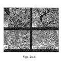
- FIGS. 2 a - d are scanning electron micrographs (SEM) of the PCL:PLA (3:1 ratio, respectively) electrospun scaffold in the presence ( FIGS. 2 c - d ) or absence ( FIGS. 2 a - b ) of MSCs.
- SEM scanning electron micrographs
- FIGS. 2 c and d the 3-D porous structure of the scaffold containing cells adhered to the scaffold fibers.
- FIGS. 2 c and d also note the initial deposition of extracellular matrix (ECM) on the non woven scaffold fibers following 7 days in culture in the presence of the osteogenic medium ( FIGS. 2 c and d ).
- ECM extracellular matrix
- FIGS. 3 a - d are SEM micrographs of bone marrow-derived MSCs cultured for 21 days on the PCL:PLA (3:1 ratio, respectively) electrospun scaffold in the presence of the osteogenic medium. Note the dense material between the cells and rigid appearance of mineralized fibers ( FIGS. 3 a - d ).
- FIGS. 6 a - f are SEM images of electrospun elements depicting the effect of polymer concentration on the fiber diameter ( d f ) and permeability (k, darcy units) of the electrospun elements.
- the present invention is of electrospun elements having a continuous or stepwise gradient of porosity, average pore size, weight per volume and/or agents attached, embedded or impregnated therein and of methods of manufacturing and using same.
- the present invention is of PCL, PLA and PGA polymers, mixtures and/or co-polymers which can be used in various combinations to fabricate electrospun scaffolds suitable for guided tissue regeneration, repair and/or implant.
- Scaffolds or medical membranes are used in tissue regeneration applications for treating diseased and/or traumatized tissues.
- Common scaffolds are made of homologous or heterologous tissue-derived materials such as Collagen (e.g., Collagen I or IV), fibronectin elastin and laminin.
- Collagen e.g., Collagen I or IV
- fibronectin elastin elastin
- laminin e.g., fibronectin elastin
- laminin fibronectin elastin
- laminin fibronectin elastin
- laminin laminin
- alloplastic or non-degradable synthetic polymers e.g., PEG, HA/PCL, PGA, PLGA and PLLA
- Biocompatible and/or biodegradable hydrogel scaffolds made of various polymers were found suitable for growth and differentiation of bone marrow derived mesenchymal stem cells (MSCs) (Tabata, 2001; Srouji and Livne 2005(a); Tabata et al., 1998; D'Ippolito, 2003; Yoshimoto et al., 2003; Li et al., 2003; 2005; Tuli et al., 2004) and repair of bone defect [Yammamoto et al.
- MSCs bone marrow derived mesenchymal stem cells
- hydrogel scaffolds are biocompatible and capable of promoting cell differentiation in vitro, their relatively low porosity and low strength prevent their use in clinical applications such as repair of bone fractures in vivo.
- electrospun scaffolds were manufactured from natural or synthetic polymers (e.g., Collagen, PLLA, PGA and PCL). Such scaffolds are made of a single continuous micro to nano-fibrous filament which is collected by a grounded target (e.g., an electrode) as a nonwoven fabric (see for example, U.S. Pat. Appl. No. 20040037813 to Simpson David G, et al; Lee Y H, et al., 2005, Biomaterials. 26: 3165-72; Khil M S, et al., 2005, J. Biomed. Mater. Res. B Appl Biomater. 72: 117-24; Li et al., 2002; Yoshimoto et al., 2003).
- the presently available electrospun scaffolds are not suitable for in vivo guided tissue regeneration and/or repair.
- an electrospun element having a substantially continuous gradient of porosity, average pore size, weight-per-volume and/or of agents attached to, embedded and/or impregnated therein, which promote proliferation and migration of one population of cells while restricting the migration of another population of cells and thereby capable of guiding tissue regeneration in vivo.
- substantially continuous porosity gradient refers to a change in the porosity of the electrospun element which is preferably below about 10% per 10 ⁇ m thickness of the electrospun element.
- electrospinning which is performed using a rotating vehicle (e.g., a drum) as a collector electrode can result in an electrospun element with a continuous gradient of porosity.
- a rotating vehicle e.g., a drum
- the manufacturing is preferably by an electrospinning process in which one or more liquefied polymers (i.e., a polymer in a liquid form such as a melted or dissolved polymer) are dispensed from a dispenser within an electrostatic field in a direction of a rotating collector.
- the dispenser can be, for example, a syringe with a metal needle or a bath provided with one or more capillary apertures from which the liquefied polymer(s) can be extruded, e.g., under the action of hydrostatic pressure, mechanical pressure, air pressure and high voltage.
- the rotating collector serves for collecting the electrospun element thereupon.
- the rotating collector has a cylindrical shape (e.g., a drum), however, it will be appreciated that the rotating collector can be also of a planar geometry.
- the dispenser e.g., a syringe with metallic needle
- the dispenser is typically connected to a source of high voltage, preferably of positive polarity, while the collector is grounded, thus forming an electrostatic field between the dispenser and the collector.
- the dispenser can be grounded while the collector is connected to a source of high voltage, preferably with negative polarity.
- any of the above configurations establishes motion of positively charged jet from the dispenser to the collector. Reverse polarity for establishing motions of a negatively charged jet from the dispenser to the collector are also contemplated.
- the charge repulsion begins to overcome the surface tension of the liquid drop.
- the charged jets depart from the dispenser and travel within the electrostatic field towards the collector. Moving with high velocity in the inter-electrode space, the jet stretches and solvent therein evaporates, thus forming fibers which are collected on the collector, thus forming the electrospun element.
- the phrase “electrospun element” refers to an element of any shape including, without limitation, a planar shape and a tubular shape, made of one or more non-woven polymer fiber(s), produced by a process of electrospinning as further detailed hereinunder.
- the electrospun element is made of a single fiber, the fiber is folded thereupon, hence can be viewed as a plurality of connected fibers. It is to be understood that a more detailed reference to a plurality of fibers is not intended to limit the scope of the present invention to such particular case. Thus, unless otherwise defined, any reference herein to a “plurality of fibers” applies also to a single fiber and vice versa.
- the polymer fibers of the electrospun element can be arranged on a single layer, but, more preferably, the fibers define a plurality of layers hence form a three dimensional structure.
- the polymer fibers can have a general random orientation, or a preferred orientation, as desired e.g., when the fibers are collected on a cylindrical collector such as a drum, the polymer fibers can be aligned predominantly axially or predominantly circumferentially.
- Different layers of the electrospun element can have different orientation characteristics. For example, without limiting the scope of the present invention to any specific ordering or number of layers, the fibers of a first layer can have a first predominant orientation, the fibers of a second layer can have a second predominant orientation, and the fibers of third layer can have general random orientation.
- At least one parameter is varied during the electrospinning process in a substantially continuous manner.
- the phrase “at least one parameter” refers to any physical parameter involved in the electrospinning process. Examples without limitations for such parameter include at least one of: the velocity of the rotating collector, the characteristic of the electrostatic field vector (magnitude and/or direction), the size of the capillary apertures of the dispenser (e.g., the size of a needle attached to the dispenser), the numbers of dispensers, the dispensing flow rate of the at least one liquefied polymer the viscosity, concentration and/or conductivity of the liquefied polymer, the concentration of the agents attached to the liquefied polymer in each of the dispensers and the concentration of charge control agent (e.g., a miscible salts).
- the concentration of charge control agent e.g., a miscible salts
- the characteristic of the electrostatic field vector can be varied during the electrospinning process in more than one way.
- the variation of the electric field is effected by varying, preferably continuously, the distance between the dispenser and the collector; in another preferred embodiment, the variation of the electric field is effected by varying, preferably continuously, the potential difference between the dispenser and the collector; in an additional embodiment, the variation of the electrostatic field is effected by varying both the distance and the potential difference in a substantially continues manner.
- the phrase “substantially continuously varying” refers to gradually changing or modifying. Preferably, such a gradual change is characterized by imperceptible (i.e., extremely slight, gradual, or subtle) increments.
- One example of “substantially continuously varying” is monotonically varying the at least one parameter described hereinbelow. It will be appreciated that the variation can be at a constant rate, in which case the effect of such variation is linear or at a variable rate in which case the effect is nonlinear.
- the advantage of using a rotating collector (such as a drum) for collecting the polymer fibers is that, as uncovered by the present inventors, such configuration allows a control over the porosity of the electrospun element.
- porosity refers to the ratio of the volume of interstices (i.e., pores) of a material to the volume of its mass. Such a ratio can be fixed and unchanged along the depth and/or surface of the electrospun element or can vary along the depth and/or surface of the electrospun element.
- the porosity and the pore size of the electrospun element are two related physical quantities. Nevertheless, these physical quantities are not identical, because the porosity depends on both the number of pores and the average pore size. Thus, different electrospun elements (or different portion of the same electrospun element) can have the same pore size yet different porosity and vice versa. For example, a first portion of the electrospun element having N pores of size S per unit area has a smaller porosity than a second portion having N+ ⁇ ( ⁇ >0) pores of the same size S per unit area.
- Independent variation of the average pore size and the porosity of the electrospun element can be achieved, for example, by varying both the density and the diameter of the fibers, substantially simultaneously.
- Variation of fiber diameter can be achieved, for example, by varying the electrostatic field, the polymer solution concentration, environmental temperature, the diameter of the dispenser's apertures (e.g., using a shutter), the flow rate of the polymer (e.g., by a syringe pump, or by controlling the back pressure), the viscosity of the polymer (e.g., by continuously adding viscous agent to the polymer solution).
- the fiber diameter depends on the concentration of the polymer solution which affects the extent of liquid evaporation from the jets prior to their sedimentation on the collector.
- a substantially continuous variation of the velocity of the rotating collector results in a substantially continuous variation of the density and/or spatial distribution of the fibers on the collector.
- the fibers are piled thereon at a substantially continuously varying density resulting in a porosity gradient along the radial and/or circumferential direction of the collector.
- a motion of the dispenser along the longitudinal direction of the collector can be established so as to form a porosity gradient also along the longitudinal direction.
- a porosity gradient which is achieved by angular velocity variation is typically accompanied by a pore-size gradient. For example, as is shown in FIGS.
- an electrospun element with a gradient porosity can be manufactured as follows.
- a 5 ml syringe or more containing 10% PCL in DCM/DMF (1:1) is positioned at a distance of 25 centimeters from the wheel rim including the collecting electrode.
- the polymer solution in the syringe is charged with 18 kV and the polymer solution flows from the syringe at an initial flow rate of 0.5 ml per hour.
- the initial speed of the wheel is 0 meter/minute linear velocity and is changed gradually at intervals of 6 meter/minute linear velocity every 10 minutes to a final speed of 96 meter/minute linear velocity.
- Such conditions result in an initial porosity of about 95% and a final porosity of about 75%
- changing the electrostatic field modifies the fiber diameter and the polymer flow rate. For example, changing the voltage per cm from 0.2 kV/cm to 1 kV/cm an/or the flow rate from 0.1 ml per hour to 2 ml per hour.
- changing the size or shape of the dispenser affects the fiber diameter.
- using a voltage of 18 kV and a needle diameter of 270 ⁇ m can yield a constant flow rate of 0.1 ml/hour and a fiber diameter of 400 nm.
- the fiber diameter can be increased from an initial diameter of 400 nm to a final diameter of about 2000 nm.
- changing the distance between the dispenser and the rotating collector affects the porosity of the electrospun element.
- a polymer solution which is charged with 18 kV and flows at a rate of 0.1 ml/hour at a distance of 20 cm results in an electrospun element with 85% porosity.
- using the same conditions i.e., 18 kV and flow rate of 0.1 ml/hour at a distance of 45 cm, results in an electrospun element having about 93% porosity.
- increments of about 2 cm in the distance are expected to result in a change of about 1% in the porosity of the electrospun element of the present invention.
- the electrospun element of the present invention is characterized by a substantially continuous porosity gradient.
- the porosity is a monotonic function.
- the porosity can continuously increase inward along a line connecting one external surface of the electrospun element with the other.
- the continuous porosity gradient has a maximal porosity of about 95% and a minimal porosity of about 50%.
- the continuous porosity gradient has a maximal porosity of about 90% and a minimal porosity of about 50%, more preferably, a maximal porosity of about 85% and a minimal porosity of about 55%, more preferably, a maximal porosity of about 80% and a minimal porosity of about 60%, more preferably, a maximal porosity of about 80% and a minimal porosity of about 65%, more preferably, a maximal porosity of about 75% and a minimal porosity of about 70%.
- a stepwise porosity gradient suitable for the present embodiment includes discrete porosity variations at about 5% intervals.
- an electrospun element having a gradient of average pore size along at least a portion thereof.
- a gradient of average pore size can be continuous or step wise gradient of average pore size.
- pore size refers to the area of a pore at a given plane which is formed between the fibers of the electrospun element.
- the pore size distribution and dimensions of the electrospun element of the present invention can be determined using a mercury porosimeter, a confocal microscope or by other known methods.
- K ⁇ L/ ⁇ P
- Another way of quantitating the pore size is by calculating the diameter of the pore at a single plane, using e.g., a confocal microscope.
- the diameter of the pore measured according to this aspect of the present invention is the largest diameter at the measured plane.
- the gradient of average pore size has a maximal average pore diameter of about 200 ⁇ m and a minimal average pore diameter of about 0.1 ⁇ m.
- a gradient of average pore size has a maximal average pore diameter of about 200 ⁇ m and a minimal average pore diameter of about 1 ⁇ m, more preferably, such a gradient of average pore size has a maximal average pore diameter of about 200 ⁇ m and a minimal average pore diameter of about 10 ⁇ m.
- electrospinning results in at least one continuous fiber which is randomly oriented and thus forms the electrospun element.
- Several parameters may affect the diameter of such a fiber. These include, the size of the dispensing hole of the dispenser, the dispensing rate, the strength of the electrostatic field, the distance between the dispenser and/or the concentration of the polymer used for fabricating the electrospun element.
- the electrospun element of the present invention exhibits a uniform fiber diameter having a variance of about 10% or less.
- the gradient of average diameter is a continuous gradient of average diameter.
- the polymer in order to form a gradient of an average diameter, can be provided by multiple dispensers, each containing a different concentration of the polymer and/or copolymer.
- the fiber comprising the electrospun element can be of a core-shell structure (e.g., a core which is formed from one polymer and a shell, which surrounds the core and is formed from another polymer). Fibers of a core-shell structure can be manufactured using methods known in the art such as using a unified double syringe structure (e.g., see Z. Sun, E. Zussman, A. L. Yarin, J. H. Wendorff, A. Greiner, “Compound Core/shell Polymer Nanofibers by Co-Electrospinning”, Advanced Materials, 15, 22:1929-1936, 2003). It will be appreciated that the fiber comprising the electrospun element can be also a hollow fiber.
- the phrase “hollow fiber” refers to a fiber with a core-shell structure from which the core is removed. Such fibers can have an inner diameter of less than about 1 ⁇ m. Removal of the core from the core-shell structure can be effected using methods known in the art such as by incubating the fibers of the electrospun element in a solvent (e.g., water) capable of dissolving the polymer (or co-polymer) used to fabricate the core but incapable of dissolving the polymer (or co-polymer) used to fabricate the shell.
- a solvent e.g., water
- removal of the core polymer from the shell can be effected by heating the fibers of the electrospun element at a temperature selected capable of melting or evaporating the polymer (or copolymer) used to fabricate the core but not the polymer (or co-polymer) used to fabricate the shell.
- a gradient of fiber weight-per-volume can be achieved by changing the flow rate and/or concentration of the polymer solution from the syringe.
- a polymer solution which is charged at 1.0 kV/cm, is dispensed at an initial flow rate of 0.1 ml/minute and gradually increases at increments of 0.1 ml/minute until a flow rate of 1.0 ml/minute is achieved.
- a gradient of fiber weight-per-volume can be also achieved by using more than one syringe alternatively or together with variable polymer concentrations in each syringe.
- electrospinning is performed using two syringes; one syringe contains a solution of 12% PCL/PLA and a second syringe contains a solution of 6% PCL/PLA. Electrospinning begins by dispensing the solution of the first syringe, which is charged at 1.2 kV per cm, for 120 minutes at a flow rate of 0.1 ml/minute, following which, dispensing is effected from both syringes (which are charged with the same voltage per cm) for 120 minutes.
- the gradient of average fiber weight-per-volume can be a continuous or step wise, depending on the rate and increments of changing the flow rate and/or the concentration of the polymer solutions used.
- the phrase “at least one liquefied polymer” refers to any polymer, polymers or co-polymers which are in a liquid form (e.g., a soluble polymer in solution or a melted polymer).
- the polymer used by the present invention can be a natural, synthetic, biocompatible and/or biodegradable polymer.
- synthetic polymer refers to polymers that are not found in nature, even if the polymers are made from naturally occurring biomaterials. Examples include, but are not limited to, aliphatic polyesters, poly(amino acids), copoly(ether-esters), polyalkylenes oxalates, polyamides, tyrosine derived polycarbonates, poly(iminocarbonates), polyorthoesters, polyoxaesters, polyamidoesters, polyoxaesters containing amine groups, poly(anhydrides), polyphosphazenes, and combinations thereof.
- Suitable synthetic polymers for use in the present invention can also include biosynthetic polymers based on sequences found in collagen, elastin, thrombin, fibronectin, starches, poly(amino acid), poly(propylene fumarate), gelatin, alginate, pectin, fibrin, oxidized cellulose, chitin, chitosan, tropoelastin, hyaluronic acid, polyethylene, polyethylene terephthalate, poly(tetrafluoroethylene), polycarbonate, polypropylene and poly(vinyl alcohol), ribonucleic acids, deoxyribonucleic acids, polypeptides, proteins, polysaccharides, polynucleotides and combinations thereof.
- natural polymer refers to polymers that are naturally occurring.
- Non-limiting examples of such polymers include, silk, collagen-based materials, chitosan, hyaluronic acid and alginate.
- co-polymer refers to a polymer of at least two chemically distinct monomers.
- Non-limiting examples of co-polymers include, PLA-PEG, PEGT/PBT, PLA-PGA PEG-PCL and PCL-PLA.
- biocompatible polymer refers to any polymer (synthetic or natural) which when in contact with cells, tissues or body fluid of an organism does not induce adverse effects such as immunological reactions and/or rejections and the like. It will be appreciated that a biocompatible polymer can also be a biodegradable polymer.
- biodegradable polymer refers to a synthetic or natural polymer which can be degraded (i.e., broken down) in the physiological environment such as by proteases. Biodegradability depends on the availability of degradation substrates (i.e., biological materials or portion thereof which are part of the polymer), the presence of biodegrading materials (e.g., microorganisms, enzymes, proteins) and the availability of oxygen (for aerobic organisms, microorganisms or portions thereof), carbon dioxide (for anaerobic organisms, microorganisms or portions thereof) and/or other nutrients.
- degradation substrates i.e., biological materials or portion thereof which are part of the polymer
- biodegrading materials e.g., microorganisms, enzymes, proteins
- oxygen for aerobic organisms, microorganisms or portions thereof
- carbon dioxide for anaerobic organisms, microorganisms or portions thereof
- biodegradable polymers include, but are not limited to, collagen (e.g., Collagen I or IV), fibrin, hyaluronic acid, polylactic acid (PLA), polyglycolic acid (PGA), polycaprolactone (PCL), polydioxanone (PDO), trimethylene carbonate (TMC), polyethyleneglycol (PEG), Collagen, PEG-DMA, Alginate, chitosan copolymers or mixtures thereof.
- the liquefied polymer can be made of one polymer or more, each can be a polymer or a co-polymer such as described hereinabove.
- the liquefied polymer of the present invention is a mixture of at least one biocompatible polymer and a co-polymer (either biodegradable or non-biodegradable).
- electrospun element of the present invention can be used as a scaffold or membrane such as for guiding tissue regeneration.
- the scaffold of the present invention refers to a two-dimensional or a three-dimensional supporting framework.
- the scaffold of the present invention is composed of electrospun fibers, each fiber composed of at least one polymer as described hereinabove. It will be appreciated that the scaffold of the present invention can be embedded within, or formed around, another scaffold or hydrogel and various configurations of electrospun elements and hydrogels can be composed.
- hydrogel refers to any material with molecular net structure in which water constitutes more than 50%.
- a hydrogel can include a cross-linked polymer with a water constitute of at least 70%.
- Non-limiting examples of hydrogels which can be used along with the present invention include, a Collagen hydrogel, a PEG hydrogel, a PEG-DMA hydrogel, and an Alginate hydrogel.
- a composite scaffold of an electrospun element and a hydrogel can be prepared by electrospinning a first polymer solution capable of forming a hydrogel (e.g., Collagen), followed by electrospinning of a second polymer (e.g., a co-polymer of PCL-PLA) on the top of the first electrospun element.
- a first polymer solution capable of forming a hydrogel e.g., Collagen
- a second polymer e.g., a co-polymer of PCL-PLA
- the composite electrospun element is soaked in water, resulting in absorption of water by the first electrospun layer (e.g., the Collagen) and the formation of a hydrogel layer (made of the first polymer) underneath the electrospun fibers (made of the second polymer).
- a first polymer solution e.g., Collagen
- a second polymer solution e.g., PCL-PLA
- an electrospun scaffold can be layered within or on a hydrogel layer.
- PCL-PLA electrospun scaffold was layered with collagen hydrogels membrane (Example 1 of the Examples section which follows).
- electrospinning can be effected using a polymer solution capable of forming a hydrogel (e.g., Collagen) which is mixed with another polymer solution (e.g., a co-polymer of PCL-PLA) which is incapable of forming a hydrogel.
- a polymer solution capable of forming a hydrogel e.g., Collagen
- another polymer solution e.g., a co-polymer of PCL-PLA
- electrospinning can be formed using the core-shell configuration in which the core polymer is selected from a polymer capable of forming a hydrogel (e.g., Collagen) and the shell polymer is selected incapable of forming a hydrogel, or vice versa.
- electrospun element of the present can also form a complex structure of two surfaces defining a volume therebetween.
- the article of manufacturing includes an electrospun element having a first surface and a second surface defining a volume therebetween, wherein an average pore size close to the first surface as well as the chemical and biological characteristics are selected so as to allow migration of at least one population of cells therethrough into the volume, and an average pore size close to the second surface as well as the chemical and biological characteristics are selected so as to restrict migration of at least one population of cells therethrough into the volume.
- volume refers to an interface formed between to two surfaces. Such a volume can be used, for example, for proliferation and/or differentiation of cells which are seeded, as described hereinbelow, within the electrospun element.
- the surfaces of the electrospun element can be made from either the same or different polymers and/or materials. Additionally and/or alternatively, the surfaces can have the same or different structure, porosity, average pore size, fiber weight-per-volume and/or agents (e.g., chemical, biological and/or mineral) attached thereto, embedded or impregnated therein.
- agents e.g., chemical, biological and/or mineral
- the scaffold of the present invention can be used to support cell growth, attachment, spreading, and thus facilitate cell growth, tissue regeneration and/or tissue repair.
- Such a scaffold is therefore being seeded with cells capable of proliferating and/or migrating therethrough.
- the electrospun element or the scaffold composed of the electrospun element with an hydrogel is placed in a culture medium for promoting proliferation of at least one population of cells being in contact with the electrospun element.
- a culture medium for promoting proliferation of at least one population of cells being in contact with the electrospun element.
- Such one population of cells can be for example, osteoblast cells, endothelial cell or stem cells.
- the culture medium used by the present invention can be any liquid medium which allows at least cell survival.
- a culture medium can include, for example, salts, sugars, amino acids and minerals in the appropriate concentrations and with various additives and those of skills in the art are capable of determining a suitable culture medium to specific cell types.
- Non-limiting examples of such culture medium include, phosphate buffered saline, DMEM, MEM, RPMI 1640, McCoy's 5A medium, medium 199 and IMDM (available e.g., from Biological Industries, Beth Ha'emek, Israel; Gibco-Invitrogen Corporation products, Grand Island, N.Y., USA).
- the culture medium is preferably supplemented with various antibiotics (e.g., penicillin and Streptomycin), growth factors or hormones, specific amino acids (e.g., L-glutamin) cytokines and the like.
- antibiotics e.g., penicillin and Streptomycin
- growth factors or hormones e.g., growth factors or hormones
- specific amino acids e.g., L-glutamin
- the culture medium can include dexamethasone which is capable of inducing the proliferation and differentiation of bone marrow derived mesenchymal stem cells (MSCs) into osteoblasts.
- MSCs bone marrow derived mesenchymal stem cells
- the culture medium includes a mineralizing agent.
- mineralizing agent refers to an agent including at least one mineral capable of forming mineral-containing substance, e.g., mineral containing tissue such as a bone.
- mineral containing tissue such as a bone.
- Non-limiting examples of such agents include, calcium which can form calcium phosphate and hydroxyapatite.
- the mineralizing agent is capable of supporting the formation and/or remodeling of a bone tissue including, but not limited to a dental tissue, a bone, a vertebrae and the like.
- the continuous porosity gradient of the electrospun element of the present invention allows the migration of at least one population of cells (e.g., osteoblast cells, endothelial cells) through one side of the electrospun element and, on the other hand, restricts the migration of at least another population of cells (e.g., fibroblast cells) through a second side of the electrospun element.
- at least one population of cells e.g., osteoblast cells, endothelial cells
- at least another population of cells e.g., fibroblast cells
- an electrospun element having a continuous gradient of average pore size can be selected such that it allows the migration of at least one population of cells (e.g., osteoblast cells, endothelial cells) through one side of the electrospun element and, on the other hand, restricts the migration of at least one population of cells (e.g., fibroblast cells) through a second side of the electrospun element.
- at least one population of cells e.g., osteoblast cells, endothelial cells
- an electrospun element having a continuous gradient of fiber weight-per-volume can be selected such that it allows the migration of at least one population of cells (e.g., osteoblast cells, endothelial cells) through one side of the electrospun element and, on the other hand, restricts the migration of at least one population of cells (e.g., fibroblast cells) through a second side of the electrospun element.
- at least one population of cells e.g., osteoblast cells, endothelial cells
- an electrospun element e.g., a scaffold containing an electrospun element
- an electrospun element further includes at least one agent.
- an agent can be a biological agent such as an amino acid, a peptide, a polypeptide, a protein, a DNA, an RNA, a lipid and/or a proteoglycan.
- Suitable proteins which can be used along with the present invention include, but are not limited to, extracellular matrix proteins [e.g., fibrinogen, Collagen, fibronectin, vimentin, microtubule-associated protein 1D, Neurite outgrowth factor (NOF), bacterial cellulose (BC), laminin and gelatin], cell adhesion proteins [e.g., integrin, proteoglycan, glycosaminoglycan, laminin, intercellular adhesion molecule (ICAM)1, N-CAM, cadherin, tenascin, gicerin, RGD peptide and nerve injury induced protein 2 (ninjurin2)], growth factors [epidermal growth factor, transforming growth factor- ⁇ , fibroblast growth factor-acidic, bone morphogenic protein, fibroblast growth factor-basic, erythropoietin, thrombopoietin, hepatocyte growth factor, insulin-like growth factor-1, insulin-like growth factor-II, Interferon- ⁇
- the at least one agent used by the present invention is an antiproliferative agent (e.g., rapamycin, paclitaxel, tranilast, Atorvastatin and trapidil), an immunosuppressant drug (e.g., sirolimus, tacrolimus and Cyclosporine) and/or a non-thrombogenic or anti-adhesive substance (e.g., tissue plasminogen activator, reteplase, TNK-tPA, glycoprotein IIb/IIIa inhibitors, clopidogrel, aspirin, heparin and low molecular weight heparins such as enoxiparin and dalteparin).
- an antiproliferative agent e.g., rapamycin, paclitaxel, tranilast, Atorvastatin and trapidil
- an immunosuppressant drug e.g., sirolimus, tacrolimus and Cyclosporine
- the at least one agent used by the present invention is a chemical or mineral which is added to the electrospun element in order to improve its biological properties.
- the electrospun element can be embedded with, attached to or impregnated with minerals which promote bone formation and/or mineralization such as calcium sulfate and/or Hydroxyapatite.
- the at least one agent of this aspect of the present invention can be attached to at least a portion of the electrospun element. Such attachment can be performed using e.g., cross-linking (chemical or light mediated) of the at least one agent with the polymer solution or the electrospun fiber formed therefrom (e.g., PEG-DMA, PLA and the agent). Additionally or alternatively, the at least one agent can be embedded in electrospun micro or nanofibers having the core-shell structure essentially as described in Sun, 2003 (Supra). Still additionally or alternatively the at least one agent can be impregnated in the electrospun element by soaking the electrospun element or at least a portion of the polymer fibers forming the electrospun element in a solution containing such an agent.
- cross-linking chemical or light mediated
- the at least one agent can be embedded in electrospun micro or nanofibers having the core-shell structure essentially as described in Sun, 2003 (Supra).
- the at least one agent can be impregnated in the electros
- the at least one agent which is attached to, embedded, mixed or impregnated in the electrospun element can form a gradient along at least a portion of the electrospun element.
- a gradient can be a continuous or step-wise gradient and can be formed, for example, by changing the concentration of the agent in the liquefied polymer.
- the syringe can have two or more feeding sources of liquefied polymers with variable concentrations of the agent attached to or mixed with. To form a gradient, each feeding source is used for a predetermined time (e.g., a few seconds or minutes).
- the different feeding sources can include various concentrations of the agents attached to or mixed with the polymer (e.g., 1%, 2%, 3% and the like, although smaller increments can be used as well) and following predetermined time periods (e.g., 1 minute) a different feeding source is utilized.
- a different feeding source is utilized.
- several feeding sources are used, each containing the polymer solution with a different concentration of agent (e.g., 1%, 10% and 20%) and for a time period sufficient for forming a “layer” of the agent.
- the gradient of the at least one agent can be formed by impregnating at least a portion of the polymer fibers with increasing concentrations of the agent.
- the gradient of the at least one agent can be formed by embedding increasing concentrations of the agent in the electrospun element.
- retinoic acid which is capable of promoting proliferation
- the lower layer of the electrospun element is capable of promoting proliferation and angiogenesis
- the higher layer of the electrospun element which is devoid of RA or, alternatively, includes another agent such as a differentiation factor, is capable of inhibiting proliferation.
- the electrospun element of the present invention can have a gradient of porosity, average pore size, or fiber weight-per-volume with or without a gradient of at least one agent attached to, embedded or impregnated therein.
- the electrospun element of the present invention can be of a uniform porosity, average pore size or fiber weight-per-volume yet with a gradient of at least one agent attached to, embedded or impregnated therein.
- bone marrow derived MSCs which were seeded in the presence of an osteogenic medium (DMEM medium supplemented with 15% fetal calf serum (FCS), 20 mM L-glutamin, Pen-Strep (100 U/ml penicillin, 100 ⁇ g/ml streptomycin), 100 ⁇ g/ml ascorbic acid, 10 mM sodium ⁇ -glycerophosphate and 10 ⁇ 8 M dexamethasone) in the PCL-PLA electrospun scaffold (in a 3:1 ratio, respectively), were capable of proliferating and differentiating into osteoprogenitor cells capable of matrix deposition.
- DMEM medium fetal calf serum
- FCS fetal calf serum
- Pen-Strep 100 U/ml penicillin, 100 ⁇ g/ml streptomycin
- 100 ⁇ g/ml ascorbic acid 100 ⁇ g/ml ascorbic acid
- a scaffold comprising an electrospun element consisting of PCL, PLA, and/or PGA polymers and/or their co-polymers, whereby when seeded with bone marrow derived stem cells in an osteoblast differentiation inducing medium containing at least one mineral the matrix is populated with osteoblasts and mineralizes so as to transform into a mineralized scaffold.
- At least one mineral refers to a mineral needed for bone formation and/or regeneration.
- Non-limiting examples of such a mineral include sodium ⁇ -glycerophosphate, ascorbic acid, and calcium phosphate.
- the phrase “mineralized scaffold” refers to a scaffold containing at least one form of the at least one mineral included in the medium.
- a calcium phosphate mineral can be formed in the presence of sodium ⁇ -glycerophosphate and calcium chloride (which is included in the DMEM medium).
- the PCL-PLA scaffold of the present invention was devoid of fibroblast cell. It will be appreciated that restricting the migration of fibroblast cells into a scaffold promotes proper bone regeneration and/or repair.
- PCL, PLA, and/or PGA polymers can be provided at various weight ratios.
- the PCL, PLA, and/or PGA polymers are provided at weight ratios such that the weight of the PLA is higher than the weight of the PCL and/or PGA.
- Suitable weight ratios for the PCL:PLA can be for example, about 1:1.5, about 1:2, about 1:2.5, about 1:3, about 1:3.5, about 1:4, about 1:4.5, about 1:5, about 1:5.5, about 1:6, about 1:6.5, about 1:7, about 1:7.5, about 1:8, about 1:8.5, at least 1:9, respectively.
- Suitable weight ratios for the PCL:PGA can be for example, about 1:1.5, respectively.
- the PCL and PLA polymers are provided at a weight ratio such that the weight of the PLA is lower than the weight of the PCL.
- Suitable weight ratios for the PCL:PLA can be for example, about 1.5:1, about 2:1, about 2.5:1, about 3:1, about 3.5:1, about 4:1, about 4.5:1, about 5:1, about 5.5:1, about 6:1, about 6.5:1, about 7:1, about 7.5:1, about 8:1, about 8.5:1, at least 9:1, respectively.
- Suitable weight ratios for the PLA:PLGA can be for example, about 1:1 or 1:2 ratios, respectively.
- Suitable weight ratios for the PCL:PLGA can be for example at 1:1 or 1:2 ratios, respectively.
- the electrospun element of the scaffold of the present invention is characterized by a predetermined average pore size selected so as to restrict migration of fibroblast cells therethrough.
- Such a predetermined pore size can be for example, a pore having a diameter lower than 1 ⁇ m, more preferably, lower than 0.2 ⁇ m, even more preferably, a pore having a diameter of about 0.1 ⁇ m.
- the predetermined average pore size is selected so as to allow penetration of oxygen molecules therethrough.
- a predetermined average pore size can be of a diameter of at least 0.1 ⁇ m, more preferably, at least 0.5 ⁇ m, even more preferably, about 1 ⁇ m.
- the predetermined average pore size is selected so as to allow penetration of nutrients therethrough.
- a predetermined average pore size can be of a diameter of at least 0.1 ⁇ m, more preferably, at least 1 ⁇ m, even more preferably, about 10 ⁇ m.
- the electrospun element is characterized by a predetermined average pore size having a diameter selected from about 0.1 micrometer to about 200 micrometer, more preferably, from 0.5 micrometer to about 200 micrometer, more preferably, from 1 ⁇ m to about 200 ⁇ m, more preferably, from 10 nanometer to about 200 ⁇ m, even more preferably, from 30 ⁇ m to about 200 ⁇ m.
- the electrospun element according to this aspect of the present invention can have a relatively uniform pore size with a variance of pore diameter of about 20% or less than the average pore diameter.
- the porosity of the electrospun element of the present invention can be of at least 50%, more preferably, at least 55%, more preferably, at least 60%, more preferably, at least 65%, more preferably, at least 70%, more preferably, at least 75%, more preferably, at least 80%, more preferably, at least 85%, more preferably, at least 90%, even more preferably, about 95% porosity.
- the electrospun element of the present invention forms a porous membrane.
- the term “membrane” refers to a pliable sheet (usually thin) of material serving as a semi-permeable covering, i.e., enables the passage of one type of material or cells and not the other type of material or cell.
- the membrane of the present invention i.e., the PCL:PLA (at a 3:1 ratio) electrospun element] enables the migration of osteoblast cells but restrict the migration of fibroblast cells.
- the electrospun element of the present invention exhibits a tubular structure.
- a method of perforating an electrospun element can be done, for example, by an electrical spark, which can be generated by any electrical spark producing element, such as, but not limited to, a needle-like electrode.
- the electrical spark can vary depending on the applied voltage, its duration and the distance between the electrode and the electrospun element.
- the electrical spark is produced with an electric field which is sufficient to generate air breakdown. At normal conditions, such breakdown occurs at about 30 kV/cm. Thus, according to a preferred embodiment of the present invention the electrical spark is produced by generating an electric field of about 30 kV/cm.
- the electric field is preferably generated by a potential difference of at least 10 kV, more preferably, at least 15 kV.
- the breakdown field is generated by positioning the electrode at a distance of about 10 mm, more preferably, at a distance of 5 mm, even more preferably, at a distance of 1 mm from the electrospun element.
- the voltage used to provide the electrical spark is provided for a time period of about 5 seconds, more preferably, for a time period of about 1 second, even more preferably, for a time period of 0.1 second
- an electrospun element made of PCL/PLA at a 3:1 ratio, respectively, with a total thickness of 200 ⁇ m can be perforated by positioning the electrospun element between a high voltage electrode (e.g., at 18 kV) and a ground electrode at a distance of about 1 mm.
- a high voltage electrode e.g., at 18 kV
- a method of perforating an electrospun element is effected by passing a puncturing element through at least a portion of the electrospun element to thereby obtain a perforated electrospun element.
- puncturing element refers to any sharp and pointed element, preferably a metal implement which is capable of being heated and thus puncturing (i.e., making a hole) the electrospun element.
- puncturing elements include, a metal needle and a metal pin.
- the puncturing element is heated to a temperature of at least 90° C., more preferably, at least 91° C., more preferably, at least 92° C., more preferably, at least 93° C., more preferably, at least 94° C., more preferably, at least 95° C., more preferably, at least 96° C., more preferably, at least 97° C., more preferably, at least 98° C., more preferably, at least 99° C., even more preferably, at least 100° C., say about 100° C., about 101° C., about 102° C.
- Passing the puncturing element according to the method of this aspect of the present invention can be effected for a time period of 0.1-10 seconds, more preferably, for a time period of 1-5 seconds.
- the electrospun scaffold can be perforated using a heated needle pillow (to about 100° C. in the case of the PCL/PLA electrospun scaffold described in Example 1 of the Examples sections which follows). It will be appreciated that the scaffold can be perforated by piercing, i.e., producing a single hole or an array of holes.
- perforating an electrospun elements can be also effected by a pulsed or continues laser beam.
- the laser beam can be generated by any laser device capable of providing laser radiation which ablate or melt the polymer fibers to some extent. These include, but are not limited to, the following laser devices: Excimer laser device, Kr based laser device, Xe based laser device, Er based laser device, Ho:YAG laser device, carbon-dioxide laser device, Nd based laser device and laser diode device.
- Kr based laser devices include, but are not limited to krypton-fluoride (KrF) laser devices.
- Xe based laser devices include, but are not limited to xenon-fluoride (XeF) laser devices.
- Er based laser devices include, but are not limited to, Er:YAG, Er:YSGG, Er:glass and the like.
- Nd based laser devices include, but are not limited to, Nd:YAG, Nd:YLF, Nd:glass and the like. Also contemplated are CO 2 and Dye laser devices.
- perforation of an electrospun element is performed using a pulsed laser beam at a specific energy (e.g., 200 Watt) which is provided at a specific rate (e.g., 200 Hz), using several pulses for each hole.
- a specific energy e.g. 200 Watt
- a specific rate e.g. 200 Hz
- Eximer, KrF or XeF lasers can be used at an output power of about 200 watt, a rate of about 200 Hz and using 5 pulses for every hole.
- perforation can be used to enable the migration and/or proliferation of specific cell types through the electrospun element, and/or to enable the administration of various agents through the electrospun element.
- electrospun elements and scaffolds of the present invention can be used for ex vivo and/or in vivo formation of a tissue.
- a method of inducing ex vivo formation of a tissue is effected by providing a scaffold having an electrospun element having a continuous gradient of average pore size along at least a portion thereof, a continuous porosity gradient, a continuous fiber weight per volume gradient, and/or a gradient of at least one agent; and (ii) seeding the scaffold with cells in a medium selected suitable for proliferation, differentiation and/or migration of the cells to thereby induce the formation of the tissue.
- the at least one agent used by the method according to this aspect of the present invention is for promoting cell colonization, differentiation, extravasation and/or migration.
- an agent can be a biological, chemical or mineral agent as described hereinabove.
- tissue refers to part of an organism consisting of an aggregate of cells having a similar structure and function. Examples include, but are not limited to, brain tissue, retina, skin tissue, hepatic tissue, pancreatic tissue, bone, cartilage, connective tissue, blood tissue, muscle tissue, cardiac tissue brain tissue, vascular tissue, renal tissue, pulmonary tissue, gonadal tissue, hematopoietic tissue.
- tissue as used herein also encompasses the phrase “organ” which refers to a fully differentiated structural and functional unit in an animal that is specialized for some particular function.
- Non-limiting examples of organs include head, brain, eye, bone (e.g., of leg and hand), heart, liver kidney, lung, pancreas, ovary, testis, and stomach.
- the tissue is a bone tissue.
- the cells used by the method of this aspect of the present invention are capable of forming a tissue.
- Such cells can be for example, stem cells such as embryonic stem cells, bone marrow stem cells, cord blood cells, mesenchymal stem cells, adult tissue stem cells, or differentiated cells such as neural cells, retina cells, epidermal cells, hepatocytes, pancreatic cells, osseous cells, cartilaginous cells, elastic cells, fibrous cells, myocytes, myocardial cells, endothelial cells, smooth muscle cells, and hematopoietic cells.
- stem cells such as embryonic stem cells, bone marrow stem cells, cord blood cells, mesenchymal stem cells, adult tissue stem cells, or differentiated cells such as neural cells, retina cells, epidermal cells, hepatocytes, pancreatic cells, osseous cells, cartilaginous cells, elastic cells, fibrous cells, myocytes, myocardial cells, endothelial cells, smooth muscle cells, and hematopoietic cells.
- stem cell refers to cells which are capable of differentiating into other cell types having a particular, specialized function (i.e., “fully differentiated” cells) or remaining in an undifferentiated state hereinafter “pluripotent stem cells”.
- Non-limiting examples of stem cells are hematopoietic stem cells obtained from bone marrow tissue of an individual at any age or from cord blood of a newborn individual, embryonic stem (ES) cells obtained from the embryonic tissue formed after gestation (e.g., blastocyst), or embryonic germ (EG) cells obtained from the genital tissue of a fetus any time during gestation, preferably before 10 weeks of gestation, and/or adult tissue stem cells [i.e., mesenchymal stem cells (MSCs)].
- ES embryonic stem
- EG embryonic germ
- the stem cells are bone marrow derived MSCs.
- seeding refers to plating, placing and/or dropping the stem cells of the present invention into the scaffold of the present invention. It will be appreciated that the concentration of stem cells which are seeded on or within the scaffold of the present invention depends on the type of stem cells used and the composition of the electrospun element comprising the scaffold.
- the medium used according to this aspect of the present invention can be any tissue culture medium supplemented with minerals and growth factors suitable for inducing the proliferation, differentiation and/or migration of the cells (e.g., stem cells) of the present invention into more specialized (i.e., differentiated) cells.
- tissue culture medium supplemented with minerals and growth factors suitable for inducing the proliferation, differentiation and/or migration of the cells (e.g., stem cells) of the present invention into more specialized (i.e., differentiated) cells.
- stem cells e.g., differentiated
- the scaffolds are routinely examined using a microscope (e.g., an inverted microscope, an axioplan light microscope or an electronic microscope) for evaluation of cell growth, spreading and tissue formation (see for example FIGS. 1-3 ).
- a microscope e.g., an inverted microscope, an axioplan light microscope or an electronic microscope
- the ex vivo formed tissue can be further implanted in a subject in need thereof (e.g., a subject suffering from a pathology requiring tissue regeneration and/or repair as described hereinbelow).
- the cells seeded on the scaffold for ex vivo formation of a tissue can be derived from the treated individual (autologous source) or from allogeneic sources such as embryonic stem cells which are not expected to induce an immunogenic reaction.
- the seeded scaffold is implanted in the subject.
- the pathology to be treated is a fractured bone
- the scaffold is seeded with MSCs or osteoblasts and following 14-21 days in culture the scaffold is preferably implanted in the damaged bone tissue.
- the teachings of the method of the present invention can be also used to induce tissue formation in vivo and thus induce tissue regeneration and/or repair in a subject.
- a method of inducing in vivo formation of a tissue is effected by: (i) providing a scaffold having an electrospun element having a continuous gradient of average pore size along at least a portion thereof, a continuous porosity gradient, a continuous fiber weight-per-volume gradient, and/or a gradient of at least one and (ii) implanting the scaffold in a subject to thereby induce the formation of the tissue.
- the scaffolds of the present invention can be used to induce tissue formation and/or regeneration and thus treat individuals suffering from tissue damage or loss.
- a method of treating a subject having a pathology characterized by a tissue damage or loss is effected by: (i) providing a scaffold having a continuous gradient of average pore size along at least a portion thereof, a continuous porosity gradient, a continuous fiber weight-per-volume gradient, and/or a gradient of at least one agent and (ii) implanting the scaffold in a subject to thereby induce the formation of the tissue, thereby treating the subject.
- tissue damage or loss refers to any disorder, disease or condition exhibiting a tissue damage (i.e., non-functioning tissue, cancerous or pre-cancerous tissue, broken tissue, fractured tissue, fibrotic tissue, or ischemic tissue) or a tissue loss (e.g., following a trauma, an infectious disease, a genetic disease, and the like) which require tissue regeneration.
- tissue damage i.e., non-functioning tissue, cancerous or pre-cancerous tissue, broken tissue, fractured tissue, fibrotic tissue, or ischemic tissue
- tissue loss e.g., following a trauma, an infectious disease, a genetic disease, and the like
- liver cirrhosis such as in hepatitis C patients (liver), Type-1 diabetes (pancreas), cystic fibrosis (lung, liver, pancreas), bone cancer (bone), burn and wound repair (skin), age related macular degeneration (retina), myocardial infarction, myocardial repair, CNS lesions (myelin), articular cartilage defects (chondrocytes), bladder degeneration, intestinal degeneration, and the like.
- the term “subject” includes both young and old human beings of both sexes. Preferably, this term encompasses individuals who suffer from pathologies as described hereinabove.
- treating refers to inhibiting or arresting the development of a disease, disorder or condition and/or causing the reduction, remission, or regression of a disease, disorder or condition in an individual suffering from, or diagnosed with, the disease, disorder or condition.
- Those of skill in the art will be aware of various methodologies and assays which can be used to assess the development of a disease, disorder or condition, and similarly, various methodologies and assays which can be used to assess the reduction, remission or regression of a disease, disorder or condition.
- an article of manufacturing comprising an electrospun element having a continuous gradient of average pore size along at least a portion thereof, a continuous porosity gradient, a continuous fiber weight per volume gradient, and/or a gradient of at least one agent.
- the at least one agent is a biological, chemical or mineral agent, preferably for promoting cell colonization, differentiation, extravasation and/or migration.
- any of the articles of manufacturing described hereinabove comprise a packaging material and the electrospun element of the present invention contained within the packaging material.
- PCL-PLA Electrospun Scaffolds are Highly Suitable for Guided Bone Regeneration
- Electrospun nanofibrous scaffolds were prepared from the following polymers: Poly(e-caprolactone) (PCL) with an average molecular weight of 80 kDa (Aldrich, USA) was dissolved in DMF (di-methyl formamide)/DCM(di-chloromethane) 25:75 to obtain 10% wt % solution.
- PCL Poly(e-caprolactone)
- electrospinning was performed by delivering a polymer solution at a constant flow rate (0.5 ml/hour) through a plastic syringe with a capillary metal needle connected to a high voltage of several kilo volts (kV).
- a fluid jet was extruded from the capillary needle towards the grounded metal collector.
- the solvent evaporated, and charged polymer fiber was deposited on a collector in the form of a nonwoven scaffold.
- the nonwoven scaffold was cut in round shapes to fit for example a single well of a 24-well plate (Nunc), sterilized and soaked with DMEM medium prior to cell seeding.
- the collagen containing solutions were crosslinked.
- the copolymer PCL:Collagen acid hydrolyzed solution (Nitta Gelatin Co. Osaka, Japan-Producers of acid hydrolyzed collagen) was chemically crosslinked for about 30 minutes in the presence of 12.5 mM glutaraldehyde at 4° C.
- the hydrogel solution was immersed for 1 hour in a 50 mM glycine aqueous solution at 37° C., rinsed with double distilled water (DDW), rinse twice with 100% ethanol and rinsed twice with PBS.
- DSW double distilled water
- Composite scaffolds can be made using from hydrogel and electrospun elements.
- the Acid hydrolyzed Collagen solution can be electrospun to produce a fiber matrix on which another Polymer solution (e.g., PCL-PLA) in another syringe can be electrospun.
- the electrospun element fibers can be soaked in water and the Acid hydrolyzed Collagen absorbs the water and forms a hydrogel.
- the Acid hydrolyzed Collagen can be prepared as a hydrogel (as described hereinabove), and the other polymer solution, e.g., PCL-PLA, can be electrospun on the top of the hydrogel.
- Human bone marrow derived mesenchymal stem cells (MSCs), human cord blood derived MSCs and rat bone marrow derived MSCs were used.
- Human cord blood MSCs were isolated using Ficoll gradient, human bone marrow MSCs were isolated from bone marrow aspirates and rat bone marrow MSCs were isolated from the femur of Sprague-Dawley rats (300-400 gram each rat) by flushing.
- DMEM medium supplemented with 15% fetal calf serum (FCS), 20 mM L-glutamin, Pen-Strep (100 U/ml penicillin, 100 ⁇ g/ml streptomycin) or a basal control medium supplemented with 100 ⁇ g/ml ascorbic acid, 10 mM sodium ⁇ -glycerophosphate and 10 ⁇ 8 M dexamethasone (“the osteogenic medium”, hereinafter). All culture medium and medium supplements were obtained from Biological Industries Beith Haemek, Israel and Sigma, USA.
- the cells were cultured on the electrospun scaffold in 24-well plates (Nunc), 2,500,000 MSC cells per well, and samples were collected following one, two or three weeks for the various tests.
- the cultured cells were subjected to various morphology staining including Hematoxylin-Eosin and Masson's Trichrome staining. Selection of adherent osteoprogenitor cell subpopulation from MSCs was established by identifying the osteogenic cells using specific osteogenic cell markers including: osteocalcin immunoreactivity (bone matrix specific marker) (Biotest, USA), positive Alizarin red, von Kossa staining (reagents obtained from Sigma), and alkaline phosphatase activity [using a substrate ⁇ -naphtol phosphate (Sigma)].
- osteocalcin immunoreactivity bone matrix specific marker
- Alizarin red positive Alizarin red
- von Kossa staining reagents obtained from Sigma
- alkaline phosphatase activity using a substrate ⁇ -naphtol phosphate (Sigma)].
- specimens of the electrospun scaffold were fixed for 60 minutes in Neutral buffered formalin (4% in 0.1 phosphate buffer, pH 7.2), following which the specimens were stained with Hematoxylin and eosin (H&E) for general morphology, with alizarin S, alkaline phosphatase and/or von kossa staining for synthesis of bone specific matrix and mineral deposition and with osoteocalcin for immunodetection of bone specific matrix formation.
- H&E Hematoxylin and eosin
- Electrospun Scaffold are Nonwoven Networks with Randomly Oriented Fibers—
- the electrospun scaffolds of the present invention e.g., PCL:PLA at a 3:1 ratio, respectively
- FIG. 1 a illustrates an electrospun scaffold with fibers having an average diameter of 300 nm.
- Bone marrow-derived MSCs were cultured for 7 days on the PCL:PLA electrospun scaffold (at a 3:1 ratio, respectively) in the presence of the osteogenic medium revealed and a net-like arrangement of cell clusters ( FIG. 1 b ).
- the amount of cell clusters increased gradually from two ( FIG. 1 c ) to three ( FIG. 1 d ) weeks in culture.
- alizarin red S staining revealed the presence of mineralized bone matrix in the MSC-derived cultures, demonstrating bone regeneration.
- These cell clusters were also positive for osteocalcin staining which is indicative of synthesis of specific bone matrix (Not shown).
- FIGS. 2 a - b Scanning electron micrographs (SEM) of the PCL:PLA (at a 3:1 ratio, respectively) electrospun scaffold of the present invention revealed the scaffold 3-D porous structure ( FIGS. 2 a - b ) and the attachment and mineral deposition by the MSCs derived osteoprogenitor cells following 7 days in culture ( FIGS. 2 c - d ). Moreover, following 21 days in culture SEM analysis revealed large amount of extracellular matrix (ECM) deposits between the cells and fibers ( FIGS. 3 a - d ). The amount of the ECM deposited increased gradually from 14 days (as seen by alizarin red S staining, FIG. 1 e ) to day 21 ( FIG. 1 f ) and was shown to be mineralized ( FIGS.
- ECM extracellular matrix
- the composition of the deposited mineralized matrix was further determined using Electron dispersive spectroscopy (EDS) and was found to be composed of Calcium and Phosphate ions at a ratio of 1.8 (not shown), similar to that observed in an intact bone.
- EDS Electron dispersive spectroscopy
- the approaches for growing cells on scaffolds involve isolated bone marrow derived MSCs, recombinant signaling molecules, and three-dimensional (3-D) matrices.
- the main purpose of the scaffold is to provide the mechanical support needed to allow tissue regeneration while at the same time guiding cell-matrix and cell-cell interactions; morphology guides structure of engineered tissue, cell differentiation and function.
- the role of the scaffold is to allow the cells to attach, multiply, differentiate and transform from a nonspecific or primitive state into cells exhibiting the specific functions needed to support the MSCs.
- the ideal scaffold requirements include biocompatibility and biodegradability during replacement by cellular ECM components.
- Cord blood stem cells cultured on electrospun polymer 3-D scaffolds create a microenvironment for the differentiation of osteogenic progenitor cells.
- Electrospun PCL:PLA:PLGA: collagen hydrogel scaffolds combinations provide an optimal microenvironment for MSCs by simulation of the natural bone environment.
- the 3-D scaffold produced by the electrospinning method was shown to accommodate MSCs in a high-density 3-D culture. Since the 3-D electrospun scaffold is manufactured at room temperature it can contain un-damaged biological additives such as growth factors, and other agents which support the implants against mechanical amortization and auto-immune phenomenon.
- the examined parameters include mechanical properties like fiber strength, morphology, the behavior of the implant in vitro for in vivo bone defect implanting.
- Bone repair is a process of reconstruction of the bone tissue in the area of injury. This process is regulated by number of systemic and local growth factors. Development of biodegradable electrospun scaffold combined with collagen hydrogel and or membrane scaffolds for the growth of MSCs will provide an ideal mechanical and biological environment for cell growth and development.
- Results obtained from the study will contribute to clinical treatment by developing scaffold copolymer containing MSCs cells. This device will improve the carriers used today for implants and will contribute for better clinical use.
- Another challenge for the scaffolding is to be reproducibly produced in a variety of shapes and compositions (chemically and morphologically) with minimal time and cost.
- the technique of electrospinning represents an exciting opportunity to meet these challenges by offering a simple approach to scaffold fabrication with controlled properties.
- the present inventors have uncovered a novel method for controlling the porosity, the average pore size and the average fiber weight per volume of electrospun elements, as follows.
- Electrospinning is performed by delivering a polymer solution from a syringe with a capillary metal needle connected to a high voltage (several kV) towards a grounded wheel as a collecting electrode.
- various parameters affect the porosity, average pore size, average fiber weight per volume, and/or fiber diameter. These include for example, the speed of the wheel to which the collecting electrode is connected, the flow rate of the polymer solution from the syringe, the concentration of the polymer solution in the dispensers (e.g., syringes), the voltage supplied to the polymer solution, the needle diameter, the distance between the syringe and the collecting vehicle (e.g., a drum or a wheel) and/or the temperature at which electrospinning occurs.
- a gradient of fiber diameter can be achieved by using more than one syringe with variable polymer concentrations in each syringe. For example, a solution of 12% PCL/PLA in one syringe and a solution of 6% PCL/PLA in a second syringe.
- changing the flow rate proportion of the polymer solution from the syringe affects the proportion of fiber diameter. It will be appreciated that as a result, such an electrospun element comprises a gradient of weight per volume. Changing the voltage supplied to the polymer solution affects the fiber diameter and the polymer flow rate (and thus may also affect the gradient of weight per volume).
- Changing the needle diameter affects the fiber diameter. For example, using a voltage of 18 kV and a needle diameter of 270 ⁇ m can yield a constant flow rate of 0.1 ml/hour and a fiber diameter of 400 nm.
- the distance between the syringe needle and the wheel rim can affect the porosity.
- a polymer solution which is charged with 18 kV and flows at a rate of 0.1 ml/hour using a distance of 20 cm can result in an electrospun element with 85% porosity as opposed to an electrospun element in which the distance is 45 cm and which results in an electrospun element with 93% porosity.
- gradually changing the distance between the syringe needle and the wheel rim can result in a gradient of porosity.
- a 5 ml syringe containing 10% PCL in DMC/DMF is positioned at a distance of 25 centimeters from the wheel rim.
- the polymer solution in the syringe is charged with 18 kV and the polymer solution flows from the syringe at an initial flow rate of 0.5 ml per hour.
- the initial speed of the wheel is 5 meter/minute (m/minute) and is changed gradually at intervals of 2 m/minute every 10 minutes to a final speed of 50 m/minute.
- the present inventors have uncovered experimental conditions suitable for perforating electrospun elements using electrical spark, laser beam and/or mechanical perforation.
- An electrospun scaffold is made of PCL/PLA at a 3:1 ratio, respectively, as described in Example 1, hereinabove, with a total thickness of 200 ⁇ m.
- the electrospun scaffold can be perforated by passing an electrical spark through the electrospun element. This is performed by positioning the scaffold between an high voltage electrode (e.g., at 18 kV) and the ground electrode. The distance between the electrodes is about 10 mm. Electrical sparks produced between the high voltage electrode and the ground electrode results in holes through the scaffold.
- An electrospun scaffold is made of PCL/PLA at a 3:1 ratio, respectively, as described in Example 1, hereinabove, with a total thickness of 200 ⁇ m.
- the electrospun scaffold can be perforated using a laser.
- Suitable laser which can be used for perforating a scaffold can be for example, the Nd/Yag, Eximer, CO2, Dye and other laser capable of material processing. The perforation ability depends on the laser energy, power and wavelength.
- the Eximer laser can be used to perforate an electrospun element (e.g., PCL/PLA at a 3:1 ratio, respectively; total thickness of 200 ⁇ m) using output power of 200 watt, a rate of 200 Hz and using 5 pulses for every hole.
- an electrospun element e.g., PCL/PLA at a 3:1 ratio, respectively; total thickness of 200 ⁇ m
- the KrF or XeF laser can be used to perforate an electrospun element (e.g., PCL/PLA at a 3:1 ratio, respectively; total thickness of 200 ⁇ m) using the following parameters: Energy: 200 Watt, rate: 200 Hz and using 5 pulses for every hole.
- an electrospun element e.g., PCL/PLA at a 3:1 ratio, respectively; total thickness of 200 ⁇ m
- An electrospun scaffold is made of PCL/PLA at a 3:1 ratio, respectively, as described in Example 1, hereinabove, with a total thickness of 200 ⁇ m.
- the electrospun scaffold can be perforated using a heated needle pillow (to about 100° C. in the case of PCL/PLA).
- the scaffold can be perforated by piercing, i.e., producing an array of holes.
- the electrospun element (the scaffold) is immersed for 2 minutes in 0.1 N NaOH (for removal of fiber debris), washed 3 times with distilled water (for removal of NaOH remains), and dried at room temperature.
- a thin layer of electrospun scaffold of about 20 ⁇ m, is weaved on either one or two sides of the perforated scaffold.
Landscapes
- Health & Medical Sciences (AREA)
- Life Sciences & Earth Sciences (AREA)
- Engineering & Computer Science (AREA)
- Chemical & Material Sciences (AREA)
- Biomedical Technology (AREA)
- Public Health (AREA)
- Animal Behavior & Ethology (AREA)
- Veterinary Medicine (AREA)
- General Health & Medical Sciences (AREA)
- Epidemiology (AREA)
- Transplantation (AREA)
- Dermatology (AREA)
- Medicinal Chemistry (AREA)
- Oral & Maxillofacial Surgery (AREA)
- Cell Biology (AREA)
- Zoology (AREA)
- Botany (AREA)
- Chemical Kinetics & Catalysis (AREA)
- Orthopedic Medicine & Surgery (AREA)
- Developmental Biology & Embryology (AREA)
- Vascular Medicine (AREA)
- Mechanical Engineering (AREA)
- Textile Engineering (AREA)
- Dispersion Chemistry (AREA)
- Urology & Nephrology (AREA)
- Materials For Medical Uses (AREA)
- Micro-Organisms Or Cultivation Processes Thereof (AREA)
Priority Applications (1)
| Application Number | Priority Date | Filing Date | Title |
|---|---|---|---|
| US11/887,768 US8871237B2 (en) | 2005-04-04 | 2006-04-04 | Medical scaffold, methods of fabrication and using thereof |
Applications Claiming Priority (3)
| Application Number | Priority Date | Filing Date | Title |
|---|---|---|---|
| US66766605P | 2005-04-04 | 2005-04-04 | |
| PCT/IL2006/000421 WO2006106506A2 (fr) | 2005-04-04 | 2006-04-04 | Structure medicale d'accrochage, procedes de fabrication et d'utilisation de cette derniere |
| US11/887,768 US8871237B2 (en) | 2005-04-04 | 2006-04-04 | Medical scaffold, methods of fabrication and using thereof |
Publications (2)
| Publication Number | Publication Date |
|---|---|
| US20090074832A1 US20090074832A1 (en) | 2009-03-19 |
| US8871237B2 true US8871237B2 (en) | 2014-10-28 |
Family
ID=37006124
Family Applications (1)
| Application Number | Title | Priority Date | Filing Date |
|---|---|---|---|
| US11/887,768 Expired - Fee Related US8871237B2 (en) | 2005-04-04 | 2006-04-04 | Medical scaffold, methods of fabrication and using thereof |
Country Status (3)
| Country | Link |
|---|---|
| US (1) | US8871237B2 (fr) |
| EP (1) | EP1868660B1 (fr) |
| WO (1) | WO2006106506A2 (fr) |
Cited By (2)
| Publication number | Priority date | Publication date | Assignee | Title |
|---|---|---|---|---|
| US10590577B2 (en) | 2016-08-02 | 2020-03-17 | Fitesa Germany Gmbh | System and process for preparing polylactic acid nonwoven fabrics |
| US11441251B2 (en) | 2016-08-16 | 2022-09-13 | Fitesa Germany Gmbh | Nonwoven fabrics comprising polylactic acid having improved strength and toughness |
Families Citing this family (72)
| Publication number | Priority date | Publication date | Assignee | Title |
|---|---|---|---|---|
| EP2079860B1 (fr) | 2006-10-05 | 2013-08-07 | Technion Research & Development Foundation Ltd. | Microtubes et leurs procédés de fabrication |
| EP2117616A2 (fr) | 2007-01-31 | 2009-11-18 | Technion Research & Development Foundation Ltd. | Échafaudages électrofilés et leurs procédés de génération et d'utilisation |
| GB0704615D0 (en) * | 2007-03-09 | 2007-04-18 | Univ Gent | A process for the preparation of highly porous nanofibrous structures and a device for preparing as such |
| CA2680586A1 (fr) * | 2007-03-26 | 2008-10-02 | Mei Wei | Charpentes nanocomposites de polymeres d'apatite electrofilees |
| WO2008157594A2 (fr) * | 2007-06-18 | 2008-12-24 | New Jersey Institute Of Technology | Composite céramique-polymère électrofilé comme structure pour réparation tissulaire |
| DE102007063395A1 (de) * | 2007-12-31 | 2009-07-02 | Ossacur Ag | Transport-, Weitergabe- und/oder Wirksystem in aseptischer Darreichung |
| JP5583601B2 (ja) * | 2008-01-25 | 2014-09-03 | スミス アンド ネフュー ピーエルシー | 多層足場 |
| US9421305B2 (en) | 2008-02-01 | 2016-08-23 | Wake Forest University Health Sciences | Aligned scaffolding system for skeletal muscle regeneration |
| ES2483465T3 (es) | 2008-02-21 | 2014-08-06 | Technion Research And Development Foundation, Ltd. | Uso de microtubos de electro-hilado para suministrar un fármaco |
| PL2274023T3 (pl) * | 2008-04-10 | 2020-09-07 | Bonus Therapeutics Ltd | Protetyczne implanty podobne do kości |
| US8048361B2 (en) * | 2008-05-20 | 2011-11-01 | National Taiwan University | Method for forming porous bio-mimicking scaffold |
| US20110082565A1 (en) * | 2008-06-10 | 2011-04-07 | Technion Research & Development | Nonwoven structure and method of fabricating the same |
| TWI374037B (en) * | 2008-08-21 | 2012-10-11 | Univ Taipei Medical | Bio-acceptable conduits and method providing the same |
| GB2463861B (en) * | 2008-09-10 | 2012-09-26 | Univ Manchester | Medical device |
| GB2466073A (en) | 2008-12-12 | 2010-06-16 | Univ Manchester | Tissue repair scaffold |
| CN101507835B (zh) * | 2009-03-10 | 2013-05-22 | 广州迈普再生医学科技有限公司 | 一种纳米仿生创面覆盖物及其制备方法 |
| US9265633B2 (en) * | 2009-05-20 | 2016-02-23 | 480 Biomedical, Inc. | Drug-eluting medical implants |
| US20150335400A1 (en) * | 2009-06-17 | 2015-11-26 | The Trustees Of Columbia University In The City Of New York | Tooth scaffolds |
| US9259334B2 (en) * | 2012-02-13 | 2016-02-16 | Board Of Regents Of The University Of Texas System | Scaffold system for tissue repair |
| PL223475B1 (pl) | 2010-06-11 | 2016-10-31 | Centrum Badań Molekularnych i Makromolekularnych Polskiej Akademii Nauk | Sposób otrzymywania produktu inżynierii tkankowej do rekonstrukcji i regeneracji tkanki kostnej i produkt inżynierii tkankowej |
| CA2965110C (fr) | 2010-06-17 | 2020-06-02 | Washington University | Pieces biomedicales avec fibres alignees |
| AU2011279250B2 (en) | 2010-07-12 | 2014-12-04 | California Institute Of Technology | Biocompatible substrate for facilitating interconnections between stem cells and target tissues and methods for implanting same |
| GB2483725A (en) * | 2010-09-20 | 2012-03-21 | Univ Manchester | Grooved poly-e-caprolactone nerve growth scaffold |
| WO2012057748A1 (fr) | 2010-10-27 | 2012-05-03 | Empire Technology Development Llc | Produits acrylates polymérisables biocompatibles et procédés correspondants |
| CN103313737B (zh) * | 2011-01-12 | 2015-06-03 | 百傲图科技有限公司 | 用于组织工程的装置及方法 |
| EP2508212A1 (fr) * | 2011-04-05 | 2012-10-10 | Universitätsklinikum Freiburg | Système de couche de gradient biocompatible et biodégradable pour la médecine régénérative et pour le support de tissus |
| US8877489B2 (en) | 2011-12-05 | 2014-11-04 | California Institute Of Technology | Ultrathin parylene-C semipermeable membranes for biomedical applications |
| WO2012149468A2 (fr) | 2011-04-29 | 2012-11-01 | University Of Southern California | Instruments et procédés d'implantation de substrats ensemencés de cellules |
| AU2012205125B2 (en) * | 2011-07-15 | 2014-03-27 | Cook Medical Technologies Llc | Method of electrospinning a graft layer |
| WO2013025819A2 (fr) * | 2011-08-16 | 2013-02-21 | The University Of Kansas | Timbre trachéal fibreux |
| US9248013B2 (en) | 2011-12-05 | 2016-02-02 | California Institute Of Technology | 3-Dimensional parylene scaffold cage |
| US9884027B2 (en) | 2012-01-12 | 2018-02-06 | Nanofiber Solutions, Inc. | Nanofiber scaffolds for biological structures |
| CN104487103A (zh) * | 2012-05-15 | 2015-04-01 | 泰克尼恩研究和发展基金有限公司 | 纤维增强水凝胶复合材料和形成纤维增强水凝胶复合材料的方法 |
| KR101408083B1 (ko) | 2012-05-18 | 2014-06-17 | 서울대학교산학협력단 | 경사기공 구조를 갖는 다공성 세라믹 소결체의 제조방법 |
| WO2014006621A1 (fr) | 2012-07-03 | 2014-01-09 | Nanospun Technologies Ltd. | Procédés et dispositifs pour l'adsorption et la biodégradation de pétrole |
| US10294449B2 (en) | 2012-08-21 | 2019-05-21 | Nanofiber Solutions, Llc | Fiber scaffolds for enhancing cell proliferation in cell culture |
| IN2015DN02299A (fr) | 2012-09-21 | 2015-08-21 | Univ Washington | |
| EP2943205B1 (fr) | 2013-01-14 | 2018-08-22 | Scripps Health | Impression de matrices de tissu |
| EP3950019A1 (fr) | 2013-03-15 | 2022-02-09 | Nanofiber Solutions, LLC | Textiles biocompatibles à base de fibres destinés à être implantés |
| CN103225172B (zh) * | 2013-04-23 | 2016-01-20 | 常州大学 | 硫酸软骨素基纳米纤维无纺布及其制备方法和医用用途 |
| DE102013008391A1 (de) * | 2013-04-23 | 2014-10-23 | Mann + Hummel Gmbh | Filtermedium, insbesondere Luftfiltermedium, sowie Filterelement, insbesondere Luftfilterelement, mit einem Filtermedium |
| US9737632B2 (en) | 2013-09-25 | 2017-08-22 | Nanofiber Solutions, Inc. | Fiber scaffolds for use creating implantable structures |
| CN103705981B (zh) * | 2013-10-10 | 2015-07-22 | 上海师范大学 | 一种负载生长因子的高细胞负载量组织工程支架的快速制备方法 |
| CN103585673B (zh) * | 2013-10-16 | 2014-12-10 | 西安交通大学 | 一种具有梯度界面的纳米纤维支架的增材制造方法 |
| ITMI20131904A1 (it) * | 2013-11-18 | 2015-05-19 | Antonio Sambusseti | Dispositivo per la ricostruzione di cute |
| CZ2013913A3 (cs) | 2013-11-21 | 2015-06-03 | Contipro Biotech S.R.O. | Objemný nanovlákenný materiál na bázi kyseliny hyaluronové, jejích solí nebo jejich derivátů, způsob jeho přípravy, způsob jeho modifikace, modifikovaný nanovlákenný materiál, nanovlákenný útvar a jejich použití |
| EP3116557B1 (fr) | 2014-03-14 | 2020-03-11 | Scripps Health | Électrofilage de matrices polymères pour cartilage et ménisque |
| JP6489750B2 (ja) * | 2014-03-26 | 2019-03-27 | キヤノン株式会社 | ポリマーナノファイバ構造体の製造方法 |
| CA2945438C (fr) | 2014-04-10 | 2023-08-22 | Bonus Therapeutics Ltd. | Compositions pour la reparation des os |
| CN104068945B (zh) * | 2014-06-27 | 2016-11-16 | 深圳齐康医疗器械有限公司 | 一种人工皮肤及其制备方法 |
| US9913700B2 (en) * | 2014-10-13 | 2018-03-13 | The Texas A&M University System | Method of producing a polymer matrix |
| CN106492274B (zh) * | 2015-11-27 | 2019-07-02 | 广州迈普再生医学科技股份有限公司 | 组织修复用纤维膜及其制备方法和应用以及组织修复用制品 |
| US10632228B2 (en) | 2016-05-12 | 2020-04-28 | Acera Surgical, Inc. | Tissue substitute materials and methods for tissue repair |
| KR102649881B1 (ko) | 2016-11-03 | 2024-03-20 | 아리조나 보드 오브 리전츠 온 비해프 오브 더 유니버시티 오브 아리조나 | 캡슐화 장치에서 이식 전과 이식 후 세포를 실시간 평가하기 위한 방법 및 시스템 |
| KR20190106996A (ko) | 2016-11-03 | 2019-09-18 | 아리조나 보드 오브 리전츠 온 비해프 오브 더 유니버시티 오브 아리조나 | 외부 산소 전달을 수반 또는 비-수반하는 산소 센서가 구비된 캡슐화 장치 시스템 |
| CN110121311B (zh) | 2016-11-03 | 2023-01-06 | 代表亚利桑那大学的亚利桑那董事会 | 有或无氧气输送的堆叠组织包封装置的系统 |
| WO2018144858A1 (fr) | 2017-02-02 | 2018-08-09 | Nanofiber Solutions, Inc. | Procédés d'amélioration de la guérison de tissu osseux-mou en utilisant des fibres électrofilées |
| CA3053898A1 (fr) | 2017-02-17 | 2018-08-23 | The Research Foundation For The State University Of New York | Membrane d'osmose inverse nanocomposite a couche mince a flux eleve pour le dessalement |
| JP7281196B2 (ja) * | 2017-04-26 | 2023-05-25 | テンプル・ユニバーシティ-オブ・ザ・コモンウェルス・システム・オブ・ハイアー・エデュケイション | 免疫調節創傷パッチとしての傾斜多孔質足場 |
| JP2022501439A (ja) * | 2018-09-24 | 2022-01-06 | プロキオン・テクノロジーズ・リミテッド・ライアビリティ・カンパニーProcyon Technologies Llc | 植込み医療装置と血管形成膜のための方法及びシステム |
| AU2019397470A1 (en) | 2018-12-11 | 2021-06-10 | Nfs Ip Holdings, Llc | Methods of treating chronic wounds using electrospun fibers |
| IL285436B2 (en) | 2019-02-07 | 2024-02-01 | Biorez Inc | Composite scaffold for repair, restoration and regeneration of soft tissues |
| CN110777448B (zh) * | 2019-10-18 | 2021-10-12 | 中山大学 | 一种核-壳结构微纳米纤维的制备方法 |
| CN111035804A (zh) * | 2019-12-16 | 2020-04-21 | 深圳市光远生物材料有限责任公司 | 一种软组织修复纤维膜材料及其制备方法和应用 |
| US12115332B2 (en) | 2020-10-30 | 2024-10-15 | Arizona Board Of Regents On Behalf Of The University Of Arizona | Methods and systems for encapsulation devices for housing cells and agents |
| DE102020131179A1 (de) | 2020-11-25 | 2022-05-25 | Universität Rostock | Resorbierbare dreidimensionale Hohlfaserstruktur zur Verwendung in Tamponaden und/oder Wundauflagen und Verfahren zur Herstellung einer resorbierbaren dreidimensionalen Hohlfaserstruktur zur Verwendung in Tamponaden und/oder Wundauflagen |
| CN112972763A (zh) * | 2021-02-25 | 2021-06-18 | 中国科学院长春应用化学研究所 | 一种表面包被矿化细胞外基质的peek多孔微球、其制备方法及应用 |
| JP2024536674A (ja) | 2021-07-29 | 2024-10-08 | アセラ サージカル インコーポレイテッド | 粒子状ハイブリッドスケール繊維マトリックス |
| AU2022321019B2 (en) | 2021-07-29 | 2026-02-05 | Acera Surgical, Inc. | Combined macro and micro-porous hybrid-scale fiber matrix |
| WO2023039381A1 (fr) | 2021-09-07 | 2023-03-16 | Acera Surgical, Inc. | Matériels et méthodes de réparation et de régénération nerveuses |
| US12403220B2 (en) * | 2021-09-20 | 2025-09-02 | Industry Foundation Of Chonnam National University | Scaffolds for cartilage regeneration and method for treatment of cartilage defects using the same |
| CN120605372B (zh) * | 2025-08-08 | 2025-11-28 | 领博生物科技(杭州)有限公司 | 一种具有梯度内外孔径的人工血管及其制备方法以及应用 |
Citations (3)
| Publication number | Priority date | Publication date | Assignee | Title |
|---|---|---|---|---|
| WO2002049536A2 (fr) | 2000-12-19 | 2002-06-27 | Nicast Ltd. | Prothese vasculaire et procede de production de celle-ci |
| US20030100944A1 (en) * | 2001-11-28 | 2003-05-29 | Olga Laksin | Vascular graft having a chemicaly bonded electrospun fibrous layer and method for making same |
| US20080160856A1 (en) * | 2004-11-02 | 2008-07-03 | Kimberly-Clark Worldwide, Inc. | Composite nanofiber materials and methods for making same |
-
2006
- 2006-04-04 WO PCT/IL2006/000421 patent/WO2006106506A2/fr not_active Ceased
- 2006-04-04 EP EP06728224.4A patent/EP1868660B1/fr not_active Not-in-force
- 2006-04-04 US US11/887,768 patent/US8871237B2/en not_active Expired - Fee Related
Patent Citations (3)
| Publication number | Priority date | Publication date | Assignee | Title |
|---|---|---|---|---|
| WO2002049536A2 (fr) | 2000-12-19 | 2002-06-27 | Nicast Ltd. | Prothese vasculaire et procede de production de celle-ci |
| US20030100944A1 (en) * | 2001-11-28 | 2003-05-29 | Olga Laksin | Vascular graft having a chemicaly bonded electrospun fibrous layer and method for making same |
| US20080160856A1 (en) * | 2004-11-02 | 2008-07-03 | Kimberly-Clark Worldwide, Inc. | Composite nanofiber materials and methods for making same |
Non-Patent Citations (11)
| Title |
|---|
| Communication Pursuant to Article 94(3) EPC Dated Apr. 3, 2008 From the European Patent Office Re.: Application No. 06728224.4. |
| Communication Pursuant to Article 94(3) EPC Dated Aug. 2, 2013 From the European Patent Office Re.: Application No. 06728224.4. |
| Communication Relating to the Results of the Partial International Search Dated Apr. 10, 2007 From the International Searching Authority Re.: Application No. PCT/IL2006/000421. |
| International Preliminary Report on Patentability Dated Oct. 18, 2007 From the International Bureau of WIPO Re.: Application No. PCT/IL2006/000421. |
| International Search Report and the Written Opinion Dated May 24, 2007 From the International Searching Authority Re.: Application No. PCT/IL2006/000421. |
| Khil et al. "Novel Fabricated Matrix Via Electrospinning for Tissue Engineering", Journal of Biomedical Materials Research, Part B: Applied Biomaterials, 72(B): 117-124, 2005. p. 117, l-h col., Paragraph 1-p. 118, l-h col., Paragraph 2, p. 121, r-h col., Paragraph 3-p. 123, l-h col., Paragraph 1. |
| Khil et al. "Novel Fabricated Matrix Via Electrospinning for Tissue Engineering", Journal of Biomedical Materials Research, Part B: Applied Biomaterials, 72(B): 117-124, 2005. p. 117, l-h col., Paragraph 1—p. 118, l-h col., Paragraph 2, p. 121, r-h col., Paragraph 3—p. 123, l-h col., Paragraph 1. |
| Office Action Dated Apr. 1, 2012 From the Israel Patent Office Re.: Application No. 186241 and Its Translation Into English. |
| Office Action Dated Mar. 4, 2010 From the Israel Patent Office Re.: Application No. 186241 and Its Translation Into English. |
| Office Action Dated Oct. 5, 2011 From the Israel Patent Office Re.: Application No. 186241 and Its Translation Into English. |
| Response Dated Sep. 5, 2010 to Office Action of Mar. 4, 2010 From the Israel Patent Office Re.: Application No. 186241. |
Cited By (2)
| Publication number | Priority date | Publication date | Assignee | Title |
|---|---|---|---|---|
| US10590577B2 (en) | 2016-08-02 | 2020-03-17 | Fitesa Germany Gmbh | System and process for preparing polylactic acid nonwoven fabrics |
| US11441251B2 (en) | 2016-08-16 | 2022-09-13 | Fitesa Germany Gmbh | Nonwoven fabrics comprising polylactic acid having improved strength and toughness |
Also Published As
| Publication number | Publication date |
|---|---|
| US20090074832A1 (en) | 2009-03-19 |
| EP1868660B1 (fr) | 2016-08-03 |
| WO2006106506A2 (fr) | 2006-10-12 |
| EP1868660A2 (fr) | 2007-12-26 |
| WO2006106506A3 (fr) | 2007-07-26 |
Similar Documents
| Publication | Publication Date | Title |
|---|---|---|
| US8871237B2 (en) | Medical scaffold, methods of fabrication and using thereof | |
| Su et al. | 3D electrospun synthetic extracellular matrix for tissue regeneration | |
| Moroni et al. | Fiber diameter and texture of electrospun PEOT/PBT scaffolds influence human mesenchymal stem cell proliferation and morphology, and the release of incorporated compounds | |
| Murugan et al. | Design strategies of tissue engineering scaffolds with controlled fiber orientation | |
| US8911996B2 (en) | Electrospun scaffolds and methods of generating and using same | |
| Liu et al. | Electrospinning of nanofibers for tissue engineering applications | |
| Li et al. | Electrospun membranes: control of the structure and structure related applications in tissue regeneration and drug delivery | |
| Yang et al. | Electrospinning of nano/micro scale poly (L-lactic acid) aligned fibers and their potential in neural tissue engineering | |
| Ekaputra et al. | Combining electrospun scaffolds with electrosprayed hydrogels leads to three-dimensional cellularization of hybrid constructs | |
| Bhattarai et al. | Hydrophilic nanofibrous structure of polylactide; fabrication and cell affinity | |
| Mohammadi et al. | Nanofibrous poly (ε-caprolactone)/poly (vinyl alcohol)/chitosan hybrid scaffolds for bone tissue engineering using mesenchymal stem cells | |
| Qian et al. | The preosteoblast response of electrospinning PLGA/PCL nanofibers: effects of biomimetic architecture and collagen I | |
| US20130274892A1 (en) | Electrospun Mineralized Chitosan Nanofibers Crosslinked with Genipin for Bone Tissue Engineering | |
| WO2010040129A2 (fr) | Echafaudages pour ingénierie tissulaire et médecine régénératrice | |
| Casanova et al. | The use of electrospinning technique on osteochondral tissue engineering | |
| Maji et al. | Electrospun scaffold for bone regeneration | |
| Gouma et al. | Electrospinning for bone tissue engineering | |
| George et al. | Biopolymer-based scaffolds: Development and biomedical applications | |
| Oprea et al. | Electrospun nanofibers for tissue engineering applications | |
| US20110082565A1 (en) | Nonwoven structure and method of fabricating the same | |
| Boudriot et al. | Role of electrospun nanofibers in stem cell technologies and tissue engineering | |
| Jang et al. | Fibroblast culture on poly (L-lactide-co-ɛ-caprolactone) an electrospun nanofiber sheet | |
| US20240299619A1 (en) | Method for obtaining injectable biocompatible drug delivery vehicles, cell carriers or combinations thereof, in the form of microscaffolds, an injectable composition containing said vehicles, and its application | |
| IL186241A (en) | Methods for manufacturing and using medical scaffolding | |
| Jana | Designing of chitosan-based scaffolds for biomedical applications |
Legal Events
| Date | Code | Title | Description |
|---|---|---|---|
| AS | Assignment |
Owner name: TECHNION RESEARCH & DEVELOPMENT FOUNDATION LTD., I Free format text: ASSIGNMENT OF ASSIGNORS INTEREST;ASSIGNORS:ZUSSMAN, EYAL;LIVNE, ERELLA;SROUJI, SAMER;AND OTHERS;REEL/FRAME:020663/0754;SIGNING DATES FROM 20071001 TO 20080220 Owner name: TECHNION RESEARCH & DEVELOPMENT FOUNDATION LTD., I Free format text: ASSIGNMENT OF ASSIGNORS INTEREST;ASSIGNORS:ZUSSMAN, EYAL;LIVNE, ERELLA;SROUJI, SAMER;AND OTHERS;SIGNING DATES FROM 20071001 TO 20080220;REEL/FRAME:020663/0754 |
|
| STCF | Information on status: patent grant |
Free format text: PATENTED CASE |
|
| FEPP | Fee payment procedure |
Free format text: MAINTENANCE FEE REMINDER MAILED (ORIGINAL EVENT CODE: REM.) |
|
| FEPP | Fee payment procedure |
Free format text: SURCHARGE FOR LATE PAYMENT, SMALL ENTITY (ORIGINAL EVENT CODE: M2554); ENTITY STATUS OF PATENT OWNER: SMALL ENTITY |
|
| MAFP | Maintenance fee payment |
Free format text: PAYMENT OF MAINTENANCE FEE, 4TH YR, SMALL ENTITY (ORIGINAL EVENT CODE: M2551); ENTITY STATUS OF PATENT OWNER: SMALL ENTITY Year of fee payment: 4 |
|
| FEPP | Fee payment procedure |
Free format text: MAINTENANCE FEE REMINDER MAILED (ORIGINAL EVENT CODE: REM.); ENTITY STATUS OF PATENT OWNER: SMALL ENTITY |
|
| LAPS | Lapse for failure to pay maintenance fees |
Free format text: PATENT EXPIRED FOR FAILURE TO PAY MAINTENANCE FEES (ORIGINAL EVENT CODE: EXP.); ENTITY STATUS OF PATENT OWNER: SMALL ENTITY |
|
| STCH | Information on status: patent discontinuation |
Free format text: PATENT EXPIRED DUE TO NONPAYMENT OF MAINTENANCE FEES UNDER 37 CFR 1.362 |
|
| FP | Lapsed due to failure to pay maintenance fee |
Effective date: 20221028 |