US9803250B2 - Broad range PCR-based compositions and methods for the detection and identification of fungal pathogens - Google Patents
Broad range PCR-based compositions and methods for the detection and identification of fungal pathogens Download PDFInfo
- Publication number
- US9803250B2 US9803250B2 US12/625,361 US62536109A US9803250B2 US 9803250 B2 US9803250 B2 US 9803250B2 US 62536109 A US62536109 A US 62536109A US 9803250 B2 US9803250 B2 US 9803250B2
- Authority
- US
- United States
- Prior art keywords
- seq
- fungal
- dna
- primer
- pcr
- Prior art date
- Legal status (The legal status is an assumption and is not a legal conclusion. Google has not performed a legal analysis and makes no representation as to the accuracy of the status listed.)
- Expired - Fee Related, expires
Links
Images




Classifications
-
- C—CHEMISTRY; METALLURGY
- C12—BIOCHEMISTRY; BEER; SPIRITS; WINE; VINEGAR; MICROBIOLOGY; ENZYMOLOGY; MUTATION OR GENETIC ENGINEERING
- C12Q—MEASURING OR TESTING PROCESSES INVOLVING ENZYMES, NUCLEIC ACIDS OR MICROORGANISMS; COMPOSITIONS OR TEST PAPERS THEREFOR; PROCESSES OF PREPARING SUCH COMPOSITIONS; CONDITION-RESPONSIVE CONTROL IN MICROBIOLOGICAL OR ENZYMOLOGICAL PROCESSES
- C12Q1/00—Measuring or testing processes involving enzymes, nucleic acids or microorganisms; Compositions therefor; Processes of preparing such compositions
- C12Q1/68—Measuring or testing processes involving enzymes, nucleic acids or microorganisms; Compositions therefor; Processes of preparing such compositions involving nucleic acids
- C12Q1/6876—Nucleic acid products used in the analysis of nucleic acids, e.g. primers or probes
- C12Q1/6888—Nucleic acid products used in the analysis of nucleic acids, e.g. primers or probes for detection or identification of organisms
- C12Q1/6895—Nucleic acid products used in the analysis of nucleic acids, e.g. primers or probes for detection or identification of organisms for plants, fungi or algae
-
- C—CHEMISTRY; METALLURGY
- C12—BIOCHEMISTRY; BEER; SPIRITS; WINE; VINEGAR; MICROBIOLOGY; ENZYMOLOGY; MUTATION OR GENETIC ENGINEERING
- C12Q—MEASURING OR TESTING PROCESSES INVOLVING ENZYMES, NUCLEIC ACIDS OR MICROORGANISMS; COMPOSITIONS OR TEST PAPERS THEREFOR; PROCESSES OF PREPARING SUCH COMPOSITIONS; CONDITION-RESPONSIVE CONTROL IN MICROBIOLOGICAL OR ENZYMOLOGICAL PROCESSES
- C12Q1/00—Measuring or testing processes involving enzymes, nucleic acids or microorganisms; Compositions therefor; Processes of preparing such compositions
- C12Q1/68—Measuring or testing processes involving enzymes, nucleic acids or microorganisms; Compositions therefor; Processes of preparing such compositions involving nucleic acids
- C12Q1/6844—Nucleic acid amplification reactions
- C12Q1/686—Polymerase chain reaction [PCR]
-
- C—CHEMISTRY; METALLURGY
- C12—BIOCHEMISTRY; BEER; SPIRITS; WINE; VINEGAR; MICROBIOLOGY; ENZYMOLOGY; MUTATION OR GENETIC ENGINEERING
- C12Q—MEASURING OR TESTING PROCESSES INVOLVING ENZYMES, NUCLEIC ACIDS OR MICROORGANISMS; COMPOSITIONS OR TEST PAPERS THEREFOR; PROCESSES OF PREPARING SUCH COMPOSITIONS; CONDITION-RESPONSIVE CONTROL IN MICROBIOLOGICAL OR ENZYMOLOGICAL PROCESSES
- C12Q2600/00—Oligonucleotides characterized by their use
- C12Q2600/156—Polymorphic or mutational markers
Definitions
- the present disclosure is directed, generally, to the detection of fungal pathogens in a patient sample. More specifically, disclosed herein are methods for detecting and/or identifying a fungal pathogen in a patient sample, involving isolating the sample, optionally extracting DNA from the sample, carrying out a PCR reaction on the sample to generate an amplicon that includes a region of the fungal ribosomal RNA (rRNA) gene, such as an internal transcribed spacer 1 (ITS-1) region and/or a 28S rRNA gene region, and detecting the PCR amplicon.
- rRNA fungal ribosomal RNA
- the present disclosure also provides primers and primer sets for specifically detecting a broad range of fungal pathogens in the presence of human ribosomal DNA (rDNA).
- the PCR amplicon is further characterized by sequencing or by using two-dimensional melt-curve analysis.
- more than one fungal pathogen is detected in a sample using the methods disclosed herein.
- the present disclosure also provides methods for identifying alternative primers that are useful for detecting fungal pathogens, and for detecting fungal pathogens in the presence of non-fungal DNA.
- Fungal infections remain a major cause of morbidity and mortality in immunocompromised patients, such as those undergoing cancer chemotherapy, solid organ transplants, or hematopoietic cell transplants.
- the rapid detection and accurate identification of fungal pathogens can be critical for initiating treatment in the earliest stages of infection and for guiding antifungal therapy.
- Cultivation and histological analysis often have poor diagnostic sensitivity, and histopathological findings frequently do not distinguish among fungal species [McLintock and Jones (2004) Br. J. Haematol. 126:289-97; Reichenberger et al. (1999) Bone Marrow Transplant 24:1195-9].
- some molecular diagnostic tests such as the galactomannan antigen assay detect only pathogens from the Aspergillus genus, and the beta-glucan antigen assay does not detect fungi in the Zygomycete or Basidiomycete taxa [Kedzierska et al. (2007) Eur. J. Clin. Microbiol. Infect. Dis. 26:755-66; McLintock and Jones (2004) Br. J. Haematol. 126:289-97; Ostrosky-Zeichner et al. (2005) Clin. Infect. Dis. 41:654-9; Yeo and Wong (2002) Clin. Microbiol. Rev. 15:465-84].
- PCR assays for the detection of fungal pathogens are an appealing approach due to their potential for rapid, sensitive, and accurate diagnosis of fungal infections.
- Ribosomal RNA genes are particularly attractive targets because they are present in multiple copies per genome, have conserved regions for designing broad-range primers, and more variable regions for identifying fungi.
- Most studies have focused on 18S rRNA genes [Einsele et al. (1997) J. Clin. Microbiol. 35:1353-60], internal transcribed spacers (ITS1 and ITS2) [Bergman et al. (2007) Eur. J. Clin. Microbiol. Infect. Dis. 26:813-8; Chen et al. (2001) J Clin Microbiol 38:2302-10; Iwen et al.
- PCR primers and methods have been developed based on amplification of fungal ITS and D1 -D2 regions within the rRNA operon, there are critical limitations of these primers and the approach.
- these PCR primers have not been designed to prevent the interaction with human DNA.
- the amplification of human DNA in a patient sample substantially diminishes the utility of such PCR primers thereby compromising the sensitivity and/or specificity of methods for the detection of a fungal pathogen in a human sample.
- Many of these fungal primers have a high degree of sequence similarity (or are exact matches) with human rRNA genes.
- ITS sequence length could result in inconsistent analytical sensitivity of the fungal PCR assay. For instance, an ITS assay may produce a 200 by amplicon from one fungus, and a 600 by amplicon from a second fungus. The detection assay thresholds for these two fungi are not likely to be the same.
- compositions and methods for achieving the rapid detection and identification of a broad-range of fungal pathogens in patient samples without interference from or interaction with human DNA are compositions and methods for achieving the rapid detection and identification of a broad-range of fungal pathogens in patient samples without interference from or interaction with human DNA.
- the methods include the steps of (a) isolating a patient sample, (b) carrying out a PCR reaction on the patient sample to generate a PCR amplicon that comprises a region of a fungal ribosomal RNA (rRNA) gene, wherein the PCR reaction uses a primer set including a forward primer and a reverse primer wherein at least one of the forward primer and the reverse primer is complementary to the fungal rRNA gene, and (c) detecting the PCR amplicon.
- rRNA fungal ribosomal RNA
- the region of the fungal rRNA gene includes an internal transcribed spacer 1 (ITS-1) region.
- the fungal rRNA gene region includes a 28S rRNA gene.
- the region of the fungal 28S rRNA gene detected by PCR includes a sequence that is 3′ to a D1-D2 highly variable region of the fungal 28S rRNA gene.
- the methods for detecting a fungal pathogen may further involve the step of sequencing the PCR amplicon generated by the PCR reaction, such as a quantitative PCR reaction.
- the PCR amplicon is between 50 and 1000 base pairs or between 75 and 400 base pairs.
- the forward primer used in the presently disclosed methods may be complementary to a fungal 18S rRNA gene and the reverse primer may be complementary to a fungal 28S rRNA gene.
- the forward primer may comprise the nucleotide sequence 5′-GTAAAAGTCGTAACAAGGTTTC-3′ (SEQ ID NO: 1).
- the forward primer may be complementary to a fungal 5.8S rRNA gene and the reverse primer may be complementary to a fungal 28S rRNA gene.
- the forward primer may comprise the nucleotide sequence 5′-GTGAATCATCGARTCTTTGAAC-3′ (SEQ ID NO: 2).
- the forward primer and the reverse primer may both be complementary to a fungal 28S rRNA gene.
- the forward primer may selected from the group consisting of:
- the methods employ primer sets that include a forward and reverse primer pair wherein the primer sets may be selected from the group consisting of (SEQ ID NO: 2 and SEQ ID NO: 31), (SEQ ID NO: 2 and SEQ ID NO: 32), (SEQ ID NO: 11 and SEQ ID NO: 41), (SEQ ID NO: 1 and SEQ ID NO: 29), (SEQ ID NO: 2 and SEQ ID NO: 30), (SEQ ID NO: 12 and SEQ ID NO: 41), (SEQ ID NO: 14 and SEQ ID NO: 42), (SEQ ID NO: 17 and SEQ ID NO: 51), (SEQ ID NO: 20 and SEQ ID NO: 51), (SEQ ID NO: 20 and SEQ ID NO: 52), and (SEQ ID NO: 25 and SEQ ID NO: 54) or may be selected from the group consisting of (SEQ ID NO: 1 and SEQ ID NO: 29), (SEQ ID NO: 2 and SEQ ID NO: 30), (SEQ ID NO: 12 and SEQ ID NO:
- primer sets are provided for detecting a fungal pathogen in a patient sample.
- Primer sets include a forward and reverse primer pair/set as exemplified by the primer sets selected from the group consisting of (SEQ ID NO: 11 and SEQ ID NO: 41), (SEQ ID NO: 12 and SEQ ID NO: 41), (SEQ ID NO: 12, SEQ ID NO: 41, and SEQ ID NO: 108), (SEQ ID NO: 14 and SEQ ID NO: 42), (SEQ ID NO: 17 and SEQ ID NO: 51), (SEQ ID NO: 20 and SEQ ID NO: 51), (SEQ ID NO: 20 and SEQ ID NO: 52), and (SEQ ID NO: 25 and SEQ ID NO: 54).
- a fungal pathogen in a patient sample, wherein the fungal pathogen is selected from the group consisting of Absidia corymbifera; Cunninghamella bertholletiae; Fusarium solani; Mucor racemosus; Paecilomyces variotii; Penicillium chrysogenum; Rhizomucor miehei; Rhodotorula glutinis; Scedosporium apiospermum; Antrodia vaillantii; Aspergillus fumigatus; Aspergillus niger; Aspergillus oryzae; Aspergillus terreus; Batracrochytrium dendrobatidis; Botrytis cinerea; Candida albicans; Candida dublineinsis; Candida glabrata; Candida gulliermundei; Candida kefyr; Candida krusei; Candida lipolytica; Candida lusitaniae; Candida parapsi
- the methods provided herein may be suitably adapted for detecting a fungal pathogen that causes a fungal infection selected from the group consisting of aspergillosis, candidiasis, zygomycosis, scedosporiosis, fusariosis, cryptococcosis, histoplasmosis, coccidioidomycosis , and blastomycosis.
- a fungal pathogen selected from the group consisting of aspergillosis, candidiasis, zygomycosis, scedosporiosis, fusariosis, cryptococcosis, histoplasmosis, coccidioidomycosis , and blastomycosis.
- Primers disclosed herein were designed to be used in PCR-based methods for detecting fungal DNA in a patient sample. Thus, these primers specifically bind to a fungal DNA but not to DNA in a patient sample. Thus, each primer of the primer set specifically binds only to a fungal DNA in the presence of a non-fungal DNA, such as mammalian DNA, typically human DNA. As demonstrated herein, primers of the present disclosure permit the amplification of fungal DNA in a patient sample where the non-fungal DNA is present in greater than 1,000,000-fold, 5,000,000-fold, or 30,000,000-fold mass excess over the amount of fungal DNA.
- the present disclosure provides primer sets for detecting a fungal DNA, wherein the primer sets include a forward primer and a reverse primer, wherein at least one of the forward primer and the reverse primer is complementary to a region in the 18S rRNA gene, 5.8S rRNA gene, and/or to a 28S rRNA gene.
- the forward primer and/or the reverse primer of the primer set is complementary to a sequence that is 3′ to a D1-D2 highly variable region in the fungal 28S ribosomal rRNA gene.
- the forward primer of the primer set is complementary to a fungal 18S rRNA gene and the reverse primer is complementary to a fungal 28S rRNA gene.
- An exemplary forward primer suitable for use in such primer sets includes the nucleotide sequence 5′-GTAAAAGTCGTAACAAGGTTTC-3′ (SEQ ID NO: 1).
- the forward primer of the primer set is complementary to a fungal 5.8S rRNA gene and the reverse primer is complementary to a fungal 28S rRNA gene.
- An exemplary forward primer suitable for use in such primer sets includes the nucleotide sequence 5′-GTGAATCATCGARTCTTTGAAC-3′ (SEQ ID NO: 2).
- the forward primer and the reverse primer of the primer set are both complementary to a fungal 28S rRNA gene and include the forward and reverse primers described above, including SEQ ID NOs: 3-27 and SEQ ID NOs: 30-54, respectively.
- Exemplary primer sets include a forward and reverse primer pair/set and may be selected from the group consisting of (SEQ ID NO: 2 and SEQ ID NO: 31), (SEQ ID NO: 2 and SEQ ID NO: 32), (SEQ ID NO: 11 and SEQ ID NO: 41), (SEQ ID NO: 1 and SEQ ID NO: 29), (SEQ ID NO: 2 and SEQ ID NO: 30), (SEQ ID NO: 12 and SEQ ID NO: 41), (SEQ ID NO: 14 and SEQ ID NO: 42), (SEQ ID NO: 17 and SEQ ID NO: 51), (SEQ ID NO: 20 and SEQ ID NO: 51), (SEQ ID NO: 20 and SEQ ID NO: 52), and (SEQ ID NO: 25 and SEQ ID NO: 54).
- the forward and reverse primer pair of the primer set is selected from the group consisting of (SEQ ID NO: 1 and SEQ ID NO: 29), (SEQ ID NO: 2 and SEQ ID NO: 30), (SEQ ID NO: 12 and SEQ ID NO: 41), (SEQ ID NO: 12, SEQ ID NO: 41, and SEQ ID NO: 108), (SEQ ID NO: 14 and SEQ ID NO: 42), (SEQ ID NO: 17 and SEQ ID NO: 51), (SEQ ID NO: 20 and SEQ ID NO: 51), (SEQ ID NO: 20 and SEQ ID NO: 52), and (SEQ ID NO: 25 and SEQ ID NO: 54).
- the forward and reverse primer set is selected from the group consisting of (SEQ ID NO: 2 and SEQ ID NO: 30), (SEQ ID NO: 12 and SEQ ID NO: 41), and (SEQ ID NO: 12, SEQ ID NO: 41, and SEQ ID NO: 108).
- the forward primer of the primer set has the sequence set forth in SEQ ID NO: 1 and the reverse primer has the sequence set forth in SEQ ID NO: 30.
- the forward primer has the sequence set forth in SEQ ID NO: 12 and the reverse primer has the sequence set forth in SEQ ID NO: 41.
- a second reverse primer with the sequence set forth in SEQ ID NO 108 may be included in a forward and reverse primer set also comprising a forward primer (SEQ ID NO: 12) and a reverse primer (SEQ ID NO: 41), and may be included and/or added at a concentration equivalent to 5-10% (e.g. 9%) of the reverse primer concentration.
- Another embodiment of the present disclosure provides methods for determining the identity of a fungal species in a patient sample.
- Such methods include the steps of: (a) isolating a patient sample; (b) carrying out a first PCR reaction to generate a first PCR amplicon, wherein the first PCR reaction includes a first primer set capable of amplifying a region in a fungal ribosomal RNA (rRNA) gene having an internal transcribed spacer 2 (ITS-2) sequence; (c) carrying out a second PCR reaction to generate a second PCR amplicon, wherein the second PCR reaction has a second primer set capable of amplifying a region in a fungal ribosomal 28-S rRNA gene; and (d) determining the melting temperature of the first PCR amplicon and of the second PCR amplicon, wherein the identity of the fungal species is determined by comparing the melting point of the first PCR amplicon and of the second PCR amplicon to known standards.
- the first and second PCR reactions are quantitative PCR (qPCR) reactions.
- the first primer set includes a forward primer sequence as set forth in SEQ ID NO: 2 and a reverse primer sequence as set forth in SEQ ID NO: 30, and the second primer set includes a forward primer sequence as set forth in SEQ ID NO: 12 and a reverse primer sequence as set forth in SEQ ID NO: 41.
- the second primer set further includes a second reverse primer sequence as set forth in SEQ ID NO: 108.
- the second reverse primer sequence may be included in or added to the second primer set at a concentration of 5-10% (e.g. 9%) of the reverse primer sequence concentration.
- Also disclosed herein are methods for identifying a primer set capable of detecting a fungal pathogen in a sample wherein the method includes the steps of: (a) obtaining the nucleic acid sequence of at least the 28S region of a fungal rRNA operon, (b) designing a forward primer capable of hybridizing with the nucleic acid sequence at a specific site in said 28S region, (c) designing a reverse primer capable of hybridizing with the nucleic acid sequence at a region in the sequence that is 3′ to the region to which the forward primer is capable of hybridizing, and (d) determining whether the forward primer and the reverse primer are capable of generating a PCR amplicon that is useful for identifying fungal DNA in a PCR reaction containing a specific fungal DNA.
- Certain embodiments of these methods further include the step of resolving the PCR amplicon on an agarose gel to determine the analytical sensitivity of the forward primer and the reverse primer.
- the agarose gel may be stained with ethidium bromide and the PCR amplicon may be visualized by ultraviolet light.
- inventions of these methods further include the step of determining the cross-reactivity of the forward primer and reverse primer with non-fungal DNA.
- the non-fungal DNA is mammalian DNA, such as human DNA.
- Yet other embodiments of these methods further include the step of determining the species resolution of the forward primer and the reverse primer, wherein the forward primer and the reverse primer are a primer set.
- the ability of the primer set to resolve a species may be determined by the following steps: (a) sequencing the PCR amplicon, (b) comparing the sequence of the PCR amplicon with a sequence of a second PCR amplicon generated using the forward and reverse primers in a PCR reaction containing DNA from a different fungal species, and (c) repeating steps (a) and (b) using fungal DNA from at least 30 different fungal species to determine sequences of amplicons for at least 31 different fungal species, and (d) comparing the sequences of each amplicon.
- the sequences of each amplicon may be compared to each other by generating a multiple sequence alignment of the sequences.
- Still further embodiments of these methods include the step of generating a distance matrix for each amplicon from the multiple sequence alignment.
- the distance matrix may be compared to the distance matrix of each other amplicon, and the comparison used to determine which of the primer sets are capable of resolving a fungal species.
- the distance matrix is generated using the Tajima-Nei algorithm.
- FIG. 1 is a matrix of all possible amplicon lengths from unique combinations of 27 broad-range fungal primers. Shaded regions indicate amplicons with lengths between 75 to 400 bp, selected for further analysis.
- FIG. 2A is a map of fungal rRNA from the 3′ end of 18S to the 3′ end of 28S rRNA gene based on Saccharomyces cerevisae.
- FIG. 2B is a schematic map showing the general location of the 27 broad-range fungal primers along the region spanning from the 3′ end of the 18S gene to the 5′ end of the 28S gene.
- FIG. 3 is a schematic diagram of the approach used for the development of broad-range PCR assays.
- FIG. 4A is table showing PCR amplification results for 11 PCR primer pairs on 27 different fungal species or on 1 ⁇ g human DNA.
- FIG. 4B is an exemplary image of PCR products run on a 1.5% agarose gel, wherein a band having a high intensity is scored as ‘+++’, medium intensity ‘++’, low intensity ‘+’, or no amplification ‘ ⁇ ’.
- FIG. 5A is a distance matrix of nucleotide differences based on the ITS1(18SF-5.8SR) amplicon of 28 human fungal pathogens.
- FIG. 5B is a distance matrix of nucleotide differences based on the ITS2(5.8SF-1R) amplicon of 30 human fungal pathogens.
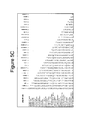
- FIG. 5C is a distance matrix of nucleotide differences based on the 28S (10F-12R) amplicon of 30 human fungal pathogens.
- FIG. 5D is a distance matrix of nucleotide differences based on the 28S (12F-13R) amplicon of 30 human fungal pathogens.
- FIG. 5E is a distance matrix of nucleotide differences based on the 28S (15F-22R) amplicon of 30 human fungal pathogens.
- FIG. 5F is a distance matrix of nucleotide differences based on the 28S (18F-22R) amplicon of 30 human fungal pathogens.
- FIG. 5G is a distance matrix of nucleotide differences based on the 28S (18F-23R) amplicon of 30 human fungal pathogens.
- FIG. 5H is a distance matrix of nucleotide differences based on the 28S (23F-25R) amplicon of 26 human fungal pathogens.
- FIG. 6 displays the ability of the 28S(10F-12R) amplicon to distinguish between 51 different fungal species spanning 30 genera as a phylogenetic tree which was constructed based on the neighbor joining tree building method and distances estimated using Tajima-Nei algorithm.
- FIG. 7 displays the ability of the ITS2(5.8SF-1R) amplicon to distinguish between 51 different fungal species spanning 30 genera as a phylogenetic tree which was constructed based on the neighbor joining tree building method and distances estimated using Tajima-Nei algorithm.
- FIG. 8 is a partial rRNA nucleic acid sequence of Absidia corymbifera strain (SEQ ID NO: 55).
- FIG. 9 is a partial rRNA nucleic acid sequence of Cunninghamella bertholletiae strain ATCC # 42115 (SEQ ID NO: 56).
- FIG. 10 is a partial rRNA nucleic acid sequence of Fusarium solani strain, ATCC # 56480 (SEQ ID NO: 57).
- FIG. 11 is a partial rRNA nucleic acid sequence of Mucor racemosus strain, ATCC # 42647, (SEQ ID NO: 58).
- FIG. 12 is a partial rRNA nucleic acid sequence of Paecilomyces variotii strain, ATCC # 10865, (SEQ ID NO: 59).
- FIG. 13 is a partial rRNA nucleic acid sequence of Penicillium chrysogenum strain, ATCC # 10108, (SEQ ID NO: 60).
- FIG. 14 is a partial rRNA nucleic acid sequence of Rhizomucor miehei strain, ATCC # 46345, (SEQ ID NO: 61).
- FIG. 15 is a partial rRNA nucleic acid sequence of Rhodotorula glutinis strain, ATCC # 16726, (SEQ ID NO: 62).
- FIG. 16 is a partial rRNA nucleic acid sequence of Scedosporium apiospermum strain, ATCC # 28206, (SEQ ID NO: 63).
- FIG. 17 is a partial rRNA nucleic acid sequence of Antrodia vaillantii strain (SEQ ID NO: 64).
- FIG. 18 is a partial rRNA nucleic acid sequence of Aspergillus fumigatus strain (SEQ ID NO: 65).
- FIG. 19 is a partial rRNA nucleic acid sequence of Aspergillus niger strain (SEQ ID NO: 66).
- FIG. 20 is a partial rRNA nucleic acid sequence of Aspergillus oryzae strain (SEQ ID NO: 67).
- FIG. 21 is a partial rRNA nucleic acid sequence of Aspergillus terreus strain (SEQ ID NO: 68).
- FIG. 22 is a partial rRNA nucleic acid sequence of Batrachochytrium dendrobatidis strain (SEQ ID NO: 69).
- FIG. 23 is a partial rRNA nucleic acid sequence of Botrytis cinerea strain (SEQ ID NO: 70).
- FIG. 24 is a partial rRNA nucleic acid sequence of Candida albicans strain (SEQ ID NO: 71).
- FIG. 25 is a partial rRNA nucleic acid sequence of Candida dublineinsis strain (SEQ ID NO: 72).
- FIG. 26 is a partial rRNA nucleic acid sequence of Candida glabrata strain (SEQ ID NO: 73).
- FIG. 27 is a partial rRNA nucleic acid sequence of Candida gulliermundei strain (SEQ ID NO: 74).
- FIG. 28 is a partial rRNA nucleic acid sequence of Candida kefyr strain (SEQ ID NO: 75).
- FIG. 29 is a partial rRNA nucleic acid sequence of Candida krusei strain (SEQ ID NO: 76).
- FIG. 30 is a partial rRNA nucleic acid sequence of Candida lipolytica strain (SEQ ID NO: 77).
- FIG. 31 is a partial rRNA nucleic acid sequence of Candida lusitaniae strain (SEQ ID NO: 78).
- FIG. 32 is a partial rRNA nucleic acid sequence of Candida parapsilosis strain (SEQ ID NO: 79).
- FIG. 33 is a partial rRNA nucleic acid sequence of Candida tropicalis strain (SEQ ID NO: 80).
- FIG. 34 is a partial rRNA nucleic acid sequence of Chaetomium globosum strain (SEQ ID NO: 81).
- FIG. 35 is a partial rRNA nucleic acid sequence of Coccidioides immitis strain (SEQ ID NO: 82).
- FIG. 36 is a partial rRNA nucleic acid sequence of Coccidioides posadasii strain (SEQ ID NO: 83).
- FIG. 37 is a partial rRNA nucleic acid sequence of Cryptococcus neoformans strain (SEQ ID NO: 84).
- FIG. 38 is a partial rRNA nucleic acid sequence of Fusarium graminearum strain (SEQ ID NO: 85).
- FIG. 39 is a partial rRNA nucleic acid sequence of Fusarium oxysporum strain (SEQ ID NO: 86).
- FIG. 40 is a partial rRNA nucleic acid sequence of Histoplasma capsulatum strain (SEQ ID NO: 87).
- FIG. 41 is a partial rRNA nucleic acid sequence of Hypocrea jecorina strain (SEQ ID NO: 88).
- FIG. 42 is a partial rRNA nucleic acid sequence of Lodderomyces elongisporus strain (SEQ ID NO: 89).
- FIG. 43 is a partial rRNA nucleic acid sequence of Magnaporthe grisea strain (SEQ ID NO: 90).
- FIG. 44 is a partial rRNA nucleic acid sequence of Metarhizium anisopliae strain (SEQ ID NO: 91).
- FIG. 45 is a partial rRNA nucleic acid sequence of Microsporum gypseum strain (SEQ ID NO: 92).
- FIG. 46 is a partial rRNA nucleic acid sequence of Mucor racemosus strain (SEQ ID NO: 93).
- FIG. 47 is a partial rRNA nucleic acid sequence of Neurospora crassa strain (SEQ ID NO: 94).
- FIG. 48 is a partial rRNA nucleic acid sequence of Paracoccidioides brasiliens strain (SEQ ID NO: 95).
- FIG. 49 is a partial rRNA nucleic acid sequence of Pneumocystis carinii strain (SEQ ID NO: 96).
- FIG. 50 is a partial rRNA nucleic acid sequence of Penicillium verrucosum strain (SEQ ID NO: 97).
- FIG. 51 is a partial rRNA nucleic acid sequence of Pichia stipitis strain (SEQ ID NO: 98).
- FIG. 52 is a partial rRNA nucleic acid sequence of Rhizomucor miehei strain (SEQ ID NO: 99).
- FIG. 53 is a partial rRNA nucleic acid sequence of Rhizopus oryzae strain (SEQ ID NO: 100).
- FIG. 54 is a partial rRNA nucleic acid sequence of Saccharomyces cerevisiae strain (SEQ ID NO: 101).
- FIG. 55 is a partial rRNA nucleic acid sequence of Schizosaccharomyces japonicus strain (SEQ ID NO: 102).
- FIG. 56 is a partial rRNA nucleic acid sequence of Schizosaccharomyces pombe strain (SEQ ID NO: 103).
- FIG. 57 is a partial rRNA nucleic acid sequence of Sclerotinia sclerotiorum strain (SEQ ID NO: 104).
- FIG. 58 is a partial rRNA nucleic acid sequence of Stagonospora nodorum strain (SEQ ID NO: 105).
- FIG. 59 is a partial rRNA nucleic acid sequence of Umbilicaria esculenta strain (SEQ ID NO: 106).
- FIG. 60 is a partial rRNA nucleic acid sequence of Uncinocarpus reesii strain (SEQ ID NO: 107).
- FIG. 61 illustrates a map of a fungal 28S rRNA gene and corresponding positions of twenty-seven broad-range fungal PCR primers for sequencing and PCR assay development.
- FIG. 62 illustrates a two-dimensional melt curve plot based on the broad-range fungal qPCR assays ITS2(5.8SF-1R) and 28S(10F-12R) allowing rapid identification of species.
- FIG. 63 illustrates melt temperature curves of pathogenic fungi amplified from blood, representing the 10-12 amplicon on the fungal 28S rRNA gene.
- the present disclosure is based on the unexpected discovery that a specific fungal pathogen in a patient sample may be rapidly identified using broad-range PCR primers that specifically amplify fungal DNA including a portion of the rRNA gene, including a portion of an internal transcribed spacer 1 (ITS-1) region and/or a portion of a 28S rRNA gene.
- Methods using the primers and primer sets provided herein uniquely identify and differentiate among at least 27 different species of fungal pathogens, even in the presence of human DNA.
- the present methods are useful in a clinical setting for the rapid identification of one or more fungal pathogen(s) in a patient sample.
- the primers, primer sets, and methods provided herein have both excellent analytical sensitivity and species level resolution which helps to overcome the potential shortcomings of the ITS regions.
- a single amplicon approach may be insufficient, therefore use of more than one PCR target may be optimal.
- the exemplary combination of ITS2(5.8SF-1R) and 28S(10F-12R) amplicons provides effective analytical sensitivity and potential for fungal species resolution.
- Optimal PCR assays were selected based on their ability to detect phylogentically diverse fungi and amplify small quantities of fungal DNA in the presence of large quantities of human DNA.
- the analysis of this region of the rRNA operon showed that there is nearly 2800 by of sequence beyond the D1-D2 region which is useful for the development of broad-range fungal PCR assays.
- the 28S rRNA gene beyond the D1-D2 region was found to be useful for the design of broad-range fungal PCR assays with good species-level resolution and the potential to detect the equivalent of a single fungal genome (30 fg) in a background of 1 ⁇ g of human DNA, representing a 30,000,000 fold excess of non-fungal DNA.
- an “individual” or “subject”, “mammal”, “patient” or “animal”, as used herein, refers to vertebrates that support a fungal infection, including, but not limited to, birds (such as water fowl and chickens) and members of the mammalian species, such as canine, feline, lupine, mustela, rodent (racine, and murine, etc.), equine, bovine, ovine, caprine, porcine species, and primates, the latter including humans.
- an isolated biological material can be free of some or all cellular components, i.e., components of the cells in which the native material occurs naturally (e.g., cytoplasmic or membrane component). A material shall be deemed isolated if it is present in a cell extract or supernatant.
- an isolated nucleic acid includes a PCR product, an isolated mRNA, a cDNA, or a restriction fragment.
- an isolated nucleic acid is preferably excised from the chromosome in which it may be found, and more preferably is no longer joined or proximal to non-coding regions (but may be joined to its native regulatory regions or portions thereof), or to other genes, located upstream or downstream of the gene contained by the isolated nucleic acid molecule when found in the chromosome.
- the isolated nucleic acid lacks one or more introns. Isolated nucleic acid molecules include sequences inserted into plasmids, cosmids, artificial chromosomes, and the like, i.e., when it forms part of a chimeric recombinant nucleic acid construct.
- a recombinant nucleic acid is an isolated nucleic acid.
- An isolated protein may be associated with other proteins or nucleic acids, or both, with which it associates in the cell, or with cellular membranes if it is a membrane-associated protein.
- An isolated organelle, cell, or tissue is removed from the anatomical site in which it is found in an organism.
- An isolated material may be, but need not be, purified.
- purified refers to material that has been isolated under conditions that reduce or eliminate the presence of unrelated materials, i.e. contaminants, including native materials from which the material is obtained.
- a purified fungal DNA is preferably substantially free of cell or culture components, including tissue culture components, contaminants, and the like.
- substantially free is used operationally, in the context of analytical testing of the material.
- purified material substantially free of contaminants is at least 50% pure; more preferably, at least 90% pure, and more preferably still at least 99% pure. Purity can be evaluated by chromatography, gel electrophoresis, immunoassay, composition analysis, biological assay, and other methods known in the art.
- the terms “include” and “comprise” are used synonymously. It should be understood that the terms “a” and “an” as used herein refer to “one or more” of the enumerated components. The use of the alternative (e.g., “or”) should be understood to mean either one, both, or any combination thereof of the alternatives.
- any concentration range, percentage range, ratio range, or integer range is to be understood to include the value of any integer within the recited range and, when appropriate, fractions thereof (such as one tenth and one hundredth of an integer), unless otherwise indicated.
- any number range recited herein relating to any physical feature, such as polymer subunits, size or thickness are to be understood to include any integer within the recited range, unless otherwise indicated.
- “about” or “consisting essentially of” mean ⁇ 20% of the indicated range, value, or structure, unless otherwise indicated.
- the term “about” or “approximately” means within a statistically meaningful range of a value. Such a range can be within an order of magnitude, preferably within 50%, more preferably within 20%, more preferably still within 10%, and even more preferably within 5% of a given value or range.
- the allowable variation encompassed by the term “about” or “approximately” depends on the particular system under study, and can be readily appreciated by one of ordinary skill in the art.
- tig refers to one of a set of overlapping clones that represent a continuous region of DNA. However, in certain embodiments, “contig” also refers to a contiguous sequence constructed from many clone sequences or PCR products, and herein, is used synonymously with the term “sequence.”
- Endpoint PCR is understood to mean a semi-quantitative approach to measuring relative amounts of template (DNA) in a sample involving the measurement of the amount of PCR product present at the end of a PCR reaction.
- end-point PCR is performed by resolving the PCR amplicon on an agarose gel and staining the gel with an “intercalating” dye, such as, for example, ethidium bromide.
- Ethidium bromide binds between the bases of the DNA helix. When it is inserted into the DNA, it becomes much more fluorescent when exposed to ultraviolet light as compared to ethidium bromide just in solution. This characteristic of ethidium bromide permits semi-quantitative measurements of the amount of DNA in the PCR product by measuring the degree of fluorescence of the PCR product in the gel.
- sample as used in the present disclosure can be any tissue, fluid, or other source of DNA from a patient or mammal.
- DNA typing involves the analysis of alleles of genomic DNA with characteristics of interest, commonly referred to as “markers.” Most typing methods in use today are specifically designed to detect and analyze differences in the length and/or sequence of one or more regions of DNA markers known to appear in at least two different forms in a population. Such length and/or sequence variation is referred to as “polymorphism.” Any region (i.e., “locus”) of DNA in which such a variation occurs is referred to as a “polymorphic locus.”
- nucleic acid and oligonucleotide refer to polydeoxyribonucleotides (containing 2-deoxy-D-ribose), to polyribonucleotides (containing D-ribose), and to any other type of polynucleotide which is an N glycoside of a purine or pyrimidine base.
- nucleic acid and oligonucleotide refer only to the primary structure of the molecule. Thus, these terms include double- and single-stranded DNA, as well as double- and single-stranded RNA.
- an oligonucleotide also can comprise non-purine or non-pyrimidine nucleotide analogs.
- the length of a nucleic acid sequence is referred to as the number of “base pairs (bp)” present in the double-stranded nucleic acid sequence.
- nucleic acid molecules of sequences disclosed herein are written according to The International Union of Pure and Applied Chemistry (IUPAC) DNA codes. Specifically, “A” is Adenine, “C” is Cytosine, “G” is Guanine, “T” is Thymine, “U” is Uracil, “R” is any Purine (A or G), “Y” is any Pyrimidine (C, T, or U), “M” is C or A, “K” is T, U, or G, “W” is T, U, or A, “S” is C or G, “B” is C, T, U, or G (not A), “D” is A, T, U, or G (not C), “H” is A, T, U, or C (not G), “V” is A, C, or G (not T, not U), and “N” is any base (A, C, G, T, or U).
- IUPAC International Union of Pure and Applied Chemistry
- the amount of fungal DNA present in a sample is described in terms of the “fold-excess” of human or non-fungal DNA over the amount of fungal DNA present in the same sample. For example, if 1 ⁇ g of human genomic DNA is present in a sample that has 0.001 ⁇ g of fungal DNA, then the human DNA is understood to be in 1000-fold excess of the fungal DNA.
- Oligonucleotides can be prepared by any suitable method, including direct chemical synthesis by a method such as the phosphotriester method of Narang et al. (1979) Meth. Enzymol. 68:90-99; the phosphodiester method of Brown et al., (1979) Meth. Enzymol. 68:109-151; the diethylphosphoramidite method of Beaucage et al. (1981) Tetrahedron Lett. 22:1859-1862; and the solid support method of U.S. Pat. No. 4,458,066, each incorporated herein by reference.
- a review of synthesis methods of conjugates of oligonucleotides and modified nucleotides is provided in Goodchild (1990) Bioconjugate Chemistry 1(3):165-187, incorporated herein by reference.
- primer refers to an oligonucleotide capable of acting as a point of initiation of DNA synthesis under conditions in which synthesis of a primer extension product complementary to a nucleic acid strand is induced, i.e., either in the presence of four different nucleoside triphosphates and an agent for extension (e.g., a DNA polymerase or reverse transcriptase) in an appropriate buffer and at a suitable temperature.
- a primer is preferably a single-stranded DNA.
- the appropriate length of a primer depends on the intended use of the primer but typically ranges from 6 to 50 nucleotides, preferably from 15-35 nucleotides.
- a primer need not reflect the exact sequence of the template nucleic acid, but must be sufficiently complementary to hybridize with the template.
- the design of suitable primers for the amplification of a given target sequence is well known in the art and described in the literature cited herein.
- a “forward primer” is understood to mean a primer that is capable of hybridizing to a region of DNA along the 5′ (coding) strand of DNA.
- a “reverse” primer is understood to mean a primer that is capable of hybridizing to a region of DNA along the 3′ (non-coding) strand of DNA.
- Primers can incorporate additional features which allow for the detection or immobilization of the primer but do not alter the basic property of the primer, that of acting as a point of initiation of DNA synthesis.
- primers may contain an additional nucleic acid sequence at the 5′ end which does not hybridize to the target nucleic acid, but which facilitates cloning of the amplified product.
- the region of the primer which is sufficiently complementary to the template to hybridize is referred to herein as the hybridizing region.
- a “primer set” or “primer pair” refers to a specific combination of a forward primer and one or more reverse primers.
- Some “primer sets” or “primer pairs” may include, for example, one forward primer and two reverse primers (e.g., a primer set comprising SEQ ID NO: 12, SEQ ID NO: 41, and SEQ ID NO: 108).
- the “primer set” or “primer pair” may be used in a PCR reaction to generate a specific PCR product or amplicon.
- amplicon refers to the DNA sequence generated by a PCR or qPCR reaction. “Amplicon” may further be used synonymously with the term “PCR product.”
- the term “primer” is also intended to encompass the oligonucleotides used in ligation-mediated amplification processes, in which one oligonucleotide is “extended” by ligation to a second oligonucleotide which hybridizes at an adjacent position.
- the term “primer extension”, as used herein, refers to both the polymerization of individual nucleoside triphosphates using the primer as a point of initiation of DNA synthesis and to the ligation of two oligonucleotides to form an extended product.
- target is synonymous and refer to a region or subsequence of a nucleic acid which is to be amplified or detected.
- hybridization refers to the formation of a duplex structure by two single-stranded nucleic acids due to complementary base pairing. Hybridization can occur between fully complementary nucleic acid strands or between “substantially complementary” nucleic acid strands that contain minor regions of mismatch. Conditions under which only fully complementary nucleic acid strands will hybridize are referred to as “stringent hybridization conditions” or “sequence-specific hybridization conditions”. Stable duplexes of substantially complementary sequences can be achieved under less stringent hybridization conditions; the degree of mismatch tolerated can be controlled by suitable adjustment of the hybridization conditions.
- nucleic acid technology can determine duplex stability empirically considering a number of variables including, for example, the length and base pair composition of the oligonucleotides, ionic strength, and incidence of mismatched base pairs, following the guidance provided by the art (see, e.g., Sambrook et al., (1989) Molecular Cloning—A Laboratory Manual (Cold Spring Harbor Laboratory, Cold Spring Harbor, N.Y.); and Wetmur (1991) Critical Review in Biochem. and Mol. Biol. 26(3/4):227-259; both incorporated herein by reference).
- Amplification reaction refers to any chemical reaction, including an enzymatic reaction, which results in increased copies of a template nucleic acid sequence or results in transcription of a template nucleic acid.
- Amplification reactions include reverse transcription and the polymerase chain reaction (PCR), including Real Time PCR (see U.S. Pat. Nos. 4,683,195 and 4,683,202; PCR Protocols: A Guide to Methods and Applications (Innis et al., eds, 1990)).
- Exemplary “amplification reactions conditions” or “amplification conditions” typically comprise either two or three step cycles. Two step cycles have a denaturation step followed by a hybridization/elongation step. Three step cycles comprise a denaturation step followed by a hybridization step followed by a separate elongation step.
- PCR Polymerase chain reaction
- thermo-stable DNA polymerase (Taq polymerase) that was isolated from Thermus aquaticus , a bacterium that grows in hot pools; as a result it is not necessary to add new polymerase in every round of amplification.
- Taq polymerase thermo-stable DNA polymerase
- the PCR product is analyzed on an agarose gel and is abundant enough to be detected with an ethidium bromide stain.
- real-time PCR also called quantitative real time PCR, quantitative PCR (Q-PCR/qPCR), or kinetic polymerase chain reaction
- quantitative real time PCR also called quantitative real time PCR, quantitative PCR (Q-PCR/qPCR), or kinetic polymerase chain reaction
- qPCR enables both detection and quantification (as absolute number of copies or relative amount when normalized to DNA input or additional normalizing genes) of a specific sequence in a DNA sample.
- qPCR may be used to quantify the amount of fungal DNA in a patient sample.
- the procedure follows the general principle of PCR; its key feature is that the amplified DNA is quantified as it accumulates in the reaction in real time after each amplification cycle.
- Two common methods of quantification are the use of fluorescent dyes that intercalate with double-stranded DNA, and modified DNA oligonucleotide probes that fluoresce upon binding to complementary DNA (such as with molecular beacons) or with completion of each PCR cycle (such as with dual labeled probes rendered more fluorescent with the 5′ exonuclease activity of polymerase enzymes).
- a “polymerase” refers to an enzyme that catalyzes the polymerization of nucleotides. Generally, the enzyme will initiate synthesis at the 3′-end of the primer annealed to a nucleic acid template sequence.
- DNA polymerase catalyzes the polymerization of deoxyribonucleotides.
- Known DNA polymerases include, for example, Pyrococcus furiosus (Pfu) DNA polymerase (Lundberg et al., (1991) Gene 108:1), E. coli DNA polymerase I (Lecomte and Doubleday (1983) Nucleic Acids Res. 11:7505), T7 DNA polymerase (Nordstrom et al.
- a primer is “specific,” for a target sequence if, when used in an amplification reaction under sufficiently stringent conditions, the primer hybridizes primarily only to the target nucleic acid.
- a primer is specific for a target sequence if the primer-target duplex stability is greater than the stability of a duplex formed between the primer and any other sequence found in the sample.
- salt conditions such as salt conditions as well as base composition of the primer and the location of the mismatches, will affect the specificity of the primer, and that routine experimental confirmation of the primer specificity will be needed in most cases.
- Hybridization conditions can be chosen under which the primer can form stable duplexes only with a target sequence.
- target-specific primers under suitably stringent amplification conditions enables the specific amplification of those target sequences which contain the target primer binding sites.
- sequence-specific amplification conditions enables the specific amplification of those target sequences which contain the exactly complementary primer binding sites.
- complementary refers to a nucleic acid molecule that can form hydrogen bond(s) with another nucleic acid molecule by either traditional Watson-Crick base pairing or other non-traditional types of pairing (e.g., Hoogsteen or reversed Hoogsteen hydrogen bonding) between complementary nucleosides or nucleotides.
- nucleic acid molecule need not be 100% complementary to a target nucleic acid sequence to be specifically hybridizable. That is, two or more nucleic acid molecules may be less than fully complementary and is indicated by a percentage of contiguous residues in a nucleic acid molecule that can form hydrogen bonds with a second nucleic acid molecule. For example, if a first nucleic acid molecule has 10 nucleotides and a second nucleic acid molecule has 10 nucleotides, then base pairing of 5, 6, 7, 8, 9, or 10 nucleotides between the first and second nucleic acid molecules represents 50%, 60%, 70%, 80%, 90%, and 100% complementarity, respectively.
- “Perfectly” or “fully” complementary nucleic acid molecules means those in which all the contiguous residues of a first nucleic acid molecule will hydrogen bond with the same number of contiguous residues in a second nucleic acid molecule, wherein the nucleic acid molecules either both have the same number of nucleotides (i.e., have the same length) or the two molecules have different lengths.
- non-specific amplification refers to the amplification of nucleic acid sequences other than the target sequence which results from primers hybridizing to sequences other than the target sequence and then serving as a substrate for primer extension.
- the hybridization of a primer to a non-target sequence is referred to as “non-specific hybridization” and is apt to occur especially during the lower temperature, reduced stringency, pre-amplification conditions.
- primer dimer refers to a template-independent non-specific amplification product, which is believed to result from primer extensions wherein another primer serves as a template. Although primer dimers frequently appear to be a concatamer of two primers, i.e., a dimer, concatamers of more than two primers also occur.
- primer dimer is used herein generically to encompass a template-independent non-specific amplification product.
- reaction mixture refers to a solution containing reagents necessary to carry out a given reaction.
- a “PCR reaction mixture” typically contains oligonucleotide primers, a DNA polymerase (most typically a thermostable DNA polymerase), dNTPs, and a divalent metal cation in a suitable buffer.
- reaction mixture is referred to as complete if it contains all reagents necessary to enable the reaction, and incomplete if it contains only a subset of the necessary reagents.
- reaction components are routinely stored as separate solutions, each containing a subset of the total components, for reasons of convenience, storage stability, or to allow for application-dependent adjustment of the component concentrations, and that reaction components are combined prior to the reaction to create a complete reaction mixture.
- reaction components are packaged separately for commercialization and that useful commercial kits may contain any subset of the reaction components which includes the blocked primers of the disclosure.
- the term “activated,” as used herein, refers to a primer or other oligonucleotide that is capable of participating in a reaction with DNA polymerase or DNA ligase.
- a primer or other oligonucleotide becomes activated when it hybridizes to a substantially complementary nucleic acid sequence and is chemically modified so that it can interact with a DNA polymerase or a DNA ligase.
- a 3′-blocking group can be removed from the primer by, for example, a cleaving enzyme such that DNA polymerase can bind to the 3′ end of the primer and promote primer extension.
- fluorescent generation probe refers either to a) an oligonucleotide having an attached fluorophore and quencher, and optionally a minor groove binder or to b) a DNA binding reagent such as Sybr® green dye.
- fluorescent label refers to compounds with a fluorescent emission maximum between about 350 and 900 nm.
- fluorophores can be used, including but not limited to: 5-FAM (also called 5-carboxyfluorescein; also called Spiro(isobenzofuran-1(3H), 9′-(9H)xanthene)-5-carboxylic acid, 3′,6′-dihydroxy-3-oxo-6-carboxyfluorescein); 5-Hexachloro-Fluorescein ([4,7,2′,4′,5′,7′-hexachloro-(3′,6′-dipivaloyl-fluoresceinyl)-6-carboxyli-c acid]); 6-Hexachloro-Fluorescein ([4,7,2′,4′,5′,7′-hexachloro-(3′,6′-dipivaloylfluoresceinyl)
- ligation refers to the covalent joining of two polynucleotide ends.
- ligation involves the covalent joining of a 3′ end of a first polynucleotide (the acceptor) to a 5′ end of a second polynucleotide (the donor). Ligation results in a phosphodiester bond being formed between the polynucleotide ends.
- ligation may be mediated by any enzyme, chemical, or process that results in a covalent joining of the polynucleotide ends.
- ligation is mediated by a ligase enzyme.
- ligase refers to an enzyme that is capable of covalently linking the 3′ hydroxyl group of a nucleotide to the 5′ phosphate group of a second nucleotide.
- examples of ligases include E. coli DNA ligase, T4 DNA ligase, etc.
- the ligation reaction can be employed in DNA amplification methods such as the “ligase chain reaction” (LCR), also referred to as the “ligase amplification reaction” (LAR), see Barany (1991) Proc. Natl. Acad. Sci. U.S.A. 88:189; and Wu and Wallace (1989) Genomics 4:560, incorporated herein by reference.
- LCR ligase chain reaction
- LAR ligase amplification reaction
- ligase will covalently link each set of hybridized molecules.
- two probes are ligated together only when they base-pair with sequences in the target sample, without gaps or mismatches. Repeated cycles of denaturation, hybridization and ligation amplify a short segment of DNA. LCR has also been used in combination with PCR to achieve enhanced detection of single-base changes, see Segev PCT Pub. No. WO/9001069.
- the term “conserved region” or “conserved sequence” refers to a nucleic acid sequence in a region of a gene that is the same or highly similar across different species.
- a sequence or region of a gene that is conserved may have the same nucleic acid sequence in several types of fungal species, or, in some cases, may have the same or highly similar sequence across different taxonomic phyla (e.g., a human DNA sequence and a fungal DNA sequence in a highly conserved region of a gene may be the same or highly similar).
- a “highly variable” or “hypervariable” region or sequence of gene is not conserved across species or phyla, and will have many nucleotides differences in the hypervariable region in the gene from each species.
- the methods disclosed herein target the fungal rRNA operon, which is a continuous sequence made of the 18S, ITS1, 5.8S, ITS2, and 28S subunit regions [Iwen et al. (2002) Med. Mycol. 40:87-109]. Because certain aspects of the operon are highly conserved among a broad range of fungi, while other regions, such as the D1-D2 hypervariable region are not conserved among species, the DNA sequences of the operon can be targeted by broad range PCR assays for the identification of fungal infection and for the determination of the specific fungal species.
- the human rRNA operon is also a continuous sequence made of the 18S, ITS1, 5.8S, ITS2, and 28S subunit regions, and has considerable sequence homology with the fungal rRNA operon.
- a critical aspect of the present disclosure provides methods and PCR primers which do not cross-react with human DNA.
- the present disclosure provides PCR primers which amplify regions that are 3′ to the D1-D2 hypervariable region specifically because they are discovered to have less cross-reactivity to human DNA. This is especially critical for the identification of fungal DNA in patient samples, which also contain human DNA.
- “Broad-range” PCR primers as disclosed herein may be understood to be primers that hybridize with conserved regions of fungal DNA, and thus are useful in PCR assays that detect the presence of a wide range of fungal pathogens.
- the method includes the steps of (a) isolating a patient sample, (b) carrying out a PCR reaction on the patient sample to generate a PCR amplicon that includes a region of a fungal 28S ribosomal RNA (rRNA) gene, wherein the PCR reaction uses a primer set having a forward primer and a reverse primer wherein at least one of the forward primer and the reverse primer is complementary to the fungal 28S rRNA gene, and (c) detecting the PCR amplicon.
- the patient sample may be, for example, a blood sample, a sputum sample, a lung lavage fluid sample, or a tissue biopsy sample. Any fluid, tissue, or other source of DNA from a patient may constitute a sample in the present disclosure.
- the PCR reaction carried out on the patient sample may be performed according to any of the methods known in the art.
- the purpose of the PCR reaction is to amplify a target sequence within a fungal DNA sequence, thereby generating a PCR amplicon.
- the region amplified by the PCR reaction is in the 28S region of the fungal rRNA gene. More preferably, the region of the fungal 28S rRNA gene detected by PCR includes a sequence that is 3′ to a D1-D2 highly variable region of the fungal 28S rRNA gene.
- the PCR assays of the present disclosure target this region, achieving resolution among different species of fungi without cross-reacting with or being inhibited by the presence of human DNA.
- PCR reactions are used to detect fungal DNA in a sample.
- qPCR reactions are used to detect fungal DNA in a sample.
- alternative methods other than PCR such as ligase chain reaction, may be used to detect the presence of fungal DNA in a sample.
- Nucleic Acid Sequence Based Amplification NASBA
- NASBA Nucleic Acid Sequence Based Amplification
- the methods for detecting a fungal pathogen disclosed herein further involve the step of sequencing the PCR amplicon derived from sequencing.
- the PCR amplicon is between 50 and 1000 base pairs, and preferably, between 75 and 400 base pairs. Smaller amplicon sizes are desirable, since they are easier to sequence and useful for qPCR reactions. However, it is also important that the amplicon be large enough to facilitate accurate species identification, e.g., enhance resolution among different fungal species.
- Sequencing of the PCR amplicon may be carried out according to any methods known in the art suitable for determining the sequence of a PCR amplicon.
- the sequences of the PCR amplicons disclosed in the present invention are unique to each type of fungal pathogen, thereby allowing identification of the specific type of fungal DNA in a sample.
- each primer of the primer set in the PCR reaction specifically binds only to a fungal DNA.
- each primer of the primer set specifically binds only to a fungal DNA in the presence of a non-fungal DNA.
- the non-fungal DNA is mammalian DNA.
- the mammalian DNA is human DNA.
- the non-fungal DNA is in greater than 1,000,000-fold, 5,000,000-fold, or 30,000,000-fold mass excess of the fungal DNA.
- methods for detecting a fungal pathogen are provided, wherein the fungal pathogen causes a fungal infection selected from the group consisting of aspergillosis, candidiasis, zygomycosis, scedosporiosis, fusariosis, cryptococcosis, histoplasmosis, coccidioidomycosis, and blastomycosis.
- a fungal infection selected from the group consisting of aspergillosis, candidiasis, zygomycosis, scedosporiosis, fusariosis, cryptococcosis, histoplasmosis, coccidioidomycosis, and blastomycosis.
- the forward primer of the PCR reaction is complementary to a fungal 18S rRNA gene and the reverse primer is complementary to a fungal 28S rRNA gene.
- the forward primer comprises the nucleotide sequence 5′-GTAAAAGTCGTAACAAGGTTTC-3′ (SEQ ID NO: 1).
- the forward primer is complementary to a fungal 5.8S rRNA gene and the reverse primer is complementary to a fungal 28S rRNA gene.
- the forward primer includes the nucleotide sequence 5′-GTGAATCATCGARTCTTTGAAC-3′ (SEQ ID NO: 2).
- the forward primer and the reverse primer of the PCR reaction for detecting fungal DNA in a patient sample are both complementary to a fungal 28S rRNA gene.
- a primer set for detecting a fungal DNA by PCR wherein the primer set includes a forward primer and a reverse primer wherein at least one of the forward primer and the reverse primer is complementary to a fungal 28S ribosomal RNA (rRNA) gene.
- the forward primer or the reverse primer of the primer set is complementary to a sequence that is 3′ to a D1-D2 highly variable region in the fungal 28S ribosomal rRNA gene.
- the forward primer of the primer set is complementary to a fungal 18S rRNA gene and the reverse primer is complementary to a fungal 28S rRNA gene.
- the forward primer and the reverse primer of the primer set are both complementary to a fungal 28S rRNA gene.
- the forward primer of the PCR reaction or of the primer set for detecting fungal DNA in a sample may have one of the following sequences:
- 5′-GTAAAAGTCGTAACAAGGTTTC-3′ (SEQ ID NO: 1) 5′-GTGAATCATCGARTCTTTGAAC-3′, (SEQ ID NO: 2) 5′-TACCCGCTGAACTTAAGCATA-3′, (SEQ ID NO: 3) 5′-GCATATCAATAAGCGGAGGAAA-3′, (SEQ ID NO: 4) 5′-AGTARCGGCGAGTGAAGCGG-3′, (SEQ ID NO: 5) 5′-AGCTCAAATTTGAAASCTGG-3′, (SEQ ID NO: 6) 5′-CTTCCCTTTCAACAATTTCACRT-3′, (SEQ ID NO: 7) 5′-AGGTAAAGCGAATGATTAG-3′, (SEQ ID NO: 8) 5′-CTTGTTRCTTARTTGAACGTG-3′, (SEQ ID NO: 9) 5′-ACCACAAAAGGTGTTAGTWCATC-3′, (SEQ ID NO: 10) 5′-GAAGTGGGGAAAGGTTCC-3′
- the PCR reaction for detecting fungal DNA includes a forward and reverse primer pair (or primer set) selected from the group consisting of (SEQ ID NO: 2 and SEQ ID NO: 31), (SEQ ID NO: 2 and SEQ ID NO: 32), (SEQ ID NO: 11 and SEQ ID NO: 41), (SEQ ID NO: 1 and SEQ ID NO: 29), (SEQ ID NO: 2 and SEQ ID NO: 30), (SEQ ID NO: 12 and SEQ ID NO: 41), (SEQ ID NO: 14 and SEQ ID NO: 42), (SEQ ID NO: 17 and SEQ ID NO: 51), (SEQ ID NO: 20 and SEQ ID NO: 51), (SEQ ID NO: 20 and SEQ ID NO: 52), and (SEQ ID NO: 25 and SEQ ID NO: 54).
- a forward and reverse primer pair selected from the group consisting of (SEQ ID NO: 2 and SEQ ID NO: 31), (SEQ ID NO: 2 and SEQ ID NO: 32), (SEQ ID NO: 11 and SEQ ID NO
- the primer set includes a forward and reverse primer pair selected from the group consisting of (SEQ ID NO: 1 and SEQ ID NO: 29), (SEQ ID NO: 2 and SEQ ID NO: 30), (SEQ ID NO: 12 and SEQ ID NO: 41), (SEQ ID NO: 14 and SEQ ID NO: 42), (SEQ ID NO: 17 and SEQ ID NO: 51), (SEQ ID NO: 20 and SEQ ID NO: 51), (SEQ ID NO: 20 and SEQ ID NO: 52), and (SEQ ID NO: 25 and SEQ ID NO: 54), and still more preferably, the primer set includes a forward and reverse primer pair selected from the group consisting of (SEQ ID NO: 2 and SEQ ID NO: 30), (SEQ ID NO: 12 and SEQ ID NO: 41), and (SEQ ID NO: 12, SEQ ID NO: 41, and SEQ ID NO: 108).
- the primer set includes a forward and reverse primer pair selected from the group consisting of (SEQ ID NO: 11 and SEQ ID NO: 41), (SEQ ID NO: 12 and SEQ ID NO: 41), (SEQ ID NO: 12, SEQ ID NO: 41, and SEQ ID NO: 108), (SEQ ID NO: 14 and SEQ ID NO: 42), (SEQ ID NO: 17 and SEQ ID NO: 51), (SEQ ID NO: 20 and SEQ ID NO: 51), (SEQ ID NO: 20 and SEQ ID NO: 52), and (SEQ ID NO: 25 and SEQ ID NO: 54).
- a forward and reverse primer pair selected from the group consisting of (SEQ ID NO: 11 and SEQ ID NO: 41), (SEQ ID NO: 12 and SEQ ID NO: 41), (SEQ ID NO: 12, SEQ ID NO: 41, and SEQ ID NO: 108), (SEQ ID NO: 14 and SEQ ID NO: 42), (SEQ ID NO: 17 and SEQ ID NO: 51), (SEQ ID NO: 20 and
- any of the primer sequences disclosed herein may be modified without departing from the intended scope of the disclosure. Specifically, nucleotide substitutions, deletions and/or additions may be introduced into any of the primer sequences disclosed herein without altering the ability of the primers to identify fungal DNA. Moreover, it is to be understood that the lengths of the primers may be shorter or longer than the sequences disclosed herein.
- methods and primer sets for detecting fungal DNA which detect DNA from a fungal species such as, but not limited to Absidia corymbifera; Cunninghamella bertholletiae; Fusarium solani; Mucor racemosus; Paecilomyces variotii; Penicillium chrysogenum; Rhizomucor miehei; Rhodotorula glutinis; Scedosporium apiospermum; Antrodia vaillantii; Aspergillus fumigatus; Aspergillus niger; Aspergillus oryzae; Aspergillus terreus; Batracrochytrium dendrobatidis; Botrytis cinerea; Candida albicans; Candida dublineinsis; Candida glabrata; Candida gulliermundei; Candida kefyr; Candida krusei; Candida lipolytica; Candida lusitaniae; Candida parapsilosis;
- a fungal species such as,
- the methods described herein may be used to detect DNA from other known fungi not specifically disclosed herein and from newly identified fungal species.
- the methods provided herein are useful for detecting a broad range of fungal DNA, and are not limited to the specific examples of fungal species disclosed herein.
- Also disclosed herein are methods for identifying a primer set capable of detecting a fungal pathogen in a sample including the steps of: (a) obtaining the DNA sequence of at least the 28S region of a fungal rRNA operon, (b) designing a forward primer capable of hybridizing with the DNA sequence, (c) designing a reverse primer capable of hybridizing with the DNA sequence at a region in the DNA that is 3′ to the region to which the forward primer is capable of hybridizing, (d) testing whether the forward and reverse primers are capable of generating a PCR amplicon that is useful for identifying fungal DNA using a PCR reaction containing fungal DNA.
- the method also includes the steps of testing the forward and reverse primers in a PCR reaction containing fungal DNA and human DNA.
- the method includes running the PCR amplicon on an agarose gel and determining the product size.
- the method includes sequencing the PCR amplicon.
- the analytical sensitivity and cross-reactivity (i.e., degree of species resolution) of a specific primer set may be determined by testing the specific primer set on a panel of individual samples, each sample containing genomic DNA isolated from a single, distinct fungal species. An amplicon is generated by each PCR reaction containing the isolated genomic fungal DNA. Each amplicon is then sequenced and the sequences of each amplicon are compared. In certain embodiments, the sequence of each amplicon is compared using multiple sequence alignment, for example using the Clustal W algorithm. The Clustal W algorithm aligns two or more sequences simultaneously, such that regions of identical and similar residues are aligned.
- Clustal W does a pairwise comparison of every sequence first and then starts the multiple alignment with the pair of sequences that is most similar. Sequences are added one by one to the alignment based on their similarities to the starting pair.
- the software for using Clustal W alignment is freely available on the World Wide Web at the European Bioinformatics Institute website (http://www.ebi.ac.uk/Tools/clustalw2/index.html). Any algorithm suitable for comparing multiple sequences may be used, such as, e.g., the Needleman-Wunsch algorithm or the Smith-Waterman algorithm.
- the number of nucleotide differences among each amplicon is determined and assembled in a distance matrix, such as for example, using Microsoft Excel.
- the distance matrix is generated using Accelrys Gene® software (Accelrys, Inc., San Diego, Calif.). If species resolution within a genus or between any two species is desired, the comparison of distance matrix data can help select which amplicon (primer pair) would provide the most species resolution.
- phylogenetic trees may be assembled based on the Neighbor-Joining tree building method and distances estimated from the Tajima-Nei or absolute difference algorithms also using the Accelrys Gene® software (Accelrys, Inc.) or other similar tools.
- the Neighbor-Joining tree building method is described in detail in Saitou and Nei (1987) Mol. Biol. Evol. 4:406-25, and the Tajima-Nei algorithm is described in detail in Tajima and Nei (1984) Mol. Biol. Evol. 1:269-85, both incorporated herein by reference in their entireties.
- a phylogenetic tree compares the distance between two species, usually interpreted as evolutionary distance, as determined by the number of varying nucleotide positions in a sequence such as a specific PCR product generated by broad-range fungal PCR.
- Other examples of algorithms that may be used to construct phylogenetic trees include maximum likelihood, minimum evolution, and parsimony.
- Other distance-based methods include unweighted pair-group method using arithmetic averages, BIONJ, and the Weighbor algorithm or “weighted NJ”. These algorithms and methods are well known in the art and are described in detail in Hollich, V. et al. (2005) Molecular Biology and Evolution; 22(11):2257-2264, which is herein incorporated by reference in its entirety.
- the distance matrices and phylogenetic trees are used to determine which PCR primer sets generate amplicons that permit the highest degree of species resolution.
- the primers that give the highest degree of resolution among distinct fungal species are selected for further use.
- methods are provided for both the detection of fungal DNA in a patient sample, and further, for determining which type of fungal infection is present using two-dimensional melt curve analysis.
- a melting profile of a PCR amplicon can be characterized by measuring fluorescence of a DNA binding dye.
- each double-stranded DNA has its own specific melting temperature (Tm), which is defined as the temperature at which 50% of the DNA becomes single stranded.
- Tm melting temperature
- These melting temperatures are primarily determined by dsDNA length, degree of GC content (Tm is higher in GC-rich fragments), and degree of complementarity between strands (e.g., especially important in heteroduplexes consisting of a probe and a single-stranded target DNA sequence).
- a thermal cycler system records the total fluorescence generated by the fluorescent DNA binding dye binding to double-stranded DNA as temperature changes, and plots the fluorescence in real time as a function of temperature.
- the first derivative of this plot, dF/dT is the rate of change of fluorescence in the reaction, and a significant change in fluorescence accompanies the melting of the double-stranded PCR products.
- a plot of ⁇ dF/dT vs. temperature will display these changes in fluorescence as distinct peaks.
- the melting temperature (Tm) of each product is defined as the temperature at which the corresponding peak maximum occurs.
- each unique amplicon will have a unique Tm that distinguishes it from each different amplicon.
- the present disclosure provides methods for characterizing the melting profiles of amplicons generated from known fungi, and further, for using this information to infer the identity of a fungus from an unknown sample. Moreover, using information from more than one amplicon, as disclosed in the present invention, further increases species resolution. Specifically, in certain embodiments, a first amplicon is generated in the ITS2 region of the rRNA operon, and a second amplicon is generated in the 28S region. The Tm is determined for each amplicon, and the combination of the two Tm uniquely identifies a fungal species.
- a method for determining the identity of a fungal species in a patient sample includes the steps of: isolating the patient sample; carrying out a first PCR reaction to generate a first PCR amplicon, wherein the first PCR reaction includes a first primer set capable of amplifying a region in a fungal ribosomal RNA (rRNA) gene having an internal transcribed spacer 2 (ITS-2) sequence; carrying out a second PCR reaction to generate a second PCR amplicon, wherein the second PCR reaction has a second primer set capable of amplifying a region in a fungal ribosomal 28-S rRNA gene; and determining the melting temperature of the first PCR amplicon and of the second PCR amplicon, wherein the identity of the fungal species is determined by comparing the melting point of the first PCR amplicon and of the second PCR amplicon to known standards.
- rRNA fungal ribosomal RNA
- This method has the advantage that the melt curve analysis can be carried out very quickly, without the need for sequencing the PCR amplicon, at the end of each PCR reaction.
- the first and second PCR reactions carried out for determining the identity of a fungal species in a patient sample are each quantitative PCR (qPCR) reactions.
- the first primer set includes a forward primer sequence as set forth in SEQ ID NO: 2 and a reverse primer sequence as set forth in SEQ ID NO: 30, and the second primer set includes a forward primer sequence as set forth in SEQ ID NO: 12 and a reverse primer sequence as set forth in SEQ ID NO: 41.
- the second primer set includes a forward primer sequence as set forth in SEQ ID NO: 12, a first reverse primer sequence as set forth in SEQ ID NO: 41, and a second primer sequence as set forth in SEQ ID NO: 108.
- the two-dimensional melt curve analysis is useful for resolving multiple fungal infections in a single patient sample. For example, if DNA is present from two fungal species, then one primer set, targeted, for example, to the ITS2 region, will generate two distinct amplicons with distinct melting curves. Then, a second primer set, targeted, for example, to the 28S region in a separate PCR reaction, will amplify two distinct amplicons with two distinct melting temperatures. Since each fungal species has a unique combination of melting temperatures for each amplicon, these melting temperatures can be combined to determine which fungal species are present. Certain fungal species are more likely to be present in a co-infection, and this information can also be used to determine the two types of fungal species present in the sample. This method is highly useful in the clinical setting because it can be performed rapidly. According to conventional methods, the individual PCR products would have to be cloned before sequencing in order to resolve multiple fungal species.
- the two-dimensional melt curve analysis may be used to narrow down the list of possible fungi. Thereafter, a taxon-specific PCR reaction may be performed to confirm the suspected type of fungal infections. These taxon specific PCR reactions amplify regions of fungal DNA that are unique to a specific fungal species.
- This Example discloses the materials and methods used in Examples 2 through 6 of the present disclosure.
- Table 1 lists 9 clinically or phylogenetically relevant fungal pathogens subjected to sequencing of their ITS, 5.8S, and 28S rRNA genes.
- Table 2 lists 43 fungi of which the same gene sequences were obtained from publicly available genomic databases or GenBank®.
- Genomic DNA of the following organisms was used for analytical sensitivity testing to screen broad-range fungal primers: Aspergillus candidus (ATCC # 20022), Aspergillus flavus (ATCC # MYA-3631), Aspergillus fumigatus (ATCC # MYA-1163), Aspergillus oryzae (ATCC # 20719), Aspergillus terreus (ATCC # 10070), Aspergillus ustus (ATCC # 20063), Candida albicans (ATCC # 90028), Candida dubliniensis (ATCC # MYA-580), Candida glabrata (ATCC # 90876), Candida guilliermondii (ATCC # 90877), Candida kefyr (ATCC # 28838), Candida krusei (clinical isolate), Candida lusitaniae (ATCC # 42720), Candida parapsilosis, Candida tropicalis (clinical isolate), Rhizopus oryzae (ATCC # 10260), Saccharomyces cerevisiae
- the fungal rRNA operon is a continuous sequence made of the 18S, ITS1, 5.8S, ITS2, and 28S subunit regions [Iwen et al. (2002) Med. Mycol. 40:87-109].
- the rRNA gene sequences were obtained using the following protocol: the 18S subunit and/or ITS1/5.8S/ITS2 subunit sequence of a specific fungus was first obtained through GenBank®, from the Sanger Institute or from the Broad Institute web sites. The sequences of the fungal species listed in Table 2 are shown in FIGS. 17-61 . This section of the sequence was then used to perform a BLASTn search within its genome.
- kbp Six kilobase pairs (kbp) of sequence was obtained on either side of the match in the genome. This large contig was trimmed to obtain the complete rRNA gene sequence using a combination of sequence analysis tools in Accelrys Gene® software (Accelrys, San Diego, Calif.). Well defined rRNA gene sequence of S. cerevisiae and C. albicans , and other smaller sequence subunits (ITS1/5.85/ITS2 region and D1-D2 region of the 28S) of each fungus, if available through GenBank® were also used to map and confirm the derived complete rRNA gene sequence.
- Primers which could be used for either sequencing or broad-range PCR were designed based on the multiple sequence alignment of about 40 fungal rRNA operons. Maximizing nucleotide differences with the human rRNA gene sequence was an important criterion in designing primers. Primers that met this criterion are listed in Tables 3 and 4 (forward and reverse complement orientations, respectively), and the location of these primers is displayed on a map of the rRNA gene operon in FIG. 2B . In addition to these primers, during the initial stages of sequencing, primers from the website of the Vilgalys Lab at Duke University [Vilgalys, conserveed primer sequences for PCR amplification and sequencing from nuclear ribosomal RNA] were used.
- FIG. 2C contains several widely used broad-range fungal primers from the literature which target the ITS, 5.8S and D1-D2 region of the 28S. Most of these primers have significant homology with human rRNA gene sequences and thus cross-react with human DNA making them unappealing for diagnostics in human samples.
- Each 50 ⁇ l PCR reaction contained 1.5 U of PfuTurbo® Hotstart Polymerase, 1 ⁇ PfuTurbo® 10 ⁇ PCR Buffer (Stratagene, La Jolla, Calif.), 0.8 mM of GeneAmp® dNTP Blend (Applied Biosystems, Foster City, Calif.), 0.4 ⁇ M each of forward and reverse primers selected from Table 3 and Table 4, respectively, and 20 ng of extracted fungal genomic DNA.
- the volume was brought up to 50 ⁇ l with DNA-grade water that was filtered through an Amicon Ultra-15 30 kDa centrifugal filter unit (Millipore Corporation, Billerica, Mass.) and UV-irradiated at 240 mJ/cm 2 (SpectrolinkerTM, Westbury, N.Y.).
- PCR cycling conditions consisted of a pre-melt time of 2 min at 95° C., followed by 30 cycles of 95° C. for 30 sec (melt), a temperature between 50° C. to 58° C. for 30 sec (annealing), 72° C. for 2 min (extension), and ending with a hold at 72° C. for 10 min.
- the annealing temperature was selected to be lower than the lowest melting temperature of the two primers chosen for the PCR reaction.
- PCR products were visualized on 1.5% agarose gels with ethidium bromide staining. (See a representative example in FIG. 2B ). Products with visible bands that came within approximately 200 by of the expected amplicon size according to the primer map ( FIG. 2A ) were considered positives. Products with one distinct band were sequenced.
- PCR products were cleaned with Montage-PCR Filters (Millipore Corporation, Billerica, Mass.), eluted with 30 ⁇ l of DNA-grade water, and frozen at ⁇ 20° C. until use. Sequencing was performed with Big Dye® terminators and an Applied Biosystems capillary sequencer. In addition to the primers used to amplify the original PCR product, 1 to 2 other primers that were expected to be contained in the amplicon were also used to sequence each product.
- a matrix of all possible amplicon lengths from the 27 broad-range primers was generated in Microsoft Excel ( FIG. 1 ).
- the primer start and end positions are based on S. cerevisae from the 3′ end of the 18S to the 3′ end of the 28S rRNA gene.
- This 4230 by segment of the S. cerevisae rRNA gene begins with - - -GGTCATTTAGAGGAACTAAA- - - - and ends with - - -GTTTTTTATTTCTTTCTAAG- - -.
- 62 amplicons were chosen for screening based on an amplicon size ranging from 75 to 400 bp.
- Endpoint PCR was used to assess successful amplification of each fungal target, the impact of human genomic DNA on fungal amplification, and the cross-reactivity of human DNA in the fungal PCR assays.
- the ability of each amplicon to identify and differentiate fungal species was analyzed using distance matrices.
- Each 50 ⁇ l PCR reaction contained 1 ⁇ Buffer A, 3 mM of MgCl 2 , 1 mM of GeneAmp® dNTP Blend (12.5 mM with dUTP), 2.2U of AmpliTaq Gold® DNA Polymerase, 0.05U AmpErase® Uracil N-glycosylase (all from Applied Biosystems, Foster City, Calif.), 0.6 ⁇ M each of forward and reverse primer, and 0.002% of Triton-X 100.
- the primer sequences are shown in FIG. 4A .
- PCR cycling conditions consisted of a Uracil N-glycosylase activation for 2 min at 50° C., pre-melt for 10 min at 95° C., then 40 cycles of 15 sec at 95° C. (melt), 30 sec at 55° C. (anneal), 40 sec at 72° C. (extend), and finished with a 7 min hold at 72° C.
- Genomic DNA was extracted based on a previously described protocol [Khot et al. (2008) BMC Infect. Dis. 8:73].
- An optimized version of the MasterPureTM Yeast DNA Purification Kit (Epicentre® Biotechnologies, Madison, Wis.) was used for fungal DNA extraction.
- the 100% isopropanol, 70% ethanol and DNA grade water used for extraction were filtered in an Amicon Ultra-15 centrifugal filter unit with a molecular weight cut-off of 30 kDa (Millipore Corporation, Billerica, Mass.).
- Yeast Cell LysisTM solution and MPC Protein Precipitation ReagentTM were UV irradiated at 240 mJ/cm 2 with pelleted fungal samples approximately 15 cm from the bulbs (SpectrolinkerTM, Westbury, N.Y.). The silicon carbide sharps were washed 10 times in DNA free water and baked at 180° C. for 48 h. DNA-free microcentrifuge tubes were used with DNA extraction (Eppendorf Biopur tubes, Eppendorf AG, Hamburg, Germany). Sham digest controls consisting of DNA-free water were processed with every extraction run serving as negative controls to monitor for contamination.
- Two milliliter sterile screw-cap tubes were loaded with silicon carbide sharps of sizes 0.1 mm and 1 mm (BioSpec Products, Inc., Bartlesville, Okla.) at a 1:1 ratio up to a volume equivalent to 250 ⁇ l.
- Yeast Cell LysisTM solution at a volume of 550 ⁇ l and BAL pellet at 100-400 ⁇ l, or 200 ⁇ l of water as digest control, were added to the tube.
- the contents of the tube were homogenized in a FastPrep®-24 System (MP Biomedicals, Solon, Ohio) at 5 m/sec for 60 sec. Each tube was incubated at 65° C. for 45 min then kept on ice for 5 min.
- MPC Protein Precipitation ReagentTM was added at a volume of 325 ⁇ l for pellet processing.
- the tubes were vortexed for 10 sec and centrifuged at 11,000 rcf for 10 min.
- the resulting supernatant was transferred to a new microcentrifuge tube containing an equal volume of 100% isopropanol pre-cooled to ⁇ 20° C.
- the contents of the tube were mixed thoroughly by inversion and incubated at ⁇ 20° C. for 1 hour.
- Precipitated DNA was pelleted by centrifugation at 11,000 rcf for 10 min. This supernatant was removed and discarded.
- the pellet containing DNA was resuspended in 0.5 ml of pre-cooled ( ⁇ 20° C.) 70% ethanol and vortexed. The tube was then centrifuged at 11,000 rcf for 5 min. This supernatant was removed to a level just short of disturbing the pellet. The remaining volume of ethanol was allowed to evaporate by air drying for 5 min within the laminar flow hood. The pellet was resuspended in 100 ⁇ l of 0.1% Triton-X prewarmed to 65° C. then incubated at room temperature for one minute with periodic gentle vortexing. The DNA was either used immediately for qPCR, stored at ⁇ 20° C. overnight or at ⁇ 80° C. for longer periods. If PCR inhibition was detected in the extracted samples, they were reprocessed from the protein precipitation step onwards.
- Cross-reactivity of the primers was assessed in the presence of human genomic DNA (Roche Applied Sciences, Indianapolis, Ind.).
- a preliminary screen of all 62 amplicons involved amplification of 1000 pg, 10 pg and 30 fg of C. albicans genomic DNA, and 30 fg of C. albicans genomic DNA in the presence of 100 ng of human genomic DNA.
- the final screen involved analytical sensitivity testing with 30 fg of genomic DNA from 27 different fungal species spanning 15 genera.
- Cross-reactivity testing was assessed using 10 fg of A. fumigatus genomic DNA in the presence of 1 ⁇ g of human genomic DNA.
- This Example discloses novel sequences of fungal rRNA genes of phylogenetically and clinically relevant fungal species.
- the sequences disclosed herein may be up to 90 by short of the true end of the 28S rRNA gene since a conserved primer (28S-25) at the 3′ end of the gene was used for both PCR and sequencing. Overlapping reads were generated from all amplicons using multiple sequencing primers. Accelrys Gene® software was used to assemble smaller amplicons into the larger sequence.
- the sequencing of Zygomycetes like Rhizomucor miehei, Cunninghamella bertholletiae and Mucor racemosus was relatively complicated due to significant divergence of these species from other fungi.
- Several custom primers had to be used to successfully complete sequencing for these species.
- Scedosporium apiospermum posed a significant sequencing challenge due to the presence of inserts in the rRNA operon, resulting in multiple bands on agarose gel electrophoresis of PCR products.
- This Example discloses primer sequences for PCR-based amplification of the fungal rRNA operon of 50 unique fungal species and the method used to design these primer sequences.
- Ten (10) primers including End18S Forward and Reverse (SEQ ID NOs: 1 and 28), 5.8S Forward and Reverse (SEQ ID NOs: 2 and 29), 28S-2 Forward and Reverse (SEQ ID NOs: 4 and 31), 28S-5 Forward and Reverse (SEQ ID NOs: 7 and 34), and 28S-24 Forward and Reverse (SEQ ID NOs: 26 and 53) overlap either completely or partially with those found in the literature, and most lie in the region spanning the 3′ end of 18S rRNA gene, the 5.8S rRNA gene, and the 5′ end of 28S rRNA gene [(Chen et al. J Clin Microbiol 38:2302-10; Hinrikson et al. (2005) J. Clin. Microbiol.
- top 11 amplicons, and the primers used to generate the amplicons are shown in Table 5, below. None of these top 11 broad-range fungal rRNA gene amplicons generated a product with 1 ⁇ g human genomic DNA or were inhibited from amplifying 10 fg of A. fumigatus DNA in the presence of 1 ⁇ g of human genomic DNA ( FIG. 4A ).
- Five amplicons, ITS2(5.8SF-1R), 28S(9F-12R), 28S(10F-12R), 28S(18F-22R) and 28S(18F-23R) detected the widest range of fungi.
- the ITS2(5.8SF-1R) amplicon detected all tested fungi, but had some weak detections as evidenced by relatively faint gel bands.
- Example 1 discloses the generation of distance matrices and phylogenetic trees for characterization of the ability of the top eleven (11) amplicons (primer pairs) of the present disclosure to resolves fungal species identity.
- distance matrices were generated from the multiple sequence alignment of 50 fungi for each amplicon. The sequences of the forward and reverse primer were excluded from the analysis. Distance matrices for 3 amplicons ITS2(5.8SF-2R), ITS2(5.8SF-3R) and 28S(9F-12R) were not estimated due to significant sequence overlap with another amplicon that showed greater breadth of analytical sensitivity.
- the distance matrices based on the absolute difference algorithm for the remaining top 8 amplicons are shown in FIG. 5A-5H , which show the amplicons ITS1 (end18SF-5.8SR), ITS2(5.8SF-1R), 28S(10F-12R), 28S(12F-13R), 28S(15F-22R), 28S(18F-22R), 28S(18F-23R), and 28S(23F-25R), respectively.
- the sum of all the elements in the distance matrix resulted in a numerical quantity that reflected the magnitude of species resolution for each amplicon.
- larger values reflect more nucleotide differences among fungi and therefore greater phylogenetic resolution for species identification.
- the amplicon ITS2(5.8SF-1R) overlapping the ITS2 region, for which the distance matrix is shown in FIG. 5B was expected to have the highest level of sequence variation and had a distance matrix sum of 1055.8 ( FIG. 4A and Table 6, below), whereas the amplicon 28S(18F-22R) which covers a highly conserved region of the 28S rRNA gene (distance matrix shown in FIG. 5F ) had a distance matrix sum of only 74.5 ( FIG. 4A and Table 6, below).
- the ITS2(5.8SF-1R) amplicon overlapping the ITS2 region also had the highest nucleotide differences with a sum of 113,388, while the 28S(18F-22R) amplicon showed the lowest sum of nucleotide differences at 9,629 ( FIG. 4A and Table 6, below). Therefore, based on genetic distances, the ITS2(5.8SF-1R) amplicon had the highest level of species resolution. Among the top 5 amplicons ranked in terms of breadth of fungi detected, 28S(10F-12R) also manifested a high degree of species resolution as evidenced by its distance matrix in FIG. 5C .
- FIG. 1 Combining information from the amplicon matrix ( FIG. 1 ), primer map ( FIG. 2 ), sensitivity data ( FIG. 4A ) and distance matrices ( FIGS. 5A-5H ) provides useful data for selecting broad range fungal PCR assays. Based on the ability to detect the widest range of fungi and simultaneously resolve species identity, the ITS2(5.8SF-1R), and 28S(10F-12R) amplicons emerged as top assays for broad-range fungal PCR.
- FIGS. 6 and 7 are phylogenetic trees for amplicons ITS2(5.8SF-1R) and 28S(10F-12R), respectively. Note that the ITS2(5.8SF-1R) amplicon is highly polymorphic and lacks the property of a molecular clock, making it unreliable for inferring evolutionary relationships. The trees demonstrate that closely related fungi are resolved using the proposed amplicon sequences.
- the following example discloses methods for the identification of DNA from one or more fungal pathogens in a patient sample using two-dimensional melt-curve analysis.
- ITS2(5.8SF-1R) and 28S(10F-12R) are located illustrated in FIG. 61 .
- FIG. 61 illustrates a map of a fungal 28S rRNA gene and corresponding positions of twenty-seven broad-range fungal PCR primers for sequencing and PCR assay development.
- FIG. 61A shows a map of fungal rRNA from the 3′ end of 18S to 3′ end of 28S rRNA gene based on Saccharomyces cerevisiae .
- FIG. 61B illustrates the positions of 27 newly designed broad-range fungal primers based on differences with human rRNA gene designed for this. See Khot et al. (2009) Appl Environ Microbiol 75(6): 1559-1565.
- primers 10F and 12R at 50 pmol/PCR each did not amplify Rhodotorula glutinis (even at 1000 pg).
- the inclusion of a second reverse primer (12R-opt1; SEQ ID NO: 57) in the 28S(10F-12R) assay at 9% of total reverse primer concentration resulted in successful amplification of 30 fg of fungal DNA from Rhodotorula glutinis .
- the 28S(10F-12R) assay included primer 10F (SEQ ID NO: 108) at 50 pmol/PCR, primer 12R (SEQ ID NO: 56) at 50 pmol/PCR, and primer 12R-opt1 (SEQ ID NO: 57) at 5 pmol/PCR.
- 12R-opt1 lacks degeneracies and differs from 12R by three bases.
- FIG. 62 illustrates a two-dimensional melt curve plot based on the broad-range fungal qPCR assays ITS2(5.8SF-1R) and 28S(10F-12R) allowing rapid identification of species.
- Different PCR products can be differentiated from each other based on the characteristic temperature at which they “melt” in going from a double-stranded to single-stranded confirmation. For instance, amplicons which are longer and have higher GC content have higher melting temperatures.
- the melting profile of an amplicon can be determined by adding a fluorescent double-stranded DNA binding dye to the PCR and measuring fluorescence as the temperature changes. As an amplicon melts, the fluorescence decreases.
- ITS2(5.8SF-1R) and 28S(10F-12R) PCR assays were transformed into a qPCR format using a double stranded DNA binding dye (EvaGreenTM, Biotium Inc., Hayward, Calif.).
- FIG. 62 shows the ability of the two amplicons to identify 25 fungal species based on their melting temperatures. Nucleotide sequencing can be used to resolve the identity of fungi for which the 2D melt temperature analysis provides ambiguous results.
- FIG. 63 illustrates melt temperature curves of pathogenic fungi amplified from blood, representing the 10-12 amplicon on the fungal 28S rRNA gene.
- the ability to distinguish between fungal amplification products in broad-range PCR was demonstrated using one dimensional melt curve analysis as displayed in FIG. 63 .
- Genomic DNA from 4 different fungal species was added to blood.
- the PCR targeted the same segment of the fungal rRNA operon (28S rRNA 10F-12R), wherein the PCR product was detected using a double stranded DNA binding dye. Amplicon melt analysis was performed after PCR, demonstrating characteristic peaks corresponding to the melting temperature of the different amplicons.
- the melt temperature at which ds DNA binding dye dissociates from the amplicon with loss of fluorescence depends on amplicon length and base composition.
- the Candida albicans PCR product has a melting temperature of 87 degrees in this assay.
- the use of more than one broad range PCR target allows the melt temperature peaks to be plotted on a graph resulting in two dimensional melt curve analysis with even greater ability to resolve fungi. See FIG. 62 for an example of this approach.
Landscapes
- Chemical & Material Sciences (AREA)
- Life Sciences & Earth Sciences (AREA)
- Analytical Chemistry (AREA)
- Engineering & Computer Science (AREA)
- Organic Chemistry (AREA)
- Proteomics, Peptides & Aminoacids (AREA)
- Health & Medical Sciences (AREA)
- Biotechnology (AREA)
- Zoology (AREA)
- Wood Science & Technology (AREA)
- Immunology (AREA)
- Mycology (AREA)
- Microbiology (AREA)
- Molecular Biology (AREA)
- Botany (AREA)
- Biophysics (AREA)
- Physics & Mathematics (AREA)
- Biochemistry (AREA)
- Bioinformatics & Cheminformatics (AREA)
- General Engineering & Computer Science (AREA)
- General Health & Medical Sciences (AREA)
- Genetics & Genomics (AREA)
- Measuring Or Testing Involving Enzymes Or Micro-Organisms (AREA)
Priority Applications (1)
| Application Number | Priority Date | Filing Date | Title |
|---|---|---|---|
| US12/625,361 US9803250B2 (en) | 2008-11-26 | 2009-11-24 | Broad range PCR-based compositions and methods for the detection and identification of fungal pathogens |
Applications Claiming Priority (2)
| Application Number | Priority Date | Filing Date | Title |
|---|---|---|---|
| US11823008P | 2008-11-26 | 2008-11-26 | |
| US12/625,361 US9803250B2 (en) | 2008-11-26 | 2009-11-24 | Broad range PCR-based compositions and methods for the detection and identification of fungal pathogens |
Publications (2)
| Publication Number | Publication Date |
|---|---|
| US20100129821A1 US20100129821A1 (en) | 2010-05-27 |
| US9803250B2 true US9803250B2 (en) | 2017-10-31 |
Family
ID=42196639
Family Applications (1)
| Application Number | Title | Priority Date | Filing Date |
|---|---|---|---|
| US12/625,361 Expired - Fee Related US9803250B2 (en) | 2008-11-26 | 2009-11-24 | Broad range PCR-based compositions and methods for the detection and identification of fungal pathogens |
Country Status (3)
| Country | Link |
|---|---|
| US (1) | US9803250B2 (de) |
| EP (1) | EP2370811A4 (de) |
| WO (1) | WO2010062909A1 (de) |
Families Citing this family (28)
| Publication number | Priority date | Publication date | Assignee | Title |
|---|---|---|---|---|
| WO2008051285A2 (en) * | 2006-04-01 | 2008-05-02 | Medical Service Consultation International, Llc | Methods and compositions for detecting fungi and mycotoxins |
| US20100068718A1 (en) | 2008-08-22 | 2010-03-18 | Hooper Dennis G | Methods and Compositions for Identifying Yeast |
| US8962251B2 (en) | 2009-10-08 | 2015-02-24 | Medical Service Consultation International, Llc | Methods and compositions for identifying sulfur and iron modifying bacteria |
| EP2388312A1 (de) * | 2010-05-17 | 2011-11-23 | Curetis AG | Universell anwendbarer Lysepuffer und Verarbeitungsverfahren zur Lyse von Körperproben |
| US8409807B2 (en) | 2010-10-22 | 2013-04-02 | T2 Biosystems, Inc. | NMR systems and methods for the rapid detection of analytes |
| CA2815085C (en) | 2010-10-22 | 2022-06-21 | T2 Biosystems, Inc. | Nmr systems and methods for the rapid detection of analytes |
| US8563298B2 (en) * | 2010-10-22 | 2013-10-22 | T2 Biosystems, Inc. | NMR systems and methods for the rapid detection of analytes |
| EP2839038B1 (de) | 2012-04-20 | 2019-01-16 | T2 Biosystems, Inc. | Zusammensetzungen und verfahren für den nachweis einer candida-spezies |
| US8956821B2 (en) * | 2013-02-06 | 2015-02-17 | Medical Service Consultation International, Llc | Methods and compositions for detecting Aspergillus terreus, Aspergillus niger, and mycotoxins |
| CN103276057B (zh) * | 2013-04-03 | 2014-10-15 | 南京农业大学 | 一种基于lamp技术快速检测灰葡萄孢的方法 |
| CN103224986B (zh) * | 2013-04-27 | 2014-06-25 | 浙江大学 | 一种鉴定发酵酸面团中真菌菌相组成的方法 |
| CN105176836B (zh) * | 2015-08-26 | 2018-11-30 | 文平兰 | 一种双出小克银汉霉wu-18菌株及用途 |
| US10612075B2 (en) * | 2015-12-28 | 2020-04-07 | PathogenDX Inc | Microarray based multiplex pathogen analysis and uses thereof |
| US10513745B2 (en) * | 2015-12-28 | 2019-12-24 | PathogenDX Inc | Microarray based multiplex pathogen analysis and uses thereof |
| US10501814B2 (en) * | 2015-12-28 | 2019-12-10 | PathogenDX Inc | Microarray based multiplex pathogen analysis and uses thereof |
| MX392304B (es) | 2016-01-07 | 2025-03-24 | Native Microbials Inc | Métodos para mejorar la producción de leche mediante la administración de consorcios microbianos. |
| JP7523776B2 (ja) | 2016-01-21 | 2024-07-29 | ティー2 バイオシステムズ,インコーポレーテッド | 細菌を迅速に検出するnmr法及びシステム |
| CN105547788B (zh) * | 2016-02-23 | 2019-06-04 | 蒋丹 | 豆粉中串珠镰刀菌定性标准样品及其制备方法 |
| UY37708A (es) | 2017-04-28 | 2018-10-31 | Ascus Biosciences Inc | Métodos para complementar dietas con alto consumo de granos y/o alto valor energético en rumiantes, mediante la administración de un bioensamble sintético de microbios, o cepas purificadas de los mismos |
| CN107287338A (zh) * | 2017-08-14 | 2017-10-24 | 北京福安华生物科技有限公司 | 多重荧光定量检测新生隐球菌和曲霉菌的试剂盒和方法 |
| CN107475123B (zh) * | 2017-08-16 | 2020-02-11 | 中国人民解放军第二军医大学 | 金线莲内生真菌及其应用 |
| CN107916295A (zh) * | 2017-12-20 | 2018-04-17 | 河南农业大学 | 一种检测苦瓜枯萎病菌的pcr引物及通过pcr试剂盒检测的方法 |
| CN112824542A (zh) * | 2019-11-20 | 2021-05-21 | 东莞市东阳光冬虫夏草研发有限公司 | 鉴别白僵菌的特异性引物、试剂盒、方法及其应用 |
| CN112553219B (zh) * | 2020-12-29 | 2022-09-13 | 华南农业大学 | 一种基于核糖体28s基因检测桑轮斑病的方法 |
| CN113462685B (zh) * | 2021-07-21 | 2023-08-22 | 翌圣生物科技(上海)股份有限公司 | 阻碍真菌保守区域逆转录的探针组合物及其应用 |
| CN114908084B (zh) * | 2022-04-07 | 2024-06-14 | 领航基因科技(杭州)有限公司 | 尖端赛多孢子菌、土曲霉、构巢曲霉的引物探针及其应用 |
| CN118291661A (zh) * | 2024-02-01 | 2024-07-05 | 中国人民解放军疾病预防控制中心 | 用于真菌四重荧光定量pcr检测的引物组合物及试剂盒、应用 |
| CN119410835A (zh) * | 2025-01-08 | 2025-02-11 | 山东康华生物医疗科技股份有限公司 | 一种呼吸系统真菌感染检测体系、试剂盒及其制备方法 |
Citations (6)
| Publication number | Priority date | Publication date | Assignee | Title |
|---|---|---|---|---|
| US6150517A (en) * | 1986-11-24 | 2000-11-21 | Gen-Probe | Methods for making oligonucleotide probes for the detection and/or quantitation of non-viral organisms |
| US6387652B1 (en) | 1998-04-15 | 2002-05-14 | U.S. Environmental Protection Agency | Method of identifying and quantifying specific fungi and bacteria |
| US20070042354A1 (en) | 2005-08-19 | 2007-02-22 | Engelhard Eric K | Detecting pathogens in companion animals |
| JP2008054563A (ja) | 2006-08-30 | 2008-03-13 | Mitsubishi Chemical Medience Corp | 深在性真菌症起因菌の検出法 |
| US7427472B2 (en) | 2001-09-26 | 2008-09-23 | United States Of America As Represented By The Secretary Of The Department Of Health And Human Services, Centers For Disease Control And Prevention | Methods for the differentiation and identification of medically important endemic fungi |
| US20110189676A1 (en) * | 2008-10-03 | 2011-08-04 | Hall Thomas A | Compositions for use in identification of fungi |
Family Cites Families (1)
| Publication number | Priority date | Publication date | Assignee | Title |
|---|---|---|---|---|
| US6180339B1 (en) * | 1995-01-13 | 2001-01-30 | Bayer Corporation | Nucleic acid probes for the detection and identification of fungi |
-
2009
- 2009-11-24 EP EP09829763A patent/EP2370811A4/de not_active Withdrawn
- 2009-11-24 WO PCT/US2009/065770 patent/WO2010062909A1/en not_active Ceased
- 2009-11-24 US US12/625,361 patent/US9803250B2/en not_active Expired - Fee Related
Patent Citations (6)
| Publication number | Priority date | Publication date | Assignee | Title |
|---|---|---|---|---|
| US6150517A (en) * | 1986-11-24 | 2000-11-21 | Gen-Probe | Methods for making oligonucleotide probes for the detection and/or quantitation of non-viral organisms |
| US6387652B1 (en) | 1998-04-15 | 2002-05-14 | U.S. Environmental Protection Agency | Method of identifying and quantifying specific fungi and bacteria |
| US7427472B2 (en) | 2001-09-26 | 2008-09-23 | United States Of America As Represented By The Secretary Of The Department Of Health And Human Services, Centers For Disease Control And Prevention | Methods for the differentiation and identification of medically important endemic fungi |
| US20070042354A1 (en) | 2005-08-19 | 2007-02-22 | Engelhard Eric K | Detecting pathogens in companion animals |
| JP2008054563A (ja) | 2006-08-30 | 2008-03-13 | Mitsubishi Chemical Medience Corp | 深在性真菌症起因菌の検出法 |
| US20110189676A1 (en) * | 2008-10-03 | 2011-08-04 | Hall Thomas A | Compositions for use in identification of fungi |
Non-Patent Citations (31)
| Title |
|---|
| Abd-Elsalam, K.A., Afr. J. Biotechn., vol. 2, pp. 91-95 (2003). * |
| Baskova, L. et al., J. Med. Microbiol., vol. 56, pp. 1167-1173 (2007). * |
| Ellepola and Morrison, Microbiology 43(S):65-84 (2005). |
| European Search Report dated Jan. 16, 2014, Application No. 09 829 763.3. |
| GenBank Accession No. AF113481 (Dec. 1999). * |
| GenBank Accession No. U13369 (Oct. 1994). * |
| Hendolin, P.H. et al., J. Clin. Microbiol., vol. 38, pp. 4186-4192 (2000). * |
| Henry et al., "Identification of Aspergillus Species Using Internal Transcribed Spacer Regions 1 and 2". Journal of Clinical Microbiology, Apr. 2000, vol. 38, No. 4, pp. 1510? 1515; abstract; p. 1511, Primers.; p. 1512, fig 1, Table 1. |
| Hinrikson et al., Assessment of Ribosomal Large-Subunit D1-D2, Internal Transcribed Spacer 1, and Internal Transcribed Spacer 2 Regions as Targets for Molecular Identification of Medically Important Aspergillus Species. Journal of Clinical Microbiology, May 2005, vol. 43, No. 5, pp. 2092?2103; abstract, p. 2093, PCR amplification of the D1-D2 region of the large subunit (28S) rRNA gene. |
| International Search Report for Application PCT/US09/65770, mailed on Apr. 29, 2010. |
| Iwen, Med. Mycol. 40(1):87-109 (2002). |
| Kappe, R. et al., J. Med. Microbiol., vol. 47, pp. 811-820 (1998). * |
| Kasai, M. et al., J. Clin. Microbiol., vol. 46, pp. 3690-3702 (Sep. 2008). * |
| Khot et al., "Development and optimization of quantitative PCR for the diagnosis of invasive aspergillosis with bronchoalveolar lavage fluid". BMC Infectious Diseases, May 29, 2008 (May 29, 2008), vol. 8, No. 73, pp. 1-13; abstract, p. 3-4, fig 1, DNA extraction from BAL fractions,; p. 5, (iii) Aspergillus targeted 18S qPCR; p. 6, right col, para 1. |
| Khot, Appl. Environ. Microbiol. 76(6):1559-65 (2009). |
| Kurtzman, C.P. et al., J. Clin. Microbiol., vol. 35, pp. 1216-1223 (1997). * |
| Kurtzman, J. Clin. Microbiol. 35(5):1216-1223 (1997). |
| Leaw, S.N. et al., J. Clin. Microbiol., vol. 44, pp. 693-699 (2006). * |
| Loeffler, J. et al., J. Infect. Dis., vol. 185, pp. 1203-1206 (2002). * |
| Maaroufi, Y. et al. (J. Clin. Microbiol., vol. 41, pp. 3293-3298 (2003). * |
| Nagao, K. et al., J. Dermatol. Sci., vol. 39, pp. 23-31 (2005). * |
| QlAamp DNA Mini and Blood Mini Handbook, pp. 1-72 (2010). * |
| Rakeman, J. Clin. Microbiol. 43(7):3324-3333 (2005). |
| Ribes, J.A. et al., Clin. Microbiol. Rev., vol. 13, pp. 236-301 (2000). * |
| Skladny, H. et al., J. Clin. Microbiol., vol. 37, pp. 3865-3871 (1999). * |
| Tarai, B. et al., Indian J. Med. Res., vol. 123, pp. 671-678 (2006). * |
| Van der Auwera, G. et al., FEBS Letters, vol. 338, pp. 133-136 (1994). * |
| Van der Auwera, G. et al., Mol. Biol. Evol., vol. 12, pp. 671-678 (1995). * |
| Vollmer, T. et al., J. Clin. Microbiol., vol. 46, pp. 1919-1926 (Apr. 2008). * |
| Written Opinion of the International Searching Authority for Application PCT/US09/65770, mailed on Apr. 29, 2010. |
| Wu, Z. et al., J. Environ. Monit., vol. 4, pp. 377-382 (2002). * |
Also Published As
| Publication number | Publication date |
|---|---|
| WO2010062909A1 (en) | 2010-06-03 |
| EP2370811A1 (de) | 2011-10-05 |
| EP2370811A4 (de) | 2012-12-19 |
| US20100129821A1 (en) | 2010-05-27 |
Similar Documents
| Publication | Publication Date | Title |
|---|---|---|
| US9803250B2 (en) | Broad range PCR-based compositions and methods for the detection and identification of fungal pathogens | |
| JP5531367B2 (ja) | 標的配列の濃縮 | |
| US10544452B2 (en) | Method and use of nucleic acid isothermal amplification via a polymerase spiral reaction | |
| JPH05292968A (ja) | 核酸増幅のための改善された方法 | |
| CN104975013A (zh) | 一种加速聚合酶螺旋反应的方法及其应用 | |
| KR102030244B1 (ko) | 뎅기 바이러스 검출용 올리고뉴클레오티드 세트 및 이의 용도 | |
| TW201908493A (zh) | 具有增加的基因突變特異性的dna聚合酶活性增強用pcr緩衝液組合物 | |
| WO2021183941A1 (en) | Looped primer and loop-de-loop method for detecting target nucleic acid | |
| US8450059B2 (en) | Quantitative PCR-based compositions and methods for the diagnosis of invasive pulmonary aspergillosis | |
| CN105705660B (zh) | 使用重叠引物和熔解探针检测单核苷酸多态性 | |
| WO2016165591A1 (zh) | 基于焦磷酸测序技术的mgmt基因启动子甲基化检测 | |
| US9434986B2 (en) | Methods and kits used in the detection of fungus | |
| JP6986015B2 (ja) | 断片化核酸を連結するための方法及びキット | |
| KR102030245B1 (ko) | 치쿤군야 바이러스 검출용 올리고뉴클레오티드 세트 및 이의 용도 | |
| Hayashida et al. | Single-tube genotyping from a human hair root by direct PCR | |
| US20130115616A1 (en) | Detection of nucleic acids by agglutination | |
| JP7335871B2 (ja) | 短い核酸の多重検出 | |
| US20150307923A1 (en) | Megasphaera cerevisiae system process control (spc) primers, probes, and methods | |
| CN114480327A (zh) | 一种TaqDNA聚合酶突变体 | |
| CN114480328B (zh) | 一种Taq DNA聚合酶突变体 | |
| Yar'adua et al. | Optimization of PCR Conditions for Improved Amplification Efficiency and Specificity on PfHRP2/3 Genes Deletion in Plasmodium falciparum | |
| JP2008536518A (ja) | 標的ゲノムの分画による生物の核酸ベースの検出の感度の増大 | |
| JP2026502046A (ja) | 複数のb型肝炎ウイルス遺伝子標的のためのデジタルpcrアッセイ設計およびそのための非伸長性ブロッカーオリゴヌクレオチド | |
| CN117487891A (zh) | 一种多片段多位点的基因点突变检测方法 | |
| JP2005341955A (ja) | 新規ポリヌクレオチド、それを用いた深在性輸入真菌症原因菌ペニシリウム・マルネッフェイ(Penicilliummarneffei)の検出用マーカ、プローブ、プライマー、検出方法およびキット |
Legal Events
| Date | Code | Title | Description |
|---|---|---|---|
| AS | Assignment |
Owner name: NATIONAL INSTITUTES OF HEALTH (NIH), U.S. DEPT. OF Free format text: CONFIRMATORY LICENSE;ASSIGNOR:FRED HUTCHINSON CANCER RESEARCH CENTER;REEL/FRAME:025185/0754 Effective date: 20101022 |
|
| AS | Assignment |
Owner name: FRED HUTCHINSON CANCER RESEARCH CENTER, WASHINGTON Free format text: ASSIGNMENT OF ASSIGNORS INTEREST;ASSIGNORS:FREDRICKS, DAVID N.;KO, DAISY L.;KHOT, PRASANNA D.;SIGNING DATES FROM 20120504 TO 20120525;REEL/FRAME:028283/0125 |
|
| AS | Assignment |
Owner name: NATIONAL INSTITUTES OF HEALTH (NIH), U.S. DEPT. OF Free format text: CONFIRMATORY LICENSE;ASSIGNOR:FRED HUTCHINSON CANCER RESEARCH CENTER;REEL/FRAME:039206/0667 Effective date: 20160505 |
|
| STCF | Information on status: patent grant |
Free format text: PATENTED CASE |
|
| FEPP | Fee payment procedure |
Free format text: MAINTENANCE FEE REMINDER MAILED (ORIGINAL EVENT CODE: REM.); ENTITY STATUS OF PATENT OWNER: SMALL ENTITY |
|
| LAPS | Lapse for failure to pay maintenance fees |
Free format text: PATENT EXPIRED FOR FAILURE TO PAY MAINTENANCE FEES (ORIGINAL EVENT CODE: EXP.); ENTITY STATUS OF PATENT OWNER: SMALL ENTITY |
|
| STCH | Information on status: patent discontinuation |
Free format text: PATENT EXPIRED DUE TO NONPAYMENT OF MAINTENANCE FEES UNDER 37 CFR 1.362 |
|
| FP | Lapsed due to failure to pay maintenance fee |
Effective date: 20211031 |