WO1995007716A1 - Pharmaceutical composition comprising monoclonal antibodies against the interferon receptor, with neutralizing activity against type i interferon - Google Patents

Pharmaceutical composition comprising monoclonal antibodies against the interferon receptor, with neutralizing activity against type i interferon Download PDF

Info

Publication number
WO1995007716A1
WO1995007716A1 PCT/EP1994/003114 EP9403114W WO9507716A1 WO 1995007716 A1 WO1995007716 A1 WO 1995007716A1 EP 9403114 W EP9403114 W EP 9403114W WO 9507716 A1 WO9507716 A1 WO 9507716A1
Authority
WO
WIPO (PCT)
Prior art keywords
ifn
human
pharmaceutical composition
anyone
interferon
Prior art date
Legal status (The legal status is an assumption and is not a legal conclusion. Google has not performed a legal analysis and makes no representation as to the accuracy of the status listed.)
Ceased
Application number
PCT/EP1994/003114
Other languages
French (fr)
Inventor
Michael G. Tovey
Eric Jacob Benizri
Current Assignee (The listed assignees may be inaccurate. Google has not performed a legal analysis and makes no representation or warranty as to the accuracy of the list.)
LABORATOIRE EUROPEEN DE BIOTECHNOLOGIE SA
Laboratoire European de Biotechnologie SA
Original Assignee
LABORATOIRE EUROPEEN DE BIOTECHNOLOGIE SA
Laboratoire European de Biotechnologie SA
Priority date (The priority date is an assumption and is not a legal conclusion. Google has not performed a legal analysis and makes no representation as to the accuracy of the date listed.)
Filing date
Publication date
Application filed by LABORATOIRE EUROPEEN DE BIOTECHNOLOGIE SA, Laboratoire European de Biotechnologie SA filed Critical LABORATOIRE EUROPEEN DE BIOTECHNOLOGIE SA
Priority to AU77828/94A priority Critical patent/AU7782894A/en
Priority to EP94928371A priority patent/EP0725654A1/en
Priority to JP7509002A priority patent/JPH11501283A/en
Publication of WO1995007716A1 publication Critical patent/WO1995007716A1/en
Anticipated expiration legal-status Critical
Ceased legal-status Critical Current

Links

Classifications

    • CCHEMISTRY; METALLURGY
    • C07ORGANIC CHEMISTRY
    • C07KPEPTIDES
    • C07K16/00Immunoglobulins [IG], e.g. monoclonal or polyclonal antibodies
    • C07K16/18Immunoglobulins [IG], e.g. monoclonal or polyclonal antibodies against material from animals or humans
    • C07K16/28Immunoglobulins [IG], e.g. monoclonal or polyclonal antibodies against material from animals or humans against receptors, cell surface antigens or cell surface determinants
    • C07K16/2866Immunoglobulins [IG], e.g. monoclonal or polyclonal antibodies against material from animals or humans against receptors, cell surface antigens or cell surface determinants against receptors for cytokines, lymphokines, interferons
    • AHUMAN NECESSITIES
    • A61MEDICAL OR VETERINARY SCIENCE; HYGIENE
    • A61PSPECIFIC THERAPEUTIC ACTIVITY OF CHEMICAL COMPOUNDS OR MEDICINAL PREPARATIONS
    • A61P37/00Drugs for immunological or allergic disorders
    • AHUMAN NECESSITIES
    • A61MEDICAL OR VETERINARY SCIENCE; HYGIENE
    • A61PSPECIFIC THERAPEUTIC ACTIVITY OF CHEMICAL COMPOUNDS OR MEDICINAL PREPARATIONS
    • A61P37/00Drugs for immunological or allergic disorders
    • A61P37/02Immunomodulators
    • AHUMAN NECESSITIES
    • A61MEDICAL OR VETERINARY SCIENCE; HYGIENE
    • A61PSPECIFIC THERAPEUTIC ACTIVITY OF CHEMICAL COMPOUNDS OR MEDICINAL PREPARATIONS
    • A61P37/00Drugs for immunological or allergic disorders
    • A61P37/02Immunomodulators
    • A61P37/04Immunostimulants
    • AHUMAN NECESSITIES
    • A61MEDICAL OR VETERINARY SCIENCE; HYGIENE
    • A61PSPECIFIC THERAPEUTIC ACTIVITY OF CHEMICAL COMPOUNDS OR MEDICINAL PREPARATIONS
    • A61P37/00Drugs for immunological or allergic disorders
    • A61P37/02Immunomodulators
    • A61P37/06Immunosuppressants, e.g. drugs for graft rejection
    • AHUMAN NECESSITIES
    • A61MEDICAL OR VETERINARY SCIENCE; HYGIENE
    • A61PSPECIFIC THERAPEUTIC ACTIVITY OF CHEMICAL COMPOUNDS OR MEDICINAL PREPARATIONS
    • A61P43/00Drugs for specific purposes, not provided for in groups A61P1/00-A61P41/00
    • AHUMAN NECESSITIES
    • A61MEDICAL OR VETERINARY SCIENCE; HYGIENE
    • A61KPREPARATIONS FOR MEDICAL, DENTAL OR TOILETRY PURPOSES
    • A61K38/00Medicinal preparations containing peptides

Definitions

  • interferons constitute a group of secreted proteins which exhibit a wide range of biological activities and are characterized by their capacity to induce an antiviral state in vertebrate cells (I. Gresser and M.G. Tovey Biochem Biophys. Acta 516-231. 1978).
  • IFN interferons
  • There are three antigenic classes of IFN : alpha ( ⁇ ) , beta ( ⁇ ) and gamma- IFN ⁇ and IFN3 together are known as the type I interferon.
  • Natural type I human interferon comprises 16 or more closely related proteins encoded by distinct genes with a high degree of structural homology (Weissmann and Weber, Prog. Nucl. Acid. Res. Mol. Biol. 33:251, 1986) .
  • the human IFN ⁇ locus comprises two subfamilies.
  • the first subfamily consists of 14 non allelic genes and 4 pseudogenes having at least 80% homology.
  • the second subfamily, all or omega ( ⁇ ) contains 5 pseudogenes and 1 functional gene which exhibits 70% homology with the IFN ⁇ genes (Weissmann and Weber 1986) .
  • IFN ⁇ The subtypes of IFN ⁇ have different specific activities but they possess the same biological spectrum (Streuli et al. PNAS-USA 7_8:2848, 1981) and have the same cellular receptor (Agnet M. et al. in "Interferon 5" Ed. I. Gresser p. 1-22, Academic Press, London 1983) .
  • the interferon ⁇ (IFN0) is encoded by a single gene which has approximately 50% homology with the IFN ⁇ genes.
  • interferon ⁇ subtypes and interferon ⁇ bind to the same receptor on the cell surface.
  • the interferon gamma (IFN gamma) is also encoded by a single copy, which has little homology with the IFN ⁇ and IFN/S genes.
  • the receptor for IFN gamma is distinct from the receptor of the ⁇ and ⁇ interferons.
  • the receptor of ⁇ and ⁇ classes of IFN will be designated IFN-R.
  • the group of proteins forming natural interferon ⁇ will be designated IFN ⁇ , and type I-IFN will represent both natural IFN ⁇ , IFN ⁇ , and IFN/- * .
  • interferon is a potent antiviral agent
  • many of the characteristic symptoms of acute virus diseases such as upper respiratory tract infections are caused by an overproduction of interferon alpha.
  • IFN alpha has been shown to contribute to the pathogenesis of certain chronic virus infections in experimental animals and the available evidence suggests that this is also the case for certain human chronic virus diseases such as those due to measles virus.
  • the interferons ⁇ are also potent immuno- regulatory molecules which stimulate polyclonal B-cell activation, enhance NK cell cytotoxicity, inhibit T- cell functions, and modulate the expression of the major histocompatibility complex (MHC) class 1 antigens, all of which are implicated in the induction of autoimmunity and in graft rejection.
  • MHC major histocompatibility complex
  • the abnormal production of interferon ⁇ is associated with a number of autoimmune diseases, immune deficiencies and inflammatory disorders including systemic lupus erythematosu ⁇ (SLE) , type I diabetes, psoriasis.
  • interferon ⁇ in the serum of patients with systemic lupus is correlated with both the clinical and humoral signs of increased disease activity.
  • the production of interferon ⁇ in HIV positive subjects is also highly predictive of disease evolution.
  • Interferon ⁇ has been reported to exacerbate underlying disease in patients with psoriasis and multiple sclerosis and to induce a SLE like syndrome in patients without a previous history of autoimmune disease.
  • Interferon ⁇ has also been shown to induce glomerulonephritis in normal mice (Gresser et al., 1976, Nature, 263:420) and to accelerate the outset of the spontaneous autoimmune disease of NZB/W mice (Adam et al., 1980, Clin. Exp. Immunol., 40:373) .
  • Interferon ⁇ is also produced during the course of graft-versus-host disease (GVHD) in parallel with the enhanced NK cell activity characteristic of systemic GVDH.
  • Interferon ⁇ is the principal modulator of NK cell cytotoxicity and administration of interferon ⁇ has been shown to enhance the intestinal consequences of GVDH in normal mice (Cleveland et al., 1987, Cell Immunol. 110:120).
  • the object of the present invention is to provide new antagonists against the biological activities of the human type I-IFN.
  • These antagonists could be used for therapeutical, including prophylaxis purposes, in cases where the type I-IFN (IFN ⁇ //?)is abnormaly produced and when this abnormal production is associated with pathological symptoms.
  • Such antagonists could also be used for the diagnosis of various diseases or for the study of the evolution of such diseases.
  • One object of the present invention is to provide an antagonist of the type I-IFN, which would be able to inhibit or neutralize, to a determined extent, the biological properties of the human type I-IFN, that is to say, to neutralize in vivo a mixture of ⁇ , ⁇ , ⁇ subspecies.
  • the present inventors having developed appropriate in vivo animal models, have been able to demonstrate that some monoclonal antibodies are capable to interact in vivo on the immune response, with the type I-IFN receptor, in a therapeutically efficient manner as regards various pathologies.
  • the inventors have been able to define conditions where the in vivo administration of monoclonal antibodies directed against the type I IFN receptor can interact and in some cases inhibit symptoms of viral diseases resulting from viral infections, especially infections due to the HIV or HT1V retroviruse ⁇ , or can inhibit allogenic graft rejection or can inhibit the effect of Graft Versus Host Disease.
  • immunomodulators will be utilized as such for the treatment of diseases involving abnormal production of interferon, or will be utilized in combination either simultaneously or separately, with other known immunomodulators or drugs especially viral drugs.
  • monoclonal antibodies directed against the type I IFN receptor can have a useful synergic effect with other therapeutic agents. This appears to be the case for instance when monoclonal antibodies against type I IFN receptor are associated with an immunosuppressive drug like cyclosporin A.
  • compositions comprising antibodies, especially monoclonal antibodies, which have the property of being antagonists to the type I-IFN due to their activity as immunomodulators. These antibodies are directed against the human type I-IFN receptor.
  • the invention thus relates to the use of a preparation of purified monoclonal antibodies in pharmaceutical compositions, for use as immunomodulator, for the in vivo treatment of symptoms associated with the abnormal production of type I-IFN.
  • monoclonal antibodies are also appropriate for the preparation of diagnosis reagents.
  • a particular monoclonal antibody appropriate for the preparation of the pharmaceutical compositions for use as immunomodulator according to the present invention is directed against the human type I- interferon receptor (IFN-R) and is characterized by the following properties : it recognizes the extracellular domain of the human IFN-R, and it has a neutralizing capacity against the biological properties of the human type I-IFN.
  • the ability to neutralize the biological properties of type I-IFN can be estimated as a function of the capacity of the monoclonal antibody to neutralize the antiviral activity of the type I-IFN.
  • Such a test is relevant in order to determine whether the antibody assayed is appropriate for the preparation of a pharmaceutical composition within the scope of the invention, although it is clear that the biological properties of type I-IFN are not limited to its antiviral properties. Detailed procedures are given in the examples in order to enable to perform such a test of the antiviral activity.
  • the cells tested can advantageously be Daudi-cells, which affinity for the type I-IFN is well known.
  • the main steps of such a test would consist in : incubating a determined concentration of human cells responsive to human type I-IFN, with human type I-IFN in the presence of a determined concentration of monoclonal antibodies to be assayed, for a time sufficient to allow the formation of a complex between the monoclonal antibodies and the IFN-R of the human cells and/or between the type I-IFN and the IFN-R of the human cells ; infecting the incubated cells with a determined virus, in a determined concentration, washing the cells, resuspending the cells in culture medium, incubating for a time sufficient to allow virus replication ; lysing the cells ; measuring the virus replication, or measuring the inhibition of the cytopathic effect.
  • the ability of the monoclonal antibodies of the invention to neutralize the biological properties of the human type I-IFN can be modulated as a function of the dose of antibodies used. Accordingly a 100% inhibition of the biological properties, or a partial inhibition can be obtained.
  • the monoclonal antibodies directed against the human type I-IFN receptor are further characterized by the fact that they are capable of inhibiting the binding of a human type I-IFN, to the human IFN-R.
  • a monoclonal antibody having the capacity to recognize the extracellar domain of the human IFN-R and capable of inhibiting the binding of the human type I- IFN to its receptor can be selected by the following steps : preincubating a determined concentration of purified monoclonal antibodies or a hybridoma culture supernatant containing monoclonal antibodies to be assayed, with human cells capable of harboring IFN-R ; adding labelled human type I-IFN, in a determined concentration, to the above preincubated medium ; incubating the medium containing the human cells, the monoclonal antibodies and the labelled type I-IFN for a time sufficient to allow an equilibrium to occur, between the monoclonal antibodies on the one hand and the type I-IFN on the other hand, with the cellular IFN-R ; washing the cells ; determining the formation of a binding complex between the human cells and the labelled type I- IFN by counting the amount of attached labelled type I-IFN.
  • Some of the monoclonal antibodies of the invention have also the capacity to neutralize the antiproliferative properties of the human type I-IFN. This property can also be assayed on Daudi cells, by performing the following steps : allowing cells to grow in presence of human type IFN and determined concentration of mAb ; counting the cells in order to detect an inhibition of the antiproliferative effect of the human type I-IFN.
  • a monocolonal antibody having an activity of immunomodulator appropriate for the preparation of a pharmaceutical composition of the invention, resides in its capacity to recognize the extracellular domain of the human IFN receptor.
  • This property of the monoclonal antibody can be assayed on human cells bearing the natural human receptor but also on the extracellular domain of a recombinant IFN-R such as expressed in a procaryotic cell, for instance in E.coli or a recombinant IFN-R such as expressed in a eucaryotic cell such as malian cell for instance a CHO-cell.
  • This receptor can indeed present different properties, depending on the fact that it is produced in a procaryotic or eucaryotic cell and accordingly depending on the fact that the post-translational maturation occurred or not.
  • the inventors interestingly showed that relevant assays, to evaluate the capacity of a monoclonal antibody according to the invention i.e. to recognize the cellular IFN-R, can be performed on a recombinant receptor expressed in malian cells. As a matter of fact, such recombinant receptor has the same properties as the cellular receptor, as far as its recognizing activity is concerned.
  • Monoclonal antibodies useful for the achievement of the invention can be obtained against various forms of the receptor, including the complete receptor, a particular domain or a peptide characteristic of the aminoacid sequence of the receptor represented in figure 3.
  • Monoclonal antibodies useful for the invention can for example be prepared against the soluble form of the receptor.
  • a hydrosoluble polypeptide corresponding to the soluble form of the INF-R is described on figure 2.
  • a soluble form of the IFN-R corresponds to a peptide or a polypeptide, capable of circulating in the body.
  • antibodies useful for the invention can also be prepared against a peptide comprised in the extracellular domain of the receptor as described on figure 2.
  • An advantageous peptide corresponds for instance to the aminoacid sequence comprised between aminoacid 1 and aminoacid 427.
  • the antibodies can be prepared against a polypeptide modified by substitution of one or more amino acids, provided that antibodies directed against the non modified extracellular domain of the IFN-R, recognize the modified polypeptide or peptide.
  • Preferred monoclonal antibodies according to the invention are those which are of the IgGl type.
  • an antibody which has the capacity of inhibiting the binding of the type I-IFN to its receptor is preferably characterized in that it inhibits the in vitro binding of human type IFN, to the human cellular IFN-R when it is co-incubated with cells harboring the hu-IFN-R, at a concentration of antibodies equal or inferior to 100 ⁇ g/ml, preferably equal or inferior to 50 ⁇ g/ml, advantageously inferior to 20 ⁇ g/ml, more preferably in the range of approximately 0.5 to 2 ⁇ g/ml.
  • the inventors have shown that the high affinity binding capacity of a monoclonal antibody is not sufficient to ensure that this antibody will be able to inhibit the binding activity of the human type I-IFN to the IFN-R. Nevertheless the high affinity binding capacity of the monoclonal antibody is necessary to investigate further the ability of the antibody to inhibit the binding of the type I-IFN to its cellular receptor.
  • Another monoclonal antibody is characterized in that it neutralizes in vitro the antiproliferative activity of human type I-IFN, on cells highly responsive to this human type I-IFN, for instance Daudi cells at a concentration in a range of 1 to 10 ⁇ g/ml.
  • a monoclonal antibody is also characterized in that it neutralizes in vitro the antiproliferative activity of human type IFN, on cells poorly responsive to this human IFN, for instance Ly28 cells, at a concentration in a range of 50 to 100 ⁇ g/ml.
  • a particular group of monoclonal antibodies appropriate for the preparation of compositions for use as immunomodulators according to the invention is characterized in that it neutralizes the antiviral activity of the human type I-IFN, on cells highly responsive to this human type I-IFN, for instance Daudi cells at a concentration in a range of 1 to 50 ⁇ g/ml, preferably 1 to 20 ⁇ g/ml, for a concentration of type I-IFN in the r-nge of 1 to 1000 units with reference to the international standard MRC 69/19.
  • the monoclonal antibody according to the invention is such that these antibodies do not bind to the human receptor for IFN gamma.
  • One particular antibody interesting for the preparation of pharmaceutical compositions of the invention is such as it directed against an epitope on the amino-acid sequence comprised between amino-acid 27 and amino-acid 427 of the extracellular domain of the human IFN-R as represented on figure 2.
  • One particularly interesting monoclonal antibody is the antibody designated 64G12 under n° 92022605 which has been deposited at the ECACC (European Collection of Animal Cell Cultures Porton Down Salisbury, Wiltshire SP4 056, United Kingdom) on February 26, 1992.
  • These antibodies may be prepared by conventional methods involving the preparation of hybridoma cells by the fusion of myeloma cells and spleen cells of an animal immunized beforehand with the peptide antigen, on the conditions such that the antigen against which the antibodies are formed is constituted by the extracellular domain of IFN-R or any polypeptide or peptide of this domain.
  • hybridomas are constructed according to the protocole of Kohler and Milstein (Nature, 1974, 256: 495-497) .
  • the hybridomas are derived from the fusion of the spleen cells above described with NSl mouse (BalbC) HGPRT " as myeloma cell.
  • a second procedure for the production of monoclonal antibodies according to the invention consists in carrying out the fusion between B-cells of blood immortalized with the Epstein/Barr virus and human B lymphocytes placed beforehand in contact with the extracellular domain or a fragment thereof of the IFN-R, against which it is decided to form monoclonal antibodies.
  • B-cells placed in contact beforehand with the extracellular domain of IFN-R or fragment thereof against which it is decided to form monoclonal antibodies may be obtained by in vitro culture contacted with the antigens, the recovery of the B- cells coated with these antigens being preceded by one or several cycles of stimulation.
  • the invention also concerns the use in the pharmaceutical compositions of human antibodies as obtained by carrying out the above procedure, having the above defined properties.
  • the invention also relates to the use in the pharmaceutical compositions of a monoclonal antibody characterized in that the variable or complementary determining regions of its heavy and/or light chains are grafted on the framework and/or constant regions of a human antibody.
  • a preferred pharmaceutical composition according to the invention is one which contains the purified preparation of monoclonal antibodies at a dose between around 0.05 mg/kg of bodyweight and 3 mg/kg, preferably between 0.5 mg/kg and 1 mg/kg.
  • composition of the invention can be administered by different ways and especially the active principle can be combined with a pharmaceuticle vehicle appropriate for the intravenous or intramuscular administration.
  • the monoclonal antibodies which have been defined hereabove can be used for a preparation of a drug having an immunomodulator effect sufficient to inhibit in vivo the disease due to the infection by a human retrovirus, especially by a human HIV or HTLV retrovirus.
  • such a drug can be used for the treatment of autoimmune and inflammatory diseases.
  • diseases include systemic lupus erythematosus, type 1 diabetes, psoriasis, rheumatoid arthritis, multiple sclerosis, Behcet's disease, asplatic anemia, acquired immunodeficiency syndrome (AIDS) , and severe combined immunodeficiency disease.
  • AIDS acquired immunodeficiency syndrome
  • the purified monoclonal antibodies defined above can be used for the preparation of a drug having an immunomodulator effect sufficient to inhibit in vivo the rejection of allografts or can be used for the preparation of a drug having an immunomodulator effect sufficent to inhibit in vivo the symptoms of the Graft Versus Host Disease.
  • Treatment of symptoms of acute virus diseases can also be performed with the antibodies of the invention.
  • upper respiratory tract infections chronic virus infections such as those due to measles virus, can be performed.
  • compositions of the invention can also be used for the in vitro diagnosis of the presence of the human soluble type I-IFN receptor or cells in a biological sample for instance in blood or in another fluid.
  • the test can be made by several techniques such as ELISA (figure 14(2)) or RIA.
  • the invention relates to a pharmaceutical composition for use as immunosuppressor, this composition being characterized in that it comprises as active principle, monoclonal antibodies having the above definition and as a combined preparation for simultaneous, separate or sequential use, an agent having an immunomodulator activity, especially an immunosuppressor activity such as cyclosporin A or FK 506.
  • an agent having an immunomodulator activity especially an immunosuppressor activity such as cyclosporin A or FK 506.
  • the monoclonal antibodies are present in a dose ranging from 0.05 mg/kg to 3 mg/kg, preferably from 0.5 mg/kg to 1 mg/kg and the agent having immunosuppressor activity, especially cyclosporin A.
  • Figure 1 binding of 15 I-labelled monoclonal antibodies 34F10 and 64G12 to :
  • IO 6 cells were incubated for 2 hours at 4°C in presence of different amounts of the labelled antibodies diluted in RPMI medium containing 10% fetal calf serum (FCS) .
  • FCS fetal calf serum
  • the cells were then washed 4 times in RPMI-1% FCS and counted for bound radioactivity. Nonspecific binding was measured by incubation with a 100 fold exces of cold antibodies and substracted from total counts.
  • Figure 2 nucleotide and corresponding amino-acid sequence of the extracellular domain of the human IFN-R
  • the monoclonal antibodies were produced against recombinant soluble forms of the human interferon alpha-beta receptor (IFN-R) synthetized in either procaryotic cells (E.coli) or a mammalian cell system (Cos cell) . These soluble forms were based on the DNA sequence described in figure 2.
  • IFN-R interferon alpha-beta receptor
  • This fragment was produced by the Polymerase Chain Reaction (PCR) and the resulting plasmids were sequenced to confirm both in- frame insertion with the Shine-Dalgarno sequence and the appropriate sequence coding for the receptor.
  • poly-histidyl tail introduced into the recombinant protein enables it to be purified rapidly by affinity chromatography on a chelated nickel support (NTA column) as described previously (Hochuli E. et al. Bio/technology, 1988, 1321-1325).
  • the plasmid was introduced into the E.coli strain, JM105, and protein synthesis induced by addition of IPTG to the culture medium (pKK233-2, tac promoter) .
  • Proteins were extracted from the bacterial pellet and the soluble receptor purified to homogeneity by affinity chromatography as described hereafter. This procedure yieled a protein that migrates as 2 bands around 50 kDa under reducing conditions and three bands under non-reducing conditions. The maximum concentration of the protein obtained by different procedures was approximately 20 ⁇ g/ml. The N-terminal sequence of the two proteins detected by gel electrophoresis has shown that both proteins are the expected fragment of the receptor.
  • NTA column Washes pH 8 urea 8M I pH 6,3 urea 8M
  • I dialysis PBS Using the same PCR approach, we also constructed an expression vector coding for the IFN-R amino acid sequence 1-427, with an additional 5-histidyl residues at the C-terminus, inserted in expression vector pXMT-3. The exact nucleotide sequence of the insert was also confirmed.
  • the resulting plasmid was introduced by electroporation into Cos7 cells for transient expression and the recombinant protein was purified to homogeneity by affinity chromotography followed by ion exchange chromatography on mono-Q (Pharmacia) as described hereafter.
  • mice were immunized by injection of recombinant soluble interferon (r sIFN-R) purified from E.coli or from a culture supernatant of Cos7 cells. Initially Balb/C mice were injected both intraperitoneally and subcutaneously with the purified protein in complete Freund's adjuvant. Subsequently mice were injected once a week intraperitoneally with the purified proteins diluted in buffered saline solution. Ten micrograms of recombinant proteins were injected each time.
  • r sIFN-R recombinant soluble interferon
  • spleen cells from the immunized animal were collected and fused to NSl (mouse) (Balbc) HGPRT " myeloma cells according to the method described by S. Fazekas et al. (J. Immunol. Methods 3J-- :1_32 / 1980). Briefly, 5xl0 7 spleen cells were fused to 3xl0 7 myeloma cells in 1ml of polyethylene glycol solution and distributed in five 96 well plates on a peritoneal macrophage feeder layer in HAT (hypoxanthine, aminoprotein and thymidine) medium. This procedure was repeated 4 times as 20xl0 7 spleen cells were obtained from the immunized mouse. Screening for specific hybridomas was undertaken when large colonies were detectable in culture wells.
  • ELISA plates were coated overnight at 4°C with purified E.coli-expressed or Cos7 cell-expressed sIFN-R diluted in PBS. Plates coated with BSA were used to detect non specific binding, b) Plates were saturated by incubation with 3% BSA in PBS for 1 hour at 37°C, c) Plates were incubated for 4 hours at room temperature with hybridoma supernatants diluted 1 in 4 with PBS-0.05% Tween 20, d) Bound antibodies were detected by a two step procedure, comprising a first incubation with goat anti-mouse biotinylated immunoglobulin followed by streptavidin-horseradish peroxidase complex (both from Amersham and diluted 1/1000 in PBS-0.05% Tween 20).
  • the reactivity of the monoclonal antibodies (mAbs) recognizing the recombinant sIFN-R was tested against the natural class I receptor expressed at the surface of Daudi cells, by membrane immunofluorescence. Briefly, 5xl0 5 Daudi cells were incubated in lOO ⁇ l of culture supernatant of chosen hybridomas for 30 min at 4°C. The cells were then washed 4 times in RPMI medium containing 1% BSA and further incubated with a diluted FITC labelled goat anti-mouse F(ab') for 30 min at 4°C. The cells were finally analyzed by flow cytometry after washing. One of the 35 tested antibodies produced against the E.coli recombinant receptor and 5 of the 6 tested antibodies produced against the COS recombinant receptor were found to recognize the natural receptor on the Daudi cells.
  • Monoclonal antibodies were purified from culture supernatants by protein G chromatography.
  • Inhibition of interferon binding to human cells was assayed as follows. IO 6 cells were preincubated at 4°C for 30 min with various dilutions of hybridoma culture supernatants or purified mAbs or with medium alone. 15 I-labelled IFN alpha 8 or alpha 2 was added at the concentration of lOOpM and cells incubated for a further 2 hours at 4°C. These incubations were performed in RPMI medium containing 20mM HEPES pH 7.4 and 10% foetal calf serum (FCS) . The cells were finally washed 4 times with RPMI - 1% FCS and counted to determine bound radioactivity.
  • FCS foetal calf serum
  • the mAb secreted by the hybridoma line 64G12 was shown in this assay to inhibit the binding of labelled IFN to the cells in a dose-dependent manner.
  • 50% inhibition of binding to the Daudi cells was obtained at a mAb concentration of 0.4 ⁇ g/ml.
  • K562 cells chronic myelogenous leukemia, Lozzio and Lozzio, Cell, 5:321-334, 1975
  • 50% inhibition was obtained at ll ⁇ g/ml for HL60 cells Promyelocytic leukemia, Collins S.J. et al., Nature, 220:347-349, 1977
  • 60 ⁇ g/ml for Ly28 cells Kerin G. et al. Int. J. Cancer, lO:44-57, 1972).
  • a dose-dependent inhibition of the antiviral activity of the various subtypes of type I IFN was demonstrated for the purified mAb 64G12.
  • the mAb 64G12 was demonstrated to block completely the antiviral activity of Leukocyte IFN (50U/ml) , recombinant IFN alpha 2
  • Daudi cells were seeded at a concentration of 10 5 cells per ml in a 96 well plate in the presence of interferon and purified inhibitory or control antibody. Cells were then counted after 24, 48 and 72 hours with a Coulter counter and checked for viability by trypan blue exclusion. Purified mAb 64G12 demonstrated a dose-dependent inhibition of the antiproliferative activity of interferon alpha 2.
  • interferon ⁇ 2 has been shown to exert an antiviral and antitumoral effect in AIDS patients with Kaposi sarcoma (Krown et al., 1991, J. Acquired Immune Deficiency Syndromes, :671), the production of inteferon does not prevent the development of AIDS in HIV infected individuals. Furthermore, the restriction of HIV replication by inteferon ⁇ may contribute to the development of a latent infection in macrophages (Gendelman et al., 1990, J. Exp. Pathol., 5:53; Gendelman et al., 1990, AIDS Res.
  • ⁇ inteferons are potent immunoregulatory molecules which enhance MHC class I antigen expression, MHC restricted T-cell killing, and non MHC restricted NK cell killing, of virus-infected cells all of which could contribute to the loss of CD 4 + T-lymphocytes observed in HIV infected individuals.
  • a marked reduction in the number of circulating CD 4 + T-lymphocytes has been observed in HIV infected individuals treated with recombinant interferon ⁇ 2 (Vento et al., 1993, The Lancet, 341:958) .
  • the product was then identified either by Southern hybridization as in the case of the IFN ⁇ and ⁇ genes or by direct di-deoxy sequencing using T7 DNA polymerase (Sequenase, USB) as in the case of the ⁇ inteferons following a further 15 cycle of asymetric amplification (using 0.5 pmol of one of the pair of specific oligonucleotide primers and 50 pmol of the other pair of primers) using standard procedures.
  • IFN ⁇ 5 was the majority species expressed in approximately 30 samples of peripheral blood cells from HIV infected individuals whereas IFN an (mainly IFN ⁇ l and IFN ⁇ 2) were the main interferon species expressed in peripheral blood cells from 5 HIV-seronegative individuals infected with rhinoviruses, influenza virus, or cytomegalovirus (Figure 5) .
  • IFN ⁇ 5 was found to be the main species expressed in cells chronically infected with HIV-1 whereas IFN ⁇ l and IFN ⁇ 2 were the most abundent species expressed in uninfected U937 cells induced with either Newcastle Disease Virus (NDV) or the polyribonucleotide Polyl-Poly C.
  • NDV Newcastle Disease Virus
  • polyclonal antibodies cannot be used for the therapeutical treatment of patients infected with HIV.
  • WISH cells a human amnion cell line
  • the anti-interferon receptor antibody 64G12 at a concentration of 50 ⁇ g/ml completely neutralized the antiviral activity of the endogenous interferon present in the serum of AIDS patients (Table 8) .
  • SIV-induced disease in macaques is characterized by lymphadenopathy, immune deficiency, and opportunistic infections and is considered to be an excellent experimental model of human AIDS (McClure et al., 1990, AIDS : Anti-HIV Agents, Therapies and Vaccines. Annals NY Academy of Sciences, 616:287) .
  • a group of 12 rhesus macaques infected with SIV and 9 uninfected control animals were followed for a period of some 30 months after infection. No interferon was detected in the sera of any of the uninfected control animals at any time during the 30-month observation period.
  • the development of AIDS-like disease in SIV-infected macaques was found to be preceded by a second peak of serum IFN ⁇ which occurs concomitantly with the loss of CD 4 + cells several months after primary infection. This second peak of interferon production, unlike that which is observed during primary infection, occurs over a period of several months, and precedes, the p27 antigenemia which occurs in late stage disease, in some cases by several months.
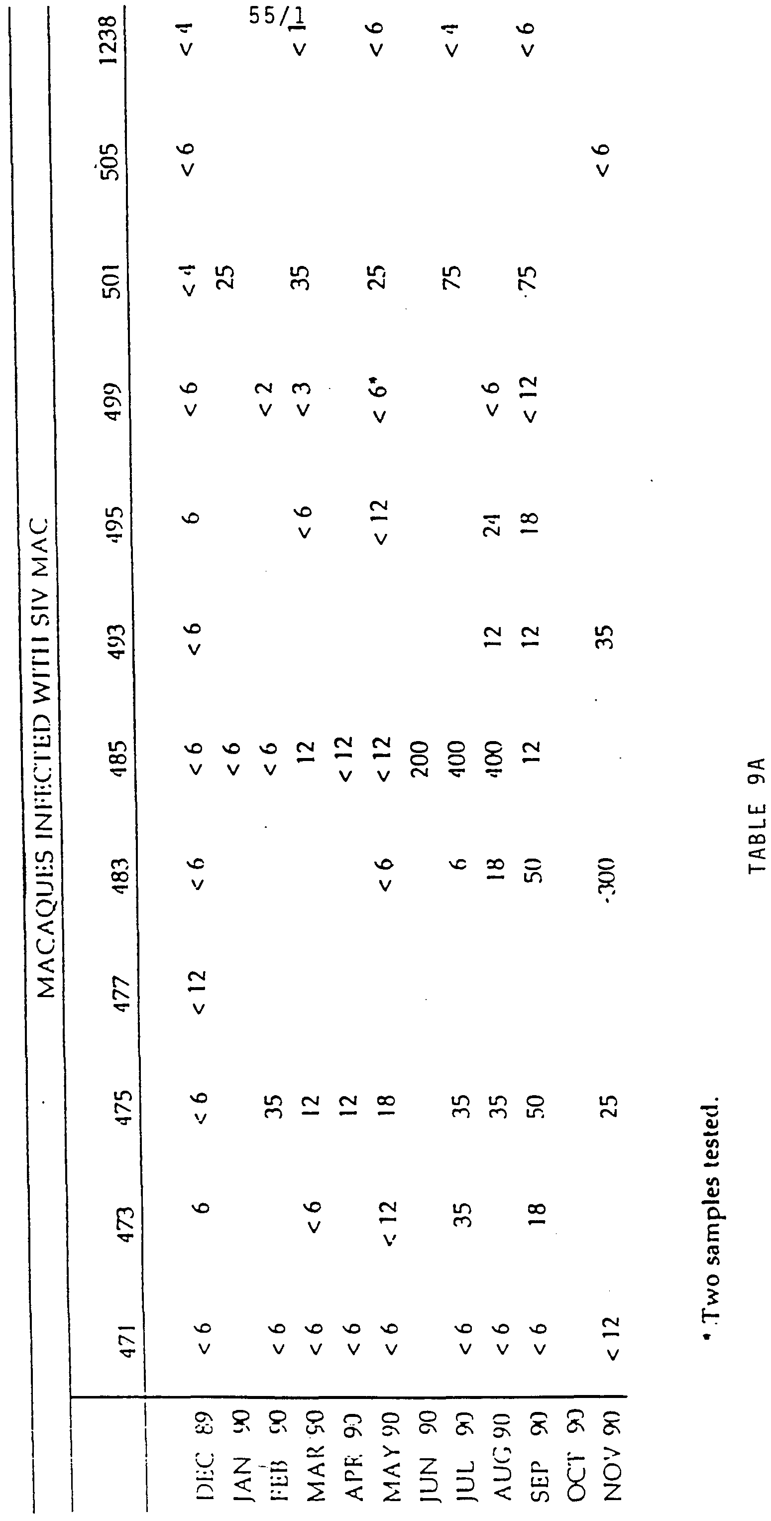
  • interferon response Three types of interferon response are seen in late stage disease; the production of a peak of interferon ⁇ some six to nine months post infection just prior to the development of the clinical signs of disease and death (Table 9, animals 483, 485, and 495, Figures 9-11) , the production of low levels of interferon ⁇ first detectable shortly after infection and which persist for up to 30 months (Table 9, animals 501, and 505, Figures 12-13), and the absence of detectable levels of interferon ⁇ during the whole 30 month observation period following infection with SIV (Table 9, animals 475, 456, 457, 489, and 489).
  • Inteferon ⁇ production was only observed in those animals which exhibited a weak or absence cytotoxic T- cell response (CTL) against autologous B-cells immortalized with Herpes papio virus and transfected with one of eight viral proteins (ENV, GAG, POL, NEF, VIF, REV, TAT, AND VPX) as described previously (Venet et al. 1992, J. Immunol., 148:2899) . None of the animals studied which exhibited a strong CTL response had detectable levels of circulating inteferon at any time during the two year period of observation. IFN ⁇ was not detected in the serum of SIV-infected macaques with normal CD 4 + counts and without signs of disease. Determination fo the Interferon Response in Chimpanzees Infected with HIV-1
  • the 64G12 antibody was well tolerated in rhesus macaques following intravenous administration at a dose of 0.5 to 1.0 mg/kg. No local reaction was observed at the site of injection. Furthermore no systemic reactions such as fever, oedema, etc. were observed in any of the animals injected with the antibody. Successive injections of the antibody were also well tolerated. The only reaction observed was a slight facial oedema seen in certain animals immediately after the third intravenous injection at 15 days (previous injections were at 0, and 5 days) . When the antibody was administered by intramuscular injection, at the third and subsequent injections no oedoma was observed.
  • An accident occured in a single monkey 20 minutes after the intravenous injection of a non-purified preparation of the 64G12 antibody at day following 4 previous intravenous injections of the antibody at days 0, 5, 10 and 15.
  • Pha ⁇ nacokinetics and Establishment of the Lowest Effective Dose of the Monoclonal Antibody 64G12 The level of the 64G12 antibody present in the serum of animals following intravenous injection was determined using an ELISA test based on the use of recombinant soluble receptor to capture antibodies which recognize the extracellular domain of the human interferon ⁇ receptor ( Figure 14) .
  • ELISA plates were coated with a recombinant protein corresponding to the extracellular domain (amino acids 1 to 427) of the human interferon ⁇ receptor produced in either COS or CHO cells at a maximum concentration of 10 to 20.0 ⁇ g/ml in either PBS or 100 mM carbonate buffer, pH 9.2.
  • the plates were then saturated with a 3% solution of bovine serum albumin or similar agent in PBS.
  • the plates were washed with PBS containing 0.5 to 1.0% Tween 20 or a similar detergent and serial dilutions of the samples to be tested, or reference preparation, were then applied to the plates.
  • the plates were then incubated overnight at 4°C or for 2 hours at 37°C, washed with PBS/Tween, and incubated for approximately 2 hours at 37°C with a polyclonal anti-mouse IgG conjugated with alkaline phosphatase, or horse-radish peroxidase.
  • Biotin- streptavidin reagents can also be used (Sheep anti- mouse lg and Streptavidin-biotinylated peroxidase performed complex from Amersham are suitable) .
  • the plates were then washed with PBS/Tween, incubated with the corresponding substrate (o-phenylene-diamine at a concentration of 0.4 mg/ml in a citrate buffer pH 5.5 is suitable for peroxidase; the reaction can then be stopped usually after 1 to 10 minutes by the addition of 0.5 M H 2 S0 4 ) and the optical density determined (at 405 nm for peroxidase reactions) according to standard procedures.
  • the corresponding substrate o-phenylene-diamine at a concentration of 0.4 mg/ml in a citrate buffer pH 5.5 is suitable for peroxidase; the reaction can then be stopped usually after 1 to 10 minutes by the addition of 0.5 M H 2 S0 4 ) and the optical density determined (at 405 nm for peroxidase reactions) according to standard procedures.
  • the intravenous injection of 1.0 mg/kg of the 64G12 anti- interferon receptor resulted in serum levels of approximately 60 ⁇ g/ml, 30 minutes after administration of the antibody which is in good keeping with the expected levels taking into account the blood volume of the animals.
  • a group of 8 rhesus macaques were infected with 10 macaque infectious dose 50 of a molecular clone of SIV strain 251.
  • Two macaques were injected intravenously with 0.5 mg/kg of the anti-interferon receptor antibody 64G12, 30 minutes prior to infection with SIV, and at days 5, 10 and 15 post infection.
  • One macaque was injected intravenously with 3.5 mg of a bull polyclonal anti-lymphoblastoid interferon IgG preparation, 30 minutes prior to infection and at days 5 post infection, and intramuscularly with 3.5 mg of the antibody at days 10 and 15 post infection.
  • the other 5 infected animals were left untreated.
  • EXAMPLE 6 The Effect of the Anti-interferon ⁇ Receptor Monoclonal Antibody on Skin Allograft Survival in Cynomologus Monkeys.
  • cytokines produced both by sensitized T-cells and non MHC restricted cells play an important role in the processes which lead to allograft rejection.
  • interferon ⁇ produced during the initial stages of recognition plays a determining role in the initiation of the processes which lead to graft rejection.
  • treatment of lethaly irradiated mice with interferon ⁇ has also been shown to enhance resistance to allogenic bone marrow grafts while treatment with antibody to interferon ⁇ was found to inhibit rejection (Affifi et al. 1985, J. Immunol., 134:3739).
  • interferon ⁇ also plays an important role in the development of graft-versus-host disease (GVHD) .
  • GVHD graft-versus-host disease
  • interferon ⁇ is produced during the course of graft-versus-host disease in parallel with the enhanced NK cell activity characteristic of systemic GVHD and administration of interferon ⁇ has been shown to enhance the intestinal consequences of GVHD in normal mice (Cleveland et al., 1987, Cell Immunol. 110:120) .
  • Human peripheral blood mononuclear cells from normal donors were isolated on a Ficoll gradient and typed for MHC class I and class II antigens by complement dependent micro-lymphocytoxicity.
  • the proliferation of the responder cells was determined by measuring the incorporation of 3 HTdR in response to allogenic stimulatory cells rendered non-proliferative by treatment with mitomycin-C.
  • Treatment of mixed lymphocyte cultures with the anti-interferon ⁇ receptor antibody resulted in a dose dependent inhibited the incorporation of 3 HTdR ( Figure 24) .
  • a mean inhibition of approximately 50% in the incorporation of 3 HTdR was observed in MLC treated with the 64G12 antibody at a concentration of 20 ⁇ g/ml of ( Figure 24) .
  • Skin allografts were exchanged between AOB compatible l to 3 year old male animals (Macaoa Fascicularis) differing in both MHC class I (Rh LA-A and B) and class II antigens (Th LA-DR) using standard procedures.
  • the grafts were taken and implanted in the left illiac fosse.
  • Animals were either left untreated, or treated (intramuscular injection) with cyclosporin A (5.0 mg/kg/day) one hour prior to grafting and each subsequent day, or with the 64G12 antibody (0.5 mg/kg) one hour prior to grafting and on day 5 and every subsequent 5th day (by intravenous injection for the first two injections and then by intramuscular injection subsequently) until day 85 or until rejection of the graft, or treated with cyclosporin A together with the 64G12 antibody at the same doses as used for each substance alone.
  • the grafts were examined daily for evidence of the clinical signs of rejection (colour, suppleness, etc.) and grafted tissue was biopsied at days 5, 10, 20, and 60 and at the day of rejection. Each biopsy was examined for histological signs of rejection and for the expression of MHC class I and Class II antigens.
  • the mean survival time of skin grafts in the untreated control animals was found to be 7.5 + 0.57 days, and 9.5 + 0.57 days in cyclosporin A treated animals (Table 12).
  • Treatment of animals with the 64G12 antibody was found to increase significantly the mean survival time of the grafted tissue (14.25 ⁇ 0.95 days) relative to both the untreated control animals (Wilcox test p ⁇ 0.01) and animals treated with cyclosporin A (Table 12) .
  • a single monkey in this group was lost at day 20 due to anaphylactic shock 20 minutes after the intravenous injection of an unpurified preparation of the 64G12 antibody.
  • the skin graft in this animal showed no macroscopic or microscopic signs or rejection at this time.
  • Biopsies taken from animals treated with cyclosporin A alone exhibited a dermic and hypodermic inflammatory infiltrate consisting essentially of mononuclear cells and some polynuclear neutrophiles with moderate oedema.
  • the skin allograft biopsies were also 'examined for the expression of MHC class I (HLA-ABC) and class II antigens (HLA-DR) -
  • HLA-ABC MHC class I
  • HLA-DR class II antigens
  • the biopsies from the untreated control animals exhibited a marked expression of both class I and class II antigens (Table 13) .
  • the expression of MHC class I antigens in the biopsies taken from animals treated with cyclosporin A was similar to that of untreated control animals while the expression of class II antigens was less intense (Table 13)
  • Such treatment regimens may also be applicable to the treatment of autoimmune disease particularly those diseases characterized by the abnormal, or prolonged production of interferon ⁇ .
  • Animals were either left untreated, or treated with anti-interferon ⁇ receptor antibody 30 minutes prior to grafting and on day 5 and every subsequent 5th day (by intravenous injection for the first two injections and by intramuscular injection thereafter) together with low dose cyclosporin A (5.0 mg/kg) 30 minutes after grafting and daily thereafter.
  • anti- interferon ⁇ receptor antibody given together with low dose cyclosporin A may be an effective treatment for GVHD.
  • Very few agents are effective in mitigating the acute from of GVHD seen in primates and in prolonging survival in these animals.
  • a beneficial effect in this model is considered to be predictive of a correspondingly greater beneficial effect in the less severe type of GVHD seen in patients receiving bone marrow from MHC matched donors.
  • Peripheral blood mononuclear cells from normal donors were typed for MHC class I class II antigens by complement dependent microlymphocytotoxicity for eacn pair of cultures.
  • Anti-IFN ⁇ receptor mAb IgG 14.25 ⁇ 0.95 (n 4)
  • CD4 + T lymphocytes and CD ⁇ + T lymphocytes were determined by FACS-SCAN using phycoerythrin conjugated anti-human CD4+ monoclonal antibody (OTK4 Ortho Diagnostics), and anti-human CD4+ monoclonal antibody (OTK4 Ortho Diagnostics), and anti-human CD4+ monoclonal antibody (OTK4 Ortho Diagnostics), and anti-human CD4+ monoclonal antibody (OTK4 Ortho Diagnostics), and anti-human
  • Leu2a monoclonal antibody (Becton-Dickinson) respectively.

Landscapes

  • Health & Medical Sciences (AREA)
  • Immunology (AREA)
  • Chemical & Material Sciences (AREA)
  • Organic Chemistry (AREA)
  • Engineering & Computer Science (AREA)
  • Life Sciences & Earth Sciences (AREA)
  • General Health & Medical Sciences (AREA)
  • Bioinformatics & Cheminformatics (AREA)
  • Medicinal Chemistry (AREA)
  • Veterinary Medicine (AREA)
  • Animal Behavior & Ethology (AREA)
  • Public Health (AREA)
  • Pharmacology & Pharmacy (AREA)
  • Nuclear Medicine, Radiotherapy & Molecular Imaging (AREA)
  • Chemical Kinetics & Catalysis (AREA)
  • General Chemical & Material Sciences (AREA)
  • Genetics & Genomics (AREA)
  • Molecular Biology (AREA)
  • Proteomics, Peptides & Aminoacids (AREA)
  • Biochemistry (AREA)
  • Biophysics (AREA)
  • Transplantation (AREA)
  • Medicines Containing Antibodies Or Antigens For Use As Internal Diagnostic Agents (AREA)
  • Preparation Of Compounds By Using Micro-Organisms (AREA)
  • Medicines That Contain Protein Lipid Enzymes And Other Medicines (AREA)
  • Pharmaceuticals Containing Other Organic And Inorganic Compounds (AREA)

Abstract

The invention relates to pharmaceutical compositions for use as immunomodulator, comprising a purified preparation of a monoclonal antibody directed against the human interferon class I receptor (IFN-R) characterized by the following properties: it recognizes the extracellular domain of the human IFN-R, and it has a neutralizing capacity against the biological properties of the human type I-IFN. It further concerns their use for the diagnosis.

Description

PHARMACEUTICAL :- COMPOSITION COMPRISING
MONOCLONAL ANTIBODIES AGAINST THE INTERFERON RECEPTOR, WITH NEUTRALIZING ACTIVITY AGAINST TYPE I INTERFERON
The interferons (IFN) constitute a group of secreted proteins which exhibit a wide range of biological activities and are characterized by their capacity to induce an antiviral state in vertebrate cells (I. Gresser and M.G. Tovey Biochem Biophys. Acta 516-231. 1978). There are three antigenic classes of IFN : alpha (α) , beta (β) and gamma- IFNα and IFN3 together are known as the type I interferon.
Natural type I human interferon comprises 16 or more closely related proteins encoded by distinct genes with a high degree of structural homology (Weissmann and Weber, Prog. Nucl. Acid. Res. Mol. Biol. 33:251, 1986) .
The human IFNα locus comprises two subfamilies. The first subfamily consists of 14 non allelic genes and 4 pseudogenes having at least 80% homology. The second subfamily, all or omega (ω) , contains 5 pseudogenes and 1 functional gene which exhibits 70% homology with the IFNα genes (Weissmann and Weber 1986) .
The subtypes of IFNα have different specific activities but they possess the same biological spectrum (Streuli et al. PNAS-USA 7_8:2848, 1981) and have the same cellular receptor (Agnet M. et al. in "Interferon 5" Ed. I. Gresser p. 1-22, Academic Press, London 1983) .
The interferon β (IFN0) is encoded by a single gene which has approximately 50% homology with the IFNα genes.
CONFIRMAVON Q PY The interferon α subtypes and interferon β bind to the same receptor on the cell surface.
The interferon gamma (IFN gamma) is also encoded by a single copy, which has little homology with the IFNα and IFN/S genes. The receptor for IFN gamma is distinct from the receptor of the α and β interferons.
For the purpose of the present invention the receptor of α and β classes of IFN will be designated IFN-R. This represents natural type I receptor. The group of proteins forming natural interferon α will be designated IFNα, and type I-IFN will represent both natural IFNα, IFNω, and IFN/-*.
Despite the fact that interferon is a potent antiviral agent, there is considerable evidence to suggest, that many of the characteristic symptoms of acute virus diseases such as upper respiratory tract infections are caused by an overproduction of interferon alpha. Furthermore, IFN alpha has been shown to contribute to the pathogenesis of certain chronic virus infections in experimental animals and the available evidence suggests that this is also the case for certain human chronic virus diseases such as those due to measles virus.
The interferons α are also potent immuno- regulatory molecules which stimulate polyclonal B-cell activation, enhance NK cell cytotoxicity, inhibit T- cell functions, and modulate the expression of the major histocompatibility complex (MHC) class 1 antigens, all of which are implicated in the induction of autoimmunity and in graft rejection. The abnormal production of interferon α is associated with a number of autoimmune diseases, immune deficiencies and inflammatory disorders including systemic lupus erythematosuε (SLE) , type I diabetes, psoriasis. rheumatoid arthritis, multiple sclerosis, Behcet's disease, aplastic anemia, the acquired immunodeficiency syndrome (AIDS) and severe combined immunodeficiency disease. The presence of interferon α in the serum of patients with systemic lupus is correlated with both the clinical and humoral signs of increased disease activity. The production of interferon α in HIV positive subjects is also highly predictive of disease evolution.
Especially a close correlation also exists between the presence of circulating acidolabile interferon α and disease progression in patients with the acquired immunodeficiency syndrome (AIDS) (Mildvan D. et al. 1992, The Lancet 339:353) .
Administration of interferon α has been reported to exacerbate underlying disease in patients with psoriasis and multiple sclerosis and to induce a SLE like syndrome in patients without a previous history of autoimmune disease. Interferon α has also been shown to induce glomerulonephritis in normal mice (Gresser et al., 1976, Nature, 263:420) and to accelerate the outset of the spontaneous autoimmune disease of NZB/W mice (Adam et al., 1980, Clin. Exp. Immunol., 40:373) .
The constitutive expression of interferon α in the pancreas of transgenic mice has also been shown to lead to the development of insulino-dependent diabetes the development of which is abrogated by treatment of the animals with specific antibody to interferon α (Stewart et al., 1993, Science, 260:1942) .
Treatment of lethaly irradiated mice with interferon α has also been shown to enhance resistance to allogenic bone marrow grafts while treatment with antibody to inteferon α was found to inhibit rejection (Affifi et al., 1985, J. Immunol. 134:3739). Treatment of rats with antibody to interferon /β has also been reported to prolong cardiac allograft survival in these animals (Gugenheim et al., 1992, Transplant. Int. 5:460) .
Interferon α is also produced during the course of graft-versus-host disease (GVHD) in parallel with the enhanced NK cell activity characteristic of systemic GVDH. Interferon α is the principal modulator of NK cell cytotoxicity and administration of interferon α has been shown to enhance the intestinal consequences of GVDH in normal mice (Cleveland et al., 1987, Cell Immunol. 110:120).
The object of the present invention is to provide new antagonists against the biological activities of the human type I-IFN. These antagonists could be used for therapeutical, including prophylaxis purposes, in cases where the type I-IFN (IFN α//?)is abnormaly produced and when this abnormal production is associated with pathological symptoms. Such antagonists could also be used for the diagnosis of various diseases or for the study of the evolution of such diseases.
In order to define such antagonists, the inventors have taken into account the fact that human natural type I-IFN is in fact constituted of a mixture of interferons (subspecies) and the fact that the composition of this association of different subtypes of interferons varies both quantitatively and qualitatively.
Some natural interferons, such as the ones secreted by Namalwa cells (Namalwa interferon) or leukocyte (leucocyte interferon) have been studied in detail (N.B. Finter and K.H. Fautes, Interferon 2, 1980, p. 65-79 I. Gresser Editor Academic Press ; K. Cantell et al, Interferon 1, 1979 p. 2-25, I. Gresser Editor Academic Press) and were used by the inventors to define natural type I interferons.
In some pathological cases, like AIDS, interferons having some special properties have been described (O.T. Preble et al. Annals of New-York Academy of Sciences p. 65-75) . This interferon involved in pathological cases like AIDS nevertheless binds to the same receptor, as described above.
One object of the present invention is to provide an antagonist of the type I-IFN, which would be able to inhibit or neutralize, to a determined extent, the biological properties of the human type I-IFN, that is to say, to neutralize in vivo a mixture of α, β , ω subspecies.
These antagonists can be utilized in the composition of drugs as immunomodulators and their characterization has been rendered possible by the identification by the inventors, of the nature of the interferon occuring in various pathologies and also by the development of animal models.
The present inventors, having developed appropriate in vivo animal models, have been able to demonstrate that some monoclonal antibodies are capable to interact in vivo on the immune response, with the type I-IFN receptor, in a therapeutically efficient manner as regards various pathologies.
Especially the inventors have been able to define conditions where the in vivo administration of monoclonal antibodies directed against the type I IFN receptor can interact and in some cases inhibit symptoms of viral diseases resulting from viral infections, especially infections due to the HIV or HT1V retroviruseε, or can inhibit allogenic graft rejection or can inhibit the effect of Graft Versus Host Disease.
These immunomodulators will be utilized as such for the treatment of diseases involving abnormal production of interferon, or will be utilized in combination either simultaneously or separately, with other known immunomodulators or drugs especially viral drugs.
For instance, the inventors have shown that in some situations monoclonal antibodies directed against the type I IFN receptor can have a useful synergic effect with other therapeutic agents. This appears to be the case for instance when monoclonal antibodies against type I IFN receptor are associated with an immunosuppressive drug like cyclosporin A.
Accordingly the inventors have defined compositions comprising antibodies, especially monoclonal antibodies, which have the property of being antagonists to the type I-IFN due to their activity as immunomodulators. These antibodies are directed against the human type I-IFN receptor.
The invention thus relates to the use of a preparation of purified monoclonal antibodies in pharmaceutical compositions, for use as immunomodulator, for the in vivo treatment of symptoms associated with the abnormal production of type I-IFN. These monoclonal antibodies are also appropriate for the preparation of diagnosis reagents.
A particular monoclonal antibody appropriate for the preparation of the pharmaceutical compositions for use as immunomodulator according to the present invention is directed against the human type I- interferon receptor (IFN-R) and is characterized by the following properties : it recognizes the extracellular domain of the human IFN-R, and it has a neutralizing capacity against the biological properties of the human type I-IFN. The ability to neutralize the biological properties of type I-IFN can be estimated as a function of the capacity of the monoclonal antibody to neutralize the antiviral activity of the type I-IFN. Such a test is relevant in order to determine whether the antibody assayed is appropriate for the preparation of a pharmaceutical composition within the scope of the invention, although it is clear that the biological properties of type I-IFN are not limited to its antiviral properties. Detailed procedures are given in the examples in order to enable to perform such a test of the antiviral activity. The cells tested can advantageously be Daudi-cells, which affinity for the type I-IFN is well known. The main steps of such a test would consist in : incubating a determined concentration of human cells responsive to human type I-IFN, with human type I-IFN in the presence of a determined concentration of monoclonal antibodies to be assayed, for a time sufficient to allow the formation of a complex between the monoclonal antibodies and the IFN-R of the human cells and/or between the type I-IFN and the IFN-R of the human cells ; infecting the incubated cells with a determined virus, in a determined concentration, washing the cells, resuspending the cells in culture medium, incubating for a time sufficient to allow virus replication ; lysing the cells ; measuring the virus replication, or measuring the inhibition of the cytopathic effect. The ability of the monoclonal antibodies of the invention to neutralize the biological properties of the human type I-IFN can be modulated as a function of the dose of antibodies used. Accordingly a 100% inhibition of the biological properties, or a partial inhibition can be obtained.
The following pages will refer to the definition of preferred monoclonal antibodies directed against the human type I-IFN receptor, which are appropriate when purified, to constitute an active principle of the pharmaceutical compositions according to the invention. The purification of the monoclonal antibodies is done by usual techniques and can be ascertain by protein determination of the content of endotoxins by the Limulus test.
According to a particular embodiment of the present invention, the monoclonal antibodies directed against the human type I-IFN receptor, are further characterized by the fact that they are capable of inhibiting the binding of a human type I-IFN, to the human IFN-R.
A monoclonal antibody having the capacity to recognize the extracellar domain of the human IFN-R and capable of inhibiting the binding of the human type I- IFN to its receptor, can be selected by the following steps : preincubating a determined concentration of purified monoclonal antibodies or a hybridoma culture supernatant containing monoclonal antibodies to be assayed, with human cells capable of harboring IFN-R ; adding labelled human type I-IFN, in a determined concentration, to the above preincubated medium ; incubating the medium containing the human cells, the monoclonal antibodies and the labelled type I-IFN for a time sufficient to allow an equilibrium to occur, between the monoclonal antibodies on the one hand and the type I-IFN on the other hand, with the cellular IFN-R ; washing the cells ; determining the formation of a binding complex between the human cells and the labelled type I- IFN by counting the amount of attached labelled type I-IFN.
Some of the monoclonal antibodies of the invention, have also the capacity to neutralize the antiproliferative properties of the human type I-IFN. This property can also be assayed on Daudi cells, by performing the following steps : allowing cells to grow in presence of human type IFN and determined concentration of mAb ; counting the cells in order to detect an inhibition of the antiproliferative effect of the human type I-IFN.
One property of a monocolonal antibody having an activity of immunomodulator, appropriate for the preparation of a pharmaceutical composition of the invention, resides in its capacity to recognize the extracellular domain of the human IFN receptor. This property of the monoclonal antibody can be assayed on human cells bearing the natural human receptor but also on the extracellular domain of a recombinant IFN-R such as expressed in a procaryotic cell, for instance in E.coli or a recombinant IFN-R such as expressed in a eucaryotic cell such as mamalian cell for instance a CHO-cell.
This receptor can indeed present different properties, depending on the fact that it is produced in a procaryotic or eucaryotic cell and accordingly depending on the fact that the post-translational maturation occurred or not. The inventors interestingly showed that relevant assays, to evaluate the capacity of a monoclonal antibody according to the invention i.e. to recognize the cellular IFN-R, can be performed on a recombinant receptor expressed in mamalian cells. As a matter of fact, such recombinant receptor has the same properties as the cellular receptor, as far as its recognizing activity is concerned.
Monoclonal antibodies useful for the achievement of the invention can be obtained against various forms of the receptor, including the complete receptor, a particular domain or a peptide characteristic of the aminoacid sequence of the receptor represented in figure 3.
Monoclonal antibodies useful for the invention can for example be prepared against the soluble form of the receptor. A hydrosoluble polypeptide corresponding to the soluble form of the INF-R is described on figure 2. According to the present invention, a soluble form of the IFN-R corresponds to a peptide or a polypeptide, capable of circulating in the body.
Other monoclonal antibodies useful for the invention can also be prepared against a peptide comprised in the extracellular domain of the receptor as described on figure 2. An advantageous peptide corresponds for instance to the aminoacid sequence comprised between aminoacid 1 and aminoacid 427. According to another embodiment of the invention, the antibodies can be prepared against a polypeptide modified by substitution of one or more amino acids, provided that antibodies directed against the non modified extracellular domain of the IFN-R, recognize the modified polypeptide or peptide.
Preferred monoclonal antibodies according to the invention are those which are of the IgGl type.
Among the antibodies described hereabove, an antibody which has the capacity of inhibiting the binding of the type I-IFN to its receptor is preferably characterized in that it inhibits the in vitro binding of human type IFN, to the human cellular IFN-R when it is co-incubated with cells harboring the hu-IFN-R, at a concentration of antibodies equal or inferior to 100 μg/ml, preferably equal or inferior to 50 μg/ml, advantageously inferior to 20 μg/ml, more preferably in the range of approximately 0.5 to 2 μg/ml.
The inventors have shown that the high affinity binding capacity of a monoclonal antibody is not sufficient to ensure that this antibody will be able to inhibit the binding activity of the human type I-IFN to the IFN-R. Nevertheless the high affinity binding capacity of the monoclonal antibody is necessary to investigate further the ability of the antibody to inhibit the binding of the type I-IFN to its cellular receptor.
Another monoclonal antibody is characterized in that it neutralizes in vitro the antiproliferative activity of human type I-IFN, on cells highly responsive to this human type I-IFN, for instance Daudi cells at a concentration in a range of 1 to 10 μg/ml.
According to another embodiment a monoclonal antibody is also characterized in that it neutralizes in vitro the antiproliferative activity of human type IFN, on cells poorly responsive to this human IFN, for instance Ly28 cells, at a concentration in a range of 50 to 100 μg/ml.
A particular group of monoclonal antibodies appropriate for the preparation of compositions for use as immunomodulators according to the invention, is characterized in that it neutralizes the antiviral activity of the human type I-IFN, on cells highly responsive to this human type I-IFN, for instance Daudi cells at a concentration in a range of 1 to 50 μg/ml, preferably 1 to 20 μg/ml, for a concentration of type I-IFN in the r-nge of 1 to 1000 units with reference to the international standard MRC 69/19.
Advantageously, the monoclonal antibody according to the invention is such that these antibodies do not bind to the human receptor for IFN gamma.
One particular antibody interesting for the preparation of pharmaceutical compositions of the invention, is such as it directed against an epitope on the amino-acid sequence comprised between amino-acid 27 and amino-acid 427 of the extracellular domain of the human IFN-R as represented on figure 2.
One particularly interesting monoclonal antibody is the antibody designated 64G12 under n° 92022605 which has been deposited at the ECACC (European Collection of Animal Cell Cultures Porton Down Salisbury, Wiltshire SP4 056, United Kingdom) on February 26, 1992.
These antibodies may be prepared by conventional methods involving the preparation of hybridoma cells by the fusion of myeloma cells and spleen cells of an animal immunized beforehand with the peptide antigen, on the conditions such that the antigen against which the antibodies are formed is constituted by the extracellular domain of IFN-R or any polypeptide or peptide of this domain.
The hybridomas are constructed according to the protocole of Kohler and Milstein (Nature, 1974, 256: 495-497) . For example the hybridomas are derived from the fusion of the spleen cells above described with NSl mouse (BalbC) HGPRT" as myeloma cell.
A second procedure for the production of monoclonal antibodies according to the invention, consists in carrying out the fusion between B-cells of blood immortalized with the Epstein/Barr virus and human B lymphocytes placed beforehand in contact with the extracellular domain or a fragment thereof of the IFN-R, against which it is decided to form monoclonal antibodies. B-cells placed in contact beforehand with the extracellular domain of IFN-R or fragment thereof against which it is decided to form monoclonal antibodies, may be obtained by in vitro culture contacted with the antigens, the recovery of the B- cells coated with these antigens being preceded by one or several cycles of stimulation.
The invention also concerns the use in the pharmaceutical compositions of human antibodies as obtained by carrying out the above procedure, having the above defined properties.
The invention also relates to the use in the pharmaceutical compositions of a monoclonal antibody characterized in that the variable or complementary determining regions of its heavy and/or light chains are grafted on the framework and/or constant regions of a human antibody.
A preferred pharmaceutical composition according to the invention is one which contains the purified preparation of monoclonal antibodies at a dose between around 0.05 mg/kg of bodyweight and 3 mg/kg, preferably between 0.5 mg/kg and 1 mg/kg.
The pharmaceutical composition of the invention can be administered by different ways and especially the active principle can be combined with a pharmaceuticle vehicle appropriate for the intravenous or intramuscular administration.
According to a particular embodiment, the monoclonal antibodies which have been defined hereabove, can be used for a preparation of a drug having an immunomodulator effect sufficient to inhibit in vivo the disease due to the infection by a human retrovirus, especially by a human HIV or HTLV retrovirus.
Although several prior results already showed that interferon would be involved in the pathology of AIDS, this is the first time that the composition of the interferon present in case of infection by a HIV retrovirus is demonstrated, and this is the first time that it is demonstrated that monoclonal antibodies against type I-IFN receptor can be useful for the treatment of the diseases associated with AIDS, by acting on the immune response following the infection.
According to another example, such a drug can be used for the treatment of autoimmune and inflammatory diseases. Such diseases include systemic lupus erythematosus, type 1 diabetes, psoriasis, rheumatoid arthritis, multiple sclerosis, Behcet's disease, asplatic anemia, acquired immunodeficiency syndrome (AIDS) , and severe combined immunodeficiency disease.
Thus the purified monoclonal antibodies defined above can be used for the preparation of a drug having an immunomodulator effect sufficient to inhibit in vivo the rejection of allografts or can be used for the preparation of a drug having an immunomodulator effect sufficent to inhibit in vivo the symptoms of the Graft Versus Host Disease.
Treatment of symptoms of acute virus diseases can also be performed with the antibodies of the invention. As example upper respiratory tract infections, chronic virus infections such as those due to measles virus, can be performed.
The compositions of the invention can also be used for the in vitro diagnosis of the presence of the human soluble type I-IFN receptor or cells in a biological sample for instance in blood or in another fluid. The test can be made by several techniques such as ELISA (figure 14(2)) or RIA.
According to another aspect, the invention relates to a pharmaceutical composition for use as immunosuppressor, this composition being characterized in that it comprises as active principle, monoclonal antibodies having the above definition and as a combined preparation for simultaneous, separate or sequential use, an agent having an immunomodulator activity, especially an immunosuppressor activity such as cyclosporin A or FK 506. Especially the monoclonal antibodies are present in a dose ranging from 0.05 mg/kg to 3 mg/kg, preferably from 0.5 mg/kg to 1 mg/kg and the agent having immunosuppressor activity, especially cyclosporin A.
The results which are give hereafter show that the monoclonal antibodies which have been defined above can be used as adjuvant for humoral or cellular immunity.
Further details and additional information will arise from the description from the description of the examples and from the figures. FIGURES
Figure 1 : binding of 15I-labelled monoclonal antibodies 34F10 and 64G12 to :
- A : Daudi cells
- B : Ly28 cells
Briefly, IO6 cells were incubated for 2 hours at 4°C in presence of different amounts of the labelled antibodies diluted in RPMI medium containing 10% fetal calf serum (FCS) . The cells were then washed 4 times in RPMI-1% FCS and counted for bound radioactivity. Nonspecific binding was mesured by incubation with a 100 fold exces of cold antibodies and substracted from total counts.
Figure 2 : nucleotide and corresponding amino-acid sequence of the extracellular domain of the human IFN-R
The monoclonal antibodies were produced against recombinant soluble forms of the human interferon alpha-beta receptor (IFN-R) synthetized in either procaryotic cells (E.coli) or a mammalian cell system (Cos cell) . These soluble forms were based on the DNA sequence described in figure 2.
- Figure 3 : nucleotide and corresponding amino-acid sequence of the human IFN-R.
Figures 4 to 28 : results relating to the immunomodulator activity of the antibodies. E X A M P L E S
EXAMPLE 1 :
Synthesis of the soluble receptors Synthesis in E.coli
A fragment of DNA containing the sequence coding for the extracellular domain (amino acids 27 to 427) of the human INF-R (figure 2) , in which an extra-sequence coding for 5 histidyl residues was introduced just before the termination codon, was cloned in the expression vectors pKK233-2. This fragment was produced by the Polymerase Chain Reaction (PCR) and the resulting plasmids were sequenced to confirm both in- frame insertion with the Shine-Dalgarno sequence and the appropriate sequence coding for the receptor.
The poly-histidyl tail introduced into the recombinant protein enables it to be purified rapidly by affinity chromatography on a chelated nickel support (NTA column) as described previously (Hochuli E. et al. Bio/technology, 1988, 1321-1325).
The plasmid was introduced into the E.coli strain, JM105, and protein synthesis induced by addition of IPTG to the culture medium (pKK233-2, tac promoter) .
Proteins were extracted from the bacterial pellet and the soluble receptor purified to homogeneity by affinity chromatography as described hereafter. This procedure yieled a protein that migrates as 2 bands around 50 kDa under reducing conditions and three bands under non-reducing conditions. The maximum concentration of the protein obtained by different procedures was approximately 20μg/ml. The N-terminal sequence of the two proteins detected by gel electrophoresis has shown that both proteins are the expected fragment of the receptor.
Synthesis and purification of an unqlycosylated soluble receptor :
Bacterial culture (250ml)
IPTG induction 3h
cell pellet 6M Guanidine hydrochloride pH8
centrifugation
NTA column: Washes pH 8 urea 8M I pH 6,3 urea 8M
I pH 5.9 urea 8M
Elution pH 4 urea 8M
refolding dilution, dialysis
I against Tris 0,1 M pH9
I dialysis PBS Using the same PCR approach, we also constructed an expression vector coding for the IFN-R amino acid sequence 1-427, with an additional 5-histidyl residues at the C-terminus, inserted in expression vector pXMT-3. The exact nucleotide sequence of the insert was also confirmed.
The resulting plasmid was introduced by electroporation into Cos7 cells for transient expression and the recombinant protein was purified to homogeneity by affinity chromotography followed by ion exchange chromatography on mono-Q (Pharmacia) as described hereafter.
Purification of the soluble IFN-R from Cos7 cells
preparative electroporation of cos cells
I 18 h
I serum free medium
supernatants taken after 48h, 72h, 96h
concentration
NTA column
I
I Wash PBS
elution 0.1 M NaOAc pH 5.5
I
I neutralization
concentration, 30 000 cut off
Mono Q (0-0.5 M Na Cl) This purification yielded to a 76 kDa protein whose N-terminal sequence corresponds to the predicted receptor sequence with some heterogeneity in the processing of the leader sequence.
EXAMPLE 2 :
Production of monoclonal antibodies against the interferon type I receptor
1) Production of the monoclonal antibodies
Mice were immunized by injection of recombinant soluble interferon (r sIFN-R) purified from E.coli or from a culture supernatant of Cos7 cells. Initially Balb/C mice were injected both intraperitoneally and subcutaneously with the purified protein in complete Freund's adjuvant. Subsequently mice were injected once a week intraperitoneally with the purified proteins diluted in buffered saline solution. Ten micrograms of recombinant proteins were injected each time.
After the fourth injection, blood was collected and the presence of specific serum antibodies were tested by both ELISA and Western blot against the recombinant receptor. The strongest responders were then boosted with a total of lOμg of antigen half of which was injected intravenously and half intraperitoneally. 2) Cell fusion
Four days after boosting, spleen cells from the immunized animal were collected and fused to NSl (mouse) (Balbc) HGPRT" myeloma cells according to the method described by S. Fazekas et al. (J. Immunol. Methods 3J--:1_32 / 1980). Briefly, 5xl07 spleen cells were fused to 3xl07 myeloma cells in 1ml of polyethylene glycol solution and distributed in five 96 well plates on a peritoneal macrophage feeder layer in HAT (hypoxanthine, aminoprotein and thymidine) medium. This procedure was repeated 4 times as 20xl07 spleen cells were obtained from the immunized mouse. Screening for specific hybridomas was undertaken when large colonies were detectable in culture wells.
For the screening, presence of specific antibodies was determined by a direct ELISA method : a) ELISA plates were coated overnight at 4°C with purified E.coli-expressed or Cos7 cell-expressed sIFN-R diluted in PBS. Plates coated with BSA were used to detect non specific binding, b) Plates were saturated by incubation with 3% BSA in PBS for 1 hour at 37°C, c) Plates were incubated for 4 hours at room temperature with hybridoma supernatants diluted 1 in 4 with PBS-0.05% Tween 20, d) Bound antibodies were detected by a two step procedure, comprising a first incubation with goat anti-mouse biotinylated immunoglobulin followed by streptavidin-horseradish peroxidase complex (both from Amersham and diluted 1/1000 in PBS-0.05% Tween 20).
Positive antibody secreting hybridomas were passaged in 24 well plates on a spleen cell feeder layer and their reactivity was again checked by ELISA, and Western-blot. 3) Identification of reactivity to the natural interferon type I receptor
The reactivity of the monoclonal antibodies (mAbs) recognizing the recombinant sIFN-R was tested against the natural class I receptor expressed at the surface of Daudi cells, by membrane immunofluorescence. Briefly, 5xl05 Daudi cells were incubated in lOOμl of culture supernatant of chosen hybridomas for 30 min at 4°C. The cells were then washed 4 times in RPMI medium containing 1% BSA and further incubated with a diluted FITC labelled goat anti-mouse F(ab') for 30 min at 4°C. The cells were finally analyzed by flow cytometry after washing. One of the 35 tested antibodies produced against the E.coli recombinant receptor and 5 of the 6 tested antibodies produced against the COS recombinant receptor were found to recognize the natural receptor on the Daudi cells.
Cloning of these hybridomas was then performed by limiting dilution. The isotype of these mAbs was determined by an ELISA method using isotype specific antibodies. All 6 mAbs were found to be IgGl with kappa light chains. A summary of the reactivity of these 6 mAbs is given in Table 1.
Monoclonal antibodies were purified from culture supernatants by protein G chromatography.
Table 1 :
Reactivity of the anti IFN-R monoclonal antibodies
Reactivity against the Reactivity against * recombinant receptor the cellular receptor
E.COLI COS
ELISA Western ELISA Western immunofIuorescence
34F10 4 4 4 4 4
6-.G12 4 4 4 4 4
63F6 6ΛG2 4 4 4 6AD10 weak 65D8
* measured on Daudi cells
EXAMPLE 3 :
Inhibition of the binding of interferon to human cell lines
Inhibition of interferon binding to human cells was assayed as follows. IO6 cells were preincubated at 4°C for 30 min with various dilutions of hybridoma culture supernatants or purified mAbs or with medium alone. 15I-labelled IFN alpha 8 or alpha 2 was added at the concentration of lOOpM and cells incubated for a further 2 hours at 4°C. These incubations were performed in RPMI medium containing 20mM HEPES pH 7.4 and 10% foetal calf serum (FCS) . The cells were finally washed 4 times with RPMI - 1% FCS and counted to determine bound radioactivity. The mAb secreted by the hybridoma line 64G12 (latter named mAb 64G12) was shown in this assay to inhibit the binding of labelled IFN to the cells in a dose-dependent manner. 50% inhibition of binding to the Daudi cells (Burkitt lymphoma cell line ; Klein et al.. Cancer Researh, ^8:1300-1310, 1968) was obtained at a mAb concentration of 0.4μg/ml. The same inhibition was obtained with K562 cells (chronic myelogenous leukemia, Lozzio and Lozzio, Cell, 5:321-334, 1975) but 50% inhibition was obtained at llμg/ml for HL60 cells (Promyelocytic leukemia, Collins S.J. et al., Nature, 220:347-349, 1977) and 60μg/ml for Ly28 cells (Klein G. et al. Int. J. Cancer, lO:44-57, 1972).
Table 2 :
The inhibition of binding of labelled IFN alpha 2 to various cell lines by mAB64G12
Cell lines Concentration of mAB which gives 50% inhibition of binding
Daudi
0,4 μg/ml K562
HL60 11 μg/ml
Ly28 60 μg/ml
The difference in the mAb concentration at which 50% inhibition of binding of IFN is obtained has been investigated by direct binding of 125I-labelled mABs 64G12 and 34F10 to the same cell lines and Scatchard plot analysis of the results. In the concentration range of 0.1 to 1.5 μg/ml, a high affinity binding of the mAb 34F10 (s-10nM) was seen on all cell lines whereas a high affinity binding of mAB 64G12 was only detected on Daudi and K562 cells (Figure 1) .
EXAMPLE 4 :
Inhibition of the function of type I interferon
Functional inhibition of type I interferon by the purified mAb 64G12 was firstly demonstrated in an antiviral assay on Daudi cells using either recombinant IFN alpha 2, IFN beta and IFN omega, or purified Namalwa and leucocyte interferons, and in an antiproliferative assay with recombinant IFN alpha 2.
* Antiviral activity
An antiviral assay on Daudi cells was performed as described (M. Dron and M.G. Tovey, J. Gen. Virol. 64_:2641-2647, 1983). Cells (0.5xl06/ml) were incubated for 24 hours in the presence of interferon and antibodies. 10° cells in 1 ml were then infected for 1 hour at 37°C with Vesicular stomatitis virus (VSV) then washed 3 times, resuspended in culture medium and incubated for 18 hours at 37°C. Cells were then lysed by freeze-thawing and virus replication measured by titration of the supernatants on L929 cells. A dose- dependent inhibition of the antiviral activity of the various subtypes of type I IFN was demonstrated for the purified mAb 64G12. For the antiviral assay with the Wish cells, cells were incubated for 24 hours with various concentrations of interferons in the presence of the mAbs prior to challenge with VSV. In this assay, the mAb 64G12 was demonstrated to block completely the antiviral activity of Leukocyte IFN (50U/ml) , recombinant IFN alpha 2
(50U/ml) and interferon from the sera of AIDS patients
Figure imgf000029_0001
* antiproliferative activity
For the antiproliferative assay, Daudi cells were seeded at a concentration of 105 cells per ml in a 96 well plate in the presence of interferon and purified inhibitory or control antibody. Cells were then counted after 24, 48 and 72 hours with a Coulter counter and checked for viability by trypan blue exclusion. Purified mAb 64G12 demonstrated a dose-dependent inhibition of the antiproliferative activity of interferon alpha 2.
EXAMPLE 5 : Inteferon and the pathoqenesis of AIDS
I) The Role of Interferon α in the Pathogenesis of AIDS
A number of studies suggest that co-factors, including cytokines, play a determining role in the development of AIDS (Kramer et al., 1992, Lancet, 340:371; Vadhan et al. , 1986, Cancer Res., 46:417) , particularly in the suppression of the immune system, following infection with the Human Immunodeficiency Virus (HIV) . Among the cytokines induced by HIV are the interferons which are thought to play an important role in host defense against viral infections. Although inteferonε can restrict the replication of HIV in vitro (Poli et al., 1989, Science, 244:575; Von Sydow et al.,
1991, AIDS Res. and Human Retroviruses , 2:437) and recombinant interferon α2 has been shown to exert an antiviral and antitumoral effect in AIDS patients with Kaposi sarcoma (Krown et al., 1991, J. Acquired Immune Deficiency Syndromes, :671), the production of inteferon does not prevent the development of AIDS in HIV infected individuals. Furthermore, the restriction of HIV replication by inteferon α may contribute to the development of a latent infection in macrophages (Gendelman et al., 1990, J. Exp. Pathol., 5:53; Gendelman et al., 1990, AIDS Res. and Human Retroviruses, :1045; Mace and Gazzolo, 1991, Res. Virol., 142:2) . An unusual acido-labile α inteferon has been shown to correlate with loss of CD4+ cells and disease progression in these patients (Milvan et al.
1992, The Lancet, 339:453) . α inteferons are potent immunoregulatory molecules which enhance MHC class I antigen expression, MHC restricted T-cell killing, and non MHC restricted NK cell killing, of virus-infected cells all of which could contribute to the loss of CD4+ T-lymphocytes observed in HIV infected individuals. Thus, a marked reduction in the number of circulating CD4+ T-lymphocytes has been observed in HIV infected individuals treated with recombinant interferon α2 (Vento et al., 1993, The Lancet, 341:958) .
II) Characterization of the Endogenous Interferon Present in the Sera of AIDS Patients
In order to characterize the endogenous interferon present in the sera of AIDS patients peripheral blood leukocytes were isolated from HIV infected individuals with high levels of circulating interferon, RNA was extracted, reverse transcribed with MLV reverse transcriptase and a forward primer (a 20 mer synthetic oligonucleotide) specific for either a single gene (Tables 3-5, Figure 4) using standard procedures. The resulting cDNA was then amplified by the polymerase chain reaction (PCR) using one of the three pairs of specific oligonucleotide primers (Table 5) for a total of 30 cycles using standard procedures. The product was then identified either by Southern hybridization as in the case of the IFN β and α genes or by direct di-deoxy sequencing using T7 DNA polymerase (Sequenase, USB) as in the case of the α inteferons following a further 15 cycle of asymetric amplification (using 0.5 pmol of one of the pair of specific oligonucleotide primers and 50 pmol of the other pair of primers) using standard procedures. The results of this analysis showed that IFN α5 was the majority species expressed in approximately 30 samples of peripheral blood cells from HIV infected individuals whereas IFN an (mainly IFN αl and IFN α2) were the main interferon species expressed in peripheral blood cells from 5 HIV-seronegative individuals infected with rhinoviruses, influenza virus, or cytomegalovirus (Figure 5) .
Similar results were also obtained when the expression of interferon genes in the promonocytic cell line U937 chronically infected or not with HIV-1 was analyzed using the same techniques. IFN α5 was found to be the main species expressed in cells chronically infected with HIV-1 whereas IFN αl and IFN α2 were the most abundent species expressed in uninfected U937 cells induced with either Newcastle Disease Virus (NDV) or the polyribonucleotide Polyl-Poly C.
These results may explain why monoclonal antibodies which neutralized IFN α2 did not neutralize the interferon present in the sera of AIDS patients (Tables 6, 7) . Serum interferon from AIDS patients was not neutralized either by monoclonal antibodies specific for IFN β or IFN x but was neutralized, however, by a polyclonal antibody raised against human lymphoblastoid inteferon (Tables 6, 7) .
For various reasons polyclonal antibodies cannot be used for the therapeutical treatment of patients infected with HIV.
III) Determination of the Capacity of a Monoclonal Anti-Interferon α Receptor Antibody to Neutralize the Biologic Activity of the Interferon Present in the Sera of AIDS Patients.
Pretreatment of WISH cells (a human amnion cell line) with the anti-interferon receptor antibody 64G12 at a concentration of 50μg/ml completely neutralized the antiviral activity of the endogenous interferon present in the serum of AIDS patients (Table 8) . Pretreatment of WISH cells with the non-neutralizing antibody 34F10 at the same concentration had no effect on the antiviral activity of the endogenous interferon from AIDS patients (Table 8) .
IV) Establishment of an animal model to test the effect of an inteferon antagonist on the evolution of the acquired immunodeficiency disease. In order to determine whether IFN α plays a role in the pathogenesis of AIDS and to test the efficacity of interferon antagonists as therapeutic agents in this disease we have developed an animal model based on the use of rhesus macaques infected with the simian immunodeficiency virus (SIV) . SIV is a CD4+ tropic lentivirus which shares considerable nucleotide sequence homology, antigenicity, and biologic properties with HIV. SIV-induced disease in macaques is characterized by lymphadenopathy, immune deficiency, and opportunistic infections and is considered to be an excellent experimental model of human AIDS (McClure et al., 1990, AIDS : Anti-HIV Agents, Therapies and Vaccines. Annals NY Academy of Sciences, 616:287) .
Primary Infection
Infection of macaques with SIV is followed by a peak of serum IFN α at 7 to 14 days post infection which falls to undetectable levels by 21 days (Figure 6) . The peak of serum IFN α occurs 2 to 3 days prior to, or concomitantly with the peak of p27 antigenemia at 14 to 15 days post-infection (Figure 7) . Both the peak of interferon α production and p27 antigenemia occur prior to the development of antibody to viral antigens which first becomes detectable at 21 to 25 days post infection (Figure 8) . The production of IFN α in SIV-infected macaques occurs in the absence of detectable levels of IFN 7, IL-6, or TNF-α.
Late-Stage Disease
A group of 12 rhesus macaques infected with SIV and 9 uninfected control animals were followed for a period of some 30 months after infection. No interferon was detected in the sera of any of the uninfected control animals at any time during the 30-month observation period. The development of AIDS-like disease in SIV-infected macaques was found to be preceded by a second peak of serum IFN α which occurs concomitantly with the loss of CD4+ cells several months after primary infection. This second peak of interferon production, unlike that which is observed during primary infection, occurs over a period of several months, and precedes, the p27 antigenemia which occurs in late stage disease, in some cases by several months. Three types of interferon response are seen in late stage disease; the production of a peak of interferon α some six to nine months post infection just prior to the development of the clinical signs of disease and death (Table 9, animals 483, 485, and 495, Figures 9-11) , the production of low levels of interferon α first detectable shortly after infection and which persist for up to 30 months (Table 9, animals 501, and 505, Figures 12-13), and the absence of detectable levels of interferon α during the whole 30 month observation period following infection with SIV (Table 9, animals 475, 456, 457, 489, and 489).
Inteferon α production was only observed in those animals which exhibited a weak or absence cytotoxic T- cell response (CTL) against autologous B-cells immortalized with Herpes papio virus and transfected with one of eight viral proteins (ENV, GAG, POL, NEF, VIF, REV, TAT, AND VPX) as described previously (Venet et al. 1992, J. Immunol., 148:2899) . None of the animals studied which exhibited a strong CTL response had detectable levels of circulating inteferon at any time during the two year period of observation. IFN α was not detected in the serum of SIV-infected macaques with normal CD4+ counts and without signs of disease. Determination fo the Interferon Response in Chimpanzees Infected with HIV-1
Infection of chimpanzees with HIV does not lead to the development of AIDS-like disease eventhough the virus is able to replicate in these animals. The interferon response of a group of 5 chimpanzees was followed for several months prior to and for up to 20 months after infection with HIV-1 (Table 10) . No inteferon α was detected in the serum of any of the 5 animals at any time either before or after infection with HIV-1 (Table 10) . No decrease in the number of CD+ T-lymphocytes or any other sign of clinical disease was observed in any of the animals studied.
Conclusions
A close relationship has been observed between the presence of circulating interferon α in the serum of SIV-infected macaques, and a weak or absent CTL response, the decrease in CD4+ cells, and the development of the clinical symptoms of immunodeficiency disease in these animals. Thus, this system appears to resemble the situation seen in man and would appear to represent a good model in which to test the potential efficacy of an interferon antagonist for the treatment of AIDS.
V) Toxicology and Pharmacokinetic Studies of The Anti-Inteferon α receptor Monoclonal 64G12 in Rhesus Macaques
Toxicology
The 64G12 antibody produced in vitro and purified by affinity chromatography on protein G sepharose. after ultracentrifugation (100 000 x g) , and filtration on a Millipore membrane (0.2μ), was found to be sterile and to be devoid of detectable endotoxin (Limulus test) .
No toxicity was detected in normal adult mice either immediately, or up to three months after, the intravenous injection of 640 μg of a preparation of the 64G12 antibody. Similarly, no immediate or longterm toxicity was observed when new-born mice were injected intrathecally with the same preparation of antibody.
The 64G12 antibody was well tolerated in rhesus macaques following intravenous administration at a dose of 0.5 to 1.0 mg/kg. No local reaction was observed at the site of injection. Furthermore no systemic reactions such as fever, oedema, etc. were observed in any of the animals injected with the antibody. Successive injections of the antibody were also well tolerated. The only reaction observed was a slight facial oedema seen in certain animals immediately after the third intravenous injection at 15 days (previous injections were at 0, and 5 days) . When the antibody was administered by intramuscular injection, at the third and subsequent injections no oedoma was observed.
An accident (anaphylactic shock) occured in a single monkey 20 minutes after the intravenous injection of a non-purified preparation of the 64G12 antibody at day following 4 previous intravenous injections of the antibody at days 0, 5, 10 and 15.
Phaπnacokinetics and Establishment of the Lowest Effective Dose of the Monoclonal Antibody 64G12 The level of the 64G12 antibody present in the serum of animals following intravenous injection was determined using an ELISA test based on the use of recombinant soluble receptor to capture antibodies which recognize the extracellular domain of the human interferon α receptor (Figure 14) .
ELISA for the Detection of Anti-interferon Type I receptor antibody
Briefly, ELISA plates were coated with a recombinant protein corresponding to the extracellular domain (amino acids 1 to 427) of the human interferon α receptor produced in either COS or CHO cells at a maximum concentration of 10 to 20.0 μg/ml in either PBS or 100 mM carbonate buffer, pH 9.2. The plates were then saturated with a 3% solution of bovine serum albumin or similar agent in PBS. The plates were washed with PBS containing 0.5 to 1.0% Tween 20 or a similar detergent and serial dilutions of the samples to be tested, or reference preparation, were then applied to the plates. The plates were then incubated overnight at 4°C or for 2 hours at 37°C, washed with PBS/Tween, and incubated for approximately 2 hours at 37°C with a polyclonal anti-mouse IgG conjugated with alkaline phosphatase, or horse-radish peroxidase. Biotin- streptavidin reagents can also be used (Sheep anti- mouse lg and Streptavidin-biotinylated peroxidase performed complex from Amersham are suitable) . The plates were then washed with PBS/Tween, incubated with the corresponding substrate (o-phenylene-diamine at a concentration of 0.4 mg/ml in a citrate buffer pH 5.5 is suitable for peroxidase; the reaction can then be stopped usually after 1 to 10 minutes by the addition of 0.5 M H2S04) and the optical density determined (at 405 nm for peroxidase reactions) according to standard procedures.
The use of this ELISA for the determination of the concentration of the 64G12 in the sera of rhesus macaques showed that the pharmacokinetics of this antibody following intravenous injection were similar to those previously described for other mouse IgGl monoclonal antibodies (Figure 15) .
Determination of the Number and Affinity of the Interferon receptors on the Peripheral Blood Cells of Rhesus Macaques
Binding studies using standard procedures (Mogensen, Uze et al 1986, Methods Enzymol. 119:267) showed that the binding of 125I labelled recombinant human IFN α2 to the peripheral blood mononuclear cells of normal untreated rhesus macaques was similar to that observed with human peripheral blood cells (Figure 16) . Scatchard analysis (Figure 17) showed that macaque peripheral blood mononuclear cells exhibited approximately 1500 to 3500 receptors per cells with a kD ranging from 2 to 8 x 10¹⁰ M (Figure 18) . The intravenous injection of 1.0 mg/kg of the 64G12 anti- interferon receptor resulted in serum levels of approximately 60 μg/ml, 30 minutes after administration of the antibody which is in good keeping with the expected levels taking into account the blood volume of the animals.
Treatment of macaques with the 64G12 anti interferon receptor antibody at a dose of 0.5 mg/kg was found to completely inhibit the binding of radiolabelled human interferon α 2 to the peripheral blood mononuclear cells of these animals for a period of up to 5 days (Figure 19) . Some 60 to 70% of the interferon receptors on the peripheral blood cells of treated animals became accessible again to the binding of radiolabelled IFN α2 by day 6 (Figure 19) . A dose of 0.5 mg/kg was used as the minimal effective dose in all subsequent experiments.
VI) Determination of the Effect of the Anti- interferon receptor Antibody 64G12 on the Early Stages of Disease in SIV Infected Rhesus Macaques
A group of 8 rhesus macaques were infected with 10 macaque infectious dose 50 of a molecular clone of SIV strain 251. Two macaques were injected intravenously with 0.5 mg/kg of the anti-interferon receptor antibody 64G12, 30 minutes prior to infection with SIV, and at days 5, 10 and 15 post infection. One macaque was injected intravenously with 3.5 mg of a bull polyclonal anti-lymphoblastoid interferon IgG preparation, 30 minutes prior to infection and at days 5 post infection, and intramuscularly with 3.5 mg of the antibody at days 10 and 15 post infection. The other 5 infected animals were left untreated.
No significant difference in the peak level of p27 antigenemia which occurs at day 14 post infection was observed in any of the treated animals relative to the untreated control animals (Figure 20) . The peak of interferon α production which occurs at dat 10 post infection was considerably higher in the animal treated with the polyclonal anti-interferon antibody, relative to that observed in the untreated control SIV-infected animals (Figure 21) . In contrast, treatment of SIV- infected macaques with the monoclonal anti-interferon receptor antibody had non significant effect on interferon production in these animals (Figure 21) . Anti-p27 antibodies also appeared earlier and in larger amounts in the animal treated with the polyclonal anti-interferon antibody than in either animals treated with the 64G12 antibody or in untreated SIV-infected control animals (Figure 22) .
Infection of macaques with SIV was followed by a marked decrease in the percentage of CD4+ T-lymphocytes at 14 to 15 days post infection. In the two animals treated with the anti-interferon α receptor antibody the percentage of CD4+ cells was found to decrease in a manner similar to untreated control animals and then to recover rapidly to attain in both animals higher levels than in either the animal treated with the polyclonal anti-interferon antibody or in untreated control SIV- infected animals (Figure 23) . The difference in CD4+ cells in the animals treated with the anti-interfeeron α receptor antibody persisted for approximately three weeks prior to decreasing slowly in parallel with that of the control animals. No significant effect on the level of CD8+ cells was observed in any of the treated animals relative to the levels seen in untreated control animals. The polyclonal anti-interferon antibody had no significant effect on either the number or percentage of CD4+ or CD8+ cells.
EXAMPLE 6 : The Effect of the Anti-interferon α Receptor Monoclonal Antibody on Skin Allograft Survival in Cynomologus Monkeys.
It is considered that cytokines produced both by sensitized T-cells and non MHC restricted cells play an important role in the processes which lead to allograft rejection. Numerous observations suggest that interferon α produced during the initial stages of recognition plays a determining role in the initiation of the processes which lead to graft rejection. For example, treatment of lethaly irradiated mice with interferon α has also been shown to enhance resistance to allogenic bone marrow grafts while treatment with antibody to interferon α was found to inhibit rejection (Affifi et al. 1985, J. Immunol., 134:3739). Treatment of rats with antibody to inteferon /β has also been reported to prolong cardiac allograft survival in these animals either when administered alone or together with cyclosporin A (Gugenheim et al., 1992, Transplant. Int. 5_:460). The increased survival of cardiac allografts in animals treated with anti-interferon α antibody was associated with a decreased expression of MHC class I antigens on the grafted tissue (Gugenheim et al., 1992, Transplant. Int. 5_:460). These results are in keeping with the fact that inteferon α is the principal activator of MHC class I antigens and NK-cell activity both of which play a major role in allograft rejection.
Numerous observations suggest that interferon α also plays an important role in the development of graft-versus-host disease (GVHD) . Thus, interferon α is produced during the course of graft-versus-host disease in parallel with the enhanced NK cell activity characteristic of systemic GVHD and administration of interferon α has been shown to enhance the intestinal consequences of GVHD in normal mice (Cleveland et al., 1987, Cell Immunol. 110:120) .
I) Effect of the Anti-interferon α Receptor Antibody on the Allogenic Response of Mixed Lymphocyte Cultures. In order to test the potential immunosuppressive activity of the anti-interferon α receptor antibody 64G12, mixed lymphocyte cultures (MLC) were treated with varying concentration of either the 64G12 antibody, or the non-neutralizing control antibody 34F10. Both antibodies had been shown previously to be without effect, on either the viability or proliferation of either human peripheral blood mononuclear cells or the human lymphoblastoid cell line Daudi after 96 hours incubation in vitro at a concentration of 50 μg/ml.
Human peripheral blood mononuclear cells from normal donors were isolated on a Ficoll gradient and typed for MHC class I and class II antigens by complement dependent micro-lymphocytoxicity. The proliferation of the responder cells was determined by measuring the incorporation of 3HTdR in response to allogenic stimulatory cells rendered non-proliferative by treatment with mitomycin-C. Treatment of mixed lymphocyte cultures with the anti-interferon α receptor antibody resulted in a dose dependent inhibited the incorporation of 3HTdR (Figure 24) . A mean inhibition of approximately 50% in the incorporation of 3HTdR was observed in MLC treated with the 64G12 antibody at a concentration of 20 μg/ml of (Figure 24) . The degree of inhibition of 3HTdR incorporation varied considerably, however, from one MLC to another (20 to 82% with a mean of 50.58 + 20.01% for a series of 12 MLC, Figure 25) without any clear relation to the MHC DR type of either the donor or responder lymphocytes (Table 11) . No inhibition of 3HTdR incorporation was observed in cultures treated with up to 50μg/ml of the non- neutralizing anti-interferon α receptor antibody 34F10. II) Effect of the Anti-interferon α Receptor Antibody on the Survival of Skin Alloqrafts in Cynomologus Monkeys.
Skin allografts were exchanged between AOB compatible l to 3 year old male animals (Macaoa Fascicularis) differing in both MHC class I (Rh LA-A and B) and class II antigens (Th LA-DR) using standard procedures. The grafts were taken and implanted in the left illiac fosse. Animals were either left untreated, or treated (intramuscular injection) with cyclosporin A (5.0 mg/kg/day) one hour prior to grafting and each subsequent day, or with the 64G12 antibody (0.5 mg/kg) one hour prior to grafting and on day 5 and every subsequent 5th day (by intravenous injection for the first two injections and then by intramuscular injection subsequently) until day 85 or until rejection of the graft, or treated with cyclosporin A together with the 64G12 antibody at the same doses as used for each substance alone.
The grafts were examined daily for evidence of the clinical signs of rejection (colour, suppleness, etc.) and grafted tissue was biopsied at days 5, 10, 20, and 60 and at the day of rejection. Each biopsy was examined for histological signs of rejection and for the expression of MHC class I and Class II antigens.
The mean survival time of skin grafts in the untreated control animals was found to be 7.5 + 0.57 days, and 9.5 + 0.57 days in cyclosporin A treated animals (Table 12). Treatment of animals with the 64G12 antibody was found to increase significantly the mean survival time of the grafted tissue (14.25 ± 0.95 days) relative to both the untreated control animals (Wilcox test p<0.01) and animals treated with cyclosporin A (Table 12) .
Treatment of animals with the anti-interferon α receptor antibody together with cyclosporin A resulted in permanent graft acceptance (Table 12) . Indeed graft rejection was observed at 63 days in a single animal only while the grafts in the remaining 4 animals in this treatment group remain fully viable and without any signs of rejection at 235 days (Table 12) . Treatment with the anti-interferon receptor antibody was stopped at day 85 in those animals with intact skin-grafts. These animals continued, however, to be treated with low dose cyclosporin A (5.0 mg/kg) until day 140 at which time all treatment was stopped.
A single monkey in this group was lost at day 20 due to anaphylactic shock 20 minutes after the intravenous injection of an unpurified preparation of the 64G12 antibody. The skin graft in this animal showed no macroscopic or microscopic signs or rejection at this time.
Histologic examination of biopsies of surviving skin-grafts, taken at 60 days, from animals treated with anti-interferon receptor antibody together with cyclosporin A showed only rare mononuclear cells under the epidermis and around certain capillaries. Skin biopsies taken at 185 days from animals which had been treated with anti-interferon receptor antibody and cyclosporin A for 85 and 140 days respectively revealed tissue which was difficult to distinguish from normal ungrafted skin. A moderate inflammatory infiltrate consisting essentially of interstitiel and or percapillary mononuclear cells was seen in biopsies taken from animals treated. This contrasted with the very severe lesions (necrosis oedema, marked inflammatory infiltrate with numerous polynuclear neutrophiles) seen in biopsies taken from the untreated control animals. Biopsies taken from animals treated with cyclosporin A alone exhibited a dermic and hypodermic inflammatory infiltrate consisting essentially of mononuclear cells and some polynuclear neutrophiles with moderate oedema.
The skin allograft biopsies were also 'examined for the expression of MHC class I (HLA-ABC) and class II antigens (HLA-DR) - The biopsies from the untreated control animals exhibited a marked expression of both class I and class II antigens (Table 13) . The expression of MHC class I antigens in the biopsies taken from animals treated with cyclosporin A was similar to that of untreated control animals while the expression of class II antigens was less intense (Table
13) whereas in biopsies taken from animals treated with the anti-interferon receptor antibody the expression of MHC class I antigens was reduced while that of class II antigens remained unchanged relative to control animals (Table 13) . In contrast, in biopsies taken from animals treated with the 64G12 antibody together with cyclosporin A, both MHC class I and class II antigens were markedly reduced relative to untreated control animals (Table 13) .
A hyperlymphocytosis was observed in animals treated with either the anti-interferon receptor antibody alone or together with cyclosporin A (Table
14) . This hyperlymphocytosis was associate with an increase in the percentage of CD8+ T-lymphocytes relative to untreated control animals. No difference in either the absolute number or percentage of CD4+ T- lymphocytes was observed in animals treated with the anti-interferon receptor antibody relative to control animals (Table 15) . No differences were observed in the erythroid or platelet cell lineages.
No differences in the levels of blood sodium or potassium creatin were observed in any of the animals following transplantation (Table 16) .
Monkeys treated with cyclosporin A (5.0 g/kg/day) together with the anti-interferon α receptor antibody exhibited significantly higher levels of serum cyclosporin A than animals treated with the same dose of cyclosporin A alone (Table 17) .
No detectable monkey antibodies (IgG) to the mouse anti-human interferon α receptor antibody were detected, using a sensitive ELISA specific for anti- interferon α receptor antibodies (Figures 26-28) , in the serum of any of the animals treated with 0.5 mg/kg of the antibody every 5 days (for up to 60 days) either alone or together with cyclosporin A (Figure 28) . The apparent absence of immunogenicity of the 64G12 monoclonal antibody may be a consequence of the very strong immunosuppression observed in animals treated with this antibody.
Six months after the initial skin graft and two months after all treatment had been stopped animals were given a second skin graft from the same donor without further treatment. The secondary grafts were rejected at 5.5 ± 0.5 days in the untreated control group and at 11.5 ± 2.0 days in animals which had been treated initially with anti-interferon α receptor antibody together with cyclosporin A (Table 18) . In contrast, the primary skin grafts remained intact in those animals which had been treated intially with anti-interferon α receptor antibody together with cyclosporin A even though secondary skin grafts were rejected in these animals. These results suggest that the local production of interferon α within and or at the site of the grafted tissue plays a determining role in the initial processes which lead to allograft rejection.
These results show that treatment of cynomologus monkeys with the 64G12 antibody markedly increases allograft survival in these animals. The amplitude of the effect observed with the antibody alone is such that the 64G12 may be considered to be among the most potent immunosuppressive monoclonal antibodies described to date. Thus, monoclonal antibodies against interferon -y or TNF α appeared to be less effective in preventing skin allograft rejection in monkeys than the 64G12 antibody (Stevens et al., 1990, Transplantation, 5_0:856), while an anti-CD3 antibody (Nooji et al., 1987, Eur. J. Immunol. 2/7:1089) is the only antibody to give an effect comparable to that of the anti- interferon α receptor antibody 64G12.
Treatment of cynomologus monkeys with anti- interferon α receptor antibody together with cyclosporin A resulted in permanent allograft acceptance in non MHC matched animals which contrasts markedly with the limited efficacy of other immunosuppressive agents in the primate model.
The very strong synergic immunosuppressive effect observed in animals treated with the anti-interferon α receptor antibody administered together with low dose cyclosporin A would enable effective immunosuppression to be attained at doses of cyclosporin A which are normally ineffective thus reducing the severe toxicity observed in patients treated over long periods of time with high doses of cyclosporin A. Furthermore, the fact that allograft survival is maintained after treatment with the anti-interferon α receptor antibody has been stopped suggests that only a limited period of treatment with the 64G12 antibody may be necessary to attain effective immunosuppression.
It is probable that given their similar mode of action comparable immunosuppressive effects would be observed if the 64G12 antibody was administered together with FK 506.
These results, which show that selective and lasting immunosuppression can be obtained by the administration of relatively low levels of an interferon antagonist given together with low dose cyclosporin A, may have important implications for the prophylaxis and treatment of human allograft rejection.
Such treatment regimens may also be applicable to the treatment of autoimmune disease particularly those diseases characterized by the abnormal, or prolonged production of interferon α.
Ill) Effect of Anti-interferon α Receptor Antibody on graft-versus-host disease in cynomologus monkeys.
Primates provide a near optimal model for preclinical investigation of bone marrow transplantation in that their radiosensitivity, the composition of their bone marrow, and the pattern of their immunological reactions to bone marrow grafting are closely similar to those of humans. Furthermore, modifications of the acute graft-versus-host-disease (GVHD) , which develops in the monkey after grafting marrow from unrelated non MHC matched donors, have proven to be highly predictive of the human situation (van Bekkam, Transplantation Proceedings, 10, 105-111, 1988) .
Young adult male animals (Macaca Fascicularis) were treated with broad spectrum antibiotics to attain intestinal sterilization prior to irradiation.
Animals were irradiated with a whole body dose of 8 Gy and grafted 4 hours later with 4 x 10⁸ allogeneic bone marrow cells/kg from random donors differing in both MHC class I (Rh LA-A and B) and class II antigens (Th LA-DR) .
Animals were either left untreated, or treated with anti-interferon α receptor antibody 30 minutes prior to grafting and on day 5 and every subsequent 5th day (by intravenous injection for the first two injections and by intramuscular injection thereafter) together with low dose cyclosporin A (5.0 mg/kg) 30 minutes after grafting and daily thereafter.
Surviving animals were housed in isolation rooms for the first month following irradiation and under open conditions thereafter.
Untreated grafted animals died at 9 ± 1.0 days with an acute form of GVHD with characteristic histologic lesions of the skin similar to those seen in man (Table 19) . Of the three grafted animals treated to date with the anti-interferon α receptor antibody together with low dose cyclosporin A one animal died from a bacterial infection at 19 days without clinical or histological signs of GVHD. While the two other animals remain healthy at 93 days post transplantation (Table 19) .
These preliminary results suggest that anti- interferon α receptor antibody given together with low dose cyclosporin A may be an effective treatment for GVHD. Very few agents are effective in mitigating the acute from of GVHD seen in primates and in prolonging survival in these animals. Indeed, a beneficial effect in this model is considered to be predictive of a correspondingly greater beneficial effect in the less severe type of GVHD seen in patients receiving bone marrow from MHC matched donors.
GENE Nucleotide Position Primer Product Size Sequence 5'. -> 3'
982-1005 Forward GATCTCATGATTTTCTGCTCTGACA
IFN α 723-743 Reverse 283 bp TCCATGAGATGATCCAGCAGA
CO g 902-924 Probe ATCCTGGCTGTGAGGAAATACTT
3
491-512 Forward N.1 ATGTTCCAGGCAGCAGAGGAGC
C IO 438-457 Forward N.2 305 bp GCATCTCATGGAGGACAGAC
IFN to 208-230 Reverse N.1 (1st round) ATGGCCCTCCTGTTCCCTC aσ 257-276 Reverse N.2 202 bp GCCCTGTTGGATCTCTGGGC m ι c — rv> 359-387 Probe (2nd round) TGTGTCTCAAGGACAGAAGAGACTTCAGG
TABLE 3
Figure imgf000051_0001
GENE Nucleotide Position Primer Product Size Sequence 5'. 3'
co 899-909 Forward GTAACCTGTAAGTCTGTTAA
IFN β 453-472
3 Reverse 457 bp AAGCAGCAATTTTCAGTGTC
— . 682-711 Probe GAGAACCTCCTGGCTAATGTCT
CO
I 868-1890 (exon 2) Forward 1410 bp CCAATTCTTCAAAATGCCTAAG
--σ (genomic DNA) c: IFN γ 481-502 (exon 1) Reverse 171 bp TATACAAGTTATATCTTGGCTT ro 531-558 (exon 1) Probe (m RNA) TGGCTGTTACTGCCAGGACCCATATGTA
TABLE 4
Figure imgf000052_0001
GENE Nucleotide Position Primer Product Size Sequence 5' 3'
2759-2777 Forward CGCAGAAGGGGTCCTGGTGA
Figure imgf000053_0001
ALDOLASE A 4205-4229 Reverse 181 bp CTGGCACAGGAGAGGGGCGGGTG
X m 2798-2827 Probe (m-RNA) m TTGAATCCACTCGCCAGCCCGCCGCCCTCT
3D c ro σ.
TABLE 5
INTERFERON TITER (U/ML)
Interferon Polyclonal Monoclonal Monoclonal Monoclonal Monoclonal Monoclonal
Untreated Anti IFN α Anti IFN α2 Anti IFN α2 Anti IFN tx2 Anti IFN ω Anti IFN β l/B I/8 I/20
Figure imgf000054_0001
IFN β 100 < 2 100 100 100 100 < 2
7628 8 < 2 4 8 12 8 8
Figure imgf000054_0002
TABLE 6
INTERFERON TITER (U/ML)
Serum Polyclonal Monoclonal Monoclonal Monoclonal Monoclonal Monoclonal Monocl
Figure imgf000055_0001
TABLE 7
Figure imgf000055_0002
TABLE 8
EFFECT OF ANTI-IFN α RECEPTOR mAb 64G12 ON THE ANTIVIRAL ACTIVITY OF HUMAN INTERFERONS
TITRE (IFN U/ml)*
SERUM INTERFERON U NTREATED TREATED WITH
FROM AIDS PATIENTS mAb ANTI-IFN α R
1 75 < 2
2 50 < 2
3 1 50 < 2
4 75 < 2
Leukocyte IFN 50 < 2
IFN α2 50 < 2
* Human amniotic cells (WISH) were incubated in medium alone or medium containing 50 μg/ml of mAb 64G12 for 30 minutes prior to the addition of dilutions of the interferons indicated in the Table. The antiviral activity of each sample was determined alone and in the presence of the 64G12 antibody as described previously (Tovey et al., 1977, Nature, 222:455).
Figure imgf000057_0001
Figure imgf000058_0001
95/07716
Figure imgf000059_0001
O 95/07716
57
HLA-DR TYPING OF MIXED LYMPHOCYTE CULTURES
MLC STIMULATOR RESPONDER CELLS
1 DR2-4 DR3-6
2 DR3-6 DR2-4
3 DR1 -7 DR4-5
4 DR4-5 DR 1 -7
5 DR2-3 DR7
6 DR 7 DR 2 - 3
7 DR2-3 DR 1 -5
8 DR 1 -5 DR2-3
9 DR6 DR 1 -5
10 DR 1 -5 DR6
11 DR1 -7 DR6
12 DR6 DR 1 -7
Peripheral blood mononuclear cells from normal donors were typed for MHC class I class II antigens by complement dependent microlymphocytotoxicity for eacn pair of cultures.
TABLE 11 58
TABLE 12
SKIN ALLOGRAFT IN SURVIVAL CYNOMOLOGUS MONKEYS
M.S.T. (Davs)
Untreated Control group 7.5 ± 0.57 (n = 4)
Cyclosporin A (5.0 mg/Kg/day) 9.5 ± 0.57 (n = 4)
Anti-IFN α receptor mAb IgG 14.25 ± 0.95 (n = 4)
Cyclosporin A + anti-IFN receptor mAb IgG 63 (n = 1)
> 325 n = 4 *
* Treatment with the antibody 64G12 and cyclosporin A was stopped at days 85 and 140 respectively TABLE 13
EXPRESSION OF MHC CLASS I AND CLASS II ANTIGENS ON THE
GRAFTED TISSUE
TREATMENT MHC CLASS I ANTIGENS MHC CLASS II ANTIGENS
Day 5 10 20 60 180 Day 5 10 20 60 180
Figure imgf000062_0001
I
UNTREATED +++ R R R R +++ R R
-X.
F m rs>
Si CyA +++ +++ R R R ++ ++ R R R
64G12 ++ ++ R R R +++ +++ R R R
CyA+G64G12 ++ + +
The degree of expression of MHC antigens was defined as : + weak ; ++ moderate ; +++ strong ; R = reject
% LYMPHOCYTES
TIME (DAYS)
0 5 15 30 60
Untreated Control group 27±1.4 29.2+4.6 24.2±3.8 26.7±3.8 34.5+7.7 Cyclosporin A (5.0 mg/Kg/day) 35±12.7 42.5±16 35.7±10.4 28.5±10.8 24.5±7.7
C Anti-IFN α R mAb IgG 39.5±12 59.2±9.7 64.5±4.4 56.5±8.5 66.5±3.5 CD Cyclosporin A + anti-IFN R mAb IgG 26.5±2.1 61.8±7.9 62.3±5 61.4±2 63±2.1
CO
TABLE 14 ro
Figure imgf000063_0001
SKIN ALLOGRAFT SURVIVAL IN CYNOMOLOGUS MONKEYS
§ sq TREATMENT % CD4+ C ELLS % CDs* C E LLS
% m U ntreated 23.3 ± 7.5 33.65 ± 2.75 ϋ-? p Treated vyith anti-IFN α 25.5 ± 2.7 51.45 ± 5.0 rn mAb receptor
The number ot CD4+ T lymphocytes and CDβ+T lymphocytes was determined by FACS-SCAN using phycoerythrin conjugated anti-human CD4+ monoclonal antibody (OTK4 Ortho Diagnostics), and anti-human
Leu2a monoclonal antibody (Becton-Dickinson) respectively.
Figure imgf000064_0001
TAB LE 15
TABLE 16A
GROUP PRIMATE CREATININ mmol/l SODIUM mmol/l POTASSIUM mmol/l
JO J5 J15 J30 J60 JO J5 J15 J30 J60 JO J5 J15 J30 J60
I 1 80 87 83 72 146 144 138 132 4 4,3 3,9 3,7
2 115 90 87 90 146 152 142 157 4,4 5,1 5 3,9 s 3 98 91 86 90 142 146 146 153 3,5 3,9 3,7 4
4 120 89 89 101 145 142 143 129 4,1 3,9 3,8 4,2
C 3
I MEAN 109 93,75 88 90,25 81 143,5 145 146,25 140,5 144,5 3,8 4,05 4,23 4,28 3,8
-XI STANDARD 15,56 14,95 1,83 7,72 12,73 2,12 2 4,03 9,95 17,68 0,42 0,24 0,64 0,50 0,14 c:
DEVIAT TON ro co
II 1 104 82 96 89 142 142 142 144 3,9 4,1 3,7 3,9
2 122 89 92 88 154 146 141 139 4,9 5 4,7 4,1
3 1 18 106 88 93 143 145 142 142 4,1 4,3 4,2 4,4
4 94 107 94 90 140 147 150 145 5, 1 4,9 4,1 4,4
MEAN 106 109,75 88,25 92,75 88,5 141,5 147 145 142,5 141,5 4,6 4,5 4,35 4,3 4
STANDARD 16,97 8,26 4,92 2,5 0,71 2,12 5,10 3,83 1,73 3,54 0,71 0,49 0,44 0,42 0,14
DEVIATION
Table: EVOLUTION OF THE LEVELS OF PLASMATIC CREATININ, NA+ AND K+ IN THE 4 GROUPS STUDIED AT JO, J5, J1 , J30 AND J60 (CORRESPONDING TO DAY 0, DAY 5, DAY 15, DAY 30 AND DAY 60).
TABLE 16B
Figure imgf000066_0001
Table: EVOLUTION OF THE LEVELS OF PLASMATIC CREATININ, NA+ AND K+ IN THE 4 GROUPS STUDIED AT JO, J5, J15, J30 AND J60 (CORRESPONDING TO DAY 0, DAY 5, DAY 15, DAY 30 AND DAY 60).
63
SERUM CYCLOSPORIN LEVELS (ng/ml)
TIME (DAYS)
15 30 60
Cyciosporin A (5mg/Kg/day)
123±27 145±89 181 ±83 180±42
Cyciosporin A + anti-IFN receptor mAb IgG
301 ±132 321±83 377±78 285±71
TAB LE 17
TABLE 18 6
SURVIVAL OF SECONDARY SKIN GRAFTS IN CYNOMOLOGUS MONKEYS
M.S.T. (Oavs)
Untreated Control group 5.5 ± 0.50 (n = 4)
Cyclosporin A + anti-IFN receptor mAb IgG 1 1.5 ± 1.5 (n = 4)*
* Animals were grafted with skin from the same donor 226 days after primary grafting without any further treatment. ( Animals had been treated previously with antibody 64G12 and cyclosporin A for 85 and 140 days respectively).
TABLE 19 65
GRAFT VERSUS HOST DISEASE
.S.T. (Oavs)
Untreated control group 9 ± 1.0 (n = 4)
Cyclosporin A + anti-IFN receptor mAb IgG 19 * (n = 1 )
> 93 (n = 2)
Died from an infection without clinical or histological signs of GVH

Claims

66 C L A I M S
1. Pharmaceutical composition, for use as immunomodulator, characterized in that it comprises a purified preparation of monoclonal antibodies directed against the human interferon class I receptor (IFN-R) and having the following properties : it recognizes the extracellular domain of the human IFN-R, and it has a neutralizing capacity against the biological properties of the human type I-IFN.
2. Pharmaceutical composition according to claim 1 wherein the monoclonal antibody is characterized by its capacity to inhibit the binding of a human pathological type I-IFN, to the type I IFN-R.
3. Pharmaceutical composition according to claim 1 or 2, wherein the monoclonal antibody is obtainable from a hybrido a cell prepared by fusion of a myeloma cell with spleen cells from an animal previously immunized with the soluble form of the human IFN-R.
4. Pharmaceutical composition according to anyone of claims 1, 2 or 3, wherein the monoclonal antibody recognizes an epitope on a soluble form of the human cellular IFN-R or of a recombinant IFN-R.
5. Pharmaceutical composition according to anyone of claims l to 4, wherein the monoclonal antibody inhibits in vitro the binding of human type I-IFN, to the human cellular IFN-R when it is co-incubated with cells harboring the hu-IFN-R, at a concentration of antibodies equal or inferior to 100 μg/ml, preferably equal or inferior to 50 μg/ml, advantageously inferior to 20 μg/ml, more preferably in the range of approximately 0,5 to 2 μg/ml.
6. Pharmaceutical composition according to anyone of claims 1 to 5, wherein the monoclonal antibody 67 neutralizes in vitro the antiproliferative activity of the human type I-IFN, on cells highly responsive to this human type I-IFN, for instance Daudi cells at a concentration in a range of 1 to 10 μg/ml.
7. Pharmaceutical composition according to anyone of claims l to 6, wherein the monoclonal antibody neutralizes in vitro the antiproliferative activity of human type I-IFN, on cells poorly responsive to this human type I-IFN, for instance Ly28 cells, at a concentration in a range of 50 to 100 μg/ml.
8. Pharmaceutical composition according to anyone of claims 1 to 7, wherein the monoclonal antibody does not bind to the human receptor of the IFN gamma.
9. Pharmaceutical composition according to anyone of claims l to 8, wherein the monoclonal antibody recognizes an epitope on the aminoacid sequence 27 to 427 of the human IFN-R.
10. Pharmaceutical composition according to anyone of claims l to 9, wherein the monoclonal antibody neutralizes in vitro the antiviral activity of the human type I-IFN, on cells highly responsive to this human type I-IFN, for instance Daudi cells at a concentration in a range of 1 to 10 μg/ml.
11. Pharmaceutical composition according to anyone of claims 1 to 10, wherein the monoclonal antibody neutralizes in vitro the antiviral activity of the human class I-IFN, on cells poorly responsive to this human IFN, for instance Ly28 cells, at a concentration in a range of 50 to 100 μg/ml.
12. Pharmaceutical composition according to anyone of claims 1 to 11, for use as immunosuppressor.
13. Pharmaceutical composition according to anyone of claims 1 to 11, for use as immunostimulator. 68
14. Pharmaceutical composition according to anyone of claims 1 to 13, wherein the monoclonal antibody is the 64G12 antibody, deposited at the ECACC on February 26, 1992 under n° 92022605.
15. Pharmaceutical composition according to anyone of claims 1 to 13, wherein the monoclonal antibody is a humanized antibody, for instance characterized in that the variable or complementary determining regions of its heavy and light chains are grafted on the framework and constant regions of a human antibody.
16. Pharmaceutical composition according to anyone of claims 1 to 13, wherein the monoclonal antibody is a human antibody.
17. Pharmaceutical composition according to anyone of claims 1 to 16, characterized in that the purified preparation of monoclonal antibodies is used at a dose between around 0.05 mg/kg of bodyweight and around 3 mg/kg.
18. Pharmaceutical composition according to anyone of claims l to 17 comprising a pharmaceutical vehicle appropriate for the intravenous administration.
19. Pharmaceutical composition according to anyone of claims 1 to 17 comprising a pharmaceutical vehicle appropriate for the intramuscular administration.
20. Use of a purified preparation of monoclonal antibodies as defined in anyone of claims 1 to 16, for the preparation of a drug having an immunomodulator effect sufficient to inhibit in vivo the disease due to the infection by a human retrovirus especially by a human HIV or HTLV retrovirus.
21. Use of a purified preparation of monoclonal antibodies as defined in anyone of claims 1 to 16, for the preparation of a drug having an immunomodulator 69 effect sufficient to inhibit in vivo the "rejection of allografts.
22. Use of a purified preparation of monoclonal antibodies as defined in anyone of claims 1 to 16, for the preparation of a drug having an immunomodulator effect sufficient to inhibit in vivo the symptoms of the Graft Versus Host Disease.
23. Pharmaceutical composition for use as immunosuppressor, characterized in that it comprises as active principle, monoclonal antibodies as defined in anyone of claims 1 to 16, and as a combined preparation for simultaneous, separate or sequential use, an agent having an immunomodulator activity especially an immunosuppressor activity.
24. Pharmaceutical composition according to claim 23, characterized in that the agent having an immunomodulator activity is cyclosporin A or FK 506.
25. Pharmaceutical composition according to claim 19, characterized in that the monoclonal antibodies are present at a dose ranging from around 0.05 mg/kg to 3 mg/kg, preferably from 0.5 mg/kg to 1 mg/kg and the agent having immunosuppressor activity, especially cyclosporin A, is present in a dose of at least 0.1 mg/kg.
26. Use of a purified preparation of monoclonal antibodies as defined in anyone of claims 1 to 16, as adjuvant for humoral or cellular immunity.
27. Use of a purified preparation of monoclonal antibodies as defined in anyone of claims 1 to 16, for the preparation of a drug applicable to the treatment of autoimmune disease, particularly those diseases characterized by the abnormal or prolonged production of type I-IFN, especially a IFN. 70
28. Use of a preparation of monoclonal antibodies as defined in anyone of claims 1 to 16, for the in vitro detection, on a biological sample such as blood or another biological fluid, of the presence of soluble type I interferon receptors, especially in case of autoimmune diseases or HIV infection.
PCT/EP1994/003114 1993-09-17 1994-09-16 Pharmaceutical composition comprising monoclonal antibodies against the interferon receptor, with neutralizing activity against type i interferon Ceased WO1995007716A1 (en)

Priority Applications (3)

Application Number Priority Date Filing Date Title
AU77828/94A AU7782894A (en) 1993-09-17 1994-09-16 Pharmaceutical composition comprising monoclonal antibodies against the interferon receptor, with neutralizing activity against type i interferon
EP94928371A EP0725654A1 (en) 1993-09-17 1994-09-16 Pharmaceutical composition comprising monoclonal antibodies against the interferon receptor, with neutralizing activity against type i interferon
JP7509002A JPH11501283A (en) 1993-09-17 1994-09-16 Pharmaceutical composition comprising a monoclonal antibody against an interferon receptor having neutralizing activity against type I interferon

Applications Claiming Priority (2)

Application Number Priority Date Filing Date Title
EP93402279 1993-09-17
EP93402279.9 1993-09-17

Publications (1)

Publication Number Publication Date
WO1995007716A1 true WO1995007716A1 (en) 1995-03-23

Family

ID=8214749

Family Applications (1)

Application Number Title Priority Date Filing Date
PCT/EP1994/003114 Ceased WO1995007716A1 (en) 1993-09-17 1994-09-16 Pharmaceutical composition comprising monoclonal antibodies against the interferon receptor, with neutralizing activity against type i interferon

Country Status (5)

Country Link
EP (1) EP0725654A1 (en)
JP (1) JPH11501283A (en)
AU (1) AU7782894A (en)
CA (1) CA2171955A1 (en)
WO (1) WO1995007716A1 (en)

Cited By (9)

* Cited by examiner, † Cited by third party
Publication number Priority date Publication date Assignee Title
WO1997041229A1 (en) * 1996-05-01 1997-11-06 Yeda Research And Development Co. Ltd. Antibodies against interferon alpha/beta receptor
US6713609B1 (en) 1996-07-16 2004-03-30 Genentech, Inc. Monoclonal antibodies to type I interferon receptor
WO2004094473A3 (en) * 2003-04-23 2005-06-02 Medarex Inc Humanized antibodies to interferon alpha receptor-1 (ifnar-1)
WO2004093908A3 (en) * 2003-04-23 2005-06-02 Medarex Inc Compositions and methods for the therapy of inflammatory bowel disease
US7544357B2 (en) 2001-01-09 2009-06-09 Baylor Research Institute Methods for treating autoimmune diseases in a subject and in vitro diagnostic assays
US7662381B2 (en) 2004-06-21 2010-02-16 Medarex, Inc. Interferon alpha receptor 1 antibodies and their uses
US7741449B2 (en) 2003-12-10 2010-06-22 Medarex, Inc. Anti-interferon alpha antibodies
AU2007203559B2 (en) * 2003-04-23 2010-09-02 E. R. Squibb & Sons, L.L.C. Compositions and methods for the therapy of inflammatory bowel disease
AU2007202840B2 (en) * 2001-01-09 2011-07-28 Baylor Research Institute Methods for treating autoimmune diseases in a subject and in vitro diagnostic assays

Citations (4)

* Cited by examiner, † Cited by third party
Publication number Priority date Publication date Assignee Title
EP0369877A1 (en) * 1988-11-14 1990-05-23 Yeda Research And Development Co. Ltd. Cloning and expression of a protein which modulates the cellular response to type I interferon
WO1991005862A1 (en) * 1989-10-20 1991-05-02 Centre National De La Recherche Scientifique (Cnrs) cDNA FRAGMENT CODING THE ALPHA INTERFERON RECEPTOR GENE AND PROCESS FOR THE PREPARATION OF A CORRESPONDING PROTEIN
WO1993004699A1 (en) * 1991-08-30 1993-03-18 Genentech, Inc. Therapeutic method for iddm
EP0563487A1 (en) * 1992-03-31 1993-10-06 Laboratoire Europeen De Biotechnologie S.A. Monoclonal antibodies against the interferon receptor, with neutralizing activity against type I interferon

Patent Citations (4)

* Cited by examiner, † Cited by third party
Publication number Priority date Publication date Assignee Title
EP0369877A1 (en) * 1988-11-14 1990-05-23 Yeda Research And Development Co. Ltd. Cloning and expression of a protein which modulates the cellular response to type I interferon
WO1991005862A1 (en) * 1989-10-20 1991-05-02 Centre National De La Recherche Scientifique (Cnrs) cDNA FRAGMENT CODING THE ALPHA INTERFERON RECEPTOR GENE AND PROCESS FOR THE PREPARATION OF A CORRESPONDING PROTEIN
WO1993004699A1 (en) * 1991-08-30 1993-03-18 Genentech, Inc. Therapeutic method for iddm
EP0563487A1 (en) * 1992-03-31 1993-10-06 Laboratoire Europeen De Biotechnologie S.A. Monoclonal antibodies against the interferon receptor, with neutralizing activity against type I interferon

Non-Patent Citations (4)

* Cited by examiner, † Cited by third party
Title
J. LIM ET AL.: "Generation and characterization of anti-idiotypic antibodies recognizing the interferon-alpha receptor: Implications for ligand-receptor interactions.", JOURNAL OF INTERFERON RESEARCH, vol. 13, no. 4, August 1993 (1993-08-01), NEW YORK, USA, pages 295 - 301 *
L. PLATANIAS ET AL.: "Interferon alpha induces rapid tyrosine phosphorylation of the alpha subunit of its receptor.", THE JOURNAL OF BIOLOGICAL CHEMISTRY, vol. 267, no. 33, 25 November 1992 (1992-11-25), BALTIMORE MD, USA, pages 24053 - 24057 *
O. COLAMONICI ET AL.: "Characterization of three monoclonal antibodies that recognize the interferon alpha2 receptor.", PROCEEDINGS OF THE NATIONAL ACADEMY OF SCIENCES OF THE USA, vol. 87, September 1990 (1990-09-01), WASHINGTON DC, USA, pages 7230 - 7234 *
P. BENOIT ET AL.: "A monoclonal antibody to recombinant human IFN-alpha receptor inhibits biologic activity of several species of human IFN-alpha, IFN-beta, and IFN-omega.", THE JOURNAL OF IMMUNOLOGY, vol. 150, no. 3, 1 February 1993 (1993-02-01), BALTIMORE MD, USA, pages 707 - 716 *

Cited By (31)

* Cited by examiner, † Cited by third party
Publication number Priority date Publication date Assignee Title
US6136309A (en) * 1996-05-01 2000-10-24 Yeda Research And Development Co. Ltd. Antibodies against the interferon (IFN) α/β receptor (IFNAR2) that preferentially block the activity of IFN-α
WO1997041229A1 (en) * 1996-05-01 1997-11-06 Yeda Research And Development Co. Ltd. Antibodies against interferon alpha/beta receptor
EP1739177A1 (en) * 1996-05-01 2007-01-03 Yeda Research And Development Company, Ltd. Antibodies against interferon alpha/beta receptor
US6713609B1 (en) 1996-07-16 2004-03-30 Genentech, Inc. Monoclonal antibodies to type I interferon receptor
US7544357B2 (en) 2001-01-09 2009-06-09 Baylor Research Institute Methods for treating autoimmune diseases in a subject and in vitro diagnostic assays
AU2007202840B2 (en) * 2001-01-09 2011-07-28 Baylor Research Institute Methods for treating autoimmune diseases in a subject and in vitro diagnostic assays
EP2404615A1 (en) * 2003-04-23 2012-01-11 Medarex, Inc. Humanized antibodies to interferon alpha receptor-1 (IFNAR-1)
US8828393B2 (en) 2003-04-23 2014-09-09 Medarex, L.L.C. Methods for the therapy of inflammatory bowel disease using a type-1 interferon antagonist
KR100817351B1 (en) * 2003-04-23 2008-03-26 메다렉스, 인코포레이티드 Compositions for the therapy of inflammatory bowel disease
AU2004232362C1 (en) * 2003-04-23 2008-05-29 E. R. Squibb & Sons, L.L.C. Compositions and methods for the therapy of inflammatory bowel disease
CN100409897C (en) * 2003-04-23 2008-08-13 梅达雷克斯公司 Use of type 1 interferon antagonists for the manufacture of a medicament for the treatment of patients with inflammatory bowel disease
AU2004232362B2 (en) * 2003-04-23 2007-09-06 E. R. Squibb & Sons, L.L.C. Compositions and methods for the therapy of inflammatory bowel disease
US7619070B2 (en) 2003-04-23 2009-11-17 Medarex, Inc. Humanized antibodies to interferon alpha receptor-1 (IFNAR-1)
WO2004094473A3 (en) * 2003-04-23 2005-06-02 Medarex Inc Humanized antibodies to interferon alpha receptor-1 (ifnar-1)
US8758757B2 (en) 2003-04-23 2014-06-24 Medarex, L.L.C. Humanized antibodies to interferon alpha receptor-1 (IFNAR-1)
AU2007203559B2 (en) * 2003-04-23 2010-09-02 E. R. Squibb & Sons, L.L.C. Compositions and methods for the therapy of inflammatory bowel disease
US7888484B2 (en) 2003-04-23 2011-02-15 Medarex, Inc. Humanized antibodies to interferon alpha receptor-1 (IFNAR-1)
US7939076B2 (en) 2003-04-23 2011-05-10 Medarex, Inc. Methods for the therapy of Inflammatory Bowel Disease using a type-1 interferon antagonist
WO2004093908A3 (en) * 2003-04-23 2005-06-02 Medarex Inc Compositions and methods for the therapy of inflammatory bowel disease
AU2004233346B2 (en) * 2003-04-23 2008-03-06 Medarex, Inc. Humanized antibodies to interferon alpha receptor-1 (IFNAR-1)
SG173919A1 (en) * 2003-04-23 2011-09-29 Medarex Inc Compositions and methods for the therapy of inflammatory bowel disease
US8025882B2 (en) 2003-12-10 2011-09-27 Medarex, Inc. Interferon alpha antibodies and their uses
US8475797B2 (en) 2003-12-10 2013-07-02 Medarex, Inc. Interferon alpha antibodies and their uses
US8722870B2 (en) 2003-12-10 2014-05-13 Medarex, L.L.C. Nucleic acids encoding interferon alpha antibodies
US7741449B2 (en) 2003-12-10 2010-06-22 Medarex, Inc. Anti-interferon alpha antibodies
US9765141B2 (en) 2003-12-10 2017-09-19 E. R. Squibb & Sons, L.L.C. Methods for preparing interferon alpha antibodies
US8460668B2 (en) 2004-06-21 2013-06-11 Medarex, Inc. Interferon alpha receptor I antibodies and their use
US7662381B2 (en) 2004-06-21 2010-02-16 Medarex, Inc. Interferon alpha receptor 1 antibodies and their uses
US9453077B2 (en) 2004-06-21 2016-09-27 E. R. Squibb & Sons, L.L.C. Interferon receptor 1 antibodies and their uses
US10385133B2 (en) 2004-06-21 2019-08-20 E.R. Squibb & Sons, L.L.C. Interferon receptor 1 antibodies and their uses
US11072664B2 (en) 2004-06-21 2021-07-27 E.R. Squibb & Sons, L.L.C. Interferon receptor 1 antibodies and their uses

Also Published As

Publication number Publication date
AU7782894A (en) 1995-04-03
EP0725654A1 (en) 1996-08-14
CA2171955A1 (en) 1995-03-23
JPH11501283A (en) 1999-02-02

Similar Documents

Publication Publication Date Title
US7179465B2 (en) Monoclonal antibodies against the interferon receptor, with neutralizing activity against type 1 interferon
JP2755395B2 (en) Antibody heteroconjugate that kills HIV-infected cells
EP0449769B1 (en) CD 25 binding molecules
AU785293B2 (en) Compositions and methods for inhibition of HIV-1 infection
JP2001517425A5 (en)
JPH05505112A (en) Anti-CD4 antibody homologues useful in the prevention and treatment of AIDS, ARC and HIV infections
EP0725654A1 (en) Pharmaceutical composition comprising monoclonal antibodies against the interferon receptor, with neutralizing activity against type i interferon
Lehner et al. Induction of inhibitory antibodies to the CCR5 chemokine receptor and their complementary role in preventing SIV infection in macaques
Speth et al. Complement receptors in HIV infection
Waldmann The multichain interleukin 2 receptor: A target for immunotherapy in lymphoma, autoimmune disorders, and organ allografts
Wang et al. Generation of CD8 suppressor factor and� chemokines, induced by xenogeneic immunization, in the prevention of simian immunodeficiency virus infection in macaques
Rosenberg et al. Soluble recombinant CD4—A potential therapeutic agent for HIV infection
Waldmann et al. The interleukin-2 receptor: a target for immunotherapy
US20030211470A1 (en) CD4-IgG2-based salvage therapy of HIV-1 infection
Agadjanyan et al. Monoclonal antibodies define a cellular antigen involved in HTLV-I infection
HK40116467A (en) Use of anti-ox40 antibodies in the treatment of inflammatory or immune diseases
Hildreth et al. Production and characterization of monoclonal antibodies against pigtailed macaque (Macaca nemestrina) cell adhesion molecules
Eibl et al. Symposium in Immunology I and II

Legal Events

Date Code Title Description
AK Designated states

Kind code of ref document: A1

Designated state(s): AU BB BG BR BY CA CN CZ HU JP KP KR KZ LK LV MG MN MW NO NZ PL RO RU SD SK UA US UZ VN

AL Designated countries for regional patents

Kind code of ref document: A1

Designated state(s): AT BE CH DE DK ES FR GB GR IE IT LU MC NL PT SE BF BJ CF CG CI CM GA GN ML MR NE SN TD TG

DFPE Request for preliminary examination filed prior to expiration of 19th month from priority date (pct application filed before 20040101)
121 Ep: the epo has been informed by wipo that ep was designated in this application
WWE Wipo information: entry into national phase

Ref document number: 2171955

Country of ref document: CA

ENP Entry into the national phase

Ref document number: 1996 617760

Country of ref document: US

Date of ref document: 19960317

Kind code of ref document: A

ENP Entry into the national phase

Ref document number: 1995 509002

Country of ref document: JP

Kind code of ref document: A

WWE Wipo information: entry into national phase

Ref document number: 1994928371

Country of ref document: EP

WWP Wipo information: published in national office

Ref document number: 1994928371

Country of ref document: EP

WWW Wipo information: withdrawn in national office

Ref document number: 1994928371

Country of ref document: EP