WO2009136476A1 - Procédé de fabrication de biocapteur et biocapteur - Google Patents
Procédé de fabrication de biocapteur et biocapteur Download PDFInfo
- Publication number
- WO2009136476A1 WO2009136476A1 PCT/JP2009/001859 JP2009001859W WO2009136476A1 WO 2009136476 A1 WO2009136476 A1 WO 2009136476A1 JP 2009001859 W JP2009001859 W JP 2009001859W WO 2009136476 A1 WO2009136476 A1 WO 2009136476A1
- Authority
- WO
- WIPO (PCT)
- Prior art keywords
- liquid sample
- biosensor
- reagent
- development layer
- sample
- Prior art date
- Legal status (The legal status is an assumption and is not a legal conclusion. Google has not performed a legal analysis and makes no representation as to the accuracy of the status listed.)
- Ceased
Links
Images




Classifications
-
- G—PHYSICS
- G01—MEASURING; TESTING
- G01N—INVESTIGATING OR ANALYSING MATERIALS BY DETERMINING THEIR CHEMICAL OR PHYSICAL PROPERTIES
- G01N33/00—Investigating or analysing materials by specific methods not covered by groups G01N1/00 - G01N31/00
- G01N33/48—Biological material, e.g. blood, urine; Haemocytometers
- G01N33/50—Chemical analysis of biological material, e.g. blood, urine; Testing involving biospecific ligand binding methods; Immunological testing
- G01N33/53—Immunoassay; Biospecific binding assay; Materials therefor
- G01N33/543—Immunoassay; Biospecific binding assay; Materials therefor with an insoluble carrier for immobilising immunochemicals
- G01N33/54366—Apparatus specially adapted for solid-phase testing
- G01N33/54386—Analytical elements
- G01N33/54387—Immunochromatographic test strips
- G01N33/54388—Immunochromatographic test strips based on lateral flow
Definitions
- the present invention relates to a biosensor for measuring a substance to be inspected in a liquid sample by developing it on a development layer, and more particularly to a biosensor for reducing the amount of liquid sample required for measurement and a method for manufacturing the same.
- POCT is a general term for tests performed in “close to patients” such as practitioners, specialists' examination rooms, wards, and clinics for outpatients. It is a method that has been attracting attention as a great help in improving the quality of medical care by performing various treatments and performing treatment processes and even prognostic monitoring. Compared to inspections in the central laboratory, such small analyzers can reduce the cost of sample transport, equipment, and unnecessary inspections, reducing total inspection costs.
- the POCT market is said to be possible, and is rapidly expanding in the US, where hospital management rationalization is progressing, and is expected to become a growth market in Japan and other countries.
- a dry-type biosensor such as an immunochromatographic sensor does not require reagent adjustment and can analyze a test substance in a liquid sample such as blood or urine to be measured. Since it is very useful when analyzing a substance to be inspected in a liquid sample simply and rapidly, it is put into practical use today as a representative of POCT. In addition, the market demands that the amount of liquid sample be as small as possible so that anyone can measure anytime, anywhere.
- a general immunochromatographic sensor is composed of a sample addition part for adding a liquid sample, a development layer composed of a plurality of layers, and a water absorption part provided at the end of the development layer.
- a part of the development layer is provided with a labeling reagent part that reacts with a test substance in a liquid sample and a reagent immobilization part that fixes a reagent that specifically reacts with the test substance or the labeling reagent. ing.
- measurement is started by adding a required amount of a liquid sample to the sample addition section and developing the liquid sample on the development layer.
- test substance in the liquid sample reacts with the labeling reagent in the labeling reagent part, and the reagent immobilized and held in the reagent immobilization part specifically reacts with the test substance or the labeling reagent. Then, qualitative or quantitative measurement of the test substance is performed based on the signal of the reagent immobilization unit.
- each member constituting the immunochromatographic sensor has been made of a water-absorbing material, so a liquid sample amount larger than the volume that is actually developed is required, and measurement is possible without a large amount of liquid sample. could not. Therefore, when the liquid sample is blood, there is a problem that a sufficient amount of specimen must be collected by painful blood collection such as upper arm blood collection.
- a device such as a dispenser or a dropper is used after performing an operation of separating blood cells and plasma, which are formed components, using a centrifuge. A specified amount of the sample had to be added using an excessive amount of sample. Thus, methods for reducing the amount of liquid sample have been studied.
- a sample adding portion is formed with a gap portion made of a liquid-impermeable material.
- a liquid sample is sucked into the gap by a specified amount, and a sufficient amount of the sucked liquid sample is developed on the development layer without loss, so that simple and highly accurate measurement can be performed.
- the sample addition part which has conventionally been composed of a water-absorbing material, is configured to form a gap with a liquid-impermeable material, so that the added liquid sample remains in the sample addition part. It became possible to deploy to the deployment layer without doing.
- the gap portion is formed as a space into which the liquid sample can flow between the development layer and the inflow amount can be defined by the volume of the gap portion.
- Patent Document 2 a cell contractor such as an inorganic salt, an amino acid, or a saccharide is supported, and the cell component is separated after contracting the cell, or the cell component is mixed after contracting or contracting the cell. Since a sufficient amount of a liquid sample to be developed on the development layer can be obtained by this method, there is no need for pretreatment of the liquid sample or a development liquid, and this method is simple and quick. High-precision measurement is possible. That is, cell components in the liquid sample are contracted by contacting with the cell contracting agent, and the liquid sample can be efficiently developed on the chromatographic carrier without clogging.
- a cell contractor such as an inorganic salt, an amino acid, or a saccharide
- the cell contraction agent When the cell contraction agent is held in the gap, which is the sample addition portion, the cell sample is dissolved and diffused at the same time as the liquid sample flows into the gap, and the cell components in the liquid sample contract. For this reason, there is no problem even if a liquid sample such as blood containing cell components is directly added to the gap portion without performing an operation such as centrifugation in advance. As a result, there is no need to separate the cell components, and the cell components can be deployed in a mixed state. For example, in blood, since it is not necessary to separate blood cells and plasma, it can be measured with an addition amount of less than half. However, even with the above-described method, the amount of liquid sample to be reduced is limited, and in order to reduce the burden on the patient, there is a limit to responding to market demands for measuring with a smaller amount of liquid sample.
- an immunochromatographic sensor often requires more blood than can be easily obtained by puncturing from a fingertip.
- Biosensors for POCT are required to be able to measure easily and quickly.
- As a sample collection method avoid blood sampling of the upper arm and cause pain from the fingertip using a lancet (puncture and scalpel). It is desirable that the amount is as small as possible.
- Patent Document 1 As a method for reducing the required amount of the liquid sample, there is a method disclosed in Patent Document 1 or the like. Since the loss of the liquid sample could be reduced by changing the material of the member or the structure, the required amount of the liquid sample could be reduced somewhat. By these methods, simple and highly accurate measurement was realized even with a relatively small amount of liquid sample. Compared with the conventional chromatographic sensor, it is possible to measure with a small amount of addition, and when the liquid sample is blood, it is also possible to measure blood obtained by puncturing from the fingertip. However, even with this method, there is a limit to the amount of liquid sample to be reduced, and it has been difficult to make it extremely small in order to reduce the burden on the patient.
- the present invention has been made in view of the above-mentioned drawbacks and problems, a biosensor capable of reducing the required amount of a liquid sample and reducing the amount thereof, and capable of measuring with higher accuracy. It aims at providing the manufacturing method.
- the biosensor manufacturing method of the present invention uses a chromatography to develop a liquid sample on a development layer and measure a test substance in the liquid sample.
- the required amount of the liquid sample is reduced by reducing the thickness of the spreading layer.
- diffusion layer here is a flow path which liquid samples develop
- the thickness refers to the height of the portion where the liquid sample is developed.
- the thickness refers to the thickness of the liquid sample, such as ⁇ TAS (micro total analysis systems) or MEMS (micro electro mechanical systems).
- ⁇ TAS micro total analysis systems
- MEMS micro electro mechanical systems
- the height of the space is indicated.
- the thickness of the development layer to be thin, the volume of the development layer can be reduced and the amount of liquid sample required for development can be controlled. This makes it possible to reduce the amount of liquid sample that is required.
- the biosensor of the present invention is a biosensor for measuring a substance to be inspected in a liquid sample by developing the liquid sample on the development layer using chromatography, and the thickness of the development layer is 20 to It is 135 ⁇ m.
- the thickness of the development layer is about 150 ⁇ m, but the required amount of the liquid sample can be reduced by making the development layer thinner than this.
- the biosensor here is contained in a liquid sample using a biological material containing a protein having specific reactivity such as an enzyme, an antigen, or an antibody, or a ribonucleic acid such as DNA or RNA. It is a sensor that specifically detects the substance to be inspected, and determines quantitative, semi-quantitative, and qualitative.
- a biological material containing a protein having specific reactivity such as an enzyme, an antigen, or an antibody, or a ribonucleic acid such as DNA or RNA. It is a sensor that specifically detects the substance to be inspected, and determines quantitative, semi-quantitative, and qualitative.
- the thickness of the development layer as described above, the background resulting from the thickness of the development layer that causes noise is reduced, so that the S / N ratio can be improved and the measurement accuracy can be improved.
- the necessary amount of liquid sample can be reduced without forcibly reducing the width and length of the biosensor, so there is no inconvenience in handling the biosensor, and immobilization of the reagent that is the measurement target location Therefore, the measurement accuracy of the substance to be inspected is not deteriorated due to the downsizing of the reagent immobilization part.
- the biosensor of the present invention includes a support that supports the development layer, a sample addition unit for adding a liquid sample to the development layer, a labeling reagent unit that reacts with a substance to be inspected in the liquid sample, And a reagent immobilization unit for immobilizing and holding a reagent that specifically reacts with the analyte or the labeling reagent.
- the amount of the labeling reagent that reacts with the analyte in the liquid sample and the amount of the reagent that specifically reacts with the analyte or the labeling reagent can be reduced as compared with the prior art. Can be reduced.
- the ease of handling of the biosensor can be favorably maintained by using a support having high strength.
- any material other than paper, metal, synthetic resin material such as ABS and PET can be selected, but a liquid-impermeable material is preferably used.
- the biosensor of the present invention is characterized in that the development layer is composed of a single layer or a plurality of layers, thereby ensuring that the reagent is held in the development layer composed of a single layer or a plurality of layers, It becomes possible to develop the liquid sample, and in the analysis of the liquid sample, it is possible to perform a simple, quick and more accurate analysis.
- the biosensor of the present invention may be constituted by an immunochromatographic sensor.
- the immunochromatographic sensor is a sensor that detects a test substance in a liquid sample using an antigen-antibody reaction on a carrier (developing layer) to be chromatographed.
- a carrier developing layer
- an immunochromatographic sensor increases the amount of a liquid sample, but an immunochromatographic sensor that can be measured with a very small amount of liquid sample can be provided by reducing the thickness of the development layer.
- the biosensor of the present invention may be constituted by a one-step measurement immunochromatographic sensor.
- the one-step measurement means that when the liquid sample is blood, it is added to the biosensor without any pretreatment including centrifugation after the blood is collected with a lancet or syringe. It is to be. Further, even in the case of a liquid sample other than blood, it is not necessary to perform pretreatment such as disruption of the bacterial solution. Thereby, although it is an immunoassay method, it is possible to perform a simple, quick, and highly accurate analysis that does not require complicated operations such as pretreatment and washing.
- the development layer may be composed of a porous film.
- the chromatographic development of the liquid sample containing the substance to be inspected is automatically performed by capillary action.
- the use of a porous membrane enables three-dimensional reagent immobilization and increases the immobilization density, so that a signal sufficient for measurement can be obtained.
- the porous membrane shown here is a reagent that has a solution permeability enough to automatically perform chromatographic development of a liquid sample containing a substance to be inspected by capillary action, and that specifically reacts with a labeling reagent, etc. As long as it can be fixed by any means.
- cellulose membranes filter paper, nitrocellulose membrane, etc.
- nylon membranes nylon membranes
- glass fiber membranes porous plastic fabrics (polyethylene, polypropylene, etc.) and the like.
- porous membrane mainly composed of nitrocellulose such as a pure nitrocellulose product or a nitrocellulose mixed product, may be used, but there is no problem with other materials.
- the required amount of the liquid sample is reduced by reducing the thickness of the development layer.
- the development layer has a thickness of 20 to 135 ⁇ m. The required amount of the sample can be reduced, and a highly accurate analysis of the substance to be inspected can be obtained easily and quickly from a very small amount of liquid sample.
- the disassembled perspective view which shows the biosensor using the chromatography which concerns on embodiment of this invention The perspective view which shows the biosensor using the chromatography which concerns on the embodiment
- the perspective view which shows the signal measurement state in the reagent immobilization part on the biosensor which concerns on the same embodiment The figure which showed the required addition amount of the liquid sample according to the thickness of a nitrocellulose membrane (development layer) in the biosensor which concerns on the same embodiment
- the result of the absorbance ratio average value of absorbance in each standard / average value of absorbance in the conventional standard
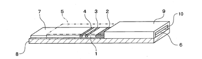
- FIG. 1 and FIG. 2 are an exploded perspective view and a perspective view showing a biosensor using chromatography according to the first embodiment.
- reference numeral 1 denotes a development layer made of nitrocellulose or the like on which a liquid sample is developed.
- the material used for the spreading layer 1 is not limited to nitrocellulose, and may be any material such as filter paper, non-woven fabric, membrane, cloth, glass fiber, and the like as long as the material can form a flow path capable of developing a liquid sample.
- the development layer is a single layer will be described as an example.
- the single layer means that it is composed of one layer, and a plurality of layers is a liquid sample in which a plurality of layers are arranged in parallel or perpendicularly and added to the first layer of the plurality of layers. It means that it is configured to be able to move sequentially to each layer.
- Reference numeral 2 denotes a labeling reagent part carried on a part of the development layer 1.
- the labeling reagent part 2 has a colloidal gold label for a test substance in the liquid sample in a dry state that can be eluted by the development of the liquid sample.
- An antibody (labeling reagent) is retained.
- the labeling reagent is obtained by labeling a specific reaction product such as an antibody with a label such as a gold colloid, and is used as a means for detecting the binding in the reagent immobilization units 3 and 4 described later.
- the labeling reagent part 2 does not need to be provided on the development layer 1 and there is no problem as long as it is an arbitrary position upstream of the reagent immobilization parts 3 and 4 described later in the development direction of the
- Reference numeral 3 denotes a first reagent immobilization unit
- 4 denotes a second reagent immobilization unit
- the first reagent immobilization unit 3 and the second reagent immobilization unit 4 are labeled reagents in the development layer 1. It is provided in a state of being supported on the downstream side in the developing direction of the liquid sample from the part 2.
- the reagents used in the first and second reagent immobilization units 3 and 4 are reagents such as antibodies capable of specific reaction with the substance to be inspected in the liquid sample.
- Both antibodies bind with an epitope different from that of the labeling reagent and are immobilized in a dry state.
- the antibody used for the first reagent immobilization unit 3 and the antibody used for the second reagent immobilization unit 4 are composed of antibodies having different affinities for the substance to be inspected in the liquid sample.
- the antibodies used in the first and second reagent immobilization units 3 and 4 only need to form a complex with the labeling reagent and the test substance, and therefore the epitope or affinity for the test substance is the same. It may be either different or different. Furthermore, although the two antibodies have different affinities, there is no problem with the same epitope.
- FIG. 1 shows an example in which two reagent immobilization units are provided, but this does not necessarily need to be two, and if there are one or more, it can be freely selected according to the purpose. .
- the shape on the development layer 1 does not need to be a line shape, and can be freely selected from a spot shape, a character shape, a key shape, and the like.
- the first reagent immobilization unit 3 and the second reagent immobilization unit 4 in FIG. 1 are spatially separated, they do not necessarily need to be separated, and seem to appear as a single line. It is also possible to make it contact.
- the liquid-impermeable sheet material 5 indicates a liquid-impermeable sheet material, which is composed of a transparent PET tape here.
- the liquid-impermeable sheet material 5 has a structure in which the spreading layer 1 is closely covered except for a portion connected to a fine space serving as the sample addition portion 6 and a terminal end where the liquid sample reaches.
- the reagent immobilization parts 3 and 4 and the labeling reagent part 2 which are reaction parts on the development layer 1 must be liquid.
- the sample passes through, and the reaction with the test substance in the liquid sample can be efficiently performed in the reaction portion.
- the contamination from the outside here means that the liquid sample is inadvertently brought into contact with the reaction portion on the development layer 1 or that the subject directly contacts the development layer 1 by hand.
- the coloration state of the first reagent immobilization unit 3 or the second reagent immobilization unit 4 is reflected using reflected light, scattered light, transmitted light, or the like. Since measurement is performed, it is preferable to use a transparent material, and the portions covering the reagent immobilization units 3 and 4 are portions for measuring a signal, and therefore must be maintained at least in a permeable state.
- the upper part of the development layer 1 is tightly sealed, particularly including the labeling reagent part 2 and the reagent immobilization parts 3 and 4, and the liquid It is also possible to adopt a structure in which side surfaces parallel to the sample development direction are similarly tightly sealed.
- Reference numeral 8 denotes a substrate that supports the development layer 1, and the substrate 8 is made of a liquid-impermeable sheet material such as a PET film.
- a liquid-impermeable sheet material such as a PET film.
- any material such as a synthetic resin material such as ABS, polystyrene, polyvinyl chloride, metal, glass, etc. can be used in addition to the PET film.
- the substrate 8 has a role of reinforcing the spreading layer 1 and also has a function of blocking the sample when there is a risk of infection such as blood, saliva or urine. Further, in the case where the spreading layer 1 is light transmissive when wet, the substrate 8 can have an effect of blocking light.
- the substrate 8 may be transparent, translucent, or opaque, but when measuring the degree of reaction optically, a transparent material is used when measuring using transmitted light, and an opaque material is used when measuring reflected light. Any material may be used depending on the application.
- Reference numeral 9 denotes a fine space forming material.
- the fine space forming material 9 has a function of forming a space into which a liquid sample flows in by capillary action, that is, a fine space serving as the sample adding portion 6, and a transparent PET film is laminated thereon. Composed of things.
- the fine space forming material 9 also has a role of protecting contamination of the liquid sample to the outside when the biosensor after the liquid sample is added is handled.
- the fine space forming material 9 can be made of a liquid-impermeable material such as metal, glass, or the like, in addition to a synthetic resin material such as ABS, polystyrene, or polyvinyl chloride, and is transparent or translucent. Is preferable, but it may be colored instead of transparent. In the case of an opaque material, any material can be used.
- the sample adding part 6 is a fine space formed by the fine space forming material 9, and the sample adding part 6 can flow a liquid sample by capillary action.
- the sample addition unit 6 is arranged to be connected to the development layer 1, and by causing the liquid sample to flow into the sample addition unit 6, the development of the liquid sample to the development layer 1 is started. Can do.
- the liquid sample When the liquid sample is brought into contact with the sample addition unit 6, the liquid sample naturally flows into the sample addition unit 6 without developing mechanical action due to capillary action, and flows and develops on the development layer 1. Whether the inflow amount of the liquid sample is sufficient can be confirmed through the fine space forming material 9.
- the amount of the fine space of the sample addition unit 6 can be set to a constant volume, so that the amount of addition can be accurately defined. When a certain amount or more of the liquid sample is required, this can be realized by adopting a structure that holds a volume of a necessary amount or more.
- the cell component contraction reagent 10 is a reagent to be installed when a cell component is contained in the liquid sample, and is not particularly necessary when a liquid sample containing no cell component is used.
- the cell component contraction reagent 10 is an inorganic compound containing potassium chloride, sodium chloride, sodium phosphate and the like, which has the effect of contracting cells, amino acids such as glycine and glutamic acid, imino acids such as proline, glucose, sucrose, It can be similarly carried out using sugars such as trehalose and sugar alcohols such as glucitol.
- Such a system containing the cell component contraction reagent 10 is particularly effective when whole blood is used as a liquid sample.
- the liquid sample that has been added and flowed into the sample addition section 6 develops from the contact portion between the sample addition section 6 and the development layer 1 to the development layer 1.
- the liquid sample reaches the labeling reagent unit 2
- elution of the labeling reagent is started.
- the development proceeds while the labeling reagent and the test substance react, and the liquid sample reaches the first reagent immobilization unit 3.
- the composite of the antibody immobilized on the first reagent immobilization unit 3 the test substance in the liquid sample, and the labeling reagent, depending on the amount of the test substance.
- the body is formed.
- the second reagent immobilization unit 4 When the liquid sample reaches the second reagent immobilization unit 4 and the substance to be inspected exists in the liquid sample, the second reagent immobilization is performed according to the amount of the substance to be inspected. A complex of the antibody immobilized on the portion 4, the substance to be inspected, and the labeling reagent is formed.
- the liquid sample reaches the second reagent immobilization unit 4 and then reaches the opening 7 in the development layer 1. Since the open portion 7 is open without the liquid-impermeable sheet 5, it volatilizes or evaporates after the liquid sample arrives or reaches. Furthermore, the liquid sample oozes out into the open part 7, and the liquid sample is only on the upper part of the development layer 1 in the open part 7 and the liquid sample is on the upper part of the development layer 1 in the fine space of the sample addition part 6. Up to the same height or equivalent height. In general, a water absorbing part is often provided instead of the open part 7, but it is possible to use a porous material having a higher water retaining effect and higher water absorbing effect than the material used for the spreading layer 1.
- the opening part 7 has the same effects as those, and in particular, the method using the sample adding part 6 or the opening part 7 is such that, for example, the liquid sample is blood by puncture from a fingertip. It is suitable when the liquid sample is a very small amount.
- the measurement value of the substance to be inspected in the liquid sample is obtained by measuring the signal derived from the labeling reagent in the first reagent immobilization unit 3 and the second reagent immobilization unit 4.
- FIG. 3 shows the signal measurement unit in a state where the signal derived from the labeling reagent in the reagent immobilization units 3 and 4 on the biosensor is being measured.
- 11 is an irradiation unit for irradiating light to the development layer 1 in an analyzer that measures and analyzes the concentration of a substance to be inspected in a liquid sample using a biosensor
- 12 is the irradiation unit 11.
- Is a light receiving unit that detects an optical change such as reflection or transmitted light of the light irradiated to the spreading layer 1, and the signal measuring unit uses the irradiation unit 11 and the light receiving unit 12 to obtain a reagent.
- the signal derived from the labeling reagent in the immobilization units 3 and 4 is measured.
- the signal rises compared to the other portions, and the intensity changes according to the coloration degree of the reagent immobilization units 3 and 4.
- LED Light Emitting Diode; Light emitting diode
- LD Laser Diode; Laser diode
- the measurement value of the substance to be inspected can be obtained by performing arithmetic processing on the signal derived from the labeling reagent in the reagent immobilization units 3 and 4 measured by the signal measurement unit (irradiation unit 11 and light receiving unit 12). it can.
- the measured value of the test substance obtained at this time is the value of the test substance obtained using a calibration curve from the signal derived from the labeling reagent obtained in the signal measuring unit.
- the calibration curve described here is a regression equation showing the relationship between the signal obtained by the signal measurement unit and the value of the substance to be inspected in the liquid sample.
- the measured value of the test substance in the unknown liquid sample can be calculated by substituting the signal obtained by the signal measuring unit.
- any means such as one that reads an optical change, an electrical change, or an electromagnetic change, or that captures as an image may be used.
- a specific reaction when a specific reaction is desired to be used, it is possible to perform measurement by a reaction other than the antigen-antibody reaction by using a reagent component of a system that forms an arbitrary reaction on the biosensor.
- the combination of a specific substance that specifically reacts and a specific binding substance thereto includes an antigen and an antibody thereto, a complementary nucleic acid sequence, an effector molecule and a receptor molecule, an enzyme and an inhibitor, an enzyme and a cofactor, an enzyme And a substrate, a compound having a sugar chain and a lectin, a certain antibody and an antibody against the antibody, a receptor molecule and an antibody against it, chemically modified to such an extent that specific binding activity is not lost, or combined with other components
- Examples include a reaction system using a complex substance.
- Example 1 Quantification of whole blood CRP when the development layer is thin
- a second reagent immobilization section 4 in which anti-CRP antibody B is immobilized and anti-CRP antibody C
- An immunochromatographic sensor which is a biosensor including a labeling reagent part 2 holding a complex of gold and colloidal gold (labeling reagent), was manufactured. This immunochromatographic sensor is shown in FIGS.
- the immunochromatographic sensor is located in a portion closer to the first reagent immobilization unit 3 and the second reagent immobilization unit 4 to which the antibody is immobilized, and the addition portion to which the liquid sample is added. It has a labeling reagent part 2 which is a region containing a complex of CRP antibody C and colloidal gold, and a sample addition part 6.
- This immunochromatographic sensor was manufactured as follows.
- an anti-CRP antibody B solution was applied to a portion 2 mm away from the sample addition portion on the downstream side.
- Six types of nitrocellulose membranes were prepared: 145 ⁇ m, 95 ⁇ m, 85 ⁇ m, 75 ⁇ m, 65 ⁇ m, and 55 ⁇ m.
- the nitrocellulose membrane was dried, immersed in a Tris-HCl buffer solution containing 1% skim milk, and gently shaken for 30 minutes. After 30 minutes, the membrane was moved to a Tris-HCl buffer solution tank, gently shaken for 10 minutes, and then gently shaken for another 10 minutes in another Tris-HCl buffer solution tank to wash the membrane.
- the nitrocellulose membrane was taken out of the liquid bath and dried at room temperature.
- the first immobilized antibody line as the first reagent immobilization unit 3 and the immobilized antibody line as the second reagent immobilization unit 4 were obtained on the nitrocellulose membrane (developing layer 1). .
- the colloidal gold was prepared by adding a 1% trisodium citrate solution to a 0.01% chloroauric acid 100 ° C solution under reflux. Refluxing was continued for 15 minutes and then allowed to cool at room temperature. After adding anti-CRP antibody C to the gold colloid solution adjusted to pH 8.9 with 0.2 M potassium carbonate solution and stirring for several minutes, 10% BSA (bovine serum albumin) solution with pH 8.9 is finally added to 1
- BSA bovine serum albumin
- the prepared reaction layer carrier containing the labeling reagent is affixed on the substrate 8 made of white PET having a thickness of 0.5 mm, and the liquid-impermeable sheet material 5 is formed from the labeling reagent part 2 to the terminal part.
- a transparent tape was applied.
- a fine space forming material 9 produced by laminating transparent PET having a thickness of 100 ⁇ m is pasted on the starting end portion where no transparent tape is pasted, and a fine space (width 2.0 mm ⁇ length 7.0 mm ⁇ A sample addition portion 6 having a height of 0.3 mm was formed.
- the space forming material 9 is preliminarily spotted with a 10% aqueous potassium chloride solution, immediately frozen in liquid nitrogen, and lyophilized, whereby the cell component contraction reagent 10 in which potassium chloride is maintained in a dry state is obtained.
- the fine space forming material 9 to be held is produced.
- an immunochromatographic sensor was manufactured.
- the CRP concentration (test substance concentration) is 0.03 mg / dL, 0.1 mg / dL, 0.3 mg / dL and 1 mg / dL of blood were prepared as liquid samples.
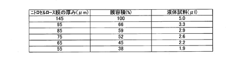
- the thickness of the nitrocellulose film used as the development layer 1 is almost the same. 30% or more thinner.
- the thickness is reduced, the volume is reduced, so that the amount of liquid sample required for measurement can be reduced by 30% or more.
- FIG. 4 shows the amount of liquid sample required for each nitrocellulose membrane thickness.
- the immobilized antibody line that is the reagent immobilization unit 3 or 4 even if the amount of the liquid sample is 2 ⁇ L. It can reach the position sufficiently, and measurement with a very small amount can be realized.
- the immunochromatographic sensor is scanned by the irradiation unit 11 composed of a semiconductor laser light source of 635 nm and the light receiving unit 12 including a light receiving element (photodiode), and the first immobilization unit
- the amount of the labeling reagent binding in the third and second immobilization units 4 is calculated from the reflected and scattered light from the spreading layer 1 to obtain a result as absorbance.
- the light source (irradiation unit 11) and the light receiving element (light receiving unit 12) were fixed, scanned by moving the immunochromatographic sensor, and the peak value (reflection absorbance) was read from the obtained waveform. In order to obtain such a waveform, it is possible to scan by moving the light source and the light receiving element while fixing the immunochromatographic sensor.
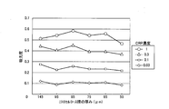
- FIG. 5 shows the absorbance results for each nitrocellulose film thickness.
- the absorbance is sufficient for quantitative measurement. It can be seen that even if the thickness of the nitrocellulose membrane is reduced, the amount of the liquid sample can be reduced without any problem in quantitative measurement.
- the thickness of the development layer 1 in the biosensor As described above, as a method of manufacturing a biosensor for developing a liquid sample in the development layer 1 and measuring a substance to be inspected in the liquid sample, by reducing the thickness of the development layer 1, It is possible to reduce the required amount of liquid sample added in accordance with the measurement of the substance to be inspected. Specifically, by setting the thickness of the development layer 1 in the biosensor to 20 to 135 ⁇ m, which is thinner than the conventional one (around 150 ⁇ m, for example, thickness 145 ⁇ m), the required amount of the liquid sample can be reduced as compared with the conventional one. it can.
- deployment layer 1 used as noise can be reduced by reducing the thickness of the expansion
- the required amount of liquid sample can be reduced without forcibly reducing the width and length of the biosensor, the handling of the biosensor does not become inconvenient and the reagent is immobilized at the measurement target location. Since the parts 3 and 4 are not reduced in size, the measurement accuracy of the substance to be inspected is not deteriorated due to the reduction of the reagent fixing parts 3 and 4.
- the amount of the labeling reagent that reacts with the test substance in the liquid sample and the reagent that specifically reacts with the test substance or the labeling reagent can be reduced as compared with the prior art. Since it can be reduced, the manufacturing cost can also be reduced.
- Example 2 Quantification of whole blood CRP when the development layer is thin
- anti-CRP antibody C An immunochromatographic sensor, which is a biosensor including a labeling reagent part 2 holding a complex of gold and colloidal gold (labeling reagent), was manufactured. This immunochromatographic sensor is shown in FIGS.
- the immunochromatographic sensor is located in a portion closer to the first reagent immobilization unit 3 and the second reagent immobilization unit 4 to which the antibody is immobilized, and the addition portion to which the liquid sample is added. It has a labeling reagent part 2 which is a region containing a complex of CRP antibody C and colloidal gold, and a sample addition part 6.
- This immunochromatographic sensor was manufactured as follows.
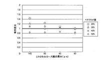
- FIG. 6 shows the results of the absorbance ratio for each nitrocellulose film thickness. The horizontal axis represents the thickness of the nitrocellulose film, and the vertical axis represents the absorbance ratio, plotted by hematocrit value.
- the absorbance ratio indicates how far the absorbance at each hematocrit value deviates from the average absorbance at all hematocrit values, and the difference in nitrocellulose membrane thickness and hematocrit value. The degree of influence on the measurement result can be confirmed.
- FIG. 7 shows the CV value results for each nitrocellulose film thickness.
- the film thickness was 145 ⁇ m, the CV was poor, and the deterioration of the CV value was particularly remarkable when the hematocrit value was high, but the influence of the hematocrit value was reduced by reducing the film thickness, and the CV was good. From the above results, by reducing the thickness of the development layer 1, the background due to the thickness of the development layer 1 that becomes noise can be reduced, so that the S / N ratio can be improved and the measurement accuracy is improved.
- the thickness of the nitrocellulose film becomes thinner, the difference in variation in the development speed of the liquid sample due to the characteristics of the liquid sample such as the hematocrit value becomes less likely to occur. Therefore, when the thickness of the nitrocellulose film was reduced, it was less affected by the hematocrit value than when the film thickness was 145 ⁇ m, and the variation in the degree of coloration was reduced and the CV was improved. Even if the amount of liquid sample added was reduced, a sensor with improved sensitivity and accuracy could be realized.
- Example 3 Quantification of whole blood CRP when changing the thickness and pore size of the spreading layer
- a second reagent immobilization section 4 in which anti-CRP antibody B is immobilized and anti-CRP antibody C
- An immunochromatographic sensor which is a biosensor including a labeling reagent part 2 holding a complex of gold and colloidal gold (labeling reagent), was manufactured. This immunochromatographic sensor is shown in FIGS.
- the immunochromatographic sensor is located in a portion closer to the first reagent immobilization unit 3 and the second reagent immobilization unit 4 to which the antibody is immobilized, and the addition portion to which the liquid sample is added. It has a labeling reagent part 2 which is a region containing a complex of CRP antibody C and colloidal gold, and a sample addition part 6.
- This immunochromatographic sensor was manufactured as follows.
- the standard of the nitrocellulose membrane which is a porous carrier, is a conventional standard with a film thickness of 145 ⁇ m and a pore diameter of 5 ⁇ m, a film thickness of 135 ⁇ m, and a pore diameter of 3 ⁇ m, 5 ⁇ m, and 8 ⁇ m.
- Sixteen types were prepared, one having a film thickness of 60 ⁇ m and a pore diameter of 5 ⁇ m, 8 ⁇ m, and 10 ⁇ m.
- An immunochromatographic sensor was produced by the same production method as in Example 1, and the following measurements were performed.
- the horizontal axis is the thickness of the nitrocellulose membrane
- the vertical axis is the absorbance ratio
- the absorbance ratio is plotted for each pore size.
- the absorbance ratio indicates how far the absorbance in the standard of each nitrocellulose membrane deviates from the absorbance in the conventional standard of 145 ⁇ m, and the difference in thickness and pore diameter of the nitrocellulose membrane The degree of influence on the measurement result can be confirmed.
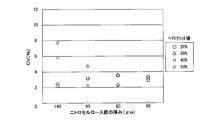
- FIG. 9 shows CV value results for each standard of the nitrocellulose membrane.
- Each CV value is plotted for each pore diameter.
- the CV value was improved by reducing the film thickness with respect to the CV value at the film thickness of 145 ⁇ m, which is the conventional standard. In particular, the improvement of the CV value at 115 ⁇ m or less was remarkable. From the above results, by reducing the thickness of the development layer 1, the background value due to the thickness of the development layer 1 that becomes noise can be reduced, so the S / N ratio can be improved and the measurement accuracy is improved. The variation in the degree of coloration can be reduced, and the CV is improved. As is clear from FIGS.
- a biosensor with improved sensitivity and accuracy can be realized by reducing the thickness of the development layer 1 regardless of the pore diameter of the nitrocellulose membrane.
- the pore diameter shown here has shown the average value of the pore diameter of a nitrocellulose membrane.
- the cross section of the nitrocellulose membrane was measured based on an image observed with a scanning electron microscope, the pore diameter was measured using any pore diameter measurement method such as the valve point method or the mercury pressure method. There is no problem.
- a porous carrier such as nitrocellulose is not composed of a single pore having the described pore size value, and is distributed in large and small pore sizes with respect to the average pore size. Are mixed.
- a comparative example is given based on the numerical value of the average pore diameter, and the pore diameter value is not limited to the described numerical value.
- liquid samples to be measured include body fluids such as water and aqueous solutions, urine, plasma, serum, and saliva, solids, powders, and gases.
- body fluids such as water and aqueous solutions, urine, plasma, serum, and saliva
- solids, powders, and gases examples include blood tests, urine tests, water quality tests, stool tests, soil analysis, and food analysis.
- Substances to be tested include proteins and protein derivatives such as antibodies, immunoglobulins, hormones, enzymes and peptides, infectious substances such as bacteria, viruses, fungi, mycoplasma, parasites and their products and components, and treatment Drugs and tumor markers such as drugs and drugs of abuse.
- chorionic gland stimulating hormone HCG
- luteinizing hormone LH
- thyroid stimulating hormone follicle forming hormone
- parathyroid stimulating hormone adrenal lipid stimulating hormone
- estradiol prostate specific antigen
- hepatitis myoglobin
- CRP Ceresoid arthritis
- cardiac troponin BNP, HBA1C, albumin and the like. It can also be used for environmental analysis such as water quality testing and soil analysis, and food analysis. Simple, quick, high-sensitivity, high-performance measurement can be realized, and anyone can perform measurement anytime, anywhere, so it can be used as an analyzer for POCT.
- the biosensor of the present invention is simple and rapid when analyzing and quantifying or semi-quantifying a test substance in a small amount of liquid sample based on an arbitrary reaction such as an antigen-antibody reaction. Since it can be used for sensitive and high-performance measurement, and anyone can measure anytime, anywhere, it is useful as a biosensor for an analyzer for POCT.
Landscapes
- Health & Medical Sciences (AREA)
- Immunology (AREA)
- Life Sciences & Earth Sciences (AREA)
- Engineering & Computer Science (AREA)
- Molecular Biology (AREA)
- Biomedical Technology (AREA)
- Chemical & Material Sciences (AREA)
- Hematology (AREA)
- Urology & Nephrology (AREA)
- Biotechnology (AREA)
- Microbiology (AREA)
- Cell Biology (AREA)
- Food Science & Technology (AREA)
- Medicinal Chemistry (AREA)
- Physics & Mathematics (AREA)
- Analytical Chemistry (AREA)
- Biochemistry (AREA)
- General Health & Medical Sciences (AREA)
- General Physics & Mathematics (AREA)
- Pathology (AREA)
- Investigating Or Analysing Biological Materials (AREA)
Abstract
Priority Applications (3)
| Application Number | Priority Date | Filing Date | Title |
|---|---|---|---|
| US12/990,054 US20110045578A1 (en) | 2008-05-07 | 2009-04-23 | Method of manufacturing biosensor and biosensor produced thereby |
| JP2010511009A JPWO2009136476A1 (ja) | 2008-05-07 | 2009-04-23 | バイオセンサの製造方法およびバイオセンサ |
| EP09742601A EP2273269A4 (fr) | 2008-05-07 | 2009-04-23 | Procédé de fabrication de biocapteur et biocapteur |
Applications Claiming Priority (2)
| Application Number | Priority Date | Filing Date | Title |
|---|---|---|---|
| JP2008-120854 | 2008-05-07 | ||
| JP2008120854 | 2008-05-07 |
Publications (1)
| Publication Number | Publication Date |
|---|---|
| WO2009136476A1 true WO2009136476A1 (fr) | 2009-11-12 |
Family
ID=41264533
Family Applications (1)
| Application Number | Title | Priority Date | Filing Date |
|---|---|---|---|
| PCT/JP2009/001859 Ceased WO2009136476A1 (fr) | 2008-05-07 | 2009-04-23 | Procédé de fabrication de biocapteur et biocapteur |
Country Status (4)
| Country | Link |
|---|---|
| US (1) | US20110045578A1 (fr) |
| EP (1) | EP2273269A4 (fr) |
| JP (1) | JPWO2009136476A1 (fr) |
| WO (1) | WO2009136476A1 (fr) |
Cited By (6)
| Publication number | Priority date | Publication date | Assignee | Title |
|---|---|---|---|---|
| JP2014062820A (ja) * | 2012-09-21 | 2014-04-10 | Toyo Roshi Kaisha Ltd | イムノクロマトグラフ試験ストリップ用メンブレン、試験ストリップ及び検査方法 |
| WO2017002882A1 (fr) * | 2015-06-30 | 2017-01-05 | 田中貴金属工業株式会社 | Dispositif d'analyse chromatographique et procédé d'analyse chromatographique |
| JP2017509889A (ja) * | 2014-03-31 | 2017-04-06 | ワットマン・ゲーエムベーハー | ラテラルフロー試験及びそれに関係する改善 |
| WO2020067233A1 (fr) * | 2018-09-27 | 2020-04-02 | 積水メディカル株式会社 | Pièce d'essai pour immunochromatographie |
| JP2020051910A (ja) * | 2018-09-27 | 2020-04-02 | 積水メディカル株式会社 | イムノクロマト用試験片 |
| JP2020125994A (ja) * | 2019-02-05 | 2020-08-20 | 積水メディカル株式会社 | イムノクロマト用試験片 |
Families Citing this family (2)
| Publication number | Priority date | Publication date | Assignee | Title |
|---|---|---|---|---|
| US9353313B2 (en) | 2012-08-09 | 2016-05-31 | Auburn University | Microdevices and methods of manufacture |
| GB201302292D0 (en) * | 2013-02-08 | 2013-03-27 | Whatman Gmbh | Medical diagnostic test systems,and a matrix therefor |
Citations (5)
| Publication number | Priority date | Publication date | Assignee | Title |
|---|---|---|---|---|
| JPH05281231A (ja) * | 1992-01-31 | 1993-10-29 | Boehringer Mannheim Gmbh | 免疫測定用分析要素 |
| JP2000258418A (ja) * | 1999-03-12 | 2000-09-22 | Arkray Inc | 免疫クロマトグラフィーを用いた測定方法およびそれに用いる検体分析用具 |
| WO2001090754A1 (fr) | 2000-05-26 | 2001-11-29 | Matsushita Electric Industrial Co., Ltd. | Biocapteur |
| JP2004189665A (ja) * | 2002-12-11 | 2004-07-08 | Matsushita Electric Ind Co Ltd | 抗c反応性タンパク質抗体、この抗体を用いたバイオセンサ、この抗体の作製方法、及びこの抗体を用いた免疫測定方法 |
| JP3655283B2 (ja) | 2000-06-01 | 2005-06-02 | 松下電器産業株式会社 | バイオセンサ、及び血液成分分析方法 |
Family Cites Families (1)
| Publication number | Priority date | Publication date | Assignee | Title |
|---|---|---|---|---|
| JPS57125847A (en) * | 1981-01-30 | 1982-08-05 | Konishiroku Photo Ind Co Ltd | Analysis element |
-
2009
- 2009-04-23 WO PCT/JP2009/001859 patent/WO2009136476A1/fr not_active Ceased
- 2009-04-23 JP JP2010511009A patent/JPWO2009136476A1/ja not_active Withdrawn
- 2009-04-23 EP EP09742601A patent/EP2273269A4/fr not_active Withdrawn
- 2009-04-23 US US12/990,054 patent/US20110045578A1/en not_active Abandoned
Patent Citations (5)
| Publication number | Priority date | Publication date | Assignee | Title |
|---|---|---|---|---|
| JPH05281231A (ja) * | 1992-01-31 | 1993-10-29 | Boehringer Mannheim Gmbh | 免疫測定用分析要素 |
| JP2000258418A (ja) * | 1999-03-12 | 2000-09-22 | Arkray Inc | 免疫クロマトグラフィーを用いた測定方法およびそれに用いる検体分析用具 |
| WO2001090754A1 (fr) | 2000-05-26 | 2001-11-29 | Matsushita Electric Industrial Co., Ltd. | Biocapteur |
| JP3655283B2 (ja) | 2000-06-01 | 2005-06-02 | 松下電器産業株式会社 | バイオセンサ、及び血液成分分析方法 |
| JP2004189665A (ja) * | 2002-12-11 | 2004-07-08 | Matsushita Electric Ind Co Ltd | 抗c反応性タンパク質抗体、この抗体を用いたバイオセンサ、この抗体の作製方法、及びこの抗体を用いた免疫測定方法 |
Non-Patent Citations (1)
| Title |
|---|
| See also references of EP2273269A4 |
Cited By (12)
| Publication number | Priority date | Publication date | Assignee | Title |
|---|---|---|---|---|
| JP2014062820A (ja) * | 2012-09-21 | 2014-04-10 | Toyo Roshi Kaisha Ltd | イムノクロマトグラフ試験ストリップ用メンブレン、試験ストリップ及び検査方法 |
| JP2017509889A (ja) * | 2014-03-31 | 2017-04-06 | ワットマン・ゲーエムベーハー | ラテラルフロー試験及びそれに関係する改善 |
| WO2017002882A1 (fr) * | 2015-06-30 | 2017-01-05 | 田中貴金属工業株式会社 | Dispositif d'analyse chromatographique et procédé d'analyse chromatographique |
| JP2017015533A (ja) * | 2015-06-30 | 2017-01-19 | 田中貴金属工業株式会社 | クロマト分析装置およびクロマト分析方法 |
| WO2020067233A1 (fr) * | 2018-09-27 | 2020-04-02 | 積水メディカル株式会社 | Pièce d'essai pour immunochromatographie |
| JP2020051910A (ja) * | 2018-09-27 | 2020-04-02 | 積水メディカル株式会社 | イムノクロマト用試験片 |
| CN112740039A (zh) * | 2018-09-27 | 2021-04-30 | 积水医疗株式会社 | 免疫层析试验片 |
| KR20210064268A (ko) * | 2018-09-27 | 2021-06-02 | 세키스이 메디칼 가부시키가이샤 | 면역 크로마토그래피용 시험편 |
| KR102766141B1 (ko) * | 2018-09-27 | 2025-02-12 | 세키스이 메디칼 가부시키가이샤 | 면역 크로마토그래피용 시험편 |
| US12613241B2 (en) | 2018-09-27 | 2026-04-28 | Sekisui Medical Co., Ltd. | Immunochromatographic test strip |
| JP2020125994A (ja) * | 2019-02-05 | 2020-08-20 | 積水メディカル株式会社 | イムノクロマト用試験片 |
| JP7137168B2 (ja) | 2019-02-05 | 2022-09-14 | 積水メディカル株式会社 | イムノクロマト用試験片 |
Also Published As
| Publication number | Publication date |
|---|---|
| EP2273269A1 (fr) | 2011-01-12 |
| US20110045578A1 (en) | 2011-02-24 |
| JPWO2009136476A1 (ja) | 2011-09-01 |
| EP2273269A4 (fr) | 2011-05-25 |
Similar Documents
| Publication | Publication Date | Title |
|---|---|---|
| JP4383860B2 (ja) | バイオセンサ、及び測定方法 | |
| CN102016584B (zh) | 生物传感器 | |
| CN1161611C (zh) | 生物传感器 | |
| CN100492010C (zh) | 生物传感器 | |
| US8093057B2 (en) | System for quantitative measurement of glycohemoglobin and method for measuring glycohemoglobin | |
| JP3543000B2 (ja) | バイオセンサ | |
| KR100596278B1 (ko) | 바이오센서 및 이를 사용한 혈액 성분 분석 방법 | |
| JPWO2001090754A1 (ja) | バイオセンサ | |
| CN101258406A (zh) | 分析装置及分析方法 | |
| JPWO2009136476A1 (ja) | バイオセンサの製造方法およびバイオセンサ | |
| WO2007055712A1 (fr) | Essai d’écoulement latéral immuno-or | |
| TW494238B (en) | Chromatographic test pieces and chromatographic assay | |
| JP2010032396A (ja) | バイオセンサ | |
| JP4980944B2 (ja) | 免疫学的測定方法 | |
| KR102766141B1 (ko) | 면역 크로마토그래피용 시험편 | |
| JP2005257468A (ja) | バイオセンサ | |
| JP4430263B2 (ja) | クロマトグラフィ測定装置 | |
| JP2010091353A (ja) | バイオセンサおよびバイオセンサ用不溶性粒状マーカー | |
| JP6939106B2 (ja) | イムノクロマト試験片およびキットおよび測定方法 | |
| JP2009294116A (ja) | バイオセンサ | |
| JP2002162397A (ja) | 乾式分析要素 | |
| HK1080937B (en) | Biosensor |
Legal Events
| Date | Code | Title | Description |
|---|---|---|---|
| 121 | Ep: the epo has been informed by wipo that ep was designated in this application |
Ref document number: 09742601 Country of ref document: EP Kind code of ref document: A1 |
|
| ENP | Entry into the national phase |
Ref document number: 2010511009 Country of ref document: JP Kind code of ref document: A |
|
| WWE | Wipo information: entry into national phase |
Ref document number: 2009742601 Country of ref document: EP |
|
| WWE | Wipo information: entry into national phase |
Ref document number: 12990054 Country of ref document: US |
|
| NENP | Non-entry into the national phase |
Ref country code: DE |