WO2010047132A1 - 細胞培養キット、スクリーニング方法、及び細胞培養キットの製造方法 - Google Patents
細胞培養キット、スクリーニング方法、及び細胞培養キットの製造方法 Download PDFInfo
- Publication number
- WO2010047132A1 WO2010047132A1 PCT/JP2009/005617 JP2009005617W WO2010047132A1 WO 2010047132 A1 WO2010047132 A1 WO 2010047132A1 JP 2009005617 W JP2009005617 W JP 2009005617W WO 2010047132 A1 WO2010047132 A1 WO 2010047132A1
- Authority
- WO
- WIPO (PCT)
- Prior art keywords
- cells
- cell culture
- cell
- culture kit
- living
- Prior art date
- Legal status (The legal status is an assumption and is not a legal conclusion. Google has not performed a legal analysis and makes no representation as to the accuracy of the status listed.)
- Ceased
Links
Images










Classifications
-
- C—CHEMISTRY; METALLURGY
- C12—BIOCHEMISTRY; BEER; SPIRITS; WINE; VINEGAR; MICROBIOLOGY; ENZYMOLOGY; MUTATION OR GENETIC ENGINEERING
- C12N—MICROORGANISMS OR ENZYMES; COMPOSITIONS THEREOF; PROPAGATING, PRESERVING, OR MAINTAINING MICROORGANISMS; MUTATION OR GENETIC ENGINEERING; CULTURE MEDIA
- C12N5/00—Undifferentiated human, animal or plant cells, e.g. cell lines; Tissues; Cultivation or maintenance thereof; Culture media therefor
- C12N5/0062—General methods for three-dimensional culture
-
- C—CHEMISTRY; METALLURGY
- C12—BIOCHEMISTRY; BEER; SPIRITS; WINE; VINEGAR; MICROBIOLOGY; ENZYMOLOGY; MUTATION OR GENETIC ENGINEERING
- C12M—APPARATUS FOR ENZYMOLOGY OR MICROBIOLOGY; APPARATUS FOR CULTURING MICROORGANISMS FOR PRODUCING BIOMASS, FOR GROWING CELLS OR FOR OBTAINING FERMENTATION OR METABOLIC PRODUCTS, i.e. BIOREACTORS OR FERMENTERS
- C12M3/00—Tissue, human, animal or plant cell, or virus culture apparatus
-
- C—CHEMISTRY; METALLURGY
- C12—BIOCHEMISTRY; BEER; SPIRITS; WINE; VINEGAR; MICROBIOLOGY; ENZYMOLOGY; MUTATION OR GENETIC ENGINEERING
- C12M—APPARATUS FOR ENZYMOLOGY OR MICROBIOLOGY; APPARATUS FOR CULTURING MICROORGANISMS FOR PRODUCING BIOMASS, FOR GROWING CELLS OR FOR OBTAINING FERMENTATION OR METABOLIC PRODUCTS, i.e. BIOREACTORS OR FERMENTERS
- C12M23/00—Constructional details, e.g. recesses, hinges
- C12M23/02—Form or structure of the vessel
- C12M23/12—Well or multiwell plates
-
- C—CHEMISTRY; METALLURGY
- C12—BIOCHEMISTRY; BEER; SPIRITS; WINE; VINEGAR; MICROBIOLOGY; ENZYMOLOGY; MUTATION OR GENETIC ENGINEERING
- C12N—MICROORGANISMS OR ENZYMES; COMPOSITIONS THEREOF; PROPAGATING, PRESERVING, OR MAINTAINING MICROORGANISMS; MUTATION OR GENETIC ENGINEERING; CULTURE MEDIA
- C12N5/00—Undifferentiated human, animal or plant cells, e.g. cell lines; Tissues; Cultivation or maintenance thereof; Culture media therefor
- C12N5/06—Animal cells or tissues; Human cells or tissues
-
- C—CHEMISTRY; METALLURGY
- C12—BIOCHEMISTRY; BEER; SPIRITS; WINE; VINEGAR; MICROBIOLOGY; ENZYMOLOGY; MUTATION OR GENETIC ENGINEERING
- C12Q—MEASURING OR TESTING PROCESSES INVOLVING ENZYMES, NUCLEIC ACIDS OR MICROORGANISMS; COMPOSITIONS OR TEST PAPERS THEREFOR; PROCESSES OF PREPARING SUCH COMPOSITIONS; CONDITION-RESPONSIVE CONTROL IN MICROBIOLOGICAL OR ENZYMOLOGICAL PROCESSES
- C12Q1/00—Measuring or testing processes involving enzymes, nucleic acids or microorganisms; Compositions therefor; Processes of preparing such compositions
- C12Q1/02—Measuring or testing processes involving enzymes, nucleic acids or microorganisms; Compositions therefor; Processes of preparing such compositions involving viable microorganisms
-
- C—CHEMISTRY; METALLURGY
- C12—BIOCHEMISTRY; BEER; SPIRITS; WINE; VINEGAR; MICROBIOLOGY; ENZYMOLOGY; MUTATION OR GENETIC ENGINEERING
- C12N—MICROORGANISMS OR ENZYMES; COMPOSITIONS THEREOF; PROPAGATING, PRESERVING, OR MAINTAINING MICROORGANISMS; MUTATION OR GENETIC ENGINEERING; CULTURE MEDIA
- C12N2502/00—Coculture with; Conditioned medium produced by
-
- C—CHEMISTRY; METALLURGY
- C12—BIOCHEMISTRY; BEER; SPIRITS; WINE; VINEGAR; MICROBIOLOGY; ENZYMOLOGY; MUTATION OR GENETIC ENGINEERING
- C12N—MICROORGANISMS OR ENZYMES; COMPOSITIONS THEREOF; PROPAGATING, PRESERVING, OR MAINTAINING MICROORGANISMS; MUTATION OR GENETIC ENGINEERING; CULTURE MEDIA
- C12N2503/00—Use of cells in diagnostics
- C12N2503/02—Drug screening
-
- C—CHEMISTRY; METALLURGY
- C12—BIOCHEMISTRY; BEER; SPIRITS; WINE; VINEGAR; MICROBIOLOGY; ENZYMOLOGY; MUTATION OR GENETIC ENGINEERING
- C12N—MICROORGANISMS OR ENZYMES; COMPOSITIONS THEREOF; PROPAGATING, PRESERVING, OR MAINTAINING MICROORGANISMS; MUTATION OR GENETIC ENGINEERING; CULTURE MEDIA
- C12N2513/00—3D culture
Definitions
- the present invention relates to a cell culture kit for culturing living cells, a screening method, and a production method thereof.
- the technique used for testing and examining cells isolated from tissues is an indispensable method in the field of biotechnology. It is widely used for diagnosing diseases and pathological conditions, searching for new drugs and determining their efficacy, animal testing, plant testing, and testing for environmental pollutants. Therefore, the cells used in the biotechnology field have been extremely diversified.
- the isolated cells may be used for the test immediately, but in many cases, the cells are cultured in a culture dish or a test tube.
- Various tests are performed using the cultured cells.
- Cell culture strains used for cell culture tests are required to exhibit drug sensitivities and toxic reactions similar to in vivo tests, so-called in vivo tests. That is, it is necessary to be able to construct a network between cells arranged with regularity on the surface of a cell culture vessel.
- cell culture strains used for cell culture tests are extremely expensive, it is desired to improve cell survival rate and growth rate. That is, a cell function similar to that in a living body is required on a cell culture container.
- the isolation procedure for obtaining the primary cells is complicated and the cell culture strain used for the cell culture test is expensive, a test method with a small number of cells is desired.
- liver slices are difficult to obtain, and liver microsomes can only be metabolized using limited metabolic enzymes. Therefore, the use of hepatocytes is considered to be most effective.
- the culture dishes used are resin petri dishes and 6-well, 12-well, 48-well, and 96-well plates.
- the size of the whole plate is substantially the same, and the larger the number of wells, the smaller the size of one well.
- One well corresponds to one culture dish.
- 384-well plates comprising a large number of culture dishes with a smaller diameter have begun to be used, and those adapted to the intended screening method are used.
- the bottom of these culture dishes is a flat plate shape, and this bottom is used as the culture surface.
- the original function may be lost and dedifferentiation may occur, or undifferentiated cells may not differentiate, and the target cell function may not be expressed. It has become.
- the metabolic enzyme function when isolated is significantly reduced in about 1 day. May be performed. That is, there are problems that cannot be used for testing while culturing for a long time, and problems that long-term metabolic stability cannot be examined.
- Patent Document 1 is expensive because the biological material to be coated is special, it is difficult to form a uniform cell aggregate in the culture vessel, and the in vivo function is prolonged. There were problems such as inability to maintain. Even the method disclosed in Patent Document 2 has problems that the size of the cell aggregate cannot be controlled, that the observation with a microscope cannot be easily performed, and that the operability as a screening substrate is complicated. Furthermore, since any method uses a commercially available dish or plate as a support container, it is difficult to efficiently screen with the minimum number of cells.
- the present invention has been made to solve such problems, and an object of the present invention is to provide a cell culture kit, a screening method, and a production method thereof, in which live cells of a plurality of donors are cultured.
- One aspect of the cell culture kit according to the present invention is a cell culture kit having a cell culture plate and a living cell cultured thereon, the cell culture plate having a plurality of microspaces, In the microspace, living cells derived from a plurality of different donors are adhered to the surface.
- a screening kit in which living cells derived from a plurality of different donors are adhered to one cell culture plate can be provided.
- the living cells derived from the plurality of different donors may be two or more types of cells.
- a tissue-like structure composed of a plurality of types of living cells is reproduced on the chip.
- each micro space either a plurality of living cells derived from different donors or a living cell derived from one donor is adhered.
- at least two adjacent microspaces have live cells from different donors attached.
- at least two adjacent microspaces have live cells from the same donor attached.
- the plurality of microspaces preferably have such a size that a cell population of a three-dimensional structure cultured in a desired number of cells is isolated, and the plurality of microspaces have a bottom area of 0.01 to It is preferably 0.1 mm 2 and the depth is 25 to 150 ⁇ m.
- the plurality of microspaces are preferably seeded with live cells at a cell seeding density of 1 ⁇ 10 2 to 1 ⁇ 10 6 cells / cm 2 , and a cell seeding density of 1 ⁇ 10 4 to 1 ⁇ 10 6 cells. More preferably, it is / cm 2 .
- the plurality of microspaces it is preferable that living cells are accumulated and a cell mass is formed, and more specifically, the diameter of the cell mass is preferably 30 to 200 ⁇ m.
- the living cells are preferably tissue precursor cells, tissue stem cells, cells differentiated from ES cells, or cells differentiated from iPS cells.
- the living cells are preferably living cells including hepatocytes, and the hepatocytes are tissue precursor cells, tissue stem cells, cells differentiated from ES cells, or cells differentiated from iPS cells, or More preferably, the living cells containing hepatocytes are isolated from the liver tissues of a plurality of donors.
- the drug is evaluated using the cell culture kit described above.
- one embodiment of the method for producing a cell culture kit according to the present invention is a method for producing a cell culture kit having a cell culture plate having a plurality of microspaces and a living cell cultured thereon, In the microspace, live cells derived from a plurality of different donors are seeded, and the seeded live cells are cultured.
- a cell culture kit it is possible to provide a cell culture kit, a screening method, and a method for producing a cell culture kit in which living cells of a plurality of donors are cultured.
- FIG. 6 is a VI-VI cross-sectional view showing still another configuration of the cell culture container according to the embodiment. It is a figure which shows an example of the cell culture kit by which the several cell culture container is arrange
- the cell culture kit according to the present invention has a cell culture plate and live cells cultured thereon, and uses a plurality of microcontainers that the cell culture plate has. Live cells derived from a plurality of different donors are adhered to the surfaces of the plurality of microcontainers.
- a micro container that is a unit for culturing a living cell, which is appropriate for cell culture.
- the cell culture container used in the cell culture kit according to the present invention for example, the following container is used.
- the cells In the cell culture container, an uneven pattern, that is, a plurality of micro containers are formed.
- the cells can be cultured in a form in which the cells are aggregated without variation in each micro container.
- the micro container can culture
- the micro space is a space formed by a micro container, and more specifically indicates a space formed by a concavo-convex pattern formed on a plane. In the following description, the micro container and the micro space are not particularly distinguished.
- the dimensions of the micro container surrounded by the side wall need to be in an optimum range for culturing cells. If the bottom area of the micro container is too large, the cells will grow thin and do not have a three-dimensional structure, similar to the culture on a flat plate. On the other hand, if the bottom area of the micro container is too small, cells cannot be accommodated. Therefore, the size of the space is preferably in a range that can accommodate one or more according to the cell type to be cultured. For example, when a hepatocyte mass in which a plurality of cells are accumulated is formed, it is preferable that the hepatocyte mass can be stored.
- the height of the side wall needs to be in an optimum range so that cells cultured in the micro container do not move to the adjacent micro container. If the height of the side wall is too low, the cells get over the side wall and are not suitable for culture. If the height of the side wall is too high, it will be difficult to produce, and it will be difficult for the substance to diffuse, and the culture environment will deteriorate. Therefore, it is preferable that the height of the side wall be in a range in which the cultured cells arranged in the micro container can be stably cultured in the micro container according to the cell type.
- the cell type to culture it can also apply to various culture systems by setting the height of a side wall, a micro container dimension, and the width
- the living cells referred to in this specification include cells that have not been subcultured from cells separated from living tissue (primary cultured cells). Live cells include fresh cells and frozen cells. In addition, cell lines, other ES cells (Enbryonic® Stem® Cells) and the like are all included.
- Live cells are hepatocytes (hepatocytes), hepatic stellate cells, adipocytes, skeletal muscle cells, cardiomyocytes, smooth muscle cells, chondrocytes, bone cells, neurons, glial cells, Schwann cells, pancreatic beta cells, epithelium It is preferable to use one type or two or more types of cells selected from cells, vascular endothelial cells, fibroblasts, and mesenchymal cells. These cell types may be primary cultured cells, or tissue precursor cells, tissue stem cells, cells differentiated from ES cells, and cells differentiated from iPS cells.
- FIG. 1 is a plan view showing the configuration of the cell culture container according to the present embodiment
- FIG. 2 is a cross-sectional view taken along the line II-II in FIG.
- the cell culture container 10 includes a micro container 11, a side wall 12, and an opening 13.
- a plurality of side walls 12 are formed in a mesh shape on the culture surface of the cell culture container 10, and a space surrounded on all sides by the side walls 12 becomes the micro container 11.
- the opening part 13 is formed in the center part of each edge
- the bottom area in the present invention refers to a projected area when parallel light is irradiated from above to the bottom of the container in the direction perpendicular to the horizontal surface of the micro container opening (the same surface as the upper surface of the side wall 12).
- the bottom of the micro container is U-shaped, the shape projected by parallel light incident on the bottom from above in the direction perpendicular to the opening surface is the bottom area.
- the major axis and minor axis of the projected bottom are, in the case of circles and ellipses, the distance on each axis at the intersection of the major axis and minor axis and the circumference passing through the center of gravity, called the major axis and minor axis.
- the difference between the area of the polygon and the extrapolated circle or ellipse that passes through each vertex is the longest and shortest axis.
- the bottom shape of the micro container 11 is not particularly limited, and various shapes other than a square, a circle, and a polygon can be adopted.
- the bottom area is preferably 0.01 mm 2 to 0.1 mm 2 .
- the major axis of the bottom is preferably 1 to 1.5 times the minor axis.
- an isotropic shape is preferable, and if it is a square, for example, when forming a hepatocyte mass with an equivalent diameter of 100 ⁇ m, the length of one side is preferably 100 ⁇ m to 300 ⁇ m.
- the portion of 50% or more from the upper part of the side surface is preferably 80 to 90 °, particularly 85 to 90 °. Preferably there is.
- the height c of the side wall 12 is not limited so long as cells cultured in the micro container 11 ride on and move to the adjacent micro container 11.
- 50 ⁇ m to 150 ⁇ m is preferable.
- the width d of the opening 13 for communicating the adjacent microcontainers 11 with each other may be such that the cultured cells cannot move from the microcontainer 11 in which the cultured cells are initially seeded to the adjacent microcontainer 11.
- the equivalent diameter of the cultured cells is 20 ⁇ m, it is preferably 5 to 15 ⁇ m.
- the opening 13 is not essential, and the four sides of the micro container 11 may be completely surrounded by the side walls 12 as shown in FIGS. 3 and 4.
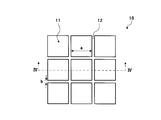
- FIG. 3 is a plan view showing the configuration of another cell culture container according to the present embodiment
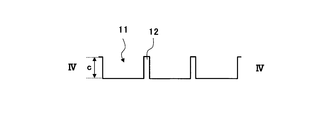
- FIG. 4 is a sectional view taken along the line IV-IV in FIG.
- the width a of the bottom surface of the micro container 11 and the width b and height c of the side wall 12 for partitioning the micro container 11 are shown.
- the cells cultured in the micro container 11 may ride on and move to the adjacent micro container 11.
- the height c of the side wall 12 is preferably 15 ⁇ m to 300 ⁇ m, and more preferably 50 ⁇ m to 150 ⁇ m.
- the side wall 12 may have a multi-stage shape.
- the cell culture section has compartmentalized spots composed of a plurality of microcontainers necessary for one screening, as shown in FIGS. 5 and 6, in order to minimize the number of necessary cells. May be. For example, if a micro container with a side of 200 ⁇ m with a high differentiation efficiency and a micro container with a height of 50 ⁇ m is used and the minimum number of cells required for screening is about 1000, nine micro containers are required.
- a spot for partitioning the micro-container and further providing a plurality of spots a high-throughput screening capable of testing a plurality of reagents and pharmaceuticals at the same time becomes possible.
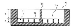
- FIG. 5 is a plan view showing the configuration of another cell culture unit according to the present embodiment
- FIG. 6 is a cross-sectional view taken along the line VI-VI in FIG.
- FIG. 5 shows a side wall 24 and a partitioned spot 23 for partitioning a plurality of micro containers.
- the height d of the side wall 24 only needs to be a volume that can hold a supernatant such as a culture solution or a reaction solution without drying, and may be set for convenience.
- the method for producing the concavo-convex pattern on the cell culture vessel is not particularly limited. For example, transfer molding using a mold, three-dimensional stereolithography, precision machine cutting, wet etching, dry etching, laser processing, electric discharge processing, etc. A method is mentioned. It is preferable to appropriately select these production methods in consideration of the use of the cell culture container, required processing accuracy, cost, and the like.
- the transfer molding method using a mold there is a method of forming a concavo-convex pattern by resin molding using a metal structure as a mold.
- This method is preferable because the shape of the metal structure can be reproduced in a concavo-convex pattern on the resin at a high transfer rate, and the cost of the material can be reduced by using a general-purpose resin material.
- the method using such a metal structure mold is excellent in that it is low in cost and can satisfy high dimensional accuracy.
- Examples of the method for manufacturing the metal structure include plating treatment on a resist pattern produced by photolithography and a resin pattern produced by three-dimensional stereolithography, precision mechanical cutting, wet etching, dry etching, laser processing, electric discharge. Processing etc. are mentioned. What is necessary is just to select suitably in consideration of a use, the required process precision, cost, etc.
- Examples of a method for forming a concavo-convex pattern on a resin using the metal structure obtained above as a mold include, for example, injection molding, press molding, monomer cast molding, solvent cast molding, hot emboss molding, roll transfer by extrusion molding, etc. Can be mentioned. It is preferable to employ injection molding from the viewpoint of productivity and mold transferability.
- the material constituting the cell culture vessel is not particularly limited as long as it has self-supporting properties, and examples thereof include synthetic resin, silicon, and glass. From the viewpoint of cost and cell visibility by microscopic observation, it is preferable to use a transparent synthetic resin as a material.
- the transparent synthetic resin include acrylic resins such as polymethyl methacrylate and methyl methacrylate-styrene copolymer, styrene resins such as polystyrene, olefin resins such as cycloolefin, polyethylene terephthalate, and polylactic acid.
- ester resins silicone resins such as polydimethylsiloxane, and polycarbonate resins.
- Such a resin may contain various additives such as a colorant, a diffusing agent, and a thickener as long as the transparency is not impaired.
- the cell culture container is subjected to surface treatment on the surface side of the concavo-convex pattern for the purpose of improving the hydrophilicity, biocompatibility, cell affinity, etc. of the container surface, and a modified layer and / or coating layer is disposed. Also good.
- the method for providing the modified layer is not particularly limited unless it is a method for losing self-supporting property or a method for causing extreme surface roughness of 100 ⁇ m or more.
- a graft polymer by chemical treatment, solvent treatment, or surface graft polymerization is used. And methods such as chemical treatment such as introduction of carbon, physical treatment such as corona discharge, ozone treatment, and plasma treatment.
- the method for providing the coating layer is not particularly limited, and examples thereof include dry coating such as sputtering and vapor deposition, wet coating such as inorganic material coating and polymer coating. It is desirable to impart hydrophilicity on the concavo-convex pattern in order to inject the culture solution without mixing bubbles, and inorganic vapor deposition is preferred as a method for forming a uniform hydrophilic film.
- a cell affinity protein such as collagen or fibronectin.
- a cell affinity protein such as collagen or fibronectin.
- the extracellular matrix suitable for the cultured cells is used. It is particularly preferable to arrange an organic film.
- the cells are placed only in the micro container for culturing the cells, and the appropriate number of cells is seeded in order to express similar forms and functions in the living body in the space.
- the cell seeding density of 1.0 ⁇ 10 2 to 1.0 ⁇ 10 6 cells / cm 2 is preferable, and the cell seeding density of 1.0 ⁇ 10 4 to 1.0 ⁇ 10 6 cells / cm 2 is more preferable. preferable.
- the micro container is square and one side is 200 ⁇ m, 5.0 ⁇ 10 4 to 5.0 ⁇ 10 5 cells / cm 2 is preferable. Under such conditions, a hepatocyte mass having a diameter of 30 to 200 ⁇ m can be obtained.
- FIG. 7 is a diagram showing a configuration example of a cell culture kit.
- the cell culture kit 30 has a flat cell culture plate 32, and the cell culture plate 32 includes a plurality of culture dishes 34.
- a cell culture vessel 31 is disposed in each culture dish 34.
- the number of culture dishes 34 set in one cell culture plate 32 is determined according to the screening method, the type of cells to be cultured and the number of cells used in the test.
- the cell culture plate 32 has at least one cell culture container 31.
- the cell culture container 31 may have any of the three types of configurations shown in FIGS.
- the bottom of the culture dish 34 has a flat plate shape, and the cell culture vessel 31 uses this bottom surface as a culture surface.
- FIG. 8 to FIG. 10 are examples of a state in which live cells are cultured in a plurality of microcontainers, and show differences in donors of cells to be seeded.
- each of the rectangles is a micro container 33
- FIGS. 8 and 9 illustrate a case where the cell culture container 31 has nine micro containers 33.
- FIG. 10 shows a case where the cell culture container 31 has 18 micro containers 33.
- D1 to D3 are cultured cells, and in the figure, the patterns are changed to show that D1, D2, and D3 are different donors.
- FIG. 8 shows a case where cells derived from one donor are adhered to each micro container 33, and living cells of different donors are adhered to adjacent micro containers 33.
- FIG. 9 shows a case where a part of the plurality of micro-containers 33 are adhered in a state where living cells derived from a plurality of donors are mixed, and the other is a case where living cells derived from one donor are adhered. .
- the case where the living cells of two donors are adhered to one micro container 33 is shown as an example.
- FIG. 10 shows an example in which a plurality of micro-containers 33 included in the cell culture container 31 are divided into two, one type of donor cell is adhered to one, and the other type of donor cell is adhered to the other. ing.
- the plurality of micro containers 33 may be divided into three or more sections. By attaching the cells of the desired donor to each section, the difference in test results between different donors can be easily confirmed.
- each micro container 33 may be a case where a plurality of living cells derived from different donors are adhered. There may be three or more types of living cells of different donors adhered to one micro container 33.
- the micro container 33 may be a case where the living cells derived from one donor are adhered, and the cell culture kit 30 as a whole (or as one cell culture container 31) has a plurality of donor living cells.
- the living cells derived from one donor may be adhered to each cell culture container 31, and the living cells derived from different donors may be adhered to the whole of the plurality of cell culture containers 31. That is, as the cell culture kit 30, it suffices if living cells derived from a plurality of different donors are adhered.
- the living cells derived from a plurality of donors are cultured on the surface of each micro container 33 of the cell culture kit 30 while being adhered.
- living cells accumulate and a cell mass is formed.
- the cell mass is cultured until a desired size is obtained. For example, the cell mass is cultured until the diameter of the cell aggregate becomes 30 to 200 ⁇ m. Further, the size of the micro container is also determined according to the size of the cell mass.
- parenchymal cells derived from different donors are used.
- parenchymal cells cells differentiated from tissue progenitor cells, tissue stem cells, ES cells, parenchymal cells differentiated from iPS cells (induced pluripotent stem cells), or parenchymal cells separated from a living body are used.
- the origin of the cells in this case may be, for example, a multi-donor parenchymal cell and other cell types derived from a single donor, or different from a single donor-derived parenchymal cell and a parenchymal cell donor.
- Other cell types derived from a donor may be used.
- one or more cell types selected from hepatic stellate cells, vascular endothelial cells, fibroblasts, and mesenchymal cells are used.
- Parenchymal cells hepatic stellate cells, vascular endothelial cells, fibroblasts, mesenchymal cells were differentiated from tissue precursor cells, tissue stem cells, cells differentiated from ES cells, or iPS cells (induced pluripotent stem cells) Cells or cells separated from the living body are used.
- a medium containing nutrients such as nutrient factors, serum, and secreted solution from cells is used.
- a method of placing a container in which cells are cultured on a membrane such as a cell culture insert may be used.
- a cell culture kit in which living cells of a plurality of donors are cultured in a state of being adhered in one container in a cell culture kit.
- the cell culture kit has a plurality of microcontainers.
- the plurality of micro containers have a structure capable of maintaining the in vivo function of living cells for a long period of time. Therefore, it is possible to provide living cells having cell functions similar to those in vivo.
- the test results of a plurality of donors can be obtained on one chip. This makes it possible to efficiently carry out tests using cells derived from a plurality of different donors over a long period of time.
- Hepatocyte culture (cell proliferation) Transformed cells (hereinafter referred to as “transformed hepatocytes”) into which human hepatic stem cells (consignment number FERM BP-11108, National Institute of Advanced Industrial Science and Technology (AIST)) were introduced with the BMI1 gene were treated with type IV collagen. The seed was sown on a coat dish (Becton Deckon) and cultured.
- Medium is 10% fetal bovine serum (FBS), human ⁇ -insulin (1.0 ⁇ g / ml), nicotinamide (10 mmol / L), dexamethasone (1 ⁇ 10 ⁇ 7 mol / L), L-glutamine (2 mmol / L) DMEM nutrient mixed F-12 Ham medium (DMEM / F12 1: 1 mixture) was used. The culture was performed at 37 ° C. in a 5% CO 2 incubator, and the medium was changed every 5 days.
- FBS fetal bovine serum
- human ⁇ -insulin 1.0 ⁇ g / ml
- nicotinamide 10 mmol / L
- dexamethasone (1 ⁇ 10 ⁇ 7 mol / L
- L-glutamine (2 mmol / L)
- DMEM nutrient mixed F-12 Ham medium DMEM nutrient mixed F-12 Ham medium
- vascular endothelial cell culture (cell proliferation) A human vascular endothelial cell line derived from a donor different from the transformed hepatocytes was seeded and cultured in an uncoated cell culture dish (Becton Dickinson). Medium is 10% fetal bovine serum (FBS), human ⁇ -insulin (1.0 ⁇ g / ml), nicotinamide (10 mmol / L), dexamethasone (1 ⁇ 10 ⁇ 7 mol / L), L-glutamine (2 mmol / L) DMEM nutrient mixed F-12 Ham medium (DMEM / F12 1: 1 mixture) was used. The culture was performed at 37 ° C. in a 5% CO 2 incubator, and the medium was changed every 5 days.
- FBS fetal bovine serum
- human ⁇ -insulin 1.0 ⁇ g / ml
- nicotinamide (10 mmol / L)
- dexamethasone (1 ⁇ 10 ⁇ 7 mol
- 1-3 Preparation of Cell Suspension
- a medium is 10% fetal bovine serum (FBS), human ⁇ -insulin (1.0 ⁇ g / ml), nicotinamide (10 mmol / L), dexamethasone (1 ⁇ 10 ⁇ 7 mol / L), L-glutamine (2 mmol / L)
- FBS fetal bovine serum
- human ⁇ -insulin 1.0 ⁇ g / ml
- nicotinamide 10 mmol / L
- dexamethasone (1 ⁇ 10 ⁇ 7 mol / L
- L-glutamine (2 mmol / L)
- DMEM nutrient mixed F-12 Ham medium DMEM nutrient mixed F-12 Ham medium
- Example 01> Mixing so that the mixing ratio of the transformed hepatocytes obtained in 1-3 and vascular endothelial cells is 1: 3, and seeding in a culture vessel so that the cell density is 3.75 ⁇ 10 4 cells / cm 2 did.
- cytochrome P450 cytochrome P450
- albumin cytochrome P450 and albumin, which are typical drug metabolizing enzymes in the liver, was evaluated by collecting RNA from cells cultured for a predetermined number of days, synthesizing cDNA, and performing real-time PCR.
- Table 1 shows the gene expression levels of albumin, CYP3A4, and CYP2C9 after 21 days of culture in Example 01 and Comparative Examples 01 and 02.
- the gene expression level represents a relative value when the value of Comparative Example 02 is 1.
- CYP3A4 and CYP2C9 are one of metabolic enzymes existing in the liver and are names of molecular species of cytochrome P450 enzyme. CYP plays an important role in protecting the living body from foreign substances such as various chemical substances (including pharmaceuticals), environmental pollutants, and organic solvents.
- all of albumin, CYP3A4, and CYP2C9 showed significantly higher expression levels than Comparative Examples 01 and 02.
- the conditions of the experimental method are not limited to those described above, except for mixing and culturing two different types of cells.
- the surface coat cells only have to be adhered, and the surface coat is not limited to those described above.
- Cell seeding human fetal hepatocytes collected from 6 donor livers, that is, cells derived from 6 donors including three types of cells, hepatic stem cells, hepatic progenitor cells, and mature hepatocytes, were used.
- FIG. 13 is a photograph showing the measurement results. In FIG. 13, the results on the 7th day of culture on the left side, the 21st day of culture on the center, and the 35th day on the right side are shown.
Landscapes
- Health & Medical Sciences (AREA)
- Life Sciences & Earth Sciences (AREA)
- Engineering & Computer Science (AREA)
- Chemical & Material Sciences (AREA)
- Organic Chemistry (AREA)
- Zoology (AREA)
- Wood Science & Technology (AREA)
- Bioinformatics & Cheminformatics (AREA)
- Biomedical Technology (AREA)
- Genetics & Genomics (AREA)
- Biotechnology (AREA)
- General Engineering & Computer Science (AREA)
- Biochemistry (AREA)
- Microbiology (AREA)
- General Health & Medical Sciences (AREA)
- Cell Biology (AREA)
- Sustainable Development (AREA)
- Clinical Laboratory Science (AREA)
- Proteomics, Peptides & Aminoacids (AREA)
- Analytical Chemistry (AREA)
- Physics & Mathematics (AREA)
- Biophysics (AREA)
- Virology (AREA)
- Immunology (AREA)
- Molecular Biology (AREA)
- Apparatus Associated With Microorganisms And Enzymes (AREA)
- Micro-Organisms Or Cultivation Processes Thereof (AREA)
- Measuring Or Testing Involving Enzymes Or Micro-Organisms (AREA)
Abstract
Description
また、前記生細胞は肝細胞を含む生細胞であることが好ましく、前記肝細胞が、組織前駆細胞、組織幹細胞、ES細胞から分化させた細胞またはiPS細胞から分化させた細胞であること、あるいは、前記肝細胞を含む生細胞が、複数のドナーの肝組織内から分離されたものであることがより好ましい。
以下に、本発明の実施の形態について説明する。ただし、本発明が以下の実施の形態に限定される訳ではない。また、説明を明確にするため、以下の記載及び図面は、適宜、簡略化されている。
1.細胞の準備
1-1.肝細胞の培養(細胞増殖)
ヒト肝幹細胞(委託番号FERM BP-11108、独立行政法人産業技術総合研究所特許生物寄託センター)にBMI1遺伝子を導入した形質転換細胞(以下、「形質転換肝細胞」と表記)を、IV型コラーゲンコートディッシュ(ベクトンデッキン社製)に播種し培養した。
培地は、10%ウシ胎仔血清(FBS)、ヒトγ-インスリン(1.0μg/ml)、ニコチンアミド(10mmol/L)、デキサメタゾン(1x10-7mol/L)、L-グルタミン(2mmol/L)を加えたDMEM栄養混合F-12Ham培地(DMEM/F12 1:1 mixture)を用いた。培養は、37℃、5%CO2インキュベータで行い、培地交換を5日毎に行った。
前記形質転換肝細胞とは異なるドナーに由来するヒト血管内皮細胞株を、何もコートされていない細胞培養ディッシュ(ベクトンデッキンソン社製)に播種し培養した。
培地は、10%ウシ胎仔血清(FBS)、ヒトγ-インスリン(1.0μg/ml)、ニコチンアミド(10mmol/L)、デキサメタゾン(1x10-7mol/L)、L-グルタミン(2mmol/L)を加えたDMEM栄養混合F-12Ham培地(DMEM/F12 1:1 mixture)を用いた。培養は、37℃、5%CO2インキュベータで行い、培地交換を5日毎に行った。
上記1-1、1-2で培養した細胞それぞれについて、0.25%トリプシン溶液を用いて剥離し細胞を回収した後、培地に分散させた。培地は、10%ウシ胎仔血清(FBS)、ヒトγ-インスリン(1.0μg/ml)、ニコチンアミド(10mmol/L)、デキサメタゾン(1x10-7mol/L)、L-グルタミン(2mmol/L)を加えたDMEM栄養混合F-12Ham培地(DMEM/F12 1:1 mixture)を用いた。それぞれ、トリパンブルーで細胞を染色し生細胞数をカウントした。
2-1 <実施例01>
1-3で得た形質転換肝細胞と血管内皮細胞の混合比が1:3になるように混合し、細胞密度が3.75×10⁴細胞/cm2になるように、培養容器に播種した。培養容器は、図3および図4に示す凹凸パターンであって、a=100μm、c=50μmのマイクロ空間を有する24ウェルタイプの培養容器を用いた。
1-3で得た形質転換肝細胞を、細胞密度が3.75×10⁴細胞/cm2になるように、培養容器に播種した。培養容器は、図3および図4に示す凹凸パターンであって、a=100μm、c=50μmのマイクロ空間を有する24ウェルタイプの培養容器を用いた。
2-3 <比較例02>
1-3で得た形質転換肝細胞を、細胞密度が3.75×10⁴細胞/cm2になるように、24ウェル細胞培養プレート(ベクトンデッキン社製)に播種した。
上記2-1から2-3に記載したように細胞を播種した後、37℃の5%CO2インキュベータで培養した。培養24時間後、1回/1~2日の頻度で培地交換を行った。培地は、10%ウシ胎仔血清(FBS)、ヒトγ-インスリン(1.0μg/ml)、ニコチンアミド(10mmol/L)、デキサメタゾン(1x10-7mol/L)、L-グルタミン(2mmol/L)を加えたDMEM栄養混合F-12Ham培地(DMEM/F12 1:1 mixture)にヒト組換えHGF(50ng/mL)及び、上皮増殖因子(EGF)(10ng/mL)を添加した培地を用いた。
肝臓の代表的な薬物代謝酵素であるチトクロームP450(CYP)及びアルブミンの遺伝子発現は、所定日数培養した細胞からRNAを回収し、cDNA合成後、リアルタイムPCRを行うことで評価した。
表1に、実施例01及び比較例01、02における培養期間21日後のアルブミン、CYP3A4、及びCYP2C9の遺伝子発現量を示す。表中、遺伝子発現量は、比較例02の値を1とした場合の相対値を示している。また、CYP3A4、CYP2C9は、肝臓に存在する代謝酵素の一つであり、チトクロームP450酵素の分子種の名前である。CYPは種々の化学物質(医薬品を含む)、環境汚染物質、有機溶媒などの異質・異物から生体を守る重要な役目を担っている。
実施例01では、アルブミン、CYP3A4、及びCYP2C9のいずれにおいても比較例01、02よりも有意に高い発現量を示した。
なお、実験方法の条件は、異なる2種の細胞を混合して培養する以外の、細胞数、細胞の混合比は上述したものに限定されない。表面コートについても細胞が接着すればよく、上述したものに限定されるものではない。

1.細胞播種
実施例では、6ドナー肝から採取したヒト胎児肝細胞、すなわち、肝幹細胞と肝前駆細胞と成熟肝細胞の3種類の細胞を含んだ、6ドナー由来の細胞を用いた。比較例では単一ドナーから採取したヒト胎児肝細胞を用いた。いずれも、タイプIVコラーゲンをコートした、図3および図4に示すa=100μm、c=50μmのマイクロ空間を有する24ウェルタイプの培養容器に、細胞密度が3.75×10⁴細胞/cm2になるように播種した。
培養は、37℃の5%CO2インキュベータ内で行った。培養24時間後、1回/1~2日の頻度で培地交換を行った。培地は、10%ウシ胎仔血清(FBS)、ヒトγ-インスリン(1.0μg/ml)、ニコチンアミド(10mmol/L)、デキサメタゾン(1x10-7mol/L)、L-グルタミン(2mmol/L)を加えたDMEM栄養混合F-12Ham培地(DMEM/F12 1:1 mixture)にヒト組換えHGF(50ng/mL)及び、上皮増殖因子(EGF)(10ng/mL)を添加した培地を用いた。
3-1.形態観察
倒立顕微鏡を用いて、培養1日、4日、7日、14日、21日、35日に観察した。
3-2.チトクロームP450(CYP)及びアルブミンの遺伝子発現と、CYP3A4のタンパク発現
肝臓の代表的表的な薬物代謝酵素であるチトクロームP450(CYP)及びアルブミンの遺伝子発現は、所定日数培養した細胞からRNAを回収し、cDNA合成後、リアルタイムPCRを行うことで評価した。タンパク発現は、免疫染色法を用いて分析した。
3-3.グリコーゲン貯蔵能
PAS染色法より、ヒト胎児肝臓細胞における分化能(グリコーゲン貯蔵能)を評価した。
4-1.形態観察結果
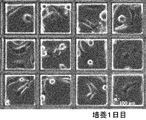
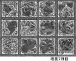
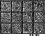
細胞は最初にフィルム底面に接着し、培養日数を追うごとに、徐々に他のマイクロ空間内(micro cavity)へと伸展してゆき、マイクロ空間内に細胞集合体を形成した。これは、以下に示した<比較例>と同様の形態であったことから、複数ドナーの細胞も単一ドナーと同じように凝集体形成が可能であることが示された。図11A~11Fは、実施例の形態観察結果を示す写真である。また、図12は、比較例の培養14日目の形態観察結果を示す写真である。
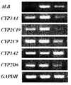
主要な薬物代謝酵素CYP3A4、2C19、2C9、1A2、2D6とアルブミン分泌能を測定した。その結果、培養7日目でこれらCYP遺伝子が発現し、培養21日目で、アルブミンとこれらCYP遺伝子が発現した。日数が経過した場合も、この機能は維持されていた。図13は測定結果を示す写真である。図13において、左側に培養7日目、中央に培養21日目、右側に培養35日目の結果を示している。
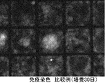
免疫染色で、ほぼすべてのマイクロ空間内で、CYP3A4(赤)の発現が確認され(図14)、これは、以下に示した<比較例>に示したCYP3A4の染色像(図15)と類似していたことから、複数ドナーの細胞も単一ドナーと同じように、肝機能を保持した状態で培養できることが示された。
ヒト胎児肝臓細胞における分化能(グリコーゲン貯蔵能)を検討した。その結果、ヒト胎児肝臓細胞においてグリコーゲン貯蔵能が認められた。さらに、この細胞においては培養21日目で半数以上の細胞が強いPAS陽性を呈した。
11 マイクロ容器
12 側壁
13 開口部
23 スポット
24 スポットの側壁
30 細胞培養キット
31 細胞培養容器
32 細胞培養プレート
33 マイクロ容器
34 培養皿
D1、D2、D3 細胞
Claims (16)
- 細胞培養プレートとその上で培養された生細胞とを有する細胞培養キットであって、
前記細胞培養プレートは、複数のマイクロ空間を有し、前記複数のマイクロ空間に複数の異なるドナーに由来する生細胞が表面に接着された細胞培養キット。 - 前記複数の異なるドナーに由来する生細胞が、2種類以上の細胞であることを特徴とする請求項1記載の細胞培養キット。
- 各マイクロ空間は、複数の異なるドナーに由来する生細胞と、一つのドナーに由来する生細胞とのいずれかが接着されていることを特徴とする請求項1または2記載の細胞培養キット。
- 少なくとも二つの隣接するマイクロ空間は、異なるドナーに由来する生細胞が接着されていることを特徴とする請求項1または2記載の細胞培養キット。
- 少なくとも二つの隣接するマイクロ空間は、同じドナーに由来する生細胞が接着されていることを特徴とする請求項1または2記載の細胞培養キット。
- 前記複数のマイクロ空間は、所望の細胞数に培養された三次元構造体の細胞集団が隔離される大きさを有すること特徴とする請求項1乃至5のいずれか一項に記載の細胞培養キット。
- 前記複数のマイクロ空間は、底部面積が0.01~0.1mm2であり、深さが25~150μmであることを特徴とする請求項1乃至6のいずれか一項に記載の細胞培養キット。
- 前記複数のマイクロ空間は、細胞播種密度が1×10²~1×10⁶細胞/cm2で生細胞が播種されたことを特徴とする請求項1乃至7のいずれか一項に記載の細胞培養キット。
- 前記複数のマイクロ空間は、生細胞が集積し、細胞塊が形成されていることを特徴とする請求項1乃至8のいずれか一項に記載の細胞培養キット。
- 前記細胞塊の直径は、30~200μmであることを特徴とする請求項1乃至9のいずれか一項に記載の細胞培養キット。
- 前記生細胞は、組織前駆細胞、組織幹細胞、ES細胞から分化させた細胞またはiPS細胞から分化させた細胞であることを特徴とする請求項1乃至10のいずれか一項に記載の細胞培養キット。
- 前記生細胞が、肝細胞を含む生細胞であることを特徴とする請求項1乃至10のいずれか一項に記載の細胞培養キット。
- 前記肝細胞が、組織前駆細胞、組織幹細胞、ES細胞から分化させた細胞、またはiPS細胞から分化させた細胞であることを特徴とする請求項12記載の細胞培養キット。
- 前記肝細胞を含む生細胞が、複数のドナーの肝組織内から分離されたものであることを特徴とする請求項12記載の細胞培養キット。
- 請求項1乃至14のいずれか一項に記載の細胞培養キットを用いて薬剤の評価を行うスクリーニング方法。
- 複数のマイクロ空間を有する細胞培養プレートとその上で培養された生細胞とを有する細胞培養キットの製造方法であって、
前記複数のマイクロ空間へ、複数の異なるドナーに由来する生細胞を播種し、
播種した生細胞を培養する細胞培養キットの製造方法。
Priority Applications (6)
| Application Number | Priority Date | Filing Date | Title |
|---|---|---|---|
| EP09821832.4A EP2343361A4 (en) | 2008-10-24 | 2009-10-23 | CELL CULTURE KIT, TEST PROCEDURE AND MANUFACTURING PROCESS FOR THE CELL CULTURE KIT |
| CA2741493A CA2741493A1 (en) | 2008-10-24 | 2009-10-23 | Cell culture kit, screening method, and method of manufacturing cell culture kit |
| US13/125,896 US20110217725A1 (en) | 2008-10-24 | 2009-10-23 | Cell culture kit, screening method, and method of manufacturing cell culture kit |
| JP2010534724A JP5607535B2 (ja) | 2008-10-24 | 2009-10-23 | 細胞培養キット、スクリーニング方法、及び細胞培養キットの製造方法 |
| CN2009801421490A CN102197129A (zh) | 2008-10-24 | 2009-10-23 | 细胞培养试剂盒、筛选方法和细胞培养试剂盒的制造方法 |
| US15/010,468 US10836996B2 (en) | 2008-10-24 | 2016-01-29 | Cell culture kit, screening method, and method of manufacturing cell culture kit |
Applications Claiming Priority (2)
| Application Number | Priority Date | Filing Date | Title |
|---|---|---|---|
| JP2008273845 | 2008-10-24 | ||
| JP2008-273845 | 2008-10-24 |
Related Child Applications (2)
| Application Number | Title | Priority Date | Filing Date |
|---|---|---|---|
| US13/125,896 A-371-Of-International US20110217725A1 (en) | 2008-10-24 | 2009-10-23 | Cell culture kit, screening method, and method of manufacturing cell culture kit |
| US15/010,468 Division US10836996B2 (en) | 2008-10-24 | 2016-01-29 | Cell culture kit, screening method, and method of manufacturing cell culture kit |
Publications (1)
| Publication Number | Publication Date |
|---|---|
| WO2010047132A1 true WO2010047132A1 (ja) | 2010-04-29 |
Family
ID=42119183
Family Applications (1)
| Application Number | Title | Priority Date | Filing Date |
|---|---|---|---|
| PCT/JP2009/005617 Ceased WO2010047132A1 (ja) | 2008-10-24 | 2009-10-23 | 細胞培養キット、スクリーニング方法、及び細胞培養キットの製造方法 |
Country Status (7)
| Country | Link |
|---|---|
| US (2) | US20110217725A1 (ja) |
| EP (1) | EP2343361A4 (ja) |
| JP (1) | JP5607535B2 (ja) |
| KR (1) | KR20110074773A (ja) |
| CN (1) | CN102197129A (ja) |
| CA (1) | CA2741493A1 (ja) |
| WO (1) | WO2010047132A1 (ja) |
Cited By (14)
| Publication number | Priority date | Publication date | Assignee | Title |
|---|---|---|---|---|
| WO2011118211A1 (ja) * | 2010-03-23 | 2011-09-29 | 株式会社クラレ | 多能性哺乳細胞を分化させる培養方法 |
| WO2014061244A1 (ja) * | 2012-10-18 | 2014-04-24 | 株式会社クラレ | 毒性スクリーニング方法 |
| JP2014079223A (ja) * | 2012-10-18 | 2014-05-08 | Kuraray Co Ltd | エストロゲン活性を評価する方法 |
| JP2014079227A (ja) * | 2012-10-18 | 2014-05-08 | Kuraray Co Ltd | 化合物のスクリーニング方法 |
| US20140227784A1 (en) * | 2011-09-20 | 2014-08-14 | Kuraray Co., Ltd. | Adherent cell culture method |
| JP2016523550A (ja) * | 2013-07-03 | 2016-08-12 | コイン アイピー ホールディングス、 エルエルシー | 化学的または生物学的物質に対する応答の予測方法 |
| JP2018064491A (ja) * | 2016-10-18 | 2018-04-26 | ヤマハ発動機株式会社 | 細胞移動装置 |
| WO2018182044A1 (ja) * | 2017-03-31 | 2018-10-04 | 国立大学法人北海道大学 | 細胞培養基材、がん細胞集合体及び該基材を用いたその製造方法、並びに該がん細胞集合体を用いた薬剤のスクリーニング方法 |
| CN109517737A (zh) * | 2018-11-06 | 2019-03-26 | 中国科学院过程工程研究所 | 一种微流控芯片和基于该芯片的肿瘤转移模型及模型构建方法和应用 |
| USD907238S1 (en) | 2018-10-31 | 2021-01-05 | Sony Corporation | Embryo culture dish |
| JP2021508442A (ja) * | 2017-12-22 | 2021-03-11 | ティスーゼ ゲゼルシャフト ミット ベシュレンクテル ハフツングTissUse GmbH | iPSC由来細胞の臓器相当物への分化を確立する新規多臓器チップ |
| USD940745S1 (en) | 2018-10-31 | 2022-01-11 | Sony Corporation | Display panel or screen or a portion thereof with an animated graphical user interface |
| US11377679B2 (en) | 2016-04-21 | 2022-07-05 | Quantamatrix Inc. | Multi-well-based cell culture test device for rapid antibiotic susceptibility testing |
| US11685902B2 (en) | 2016-10-11 | 2023-06-27 | Tokushima University | Production method for kidney-like tissue |
Families Citing this family (9)
| Publication number | Priority date | Publication date | Assignee | Title |
|---|---|---|---|---|
| CN102533537B (zh) * | 2011-12-29 | 2013-10-23 | 云南师范大学 | 一种高通量筛选细胞在三维基质中生长迁移行为的装置及方法 |
| WO2018009870A1 (en) | 2016-07-07 | 2018-01-11 | David Beebe | Microtiter plate and uses thereof |
| EP4478049A3 (en) * | 2016-08-03 | 2024-12-25 | Wake Forest University Health Sciences | Cancer modeling platforms and methods of using the same |
| KR20190003413A (ko) * | 2017-06-30 | 2019-01-09 | 코오롱생명과학 주식회사 | 골관절염 예방 또는 치료용 약학적 조성물 |
| CN108029678A (zh) * | 2017-12-28 | 2018-05-15 | 重庆斯德姆生物技术有限公司 | 细胞保存方法和细胞运输方法 |
| US20210277345A1 (en) * | 2020-03-06 | 2021-09-09 | Tomoyuki Aratani | Cell-containing container, method for evaluating test substance, and method for manufacturing cell-containing container |
| US12270048B2 (en) | 2020-03-23 | 2025-04-08 | Ricoh Company, Ltd. | Manufacturing method for substrate on which nerve cells are arranged |
| US12069391B2 (en) * | 2021-07-29 | 2024-08-20 | Omnivision Technologies, Inc. | Readout architectures for dark current reduction in indirect time-of-flight sensors |
| CN119120360B (zh) * | 2024-09-06 | 2025-11-07 | 南方科技大学 | 一种诱导间充质干细胞成软骨分化的方法和促进关节软骨再生的组合物及应用 |
Citations (10)
| Publication number | Priority date | Publication date | Assignee | Title |
|---|---|---|---|---|
| JPH08308562A (ja) | 1995-05-16 | 1996-11-26 | Sumitomo Bakelite Co Ltd | 動物細胞培養器及びそれを用いる薬物代謝活性の測定方法 |
| JPH08319317A (ja) | 1995-03-17 | 1996-12-03 | Kanagawa Kagaku Gijutsu Akad | 糖鎖高分子及びそれを用いた細胞処理剤並びに処理方法 |
| WO2004113515A2 (en) * | 2003-06-20 | 2004-12-29 | Axiogenesis Ag | Tissue modeling in embryonic stem (es) cell system |
| JP2005080607A (ja) * | 2003-09-10 | 2005-03-31 | National Food Research Institute | 細胞培養プレートおよびその製造方法 |
| WO2006136953A2 (en) * | 2005-06-15 | 2006-12-28 | Capsant Neurotechnologies Ltd | Method of producing organotypic cell cultures |
| JP2007228818A (ja) * | 2006-02-28 | 2007-09-13 | Kuraray Co Ltd | 細胞培養容器、その製造方法および細胞培養方法 |
| WO2007105418A1 (ja) * | 2006-02-24 | 2007-09-20 | Kuraray Co., Ltd. | 細胞培養容器およびその製造方法 |
| WO2007126127A1 (ja) * | 2006-04-28 | 2007-11-08 | Kuraray Co., Ltd. | 細胞培養容器およびその製造方法 |
| WO2008002662A2 (en) * | 2006-06-28 | 2008-01-03 | The Univeristy Of Kansas | Differentiation of stem cells from umbilical cord matrix into hepatocyte lineage cells |
| JP2008148702A (ja) * | 2001-12-21 | 2008-07-03 | Immunex Corp | 内皮幹細胞、該細胞集団、該細胞の単離法および使用法 |
Family Cites Families (16)
| Publication number | Priority date | Publication date | Assignee | Title |
|---|---|---|---|---|
| EP0400047B1 (en) * | 1988-02-05 | 1997-04-23 | Whitehead Institute For Biomedical Research | Modified hepatocytes and uses therefor |
| CN2185755Y (zh) | 1994-03-05 | 1994-12-21 | 青岛胶南利达机械制造公司 | 绒去粗刀装置 |
| CN2186755Y (zh) * | 1994-03-25 | 1995-01-04 | 中国科学院成都生物研究所 | 多用途培养板 |
| EP1064353B1 (en) * | 1998-03-18 | 2002-11-06 | Massachusetts Institute Of Technology | Vascularized perfused microtissue/micro-organ arrays |
| US6458589B1 (en) * | 2000-04-27 | 2002-10-01 | Geron Corporation | Hepatocyte lineage cells derived from pluripotent stem cells |
| GB0027516D0 (en) * | 2000-11-10 | 2000-12-27 | Amersham Pharm Biotech Uk Ltd | Support and method for cell based assays |
| US20060110826A1 (en) * | 2002-07-30 | 2006-05-25 | Massachusetts Institute Of Technology | Methods for perfusion and plating of primary hepatocytes and a medium therefore |
| AU2003272719A1 (en) * | 2002-09-26 | 2004-04-19 | Carbomer, Inc. | Inhibitors of the nitrix oxide synthase iii (nos iii) as neuroprotective agents |
| WO2004044158A2 (en) * | 2002-11-08 | 2004-05-27 | The Johns Hopkins University | Human embryonic stem cell cultures, and compositions and methods for growing same |
| JP2005027598A (ja) * | 2003-07-09 | 2005-02-03 | Kitakyushu Foundation For The Advancement Of Industry Science & Technology | 細胞培養チップ及び培養器、それらを用いた細胞培養方法、球状細胞組織体を担持させた細胞担持モジュール、球状細胞組織体 |
| US7186548B2 (en) * | 2003-11-10 | 2007-03-06 | Advanced Pharmaceutical Sciences, Inc. | Cell culture tool and method |
| JPWO2006123570A1 (ja) * | 2005-05-17 | 2008-12-25 | 株式会社クラレ | 細胞培養容器 |
| JP5161581B2 (ja) * | 2005-10-28 | 2013-03-13 | 株式会社クラレ | 細胞培養容器及び細胞培養方法 |
| WO2007055171A1 (ja) * | 2005-11-08 | 2007-05-18 | Kuraray Co., Ltd. | 細胞培養容器、細胞培養方法及び培養細胞のシグナル伝達の制御方法 |
| US20080220516A1 (en) * | 2006-10-12 | 2008-09-11 | Eddington David T | Multi-well micropatterning by ablation |
| US9121003B2 (en) * | 2007-09-12 | 2015-09-01 | Kitakyushu Foundation For The Advancement Of Industry, Science And Technology | Cell culture instrument and cell culture method using the same |
-
2009
- 2009-10-23 US US13/125,896 patent/US20110217725A1/en not_active Abandoned
- 2009-10-23 KR KR1020117011300A patent/KR20110074773A/ko not_active Ceased
- 2009-10-23 EP EP09821832.4A patent/EP2343361A4/en not_active Withdrawn
- 2009-10-23 CA CA2741493A patent/CA2741493A1/en not_active Abandoned
- 2009-10-23 JP JP2010534724A patent/JP5607535B2/ja not_active Expired - Fee Related
- 2009-10-23 CN CN2009801421490A patent/CN102197129A/zh active Pending
- 2009-10-23 WO PCT/JP2009/005617 patent/WO2010047132A1/ja not_active Ceased
-
2016
- 2016-01-29 US US15/010,468 patent/US10836996B2/en not_active Expired - Fee Related
Patent Citations (10)
| Publication number | Priority date | Publication date | Assignee | Title |
|---|---|---|---|---|
| JPH08319317A (ja) | 1995-03-17 | 1996-12-03 | Kanagawa Kagaku Gijutsu Akad | 糖鎖高分子及びそれを用いた細胞処理剤並びに処理方法 |
| JPH08308562A (ja) | 1995-05-16 | 1996-11-26 | Sumitomo Bakelite Co Ltd | 動物細胞培養器及びそれを用いる薬物代謝活性の測定方法 |
| JP2008148702A (ja) * | 2001-12-21 | 2008-07-03 | Immunex Corp | 内皮幹細胞、該細胞集団、該細胞の単離法および使用法 |
| WO2004113515A2 (en) * | 2003-06-20 | 2004-12-29 | Axiogenesis Ag | Tissue modeling in embryonic stem (es) cell system |
| JP2005080607A (ja) * | 2003-09-10 | 2005-03-31 | National Food Research Institute | 細胞培養プレートおよびその製造方法 |
| WO2006136953A2 (en) * | 2005-06-15 | 2006-12-28 | Capsant Neurotechnologies Ltd | Method of producing organotypic cell cultures |
| WO2007105418A1 (ja) * | 2006-02-24 | 2007-09-20 | Kuraray Co., Ltd. | 細胞培養容器およびその製造方法 |
| JP2007228818A (ja) * | 2006-02-28 | 2007-09-13 | Kuraray Co Ltd | 細胞培養容器、その製造方法および細胞培養方法 |
| WO2007126127A1 (ja) * | 2006-04-28 | 2007-11-08 | Kuraray Co., Ltd. | 細胞培養容器およびその製造方法 |
| WO2008002662A2 (en) * | 2006-06-28 | 2008-01-03 | The Univeristy Of Kansas | Differentiation of stem cells from umbilical cord matrix into hepatocyte lineage cells |
Non-Patent Citations (2)
| Title |
|---|
| MOTOHIRO FUKUDA ET AL.: "Nanotechnology-MEMS de Shinka suru Saibo Baiyo-Manipulation Micro Kukan Saibo Baiyo Dish", BIO INDUSTRY, vol. 23, no. 2, 12 February 2006 (2006-02-12), pages 9 - 13, XP003019140 * |
| See also references of EP2343361A4 |
Cited By (24)
| Publication number | Priority date | Publication date | Assignee | Title |
|---|---|---|---|---|
| US10513685B2 (en) | 2010-03-23 | 2019-12-24 | Corning Incorporated | Method for differentiating pluripotent mammalian stem cells into a population of hepatic cells in a microchamber |
| WO2011118211A1 (ja) * | 2010-03-23 | 2011-09-29 | 株式会社クラレ | 多能性哺乳細胞を分化させる培養方法 |
| US20140227784A1 (en) * | 2011-09-20 | 2014-08-14 | Kuraray Co., Ltd. | Adherent cell culture method |
| JPWO2014061244A1 (ja) * | 2012-10-18 | 2016-09-05 | 株式会社クラレ | 毒性スクリーニング方法 |
| JP2014079223A (ja) * | 2012-10-18 | 2014-05-08 | Kuraray Co Ltd | エストロゲン活性を評価する方法 |
| JP2014079227A (ja) * | 2012-10-18 | 2014-05-08 | Kuraray Co Ltd | 化合物のスクリーニング方法 |
| WO2014061244A1 (ja) * | 2012-10-18 | 2014-04-24 | 株式会社クラレ | 毒性スクリーニング方法 |
| JP2016523550A (ja) * | 2013-07-03 | 2016-08-12 | コイン アイピー ホールディングス、 エルエルシー | 化学的または生物学的物質に対する応答の予測方法 |
| US11377679B2 (en) | 2016-04-21 | 2022-07-05 | Quantamatrix Inc. | Multi-well-based cell culture test device for rapid antibiotic susceptibility testing |
| US11685902B2 (en) | 2016-10-11 | 2023-06-27 | Tokushima University | Production method for kidney-like tissue |
| JP2018064491A (ja) * | 2016-10-18 | 2018-04-26 | ヤマハ発動機株式会社 | 細胞移動装置 |
| WO2018074086A1 (ja) * | 2016-10-18 | 2018-04-26 | ヤマハ発動機株式会社 | 細胞移動装置 |
| EP3524667A4 (en) * | 2016-10-18 | 2019-08-14 | Yamaha Hatsudoki Kabushiki Kaisha | CELL TRANSFER DEVICE |
| JP2022130707A (ja) * | 2017-03-31 | 2022-09-06 | 国立大学法人北海道大学 | 細胞培養基材、がん細胞集合体及び該基材を用いたその製造方法、並びに該がん細胞集合体を用いた薬剤のスクリーニング方法 |
| JPWO2018182044A1 (ja) * | 2017-03-31 | 2020-04-16 | 国立大学法人北海道大学 | 細胞培養基材、がん細胞集合体及び該基材を用いたその製造方法、並びに該がん細胞集合体を用いた薬剤のスクリーニング方法 |
| JP7106141B2 (ja) | 2017-03-31 | 2022-07-26 | 国立大学法人北海道大学 | 細胞培養基材、がん細胞集合体及び該基材を用いたその製造方法、並びに該がん細胞集合体を用いた薬剤のスクリーニング方法 |
| US11603515B2 (en) | 2017-03-31 | 2023-03-14 | National University Corporation Hokkaido University | Cell culture substrate, cancer cell aggregate and method for manufacturing same using said substrate, and drug screening method using said cancer cell aggregate |
| WO2018182044A1 (ja) * | 2017-03-31 | 2018-10-04 | 国立大学法人北海道大学 | 細胞培養基材、がん細胞集合体及び該基材を用いたその製造方法、並びに該がん細胞集合体を用いた薬剤のスクリーニング方法 |
| JP7390751B2 (ja) | 2017-03-31 | 2023-12-04 | 国立大学法人北海道大学 | 細胞培養基材、がん細胞集合体及び該基材を用いたその製造方法、並びに該がん細胞集合体を用いた薬剤のスクリーニング方法 |
| JP2021508442A (ja) * | 2017-12-22 | 2021-03-11 | ティスーゼ ゲゼルシャフト ミット ベシュレンクテル ハフツングTissUse GmbH | iPSC由来細胞の臓器相当物への分化を確立する新規多臓器チップ |
| JP7256553B2 (ja) | 2017-12-22 | 2023-04-12 | ティスーゼ ゲゼルシャフト ミット ベシュレンクテル ハフツング | iPSC由来細胞の臓器相当物への分化を確立する新規多臓器チップ |
| USD907238S1 (en) | 2018-10-31 | 2021-01-05 | Sony Corporation | Embryo culture dish |
| USD940745S1 (en) | 2018-10-31 | 2022-01-11 | Sony Corporation | Display panel or screen or a portion thereof with an animated graphical user interface |
| CN109517737A (zh) * | 2018-11-06 | 2019-03-26 | 中国科学院过程工程研究所 | 一种微流控芯片和基于该芯片的肿瘤转移模型及模型构建方法和应用 |
Also Published As
| Publication number | Publication date |
|---|---|
| EP2343361A1 (en) | 2011-07-13 |
| JPWO2010047132A1 (ja) | 2012-03-22 |
| US20110217725A1 (en) | 2011-09-08 |
| US20160160175A1 (en) | 2016-06-09 |
| CN102197129A (zh) | 2011-09-21 |
| CA2741493A1 (en) | 2010-04-29 |
| KR20110074773A (ko) | 2011-07-01 |
| EP2343361A4 (en) | 2013-10-09 |
| US10836996B2 (en) | 2020-11-17 |
| JP5607535B2 (ja) | 2014-10-15 |
Similar Documents
| Publication | Publication Date | Title |
|---|---|---|
| JP5607535B2 (ja) | 細胞培養キット、スクリーニング方法、及び細胞培養キットの製造方法 | |
| JP5932206B2 (ja) | 細胞培養方法及びスクリーニング方法 | |
| EP2551341B1 (en) | Culture method for causing differentiation of pluripotent mammalian cells | |
| US8415157B2 (en) | Cell culture container and cell culture method | |
| JP5676265B2 (ja) | 細胞保存方法、及び細胞輸送方法 | |
| JP6021802B2 (ja) | 培養方法及び薬物スクリーニング方法 | |
| JP5940758B2 (ja) | 細胞培養方法 | |
| JPWO2008130025A1 (ja) | 肝細胞培養容器及び肝細胞培養方法 |
Legal Events
| Date | Code | Title | Description |
|---|---|---|---|
| WWE | Wipo information: entry into national phase |
Ref document number: 200980142149.0 Country of ref document: CN |
|
| 121 | Ep: the epo has been informed by wipo that ep was designated in this application |
Ref document number: 09821832 Country of ref document: EP Kind code of ref document: A1 |
|
| WWE | Wipo information: entry into national phase |
Ref document number: 2010534724 Country of ref document: JP |
|
| WWE | Wipo information: entry into national phase |
Ref document number: 2741493 Country of ref document: CA |
|
| WWE | Wipo information: entry into national phase |
Ref document number: 13125896 Country of ref document: US |
|
| NENP | Non-entry into the national phase |
Ref country code: DE |
|
| REEP | Request for entry into the european phase |
Ref document number: 2009821832 Country of ref document: EP |
|
| WWE | Wipo information: entry into national phase |
Ref document number: 2009821832 Country of ref document: EP |
|
| ENP | Entry into the national phase |
Ref document number: 20117011300 Country of ref document: KR Kind code of ref document: A |
|
| WWE | Wipo information: entry into national phase |
Ref document number: 3414/CHENP/2011 Country of ref document: IN |