WO2010103661A1 - Imagerie de tissu corporel utilisant la lumière de diffusion raman - Google Patents
Imagerie de tissu corporel utilisant la lumière de diffusion raman Download PDFInfo
- Publication number
- WO2010103661A1 WO2010103661A1 PCT/JP2009/054930 JP2009054930W WO2010103661A1 WO 2010103661 A1 WO2010103661 A1 WO 2010103661A1 JP 2009054930 W JP2009054930 W JP 2009054930W WO 2010103661 A1 WO2010103661 A1 WO 2010103661A1
- Authority
- WO
- WIPO (PCT)
- Prior art keywords
- raman
- collagen
- sample
- imaging
- cardiomyocytes
- Prior art date
- Legal status (The legal status is an assumption and is not a legal conclusion. Google has not performed a legal analysis and makes no representation as to the accuracy of the status listed.)
- Ceased
Links
Images

Classifications
-
- G—PHYSICS
- G01—MEASURING; TESTING
- G01N—INVESTIGATING OR ANALYSING MATERIALS BY DETERMINING THEIR CHEMICAL OR PHYSICAL PROPERTIES
- G01N21/00—Investigating or analysing materials by the use of optical means, i.e. using sub-millimetre waves, infrared, visible or ultraviolet light
- G01N21/62—Systems in which the material investigated is excited whereby it emits light or causes a change in wavelength of the incident light
- G01N21/63—Systems in which the material investigated is excited whereby it emits light or causes a change in wavelength of the incident light optically excited
- G01N21/65—Raman scattering
Definitions
- the present invention relates to a method for imaging a biological tissue using a Raman scattering spectrum from the biological tissue.
- the present invention also relates to an apparatus for imaging a biological tissue using a Raman scattering spectrum.
- the present inventors have made further studies and are able to visually distinguish a region composed of normal cardiomyocytes, a collagen-rich region, and a blood vessel in a non-invasive and real-time manner. It was possible to clearly image and completed the present invention. That is, the present invention provides the following aspects of the invention.
- the present invention is a method for imaging a sample containing myocardial tissue and blood vessels and / or collagen-rich regions, comprising the following steps (1) to (3). (1) irradiating a sample containing myocardial tissue and blood vessels and / or collagen-rich regions with excitation light; (2) a step of detecting Raman scattered light from the sample; (3) A step of analyzing and imaging the detected Raman scattered light using as an index a Raman scattering spectrum characteristic of cardiomyocytes and red blood cells and / or collagen.
- the Raman scattered light from the sample is separated from reflected light having the same wavelength as the excitation light by a dichroic filter or the like prior to detection. Further, the separated Raman scattered light is spatially dispersed according to the wavelength of the light by a spectroscopic element comprising a diffraction grating or a prism. The spectrally scattered Raman scattered light is converted into a signal representing a Raman spectrum by the detection means as described above and output to an analysis means such as a personal computer.
- excitation light irradiation means and (B) Raman scattered light detection means are both elongated from the viewpoint of irradiating the heart with laser light and detecting Raman scattered light from the heart during surgery. It has a shape capable of emitting laser light and detecting Raman scattered light at the tip of the arm.
- Example 2 Raman tissue image of living ex-vivo heart cardiomyocytes and blood vessels
- the Raman spectrum of cytochrome has much in common with the Raman spectrum of hemoglobin.
- Oxygenated and deoxygenated red blood cells were collected from a left atrium and a right atrium, respectively, into a heparin-containing syringe. A small amount of each was dropped on a quartz substrate, the morphology of red blood cells was confirmed by autofluorescence images, and Raman spectra were collected.
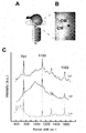
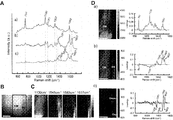
- FIG. 2A shows Raman spectra for cardiomyocytes, oxygenated red blood cells and deoxygenated red blood cells. These characteristic Raman bands correspond well with the spectra obtained under physiological conditions using the reported 532 nm excitation light. In particular, it is a band corresponding to the ⁇ 4 mode (oxygenation marker band, 1350-1380 cm ⁇ 1 range) and the ⁇ 19 and ⁇ 10 modes (1500-1650 cm ⁇ 1 range). These Raman bands originate from hemoglobin in erythrocytes.
- FIG. 2B is an autofluorescence image of a subepicardial cardiomyocyte and a small blood vessel in the vicinity thereof.
Landscapes
- Health & Medical Sciences (AREA)
- Nuclear Medicine, Radiotherapy & Molecular Imaging (AREA)
- Physics & Mathematics (AREA)
- Life Sciences & Earth Sciences (AREA)
- Chemical & Material Sciences (AREA)
- Analytical Chemistry (AREA)
- Biochemistry (AREA)
- General Health & Medical Sciences (AREA)
- General Physics & Mathematics (AREA)
- Immunology (AREA)
- Pathology (AREA)
- Investigating, Analyzing Materials By Fluorescence Or Luminescence (AREA)
Abstract
Priority Applications (4)
| Application Number | Priority Date | Filing Date | Title |
|---|---|---|---|
| EP09841489A EP2425785A1 (fr) | 2009-03-13 | 2009-03-13 | Imagerie de tissu corporel utilisant la lumière de diffusion raman |
| JP2011503629A JP5372137B2 (ja) | 2009-03-13 | 2009-03-13 | ラマン散乱を用いた生体組織イメージング |
| US13/256,259 US20120046555A1 (en) | 2009-03-13 | 2009-03-13 | Body tissue imaging using raman scattering light |
| PCT/JP2009/054930 WO2010103661A1 (fr) | 2009-03-13 | 2009-03-13 | Imagerie de tissu corporel utilisant la lumière de diffusion raman |
Applications Claiming Priority (1)
| Application Number | Priority Date | Filing Date | Title |
|---|---|---|---|
| PCT/JP2009/054930 WO2010103661A1 (fr) | 2009-03-13 | 2009-03-13 | Imagerie de tissu corporel utilisant la lumière de diffusion raman |
Publications (1)
| Publication Number | Publication Date |
|---|---|
| WO2010103661A1 true WO2010103661A1 (fr) | 2010-09-16 |
Family
ID=42727967
Family Applications (1)
| Application Number | Title | Priority Date | Filing Date |
|---|---|---|---|
| PCT/JP2009/054930 Ceased WO2010103661A1 (fr) | 2009-03-13 | 2009-03-13 | Imagerie de tissu corporel utilisant la lumière de diffusion raman |
Country Status (4)
| Country | Link |
|---|---|
| US (1) | US20120046555A1 (fr) |
| EP (1) | EP2425785A1 (fr) |
| JP (1) | JP5372137B2 (fr) |
| WO (1) | WO2010103661A1 (fr) |
Cited By (7)
| Publication number | Priority date | Publication date | Assignee | Title |
|---|---|---|---|---|
| WO2013146779A1 (fr) | 2012-03-30 | 2013-10-03 | 京都府公立大学法人 | Procédé pour la détection nerveuse par spectroscopie de diffusion raman |
| JP2014038044A (ja) * | 2012-08-17 | 2014-02-27 | Olympus Corp | 分光スペクトル解析方法 |
| JP2014224724A (ja) * | 2013-05-15 | 2014-12-04 | 京都府公立大学法人 | ラマン散乱を用いた心臓組織の識別方法及び装置 |
| WO2015128946A1 (fr) * | 2014-02-25 | 2015-09-03 | オリンパス株式会社 | Procédé d'analyse de spectre optique |
| JP2017523423A (ja) * | 2014-07-29 | 2017-08-17 | ユニヴェルシテ ドゥ ランス シャンパーニュ アルデンヌUniversite De Reims Champagne Ardenne | 線維化を検出及び定量化する方法 |
| JP2023056677A (ja) * | 2021-10-08 | 2023-04-20 | 国立大学法人島根大学 | 細胞評価方法および細胞評価プログラム |
| WO2023136308A1 (fr) | 2022-01-17 | 2023-07-20 | 国立大学法人大阪大学 | Méthode d'évaluation de cardiocytes par diffusion raman |
Families Citing this family (12)
| Publication number | Priority date | Publication date | Assignee | Title |
|---|---|---|---|---|
| US11399898B2 (en) | 2012-03-06 | 2022-08-02 | Briteseed, Llc | User interface for a system used to determine tissue or artifact characteristics |
| EP3122237B1 (fr) | 2014-03-25 | 2018-10-17 | Briteseed, LLC | Détecteur de vaisseaux et procédé de détection |
| US10820838B2 (en) | 2015-02-19 | 2020-11-03 | Briteseed, Llc | System for determining vessel size using light absorption |
| ES2907824T3 (es) | 2015-02-19 | 2022-04-26 | Briteseed Llc | Sistema para determinar el borde de un vaso |
| JP7057277B2 (ja) | 2015-10-08 | 2022-04-19 | ブライトシード・エルエルシー | 脈管サイズを決定するシステム及びその方法 |
| JP6951348B2 (ja) | 2016-02-12 | 2021-10-20 | ブライトシード・エルエルシーBriteseed,Llc | 組織特性を決定するためのシステム及び方法 |
| EP3506815B1 (fr) | 2016-08-30 | 2022-03-09 | Briteseed, LLC | Système optique chirugical avec des pinces non-parallèle avec compensation de distorsion angulaire |
| EP3678535B1 (fr) | 2017-09-05 | 2021-11-03 | Briteseed, LLC | Système et procédé utilisés pour déterminer des caractéristiques de tissu et/ou d'artéfact |
| JP7313353B2 (ja) | 2017-12-22 | 2023-07-24 | ブライトシード・エルエルシー | 組織又はアーチファクト特性を決定するために使用される小型システム |
| JP7204121B2 (ja) * | 2018-08-24 | 2023-01-16 | 国立大学法人鳥取大学 | 細胞識別装置及び細胞識別方法 |
| EP3902471B1 (fr) | 2018-12-30 | 2024-09-18 | Briteseed, LLC | Système utilisé pour détecter ou différencier un tissu ou un artefact |
| CN113933285A (zh) * | 2021-10-18 | 2022-01-14 | 安徽理工大学 | 一种检测肺组织胶原的非标记定量方法的建立 |
Citations (6)
| Publication number | Priority date | Publication date | Assignee | Title |
|---|---|---|---|---|
| JPH06505183A (ja) * | 1991-02-26 | 1994-06-16 | マサチユセツツ・インスチチユート・オブ・テクノロジー | 組織を診断するための分子分光計のシステムおよび方法 |
| JP2000055809A (ja) | 1998-08-13 | 2000-02-25 | Nikon Corp | 顕微ラマン分光装置及び顕微ラマン分光測定方法 |
| JP2005522293A (ja) * | 2002-04-05 | 2005-07-28 | マサチユセツツ・インスチチユート・オブ・テクノロジイ | 生物学的組織の分光法のための系および方法 |
| JP2007147357A (ja) | 2005-11-25 | 2007-06-14 | Nano Photon Kk | ラマン顕微鏡及びラマンスペクトルイメージの表示方法 |
| JP2007147350A (ja) | 2005-11-25 | 2007-06-14 | Hitachi Ltd | 無線通信測位システム |
| JP2008525158A (ja) * | 2004-12-28 | 2008-07-17 | ハイパームド・インコーポレーテッド | 全身生理機能およびショックの決定、評価および監視におけるハイパースペクトル/マルチスペクトルイメージング |
Family Cites Families (3)
| Publication number | Priority date | Publication date | Assignee | Title |
|---|---|---|---|---|
| US7697576B2 (en) * | 2004-05-05 | 2010-04-13 | Chem Image Corporation | Cytological analysis by raman spectroscopic imaging |
| WO2006005022A2 (fr) * | 2004-06-30 | 2006-01-12 | Chemimage Corporation | Imagerie chimique dynamique de cellules biologiques et d'autres sujets |
| WO2008144600A1 (fr) * | 2007-05-17 | 2008-11-27 | Prescient Medical, Inc. | Systèmes de spectroscopie par fibres optiques multicanal utilisant des modules optiques intégrés |
-
2009
- 2009-03-13 EP EP09841489A patent/EP2425785A1/fr not_active Withdrawn
- 2009-03-13 WO PCT/JP2009/054930 patent/WO2010103661A1/fr not_active Ceased
- 2009-03-13 JP JP2011503629A patent/JP5372137B2/ja active Active
- 2009-03-13 US US13/256,259 patent/US20120046555A1/en not_active Abandoned
Patent Citations (6)
| Publication number | Priority date | Publication date | Assignee | Title |
|---|---|---|---|---|
| JPH06505183A (ja) * | 1991-02-26 | 1994-06-16 | マサチユセツツ・インスチチユート・オブ・テクノロジー | 組織を診断するための分子分光計のシステムおよび方法 |
| JP2000055809A (ja) | 1998-08-13 | 2000-02-25 | Nikon Corp | 顕微ラマン分光装置及び顕微ラマン分光測定方法 |
| JP2005522293A (ja) * | 2002-04-05 | 2005-07-28 | マサチユセツツ・インスチチユート・オブ・テクノロジイ | 生物学的組織の分光法のための系および方法 |
| JP2008525158A (ja) * | 2004-12-28 | 2008-07-17 | ハイパームド・インコーポレーテッド | 全身生理機能およびショックの決定、評価および監視におけるハイパースペクトル/マルチスペクトルイメージング |
| JP2007147357A (ja) | 2005-11-25 | 2007-06-14 | Nano Photon Kk | ラマン顕微鏡及びラマンスペクトルイメージの表示方法 |
| JP2007147350A (ja) | 2005-11-25 | 2007-06-14 | Hitachi Ltd | 無線通信測位システム |
Non-Patent Citations (5)
| Title |
|---|
| HAKA, A.S. ET AL., CANCER RES., vol. 66, 2006, pages 3317 - 22 |
| HARADA, Y. ET AL., PROC. SPIE, vol. 6853, 2008, pages 685308 |
| KATHLEEN M.GOUGH: "Fourier transform infrared evalution of microscopic scarring in the cardiomyopathic heart:Effect of chronic ATI suppression", ANALYTICAL BIOCHEMISTRY, vol. 316, no. 2, 2003, pages 232 - 242, XP008166161 * |
| MOTZ, J.T. ET AL., J. BIOMED. OPT., vol. 11, 2006, pages 021003 |
| TORRES FILHO, I.P. ET AL., J. APPL. PHYSIOL., vol. 104, 2008, pages 1809 - 17 |
Cited By (11)
| Publication number | Priority date | Publication date | Assignee | Title |
|---|---|---|---|---|
| WO2013146779A1 (fr) | 2012-03-30 | 2013-10-03 | 京都府公立大学法人 | Procédé pour la détection nerveuse par spectroscopie de diffusion raman |
| JPWO2013146779A1 (ja) * | 2012-03-30 | 2015-12-14 | 京都府公立大学法人 | ラマン散乱分光法による神経検出法 |
| US9700212B2 (en) | 2012-03-30 | 2017-07-11 | Kyoto Prefectural Public University Corporation | Method for nerve detection by raman scattering spectroscopy |
| JP2014038044A (ja) * | 2012-08-17 | 2014-02-27 | Olympus Corp | 分光スペクトル解析方法 |
| JP2014224724A (ja) * | 2013-05-15 | 2014-12-04 | 京都府公立大学法人 | ラマン散乱を用いた心臓組織の識別方法及び装置 |
| WO2015128946A1 (fr) * | 2014-02-25 | 2015-09-03 | オリンパス株式会社 | Procédé d'analyse de spectre optique |
| US10048210B2 (en) | 2014-02-25 | 2018-08-14 | Olypmus Corporation | Spectroscopic analysis method |
| JP2017523423A (ja) * | 2014-07-29 | 2017-08-17 | ユニヴェルシテ ドゥ ランス シャンパーニュ アルデンヌUniversite De Reims Champagne Ardenne | 線維化を検出及び定量化する方法 |
| JP2023056677A (ja) * | 2021-10-08 | 2023-04-20 | 国立大学法人島根大学 | 細胞評価方法および細胞評価プログラム |
| JP7763463B2 (ja) | 2021-10-08 | 2025-11-04 | 国立大学法人島根大学 | 細胞評価方法および細胞評価プログラム |
| WO2023136308A1 (fr) | 2022-01-17 | 2023-07-20 | 国立大学法人大阪大学 | Méthode d'évaluation de cardiocytes par diffusion raman |
Also Published As
| Publication number | Publication date |
|---|---|
| JPWO2010103661A1 (ja) | 2012-09-10 |
| EP2425785A1 (fr) | 2012-03-07 |
| US20120046555A1 (en) | 2012-02-23 |
| JP5372137B2 (ja) | 2013-12-18 |
Similar Documents
| Publication | Publication Date | Title |
|---|---|---|
| JP5372137B2 (ja) | ラマン散乱を用いた生体組織イメージング | |
| Karreman et al. | Intravital correlative microscopy: imaging life at the nanoscale | |
| Krafft et al. | Raman and CARS microspectroscopy of cells and tissues | |
| CN112674861B (zh) | 用于使用荧光成像来确定损伤灶深度的系统和方法 | |
| Cicchi et al. | Clinical nonlinear laser imaging of human skin: a review | |
| US12539043B2 (en) | Lesion visualization using dual wavelength approach | |
| Tseng et al. | Multiphoton spectral microscopy for imaging and quantification of tissue glycation | |
| Campbell et al. | Clinical applications of non‐invasive multi and hyperspectral imaging of cell and tissue autofluorescence beyond oncology | |
| Schneidereit et al. | An advanced optical clearing protocol allows label-free detection of tissue necrosis via multiphoton microscopy in injured whole muscle | |
| US20120050720A1 (en) | System for diagnosing pathological change of lipid in blood vessels using non-linear optical microscopy | |
| Muselimyan et al. | Key factors behind autofluorescence changes caused by ablation of cardiac tissue | |
| Ghazaryan et al. | Spatial and temporal analysis of skin glycation by the use of multiphoton microscopy and spectroscopy | |
| Cicchi et al. | Multimodal nonlinear microscopy: A powerful label-free method for supporting standard diagnostics on biological tissues | |
| KR20100121807A (ko) | 비선형광학현미경을 이용한 혈관 내 지질의 병리적 변화 진단 시스템 | |
| Buttner et al. | Label-free imaging of myocardial remodeling in atrial fibrillation using nonlinear optical microscopy: A feasibility study | |
| JP6103700B2 (ja) | ラマン散乱を用いた心臓組織の識別方法及び装置 | |
| Watson et al. | (Second) harmonic disharmony: nonlinear microscopy shines new light on the pathology of atherosclerosis | |
| Ragan et al. | [22] In Vivo andex Vivo tissue applications of two-photon microscopy | |
| Chen et al. | A Femtosecond Fiber Laser‐Based Compact Label‐Free Multimodality Nonlinear Optical Microscopy and Its Ex Vivo Bioimaging | |
| Berndl et al. | Photoacoustic imaging for assessing ischemic kidney damage in vivo | |
| Deka et al. | Skin wound healing revealed by multimodal optical microscopies | |
| Zhuo et al. | High-contrast multimodel nonlinear optical imaging of collagen and elastin | |
| Pathak et al. | A REVIEW ON LATEST TRENDS IN BIOPHOTONICS | |
| RU2676647C1 (ru) | Способ определения типа биологической ткани | |
| Schucht et al. | Wide-Field Mueller Polarimetry of Brain Tissue Sections for Visualization of White Matter Fiber Tracts |
Legal Events
| Date | Code | Title | Description |
|---|---|---|---|
| 121 | Ep: the epo has been informed by wipo that ep was designated in this application |
Ref document number: 09841489 Country of ref document: EP Kind code of ref document: A1 |
|
| DPE1 | Request for preliminary examination filed after expiration of 19th month from priority date (pct application filed from 20040101) | ||
| WWE | Wipo information: entry into national phase |
Ref document number: 2011503629 Country of ref document: JP |
|
| NENP | Non-entry into the national phase |
Ref country code: DE |
|
| REEP | Request for entry into the european phase |
Ref document number: 2009841489 Country of ref document: EP |
|
| WWE | Wipo information: entry into national phase |
Ref document number: 2009841489 Country of ref document: EP |
|
| WWE | Wipo information: entry into national phase |
Ref document number: 13256259 Country of ref document: US |