WO2010114031A1 - Procédé de détection d'une substance dans un échantillon biologique - Google Patents
Procédé de détection d'une substance dans un échantillon biologique Download PDFInfo
- Publication number
- WO2010114031A1 WO2010114031A1 PCT/JP2010/055887 JP2010055887W WO2010114031A1 WO 2010114031 A1 WO2010114031 A1 WO 2010114031A1 JP 2010055887 W JP2010055887 W JP 2010055887W WO 2010114031 A1 WO2010114031 A1 WO 2010114031A1
- Authority
- WO
- WIPO (PCT)
- Prior art keywords
- protein
- biotin
- binding
- binding protein
- substance
- Prior art date
- Legal status (The legal status is an assumption and is not a legal conclusion. Google has not performed a legal analysis and makes no representation as to the accuracy of the status listed.)
- Ceased
Links
Images
Classifications
-
- G—PHYSICS
- G01—MEASURING; TESTING
- G01N—INVESTIGATING OR ANALYSING MATERIALS BY DETERMINING THEIR CHEMICAL OR PHYSICAL PROPERTIES
- G01N33/00—Investigating or analysing materials by specific methods not covered by groups G01N1/00 - G01N31/00
- G01N33/48—Biological material, e.g. blood, urine; Haemocytometers
- G01N33/50—Chemical analysis of biological material, e.g. blood, urine; Testing involving biospecific ligand binding methods; Immunological testing
- G01N33/68—Chemical analysis of biological material, e.g. blood, urine; Testing involving biospecific ligand binding methods; Immunological testing involving proteins, peptides or amino acids
-
- G—PHYSICS
- G01—MEASURING; TESTING
- G01N—INVESTIGATING OR ANALYSING MATERIALS BY DETERMINING THEIR CHEMICAL OR PHYSICAL PROPERTIES
- G01N33/00—Investigating or analysing materials by specific methods not covered by groups G01N1/00 - G01N31/00
- G01N33/48—Biological material, e.g. blood, urine; Haemocytometers
- G01N33/50—Chemical analysis of biological material, e.g. blood, urine; Testing involving biospecific ligand binding methods; Immunological testing
- G01N33/53—Immunoassay; Biospecific binding assay; Materials therefor
- G01N33/543—Immunoassay; Biospecific binding assay; Materials therefor with an insoluble carrier for immobilising immunochemicals
- G01N33/54393—Improving reaction conditions or stability, e.g. by coating or irradiation of surface, by reduction of non-specific binding, by promotion of specific binding
-
- C—CHEMISTRY; METALLURGY
- C07—ORGANIC CHEMISTRY
- C07K—PEPTIDES
- C07K17/00—Carrier-bound or immobilised peptides; Preparation thereof
-
- C—CHEMISTRY; METALLURGY
- C12—BIOCHEMISTRY; BEER; SPIRITS; WINE; VINEGAR; MICROBIOLOGY; ENZYMOLOGY; MUTATION OR GENETIC ENGINEERING
- C12N—MICROORGANISMS OR ENZYMES; COMPOSITIONS THEREOF; PROPAGATING, PRESERVING, OR MAINTAINING MICROORGANISMS; MUTATION OR GENETIC ENGINEERING; CULTURE MEDIA
- C12N15/00—Mutation or genetic engineering; DNA or RNA concerning genetic engineering, vectors, e.g. plasmids, or their isolation, preparation or purification; Use of hosts therefor
- C12N15/09—Recombinant DNA-technology
- C12N15/63—Introduction of foreign genetic material using vectors; Vectors; Use of hosts therefor; Regulation of expression
-
- G—PHYSICS
- G01—MEASURING; TESTING
- G01N—INVESTIGATING OR ANALYSING MATERIALS BY DETERMINING THEIR CHEMICAL OR PHYSICAL PROPERTIES
- G01N33/00—Investigating or analysing materials by specific methods not covered by groups G01N1/00 - G01N31/00
- G01N33/48—Biological material, e.g. blood, urine; Haemocytometers
- G01N33/50—Chemical analysis of biological material, e.g. blood, urine; Testing involving biospecific ligand binding methods; Immunological testing
- G01N33/53—Immunoassay; Biospecific binding assay; Materials therefor
- G01N33/543—Immunoassay; Biospecific binding assay; Materials therefor with an insoluble carrier for immobilising immunochemicals
-
- G—PHYSICS
- G01—MEASURING; TESTING
- G01N—INVESTIGATING OR ANALYSING MATERIALS BY DETERMINING THEIR CHEMICAL OR PHYSICAL PROPERTIES
- G01N33/00—Investigating or analysing materials by specific methods not covered by groups G01N1/00 - G01N31/00
- G01N33/48—Biological material, e.g. blood, urine; Haemocytometers
- G01N33/50—Chemical analysis of biological material, e.g. blood, urine; Testing involving biospecific ligand binding methods; Immunological testing
- G01N33/53—Immunoassay; Biospecific binding assay; Materials therefor
- G01N33/543—Immunoassay; Biospecific binding assay; Materials therefor with an insoluble carrier for immobilising immunochemicals
- G01N33/54306—Solid-phase reaction mechanisms
-
- G—PHYSICS
- G01—MEASURING; TESTING
- G01N—INVESTIGATING OR ANALYSING MATERIALS BY DETERMINING THEIR CHEMICAL OR PHYSICAL PROPERTIES
- G01N33/00—Investigating or analysing materials by specific methods not covered by groups G01N1/00 - G01N31/00
- G01N33/48—Biological material, e.g. blood, urine; Haemocytometers
- G01N33/50—Chemical analysis of biological material, e.g. blood, urine; Testing involving biospecific ligand binding methods; Immunological testing
- G01N33/53—Immunoassay; Biospecific binding assay; Materials therefor
- G01N33/543—Immunoassay; Biospecific binding assay; Materials therefor with an insoluble carrier for immobilising immunochemicals
- G01N33/54353—Immunoassay; Biospecific binding assay; Materials therefor with an insoluble carrier for immobilising immunochemicals with ligand attached to the carrier via a chemical coupling agent
-
- G—PHYSICS
- G01—MEASURING; TESTING
- G01N—INVESTIGATING OR ANALYSING MATERIALS BY DETERMINING THEIR CHEMICAL OR PHYSICAL PROPERTIES
- G01N33/00—Investigating or analysing materials by specific methods not covered by groups G01N1/00 - G01N31/00
- G01N33/48—Biological material, e.g. blood, urine; Haemocytometers
- G01N33/50—Chemical analysis of biological material, e.g. blood, urine; Testing involving biospecific ligand binding methods; Immunological testing
- G01N33/53—Immunoassay; Biospecific binding assay; Materials therefor
- G01N33/569—Immunoassay; Biospecific binding assay; Materials therefor for microorganisms, e.g. protozoa, bacteria, viruses
- G01N33/56983—Viruses
- G01N33/56994—Herpetoviridae, e.g. cytomegalovirus, Epstein-Barr virus
-
- G—PHYSICS
- G01—MEASURING; TESTING
- G01N—INVESTIGATING OR ANALYSING MATERIALS BY DETERMINING THEIR CHEMICAL OR PHYSICAL PROPERTIES
- G01N33/00—Investigating or analysing materials by specific methods not covered by groups G01N1/00 - G01N31/00
- G01N33/48—Biological material, e.g. blood, urine; Haemocytometers
- G01N33/50—Chemical analysis of biological material, e.g. blood, urine; Testing involving biospecific ligand binding methods; Immunological testing
- G01N33/68—Chemical analysis of biological material, e.g. blood, urine; Testing involving biospecific ligand binding methods; Immunological testing involving proteins, peptides or amino acids
- G01N33/6854—Immunoglobulins
-
- G—PHYSICS
- G01—MEASURING; TESTING
- G01N—INVESTIGATING OR ANALYSING MATERIALS BY DETERMINING THEIR CHEMICAL OR PHYSICAL PROPERTIES
- G01N2333/00—Assays involving biological materials from specific organisms or of a specific nature
- G01N2333/005—Assays involving biological materials from specific organisms or of a specific nature from viruses
- G01N2333/01—DNA viruses
- G01N2333/03—Herpetoviridae, e.g. pseudorabies virus
- G01N2333/035—Herpes simplex virus I or II
-
- G—PHYSICS
- G01—MEASURING; TESTING
- G01N—INVESTIGATING OR ANALYSING MATERIALS BY DETERMINING THEIR CHEMICAL OR PHYSICAL PROPERTIES
- G01N2333/00—Assays involving biological materials from specific organisms or of a specific nature
- G01N2333/195—Assays involving biological materials from specific organisms or of a specific nature from bacteria
- G01N2333/36—Assays involving biological materials from specific organisms or of a specific nature from bacteria from Actinomyces; from Streptomyces (G)
Definitions
- the present invention relates to a method for qualitatively and / or quantitatively detecting a substance in a biological sample. According to the method of the present invention, it is possible to detect a substance that is present in a trace amount in a biological sample and is usually difficult to detect.
- test substance In order to detect a substance in a biological sample, a method using a substance that specifically binds to the substance (test substance) has been widely used. For example, a so-called immunoassay using an antigen-antibody reaction, nucleic acid hybridization using a hydrogen bond between nucleic acid chains, and the like.
- the test substance if the test substance is an antibody, the antigen is immobilized. Conversely, if the test substance is an antigen, the antibody is immobilized on a carrier such as a microplate, a microbead, or a sensor chip and reacted with the test substance. Then, the presence / absence and extent of the antigen-antibody reaction is detected and measured.
- Hydrophobic bonds, covalent bonds, etc. are known as general methods for immobilization.
- “Hydrophobic binding” utilizes the interaction between the hydrophobic surface of the carrier and the hydrophobic portion of a protein that binds specifically to the substance to be detected (hereinafter sometimes referred to as “specific protein”).
- specific protein a protein that binds specifically to the substance to be detected
- the binding force is generally weak, and when used for ELISA (Enzyme linked immunosorbent assay) or the like, the protein often leaves the carrier by a washing operation after the binding.
- ELISA Enzyme linked immunosorbent assay
- the “covalent bond” uses an interaction between a functional group (for example, an amino group) in a specific protein and a functional group (for example, a carboxyl group) arranged on the surface of the carrier, and has a strong binding force.
- a functional group for example, an amino group
- a functional group for example, a carboxyl group
- biotin vitamin H
- avidin is an egg white-derived glycoprotein that binds very strongly to biotin.
- the interaction between avidin and biotin is one of the strongest non-covalent bonds (Green (1975) Adv Protein Chem 29: 85-133).
- streptavidin is an avidin-like protein derived from actinomycetes and also binds strongly to biotin.
- Biotin is a small molecule with a molecular weight of 244, and can be easily bound to various biomolecules such as proteins, nucleic acids, lipids, sugar chains, etc. using commercially available kits.
- (strept) avidin-biotin has been widely used in the field of molecular biology and biochemistry such as detection of antigens and antibodies due to the strength of its action (Green (1990). ) Methods Enzymol 184: 51-67).
- (strept) avidin is first bound to a substrate such as a microplate by covalent bond or hydrophobic bond, and then bound to a biotinylated protein. There is a way to fix it.
- avidin is first bound to a biotin-bound substrate by an avidin-biotin bond, and the desired biotinylated protein is bound to another biotin pocket of the avidin, whereby the substrate-biotin-avidin is bound.
- a technique of immobilizing in the order of -biotin-desired protein has been reported (JP-A-4-236353).
- a test substance can be detected using a plate immobilized by such a method.
- Non-specific binding that causes background signals is reduced in a specific binding assay system using a solid phase in which proteins that specifically bind to test substances such as antigens and antibodies are bound by hydrophobic or covalent bonds.
- Methods for solving this problem include, for example, a method in which a bacterial component extract is contained in a detection reagent (Japanese Patent Laid-Open No. 59-99257), and the same type of vector used for production of a recombinant protein that specifically binds to a test substance. And a method for adding a culture component of a host cell introduced with a vector not containing a gene encoding the protein to a sample (Japanese Patent Laid-Open No.
- An object of the present invention is to provide a method for qualitatively and / or quantitatively detecting a substance in a biological sample.
- the object of the present invention is to provide a method for detecting and measuring a substance present in a trace amount in a biological sample qualitatively and / or quantitatively while suppressing a background signal.
- the present inventors immobilized a biotinylated protein obtained by binding biotin to a protein that specifically binds to a test substance using a bond between biotin and biotin-binding protein.
- the present invention has been conceived by devising to reduce the background signal particularly in detecting a substance present in a trace amount in a biological sample. Specifically, when a cell disruption extract and a biotin-binding protein were added to a biological sample, the effect of reducing nonspecific binding was significant. Alternatively, the same effect was obtained by adding a cell disruption extract prepared from cells in which biotin-binding protein was expressed by genetic engineering technology instead of adding biotin-binding protein.
- the present invention provides a more sensitive detection method that reduces nonspecific binding in a system in which a substance that specifically detects a test substance is immobilized on a carrier using an avidin-biotin bond. It is to provide.
- the present invention includes the following aspects.
- a method for detecting a substance in a biological sample comprising: 1) Prepare a biotinylated protein in which biotin is bound to a carrier bound to a biotin-binding protein and a protein that specifically binds to the substance to be detected; 2) A biotinylated protein is bound to the carrier prepared in step 1) via a bond between biotin and biotin-binding protein to prepare a carrier to which the biotinylated protein is bound; 3) To the carrier to which the biotinylated protein prepared in step 2) is bound, (A) a biological sample, and (bi) a biotin-binding protein of step 1), a protein that specifically binds to a substance to be detected, and / or a host cell used to express a biotinylated protein Cell disruption extract prepared from the same type of cells, biotin-binding protein, or (b-ii) biotin-binding protein of step 1), protein that specifically binds to the substance to be detected;
- a kit for detecting a substance in a biological sample A) a carrier in which a biotinylated protein obtained by binding biotin to a protein that specifically binds to a substance to be detected is bound by biotin-biotin-binding protein binding; and Bi) Cell disruption extract prepared from cells of the same type as the host cell used to express the biotin-binding protein of A), the protein that specifically binds to the substance to be detected, and / or the biotinylated protein Biotin-binding protein, or B-ii) A) biotin-binding protein, protein that specifically binds to the substance to be detected, and / or host cell used to express biotinylated protein An agent for diluting a biological sample containing a cell disruption extract prepared from a cell in which a biotin-binding protein is expressed by genetic engineering technology in the cell; Including kit.
- a background signal in detecting a test substance in a biological sample, a background signal can be suppressed and more sensitive detection can be stably performed. According to the method of the present invention, it is possible to detect and measure a substance that is present in a trace amount in a biological sample and is usually difficult to detect or accurately quantify.
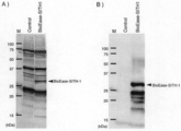
- FIG. 1 shows the expression of a BioEase tag (biotinylated tag) fused SITH-1 protein. Specifically, FIG. 1A shows the result of Western blotting for detection of SITH-1 protein, and FIG. 1B shows the result of activity staining for detection of biotinylated protein.
- E. coli BL21 (DE3) expressing the BioEase tag-fused SITH-1 protein was sonicated, and the resulting E. coli crude extract fraction was subjected to SDS-PAGE (15% acrylamide gel) to give 20 ⁇ g total protein / lane. And then transferred to a PVDF membrane.
- FIG. 1A reacted with anti-SITH-1 antibody (diluted 1/1000), reacted with alkaline phosphatase (AP) -labeled anti-rabbit IgG antibody (diluted 1/1000), and then stained with AP.
- FIG. 1B is a stained image after a streptavidin-horseradish peroxidase (HRP) (1/1000 dilution) reaction.
- HRP streptavidin-horseradish peroxidase
- Control indicates an extract sample derived from Escherichia coli having only an expression vector.
- the position of the BioEase tag-fused SITH-1 protein is indicated by an arrow.
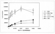
- FIG. 2 is a graph showing the effect of various serum dilutions on nonspecific binding. Serum diluted with PBS (white squares (dashed line)), diluted with PBS plus purified tamavidin 2 (TM2), white square (solid lines), diluted with E. coli disruption extract containing only expression vector The asterisk (broken line) is the asterisk (broken line), the asterisk (solid line) is diluted with the E.
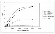
- FIG. 3 is a graph showing the S / N ratio for each dilution ratio of the anti-SITH-1 antibody based on the results shown in FIG.
- Serum diluted with PBS (white squares (dashed line)), diluted with PBS plus purified tamavidin 2 (TM2), white square (solid lines), diluted with E. coli disruption extract containing only expression vector
- the asterisk (broken line) is the asterisk (broken line)
- the asterisk (solid line) is diluted with the E. coli disrupted extract containing only the expression vector
- the black circle (solid line) is diluted with the E. coli disrupted extract expressing TM2.
- the detection method of the present invention comprises: 1) Prepare a biotinylated protein in which biotin is bound to a carrier bound to a biotin-binding protein and a protein that specifically binds to the substance to be detected; 2) A biotinylated protein is bound to the carrier prepared in step 1) via a bond between biotin and biotin-binding protein to prepare a carrier to which the biotinylated protein is bound; 3) To the carrier to which the biotinylated protein prepared in step 2) is bound, (A) a biological sample, and (bi) a biotin-binding protein of step 1), a protein that specifically binds to a substance to be detected, and / or a host cell used to express a biotinylated protein Cell disruption extract prepared from the same type of cells, biotin-binding protein, or (b-ii) biotin-binding protein of step 1), protein that specifically binds to the substance to
- the present invention relates to a method for detecting a substance in a biological sample.
- the biological sample in the present invention is not particularly limited as long as it can contain a substance to be detected.
- the body fluids should be diluted as necessary.
- the dilution rate is usually about 2 to 10,000 times, preferably about 100 to 1000 times, but is not limited thereto.
- Any buffer may be used as the solution for dilution, but it may contain an appropriate blocking agent.
- the blocking agent those having a high effect of suppressing non-specific binding are good, and blocking agents well known to those skilled in the art, such as BSA and casein, can be used.
- test substance of the present invention is not particularly limited as long as it is a substance that is desired to be detected or measured in a biological sample.
- proteins such as antibodies and antigens and fragments thereof, peptides, nucleic acids, hormones, carbohydrates
- Preferred examples thereof include glycolipids, bacteria and viruses contained in biological samples.
- the present invention makes it possible to measure substances in biological samples that have a low concentration and have been difficult to detect or accurately quantify by conventional methods.
- the test substance is an antibody in serum and the antibody titer is lower (for example, detection is difficult when the serum is diluted 1000-fold, but detection is finally possible when the serum is diluted 100-fold.
- the antibody titer it is necessary to suppress the dilution rate of serum, and as a result, nonspecific binding derived from serum components also increases.
- detection and quantification of an antibody are difficult, it can be easily detected or quantified more accurately by the method of the present invention.
- test substance in the present invention is an antibody
- Small protein encoded by the intermediate Transform of HHV-6 small protein encoded by HHV-6 latent infection intermediate transcript
- Antibodies against SITH-1 are included.
- antibodies against other antigens of herpes virus antibodies against virus-related antigens derived from cytomegalovirus, hepatitis virus, HIV virus, HTLV virus, measles virus, influenza virus, etc. or derived from Helicobacter pylori Antibody against a bacterial-related antigen, or an antibody against a fungus-related antigen.
- test substance in the present invention is an antigen
- examples include antigens derived from the above pathogens, cancer antigens, prostate-specific antigens, and the like.
- SITH-1 based on the description in PCT / JP2008 / 67300 (1) SITH-1 protein and nucleic acid The structure and function of SITH-1 protein and nucleic acid are disclosed in PCT / JP2008 / 67300, the entire contents of which are incorporated herein.
- SITH-1 is a factor involved in latent infection of herpesvirus, more specifically, a protein specifically expressed during latent infection of herpesvirus.
- “specifically expressed at the time of latent infection of herpes virus” specifically means that when a herpes virus is latently infected (not proliferatively infected) in a host infected with herpes virus, It means that a gene or gene product derived from a herpes virus is expressed.
- Examples of the protein and nucleic acid of SITH-1 include (a) a protein consisting of the amino acid sequence shown in SEQ ID NO: 1, and a nucleic acid encoding this protein.
- the SITH-1 protein consisting of the amino acid sequence shown in SEQ ID NO: 1 is isolated and identified as a protein that is specifically expressed during latent infection with human herpesvirus-6 (HHV-6), as shown in a reference example described later. It has been done.
- the SITH-1 protein is a protein having an amino acid sequence shown in SEQ ID NO: 1 and consisting of 159 amino acids and having a molecular weight of about 17.5 kDa.
- SITH-1 protein is encoded by the nucleic acid of the SITH-1 gene.
- the cDNA of this SITH-1 gene has a size of 1795 base pairs (about 1.79 kbp), and the 954th to 956th base sequences are the start codon (Kozak ATG).
- the base sequence from the 1431st to 1433th is a stop codon (TAA). Therefore, the SITH-1 nucleic acid has a 954 to 1430 base sequence as an open reading frame (ORF) in the base sequence shown in SEQ ID NO: 3, and this ORF has 477 base pairs (about 0.48 kbp).
- ORF open reading frame
- the base sequence representing the ORF region is shown in SEQ ID NO: 2.
- the base sequence shown in SEQ ID NO: 2 is described including the 3 bases of the stop codon.
- the SITH-1 nucleic acid is always expressed in the cytoplasm of cells latently infected with HHV-6, whereas no expression is observed in proliferatively infected cells.
- the nucleic acid encoding the SITH-1 protein is encoded by DNA complementary to the HHV-6 latent infection specific gene (H6LT) reported so far, and its expression is intermediate between the latent infection of HHV-6. Strengthen in stages. From these facts, the SITH-1 protein is considered to be a protein that is specifically expressed during latent infection with HHV-6.
- SITH-1 protein binds to host protein CAML (calcium-modulating cyclophilin ligand, Access #; U18242) to increase the intracellular calcium concentration in glial cells.
- CAML is a protein that is abundant in the brain and lymphocytes in the host organism and is known to increase intracellular calcium concentration.
- an increase in intracellular calcium concentration due to the expression of SITH-1 protein leads to activation of general signal transduction in latently infected cells, and is considered to contribute to efficient reactivation of HHV-6. .
- HHV-6 is known to latently infect glial cells in the brain, and when HHV-6 in the intermediate stage of latent infection or in a highly active latent infection state expresses SITH-1, glial cells It is thought that the calcium concentration inside increases. An increase in intracellular calcium concentration in brain cells is considered to be greatly related to mental disorders such as mood disorders (RIKEN Annual Report 2003).
- SITH-1 protein retains the activity of binding to CAML, which is a host protein, and has a function of increasing intracellular calcium concentration.
- mental disorders can be induced by expressing the SITH-1 protein in glial cells in the brain where the protein is most strongly expressed. Therefore, the SITH-1 protein is expressed during the latent infection of herpes virus or at the early stage of reactivation, and is considered to have a function of causing psychiatric disorders in the host.
- An antibody against SITH-1 can be obtained as a polyclonal antibody or a monoclonal antibody by a known method using SITH-1 protein or a variant thereof, or a partial peptide thereof as an antigen.
- Known methods include, for example, literature (Harlow et al., “Antibodies: A laboratory manual (Cold Spring Harbor Laboratory, New York (1988))”, Iwasaki et al., “Monoclonal antibody hybridoma and ELISA 91, Kodansha”. The method of description is mentioned. The antibody thus obtained can be used for detection and measurement of SITH-1 protein.
- antibody means an immunoglobulin (IgA, IgD, IgE, IgG, IgM and Fab fragments thereof, F (ab ′) 2 fragment, Fc fragment), and examples include polyclonal antibodies, monoclonal antibodies, Examples include, but are not limited to, chain antibodies, anti-idiotype antibodies, and humanized antibodies.
- immunoglobulin IgA, IgD, IgE, IgG, IgM and Fab fragments thereof, F (ab ′) 2 fragment, Fc fragment
- antibody recognizing SITH-1 protein is meant to include complete molecules and antibody fragments (eg, Fab and F (ab ′) 2 fragments) that can specifically bind to SITH-1 protein.
- Fab and F (ab ′) 2 and other fragments of the SITH-1 antibody can be used according to the methods disclosed herein or known methods. Such fragments are typically produced by proteolytic cleavage using enzymes such as papain (resulting in Fab fragments) or pepsin (resulting in F (ab ′) 2 fragments).
- the present invention makes it possible to identify a mood disorder patient or an individual with a potential mood disorder by detecting a SITH-1 antibody in a biological sample.
- a biotinylated protein-bound carrier in which biotin is bound to a protein that specifically binds to a substance to be detected is a biotinylated protein in which biotin is bound to a protein that specifically binds to a substance to be detected. Utilizes a carrier that is bound by a biotin-biotin binding protein-protein bond.
- the carrier of the present invention comprises 1) Prepare a biotinylated protein in which biotin is bound to a carrier bound to a biotin-binding protein and a protein that specifically binds to the substance to be detected; 2) The biotinylated protein is bound to the carrier prepared in step 1) via the biotin-biotin-binding protein bond, thereby creating a carrier to which the biotinylated protein is bound. Can be created.
- Biotin is the generic name for D-[(+)-cis-hexahydro-2-oxo-1H-thieno- (3,4) -imidazole-4-valeric acid]. It is a kind of water-soluble vitamin classified into the vitamin B group and is also called Vitamin B 7 (vitamin B 7 ), or sometimes called vitamin H or coenzyme R. Biotin binds very strongly to avidin, a kind of glycoprotein contained in egg white, and its absorption is inhibited. Therefore, a large amount of raw egg white can cause biotin deficiency.
- biotin refers to iminobiotin (Hofmann et al. (1980) Proc Natl Acad Sci USA 77: 4666-4668) and desthiobiotin (Hirsch) in addition to the above biotin. et al. (2002) Anal Biochem 308: 343-357), or biotin analogues such as biocytin and biotin sulfoxide.
- biotin-avidin biotin-binding protein
- the present invention includes immobilizing a protein that specifically binds to a substance to be detected on a carrier using the binding between biotin and biotin-binding protein.
- binding between biotin and biotin-binding protein may be referred to as “avidin-biotin binding”.
- Biotin-binding proteins include avidin, streptavidin, neutravidin, AVR protein (Biochem. J., (2002), 363: 609-617), bradavidin (J. Biol. Chem., (2005), 280: 13250-13255), Rhizavidin (Biochem. J., (2007), 405: 397-405), tamavidin (WO02 / 072817) and their mutants, etc., bind strongly to biotin. Any protein can be suitably used.
- the dissociation constant (KD) with biotin is 10 ⁇ 8 M or less, more preferably 10 ⁇ 10 M or less, and further preferably 10 ⁇ 12 M or less.
- the biotin-binding protein added to the test sample is as described later.
- biotin-binding protein tamavidin highly expressed in E. coli and mutants thereof can be preferably used.
- Tamavidin is a biotin-binding protein discovered from the edible mushroom Pleurotus concopiae (WO 02/072817, Takakura et al. (2009) FEBS J 276: 1383-1397).
- tamavidin variants include high binding ability and low non-specific binding tamavidin (PCT / JP2009 / 64302).
- “Tamavidin” in the present invention means tamavidin 1 (TM1), tamavidin 2 (TM2), or a variant thereof.
- the tamavidin of the present invention is typically encoded by a protein comprising the amino acid sequence of SEQ ID NO: 5 or SEQ ID NO: 7, or a nucleic acid comprising the base sequence of SEQ ID NO: 4 or SEQ ID NO: 6.
- the tamavidin of the present invention is a variant of a protein comprising the amino acid sequence of SEQ ID NO: 5 or SEQ ID NO: 7, or a protein encoded by a nucleic acid comprising the base sequence of SEQ ID NO: 4 or SEQ ID NO: 6.
- tamavidin 1 may be a protein having biotin binding activity similar to that of tamavidin 1 or 2, or a protein having high binding ability and low non-specific binding activity.
- tamavidin 1, tamavidin 2, and variants thereof may be collectively referred to simply as tamavidin.
- a tamavidin 1 or 2 variant is a protein comprising an amino acid sequence comprising a deletion, substitution, insertion and / or addition of one or more amino acids in the amino acid sequence of SEQ ID NO: 5 or 7, Alternatively, it may be a protein having biotin binding activity similar to 2.
- the substitution may be a conservative substitution, which is the replacement of a particular amino acid residue with a residue having similar physicochemical characteristics.
- Non-limiting examples of conservative substitutions include substitutions between aliphatic group-containing amino acid residues such as Ile, Val, Leu or Ala mutual substitutions, Lys and Arg, Glu and Asp, Gln and Asn mutual substitutions. Substitution between polar residues such as substitution is included.
- Mutants due to amino acid deletion, substitution, insertion and / or addition may be performed on, for example, site-directed mutagenesis (eg, Nucleic Acid Research, Vol. 10, No. 20), which is a well-known technique, on DNA encoding a wild-type protein. , P. 6487-6500, 1982, the entire contents of which are incorporated herein by reference).
- site-directed mutagenesis eg, Nucleic Acid Research, Vol. 10, No. 20
- one or more amino acids means amino acids that can be deleted, substituted, inserted and / or added by site-directed mutagenesis.
- “one or more amino acids” may mean one or several amino acids depending on the case. Without limitation, “one or more amino acids” means 50 or less, preferably 40 or less, 30 or less, 20 or less, 10 or less, 8 or less, 5 or less, 3 or less, Means within amino acids.
- the tamavidin 1 or 2 variant further comprises at least 60% or more of the amino acid sequence of SEQ ID NO: 5 or 7, preferably 65% or more, 70% or more, 75% or more, 80% or more, 85% or more, 90% or more, A protein comprising an amino acid sequence having 95% or more, 96% or more, 97% or more, 98% or more, or 99% or more, and more preferably 99.3% or more, comprising tamavidin 1 or 2 It may be a protein having the same biotin binding activity, or a protein having a high binding ability and a low non-specific binding activity.
- The% identity between two amino acid sequences may be determined by visual inspection and mathematical calculation. Alternatively, the percent identity of two protein sequences can be determined by Needleman, S .; B. And Wunsch, C.I. D. (J. Mol. Biol., 48: 443-453, 1970) and determined by comparing sequence information using the GAP computer program available from the University of Wisconsin Genetics Computer Group (UWGCG). May be.
- Preferred default parameters for the GAP program include: (1) Henikoff, S .; And Henikoff, J.H. G. (Proc. Natl. Acad. Sci. USA, 89: 10915-10919, 1992), scoring matrix, blossum 62; (2) gap weight of 12, (3) gap length weight of 4; And (4) no penalty for end gaps.
- the percent identity can be determined by comparison with sequence information using, for example, the BLAST program described in Altschul et al. (Nucl. Acids. Res., 25, p. 3389-3402, 1997).
- the program can be used on the Internet from the National Center for Biotechnology Information (NCBI) or the DNA Data Bank of Japan (DDBJ) website.
- NCBI National Center for Biotechnology Information
- DDBJ DNA Data Bank of Japan
- Various conditions (parameters) for identity search by the BLAST program are described in detail on the same site, and some settings can be changed as appropriate, but the search is usually performed using default values.
- the percent identity between two amino acid sequences can be determined by genetic information processing software GENETYX Ver. It may be determined using a program such as 7 (manufactured by Genetics) or the FASTA algorithm. At that time, the default value may be used for the search.
- the percent identity between two nucleic acid sequences can be determined by visual inspection and mathematical calculation, or more preferably, this comparison is made by comparing the sequence information using a computer program.
- a typical preferred computer program is the Wisconsin package, version 10.0 program “GAP” from the Genetics Computer Group (GCG; Madison, Wis.) (Devereux, et al., 1984, Nucl. Acids Res. , 12: 387).
- GAP Genetics Computer Group
- two amino acid sequences can be compared, and a nucleic acid sequence and an amino acid sequence can be compared.
- biotin-binding protein that binds to a carrier is used to create a carrier to which a biotinylated protein is bound through a bond between biotin and a biotin-binding protein. Therefore, the tamavidin 1 or 2 mutant is not significantly reduced in biotin-binding activity as compared to the case where a biotinylated protein is formed using these wild-types. Is preferred.
- the mutant of tamavidin 1 is not modified in N14, S18, Y34, S36, S78, W82, W98, W110, and D118 in the amino acid sequence of SEQ ID NO: 5.
- Y34 in the notation means the 34th tyrosine residue in the amino acid sequence of SEQ ID NO: 5.
- the mutant of tamavidin 2 does not modify four tryptophan residues (W69, W80, W96, W108) in the amino acid sequence of SEQ ID NO: 7.
- modification to an amino acid having similar properties or structure for example, phenylalanine (F) is preferable.
- the amino acid residues (N14, S18, Y34, S36, S76, T78, D116) considered to directly interact with biotin are not modified.
- glutamine (Q) or aspartic acid (D ) Preferably to aspartic acid, in the case of aspartic acid (D40) to asparagine (N), in the case of serine (S18, S36, S76), to threonine (T) or tyrosine (Y), preferably threonine
- tyrosine (Y34) serine (S), threonine (T) or phenylalanine (F), preferably to phenylalanine, and in the case of threonine (T78), to serine (S) or tyrosine (Y)
- serine in the case of aspartic acid (D116), glutamic acid (E) or aspa Gin to (N)
- preferable tamavidin variants include the following. (PCT / JP2009 / 64302).
- a protein showing biotin-binding activity comprising the amino acid sequence shown in SEQ ID NO: 7, or an amino acid sequence having one to several amino acid mutations in this sequence, or an amino acid sequence having 80% or more identity with this sequence
- Group 1 Arginine residue at position 104 of SEQ ID NO: 7; 2) 141st lysine residue of SEQ ID NO: 7; 3) the 26th lysine residue of SEQ ID NO: 7; and 4) one or more residues selected from the 73rd lysine residue of SEQ ID NO: 7 are substituted with an acidic amino acid residue or a neutral amino acid residue
- a modified biotin-binding protein in which the 104th arginine residue is substituted with a glutamic acid residue and the 141st lysine residue is substituted with a glutamic acid residue.
- Biotin-binding protein-bound carrier The material constituting the solid carrier is cellulose, Teflon (registered trademark), nitrocellulose, agarose, highly crosslinked spherical agarose, dextran, chitosan, polystyrene, polyacrylamide, polyester, polycarbonate, polyamide, polypropylene , Nylon, polyvinylidene difluoride, latex, polystyrene latex, silica, glass, glass fiber, gold, platinum, silver, copper, iron, stainless steel, ferrite, silicon wafer, polyethylene, polyethyleneimine, polylactic acid, resin, many Including but not limited to sugars, proteins (such as albumin), carbon or combinations thereof. Further, those having a certain strength, a stable composition, and little nonspecific binding are preferable.
- the shape of the solid support includes, but is not limited to, beads, magnetic beads, thin films, microtubes, filters, plates, microplates, carbon nanotubes, sensor chips, and the like.
- Flat solid carriers such as thin films and plates may be provided with pits, grooves, filter bottoms, etc., as is known in the art.
- the beads can have a sphere diameter ranging from about 25 nm to about 1 mm. In preferred embodiments, the beads have a diameter in the range of about 50 nm to about 10 ⁇ m. The size of the beads can be selected depending on the particular application.
- the beads as described above can be preferably used.
- the biotin-binding protein may be directly bound to the carrier, or a carrier on which the biotin-binding protein is immobilized in advance may be purchased. Alternatively, it may be bound to a biotinylated carrier via a biotin-biotin-binding protein-protein bond.
- a biotin-binding protein As a method for directly binding a biotin-binding protein, there is a method using a hydrophobic bond or a covalent bond.
- a biotin-binding protein may be directly bound and immobilized on a microplate such as NEW ELISA Plate Kit (Sumitomo Bakelite) according to the instructions attached to the kit.
- Avidin and streptavidin are commercially available from, for example, SIGMA.
- the biotin-binding protein solution is, for example, a microplate or the like (for example, but not limited to, Nunc-Immuno? Plate (Nunc), SpectraPlate-96 HB (Perkin Elmer), or Reacti -Direct contact with the support surface of Bind (trademark) 96-Well Plates Corner Notch (PIERCE), etc., and binding by interaction between the hydrophobic part of the biotin-binding protein and the hydrophobic part of the support by placing it for a certain period of time , Immobilize.
- a microplate or the like for example, but not limited to, Nunc-Immuno? Plate (Nunc), SpectraPlate-96 HB (Perkin Elmer), or Reacti -Direct contact with the support surface of Bind (trademark) 96-Well Plates Corner Notch (PIERCE), etc.
- a functional group is arranged on the surface of the carrier, and this is bonded to the functional group in the biotin-binding protein.
- various carriers having various functional groups arranged on the surface are commercially available and can be preferably used.
- a microplate on which functional groups are arranged on the surface as a maleic anhydride plate, for example, Reacti-Bind (trademark), Maleic Anhydride Activated Polyplate 96-Well Plates (PIERCE), As an active amino group plate, for example, Immobilizer?
- -Amino Modules / Plates or as a carboxyl group plate, for example, an ELISA plate MS-8896F (96 well, C type, flat bottom, carbo) (Sumitomo Bakelite).
- the microbeads having functional groups arranged on the surface include Sepharose (trademark) (GE Healthcare Bioscience) as a highly crosslinked agarose bead, and Dynabeads (trademark) (Dynal) as a magnetic bead. However, it is not limited to these.
- the biotin-binding protein and the solid support may be linked according to the instructions attached to the support.
- the carboxyl group of a solid support whose surface is modified so that the carboxyl group is exposed and the amino group of a biotin-binding protein can be converted into the crosslinking reagent 1-ethyl-3- (3-dimethylaminopropyl) carbodiimide (EDC).
- EDC crosslinking reagent 1-ethyl-3- (3-dimethylaminopropyl) carbodiimide
- a biotin-binding protein and a solid support can be linked by a coupling reaction in the presence.
- a biotin-binding protein is mixed with a solid support whose surface is active esterified with N-hydroxysuccinide (NHS) in a buffer solution of pH 6.5 to 9 containing no primary amino group, and the carboxyl group on the surface of the solid support is mixed.
- the amino group of the biotin-binding protein can be linked.
- the crosslinking reagent BS3 bis [sulfosuccinimidyl] suberate
- DSS disuccinimidyl suberate
- the amino group on the surface of the solid support and the amino group of the biotin-binding protein or the crosslinking reagent SPDP Using (N-succinimidyl 3- [2-pyridyldithio] propionate) or GMBS (N- (4-maleimidobutyryloxy) succinimide) to bind the amino group on the surface of the solid support and the thiol group of the biotin-binding protein.
- SPDP Using (N-succinimidyl 3- [2-pyridyldithio] propionate) or GMBS (N- (4-maleimidobutyryloxy) succinimide) to bind the amino group on the surface of the solid support and the thiol group of the biotin-binding protein.
- the biotin-binding protein can be biotinylated on the carrier and bound via an avidin-biotin bond. That is, by utilizing the fact that most of biotin-binding proteins are tetramers, biotin-binding protein is bound to a biotin-bonded carrier, and then biotinylated protein is further bound.
- Examples of a method for binding biotin to a carrier include a method using a biotinylation reagent.
- the biotinylation reagent include PZERCE (linker length in parentheses, reactive group) EZ-Link (registered trademark) Sulfo-NHS-Biotin (13.5 ⁇ , primary amine), EZ-Link (registered trademark) Sulfo-NHS-LC-Biotin (22.4 ⁇ , primary amine), EZ-Link (registered trademark) Sulfo-NHS-LCLC-Biotin (30.5 ⁇ , primary amine), EZ-Link (registered trademark) PFP- Biotin (9.6 ⁇ , amine), EZ-Link (registered trademark) Maleimide-PEO 2 -Biotin (29.1 ⁇ , thiol group), EZ-Link (registered trademark) Biotin-PEO 2 Amine (20.4 ⁇ , carboxyl group) ), EZ-Link (registered trademark) Biotin-PE
- biotin can be bound to a desired carrier such as a microplate, microbeads, or sensor chip using a known method.
- a desired carrier such as a microplate, microbeads, or sensor chip using a known method.
- carriers having various functional groups such as amino group, carboxyl group, thiol group, tosyl group, epoxy group, maleimide group, activated ester (for example, magnetic beads, sepharose beads, agarose beads, latex beads, microtiter plates, etc.)
- a method of using is a method of using.
- a biotinylation reagent containing NHS ester when used, it should be dissolved in an organic solvent such as DMSO (demethylsulfoxide) or a phosphate buffer of pH 7-9 and added to an immobilization carrier having an amino group.
- an organic solvent such as DMSO (demethylsulfoxide) or a phosphate buffer of pH 7-9
- Biotin can be bound by
- a biotinylation reagent containing an amino group is used, the carboxyl group of the immobilization carrier is converted into an activated ester using a carbodiimide such as EDC (1-ethyl-3- (3-dimethylaminopropyl) carbodiimide hydrochloride).
- biotinylation reagent dissolved in a buffer solution at around pH 5 may be added to bind biotin.
- the biotinylated immobilization carrier is preferably blocked with BSA or the like after inactivating unreacted functional groups.
- biotinylated commercially available carrier can be used.
- Reacti-Bind (trademark) Biotin Coated Polystyrene Plates (manufactured by PIERCE) can be used as the biotinylated microplate, but is not limited thereto.
- biotinylated microbeads include, for example, BioMag Biotin (manufactured by Polysciences) as magnetic beads, and Nanomag (registered trademark) -D biotin, nanomag (registered trademark) -silica manufactured by Corefront Co., Ltd. as nanomagnetic beads.
- Biotin is a polystyrene microbead
- Beadlyte registered trademark
- Biotin Beads Upstate
- Sigma Biotin Agarose
- 2-iminobiotin-Agarose is a highly crosslinked agarose
- Biotin-Sepharose Technology can be used, but is not limited thereto.
- the length of the linker connecting the carrier and biotin is preferably at least longer than 5 mm, more preferably 13.5 mm or longer.
- a carrier having a biotin-binding protein bound thereto By bringing a biotin-binding protein into contact with such a biotinylated carrier, a carrier having a biotin-binding protein bound thereto can be produced.
- Biotinylated protein In the present invention, biotin is bound to a protein that specifically binds to a test substance, a biotinylated protein is prepared, and this is bound to a carrier using a biotin-biotin-binding protein-binding bond. It's okay.
- the biotinylated protein production method is not particularly limited, but a biotin labeling kit (for example, but not limited to, EZ-Link? NHS-Lc-Biotin PIERCE) or Biotin Labeling Kit- NH2 (DOJINDO MOLECULAR TECHNOLOGIES INC.) Or the like may be used to bind biotin to a protein that specifically binds to the test substance.
- a protein gene that specifically binds to a test substance is fused with a DNA encoding a peptide containing a biotinylated sequence, and a vector that expresses the fusion gene is constructed.
- a fusion protein with a biotinylated sequence in any host As a biotinylated protein (Schwarz et al., (1988). J. Biol. Chem. 263: 9640-9645.).
- a vector include, but are not limited to, a vector containing an Invitrogen BioEase TM tag (Example 1 in this specification).
- Invitrogen BioEase TM tag Example 1 in this specification.
- pcDNA TM 6 vectors for mammalian cell expression there are pET104 vectors for E. coli expression, and pMT / BioEase vectors for Drosophila expression.
- biotinylation reagent examples include PZERCE (linker length in parentheses, reactive group) EZ-Link (registered trademark) Sulfo-NHS-Biotin (13.5 ⁇ , primary amine), EZ-Link (registered trademark) Sulfo-NHS-LC-Biotin (22.4 ⁇ , primary amine), EZ-Link (registered trademark) Sulfo-NHS-LCLC-Biotin (30.5 ⁇ , primary amine), EZ-Link (registered trademark) PFP- Biotin (9.6 ⁇ , amine), EZ-Link (registered trademark) Maleimide-PEO 2 -Biotin (29.1 ⁇ , thiol group), EZ-Link (registered trademark) Biotin-PEO
- biotin can be bound to a desired protein using a known method.
- biotinylation reagent containing NHS ester when a biotinylation reagent containing NHS ester is used, biotin is bound by dissolving in an organic solvent such as DMSO (demethylsulfoxide) or a pH 7-9 phosphate buffer and adding it to the desired protein. Can do.
- an organic solvent such as DMSO (demethylsulfoxide) or a pH 7-9 phosphate buffer
- DMSO demethylsulfoxide
- pH 7-9 phosphate buffer a pH 7-9 phosphate buffer
- a carboxyl group of a desired protein is converted into an activated ester using a carbodiimide such as EDC (1-ethyl-3- (3-dimethylaminopropyl) carbohydride hydrochloride).
- EDC 1-ethyl-3- (3-dimethylaminopropyl) carbohydride hydrochloride
- a protein that specifically binds to a test substance in advance, Purification may be performed from the organism from which the protein is originally derived, or may be purified after the gene encoding the protein is incorporated into an expression vector and expressed in a desired host cell through genetic engineering. Good.
- Host cells include mammalian cells (eg, but not limited to cells derived from primates such as humans and monkeys, rodents such as mice, rats, Chinese hamsters, and dogs), insect cells (using baculoviruses) Expression system, Drosophila system, etc.), yeast, Escherichia coli, plant, Bacillus subtilis and the like.
- Escherichia coli can be mentioned.
- HEK293, HeLa, HepG2, 293T for humans CHO, NIH3T3, PC12 for rodents, COS-1, COS-7, MDCK, Vero for insects, and insect cells for other mammals.
- Established cultured cell lines such as Sf9 and S2 can be preferably used.
- the protein can be expressed using a cell-free expression system using a wheat germ extract or an insect cell extract.
- the expression vector can be appropriately selected by those skilled in the art as appropriate for the host cell to be used.
- Purification of the expressed protein can use methods well known to those skilled in the art. Ordinary ion exchange chromatography, hydrophobic chromatography, gel filtration chromatography and the like may be used in combination, but a purification tag sequence may be used.
- a protein that specifically binds to the test substance as a fusion protein with glutathione-S-transferase, maltose binding protein, cellulose binding protein, chitin binding protein, thioredoxin binding protein, etc. It may be expressed in a host and purified using the affinity for glutathione, maltose, cellulose, chitin, thioredoxin (for example, using a glutathione-immobilized column).
- a protease recognition site is introduced into a fusion site with a protein that specifically binds to a test substance in advance, it can be removed by treating the tag sequence with the protease after purification.
- the protease those known to those skilled in the art such as enterokinase and Factor Xa may be used.
- HisTag or Strep (II) -Tag it is also possible to use HisTag or Strep (II) -Tag to purify using a column in which ionized nickel and StrepTactin are immobilized.
- a plurality of tags may be fused to a protein that specifically binds to the test substance, and purification methods may be combined.
- HisTag and a biotinylated sequence such as BioEASE (trademark) (Invitrogen) are fused to the end of a protein that specifically binds to a test substance, and expressed as a recombinant protein in a host, followed by purification with a nickel column. And may be further purified with a low affinity avidin or low affinity streptavidin (eg, SA mutein, Roche) column.
- biotinylated protein and a carrier to which a biotin-binding protein is bound are prepared and brought into contact with each other to bind the protein to the carrier via an avidin-biotin bond. Can be made.
- a crude cell disruption extract containing biotinylated protein is prepared at a total protein concentration of 0.1 mg / ml to 5 mg / ml, preferably 0.2 mg / ml to 2 mg / ml. This is contacted with a carrier to which a biotin-binding protein is bound at 10 ° C. to 40 ° C., preferably 20 ° C. to 30 ° C., for 5 minutes to 2 hours, preferably 30 minutes to 1 hour. By this operation, the biotinylated protein is immobilized on a carrier to which a biotin-binding protein is bound.
- the excess crude cell disruption extract is washed in a buffer solution such as PBS or TBS containing 0.05% to 1%, preferably 0.1% to 0.3% surfactant such as Tween20. It is preferable to do.
- a buffer solution such as PBS or TBS containing 0.05% to 1%, preferably 0.1% to 0.3% surfactant such as Tween20. It is preferable to do.
- the biotinylated protein is bound by contacting the cell disruption crude extract in this manner, the purification and the immobilization are actually performed at the same time. Therefore, in this case, a separate purification work is unnecessary.
- purified biotinylated protein at a concentration of 0.1 ⁇ g / ml to 5 ⁇ g / ml may be contacted with a carrier to which a biotin-binding protein is bound.
- step 3 Addition of biological sample to carrier bound to biotinylated protein
- the carrier bound to biotinylated protein prepared in step 2) (A) a biological sample, and (b) the same species as the host cell used to express the biotin-binding protein of step 1), the protein that specifically binds to the substance to be detected, and / or the biotinylated protein.
- step 3 the carrier bound to biotinylated protein prepared in step 2) (A) a biological sample, and (b) the same species as the host cell used to express the biotin-binding protein of step 1), the protein that specifically binds to the substance to be detected, and / or the biotinylated protein.
- a cell disruption extract prepared from the cells prepared from the cells.
- a method of adding a culture component of a host cell introduced with a vector that is the same type as a vector used for production of a recombinant protein that specifically binds to a test substance and does not contain a gene encoding the protein to the sample JP-A-8-43392
- a water extract from a cell that is the same type as a cell that has produced a recombinant protein that specifically binds to a test substance and does not contain the protein and then its water-soluble fraction
- a method of adding a minute to a sample Japanese Patent Laid-Open No. 2004-301646) is known.
- the present inventors have come up with a method for obtaining a more remarkable effect in a system in which a detection protein is bound to a carrier using a bond between biotin and biotin-binding protein. Specifically, it has been found that it is preferable that both the cell disruption extract and the biotin-binding protein are present simultaneously when the test sample is brought into contact with the carrier.
- the cells from which the cell disruption extract is derived are not particularly limited, such as E. coli cells, yeast cells, mammalian cells, insect cells, plant cells and the like. However, it is preferably a cell of the same type as the host cell used to express the biotin-binding protein, the protein that specifically binds to the substance to be detected, and / or the biotinylated protein.
- a biotin-binding protein a protein that specifically binds to a substance to be detected, and / or a biotinylated protein is prepared in E. coli, it is desirable to prepare a cell disruption extract from E. coli.
- a biotin-binding protein a protein that specifically binds to the substance to be detected, and / or a biotinylated protein is expressed in a cell-free system
- the used cell disruption extract is used as it is or in a desired buffer. It can be used suspended in a liquid.
- two or more types of cell disruption extracts May be used.
- biotin-binding proteins and / or proteins that specifically bind to substances to be detected are not expressed by genetic engineering techniques, but are originally extracted and purified from cells containing these proteins. May be.
- tamavidin when used as the biotin-binding protein, a cell disruption extract of basidiomycete Pleurotus concopiae cells can be used.
- the cell extract of the present invention also includes a cell disruption extract that inherently contains a biotin-binding protein and / or a protein that specifically binds to the substance to be detected.
- the cells for preparing the cell disruption extract may contain any vector, preferably an empty vector.
- An empty vector includes a biotin-binding protein, a protein that specifically binds to a test substance, and / or a gene that is the same type as the vector used to express the biotinylated protein and encodes these proteins. There is no vector.
- any vector may be used which further contains any nucleic acid in these empty vectors.
- the vector used when the biotin-binding protein, the protein that specifically binds to the test substance, and / or the biotinylated protein is expressed may be an unrelated vector.
- the cell disruption extract is not particularly limited as long as it is a cell-derived component, and for example, a protein component, a carbohydrate component, a lipid component, or a mixed component thereof can be used.
- a soluble extract of cells can be used.
- the method for preparing the cell disruption extract is not particularly limited, and various methods can be used. Usually, cells cultured in an appropriate medium are disrupted or solubilized by physical means such as ultrasonic waves, chemical means using a surfactant, enzyme treatment, etc., and dissolved by operations such as centrifugation or filtration. It can be prepared as an ingredient. In order to extend the shelf life, the liquid clarified by centrifugation or filtration is subjected to heat treatment such as addition of a protease inhibitor or autoclaving to suppress or deactivate various cell-derived enzymes. It is preferable to make it.
- the concentration at which the cell disruption extract is added may vary depending on the strength of the non-specific reaction that occurs, and a concentration sufficient to absorb the non-specific reaction can be set as appropriate.
- E. coli a vector may be included, and the vector encodes a biotin-binding protein.
- the absorbance at OD600 reaches 0.1 to 2, preferably 0.25 to 1, more preferably 0.4 to 0.6.
- the shaking culture is carried out at a temperature between 37 ° C. and 37 ° C. for 2 to 24 hours, preferably 4 to 16 hours.
- the cells are collected from the culture solution by centrifugation, suspended in a desired buffer solution, disrupted, the disrupted solution is centrifuged, and the supernatant is recovered as an E. coli crude extract.
- a crude cell disruption extract When a crude cell disruption extract is mixed with a biological sample, the sample is not limited, but 0.05 mg in a desired buffer solution (may contain BSA, casein, a commercially available blocking agent, etc.).
- Cell disrupted crude extract prepared to a total protein concentration of 0.5 mg / ml to 5 mg / ml, preferably 0.5 mg / ml to 5 mg / ml, and mixed at 10 ° C. to 30 ° C., preferably 20 ° C. to 30 ° C. The reaction is carried out for 1 minute to 4 hours, preferably 10 minutes to 1 hour.
- the biological sample is serum or the like, the serum is usually diluted 10 to 10,000 times, preferably 100 to 1000 times, more preferably 100 to 500 times with a cell disruption extract.
- a background signal can be finally suppressed by adding a biotin-binding protein to a biological sample.
- Such a biotin-binding protein may be the same as or different from the biotin-binding protein bound to the above-described carrier. Moreover, it may be a wild type or a mutant, and the biotin binding ability may be the same, higher or lower than that of the wild type.
- a powder of biotin-binding protein (either naturally derived or genetically expressed) may be added directly, or it may be added after dissolving in an appropriate solution. .
- the mixture of the sample and the cell disruption extract may be treated with a carrier on which the biotin-binding protein is immobilized (for example, passed through a column) (step b-). i).
- the concentration of the biotin-binding protein is 1 ⁇ g / ml to 500 ⁇ g / ml, preferably 10 ⁇ g / ml to the final concentration. Add at 100 ⁇ g / ml.
- concentration of the biotin-binding protein when the biotin-binding protein is expressed in the cells by genetic engineering is not limited to this, but may be the same concentration.
- biotin-binding protein obtained by introducing a gene encoding a biotin-binding protein into a host cell, expressing it, and disrupting the host cell (step b-ii).
- the biotin-binding protein may be expressed in a desired host by a method well known to those skilled in the art, but the desired protein and / or the desired biotinylated protein that specifically binds to the test substance is genetically engineered. When it is expressed in the same species as that host.
- a gene encoding a biotin-binding protein is incorporated into an expression vector, which is introduced into E. coli, and E. coli is cultured while inducing protein expression.
- the induction conditions such as the expression vector, host E. coli strain, medium component, IPTG concentration and culture temperature may be appropriately selected from conditions suitable for the expression of the biotin-binding protein.
- Addition of a biological sample and a cell disruption extract to an added carrier such as a biological sample to the carrier can be performed by any method.
- the biological sample must contact the cell disruption extract at the same time or before the biological sample contacts the carrier. That is, it is sufficient that the biological sample and the cell disruption extract are in sufficient contact, and the component derived from the cell disruption extract does not necessarily need to be finally added to the carrier together with the biological sample.
- a carrier in which a cell disruption extract component is bound may be prepared and a biological sample may be processed there.
- the biological sample can be passed through a cell disruption extract component column.
- the biological sample and the cell disruption extract are mixed with the carrier at 10 ° C. to 30 ° C., preferably at 20 ° C. to 30 ° C., for 10 minutes to 4 hours.
- the reaction is preferably performed for 30 minutes to 2 hours.
- test substance that specifically binds to a biotinylated protein is detected in step 4).
- the method for detecting the test substance can be appropriately selected by those skilled in the art based on the properties of the desired protein.
- Preferable examples include immunoassay methods such as enzyme-linked immunosorbent assay methods (including ELISA and sandwich ELISA methods) and radioimmunoassay methods (RIA), nucleic acid hybridization assays, surface plasmon resonance methods, and the like.
- immunoassay methods such as enzyme-linked immunosorbent assay methods (including ELISA and sandwich ELISA methods) and radioimmunoassay methods (RIA), nucleic acid hybridization assays, surface plasmon resonance methods, and the like.
- the antigen is immobilized, the antibody present in the test sample is reacted with the antigen, and detected by a method well known to those skilled in the art.
- a human antibody bound to the antigen is detected using an anti-human antibody.
- this anti-human antibody is labeled with fluorescence, enzyme, or radioisotope, and finally the amount of antibody is indirectly measured by measuring the amount of fluorescence, enzyme activity, or radioactivity, Quantify.
- the substance to be measured is an antigen
- an antibody against a certain part (epitope) of the antigen is immobilized, reacted with an antigen present in a test sample, and then reacted with an antibody against another epitope of the antigen.
- the secondary antibody against this different epitope is labeled as described above, the amount of antigen can be indirectly measured.
- a nucleic acid hybridization assay a nucleic acid of several tens to several hundreds or several thousand bases having a sequence region complementary to the nucleic acid to be measured is immobilized on a solid phase using a biotin-biotin-binding protein-protein bond. .
- a test sample containing a nucleic acid previously labeled with fluorescence or a radioisotope is reacted therewith, and the amount of fluorescence or the amount of radioactivity is measured.
- the labeling may be performed by a method well known to those skilled in the art, or commercially available fluorescent or enzyme-labeled anti-human antibodies may be used.
- the fluorescent label for example, a label with fluorescein and rhodamine, or a label with a fluorescent protein such as GFP can be considered.
- the enzyme label peroxidase, alkaline phosphatase, luciferase, glucose oxidase and the like can be used, but are not limited thereto. Substrates for measurement with these enzymes are commercially available.
- a substrate for TBA or chemiluminescence can be used.
- the radioisotope include iodine ( 125 I, 121 I), carbon ( 14 C), sulfur ( 35 S), tritium ( 3 H), and phosphoric acid ( 32 P) in the case of nucleic acids.
- the amount of antigen or antibody present in a biological sample can be determined, for example, using a linear regression computer algorithm, using standard preparations (e.g. It can be simply calculated by comparison with the amount present in the patient's standard sample.
- assay methods for detecting antigens and antibodies are described, for example, for ELISA, in Iacobelli et al., Breast Cancer Research and Treatment 11: 19-30 (1988).
- the substance to be detected in a biological sample for example, when there are few antibodies (when the antibody titer is low) or when there is a lot of nonspecific binding to the biological sample itself such as serum, The influence of background signal due to non-specific binding is increased. Therefore, the substance to be detected can be measured more accurately by appropriately subtracting the background signal from the measured value.
- the background to be subtracted can be appropriately determined by those skilled in the art depending on the experimental system.
- a “human / monkey anti-type I and type II collagen IgG antibody measurement kit” manufactured by Chondrex
- this kit calculation is performed by subtracting the background value (measured value using only the secondary antibody without adding serum) from the sample measurement value.
- biotinylation was performed as described in Example 2 to subtract the background due to such non-specific reaction. From the measured value of the carrier in which the SITH-1 antigen (protein that specifically binds to the substance to be detected) is immobilized via tamavidin, the biotinylated SITH-1 antigen is not immobilized (however, tamavidin is not immobilized on the carrier).
- SITH-1 antigen protein that specifically binds to the substance to be detected
- the SITH-1 antigen Similar to the group in which the SITH-1 antigen is immobilized, a blocking operation with BSA or the like is performed, and serum containing an anti-SITH-1 antibody (a substance to be detected in a biological sample) (biological It is also effective to subtract the measured value in the section where the biological sample is added.
- the organism from which the sample is derived for example, human in the case of SITH-1 does not have any protein (but is not limited to, but when the organism is a mammal, for example, GFP (green It can be obtained more accurately by subtracting the measured value of the section in which the solid phase is immobilized.
- the immobilization method is not particularly limited, but it is preferable to biotinylate the protein and immobilize it on a carrier on which the biotin-binding protein is immobilized by binding between biotin and biotin-binding protein.
- the calculation method as described above can be appropriately designed and selected by those skilled in the art based on the properties of the biological sample and the characteristics of the antibody used.
- the detection method of the present invention is preferably capable of specifically detecting an antibody having a low antibody titer in serum.
- the present invention also provides an agent for diluting a biological sample for use in a system for detecting a substance in a biological sample.
- the diluent of the present invention is 1) Cell disruption extract and biotin-binding protein, or 2) Cell disruption extract prepared from cells expressing biotin-binding protein by genetic engineering technology.
- This diluent is a biotinylated protein that binds biotin to a protein that specifically binds to the substance to be detected in a system that detects the substance in the biological sample.
- the composition of the diluent of the present invention is selected according to the production process of the protein bound to the carrier, that is, the biotin-binding protein, the protein that specifically binds to the substance to be detected, and / or the biotinylated protein. It is preferable to do.
- a host cell used to express a biotin-binding protein, a protein that specifically binds to the substance to be detected, and / or a biotinylated protein It is preferable to use the same type of cells.
- the diluent of the present invention may include 1) a cell disruption extract and a biotin-binding protein. This can be used in the embodiment of step 3) (bi) in the method for detecting a substance in a biological sample of the present invention.
- a cell disruption extract prepared from cells in which a biotin-binding protein is expressed by genetic engineering technology may be included. This can be used in the embodiment of step 3) (b-ii).
- the “agent for diluting a biological sample” may be the cell disruption extract (and biotin-binding protein) itself, or an agent for further diluting the cell disruption extract together with the biological sample.
- a suitable buffer solution or a solvent such as a commercially available cell diluent or serum diluent may be contained.
- kits for detecting a substance in a biological sample.
- the kit of the present invention comprises A) a carrier in which a biotinylated protein obtained by binding biotin to a protein that specifically binds to a substance to be detected is bound by biotin-biotin-binding protein binding; and Bi) Cell disruption extract prepared from cells of the same type as the host cell used to express the biotin-binding protein of A), the protein that specifically binds to the substance to be detected, and / or the biotinylated protein Biotin-binding protein, or B-ii) A) biotin-binding protein, protein that specifically binds to the substance to be detected, and / or host cell used to express biotinylated protein An agent for diluting a biological sample containing a cell disruption extract prepared from a cell in which a biotin-binding protein is expressed by genetic engineering technology in the cell; including.
- the “agent for diluting a biological sample” may be the cell disruption extract (and biotin-binding protein) itself, or an agent for further diluting the cell disruption extract together with the biological sample.
- a suitable buffer solution or a solvent such as a commercially available cell diluent or serum diluent may be contained.
- a fusion protein of SITH-1 protein derived from human herpesvirus 6 (HHV-6) and a biotinylated sequence (BioEase tag from Invitrogen) was expressed in E. coli and obtained therefrom.
- the Escherichia coli crude extract was directly reacted with a tamavidin 2 (hereinafter referred to as “TM2”)-immobilized microplate, and the fusion protein was bound to the microplate through a tamavidin-biotin bond.
- TM2 tamavidin 2
- the thus obtained SITH-1 protein-binding plate contains human serum diluted with an E. coli crude extract (including rabbit anti-SITH-1 antibody. Commercial human serum does not contain anti-SITH-1 antibody.
- E. coli crude extract including rabbit anti-SITH-1 antibody.
- Commercial human serum does not contain anti-SITH-1 antibody.
- BioEase Tag A gene encoding a fusion protein in which a BioEase tag was arranged at the N-terminal side of the SITH-1 protein was designed.
- This BioEase tag is a peptide tag including a sequence that is biotinylated by a biotinylated enzyme in a living body (in this case, E. coli).
- the amino acid sequence of the BioEase-SITH-1 fusion protein is shown in SEQ ID NO: 8, and the encoded nucleotide sequence is shown in SEQ ID NO: 9.
- a primer for amplifying the SITH-1 gene was designed. That is, a primer (SITH1NtermGW-F) consisting of a DNA sequence encoding the N-terminal part of SITH-1 protein and a primer (SITH1CtermGW-R) consisting of a DNA sequence encoding the C-terminal part of SITH-1 protein in the reverse direction Designed.
- PCR reaction conditions were as follows: 500 ng of template DNA, 2 ⁇ l of 10 (ExTaq buffer (TaKaRa)), 1.6 ⁇ l of 2.5 mM dNTP, 20 ⁇ moles of each primer, 0.1 ⁇ l of 5 U / ⁇ l ExTaq in 20 ⁇ l of reaction solution Then, using GeneAmp PCR System 9600 (PERKIN ELMER), 96 ° C for 3 minutes once, 95 ° C for 1 minute, 60 ° C for 1 minute, 72 ° C for 2 minutes 20 times, 72 ° C for 6 minutes once As a result, a 477 bp PCR product was obtained.
- the plasmid in which the SITH-1 gene was incorporated was used as an entry clone, and recombination reaction was performed with Gateway System using pET104.1 destination vector (manufactured by Invitrogen) which is a BioEase tag fusion protein expression vector.
- the recombinant product was transformed into E. coli TB1, plasmid DNA was extracted, and this plasmid was further transformed into E. coli BL21 (DE3).
- E. coli colony was amplified by PCR using SITH1NtermGW-F and SITH1CtermGW-R to confirm the presence or absence of the inserted gene.
- BioEase tag fusion vector BioEase-SITH1 / pET104.1 for expression of SITH-1 protein was completed.
- the cells were suspended in 3 ml of 0.1 M HEPES / KOH (pH 7.4) and then crushed by ultrasonic waves. The disrupted solution was centrifuged (15,000 rpm), and the supernatant was used as an E. coli crude extract.
- the protein contained in the crude extract was fractionated by SDS-PAGE and subjected to Western blotting.
- Rabbit anti-SITH-1 antibody unpublished
- alkaline phosphatase-labeled anti-rabbit IgG antibody BIO RAD
- Results are shown in FIG. 1A.
- a band of about 30 kDa that was not present in the control was detected from BioEase tag-fused SITH-1-expressing E. coli. This size almost coincided with the molecular weight of 28.6 kDa of BioEase tag fusion SITH-1.
- E. coli BL21 introduced with pTrc99A or TM2 / pTrc99A was prepared in the presence of IPTG, and then a crude E. coli extract was prepared for use in subsequent ELISA experiments. did.
- Example 2 Detection of Anti-SITH-1 Antibody in Human Serum by ELISA Purified TM2 was immobilized on a microplate using a New ELISA plate kit (Sumitomo Bakelite). The immobilization method followed the instructions attached to the kit.
- the crude extract of E. coli expressing the BioEase tag-fused SITH-1 protein obtained in Example 1 was prepared with 0.1 M HEPES / KOH (pH 7.4) so that the total protein concentration was 2 mg / ml, and then fixed to TM2. 100 ⁇ l was added to an activated plate (Sumitomo Bakelite, New ELISA). The plate was allowed to stand at room temperature for 1 hour, and BioEase tag-fused SITH-1 protein was bound to the TM2-immobilized plate by tamavidin-biotin binding.
- each well of the plate was washed 3 times with TBS buffer (TBST) containing 0.1% Tween 20, and 250 ⁇ l of 5 ⁇ g / ml BSA / TBST solution was added to each well, and allowed to stand at room temperature for 1 hour for blocking. Went. Thereafter, each well was washed 3 times with TBST.
- TBS buffer TBST
- human serum Human Serum pool, manufactured by Cosmo-Bio
- PBS or 1 mg total soluble protein / ml pTrc99A-introduced Escherichia coli crude extract prepared in Example 1 (1-4), or 1 mg total soluble.
- rabbit anti-SITH-1 antibody was 1/500, 1/1000, 1/2000, 1/4000, 1/8000 in volume ratio. It was added after serial dilution.
- the above antibody dilution ratio can be regarded as the same level as when the depression patient serum is diluted approximately 10 to 80 times. Furthermore, in order to confirm the effect of TM2 in the Escherichia coli crude extract used for dilution of serum, a solution obtained by adding purified TM2 to PBS or the pTrc99A-introduced Escherichia coli crude extract to a final concentration of 50 ⁇ g / ml.
- the rabbit anti-SITH-1 antibody was added in a volume ratio of 1/500, 1/1000, 1/2000, 1/4000, 1 / 100 ⁇ l each was added to a plate on which BioEase tag fusion SITH-1 was immobilized, and the mixture was allowed to react at room temperature for 1 hour.
- the blocking operation was performed on a TM2 fixed plate to which nothing was bound, and then PBS solution, pTrc99A-introduced or TM2 / pTrc99A-introduced Escherichia coli crude extract (1 mg total soluble protein / ml) was used.
- the results are shown in FIGS.
- the anti-SITH-1 antibody contained in the serum had a lot of non-specific binding when the serum was diluted with PBS, and a high amount of luminescence was detected even when the anti-SITH-1 antibody was not added. In addition, this non-specific binding was not improved even in the presence of 50 ⁇ g / ml TM2.
- the S / N ratio was particularly high in the group where 50 ⁇ g / ml TM2 was added to the pTrc99A-introduced Escherichia coli crude extract and in the group where the TM2 / pTrc99A-introduced Escherichia coli crude extract was added.
- SEQ ID NO: 1 amino acid sequence of SITH-1.
- SEQ ID NO: 2 Base sequence of SITH-1 ORF.
- SEQ ID NO: 3 nucleotide sequence of SITH-1 cDNA.
- SEQ ID NO: 4 Base sequence of tamavidin 1.
- SEQ ID NO: 5 amino acid sequence of tamavidin 1.
- SEQ ID NO: 6 Base sequence of tamavidin 2.
- SEQ ID NO: 7 Amino acid sequence of tamavidin 2.
- SEQ ID NO: 8 Amino acid sequence of BioEase-SITH-1 fusion protein
- SEQ ID NO: 9 Base sequence encoding BioEase-SITH-1 fusion protein
- SEQ ID NO: 10 Primer for PCR SITH1NtermGW-F
- SEQ ID NO: 11 Primer for PCR SITH1NtermGW-R
Landscapes
- Health & Medical Sciences (AREA)
- Life Sciences & Earth Sciences (AREA)
- Immunology (AREA)
- Engineering & Computer Science (AREA)
- Chemical & Material Sciences (AREA)
- Biomedical Technology (AREA)
- Molecular Biology (AREA)
- Hematology (AREA)
- Urology & Nephrology (AREA)
- Biochemistry (AREA)
- General Health & Medical Sciences (AREA)
- Biotechnology (AREA)
- Microbiology (AREA)
- Medicinal Chemistry (AREA)
- Physics & Mathematics (AREA)
- Cell Biology (AREA)
- Pathology (AREA)
- General Physics & Mathematics (AREA)
- Analytical Chemistry (AREA)
- Food Science & Technology (AREA)
- Virology (AREA)
- Genetics & Genomics (AREA)
- Chemical Kinetics & Catalysis (AREA)
- Proteomics, Peptides & Aminoacids (AREA)
- Organic Chemistry (AREA)
- Tropical Medicine & Parasitology (AREA)
- Zoology (AREA)
- Bioinformatics & Cheminformatics (AREA)
- Wood Science & Technology (AREA)
- General Engineering & Computer Science (AREA)
- Biophysics (AREA)
- Plant Pathology (AREA)
- Peptides Or Proteins (AREA)
- Measuring Or Testing Involving Enzymes Or Micro-Organisms (AREA)
- Investigating Or Analysing Biological Materials (AREA)
Abstract
Priority Applications (10)
| Application Number | Priority Date | Filing Date | Title |
|---|---|---|---|
| RU2011143787/15A RU2011143787A (ru) | 2009-03-31 | 2010-03-31 | Способ детектирования вещества в биологическом образце |
| BRPI1015445A BRPI1015445A2 (pt) | 2009-03-31 | 2010-03-31 | método para detecção de uma substância em amostra biológica |
| AU2010232305A AU2010232305B2 (en) | 2009-03-31 | 2010-03-31 | Method for detecting substance in biological sample |
| EP10758802A EP2416158A4 (fr) | 2009-03-31 | 2010-03-31 | Procédé de détection d'une substance dans un échantillon biologique |
| JP2011507265A JP5628793B2 (ja) | 2009-03-31 | 2010-03-31 | 生物学的試料中の物質を検出する方法 |
| US13/258,247 US9034590B2 (en) | 2009-03-31 | 2010-03-31 | Method for detecting substance in biological sample |
| CN2010800156081A CN102439446A (zh) | 2009-03-31 | 2010-03-31 | 检测生物学样品中的物质的方法 |
| SG2011069416A SG174558A1 (en) | 2009-03-31 | 2010-03-31 | Method for detecting substance in biological sample |
| CA2756109A CA2756109A1 (fr) | 2009-03-31 | 2010-03-31 | Procede de detection d'une substance dans un echantillon biologique |
| IL215321A IL215321A0 (en) | 2009-03-31 | 2011-09-22 | Method for detecting substance in biological sample |
Applications Claiming Priority (2)
| Application Number | Priority Date | Filing Date | Title |
|---|---|---|---|
| JP2009087839 | 2009-03-31 | ||
| JP2009-087839 | 2009-03-31 |
Publications (1)
| Publication Number | Publication Date |
|---|---|
| WO2010114031A1 true WO2010114031A1 (fr) | 2010-10-07 |
Family
ID=42828320
Family Applications (1)
| Application Number | Title | Priority Date | Filing Date |
|---|---|---|---|
| PCT/JP2010/055887 Ceased WO2010114031A1 (fr) | 2009-03-31 | 2010-03-31 | Procédé de détection d'une substance dans un échantillon biologique |
Country Status (12)
| Country | Link |
|---|---|
| US (1) | US9034590B2 (fr) |
| EP (1) | EP2416158A4 (fr) |
| JP (1) | JP5628793B2 (fr) |
| KR (1) | KR20120003903A (fr) |
| CN (1) | CN102439446A (fr) |
| AU (1) | AU2010232305B2 (fr) |
| BR (1) | BRPI1015445A2 (fr) |
| CA (1) | CA2756109A1 (fr) |
| IL (1) | IL215321A0 (fr) |
| RU (1) | RU2011143787A (fr) |
| SG (1) | SG174558A1 (fr) |
| WO (1) | WO2010114031A1 (fr) |
Cited By (5)
| Publication number | Priority date | Publication date | Assignee | Title |
|---|---|---|---|---|
| EP2314681A4 (fr) * | 2008-08-13 | 2012-01-18 | Japan Tobacco Inc | Protéine de liaison à la biotine modifiée |
| JPWO2010114029A1 (ja) * | 2009-03-31 | 2012-10-11 | 日本たばこ産業株式会社 | 生物学的試料中のsith−1の抗体を検出する方法 |
| WO2013018836A1 (fr) * | 2011-08-01 | 2013-02-07 | 日本たばこ産業株式会社 | Procédé d'inhibition de liaison non spécifique lors d'une étape de détection de substance dans un échantillon biologique, et agent destiné à être utilisé dans le procédé |
| JP2014514569A (ja) * | 2011-04-20 | 2014-06-19 | コリア アドバンスト インスティテュート オブ サイエンスアンド テクノロジー | 細胞環境内での単一分子水準のタンパク質−タンパク質相互作用分析方法及び装置 |
| CN107462708A (zh) * | 2017-06-28 | 2017-12-12 | 迈克生物股份有限公司 | 抗原或抗体包被磁微粒的方法、应用及试剂盒 |
Families Citing this family (5)
| Publication number | Priority date | Publication date | Assignee | Title |
|---|---|---|---|---|
| CN101809153B (zh) | 2007-09-27 | 2014-01-01 | 日本烟草产业株式会社 | 参与疱疹病毒的潜伏感染的因子及其利用 |
| WO2015199247A1 (fr) * | 2014-06-27 | 2015-12-30 | 日本たばこ産業株式会社 | Méthode de diagnostic, de traitement ou de prévention d'un trouble de l'humeur |
| CN108431046A (zh) | 2015-12-28 | 2018-08-21 | 日本烟草产业株式会社 | 诊断、治疗或预防心境障碍的方法 |
| NL2017204B1 (en) | 2016-06-08 | 2017-12-18 | Kei International Ltd | Solid substrate comprising antigens immobilised thereto, biosensor comprising said solid substrate and method for detecting the presence of mycobacterial material in a sample |
| NL2021443B1 (en) * | 2018-08-08 | 2020-02-20 | Kei International Ltd | Synthetic antigens for tuberculosis detection |
Citations (8)
| Publication number | Priority date | Publication date | Assignee | Title |
|---|---|---|---|---|
| JPS5999257A (ja) | 1982-11-29 | 1984-06-07 | Green Cross Corp:The | 抗体検出用試薬の水性溶媒 |
| JPH04236353A (ja) | 1991-01-17 | 1992-08-25 | Shin Etsu Chem Co Ltd | 抗体の測定方法 |
| JPH0843392A (ja) | 1994-07-29 | 1996-02-16 | Fujirebio Inc | 抗体検出用試薬の非特異反応吸収剤 |
| JPH08114590A (ja) | 1994-09-23 | 1996-05-07 | Boehringer Mannheim Gmbh | 測定すべき物質の定性的または/および定量的検出法 |
| JPH11211727A (ja) | 1997-11-03 | 1999-08-06 | Boehringer Mannheim Gmbh | ポリエチレングリコール誘導化生体分子及び不均質検出法におけるその使用 |
| JP2002048794A (ja) | 2000-08-04 | 2002-02-15 | Tosoh Corp | ビオチンを含む洗浄液、反応試薬および特異的結合アッセイ方法 |
| WO2002072817A1 (fr) | 2001-03-12 | 2002-09-19 | Japan Tobacco Inc. | Nouvelle proteine, gene codant ladite proteine et procede d'utilisation correspondant |
| JP2004301646A (ja) | 2003-03-31 | 2004-10-28 | Sysmex Corp | 免疫検査用非特異反応吸収剤 |
Family Cites Families (8)
| Publication number | Priority date | Publication date | Assignee | Title |
|---|---|---|---|---|
| JPH10246725A (ja) | 1997-03-03 | 1998-09-14 | Mikio Nakayama | 抗体又は抗原の検出方法 |
| CN101529251B (zh) * | 2006-11-01 | 2014-04-02 | 贝克曼考尔特公司 | 用于亲合分析的结合性表面 |
| CA2673968A1 (fr) * | 2006-12-28 | 2008-07-10 | Japan Tobacco, Inc. | Utilisation d'une proteine thermostable de liaison a la biotine et support solide ayant la proteine liee a celui-ci |
| BRPI0816103A2 (pt) * | 2007-08-28 | 2020-08-18 | Japan Tobacco Inc. | método para ligar proteínas a carregadores por fazer uso de tamavidinas. |
| CN101809153B (zh) * | 2007-09-27 | 2014-01-01 | 日本烟草产业株式会社 | 参与疱疹病毒的潜伏感染的因子及其利用 |
| US20120064543A1 (en) * | 2009-03-02 | 2012-03-15 | Virus Ikagaku Kenkyusho Inc. | Method for detecting substance in biological sample |
| TW201040529A (en) * | 2009-03-31 | 2010-11-16 | Japan Tobacco Inc | Method for detecting antibody against SITH-1 in biological sample |
| CN102803485A (zh) * | 2009-06-24 | 2012-11-28 | 日本烟草产业株式会社 | 修饰型生物素结合蛋白 |
-
2010
- 2010-03-31 AU AU2010232305A patent/AU2010232305B2/en not_active Expired - Fee Related
- 2010-03-31 US US13/258,247 patent/US9034590B2/en not_active Expired - Fee Related
- 2010-03-31 KR KR1020117025225A patent/KR20120003903A/ko not_active Withdrawn
- 2010-03-31 WO PCT/JP2010/055887 patent/WO2010114031A1/fr not_active Ceased
- 2010-03-31 SG SG2011069416A patent/SG174558A1/en unknown
- 2010-03-31 JP JP2011507265A patent/JP5628793B2/ja not_active Expired - Fee Related
- 2010-03-31 EP EP10758802A patent/EP2416158A4/fr not_active Withdrawn
- 2010-03-31 RU RU2011143787/15A patent/RU2011143787A/ru unknown
- 2010-03-31 CA CA2756109A patent/CA2756109A1/fr not_active Abandoned
- 2010-03-31 BR BRPI1015445A patent/BRPI1015445A2/pt not_active Application Discontinuation
- 2010-03-31 CN CN2010800156081A patent/CN102439446A/zh active Pending
-
2011
- 2011-09-22 IL IL215321A patent/IL215321A0/en unknown
Patent Citations (8)
| Publication number | Priority date | Publication date | Assignee | Title |
|---|---|---|---|---|
| JPS5999257A (ja) | 1982-11-29 | 1984-06-07 | Green Cross Corp:The | 抗体検出用試薬の水性溶媒 |
| JPH04236353A (ja) | 1991-01-17 | 1992-08-25 | Shin Etsu Chem Co Ltd | 抗体の測定方法 |
| JPH0843392A (ja) | 1994-07-29 | 1996-02-16 | Fujirebio Inc | 抗体検出用試薬の非特異反応吸収剤 |
| JPH08114590A (ja) | 1994-09-23 | 1996-05-07 | Boehringer Mannheim Gmbh | 測定すべき物質の定性的または/および定量的検出法 |
| JPH11211727A (ja) | 1997-11-03 | 1999-08-06 | Boehringer Mannheim Gmbh | ポリエチレングリコール誘導化生体分子及び不均質検出法におけるその使用 |
| JP2002048794A (ja) | 2000-08-04 | 2002-02-15 | Tosoh Corp | ビオチンを含む洗浄液、反応試薬および特異的結合アッセイ方法 |
| WO2002072817A1 (fr) | 2001-03-12 | 2002-09-19 | Japan Tobacco Inc. | Nouvelle proteine, gene codant ladite proteine et procede d'utilisation correspondant |
| JP2004301646A (ja) | 2003-03-31 | 2004-10-28 | Sysmex Corp | 免疫検査用非特異反応吸収剤 |
Non-Patent Citations (19)
| Title |
|---|
| ALTSCHUL, NUCL. ACIDS. RES., vol. 25, 1997, pages 3389 - 3402 |
| BIOCHEM. J., vol. 363, 2002, pages 609 - 617 |
| BIOCHEM. J., vol. 405, 2007, pages 397 - 405 |
| DEVEREUX ET AL., NUCL. ACIDS RES., vol. 12, 1984, pages 387 |
| GREEN, ADV PROTEIN CHEM, vol. 29, 1975, pages 85 - 133 |
| GREEN, METHODS ENZYMOL, vol. 184, 1990, pages 51 - 67 |
| HARLOW ET AL.: "Antibodies: A laboratory manual", 1988, COLD SPRING HARBOR LABORATORY |
| HENIKOFF, S., HENIKOFF, J. G, PROC. NATL. ACAD. SCI. USA, vol. 89, 1992, pages 10915 - 10919 |
| HIRSCH ET AL., ANAL BIOCHEM, vol. 308, 2002, pages 343 - 357 |
| HOFMANN ET AL., PROC NATL ACAD SCI USA, vol. 77, 1980, pages 4666 - 4668 |
| IACOBELLI ET AL., BREAST CANCER RESEARCH AND TREATMENT, vol. 11, 1988, pages 19 - 30 |
| IWASAKI ET AL.: "Monoclonal Antibody: Hybridoma and ELISA", 1991, KODANSHA |
| J. BIOL. CHEM., vol. 280, 2005, pages 13250 - 13255 |
| NEEDLEMAN, S. B., WUNSCH, C. D., J. MOL. BIOL., vol. 48, 1970, pages 443 - 453 |
| NUCLEIC ACID RESEARCH, vol. 10, no. 20, 1982, pages 6487 - 6500 |
| SAMBROOK ET AL.: "Molecular Cloning, A laboratory manual, 2nd edition", 1989 |
| SCHWARZ ET AL., J. BIOL. CHEM., vol. 263, 1988, pages 9640 - 9645 |
| See also references of EP2416158A4 |
| TAKAKURA ET AL., FEBS J, vol. 276, 2009, pages 1383 - 1397 |
Cited By (12)
| Publication number | Priority date | Publication date | Assignee | Title |
|---|---|---|---|---|
| EP2314681A4 (fr) * | 2008-08-13 | 2012-01-18 | Japan Tobacco Inc | Protéine de liaison à la biotine modifiée |
| US8304236B2 (en) | 2008-08-13 | 2012-11-06 | Japan Tobacco Inc. | Modified biotin-binding protein |
| JPWO2010114029A1 (ja) * | 2009-03-31 | 2012-10-11 | 日本たばこ産業株式会社 | 生物学的試料中のsith−1の抗体を検出する方法 |
| JP2014514569A (ja) * | 2011-04-20 | 2014-06-19 | コリア アドバンスト インスティテュート オブ サイエンスアンド テクノロジー | 細胞環境内での単一分子水準のタンパク質−タンパク質相互作用分析方法及び装置 |
| JP2016105094A (ja) * | 2011-04-20 | 2016-06-09 | コリア アドバンスト インスティテュート オブ サイエンスアンド テクノロジーKorea Advanced Institute Of Science And Technology | 細胞環境内での単一分子レベルにおけるタンパク質−タンパク質相互作用分析方法及び装置 |
| US9377462B2 (en) | 2011-04-20 | 2016-06-28 | Korea Advanced Institute Of Science And Technology | Method for analyzing protein-protein interaction on single-molecule level in cell environment, and method for measuring density of protein activated in cytosol |
| US9423400B2 (en) | 2011-04-20 | 2016-08-23 | Korea Advanced Institute Of Science And Technology | Method and apparatus for analyzing protein-protein interaction on single-molecule level within the cellular environment |
| US9733255B2 (en) | 2011-04-20 | 2017-08-15 | Korea Advanced Institute Of Science And Technology | Method and apparatus for analyzing protein-protein interaction on single-molecule level within the cellular environment |
| US9964544B2 (en) | 2011-04-20 | 2018-05-08 | Korea Advanced Institute Of Science And Technology | Method and apparatus for analyzing protein-protein interaction on single-molecule level within the cellular environment |
| US10401367B2 (en) | 2011-04-20 | 2019-09-03 | Korea Advanced Institute Of Science And Technology | Method and apparatus for analyzing protein-protein interaction on single molecule level within the cellular environment |
| WO2013018836A1 (fr) * | 2011-08-01 | 2013-02-07 | 日本たばこ産業株式会社 | Procédé d'inhibition de liaison non spécifique lors d'une étape de détection de substance dans un échantillon biologique, et agent destiné à être utilisé dans le procédé |
| CN107462708A (zh) * | 2017-06-28 | 2017-12-12 | 迈克生物股份有限公司 | 抗原或抗体包被磁微粒的方法、应用及试剂盒 |
Also Published As
| Publication number | Publication date |
|---|---|
| RU2011143787A (ru) | 2013-05-10 |
| IL215321A0 (en) | 2011-12-29 |
| EP2416158A1 (fr) | 2012-02-08 |
| US20120088313A1 (en) | 2012-04-12 |
| CA2756109A1 (fr) | 2010-10-07 |
| EP2416158A4 (fr) | 2012-05-30 |
| AU2010232305A1 (en) | 2011-11-10 |
| JPWO2010114031A1 (ja) | 2012-10-11 |
| JP5628793B2 (ja) | 2014-11-19 |
| KR20120003903A (ko) | 2012-01-11 |
| US9034590B2 (en) | 2015-05-19 |
| CN102439446A (zh) | 2012-05-02 |
| AU2010232305B2 (en) | 2015-03-05 |
| BRPI1015445A2 (pt) | 2016-04-19 |
| SG174558A1 (en) | 2011-10-28 |
Similar Documents
| Publication | Publication Date | Title |
|---|---|---|
| JP5628793B2 (ja) | 生物学的試料中の物質を検出する方法 | |
| Guirgis et al. | Gold nanoparticle-based fluorescence immunoassay for malaria antigen detection | |
| JP5647599B2 (ja) | 生物学的試料中の物質を検出する方法 | |
| US9139617B2 (en) | Method for detecting antibody against SITH-1 in biological sample | |
| JPWO2009123347A1 (ja) | ウイルス定量法 | |
| JP2023011764A (ja) | レクチンの固定化方法 | |
| US20140242620A1 (en) | Method of inhibiting non-specific binding in step of detecting substance in biological sample, and agent for use in the method | |
| CN113447658B (zh) | 一种检测抗过氧化物还原酶-1-IgG抗体的试剂盒 | |
| CN113447649B (zh) | 一种检测抗粘着斑蛋白-IgG抗体的试剂盒 | |
| KR101311736B1 (ko) | 샌드위치 면역분석 바이오칩 및 이를 사용한 면역분석법 | |
| HK1167892A (en) | Method for detecting substance in biological sample | |
| HK1167890A (en) | Method for detecting antibody against sith-1 in biological sample | |
| HK1167891A (en) | Method for detecting substance in biological sample | |
| Blatch et al. | Bassem SS Guirgis, Cláudia Sá e Cunha, Inês Gomes, Miguel Cavadas, Isabel Silva, Gonçalo Doria, Gregory |
Legal Events
| Date | Code | Title | Description |
|---|---|---|---|
| WWE | Wipo information: entry into national phase |
Ref document number: 201080015608.1 Country of ref document: CN |
|
| 121 | Ep: the epo has been informed by wipo that ep was designated in this application |
Ref document number: 10758802 Country of ref document: EP Kind code of ref document: A1 |
|
| WWE | Wipo information: entry into national phase |
Ref document number: 2011507265 Country of ref document: JP |
|
| WWE | Wipo information: entry into national phase |
Ref document number: 2756109 Country of ref document: CA |
|
| WWE | Wipo information: entry into national phase |
Ref document number: 13258247 Country of ref document: US |
|
| NENP | Non-entry into the national phase |
Ref country code: DE |
|
| REEP | Request for entry into the european phase |
Ref document number: 2010758802 Country of ref document: EP |
|
| WWE | Wipo information: entry into national phase |
Ref document number: 2010758802 Country of ref document: EP |
|
| ENP | Entry into the national phase |
Ref document number: 20117025225 Country of ref document: KR Kind code of ref document: A |
|
| WWE | Wipo information: entry into national phase |
Ref document number: 7827/CHENP/2011 Country of ref document: IN |
|
| ENP | Entry into the national phase |
Ref document number: 2011143787 Country of ref document: RU Kind code of ref document: A |
|
| ENP | Entry into the national phase |
Ref document number: 2010232305 Country of ref document: AU Date of ref document: 20100331 Kind code of ref document: A |
|
| REG | Reference to national code |
Ref country code: BR Ref legal event code: B01A Ref document number: PI1015445 Country of ref document: BR |
|
| ENP | Entry into the national phase |
Ref document number: PI1015445 Country of ref document: BR Kind code of ref document: A2 Effective date: 20110930 |