WO2011102507A1 - Procédé d'identification d'un oncogène, procédé d'établissement d'une cellule capable d'exprimer l'oncogène, et procédé de criblage de la substance ciblant l'oncogène - Google Patents
Procédé d'identification d'un oncogène, procédé d'établissement d'une cellule capable d'exprimer l'oncogène, et procédé de criblage de la substance ciblant l'oncogène Download PDFInfo
- Publication number
- WO2011102507A1 WO2011102507A1 PCT/JP2011/053647 JP2011053647W WO2011102507A1 WO 2011102507 A1 WO2011102507 A1 WO 2011102507A1 JP 2011053647 W JP2011053647 W JP 2011053647W WO 2011102507 A1 WO2011102507 A1 WO 2011102507A1
- Authority
- WO
- WIPO (PCT)
- Prior art keywords
- oncogene
- cell
- gene
- cancer
- cells
- Prior art date
- Legal status (The legal status is an assumption and is not a legal conclusion. Google has not performed a legal analysis and makes no representation as to the accuracy of the status listed.)
- Ceased
Links
Images


Classifications
-
- G—PHYSICS
- G01—MEASURING; TESTING
- G01N—INVESTIGATING OR ANALYSING MATERIALS BY DETERMINING THEIR CHEMICAL OR PHYSICAL PROPERTIES
- G01N33/00—Investigating or analysing materials by specific methods not covered by groups G01N1/00 - G01N31/00
- G01N33/48—Biological material, e.g. blood, urine; Haemocytometers
- G01N33/50—Chemical analysis of biological material, e.g. blood, urine; Testing involving biospecific ligand binding methods; Immunological testing
- G01N33/5005—Chemical analysis of biological material, e.g. blood, urine; Testing involving biospecific ligand binding methods; Immunological testing involving human or animal cells
- G01N33/5008—Chemical analysis of biological material, e.g. blood, urine; Testing involving biospecific ligand binding methods; Immunological testing involving human or animal cells for testing or evaluating the effect of chemical or biological compounds, e.g. drugs, cosmetics
- G01N33/5011—Chemical analysis of biological material, e.g. blood, urine; Testing involving biospecific ligand binding methods; Immunological testing involving human or animal cells for testing or evaluating the effect of chemical or biological compounds, e.g. drugs, cosmetics for testing antineoplastic activity
-
- C—CHEMISTRY; METALLURGY
- C12—BIOCHEMISTRY; BEER; SPIRITS; WINE; VINEGAR; MICROBIOLOGY; ENZYMOLOGY; MUTATION OR GENETIC ENGINEERING
- C12N—MICROORGANISMS OR ENZYMES; COMPOSITIONS THEREOF; PROPAGATING, PRESERVING, OR MAINTAINING MICROORGANISMS; MUTATION OR GENETIC ENGINEERING; CULTURE MEDIA
- C12N5/00—Undifferentiated human, animal or plant cells, e.g. cell lines; Tissues; Cultivation or maintenance thereof; Culture media therefor
- C12N5/06—Animal cells or tissues; Human cells or tissues
- C12N5/0602—Vertebrate cells
- C12N5/0693—Tumour cells; Cancer cells
-
- C—CHEMISTRY; METALLURGY
- C12—BIOCHEMISTRY; BEER; SPIRITS; WINE; VINEGAR; MICROBIOLOGY; ENZYMOLOGY; MUTATION OR GENETIC ENGINEERING
- C12Q—MEASURING OR TESTING PROCESSES INVOLVING ENZYMES, NUCLEIC ACIDS OR MICROORGANISMS; COMPOSITIONS OR TEST PAPERS THEREFOR; PROCESSES OF PREPARING SUCH COMPOSITIONS; CONDITION-RESPONSIVE CONTROL IN MICROBIOLOGICAL OR ENZYMOLOGICAL PROCESSES
- C12Q1/00—Measuring or testing processes involving enzymes, nucleic acids or microorganisms; Compositions therefor; Processes of preparing such compositions
- C12Q1/48—Measuring or testing processes involving enzymes, nucleic acids or microorganisms; Compositions therefor; Processes of preparing such compositions involving transferase
-
- G—PHYSICS
- G01—MEASURING; TESTING
- G01N—INVESTIGATING OR ANALYSING MATERIALS BY DETERMINING THEIR CHEMICAL OR PHYSICAL PROPERTIES
- G01N33/00—Investigating or analysing materials by specific methods not covered by groups G01N1/00 - G01N31/00
- G01N33/48—Biological material, e.g. blood, urine; Haemocytometers
- G01N33/50—Chemical analysis of biological material, e.g. blood, urine; Testing involving biospecific ligand binding methods; Immunological testing
- G01N33/5005—Chemical analysis of biological material, e.g. blood, urine; Testing involving biospecific ligand binding methods; Immunological testing involving human or animal cells
- G01N33/5008—Chemical analysis of biological material, e.g. blood, urine; Testing involving biospecific ligand binding methods; Immunological testing involving human or animal cells for testing or evaluating the effect of chemical or biological compounds, e.g. drugs, cosmetics
- G01N33/502—Chemical analysis of biological material, e.g. blood, urine; Testing involving biospecific ligand binding methods; Immunological testing involving human or animal cells for testing or evaluating the effect of chemical or biological compounds, e.g. drugs, cosmetics for testing non-proliferative effects
- G01N33/5023—Chemical analysis of biological material, e.g. blood, urine; Testing involving biospecific ligand binding methods; Immunological testing involving human or animal cells for testing or evaluating the effect of chemical or biological compounds, e.g. drugs, cosmetics for testing non-proliferative effects on expression patterns
-
- C—CHEMISTRY; METALLURGY
- C12—BIOCHEMISTRY; BEER; SPIRITS; WINE; VINEGAR; MICROBIOLOGY; ENZYMOLOGY; MUTATION OR GENETIC ENGINEERING
- C12N—MICROORGANISMS OR ENZYMES; COMPOSITIONS THEREOF; PROPAGATING, PRESERVING, OR MAINTAINING MICROORGANISMS; MUTATION OR GENETIC ENGINEERING; CULTURE MEDIA
- C12N2501/00—Active agents used in cell culture processes, e.g. differentation
- C12N2501/998—Proteins not provided for elsewhere
Definitions
- the present invention relates to an oncogene identification method, an oncogene-expressing cell establishment method, and an oncogene target drug screening method.
- anticancer drugs are actively conducted. Effective techniques for research and development of anticancer agents include (1) identification of oncogenes, (2) establishment of oncogene-expressing cells, and (3) screening for oncogene-targeted drugs.
- Non-Patent Document 1 discloses that a novel oncogene EML4-ALK was found by a transformation experiment using mouse-derived 3T3 cells (non-cancerous cells).
- Another example of an oncogene identification method is to comprehensively analyze gene expression in cancer cells or cancer tissue samples and normal cells or normal tissue samples using DNA chips or two-dimensional electrophoresis, and the difference in expression between the two samples. This is a method for selecting an oncogene from the genes for which the above is recognized (hereinafter, conventional identification method 2).
- Methods for establishing oncogene-expressing cells include a method of isolating a cell line that naturally expresses an oncogene from a subject, and a drug resistance gene (eg, G418 resistance gene together with the oncogene). ) Is introduced into the cell, and then the cell into which the oncogene has been introduced is selected with a drug (eg, G418).
- a drug eg, G418
- Patent Document 1 introduces a ligand receptor gene and a drug resistance gene into a cytokine-dependent cell line (non-cancer cell), It discloses that cell lines that can grow in the presence of ligands or ligand-like substances and drugs against drug resistance genes can be established.
- Non-Patent Document 1 describes the effect of a molecular targeted drug targeting EML4-ALK gene by introducing EML4-ALK gene into mouse-derived IL-3-dependent cell line (Ba / F3 cell which is a non-cancer cell). Is disclosed.
- patent document 1 uses the above-mentioned cell line derived from a cytokine dependence cell line (non-cancer cell), and a ligand or a ligand-like substance. Is disclosed.
- the conventional methods have the following problems when used in the research and development of anticancer agents.
- (1) Identification of Oncogene Since the conventional identification method 1 is carried out by directly testing the transformation ability as an oncogene, there is a high probability that the gene identified by this method is an oncogene.
- this method has problems in human application and versatility. As normal cultured cells used in this method, only rodent-derived fibroblasts are generally available, and human-derived cells and cancer cells, or epithelial cells and blood cells that are the origin of many cancers Experimental methods using cells have not been established. In addition, the rodent-derived fibroblasts used in the conventional identification method 1 are difficult to be passaged while maintaining their initial properties for a long period of time.
- Conventional identification method 2 has a problem that many genes that are induced or suppressed by gene expression of an oncogene other than the oncogene are selected as candidate genes. In addition, the conventional identification method 2 requires more detailed analysis of many candidate genes in order to finally identify an oncogene from among many selected candidate genes.
- an object of the present invention is to provide a novel methodology capable of solving these problems in the identification of oncogenes, establishment of oncogene-expressing cell lines, and screening for oncogene target drugs.
- cancer gene addiction or addiction (addition) is a state in which cancer cells are dependent on the growth of specific genes. It is known that when cancer cells that grow depending on a specific gene are treated with an inhibitor of the gene, the growth of the cancer cells can be suppressed.
- the inventors of the present application have found that dependence on a specific oncogene in cancer cells can be replaced by introduction of another oncogene. Based on this finding, the present inventors also treated cancer cells with an exogenous oncogene expression vector, and then treated the cancer cells treated with the expression vector with the expression or function of an oncogene specific to the cancer cell. It has been found that the method of culturing under conditions that inhibit the growth of artificial cells that can be selectively proliferated, and that the selectively proliferated artificial cells can be used as a cancer cell model.
- Patent Document 1 and Non-Patent Document 1 disclose that a cancer cell is used as a cell for introducing an oncogene and that the cancer cell into which the oncogene has been introduced is a condition that inhibits the oncogene specific to the cancer cell There is no description or suggestion of selecting by culturing below.
- the present invention is as follows.
- Artificial cells having the following characteristics (a) and (b): (A) derived from a cancer cell; and (b) capable of expressing a foreign oncogene and proliferating depending on the foreign oncogene.
- the artificial cell according to [1], further having the following characteristics (c): (C) retains the ability to express an oncogene unique to the cancer cell and the ability to proliferate depending on the unique oncogene.
- a method for establishing an artificial cell comprising the following steps (a) to (c): (A) treating cancer cells with a foreign oncogene expression vector; (B) culturing cancer cells treated with the expression vector under conditions that inhibit expression or function of an oncogene specific to the cancer cells; and (c) cancer cells grown in step (b) A step of obtaining an exogenous oncogene as an artificial cell having the ability to express and proliferate depending on the exogenous oncogene.
- a screening method for a substance having anticancer activity comprising the following steps (a) and (b): (A) a step of evaluating whether or not the test substance inhibits the proliferation of the artificial cell of any one of the above [1] to [10]; and (b) a test substance that inhibits the proliferation of the artificial cell, A step of selecting as a substance having an action.
- step (a) is performed under conditions that inhibit the expression or function of a specific oncogene.
- a method for identifying an oncogene comprising the following steps (a) to (c): (A) treating cancer cells with an expression vector for a test gene; (B) a step of culturing cancer cells treated with the expression vector under conditions that inhibit the expression or function of an oncogene unique to the cancer cells; and (c) the cancer cells cultured in step (b). Confirming whether or not is proliferated depending on the test gene. [16] The method of [15] above, wherein the test gene is a single gene.
- test gene is a plurality of genes and further comprises the following steps: (D) cloning the cancer cells grown in step (c); and (e) identifying the test gene introduced into the cloned cancer cells as an oncogene.
- test gene is a plurality of genes and further comprises the following steps: (F) A step of obtaining a test gene introduced into the cancer cell from the cancer cell grown in the step (c). (G) A step of cloning the acquired test gene and identifying it as an oncogene.
- the artificial cell of the present invention enables, for example, the development of an excellent anticancer agent against various cancers.
- the establishment method of the present invention is excellent in, for example, the establishment efficiency of cells expressing a foreign oncogene.
- the screening method of the present invention enables, for example, the development of an anticancer agent using any cancer cell that expresses any oncogene.
- the identification method of the present invention is excellent in sensitivity and accuracy of oncogene identification, for example, since the method of establishing the method and the methodology of the present invention, which are excellent in the efficiency of establishing cells expressing foreign oncogenes, are similar. Further, these methods of the present invention have the advantage that they can be carried out by a simple and conventional technique using commonly used cultured cells (eg, cancer cell lines).
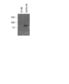
- FIG. 1 shows the expression of EML4-ALKvariant3a protein in EML4-ALKvariant3a gene-introduced PC-9 cells grown in a G418-containing medium (G418) or a gefitinib-containing medium (Gefitinib).
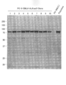
- FIG. 2 shows the expression of EML4-ALKvariant3a protein in 10 clones of EML4-ALKvariant3a gene-introduced PC-9 cells selectively proliferated in a gefitinib-containing culture medium.
- FIG. 1 shows the expression of EML4-ALKvariant3a protein in EML4-ALKvariant3a gene-introduced PC-9 cells grown in a G418-containing medium (G418) or a gefitinib-containing medium (Gefitinib).
- FIG. 2 shows the expression of EML4-ALKvariant3a protein in 10 clones of EML4-ALKvariant3a gene-introduced PC-9 cells selectively
- FIG. 3 shows EML4-ALKvariant3a transgenic PC-9 cells (PC-9-EML4-ALKvar3a), EML4-ALKvariant1 transgenic PC-9 cells (PC-9-EML4-ALKvar1) grown in a gefitinib-containing culture medium, and It is a figure which shows the expression of EML4-ALKvariant3a or EML4-ALKvariant1 protein in EML4-ALKvariant3a gene transfer HCC827 cell (HCC827-EML4-ALKvar3a).
- FIG. 4 is a diagram showing the expression of PDGFR ⁇ V561D protein in PD-9R ⁇ V561D gene-introduced PC-9 cells grown in a gefitinib-containing culture medium.
- FIG. 4 is a diagram showing the expression of PDGFR ⁇ V561D protein in PD-9R ⁇ V561D gene-introduced PC-9 cells grown in a gefitinib-containing culture medium.
- FIG. 5 is a graph showing the proliferation percentage (%) of PC-9 cells or PDGFR ⁇ V561D gene-introduced PC-9 cells in the presence of various concentrations of gefitinib.
- FIG. 6 is a graph showing the percentage proliferation of PDGFR ⁇ V561D gene-introduced PC-9 cells (in the presence or absence of 1 ⁇ M gefitinib) in the presence of various concentrations of the PDGFR ⁇ inhibitor Imatinib. .
- FIG. 7 is a diagram showing agarose gel electrophoresis of a novel oncogene candidate that can complement mutant EGFR-dependent growth in PC-9 cells.
- artificial cells The present invention provides specific artificial cells.
- artificial cell means a cell that is artificially produced.
- the artificial cell of the present invention may be an isolated or purified cell. Isolation or purification of the cells can be performed by a method known per se such as FACS.
- the artificial cell of the present invention can be derived from a cancer cell.
- the artificial cells of the present invention can inherit the cytological properties retained by cancer cells.
- the cancer cell is a human cell
- the artificial cell of the present invention can be a human cell.
- the cancer cell is a lung cell
- the artificial cell of the present invention can be a lung cell.
- cancer cell means a tumor cell having the ability to proliferate depending on a specific oncogene expressed by the cancer cell.
- Cancer cells include primary cultured cells, cell lines or cancer stem cells.
- the term “dependence” for cell growth refers to an oncogene addiction or addiction state in which the cell relies on growth for a particular oncogene.
- Whether a cell is dependent on a specific oncogene for growth can be confirmed by treating the cell with an inhibitor of a specific oncogene and then evaluating the proliferative capacity of the treated cell.
- the proliferative ability can be evaluated by, for example, an MTT assay or an MTS assay.
- cell death due to apoptosis can be induced by treating an oncogene-addicted cell for a specific oncogene with an inhibitor of the oncogene.
- cellular oncogene poisoning against a particular oncogene may be confirmed, for example, by evaluating whether apoptosis can be induced by inhibition of the oncogene.
- Apoptosis induction can be evaluated by, for example, TUNEL assay, detection of active caspase, and detection of ANNEXIN V.
- Cancer cells can be derived from any tissue.
- tissues include respiratory tissues (eg, lung, trachea, bronchi, pharynx, nasal cavity, sinus), digestive tissues (eg, stomach, small intestine, large intestine, rectum), pancreas, kidney, Examples include liver, thymus, spleen, heart, thyroid, adrenal gland, prostate, ovary, uterus, brain, skin, and blood tissue (eg, bone marrow, peripheral blood).
- cancer cells can be adherent cells or non-adherent cells (ie, blood cells), although adherent cells are preferred.
- the cancer cell can be a cell present in the tissue or a tissue other than the tissue.
- gland cells eg, lung gland cells, breast cells
- epithelial cells eg, endothelial cells
- epidermal cells e.g., stromal cells
- fibroblasts e.g., fibroblasts
- adipocytes e.g., mesangial cells
- pancreatic ⁇ cells e.g., nerve cells
- nerve cells examples include glial cells and blood cells.
- the cancer cell is preferably a lung adenocarcinoma cell.
- cancer refers to any malignant tumor in the tissues and cell types described above.
- cancer include cancer that may be caused by abnormal adhesion cells and cancer that may be caused by abnormal blood cells (eg, leukemia, lymphoma, multiple myeloma), but may be caused by abnormal adhesion cells. Cancer is preferred.
- cancers that can be caused by abnormal adhesion cells include lung cancer (eg, non-small cell cancer such as squamous cell carcinoma, adenocarcinoma and large cell cancer, and small cell cancer), digestive system.
- Cancer eg, stomach cancer, small intestine cancer, colon cancer, rectal cancer
- pancreatic cancer kidney cancer, liver cancer, thymic cancer, spleen cancer, thyroid cancer, adrenal cancer, prostate cancer, bladder cancer, ovarian cancer, uterine cancer (eg Endometrial cancer, cervical cancer), bone cancer, skin cancer, brain tumor, sarcoma, melanoma, blastoma (eg, neuroblastoma), adenocarcinoma, squamous cell carcinoma, solid cancer, epithelial cancer, Mesothelioma is mentioned.
- “Cancer” in cancer-related terms such as the terms “cancer cell” and “oncogene” may also have the same meaning.
- Cancer cells can be derived from any mammalian species. Examples of such mammalian species include humans, monkeys, cows, pigs, mice, rats, guinea pigs, hamsters, and rabbits. From the viewpoint of clinical application, the mammalian species is preferably human. Thus, the cancer cell may be a cancer cell isolated from a cancer patient or a cancer cell derived therefrom.
- Cancer cells may be non-virus-infected cells or virus-infected cells.
- oncogenic viruses that can infect cells include Epstein Barr virus, hepatitis virus, human papilloma virus, human T cell leukemia virus, and Kaposi's sarcoma-associated herpes virus.
- the cancer cells may also be cancer cells derived from embryonic stem cells, somatic stem cells, or artificial stem cells (eg, iPS cells) made from normal cells.
- the cancer cell from which the artificial cell of the present invention is derived can express a unique oncogene.
- unique oncogene means an oncogene responsible for the growth of cancer cells that is expressed by cancer cells that can be used as a material in the establishment of the artificial cells of the present invention.
- Oncogenes are overexpressed in cancer cells (eg, overexpression with increased gene copy number), genes that transmit excessive growth signals, or mutations that continue to transmit growth signals in cancer cells It can be the gene that occurs. Examples of the mutation include point mutation (eg, substitution), deletion, addition, insertion, and mutation that causes fusion (eg, inversion, translocation).
- the term “gene” may mean a mutated gene.
- the term “unique” means a concept that is in conflict with the term “foreign”. Therefore, when a gene is introduced into a cancer cell in the process of establishing the artificial cell of the present invention using the cancer cell as a material, the gene is foreign to the cancer cell. On the other hand, even if a gene possessed by a cancer cell that can be used as a material in the process of establishing the artificial cell of the present invention is a gene introduced or mutated in the process of establishing the cancer cell itself, It shall be unique.
- oncogenes include oncogenes derived from mammals or viruses.
- mammalian species include humans, monkeys, cows, pigs, mice, rats, guinea pigs, hamsters, and rabbits. From the viewpoint of clinical application, the mammalian species is preferably human.
- the virus include Epstein Barr virus, hepatitis virus, human papilloma virus, human T cell leukemia virus, and Kaposi's sarcoma herpes virus.
- oncogenes include tyrosine kinases (receptor type, non-receptor type), kinases such as serine / threonine kinases, low molecular weight G proteins, and transcription factors, with tyrosine kinases being preferred.
- tyrosine kinases that can be responsible for the growth of cancer cells include molecules belonging to the epidermal growth factor receptor (EGFR) family (eg, EGFR, HER2, HER3, HER4), platelet-derived growth factor receptor (PDGFR) gene family. Examples include molecules (eg, PDGFR ⁇ , PDGFR ⁇ ), anaplastic lymphoma kinase (ALK), hepatocyte growth factor receptor (c-MET), and stem cell factor receptor (c-KIT).
- EGFR epidermal growth factor receptor
- ALK anaplastic lymphoma kinase
- c-MET hepatocyte growth factor receptor
- c-KIT stem cell factor receptor
- the unique oncogene may be an oncogene capable of transmitting a proliferation signal in a signal transduction pathway of a molecule belonging to the EGFR gene family (eg, EGFR, HER2, HER3, HER4).
- oncogenes include EGFR, HER2, HER3, HER4, RAS, RAF, MYC, AKT, MAP kinase, PI3 kinase, and PKC.
- the unique oncogene may be an oncogene capable of transmitting a proliferation signal in a signal transduction pathway of a molecule belonging to the PDGFR gene family (eg, PDGFR ⁇ , PDGFR ⁇ ).
- oncogenes include PDGFR ⁇ , PDGFR ⁇ , RAS, RAF, MYC, AKT, MAP kinase, PI3 kinase, and PKC.
- the unique oncogene can be an oncogene capable of transmitting a proliferation signal in the signal transduction pathway of the ALK gene (eg, ALK mutant gene described below).
- oncogenes include RAS, MAP kinase, AKT, PI3 kinase, and STAT3.
- the unique oncogene may also be the type of oncogene described in the exogenous oncogene described below.
- the cancer cell from which the artificial cell of the present invention is derived may have the ability to proliferate depending on the specific oncogene.
- the term “ability to grow depending on a specific oncogene” for a cancer cell means that the cancer cell that expresses the specific oncogene is proliferating due to the specific oncogene. It means that it can grow by signal. Therefore, a cancer cell that expresses a unique oncogene cannot proliferate depending on the specific oncogene when the expression or function of the unique oncogene is inhibited.
- Whether a cancer cell grows dependent on a specific oncogene is determined, for example, under conditions that inhibit the expression or function of the specific oncogene (eg, in the presence of a specific oncogene inhibitor). This can be confirmed by evaluating whether the proliferation ability of the cells can be reduced.
- Inhibition of the function of a specific oncogene in a cancer cell can be achieved, for example, by a specific oncogene inhibitor or gene disruption.
- the term “inhibitor of a specific oncogene” may inhibit the growth of the aforementioned cancer cells by inhibiting the expression or function of the mRNA or protein of the unique oncogene described above. Means a substance.
- an inhibitor of a specific oncogene includes, for example, an antisense nucleic acid (eg, an artificial nucleic acid such as DNA, RNA, and PNA) against an mRNA expressed from a specific oncogene, an RNA interference-inducing nucleic acid ( Examples, siRNA: Either double-stranded RNA or single-stranded RNA having a stem-loop structure) and aptamers, and antibodies to proteins expressed from unique oncogenes (eg, polyclonal antibodies, monoclonal antibodies, chimeric antibodies, humans) Antibody, human antibody, single chain antibody such as scFv), and expression vectors thereof.
- Inhibitors of intrinsic oncogenes can also be small molecule compounds.
- the intrinsic oncogene inhibitor may be a substance that inhibits cell proliferation due to cell death caused by apoptosis. Destruction of a unique oncogene can be performed with a targeting vector for the unique oncogene. A person skilled in the art can easily prepare such an inhibitor and disrupt a gene by obtaining information (eg, sequence information) of a target gene.
- the unique oncogene inhibitor is a molecular targeted drug that targets the oncogene.
- the following are known as molecular target drugs.
- Trastuzumab for HER2 3) Imatinib for BCR-ABL fusion protein, c-KIT and PDGFR ⁇ 4)
- PHA-666552 and SU-11274 for c-MET see, for example, Science, 2007, vol. 316, p1039-1043, Cancer Research, 2005, vol.
- NVP-TAE684 and PF-02341066 against ALK see, eg, PNAS, 2007, vol. 104, p270-275, Molecular Cancer Therapy, 2007, Vol. 6, p3314-3322)
- the cancer cell from which the artificial cell of the present invention is derived can be targeted by the existing molecular target drug from the viewpoint of easily establishing the artificial cell of the present invention using the existing cancer cell and the existing molecular targeted drug.
- Cancer cells expressing an oncogene as a unique oncogene are preferred. Examples of such cancer cells include EGFR gene expression cells, HER2 gene expression cells, BCR / ABL fusion gene expression cells, EML4-ALK gene expression cells, PDGFR ⁇ gene expression cells, and c-MET gene gene expression cells. .
- Examples of the EGFR gene-expressing cells include PC-9 cells (derived from human non-small cell lung cancer), HCC827 cells (derived from human non-small cell lung cancer), and HCC4006 (derived from human non-small cell lung cancer).
- Examples of HER2 gene-expressing cells include NCI-H2170 (derived from human non-small cell lung cancer), BT474 (derived from human breast cancer), HCC1419 (derived from human breast cancer), and MDA-MB-361 (derived from human breast cancer).
- Examples of BCR / ABL fusion gene-expressing cells include K562 (derived from human leukemia), KCL22 (derived from human leukemia), KU812 (derived from human leukemia), and AR230 (derived from human leukemia).
- Examples of PDGFR ⁇ gene-expressing cells include NCI-H1703 (derived from human non-small cell lung cancer).
- EML4-ALK gene-expressing cells include NCI-H2228 (derived from human non-small cell lung cancer), NCI-H3122 (derived from human non-small cell lung cancer), and DFCI032 (derived from human non-small cell lung cancer).
- Examples of c-MET gene-expressing cells include NCI-H1993 (derived from human non-small cell lung cancer), NCI-H820 (derived from human non-small cell lung cancer), and MKN45 (derived from human gastric cancer).
- the artificial cell of the present invention can also express a foreign oncogene.
- the term “foreign oncogene” means an oncogene introduced into a cancer cell in the process of establishing the artificial cell of the present invention using the above-described cancer cell as a material.
- a foreign oncogene is an oncogene of a different type from a unique oncogene.
- the foreign oncogene may be a single gene or a plurality of genes.
- the term “foreign” means a concept that conflicts with the term “native” as described above.
- exogenous oncogene examples include an oncogene derived from a mammal.
- examples of mammalian species include humans, monkeys, cows, pigs, mice, rats, guinea pigs, hamsters, and rabbits. From the viewpoint of clinical application, the mammalian species is preferably human.
- exogenous oncogenes examples include tyrosine kinases (receptor type, non-receptor type), serine / threonine kinases, low molecular weight G proteins, and transcription factors, with tyrosine kinases being preferred.
- Tyrosine kinases that can be responsible for the growth of cancer cells are similar to those described above for native oncogenes.
- the exogenous oncogene can be an oncogene capable of transmitting a proliferation signal in a signal transduction pathway of a molecule belonging to the EGFR gene family.
- oncogenes are similar to those described above for the unique oncogenes.
- the exogenous oncogene can be an oncogene that can transmit a proliferation signal in the signal transduction pathway of molecules belonging to the PDGFR gene family.
- Such oncogenes are similar to those described above for the unique oncogenes.
- the unique oncogene may be an oncogene capable of transmitting a proliferation signal in the signal transduction pathway of an ALK gene (eg, an ALK mutant gene such as EML4-ALK described below).
- an ALK mutant gene such as EML4-ALK described below.
- the foreign oncogene can be a PDGFR ⁇ mutant gene or an ALK mutant gene.
- PDGFR ⁇ mutant genes include genes generated by the mutations described above.
- PDGFR ⁇ mutant genes that are oncogenes include genes in which point mutations have occurred in PDGFR ⁇ (eg, V561D, D842V).
- ALK mutant genes that are oncogenes include ALK fusion genes and genes in which point mutations have occurred in ALK.
- Examples of the ALK fusion gene include EML4-ALK gene, NPM-ALK gene, TPM3-ALK gene, TPM4-ALK gene, ATIC-ALK gene, TFG-ALK gene, CLTC-ALK gene, MSN-ALK gene, MYH9- Examples include ALK gene, ALO17-ALK gene, CARS-ALK gene, RANBD2-ALK gene, and SFC31L1-ALK gene.
- EML4-ALK gene various variants are known as ALK. Examples of such variants include Variant 1, Variant 2, Variant 3 (eg, 3a, 3b), Variant 4, Variant 5 (eg, 5a, 5b), Variant 6, and Variant 7 (eg, J Clin Oncol. 2009 Sep 10; 27 (26): 4232-5).
- the expression of a foreign oncogene can be achieved by introducing an artificial expression vector (eg, plasmid, adenovirus, retrovirus) into which a promoter that induces the expression of the gene is incorporated together with the foreign gene.
- Expression can be transient or constitutive (ie, stable) expression.
- transient expression of a foreign oncogene can be achieved by a cancer cell immediately after the expression vector of the foreign oncogene is introduced into the cancer cell and expression of the gene is started.
- the constitutive expression of a foreign oncogene can be achieved by introducing a foreign oncogene into a cancer cell and selecting a cell in which the insert is incorporated into the genome.
- the artificial cells of the present invention may also have the ability to proliferate depending on foreign oncogenes.
- the term “ability to proliferate depending on a foreign oncogene” for an artificial cell means that the artificial cell that expresses the foreign oncogene proliferates due to the foreign oncogene. It means that it can grow by signal. Therefore, an artificial cell that expresses an exogenous oncogene depends on the exogenous oncogene, even if it inhibits the expression or function of the intrinsic oncogene when it further expresses the intrinsic oncogene. Can proliferate.
- Whether or not the artificial cell grows depending on the foreign oncogene is evaluated, for example, whether or not the proliferative ability of the artificial cell can be reduced under conditions that inhibit the expression or function of the foreign oncogene. This can be confirmed. In addition, whether or not artificial cells grow depending on foreign oncogenes can be confirmed by evaluating whether or not the proliferative ability of artificial cells can be maintained under conditions that inhibit specific oncogenes. it can.
- Inhibition of the function of a foreign oncogene in an artificial cell can be achieved by, for example, a foreign oncogene inhibitor.
- a foreign oncogene inhibitor As used herein, the term “inhibitor of a foreign oncogene” can inhibit the growth of cancer cells described above by inhibiting the expression or function of the mRNA or protein of the foreign cancer gene described above. Means a substance.
- the foreign oncogene inhibitor includes, for example, an antisense nucleic acid (eg, an artificial nucleic acid such as DNA, RNA, and PNA) against an mRNA expressed from a foreign oncogene, an RNA interference-inducing nucleic acid ( Examples, siRNA: Either double-stranded RNA or single-stranded RNA having a stem-loop structure) and aptamers, and antibodies to proteins expressed from foreign oncogenes (eg, polyclonal antibodies, monoclonal antibodies, chimeric antibodies, humans) Antibody, human antibody, single chain antibody such as scFv), and expression vectors thereof.
- An inhibitor of a foreign oncogene can also be a small molecule compound.
- the intrinsic oncogene inhibitor may be a substance that inhibits cell proliferation due to cell death caused by apoptosis. A person skilled in the art can easily prepare such an inhibitor by obtaining information (eg, sequence information) of a target gene.
- the foreign oncogene inhibitor is a molecularly targeted drug that targets the oncogene.
- a molecular target drug examples include the molecular target drugs described in the above-mentioned inhibitors of intrinsic oncogenes.
- the artificial cell of the present invention can also retain the ability to express an oncogene specific to a cancer cell and to proliferate depending on the specific oncogene (the ability of a cancer cell that can be used to establish an artificial cell).
- the term “ability to proliferate depending on a unique oncogene” for an artificial cell refers to a foreign cancer gene and an artificial cell that expresses the unique oncogene. It means that it can proliferate by a proliferation signal caused by a gene. Therefore, an artificial cell that expresses a foreign oncogene and a specific oncogene can proliferate depending on the specific oncogene even when the expression or function of the foreign oncogene is inhibited.
- an artificial cell proliferates depending on a specific oncogene is evaluated, for example, whether or not the proliferative ability of the artificial cell can be reduced under conditions that inhibit the expression or function of the specific oncogene. This can be confirmed. In addition, whether or not the artificial cell grows depending on the unique oncogene is confirmed by evaluating whether or not the proliferative ability of the artificial cell can be maintained under conditions that inhibit the foreign oncogene. it can.
- cancer cells, unique oncogenes and foreign oncogenes may be derived from the same mammalian species. From the viewpoint of clinical application, cancer cells, unique oncogenes and foreign oncogenes are preferably of human origin. However, since oncogenes may be derived from viruses, unique oncogenes and / or foreign oncogenes may be derived from viruses.
- the artificial cell of the present invention may express one or more other exogenous genes in addition to the exogenous oncogene.
- foreign genes include foreign oncogene activators (eg, secreted proteins such as EGF, PDGF, HGF, etc.).
- the artificial cell of the present invention can also be characterized as follows (A) and (B).
- the present invention provides the artificial cell establishment method (ie, production method) of the present invention.
- the establishment method of the present invention may include the following steps (a) to (c): (A) treating cancer cells with a foreign oncogene expression vector; (B) culturing cancer cells treated with the expression vector under conditions that inhibit expression or function of an oncogene specific to the cancer cells; and (c) cancer cells grown in step (b) A step of obtaining an exogenous oncogene as an artificial cell having the ability to express and proliferate depending on the exogenous oncogene.
- a cancer cell is treated with an exogenous oncogene expression vector in a culture medium, whereby the exogenous oncogene expression vector can be introduced into the cancer cell.
- the treatment for introducing the expression vector into cancer cells include an electroporation method, a calcium phosphate method, and a liposome method.
- the culture medium can be prepared using a medium used for culturing mammalian cells as a basal medium.
- the basal medium include MEM medium, DMEM medium, ⁇ MEM medium, ham medium, RPMI 1640 medium, Fischer's medium, and mixed media thereof.
- the culture medium is, for example, serum (eg, FCS), serum substitute (eg, KSR), fatty acid or lipid, amino acid, vitamin, cytokine, antioxidant, 2-mercaptoethanol, pyruvate, buffer, inorganic salts, etc. Can be included. Other conditions such as the number of cells in culture and the concentration of various factors can be appropriately set.
- Cancer cells can be obtained by a method known per se.
- cancer cells can be isolated from a mammalian species afflicted with cancer and can be obtained by establishing a cell line after isolation. Existing cell lines can also be used as cancer cells.
- One skilled in the art can identify oncogenes specific to the obtained cancer cells and easily determine whether the cancer cells have the ability to grow depending on the specific cell growth factor gene. Can do.
- the promoter used in the expression vector is not particularly limited as long as it can function in the introduced cell.
- a viral promoter eg, SV40-derived early promoter, cytomegalovirus LTR, Rous sarcoma virus LTR, MoMuLV-derived LTR).
- Adenovirus-derived early promoter eg, ⁇ -actin gene promoter, PGK gene promoter, transferrin gene promoter.
- the expression vector preferably contains a transcription termination signal (ie, terminator region) downstream of the oligo (poly) nucleotide encoding the nucleic acid molecule.
- a transcription termination signal ie, terminator region
- the expression vector may contain a resistance gene for a drug (eg, G418), such a resistance gene is essentially unnecessary in the establishment method of the present invention. It does not have to be included.
- the basic vector of an expression vector used for introducing a foreign gene into a cancer cell is, for example, a plasmid or a viral vector (eg, adenovirus, retrovirus, adeno-associated virus, herpes virus, vaccinia virus, poxvirus, poliovirus). , Virus-derived vectors such as Sindbis virus, Sendai virus, and lentivirus).
- a viral vector eg, adenovirus, retrovirus, adeno-associated virus, herpes virus, vaccinia virus, poxvirus, poliovirus.
- Virus-derived vectors such as Sindbis virus, Sendai virus, and lentivirus.
- the cancer cells treated with the expression vector in the step (a) are cultured under conditions that inhibit the expression or function of the intrinsic oncogene, thereby introducing the introduced foreign oncogene. Cancer cells that have acquired dependence on can selectively proliferate. Conditions that inhibit the expression or function of native oncogenes can be achieved by culturing in the presence of native oncogene inhibitors (and in the absence of foreign oncogene inhibitors). This step can be performed in the absence of the drug even if the expression vector contains a drug resistance gene for the specific drug.
- the culture temperature is, for example, about 30 to 40 ° C., preferably about 37 ° C.
- the CO 2 concentration is, for example, about 1 to 10%, preferably about 5%.
- an artificial cell having the ability to express a foreign oncogene and proliferate depending on the foreign oncogene is obtained by collecting the cells grown in the step (b). .
- the establishment method of the present invention may further include a step of cloning the artificial cells obtained in step (c) in order to establish a cell line of artificial cells.
- Artificial cells can be cloned by a method known per se, such as a limiting dilution method.
- the establishment method of the present invention may further include a step of confirming the expression of a foreign oncogene for the artificial cell and / or a step of confirming the ability of the artificial cell to proliferate depending on the foreign oncogene. Good. Such a process can be performed in the same manner as described above for evaluating whether or not cancer cells grow depending on a foreign oncogene.
- the establishment method of the present invention may further include a step of confirming whether or not the artificial cell retains the ability to proliferate depending on a unique oncogene.
- a step can be performed, for example, in the same manner as described above for evaluating whether or not a cancer cell grows depending on a unique oncogene.
- the establishment method of the present invention comprises a cancer cell that has acquired dependence on an introduced foreign oncogene for the purpose of establishing an artificial cell such as (B1) and (B3) described above, and a specific oncogene inhibitor.
- the process of processing by may be further included.
- the establishment method of the present invention also destroys a unique oncogene in a cancer cell that has acquired dependence on an introduced foreign oncogene for the establishment of an artificial cell as in (B2) and (B4) above.
- the process of carrying out may be further included.
- Cancer cells that have acquired dependence on foreign oncogenes can proliferate without requiring unique oncogenes.
- the destruction of the intrinsic oncogene can be performed, for example, by using a gene knockout technique using a targeting vector for the gene. Artificial cells in which unique oncogenes are destroyed are useful for the development of substances having specific anticancer activity against foreign oncogenes.
- the establishment method of the present invention may further include a step of providing cancer cells used in step (a).
- Providing a cancer cell comprises obtaining the cancer cell, then identifying a unique oncogene that the cancer cell expresses, and whether the cancer cell can proliferate depending on the identified unique oncogene. It may include confirming.
- the step of providing the cancer cell obtains the cancer cell. May contain only to do.
- Cancer cells can be isolated from a mammalian species afflicted with cancer, and may be obtained by establishing a cell line after isolation. Existing cell lines can also be used as cancer cells.
- the present invention provides a screening method for a substance having anticancer activity.
- the screening method of the present invention may comprise the following steps (a) and (b): (A) evaluating whether or not the test substance inhibits the proliferation of artificial cells; and (b) selecting a test substance that inhibits the proliferation of artificial cells as a substance having anticancer activity.
- the evaluation can be performed by culturing the artificial cell in the presence of the test substance, and then determining whether the test substance inhibits the proliferation of the artificial cell.
- the culture medium and the culture conditions are the same as those described in the establishment method of the present invention. Whether or not the test substance inhibits the growth of artificial cells is determined by, for example, comparing the number of cells cultured in the presence of the test substance with the number of cells cultured in the absence of the test substance. be able to. The determination can also be performed by measuring an index of cell proliferation (eg, activity of a specific protein, amount of phosphorylated protein). The determination may also be made based on whether the test substance can induce apoptosis.
- an index of cell proliferation eg, activity of a specific protein, amount of phosphorylated protein
- the test substance can be any compound, for example, a low molecular weight compound, a compound library prepared using combinatorial chemistry techniques, a nucleic acid (eg, nucleoside, oligonucleotide, polynucleotide), a carbohydrate (eg, a monosaccharide) , Disaccharides, oligosaccharides, polysaccharides), lipids (eg, saturated or unsaturated linear, branched and / or cyclic fatty acids), amino acids, proteins (eg, oligopeptides, polypeptides), solid phase synthesis or Examples include random peptide libraries prepared by the phage display method, or natural components derived from microorganisms, animals and plants, marine organisms, and the like.
- a nucleic acid eg, nucleoside, oligonucleotide, polynucleotide
- a carbohydrate eg, a monosaccharide
- the test substance can also be a mutant of a tumor suppressor gene.
- a tumor suppressor gene mutant expression vector can be introduced into the artificial cell.
- Step (a) may be performed under conditions that inhibit the expression or function of a specific oncogene.
- Conditions that inhibit the expression or function of native oncogenes can be achieved, for example, by culturing cells in the presence of inhibitors of native oncogenes.
- the step (a) is carried out in the presence of an inhibitor of a specific oncogene and a non-inhibitor of a foreign oncogene. It is preferably carried out in the presence.
- This step can be performed in the absence of the drug even when the expression vector used for establishment of the artificial cell of the present invention contains a drug resistance gene for the specific drug.
- a test substance that inhibits the proliferation of artificial cells is selected as a substance having an anticancer effect.
- the test substance can be selected as a substance having an anticancer effect.
- a test substance that inhibits the proliferation of artificial cells may be selected based on an index of cell proliferation (eg, activity of specific protein, amount of phosphorylated protein) or apoptosis induction.
- the present invention provides a method for identifying an oncogene.
- the identification method of the present invention may include the following steps (a) to (c): (A) treating cancer cells with an expression vector for a test gene; (B) a step of culturing cancer cells treated with the expression vector under conditions that inhibit the expression or function of an oncogene unique to the cancer cells; and (c) the cancer cells cultured in step (b). Confirming whether or not is proliferated depending on the test gene.
- step (a) of the identification method the above-described cancer cell is treated with the test gene expression vector in a culture medium, whereby the test gene expression vector can be introduced into the cancer cell.
- the treatment for introducing the expression vector into cancer cells include an electroporation method, a calcium phosphate method, and a liposome method.
- the cancer cells, expression vectors, and culture medium used in this step are the same as those described in step (a) in the establishment method of the present invention.
- the test gene is any gene derived from a mammalian species or a pathogen that can cause cancer (eg, virus), and is not particularly limited, and examples thereof include an overexpressed gene or a mutant gene found in cancer cells. According to the identification method of the present invention, such an overexpressed gene or mutant gene is only generated as a result of canceration of cancer cells, or is a causative factor of canceration (ie, an oncogene). It can be found out.
- the test gene can also be a single gene or multiple genes.
- a gene library containing a large number of genes may be used as the plurality of genes.
- An example of a gene library is a cDNA library prepared from a cancer tissue sample prepared from cultured cancer cells or taken from a cancer patient.
- Another example of a gene library is a cDNA library prepared from stem or germ cells, or normal cells.
- step (b) of the identification method the cancer cell treated with the expression vector in step (a) is cultured under conditions that inhibit the expression or function of a specific oncogene, whereby the introduced test gene is cancerous.
- a specific oncogene cancer cells that have acquired dependence on the test gene can selectively proliferate.
- Conditions that inhibit the expression or function of native oncogenes can be achieved by culturing in the presence of inhibitors of native oncogenes.
- the culture conditions are the same as those described in step (b) in the establishment method of the present invention. This step can be performed in the absence of the drug even if the expression vector that can be included in the establishment of the artificial cell of the present invention contains a drug resistance gene for the specific drug.
- step (c) of the identification method it is confirmed whether the cancer cells cultured in step (b) proliferate depending on the test gene. Specifically, when the test gene is a single gene and the growth of cancer cells is confirmed, the test gene can be identified as an oncogene. In addition, when the test gene is a plurality of genes (eg, gene library) and the growth of cancer cells is confirmed, it can be determined that the cancer gene is included in the plurality of test genes.
- the test gene is a single gene and the growth of cancer cells is confirmed, the test gene can be identified as an oncogene.
- the test gene is a plurality of genes (eg, gene library) and the growth of cancer cells is confirmed, it can be determined that the cancer gene is included in the plurality of test genes.
- the identification method of the present invention further includes the following steps in order to identify the oncogene contained in the plurality of test genes. May be: (D) cloning the proliferated cancer cells; and (e) identifying the test gene introduced into the cloned cancer cells as an oncogene.
- step (d) of the identification method cancer cells into which a test gene that is an oncogene has been introduced are cloned. Cloning can be performed in the same manner as the above-described cloning of artificial cells.
- the test gene introduced into the cloned cancer cell is identified as an oncogene.
- the identification of a test gene introduced into a cancer cell can be accomplished by using the homologous recombination unit of the expression vector used (the test gene and the adjacency derived from the expression vector present 5 ′ and 3 ′ to the test gene
- the test gene can also be identified by expression analysis of a DNA chip or the like.
- the identification method of the present invention may further include the following steps: (F) A step of obtaining a test gene introduced into the cancer cell from the cancer cell grown in the step (c). (G) A step of cloning the acquired test gene and identifying it as an oncogene.
- the test gene introduced into the cancer cell is obtained as a DNA fragment from the proliferated cancer cell.
- the acquisition of a test gene introduced into a cancer cell can be accomplished by homologous recombination units of the expression vector used (the test gene and the adjacency derived from the expression vector existing 5 ′ and 3 ′ to the test gene). This can be carried out by amplifying (eg, PCR) DNA encoding an oncogene using the nucleotide sequence of the adjacent site in (including the site).
- step (g) of the identification method the acquired test gene is cloned and an oncogene is identified.
- the test gene is cloned by incorporating the DNA fragment of the test gene obtained in the step (f) into a plasmid that can be amplified in E. coli, introducing the plasmid into which the test gene is incorporated into E. coli, and then introducing the plasmid. This can be done by cloning E. coli.
- Identification of the oncogene is performed by, for example, recovering a plasmid from cloned E. coli, amplifying the DNA encoding the oncogene incorporated in the plasmid (eg, PCR), and then determining the nucleotide sequence of the amplified DNA. This can be done.
- Example 1 PC-9 cells (human non-small cell lung cancer (pulmonary adenocarcinoma)) that expresses a mutant EGFR that is an oncogene and that induces cell death due to apoptosis by EGFR inhibitor gefitinib
- EGFR inhibitor gefitinib A plasmid pcDNA3.1 ( ⁇ ) in which another oncogene EML4-ALKvariant3a (see J Clin Oncol. 2009 Sep 10; 27 (26): 4232-5) and a G418 resistance gene are incorporated into It was introduced using a transfection reagent.
- the plasmid-introduced cells were prepared in a G418-containing culture solution (1 mg / mL G418) prepared from RPMI1640 culture solution supplemented with 10% fetal bovine serum and the antibiotic kanamycin, or in a gefitinib-containing culture solution (1 ⁇ M gefitinib).
- the cells were selectively cultured at 37 ° C. for 3 weeks. Extracts of cells grown in each selective culture medium were developed by SDS-PAGE, and transferred to a PVDF membrane, and subjected to Western blotting using a monoclonal antibody against ALK.
- Example 2 The cells selectively grown in the culture solution containing gefitinib in Example 1 were limiting diluted to a 96-well plate. Cells grown as clones in a gefitinib-containing medium were selected, and each clonal cell extract was subjected to Western blotting using a monoclonal antibody against ALK. As a result, expression of EML4-ALKvariant3a was confirmed in the cell extracts of all 10 clones obtained (FIG. 2). This shows that this methodology is very excellent in the establishment efficiency of an oncogene expression cell.
- Example 3 Experiments similar to Example 1 were conducted using gefitinib-sensitive cells HCC827 (human non-small cell lung cancer (lung adenocarcinoma) cell line) other than PC-9 cells, and EML4-ALKvariant3a gene-introduced cells were established. Further, EML4-ALKvariant1 gene (J Clin Oncol. 2009 Sep 10; 27 (26): 4232-5), which is a variant different from EML4-ALKvariant3a, was introduced into PC-9 cells in the same manner to establish expression cells. When the cell extract was subjected to SDS-PAGE and transferred to a PVDF membrane and Western blotted with an anti-ALK antibody, each target gene product was observed (FIG. 3). This result indicates that any ALK mutant gene can complement the growth of any cancer cell.
- HCC827 human non-small cell lung cancer (lung adenocarcinoma) cell line
- Example 4 Experiments similar to Example 1 were performed using PC-9 cells, and cells into which an oncogene PDGFR ⁇ V561D (Science 299 (5607), 708-10 (2003)) gene other than the EML4-ALK gene was established. When the cell extract was subjected to SDS-PAGE and transferred to a PVDF membrane and Western blotted with an anti-PDGFR ⁇ antibody, the target gene product was observed (FIG. 4).
- PC-9 cells or cells obtained by introducing the oncogene PDGFR ⁇ V561D gene into PC-9 cells obtained in Example 4 were seeded in 96-well plates. After culturing the cells for 72 hours in culture solutions containing different concentrations of gefitinib, live cells were quantified by MTS assay. As a result, the proliferation of PC-9 cells was inhibited depending on the concentration of gefitinib, but the proliferation of the cells into which the PDGFR ⁇ V561D gene had been introduced was not inhibited (FIG. 5). This result indicates that PDGFR ⁇ V561D can complement mutant EGFR-dependent proliferation in PC-9 cells. This result also shows that any foreign oncogene can complement the growth of any cancer cell, considering the results of Examples 1-4 as well.
- Example 6 Cells obtained by introducing the PDGFR ⁇ V561D gene into PC-9 cells obtained in Example 4 were seeded in 96-well plates. After culturing in a culture solution containing the PDGFR ⁇ inhibitor imatinib having a different concentration or a culture solution containing imatinib having a concentration different from that of 1 ⁇ M gefitinib, viable cells were quantified by MTS assay. As a result, it was confirmed that in the presence of 1 ⁇ M gefitinib, the proliferation of the cell into which the PDGFR ⁇ V561D gene was introduced was inhibited depending on the concentration of the PDGFR ⁇ inhibitor imatinib (FIG. 6).
- Example 7 In place of the known oncogene expression plasmid, a gene library SuperScript Human Stem Cell cDNA Library (manufactured by Invitrogen) consisting of a plasmid group capable of expressing human embryonic stem cell-derived genes was prepared in the same manner as in Example 1 by PC- 9 cells were introduced. Cells into which the plasmid was introduced were selectively cultured at 37 ° C. for 3 weeks in a gefitinib-containing culture solution (1 ⁇ M gefitinib) prepared from RPMI1640 culture solution supplemented with 10% fetal bovine serum and the antibiotic kanamycin.
- a gefitinib-containing culture solution (1 ⁇ M gefitinib) prepared from RPMI1640 culture solution supplemented with 10% fetal bovine serum and the antibiotic kanamycin.
- the aTTB1 sequence located in the vicinity of the 5 ′ end upstream of the human embryonic stem cell-derived gene inserted into the plasmid which is a sequence commonly present in the plasmid group constituting the gene library, is a sense primer (aTTB1 primer), human Gene introduced into proliferated PC-9 cells by PCR reaction using the synthesized first strand cDNA as a template with the aTTB2 sequence located near the 3 'end downstream of the embryonic stem cell-derived gene as an antisense primer (aTTB2 primer) was amplified.
- the amplified gene was inserted into a pGEM-T plasmid (Promega) by TA cloning, and Escherichia coli strain DH5 ⁇ was transformed with the gene insertion plasmid.
- the transformed DH5 ⁇ was inoculated on ampicillin-containing LB agar medium and incubated at 37 ° C. overnight. Clones that formed colonies on the agar medium were cultured in 1 mL of ampicillin-containing LB liquid medium.
- the proliferated Escherichia coli was recovered, the plasmid was recovered with a plasmid Miniprep Kit (manufactured by Qiagen), and the recovered plasmid was amplified by PCR using aTTB1 primer and aTTB2 primer as templates.
- pGEM-T was obtained by using the recovered plasmid as a template and pGEM-T-derived sequence T7 and Sp6 sites as primers.
- the DNA sequence inserted into the T plasmid was analyzed.
- genes were found as gene sequence-derived gene sequences that can be amplified by the aTTB1 primer and the aTTB2 primer, and the full-length coding sequence (CDS) deciphered between them. These genes are thought to be novel oncogene candidates that can complement mutant EGFR-dependent growth in PC-9 cells.
- the present invention is useful, for example, in the development of anticancer agents.
Landscapes
- Health & Medical Sciences (AREA)
- Life Sciences & Earth Sciences (AREA)
- Engineering & Computer Science (AREA)
- Chemical & Material Sciences (AREA)
- Biomedical Technology (AREA)
- Immunology (AREA)
- Biotechnology (AREA)
- Molecular Biology (AREA)
- Organic Chemistry (AREA)
- Bioinformatics & Cheminformatics (AREA)
- Zoology (AREA)
- Biochemistry (AREA)
- Wood Science & Technology (AREA)
- Hematology (AREA)
- Microbiology (AREA)
- Urology & Nephrology (AREA)
- General Health & Medical Sciences (AREA)
- Genetics & Genomics (AREA)
- Cell Biology (AREA)
- Physics & Mathematics (AREA)
- Analytical Chemistry (AREA)
- Tropical Medicine & Parasitology (AREA)
- Toxicology (AREA)
- Proteomics, Peptides & Aminoacids (AREA)
- Food Science & Technology (AREA)
- General Physics & Mathematics (AREA)
- Medicinal Chemistry (AREA)
- General Engineering & Computer Science (AREA)
- Pathology (AREA)
- Biophysics (AREA)
- Oncology (AREA)
- Measuring Or Testing Involving Enzymes Or Micro-Organisms (AREA)
- Medicines That Contain Protein Lipid Enzymes And Other Medicines (AREA)
- Micro-Organisms Or Cultivation Processes Thereof (AREA)
- Pharmaceuticals Containing Other Organic And Inorganic Compounds (AREA)
Abstract
Priority Applications (3)
| Application Number | Priority Date | Filing Date | Title |
|---|---|---|---|
| US13/580,207 US9157905B2 (en) | 2010-02-22 | 2011-02-21 | Method for identifying oncogene, method for establishing oncogene-expressing cell, and method of screening oncogene targeting drug |
| EP11744793.8A EP2540822B1 (fr) | 2010-02-22 | 2011-02-21 | Procédé d'identification d'un oncogène, procédé d'établissement d'une cellule capable d'exprimer l'oncogène, et procédé de criblage de la substance ciblant l'oncogène |
| JP2012500678A JP5861629B2 (ja) | 2010-02-22 | 2011-02-21 | 癌遺伝子の同定方法、癌遺伝子発現細胞の樹立方法、および癌遺伝子標的薬のスクリーニング方法 |
Applications Claiming Priority (2)
| Application Number | Priority Date | Filing Date | Title |
|---|---|---|---|
| JP2010-036483 | 2010-02-22 | ||
| JP2010036483 | 2010-02-22 |
Publications (1)
| Publication Number | Publication Date |
|---|---|
| WO2011102507A1 true WO2011102507A1 (fr) | 2011-08-25 |
Family
ID=44483090
Family Applications (1)
| Application Number | Title | Priority Date | Filing Date |
|---|---|---|---|
| PCT/JP2011/053647 Ceased WO2011102507A1 (fr) | 2010-02-22 | 2011-02-21 | Procédé d'identification d'un oncogène, procédé d'établissement d'une cellule capable d'exprimer l'oncogène, et procédé de criblage de la substance ciblant l'oncogène |
Country Status (4)
| Country | Link |
|---|---|
| US (1) | US9157905B2 (fr) |
| EP (1) | EP2540822B1 (fr) |
| JP (1) | JP5861629B2 (fr) |
| WO (1) | WO2011102507A1 (fr) |
Families Citing this family (1)
| Publication number | Priority date | Publication date | Assignee | Title |
|---|---|---|---|---|
| CN105695414B (zh) * | 2016-03-10 | 2019-02-26 | 浙江大学 | 一株稳定表达eml4-alk基因的肺癌细胞株及其构建方法 |
Citations (2)
| Publication number | Priority date | Publication date | Assignee | Title |
|---|---|---|---|---|
| WO2003012087A1 (fr) | 2001-07-31 | 2003-02-13 | Ssp Co., Ltd. | Lignee cellulaire pour le criblage d'un ligand ou d'un compose de faible poids moleculaire de type ligand |
| JP2010501175A (ja) * | 2007-04-13 | 2010-01-21 | セル・シグナリング・テクノロジー・インコーポレイテツド | ヒト固形腫瘍における遺伝子欠失及び突然変異alkキナーゼ |
Family Cites Families (4)
| Publication number | Priority date | Publication date | Assignee | Title |
|---|---|---|---|---|
| SE9504205D0 (sv) * | 1995-11-24 | 1995-11-24 | Pharmacia Biotech Ab | A chromatographic separation method and device |
| WO2006081158A2 (fr) * | 2005-01-25 | 2006-08-03 | The Regents Of The University Of California | Marqueurs predictifs et therapeutiques du cancer de l'ovaire |
| CA2598893C (fr) * | 2006-10-11 | 2012-04-10 | Astellas Pharma Inc. | Gene de fusion eml4-alk |
| WO2009059115A1 (fr) * | 2007-10-31 | 2009-05-07 | Trustees Of The University Of Pennsylvania | Analyse de criblage à base de cultures et ses procédés d'utilisation afin d'identifier des agents qui modulent le développement, l'invasion et la différenciation des tumeurs |
-
2011
- 2011-02-21 WO PCT/JP2011/053647 patent/WO2011102507A1/fr not_active Ceased
- 2011-02-21 EP EP11744793.8A patent/EP2540822B1/fr not_active Not-in-force
- 2011-02-21 US US13/580,207 patent/US9157905B2/en not_active Expired - Fee Related
- 2011-02-21 JP JP2012500678A patent/JP5861629B2/ja not_active Expired - Fee Related
Patent Citations (2)
| Publication number | Priority date | Publication date | Assignee | Title |
|---|---|---|---|---|
| WO2003012087A1 (fr) | 2001-07-31 | 2003-02-13 | Ssp Co., Ltd. | Lignee cellulaire pour le criblage d'un ligand ou d'un compose de faible poids moleculaire de type ligand |
| JP2010501175A (ja) * | 2007-04-13 | 2010-01-21 | セル・シグナリング・テクノロジー・インコーポレイテツド | ヒト固形腫瘍における遺伝子欠失及び突然変異alkキナーゼ |
Non-Patent Citations (12)
| Title |
|---|
| CANCER RESEARCH, vol. 65, 2005, pages 1479 - 1488 |
| DAISUKE NANBARA ET AL.: "Shinki Gan Idenshi Donyu Gijutsu ni yoru EML4-ALK Kakushu Variant no ALK Sogaizai Kanjusei Hyoka-kei no Kochiku", BMB2010, 19 November 2010 (2010-11-19), pages 4P-0972 * |
| J CLIN ONCOL., vol. 27, no. 26, 10 September 2009 (2009-09-10), pages 4232 - 5 |
| J CLIN. ONCOL., vol. 27, no. 26, 10 September 2009 (2009-09-10), pages 4232 - 5 |
| MOLECULAR CANCER THERAPY, vol. 6, 2007, pages 3314 - 3322 |
| MUNEYUKI MASUDA ET AL.: "Stet3 o Tokeibugan Chiryo no Bunshi Hyoteki to suru Rironteki Konkyo -Stat3 wa Tokeibugan no Achilles-ken ka?", JIBI TO RINSHO, vol. 54, 20 September 2008 (2008-09-20), pages 227 - 234, XP008168053 * |
| NATURE, vol. 448, 2007, pages 561 - 566 |
| PNAS, vol. 104, 2007, pages 270 - 275 |
| SCIENCE, vol. 299, no. 5607, 2003, pages 708 - 10 |
| SCIENCE, vol. 316, 2007, pages 1039 - 1043 |
| See also references of EP2540822A4 * |
| SHIN'ICHI TOYOOKA ET AL.: "Molecular Biology of Lung Cancer", JAPANESE JOURNAL OF LUNG CANCER, vol. 50, no. 4, 20 August 2010 (2010-08-20), pages 329 - 341, XP008167900 * |
Also Published As
| Publication number | Publication date |
|---|---|
| JPWO2011102507A1 (ja) | 2013-06-17 |
| EP2540822B1 (fr) | 2017-05-03 |
| US20120315628A1 (en) | 2012-12-13 |
| JP5861629B2 (ja) | 2016-02-16 |
| EP2540822A4 (fr) | 2013-09-11 |
| EP2540822A1 (fr) | 2013-01-02 |
| US9157905B2 (en) | 2015-10-13 |
Similar Documents
| Publication | Publication Date | Title |
|---|---|---|
| Yu et al. | PCDH8, the human homolog of PAPC, is a candidate tumor suppressor of breast cancer | |
| Jagadeeswaran et al. | Paxillin is a target for somatic mutations in lung cancer: implications for cell growth and invasion | |
| Pérez-Gómez et al. | Co-expression of glutaminase K and L isoenzymes in human tumour cells | |
| US11149092B2 (en) | Bruton's tyrosine kinase as anti-cancer drug target | |
| de Miranda et al. | Transforming growth factor β signaling in colorectal cancer cells with microsatellite instability despite biallelic mutations in TGFBR2 | |
| Zhao et al. | Hypoxia-inducible factor-1α regulates chemotactic migration of pancreatic ductal adenocarcinoma cells through directly transactivating the CX3CR1 gene | |
| Jiang et al. | miR-21 regulates tumor progression through the miR-21-PDCD4-Stat3 pathway in human salivary adenoid cystic carcinoma | |
| Xu et al. | Tyrosylprotein sulfotransferase-1 and tyrosine sulfation of chemokine receptor 4 are induced by Epstein-Barr virus encoded latent membrane protein 1 and associated with the metastatic potential of human nasopharyngeal carcinoma | |
| Wang et al. | CKB inhibits epithelial-mesenchymal transition and prostate cancer progression by sequestering and inhibiting AKT activation | |
| Gomes et al. | An antisense transcript mediates MALAT1 response in human breast cancer | |
| Zhang et al. | Knockdown of AXL receptor tyrosine kinase in osteosarcoma cells leads to decreased proliferation and increased apoptosis | |
| Egashira et al. | Chronic sun exposure-related fusion oncogenes EGFR-PPARGC1A in cutaneous squamous cell carcinoma | |
| Ognjenovic et al. | Limiting self-renewal of the basal compartment by PKA activation induces differentiation and alters the evolution of mammary tumors | |
| Lenfert et al. | Mutant p53 promotes epithelial‐mesenchymal plasticity and enhances metastasis in mammary carcinomas of WAP‐T mice | |
| Kikuchi et al. | Expression of a splicing variant of the CADM1 specific to small cell lung cancer | |
| Xing et al. | Analysis of mutations in primary and metastatic synovial sarcoma | |
| US20100261776A1 (en) | Bruton's tyrosine kinase as anti-cancer drug target | |
| Mochmann et al. | Genome-wide screen reveals WNT11, a non-canonical WNT gene, as a direct target of ETS transcription factor ERG | |
| CN110172462B (zh) | 一种对肿瘤的发生和发展具有促进作用的基因及其表达产物和应用 | |
| Olotu et al. | Germline-specific RNA helicase DDX4 forms cytoplasmic granules in cancer cells and promotes tumor growth | |
| JP7033143B2 (ja) | Dctn1タンパク質とretタンパク質との融合タンパク質 | |
| Andolfo et al. | Correlation of NM23-H1 cytoplasmic expression with metastatic stage in human prostate cancer tissue | |
| JP5861629B2 (ja) | 癌遺伝子の同定方法、癌遺伝子発現細胞の樹立方法、および癌遺伝子標的薬のスクリーニング方法 | |
| Tang et al. | Long noncoding RNA‐LINC00478 promotes the progression of clear cell renal cell carcinoma through PBX3 | |
| CN113498440A (zh) | 新型胰腺癌上皮间质转化标记物 |
Legal Events
| Date | Code | Title | Description |
|---|---|---|---|
| 121 | Ep: the epo has been informed by wipo that ep was designated in this application |
Ref document number: 11744793 Country of ref document: EP Kind code of ref document: A1 |
|
| WWE | Wipo information: entry into national phase |
Ref document number: 2012500678 Country of ref document: JP |
|
| REEP | Request for entry into the european phase |
Ref document number: 2011744793 Country of ref document: EP |
|
| WWE | Wipo information: entry into national phase |
Ref document number: 2011744793 Country of ref document: EP |
|
| WWE | Wipo information: entry into national phase |
Ref document number: 13580207 Country of ref document: US |
|
| NENP | Non-entry into the national phase |
Ref country code: DE |