WO2012108394A1 - Procédé pour cribler une substance associée au stress du réticulum endoplasmique participant à l'apparition du diabète - Google Patents
Procédé pour cribler une substance associée au stress du réticulum endoplasmique participant à l'apparition du diabète Download PDFInfo
- Publication number
- WO2012108394A1 WO2012108394A1 PCT/JP2012/052650 JP2012052650W WO2012108394A1 WO 2012108394 A1 WO2012108394 A1 WO 2012108394A1 JP 2012052650 W JP2012052650 W JP 2012052650W WO 2012108394 A1 WO2012108394 A1 WO 2012108394A1
- Authority
- WO
- WIPO (PCT)
- Prior art keywords
- endoplasmic reticulum
- protein
- transformant
- atf6
- stressor
- Prior art date
- Legal status (The legal status is an assumption and is not a legal conclusion. Google has not performed a legal analysis and makes no representation as to the accuracy of the status listed.)
- Ceased
Links
Images




Classifications
-
- C—CHEMISTRY; METALLURGY
- C12—BIOCHEMISTRY; BEER; SPIRITS; WINE; VINEGAR; MICROBIOLOGY; ENZYMOLOGY; MUTATION OR GENETIC ENGINEERING
- C12Q—MEASURING OR TESTING PROCESSES INVOLVING ENZYMES, NUCLEIC ACIDS OR MICROORGANISMS; COMPOSITIONS OR TEST PAPERS THEREFOR; PROCESSES OF PREPARING SUCH COMPOSITIONS; CONDITION-RESPONSIVE CONTROL IN MICROBIOLOGICAL OR ENZYMOLOGICAL PROCESSES
- C12Q1/00—Measuring or testing processes involving enzymes, nucleic acids or microorganisms; Compositions therefor; Processes of preparing such compositions
- C12Q1/02—Measuring or testing processes involving enzymes, nucleic acids or microorganisms; Compositions therefor; Processes of preparing such compositions involving viable microorganisms
- C12Q1/025—Measuring or testing processes involving enzymes, nucleic acids or microorganisms; Compositions therefor; Processes of preparing such compositions involving viable microorganisms for testing or evaluating the effect of chemical or biological compounds, e.g. drugs, cosmetics
-
- G—PHYSICS
- G01—MEASURING; TESTING
- G01N—INVESTIGATING OR ANALYSING MATERIALS BY DETERMINING THEIR CHEMICAL OR PHYSICAL PROPERTIES
- G01N33/00—Investigating or analysing materials by specific methods not covered by groups G01N1/00 - G01N31/00
- G01N33/48—Biological material, e.g. blood, urine; Haemocytometers
- G01N33/50—Chemical analysis of biological material, e.g. blood, urine; Testing involving biospecific ligand binding methods; Immunological testing
- G01N33/5005—Chemical analysis of biological material, e.g. blood, urine; Testing involving biospecific ligand binding methods; Immunological testing involving human or animal cells
- G01N33/5008—Chemical analysis of biological material, e.g. blood, urine; Testing involving biospecific ligand binding methods; Immunological testing involving human or animal cells for testing or evaluating the effect of chemical or biological compounds, e.g. drugs, cosmetics
-
- C—CHEMISTRY; METALLURGY
- C07—ORGANIC CHEMISTRY
- C07K—PEPTIDES
- C07K14/00—Peptides having more than 20 amino acids; Gastrins; Somatostatins; Melanotropins; Derivatives thereof
- C07K14/435—Peptides having more than 20 amino acids; Gastrins; Somatostatins; Melanotropins; Derivatives thereof from animals; from humans
- C07K14/46—Peptides having more than 20 amino acids; Gastrins; Somatostatins; Melanotropins; Derivatives thereof from animals; from humans from vertebrates
- C07K14/47—Peptides having more than 20 amino acids; Gastrins; Somatostatins; Melanotropins; Derivatives thereof from animals; from humans from vertebrates from mammals
- C07K14/4701—Peptides having more than 20 amino acids; Gastrins; Somatostatins; Melanotropins; Derivatives thereof from animals; from humans from vertebrates from mammals not used
- C07K14/472—Complement proteins, e.g. anaphylatoxin, C3a, C5a
-
- C—CHEMISTRY; METALLURGY
- C12—BIOCHEMISTRY; BEER; SPIRITS; WINE; VINEGAR; MICROBIOLOGY; ENZYMOLOGY; MUTATION OR GENETIC ENGINEERING
- C12Y—ENZYMES
- C12Y104/00—Oxidoreductases acting on the CH-NH2 group of donors (1.4)
- C12Y104/03—Oxidoreductases acting on the CH-NH2 group of donors (1.4) with oxygen as acceptor (1.4.3)
- C12Y104/03002—L-Amino-acid oxidase (1.4.3.2)
-
- G—PHYSICS
- G01—MEASURING; TESTING
- G01N—INVESTIGATING OR ANALYSING MATERIALS BY DETERMINING THEIR CHEMICAL OR PHYSICAL PROPERTIES
- G01N33/00—Investigating or analysing materials by specific methods not covered by groups G01N1/00 - G01N31/00
- G01N33/48—Biological material, e.g. blood, urine; Haemocytometers
- G01N33/50—Chemical analysis of biological material, e.g. blood, urine; Testing involving biospecific ligand binding methods; Immunological testing
- G01N33/5005—Chemical analysis of biological material, e.g. blood, urine; Testing involving biospecific ligand binding methods; Immunological testing involving human or animal cells
- G01N33/5008—Chemical analysis of biological material, e.g. blood, urine; Testing involving biospecific ligand binding methods; Immunological testing involving human or animal cells for testing or evaluating the effect of chemical or biological compounds, e.g. drugs, cosmetics
- G01N33/5076—Chemical analysis of biological material, e.g. blood, urine; Testing involving biospecific ligand binding methods; Immunological testing involving human or animal cells for testing or evaluating the effect of chemical or biological compounds, e.g. drugs, cosmetics involving cell organelles, e.g. Golgi complex, endoplasmic reticulum
-
- C—CHEMISTRY; METALLURGY
- C07—ORGANIC CHEMISTRY
- C07K—PEPTIDES
- C07K2319/00—Fusion polypeptide
- C07K2319/60—Fusion polypeptide containing spectroscopic/fluorescent detection, e.g. green fluorescent protein [GFP]
-
- G—PHYSICS
- G01—MEASURING; TESTING
- G01N—INVESTIGATING OR ANALYSING MATERIALS BY DETERMINING THEIR CHEMICAL OR PHYSICAL PROPERTIES
- G01N2333/00—Assays involving biological materials from specific organisms or of a specific nature
- G01N2333/435—Assays involving biological materials from specific organisms or of a specific nature from animals; from humans
- G01N2333/46—Assays involving biological materials from specific organisms or of a specific nature from animals; from humans from vertebrates
- G01N2333/47—Assays involving proteins of known structure or function as defined in the subgroups
- G01N2333/4701—Details
- G01N2333/4703—Regulators; Modulating activity
- G01N2333/4706—Regulators; Modulating activity stimulating, promoting or activating activity
-
- G—PHYSICS
- G01—MEASURING; TESTING
- G01N—INVESTIGATING OR ANALYSING MATERIALS BY DETERMINING THEIR CHEMICAL OR PHYSICAL PROPERTIES
- G01N2500/00—Screening for compounds of potential therapeutic value
- G01N2500/10—Screening for compounds of potential therapeutic value involving cells
-
- G—PHYSICS
- G01—MEASURING; TESTING
- G01N—INVESTIGATING OR ANALYSING MATERIALS BY DETERMINING THEIR CHEMICAL OR PHYSICAL PROPERTIES
- G01N2800/00—Detection or diagnosis of diseases
- G01N2800/04—Endocrine or metabolic disorders
- G01N2800/042—Disorders of carbohydrate metabolism, e.g. diabetes, glucose metabolism
Definitions
- the present invention relates to a screening method for endoplasmic reticulum stressors, endoplasmic reticulum stress inhibitors, or antidiabetic drug candidate substances.
- Oxidative stress is known as a biological stress related to the onset of diabetes, and recently, endoplasmic reticulum stress has been found as a new stress, and its involvement in diabetes has been gradually clarified in the same way as oxidative stress.
- Endoplasmic reticulum stress refers to a state in which an unstable protein during biosynthesis in the endoplasmic reticulum fails to build a normal folding structure due to physical or chemical stimulation and accumulates in the endoplasmic reticulum as an abnormal protein.
- unstable proteins in the course of biosynthesis are susceptible to physical and chemical stimuli, and these stimuli change into abnormal proteins with an abnormal folding structure. Proteins that are correctly folded in the endoplasmic reticulum are transported to the Golgi apparatus, but the abnormal proteins that fail to be folded are stored in the endoplasmic reticulum.
- the cells usually counter such endoplasmic reticulum stress by at least the following three methods.
- cell death occurs when the stress is much greater than these defense mechanisms or when there is a problem somewhere in the defense mechanism and it does not work well.
- the characteristics of cell death derived from such endoplasmic reticulum stress are morphologically apoptotic and accompanied by induction or activation of molecules called CHOP, JNK, and caspase.
- endoplasmic reticulum stress-derived cell death is caused by various diseases such as diabetes, Parkinson's disease, Alzheimer's disease, polyglutamine disease, prion disease, neurodegenerative diseases such as amyotrophic lateral sclerosis (ALS), and ischemic diseases. It has been shown to be deeply involved in morbidity. Therefore, overcoming endoplasmic reticulum stress-derived cell death is considered to provide a new treatment for various diseases.
- diseases such as diabetes, Parkinson's disease, Alzheimer's disease, polyglutamine disease, prion disease
- neurodegenerative diseases such as amyotrophic lateral sclerosis (ALS), and ischemic diseases. It has been shown to be deeply involved in morbidity. Therefore, overcoming endoplasmic reticulum stress-derived cell death is considered to provide a new treatment for various diseases.
- endoplasmic reticulum stress occurring in the endoplasmic reticulum is transmitted to the cytoplasm across the endoplasmic reticulum membrane via three types of endoplasmic reticulum transmembrane proteins (IRE1 ⁇ , PERK, and ATF6).
- the intracellular response to endoplasmic reticulum stress depends on which of the above three types of endoplasmic reticulum transmembrane protein is activated, or whether all pathways are activated. Affected by. Therefore, there is a system for evaluating whether a substance or a physiological / pathological environment activates all or three of the above three types of endoplasmic reticulum transmembrane proteins. Construction is very important for understanding the intracellular response mechanism to endoplasmic reticulum stress, and for establishing treatment methods for diabetes caused by endoplasmic reticulum stress and neurodegenerative diseases.
- An object of the present invention is to provide a system for evaluating the activation of a pathway via ATF6. That is, the present invention aims to provide a screening method for an endoplasmic reticulum stressor that activates a pathway mediated by ATF6, and further, a substance that suppresses endoplasmic reticulum stress caused by activation of a pathway mediated by ATF6. It is another object of the present invention to provide a screening method and a screening method for an antidiabetic drug candidate substance.
- the present inventors have made use of a non-fluorescent peptide domain derived from a fluorescent protein and a polynucleotide encoding an ATF6 protein domain, thereby activating pathways mediated by ATF6. It was found that it can be evaluated in real time and with high sensitivity.
- the present invention has the following configuration.
- Item 1 A polynucleotide encoding an amino acid sequence having a non-fluorescent peptide domain 1 derived from a fluorescent protein consisting of a non-fluorescent peptide domain 1 and a non-fluorescent peptide domain 2, and an ATF6 protein domain.
- Item 2. The polynucleotide according to Item 1, wherein the polynucleotide has a region encoding non-fluorescent peptide domain 1 derived from a fluorescent protein on the 5 'end side and a region encoding ATF6 protein domain on the 3' end side.
- Item 3 The polynucleotide according to Item 1 or 2, which encodes an amino acid sequence in which a non-fluorescent peptide domain 1 derived from a fluorescent protein and an ATF6 protein domain are linked via a spacer.
- Item 4 A recombinant vector containing the polynucleotide according to any one of items 1 to 3.
- Item 5 A transformant containing the recombinant vector according to Item 4.
- non-fluorescent peptide domain 2 derived from a fluorescent protein consisting of non-fluorescent peptide domain 1 and non-fluorescent peptide domain 2, and a polynucleotide encoding an amino acid sequence having a nuclear translocation signal peptide domain, Item 6.
- the transformant according to Item 5 wherein the transformant is contained in an expressible state.
- Item 7 A polynucleotide encoding an amino acid sequence having the vector according to Item 4 and a non-fluorescent peptide domain 2 derived from a fluorescent protein consisting of a non-fluorescent peptide domain 1 and a non-fluorescent peptide domain 2, and a nuclear translocation signal peptide domain, Item 7.
- An endoplasmic reticulum stressor screening method comprising the following steps (a), (b), and (c): (A) a step of bringing the endoplasmic reticulum stressor candidate substance into contact with the transformant according to Item 6; (B) measuring the fluorescence intensity of the transformant contacted with the endoplasmic reticulum stressor candidate substance, and comparing the fluorescence intensity with the fluorescence intensity of the control transformant not contacted with the endoplasmic reticulum stressor candidate substance; (C) A step of selecting the endoplasmic reticulum stressor candidate substance as an endoplasmic reticulum stressor when the fluorescence intensity of the transformant contacted with the endoplasmic reticulum stressor candidate substance is higher than the fluorescence intensity of the control transformant.
- An organ-specific endoplasmic reticulum stressor screening method comprising the following steps (a ′), (b ′), and (c ′): (A ′) contacting the organ-specific endoplasmic reticulum stressor candidate substance with the transformant according to item 6; (B ′) The fluorescence intensity of the transformant contacted with the organ-specific endoplasmic reticulum stressor candidate substance is measured, and the fluorescence intensity is compared with the fluorescence intensity of the control transformant not contacted with the organ-specific endoplasmic reticulum stressor candidate substance.
- Endoplasmic reticulum stressor Item 8-1 which is an endoplasmic reticulum stressor that activates the ATF6 pathway and does not activate the IRE1 ⁇ pathway and the PERK pathway, or an endoplasmic reticulum stressor that activates the ATF6 pathway and activates the IRE1 ⁇ pathway and / or the PERK pathway.
- Item 8-2 The screening method according to Item 8-2.
- Item 9-2 Endoplasmic reticulum stressor Endoplasmic reticulum stressor that activates the ATF6 pathway and does not activate the IRE1 ⁇ and PERK pathways; An endoplasmic reticulum stressor that activates the ATF6 pathway and activates the IRE1 ⁇ pathway and / or the PERK pathway, or an endoplasmic reticulum stressor that does not activate the ATF6 pathway and activates the IRE1 ⁇ pathway and / or the PERK pathway, The screening method according to Item 8-1 or Item 8-2.
- a screening method for an endoplasmic reticulum stress inhibitor comprising the following steps (d), (e), and (f): (D) a step of bringing an endoplasmic reticulum stressor and a test substance into contact with the transformant according to item 6, (E) The fluorescence intensity of the transformant contacted with the endoplasmic reticulum stressor and the test substance is measured, and the fluorescence intensity of the transformant contacted with the endoplasmic reticulum stressor without contacting the test substance Process to compare with strength, (F) a step of selecting the test substance as an endoplasmic reticulum stress inhibitor when the fluorescence intensity of the transformant brought into contact with the endoplasmic reticulum stressor and the test substance is lower than the fluorescence intensity of the control transformant. .
- Item 10-2. A method for screening for a substance that suppresses endoplasmic reticulum stress in an organ-specific manner, comprising the following steps (d ′), (e ′), and (f ′): (D ′) a step of bringing the endoplasmic reticulum stressor and the test substance into contact with the transformant according to item 6.
- a method for screening an antidiabetic drug candidate substance comprising the following steps (g), (h), and (i): (G) a step of bringing an endoplasmic reticulum stressor and a test substance into contact with the transformant according to Item 7, (H) The fluorescence intensity of the transformant contacted with the endoplasmic reticulum stressor and the test substance was measured, and the fluorescence intensity of the transformant contacted with the endoplasmic reticulum stressor without contacting the test substance.
- a non-fluorescent peptide domain 1 derived from a fluorescent protein consisting of a non-fluorescent peptide domain 1 and a non-fluorescent peptide domain 2 and a polynucleotide encoding an ATF6 protein domain are used.
- the activated pathway can be evaluated in real time and with high sensitivity in the state of living cells.
- it is possible to screen whether a certain substance or a certain physiological / pathological environment is an endoplasmic reticulum stressor that activates a pathway through ATF6. .
- a test substance is an endoplasmic reticulum stress inhibitor that suppresses endoplasmic reticulum stress caused by activation of a pathway via ATF6.
- an antidiabetic drug candidate substance can be screened.
- the screening method of the present invention it is possible to simultaneously evaluate endoplasmic reticulum stress through three pathways related to endoplasmic reticulum stress responses (routes through IRE1 ⁇ , PERK, and ATF6) in the same cell. Since this evaluation was made possible by the present invention, it has been found that the degree of response to endoplasmic reticulum stress differs among cells with different organs for each of the three pathways.
- FIG. 3 shows examples of three pathways activated by endoplasmic reticulum stress and endoplasmic reticulum stress responses caused by activation of the pathway.
- the function in the cell of ATF6 protein is shown.
- the principle of the screening method of the present invention is shown.
- the insertion DNA sequence of an EGFP-ATF6d expression vector is shown. 1): Green fluorescent protein cDNA fragment, 2): Spacer, 3): 3 ⁇ FLAG tag, 4): Nuclear translocation signal, 5): PEST sequence, 6): Partial cDNA fragment of mouse ATF6 ⁇ .
- the insertion DNA sequence of an EGFPL expression vector is shown.
- 8) Green fluorescent protein cDNA fragment. The result of having confirmed that the ATF6 activity reporter system functions is shown.
- the result of having confirmed the endoplasmic reticulum stress specificity of the ATF6 activity reporter system is shown.
- the result of having confirmed the specificity of the endoplasmic reticulum stress response pathway of an ATF6 activity reporter system is shown.
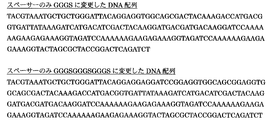
- the DNA sequence used for the construction of the EGFP-ATF6d expression vector in which the spacer portion is changed to GGGS or GGGSGGGSGGGS is shown.
- the result of having confirmed the importance of the spacer in an EGFP-ATF6d expression vector is shown.
- the result of having confirmed the importance of the PEST sequence in an EGFP-ATF6d expression vector is shown.
- the vector map of the Ire1 activation reporter vector which measures an Ire1 pathway by XBP splicing, and the PERK activation reporter expression vector which measures a PERK pathway by translation of ATF4 is shown.
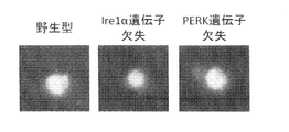
- the difference between endoplasmic reticulum stress response signals between organs is shown (fluorescence micrograph).
- the difference between endoplasmic reticulum stress response signals between organs is shown (fluorescence intensity measurement results). It shows that the endoplasmic reticulum stressor inhibits insulin secretion of pancreatic ⁇ cells.
- the ATF6 activity reporter system is superior in detection sensitivity for the activation of the ATF6 pathway compared to the conventional method (Western blot).
- the gene construction in the ERAI system and the mechanism of the ERAI system are shown.
- the gene construction in the UMAI system and the mechanism of the UMAI system are shown.
- the fluorescent protein consists of a non-fluorescent peptide domain 1 and a non-fluorescent peptide domain 2.
- the non-fluorescent peptide domain 1 and the non-fluorescent peptide domain 2 are derived from the same fluorescent protein, and have a relationship of two fragments generated by dividing the fluorescent protein within the molecule. Therefore, when non-fluorescent peptide domain 1 is the N-terminal fragment of the two fragments, non-fluorescent peptide domain 2 is the C-terminal fragment of the two fragments.
- the non-fluorescent peptide domain 1 is a C-terminal fragment of the two fragments
- the non-fluorescent peptide domain 2 is an N-terminal fragment of the two fragments.
- Each of the non-fluorescent peptide domain 1 and the non-fluorescent peptide domain 2 does not have fluorescence activity by itself, but exhibits fluorescence activity by associating in the cell.
- the ATF6 protein consists of an endoplasmic reticulum lumen domain (A in FIG. 2), a transmembrane domain (B in FIG. 2), and a cytoplasmic domain (C in FIG. 2).
- the endoplasmic reticulum stress that activates the pathway through the ATF6 protein cleaves the cytoplasmic domain part (C in FIG. 2) of the ATF6 protein, and the excised cytoplasmic domain part (C in FIG. 2) Is transferred to the nucleus (Nuc in FIG. 2) by a nuclear translocation signal inherent in, and serves as a transcription factor to regulate transcription of genes related to endoplasmic reticulum stress response.
- the present invention uses this mechanism to construct a system for evaluating endoplasmic reticulum stress that activates the pathway through ATF6. Specifically, it is evaluated by the following principle (see FIG. 3).
- Non-fluorescent peptide domain 1 (GFP-S in FIG. 3, hereinafter referred to as “non-fluorescent peptide domain 1”) derived from fluorescent protein expressed in cells and ATF6 protein domain (A, B, and The fusion protein of C ′) is placed on the endoplasmic reticulum membrane by the transmembrane domain in the ATF6 protein domain (B in FIG. 3).
- the endoplasmic reticulum lumen domain (A in FIG. 3) in the ATF6 protein domain is present on the endoplasmic reticulum lumen side, the non-fluorescent peptide domain 1 (GFP-S in FIG. 3), and the ATF6 protein domain
- the inner cytoplasmic domain (C ′ in FIG. 3) exists on the cytoplasm side.
- the non-fluorescent peptide domain 1 (GFP-S in FIG. 3) and the cytoplasmic domain in the ATF6 protein domain (C ′ in FIG. 3) were excised from the endoplasmic reticulum membrane, and the ATF6 protein domain It is translocated into the nucleus by a nuclear translocation signal in the middle or a nuclear translocation signal artificially inserted into the fusion protein.
- the non-fluorescent peptide domain 1 (GFP-S in FIG. 3) in the fusion protein transferred into the nucleus is the non-fluorescent peptide domain 2 derived from the fluorescent protein (GFP in FIG. 3) that is also present in the nucleus. -L) to restore the fluorescence activity of the original fluorescent protein.
- This recovery of fluorescence activity is confirmed by detecting fluorescence emitted by irradiating excitation light having a wavelength corresponding to the type of fluorescent protein.
- endoplasmic reticulum stress via ATF6 can be evaluated by detecting the fluorescence.
- polynucleotide of the present invention encodes an amino acid sequence having a non-fluorescent peptide domain 1 derived from a fluorescent protein comprising a non-fluorescent peptide domain 1 and a non-fluorescent peptide domain 2, and an ATF6 protein domain Polynucleotide.
- Non-fluorescent peptide domain 1 derived from the fluorescent protein of the present invention (hereinafter referred to as non-fluorescent peptide domain 1 of the present invention)
- the fluorescent protein is not particularly limited as long as it loses the fluorescent activity by splitting in the molecule and can recover the fluorescent activity by reassociation of the split fragments, but preferably GFP, GFP variant, or GFP GFP-like protein family not derived from GFP, more preferably GFP, or a GFP variant.
- GFP is a green fluorescent protein derived from Aequorea jellyfish, and its amino acid sequence is represented by SEQ ID NO: 4.
- one or more amino acids are substituted, deleted or deleted. It may be added.
- substitution with an amino acid having properties similar to the amino acid before substitution in terms of polarity, charge, solubility, hydrophilicity / hydrophobicity, polarity, etc. can do.
- glycine, alanine, valine, leucine, isoleucine, and proline are classified as nonpolar amino acids; serine, threonine, cysteine, methionine, asparagine, and glutamine are classified as polar amino acids; phenylalanine, tyrosine, and tryptophan are aromatic side chains.
- Lysine, arginine and histidine are classified as basic amino acids; aspartic acid and glutamic acid are classified as acidic amino acids. Accordingly, substitution can be made by selecting from the same group of amino acids.
- the GFP variant is one in which the fluorescence intensity, excitation wavelength, fluorescence wavelength, intracellular stability, etc. are modified by substituting amino acids in GFP, and examples thereof include EGFP, YFP, CFP, and RFP. .
- the amino acid sequence of EGFP is represented by SEQ ID NO: 1.
- a fluorescent protein belonging to the GFP-like protein family is a fluorescent protein having a structure similar to that of GFP (11 ⁇ -sheet units are helically organized to form a cylindrical structure enclosing a chromophore (white part)).
- CopGFP derived from crustacean
- DsRed derived from sea anemone, and the like.
- the non-fluorescent peptide domain 1 of the present invention is an N-terminal fragment or a C-terminal fragment generated by dividing a fluorescent protein in a molecule.
- the dividing site is not limited as long as each of the divided fragments (N-terminal side fragment and C-terminal side fragment) does not have fluorescence activity alone, but the fluorescence activity can be recovered by association of both.
- part can be suitably set with reference to well-known literature.
- An example of such a document is Document 2 (Nature biotechnology, 2003, vol 21, 539-545).
- the division site in the case of EGFP is preferably any of amino acids 195 to 226 in SEQ ID NO: 1, more preferably amino acids 205 to 222 in the amino acid sequence shown in SEQ ID NO: 1. Any one of the amino acids 212 to 218 in SEQ ID NO: 1.
- the C-terminal fragment is preferably the non-fluorescent peptide domain 1 of the present invention.
- ATF6 protein domain of the present invention The biological species from which the ATF6 protein is derived is preferably not only mouse (Mus musculus) but also mouse ATF6 protein and amino acid sequence, preferably 70% or more identity, more preferably Is an organism having a genomic region encoding an ATF6 protein having 80% or more identity, more preferably 85% or more identity, more preferably 90% or more identity, particularly preferably 95% or more identity Species are mentioned. Examples of such species include rodents represented by rats, mammals such as rabbits, monkeys, and humans (Homo sapiens). The most preferred species is human (Homo sapiens) or mouse (Mus musculus).
- the ATF6 protein consists of a cytoplasmic domain, an endoplasmic reticulum transmembrane domain, and an endoplasmic reticulum lumen domain.
- the 1st to 364th amino acid region in the amino acid sequence shown in SEQ ID NO: 2 is the cytoplasmic domain
- the 365th to 384th amino acid region in the amino acid sequence shown in SEQ ID NO: 2 is the endoplasmic reticulum. This is a transmembrane domain
- the amino acid region at positions 385 to 656 in the amino acid sequence shown in SEQ ID NO: 2 is the endoplasmic reticulum lumen domain.
- the 1st to 377th amino acid region in the amino acid sequence shown in SEQ ID NO: 3 is the cytoplasmic domain
- the 378th to 398th amino acid region in the amino acid sequence shown in SEQ ID NO: 3 is the endoplasmic reticulum. This is a transmembrane domain
- the amino acid region of the 399th to 670th amino acids in the amino acid sequence shown in SEQ ID NO: 3 is the endoplasmic reticulum lumen domain.
- the ATF6 protein domain of the present invention includes a nuclear translocation signal peptide region (nuclear translocation signal peptide and / or exogenous nuclear translocation signal peptide in ATF6 protein), a region involved in cleavage by endoplasmic reticulum stress, an endoplasmic reticulum transmembrane domain , And a portion that accepts endoplasmic reticulum stress in the endoplasmic reticulum lumen domain, and may be the full length of the ATF6 protein to this extent.
- the nuclear translocation signal in the ATF6 protein is shown by the amino acid sequence RKKKKE, and when the biological species is a mouse, it is the 311 to 316th amino acid region in the amino acid sequence shown in SEQ ID NO: 2, and the biological species is In the case of humans, this is the amino acid region of positions 324 to 329 in the amino acid sequence shown in SEQ ID NO: 3.
- a known signal peptide can be used as the exogenous nuclear translocation signal peptide.
- a specific example is DPKKRKVV.
- a known signal peptide repeat sequence may be used as the nuclear translocation signal peptide.
- the part of the endoplasmic reticulum lumen domain that receives endoplasmic reticulum stress is a region that is required to accept endoplasmic reticulum stress and cause cleavage of the cytoplasmic domain of ATF6.
- the region involved in cleavage by endoplasmic reticulum stress is the 365th to 411st amino acid region in the amino acid sequence shown in SEQ ID NO: 2 when the biological species is mouse, and SEQ ID NO: 3 when the biological species is human.
- SEQ ID NO: 2 when the biological species is mouse
- SEQ ID NO: 3 when the biological species is human.
- the ATF6 protein domain of the present invention includes a nuclear translocation signal peptide region (nuclear translocation signal peptide in ATF6 protein or exogenous nuclear translocation signal peptide), a region involved in cleavage by endoplasmic reticulum stress, an endoplasmic reticulum transmembrane domain, and While not limited as long as it includes a portion of the endoplasmic reticulum lumen domain that accepts endoplasmic reticulum stress, If the species is a mouse, Preferably, the amino acid region at positions 100 to 656 in the amino acid sequence of the ATF6 protein shown in SEQ ID NO: 2 or the amino acid region has 90% or more, preferably 95% or more, and more preferably 98% or more identity.
- a domain consisting of amino acid regions, More preferably, the amino acid region at positions 250 to 656 in the amino acid sequence of the ATF6 protein represented by SEQ ID NO: 2, or 90% or more, preferably 95% or more, more preferably 98% or more identity with the amino acid region.
- a domain consisting of amino acid regions Particularly preferably, an exogenous nuclear translocation signal peptide region and the 360-656th amino acid region in the amino acid sequence of the ATF6 protein shown in SEQ ID NO: 2, or 90% or more, preferably 95% or more, more than the amino acid region
- it is a domain consisting of an amino acid region having 98% or more identity
- the amino acid region at positions 100 to 670 in the amino acid sequence of the ATF6 protein shown in SEQ ID NO: 3 or the amino acid region has 90% or more, preferably 95% or more, more preferably 98% or more identity.
- a domain consisting of amino acid regions More preferably, the amino acid region at positions 250 to 670 in the amino acid sequence of the ATF6 protein represented by SEQ ID NO: 3, or the amino acid region has 90% or more, preferably 95% or more, more preferably 98% or more identity.
- a domain consisting of amino acid regions Particularly preferably, the foreign nuclear translocation signal peptide region and the amino acid region of 373 to 670 in the amino acid sequence of the ATF6 protein shown in SEQ ID NO: 3, or 90% or more, preferably 95% or more, more than the amino acid region
- a domain consisting of an amino acid region having 98% or more identity is preferred.
- the ATF6 protein domain may be one in which one or more amino acids in the above-exemplified domains are substituted, deleted or added. Such mutations have functions in the nuclear translocation signal peptide region, the part that accepts endoplasmic reticulum stress in the endoplasmic reticulum lumen domain, the endoplasmic reticulum transmembrane domain, and the part that is cleaved by endoplasmic reticulum stress in the cytoplasmic domain. It can design suitably so that it may not be impaired.
- substitution with an amino acid having properties similar to the amino acid before substitution in terms of polarity, charge, solubility, hydrophilicity / hydrophobicity, polarity, etc. can do.
- glycine, alanine, valine, leucine, isoleucine, and proline are classified as nonpolar amino acids; serine, threonine, cysteine, methionine, asparagine, and glutamine are classified as polar amino acids; phenylalanine, tyrosine, and tryptophan are aromatic side chains.
- Lysine, arginine and histidine are classified as basic amino acids; aspartic acid and glutamic acid are classified as acidic amino acids. Accordingly, substitution can be made by selecting from the same group of amino acids.
- the arrangement of two domains in the polynucleotide encoding non-fluorescent peptide domain 1 and ATF6 protein domain is not particularly limited. However, it is preferable that the region encoding the non-fluorescent peptide domain 1 is disposed on the 5 ′ end side and the region encoding the ATF6 protein domain is disposed on the 3 ′ end side.
- non-fluorescent peptide domain 1 derived from the fluorescent protein and the ATF6 protein domain are preferably arranged via a spacer.
- the spacer is a natural protein-derived or artificial amino acid sequence.
- the amino acid sequence of the spacer is not particularly limited, but it is preferably an amino acid sequence that does not form a secondary structure such as an ⁇ -helix structure or a ⁇ -sheet structure and can be bent freely.
- Examples of the amino acid sequence of the spacer include 4 residues of GGGS or a repeating sequence of the sequence.
- the number of amino acid residues in the spacer is preferably 3 amino acid residues or more, more preferably 4 to 50 amino acid residues, still more preferably 6 to 20 amino acid residues, and particularly preferably 7 to 15 amino acid residues.
- the number of amino acid residues of the spacer is too small, it is considered that the association of non-fluorescent peptide domains 1 and 2 derived from the fluorescent protein is hindered by the ATF6 protein domain.
- the polynucleotide of the present invention may include a polynucleotide encoding a protein tag and a polynucleotide encoding another peptide as other sequences.
- protein tag a FLAG tag, a His tag, a GST tag, or the like can be used. These protein tags may be used as repeating sequences of tag sequences.
- PEST sequences By inserting the PEST sequence, degradation of the fusion protein is promoted.
- a known PEST sequence can be used, and is not particularly limited as long as the function can be exhibited. Examples thereof include an amino acid sequence consisting of SHGFPPEVEEQDDGTLPMSCAQESGMDRHPAACASARINV.
- the arrangement of the PEST sequence is not particularly limited as long as it can exert its function, but is preferably arranged between the non-fluorescent peptide domain 1 and the ATF6 protein domain.
- the PEST sequence is not essential, but in the method for screening an endoplasmic reticulum stressor, endoplasmic reticulum stress inhibitor, or antidiabetic drug candidate substance using the polynucleotide of the present invention by inserting the PEST sequence, Fluorescence can be reduced, and fluorescence can be detected with higher sensitivity.
- the polynucleotide of the present invention can be synthesized by an artificial gene synthesis technique, or a nucleic acid amplification technique (such as PCR) from a commercially available polynucleotide or cDNA library. .
- the polynucleotide encoding each domain can be ligated by a known gene ligation technique with a known restriction enzyme sequence added at both ends.
- polynucleotide encoding the fluorescent protein various expression vectors having the polynucleotide are commercially available (for example, Clontech), and the sequence information of the polynucleotide is also disclosed.
- the polynucleotide encoding the ATF6 protein domain is designed, for example, by designing a primer at an appropriate site on the basis of information on the ATF6 gene sequence published on a gene database such as the NCBI database. It can be amplified and obtained by the gene amplification technique used.
- sequences that can be included in the polynucleotide according to the present invention for example, a polynucleotide sequence encoding a protein tag, a polynucleotide sequence encoding a nuclear translocation signal, or a polynucleotide sequence encoding a PEST sequence are known. Based on the known sequence information, for example, it can be obtained by an artificial gene synthesis technique.
- the recombinant vector of the present invention is a recombinant vector containing the polynucleotide of the present invention, and can be obtained by linking the polynucleotide of the present invention to an appropriate vector.
- the vector is not particularly limited as long as it can contain the polynucleotide of the present invention, but is preferably a eukaryotic cell, more preferably an animal cell, still more preferably a mammalian cell, particularly preferably a rodent (particularly Examples thereof include those capable of expressing the fusion protein encoded by the polynucleotide of the present invention in mouse or human cells.
- E. coli vectors such as pBR322, pUC19, pKK233-2, and pET21a, yeast vectors Yip5, Yrp17, and Yep24
- animal cell vectors such as pcDNA and pBAC can be exemplified.
- the recombinant vector contains a marker gene in order to enable selection of transformed cells.
- the marker gene include a gene that complements the host's auxotrophy, a drug resistance gene, and the like.
- the recombinant vector preferably contains a promoter and other control sequences (for example, an enhancer sequence, terminator sequence, polyadenylation sequence, etc.) for enabling expression of the gene in the host.
- specific examples of the promoter include promoters such as SV40, CMV, ie1, T7, lac, trp, and tac.
- Transformant of the present invention A transformant containing the recombinant vector can be obtained by transforming a host using the above recombinant vector.
- the host is not particularly limited as long as it can be transformed with the recombinant vector of the present invention.
- bacteria such as E. coli, yeast, filamentous fungi, animal cells, and the like.
- eukaryotic cells are preferable, animal cells are more preferable, and rodent (especially mouse) or human cells are more preferable.
- the host is an animal cell, cells derived from various organs such as pancreatic cells, kidney cells, hepatocytes, adipocytes, or skeletal cells can be used as the host.
- pancreatic cells are preferable among these, and pancreatic ⁇ cells are more preferable among pancreatic cells.
- Transformation can be performed according to a known method depending on the type of host.
- Known methods include, for example, calcium chloride method, electroporation method, lipofection method, DEAE dextran method and the like.
- the transformant of the present invention may be selected using, as an index, a drug resistance marker possessed by the vector.
- the transformant of the present invention comprises a non-fluorescent peptide domain 2 derived from a fluorescent protein comprising non-fluorescent peptide domain 1 and non-fluorescent peptide domain 2 (hereinafter referred to as “non-fluorescent peptide domain 2”), and nucleus A polynucleotide encoding an amino acid sequence having a transition signal peptide domain is preferably contained in a state in which a protein comprising the amino acid sequence can be expressed.
- This transformant (hereinafter referred to as “screening transformant”) can be used in the screening method described below.
- transformant for screening (hereinafter referred to as “transformant for screening an antidiabetic drug candidate substance”) whose host is a pancreatic ⁇ cell is a screening method described later, particularly a screening method for an antidiabetic drug candidate substance. Can be suitably used.
- Non-fluorescent peptide domain 2 is an N-terminal fragment or a C-terminal fragment generated by dividing a fluorescent protein in a molecule.
- Non-fluorescent peptide domain 2 is derived from the same fluorescent protein as non-fluorescent peptide domain 1 described above, and has a relationship of two fragments generated by dividing the fluorescent protein in the molecule. Therefore, when non-fluorescent peptide domain 1 is an N-terminal fragment, non-fluorescent peptide domain 2 is a C-terminal fragment.
- the non-fluorescent peptide domains 1 and 2 are not particularly limited as to which is the N-terminal fragment or the C-terminal fragment, but the non-fluorescent peptide domain 1 is the C-terminal fragment, and the non-fluorescent peptide domain 2 is It is preferably an N-terminal fragment.
- a known signal peptide can be used as the nuclear translocation signal peptide domain. Specific examples include Asp-Pro-Lys-Lys-Lys-Arg-Lys-Val. As the nuclear translocation signal peptide, a known signal peptide repeat sequence may be used.
- Contains in an expressible state is not particularly limited as long as a protein comprising an amino acid sequence having a non-fluorescent peptide domain 2 and a nuclear translocation signal peptide domain can be expressed in a cell.
- a promoter sequence is preferably present on the 5 ′ end side of the polynucleotide encoding the amino acid sequence having the peptide domain 2 and the nuclear translocation signal peptide domain.
- a known sequence can be used as the promoter sequence.
- Examples of such a promoter sequence include a CMV promoter sequence and an SV40 promoter.
- the endoplasmic reticulum stressor, endoplasmic reticulum stress inhibitor, or antidiabetic drug candidate substance can be screened using the transformant.
- a screening method for the endoplasmic reticulum stressor comprises: (a) a candidate endoplasmic reticulum stressor, and “the transformant for screening” described in “(5) Transformant of the Present Invention” (B) measuring the fluorescence intensity of the transformant contacted with the endoplasmic reticulum stressor candidate substance, and calculating the fluorescence intensity with the fluorescence intensity of the control transformant not contacting the endoplasmic reticulum stressor candidate substance.
- the endoplasmic reticulum stressor candidate substance is used as an endoplasmic reticulum stressor. Including a step of selecting.
- the type of endoplasmic reticulum stressor candidate substance is not particularly limited. Examples include proteins, peptides, non-peptidic compounds (nucleotides, amines, carbohydrates, lipids, etc.), organic low molecular compounds, inorganic low molecular compounds, fermentation products, cell extracts, plant extracts, animal tissue extracts, etc. It is done.
- the contact of the candidate endoplasmic reticulum stressor to the cell does not kill the cell, and is derived from the fusion protein of the non-fluorescent peptide domain 1 and ATF6 protein domain derived from the fluorescent protein, and the fluorescent protein from the introduced recombinant vector What is necessary is just to carry out on the conditions (temperature, pH, a culture medium component, etc.) which can express the protein containing the non-fluorescent peptide domain 2 to do.
- concentration of the endoplasmic reticulum stressor candidate substance to be brought into contact with cells varies depending on the kind of the substance, but can be, for example, about 0.001 to 100 ⁇ g / ml.
- Fluorescence can be detected by irradiating excitation light having a wavelength suitable for the fluorescent protein to generate fluorescence.
- the method for measuring the fluorescence intensity is not particularly limited as long as it can compare the fluorescence intensity of the transformant contacted with the endoplasmic reticulum stressor and the fluorescence intensity of the control transformant not contacted with the endoplasmic reticulum stressor.
- the fluorescence intensity can be measured by capturing an image of a transformant that has produced fluorescence on a computer and measuring the color intensity according to the fluorescence in the image.
- Endoplasmic reticulum stressor that activates the ATF6 pathway and does not activate the IRE1 ⁇ and PERK pathways; It is an endoplasmic reticulum stressor that activates the ATF6 pathway and activates the IRE1 ⁇ pathway and / or the PERK pathway, or an endoplasmic reticulum stressor that does not activate the ATF6 pathway and activates the IRE1 ⁇ pathway and / or the PERK pathway. You can choose.
- An endoplasmic reticulum stressor that activates the ATF6 pathway and does not activate the IRE1 ⁇ and PERK pathways (P) an endoplasmic reticulum stressor that activates the ATF6 pathway and the IRE1 ⁇ pathway and does not activate the PERK pathway; (Q) an endoplasmic reticulum stressor that activates the ATF6 pathway and PERK pathway and does not activate the IRE1 ⁇ pathway, or (r) an endoplasmic reticulum stressor that activates the ATF6 pathway, IRE1 ⁇ pathway, and PERK pathway (s) activates the ATF6 pathway Endoplasmic reticulum stressor that does not become active and activates the IRE1 ⁇ pathway and the PERK pathway, (T) an endoplasmic reticulum stressor that does not activate the ATF6 pathway and IRE1 ⁇ pathway and activates the PERK pathway, or (u) an endoplasmic reticulum stressor that does not activate the ATF6 pathway and PERK pathway and
- the known screening method is a known method by which the endoplasmic reticulum stressor candidate substance can select whether to activate the IRE1 ⁇ pathway and / or the PERK pathway.
- the method described in JP-A-2005-204516 It is. Therefore, the endoplasmic reticulum stressor that activates the ATF6 pathway by combining the present screening method capable of selecting an endoplasmic reticulum stressor that activates the ATF6 pathway from candidate endoplasmic reticulum stressors and a known screening method is described above. It becomes possible to select one of the endoplasmic reticulum stressors described in (o) to (u). Furthermore, by examining in combination with known screening methods, it is possible to select an endoplasmic reticulum stressor that does not activate the ATF6 pathway.
- V an expression vector comprising a fusion gene of the XBP1 gene and a gene encoding the first reporter protein, and the fusion of the XBP1 protein and the first reporter protein only when the XBP1 gene is spliced
- An expression vector in which the gene to be placed is located downstream from the true translation start point of the ATF4 gene; Transgenic cells co-introduced with Is used.
- the candidate substance is selected as an endoplasmic reticulum stressor that activates the IRE1 ⁇ pathway and does not activate the PERK pathway;
- the candidate substance is selected as an endoplasmic reticulum stressor that activates the PERK pathway and does not activate the IRE1 ⁇ pathway.
- the candidate substance is selected by a method comprising a step of selecting the candidate substance as an endoplasmic reticulum stressor that activates the IRE1 ⁇ pathway and the PERK pathway. Is an endoplasmic reticulum stressor that activates the IRE1 ⁇ pathway and / or the PERK pathway.
- the expression vector (v) is constructed so that the ERAI system operates as shown in FIG.
- the expression vector (v) includes a fusion gene of the XBP1 gene and a gene encoding the first reporter protein, and only when the XBP1 gene is spliced, a fusion protein of the XBP1 protein and the first reporter protein Is arranged to express.
- the XBP1 gene is a kind of stimulation response gene, and a 26 nucleotide intron is spliced by IRE1 under endoplasmic reticulum stress.
- a 26 nucleotide intron is spliced by IRE1 under endoplasmic reticulum stress.
- the cDNA sequences of XBP1 of human, bovine, mouse, Xenopus, and zebrafish are known and are registered on the EST database as BE170119, AV604667, BF143019, AF358133, and AF399918, respectively.
- a human XBP1 cDNA of NCBI registration number AB076383 hereinafter referred to as “sequence 1” will be described as an example.
- sequence 2 the 9th to 794th positions of the sequence 1 become a coding region and is translated into an XBP1 protein (short form) consisting of 261 amino acid residues (hereinafter referred to as “sequence 2”).
- sequence 2 the intron of cDNA is the base sequence of positions 502 to 527 of Sequence 1.
- a frameshift occurs due to splicing of the XBP1 gene by IRE1, and the 9th to 501st positions and the 528th to 1165th positions of Sequence 1 become coding regions and are translated into an active XBP1 protein consisting of 376 amino acid residues.
- the gene encoding the first reporter protein is fused so as to match the reading frame at an appropriate position downstream of the active XBP1 protein coding region expressed by splicing. That is, the expression vector (v) is constructed so that the fusion protein of the active XBP1 protein and the first reporter protein is expressed only when spliced by IRE1.
- the first reporter protein is not particularly limited as long as its expression can be confirmed.
- a protein having fluorescence activity is preferable because it can be easily confirmed in vivo.
- the protein having fluorescence activity include luciferase, green fluorescent protein (GFP), and a modified form thereof.
- luciferase include firefly luciferase and Renilla luciferase.
- the green fluorescent protein and the modified form thereof include Aequorea green fluorescent protein, EGFP (enhanced green fluoroprotein), YFP (yellow fluoroprotein), BFP (blue fluoroprotein), RFP (red fluoroprotein) and the like. These are all commercially available and can be easily obtained.
- the expression vector (w) includes a fusion gene of an ATF4 gene and a gene encoding a second reporter protein, and the gene encoding the second reporter protein is located downstream from the true translation start point of the ATF4 gene. Has been placed.
- PERK is activated, eIF-2 ⁇ is phosphorylated and the function of eIF-2 ⁇ is reduced, and the true translation start that is the most downstream of the translation start points in the mRNA of ATF4 Translation from point is promoted.
- ATF4 such as human, mouse, and aphid are known, and are registered in the EST database as HSU03712, NM_009716, and ACU40851, respectively.
- the human ATF4 cDNA of NCBI registration number BC011994 hereinafter referred to as “sequence 3” will be described as an example.
- ATF4 cDNA is transcribed into mRNA
- ATF4 is translated from a plurality of false upstream translation start points under normal conditions, that is, when eIF-2 ⁇ is functioning normally. That is, positions 4 to 9, position 67 to 78, and position 166 to 345 of sequence 3 are translated.
- positions 263 to 1318 of sequence 3 which is the true translation start point of ATF4 mRNA, are translated.
- the gene encoding the second reporter protein is located downstream from the true translation start point of the ATF4 gene. For example, taking the cDNA of human ATF4 of NCBI registration number HSU03712 as an example, it is more downstream than position 263 of sequence 3, more specifically, from a true translation initiation codon (positions 263 to 265 of SEQ ID NO: 5). Is also arranged downstream. Therefore, the second reporter protein is expressed only when PERK is activated and translated from the true translation start point.
- the second reporter protein is not particularly limited as long as the expression can be confirmed in the same manner as the first reporter protein.
- a fluorescent protein is preferable because it can be easily confirmed in vivo.
- the protein should be different from the first reporter protein so that it can be easily distinguished from the expression of the first reporter protein.
- the combination of the first reporter protein and the second reporter protein is preferably a combination of firefly luciferase and Renilla luciferase, EGFP and RFP, for example.
- transgenic cell containing the expression vectors (v) and (w), for example, culturing the cell in an appropriate medium usually used by those skilled in the art under appropriate conditions, and then contacting the agent to be screened
- the contact means include adding a drug to the medium and injecting the drug into cells. After contacting the cell and the drug for a certain period, the expression of the first reporter protein and the second reporter protein is measured. Measurement of the expression of each reporter protein usually measures the activity of the expressed protein. For example, when the reporter protein is a fluorescent protein, the fluorescence intensity of the cell is measured.
- the method of combining the above-described known screening method and the present screening method is not particularly limited, and examples thereof include the following combination methods.
- transformant for screening As a transformant to be used, instead of the “transformant for screening” described in the above “(5) transformant of the present invention”, the transformant is further transformed with the above expression vectors (v) and (w).
- the contained transformant hereinafter referred to as “transformant for three-path detection”.
- the fluorescent protein used in the screening method, the fluorescent intensity or activity of the first reporter protein, and the fluorescent intensity or activity of the second reporter protein can be distinguished from each other. And the type of the first or second reporter protein.
- the endoplasmic reticulum stressor candidate substance is brought into contact with the transformant for three-path detection, and the endoplasmic reticulum stressor candidate substance is compared with the control cell by comparing the fluorescence intensity of the fluorescent protein and the fluorescence intensity or activity of the reporter protein with the above-mentioned (o).
- One of the endoplasmic reticulum stressors described in (u) to (u) can be selected.
- This screening method can examine what substances cause endoplasmic reticulum stress, elucidate the mechanism of endoplasmic reticulum stress, and further research on the treatment of diseases such as diabetes and neurodegenerative diseases caused by endoplasmic reticulum stress Useful for.
- a step of comparing the fluorescence intensity of the control transformant contacted with the endoplasmic reticulum stressor; and (f) the fluorescence intensity of the transformant contacted with the endoplasmic reticulum stressor and the test substance is the fluorescence intensity of the control transformant. If lower than that, the method includes a step of selecting the test substance as an endoplasmic reticulum stress inhibitor.
- the endoplasmic reticulum stressor is not particularly limited as long as it is a substance known to cause endoplasmic reticulum stress. Examples of such substances include tunicamycin, thapsigargin, and DTT (dithiothreitol). Further, an endoplasmic reticulum stressor selected by the above-mentioned endoplasmic reticulum stressor screening method can also be used.
- test substance is not particularly limited. Examples include proteins, peptides, non-peptidic compounds (nucleotides, amines, carbohydrates, lipids, etc.), organic low molecular compounds, inorganic low molecular compounds, fermentation products, cell extracts, plant extracts, animal tissue extracts, etc. It is done.
- a fusion protein of non-fluorescent peptide domain 1 and ATF6 protein domain derived from fluorescent protein, and non-fluorescent peptide domain derived from fluorescent protein 2 may be performed under conditions (temperature, pH, medium components, etc.) that allow the protein containing 2 to be expressed.
- concentration of the endoplasmic reticulum stressor candidate substance to be brought into contact with cells varies depending on the kind of the substance, but can be, for example, about 0.001 to 100 ⁇ g / ml.
- screening can be performed using the transformant of the present invention derived from the organ.
- screening can be performed using the transformant of the present invention derived from the liver.
- This screening method can screen for a substance that suppresses endoplasmic reticulum stress, and is useful for research on diseases such as diabetes and neurodegenerative diseases caused by endoplasmic reticulum stress.
- the method includes a step of selecting the test substance as an antidiabetic drug candidate substance when it is lower than the fluorescence intensity of the body.
- the endoplasmic reticulum stressor is not particularly limited as long as it is a substance known to cause endoplasmic reticulum stress. Examples of such substances include tunicamycin, thapsigargin, and DTT. Further, an endoplasmic reticulum stressor selected by the above-mentioned endoplasmic reticulum stressor screening method can also be used.
- test substance is not particularly limited. Examples include proteins, peptides, non-peptidic compounds (nucleotides, amines, carbohydrates, lipids, etc.), organic low molecular compounds, inorganic low molecular compounds, fermentation products, cell extracts, plant extracts, animal tissue extracts, etc. It is done.
- a fusion protein of non-fluorescent peptide domain 1 and ATF6 protein domain derived from fluorescent protein, and non-fluorescent peptide domain derived from fluorescent protein 2 may be performed under conditions (temperature, pH, medium components, etc.) that allow the protein containing 2 to be expressed.
- concentration of the endoplasmic reticulum stressor candidate substance to be brought into contact with cells varies depending on the kind of the substance, but can be, for example, about 0.001 to 100 ⁇ g / ml.
- the candidate antidiabetic drug selected by this screening method can be obtained as a more practical antidiabetic drug by further subjecting it to a drug efficacy test and safety test using a disease model animal.
- the EGFPS-ATF6d expression vector is a green fluorescent protein cDNA fragment (a DNA fragment encoding the amino acid region from amino acid numbers 216 to 231 in SEQ ID NO: 1: 1 in FIG. 4), Spacer (DNA fragment encoding amino acid sequence consisting of GGGSGGGS: part 2 in FIG. 4), 3XFLAG tag (DNA fragment encoding amino acid sequence consisting of DYKDHDGDYKDHDIDYKDDDDK: part 3 in FIG.
- nuclear translocation signal (DPKKKRKV 3) DNA fragment encoding amino acid sequence consisting of 3 repeats: 4) part in FIG. 4), PEST sequence (DNA fragment encoding amino acid sequence consisting of SHGFPPEVEEQDDGTLPMSCAQESGMDRHPAACASARINV: 5) part in FIG. 3), mouse ATF6 ⁇ A partial cDNA fragment (DNA fragment encoding the amino acid region of amino acid numbers 361 to 656 in SEQ ID NO: 2: 6 in FIG. 4)) It was produced and inserted into pEGFP-Puro. As shown in FIG. 5, the EGFPL expression vector has a nuclear translocation signal (DNA fragment encoding an amino acid sequence consisting of DPKKKRKV repeated three times: 7 in FIG.
- pEGFP-N1 a green fluorescent protein cDNA fragment (sequence A DNA fragment encoding the amino acid region of amino acid sequence Nos. 1 to 215 in No. 1 (8) part) in FIG. 5 was inserted into pEGFP-N1.
- pEGFP-Puro and pEGFP-N1 can highly express a gene inserted downstream of the cytomegalovirus promoter and enhancer in mammalian cells.
- EGFPS-ATF6d expression vector The actual EGFPS-ATF6d expression vector was prepared in three steps. Step 1 Using artificial gene synthesis technology, green fluorescent protein cDNA fragment, spacer, 3XFLAG tag, and double-stranded DNA encoding nuclear translocation signal (1), 2), 3), and 4) part) in FIG. It was synthesized and inserted into a pEGFP-Puro expression vector to prepare an EGFPS expression vector.
- Step 2 Partial cDNA fragment of mouse ATF6 ⁇ generated by PCR of kpn.mATF6d.SP1 (sequence: 5'-CTAGGGTACCCCAAAGCGAAGAGCTGTCTG -3 ') and not.mATF6d.AP1 (sequence: 5'-TTTTTTCCTTGCGGCCGCCTACTGCAACGACTCAGGGA-3') from mouse cDNA (Part 6 in FIG. 4) was inserted into an EGFPS expression vector to prepare an EGFPS-ATF6d-PEST ( ⁇ ) expression vector.
- Step 3 Mouse ODC PEST sequence (Fig. 5) 5) part 4) was inserted into an EGFPS-ATF6d-PEST (-) expression vector to prepare an EGFPS-ATF6d expression vector.
- EGFPS expression vector, EGFPS-ATF6d-PEST (-) expression vector, and EGFPS-ATF6d expression vector also have a puromycin drug resistance gene derived from pEGFP-Puro.
- EGFPL expression vector was synthesized by the artificial gene synthesis technique and double-stranded DNA (7 and 8) in Fig. 5 encoding the nuclear translocation signal and green fluorescent protein. An EGFPL expression vector was prepared by inserting into an expression vector.
- the EGFPL expression vector also has a neomycin drug resistance gene.
- Cell culture HEK293 cells (ATCC number: CRL-1573), MIN6 cells (pancreatic ⁇ cells reported in Miyazaki J et al., Endocrinology 127: 126-132 (1990)), HepG2 cells (ATCC number: HB-8065) , 3T3-L1 cells (ATCC number: CL-173), L6 cells (ATCC number: CRL-1458), ATF6 activity reporter system introduced cells (described later), add 10% fetal bovine serum to DMEM unless otherwise specified was cultured in a 37 ° C., 5% CO 2 environment.
- HEK293 cells were transfected with an EGFPS-ATF6d expression vector and an EGFPL expression vector with an efficiency of 90% or more by the polyethyleneimine method.
- EGFPS-ATF6d expression vector and EGFPL expression vector were added to 80%, 60%, 90%, MIN6 cells, HepG2 cells, 3T3-L1 cells, and L6 cells, respectively.
- the gene was introduced with an efficiency of 70% or more.
- EGFPS-ATF6d expression vector and EGFPL expression vector were individually expressed or coexpressed in HEK293 cells.
- the fluorescence signal of the cells was observed with an inverted fluorescence microscope DMI6000B (Leica), the fluorescence signal was acquired with a CCD camera Rolla-XR (Qiaging), and analyzed with image analysis software Image-Pro Plus (Media Cybernetics).
- HEK293 cells co-expressed with EGFPS-ATF6d expression vector and EGFPL expression vector were mixed with 100 uM each of DNA replication inhibitor etoposide, endoplasmic reticulum stress inducer thapsigargin, and endoplasmic reticulum stress inducer DTT. , 0.2 uM, 1 mM, and fluorescence was observed. Both conditions were confirmed by Hoechst33258 staining to be stress causing cell death after 36 hours.
- the ATF6 activity reporter system can detect the activation of ATF6 specifically in the endoplasmic reticulum stress.
- the ATF6 activity reporter system can detect the activation of ATF6 without being affected by the Ire1 pathway or PERK pathway.
- Tunicamycin an ER stress inducer, was observed 6 hours after stimulation at 0.2 ug / ml.
- Endoplasmic reticulum stress response is essential for maintaining homeostasis of endoplasmic reticulum and induces endoplasmic reticulum stress response not only in pathological conditions but also in physiological conditions. ing. Even in cultured cells under normal culture conditions, an endoplasmic reticulum stress response is induced although it is weak. It is expected that cells expressing the ATF6 activity reporter system will accumulate a high background signal due to the accumulation of the reporter green fluorescent protein.
- EGFPS-ATF6d expression vector that expresses mouse ODC PEST sequence that promotes degradation of green fluorescent protein or EGFPS-ATF6d-PEST (-) expression vector that does not have mouse ODC PEST sequence and EGFPL expression vector are co-expressed
- the HEK293 cells were observed before stimulation and tunicamycin, an endoplasmic reticulum stress inducer, at 0.2 ug / ml for 6 hours after stimulation.
- the green fluorescence was significantly attenuated under normal culture conditions by the PEST sequence.
- the induction of green fluorescence was slightly reduced by the PEST sequence, but the increase rate of the green fluorescence brightness with and without stimulation was improved by the PEST sequence.
- the endoplasmic reticulum stress response is controlled by the Ire1 pathway, the PERK pathway, and the ATF6 pathway. In addition, each is independently associated with organ-specific metabolic control. The detection of three endoplasmic reticulum stress response pathways can be expected to be important for drug discovery targeting an organ-specific endoplasmic reticulum stress response with few side effects. Therefore, as shown in FIG. 12, an Ire1 activation reporter expression vector that measures the Ire1 pathway by splicing XBP1 reported in Nat Med. (2004) 10 98-102 and J Cell Biol.
- activation of the IRE1 pathway for tunicamycin occurs relatively strongly in the hepatocyte cell line HepG2 cell, pancreatic ⁇ cell line MIN6 cell, and skeletal muscle cell line L6 cell, and the adipocyte cell line 3T3-L1. It was shown to be relatively weak in cells.
- the activation of the ATF6 pathway for tunicamycin is in the order of hepatocyte cell line HepG2 cell, pancreatic ⁇ cell line MIN6 cell, skeletal muscle cell line L6 cell, and adipocyte cell line 3T3-L1 there were.
- the degree of activation of the PERK pathway for tunicamycin was determined in the order of hepatocyte cell line HepG2 cells, (pancreatic ⁇ cell line MIN6 cell and skeletal muscle cell line L6 cell), and adipocyte cell line 3T3-L1 cells in order from the most intense cells. It was in order.
- pancreatic ⁇ cells MIN6 cells A culture solution in which pancreatic ⁇ cells MIN6 cells are cultured (DMEM containing 10% fetal bovine serum) is added to a control solution (DMSO), a thapsigargin solution (thapsigargin / DMSO solution), or a DTT solution ( DTT / DMSO solution) was added, and after 30 minutes, the culture solution was replaced with a normal culture solution (10% fetal bovine serum-containing DMEM) and cultured for 36 hours.
- the thapsigargin solution was added so that the thapsigargin concentration in the culture solution was 0.2 uM
- the DTT solution was added so that the DTT concentration in the culture solution was 1 M.
- the culture solution was replaced with a KREBS buffer containing 3 mM or 20 mM glucose and cultured for 1 hour, and the concentration of insulin secreted in the culture solution was measured by ELISA.
- the results are shown in FIG.
- the detection sensitivity of the ATF6 activity reporter system was measured as follows. Stimulate adipose cell line 3T3-L1 cells obtained according to “4. Acquisition of ATF6 activity reporter system introduced cells” with known differentiation-inducing stimuli (exposure to insulin, dexamethasone, and 3-isobutyl-1-methylxanthine) Then, 14 days after the stimulation, a test compound (any of compounds 1 to 10) is added as an endoplasmic reticulum stressor candidate substance, and a stimulating agent (palmitic acid) such as endoplasmic reticulum stress is brought to a final concentration of 400 ⁇ M after 6 hours. In addition, after 16 hours, cell images were obtained with an inverted fluorescence microscope DMI6000B (Leica), and the fluorescence intensity emitted by the cells was quantified with image analysis software Image-Pro Plus (Media Cybernetics).
- the conventional method was measured as follows. Adipocyte cell line 3T3-L1 cells were exposed to known differentiation-inducing stimuli (exposure to insulin, dexamethasone, and 3-isobutyl-1-methylxanthine), 14 days later, a test compound (any of compounds 1 to 10) was added, and 6 After a time, stimulants such as endoplasmic reticulum stress were added, and the cells were collected after another 16 hours. A cell extract was prepared from the cells, and the expression level of ATF6 ⁇ protein was detected by Western blotting using Anti-ATF6 alfa antibody monoclonal (BioAcademia 73-505). The detected signal intensity was quantified with Image-Pro Plus (Media Cybernetics).
- the results are shown in FIG.
- the fluorescence intensity or signal intensity shown on the vertical axis is expressed as “1” when no test compound is added (control). That is, if the numerical value on the vertical axis is 1 or more, it indicates that the ATF6 pathway is activated by the test compound.
- the test compound numbers (1 to 10) are shown on the horizontal axis.
Landscapes
- Health & Medical Sciences (AREA)
- Life Sciences & Earth Sciences (AREA)
- Chemical & Material Sciences (AREA)
- Engineering & Computer Science (AREA)
- Immunology (AREA)
- Biomedical Technology (AREA)
- Organic Chemistry (AREA)
- Molecular Biology (AREA)
- General Health & Medical Sciences (AREA)
- Biochemistry (AREA)
- Bioinformatics & Cheminformatics (AREA)
- Hematology (AREA)
- Urology & Nephrology (AREA)
- Toxicology (AREA)
- Medicinal Chemistry (AREA)
- Zoology (AREA)
- Biotechnology (AREA)
- Analytical Chemistry (AREA)
- Microbiology (AREA)
- Physics & Mathematics (AREA)
- Genetics & Genomics (AREA)
- Wood Science & Technology (AREA)
- Proteomics, Peptides & Aminoacids (AREA)
- Tropical Medicine & Parasitology (AREA)
- Food Science & Technology (AREA)
- General Physics & Mathematics (AREA)
- Pathology (AREA)
- Cell Biology (AREA)
- Biophysics (AREA)
- General Engineering & Computer Science (AREA)
- Gastroenterology & Hepatology (AREA)
- Micro-Organisms Or Cultivation Processes Thereof (AREA)
- Peptides Or Proteins (AREA)
- Measuring Or Testing Involving Enzymes Or Micro-Organisms (AREA)
Abstract
Priority Applications (2)
| Application Number | Priority Date | Filing Date | Title |
|---|---|---|---|
| US13/983,864 US9085791B2 (en) | 2011-02-07 | 2012-02-06 | Method for screening substance relating to endoplasmic reticulum stress participating in onset of diabetes |
| JP2012556883A JPWO2012108394A1 (ja) | 2011-02-07 | 2012-02-06 | 糖尿病発症に係る小胞体ストレスに関与する物質のスクリーニング方法 |
Applications Claiming Priority (2)
| Application Number | Priority Date | Filing Date | Title |
|---|---|---|---|
| JP2011023697 | 2011-02-07 | ||
| JP2011-023697 | 2011-02-07 |
Publications (1)
| Publication Number | Publication Date |
|---|---|
| WO2012108394A1 true WO2012108394A1 (fr) | 2012-08-16 |
Family
ID=46638609
Family Applications (1)
| Application Number | Title | Priority Date | Filing Date |
|---|---|---|---|
| PCT/JP2012/052650 Ceased WO2012108394A1 (fr) | 2011-02-07 | 2012-02-06 | Procédé pour cribler une substance associée au stress du réticulum endoplasmique participant à l'apparition du diabète |
Country Status (3)
| Country | Link |
|---|---|
| US (1) | US9085791B2 (fr) |
| JP (1) | JPWO2012108394A1 (fr) |
| WO (1) | WO2012108394A1 (fr) |
Cited By (1)
| Publication number | Priority date | Publication date | Assignee | Title |
|---|---|---|---|---|
| US11559518B2 (en) | 2017-12-28 | 2023-01-24 | Er Stress Research Institute, Inc. | Endoplasmic reticulum stress regulator comprising benzothiazoimidazolyl compound |
Families Citing this family (3)
| Publication number | Priority date | Publication date | Assignee | Title |
|---|---|---|---|---|
| WO2017117430A1 (fr) | 2015-12-29 | 2017-07-06 | The Scripps Research Institute | Régulateurs du réseau du réticulum endoplasmique impliqué dans la protéostasie |
| WO2020028381A1 (fr) | 2018-07-30 | 2020-02-06 | Montana Molecular, Llc | Biocapteurs pour détecter une signalisation de l'arrestine |
| KR102682837B1 (ko) | 2021-08-10 | 2024-07-09 | 울산대학교 산학협력단 | N-말단의 일부가 절단된 ATF6β 단백질을 이용한 소포체 스트레스 조절 물질 스크리닝 방법 |
Citations (3)
| Publication number | Priority date | Publication date | Assignee | Title |
|---|---|---|---|---|
| JP2001066302A (ja) * | 1999-06-25 | 2001-03-16 | Tanabe Seiyaku Co Ltd | 細胞死抑制薬の同定方法およびスクリーニング方法 |
| WO2005030255A1 (fr) * | 2003-09-30 | 2005-04-07 | Daiichi Pharmaceutical Co., Ltd. | Procede de traitement du diabete par inhibition de la degradation de crebl1 et/ou de atf6 et/ou de hnf-4$g(a) par htra2 |
| WO2006062877A2 (fr) * | 2004-12-04 | 2006-06-15 | The Regents Of The University Of California | Bioessais de localisation d'elements subcellulaires de proteines utilisant des proteines fluorescentes scindees |
Family Cites Families (5)
| Publication number | Priority date | Publication date | Assignee | Title |
|---|---|---|---|---|
| JP4446057B2 (ja) | 2002-08-28 | 2010-04-07 | 独立行政法人理化学研究所 | 生体刺激存在下でのmRNAのフレームシフトを利用した蛋白質の発現方法 |
| JP2005065692A (ja) | 2003-08-07 | 2005-03-17 | Nippon Kayaku Co Ltd | 小胞体ストレスを簡便に検知する方法ならびにその利用法 |
| JP2005204516A (ja) | 2004-01-20 | 2005-08-04 | Japan Science & Technology Agency | 小胞体ストレスの2元的検査システム |
| JP2008131899A (ja) | 2006-11-28 | 2008-06-12 | Japan Advanced Institute Of Science & Technology Hokuriku | 小胞体ストレス関与物質のスクリーニング方法及び小胞体ストレス関与物質スクリーニング用細胞評価チップ |
| WO2009137795A2 (fr) * | 2008-05-08 | 2009-11-12 | University Of Massachusetts | Procédés de traitement de troubles de stress de réticulum endoplasmique (er) |
-
2012
- 2012-02-06 WO PCT/JP2012/052650 patent/WO2012108394A1/fr not_active Ceased
- 2012-02-06 JP JP2012556883A patent/JPWO2012108394A1/ja active Pending
- 2012-02-06 US US13/983,864 patent/US9085791B2/en active Active
Patent Citations (3)
| Publication number | Priority date | Publication date | Assignee | Title |
|---|---|---|---|---|
| JP2001066302A (ja) * | 1999-06-25 | 2001-03-16 | Tanabe Seiyaku Co Ltd | 細胞死抑制薬の同定方法およびスクリーニング方法 |
| WO2005030255A1 (fr) * | 2003-09-30 | 2005-04-07 | Daiichi Pharmaceutical Co., Ltd. | Procede de traitement du diabete par inhibition de la degradation de crebl1 et/ou de atf6 et/ou de hnf-4$g(a) par htra2 |
| WO2006062877A2 (fr) * | 2004-12-04 | 2006-06-15 | The Regents Of The University Of California | Bioessais de localisation d'elements subcellulaires de proteines utilisant des proteines fluorescentes scindees |
Non-Patent Citations (4)
| Title |
|---|
| BALE SS. ET AL.: "A GFP complementation system for monitoring and directing nanomaterial mediated protein delivery to human cellular organelles", BIOTECHNOL. BIOENG., vol. 107, no. 6, 2010, pages 1040 - 1047 * |
| CAWLEY K. ET AL.: "Assays for detecting the unfolded protein response", METH. ENZYMOL., vol. 490, January 2011 (2011-01-01), pages 31 - 51 * |
| SHYU YJ. ET AL.: "Fluorescence complementation: an emerging tool for biological research", TRENDS BIOTECHNOL., vol. 26, no. 11, 2008, pages 622 - 630 * |
| TSUTSUMI S. ET AL.: "Endoplasmic reticulum stress response is involved in nonsteroidal anti-inflammatory drug-induced apoptosis", CELL DEATH DIFFER., vol. 11, no. 9, 2004, pages 1009 - 1016 * |
Cited By (1)
| Publication number | Priority date | Publication date | Assignee | Title |
|---|---|---|---|---|
| US11559518B2 (en) | 2017-12-28 | 2023-01-24 | Er Stress Research Institute, Inc. | Endoplasmic reticulum stress regulator comprising benzothiazoimidazolyl compound |
Also Published As
| Publication number | Publication date |
|---|---|
| US20130344530A1 (en) | 2013-12-26 |
| JPWO2012108394A1 (ja) | 2014-07-03 |
| US9085791B2 (en) | 2015-07-21 |
Similar Documents
| Publication | Publication Date | Title |
|---|---|---|
| Koval et al. | Mix and match: investigating heteromeric and heterotypic gap junction channels in model systems and native tissues | |
| Lindhout et al. | VAP‐SCRN1 interaction regulates dynamic endoplasmic reticulum remodeling and presynaptic function | |
| Han et al. | Fluorescent protein voltage probes derived from ArcLight that respond to membrane voltage changes with fast kinetics | |
| Zhao et al. | A comprehensive analysis of Plasmodium circumsporozoite protein binding to hepatocytes | |
| Munch et al. | Extension of helix 12 in Munc18-1 induces vesicle priming | |
| Boassa et al. | Split-miniSOG for spatially detecting intracellular protein-protein interactions by correlated light and electron microscopy | |
| Sung et al. | Developing fast fluorescent protein voltage sensors by optimizing FRET interactions | |
| JP7278634B2 (ja) | pH応答性のタンパク質分解プローブ | |
| US11448654B2 (en) | Protein-protein interaction assessed by detecting localized coiled coil subunits | |
| JPWO2017094885A1 (ja) | リガンド蛍光センサータンパク質とその使用 | |
| WO2012108394A1 (fr) | Procédé pour cribler une substance associée au stress du réticulum endoplasmique participant à l'apparition du diabète | |
| KR20220084046A (ko) | Rbm20 응축물 및/또는 rbm20 폴리펩타이드와 연관된 하나 이상의 특성을 조정하는 화합물을 식별하기 위한 조성물, 시스템 및 방법 | |
| Fink et al. | PinkyCaMP a mScarlet-based calcium sensor with exceptional brightness, photostability, and multiplexing capabilities | |
| Sakamaki et al. | Dysregulation of a potassium channel, THIK-1, targeted by caspase-8 accelerates cell shrinkage | |
| JP6986054B2 (ja) | バイオセンサーを有するヒト細胞モデル | |
| Kim et al. | Evidence for functional diversity between the voltage-gated proton channel Hv1 and its closest related protein HVRP1 | |
| RU2727685C1 (ru) | Генетически кодируемые индикаторы ионов калия | |
| Kotzamani et al. | Alpha helical structures in the leader sequence of human GLUD2 glutamate dehydrogenase responsible for mitochondrial import | |
| US10787650B2 (en) | Artificial bioluminescent enzymes | |
| Yoo et al. | Quantification of nuclear transport inhibition by SARS-CoV-2 ORF6 using a broadly applicable live-cell dose-response pipeline | |
| Zhang et al. | A novel BRET based genetic coded biosensor for apoptosis detection at deep tissue level in live animal | |
| US8031338B2 (en) | Measuring Forster resonance energy transfer with polarized and depolarized light | |
| US20240248078A1 (en) | Compositions For and Methods of Evaluating Gap Junction Formation and Function | |
| Parra-Rivas et al. | Syntaxin Habc is required to open Unc18 to template SNARE assembly | |
| JP2020529201A (ja) | 医薬品開発のための標的としてのオトペトリンプロトンチャネル |
Legal Events
| Date | Code | Title | Description |
|---|---|---|---|
| 121 | Ep: the epo has been informed by wipo that ep was designated in this application |
Ref document number: 12744916 Country of ref document: EP Kind code of ref document: A1 |
|
| DPE2 | Request for preliminary examination filed before expiration of 19th month from priority date (pct application filed from 20040101) | ||
| ENP | Entry into the national phase |
Ref document number: 2012556883 Country of ref document: JP Kind code of ref document: A |
|
| WWE | Wipo information: entry into national phase |
Ref document number: 13983864 Country of ref document: US |
|
| NENP | Non-entry into the national phase |
Ref country code: DE |
|
| 122 | Ep: pct application non-entry in european phase |
Ref document number: 12744916 Country of ref document: EP Kind code of ref document: A1 |