WO2012120841A1 - Procédé et dispositif d'imagerie radiologique - Google Patents
Procédé et dispositif d'imagerie radiologique Download PDFInfo
- Publication number
- WO2012120841A1 WO2012120841A1 PCT/JP2012/001403 JP2012001403W WO2012120841A1 WO 2012120841 A1 WO2012120841 A1 WO 2012120841A1 JP 2012001403 W JP2012001403 W JP 2012001403W WO 2012120841 A1 WO2012120841 A1 WO 2012120841A1
- Authority
- WO
- WIPO (PCT)
- Prior art keywords
- imaging
- radiation
- switch
- threshold
- period
- Prior art date
- Legal status (The legal status is an assumption and is not a legal conclusion. Google has not performed a legal analysis and makes no representation as to the accuracy of the status listed.)
- Ceased
Links
Images


Classifications
-
- A—HUMAN NECESSITIES
- A61—MEDICAL OR VETERINARY SCIENCE; HYGIENE
- A61B—DIAGNOSIS; SURGERY; IDENTIFICATION
- A61B6/00—Apparatus or devices for radiation diagnosis; Apparatus or devices for radiation diagnosis combined with radiation therapy equipment
- A61B6/54—Control of apparatus or devices for radiation diagnosis
-
- A—HUMAN NECESSITIES
- A61—MEDICAL OR VETERINARY SCIENCE; HYGIENE
- A61B—DIAGNOSIS; SURGERY; IDENTIFICATION
- A61B6/00—Apparatus or devices for radiation diagnosis; Apparatus or devices for radiation diagnosis combined with radiation therapy equipment
- A61B6/02—Arrangements for diagnosis sequentially in different planes; Stereoscopic radiation diagnosis
- A61B6/022—Stereoscopic imaging
-
- A—HUMAN NECESSITIES
- A61—MEDICAL OR VETERINARY SCIENCE; HYGIENE
- A61B—DIAGNOSIS; SURGERY; IDENTIFICATION
- A61B6/00—Apparatus or devices for radiation diagnosis; Apparatus or devices for radiation diagnosis combined with radiation therapy equipment
- A61B6/04—Positioning of patients; Tiltable beds or the like
- A61B6/0407—Supports, e.g. tables or beds, for the body or parts of the body
- A61B6/0414—Supports, e.g. tables or beds, for the body or parts of the body with compression means
-
- A—HUMAN NECESSITIES
- A61—MEDICAL OR VETERINARY SCIENCE; HYGIENE
- A61B—DIAGNOSIS; SURGERY; IDENTIFICATION
- A61B6/00—Apparatus or devices for radiation diagnosis; Apparatus or devices for radiation diagnosis combined with radiation therapy equipment
- A61B6/10—Safety means specially adapted therefor
-
- A—HUMAN NECESSITIES
- A61—MEDICAL OR VETERINARY SCIENCE; HYGIENE
- A61B—DIAGNOSIS; SURGERY; IDENTIFICATION
- A61B6/00—Apparatus or devices for radiation diagnosis; Apparatus or devices for radiation diagnosis combined with radiation therapy equipment
- A61B6/50—Apparatus or devices for radiation diagnosis; Apparatus or devices for radiation diagnosis combined with radiation therapy equipment specially adapted for specific body parts; specially adapted for specific clinical applications
- A61B6/502—Apparatus or devices for radiation diagnosis; Apparatus or devices for radiation diagnosis combined with radiation therapy equipment specially adapted for specific body parts; specially adapted for specific clinical applications for diagnosis of breast, i.e. mammography
-
- A—HUMAN NECESSITIES
- A61—MEDICAL OR VETERINARY SCIENCE; HYGIENE
- A61B—DIAGNOSIS; SURGERY; IDENTIFICATION
- A61B6/00—Apparatus or devices for radiation diagnosis; Apparatus or devices for radiation diagnosis combined with radiation therapy equipment
- A61B6/54—Control of apparatus or devices for radiation diagnosis
- A61B6/542—Control of apparatus or devices for radiation diagnosis involving control of exposure
-
- A—HUMAN NECESSITIES
- A61—MEDICAL OR VETERINARY SCIENCE; HYGIENE
- A61B—DIAGNOSIS; SURGERY; IDENTIFICATION
- A61B6/00—Apparatus or devices for radiation diagnosis; Apparatus or devices for radiation diagnosis combined with radiation therapy equipment
- A61B6/58—Testing, adjusting or calibrating thereof
- A61B6/586—Detection of faults or malfunction of the device
Definitions
- the present invention relates to a radiation image capturing method and apparatus for irradiating a subject with radiation from a plurality of mutually different capturing directions and detecting a radiation image for each capturing direction.
- stereoscopic viewing can be performed using parallax by displaying a combination of a plurality of images.
- a stereoscopically viewable image hereinafter referred to as a stereoscopic image or a stereo image
- a stereoscopic image or a stereo image is generated based on a plurality of images having parallax obtained by photographing the same subject from different directions.
- stereoscopic images is used not only in the fields of digital cameras and televisions, but also in the field of radiographic imaging. That is, the patient is irradiated with radiation from different directions, the radiation transmitted through the subject is detected by a radiation image detector, and a plurality of radiation images having parallax are obtained, and these radiations are acquired.
- a stereoscopic image is generated based on the image (see, for example, Patent Document 1). And by generating a stereoscopic image in this way, a radiographic image with a sense of depth can be observed, and a radiographic image more suitable for diagnosis can be observed.
- this tomosynthesis imaging apparatus performs imaging by irradiating a subject with radiation from mutually different imaging directions, and adds a plurality of radiographic images acquired by the imaging to obtain an image in which a desired tomographic plane is emphasized. It is something that can be done.
- a series of imaging sequences including radiation irradiation are executed while the photographer presses the imaging switch, and the photographer switches the imaging switch.
- the shooting sequence is canceled when released.
- the entire shooting sequence was canceled until the photographer accidentally left the shooting switch for a short time or the shooting switch was turned off for a short time due to chattering or electrical noise. This may be inconvenient.
- imaging is performed a plurality of times while the breast is compressed by a compression plate.
- the imaging switch is pressed again to restart the imaging sequence, and when breast compression is performed, the breast shape before the imaging sequence is canceled It is almost impossible to return again, and breasts having different shapes are photographed between a plurality of photographing operations, and appropriate stereoscopic images and tomographic images cannot be acquired.
- the imaging switch when the imaging switch is turned off between a plurality of imaging and the imaging sequence is stopped, if the X-ray tube is lowered, the X-ray tube is lowered again. It takes a long time to complete the start-up operation of the tube, and the imaging time becomes long. In particular, in the case of mammography imaging, since imaging is performed with the breast pressed as described above, the burden on the patient increases as the imaging time increases.
- the present invention when the photographer accidentally releases the shooting switch for a short time, or when the shooting switch is turned off for a short time due to chattering or electrical noise, It is an object of the present invention to provide a radiographic imaging method and apparatus capable of quickly returning to a normal imaging sequence and capable of imaging an appropriate radiographic image.
- the radiographic imaging device of the present invention includes a radiation irradiation unit that irradiates a subject with radiation emitted from a radiation source from a plurality of different imaging directions, and a radiographic image for each imaging direction by irradiation of radiation from the radiation irradiation unit.
- a radiographic imaging device including a radiographic image detector for detecting radiography, imaging is controlled so as to continuously perform a series of imaging sequences including irradiation of radiation for each imaging direction when an imaging switch is continuously on.
- a control unit and when the shooting switch is turned off for a predetermined period, if the predetermined period is equal to or less than a predetermined threshold, some operations of the shooting sequence are continued. It is characterized by being performed.
- the subject is a breast
- a compression plate for compressing the breast is provided
- the imaging control unit is configured to perform the imaging sequence when the off-state period is equal to or less than the threshold value.
- the imaging control unit sets the X-ray tube startup operation as a part of the imaging sequence when the off-state period is equal to or less than the threshold value. If the off-state period is longer than the threshold value, the X-ray tube can be controlled to be lowered.
- the imaging control unit can stop the radiation irradiation when the period of the imaging switch OFF state is less than or equal to the threshold value.
- the imaging control unit can stop the movement of the radiation source when the period of the imaging switch OFF state is less than or equal to the threshold value.
- the threshold value can be set to 1 second or more and 2 seconds or less.
- a threshold value setting reception unit that receives setting of an arbitrary value as the threshold value can be provided.
- a plurality of the above threshold values can be set in advance.
- a plurality of the threshold values can be set in advance for each photographer.
- the photographing control unit can accept the photographer's information and set the threshold value corresponding to the accepted photographer.
- the radiographic imaging method of the present invention irradiates a subject with radiation emitted from a radiation source from a plurality of different imaging directions, and detects a radiographic image for each imaging direction by irradiation of the radiation.
- a series of imaging sequences including irradiation of radiation for each imaging direction is continuously performed when the imaging switch is continuously on, when the imaging switch is in an off state for a predetermined period, When the period is equal to or less than a predetermined threshold, some operations of the imaging sequence are continuously performed.
- the imaging switch when the imaging switch is turned off only for a predetermined period, when the predetermined period is equal to or less than a predetermined threshold, a part of the operation of the imaging sequence is performed. Since, for example, the above-described compression by the compression plate and the X-ray tube start-up operation can be performed without interruption, normal imaging can be performed promptly. It is possible to return to the sequence and to capture an appropriate stereoscopic image or tomographic image.
- the radiographing sequence is not an error of the radiographer.
- the imaging switch is turned off for the purpose of stopping, radiation irradiation can be stopped immediately, and safety against radiation exposure can be ensured.
- the imaging switch OFF state is equal to or less than the threshold value, if the movement of the radiation source is stopped, the imaging switch is in the OFF state for the purpose of canceling the imaging sequence rather than the photographer's error. In this case, the movement of the radiation source can be stopped immediately, and for example, the collision of the radiation source with the subject can be avoided.
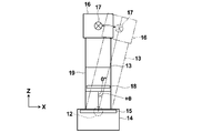
- FIG. 1 is a schematic configuration diagram of a breast image photographing display system using an embodiment of a radiographic image photographing device of the present invention.
- the figure which shows the state which moved the radiation source unit in the mammography imaging display system shown in FIG. 1 is a block diagram showing a schematic configuration inside a computer of the breast image capturing and displaying system shown in FIG.
- Timing chart showing the ON / OFF state of the imaging switch, the irradiation timing of radiation from the radiation source, and the movement timing of the radiation source in a series of imaging sequences
- the flowchart for demonstrating an effect
- the figure which shows an example of the table which matched the some photographer and the threshold value of the off period preset for every photographer.
- FIG. 1 is a diagram showing a schematic configuration of the entire breast image photographing display system of the present embodiment.
- a breast image radiographing display system 1 includes a mammography apparatus 10, a computer 2 connected to the mammography apparatus 10, a monitor 3 connected to the computer 2, and an input unit. 4 and a photographing switch 5.
- the mammography apparatus 10 includes a base 11, a rotary shaft 12 that can move in the vertical direction (Z direction) with respect to the base 11, and can be rotated.
- the arm part 13 connected with the base 11 is provided.
- FIG. 2 shows the arm 13 viewed from the right direction in FIG.
- the arm portion 13 has an alphabet C shape, and an imaging table 14 on which a breast is installed is attached to one end, and a radiation source unit 16 is attached to the other end so as to face the imaging table 14.
- the movement of the arm unit 13 in the vertical direction is controlled by an arm controller 31 incorporated in the base 11.
- a radiation image detector 15 such as a flat panel detector, and a detector controller 33 for controlling reading of a charge signal from the radiation image detector 15 are provided.
- a charge amplifier that converts the charge signal read from the radiation image detector 15 into a voltage signal
- a correlated double sampling circuit that samples the voltage signal output from the charge amplifier
- a circuit board provided with an AD conversion unit for converting a voltage signal into a digital signal is also installed.
- the radiation image detector 15 can repeatedly perform recording and reading of a radiation image, and may use a so-called direct type radiation image detector that directly receives radiation and generates charges. Alternatively, a so-called indirect radiation image detector that converts radiation once into visible light and converts the visible light into a charge signal may be used.
- a radiation image signal readout method a radiation image signal is read out by turning on / off a TFT (thin film transistor) switch, or by irradiating reading light.
- TFT thin film transistor
- a radiation source 17 and a radiation source controller 32 are accommodated in the radiation source unit 16.
- the radiation source 17 is provided with an X-ray tube, and the X-ray tube is subjected to collision of an electron beam while performing a rotating operation with a capacitor used when applying a high voltage tube voltage. And an anode that emits radiation.
- the radiation source controller 32 raises and lowers the X-ray tube in the radiation source 17, the timing of irradiating the radiation from the radiation source 17, and the radiation generation conditions (tube current, time, tube voltage, etc.) in the radiation source 17. ) And the like are controlled.
- the compression plate 18 is disposed above the imaging table 14 and presses the breast to press it, the support portion 20 that supports the compression plate 18, and the support portion 20 in the vertical direction.
- a moving mechanism 19 for moving is provided. The position of the compression plate 18 and the compression pressure are controlled by the compression plate controller 34.
- the computer 2 includes a central processing unit (CPU), a storage device such as a semiconductor memory, a hard disk, and an SSD.
- the hardware includes an imaging control unit 40, a radiographic image storage unit 41, and a storage device such as shown in FIG.
- a display control unit 42 is configured.
- the imaging control unit 40 outputs predetermined control signals to the various controllers 31 to 34, and controls the imaging sequence of the entire system. A specific control method will be described in detail later.
- the radiation image storage unit 41 stores two radiation image signals detected by the radiation image detector 15 by photographing from two different photographing directions.
- the display control unit 42 displays a breast stereo image on the monitor 3 after performing predetermined signal processing on the radiographic image signal read from the radiographic image storage unit 41.
- the input unit 4 is composed of a pointing device such as a keyboard and a mouse, for example, and accepts input of shooting conditions by a photographer.
- the shooting switch 5 is a button for instructing execution of a series of shooting sequences including shooting from two different shooting directions.
- the photographing control unit 40 of the computer 2 controls each unit so that a series of photographing sequences is executed when the photographing switch 5 is turned on by the photographer. If the shooting switch 5 is accidentally released or if the shooting switch 5 is turned off due to chattering or electrical noise, the operation of a part of the shooting sequence is stopped according to the length of the off-state period. Or continue. The control of the operation of part of the imaging sequence will be described in detail later.
- the monitor 3 is configured to be able to display a stereo image using two radiation image signals output from the computer 2.
- a configuration for displaying a stereo image for example, a radiographic image based on two radiographic image signals is displayed using two screens, and one of the radiographic images is observed by using a half mirror or a polarizing glass. It is possible to adopt a configuration in which a stereo image is displayed by being incident on the right eye of the observer and the other radiation image is incident on the left eye of the observer.
- two radiographic images may be displayed in a superimposed manner while being shifted by a predetermined amount of parallax, and this may be configured to generate a stereo image by observing with a polarizing glass, or a parallax barrier method and a lenticular method
- a stereo image may be generated by displaying two radiation images on a stereoscopically viewable 3D liquid crystal.
- FIG. 5 shows on / off states of the imaging switch 5 in a series of imaging sequences, radiation irradiation timing of the radiation source 17, and movement timing of the radiation source 17 (operation timing of the drive motor that moves the radiation source 17). It is shown.
- a series of shooting sequences when the shooting switch 5 is not in the OFF state as described above will be described first.
- the computer 2, and the monitor 3 are turned on, the breast of the subject M is placed on the imaging table 14, and the breast is compressed with a predetermined pressure by the compression plate 18. (S10).
- the photographing switch 5 is turned on as shown in FIG. 5 (S12), whereby a series of photographing sequences is started by the photographing control unit 40 (S14). ).
- a control signal is output from the imaging control unit 40 to the radiation source controller 32, and the radiation source controller 32 performs an X-ray tube startup operation in the radiation source 17 in accordance with the input control signal.
- Start (S16) For example, the start-up operation of the X-ray tube includes the start of an anode rotation operation and the charging of a capacitor.
- the imaging control unit 40 captures the first radiographic image of the two radiographic images constituting the stereo image.
- the shooting control unit 40 reads a preset shooting angle ⁇ for shooting a stereo image, and outputs information of the read shooting angle ⁇ to the arm controller 31.
- the shooting angle can be set.
- the imaging control unit 40 includes the radiation source controller 32 and the detector controller 33.
- a control signal is output so as to perform radiation irradiation and readout of the radiation image signal.
- the radiation image obtained by photographing the breast from the 0 ° direction is detected by the radiation image detector 15 by the first radiation irradiation from the radiation source 17 (S20).
- V 0 is a tube voltage at which the emission of radiation from the radiation source 17 is stopped, and may be a predetermined voltage of about 5 V or 0 V.
- a radiation image signal is read from the radiation image detector 15 by the detector controller 33, subjected to predetermined signal processing on the radiation image signal, and then stored in the radiation image storage unit 41 of the computer 2. (S24).
- the arm controller 31 controls the arm unit 13 to rotate by + ⁇ ° with respect to the direction perpendicular to the imaging table 14, as shown in FIG. Output a signal. That is, in the present embodiment, the control signal is output so that the arm unit 13 is rotated by 4 ° with respect to the direction perpendicular to the imaging table 14.
- the arm unit 13 moves in the 4 ° direction based on the control signal output from the arm controller 31, and the radiation source unit 16 also moves in the 4 ° direction.
- the imaging control unit 40 applies radiation to the radiation source controller 32 and the detector controller 33 and the radiation image signal.
- a control signal is output so as to read out.
- a radiation image signal is read from the radiation image detector 15 by the detector controller 33, subjected to predetermined signal processing on the radiation image signal, and then stored in the radiation image storage unit 41 of the computer 2. (S34).
- the photographing control unit 40 ends the series of photographing sequences and outputs a control signal to the compression plate controller 34. Then, the compression plate controller 34 releases the compression of the breast by the compression plate 18 according to the input control signal (S38).
- the imaging control unit 40 outputs a control signal to the radiation source controller 32, and the radiation source controller 32 performs the operation of lowering the X-ray tube of the radiation source 17 of the radiation source unit 16 according to the input control signal. Perform (S40).
- the X-ray tube falling operation mentioned here includes, for example, stopping the rotating operation of the anode and stopping charging of the capacitor.
- the two radiographic image signals stored in the radiographic image storage unit 41 as described above are read out by the display control unit 42, and the display control unit 42 performs predetermined processing on these radiographic image signals. After being applied, it is output to the monitor 3. Then, on the monitor 3, the radiographic image for the right eye and the radiographic image for the left eye are respectively displayed, and a stereo image of the breast is displayed (S42).
- the photographing switch 5 is erroneously turned off after the photographing switch 5 is pressed by the photographer until the photographer stops pressing the photographing switch 5 after the two radiographic images are photographed.
- the operation in this case will be described with reference to the flowchart shown in FIG.
- the imaging control unit 40 measures the time from when the imaging switch 5 is turned off (S60), and while the off period is equal to or less than the predetermined threshold (S62, YES), The imaging sequence excluding irradiation and movement of the radiation source 17 is continuously executed (S64). Specifically, for example, the compression of the breast by the compression plate 18 and the startup state of the X-ray tube of the radiation source 17 are maintained.
- the photographing switch 5 is turned on (S66).
- the imaging control unit 40 resumes the movement of the radiation source 17 and continuously executes the normal imaging sequence (S68).
- the photographing control unit 40 when the OFF period of the photographing switch 5 exceeds a predetermined threshold (S62, NO), the photographing control unit 40 outputs a control signal to the compression plate controller 34, and the compression plate controller 34 receives the input control. In response to the signal, the compression of the breast by the compression plate 18 is released (S70). Further, the imaging control unit 40 outputs a control signal to the radiation source controller 32, and the radiation source controller 32 performs an X-ray tube falling operation in the radiation source 17 in accordance with the input control signal (S72). Then, the shooting control unit 40 stops a series of shooting sequences (S74).
- the radiographing switch when the radiographing switch is turned off only for a predetermined period, when the off period is equal to or less than a predetermined threshold, the compression by the compression plate or the X-ray tube Since the start-up operation and the like are continuously performed without being stopped, it is possible to quickly return to the normal shooting sequence and to capture an appropriate stereo image.
- the breast compression and the X-ray tube start-up operation are continuously performed in a series of imaging sequences.
- the present invention is not limited to this, and other operations may be continued.
- the reason why the X-ray tube start-up operation is not stopped is that it takes time to re-start up once it is started down, but it is stopped from the viewpoint of the time until it is restored again. Other operations that are not desirable to be performed may be continued.
- the movement of the radiation source unit 16 is stopped when the imaging switch 5 is turned off. This is set in this way from the viewpoint of safety. For example, in the case where reduction in imaging time is given priority over safety, when the imaging switch 5 is turned off and the off period is equal to or less than a predetermined threshold, the radiation source unit You may make it perform 16 movements continuously.
- the threshold value of the off period used in the above embodiment is desirable to, for example, 1 second or more and 2 seconds or less.
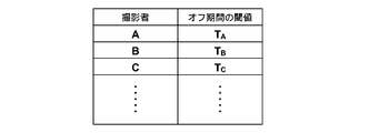
- the off-period threshold may be set in advance, but the photographer may be able to set an arbitrary value using the input unit 4.
- a plurality of off-period threshold values are set in advance, and the photographer selects one of the plurality of threshold values using the input unit 4 and sets the selected threshold value. Good.
- threshold values (T A , T B , T C ,%) For each photographer (A, B, C,%) are set in advance as a table.
- the input unit 4 may receive photographer information, and may set a threshold of an off period corresponding to the received photographer information.
- radiographic imaging apparatus of this invention applies the radiographic imaging apparatus of this invention to the imaging
- a radiographic image is moved from several imaging directions by moving a radiation source. Any other apparatus can be applied as long as the apparatus performs the above imaging, and for example, there is a tomosynthesis imaging apparatus as such an apparatus.
Landscapes
- Health & Medical Sciences (AREA)
- Life Sciences & Earth Sciences (AREA)
- Medical Informatics (AREA)
- Engineering & Computer Science (AREA)
- Radiology & Medical Imaging (AREA)
- Molecular Biology (AREA)
- Biophysics (AREA)
- Nuclear Medicine, Radiotherapy & Molecular Imaging (AREA)
- Optics & Photonics (AREA)
- Pathology (AREA)
- Physics & Mathematics (AREA)
- Biomedical Technology (AREA)
- Heart & Thoracic Surgery (AREA)
- High Energy & Nuclear Physics (AREA)
- Surgery (AREA)
- Animal Behavior & Ethology (AREA)
- General Health & Medical Sciences (AREA)
- Public Health (AREA)
- Veterinary Medicine (AREA)
- Dentistry (AREA)
- Oral & Maxillofacial Surgery (AREA)
- Apparatus For Radiation Diagnosis (AREA)
Abstract
[Problème] Le problème réside en l'obtention d'une commande de dispositif d'imagerie radiologique qui réalise en continu une série de séquences d'imagerie quand un commutateur d'imagerie est allumé en continu, de manière telle que, si le commutateur d'imagerie est éteint pendant seulement un temps court lorsque la main d'un opérateur s'est accidentellement enlevée du commutateur d'imagerie etc., le dispositif d'imagerie radiologique revient rapidement à la séquence d'imagerie normale et prend des images radiologiques appropriées. [Solution] Le problème est résolu selon l'invention par une commande réalisée de manière telle que, quand un commutateur d'imagerie (5) est éteint pendant une période prédéterminée, une partie des opérations dans la séquence d'imagerie continue à être réalisée si la période prédéterminée est égale ou inférieure à une valeur seuil.
Priority Applications (4)
| Application Number | Priority Date | Filing Date | Title |
|---|---|---|---|
| CN201280011759.9A CN103402432B (zh) | 2011-03-04 | 2012-03-01 | 射线照相摄影方法和装置 |
| EP12755197.6A EP2682058B1 (fr) | 2011-03-04 | 2012-03-01 | Procédé et dispositif d'imagerie radiologique |
| US13/973,082 US9226724B2 (en) | 2011-03-04 | 2013-08-22 | Radiographic imaging method and apparatus |
| US14/951,845 US9510801B2 (en) | 2011-03-04 | 2015-11-25 | Radiographic imaging method and apparatus |
Applications Claiming Priority (2)
| Application Number | Priority Date | Filing Date | Title |
|---|---|---|---|
| JP2011047808A JP5600305B2 (ja) | 2011-03-04 | 2011-03-04 | 放射線画像撮影方法および装置 |
| JP2011-047808 | 2011-03-04 |
Related Child Applications (1)
| Application Number | Title | Priority Date | Filing Date |
|---|---|---|---|
| US13/973,082 Continuation US9226724B2 (en) | 2011-03-04 | 2013-08-22 | Radiographic imaging method and apparatus |
Publications (1)
| Publication Number | Publication Date |
|---|---|
| WO2012120841A1 true WO2012120841A1 (fr) | 2012-09-13 |
Family
ID=46797814
Family Applications (1)
| Application Number | Title | Priority Date | Filing Date |
|---|---|---|---|
| PCT/JP2012/001403 Ceased WO2012120841A1 (fr) | 2011-03-04 | 2012-03-01 | Procédé et dispositif d'imagerie radiologique |
Country Status (5)
| Country | Link |
|---|---|
| US (2) | US9226724B2 (fr) |
| EP (1) | EP2682058B1 (fr) |
| JP (1) | JP5600305B2 (fr) |
| CN (1) | CN103402432B (fr) |
| WO (1) | WO2012120841A1 (fr) |
Cited By (1)
| Publication number | Priority date | Publication date | Assignee | Title |
|---|---|---|---|---|
| CN104994790A (zh) * | 2013-02-14 | 2015-10-21 | 株式会社东芝 | X射线诊断装置 |
Families Citing this family (6)
| Publication number | Priority date | Publication date | Assignee | Title |
|---|---|---|---|---|
| JP5600305B2 (ja) * | 2011-03-04 | 2014-10-01 | 富士フイルム株式会社 | 放射線画像撮影方法および装置 |
| CN105228526B (zh) * | 2013-03-29 | 2018-06-08 | 通用电气公司 | 乳房造影装置 |
| US10830712B2 (en) * | 2017-03-27 | 2020-11-10 | KUB Technologies, Inc. | System and method for cabinet x-ray systems with camera |
| CN107179650A (zh) * | 2017-05-12 | 2017-09-19 | 太仓诚泽网络科技有限公司 | 一种基于射线的摄影方法 |
| CN108919608B (zh) * | 2018-06-27 | 2023-07-25 | 上海联影医疗科技股份有限公司 | 曝光流程控制方法、装置、设备及介质 |
| JP7208849B2 (ja) * | 2019-03-28 | 2023-01-19 | 富士フイルム株式会社 | 放射線撮影システム |
Citations (4)
| Publication number | Priority date | Publication date | Assignee | Title |
|---|---|---|---|---|
| JPH03251232A (ja) * | 1990-02-28 | 1991-11-08 | Shimadzu Corp | 血管造影撮影装置 |
| JP2006055633A (ja) * | 2004-07-21 | 2006-03-02 | Toshiba Corp | X線イメージング装置 |
| JP2010264194A (ja) * | 2009-05-18 | 2010-11-25 | Canon Inc | 放射線撮影装置およびその撮影方法 |
| JP2010279516A (ja) * | 2009-06-04 | 2010-12-16 | Fujifilm Corp | 乳房撮影定位装置 |
Family Cites Families (9)
| Publication number | Priority date | Publication date | Assignee | Title |
|---|---|---|---|---|
| JPH05237080A (ja) * | 1992-02-28 | 1993-09-17 | Hitachi Medical Corp | X線撮影装置 |
| US5594772A (en) * | 1993-11-26 | 1997-01-14 | Kabushiki Kaisha Toshiba | Computer tomography apparatus |
| JPH118934A (ja) * | 1997-06-17 | 1999-01-12 | Aloka Co Ltd | 医療用装置 |
| JP4460695B2 (ja) * | 1999-11-24 | 2010-05-12 | 株式会社東芝 | X線コンピュータ断層撮影装置 |
| JP3677199B2 (ja) * | 2000-07-31 | 2005-07-27 | 和泉電気株式会社 | 押しボタンスイッチ及びこれを備えた教示ペンダント |
| DE10347735B4 (de) * | 2003-10-14 | 2012-01-26 | Siemens Ag | Motorisch verstellbares Röntgengerät |
| US7177393B2 (en) | 2004-07-21 | 2007-02-13 | Kabushiki Kaisha Toshiba | X-ray imaging apparatus |
| JP2010131170A (ja) | 2008-12-04 | 2010-06-17 | Fujifilm Corp | 断層画像撮影装置 |
| JP5600305B2 (ja) * | 2011-03-04 | 2014-10-01 | 富士フイルム株式会社 | 放射線画像撮影方法および装置 |
-
2011
- 2011-03-04 JP JP2011047808A patent/JP5600305B2/ja active Active
-
2012
- 2012-03-01 EP EP12755197.6A patent/EP2682058B1/fr active Active
- 2012-03-01 CN CN201280011759.9A patent/CN103402432B/zh active Active
- 2012-03-01 WO PCT/JP2012/001403 patent/WO2012120841A1/fr not_active Ceased
-
2013
- 2013-08-22 US US13/973,082 patent/US9226724B2/en active Active
-
2015
- 2015-11-25 US US14/951,845 patent/US9510801B2/en active Active
Patent Citations (4)
| Publication number | Priority date | Publication date | Assignee | Title |
|---|---|---|---|---|
| JPH03251232A (ja) * | 1990-02-28 | 1991-11-08 | Shimadzu Corp | 血管造影撮影装置 |
| JP2006055633A (ja) * | 2004-07-21 | 2006-03-02 | Toshiba Corp | X線イメージング装置 |
| JP2010264194A (ja) * | 2009-05-18 | 2010-11-25 | Canon Inc | 放射線撮影装置およびその撮影方法 |
| JP2010279516A (ja) * | 2009-06-04 | 2010-12-16 | Fujifilm Corp | 乳房撮影定位装置 |
Cited By (2)
| Publication number | Priority date | Publication date | Assignee | Title |
|---|---|---|---|---|
| CN104994790A (zh) * | 2013-02-14 | 2015-10-21 | 株式会社东芝 | X射线诊断装置 |
| CN104994790B (zh) * | 2013-02-14 | 2017-12-19 | 东芝医疗系统株式会社 | X射线诊断装置 |
Also Published As
| Publication number | Publication date |
|---|---|
| CN103402432B (zh) | 2016-08-17 |
| EP2682058B1 (fr) | 2015-09-30 |
| JP5600305B2 (ja) | 2014-10-01 |
| EP2682058A4 (fr) | 2014-08-13 |
| CN103402432A (zh) | 2013-11-20 |
| US9226724B2 (en) | 2016-01-05 |
| US20130336444A1 (en) | 2013-12-19 |
| EP2682058A1 (fr) | 2014-01-08 |
| US9510801B2 (en) | 2016-12-06 |
| JP2012183169A (ja) | 2012-09-27 |
| US20160073996A1 (en) | 2016-03-17 |
Similar Documents
| Publication | Publication Date | Title |
|---|---|---|
| JP5600305B2 (ja) | 放射線画像撮影方法および装置 | |
| JP2012050519A (ja) | 乳房画像撮影装置 | |
| WO2012114765A1 (fr) | Dispositif d'imagerie mammaire | |
| US20120051501A1 (en) | Radiation image radiographing apparatus and radiation image radiographing and displaying method | |
| WO2012090472A1 (fr) | Dispositif et procédé de commande d'imagerie | |
| JP2012024519A (ja) | 放射線画像撮影表示方法および装置 | |
| JP2012029759A (ja) | 放射線画像撮影表示方法および装置 | |
| JP2012066049A (ja) | 放射線画像撮影装置および立体視画像表示方法 | |
| WO2012114757A1 (fr) | Procédé et dispositif d'imagerie radiographique | |
| WO2012127819A1 (fr) | Appareil et procédé radiographique tridimensionnel | |
| JP2012050517A (ja) | 放射線画像撮影装置 | |
| WO2012176645A1 (fr) | Dispositif d'imagerie radiographique et son procédé de fonctionnement | |
| WO2012056695A1 (fr) | Dispositif d'affichage d'image tridimensionnelle, procédé et programme associés | |
| JP2013000490A (ja) | 放射線画像撮影方法および装置 | |
| WO2012039121A1 (fr) | Dispositif de capture d'image radiologique et procédé de capture d'image radiologique | |
| JP2012011036A (ja) | 放射線画像撮影方法および装置 | |
| JP2012050518A (ja) | 放射線画像撮影装置 | |
| WO2012056722A1 (fr) | Dispositif d'affichage d'image radiologique tridimensionnelle, procédé et programme associés | |
| JP2012071107A (ja) | 放射線画像撮影装置およびその撮影方法 | |
| JP2012070826A (ja) | 放射線画像撮影装置および方法 | |
| WO2012114766A1 (fr) | Programme, procédé et dispositif de génération d'images radiologiques stéréoscopiques | |
| WO2012120886A1 (fr) | Appareil et procédé radiographique tridimensionnel | |
| WO2012114758A1 (fr) | Dispositif et procédé d'imagerie radiographique | |
| JP2013000491A (ja) | 放射線画像撮影方法および装置 | |
| JP2012050473A (ja) | 乳房画像撮影表示方法および装置 |
Legal Events
| Date | Code | Title | Description |
|---|---|---|---|
| 121 | Ep: the epo has been informed by wipo that ep was designated in this application |
Ref document number: 12755197 Country of ref document: EP Kind code of ref document: A1 |
|
| NENP | Non-entry into the national phase |
Ref country code: DE |
|
| WWE | Wipo information: entry into national phase |
Ref document number: 2012755197 Country of ref document: EP |