WO2012132453A1 - Méthode, dispositif et programme d'affichage d'image radiologique des seins - Google Patents
Méthode, dispositif et programme d'affichage d'image radiologique des seins Download PDFInfo
- Publication number
- WO2012132453A1 WO2012132453A1 PCT/JP2012/002188 JP2012002188W WO2012132453A1 WO 2012132453 A1 WO2012132453 A1 WO 2012132453A1 JP 2012002188 W JP2012002188 W JP 2012002188W WO 2012132453 A1 WO2012132453 A1 WO 2012132453A1
- Authority
- WO
- WIPO (PCT)
- Prior art keywords
- image
- breast
- images
- eye
- captured
- Prior art date
- Legal status (The legal status is an assumption and is not a legal conclusion. Google has not performed a legal analysis and makes no representation as to the accuracy of the status listed.)
- Ceased
Links
Images


Classifications
-
- A—HUMAN NECESSITIES
- A61—MEDICAL OR VETERINARY SCIENCE; HYGIENE
- A61B—DIAGNOSIS; SURGERY; IDENTIFICATION
- A61B6/00—Apparatus or devices for radiation diagnosis; Apparatus or devices for radiation diagnosis combined with radiation therapy equipment
- A61B6/50—Apparatus or devices for radiation diagnosis; Apparatus or devices for radiation diagnosis combined with radiation therapy equipment specially adapted for specific body parts; specially adapted for specific clinical applications
- A61B6/502—Apparatus or devices for radiation diagnosis; Apparatus or devices for radiation diagnosis combined with radiation therapy equipment specially adapted for specific body parts; specially adapted for specific clinical applications for diagnosis of breast, i.e. mammography
-
- A—HUMAN NECESSITIES
- A61—MEDICAL OR VETERINARY SCIENCE; HYGIENE
- A61B—DIAGNOSIS; SURGERY; IDENTIFICATION
- A61B6/00—Apparatus or devices for radiation diagnosis; Apparatus or devices for radiation diagnosis combined with radiation therapy equipment
- A61B6/02—Arrangements for diagnosis sequentially in different planes; Stereoscopic radiation diagnosis
- A61B6/022—Stereoscopic imaging
-
- A—HUMAN NECESSITIES
- A61—MEDICAL OR VETERINARY SCIENCE; HYGIENE
- A61B—DIAGNOSIS; SURGERY; IDENTIFICATION
- A61B6/00—Apparatus or devices for radiation diagnosis; Apparatus or devices for radiation diagnosis combined with radiation therapy equipment
- A61B6/46—Arrangements for interfacing with the operator or the patient
- A61B6/461—Displaying means of special interest
- A61B6/466—Displaying means of special interest adapted to display 3D data
Definitions
- the left and right breasts are displayed in the same image based on the four images of the upper image and the oblique image captured so that the left and right breasts have a parallax in the left-right direction.
- Radiation breast image display method and radiation breast image display apparatus for generating two images of right eye image and left eye image and displaying a stereoscopic image composed of these images on display means capable of displaying the stereoscopic image As well as programs.
- stereoscopic viewing can be performed using parallax by displaying a combination of two images, a right-eye image and a left-eye image.
- a stereoscopically viewable image hereinafter referred to as a stereoscopic image or a stereo image
- a stereoscopic image or a stereo image is generated based on a plurality of images having parallax obtained by photographing the same subject from different positions.
- stereoscopic images is used not only in the fields of digital cameras and televisions, but also in the field of radiographic imaging. That is, the patient is irradiated with radiation from different directions, the radiation transmitted through the subject is detected by a radiation image detector, and a plurality of radiation images having parallax are obtained, and these radiations are acquired. A stereoscopic image is generated based on the image. And by generating a stereoscopic image in this way, a radiographic image with a sense of depth can be observed, and a radiographic image more suitable for diagnosis can be observed. (For example, see Patent Document 1)
- one of the two images with parallax in each breast is preferably an image taken from above the breast, as shown in FIG. 4, for example. This is because when two-dimensional diagnosis is performed using only one of the right-eye image and the left-eye image constituting the stereoscopic image, the image taken from above the breast is most suitable for diagnosis. Because.
- both the right-eye image and the left-eye image are based on the four images of the upper image and the oblique image captured so as to have a parallax in the left-right direction with respect to each of the left and right breasts. It is necessary to combine and combine one of the upper image and the oblique image and generate two images, a right-eye image and a left-eye image in which the left and right breasts are displayed in the same image, Since the visibility of the stereoscopic image changes depending on the combination method at this time, it is required to optimally combine them.
- the present invention provides a radiation breast image display method capable of displaying a stereoscopic image with high visibility when displaying a stereoscopic image in which left and right breasts are simultaneously displayed in the same image.
- An object of the present invention is to provide a radiation breast image display device and a program.
- the upper image is easier to visually recognize because it is more suitable for diagnosis.
- a left-eye image and a left-eye image are generated and a stereoscopic image including these images is displayed on a display unit capable of displaying a stereoscopic image, as shown in FIG.
- the left image of the upper breast (0 ° image in the figure) and the left (right) oblique image of the breast (4 ° image in the figure) are displayed in combination, the lower half of the left breast in the right-eye image is displayed.
- the upper captured image is easier to see, and in the left-eye image, the upper captured image of the upper right breast is easier to visually recognize.
- the left and right eyes are easily visible. If they are different, the burden on the eyes will increase.
- the applicant of the present application combines the upper photographed images of the left and right breasts together with one of the right eye image and the left eye image. It has been found that the visibility of the stereoscopic image is improved.
- the applicant of the present application has already found that a stereoscopic image can be stereoscopically viewed even if the image quality is different between the right-eye image and the left-eye image.
- the image quality of obliquely captured images which are less important for diagnosis, may be low, so the radiation dose can be reduced during imaging to reduce patient exposure, or the image signal from the detector It is considered to reduce the data size by reducing the resolution by reducing the resolution or reducing the compression rate when saving the image signal, etc. In some cases, the above phenomenon becomes more prominent.
- the radiological breast image display method of the present invention is such that an upper photographed image and an oblique photographing that are photographed so as to have a parallax in the horizontal direction with respect to each of the left and right breasts.
- Other images and left eye images The image is a stereoscopic image composed of two images of the right eye image and the left eye image which is characterized in that to be displayed on the display means.
- the upper photographed image means an image photographed at a photographing angle closer to the upper part of the breast when standing upright than the oblique photographed image.
- the photographing angle of the upper photographed image (the upward direction of the breast when standing upright is used as a reference). Is preferably 0 °, but is not necessarily 0 °, and may be any number as long as the angle is smaller than the shooting angle of the oblique shooting image.
- the radiation breast image display method according to the present invention may be provided as a program for causing a computer to execute the method.
- the radiological breast image display device of the present invention is based on four images, that is, an upper photographed image and an oblique photographed image that are photographed so as to have a parallax in the left-right direction for each of the left and right breasts.
- Display that can generate two images, a right-eye image and a left-eye image on which the left and right breasts are displayed, and display a stereoscopic image composed of the right-eye image and the left-eye image.
- Combining means, right eye image and And it is characterized in that comprising a display control means for displaying a stereoscopic image composed of two images of the eye image on the display unit.
- the right-eye image is synthesized by synthesizing the upper captured image of one breast and the upper captured image of the other breast among the left and right breast images.
- One of the left and right eye images is generated, and the right and left eye images are synthesized by synthesizing the oblique image of one breast and the oblique image of the other of the left and right breast images.
- One of the right-eye image and the left-eye image is generated by generating the other image and displaying a stereoscopic image including the generated right-eye image and left-eye image on the display means.
- a stereoscopic image with high visibility can be displayed.
- FIG. 1 is a schematic configuration diagram of a breast stereoscopic image photographing display system using a radiation breast image display device according to an embodiment of the present invention.
- FIG. 1 is a diagram of the arm part of the breast stereoscopic image photographing display system as seen from the right direction in FIG.
- the block diagram which shows schematic structure inside the computer of the said stereoscopic image imaging display system for breasts
- the figure which shows the state of imaging
- photography state of the right and left breasts in the said stereoscopic image imaging display system for breasts The figure which shows the example of the image for right eyes and the image for left eyes at the time of imaging
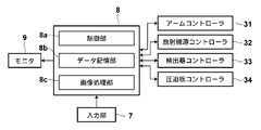
- FIG. 1 is a schematic configuration diagram of a stereoscopic image capturing / displaying system for breasts using a radiation breast image display device according to an embodiment of the present invention
- FIG. 2 is a diagram illustrating an arm portion of the stereoscopic image capturing / displaying system for breasts.
- FIG. 3 is a block diagram showing a schematic configuration inside the computer of the stereoscopic image capturing and displaying system for breasts.
- a stereoscopic image capturing / displaying system 1 for a breast includes a breast image capturing apparatus 10, a computer 8 connected to the breast image capturing apparatus 10, and a monitor ( Display means) 9 and an input unit 7.
- the mammography apparatus 10 includes a base 11, a rotary shaft 12 that can move in the vertical direction (Z direction) with respect to the base 11, and can rotate.
- the arm part 13 connected with the base 11 is provided.
- FIG. 2 shows the arm 13 viewed from the right direction in FIG.
- the arm section 13 has an alphabet C shape, and a radiation table 16 is attached to one end of the arm section 13 so as to face the imaging table 14 at the other end.
- the rotation and vertical movement of the arm unit 13 are controlled by an arm controller 31 incorporated in the base 11.
- a radiation image detector 15 such as a flat panel detector and a detector controller 33 that controls reading of a charge signal from the radiation image detector 15.
- a charge amplifier that converts the charge signal read from the radiation image detector 15 into a voltage signal
- a correlated double sampling circuit that samples the voltage signal output from the charge amplifier
- a circuit board provided with an AD conversion unit for converting a voltage signal into a digital signal is also installed.
- the photographing table 14 is configured to be rotatable with respect to the arm unit 13, and even when the arm unit 13 rotates with respect to the base 11, the direction of the photographing table 14 is fixed to the base 11. can do.
- the radiation image detector 15 can repeatedly perform recording and reading of a radiation image, and may use a so-called direct type radiation image detector that directly receives radiation and generates charges. Alternatively, a so-called indirect radiation image detector that converts radiation once into visible light and converts the visible light into a charge signal may be used.
- a radiation image signal readout method a radiation image signal is read out by turning on / off a TFT (thin film transistor) switch, or by irradiating reading light. It is desirable to use a so-called optical readout system from which a radiation image signal is read out, but the present invention is not limited to this, and other systems may be used.
- a radiation source 17 and a radiation source controller 32 are accommodated in the radiation irradiation unit 16.
- the radiation source controller 32 controls the timing of irradiating radiation from the radiation source 17 and the radiation generation conditions (tube current, tube voltage, time, etc.) in the radiation source 17.
- a compression plate 18 that is disposed above the imaging table 14 and presses and compresses the breast M, a support portion 20 that supports the compression plate 18, and a support portion 20 that extends in the vertical direction.
- a moving mechanism 19 for moving in the (Z direction) is provided. The position of the compression plate 18 and the compression pressure are controlled by the compression plate controller 34.
- the computer 8 includes a central processing unit (CPU), a storage device such as a semiconductor memory, a hard disk, and an SSD.
- the control unit 8a, the data storage unit 8b, and the image processing unit shown in FIG. Part 8c is configured.
- the control unit 8a has a function as display control means for displaying on the monitor (display means) 9 a stereoscopic image composed of two images, a right-eye image and a left-eye image, in addition to various controllers 31 to 34. A predetermined control signal is output to control the entire system. A specific control method will be described in detail later.
- the data storage unit 8b stores radiation image data and the like for each imaging angle acquired by the radiation image detector 15.
- the image processing unit 8c based on the four images of the upper image and the oblique image captured with parallax in the left-right direction for each of the left and right breasts, In addition to having a function as an image synthesizing means for generating two images of the displayed right-eye image and left-eye image, it is for performing various image processing. That is, the computer 8 is a device that also functions as a radiation breast image display device.
- the input unit 7 is composed of a pointing device such as a keyboard and a mouse, for example, and is used for receiving inputs such as shooting conditions and operation instructions.
- the monitor 9 serving as a display means displays two radiographic images as two-dimensional images using two radiographic image (right-eye image and left-eye image) signals output from the computer 8, thereby providing a stereoscopic image. Is configured to be displayed in a stereoscopic manner.
- radiographic images based on two radiographic image signals are displayed using two screens, and one radiographic image is obtained by using a half mirror or a polarizing glass. It is possible to adopt a configuration in which a stereoscopic image is displayed by being incident on the observer's right eye and the other radiation image being incident on the observer's left eye.
- two radiographic images may be displayed by being shifted by a predetermined amount of parallax and superimposed, and a stereoscopic image may be generated by observing this with a polarizing glass, or a parallax barrier method and a lenticular It is good also as a structure which produces
- the device for displaying a stereoscopic image and the device for displaying a two-dimensional image may be configured separately, or may be configured as the same device if they can be displayed on the same screen.
- one of the left and right breasts M is installed on the imaging table 14, and the breast M is compressed by the compression plate 18 with a predetermined pressure.
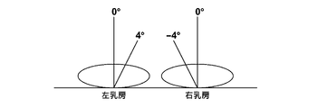
- the control unit 8 a outputs information about the convergence angle ⁇ and the imaging angle ⁇ ′ constituting the convergence angle ⁇ to the arm controller 31.
- ⁇ 4 ° as information on the convergence angle ⁇ at this time
- the convergence angle ⁇ is preferably set to 4 ° or more and 15 ° or less because it is difficult to perform appropriate stereoscopic viewing if the convergence angle ⁇ is too small or too large.
- the one shooting angle ⁇ ′ that is, the shooting angle ⁇ ′ for shooting an image for two-dimensional observation is preferably 0 °. This is because an image taken from the front of the radiation image detector 15 is most suitable for two-dimensional observation.
- the arm controller 31 receives the information on the convergence angle ⁇ output from the control unit 8a, and the arm controller 31 causes the arm unit 13 to be in a direction perpendicular to the imaging table 14 based on the information on the convergence angle ⁇ . Output a control signal.
- a control signal is output so that the shooting angle ⁇ ′ with the arm 13 in the direction perpendicular to the detection surface 15a is 0 °.
- the arm unit 13 rotates to the 0 ° position.
- the control unit 8a outputs a control signal to the radiation source controller 32 and the detector controller 33 so as to perform radiation irradiation and readout of the radiation image signal.
- radiation is emitted from the radiation source 17, and a radiation image obtained by photographing the breast M from the direction where the imaging angle ⁇ ′ is 0 ° is detected by the radiation detector 15. Is read and stored in the data storage unit 8b of the computer 8.
- the arm portion 13 rotates to a position of 4 °.
- the control unit 8a outputs a control signal to the radiation source controller 32 and the detector controller 33 so as to perform radiation irradiation and readout of the radiation image signal.
- radiation is emitted from the radiation source 17, and a radiation image obtained by photographing the breast M from the direction in which the imaging angle ⁇ ′ is 4 ° is detected by the radiation detector 15, and a radiation image signal is detected by the detector controller 33. Is read and stored in the data storage unit 8b of the computer 8.
- the other breasts of the left and right are also photographed in the same manner.
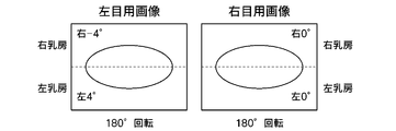
- the upper photographed image (0 ° photographed image) photographed so that the left and right breasts are parallaxed in the left-right direction.
- four images of the obliquely photographed image (4 ° photographed image) are acquired.
- signals of four images of the upper captured image (0 ° captured image) and the oblique captured image (4 ° captured image) of each of the left and right breasts are read from the data storage unit 8b of the computer 8, and the image It is transmitted to the processing unit (image composition means) 8c.
- the image processing unit (image composition means) 8c vertically inverts the right breast oblique image (4 ° image) and the left breast oblique image (4 ° image).
- the images for the right eye are generated by synthesizing the images so that their chest walls face each other, and an upper captured image of the right breast (0 ° captured image) and an upper captured image of the left breast (0 ° captured image)
- the left-eye image is generated by combining the vertically inverted image with the chest walls facing each other.
- the two image signals of the right eye image and the left eye image generated as described above are output to the monitor 9, and a stereoscopic image in which the left and right breasts are displayed on the same screen is displayed on the monitor 9.
- an upper photographed image (0-degree photographed image) of the left and right breasts is combined with one image of the right-eye image or the left-eye image.
- a visual image can be displayed.
- the synthesis of the images of the left and right breasts is not limited to a mode in which the chest walls are opposed to each other, and any mode may be used, for example, by arranging them in the vertical direction or the horizontal direction as they are.
Landscapes
- Health & Medical Sciences (AREA)
- Life Sciences & Earth Sciences (AREA)
- Engineering & Computer Science (AREA)
- Medical Informatics (AREA)
- Radiology & Medical Imaging (AREA)
- Molecular Biology (AREA)
- Biophysics (AREA)
- Nuclear Medicine, Radiotherapy & Molecular Imaging (AREA)
- Optics & Photonics (AREA)
- Pathology (AREA)
- Physics & Mathematics (AREA)
- Biomedical Technology (AREA)
- Heart & Thoracic Surgery (AREA)
- High Energy & Nuclear Physics (AREA)
- Surgery (AREA)
- Animal Behavior & Ethology (AREA)
- General Health & Medical Sciences (AREA)
- Public Health (AREA)
- Veterinary Medicine (AREA)
- Dentistry (AREA)
- Oral & Maxillofacial Surgery (AREA)
- Human Computer Interaction (AREA)
- Apparatus For Radiation Diagnosis (AREA)
Abstract
[Problème] Afficher une image tridimensionnelle hautement lisible lors de l'affichage d'une image tridimensionnelle dans laquelle le sein gauche et droit sont affichés simultanément dans la même image, en fonction de quatre images des seins droit et gauche, qui sont des images capturées de manière oblique et des images capturées depuis le dessus et qui ont été capturées de manière qu'une parallaxe soit présente dans le sens transversal. [Solution] Générer une image pour l'oeil droit par la combinaison d'une image du sein droit capturée de manière oblique (image capturée à 4°) et une image du sein gauche capturée de manière oblique (image capturée à 4°) et inversée verticalement, de sorte que les parois de la cage thoracique dans les images soient en face l'une de l'autre, et générer une image pour l'oeil gauche par la combinaison d'une image du sein droit capturée depuis le dessus (image capturée à 0°) et une image du sein gauche qui a été capturée depuis le dessus (image capturée à 0°) et inversée verticalement, de sorte que les parois de la cage thoracique soient en face l'une de l'autre; afficher une image tridimensionnelle sur un moniteur en fonction de l'image pour l'oeil gauche et de l'image pour l'oeil droit générées comme décrit ci-dessus.
Applications Claiming Priority (2)
| Application Number | Priority Date | Filing Date | Title |
|---|---|---|---|
| US201161470271P | 2011-03-31 | 2011-03-31 | |
| US61/470,271 | 2011-03-31 |
Publications (1)
| Publication Number | Publication Date |
|---|---|
| WO2012132453A1 true WO2012132453A1 (fr) | 2012-10-04 |
Family
ID=46930224
Family Applications (1)
| Application Number | Title | Priority Date | Filing Date |
|---|---|---|---|
| PCT/JP2012/002188 Ceased WO2012132453A1 (fr) | 2011-03-31 | 2012-03-29 | Méthode, dispositif et programme d'affichage d'image radiologique des seins |
Country Status (1)
| Country | Link |
|---|---|
| WO (1) | WO2012132453A1 (fr) |
Citations (4)
| Publication number | Priority date | Publication date | Assignee | Title |
|---|---|---|---|---|
| JP2004321783A (ja) * | 2004-03-10 | 2004-11-18 | Konica Minolta Medical & Graphic Inc | 乳房画像撮影システム及び乳房画像撮影装置 |
| JP2007195663A (ja) * | 2006-01-25 | 2007-08-09 | Toshiba Corp | 画像表示装置及びプログラム |
| JP2010167129A (ja) * | 2009-01-23 | 2010-08-05 | Fujifilm Corp | X線撮像装置 |
| JP2010188002A (ja) * | 2009-02-19 | 2010-09-02 | Fujifilm Corp | 放射線撮影装置 |
-
2012
- 2012-03-29 WO PCT/JP2012/002188 patent/WO2012132453A1/fr not_active Ceased
Patent Citations (4)
| Publication number | Priority date | Publication date | Assignee | Title |
|---|---|---|---|---|
| JP2004321783A (ja) * | 2004-03-10 | 2004-11-18 | Konica Minolta Medical & Graphic Inc | 乳房画像撮影システム及び乳房画像撮影装置 |
| JP2007195663A (ja) * | 2006-01-25 | 2007-08-09 | Toshiba Corp | 画像表示装置及びプログラム |
| JP2010167129A (ja) * | 2009-01-23 | 2010-08-05 | Fujifilm Corp | X線撮像装置 |
| JP2010188002A (ja) * | 2009-02-19 | 2010-09-02 | Fujifilm Corp | 放射線撮影装置 |
Similar Documents
| Publication | Publication Date | Title |
|---|---|---|
| US20130300737A1 (en) | Stereoscopic image generating apparatus, stereoscopic image generating method, and stereoscopic image generating program | |
| JP2012066049A (ja) | 放射線画像撮影装置および立体視画像表示方法 | |
| JP5658818B2 (ja) | 放射線乳房画像表示方法、放射線乳房画像表示装置ならびにプログラム | |
| JP2012165358A (ja) | 立体視画像表示装置 | |
| JP5695524B2 (ja) | 立体視画像表示装置および方法並びにプログラム | |
| WO2012056695A1 (fr) | Dispositif d'affichage d'image tridimensionnelle, procédé et programme associés | |
| JP2012068610A (ja) | 立体視画像表示装置、放射線画像撮影表示システムおよび立体視画像表示方法 | |
| WO2012063419A1 (fr) | Dispositif et procédé d'affichage d'image stéréoscopique, et programme | |
| JP2012024516A (ja) | 放射線画像撮影表示方法および装置 | |
| WO2012132453A1 (fr) | Méthode, dispositif et programme d'affichage d'image radiologique des seins | |
| US20120076261A1 (en) | Radiological image displaying apparatus and method | |
| WO2012039121A1 (fr) | Dispositif de capture d'image radiologique et procédé de capture d'image radiologique | |
| WO2012056718A1 (fr) | Dispositif d'affichage d'image radiologique tridimensionnelle, procédé et programme associés | |
| WO2012105188A1 (fr) | Dispositif, procédé et programme pour afficher une image stéréoscopique | |
| JP2012100246A (ja) | 立体視画像表示装置および立体視画像表示方法 | |
| WO2012056679A1 (fr) | Système et dispositif d'affichage d'images 3d | |
| JP2012178626A (ja) | 立体視放射線画像表示方法および装置 | |
| WO2012056677A1 (fr) | Dispositif d'affichage d'image tridimensionnelle | |
| WO2012056722A1 (fr) | Dispositif d'affichage d'image radiologique tridimensionnelle, procédé et programme associés | |
| WO2012132467A1 (fr) | Procédé de capture d'images radiologiques du sein, dispositif de capture d'images radiologiques du sein et programme associé | |
| JP2012170044A (ja) | 立体視画像表示装置 | |
| JP2012105047A (ja) | 立体視画像表示装置および方法並びにプログラム | |
| WO2013024572A1 (fr) | Dispositif de reproduction d'image, procédé de reproduction d'image, et programme | |
| JP2012050518A (ja) | 放射線画像撮影装置 | |
| JP2013154165A (ja) | 画像再生装置および画像再生方法並びにプログラム |
Legal Events
| Date | Code | Title | Description |
|---|---|---|---|
| 121 | Ep: the epo has been informed by wipo that ep was designated in this application |
Ref document number: 12763861 Country of ref document: EP Kind code of ref document: A1 |
|
| NENP | Non-entry into the national phase |
Ref country code: DE |
|
| 122 | Ep: pct application non-entry in european phase |
Ref document number: 12763861 Country of ref document: EP Kind code of ref document: A1 |
|
| NENP | Non-entry into the national phase |
Ref country code: JP |