WO2013027802A1 - Nouvel anticorps anti-ddr1 ayant une activité anti-tumorale - Google Patents
Nouvel anticorps anti-ddr1 ayant une activité anti-tumorale Download PDFInfo
- Publication number
- WO2013027802A1 WO2013027802A1 PCT/JP2012/071332 JP2012071332W WO2013027802A1 WO 2013027802 A1 WO2013027802 A1 WO 2013027802A1 JP 2012071332 W JP2012071332 W JP 2012071332W WO 2013027802 A1 WO2013027802 A1 WO 2013027802A1
- Authority
- WO
- WIPO (PCT)
- Prior art keywords
- antibody
- ddr1
- cell
- cancer
- cells
- Prior art date
- Legal status (The legal status is an assumption and is not a legal conclusion. Google has not performed a legal analysis and makes no representation as to the accuracy of the status listed.)
- Ceased
Links
Images


Classifications
-
- C—CHEMISTRY; METALLURGY
- C07—ORGANIC CHEMISTRY
- C07K—PEPTIDES
- C07K16/00—Immunoglobulins [IG], e.g. monoclonal or polyclonal antibodies
- C07K16/40—Immunoglobulins [IG], e.g. monoclonal or polyclonal antibodies against enzymes
-
- A—HUMAN NECESSITIES
- A61—MEDICAL OR VETERINARY SCIENCE; HYGIENE
- A61K—PREPARATIONS FOR MEDICAL, DENTAL OR TOILETRY PURPOSES
- A61K39/00—Medicinal preparations containing antigens or antibodies
- A61K39/395—Antibodies; Immunoglobulins; Immune serum, e.g. antilymphocytic serum
- A61K39/39533—Antibodies; Immunoglobulins; Immune serum, e.g. antilymphocytic serum against materials from animals
- A61K39/3955—Antibodies; Immunoglobulins; Immune serum, e.g. antilymphocytic serum against materials from animals against proteinaceous materials, e.g. enzymes, hormones, lymphokines
-
- A—HUMAN NECESSITIES
- A61—MEDICAL OR VETERINARY SCIENCE; HYGIENE
- A61P—SPECIFIC THERAPEUTIC ACTIVITY OF CHEMICAL COMPOUNDS OR MEDICINAL PREPARATIONS
- A61P35/00—Antineoplastic agents
-
- C—CHEMISTRY; METALLURGY
- C07—ORGANIC CHEMISTRY
- C07K—PEPTIDES
- C07K16/00—Immunoglobulins [IG], e.g. monoclonal or polyclonal antibodies
- C07K16/18—Immunoglobulins [IG], e.g. monoclonal or polyclonal antibodies against material from animals or humans
- C07K16/28—Immunoglobulins [IG], e.g. monoclonal or polyclonal antibodies against material from animals or humans against receptors, cell surface antigens or cell surface determinants
- C07K16/2851—Immunoglobulins [IG], e.g. monoclonal or polyclonal antibodies against material from animals or humans against receptors, cell surface antigens or cell surface determinants against the lectin superfamily, e.g. CD23, CD72
-
- C—CHEMISTRY; METALLURGY
- C07—ORGANIC CHEMISTRY
- C07K—PEPTIDES
- C07K16/00—Immunoglobulins [IG], e.g. monoclonal or polyclonal antibodies
- C07K16/18—Immunoglobulins [IG], e.g. monoclonal or polyclonal antibodies against material from animals or humans
- C07K16/28—Immunoglobulins [IG], e.g. monoclonal or polyclonal antibodies against material from animals or humans against receptors, cell surface antigens or cell surface determinants
- C07K16/30—Immunoglobulins [IG], e.g. monoclonal or polyclonal antibodies against material from animals or humans against receptors, cell surface antigens or cell surface determinants from tumour cells
- C07K16/3023—Lung
-
- A—HUMAN NECESSITIES
- A61—MEDICAL OR VETERINARY SCIENCE; HYGIENE
- A61K—PREPARATIONS FOR MEDICAL, DENTAL OR TOILETRY PURPOSES
- A61K39/00—Medicinal preparations containing antigens or antibodies
- A61K2039/505—Medicinal preparations containing antigens or antibodies comprising antibodies
-
- C—CHEMISTRY; METALLURGY
- C07—ORGANIC CHEMISTRY
- C07K—PEPTIDES
- C07K2317/00—Immunoglobulins specific features
- C07K2317/30—Immunoglobulins specific features characterized by aspects of specificity or valency
- C07K2317/34—Identification of a linear epitope shorter than 20 amino acid residues or of a conformational epitope defined by amino acid residues
-
- C—CHEMISTRY; METALLURGY
- C07—ORGANIC CHEMISTRY
- C07K—PEPTIDES
- C07K2317/00—Immunoglobulins specific features
- C07K2317/70—Immunoglobulins specific features characterized by effect upon binding to a cell or to an antigen
- C07K2317/76—Antagonist effect on antigen, e.g. neutralization or inhibition of binding
-
- C—CHEMISTRY; METALLURGY
- C07—ORGANIC CHEMISTRY
- C07K—PEPTIDES
- C07K2317/00—Immunoglobulins specific features
- C07K2317/70—Immunoglobulins specific features characterized by effect upon binding to a cell or to an antigen
- C07K2317/77—Internalization into the cell
Definitions
- the present invention relates to a novel DDR1 antibody having antitumor activity and a cancer therapeutic agent containing the same as an active ingredient.
- Discoidin Domain Receptor 1 (also referred to as DDR1, EDDR1, NEP, NTRK1, and CAK, hereinafter referred to as DDR1) is a RTK having a molecular weight of 105 kDa cloned from human placental tissue as a homologous protein of receptor tyrosine kinase (RTK) ( Non-patent document 1), it is known to cause signal transduction to downstream molecules through autophosphorylation caused by binding to the ligand collagen (non-patent document 2).
- RTK receptor tyrosine kinase
- DDR1 is a single transmembrane type, and its extracellular domain is composed of a discoidin (DS) region and a stalk region from the N-terminus, the former is required for binding to collagen, the latter is required for DDR1 dimerization, and DDR1 by collagen Both have been reported to be necessary for autophosphorylation of (Non-patent Documents 3 and 4).
- DDR1 the molecular function of DDR1 contributes to cell morphological change, adhesion, migration, invasion, proliferation, apoptosis inhibition, and the like.
- the experimental facts that have been the basis for these functional estimations are based on phenomena that occur in cells due to phenotype analysis and collagen treatment in DDR1 overexpressing strains or expression-suppressing strains.
- Non-patent document 5 enhanced invasion
- Non-patent document 6 enhanced invasion ability in human prostate cancer cells
- inhibition of apoptosis in human colon cancer cells enhanced proliferation
- Non-patent document 7 experimental results such as increased migration ability and increased invasion ability in human lung cancer cells
- DDR1 is involved in molecular function in cancer growth and metastasis.
- DDR1 high expression and activation of DDR1 in cancer tissues have been reported in a plurality of cancer types shown in the following cases; glioma (Non-patent document 9), breast cancer (Non-patent document 10), endometrial cancer (non- Patent document 11), ovarian cancer (non-patent document 12), lung cancer (non-patent document 13), bile duct cancer (non-patent document 14).
- glioma Non-patent document 9
- breast cancer Non-patent document 10
- endometrial cancer non- Patent document 11
- ovarian cancer non-patent document 12
- lung cancer non-patent document 13
- bile duct cancer non-patent document 14
- DDR1 has a function not mediated by collagen binding activity or kinas
- a monoclonal antibody against DDR1 is prepared, and it binds to an epitope centered on the DS region of DDR1, particularly the 53rd tryptophan residue, inhibits phosphorylation of DDR1 induced by collagen,
- the antibody alone did not show significant antitumor activity, but it has been reported that it shows antitumor activity when used in combination with irinotecan, which is a chemotherapeutic agent (Patent Document 3).
- Patent Document 3 there are no known examples of anti-DDR1 antibodies that can exhibit high antitumor effects in vivo even with antibodies alone.
- the present invention has been made in view of such circumstances, and an object thereof is to provide a novel anti-DDR1 antibody having antitumor activity. Another object of the present invention is to provide a cancer therapeutic or prophylactic agent comprising the antibody as an active ingredient.
- the inventors of the present invention made an anti-DDR1 antibody and conducted extensive research on its antitumor activity. As a result, antibodies that bind to the stalk region in the amino acid sequence of human DDR1 are compared with antibodies that bind to other regions. Thus, it was newly found that the antibody alone has strong antitumor activity.
- those antibodies (I) an activity of inhibiting cell proliferation, (Ii) an activity that inhibits cell migration; (Iii) activity of inhibiting phosphorylation of DDR1 in cells, (Iv) the activity taken up into the cell, (V) an activity that reduces the expression level of DDR1 in cells, (Vi) an activity of reducing the expression level of TGF ⁇ in the cell, It was found to have one or more activities selected from the group of
- the present invention is based on such knowledge, and specifically relates to the following inventions.
- [5] The antibody according to any one of [1] to [4], which is taken up into a cell.
- [7] The antibody according to any one of [1] to [6], wherein the expression level of TGF ⁇ in the cell is reduced.
- [8] The antibody according to any one of [2] to [7], wherein the cell is a cancer cell.
- [9] The antibody according to [8], wherein the cancer is lung cancer, breast cancer, glioma, ovarian cancer, gastric cancer, pancreatic cancer, esophageal cancer, endometrial cancer or bile duct cancer.
- [12] An antibody that competes for binding to DDR1 with the antibody according to any one of [1] to [11].
- [13] An antibody that binds to the same epitope as the epitope to which the antibody according to any one of [1] to [11] binds.
- [14] The antibody according to any one of [1] to [13], wherein one or more amino acids are added, deleted, and / or substituted with other amino acids, the addition, deletion An antibody having a binding activity to the Stalk region of DDR1 equivalent to that of the antibody before deletion and / or substitution.
- [16] The antibody according to any one of [1] to [15], which is a chimeric antibody or a humanized antibody.
- [17] The antibody according to any one of [1] to [16], which is a low molecular weight antibody.
- [18] The antibody according to any one of [1] to [17], wherein a cytotoxic agent is linked.
- [19] A nucleic acid encoding the antibody according to any one of [1] to [18].
- [20] A vector comprising the nucleic acid according to [19].
- [21] A host cell carrying the vector according to [20].
- [22] An antibody recovered from the culture supernatant after culturing the cell according to [21].
- the hybridoma according to any of the following (a) to (c); (A) the hybridoma deposited with accession number FERM BP-11399 (# 115), (B) the hybridoma deposited as deposit number FERM BP-11398 (# 27), (C) Hybridoma deposited with accession number FERM BP-11397 (# 24).
- a therapeutic or prophylactic agent for cancer comprising the antibody according to any one of [1] to [18] or [22] as an active ingredient.
- a cell growth inhibitor comprising the antibody according to any one of [1] to [18] or [22] as an active ingredient.
- a cell migration inhibitor comprising the antibody according to any one of [1] to [18] or [22] as an active ingredient.
- a phosphorylation inhibitor of DDR1 in cells comprising the antibody according to any one of [1] to [18] or [22] as an active ingredient.
- a TGF ⁇ expression level inhibitor in a cell comprising as an active ingredient the antibody according to any one of [1] to [18] or [22].
- a method for treating or preventing cancer comprising administering an effective amount of the antibody according to any one of [1] to [18] or [22] to a mammal.
- a method for suppressing cell proliferation comprising administering an effective amount of the antibody according to any one of [1] to [18] or [22] to a mammal.
- a method for inhibiting cell migration comprising administering an effective amount of the antibody according to any one of [1] to [18] or [22] to a mammal.
- a method for inhibiting phosphorylation in cells comprising administering an effective amount of the antibody according to any one of [1] to [18] or [22] to a mammal.
- Inhibiting the expression of DDR1 in cells comprising administering an effective amount of the antibody according to any one of [1] to [18] or [22] to a mammal Method.
- the present invention also relates to the following inventions.
- Treatment of cancer comprising administering an effective amount of the antibody according to any one of [1] to [18] or [22] to a subject suffering from cancer Method.
- a cancer characterized by administering an effective amount of the antibody according to any one of [1] to [18] or [22] to a subject diagnosed as having carcinogenesis Treatment methods.
- a step of diagnosing whether the subject is carcinogenic, and an effective amount of the antibody according to any one of [1] to [18] or [22] for the subject diagnosed as having carcinogenesis A method of treating cancer comprising the step of administering [44] The method according to [42] or [43], wherein the diagnosis uses the expression level of DDR1 in a biological sample obtained from a subject as an index. [45] The method according to [44], wherein, in the diagnosis, an increase in the expression level when compared with a normal control level of DDR1 suggests that the subject is carcinogenic. [46] The method according to any one of [41] to [45], wherein the cancer is a cancer expressing DDR1.
- the cancer is a cancer that expresses more DDR1 than normal.
- the cancer is lung cancer, breast cancer, glioma, ovarian cancer, gastric cancer, pancreatic cancer, esophageal cancer, endometrial cancer or bile duct cancer.
- the present invention relates to the following inventions.
- [A] Production of cancer therapeutic or preventive agent, cell growth inhibitor, cell migration inhibitor, DDR1 phosphorylation inhibitor in cells, DDR1 expression inhibitor in cells, or TGF ⁇ expression inhibitor in cells Use of an antibody of the invention in [B] Cancer treatment or prevention method, method of suppressing cell proliferation, method of inhibiting cell migration, method of inhibiting phosphorylation in cell, method of suppressing DDR1 expression in cell, or expression of TGF ⁇ in cell
- PBS negative control
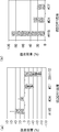
- anti-DDR1 antibody was intraperitoneally administered to mice transplanted with human lung cancer cell line NCI-H1993, and changes in tumor volume over time were measured.
- # 115, # 24, and # 27 an inhibitory effect on tumor growth was observed.
- # 115 showed the strongest tumor growth inhibitory effect.
- 20M102 showed no inhibitory effect on tumor growth.
- It is a graph showing the inhibitory activity of ligand-dependent cell migration of an anti-DDR1 antibody.
- A Ligand-dependent cell migration activity of human lung cancer cell line NCI-H1993 was measured using xCELLligence system TM . Collagen type 4 was used as the ligand.
- the vertical axis represents the degree of migration inhibition (%) of each anti-DDR1 antibody, where 100 is the case where cell migration by the ligand is completely inhibited. A negative degree of inhibition indicates that the amount of cell migration has increased due to the addition of antibody as compared to the case of ligand alone. In # 115 and # 24, inhibition of ligand-dependent cell migration was observed.
- B The ligand-dependent cell migration activity of the human lung cancer cell line NCI-H1993 was measured using a Cultrex assay kit. Collagen type 4 was used as the ligand.
- the vertical axis represents the degree of migration inhibition (%) of each anti-DDR1 antibody, where 100 is the case where cell migration by the ligand is completely inhibited.
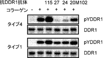
- FIG. 115 is a photograph showing the ligand-dependent phosphorylation inhibitory activity of DDR1 of an anti-DDR1 antibody.
- Ligand-dependent phosphorylation of DDR1 in the human breast cancer cell line T47D was detected by Western blotting using a polyclonal antibody that specifically recognizes DDR1 (pYDDR1) phosphorylated at tyrosine 796. Collagen type 1 or collagen type 4 was used as the ligand.
- pYDDR1 phosphorylation inhibitory activity of DDR1 of an anti-DDR1 antibody.
- Anti-DDR1 antibody uptake into cells was evaluated by adding anti-DDR1 antibody and MabZAP (saporin-labeled anti-mouse IgG antibody) to human breast cancer cell line T47D and examining whether cell proliferation was inhibited.
- the vertical axis represents the ratio of cell growth when anti-DDR1 antibody and MabZAP are added, assuming that cell growth when neither anti-DDR1 antibody nor MabZAP is added is 1. Uptake into cells was observed at # 115 and # 24. It is a photograph showing the fall of the expression level of DDR1 by an anti- DDR1 antibody.
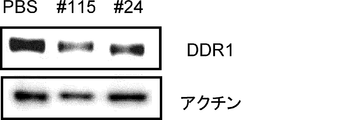
- PBS negative control
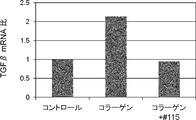
- anti-DDR1 antibody was added to human breast cancer cell line T47D, and the expression level of DDR1 in the cells was detected by Western blotting. Actin was used as an internal control. In # 115 and # 24, a decrease in the expression level of DDR1 was observed. It is a graph showing the ligand-dependent TGF ⁇ expression suppression activity of anti-DDR1 antibody.
- the expression level of ligand-dependent TGF ⁇ mRNA in a co-culture system of human lung cancer cell line NCI-H1993 and mouse fibroblast MRC5 was measured by quantitative RT-PCR (qRT-PCR). Collagen type 1 was used as the ligand.
- the vertical axis represents the ratio of the amount of TGF ⁇ mRNA when no ligand / antibody is added (control). In # 115, a ligand-dependent suppression of TGF ⁇ expression was observed.
- the present invention provides a novel anti-DDR1 antibody having antitumor activity.
- DDR1 Discoidin Domain Receptor 1
- the present inventors have found that antibodies that bind to the stalk region are antibodies alone or in comparison with antibodies that bind to other discoidin (DS) regions. It was found for the first time that it has strong antitumor activity. That is, the present invention provides an antibody that binds to the DDR1 stalk region.
- the animal species of DDR1 used in the present invention is preferably a mammal, and most preferably a human.
- the gene sequence and amino acid sequence of human DDR1 are registered as GenBank accession numbers NM_013993 and NP_054699, respectively.
- the gene sequence and amino acid sequence of mouse DDR1 are registered as GenBank accession numbers NM_007584 and NP_031610, and the gene sequence and amino acid sequence of rat DDR1 are registered as NM_013137 and NP_037269, respectively.
- those skilled in the art can determine the sequence by gene cloning using homology between species.
- DDR1 is a single transmembrane receptor tyrosine kinase (RTK), and is structurally divided into an extracellular region, a transmembrane (TM) region, and an intracellular region (kinase region).
- the amino acid sequence of human DDR1 is shown in SEQ ID NO: 2, and the base sequence is shown in SEQ ID NO: 1.
- the extracellular region includes a Discoidin (DS) region and a stalk region, and the DS region is considered to be involved in binding to collagen as a ligand.
- the region consisting of the amino acid sequence from the 32nd to the 185th is the DS region
- the region consisting of the amino acid sequence from the 199th to 412th is the stalk region It is called.
- the amino acid sequence of the DS region of human DDR1 is shown in SEQ ID NO: 3
- the amino acid sequence of the stalk region is shown in SEQ ID NO: 4.
- regions corresponding to each region can be similarly determined from sequence homology with human DDR1.
- binding activity of the antibody in the present invention can be measured using techniques known to those skilled in the art, such as ELISA (Enzyme-linked immunosorbent assay), Biacore, Western blotting, FACS and the like.
- binding means that the binding activity value measured by the method as described above is at least twice as high as the binding activity value of the negative control or the background value of the measurement method. It is preferably 3 times or more, more preferably 5 times or more, and most preferably 10 times or more.
- an antibody that binds to the DDR1 stalk region is obtained by immunizing an animal such as a mouse with a DDR1 protein to obtain a plurality of anti-DDR1 antibodies, and then screening to a stalk region.
- a partial protein corresponding to the DDR1 stalk region can be prepared in advance using a genetic engineering technique known to those skilled in the art, and immunized to an animal such as a mouse. It is also possible to produce it.
- One preferred embodiment of the anti-DDR1 antibody provided by the present invention includes an anti-DDR1 antibody characterized by suppressing cell proliferation.
- the cells in the present invention may be primary cultured cells collected from living tissue, or may be cell lines established by immortalizing them by some method.
- the cell phenotype is preferably a cell that expresses more DDR1 gene or DDR1 protein than normal cells.
- the amount of DDR1 gene expressed in the cell is determined by the amount of DDR1 protein expressed in the cell using techniques known to those skilled in the art such as RT-PCR using a primer specific for the DDR1 gene and GeneChip analysis. Can be evaluated using techniques known to those skilled in the art, such as Western blotting using an antibody specific for DDR1 protein and immunohistochemical staining (IHC).
- IHC immunohistochemical staining
- “suppressing cell proliferation” means that when an anti-DDR1 antibody is brought into contact with a cell, the proliferation of the cell is reduced as compared to when the antibody is not brought into contact.
- the decrease in cell proliferation includes a decrease in the growth rate while the cells are alive, and also includes the induction of cell death by apoptosis, necrosis, and the like.
- Inhibition of cell proliferation is preferably caused as a result of anti-DDR1 antibody binding to DDR1 on the cell surface. Inhibition of cell proliferation may be observed in vitro or in vivo.
- Suppression of cell proliferation in vitro can be measured by assay systems known to those skilled in the art, such as [ 3 H] thymidine incorporation method, MTT method, WST method, etc.
- Inhibition of cell proliferation in vivo It can be measured by an assay system known to those skilled in the art such as a xenograft model for transplanting human cells.
- the growth of cells in the evaluation system is completely suppressed to 100%, the growth is suppressed by, for example, 30% or more, 40% or more, 50% or more, 60% or more, 70% or more by the anti-DDR1 antibody.
- the antibody 20M102 that binds to the DS region of DDR1 disclosed in the prior document (Patent Document 3) showed about 20% growth inhibition in vivo (see FIG. 6 above), the present invention provides The present inventors have found that antibodies # 24 and # 27 that bind to the stalk region of DDR1 exhibit 48%, 61%, and # 115, respectively, 71% growth inhibition in vivo (Example 3 described later). reference).
- One preferred embodiment of the anti-DDR1 antibody provided by the present invention includes an anti-DDR1 antibody characterized by inhibiting cell migration.
- Cell migration is a phenomenon that explains the spontaneous movement of cells in a living body, and antibodies that inhibit cell migration may be able to suppress invasion or metastasis of cancer cells and are considered useful.
- “inhibiting cell migration” means that when an anti-DDR1 antibody is brought into contact with a cell, the cell migration activity is reduced as compared with the case where the antibody is not brought into contact. Inhibition of cell migration can be measured by an assay system that detects cell migration between chambers as described in Example 4 and the like. In cells expressing DDR1 protein on the cell surface, cell migration is stimulated by binding of a ligand to the extracellular region of DDR1, and cell migration is particularly induced by binding of collagen as a ligand.
- type I collagen or type IV collagen is suitable. Inhibition of cell migration is preferably caused as a result of the anti-DDR1 antibody inhibiting binding of DDR1 to the ligand.
- One preferred embodiment of the anti-DDR1 antibody provided by the present invention includes an anti-DDR1 antibody characterized by inhibiting phosphorylation of DDR1 in cells.
- the phosphorylation of DDR1 is preferably phosphorylation of a tyrosine residue contained in DDR1, and particularly preferably phosphorylation of the 796th tyrosine residue in the amino acid sequence of DDR1.
- DDR1 phosphorylation is known to transmit signals such as cell survival or cell invasion / metastasis, and antibodies that inhibit DDR1 phosphorylation may suppress cancer cell proliferation, invasion / metastasis, etc. There seems to be useful.
- inhibiting DDR1 phosphorylation means that when an anti-DDR1 antibody is brought into contact with a cell, the proportion of phosphorylated DDR1 is lower than when the antibody is not brought into contact. To do. Inhibition of DDR1 phosphorylation can be measured by an assay system known to those skilled in the art, such as Western blotting using an anti-phosphotyrosine antibody. In cells expressing DDR1 protein on the cell surface, DDR1 phosphorylation occurs when a ligand binds to the extracellular region of DDR1, and in particular, phosphorylation of DDR1 is induced by binding of collagen as a ligand. (Vogel W. et al., Mol. Cell (1997) 1, 13-23).
- DDR1 phosphorylation is preferably caused as a result of an anti-DDR1 antibody inhibiting binding of DDR1 to a ligand.
- DDR1 phosphorylation may be caused by DDR1 autophosphorylation or by phosphorylation by other kinases.
- the phosphorylation of DDR1 can be measured, for example, by the following method.
- DDR1-expressing cells eg, A549, NCI-H1993, SK-MES-1, Panc-1, MFE-280, HCT-116, BT474, ZR-75-1, T47D, BxPC3, etc.
- DDR1-expressing cells eg, A549, NCI-H1993, SK-MES-1, Panc-1, MFE-280, HCT-116, BT474, ZR-75-1, T47D, BxPC3, etc.
- the tyrosine residue of the extracted DDR1 protein is phosphorylated by Western blotting using an anti-phosphotyrosine antibody. More specifically, it can be measured by the method described in Example 5.
- the expression of DDR1 in the above cells is indicated by the following documents (L'HOTE CMM et al., FASEB J.
- One preferred embodiment of the anti-DDR1 antibody provided by the present invention is an anti-DDR1 antibody that is incorporated into cells. It is already known that there is a phenomenon in which substances existing on the cell surface are actively taken into cells through some mechanism. The uptake of the anti-DDR1 antibody into the cell is preferably caused as a result of binding of the anti-DDR1 antibody to the DDR1 protein expressed on the cell surface. Antibodies incorporated into cells are thought to be useful because they may suppress the growth of cancer cells by conjugating a compound having cytotoxic activity such as a toxin.
- incorporated into a cell means that when an anti-DDR1 antibody is brought into contact with a cell, a larger amount of the antibody is taken up than when a negative control antibody is brought into contact.
- the uptake of the antibody into the cell was taken into the cell by directly labeling the antibody with a toxin or by binding a secondary antibody labeled with the toxin to the antibody as in Example 6. It can be measured as the amount of toxin.
- the cell phenotype is preferably a cell that expresses more DDR1 than a normal cell, and such a cell has a gene level such as RT-PCR using a primer specific for the DDR1 gene or GeneChip analysis. Or analysis of protein level such as Western blotting using an antibody specific for DDR1 protein or immunohistochemical staining (IHC).
- One preferred embodiment of the anti-DDR1 antibody provided by the present invention includes an anti-DDR1 antibody characterized by reducing the expression level of DDR1 in cells.
- the decrease in the expression level of DDR1 may be due to promotion of DDR1 protein degradation or may be due to suppression of translation of DDR1 protein. Further, it may be due to promotion of DDR1 mRNA degradation, or may be due to suppression of transcription of DDR1 mRNA.
- An antibody that reduces the expression level of DDR1 is considered useful because it may be able to suppress phenomena such as survival, invasion, and metastasis involving DDR1 in cancer cells.
- reducing the expression level of DDR1 means that when the anti-DDR1 antibody is brought into contact with a cell, the expression level of DDR1 is lowered as compared with the case where the antibody is not brought into contact.
- the decrease in the expression level of DDR1 is preferably caused as a result of binding of anti-DDR1 antibody to DDR1 on the cell surface.
- the amount of DDR1 mRNA can be measured by an assay system known to those skilled in the art, such as RT-PCR using a primer specific for the DDR1 gene.
- the amount of DDR1 protein can be measured by an assay system known to those skilled in the art, such as Western blotting using an antibody specific for DDR1 protein.
- DDR1 protein is extracted from DDR1-expressing cells (for example, A549, NCI-H1993, SK-MES-1, Panc-1, MFE-280, HCT-116, BT474, ZR-75-1, T47D, BxPC3, etc.). Extracted DDR1 protein is detected by Western blotting. More specifically, it can be measured by the method described in Example 7.
- One preferred embodiment of the anti-DDR1 antibody provided by the present invention is an anti-DDR1 antibody characterized by reducing the expression level of TGF ⁇ in a cell.
- the decrease in the expression level of TGF ⁇ may be due to promotion of degradation of TGF ⁇ protein, or may be due to suppression of translation of TGF ⁇ protein. Further, it may be due to promotion of degradation of TGF ⁇ mRNA, or may be due to suppression of transcription of TGF ⁇ mRNA.
- TGF ⁇ is a marker molecule that is known to increase expression during epithelial-mesenchymal transition (EMT), which is reported to promote tumor formation, and reduces the expression level of TGF ⁇ .
- EMT epithelial-mesenchymal transition
- Antibodies are thought to be useful because they may inhibit tumor formation by suppressing cell epithelial-mesenchymal transition.
- one aspect of the anti-DDR1 antibody in the present invention may include an anti-DDR1 antibody characterized by inhibiting cell epithelial-mesenchymal transition (EMT).
- EMT cell epithelial-mesenchymal transition
- “reducing the expression level of TGF ⁇ ” means that when the anti-DDR1 antibody is brought into contact with a cell, the expression level of TGF ⁇ is lowered as compared with the case where the antibody is not brought into contact.
- collagen may induce the expression of TGF ⁇ via DDR1 (Guerrot D. et al., Am. J. Pathol. (2011) 179). 83-91).
- type I collagen or type IV collagen is suitable.
- the decrease in the expression level of TGF ⁇ is preferably caused as a result of the anti-DDR1 antibody inhibiting the binding between DDR1 and collagen.
- the amount of TGF ⁇ mRNA can be measured by an assay system known to those skilled in the art, such as RT-PCR using a primer specific for the TGF ⁇ gene.
- the amount of TGF ⁇ protein can be measured by an assay system known to those skilled in the art, such as Western blotting using an antibody specific for TGF ⁇ protein.
- the expression level of TGF ⁇ can be measured, for example, by the following method.
- DDR1-expressing cells eg, A549, NCI-H1993, SK-MES-1, Panc-1, MFE-280, HCT-116, BT474, ZR-75-1, T47D, BxPC3, etc.
- fibroblasts such as MRC5
- the amount of TGF ⁇ mRNA is measured by RT-PCR using a primer specific for TGF ⁇ . More specifically, it can be measured by the method described in Example 8.
- the cell in the present invention is preferably a cancer cell, more preferably a cancer cell expressing DDR1.
- cancer cells that express more DDR1 than normal cells.
- Such cells include gene-level analysis such as RT-PCR and GeneChip analysis using primers specific for the DDR1 gene, Western blotting and immunohistochemical staining (IHC) using antibodies specific for the DDR1 protein, etc.
- the protein level can be selected by analysis.
- the cancer cell type of the cancer cell of the present invention is not particularly limited.
- lung cancer small cell lung cancer, non-small cell lung cancer, etc.
- breast cancer glioma, ovarian cancer
- gastric cancer pancreatic cancer
- esophageal cancer endometrial cancer
- bile duct examples include cancer, colon cancer, liver cancer, leukemia, lymphoma, kidney cancer, prostate cancer, melanoma, thyroid cancer, bladder cancer, osteosarcoma and the like.
- lung cancer non-small cell lung cancer
- breast cancer glioma, ovarian cancer
- gastric cancer pancreatic cancer
- esophageal cancer endometrial cancer
- bile duct cancer examples include cancer, colon cancer, liver cancer, leukemia, lymphoma, kidney cancer, prostate cancer, melanoma, thyroid cancer, bladder cancer, osteosarcoma and the like.
- lung cancer non-small cell lung cancer
- breast cancer glioma, ovarian cancer
- the present invention provides the following antibodies (a) to (c): (A) an antibody (# 115) having the same amino acid sequence as the antibody produced by the hybridoma deposited under accession number FERM BP-11399; (B) an antibody (# 27) having the same amino acid sequence as the antibody produced by the hybridoma deposited under accession number FERM BP-11398; (C) An antibody (# 24) having the same amino acid sequence as the antibody produced by the hybridoma deposited under accession number FERM BP-11397.
- hybridomas are deposited internationally as follows. The contents specifying the international deposit of each hybridoma are described below. The following depositary organization (independent administrative agency, National Institute of Advanced Industrial Science and Technology, Patent Biological Depositary Center) has been handed over to the National Institute of Technology and Evaluation (NITE) on April 1, 2012.
- the antibodies described in (a) to (c) above are all antibodies that bind to the DDR1 stalk region.
- a person skilled in the art can determine the base sequence and amino acid sequence of an antibody produced by a hybridoma using, for example, the methods described in the Examples below.
- a known gene can be determined based on the sequence.
- Engineering techniques can be used to produce recombinant antibodies.
- the present invention also provides an antibody having the same CDR sequence as that of the antibodies described in (a) to (c) above.
- There are 6 CDRs in an antibody that is, H chain CDR1, CDR2, CDR3, L chain CDR1, CDR2, CDR3, and any one of these CDRs may be the same, and more preferably the H chain. It is sufficient that the three CDRs or the three CDRs of the L chain are the same, and it is more preferable that all the six CDRs are the same.
- a person skilled in the art can bind the CDR of the antibody described in any one of (a) to (c) above to another appropriate antibody, thereby binding the DDR1 to the stalk region substantially equivalent to the antibody.
- CDR-grafted antibody can be prepared, and such an antibody is useful in the same manner as the antibody.
- the position and numbering system of the CDR region and the FR region of the antibody are defined by, for example, Kabat et al. (Kabat EA et al., (1991) Sequences of Proteins of Immunological Edition, Fifth Edition, US Department). Health and Human Services, US Government Printing Office).
- the present invention provides an antibody that competes with the antibody of the present invention for binding to DDR1.
- “competing binding to DDR1” means that the binding activity of the antibody of the present invention to DDR1 when an anti-DDR1 antibody coexists in an assay system for measuring the binding of an antibody to DDR1. It means to decline.
- Such an antibody is considered to be an antibody that binds to an antigenic determinant (epitope) that is the same as or very close to that of the antibody of the present invention, and thus is useful in the same manner as the antibody of the present invention.
- the full length of the DDR1 protein may be used, or the extracellular region of the DDR1 protein may be used. Alternatively, the stalk region of the DDR1 protein may be used.
- Competition for binding to DDR1 can be measured by an assay system known to those skilled in the art such as a cross-blocking assay.
- a competitive ELISA assay that utilizes an enzyme label is a preferred cross-blocking assay.
- the competition for binding to DDR1 can be measured, for example, by the following method.
- the anti-DDR1 antibody is added after preincubation of the DDR1 protein coated on the wells of the microtiter plate in the presence or absence of the test antibody.
- the amount of anti-DDR1 antibody that binds to the DDR1 protein in the well decreases.
- the amount of antibody to be bound can be easily measured by labeling the anti-DDR1 antibody in advance.
- the amount of antibody bound can be measured by labeling anti-DDR1 antibody with biotin and using an avidin-peroxidase conjugate and its appropriate substrate.
- the amount of bound antibody can be measured by radioactively labeling or fluorescently labeling the anti-DDR1 antibody.
- the amount of antibody to be bound is determined by a labeled antibody that specifically recognizes the constant region of the antibody derived from the animal species. It can also be measured. Alternatively, if the antibodies are derived from the same animal species but have different subclasses, the amount of antibody to be bound can be measured with a labeled antibody that specifically recognizes each subclass.
- the present invention provides an antibody that binds to the same epitope to which the antibody of the present invention binds.
- An epitope of an antibody can be identified by a method of synthesizing a group of peptides (such as a peptide array) that covers the amino acid sequences of the antigen so as to overlap each other and measuring the binding activity of the antibody to each peptide (Poetz O Et al., Proteomics (2005) 5, 2402-11).
- a method for analyzing the crystal structure of an antigen-antibody (Vyas NK, et al., Biochemistry (2004) 41, 13575-86), a group of mutant proteins in which the amino acid sequence of the antigen is substituted with alanine one by one is prepared.
- epitope thus identified is identical to the epitope to which the antibody of the present invention binds or is very close to the epitope to which the antibody of the present invention binds, then the antibody that binds to the epitope Since the antibody is considered to have a binding activity equivalent to that of the antibody of the invention, it is useful in the same manner as the antibody of the present invention.
- “Epitopes are very close” means that the difference in epitope positions is preferably within 5 amino acids, more preferably within 4 amino acids, even more preferably within 3 amino acids, particularly preferably within 2 amino acids, most preferably 1 Means an amino acid.
- an antibody that binds to an epitope to which the antibody of the present invention binds can be obtained by methods known to those skilled in the art.
- the epitope to which the antibody of the present invention binds is determined by the above-described method, and an antibody is prepared using a polypeptide having an amino acid sequence contained in the epitope as an immunogen, or an epitope of an antibody prepared by a normal method And a method for selecting an antibody having the same epitope as that of the antibody of the present invention.
- the present invention relates to an antibody in which one or more amino acids are added, deleted and / or substituted with other amino acids in the antibody of the present invention, and the antibody before the addition, deletion and / or substitution is performed.
- Antibodies having equivalent binding activity to the DDR1 stalk region are provided.
- “having an equivalent binding activity to the DDR1 stalk region” means that an antibody in which one or more amino acids are added, deleted, and / or substituted with another amino acid to the DDR1 stalk region. Is preferably 70% or more, more preferably 80% or more, still more preferably 90% or more, and most preferably 95% or more compared to the antibody before the addition, deletion and / or substitution. Means. Since such an antibody is considered to have almost the same properties as the antibody of the present invention, it is useful as well as the antibody of the present invention.
- Amino acid additions, deletions and / or substitutions are described, for example, by site-directed mutagenesis (Hashimoto-Gotoh T. et al., Gene (1995) 152, 271-275, Zoller MJ & Smith M., Methods Enzymol (1983). ) 100, 468-500, Kramer W. et al., Nucleic Acids Res (1987) 12, 9441-9456, Kramer W. & Fritz H. J., Methods Enzymol (1987) 154, 350-367, Kunkel TA,. Proc Natl Acad Sci USA (1985) 82, 488-492) can be used by methods known to those skilled in the art.
- amino acid side chain is substituted with another amino acid that preserves the properties of the amino acid side chain.
- amino acid substitutions that preserve the properties of amino acid side chains include, for example, hydrophobic amino acids (A, I, L, M, F, P, W, Y, V), hydrophilic amino acids (R, D, N , C, E, Q, G, H, K, S, T) amino acids having aliphatic side chains (G, A, V, L, I, P), amino acids having hydroxyl group-containing side chains (S, T, Y), amino acids having sulfur atom-containing side chains (C, M), amino acids having carboxylic acid and amide-containing side chains (D, N, E, Q), amino acids having base-containing side chains (R, K, H) ), Amino acid substitutions within each group such as amino acids (H, F, Y, W) having aromatic-containing side chains.
- the position of the amino acid to which addition, deletion and / or substitution is performed is not particularly limited, but addition, deletion and / or substitution to an amino acid which is not involved in antigen binding or antibody structure maintenance Is preferred.
- an antibody is divided into a constant region and a variable region, those skilled in the art can easily specify such a position in the constant region.
- the variable region is divided into a framework region and a CDR region, those skilled in the art can specify such a position in the framework region without undue burden.
- One skilled in the art can also identify such positions within the CDR regions.
- the antibody of the present invention may be a polyclonal antibody or a monoclonal antibody, but is preferably a monoclonal antibody.
- the monoclonal antibody can be obtained by using a known means such as a hybridoma method for immunizing an animal with an antigen or a phage display method for screening an antibody library.
- the monoclonal antibodies of the present invention include not only antibodies obtained from clones derived from antibody-producing cells such as hybridomas, but also humanized antibodies and chimeric antibodies not derived from hybridomas.
- the antibody subclass is not particularly limited.
- IgG, IgM, IgA, IgD, IgE and the like are preferable, and IgG is more preferable.
- a monoclonal antibody can be obtained as follows. First, a DDR1 protein serving as an antigen is prepared, and this is immunized to an animal by a normal immunization method. Immune cells obtained from the immunized animal are fused with known parental cells by a conventional cell fusion method to obtain a hybridoma. From the obtained hybridomas, a hybridoma producing the desired anti-DDR1 antibody is selected by a usual screening method. Specifically, monoclonal antibodies can be obtained by the method described in Example 1.
- Monoclonal antibodies are prepared as follows, for example. First, by expressing the DDR1 gene, a DDR1 protein used as a sensitizing antigen for antibody acquisition can be obtained.
- the base sequence of the human DDR1 gene is already known (GenBank accession number NM — 013993). That is, after inserting a gene sequence encoding DDR1 into a known expression vector to transform an appropriate host cell, the target DDR1 protein can be purified from the host cell or culture supernatant by a known method.
- a purified natural DDR1 protein can also be used in the same manner. The purification can be generated by using a plurality of chromatographies such as ordinary ion chromatography and affinity chromatography one time or a plurality of times, in combination or independently.
- a fusion protein obtained by fusing a partial polypeptide containing at least a part of the DDR1 stalk region with a different polypeptide can also be used as an immunogen.
- an immunogen for example, an Fc fragment of an antibody, a peptide tag, or the like can be used.
- a vector that expresses a fusion protein can be prepared by fusing genes encoding two or more desired polypeptide fragments in-frame and inserting the fusion gene into an expression vector as described above ( Sambrook J. et al., Molecular Cloning 2nd ed. (1989) 9.47-9.58, Cold Spring Harbor Lab. Press).
- the DDR1 protein thus purified can be used as a sensitizing antigen used for immunization against mammals.
- a peptide having the entire DDR1 stalk region or at least 5 or more consecutive amino acid sequences can be used as the partial peptide.
- the at least 5 or more consecutive amino acid sequences are preferably 6 or more, more preferably 8 or more consecutive amino acid sequences. Further, at least 5 or more consecutive amino acid sequences are sequences specific to the DDR1 stalk region and have an antigenicity.
- the mammal immunized with the sensitizing antigen is not particularly limited.
- an immunized animal in consideration of compatibility with a parent cell used for cell fusion.
- mice, rats, hamsters, rabbits, chickens, monkeys and the like can be used as immunized animals.
- rodent animals such as mice, rats and hamsters are preferred as immunized animals.
- the above animals can be immunized with a sensitizing antigen.
- mammals can be immunized by injecting a sensitizing antigen intraperitoneally or subcutaneously. Specifically, the sensitizing antigen is administered to mammals several times every 4 to 21 days.
- the sensitizing antigen is used for immunization after being diluted with PBS (Phosphate-Buffered Saline) or physiological saline at an appropriate dilution rate.
- a sensitizing antigen can be administered with an adjuvant. For example, it can be mixed with Freund's complete adjuvant and emulsified to obtain a sensitizing antigen.
- An appropriate carrier can be used for immunization with the sensitizing antigen.
- a partial peptide having a small molecular weight is used as a sensitizing antigen, it is desirable to immunize the sensitizing antigen peptide by binding it to a carrier protein such as albumin or keyhole limpet hemocyanin.
- immune cells are collected from the mammal and subjected to cell fusion.
- spleen cells can be used.
- Mammalian myeloma cells are used as the cells fused with the immune cells.
- the myeloma cell is preferably provided with an appropriate selection marker for screening.
- a selectable marker refers to a trait that can (or cannot) survive under certain culture conditions.
- Known selection markers include hypoxanthine-guanine-phosphoribosyltransferase deficiency (hereinafter abbreviated as HGPRT deficiency) or thymidine kinase deficiency (hereinafter abbreviated as TK deficiency).
- HGPRT deficiency hypoxanthine-guanine-phosphoribosyltransferase deficiency
- TK deficiency thymidine kinase deficiency
- Cells having HGPRT or TK deficiency have hypoxanthine-aminopterin-thymidine sensitivity (hereinafter abbreviated as HAT sensitivity).
- HGPRT-deficient and TK-deficient cells can be selected in a medium containing 6 thioguanine, 8 azaguanine (hereinafter abbreviated as 8AG), or 5 ′ bromodeoxyuridine, respectively.
- 8AG 8 azaguanine
- 5 ′ bromodeoxyuridine normal cells die because they incorporate these pyrimidine analogs into the DNA, but cells deficient in these enzymes cannot survive these pyrimidine analogs and can survive in selective media.
- G418 resistance confers resistance to 2-deoxystreptamine antibiotics (gentamicin analogs) with a neomycin resistance gene.
- Various myeloma cells suitable for cell fusion are known. For example, P3 (P3x63Ag8.653) (J. Immunol.
- the cell fusion between the immune cells and myeloma cells can be performed according to a known method, for example, the method of Kohler and Milstein (Kohler G. & Milstein C., Methods Enzymol. (1981) 73, 3-46). Is possible. More specifically, for example, the cell fusion can be carried out in a normal nutrient culture medium in the presence of a cell fusion promoter.
- a cell fusion promoter for example, polyethylene glycol (PEG), Sendai virus (HVJ) or the like can be used.
- an auxiliary agent such as dimethyl sulfoxide can be added as desired in order to increase the fusion efficiency.
- the usage ratio of immune cells and myeloma cells can be set arbitrarily.
- the number of immune cells is preferably 1 to 10 times that of myeloma cells.
- the culture solution used for the cell fusion for example, RPMI1640 culture solution suitable for growth of the myeloma cell line, MEM culture solution, and other normal culture solutions used for this type of cell culture can be used.
- serum supplements such as fetal calf serum (FCS) can be added to the culture medium.
- a predetermined amount of the immune cells and myeloma cells are mixed well in the culture solution, and a target PEG (hybridoma) is formed by mixing a PEG solution preheated to about 37 ° C.
- the In the cell fusion method for example, PEG having an average molecular weight of about 1000 to 6000 can be usually added at a concentration of 30 to 60% (w / v).
- cell fusion agents and the like that are undesirable for the growth of hybridomas are removed by sequentially adding the appropriate culture solution listed above, and centrifuging to remove the supernatant.
- the hybridoma obtained in this manner can be selected by using a selective culture solution corresponding to the selection marker possessed by the myeloma used for cell fusion.
- a selective culture solution corresponding to the selection marker possessed by the myeloma used for cell fusion.
- cells having HGPRT or TK deficiency can be selected by culturing in a HAT culture solution (a culture solution containing hypoxanthine, aminopterin and thymidine). That is, when HAT-sensitive myeloma cells are used for cell fusion, cells that have succeeded in cell fusion with normal cells can be selectively proliferated in the HAT culture solution.
- the culture using the HAT culture solution is continued for a time sufficient for cells other than the target hybridoma (non-fused cells) to die.
- the target hybridoma can be selected by culturing for several days to several weeks. Subsequently, by carrying out the usual limiting dilution method, screening and single cloning of the hybridoma producing the target antibody can be performed.
- Screening and single cloning of the target antibody can be suitably performed by a screening method based on a known antigen-antibody reaction.
- the antigen is bound to a carrier such as beads made of polystyrene or the like, or a commercially available 96-well microtiter plate, and reacted with the culture supernatant of the hybridoma.
- a secondary antibody labeled with an enzyme is reacted.
- the secondary antibody binds to the carrier via this antibody.
- the DDR1 protein that is practically homogeneous, including those used for immunization, can be preferably used as the antigen.
- a target antibody can be obtained by sensitizing human lymphocytes with an antigen.
- human lymphocytes are first sensitized with DDR1 protein in vitro.
- the immunized lymphocytes are then fused with an appropriate fusion partner.
- the fusion partner for example, a myeloma cell derived from human and having a permanent division ability can be used (Japanese Patent Publication No. 1-59878).
- the anti-DDR1 antibody obtained by this method is a human antibody having a binding activity to the DDR1 protein.
- an anti-DDR1 human antibody can also be obtained by immunizing a transgenic animal having all repertoires of human antibody genes with DDR1 protein as an antigen.
- Antibody-producing cells of the immunized animal can be immortalized by treatment such as cell fusion with an appropriate fusion partner or Epstein-Barr virus infection.
- Human antibodies can also be isolated from the immortalized cells thus obtained (WO94 / 25585, WO93 / 12227, WO92 / 03918, WO94 / 02602). Further, by cloning the immortalized cells, it is possible to clone cells that produce an antibody having the desired reaction specificity.
- the immune system of the animal recognizes human DDR1 as a foreign substance. Therefore, a human antibody against human DDR1 can be easily obtained.
- the hybridoma producing the monoclonal antibody thus produced can be subcultured in a normal culture solution.
- the hybridoma can also be stored for a long time in liquid nitrogen.
- the hybridoma can be cultured according to a usual method, and the target monoclonal antibody can be obtained from the culture supernatant.
- a hybridoma can be administered to a mammal compatible therewith to proliferate and a monoclonal antibody can be obtained as its ascites.
- the former method is suitable for obtaining a highly pure antibody.
- an antibody encoded by an antibody gene cloned from an antibody-producing cell can also be used.
- the cloned antibody gene can be expressed as an antibody by incorporating it into a suitable vector and introducing it into a host. Methods for isolation of antibody genes, introduction into vectors, and transformation of host cells have already been established (Vandam AM et al., Eur. J. Biochem. (1990) 192, 767-775. ).
- cDNA encoding a variable region (V region) of an anti-DDR1 antibody can be obtained from a hybridoma cell that produces the anti-DDR1 antibody.
- V region variable region
- RNA is extracted from the hybridoma.
- Methods for extracting total RNA from cells include, for example, guanidine ultracentrifugation (Chirgwin J.M. et al., Biochemistry (1979) 18, 5294-5299), AGPC method (Chomczynski P. et al., Anal. Biochem. ( 1987) 162, 156-159) and the like can be used.
- MRNA can be purified from the extracted total RNA using mRNA Purification Kit (GE Healthcare) or the like.
- kits for directly extracting mRNA from cells such as QuickPrep mRNA Purification Kit (GE Healthcare) are also commercially available.
- mRNA can also be obtained from the hybridoma.
- CDNA encoding the antibody V region can be synthesized from the obtained mRNA using reverse transcriptase.
- cDNA can be synthesized by AMV Reverse Transscriptase First-strand cDNA Synthesis Kit (Seikagaku Corporation).
- 5'-RACE method Frohman MA et al., Proc. Natl. Acad. Sci.
- the target cDNA fragment is purified from the obtained PCR product and then ligated with vector DNA.
- a recombinant vector is produced, introduced into Escherichia coli or the like and a colony is selected, a desired recombinant vector can be prepared from Escherichia coli that has formed the colony.
- Whether or not the recombinant vector has the target cDNA base sequence can be confirmed by a known method such as the dideoxynucleotide chain termination method.
- a PCR method using primers for variable region gene amplification can also be used.
- cDNA is synthesized using the extracted mRNA as a template to obtain a cDNA library. It is convenient to use a commercially available kit for the synthesis of the cDNA library. Actually, the amount of mRNA obtained from only a small number of cells is extremely small, so the yield is low when it is purified directly. Therefore, it is usually purified after adding carrier RNA that is apparently free of antibody genes. Alternatively, when a certain amount of RNA can be extracted, it is possible to efficiently extract only RNA of antibody-producing cells. For example, it may not be necessary to add carrier RNA for RNA extraction from 10 or more, 30 or more, preferably 50 or more antibody-producing cells.
- the antibody gene is amplified by the PCR method using the obtained cDNA library as a template.
- Primers for amplifying antibody genes by PCR are known. For example, J. et al. Mol. Biol. (1991) 222, 581-597 and the like can be used to design primers for human antibody gene amplification. These primers have different nucleotide sequences for each immunoglobulin subclass. Therefore, when a cDNA library whose subclass is unknown is used as a template, the PCR method is performed in consideration of all possibilities.
- a primer capable of amplifying genes encoding ⁇ 1 to ⁇ 5 as a heavy chain and ⁇ chain and ⁇ chain as a light chain may be used. it can.
- a primer that anneals to a portion corresponding to the hinge region is generally used as the 3 ′ primer.
- a primer corresponding to each subclass can be used as the 5′-side primer.
- PCR products using primers for gene amplification of each subclass of heavy chain and light chain should be independent libraries.
- an immunoglobulin comprising a combination of a heavy chain and a light chain can be reconstructed.
- the target antibody can be screened using the binding activity of the reconstituted immunoglobulin to the antigen as an index.
- the cDNA is digested with a restriction enzyme that recognizes restriction enzyme sites inserted at both ends of the cDNA.
- a preferred restriction enzyme is a restriction enzyme that recognizes and digests a base sequence that is unlikely to appear in a base sequence constituting an antibody gene.
- a restriction enzyme that provides a sticky end is preferred.
- An antibody expression vector can be obtained by inserting cDNA encoding the V region of the antibody digested as described above into an appropriate expression vector.
- a chimeric antibody can be obtained by fusing the gene encoding the antibody constant region (C region) and the gene encoding the V region in-frame.
- the chimeric antibody refers to an antibody having a different origin from the constant region and the variable region.
- a heterologous chimeric antibody such as mouse-human
- a human-human homologous chimeric antibody is also included in the chimeric antibody of the present invention.
- a chimeric antibody expression vector can also be constructed by inserting the V region gene into an expression vector having a constant region in advance.
- a restriction enzyme recognition sequence for a restriction enzyme that digests the V region gene is placed on the 5 ′ side of an expression vector holding a DNA encoding a desired antibody constant region (C region).
- a chimeric antibody expression vector is constructed by digesting both with the same combination of restriction enzymes and fusing them in frame.
- the antibody gene can be incorporated into an expression vector so as to be expressed under the control of the expression control region.
- An expression control region for expressing an antibody includes, for example, an enhancer and a promoter. Subsequently, by transforming an appropriate host cell with this expression vector, a recombinant cell that expresses the DNA encoding the antibody can be obtained.
- DNAs encoding antibody heavy chains (H chains) and light chains (L chains) can be incorporated into separate expression vectors. By co-transfecting a vector incorporating the H chain and the L chain into the same host cell at the same time, an antibody molecule having the H chain and the L chain can be expressed.
- host cells may be transformed by incorporating DNAs encoding H and L chains into a single expression vector (WO94 / 11523).
- hosts and expression vectors for expressing antibodies are known. Any of these expression systems can be applied to the present invention.
- animal cells When eukaryotic cells are used as hosts, animal cells, plant cells, or fungal cells can be used.
- animal cells that can be used in the present invention include mammalian cells (CHO, COS, 3T3, myeloma, BHK (baby hamster kidney), Hela, C127, HEK293, Bowes melanoma cells, Vero, and the like. ), Amphibian cells (Xenopus oocytes, etc.), insect cells (Drosophila S2, sf9, sf21, Tn5, etc.) can be used.
- mammalian cells CHO, COS, 3T3, myeloma, BHK (baby hamster kidney), Hela, C127, HEK293, Bowes melanoma cells, Vero, and the like.
- Amphibian cells Xenopus oocytes, etc.
- Nicotiana As plant cells, antibody gene expression systems using cells derived from the genus Nicotiana, such as Nicotiana tabacum, are known. Callus cultured cells can be used for transformation of plant cells.
- fungal cells include yeasts (eg, Saccharomyces cerevisiae, Saccharomyces pombe), methanol-utilizing yeasts (Pichia pistia piapia, etc.).
- yeasts eg, Saccharomyces cerevisiae, Saccharomyces pombe
- methanol-utilizing yeasts Piichia pistia piapia, etc.
- a fungus can be used.
- antibody gene expression systems using prokaryotic cells are also known.
- bacterial cells such as E. coli, Streptococcus, Staphylococcus, Streptomyces and Bacillus subtilis can be used in the present invention.
- the promoter / enhancer can include human cytomegalovirus early promoter / enhancer (human cytomegalovirus immediate early promoter / enhancer).
- viral promoters / enhancers or promoters / enhancers derived from mammalian cells such as human elongation factor 1 ⁇ (HEF1 ⁇ ).
- viral promoters / enhancers or promoters / enhancers derived from mammalian cells such as human elongation factor 1 ⁇ (HEF1 ⁇ ).
- viruses that can utilize promoters / enhancers include retroviruses, polyomaviruses, adenoviruses, and simian virus 40 (SV40).
- the method of Mulligan et al. (Nature (1979) 277, 108) can be used. Further, the HEF1 ⁇ promoter / enhancer can be easily used for target gene expression by the method of Mishimashima et al. (Nucleic Acids Res. (1990) 18, 5322).
- the gene can be expressed by functionally combining a useful promoter commonly used, a signal sequence for antibody secretion, and an antibody gene to be expressed.
- the promoter include lacZ promoter and araB promoter.
- the lacZ promoter the method of Ward et al. (Nature (1989) 341, 544-546, FASEB J. (1992) 6, 2422-2427) can be used.
- the araB promoter can be used for expression of a target gene by the method of Better et al. (Science (1988) 240, 1041-1043).
- a pelB signal sequence (Lei SP et al., J. Bacteriol. (1987) 169, 4379) may be used in the case of production in the periplasm of E. coli. Then, after separating the antibody produced in the periplasm, the structure of the antibody is reshaped so as to have a desired binding activity by using a protein denaturant such as urea or guanidine hydrochloride.
- a selectable marker can be inserted into the expression vector for amplification of the gene copy number in the host cell system.
- selection markers such as aminoglycoside transferase (APH) gene, thymidine kinase (TK) gene, E. coli xanthine guanine phosphoribosyltransferase (Ecogpt) gene, dihydrofolate reductase (dhfr) gene and the like can be used.
- These expression vectors are introduced into host cells, and the transformed host cells are cultured in vitro or in vivo to produce the desired antibody.
- Host cells are cultured according to a known method.
- DMEM, MEM, RPMI 1640, and IMDM can be used as a culture solution, and a serum supplement such as fetal calf serum (FCS) can be used in combination.
- FCS fetal calf serum
- the present invention relates to an antibody produced by culturing a host cell transformed in this manner. For example, antibodies obtained by culturing transformed host cells and recovering them from the culture supernatant and the like are included.
- the present invention also provides a method for producing an antibody, comprising a step of culturing transformed host cells and recovering the antibody.
- a method for producing an antibody comprising a step of culturing transformed host cells and recovering the antibody.
- the production method when the antibody of the present invention is secreted into the medium or culture supernatant, the medium or culture supernatant is collected.
- the antibody of the present invention is produced intracellularly, the cell is first lysed, and then the antibody is recovered.
- mammals and insects can also be used for the production of recombinant antibodies.
- mammals goats, pigs, sheep, mice, cows and the like can be used (Vicki Glaser, SPECTRUM Biotechnology Applications (1993)).
- a transgenic animal can be used. That is, the antibody can be obtained from an animal into which a gene encoding the target antibody has been introduced.
- an antibody gene can be constructed as a fusion gene by inserting in frame into a gene that encodes a protein that is uniquely produced in milk.
- a protein secreted into milk for example, goat ⁇ -casein can be used.
- the DNA fragment containing the fusion gene into which the antibody gene has been inserted is injected into a goat embryo, and the injected embryo is introduced into a female goat.
- the desired antibody can be obtained as a fusion protein with milk protein from milk produced by a transgenic goat (or its offspring) born from a goat that has received the embryo.
- hormones can be used as appropriate in transgenic goats to increase the amount of milk containing the desired antibody produced from the transgenic goat (Ebert KM et al., Bio / Technology (1994) 12, 699-702). ).
- the target antibody can be obtained from the body fluid of the silkworm by infecting the silkworm with a baculovirus into which a nucleic acid encoding the target antibody is inserted (Susumu et al., Nature (1985) 315: 592-4).
- a plant when a plant is used for producing the antibody of the present invention, for example, tobacco can be used.
- a nucleic acid encoding the target antibody is inserted into a plant expression vector such as pMON 530, and this vector is introduced into a bacterium such as Agrobacterium tumefaciens.
- This bacterium can be infected with tobacco, for example, Nicotiana tabacum, and the desired antibody can be obtained from the leaves of this tobacco (Ma et al., Eur. J. Immunol. (1994) 24: 131- 8).
- the antibody expressed and produced as described above can be isolated from inside or outside the host cell (medium, milk, etc.) and purified as a substantially pure and homogeneous antibody. Separation and purification of the antibody can be performed by using a known method used in normal protein purification alone or in appropriate combination. For example, ammonium sulfate or ethanol precipitation, acid extraction, chromatography column, filter, ultrafiltration, salting out, solvent precipitation, solvent extraction, distillation, immunoprecipitation, SDS-polyacrylamide gel electrophoresis, isoelectric focusing, dialysis
- the antibodies can be separated and purified by appropriately selecting and combining recrystallization and the like (Harrow E. & Lane D., Antibodies: A Laboratory Manual (1988) Cold Spring Harbor Laboratory).
- chromatography examples include affinity chromatography, ion exchange chromatography such as anion or cation exchange chromatography, phosphocellulose chromatography, hydrophobic (interaction) chromatography, gel filtration, reverse phase chromatography, adsorption chromatography, hydroxyl Examples include apatite chromatography and lectin chromatography (Strategies for Protein Purification and Characterization: A Laboratory Course Manual. Ed Daniel R. Marshak et al. (1996) Cold Spring Harbor Laboratory Press). These chromatography can be performed using liquid phase chromatography, for example, liquid phase chromatography such as HPLC and FPLC.
- Columns used for affinity chromatography include protein A columns and protein G columns. Examples of the column using protein A include Hyper D, POROS, Sepharose F F F (Pharmacia), and the like.
- an appropriate protein modifying enzyme can be allowed to act before or after purification of the antibody, and the peptide can be partly removed.
- protein modifying enzymes include trypsin, chymotrypsin, lysyl endopeptidase, protein kinase, glucosidase, and the like.
- An animal antibody-derived C region can be used as the C region of the recombinant antibody of the present invention.
- C ⁇ 1, C ⁇ 2a, C ⁇ 2b, C ⁇ 3, C ⁇ , C ⁇ , C ⁇ 1, C ⁇ 2, and C ⁇ can be used as the H chain C region of the mouse antibody, and C ⁇ and C ⁇ can be used as the L chain C region.
- animal antibodies such as rats, rabbits, goats, sheep, camels and monkeys can be used as animal antibodies other than mouse antibodies. These sequences are known.
- the C region can be modified in order to improve the stability of the antibody itself or the production of the antibody.
- an antibody when administered to a human, it can be a genetically modified antibody artificially modified for the purpose of reducing the immunogenicity of the human.
- the recombinant antibody includes, for example, a chimeric antibody and a humanized antibody.
- a chimeric antibody refers to an antibody in which different variable regions and constant regions derived from each other are linked.
- an antibody comprising a heavy chain and light chain variable region of a mouse antibody and a human antibody heavy chain and light chain constant region is a mouse-human heterochimeric antibody.
- a recombinant vector that expresses a chimeric antibody can be prepared by ligating DNA encoding the variable region of a mouse antibody with DNA encoding the constant region of a human antibody in frame and incorporating it into an expression vector.
- the chimeric antibody produced in the culture can be obtained by culturing recombinant cells transformed with the vector and expressing the incorporated DNA.
- Human antibodies can be used for the C region of chimeric and humanized antibodies.
- C ⁇ 1, C ⁇ 2, C ⁇ 3, C ⁇ 4, C ⁇ , C ⁇ , C ⁇ 1, C ⁇ 2, and C ⁇ can be used as the C region.
- C ⁇ and C ⁇ can be used as the C region.
- the amino acid sequences of these C regions and the base sequences encoding them are known.
- the human antibody C region can be modified in order to improve the antibody itself or the stability of antibody production.
- a chimeric antibody is composed of a V region of an antibody derived from a non-human animal and a C region derived from a human antibody.
- humanized antibodies include complementarity determining regions (CDRs) of non-human animal-derived antibodies, framework regions (FR) of human antibodies, and C regions derived from human antibodies. Consists of Since humanized antibodies have reduced immunogenicity in the human body, they are useful as active ingredients of the therapeutic agent of the present invention.
- variable region of an antibody is usually composed of three complementarity determining regions (CDRs) sandwiched between four framework regions (FR).
- CDR complementarity determining regions
- FR framework regions
- the CDR is a region that substantially determines the binding specificity of the antibody.
- the amino acid sequence of CDR is rich in diversity.
- the amino acid sequence constituting FR often shows high homology among antibodies having different binding specificities. Therefore, it is generally said that the binding specificity of one antibody can be transplanted to another antibody by CDR grafting.
- Humanized antibodies are also referred to as reshaped human antibodies.
- non-human animals for example, humanized antibodies obtained by transplanting CDRs of mouse antibodies into human antibodies are known.
- General genetic recombination techniques for obtaining humanized antibodies are also known.
- Overlap Extension PCR is known as a method for transplanting a CDR of a mouse antibody into a human FR.
- overlap extension PCR a base sequence encoding the CDR of a mouse antibody to be transplanted is added to a primer for synthesizing the FR of a human antibody. Primers are prepared for each of the four FRs.
- the base sequences to be linked are designed to be connected to each other in frame.
- Human FRs are synthesized individually with specific primer sets.
- a product in which DNA encoding mouse CDR is added to each FR is obtained.
- the base sequences encoding the mouse CDRs of each product are designed to overlap each other.
- the overlapping CDR portions of the products are annealed with each other to perform a complementary strand synthesis reaction.
- human FRs are linked via the mouse CDR sequence.
- a humanized antibody expression vector can be prepared by inserting the DNA obtained as described above and a DNA encoding the human antibody C region into an expression vector so as to be fused in frame. After introducing the expression vector into a host to establish a recombinant cell, the recombinant cell is cultured, and a DNA encoding the humanized antibody is expressed, whereby the humanized antibody is cultured in the cultured cell. (EP239400, WO96 / 02576).
- the CDR forms a favorable antigen-binding site when linked via CDR.
- a human antibody FR can be suitably selected.
- FR amino acid residues can be substituted so that the CDR of the reshaped human antibody forms an appropriate antigen-binding site.
- amino acid sequence mutations can be introduced into FRs by applying the PCR method used for transplantation of mouse CDRs into human FRs.
- partial nucleotide sequence mutations can be introduced into primers that anneal to the FR. A base sequence mutation is introduced into the FR synthesized by such a primer.
- a mutant FR sequence having a desired property can be selected by measuring and evaluating the binding activity of the mutant antibody substituted with an amino acid to the antigen by the above method (Sato K. et al., Cancer Res (1993) 53, 851-856). ).
- a method for obtaining a human antibody is also known.
- human lymphocytes are sensitized with the desired antigen or cells expressing the desired antigen in vitro.
- a desired human antibody having an antigen-binding activity can be obtained by fusing sensitized lymphocytes with human myeloma cells (Japanese Patent Publication No. 1-59878).
- U266 can be used for human myeloma cells which are fusion partners.
- a desired human antibody can be obtained by immunizing a transgenic animal having a repertoire of human antibody genes with a desired antigen (WO93 / 12227, WO92 / 03918, WO94 / 02602, WO94 / 25585, WO96 / 334096, WO96). / 33735).
- a technique for obtaining a human antibody by panning using a human antibody library is also known.
- the V region of a human antibody is expressed as a single chain antibody (scFv) on the surface of the phage by the phage display method, and a phage that binds to the antigen can be selected.
- the DNA sequence encoding the V region of the human antibody that binds to the antigen can be determined. After determining the DNA sequence of scFv that binds to the antigen, the V region sequence is fused in-frame with the sequence of the desired human antibody C region, and then inserted into an appropriate expression vector, whereby an expression vector can be prepared.
- the human antibody can be obtained by introducing the expression vector into a suitable expression cell as described above and expressing the gene encoding the human antibody. These methods are already known (WO92 / 01047, WO92 / 20791, WO93 / 06213, WO93 / 11236, WO93 / 19172, WO95 / 01438, WO95 / 15388).
- the antibody of the present invention includes not only a bivalent antibody typified by IgG but also a monovalent antibody or a multivalent antibody typified by IgM as long as it binds to the DDR1 stalk region.
- the multivalent antibodies of the present invention include multivalent antibodies that all have the same antigen-binding site, or multivalent antibodies that have some or all different antigen-binding sites.
- the antibody of the present invention may be a bispecific antibody as long as it binds to the stalk region of DDR1.
- Bispecific antibody refers to an antibody having variable regions that recognize different epitopes in the same antibody molecule, but the epitope may exist in different molecules or in the same molecule. It may be. That is, in the present invention, the bispecific antibody can also have an antigen-binding site that recognizes a different epitope in the DDR1 stalk region. It is also possible to use a bispecific antibody in which one recognition site recognizes the DDR1 stalk region and the other recognition site recognizes an antigen other than DDR1.
- the antigen other than DDR1 may be, for example, an antigen that is specifically expressed on the cell surface of a target cancer cell as in DDR1, or the surface of an immune cell such as a cytotoxic substance or T cell. It may be an antigen.
- an antigen such as a cytotoxic substance or T cell.
- These antibodies are also included in the “antibody” in the present invention.
- bispecific antibodies can be produced by combining two types of antibodies with different recognition antigens.
- the antibody to be bound may be a half molecule of an antibody each having an H chain and an L chain, or may be a quarter molecule of an antibody consisting of only an H chain.
- bispecific antibody-producing fused cells can be prepared by fusing hybridomas that produce different monoclonal antibodies.
- bispecific antibodies can be produced by genetic engineering techniques.
- the antibody of the present invention may be a conjugated antibody bound to various molecules such as a polymer substance such as polyethylene glycol (PEG) and hyaluronic acid, a fluorescent substance, a luminescent substance, and an enzyme.
- a conjugated antibody can be obtained by chemically modifying the antibody. Methods for modifying antibodies have already been established in this field.
- the present invention provides an antibody in which the antibody of the present invention has been reduced in molecular weight.
- the low molecular weight antibody includes an antibody fragment in which a part of a full-length antibody (such as whole IgG, such as whole IgG) is deleted. As long as it binds to the DDR1 stalk region, partial deletion of the antibody molecule is tolerated.
- the antibody fragment in the present invention preferably contains either or both of a heavy chain variable region (VH) and a light chain variable region (VL).
- VH heavy chain variable region
- VL light chain variable region
- the amino acid sequence of VH or VL can include additions, deletions and / or substitutions. Furthermore, as long as it binds to the DDR1 stalk region, either or both of VH and VL can be deleted.
- the antibody fragment may be chimerized or humanized.
- the antibody fragment include Fab, Fab ′, F (ab ′) 2, Fv and the like.
- Specific examples of the low molecular weight antibody include, for example, Fab, Fab ′, F (ab ′) 2, Fv, scFv (single-chain Fv), diabody (Diabody), sc (Fv) 2 (single- chain (Fv) 2) and the like. Multimers of these antibodies (eg, dimer, trimer, tetramer, polymer) are also included in the low molecular weight antibody of the present invention.
- Antibody fragments can be generated by digesting antibodies with enzymes.
- enzymes that produce antibody fragments include, for example, papain, pepsin, and plasmin.
- DNA encoding these antibody fragments can be constructed, introduced into an expression vector, and then expressed in an appropriate host cell (for example, Co MS et al., J. Immunol. (1994) 152). , 2968-2976, Better M. & Horwitz AH, Methods in Enzymology (1989) 178, 476-496, Puckthun A. & Skerra A., Methods in Enzymology, (1989) 15L4 , Methods in Enzymology (1986) 121, 652-663, Rousseaux J. et al., Methods in Enzymology (1986) 121, 663-669, ird R.E. & Walker B.W., Trends Biotechnol. (1991) 9,132-137).
- a digestive enzyme cleaves an antibody at a specific position to give an antibody fragment having a specific structure as follows.
- any part of the antibody can be deleted: Papain digestion: Fab; Pepsin digest: F (ab ′) 2 or F (ab ′); Plasmin digestion: Facb.
- ScFv can be obtained by linking antibody VH and VL.
- VH and VL are linked via a linker, preferably a peptide linker (Huston JS et al., Proc. Natl. Acad. Sci. USA (1988) 85, 5879-5883).
- VH and VL in scFv may be derived from any of the antibodies described herein as antibodies.
- any single chain peptide consisting of about 3 to 25 residues can be used as a linker.
- V regions can be linked by, for example, the PCR method as described above.
- DNA sequence encoding antibody H chain or H chain V region and all or desired partial amino acid sequence of DNA sequence encoding antibody L chain or L chain V region The encoding DNA is used as a template.
- DNAs encoding the V regions of the H chain and the L chain are each amplified by PCR using primers having sequences corresponding to the sequences at both ends of the DNA to be amplified.
- DNA encoding a peptide linker moiety is prepared.
- DNA encoding a peptide linker can also be synthesized using PCR.
- the assembly PCR primer is a primer set capable of amplifying DNA encoding the full-length sequence of scFv to be synthesized.
- a base sequence that can be linked to each V region DNA is added to [peptide linker DNA].
- these DNAs are ligated, and the full length of scFv is finally generated as an amplification product by the primer for assembly PCR.
- an expression vector containing them and a recombinant cell transformed with the expression vector can be obtained according to a conventional method. Further, the scFv can be obtained by culturing the resulting recombinant cells and expressing the DNA encoding the scFv.
- Diabodies refer to bivalent low molecular weight antibodies constructed by gene fusion (Holliger P. et al., Proc. Natl. Acad. Sci. USA (1993) 90, 6444-6448, EP 404097, WO 93 / 11161). Diabodies are dimers composed of two polypeptide chains. Usually, in the polypeptide chain constituting the dimer, VL and VH are connected to each other by a linker in the same chain.
- the polypeptide chain linker in the diabody is generally short enough that VL and VH in the same chain cannot bind to each other.
- the amino acid residues constituting the linker are preferably 2 to 12 residues, more preferably 3 to 10 residues, and particularly about 5 residues. Therefore, VL and VH encoded on the same polypeptide chain cannot form scFv, but dimerize so as to form two Fvs between different polypeptide chains. As a result, the diabody has two antigen binding sites.
- sc (Fv) 2 is a low molecular weight antibody in which two VHs and two VLs are combined with a linker or the like to form a single chain (Hudson PJ & Kortt AA, J. Immunol. Methods). (1999) 231, 177-189). sc (Fv) 2 can be prepared, for example, by linking two scFvs with a linker.
- two VH and two VL with a linker starting from the N-terminal side of the single-chain polypeptide [VH]-[Linker]-[VL]-[Linker]-[VH]-[Linker]-[VL] It can also be produced by tying in the order of The order of the two VHs and the two VLs is not particularly limited to the above arrangement, and may be arranged in any order. For example, the following arrangements can also be mentioned.
- the plurality of linkers may be the same type of linker, or may be different types of linkers.
- any peptide linker that can be introduced by genetic engineering, or a synthetic compound linker (for example, a linker disclosed in Protein Engineering (1996) 9, 299-305) or the like may be used. it can.
- a peptide linker is preferred.
- the length of the peptide linker is not particularly limited, and can be appropriately selected by those skilled in the art according to the purpose.
- the amino acid residue constituting the peptide linker is 1 to 100 amino acids, preferably 3 to 50 amino acids, more preferably 5 to 30 amino acids, particularly preferably 12 to 18 amino acids (for example, 15 amino acids).
- the amino acid sequence constituting the peptide linker can be any sequence as long as it does not inhibit the scFv binding action.
- the V regions can be linked using a synthetic chemical linker (chemical crosslinking agent).
- a crosslinking agent usually used for crosslinking such as peptide compounds can be used in the present invention.
- a crosslinking agent usually used for crosslinking such as peptide compounds can be used in the present invention.
- the present invention also provides an antibody in which a cytotoxic agent is linked to the antibody of the present invention.
- the cytotoxic agent means a substance that suppresses cell growth or induces cell death by inhibiting cell function. Examples include chemotherapeutic agents, toxins, cytokines, enzymes, radioisotopes and the like.
- the enzyme itself does not have cytotoxic activity like Antibody Enzyme / Prodrug Therapy (ADEPT), but it can be used for the purpose of cytotoxicity such as activating prodrug. Also included.
- the cytotoxic agent can be linked to the antibody of the present invention via a covalent bond by using a chemical modification technique.
- cytotoxic agent When the cytotoxic agent is a protein, it can be linked as a fusion protein with the antibody of the present invention.
- a method for producing a fusion protein may be a method in which a polynucleotide encoding the antibody of the present invention and a polynucleotide encoding a cytotoxic agent are linked in frame, introduced into an expression vector, and expressed in a host. This can be done using techniques known to those skilled in the art.
- the antibody of the present invention and the cytotoxic agent may be directly linked or may be linked via the peptide linker described above.
- the present invention also provides a nucleic acid encoding the antibody of the present invention.
- the present invention also provides a vector comprising a nucleic acid encoding the antibody of the present invention.
- the vector that can be used is not particularly limited as long as it can stably retain the inserted nucleic acid, and various commercially available vectors can be used. Examples of vectors for gene cloning include M13 vectors and pUC vectors.
- An expression vector is particularly useful when a vector is used for the purpose of producing the antibody of the present invention.
- the expression vector is not particularly limited as long as it is a vector that expresses a polypeptide in a test tube, E. coli, cultured cells, or an individual organism.
- pBEST vector manufactured by Promega
- pGEX pGEX
- pET pBluescript vector
- pME18S-FL3 vector GenBank Accession No. AB009864
- pcDNA as animal cell expression vector
- pME18S vector Mol Cell Biol. 8: 466-472 (1988)
- the nucleic acid of the present invention can be inserted into a vector using, for example, In-Fusion Advantage PCR Cloning Kit (manufactured by Clontech).
- the present invention provides a host cell carrying the above vector.
- the host cell is not particularly limited, and various host cells such as Escherichia coli (for example, JM109, DH5 ⁇ , BL21 (DE3)) and various animal cells (for example, CHO, COS) are preferably used depending on the purpose. it can.
- the host cell can be used, for example, as a production system for production and expression of the antibody of the present invention. Production systems include in vitro and in vivo production systems.
- the present invention also provides a hybridoma that produces an antibody that binds to the DDR1 Stalk region.
- the present invention provides a hybridoma according to any of the following (a) to (c); (A) the hybridoma deposited with accession number FERM BP-11399 (# 115), (B) the hybridoma deposited as deposit number FERM BP-11398 (# 27), (C) Hybridoma deposited with accession number FERM BP-11397 (# 24).
- the hybridomas (a) to (c) above all produce antibodies that bind to the DDR1 stalk region.
- the hybridomas (a) to (c) include an antibody that suppresses cell growth, an antibody that inhibits cell migration, an antibody that inhibits DDR1 phosphorylation in the cell, an antibody that is incorporated into the cell, a cell Produces an antibody that decreases the expression level of DDR1 and / or an antibody that decreases the expression level of TGF ⁇ in cells.
- the present invention also provides a cancer therapeutic or prophylactic agent comprising the antibody of the present invention as an active ingredient.
- treatment means obtaining a pharmacological and / or physiological effect. An effect can be prophylactic in that it completely or partially interferes with cancer cell growth and metastasis, cancer symptoms, and is therapeutic in that it completely or partially treats cancer symptoms. You can also.
- treatment includes all cancer treatments in mammals, particularly humans. And further, “treatment” to prevent cancer in subjects who are predisposed to cancer but have not yet been diagnosed with cancer, to suppress cancer progression or symptoms, or to reduce cancer progression or symptoms include.
- the antibody of the present invention has the activity of suppressing cell proliferation, the activity of inhibiting cell migration, the activity of inhibiting phosphorylation of DDR1 in cells, the activity of being taken into cells, and the activity of reducing the expression level of DDR1 in cells Developed as an active ingredient for cancer treatment or prevention because it has features useful for suppressing the growth of cancer cells and invasion / metastasis, such as the activity of reducing the expression level of TGF ⁇ in cells. Is possible.
- the antibody of the present invention can also be used in a method for treating cancer by administering it to a subject diagnosed as having carcinogenesis.
- the diagnosis preferably uses the expression level of DDR1 in the biological sample obtained from the subject as an index, and an increase in the expression level when compared with the normal control level of DDR1 suggests that the subject is carcinogenic. To do.
- the type of cancer that is the target of the drug of the present invention is not particularly limited, but is preferably a cancer that expresses DDR1, and more preferably a cancer that expresses DDR1 more than normal.
- Such cancers include gene-level analysis such as RT-PCR and GeneChip analysis using primers specific to the DDR1 gene, Western blotting and immunohistochemical staining (IHC) using antibodies specific to the DDR1 protein, etc.
- the protein level can be selected by analysis.
- the cancer type that is the target of the drug of the present invention is not particularly limited.
- lung cancer small cell lung cancer, non-small cell lung cancer, etc.
- breast cancer glioma, ovarian cancer
- gastric cancer pancreatic cancer
- esophageal cancer endometrial cancer
- bile duct cancer colon cancer, liver cancer, leukemia, lymphoma, kidney cancer, prostate cancer, melanoma, thyroid cancer, bladder cancer, osteosarcoma and the like.
- lung cancer non-small cell lung cancer
- breast cancer glioma, ovarian cancer
- gastric cancer pancreatic cancer
- esophageal cancer endometrial cancer
- bile duct cancer colon cancer
- liver cancer leukemia, lymphoma, kidney cancer, prostate cancer
- melanoma thyroid cancer
- bladder cancer osteosarcoma and the like.
- lung cancer non-small cell lung cancer
- breast cancer glioma, ovarian cancer
- gastric cancer pancreatic cancer
- the present invention also includes a cell growth inhibitor, a cell migration inhibitor, a DDR1 phosphorylation inhibitor in a cell, a DDR1 expression level inhibitor in a cell, or a TGF ⁇ expression level in a cell, comprising the antibody of the present invention as an active ingredient. Relates to inhibitors.
- the agent of the present invention may further contain a cytotoxic agent.
- a drug containing the antibody of the present invention as an active ingredient inhibits phosphorylation in a method for treating or preventing cancer using the antibody of the present invention, a method for suppressing cell proliferation, a method for inhibiting cell migration. It can also be expressed as a method, a method of suppressing the expression of DDR1 in cells, or a method of suppressing the expression of TGF ⁇ in cells. That is, the present invention provides a method for treating or preventing cancer, a method for inhibiting cell proliferation, a method for inhibiting cell migration, a method for inhibiting cell migration, comprising administering an effective amount of the antibody of the present invention to a subject animal.
- the present invention relates to a method for inhibiting oxidation, a method for suppressing DDR1 expression in cells, or a method for suppressing TGF ⁇ expression in cells.
- the target animal here is preferably a mammal, and most preferably a human.
- a cytotoxic agent can also be administered.
- the use of the antibody of the present invention for producing the medicament of the present invention treatment or prevention of cancer, suppression of cell proliferation, inhibition of cell migration, inhibition of cell phosphorylation, suppression of cell DDR1 expression, or cell TGF ⁇ It can also be expressed as an antibody of the present invention used for expression suppression.
- the therapeutic or prophylactic agent for cancer of the present invention can be formulated by a known pharmaceutical method in addition to being directly administered to a subject (patient etc.) (for example, Remington's Pharmaceutical Science, latest edition, Mark Publishing Company, Easton, USA).
- the drug of the present invention can be formulated by combining the antibody of the present invention with other pharmaceutical ingredients as necessary. For example, it may contain both pharmaceutically acceptable carriers and additives.
- the drug of the present invention can be used parenterally, for example, in the form of a sterile solution with water or other pharmaceutically acceptable liquid, or a suspension injection.
- Dosage forms for oral administration and parenteral administration and methods for producing the same are well known to those skilled in the art, and they can be produced according to conventional methods by mixing pharmaceutically acceptable carriers and the like with the drug of the present invention.
- Examples include tonicity agents, binders, disintegrants, lubricants, fluidity promoters, flavoring agents, and corrigents, but are not limited thereto, and other conventional carriers can be used as appropriate.
- Examples thereof include polyoxyethylene hydrogenated castor oil 60, sucrose, carboxymethylcellulose, corn starch, inorganic salts, and the like. It is conceivable to formulate the drug of the present invention by appropriately combining these and mixing them in a unit dosage form generally required for pharmaceutical practice. The amount of the active ingredient in these preparations is set so that an appropriate volume within the indicated range can be obtained.
- the drug of the present invention can be administered either orally or parenterally, but is preferably parenterally administered, and specific examples include injection administration, nasal administration, pulmonary administration, and transdermal administration. It is done. Examples of administration by injection include intravenous administration, intramuscular administration, intraperitoneal administration, and subcutaneous administration.
- the dose can be appropriately selected within the range of 0.0001 mg to 1000 mg per kg of the patient's body weight or within the range of 0.001 mg to 10000 mg per patient, but is not limited thereto.
- the subject to be administered is a mammal, preferably a human.
- the present invention also provides a kit containing the antibody or drug of the present invention and a kit for use in various methods of the present invention.
- the kit of the present invention contains the antibody or drug of the present invention.
- the kit of the present invention can be appropriately packaged with instructions describing the method of use.
- Example 1 Preparation of antigen A hamster ovary cell (CHO (dhfr-) cells) was transfected with a fusion protein (hDDR1-ECD-mIgG2aFc) expression vector in which a human DDR1 extracellular region and an Fc region of mouse IgG2a were fused, and G418 selection was performed.
- the hDDR1-ECD-mIgG2aFc protein-producing CHO cell line was cloned by the method.
- the nucleotide sequence and amino acid sequence of hDDR1-ECD-mIgG2aFc are shown in SEQ ID NOs: 5 and 6, respectively.
- the culture supernatant of the hDDR1-ECD-mIgG2aFc protein-producing CHO cell line collected using a serum-free medium (CHO-S-SFMII, GIBCO) was equilibrated with a binding buffer (20 mM phosphate buffer, pH 7.0).
- the protein G column HisTrap Protein G HP, GE Healthcare was added. After washing unbound proteins with binding buffer, hDDR1 was added to the tube into which neutralization buffer (1M Tris-HCl, pH 9.0) was dispensed using elution buffer (100 mM Glycine-HCl, pH 2.7).
- the ECD-mIgG2aFc protein fraction is recovered and purified protein phosphate buffered saline (pH 7.35-7.65, Takara) using an ultrafiltration kit (Centricon TM , Millipore) with a molecular weight cut off of 10 kDa. Buffer substitution and concentration to (Bio) were performed. The concentration of the purified protein was calculated from the absorbance at 280 nm using the molar extinction coefficient calculated according to the calculation formula of the literature (Pace CN, et al., Protein Sci. (1995) 4: 2411-2423).
- mouse spleen cells and mouse myeloma cells P3X63Ag8U. 1 (referred to as P3U1, ATCC CRL-1597) was cell-fused according to a conventional method using PEG1500 (Roche Diagnostics).
- the fused cells were cultured in an RPMI1640 medium (Invitrogen) containing 10% FBS (Invitrogen) (hereinafter referred to as 10% FBS / RPMI1640).
- 10% FBS / RPMI1640 10% FBS
- the fused cells were suspended in a semi-fluid medium (StemCells) to perform selective culture of the hybridoma and colonize the hybridoma.
- HAT selection medium (10% FBS / DMEM, 2 vol% HAT 50x concentrate (Dainippon Pharmaceutical), 5 vol% BM-Condimed H1 (Roche Diagnostics)
- the 96-well plate was seeded with 1 colony per well. After culturing for 3 to 4 days, the culture supernatant of each well is collected, and the binding activity to the control protein fused with the antigen and the Fc region of mouse IgG2a is measured by the ELISA method described in section 1-4.
- a hybridoma having a binding activity to the extracellular region of human DDR1 was selected.
- the antibody was eluted with elution Buffer (50 mM sodium acetate buffer, pH 3.3), and immediately neutralized with Buffer (Tris-HCl buffer, pH 7.8). It was summed up.
- the purified antibody was subjected to buffer replacement and concentration in phosphate buffered saline (pH 7.35-7.65, Nissui Pharmaceutical) using an ultrafiltration kit (Amicon TM , Millipore) with a molecular weight cut off of 10 kDa. And sterilized with a 0.22 ⁇ m sterilizing filter (Millipore GV, Millipore).
- binding activity of the anti-DDR1 antibody was measured by the following ELISA method.
- An antigen hDDR1-ECD-mIgG2aFc protein
- a coating buffer 100 mM sodium bicarbonate, pH 9.6, 0.02% sodium azide
- a control protein fused with the Fc region of mouse IgG2a
- the well plate (Nunc-Immuno TM 96 MicroWell TM plates MaxiSorp TM (Nalge Nunc International)) was dispensed at 80 ⁇ L / well and incubated at 4 ° C. overnight or longer.
- the plate was diluted overnight at 4 ° C. with Diluent buffer (BlockingOne (Nacalai Tesque) dilution). It blocked above. After removing the buffer, 80 ⁇ L / well of mouse antiserum or hybridoma culture supernatant diluted with Diluent buffer was added to the plate and incubated at room temperature for 1 hour.
- Diluent buffer BlockingOne (Nacalai Tesque) dilution). It blocked above.
- 80 ⁇ L / well of mouse antiserum or hybridoma culture supernatant diluted with Diluent buffer was added to the plate and incubated at room temperature for 1 hour.
- the plate was washed 3 times with tPBS ( ⁇ ), HRP-labeled anti-mouse IgG antibody (Stressgen) diluted 1/5000 with Diluent buffer was added at 80 ⁇ L / well, and incubated at room temperature for 1 hour. After the plate was washed 5 times with tPBS ( ⁇ ), a chromogenic substrate Peroxidase Substrate (Kirkegaad & Perry Laboratories) was added at 80 ⁇ L / well and incubated at room temperature for 20 minutes.
- each antibody was shown to be an antibody that binds specifically to DDR1, while binding to DDR1 strongly, but hardly binding to DDR2.
- Example 2 Binding activity to human ⁇ DS-DDR1, ⁇ Stark-DDR1 and FL-DDR1 To which region of DDR1 an anti-DDR1 monoclonal antibody binds was examined. Mutants lacking the DDR1 DS region and stalk region were transiently expressed in the Chinese hamster ovary cell line CHO, and immunoprecipitation experiments with anti-DDR1 monoclonal antibodies were performed.
- ⁇ Stark-DDR1 protein In order to express the ⁇ Stark-DDR1 protein from which amino acids from 199th to 412th of human DDR1 were deleted, the corresponding cDNA region was removed by PCR, and the 198th amino acid and the 413th amino acid were in-frame.
- the cDNA sequence to be fused was inserted into the expression vector pCXND3.
- a FLAG tag was fused to the carboxyl terminus of human DDR1.
- this expression vector is referred to as pCXND3- ⁇ Stalk-DDR1-FLAG.
- the amino acid sequence of ⁇ Stalk-DDR1 is shown in SEQ ID NO: 10, and the base sequence is shown in SEQ ID NO: 9.
- FL-DDR1 full-length DDR1
- pCXND3-DDR1-FLAG The amino acid sequence of FL-DDR1 is shown in SEQ ID NO: 12, and the base sequence is shown in SEQ ID NO: 11.
- FIG. 2 (a) shows a schematic diagram of ⁇ DS-DDR1, ⁇ Stark-DDR1, and FL-DDR1.
- a rabbit anti-FLAG antibody (1: 1000 diluted with TBS-T / 3% BSA, SIGMA) was added and incubated overnight at 4 ° C.
- the filter was washed 3 times for 10 minutes with TBS-T, Alexa680-labeled anti-rabbit IgG (Invitrogen) diluted 1: 24000 with TBS-T / 3% BSA was added, and incubated at room temperature for 2 hours. After washing with TBS-T for 3 minutes for 10 minutes and further once with TBS for 5 minutes, the filter was scanned using an infrared imaging system ODYSSEY (Li-COR).
- Anti-DDR1 antibody was prepared to 2 mg / mL with PBS and administered intraperitoneally at 40 mg / kg twice a week for 2 weeks at 40 mg / kg. PBS was similarly administered as a negative control.
- Tumor growth inhibitory effect (%) (1-tumor growth amount in antibody treatment group / tumor growth amount in control group) ⁇ 100
- Anti-DDR1 antibody # 115 showed the strongest 71% tumor growth inhibitory effect. Strong tumor growth inhibitory effects of 48% and 61% were observed for # 24 and # 27, respectively. On the other hand, 20M102 did not show an inhibitory effect on tumor growth (FIG. 3).
- Example 4 Ligand-dependent cell migration assay with anti-DDR1 antibody 4-1 Cell Migration Assay Using RT-CIM System The inhibitory activity of anti-DDR1 antibodies on collagen-dependent cell migration in the lung cancer cell line NCI-H1993 was evaluated using the xCEL Ligence System (Roche Applied Science). The experimental procedure followed the protocol attached to the instrument. The cells were collected with a cell dissociation buffer (GIBCO) and then centrifuged at 12,000 rpm, 5 minutes, 4 ° C.
- GEBCO cell dissociation buffer
- the culture solution for Lower chamber was prepared in a medium excluding serum so as to be 10 ⁇ g / mL anti-DDR1 antibody and 100 ⁇ g / mL collagen type 4 (Cellmatrix).
- Each membrane of the upper chamber of CIM-plate consisting of upper chamber and lower chamber was coated with fibrectin solution (Sigma) 40 ⁇ L / well prepared in PBS to 5 ⁇ g / mL.
- the amount of migrated cells when no collagen was added was taken as the background value, and the value obtained by subtracting the background value from the measured value of each group was taken as the cell migration amount of each group.
- the migration inhibitory activity of the anti-DDR1 antibody was calculated by the following formula.
- Migration inhibitory activity (%) (1 ⁇ Amount of migrated cells in antibody-treated group / Amount of migrated cells in control group) ⁇ 100
- the culture solution for Lower chamber was prepared in a medium excluding serum so as to be 10 ⁇ g / mL anti-DDR1 antibody and 100 ⁇ g / mL collagen type 4 (Cellmatrix).
- Each membrane on the bottom of the upper chamber of the Cartrex kit consisting of the upper chamber and the lower chamber was coated with a fibrectin solution (Sigma) prepared in PBS to 5 ⁇ g / mL at 40 ⁇ L / well.
- An antibody / collagen solution was added to the lower chamber at 150 ⁇ l / well, and the upper chamber was combined. Subsequently, antibody-treated cells were added to each upper chamber at 1 ⁇ 10 4 cells / well and allowed to migrate at 37 ° C. for 19 hours.
- the upper chamber membrane was washed with the attached wash buffer, immersed in a dissociation buffer to which Calcein-AM was added, and the amount of cells that migrated through the membrane was measured at a fluorescence wavelength of 485 nm / 520 nm.
- a group in which only the ligand (collagen) was added and no anti-DDR1 antibody was added was used as a negative control.
- the amount of migrated cells when no collagen was added was taken as the background value, and the value obtained by subtracting the background value from the measured value of each group was taken as the cell migration amount of each group.
- the migration inhibitory activity of the anti-DDR1 antibody was calculated by the following formula.
- Migration inhibitory activity (%) (1 ⁇ Amount of migrated cells in antibody-treated group / Amount of migrated cells in control group) ⁇ 100
- the filter was washed 4 times for 5 minutes with TBST (TBS containing 0.05 vol% Tween 20), anti-DDR1 antibody (diluted 1: 3000 with Santa Cruz, Can Get Signal Solution 1 (Toyobo)), anti-pY796DDR1 antibody (LYAGD Y YRVQG) Rabbit polyclonal antibody against peptide (wherein Y represents phosphorylated tyrosine) (SEQ ID NO: 15), prepared with MBL (diluted 1: 3000 with Can Get Signal Solution 1 (Toyobo)) for 2 hours at room temperature Incubated.
- the filter was washed 4 times for 5 minutes with TBST, and incubated for 1 hour with HRP-labeled anti-rabbit second antibody (CST) diluted 1: 10,000 with Can Get Signal Solution 2 (Toyobo). After washing with TBST three times for 5 minutes and further with TBS once for 5 minutes, the filter was scanned using LAS4000 (Fuji Film).
- CST HRP-labeled anti-rabbit second antibody
- Anti-DDR1 antibody was tested for its ability to inhibit ligand-dependent phosphorylation in cancer cells.
- Human breast cancer cell line T47D was seeded on a 6-well plate at a density of 1 ⁇ 10 6 cells / well. After 24 hours, the medium was replaced with a medium excluding serum, and cultured for 3 hours.
- Add anti-DDR1 antibody to 10 ⁇ g / mL incubate at 37 ° C. for 30 minutes, then add collagen type 1 (Cellmatrix) and collagen type 4 (Cellmatrix) to 100 ⁇ g / mL, at 37 ° C. Incubated for 2 hours.
- Example 6 Cell surface DDR1 uptake assay using anti-DDR1 antibody
- a human breast cancer cell line T47D was seeded at 5 ⁇ 10 3 cells / well and cultured at 37 ° C. for 24 hours.
- Anti-DDR1 antibody and MabZAP saporin-labeled anti-mouse IgG antibody, Advanced Targeting Systems
- WST-8 Cell Counting Kit-8, Dojindo Laboratories
- Example 7 Reduction of expression level of DDR1 by anti-DDR1 antibody The ability of anti-DDR1 antibody to reduce the expression level of DDR1 in cells was tested.
- Human breast cancer cell line T47D was seeded in a 6-well plate at a density of 5 ⁇ 10 5 cells / well and cultured for 24 hours.
- Anti-DDR1 antibody was added so that it might become 10 microgram / mL, and it incubated at 37 degreeC for 24 hours. Next, the cells were washed with PBS ( ⁇ ), and proteins were extracted from the cells with the cell lysis buffer described in Section 5-1. Separation via SuperSep TM Ace 7.5% (Wako) and immunoblotting according to Western blotting in section 5-2.
- Anti-DDR1 antibody (diluted 1: 3000 with Santa Cruz, Can Get Signal 1) and anti-actin antibody (diluted 1: 3000 with Santa Cruz, Can Get Signal 1) were used as detection antibodies. Incubated for hours. The filter was washed 4 times for 5 minutes with TBST, and incubated for 1 hour with HRP-labeled anti-rabbit second antibody (CST) and HRP-labeled anti-sheep second antibody (Invitrogen) diluted 1: 10,000 with Can Get Signal 2. did. After washing with TBST three times for 5 minutes and further with TBS once for 5 minutes, the filter was scanned using LAS4000 (Fuji Film). As a result, a decrease in the expression level of DDR1 was observed in # 115 and # 24 (FIG. 7).
- Example 8 Analysis of in vitro TGF ⁇ mRNA fluctuation by anti-DDR1 antibody The activity of suppressing anti-DDR1 antibody cancer cell TGF ⁇ mRNA expression in a cancer cell / fibroblast co-culture system was measured.
- Human lung cancer cell line NCI-H1993 and mouse fibroblast cell line MRC5 were seeded in Nano Culture plate (SCIVAX) so as to be 10,000 and 3333 / well respectively, and collagen type 1 (Cellmatrix) so that the final concentration would be 100 ⁇ g / mL. was added and cultured at 37 ° C. for 24 hours.
- SCIVAX Nano Culture plate
- RTQ was performed by RTq by PCR using Human Cycler 480 (Roche) using Human TGF ⁇ probe / primer mix (Applied Biosystems) and Human actin probe / primer mix (Applied Biosystems).
- Human TGF ⁇ probe / primer mix Applied Biosystems
- Human actin probe / primer mix Applied Biosystems
- TGF ⁇ is a marker molecule that is known to be up-regulated during epithelial-mesenchymal transition (EMT), which has been reported to promote the tumor formation. This result indicates that DDR1 is expressed by collagen. This suggests the possibility that # 115 inhibits EMT induced through the interferon.
- PrimeSTAR HS DNA polymerase Takara Bio
- the base sequence of each isolated DNA fragment was determined using the BigDye Terminator Cycle Sequencing Kit (Applied Biosystems) according to the DNA sequencer ABI PRISM 3730xl DNA Analyzer (Applied Biosystems description method).
- Example 10 Preparation of anti-DDR1 antibody 20M102 10-1
- Expression Vector 3 A cDNA sequence encoding the heavy chain and light chain of the anti-DDR1 antibody 20M102 described in SEQ ID NOs: 16 to 19 of WO2010 / 019702 (Patent Document 3) was synthesized, and the expression vector pCXND3, Each was inserted into pCXZD1 using a restriction enzyme site.
- pCXND3-20M102 Heavy Chain and pCXZD1-20M102 Light Chain are referred to as pCXND3-20M102 Heavy Chain and pCXZD1-20M102 Light Chain.
- Both pCXND3 and pCXZD1 are expression vectors having a cytomegalovirus enhancer and a chicken ⁇ -actin-rabbit ⁇ -globin promoter.
- pCXND3 has a neomycin resistance gene inserted
- pCXZD1 has a zeocin resistance gene inserted.
- Anti-DDR1 antibody was prepared to 2 mg / mL with PBS and administered into the abdominal cavity of mice transplanted with human lung cancer twice a week for 2 weeks at 40 mg / kg. PBS was similarly administered as a negative control.
- Tumor growth inhibitory effect (%) (1-tumor growth amount in antibody treatment group / tumor growth amount in control group) ⁇ 100
- an anti-DDR1 antibody capable of exhibiting a high antitumor effect in vivo even with an antibody alone could be obtained.
- the present invention makes it possible to treat a tumor such as cancer without using a chemotherapeutic agent, which is considered beneficial to the patient.
Landscapes
- Health & Medical Sciences (AREA)
- Chemical & Material Sciences (AREA)
- Organic Chemistry (AREA)
- Life Sciences & Earth Sciences (AREA)
- Immunology (AREA)
- Medicinal Chemistry (AREA)
- General Health & Medical Sciences (AREA)
- Proteomics, Peptides & Aminoacids (AREA)
- Molecular Biology (AREA)
- Genetics & Genomics (AREA)
- Biophysics (AREA)
- Biochemistry (AREA)
- Pharmacology & Pharmacy (AREA)
- Veterinary Medicine (AREA)
- Public Health (AREA)
- Animal Behavior & Ethology (AREA)
- Cell Biology (AREA)
- Epidemiology (AREA)
- Endocrinology (AREA)
- Engineering & Computer Science (AREA)
- Bioinformatics & Cheminformatics (AREA)
- Microbiology (AREA)
- Mycology (AREA)
- General Chemical & Material Sciences (AREA)
- Chemical Kinetics & Catalysis (AREA)
- Nuclear Medicine, Radiotherapy & Molecular Imaging (AREA)
- Peptides Or Proteins (AREA)
- Medicines Containing Antibodies Or Antigens For Use As Internal Diagnostic Agents (AREA)
- Preparation Of Compounds By Using Micro-Organisms (AREA)
- Micro-Organisms Or Cultivation Processes Thereof (AREA)
Abstract
La présente invention concerne des anticorps anti-DDR1 qui ont été produits et dont les activités anti-tumorales ont fait l'objet d'études exhaustives. Par conséquent, on a constaté qu'un anticorps capable de liaison à une région tige de la neuraminidase parmi des séquences d'acides aminés pour le DDR1 humain présentait une activité supérieure par lui-même comparée à celles des anticorps capables de liaison à d'autres régions. En même temps, on a également constaté que l'anticorps avait au moins une activité choisie parmi le groupe constitué (i) d'une activité d'inhibition de la prolifération d'une cellule, (ii) d'une activité d'inhibition de la migration d'une cellule, (iii) d'une activité d'inhibition de la phosphorylation de DDR1 dans une cellule, (iv) d'une activité de reprise par une cellule, (v) d'une activité de réduction du niveau d'expression de DDR1 dans une cellule, et (vi) d'une activité de réduction du niveau d'expression de TGFβ dans une cellule.
Priority Applications (3)
| Application Number | Priority Date | Filing Date | Title |
|---|---|---|---|
| US14/240,057 US9550835B2 (en) | 2011-08-23 | 2012-08-23 | Anti-DDR1 antibody having anti-tumor activity |
| EP12826213.6A EP2749572A4 (fr) | 2011-08-23 | 2012-08-23 | Nouvel anticorps anti-ddr1 ayant une activité anti-tumorale |
| JP2013530059A JP6101205B2 (ja) | 2011-08-23 | 2012-08-23 | 抗腫瘍活性を有する新規な抗ddr1抗体 |
Applications Claiming Priority (2)
| Application Number | Priority Date | Filing Date | Title |
|---|---|---|---|
| JP2011181683 | 2011-08-23 | ||
| JP2011-181683 | 2011-08-23 |
Publications (1)
| Publication Number | Publication Date |
|---|---|
| WO2013027802A1 true WO2013027802A1 (fr) | 2013-02-28 |
Family
ID=47746540
Family Applications (1)
| Application Number | Title | Priority Date | Filing Date |
|---|---|---|---|
| PCT/JP2012/071332 Ceased WO2013027802A1 (fr) | 2011-08-23 | 2012-08-23 | Nouvel anticorps anti-ddr1 ayant une activité anti-tumorale |
Country Status (4)
| Country | Link |
|---|---|
| US (1) | US9550835B2 (fr) |
| EP (1) | EP2749572A4 (fr) |
| JP (1) | JP6101205B2 (fr) |
| WO (1) | WO2013027802A1 (fr) |
Cited By (80)
| Publication number | Priority date | Publication date | Assignee | Title |
|---|---|---|---|---|
| WO2014047624A1 (fr) * | 2012-09-24 | 2014-03-27 | Gilead Sciences, Inc. | Anticorps anti-ddr1 |
| WO2015081127A2 (fr) | 2013-11-26 | 2015-06-04 | Gilead Sciences, Inc. | Therapies pour le traitement de troubles myeloproliferatifs |
| WO2015109286A1 (fr) | 2014-01-20 | 2015-07-23 | Gilead Sciences, Inc. | Thérapies pour le traitement de cancers |
| WO2016126552A1 (fr) | 2015-02-03 | 2016-08-11 | Gilead Sciences, Inc. | Polythérapies pour le traitement de cancers |
| WO2016141092A1 (fr) | 2015-03-04 | 2016-09-09 | Gilead Sciences, Inc. | Composés 4,6-diamino-pyrido[3,2-d]pyrimidine modulateurs du récepteur de type toll |
| WO2017059224A2 (fr) | 2015-10-01 | 2017-04-06 | Gilead Sciences, Inc. | Combinaison d'un inhibiteur de btk et d'un inhibiteur de point de contrôle pour le traitement de cancers |
| WO2017106556A1 (fr) | 2015-12-17 | 2017-06-22 | Gilead Sciences, Inc. | Composés inhibiteurs de kinases se liant à tank |
| WO2017152062A1 (fr) | 2016-03-04 | 2017-09-08 | Gilead Sciences, Inc. | Compositions et combinaisons d'inhibiteurs de l'autotaxine |
| WO2017177179A1 (fr) | 2016-04-08 | 2017-10-12 | Gilead Sciences, Inc. | Compositions et méthodes de traitement du cancer, de maladies inflammatoires et de maladies autoimmunes |
| WO2017215590A1 (fr) | 2016-06-13 | 2017-12-21 | I-Mab | Anticorps anti-pd-l1 et leurs utilisations |
| WO2018026835A1 (fr) | 2016-08-04 | 2018-02-08 | Gilead Sciences, Inc. | Cobicistat destiné à être utilisé dans des traitements du cancer |
| WO2018045144A1 (fr) | 2016-09-02 | 2018-03-08 | Gilead Sciences, Inc. | Composés modulateurs du recepteur de type toll |
| WO2018045150A1 (fr) | 2016-09-02 | 2018-03-08 | Gilead Sciences, Inc. | Dérivés de 4,6-diamino-pyrido [3,2-d] pyrimidine en tant que modulateurs du récepteur de type toll |
| WO2018085069A1 (fr) | 2016-11-03 | 2018-05-11 | Gilead Sciences, Inc. | Combinaison d'un inhibiteur de bcl-2 et d'un inhibiteur de bromodomaine pour le traitement du cancer |
| WO2018097977A1 (fr) | 2016-11-22 | 2018-05-31 | Gilead Sciences, Inc. | Formes cristallines d'un complexe phosphate d'un inhibiteur de bet |
| WO2018137598A1 (fr) | 2017-01-24 | 2018-08-02 | I-Mab | Anticorps anti-cd73 et leurs utilisations |
| WO2018156901A1 (fr) | 2017-02-24 | 2018-08-30 | Gilead Sciences, Inc. | Inhibiteurs de tyrosine kinase de bruton |
| WO2018156895A1 (fr) | 2017-02-24 | 2018-08-30 | Gilead Sciences, Inc. | Inhibiteurs de la tyrosine kinase de bruton |
| WO2018195321A1 (fr) | 2017-04-20 | 2018-10-25 | Gilead Sciences, Inc. | Inhibiteurs pd-1/pd-l1 |
| WO2019160882A1 (fr) | 2018-02-13 | 2019-08-22 | Gilead Sciences, Inc. | Inhibiteurs pd -1/pd-l1 |
| WO2019193533A1 (fr) | 2018-04-06 | 2019-10-10 | Institute Of Organic Chemistry And Biochemistry Ascr, V.V.I. | Dinucléotides 2'2'-cycliques |
| WO2019193543A1 (fr) | 2018-04-06 | 2019-10-10 | Institute Of Organic Chemistry And Biochemistry Ascr, V.V.I. | Dinucléotides 3'3'-cycliques |
| WO2019193542A1 (fr) | 2018-04-06 | 2019-10-10 | Institute Of Organic Chemistry And Biochemistry Ascr, V.V.I. | Dinucléotides 2'3'-cycliques |
| WO2019204609A1 (fr) | 2018-04-19 | 2019-10-24 | Gilead Sciences, Inc. | Inhibiteurs pd-1/pd-l1 |
| WO2019211799A1 (fr) | 2018-05-03 | 2019-11-07 | Institute Of Organic Chemistry And Biochemistry Ascr, V.V.I. | Analogue de dinucléotide 2'3'-cyclique comprenant un nucléotide modifié par cyclopentanyle |
| WO2019217780A1 (fr) | 2018-05-11 | 2019-11-14 | Phosphorex, Inc. | Microparticules et nanoparticules ayant des charges de surface négatives |
| WO2019222112A1 (fr) | 2018-05-14 | 2019-11-21 | Gilead Sciences, Inc. | Inhibiteurs de mcl-1 |
| WO2020014643A1 (fr) | 2018-07-13 | 2020-01-16 | Gilead Sciences, Inc. | Inhibiteurs de pd-1/pd-l1 |
| WO2020072656A1 (fr) | 2018-10-03 | 2020-04-09 | Gilead Sciences, Inc. | Dérivés d'imidozopyrimidine |
| WO2020086556A1 (fr) | 2018-10-24 | 2020-04-30 | Gilead Sciences, Inc. | Inhibiteurs de pd-1/pd-l1 |
| WO2020092621A1 (fr) | 2018-10-31 | 2020-05-07 | Gilead Sciences, Inc. | Composés de 6-azabenzimidazole substitués en tant qu'inhibiteurs de hpk1 |
| WO2020092528A1 (fr) | 2018-10-31 | 2020-05-07 | Gilead Sciences, Inc. | Composés 6-azabenzimidazole substitués ayant une activité inhibitrice de hpk1 |
| WO2020178769A1 (fr) | 2019-03-07 | 2020-09-10 | Institute Of Organic Chemistry And Biochemistry Ascr, V.V.I. | Dinucléotides cycliques en 2'3' et leurs promédicaments |
| WO2020178768A1 (fr) | 2019-03-07 | 2020-09-10 | Institute Of Organic Chemistry And Biochemistry Ascr, V.V.I. | Analogue du dinucléotide 3'3'-cyclique comprenant un nucléotide modifié par cyclopentanyle utilisé en tant que modulateur de sting |
| WO2020178770A1 (fr) | 2019-03-07 | 2020-09-10 | Institute Of Organic Chemistry And Biochemistry Ascr, V.V.I. | Dinucléotides 3'3'-cycliques et leurs promédicaments |
| WO2020214652A1 (fr) | 2019-04-17 | 2020-10-22 | Gilead Sciences, Inc. | Formes solides d'un modulateur de récepteur de type toll |
| WO2020214663A1 (fr) | 2019-04-17 | 2020-10-22 | Gilead Sciences, Inc. | Formes solides d'un modulateur de récepteur de type toll |
| WO2020237025A1 (fr) | 2019-05-23 | 2020-11-26 | Gilead Sciences, Inc. | Exo-méthylène-oxindoles substitués qui sont des inhibiteurs de hpk1/map4k1 |
| WO2020255038A1 (fr) | 2019-06-18 | 2020-12-24 | Janssen Sciences Ireland Unlimited Company | Association de vaccins contre le virus de l'hépatite b (vhb) et de dérivés de pyridopyrimidine |
| WO2020263830A1 (fr) | 2019-06-25 | 2020-12-30 | Gilead Sciences, Inc. | Protéines de fusion flt3l-fc et procédés d'utilisation |
| US10966999B2 (en) | 2017-12-20 | 2021-04-06 | Institute Of Organic Chemistry And Biochemistry Ascr, V.V.I. | 3′3′ cyclic dinucleotides with phosphonate bond activating the sting adaptor protein |
| WO2021076908A1 (fr) | 2019-10-18 | 2021-04-22 | Forty Seven, Inc. | Polythérapies pour le traitement de syndromes myélodysplasiques et de la leucémie myéloïde aiguë |
| WO2021087064A1 (fr) | 2019-10-31 | 2021-05-06 | Forty Seven, Inc. | Traitement d'un cancer du sang basé sur une thérapie anti-cd47 et anti-cd20 |
| WO2021096860A1 (fr) | 2019-11-12 | 2021-05-20 | Gilead Sciences, Inc. | Inhibiteurs de mcl1 |
| WO2021130638A1 (fr) | 2019-12-24 | 2021-07-01 | Carna Biosciences, Inc. | Composés modulant la diacylglycérol kinase |
| WO2021163064A2 (fr) | 2020-02-14 | 2021-08-19 | Jounce Therapeutics, Inc. | Anticorps et protéines de fusion se liant à ccr8, et leurs utilisations |
| WO2021222522A1 (fr) | 2020-05-01 | 2021-11-04 | Gilead Sciences, Inc. | Composés de 2,4-dioxopyrimidine inhibant cd73 |
| US11203610B2 (en) | 2017-12-20 | 2021-12-21 | Institute Of Organic Chemistry And Biochemistry Ascr, V.V.I. | 2′3′ cyclic dinucleotides with phosphonate bond activating the sting adaptor protein |
| WO2022221304A1 (fr) | 2021-04-14 | 2022-10-20 | Gilead Sciences, Inc. | CO-INHIBITION DE LA LIAISON CD47/SIRPα ET DE LA SOUS-UNITÉ RÉGULATRICE DE L'ENZYME E1 ACTIVANT NEDD8 POUR LE TRAITEMENT DU CANCER |
| WO2022245671A1 (fr) | 2021-05-18 | 2022-11-24 | Gilead Sciences, Inc. | Méthodes d'utilisation de protéines de fusion flt3l-fc |
| WO2022271677A1 (fr) | 2021-06-23 | 2022-12-29 | Gilead Sciences, Inc. | Composés de modulation de la diacylglycérol kinase |
| WO2022271684A1 (fr) | 2021-06-23 | 2022-12-29 | Gilead Sciences, Inc. | Composés modulant les diacylglycérol kinases |
| WO2022271659A1 (fr) | 2021-06-23 | 2022-12-29 | Gilead Sciences, Inc. | Composés modulant les diacylglycérol kinases |
| WO2022271650A1 (fr) | 2021-06-23 | 2022-12-29 | Gilead Sciences, Inc. | Composés de modulation de la diacylglycérol kinase |
| JP2023508277A (ja) * | 2019-12-17 | 2023-03-02 | ザ ボード オブ リージェンツ オブ ザ ユニバーシティー オブ テキサス システム | 新規のddr1抗体およびその使用 |
| WO2023076983A1 (fr) | 2021-10-28 | 2023-05-04 | Gilead Sciences, Inc. | Dérivés de pyridine-3(2h)-one |
| WO2023077030A1 (fr) | 2021-10-29 | 2023-05-04 | Gilead Sciences, Inc. | Composés cd73 |
| WO2023107956A1 (fr) | 2021-12-08 | 2023-06-15 | Dragonfly Therapeutics, Inc. | Protéines se liant à nkg2d, cd16 et 5t4 |
| WO2023107954A1 (fr) | 2021-12-08 | 2023-06-15 | Dragonfly Therapeutics, Inc. | Anticorps ciblant 5t4 et leurs utilisations |
| WO2023122615A1 (fr) | 2021-12-22 | 2023-06-29 | Gilead Sciences, Inc. | Agents de dégradation des doigts de zinc de la famille ikaros et leurs utilisations |
| WO2023122581A2 (fr) | 2021-12-22 | 2023-06-29 | Gilead Sciences, Inc. | Agents de dégradation de doigt de zinc de la famille ikaros et utilisations associées |
| WO2023147418A1 (fr) | 2022-01-28 | 2023-08-03 | Gilead Sciences, Inc. | Inhibiteurs de parp7 |
| EP4245756A1 (fr) | 2022-03-17 | 2023-09-20 | Gilead Sciences, Inc. | Agents de dégradation de la famille des doigts de zinc de l'ikaros et leurs utilisations |
| WO2023183817A1 (fr) | 2022-03-24 | 2023-09-28 | Gilead Sciences, Inc. | Polythérapie pour le traitement de cancers exprimant trop -2 |
| WO2023196784A1 (fr) | 2022-04-05 | 2023-10-12 | Gilead Sciences, Inc. | Combinaisons de thérapies par anticorps pour traiter le cancer colorectal |
| WO2023205719A1 (fr) | 2022-04-21 | 2023-10-26 | Gilead Sciences, Inc. | Composés modulateurs de kras g12d |
| WO2024006929A1 (fr) | 2022-07-01 | 2024-01-04 | Gilead Sciences, Inc. | Composés cd73 |
| WO2024064668A1 (fr) | 2022-09-21 | 2024-03-28 | Gilead Sciences, Inc. | POLYTHÉRAPIE ANTICANCÉREUSE PAR RAYONNEMENT IONISANT FOCAL ET PERTURBATION CD47/SIRPα |
| WO2024137852A1 (fr) | 2022-12-22 | 2024-06-27 | Gilead Sciences, Inc. | Inhibiteurs de prmt5 et leurs utilisations |
| WO2024215754A1 (fr) | 2023-04-11 | 2024-10-17 | Gilead Sciences, Inc. | Composés modulateurs de kras |
| WO2024220917A1 (fr) | 2023-04-21 | 2024-10-24 | Gilead Sciences, Inc. | Inhibiteurs de prmt5 et leurs utilisations |
| WO2025006720A1 (fr) | 2023-06-30 | 2025-01-02 | Gilead Sciences, Inc. | Composés modulateurs de kras |
| WO2025024811A1 (fr) | 2023-07-26 | 2025-01-30 | Gilead Sciences, Inc. | Inhibiteurs de parp7 |
| WO2025024663A1 (fr) | 2023-07-26 | 2025-01-30 | Gilead Sciences, Inc. | Inhibiteurs de parp7 |
| WO2025054530A1 (fr) | 2023-09-08 | 2025-03-13 | Gilead Sciences, Inc. | Dérivés polycycliques contenant une pyrimidine utilisés comme composés de modulation de kras g12d |
| WO2025054347A1 (fr) | 2023-09-08 | 2025-03-13 | Gilead Sciences, Inc. | Composés de modulation de kras g12d |
| WO2025096589A1 (fr) | 2023-11-03 | 2025-05-08 | Gilead Sciences, Inc. | Inhibiteurs de prmt5 et leurs utilisations |
| WO2025137640A1 (fr) | 2023-12-22 | 2025-06-26 | Gilead Sciences, Inc. | Inhibiteurs azaspiro de wrn |
| WO2025245003A1 (fr) | 2024-05-21 | 2025-11-27 | Gilead Sciences, Inc. | Inhibiteurs de prmt5 et leurs utilisations |
| WO2026039365A1 (fr) | 2024-08-12 | 2026-02-19 | Gilead Sciences, Inc. | Composés de modulation de kras |
Families Citing this family (6)
| Publication number | Priority date | Publication date | Assignee | Title |
|---|---|---|---|---|
| US8652843B2 (en) | 2008-08-12 | 2014-02-18 | Oncomed Pharmaceuticals, Inc. | DDR1-binding agents and methods of use thereof |
| WO2018083237A1 (fr) * | 2016-11-03 | 2018-05-11 | Roche Diagnostics Operations Inc. | Nouveaux anticorps anti-py520-ddr1 |
| WO2018083238A1 (fr) * | 2016-11-03 | 2018-05-11 | Roche Diagnostics Gmbh | Nouveaux anticorps anti-py792-ddr1 |
| WO2018083240A1 (fr) * | 2016-11-03 | 2018-05-11 | Roche Diagnostics Gmbh | Nouveaux anticorps anti-py796-ddr1 |
| CN120418294A (zh) * | 2022-11-16 | 2025-08-01 | 英森迪亚治疗公司 | 使用抗ddr1抗体治疗癌症的方法 |
| CN118909125B (zh) * | 2024-10-10 | 2025-01-03 | 杭州美赛生物医药科技有限公司 | 抗ddr1抗体及其应用 |
Citations (26)
| Publication number | Priority date | Publication date | Assignee | Title |
|---|---|---|---|---|
| EP0239400A2 (fr) | 1986-03-27 | 1987-09-30 | Medical Research Council | Anticorps recombinants et leurs procédés de production |
| JPH0159878B2 (fr) | 1982-05-21 | 1989-12-20 | Yunibaashitei Obu Karifuorunia | |
| EP0404097A2 (fr) | 1989-06-22 | 1990-12-27 | BEHRINGWERKE Aktiengesellschaft | Récepteurs mono- et oligovalents, bispécifiques et oligospécifiques, ainsi que leur production et application |
| US5057313A (en) | 1986-02-25 | 1991-10-15 | The Center For Molecular Medicine And Immunology | Diagnostic and therapeutic antibody conjugates |
| WO1992001047A1 (fr) | 1990-07-10 | 1992-01-23 | Cambridge Antibody Technology Limited | Procede de production de chainon de paires a liaison specifique |
| WO1992003918A1 (fr) | 1990-08-29 | 1992-03-19 | Genpharm International, Inc. | Animaux non humains transgeniques capables de produire des anticorps heterologues |
| US5156840A (en) | 1982-03-09 | 1992-10-20 | Cytogen Corporation | Amine-containing porphyrin derivatives |
| WO1992020791A1 (fr) | 1990-07-10 | 1992-11-26 | Cambridge Antibody Technology Limited | Methode de production de chainons de paires de liaison specifique |
| WO1993006213A1 (fr) | 1991-09-23 | 1993-04-01 | Medical Research Council | Production d'anticorps chimeriques - une approche combinatoire |
| WO1993011236A1 (fr) | 1991-12-02 | 1993-06-10 | Medical Research Council | Production d'anticorps anti-auto-antigenes a partir de repertoires de segments d'anticorps affiches sur phage |
| WO1993011161A1 (fr) | 1991-11-25 | 1993-06-10 | Enzon, Inc. | Proteines multivalentes de fixation aux antigenes |
| WO1993012227A1 (fr) | 1991-12-17 | 1993-06-24 | Genpharm International, Inc. | Animaux transgeniques non humains capables de produire des anticorps heterologues |
| WO1993019172A1 (fr) | 1992-03-24 | 1993-09-30 | Cambridge Antibody Technology Limited | Procedes de production d'elements de paires de liaison specifiques |
| WO1994002602A1 (fr) | 1992-07-24 | 1994-02-03 | Cell Genesys, Inc. | Production d'anticorps xenogeniques |
| WO1994011523A2 (fr) | 1992-11-13 | 1994-05-26 | Idec Pharmaceuticals Corporation | Sequences kozak consensus totalement alterees destinees a l'expression chez les mammiferes |
| WO1994025585A1 (fr) | 1993-04-26 | 1994-11-10 | Genpharm International, Inc. | Animaux transgeniques capables de produire des anticorps heterologues |
| WO1995001438A1 (fr) | 1993-06-30 | 1995-01-12 | Medical Research Council | Membres d'une paire de liaison specifique dont une fraction chimique est liee par covalence dans le site de liaison; production et selection de ces membres |
| WO1995002187A1 (fr) | 1993-07-09 | 1995-01-19 | The Institute Of Cancer Research | Recepteurs du facteur de croissance cellulaire |
| WO1995015388A1 (fr) | 1993-12-03 | 1995-06-08 | Medical Research Council | Proteines et peptides de liaison recombines |
| WO1996002576A1 (fr) | 1994-07-13 | 1996-02-01 | Chugai Seiyaku Kabushiki Kaisha | Anticorps humain reconstitue contre l'interleukine-8 humaine |
| WO1996033735A1 (fr) | 1995-04-27 | 1996-10-31 | Abgenix, Inc. | Anticorps humains derives d'une xenosouris immunisee |
| WO1996034096A1 (fr) | 1995-04-28 | 1996-10-31 | Abgenix, Inc. | Anticorps humains derives de xeno-souris immunisees |
| JP2005521420A (ja) * | 2002-04-03 | 2005-07-21 | エージーワイ セラピューティクス インコーポレイティッド | 脳腫瘍の処置及び可視化における生体分子標的の使用法 |
| WO2006098465A1 (fr) | 2005-03-15 | 2006-09-21 | Takeda Pharmaceutical Company Limited | Agent prophylactique/thérapeutique pour le cancer |
| WO2010001972A1 (fr) | 2008-07-04 | 2010-01-07 | 株式会社高木化学研究所 | Fibres de polyester teintes dans la masse retardatrices de flamme, matériau retardateur de flamme les comprenant, et procédé de fabrication de fibres de polyester teintes dans la masse retardatrices de flamme |
| WO2010019702A2 (fr) | 2008-08-12 | 2010-02-18 | Oncomed Pharmaceuticals, Inc. | Agents de liaison à ddr1 et leurs méthodes d'utilisation |
-
2012
- 2012-08-23 WO PCT/JP2012/071332 patent/WO2013027802A1/fr not_active Ceased
- 2012-08-23 US US14/240,057 patent/US9550835B2/en active Active
- 2012-08-23 JP JP2013530059A patent/JP6101205B2/ja active Active
- 2012-08-23 EP EP12826213.6A patent/EP2749572A4/fr not_active Withdrawn
Patent Citations (26)
| Publication number | Priority date | Publication date | Assignee | Title |
|---|---|---|---|---|
| US5156840A (en) | 1982-03-09 | 1992-10-20 | Cytogen Corporation | Amine-containing porphyrin derivatives |
| JPH0159878B2 (fr) | 1982-05-21 | 1989-12-20 | Yunibaashitei Obu Karifuorunia | |
| US5057313A (en) | 1986-02-25 | 1991-10-15 | The Center For Molecular Medicine And Immunology | Diagnostic and therapeutic antibody conjugates |
| EP0239400A2 (fr) | 1986-03-27 | 1987-09-30 | Medical Research Council | Anticorps recombinants et leurs procédés de production |
| EP0404097A2 (fr) | 1989-06-22 | 1990-12-27 | BEHRINGWERKE Aktiengesellschaft | Récepteurs mono- et oligovalents, bispécifiques et oligospécifiques, ainsi que leur production et application |
| WO1992001047A1 (fr) | 1990-07-10 | 1992-01-23 | Cambridge Antibody Technology Limited | Procede de production de chainon de paires a liaison specifique |
| WO1992020791A1 (fr) | 1990-07-10 | 1992-11-26 | Cambridge Antibody Technology Limited | Methode de production de chainons de paires de liaison specifique |
| WO1992003918A1 (fr) | 1990-08-29 | 1992-03-19 | Genpharm International, Inc. | Animaux non humains transgeniques capables de produire des anticorps heterologues |
| WO1993006213A1 (fr) | 1991-09-23 | 1993-04-01 | Medical Research Council | Production d'anticorps chimeriques - une approche combinatoire |
| WO1993011161A1 (fr) | 1991-11-25 | 1993-06-10 | Enzon, Inc. | Proteines multivalentes de fixation aux antigenes |
| WO1993011236A1 (fr) | 1991-12-02 | 1993-06-10 | Medical Research Council | Production d'anticorps anti-auto-antigenes a partir de repertoires de segments d'anticorps affiches sur phage |
| WO1993012227A1 (fr) | 1991-12-17 | 1993-06-24 | Genpharm International, Inc. | Animaux transgeniques non humains capables de produire des anticorps heterologues |
| WO1993019172A1 (fr) | 1992-03-24 | 1993-09-30 | Cambridge Antibody Technology Limited | Procedes de production d'elements de paires de liaison specifiques |
| WO1994002602A1 (fr) | 1992-07-24 | 1994-02-03 | Cell Genesys, Inc. | Production d'anticorps xenogeniques |
| WO1994011523A2 (fr) | 1992-11-13 | 1994-05-26 | Idec Pharmaceuticals Corporation | Sequences kozak consensus totalement alterees destinees a l'expression chez les mammiferes |
| WO1994025585A1 (fr) | 1993-04-26 | 1994-11-10 | Genpharm International, Inc. | Animaux transgeniques capables de produire des anticorps heterologues |
| WO1995001438A1 (fr) | 1993-06-30 | 1995-01-12 | Medical Research Council | Membres d'une paire de liaison specifique dont une fraction chimique est liee par covalence dans le site de liaison; production et selection de ces membres |
| WO1995002187A1 (fr) | 1993-07-09 | 1995-01-19 | The Institute Of Cancer Research | Recepteurs du facteur de croissance cellulaire |
| WO1995015388A1 (fr) | 1993-12-03 | 1995-06-08 | Medical Research Council | Proteines et peptides de liaison recombines |
| WO1996002576A1 (fr) | 1994-07-13 | 1996-02-01 | Chugai Seiyaku Kabushiki Kaisha | Anticorps humain reconstitue contre l'interleukine-8 humaine |
| WO1996033735A1 (fr) | 1995-04-27 | 1996-10-31 | Abgenix, Inc. | Anticorps humains derives d'une xenosouris immunisee |
| WO1996034096A1 (fr) | 1995-04-28 | 1996-10-31 | Abgenix, Inc. | Anticorps humains derives de xeno-souris immunisees |
| JP2005521420A (ja) * | 2002-04-03 | 2005-07-21 | エージーワイ セラピューティクス インコーポレイティッド | 脳腫瘍の処置及び可視化における生体分子標的の使用法 |
| WO2006098465A1 (fr) | 2005-03-15 | 2006-09-21 | Takeda Pharmaceutical Company Limited | Agent prophylactique/thérapeutique pour le cancer |
| WO2010001972A1 (fr) | 2008-07-04 | 2010-01-07 | 株式会社高木化学研究所 | Fibres de polyester teintes dans la masse retardatrices de flamme, matériau retardateur de flamme les comprenant, et procédé de fabrication de fibres de polyester teintes dans la masse retardatrices de flamme |
| WO2010019702A2 (fr) | 2008-08-12 | 2010-02-18 | Oncomed Pharmaceuticals, Inc. | Agents de liaison à ddr1 et leurs méthodes d'utilisation |
Non-Patent Citations (80)
| Title |
|---|
| "Remington's Pharmaceutical Science", MARK PUBLISHING COMPANY |
| "Strategies for Protein Purification and Characterization: A Laboratory Course Manual", 1996, COLD SPRING HARBOR LABORATORY PRESS |
| ABDULHUSSEIN R. ET AL., J.BIOL.CHEM., vol. 283, 2008, pages 12026 - 12033 |
| ABDULHUSSEIN R. ET AL.: "Identification of Disulfide-linked Dimers of the Receptor Tyrosine Kinase DDR1", THE JOURNAL OF BIOLOGICAL CHEMISTRY, vol. 283, no. 18, 2008, pages 12026 - 12033, XP055145958 * |
| BELYAVSKY, A. ET AL., NUCLEIC ACIDS RES., vol. 17, 1989, pages 2919 - 2932 |
| BETTER ET AL., SCIENCE, vol. 240, 1988, pages 1041 - 1043 |
| BETTER M.; HORWITZ A.H., METHODS IN ENZYMOLOGY, vol. 178, 1989, pages 476 - 496 |
| BIRD R.E.; WALKER B.W., TRENDS BIOTECHNOL., vol. 9, 1991, pages 132 - 137 |
| CARAFOLI F. ET AL.: "Structure of the Discoidin Domain Receptor 1 Extracellular Region Bound to an Inhibitory Fab Fragment Reveals Features Important for Signaling", STRUCTURE, vol. 20, April 2012 (2012-04-01), pages 688 - 697, XP055090582 * |
| CHIRGWIN, J.M. ET AL., BIOCHEMISTRY, vol. 18, 1979, pages 5294 - 5299 |
| CHOMCZYNSKI, P. ET AL., ANAL. BIOCHEM., vol. 162, 1987, pages 156 - 159 |
| CO M.S. ET AL., J.IMMUNOL., vol. 152, 1994, pages 2968 - 2976 |
| COLAS E. ET AL., INT.J.CANCER, 2011, pages 129 |
| CUNNINGHAM, B. C.; WELLS, J. A., SCIENCE, vol. 244, 1989, pages 1081 - 5 |
| CURAT C.A. ET AL., J.BIOL.CHEM., vol. 276, 2001, pages 45952 - 58 |
| CURRENT TOPICS IN MICROBIOLOGY AND IMMUNOLOGY, vol. 81, 1978, pages 1 - 7 |
| DE ST.GROTH S.F. ET AL., J.IMMUNOL.METHODS, vol. 35, 1980, pages 1 - 21 |
| EBERT, K. M. ET AL., BIO/TECHNOLOGY, vol. 12, 1994, pages 699 - 702 |
| FASEB J, vol. 6, 1992, pages 2422 - 2427 |
| FRANCO C. ET AL., CIRC.RES., vol. 105, 2009, pages 1141 - 8 |
| FROHMAN, M.A. ET AL., PROC. NATL ACAD. SCI. U. S. A., vol. 85, 1988, pages 8998 - 9002 |
| GALFRE G ET AL., NATURE, vol. 277, 1979, pages 131 - 133 |
| GU T.L. ET AL., PLOS ONE, vol. 6, 2011, pages E15640 |
| GUERROT D. ET AL.: "Discoidin Domain Receptor 1 Is a Major Mediator of Inflammation and Fibrosis in Obstructive Nephropathy", THE AMERICAN JOURNAL OF PATHOLOGY, vol. 179, no. 1, July 2011 (2011-07-01), pages 83 - 91, XP055145961 * |
| GUERROT, D. ET AL., AM. J. PATHOL., vol. 179, 2011, pages 83 - 91 |
| HARLOW, E.; LANE, D.: "Antibodies: A Laboratory Manual", 1988, COLD SPRING HARBOR LABORATORY |
| HASHIMOTO-GOTOH, T. ET AL., GENE, vol. 152, 1995, pages 271 - 275 |
| HEINZELMANN-SCHWARZ VA. ET AL., CLIN.CANCER RES., vol. 10, 2004, pages 4427 - 36 |
| HIDALGO-CARCEDO C. ET AL., NAT.CELL BIOL., vol. 13, 2011, pages 49 - 58 |
| HOLLIGER, P. ET AL., PROC. NATL. ACAD. SCI. USA, vol. 90, 1993, pages 6444 - 6448 |
| HUDSON, P. J.; KORTT, A. A., J. IMMUNOL. METHODS, vol. 231, 1999, pages 177 - 189 |
| HUSTON, J. S. ET AL., PROC. NATL ACAD. SCI. U. S. A., vol. 85, 1988, pages 5879 - 5883 |
| J. MOL. BIOL., vol. 222, 1991, pages 581 - 597 |
| J.IMMUNOL., vol. 123, 1979, pages 1548 - 1550 |
| JOHNSON J.D. ET AL., PROC.NATL.ACAD.SCI.USA, vol. 90, 1993, pages 5677 - 81 |
| JONES, S. T.; BENDIG, M. M., BIOTECHNOLOGY, vol. 9, 1991, pages 579 |
| KABAT, E. A. ET AL.: "Sequences of Proteins of Immunological Interest", 1991, U.S. DEPARTMENT OF HEALTH AND HUMAN SERVICES |
| KIM H.G. ET AL., J.BIOL.CHEM., vol. 286, 2011, pages 17672 - 81 |
| KIMMAN M.L. ET AL., BMC CANCER, vol. 7, 2007, pages 1 - 20 |
| KOHLER G; MILSTEIN C., EUR.J.IMMUNOL., vol. 6, 1976, pages 511 - 519 |
| KOHLER G; MILSTEIN C., METHODS ENZYMOL., vol. 73, 1981, pages 3 - 46 |
| KRAMER W; FRITZ H. J., METHODS ENZYMOL, vol. 154, 1987, pages 350 - 367 |
| KRAMER, W. ET AL., NUCLEIC ACIDS RES., vol. 12, 1987, pages 9441 - 9456 |
| KUNKEL, T. A., PROC. NATL. ACAD. SCI. USA, vol. 82, 1985, pages 488 - 492 |
| LAMOYI E., METHODS IN ENZYMOLOGY, vol. 121, 1986, pages 652 - 663 |
| LARRICK, J. W. ET AL., BIOCHEM. BIOPHYS. RES. COMMUN, vol. 160, 1989, pages 1250 - 1256 |
| LEI, S. P ET AL., J. BACTERIOL., vol. 169, 1987, pages 4379 |
| L'HOTE, C. G M. ET AL., FASEB J, vol. 16, 2002, pages 234 - 6 |
| MA ET AL., EUR. J. IMMUNOL., vol. 24, 1994, pages 131 - 8 |
| MARGULIES D.H. ET AL., CELL, vol. 8, 1976, pages 405 - 415 |
| MARK, D. F. ET AL., PROC. NATL. ACAD. SCI. USA, vol. 81, 1984, pages 5662 - 5666 |
| MIZUSHIMA ET AL., NUCLEIC ACIDS RES., vol. 18, 1990, pages 5322 |
| MOL. CELL. BIOL., vol. 8, 1988, pages 466 - 472 |
| NATURE, vol. 277, 1979, pages 108 |
| NATURE, vol. 341, 1989, pages 544 - 546 |
| PACE C.N. ET AL., PROTEIN SCI., vol. 4, 1995, pages 2411 - 2423 |
| PLUCKTHUN A.; SKERRA A., METHODS IN ENZYMOLOGY, vol. 178, 1989, pages 497 - 515 |
| POETZ, O. ET AL., PROTEOMICS, vol. 5, 2005, pages 2402 - 11 |
| PROTEIN ENGINEERING, vol. 9, 1996, pages 299 - 305 |
| RAM R. ET AL., J.NEUROONCO1., vol. 76, 2006, pages 239 - 48 |
| RIKOVA K. ET AL., CELL, vol. 131, 2007, pages 1190 - 203 |
| RIKOVA, K. ET AL., CELL, vol. 131, 2007, pages 1190 - 203 |
| ROUSSEAUX J. ET AL., METHODS IN ENZYMOLOGY, vol. 121, 1986, pages 663 - 669 |
| SAMBROOK J. ET AL.: "Molecular Cloning", 1989, COLD SPRING HARBOR LAB.PRESS, pages: 9.47 - 9.58 |
| SATO, K. ET AL., CANCER RES., vol. 53, 1993, pages 851 - 856 |
| SHIMADA K. ET AL., CANCER SCI., vol. 99, 2008, pages 39 - 45 |
| SHINTANI Y ET AL., J. CELL BIOL., vol. 180, 2008, pages 1277 - 89 |
| SHINTANI, Y ET AL., J. CELL. BIOL., vol. 180, 2008, pages 1277 - 1289 |
| SHULMAN M. ET AL., NATURE, vol. 276, 1978, pages 269 - 270 |
| SMITH, G. P.; PETRENKO, V A., CHEM. REV., vol. 97, 1997, pages 391 - 410 |
| SUSUMU ET AL., NATURE, vol. 315, 1985, pages 592 - 4 |
| TROWBRIDGE I.S.J., EXP.MED., vol. 148, 1978, pages 313 - 323 |
| VANDAMME, A.M. ET AL., EUR. J. BIOCHEM., vol. 192, 1990, pages 767 - 775 |
| VOGEL, W. ET AL., MOL. CELL, vol. 1, 1997, pages 13 - 23 |
| VYAS, N. K. ET AL., BIOCHEMISTRY, vol. 41, 2004, pages 13575 - 86 |
| WANG, A. ET AL., SCIENCE, vol. 224, 1984, pages 1431 - 1433 |
| YAMANAKA R. ET AL., ONCOGENE, vol. 25, 2006, pages 5994 - 6002 |
| YANG S.H. ET AL., ONCOL.REP, vol. 24, 2010, pages 311 - 9 |
| YANG, S. H. ET AL., ONCOL. REP., vol. 24, 2010, pages 311 - 319 |
| ZOLLER, M. J.; SMITH, M., METHODS ENZYMOL., vol. 100, 1983, pages 468 - 500 |
Cited By (108)
| Publication number | Priority date | Publication date | Assignee | Title |
|---|---|---|---|---|
| WO2014047624A1 (fr) * | 2012-09-24 | 2014-03-27 | Gilead Sciences, Inc. | Anticorps anti-ddr1 |
| WO2015081127A2 (fr) | 2013-11-26 | 2015-06-04 | Gilead Sciences, Inc. | Therapies pour le traitement de troubles myeloproliferatifs |
| WO2015109286A1 (fr) | 2014-01-20 | 2015-07-23 | Gilead Sciences, Inc. | Thérapies pour le traitement de cancers |
| WO2016126552A1 (fr) | 2015-02-03 | 2016-08-11 | Gilead Sciences, Inc. | Polythérapies pour le traitement de cancers |
| EP3321265A1 (fr) | 2015-03-04 | 2018-05-16 | Gilead Sciences, Inc. | Composés 4,6-diamino-pyrido[3,2-d]pyrimidine et leur utilisation comme modulateurs du récepteurs de type toll |
| WO2016141092A1 (fr) | 2015-03-04 | 2016-09-09 | Gilead Sciences, Inc. | Composés 4,6-diamino-pyrido[3,2-d]pyrimidine modulateurs du récepteur de type toll |
| EP3722297A1 (fr) | 2015-03-04 | 2020-10-14 | Gilead Sciences, Inc. | Composés modulateurs de récepteurs de type toll |
| WO2017059224A2 (fr) | 2015-10-01 | 2017-04-06 | Gilead Sciences, Inc. | Combinaison d'un inhibiteur de btk et d'un inhibiteur de point de contrôle pour le traitement de cancers |
| WO2017106556A1 (fr) | 2015-12-17 | 2017-06-22 | Gilead Sciences, Inc. | Composés inhibiteurs de kinases se liant à tank |
| WO2017152062A1 (fr) | 2016-03-04 | 2017-09-08 | Gilead Sciences, Inc. | Compositions et combinaisons d'inhibiteurs de l'autotaxine |
| WO2017177179A1 (fr) | 2016-04-08 | 2017-10-12 | Gilead Sciences, Inc. | Compositions et méthodes de traitement du cancer, de maladies inflammatoires et de maladies autoimmunes |
| WO2017215590A1 (fr) | 2016-06-13 | 2017-12-21 | I-Mab | Anticorps anti-pd-l1 et leurs utilisations |
| WO2018026835A1 (fr) | 2016-08-04 | 2018-02-08 | Gilead Sciences, Inc. | Cobicistat destiné à être utilisé dans des traitements du cancer |
| WO2018045144A1 (fr) | 2016-09-02 | 2018-03-08 | Gilead Sciences, Inc. | Composés modulateurs du recepteur de type toll |
| WO2018045150A1 (fr) | 2016-09-02 | 2018-03-08 | Gilead Sciences, Inc. | Dérivés de 4,6-diamino-pyrido [3,2-d] pyrimidine en tant que modulateurs du récepteur de type toll |
| WO2018085069A1 (fr) | 2016-11-03 | 2018-05-11 | Gilead Sciences, Inc. | Combinaison d'un inhibiteur de bcl-2 et d'un inhibiteur de bromodomaine pour le traitement du cancer |
| WO2018097977A1 (fr) | 2016-11-22 | 2018-05-31 | Gilead Sciences, Inc. | Formes cristallines d'un complexe phosphate d'un inhibiteur de bet |
| EP4050030A1 (fr) | 2017-01-24 | 2022-08-31 | I-Mab Biopharma US Limited | Anticorps anti-cd73 et leurs utilisations |
| WO2018137598A1 (fr) | 2017-01-24 | 2018-08-02 | I-Mab | Anticorps anti-cd73 et leurs utilisations |
| US10314844B2 (en) | 2017-02-24 | 2019-06-11 | Gilead Sciences, Inc. | Inhibitors of Bruton's tyrosine kinase |
| US10370381B2 (en) | 2017-02-24 | 2019-08-06 | Gilead Sciences, Inc. | Inhibitors of bruton'S tyrosine kinase |
| WO2018156895A1 (fr) | 2017-02-24 | 2018-08-30 | Gilead Sciences, Inc. | Inhibiteurs de la tyrosine kinase de bruton |
| WO2018156901A1 (fr) | 2017-02-24 | 2018-08-30 | Gilead Sciences, Inc. | Inhibiteurs de tyrosine kinase de bruton |
| US12590062B2 (en) | 2017-04-20 | 2026-03-31 | Gilead Sciences, Inc. | PD-1/PD-L1 inhibitors |
| EP4026835A2 (fr) | 2017-04-20 | 2022-07-13 | Gilead Sciences, Inc. | Inhibiteurs de pd-1/pd-l1 |
| WO2018195321A1 (fr) | 2017-04-20 | 2018-10-25 | Gilead Sciences, Inc. | Inhibiteurs pd-1/pd-l1 |
| US10966999B2 (en) | 2017-12-20 | 2021-04-06 | Institute Of Organic Chemistry And Biochemistry Ascr, V.V.I. | 3′3′ cyclic dinucleotides with phosphonate bond activating the sting adaptor protein |
| US11203610B2 (en) | 2017-12-20 | 2021-12-21 | Institute Of Organic Chemistry And Biochemistry Ascr, V.V.I. | 2′3′ cyclic dinucleotides with phosphonate bond activating the sting adaptor protein |
| EP4227302A1 (fr) | 2018-02-13 | 2023-08-16 | Gilead Sciences, Inc. | Inhibiteurs de pd-1/pd-l1 |
| WO2019160882A1 (fr) | 2018-02-13 | 2019-08-22 | Gilead Sciences, Inc. | Inhibiteurs pd -1/pd-l1 |
| WO2019193542A1 (fr) | 2018-04-06 | 2019-10-10 | Institute Of Organic Chemistry And Biochemistry Ascr, V.V.I. | Dinucléotides 2'3'-cycliques |
| WO2019193543A1 (fr) | 2018-04-06 | 2019-10-10 | Institute Of Organic Chemistry And Biochemistry Ascr, V.V.I. | Dinucléotides 3'3'-cycliques |
| US11292812B2 (en) | 2018-04-06 | 2022-04-05 | Institute Of Organic Chemistry And Biochemistry Ascr, V.V.I. | 3′3′-cyclic dinucleotides |
| WO2019193533A1 (fr) | 2018-04-06 | 2019-10-10 | Institute Of Organic Chemistry And Biochemistry Ascr, V.V.I. | Dinucléotides 2'2'-cycliques |
| US11149052B2 (en) | 2018-04-06 | 2021-10-19 | Institute Of Organic Chemistry And Biochemistry Ascr, V.V.I. | 2′3′-cyclic dinucleotides |
| EP4600247A2 (fr) | 2018-04-19 | 2025-08-13 | Gilead Sciences, Inc. | Inhibiteurs de pd-1/pd-l1 |
| WO2019204609A1 (fr) | 2018-04-19 | 2019-10-24 | Gilead Sciences, Inc. | Inhibiteurs pd-1/pd-l1 |
| WO2019211799A1 (fr) | 2018-05-03 | 2019-11-07 | Institute Of Organic Chemistry And Biochemistry Ascr, V.V.I. | Analogue de dinucléotide 2'3'-cyclique comprenant un nucléotide modifié par cyclopentanyle |
| WO2019217780A1 (fr) | 2018-05-11 | 2019-11-14 | Phosphorex, Inc. | Microparticules et nanoparticules ayant des charges de surface négatives |
| EP4029868A1 (fr) | 2018-05-14 | 2022-07-20 | Gilead Sciences, Inc. | Inhibiteurs de mcl-1 |
| WO2019222112A1 (fr) | 2018-05-14 | 2019-11-21 | Gilead Sciences, Inc. | Inhibiteurs de mcl-1 |
| EP4234030A2 (fr) | 2018-07-13 | 2023-08-30 | Gilead Sciences, Inc. | Inhibiteurs de pd-1/pd-l1 |
| WO2020014643A1 (fr) | 2018-07-13 | 2020-01-16 | Gilead Sciences, Inc. | Inhibiteurs de pd-1/pd-l1 |
| WO2020072656A1 (fr) | 2018-10-03 | 2020-04-09 | Gilead Sciences, Inc. | Dérivés d'imidozopyrimidine |
| WO2020086556A1 (fr) | 2018-10-24 | 2020-04-30 | Gilead Sciences, Inc. | Inhibiteurs de pd-1/pd-l1 |
| EP4671244A1 (fr) | 2018-10-31 | 2025-12-31 | Gilead Sciences, Inc. | Composés de 6-azabenzimidazole substitués utilisés en tant qu'inhibiteurs de hpk1 |
| EP4371987A1 (fr) | 2018-10-31 | 2024-05-22 | Gilead Sciences, Inc. | Composés de 6-azabenzimidazole substitués utilisés en tant qu'inhibiteurs de hpk1 |
| WO2020092528A1 (fr) | 2018-10-31 | 2020-05-07 | Gilead Sciences, Inc. | Composés 6-azabenzimidazole substitués ayant une activité inhibitrice de hpk1 |
| WO2020092621A1 (fr) | 2018-10-31 | 2020-05-07 | Gilead Sciences, Inc. | Composés de 6-azabenzimidazole substitués en tant qu'inhibiteurs de hpk1 |
| WO2020178770A1 (fr) | 2019-03-07 | 2020-09-10 | Institute Of Organic Chemistry And Biochemistry Ascr, V.V.I. | Dinucléotides 3'3'-cycliques et leurs promédicaments |
| WO2020178768A1 (fr) | 2019-03-07 | 2020-09-10 | Institute Of Organic Chemistry And Biochemistry Ascr, V.V.I. | Analogue du dinucléotide 3'3'-cyclique comprenant un nucléotide modifié par cyclopentanyle utilisé en tant que modulateur de sting |
| US12318403B2 (en) | 2019-03-07 | 2025-06-03 | Institute Of Organic Chemistry And Biochemistry Ascr, V.V.I. | 2′3′-cyclic dinucleotides and prodrugs thereof |
| WO2020178769A1 (fr) | 2019-03-07 | 2020-09-10 | Institute Of Organic Chemistry And Biochemistry Ascr, V.V.I. | Dinucléotides cycliques en 2'3' et leurs promédicaments |
| US11766447B2 (en) | 2019-03-07 | 2023-09-26 | Institute Of Organic Chemistry And Biochemistry Ascr, V.V.I. | 3′3′-cyclic dinucleotide analogue comprising a cyclopentanyl modified nucleotide as sting modulator |
| WO2020214663A1 (fr) | 2019-04-17 | 2020-10-22 | Gilead Sciences, Inc. | Formes solides d'un modulateur de récepteur de type toll |
| WO2020214652A1 (fr) | 2019-04-17 | 2020-10-22 | Gilead Sciences, Inc. | Formes solides d'un modulateur de récepteur de type toll |
| EP4458416A2 (fr) | 2019-04-17 | 2024-11-06 | Gilead Sciences, Inc. | Formes solides d'un modulateur de récepteur de type toll |
| WO2020237025A1 (fr) | 2019-05-23 | 2020-11-26 | Gilead Sciences, Inc. | Exo-méthylène-oxindoles substitués qui sont des inhibiteurs de hpk1/map4k1 |
| WO2020255038A1 (fr) | 2019-06-18 | 2020-12-24 | Janssen Sciences Ireland Unlimited Company | Association de vaccins contre le virus de l'hépatite b (vhb) et de dérivés de pyridopyrimidine |
| WO2020263830A1 (fr) | 2019-06-25 | 2020-12-30 | Gilead Sciences, Inc. | Protéines de fusion flt3l-fc et procédés d'utilisation |
| EP4349413A2 (fr) | 2019-10-18 | 2024-04-10 | Forty Seven, Inc. | Polythérapies pour le traitement de syndromes myélodysplasiques et de leucémie myéloïde aiguë |
| WO2021076908A1 (fr) | 2019-10-18 | 2021-04-22 | Forty Seven, Inc. | Polythérapies pour le traitement de syndromes myélodysplasiques et de la leucémie myéloïde aiguë |
| WO2021087064A1 (fr) | 2019-10-31 | 2021-05-06 | Forty Seven, Inc. | Traitement d'un cancer du sang basé sur une thérapie anti-cd47 et anti-cd20 |
| WO2021096860A1 (fr) | 2019-11-12 | 2021-05-20 | Gilead Sciences, Inc. | Inhibiteurs de mcl1 |
| JP2023508277A (ja) * | 2019-12-17 | 2023-03-02 | ザ ボード オブ リージェンツ オブ ザ ユニバーシティー オブ テキサス システム | 新規のddr1抗体およびその使用 |
| US12331130B2 (en) | 2019-12-17 | 2025-06-17 | The Board Of Regents Of The University Of Texas System | DDR1 antibodies and uses thereof |
| JP2024164067A (ja) * | 2019-12-17 | 2024-11-26 | ザ ボード オブ リージェンツ オブ ザ ユニバーシティー オブ テキサス システム | 新規のddr1抗体およびその使用 |
| EP4076526A4 (fr) * | 2019-12-17 | 2024-07-03 | The Board Of Regents Of The University Of Texas System | Nouveaux anticorps ddr1 et leurs utilisations |
| WO2021130638A1 (fr) | 2019-12-24 | 2021-07-01 | Carna Biosciences, Inc. | Composés modulant la diacylglycérol kinase |
| EP4445902A2 (fr) | 2019-12-24 | 2024-10-16 | Carna Biosciences, Inc. | Composés modulant la diacylglycérol kinase |
| US12297282B2 (en) | 2020-02-14 | 2025-05-13 | Gilead Sciences, Inc. | Nucleic acids encoding, and methods of producing, antibodies that bind human chemokine (C—C motif) receptor 8 (CCR8) |
| US11692038B2 (en) | 2020-02-14 | 2023-07-04 | Gilead Sciences, Inc. | Antibodies that bind chemokine (C-C motif) receptor 8 (CCR8) |
| WO2021163064A2 (fr) | 2020-02-14 | 2021-08-19 | Jounce Therapeutics, Inc. | Anticorps et protéines de fusion se liant à ccr8, et leurs utilisations |
| WO2021222522A1 (fr) | 2020-05-01 | 2021-11-04 | Gilead Sciences, Inc. | Composés de 2,4-dioxopyrimidine inhibant cd73 |
| WO2022221304A1 (fr) | 2021-04-14 | 2022-10-20 | Gilead Sciences, Inc. | CO-INHIBITION DE LA LIAISON CD47/SIRPα ET DE LA SOUS-UNITÉ RÉGULATRICE DE L'ENZYME E1 ACTIVANT NEDD8 POUR LE TRAITEMENT DU CANCER |
| WO2022245671A1 (fr) | 2021-05-18 | 2022-11-24 | Gilead Sciences, Inc. | Méthodes d'utilisation de protéines de fusion flt3l-fc |
| WO2022271684A1 (fr) | 2021-06-23 | 2022-12-29 | Gilead Sciences, Inc. | Composés modulant les diacylglycérol kinases |
| WO2022271677A1 (fr) | 2021-06-23 | 2022-12-29 | Gilead Sciences, Inc. | Composés de modulation de la diacylglycérol kinase |
| WO2022271659A1 (fr) | 2021-06-23 | 2022-12-29 | Gilead Sciences, Inc. | Composés modulant les diacylglycérol kinases |
| WO2022271650A1 (fr) | 2021-06-23 | 2022-12-29 | Gilead Sciences, Inc. | Composés de modulation de la diacylglycérol kinase |
| WO2023076983A1 (fr) | 2021-10-28 | 2023-05-04 | Gilead Sciences, Inc. | Dérivés de pyridine-3(2h)-one |
| WO2023077030A1 (fr) | 2021-10-29 | 2023-05-04 | Gilead Sciences, Inc. | Composés cd73 |
| WO2023107956A1 (fr) | 2021-12-08 | 2023-06-15 | Dragonfly Therapeutics, Inc. | Protéines se liant à nkg2d, cd16 et 5t4 |
| WO2023107954A1 (fr) | 2021-12-08 | 2023-06-15 | Dragonfly Therapeutics, Inc. | Anticorps ciblant 5t4 et leurs utilisations |
| EP4667056A1 (fr) | 2021-12-22 | 2025-12-24 | Gilead Sciences, Inc. | Agents de dégradation de la famille des doigts de zinc de l'ikaros et leurs utilisations |
| WO2023122581A2 (fr) | 2021-12-22 | 2023-06-29 | Gilead Sciences, Inc. | Agents de dégradation de doigt de zinc de la famille ikaros et utilisations associées |
| WO2023122615A1 (fr) | 2021-12-22 | 2023-06-29 | Gilead Sciences, Inc. | Agents de dégradation des doigts de zinc de la famille ikaros et leurs utilisations |
| WO2023147418A1 (fr) | 2022-01-28 | 2023-08-03 | Gilead Sciences, Inc. | Inhibiteurs de parp7 |
| WO2023178181A1 (fr) | 2022-03-17 | 2023-09-21 | Gilead Sciences, Inc. | Agents de dégradation des doigts de zinc de la famille ikaros et leurs utilisations |
| EP4464703A2 (fr) | 2022-03-17 | 2024-11-20 | Gilead Sciences, Inc. | Agents de dégradation de la famille des doigts de zinc de l'ikaros et leurs utilisations |
| EP4245756A1 (fr) | 2022-03-17 | 2023-09-20 | Gilead Sciences, Inc. | Agents de dégradation de la famille des doigts de zinc de l'ikaros et leurs utilisations |
| WO2023183817A1 (fr) | 2022-03-24 | 2023-09-28 | Gilead Sciences, Inc. | Polythérapie pour le traitement de cancers exprimant trop -2 |
| WO2023196784A1 (fr) | 2022-04-05 | 2023-10-12 | Gilead Sciences, Inc. | Combinaisons de thérapies par anticorps pour traiter le cancer colorectal |
| WO2023205719A1 (fr) | 2022-04-21 | 2023-10-26 | Gilead Sciences, Inc. | Composés modulateurs de kras g12d |
| WO2024006929A1 (fr) | 2022-07-01 | 2024-01-04 | Gilead Sciences, Inc. | Composés cd73 |
| WO2024064668A1 (fr) | 2022-09-21 | 2024-03-28 | Gilead Sciences, Inc. | POLYTHÉRAPIE ANTICANCÉREUSE PAR RAYONNEMENT IONISANT FOCAL ET PERTURBATION CD47/SIRPα |
| WO2024137852A1 (fr) | 2022-12-22 | 2024-06-27 | Gilead Sciences, Inc. | Inhibiteurs de prmt5 et leurs utilisations |
| WO2024215754A1 (fr) | 2023-04-11 | 2024-10-17 | Gilead Sciences, Inc. | Composés modulateurs de kras |
| WO2024220917A1 (fr) | 2023-04-21 | 2024-10-24 | Gilead Sciences, Inc. | Inhibiteurs de prmt5 et leurs utilisations |
| WO2025006720A1 (fr) | 2023-06-30 | 2025-01-02 | Gilead Sciences, Inc. | Composés modulateurs de kras |
| WO2025024663A1 (fr) | 2023-07-26 | 2025-01-30 | Gilead Sciences, Inc. | Inhibiteurs de parp7 |
| WO2025024811A1 (fr) | 2023-07-26 | 2025-01-30 | Gilead Sciences, Inc. | Inhibiteurs de parp7 |
| WO2025054530A1 (fr) | 2023-09-08 | 2025-03-13 | Gilead Sciences, Inc. | Dérivés polycycliques contenant une pyrimidine utilisés comme composés de modulation de kras g12d |
| WO2025054347A1 (fr) | 2023-09-08 | 2025-03-13 | Gilead Sciences, Inc. | Composés de modulation de kras g12d |
| WO2025096589A1 (fr) | 2023-11-03 | 2025-05-08 | Gilead Sciences, Inc. | Inhibiteurs de prmt5 et leurs utilisations |
| WO2025137640A1 (fr) | 2023-12-22 | 2025-06-26 | Gilead Sciences, Inc. | Inhibiteurs azaspiro de wrn |
| WO2025245003A1 (fr) | 2024-05-21 | 2025-11-27 | Gilead Sciences, Inc. | Inhibiteurs de prmt5 et leurs utilisations |
| WO2026039365A1 (fr) | 2024-08-12 | 2026-02-19 | Gilead Sciences, Inc. | Composés de modulation de kras |
Also Published As
| Publication number | Publication date |
|---|---|
| US9550835B2 (en) | 2017-01-24 |
| JPWO2013027802A1 (ja) | 2015-03-19 |
| JP6101205B2 (ja) | 2017-03-22 |
| US20140248282A1 (en) | 2014-09-04 |
| EP2749572A1 (fr) | 2014-07-02 |
| EP2749572A4 (fr) | 2015-04-01 |
Similar Documents
| Publication | Publication Date | Title |
|---|---|---|
| JP6101205B2 (ja) | 抗腫瘍活性を有する新規な抗ddr1抗体 | |
| JP5554993B2 (ja) | Anexelektoに結合するモノクローナル抗体、およびその利用 | |
| US20210380715A1 (en) | Anti-dll3 antibody | |
| JP5808052B2 (ja) | Egfファミリーリガンドのアンタゴニストを成分とする医薬組成物 | |
| AU2012341450A1 (en) | Anti-human TROP-2 antibody having antitumor activity in vivo | |
| JP6491633B2 (ja) | 抗ヒトclcp1抗体とその用途 | |
| US20250154240A1 (en) | Anti-human cxcl1 antibody | |
| HK1149767A (en) | Monoclonal antibody capable of binding to anexelekto, and use thereof | |
| HK1185535B (en) | Preventive or remedy for inflammatory disease |
Legal Events
| Date | Code | Title | Description |
|---|---|---|---|
| 121 | Ep: the epo has been informed by wipo that ep was designated in this application |
Ref document number: 12826213 Country of ref document: EP Kind code of ref document: A1 |
|
| ENP | Entry into the national phase |
Ref document number: 2013530059 Country of ref document: JP Kind code of ref document: A |
|
| NENP | Non-entry into the national phase |
Ref country code: DE |
|
| WWE | Wipo information: entry into national phase |
Ref document number: 14240057 Country of ref document: US |