WO2013133220A1 - Méthode de test de la malignité du cancer, cellules tumorigènes pluripotentes et procédé de préparation associé - Google Patents
Méthode de test de la malignité du cancer, cellules tumorigènes pluripotentes et procédé de préparation associé Download PDFInfo
- Publication number
- WO2013133220A1 WO2013133220A1 PCT/JP2013/055865 JP2013055865W WO2013133220A1 WO 2013133220 A1 WO2013133220 A1 WO 2013133220A1 JP 2013055865 W JP2013055865 W JP 2013055865W WO 2013133220 A1 WO2013133220 A1 WO 2013133220A1
- Authority
- WO
- WIPO (PCT)
- Prior art keywords
- cells
- ssea
- cancer
- tra
- cell
- Prior art date
- Legal status (The legal status is an assumption and is not a legal conclusion. Google has not performed a legal analysis and makes no representation as to the accuracy of the status listed.)
- Ceased
Links
Images


Classifications
-
- G—PHYSICS
- G01—MEASURING; TESTING
- G01N—INVESTIGATING OR ANALYSING MATERIALS BY DETERMINING THEIR CHEMICAL OR PHYSICAL PROPERTIES
- G01N33/00—Investigating or analysing materials by specific methods not covered by groups G01N1/00 - G01N31/00
- G01N33/48—Biological material, e.g. blood, urine; Haemocytometers
- G01N33/50—Chemical analysis of biological material, e.g. blood, urine; Testing involving biospecific ligand binding methods; Immunological testing
- G01N33/53—Immunoassay; Biospecific binding assay; Materials therefor
- G01N33/575—Immunoassay; Biospecific binding assay; Materials therefor for cancer
- G01N33/5758—Immunoassay; Biospecific binding assay; Materials therefor for cancer involving compounds serving as markers for tumours, cancers or neoplasias, e.g. cellular determinants, receptors, heat shock/stress proteins, A-protein, oligosaccharides or metabolites
-
- C—CHEMISTRY; METALLURGY
- C12—BIOCHEMISTRY; BEER; SPIRITS; WINE; VINEGAR; MICROBIOLOGY; ENZYMOLOGY; MUTATION OR GENETIC ENGINEERING
- C12N—MICROORGANISMS OR ENZYMES; COMPOSITIONS THEREOF; PROPAGATING, PRESERVING, OR MAINTAINING MICROORGANISMS; MUTATION OR GENETIC ENGINEERING; CULTURE MEDIA
- C12N5/00—Undifferentiated human, animal or plant cells, e.g. cell lines; Tissues; Cultivation or maintenance thereof; Culture media therefor
- C12N5/06—Animal cells or tissues; Human cells or tissues
- C12N5/0602—Vertebrate cells
- C12N5/0693—Tumour cells; Cancer cells
- C12N5/0695—Stem cells; Progenitor cells; Precursor cells
-
- C—CHEMISTRY; METALLURGY
- C12—BIOCHEMISTRY; BEER; SPIRITS; WINE; VINEGAR; MICROBIOLOGY; ENZYMOLOGY; MUTATION OR GENETIC ENGINEERING
- C12Q—MEASURING OR TESTING PROCESSES INVOLVING ENZYMES, NUCLEIC ACIDS OR MICROORGANISMS; COMPOSITIONS OR TEST PAPERS THEREFOR; PROCESSES OF PREPARING SUCH COMPOSITIONS; CONDITION-RESPONSIVE CONTROL IN MICROBIOLOGICAL OR ENZYMOLOGICAL PROCESSES
- C12Q1/00—Measuring or testing processes involving enzymes, nucleic acids or microorganisms; Compositions therefor; Processes of preparing such compositions
- C12Q1/68—Measuring or testing processes involving enzymes, nucleic acids or microorganisms; Compositions therefor; Processes of preparing such compositions involving nucleic acids
- C12Q1/6876—Nucleic acid products used in the analysis of nucleic acids, e.g. primers or probes
- C12Q1/6883—Nucleic acid products used in the analysis of nucleic acids, e.g. primers or probes for diseases caused by alterations of genetic material
- C12Q1/6886—Nucleic acid products used in the analysis of nucleic acids, e.g. primers or probes for diseases caused by alterations of genetic material for cancer
-
- C—CHEMISTRY; METALLURGY
- C12—BIOCHEMISTRY; BEER; SPIRITS; WINE; VINEGAR; MICROBIOLOGY; ENZYMOLOGY; MUTATION OR GENETIC ENGINEERING
- C12Q—MEASURING OR TESTING PROCESSES INVOLVING ENZYMES, NUCLEIC ACIDS OR MICROORGANISMS; COMPOSITIONS OR TEST PAPERS THEREFOR; PROCESSES OF PREPARING SUCH COMPOSITIONS; CONDITION-RESPONSIVE CONTROL IN MICROBIOLOGICAL OR ENZYMOLOGICAL PROCESSES
- C12Q2600/00—Oligonucleotides characterized by their use
- C12Q2600/158—Expression markers
Definitions
- the present invention relates to a method for testing the potential cancer malignancy of a cell population.
- the present invention also relates to pluripotent tumorigenic cells generated from non-tumorigenic cells by reprogramming induced by hypoxia, methods for preparing the cells, and the like.
- Non-Patent Document 1 clonal evolution model
- CSC cancer stem cell
- Non-Patent Documents 3, 5 and 6 One hypothesis is that the CSC considers the stem cells of the tissue in which it is present to be cancerous. Alternatively, CSCs can arise when non-CSCs acquire stem cell-like properties. Epithelial-to-Mesenchymal Transition (EMT) has been reported to be one mechanism in which such a process can occur (Non-patent Document 7). In any case, a specific cell marker that can distinguish between a tumorigenic cell and a non-tumorous cell has not been determined at present (Non-Patent Documents 8 and 9).
- EMT Epithelial-to-Mesenchymal Transition
- Non-Patent Documents 10 to 12 The hypoxic environment in the tumor is considered to be at least one of the causes.
- Non-patent Document 13 The presence of a hypoxic region within a tumor is known to correlate well with the presence of undifferentiated phenotype cancer cells and clinically with a poor prognosis.
- Hypoxia activates many genes by activating hypoxia-inducibleducFactors (HIF) (Non-patent Documents 15 and 16).
- HIF hypoxia-inducibleducFactors
- glioblastoma, colorectal cancer, and non-small cell lung cancer targeting HIF2 ⁇ inhibited self-replication and reduced tumorigenicity (Non-patent Documents 17 and 18).
- the present invention has been made in view of the above-described present state of the art, and has determined the mechanism by which cancer cells having tumorigenic potential (that is, cancer stem cells) are generated, and thus the potential of the cell population.
- the object is to provide a method by which cancer malignancy can be tested.
- Another object of the present invention is to provide a pluripotent tumorigenic cell generated from a non-tumor cell by reprogramming induced by a low oxygen load, a method for preparing the cell, and the like.
- the present inventors have intensively studied to solve the above problems. First, based on the knowledge obtained in the technical field, the present inventors induced reprogramming in non-cancer stem cells by hypoxia, whereby tumorigenic cells having multipotency or pluripotency were produced. I hypothesized that it would occur. In order to prove such a hypothesis, the present inventors tried to identify a cell surface marker capable of specifically discriminating tumorigenic cells and non-tumorigenic cells using several human lung cancer cell lines, It has been found that SSEA-1 and SSEA-3, known as ES cell specific cell surface markers, can be used as such markers.
- cancer stem cells not only can generate non-tumorigenic offspring with a heterogeneous phenotype, but also show a broad differentiation potential across all three germ layers.
- the present inventors further identified TRA-1-60 and TRA-1-81, specifically SSIA-1 and SSEA-3, specifically distinguishing pluripotent tumorigenic cells from non-tumorigenic cells. It was found that it can be used as a cell surface marker to be obtained. The present inventors have found that expression of these markers is significantly correlated with cancer metastasis and survival rate in subjects. Based on these findings, the present inventors have advanced further research and have completed the present invention.
- a method for testing the potential cancer malignancy of a cell population (1) preparing a cell population to be tested; (3) measuring the expression level of one or more marker proteins selected from the group consisting of SSEA-1, SSEA-3, TRA-1-60 and TRA-1-81 in the cell population, and (4 ) If any of the expression of the one or more marker proteins is positive, the method comprises determining that the cell population to be tested is of high potential malignancy.
- (2) The method further comprises the step of subjecting the cell population to a hypoxic load, wherein the cell population in which the expression level of the one or more marker proteins is measured in the measuring step is a cell population after the hypoxic load step.
- the one or more marker proteins include at least one selected from the group consisting of SSEA-1, TRA-1-60 and TRA-1-81 Method.
- the one or more marker proteins comprise SSEA-1.
- the mRNA or protein expression level of one or more blast marker genes other than SSEA-1, SSEA-3, TRA-1-60 and TRA-1-81 is also measured. The method according to any one of [1] to [4] above.
- the rejuvenation marker gene is one or more genes selected from the group consisting of OCT3 / 4, SOX2, KLF4, c-MYC, NANOG, LIN28, REX1 and ERAS. the method of.
- [7] The method according to any one of [2] to [6] above, wherein in the low oxygen load step, low oxygen load for 10 hours or more at an oxygen level of less than 10% is performed at least once.
- a method for preparing pluripotent tumorigenic cells generated from non-tumorigenic cells by reprogramming induced by hypoxia comprising (1) preparing a cell population containing non-tumorigenic cells; (2) From the prepared cell population, all of the expression of one or more marker proteins selected from the group consisting of SSEA-1, SSEA-3, TRA-1-60 and TRA-1-81 are negative. Selecting non-tumorigenic cells by using as an index, (3) subjecting the selected cell population to a hypoxic load; and (4) from the cell population after the hypoxic load step, SSEA-1, SSEA-3, TRA-1-60 and TRA-1- 81.
- a method comprising a step of selecting a pluripotent tumorigenic cell using as an index one of the expression of one or more marker proteins selected from the group consisting of 81.
- the one or more marker proteins are SSEA-1, TRA-1-60 and TRA-
- the one or more marker proteins include SSEA-1.
- mRNA of one or more rejuvenation marker genes other than SSEA-1, SSEA-3, TRA-1-60 and TRA-1-81 or The method according to any one of [13] to [15], further comprising confirming that protein expression is positive.
- the rejuvenation marker gene is one or more genes selected from the group consisting of OCT3 / 4, SOX2, KLF4, c-MYC, NANOG, LIN28, REX1, and ERAS. the method of.
- the test method of the present invention simply detecting the expression of a marker protein in a clinical specimen such as a cancer tissue, the presence of tumorigenic cells having pluripotency can be detected easily and reliably. Can test for cancer malignancy. Alternatively, prior to detection of marker protein expression, the cell population in a sample of cancer tissue or the like is subjected to a hypoxic load to promote reprogramming of non-tumoral cells to pluripotent tumorigenic cells. Thereby, the sensitivity of the test can be further improved.
- tumor-forming cells existing in a living body or non-tumor-forming cells that are likely to be converted to tumor-forming cells due to hypoxia before cancer is first developed or recurred.
- test method, novel cell and method for preparing the cell include, for example, research on the mechanism of reprogramming from non-tumor cells to pluripotent tumor cells, malignant cancer It is useful not only for the establishment of effective treatment methods, but also for the development of production methods for preventing canceration in iPS cell production.
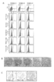
- FIG. 1 shows activation of stem cell markers in cancer cells by hypoxia induction.
- Various cancer cells and fibroblasts (TIG3) A, B) or N417 cells (AH), either normoxia (shown with N or white column) or 5% O 2 (shown with Hy or black column) RT-PCR (A, C), quantitative real-time PCR (qPCR) (B), FACS analysis (D) or immune tissue for gene activation and protein expression of stem cell markers after 12 hours incubation under conditions Subject to chemistry (E). N417 gene activation at various time points under hypoxia conditions was examined by RT-PCR (C).
- a side population (SP) assay (H) with alkaline phosphatase activity (F), sphere formation (G), and Hoechst 33342 (5 ⁇ g / mL) was also performed.
- SP (H) was measured in the presence (+ Ver) or absence ( ⁇ ) of verapamil (50 ⁇ M).
- FIG. 2 shows that “differentiated” non-tumorigenic cells revert to cancer stem cells under hypoxia.
- B Cancer cells isolated from xenograft tumors formed by either unsorted cells, SSEA-1 + cells or SSEA-3 + cells were analyzed for SSEA-1 and SSEA-3 expression by FACS. From the left for each item, the case of non-sorted cells, SSEA-1 + cells, and SSEA-3 + cells are shown.
- FIG. 3 shows that cancer stem cells generated by hypoxic load are pluripotent.
- A Shows experimental protocol including various analyses.
- B Stem cell genes and various marker genes for cells exposed to hypoxia and not exposed to hypoxia at various time points during embryoid body formation, as well as cells prior to any treatment (shown as C) Was subjected to RT-PCR.
- C Incubate cells exposed to hypoxia (Hy) and cells not exposed to hypoxia (N) in the presence or absence of VEGF-A + PDGF-AA (each 10ng / mL) or 2 days after EB formation After that, it was subjected to FACS analysis for expression of human VEGFR2 and human CD31. The RT-PCR analysis on day 1 for angiogenic growth factors is also shown.
- FIG. 4 shows that xenograft tumors subsequently differentiated into functional endothelial cells and vascular smooth muscle cells, indicating hypoxia-induced reprogramming of cancer cells.
- A, B N417-GFP xenograft tumors, as shown in individual figures, CA9 (unnumbered and 1), SOX2 (2), OCT3 / 4 (3), von Willebrand factor (vWF ) (4), hCD31 (5), HIF1 ⁇ (6), HIF2 ⁇ (7), NANOG (8), SSEA-1 (9) or Ki67 (10) were used for immunohistochemistry. According to the state of CA9 staining, the tumor area was classified into 4 groups. a: highly hypoxic region, b: hypoxic region, c: intermediate region, d: non-hypoxic region.
- a xenograft tumor formed by any of N417-GFP cells, H358 cells, H460 cells or H157 cells was analyzed.
- antibodies that recognize both human and mouse ⁇ SMA shown as ⁇ SMA
- Several sections from N417-GFP xenograft tumors were double stained with antibodies against GFP and antibodies against either hCD31 or h ⁇ SMA. This result indicates that hCD31 or h ⁇ SMA-expressing cells are actually derived from cancer cells.
- mCD31 + EC and ⁇ SMA + cells are not organized components.
- FIG. 5 shows that SSEA-1 positive cells and SSEA-3 positive cells are TRA-1-60 and TRA-1-81 positive.
- A After incubating N417 cells under 5% O 2 for 12 hours, SSEA-1, SSEA-3, SSEA-4, OCT3 / 4, SOX2, NANOG, TRA-1-60 and TRA-1-81 Expression was analyzed by FACS. SSEA positive or negative fractions are shown in blue or red, respectively.
- B SSEA-1-positive cells were transplanted subcutaneously into immunodeficient mice, and 12 weeks later, the grown xenograft tumor (xenograft) was analyzed by immunostaining.
- C Human lung cancer specimen sections were analyzed by immunostaining.
- cancer may be any kind of cancer, and solid cancer [eg, gastrointestinal cancer (eg, stomach cancer, esophageal cancer, small intestine cancer, colon cancer, rectal cancer, anus Cancer, liver cancer, biliary tract cancer, pancreatic cancer, etc.), urological or genital cancer (eg, renal cancer, renal cell cancer, bladder cancer, prostate cancer, renal pelvis and ureteral cancer, gallbladder) Cancer, bile duct cancer, testicular cancer, penile cancer, uterine cancer, endometrial cancer, uterine sarcoma, cervical cancer, vaginal cancer, vulvar cancer, ovarian cancer, fallopian tube cancer, etc.
- gastrointestinal cancer eg, stomach cancer, esophageal cancer, small intestine cancer, colon cancer, rectal cancer, anus Cancer, liver cancer, biliary tract cancer, pancreatic cancer, etc.
- urological or genital cancer eg, renal cancer, renal cell cancer, bladder cancer, prostate cancer, renal
- Brain / nervous cancer eg, brain tumor (glioblastoma, etc.), spinal cord tumor, etc.
- head and neck cancer eg, laryngeal cancer, oral cancer, salivary gland cancer, sinus cancer, thyroid gland) Cancer
- respiratory cancer eg, lung cancer (including small cell lung cancer, non-small cell lung cancer, metastatic lung cancer), bronchial cancer, etc.)
- breast cancer Skin cancer (eg, malignant melanoma), bone cancer (eg, osteosarcoma, etc.), muscle cancer (eg, rhabdomyosarcoma, etc.)], blood cancer [eg, myeloma, leukemia, etc. , Lymphoma, etc.], but is not limited thereto.
- solid cancer is preferably lung cancer, digestive organ cancer, breast cancer, skin cancer and brain tumor, more preferably lung cancer, digestive organ cancer and breast cancer, even more preferably lung cancer and digestive organ cancer, lung cancer Is particularly preferred.
- the origin of the cell population used in the present invention or the subject of the present invention is usually a mammal.
- mammals include, for example, laboratory animals such as rodents and rabbits such as mice, rats, hamsters, and guinea pigs, domestic animals such as pigs, cows, goats, horses, sheep and minks, pets such as dogs and cats, humans, Examples include, but are not limited to, primates such as monkeys, rhesus monkeys, marmosets, orangutans and chimpanzees.
- stage-Specific Embryonic Antigen SSEA
- SSEA Stage Specific Fetal Antigen
- SSEA-3 TRA-1-60
- TRA-1-81 (hereinafter collectively referred to as expression level detection targets in the present invention)
- the “marker protein of the present invention” or simply “the marker of the present invention”) is a surface sugar antigen specifically recognized by a specific monoclonal antibody, It is known to be expressed in pluripotent stem cells such as ES cells.
- Specific examples of the marker of the present invention include Myung, JS et al., Cell Stem Cell 4, 440-452 (2009) (SSEA-1), MuramatsuMT. Et al., Trends Glycosci Glyc 21, 197- 206 (2009) (SSEA-3), William, MS MS et al., Stem Cells 25, 720-730 (2007) (TRA-1-60 and TRA-1-81) and the like.
- tumor-forming cells refers to cells that can induce the formation of benign or malignant tumors that exhibit progressive growth.
- karyotype analysis for example, soft agar colony formation test, tumorigenicity test in immunodeficient animals, etc. are known, and those skilled in the art can use any or a combination of these tests. It can be assessed whether a given cell population has tumorigenic potential.
- Tumor-forming cells can be cells commonly known as cancer stem cells. Cancer stem cells are cancer cells that are needed to reconstruct a continuously proliferative tumor and produce all cancer cells that have the ability to self-renew and have diverse phenotypes within the cancer cell population.
- Self-renewal ability refers to the ability of one or both of two divided daughter cells to produce cells that have the same ability and degree of differentiation as the parent cell in the cell lineage. .
- non-tumor cell refers to a cell that is not the above-described tumor cell.
- Non-tumor-forming cells may be cancer cells that do not have tumorigenicity, or may not be cancer cells.
- pluripotency refers to pluripotency, which is a cell that can differentiate into cells of any of the three germ layers of endoderm, mesoderm, and ectoderm. The ability to have.
- Methods for evaluating whether a cell has pluripotency are known in the art. For example, cells are suspended in culture to form embryoid bodies, and embryoid bodies are cultured in various differentiation media. Then, a method for confirming whether or not it has been differentiated into all three germ layers by detecting the presence or absence of tissue-specific marker expression, a method for confirming that teratoma is formed by transplanting cells into immunodeficient mice, Etc.
- the “juvenile marker gene” refers to a gene exhibiting specific expression in pluripotent stem cells such as ES cells and iPS cells (for example, Experimental Medicine Vol. 26, No. 5, p. See .29-34).
- specific expression means that high mRNA and / or protein expression is observed in pluripotent stem cells such as ES cells and iPS cells, but the expression rapidly decreases when the cells differentiate. means.
- Examples of the rejuvenation marker gene that can be used in the present invention include, but are not limited to, OCT3 / 4, SOX2, KLF4, c-MYC, NANOG, LIN28, REX1, ERAS, and the like. Are OCT3 / 4, SOX2, KLF4, c-MYC and NANOG.
- “juvenile marker protein” means a protein of the “juvenile marker gene” defined above.
- the measurement of the expression level of the marker protein is usually performed by flow cytometry analysis using a specific antibody or the like for the marker protein, as will be described later.
- “Positive” expression of the marker means that the marker is expressed on the cell surface (or in the cell) and specific binding by an antibody to the marker can be detected.
- “Negative” expression of a marker means that the marker is not substantially expressed on the cell surface (or in the cell) and specific binding by an antibody to the marker cannot be detected.
- the expression level may be qualitative or quantitative.
- the present invention provides a method for testing the potential cancer malignancy of a cell population (hereinafter also referred to as the test method of the present invention).
- the test method of the present invention comprises: (1) preparing a cell population to be tested; (3) measuring the expression level of one or more marker proteins selected from the group consisting of SSEA-1, SSEA-3, TRA-1-60 and TRA-1-81 in the cell population, and (4 ) If any of the expression of the one or more marker proteins is positive, the step comprises determining that the cell population to be tested is of high potential malignancy.
- the potential cancer malignancy of a cell population refers to a specific site (for example, a tissue from which the cell population is derived) in which the cell population is in vivo (for example, in the mammal in vivo). ) Is the malignancy of cancer that is expected to occur when present.
- malignancy of cancer means any cancer malignancy that can be associated with the presence or absence of pluripotent tumorigenic cells or cancer stem cells in vivo by a person skilled in the art, for example, prognosis It may be related to the degree of failure (eg, survival rate, resistance to treatment, and the possibility of worsening or recurrence, etc.) and / or the likelihood and / or speed of metastasis, growth and / or invasion.
- the marker protein of the present invention can be used as a cell surface marker (or cancer stem cell marker) that can specifically distinguish pluripotent tumorigenic cells and non-tumorigenic cells. It was.
- the cancer malignancy can be tested easily and with high reliability simply by detecting the expression of the marker protein of the present invention in a clinical specimen such as cancer tissue.
- a clinical specimen such as cancer tissue.
- pluripotency where differentiated non-tumorigenic cells (and thus do not express the marker protein of the invention) are reprogrammed by hypoxia to express the marker protein of the invention. It has also been found that it can be a tumorigenic cell.
- the marker protein of the present invention when a certain cell population is subjected to a hypoxic load as described later and the expression level of the marker protein of the present invention is further measured, if the marker protein of the present invention is expressed, the original cell population Among them, there are pluripotent tumorigenic cells or differentiated non-tumorigenic cells that have a high probability of being reprogrammed to pluripotent tumorigenic cells due to hypoxia. It is understood that it existed. In this way, the potential cancer malignancy of the cell population can be determined by the test method of the present invention. In particular, even if detection is not easy due to the low frequency of cancer stem cells in cancer tissue, the cell population is reprogrammed to pluripotent tumorigenic cells by subjecting it to hypoxic load.
- the biological sample used is usually a sample collected from a tissue suspected of having cancer in a subject.
- tissues suspected of having cancer include, for example, tissues suspected of primary or metastasis of the above-mentioned cancer types, tissues known to contain tumor cells or cancer cells, treatment (for example, surgical treatment, Anticancer drug treatment, radiation therapy, immunotherapy, hyperthermia, etc.).
- treatment for example, surgical treatment, Anticancer drug treatment, radiation therapy, immunotherapy, hyperthermia, etc.
- recurrence after cancer surgery may be related to the survival of cancer stem cells. Therefore, surgery taken from patients after cancer surgery It is also preferable to use a post-tissue sample.
- the biological sample used in the test method of the present invention is not particularly limited as long as it can be collected from a subject, and depending on the purpose of the test, that is, the cancer type to be tested, blood, bone marrow Body fluid samples such as fluid and lymph fluid, biopsy samples of various organs such as lung, various digestive organs, breast, skin, and brain tissue can be used. Preparation of a cell population to be cultured from a body fluid sample or a biopsy sample may be performed according to a procedure commonly used by those skilled in the art.
- the cell population used for the aforementioned research, development or medical purposes may also be any cell population for which testing for potential cancer malignancy according to the present invention is desired.
- a given cell population can be used for studies on the mechanism of reprogramming from non-tumorigenic cells to pluripotent tumorigenic cells.
- against all the specific antibodies used in the flow cytometry method using specific antibodies against one or more marker proteins of the present invention preferably specific antibodies against all the marker proteins of the present invention.
- the selection can be performed using negative binding as an index.
- a cancer cell population that has been subjected to a specific treatment in vitro for example, administration of a candidate substance for an anticancer drug
- Evaluate the effectiveness of the treatment for example, a candidate substance
- This contributes particularly to the development of effective anticancer therapies targeting pluripotent tumorigenic cells (or cancer stem cells) and / or non-tumorigenic cells that are likely to be reprogrammed into the cells. be able to.
- the safety of the cell population can also be evaluated by subjecting the cell population for transplantation for medical purposes to the test method of the present invention.
- the possibility of canceration of the iPS cell can be evaluated by subjecting the produced iPS cell to the test method of the present invention, it can contribute to the development of a method for producing iPS cell that prevents canceration.
- the cell population prepared in the above step (1) may be cultured in a low oxygen atmosphere in this step.
- this step is optional.
- the hypoxic atmosphere means that the oxygen concentration in the atmosphere when culturing cells is significantly lower than that in the air.
- the oxygen concentration is lower than the oxygen concentration in the atmosphere generally used in normal cell culture.
- the oxygen concentration in the atmosphere is less than 18%.
- the oxygen concentration in the atmosphere is less than 15% (eg, less than 14%, less than 13%, less than 12%, less than 11%, etc.), less than 10% (eg, less than 9%, less than 8%, less than 7%) , Less than 6%, etc.), or less than 5% (eg, less than 4%, less than 3%, less than 2%, etc.).
- the atmosphere needs to contain a minimum amount of oxygen in order to maintain the survival of the cells to be cultured.
- the oxygen concentration in the atmosphere is usually 0.01% or more, and preferably the oxygen concentration in the atmosphere is 0.1% or more (eg, 0.2% or more, 0.3% or more, 0 0.4% or more), 0.5% or more (eg, 0.6% or more, 0.7% or more, 0.8% or more, 0.9% or more, etc.), or 1.0% or more (eg, 1.1% or more, 1.2% or more, 1.3% or more, 1.4% or more, etc.).
- Conditions generally used in normal cell culture can be applied to atmospheric conditions other than the oxygen concentration.
- the CO 2 concentration in the atmosphere is usually in the range of about 1 to 10%, preferably about 5%.
- the humidity in the atmosphere is usually in the range of about 70 to 100%, preferably about 95 to 100%.
- a method for creating a hypoxic state in the cell environment is not particularly limited, but a method of culturing cells in a CO 2 incubator in which the oxygen concentration can be adjusted is easy, and a preferable example is given.
- CO 2 incubators with adjustable oxygen concentrations are available from various equipment manufacturers (for example, CO for low oxygen culture manufactured by Thermo scientific, Ikemoto Riken, Toji Field, Waken Pharmaceutical, etc.) 2 incubators can be used).
- CO for low oxygen culture manufactured by Thermo scientific, Ikemoto Riken, Toji Field, Waken Pharmaceutical, etc. 2 incubators can be used.
- an anaerobic culture product such as Anero Pack commercially available from Mitsubishi Gas Chemical Co., Ltd. is also a suitable example. By using such a product, low oxygen conditions can be realized quickly and easily.
- the period during which cells are cultured under hypoxic conditions is not particularly limited as long as the test method of the present invention can be realized.
- the culture period should be a period in which the reprogramming can be induced by hypoxic load in differentiated non-tumor cells that have a high probability of being reprogrammed into pluripotent tumorigenic cells. From such a viewpoint, the culture period is, for example, 1 hour or more, 3 hours or more, 5 hours or more, 7 hours or more, 10 hours or more, 12 hours or more, 15 hours or more, 20 hours or more, 1 day or more, or 2 days or more.
- the culture since the culture is performed under conditions unfavorable for cell survival (that is, under a low oxygen atmosphere condition), if the culture period is too long, the cells may die and an appropriate test may not be performed.
- the upper limit of the culture period can be appropriately determined by those skilled in the art. For example, 50 days or less, 40 days or less, 30 days or less, 20 days or less, 10 days or less, 7 days or less, 5 days or less, 4 days or less Below, 3 days or less or 2 days or less.
- the preferred culture period under hypoxic conditions also varies depending on the cell population to be tested, the oxygen concentration in the atmosphere, etc., and those skilled in the art will appropriately adjust the culture period according to the cell population used, oxygen concentration, etc. be able to.
- the culture in a low oxygen atmosphere is performed at least once.
- the culture in the above period is repeated a plurality of times (examples (2 times, 3 times, 4 times, 5 times or more) is also preferred.
- the cell population to be prepared is subjected to each session before the hypoxic load and at the same time as the hypoxic load.
- the cell population can be subjected to a predetermined treatment (for example, exposure to an arbitrary reagent, etc.) during or after the hypoxic load.
- basal medium of the medium used in the test method of the present invention those generally used in normal cell culture can be used, and can be appropriately selected according to the type of cell population to be cultured. is there.
- the basal medium include, but are not limited to, DMEM, EMEM, RPMI-1640, ⁇ -MEM, F-12, F-10, M-199, and the like.
- a mixture of the above basal media may also be used.
- the medium used in the test method of the present invention can contain additives known per se.
- the additive include, but are not limited to, serum, growth factors (eg, insulin), iron sources (eg, transferrin), polyamines (eg, putrescine), minerals (eg, sodium selenate), organic acids (eg, Pyruvate, lactic acid, etc.), serum proteins (eg, albumin, etc.), amino acids (eg, L-glutamine, etc.), reducing agents (eg, 2-mercaptoethanol, etc.), vitamins (eg, ascorbic acid, d-biotin, etc.), sugars ( For example, glucose etc.), steroids (eg ⁇ -estradiol, progesterone etc.), antibiotics (eg streptomycin, penicillin, gentamicin etc.), buffering agents (eg HEPES etc.) and the like.
- Each of the additives is preferably contained within a concentration range known per se.
- the culture temperature used in the method of the present invention can be appropriately set according to the type of cell population to be cultured.
- the culture temperature is usually in the range of about 30-40 ° C, preferably about 37 ° C.
- the expression level of at least one of the marker proteins of the present invention in the cell population prepared in the above step (1) or the cell population after the treatment in the above step (2) is measured.
- which of the marker proteins of the present invention is particularly preferred may vary, but from the viewpoint of high specificity for pluripotent tumorigenic cells, for example, SSEA- 1, the expression level of at least one of TRA-1-60 or TRA-1-81 is preferably measured, and more preferably at least the expression level of SSEA-1 is measured.
- the accuracy of the test can be further increased by detecting expression for both SSEA-1 and TRA-1-60 or TRA-1-81.
- the expression level of either SSEA-1 or SSEA-3, or both may be measured.
- the expression levels of these genes are useful for verifying whether cells positive for the expression of the marker protein of the present invention are actually pluripotent tumorigenic cells.
- an antibody specifically recognizing the marker protein or the blastogenesis marker protein of the present invention an antibody commercially available from suppliers such as R & D Systems, Becton Dickinson, eBioscience, etc. can be used (Experiment described below) See also example).
- antibodies include natural antibodies such as polyclonal antibodies and monoclonal antibodies, chimeric antibodies that can be produced using genetic recombination techniques, humanized antibodies and single-chain antibodies, and binding fragments thereof.
- the antibody is a polyclonal antibody, a monoclonal antibody or a binding fragment thereof.
- the binding fragment means a partial region of the aforementioned antibody having specific binding activity, and specifically includes, for example, F (ab ′) 2 , Fab ′, Fab, Fv, sFv, dsFv, sdAb and the like.
- the class of the antibody is not particularly limited, and includes antibodies having any isotype such as IgG, IgM, IgA, IgD, or IgE. IgG or IgM is preferable, and IgG is more preferable in consideration of ease of purification.
- RNA can be isolated from a biological sample according to a conventional method.
- General methods for extracting RNA are well known in the art, and are described in standard textbooks of molecular biology such as Ausubel et al., Current Protocols of Molecular Molecular Biology, John Wiley and Sons (1997). It is disclosed. Specifically, RNA isolation can be performed according to the manufacturer's instructions using a purification kit, buffer set, and protease obtained from a manufacturer such as Qiagen.
- the method for measuring the mRNA expression level of the blastogenesis marker is not particularly limited, but Northern blotting and in situ hybridization (Parker & Barnes, Methods in Molecular Biology 106: 247-283 (1999)); RNase protection Assay method (Hod, Biotechniques 13: 852-854 (1992)); Reverse Transcription Polymerase Chain Reaction (RT-PCR) (Weis et al., Trends, Genetics 8: 263-264 (1992)); Real-time quantitative RT-PCR (Held et al., Genome Research 6: 986-994 (1996)); and microarray analysis methods.
- Microarray analysis methods can be performed with commercially available equipment, such as using Affymetrix GeneChip technology, Agilent Technologies microarray technology or Incyte microarray technology, according to the manufacturer's instructions.
- the expression level is similarly measured using the same isotype antibody (isotype control) as the antibody used for detecting the marker protein of the present invention, and the specific antibody You may compare the expression level obtained by measurement by (1) and the expression level obtained by measurement by isotype control. Such procedures are well known in the art.
- the comparison of the expression level can be preferably performed based on the presence or absence of a significant difference.
- the cells positive for any of the expression of the marker protein of the present invention are actually pluripotent tumorigenic cells by other means in order to further improve the reliability of the test.
- Such means include confirmation of positive expression of one or more blast marker genes (described above), confirmation of differentiation pluripotency (described above and also shown in experimental examples), Confirmation of tumor ability (described above and also shown in experimental examples), expression patterns of epigenetic regulatory enzymes (eg, histone deacetylase, DNA methylase, etc.) with ES cells or iPS cells Confirmation of similarity, etc. are mentioned.
- Cells that specifically bind to the antibody may be isolated using a cell sorter, magnetic beads, a cell adsorption column, or the like.
- the present invention also provides a reagent for testing the potential cancer malignancy of a cell population.
- the reagent includes an antibody that specifically recognizes the marker protein of the present invention.
- the reagent may further contain an antibody that specifically recognizes the blastogenesis marker protein.
- these antibodies those described above in the test method of the present invention can be used, and preferred embodiments are also as described above.
- the present invention further comprises a system (kit) for testing the potential cancer malignancy of a cell population comprising a hypoxic culture apparatus and the test reagent described above.
- kit for testing the potential cancer malignancy of a cell population comprising a hypoxic culture apparatus and the test reagent described above.
- “Low oxygen culture device” means a device that enables cell culture in a low oxygen atmosphere.
- the apparatus those described above for the test method of the present invention can be used, and examples include an incubator capable of culturing cells in a low oxygen atmosphere, an anaerobic culture product (eg, anero pack described above), and the like. it can.
- the definition of “low oxygen atmosphere” is as described above.
- the present invention also provides a method for treating the cancer in a subject having a highly malignant cancer.
- the method includes a step of applying the test method of the present invention to a sample collected from a tissue suspected of having cancer in the subject, and cells having high potential malignancy of cancer in the sample.
- the method includes the step of applying a therapy for cancer to the subject.
- the test method of the present invention and preferred embodiments thereof are as described above.
- the therapy for cancer includes any therapy generally performed for high-grade cancer, such as surgical treatment, anticancer drug treatment, radiation therapy, immunotherapy, thermotherapy. Etc.
- the present invention also provides a screening method for a preventive and / or therapeutic agent for cancer.
- the method is (1) A step of preparing a cell population containing cancer cells, wherein the cell population is selected from the group consisting of SSEA-1, SSEA-3, TRA-1-60 and TRA-1-81 The process comprising a cell expressing the above marker protein, (2) A step of preparing, from the prepared cell population, a cell population subjected to exposure to a candidate substance for cancer prevention and / or treatment and a control cell population not subjected to exposure to the candidate substance.
- step (3) The expression level of one or more marker proteins selected from the group consisting of SSEA-1, SSEA-3, TRA-1-60 and TRA-1-81 in the cell population after exposure and the control cell population And (4) when all of the expression of the one or more marker proteins in the cell population after exposure is at a significantly lower level compared to the expression of the corresponding marker protein in the control cell population, Selecting the candidate substance as an agent for preventing and / or treating cancer.
- Said process (1), (3) and (4) can be performed according to description of the test method of this invention, and it is the same also about preferable embodiment.
- the candidate substance in the above step (2) may be any substance (eg, natural or synthetic compound), and the exposure conditions can be appropriately set according to the purpose of the screening test.
- the cell population may be subjected to a low oxygen load after the above step (1) and before the step (3).
- the exposure of the candidate substance may be performed before the low oxygen load, simultaneously with the low oxygen load, during each low oxygen load, or after the low oxygen load.
- the cell population prepared by said process (1) does not necessarily need to contain the cell which expresses said 1 or more marker.
- the present invention further provides a screening method for factors that promote reprogramming from non-tumor cells to pluripotent tumor cells.
- the method is (1) preparing a cell population containing non-tumorigenic cells; (2) From the prepared cell population, all of the expression of one or more marker proteins selected from the group consisting of SSEA-1, SSEA-3, TRA-1-60 and TRA-1-81 are negative.
- Selecting non-tumorigenic cells by using as an index, (3) subjecting the selected non-tumorigenic cell population to exposure to a predetermined factor; (4) measuring the expression level of one or more marker proteins selected from the group consisting of SSEA-1, SSEA-3, TRA-1-60 and TRA-1-81 in the cell population after the exposure; and (5) when any one of the expression of the one or more marker proteins is positive, the step of selecting the factor as a factor that promotes reprogramming from a non-tumor cell to a pluripotent tumor cell.
- Said process (1) and (2) can be performed as it demonstrates regarding the preparation method of this invention mentioned later, and is the same also about preferable embodiment.
- the factor in the above step (3) may be any factor desired to be screened, such as a substance (eg, natural or synthetic compound), environmental conditions (eg, temperature, ambient gas, etc.), radiation, etc. It can be.
- the exposure conditions can be appropriately set according to the purpose of the screening test.
- Said process (4) can be performed as it demonstrated regarding the test method of this invention, and it is the same also about preferable embodiment.
- the selected non-tumorigenic cells may be subjected to a hypoxic load before detecting the expression of the marker protein.
- the exposure of the factor may be performed before being subjected to the low oxygen load, simultaneously with the low oxygen load, during each low oxygen load, or after the low oxygen load.
- any cell population can be used as long as it contains non-tumor-forming cells.
- Examples of the cell population to be tested in the test method of the present invention include those described above.
- a cell population comprising tumor cells or cancer cells. This step can be performed in the same manner as step (1) of the test method of the present invention.
- the selection in this step is performed by, for example, selecting specific antibodies against one or more marker proteins of the present invention, preferably specific antibodies against all the marker proteins of the present invention, from the cell population prepared in step (1). It can be carried out by isolating cells that do not specifically bind to any of the antibodies used with a cell sorter, magnetic beads, cell adsorption column, or the like. Depending on the origin of the cell population, which of the marker proteins of the present invention is particularly preferred may vary, but from the viewpoint of high specificity for pluripotent tumorigenic cells, for example, SSEA-1 or Preferably, expression of at least one of TRA-1-60 or TRA-1-81 is utilized for selection, and more preferably, expression of at least SSEA-1 is utilized.
- the accuracy of selection can be further increased by utilizing expression for both SSEA-1 and TRA-1-60 or TRA-1-81.
- expression of either SSEA-1 or SSEA-3, or both may be utilized.
- the specific antibody those described for the test method of the present invention can be used.
- the preparation method of the present invention is particularly suitable for hypoxia.
- there is substantially no pluripotent tumorigenic cells in this step For example, it is preferably removed to less than 0.1%.
- the non-tumorigenic cell population obtained in step (2) is subjected to hypoxic load in this step.
- This step can be performed in the same manner as described for step (2) of the test method of the present invention.
- the hypoxic load of this step at least a part of the non-tumorigenic cells in the cell population can be converted to pluripotent tumorigenic cells induced by reprogramming.
- the pluripotent tumorigenic cells that can be generated in the step (3) are selectively collected using as an index that one of the expression of the marker protein of the present invention is positive.
- the selection of this step for example, using the antibody specific for the marker protein of the present invention against the cell population after step (3), the antibody is obtained by cell sorter, magnetic beads, cell adsorption column, etc. This can be done by isolating cells that specifically bind to.
- the marker proteins of the present invention may vary, but from the viewpoint of high specificity for pluripotent tumorigenic cells, for example, SSEA-1 or Preferably, expression of at least one of TRA-1-60 or TRA-1-81 is utilized for selection, and more preferably, expression of at least SSEA-1 is utilized. In addition, the accuracy of selection can be further increased by utilizing expression for both SSEA-1 and TRA-1-60 or TRA-1-81. Alternatively, expression of either SSEA-1 or SSEA-3, or both, may be utilized. As the specific antibody, those described for the test method of the present invention can be used. It is preferable to verify that the obtained cell population is actually a pluripotent tumorigenic cell by other means.
- confirmation of the expression of one or more of the above-mentioned blastogenesis marker genes is positive for a part of the obtained cell population (described above), confirmation of differentiation pluripotency (described above, (Also shown in experimental examples), confirmation of tumorigenicity (described above, also shown in experimental examples), epigenetic regulatory enzymes (for example, histone deacetylase, DNA methylase, etc.)
- confirmation of the expression of one or more of the above-mentioned blastogenesis marker genes is positive for a part of the obtained cell population (described above), confirmation of differentiation pluripotency (described above, (Also shown in experimental examples), confirmation of tumorigenicity (described above, also shown in experimental examples), epigenetic regulatory enzymes (for example, histone deacetylase, DNA methylase, etc.)
- epigenetic regulatory enzymes for example, histone deacetylase, DNA methylase, etc.
- the present invention further provides pluripotent tumorigenic cells (hereinafter also referred to as tumorigenic cells of the present invention) generated from non-tumorigenic cells by reprogramming induced by hypoxia.
- the non-tumor cell from which the tumorigenic cell of the present invention is derived may be any non-tumor cell, for example, a non-tumor cell of any cancer type described above.
- the tumorigenic cells of the present invention are not limited thereto, but can be obtained by, for example, the preparation method of the present invention described above.
- the tumorigenic cells of the present invention are preferably isolated and purified. “Isolation and purification” means that treatment to remove contamination of cells other than the tumorigenic cells of the present invention has been performed.
- the purity of the tumorigenic cell of the present invention is preferably as high as possible. The purity is, for example, 60% or more, preferably 70% or more, more preferably 80% or more, still more preferably 95% or more, and particularly preferably 99% or more (for example, substantially 100%).
- cancer research is promoted by performing gene expression analysis, protein analysis, metabolism analysis, etc. of the tumorigenic cells of the present invention.
- an efficient tumor model can be constructed
- Such tumor models are useful for studying tumor development / formation processes, treatment resistance, metastasis processes, and the like. These studies will lead to an understanding of pluripotent tumorigenic cells (or cancer stem cells) and cancer, and drug development targeting them will be possible.
- Oligonucleotides for microRNA were designed using the BLOCK-iT program (Invitrogen). For differentiation assay (FIG. 3), cells after embryoid body (EB) formation were incubated in SmGM-, FGM-2- or HCM-BulletKit (Lonza, Switzerland). RT-PCR analysis RT-PCR analysis and real-time RT-PCR analysis using SYBR-Green were performed by standard methods. The amount of RNA input was normalized using GAPDH mRNA. The antibodies used for FACS analysis and immunohistochemistry FACS and immunostaining are shown in Table 1 below.
- xenograft tumors were digested with TrypLE Express (Invitrogen) for 5 minutes for separation into single cells. FACS analysis, side population (SP) analysis, and cell sorting were performed using FACSAria II (Becton Dickinson). 7AAD (1 ⁇ g / mL) was used for removal of dead cells. In the figure, the FACS signal obtained using the isotype control antibody is indicated by a gray shadow. Cells showing fluorescence above the maximum fluorescence observed with isotype antibodies were considered positive. The percentage of such positive cells is shown in some figures. Xenograft tumors were fixed in 4% paraformaldehyde and embedded in paraffin.
- Detailed protocols for immunostaining and flow cytometry are as follows. (Immunostaining) Tumors were fixed with 4% paraformaldehyde at 4 ° C., allowed to stand overnight at 20 ° C. in a 20% sucrose / PBS solution, cleared with alcohol, and then embedded in paraffin. The thin slice was 5 ⁇ m, fixed on a slide glass, and deparaffinized with alcohol again. Activation treatment was performed by heating at 95 ° C. for 20 minutes with an antigen activation solution (Antigen Retrieval Reagent, Basic; R & D systems).
- Antigen Retrieval Reagent Basic
- R & D systems an antigen activation solution
- mice Stem cell marker is activated in cancer cells by hypoxia
- Five human lung cancer cell lines and malignant melanoma to confirm whether ES cell marker gene is activated by hypoxia Cell lines were screened. Cells were incubated with 5% O 2 for 12 hours before being subjected to either RT-PCR analysis or quantitative real-time PCR analysis. All four reprogramming-inducible genes (OCT3 / 4, SOX2, KLF4, c-MYC) were activated in N417, H358, H460 and Num2B cells (FIGS. 1A and 1B), but human fibroblasts (TIG3) Did not show any activation of these genes.
- N417 and Num2B all tested embryonic genes, including the “naive” marker REX1, were activated. In subsequent experiments, N417 was mainly used. Most of the results obtained with N417 were also confirmed in H358 cells.
- TGF- ⁇ was immediately down-regulated, followed by activation of the stemness gene (Figure 1C).
- Hypoxia-induced expression of stem cell genes and ES cell-specific cell surface antigens (SSEA-1, SSEA-3, SSEA-4, TRA1-60, TRA1-81) was confirmed by FACS and immunohistochemistry (Fig. 1D, 1E). Under hypoxia, N417 showed increased alkaline phosphatase activity (FIG.
- FIG. 1F Verapamil-sensitive side population (SP) observed after staining with Hoechst 33342 increased from 0.27% in normoxia to 7.05% in hypoxia (FIG. 1H).
- SP verapamil-sensitive side population
- the hypoxia-inducible reaction described above was confirmed in 6 different N417 clones (including 3 clones of N417 (N417-GFP) expressing GFP) and was also observed in 3 clones of H358 cells. From these results, some cancer non-tumorigenic cells are reprogrammed under hypoxia and have multipotency or pluripotency like iPS cells as well as tumorigenic cells We hypothesized that they could return to stem cell-like cells.
- SSEA-1 and SSEA-3 demonstrate the return of non-cancer stem cells (non-CSC) to cancer stem cells (CSC), a marker that can distinguish between tumor-forming cells and non-tumor-forming cells In order to do this, it was necessary to separate these two cell populations.
- ES cell-specific cell surface antigens SSEA-1, SSEA-3, SSEA-4, Tra1-81 were used for CSC identification.
- the expression of SSEA-1 and SSEA-3 was mutually exclusive in N417 cells.
- the SSEA-1 / SSEA-3 negative fraction did not contain Tra1-81 positive cells.
- SSEA-1 + cells or SSEA-3 + cells sorted from cells exposed to hypoxia form continuously transplantable xenograft tumors, which are more than unsorted parental cells. Proliferated rapidly and actively (FIG. 2A).
- Example 4 CSC produced by hypoxia is pluripotent
- EB embryoid bodies
- EMT marker genes vimentin and fibronectin are activated only in the fraction of cells exposed to hypoxia, and TGF- ⁇ 1 and its receptor also maintain cells in normoxia, It was similarly activated when exposed to hypoxia.
- angiogenic growth factors were activated from day 1 and on day 4 expressed the endothelial cell (EC) markers human CD31 (hCD31) and human VEGFR-2 (Fig. 3C). The number of positive cells was further increased when cells were cultured with VEGF and PDGF.
- tumors formed with 2 out of 3 injections in unsorted cells and all 6 injections in SSEA-1 + or SSEA-3 + cells, ⁇ III tubulin, ⁇ FP and ⁇ - SMA was detected in tumor cells (Figure 3E). Furthermore, the tumor contained tissues induced by various differentiations from all three germ layers, such as cytokeratin 14-positive dermis, glandular tissue, and muscle (FIG. 3F). It was confirmed by immunohistochemistry using antibodies against GFP (FIG. 3F-e, f), human mitochondria and human nucleus that these organized tissues were composed of human cells.
- Experimental Example 5 Xenograft tumors were subsequently differentiated into functional endothelial cells and vascular smooth muscle cells, and N417-GFP xenograft tumors showing cancer cell reprogramming were histologically analyzed. Evaluation by carbonic anhydrase 9 (CA9) staining revealed that the hypoxic region was scattered in a mottled or patchy manner, and OCT3 / 4-positive cells and SOX2-positive cells were detected in such hypoxic region. (FIG. 4A).
- CA9 carbonic anhydrase 9
- tumor areas were classified into 4 groups: (a) areas where most cancer cells are CA9 positive (highly hypoxic areas), (b) areas surrounded by CA9 positive cells (hypoxia) (Region), (c) a region surrounded by slightly CA9-stained cells (intermediate region), and (d) a region substantially lacking CA9-positive cells (non-hypoxic region) (FIG. 4B).
- cells are SOX2 (a2, b2), OCT3 / 4 (a3, b3), HIF1 ⁇ (a6), HIF2 ⁇ (a7), NANOG (a8) or SSEA -1 (a9) staining was positive.
- the cells In such a hypoxic region, the cells almost lacked the expression of the proliferation marker Ki-67 (a10). In the outer part of the hypoxic region, many apoptotic cells were observed, probably derived from either cancer cells or host inflammatory cells. In the middle region, the detection of SOX2 and OCT3 / 4 was negligible or hardly detected (arrows c2, c3). In the non-hypoxic region, SOX2 (d2) and OCT3 / 4 (d3) were not detected, and the cells showed proliferative activity (d10). Blood vessels containing erythrocytes were found in the central region of each cell island. Interestingly, hCD31 positive cells were detected relatively scattered in the hypoxic region (a5, b5).
- hCD31 positive cells are assembled together with ⁇ -SMA positive cells (Fig. 4C) and form blood vessels in non-hypoxic regions (c5, d5), and blood vessels gradually mature and become more functional It seemed to be.
- von Willebrand factor usually detected in ECs of relatively large vessels, was stained only in vessels in non-hypoxic regions (d4 arrow).
- hCD31 positive cells also expressed human VEGFR-2.
- HCD31, mouse CD31 (mCD31) and human ⁇ SMA (h ⁇ SMA) specific antibodies were used to detect EC and vascular smooth muscle cells (VSMC). None of these were confirmed to react with foreign antigens.
- EC and ⁇ SMA positive cells detected using antibodies recognizing both human and mouse ⁇ SMA did not have a blood vessel-like structure.
- a limited region was hypoxic in H460 xenograft tumors, and SOX2, OCT3 / 4, SSEA-1, and hCD31 were detected in cells within this hypoxic region.
- N417 and H358 cells were much less proliferative than H157 and A549 cells, whereas xenograft tumors formed by N417 or H358 cells were formed by H157 or A549 cells It grew faster than xenograft tumors.
- TRA-1-60 / -81 is also a diagnostic marker for malignant cancer, like SSEA-1 / -3.
- We further distinguish between pluripotent and non-tumor cells We searched for markers other than SSEA-1 or SSEA-3.
- the correlation of expression with SSEA-1 and / or SSEA-3 was examined for various reprogramming genes and ES cell specific cell surface antigens. Specifically, first, the above human lung cancer cells were incubated with 5% O 2 for 12 hours, and then FACS analysis was performed. The expression of OCT3 / 4, SOX2, NANOG, TRA-1-60 and TRA-1-81 for the positive and negative fractions of SSEA-1, SSEA-3 and SSEA-4 is shown in FIG. 5A.
- SSEA-1 negative cells and SSEA-3 negative cells are TRA-1-60 / -81 negative
- SSEA-1 positive cells and SSEA-3 positive cells are TRA-1-60 / -81. It shows that it is positive.
- not all SSEA-1 / -3 positive cells are TRA-1-60 / -81 positive, but almost all SSEA-1 / -3 positive cells are TRA-1-60 /
- almost all SSEA-1 / -3 negative cells were TRA-1-60 / -81 negative. This was particularly noticeable for SSEA-1.
- SSEA-4 positive cells were TRA-1-60 / -81 negative.
- FIG. 5B shows 10 3 FACS-sorted SSEA-1-positive cells encapsulated in Matrigel and transplanted subcutaneously into immunodeficient mice, and 12 weeks later, the tumors grown (xenograft) were analyzed by immunostaining.
- FIG. 5C shows an immunostaining analysis of human lung cancer specimen sections collected with informed consent (analyzed in 98 randomly selected cases). Both results indicate that SSEA-1 positive cells are simultaneously TRA-1-60 / -81 positive. From the above, it was demonstrated that TRA-1-60 / -81 can also be used as a marker for discriminating between pluripotent tumorigenic cells and non-tumorigenic cells, like SSEA-1 / -3.
- the presence of a pluripotent tumorigenic cell is detected simply by detecting the expression of a marker protein in a clinical specimen such as a cancer tissue, and thus simple and highly reliable. Can test for cancer malignancy. Alternatively, prior to detection of marker protein expression, the cell population in a sample of cancer tissue or the like is subjected to a hypoxic load to promote reprogramming of non-tumoral cells to pluripotent tumorigenic cells. Thereby, the sensitivity of the test can be further improved.
- tumor-forming cells existing in a living body or non-tumor-forming cells that are likely to be converted to tumor-forming cells due to hypoxia before cancer is first developed or recurred.
- test method, novel cell and method for preparing the cell include, for example, research on the mechanism of reprogramming from non-tumor cells to pluripotent tumor cells, malignant cancer It is useful not only for the establishment of effective treatment methods, but also for the development of production methods for preventing canceration in iPS cell production.
Landscapes
- Health & Medical Sciences (AREA)
- Life Sciences & Earth Sciences (AREA)
- Engineering & Computer Science (AREA)
- Chemical & Material Sciences (AREA)
- Biomedical Technology (AREA)
- Immunology (AREA)
- Organic Chemistry (AREA)
- Genetics & Genomics (AREA)
- Zoology (AREA)
- Wood Science & Technology (AREA)
- Biotechnology (AREA)
- Biochemistry (AREA)
- General Health & Medical Sciences (AREA)
- Bioinformatics & Cheminformatics (AREA)
- Microbiology (AREA)
- Molecular Biology (AREA)
- Proteomics, Peptides & Aminoacids (AREA)
- Analytical Chemistry (AREA)
- Pathology (AREA)
- Cell Biology (AREA)
- Physics & Mathematics (AREA)
- General Engineering & Computer Science (AREA)
- Oncology (AREA)
- Urology & Nephrology (AREA)
- Hematology (AREA)
- Biophysics (AREA)
- Hospice & Palliative Care (AREA)
- Developmental Biology & Embryology (AREA)
- Food Science & Technology (AREA)
- Medicinal Chemistry (AREA)
- General Physics & Mathematics (AREA)
- Measuring Or Testing Involving Enzymes Or Micro-Organisms (AREA)
- Micro-Organisms Or Cultivation Processes Thereof (AREA)
- Investigating Or Analysing Biological Materials (AREA)
Abstract
La présente invention concerne : une méthode pour le test de la malignité latente d'une population cellulaire ; de nouvelles cellules tumorigènes qui sont utiles pour la découverte des mécanismes associés au cancer et le développement d'une thérapie anticancéreuse ; et un procédé de préparation desdites cellules. La présente invention est caractérisée par la création de cellules tumorigènes pluripotentes à partir de cellules non tumorigènes par l'intermédiaire d'une reprogrammation induite par l'hypoxie et/ou par l'utilisation de SSEA-1, SSEA-3, TRA-1-60 ou TRA-1-81 en tant que marqueur de surface cellulaire spécifique associé aux cellules tumorigènes pluripotentes.
Priority Applications (1)
| Application Number | Priority Date | Filing Date | Title |
|---|---|---|---|
| JP2014503838A JP6187939B2 (ja) | 2012-03-08 | 2013-03-04 | がんの悪性度の試験方法、ならびに多能性を有する造腫瘍細胞およびその調製方法 |
Applications Claiming Priority (2)
| Application Number | Priority Date | Filing Date | Title |
|---|---|---|---|
| JP2012-052348 | 2012-03-08 | ||
| JP2012052348 | 2012-03-08 |
Publications (1)
| Publication Number | Publication Date |
|---|---|
| WO2013133220A1 true WO2013133220A1 (fr) | 2013-09-12 |
Family
ID=49116697
Family Applications (1)
| Application Number | Title | Priority Date | Filing Date |
|---|---|---|---|
| PCT/JP2013/055865 Ceased WO2013133220A1 (fr) | 2012-03-08 | 2013-03-04 | Méthode de test de la malignité du cancer, cellules tumorigènes pluripotentes et procédé de préparation associé |
Country Status (2)
| Country | Link |
|---|---|
| JP (1) | JP6187939B2 (fr) |
| WO (1) | WO2013133220A1 (fr) |
Citations (7)
| Publication number | Priority date | Publication date | Assignee | Title |
|---|---|---|---|---|
| JP2008278798A (ja) * | 2007-05-10 | 2008-11-20 | Tokyo Medical & Dental Univ | 膀胱癌の検出方法 |
| JP2009296895A (ja) * | 2008-06-10 | 2009-12-24 | Univ Of Fukui | 低酸素培養機器及び無糖培地を用いたがん幹細胞濃縮法 |
| JP2010075186A (ja) * | 2004-04-01 | 2010-04-08 | Wisconsin Alumni Research Foundation | 幹細胞の内胚葉および膵臓系統への分化 |
| JP2010099039A (ja) * | 2008-10-27 | 2010-05-06 | Fujifilm Corp | 神経芽腫の検出方法 |
| WO2010067722A1 (fr) * | 2008-12-08 | 2010-06-17 | 株式会社バイオマトリックス研究所 | Procédé pour évaluer le degré de malignité d’un cancer du sein, et kit pour l’évaluation |
| JP2011511625A (ja) * | 2008-01-25 | 2011-04-14 | ハンサビオメド・オサウヒング | 新規ヒト転移性腫瘍関連分子、活性化遺伝子およびタンパク質を検出する方法ならびに遺伝子発現を妨害する方法 |
| JP2011524417A (ja) * | 2008-06-16 | 2011-09-01 | アカデミア シニカ | GloboHおよび新規な糖脂質アジュバントを有する関連抗がんワクチン |
-
2013
- 2013-03-04 WO PCT/JP2013/055865 patent/WO2013133220A1/fr not_active Ceased
- 2013-03-04 JP JP2014503838A patent/JP6187939B2/ja not_active Expired - Fee Related
Patent Citations (7)
| Publication number | Priority date | Publication date | Assignee | Title |
|---|---|---|---|---|
| JP2010075186A (ja) * | 2004-04-01 | 2010-04-08 | Wisconsin Alumni Research Foundation | 幹細胞の内胚葉および膵臓系統への分化 |
| JP2008278798A (ja) * | 2007-05-10 | 2008-11-20 | Tokyo Medical & Dental Univ | 膀胱癌の検出方法 |
| JP2011511625A (ja) * | 2008-01-25 | 2011-04-14 | ハンサビオメド・オサウヒング | 新規ヒト転移性腫瘍関連分子、活性化遺伝子およびタンパク質を検出する方法ならびに遺伝子発現を妨害する方法 |
| JP2009296895A (ja) * | 2008-06-10 | 2009-12-24 | Univ Of Fukui | 低酸素培養機器及び無糖培地を用いたがん幹細胞濃縮法 |
| JP2011524417A (ja) * | 2008-06-16 | 2011-09-01 | アカデミア シニカ | GloboHおよび新規な糖脂質アジュバントを有する関連抗がんワクチン |
| JP2010099039A (ja) * | 2008-10-27 | 2010-05-06 | Fujifilm Corp | 神経芽腫の検出方法 |
| WO2010067722A1 (fr) * | 2008-12-08 | 2010-06-17 | 株式会社バイオマトリックス研究所 | Procédé pour évaluer le degré de malignité d’un cancer du sein, et kit pour l’évaluation |
Non-Patent Citations (9)
| Title |
|---|
| BEN-PORATH, I. ET AL.: "An embryonic stem cell-like gene expression signature in poorly differentiated aggressive human tumors", NAT. GENET., vol. 40, no. 5, 2008, pages 499 - 507 * |
| CONLEY, S. J. ET AL.: "Antiangiogenic agents increase breast cancer stem cells via the generation of tumor hypoxia", PROC. NATL. ACAD. SCI. USA, vol. 109, no. 8, February 2012 (2012-02-01), pages 2784 - 2789 * |
| GILBERT, C. A. ET AL.: "Cancer stem cells: cell culture, markers, and targets for new therapies", J. CELL BIOCHEM., vol. 108, no. 5, 2009, pages 1031 - 1038 * |
| MATHIEU, J. ET AL.: "HIF induces human embryonic stem cell markers in cancer cells", CANCER RES., vol. 71, no. 13, 2011, pages 4640 - 4652 * |
| SCHOPPERLE, W. M. ET AL.: "The TRA-1-60 and TRA-1-81 human pluripotent stem cell markers are expressed on podocalyxin in embryonal carcinoma", STEM CELLS, vol. 25, 2007, pages 723 - 730 * |
| SON, M. J. ET AL.: "SSEA-1 is an enrichment marker for tumor-initiating cells in human glioblastoma", CELL STEM CELL, vol. 4, no. 5, 2009, pages 440 - 452 * |
| TANIMOTO, K.: "Cancer and hypoxia", JOURNAL OF CLINICAL AND EXPERIMENTAL MEDICINE, vol. 225, no. 13, 2008, pages 1315 - 1318 * |
| WONG, D. J. ET AL.: "Module map of stem cell genes guides creation of epithelial cancer stem cells", CELL STEM CELL, vol. 2, 2008, pages 333 - 344 * |
| ZENG, W. ET AL.: "Hypoxia, stem cells and bone tumor", CANCER LETT., vol. 313, 2011, pages 129 - 136, XP028109311, DOI: doi:10.1016/j.canlet.2011.09.023 * |
Also Published As
| Publication number | Publication date |
|---|---|
| JPWO2013133220A1 (ja) | 2015-07-30 |
| JP6187939B2 (ja) | 2017-08-30 |
Similar Documents
| Publication | Publication Date | Title |
|---|---|---|
| Kim et al. | An iPSC line from human pancreatic ductal adenocarcinoma undergoes early to invasive stages of pancreatic cancer progression | |
| Zhan et al. | Overexpression of B7-H3 in α-SMA-positive fibroblasts is associated with cancer progression and survival in gastric adenocarcinomas | |
| Thon et al. | Presence of pluripotent CD133+ cells correlates with malignancy of gliomas | |
| JP2020073920A (ja) | 膵臓がんを診断するための方法 | |
| Wang et al. | Identification and characterization of CD133+ CD44+ cancer stem cells from human laryngeal squamous cell carcinoma cell lines | |
| Fridriksdottir et al. | Propagation of oestrogen receptor-positive and oestrogen-responsive normal human breast cells in culture | |
| EP2529223B1 (fr) | Biomarqueurs pour cellules tumorales en circulation | |
| Habu et al. | Expression of Oct3/4 and Nanog in the head and neck squamous carcinoma cells and its clinical implications for delayed neck metastasis in stage I/II oral tongue squamous cell carcinoma | |
| Guadagno et al. | PATZ1 is a new prognostic marker of glioblastoma associated with the stem-like phenotype and enriched in the proneural subtype | |
| Miconi et al. | Immunophenotypic characterization of human glioblastoma stem cells: correlation with clinical outcome | |
| Hopkinson et al. | Establishment of a normal-derived estrogen receptor-positive cell line comparable to the prevailing human breast cancer subtype | |
| US9846164B2 (en) | Detection of human somatic cell reprogramming | |
| Kitazono et al. | PCP4/PEP19 downregulates neurite outgrowth via transcriptional regulation of Ascl1 and NeuroD1 expression in human neuroblastoma M17 cells | |
| Xu et al. | γ-Glutamyl cyclotransferase contributes to endometrial carcinoma malignant progression and upregulation of PD-L1 expression during activation of epithelial-mesenchymal transition | |
| KR101360409B1 (ko) | 위암 암 줄기세포 특성에 기초한 위암의 치료용 타겟 | |
| JP6990586B2 (ja) | デルタ133p53ベータおよびデルタ133p53ガンマアイソフォームはがん幹細胞のバイオマーカーである | |
| US20150017638A1 (en) | Methods for assessing risk for cancer using biomarkers | |
| JP6187939B2 (ja) | がんの悪性度の試験方法、ならびに多能性を有する造腫瘍細胞およびその調製方法 | |
| Mu et al. | Human intermediate prostate cancer stem cells contribute to the initiation and development of prostate adenocarcinoma | |
| Sadahira et al. | WRN protein as a novel erythroblast immunohistochemical marker with applications for the diagnosis of Werner syndrome | |
| Zavros et al. | Development of Human Pituitary Adenoma Organoids to Facilitate Effective Targeted Treatments of Cushing's Disease. | |
| Alghezi | Identifying potential new stem cell biomarkers for prostate cancer | |
| Utikal et al. | Direct transdifferentiation of tumorigenic melanoma cells induces tumor cell reversion | |
| KR102064588B1 (ko) | 부갑상선 세포로의 분화 확인용 바이오마커 및 이의 용도 | |
| Hussain et al. | Distinct cancer-associated fibroblast states drive clinical outcomes in high-grade serous ovarian cancer and are regulated by TCF21 |
Legal Events
| Date | Code | Title | Description |
|---|---|---|---|
| 121 | Ep: the epo has been informed by wipo that ep was designated in this application |
Ref document number: 13758578 Country of ref document: EP Kind code of ref document: A1 |
|
| DPE1 | Request for preliminary examination filed after expiration of 19th month from priority date (pct application filed from 20040101) | ||
| NENP | Non-entry into the national phase |
Ref country code: DE |
|
| ENP | Entry into the national phase |
Ref document number: 2014503838 Country of ref document: JP Kind code of ref document: A |
|
| 122 | Ep: pct application non-entry in european phase |
Ref document number: 13758578 Country of ref document: EP Kind code of ref document: A1 |