WO2013154206A1 - Anticorps anti-fgfr2 - Google Patents
Anticorps anti-fgfr2 Download PDFInfo
- Publication number
- WO2013154206A1 WO2013154206A1 PCT/JP2013/061340 JP2013061340W WO2013154206A1 WO 2013154206 A1 WO2013154206 A1 WO 2013154206A1 JP 2013061340 W JP2013061340 W JP 2013061340W WO 2013154206 A1 WO2013154206 A1 WO 2013154206A1
- Authority
- WO
- WIPO (PCT)
- Prior art keywords
- amino acid
- antibody
- acid sequence
- seq
- human
- Prior art date
- Legal status (The legal status is an assumption and is not a legal conclusion. Google has not performed a legal analysis and makes no representation as to the accuracy of the status listed.)
- Ceased
Links
Images


























































Classifications
-
- C—CHEMISTRY; METALLURGY
- C07—ORGANIC CHEMISTRY
- C07K—PEPTIDES
- C07K16/00—Immunoglobulins [IG], e.g. monoclonal or polyclonal antibodies
- C07K16/40—Immunoglobulins [IG], e.g. monoclonal or polyclonal antibodies against enzymes
-
- A—HUMAN NECESSITIES
- A61—MEDICAL OR VETERINARY SCIENCE; HYGIENE
- A61K—PREPARATIONS FOR MEDICAL, DENTAL OR TOILETRY PURPOSES
- A61K39/00—Medicinal preparations containing antigens or antibodies
- A61K39/395—Antibodies; Immunoglobulins; Immune serum, e.g. antilymphocytic serum
- A61K39/39533—Antibodies; Immunoglobulins; Immune serum, e.g. antilymphocytic serum against materials from animals
- A61K39/3955—Antibodies; Immunoglobulins; Immune serum, e.g. antilymphocytic serum against materials from animals against proteinaceous materials, e.g. enzymes, hormones, lymphokines
-
- A—HUMAN NECESSITIES
- A61—MEDICAL OR VETERINARY SCIENCE; HYGIENE
- A61P—SPECIFIC THERAPEUTIC ACTIVITY OF CHEMICAL COMPOUNDS OR MEDICINAL PREPARATIONS
- A61P35/00—Antineoplastic agents
-
- C—CHEMISTRY; METALLURGY
- C07—ORGANIC CHEMISTRY
- C07K—PEPTIDES
- C07K16/00—Immunoglobulins [IG], e.g. monoclonal or polyclonal antibodies
- C07K16/18—Immunoglobulins [IG], e.g. monoclonal or polyclonal antibodies against material from animals or humans
- C07K16/28—Immunoglobulins [IG], e.g. monoclonal or polyclonal antibodies against material from animals or humans against receptors, cell surface antigens or cell surface determinants
- C07K16/2863—Immunoglobulins [IG], e.g. monoclonal or polyclonal antibodies against material from animals or humans against receptors, cell surface antigens or cell surface determinants against receptors for growth factors, growth regulators
-
- G—PHYSICS
- G01—MEASURING; TESTING
- G01N—INVESTIGATING OR ANALYSING MATERIALS BY DETERMINING THEIR CHEMICAL OR PHYSICAL PROPERTIES
- G01N33/00—Investigating or analysing materials by specific methods not covered by groups G01N1/00 - G01N31/00
- G01N33/48—Biological material, e.g. blood, urine; Haemocytometers
- G01N33/50—Chemical analysis of biological material, e.g. blood, urine; Testing involving biospecific ligand binding methods; Immunological testing
- G01N33/5005—Chemical analysis of biological material, e.g. blood, urine; Testing involving biospecific ligand binding methods; Immunological testing involving human or animal cells
- G01N33/5008—Chemical analysis of biological material, e.g. blood, urine; Testing involving biospecific ligand binding methods; Immunological testing involving human or animal cells for testing or evaluating the effect of chemical or biological compounds, e.g. drugs, cosmetics
- G01N33/5011—Chemical analysis of biological material, e.g. blood, urine; Testing involving biospecific ligand binding methods; Immunological testing involving human or animal cells for testing or evaluating the effect of chemical or biological compounds, e.g. drugs, cosmetics for testing antineoplastic activity
-
- G—PHYSICS
- G01—MEASURING; TESTING
- G01N—INVESTIGATING OR ANALYSING MATERIALS BY DETERMINING THEIR CHEMICAL OR PHYSICAL PROPERTIES
- G01N33/00—Investigating or analysing materials by specific methods not covered by groups G01N1/00 - G01N31/00
- G01N33/48—Biological material, e.g. blood, urine; Haemocytometers
- G01N33/50—Chemical analysis of biological material, e.g. blood, urine; Testing involving biospecific ligand binding methods; Immunological testing
- G01N33/53—Immunoassay; Biospecific binding assay; Materials therefor
- G01N33/575—Immunoassay; Biospecific binding assay; Materials therefor for cancer
- G01N33/5753—Immunoassay; Biospecific binding assay; Materials therefor for cancer of the stomach or small intestine
-
- G—PHYSICS
- G01—MEASURING; TESTING
- G01N—INVESTIGATING OR ANALYSING MATERIALS BY DETERMINING THEIR CHEMICAL OR PHYSICAL PROPERTIES
- G01N33/00—Investigating or analysing materials by specific methods not covered by groups G01N1/00 - G01N31/00
- G01N33/48—Biological material, e.g. blood, urine; Haemocytometers
- G01N33/50—Chemical analysis of biological material, e.g. blood, urine; Testing involving biospecific ligand binding methods; Immunological testing
- G01N33/53—Immunoassay; Biospecific binding assay; Materials therefor
- G01N33/575—Immunoassay; Biospecific binding assay; Materials therefor for cancer
- G01N33/57535—Immunoassay; Biospecific binding assay; Materials therefor for cancer of the large intestine, e.g. colon, rectum or anus
-
- G—PHYSICS
- G01—MEASURING; TESTING
- G01N—INVESTIGATING OR ANALYSING MATERIALS BY DETERMINING THEIR CHEMICAL OR PHYSICAL PROPERTIES
- G01N33/00—Investigating or analysing materials by specific methods not covered by groups G01N1/00 - G01N31/00
- G01N33/48—Biological material, e.g. blood, urine; Haemocytometers
- G01N33/50—Chemical analysis of biological material, e.g. blood, urine; Testing involving biospecific ligand binding methods; Immunological testing
- G01N33/53—Immunoassay; Biospecific binding assay; Materials therefor
- G01N33/575—Immunoassay; Biospecific binding assay; Materials therefor for cancer
- G01N33/5758—Immunoassay; Biospecific binding assay; Materials therefor for cancer involving compounds serving as markers for tumours, cancers or neoplasias, e.g. cellular determinants, receptors, heat shock/stress proteins, A-protein, oligosaccharides or metabolites
- G01N33/5759—Immunoassay; Biospecific binding assay; Materials therefor for cancer involving compounds serving as markers for tumours, cancers or neoplasias, e.g. cellular determinants, receptors, heat shock/stress proteins, A-protein, oligosaccharides or metabolites involving compounds localised on the membrane of tumour or cancer cells
-
- A—HUMAN NECESSITIES
- A61—MEDICAL OR VETERINARY SCIENCE; HYGIENE
- A61K—PREPARATIONS FOR MEDICAL, DENTAL OR TOILETRY PURPOSES
- A61K39/00—Medicinal preparations containing antigens or antibodies
- A61K2039/505—Medicinal preparations containing antigens or antibodies comprising antibodies
-
- C—CHEMISTRY; METALLURGY
- C07—ORGANIC CHEMISTRY
- C07K—PEPTIDES
- C07K2317/00—Immunoglobulins specific features
- C07K2317/10—Immunoglobulins specific features characterized by their source of isolation or production
- C07K2317/14—Specific host cells or culture conditions, e.g. components, pH or temperature
-
- C—CHEMISTRY; METALLURGY
- C07—ORGANIC CHEMISTRY
- C07K—PEPTIDES
- C07K2317/00—Immunoglobulins specific features
- C07K2317/20—Immunoglobulins specific features characterized by taxonomic origin
- C07K2317/24—Immunoglobulins specific features characterized by taxonomic origin containing regions, domains or residues from different species, e.g. chimeric, humanized or veneered
-
- C—CHEMISTRY; METALLURGY
- C07—ORGANIC CHEMISTRY
- C07K—PEPTIDES
- C07K2317/00—Immunoglobulins specific features
- C07K2317/30—Immunoglobulins specific features characterized by aspects of specificity or valency
- C07K2317/33—Crossreactivity, e.g. for species or epitope, or lack of said crossreactivity
-
- C—CHEMISTRY; METALLURGY
- C07—ORGANIC CHEMISTRY
- C07K—PEPTIDES
- C07K2317/00—Immunoglobulins specific features
- C07K2317/40—Immunoglobulins specific features characterized by post-translational modification
-
- C—CHEMISTRY; METALLURGY
- C07—ORGANIC CHEMISTRY
- C07K—PEPTIDES
- C07K2317/00—Immunoglobulins specific features
- C07K2317/40—Immunoglobulins specific features characterized by post-translational modification
- C07K2317/41—Glycosylation, sialylation, or fucosylation
-
- C—CHEMISTRY; METALLURGY
- C07—ORGANIC CHEMISTRY
- C07K—PEPTIDES
- C07K2317/00—Immunoglobulins specific features
- C07K2317/50—Immunoglobulins specific features characterized by immunoglobulin fragments
- C07K2317/55—Fab or Fab'
-
- C—CHEMISTRY; METALLURGY
- C07—ORGANIC CHEMISTRY
- C07K—PEPTIDES
- C07K2317/00—Immunoglobulins specific features
- C07K2317/50—Immunoglobulins specific features characterized by immunoglobulin fragments
- C07K2317/56—Immunoglobulins specific features characterized by immunoglobulin fragments variable (Fv) region, i.e. VH and/or VL
- C07K2317/565—Complementarity determining region [CDR]
-
- C—CHEMISTRY; METALLURGY
- C07—ORGANIC CHEMISTRY
- C07K—PEPTIDES
- C07K2317/00—Immunoglobulins specific features
- C07K2317/70—Immunoglobulins specific features characterized by effect upon binding to a cell or to an antigen
- C07K2317/73—Inducing cell death, e.g. apoptosis, necrosis or inhibition of cell proliferation
- C07K2317/732—Antibody-dependent cellular cytotoxicity [ADCC]
-
- C—CHEMISTRY; METALLURGY
- C07—ORGANIC CHEMISTRY
- C07K—PEPTIDES
- C07K2317/00—Immunoglobulins specific features
- C07K2317/70—Immunoglobulins specific features characterized by effect upon binding to a cell or to an antigen
- C07K2317/76—Antagonist effect on antigen, e.g. neutralization or inhibition of binding
-
- G—PHYSICS
- G01—MEASURING; TESTING
- G01N—INVESTIGATING OR ANALYSING MATERIALS BY DETERMINING THEIR CHEMICAL OR PHYSICAL PROPERTIES
- G01N2333/00—Assays involving biological materials from specific organisms or of a specific nature
- G01N2333/435—Assays involving biological materials from specific organisms or of a specific nature from animals; from humans
- G01N2333/705—Assays involving receptors, cell surface antigens or cell surface determinants
- G01N2333/71—Assays involving receptors, cell surface antigens or cell surface determinants for growth factors; for growth regulators
-
- G—PHYSICS
- G01—MEASURING; TESTING
- G01N—INVESTIGATING OR ANALYSING MATERIALS BY DETERMINING THEIR CHEMICAL OR PHYSICAL PROPERTIES
- G01N2500/00—Screening for compounds of potential therapeutic value
- G01N2500/04—Screening involving studying the effect of compounds C directly on molecule A (e.g. C are potential ligands for a receptor A, or potential substrates for an enzyme A)
-
- G—PHYSICS
- G01—MEASURING; TESTING
- G01N—INVESTIGATING OR ANALYSING MATERIALS BY DETERMINING THEIR CHEMICAL OR PHYSICAL PROPERTIES
- G01N2800/00—Detection or diagnosis of diseases
- G01N2800/52—Predicting or monitoring the response to treatment, e.g. for selection of therapy based on assay results in personalised medicine; Prognosis
-
- G—PHYSICS
- G01—MEASURING; TESTING
- G01N—INVESTIGATING OR ANALYSING MATERIALS BY DETERMINING THEIR CHEMICAL OR PHYSICAL PROPERTIES
- G01N2800/00—Detection or diagnosis of diseases
- G01N2800/54—Determining the risk of relapse
Definitions
- the present invention includes a novel antibody, a nucleotide comprising a nucleotide sequence encoding the amino acid sequence of the antibody, a vector having the nucleotide inserted therein, a cell into which the nucleotide or vector has been introduced, and a step of culturing the cell.
- the present invention relates to an antibody production method, a pharmaceutical composition containing the antibody, a diagnostic composition containing the antibody, a functional fragment of the antibody, a modified form of the antibody, and the like.
- Fibroblast growth factor is known to play an important role in embryogenesis, tissue homeostasis and metabolism via FGF receptor (FGFR) signal (Non-patent Document 1).
- FGF FGF receptor
- FGFR FGF receptor
- FGF1-14 and FGF16-23 FGF receptors
- FGFR1-4: collectively referred to as “FGFRs” FGF receptors
- FGFRs are composed of an extracellular region containing a ligand binding site consisting of two or three immunoglobulin-like domains (IgD1 to 3), a single transmembrane region, and an intracellular region containing a tyrosine kinase domain.
- IIIb and IIIc are two splicing variants of FGFR1, FGFR2 and FGFR3, called IIIb and IIIc. These isoforms differ by about 50 amino acid sequences in the second half of IgD3 and show different tissue distribution and ligand specificity. In general, it is known that IIIb isoform is expressed in epithelial cells and IIIc is expressed in mesenchymal cells. When FGFs bind to FGFRs, FGFRs dimerize and certain tyrosine residues are phosphorylated. This phenomenon facilitates the recruitment of important adapter proteins such as FGFR substrate 2 ⁇ (FRS2 ⁇ ) and induces activation of a number of signal pathways, including the MAPK and PI3K / Akt pathways (1).
- FGFs FGFR substrate 2 ⁇ (FRS2 ⁇ )
- Non-patent Documents 1 and 2 Abnormal activation of FGFRs is known to be associated with specific human malignant tumors.
- Non-patent Documents 1 and 2 Regard the relationship between FGFR2 signal abnormality and cancer, there are findings such as overexpression of FGFR2 and its ligand, receptor mutation and gene amplification, and isoform switching.
- SNP single nucleotide polymorphism
- Non-patent Documents 2, 3, and 5 Missense mutations that constantly activate FGFR2 have been reported in endometrial cancer, ovarian cancer, breast cancer, lung cancer, and gastric cancer. In addition, amplification and overexpression of the FGFR2 gene have been reported in gastric cancer and breast cancer (Non-Patent Documents 2, 3 and 5). Furthermore, it is also known that a class switch from FGFR2IIIb to FGFR2IIIc occurs during the progression of prostate cancer and renal cancer and correlates with poor prognosis (Non-patent Documents 6 and 7). As described above, the relationship between FGFR2 overexpression or mutation, switching from IIIb to IIIc and many types of cancers suggests that FGFR2 may be an excellent therapeutic target for cancer.
- One object of the present invention is to provide an antibody against FGFR2.
- Another object of the present invention is to provide a pharmaceutical composition containing an anti-FGFR2 antibody having anticancer activity.
- the subject of the present invention also includes a nucleotide encoding the amino acid sequence of the antibody, a vector into which the nucleotide has been inserted, a cell into which the nucleotide or vector has been introduced, and a step of culturing the cell. Methods etc. are included.
- another object of the present invention is to provide a method for treating cancer using the antibody.
- the inventors have intensively studied to solve the above-mentioned problems, created a novel anti-FGFR2 antibody, found that the antibody has anticancer activity, and completed the present invention.
- the present invention (1) An antibody or a functional fragment thereof having antibody-dependent cytotoxic activity and binding to a fibroblast growth factor receptor (FGFR), (2) The antibody or functional fragment thereof according to (1), wherein the fibroblast growth factor receptor (FGFR) is human FGFR, (3) The antibody or functional fragment thereof according to (1) or (2), wherein the fibroblast growth factor receptor (FGFR) is FGFR2.
- the human fibroblast growth factor receptor 2 (human FGFR2) IIIb and / or the human fibroblast growth factor receptor 2 (human FGFR2) IIIc binds to any one of (1) to (3) An antibody or a functional fragment thereof, (5) The antibody according to any one of (1) to (4), which binds to human fibroblast growth factor receptor 2 (human FGFR2) IIIb and human fibroblast growth factor receptor (human FGFR2) IIIc, or its Functional fragment, (6) The antibody or functional fragment thereof according to any one of (1) to (5), which binds to one or more immunoglobulin-like domains of human fibroblast growth factor receptor 2; (7) The antibody or functional fragment thereof according to any one of (1) to (6), which binds to immunoglobulin-like domain 2 of human fibroblast growth factor receptor 2; (8) The antibody or functional fragment thereof according to any one of (1) to (4) and (6), which binds to immunoglobulin-like domain 3 of human fibroblast growth factor receptor 2; (9) Any one of (1) to (8) having
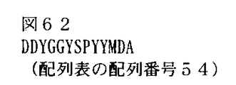
- CDRH2 consisting of an amino acid sequence or an amino acid sequence in which one or two amino acids are substituted in the amino acid sequence and the amino acid sequence shown in SEQ ID NO: 54 (FIG. 62) in the sequence listing or one or two amino acids in the amino acid sequence
- CDRL1 consisting of the amino acid sequence of SEQ ID NO: 62 of the sequence listing (FIG.
- CDRH2 consisting of an amino acid sequence or an amino acid sequence in which one or two amino acids are substituted in the amino acid sequence and the amino acid sequence shown in SEQ ID NO: 60 (FIG.
- the antibody or functional fragment thereof according to (16) The antibody or functional fragment thereof according to (15), wherein CDRH3 consists of an amino acid sequence in which one or two amino acids are substituted in the amino acid sequence shown in SEQ ID NO: 60 (FIG.
- a humanized antibody comprising (hFR2-14_H19 / L1); (Ii) a light chain comprising amino acid numbers 21 to 235 of the amino acid sequence represented by SEQ ID NO: 73 (FIG. 81) and a heavy chain comprising amino acid numbers 20 to 467 of the amino acid sequence represented by SEQ ID NO: 97 (FIG. 105)
- a humanized antibody comprising: (Iii) A light chain comprising amino acid numbers 21 to 235 of the amino acid sequence represented by SEQ ID NO: 73 (FIG.
- FIG. 97 A humanized antibody comprising (hFR2-14_H8 / L1); (Iv) a light chain comprising amino acid numbers 21 to 235 of the amino acid sequence represented by SEQ ID NO: 73 (FIG. 81) and a heavy chain comprising amino acid numbers 20 to 467 of the amino acid sequence represented by SEQ ID NO: 95 (FIG. 103) A humanized antibody comprising (hFR2-14_H11 / L1); (V) a light chain comprising amino acid numbers 21 to 235 of the amino acid sequence represented by SEQ ID NO: 73 (FIG.
- FIG. 91 A humanized antibody comprising (hFR2-14_H5 / L1); (Vi) a light chain comprising amino acid numbers 21 to 235 of the amino acid sequence represented by SEQ ID NO: 73 (FIG. 81) and a heavy chain comprising amino acid numbers 20 to 467 of the amino acid sequence represented by SEQ ID NO: 75 (FIG. 83) A humanized antibody comprising (hFR2-14_H1 / L1); (Vii) a light chain comprising amino acid numbers 21 to 235 of the amino acid sequence represented by SEQ ID NO: 73 (FIG.
- a humanized antibody comprising (hFR2-14_H2 / L1); (Viii) a light chain comprising amino acid numbers 21 to 235 of the amino acid sequence represented by SEQ ID NO: 73 (FIG. 81) and a heavy chain comprising amino acid numbers 20 to 467 of the amino acid sequence represented by SEQ ID NO: 79 (FIG. 87)
- a humanized antibody comprising (hFR2-14_H3 / L1); (Ix) a light chain comprising amino acid numbers 21 to 235 of the amino acid sequence represented by SEQ ID NO: 73 (FIG.
- FIG. 89 A humanized antibody comprising (hFR2-14_H4 / L1); (X) a light chain comprising amino acid numbers 21 to 235 of the amino acid sequence represented by SEQ ID NO: 73 (FIG. 81) and a heavy chain comprising amino acid numbers 20 to 467 of the amino acid sequence represented by SEQ ID NO: 85 (FIG. 93) A humanized antibody comprising (hFR2-14_H6 / L1); (Xi) a light chain comprising amino acid numbers 21 to 235 of the amino acid sequence represented by SEQ ID NO: 73 (FIG.
- a humanized antibody comprising (hFR2-14_H7 / L1); (Xii) a light chain comprising amino acid numbers 21 to 235 of the amino acid sequence represented by SEQ ID NO: 73 (FIG. 81) and a heavy chain comprising amino acid numbers 20 to 467 of the amino acid sequence represented by SEQ ID NO: 91 (FIG. 99) A humanized antibody comprising (hFR2-14_H9 / L1); (Xiii) a light chain comprising amino acid numbers 21 to 235 of the amino acid sequence represented by SEQ ID NO: 73 (FIG.
- a humanized antibody comprising (hFR2-14_H16 / L1); (Xiii) a light chain comprising amino acid numbers 21 to 235 of the amino acid sequence represented by SEQ ID NO: 73 (FIG. 81) and a heavy chain comprising amino acid numbers 20 to 467 of the amino acid sequence represented by SEQ ID NO: 107 (FIG. 115)
- a humanized antibody (hFR2-14_H17 / L1) comprising: a light chain comprising amino acid numbers 21 to 235 of the amino acid sequence represented by SEQ ID NO: 73 (FIG.
- a humanized antibody (hFR2-14_H18 / L1) comprising a heavy chain comprising amino acid numbers 20 to 467 of the amino acid sequence represented by (21) (1) to (12) comprising a heavy chain and a light chain each comprising 95% or more of the amino acid sequence identical to the heavy chain and light chain amino acid sequences of the antibody according to (20) and binding to human FGFR2.
- object (48) The composition according to (46) or (47), wherein the antibody is a chimeric antibody or a rat antibody; (49) The composition according to any one of (45) to (48), which is for detection or measurement of human FGFR2IIIb, (50) (45) A method for detecting or measuring human FGFR2IIIb, comprising a step of contacting the test sample with the composition according to any one of (48) to (48), (51) A method for detecting or measuring human FGFR2IIIc, comprising the following steps (i) to (iii): (I) A test sample is contacted with a composition containing an antibody that selectively binds to human FGFR2IIIb and human 2FGFRIIIc, or a functional fragment thereof, or a modification thereof, and human FGFR2IIIb and human FGFR2IIIc in the test sample are detected or measured Process; (Ii) a
- the use of the antibody provided by the present invention makes it possible to treat or prevent various cancers and to test or diagnose various cancers.
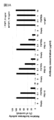
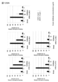
- FIG. 1 is a view in which the binding activity of rat anti-FGFR2 antibodies (FR2-10, FR2-13 and FR2-14) to human FGFR2 was verified by flow cytometry.
- shaft shows the relative value of the average fluorescence intensity measured by the flow cytometry method.
- FIG. 2 is a diagram in which human FGFR2 binding epitopes of rat anti-FGFR2 antibodies (FR2-10, FR2-13 and FR2-14) were verified by flow cytometry. The vertical axis shows the relative value of the average fluorescence intensity measured by flow cytometry.
- FIG. 1 is a view in which the binding activity of rat anti-FGFR2 antibodies (FR2-10, FR2-13 and FR2-14) to human FGFR2 was verified by flow cytometry.
- shaft shows the relative value of the average fluorescence intensity measured by the flow cytometry method.
- FIG. 2 is a diagram in which human FGFR2 binding epitopes
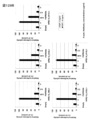
- FIG. 3A is a diagram showing signal neutralizing activity of rat anti-FGFR2 antibodies (FR2-10, FR2-13 and FR2-14) against human FGFR2IIIb by Elk1 transreporter assay.
- FIG. 3B shows signal neutralization activity of rat anti-FGFR2 antibodies (FR2-10, FR2-13 and FR2-14) against human FGFR2IIIc by Elk1 transreporter assay.
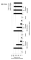
- FIG. 4 is a diagram showing the signal inhibitory action of rat anti-FGFR2 antibody FR2-10 on FGFR2 by Western blotting.
- FIG. 5 is a diagram in which the binding activity of human chimerized anti-FGFR2 antibodies (cFR2-10, cFR2-13, cFR2-14) to human FGFR2 was verified by the Cell-ELISA method.
- FIG. 6A shows signal neutralization activity of human chimerized anti-FGFR2 antibodies (cFR2-10, cFR2-13, cFR2-14) against human FGFR2IIIb by Elk1 transreporter assay.
- FIG. 6B shows the signal neutralization activity of human chimerized anti-FGFR2 antibodies (cFR2-10, cFR2-13, cFR2-14) against human FGFR2IIIc by Elk1 transreporter assay.
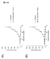
- FIG. 7 is a diagram showing ADCC activity of human chimerized anti-FGFR2 antibodies (cFR2-10, cFR2-13, cFR2-14). Human FGFR2IIIb-expressing 293T-lacZ cells were used as target cells and human PBMC as effector cells.
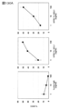
- FIG. 8 shows the in vivo antitumor activity of human chimerized anti-FGFR2 antibodies (cFR2-10, cFR2-13, cFR2-14) in human gastric cancer cell line SNU-16-transplanted nude mice.
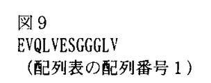
- FIG. 9 shows the N-terminal amino acid sequence of the band corresponding to the heavy chain of the rat anti-FGFR2 antibody FR2-10 (SEQ ID NO: 1 in the sequence listing).
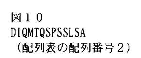
- FIG. 10 shows the N-terminal amino acid sequence of the band corresponding to the light chain of the rat anti-FGFR2 antibody FR2-10 (SEQ ID NO: 2 in the sequence listing).
- FIG. 11 shows the N-terminal amino acid sequence of the band corresponding to the heavy chain of the rat anti-FGFR2 antibody FR2-13 (SEQ ID NO: 3 in the Sequence Listing).
- FIG. 9 shows the N-terminal amino acid sequence of the band corresponding to the heavy chain of the rat anti-FGFR2 antibody FR2-10 (SEQ ID NO: 1 in the sequence listing).
- FIG. 10 shows the N-terminal amino acid sequence of the band corresponding to the light chain of the rat anti-FGFR2 antibody FR2-10 (SEQ ID NO: 2 in the sequence listing).
- FIG. 11 shows the N-termin
- FIG. 12 shows the N-terminal amino acid sequence of the band corresponding to the light chain of the rat anti-FGFR2 antibody FR2-13 (SEQ ID NO: 4 in the sequence listing).
- FIG. 13 shows the N-terminal amino acid sequence of the band corresponding to the rat anti-FGFR2 antibody FR2-14 heavy chain (SEQ ID NO: 5 in the Sequence Listing).
- FIG. 14 shows the N-terminal amino acid sequence of the band corresponding to the light chain of the rat anti-FGFR2 antibody FR2-14 (SEQ ID NO: 6 in the Sequence Listing).
- FIG. 15 shows a rat heavy chain amplification primer (SEQ ID NO: 7 in the Sequence Listing).
- FIG. 16 shows a sequence primer for FR2-10 heavy chain (SEQ ID NO: 8 in the sequence listing).
- FIG. 17 shows a sequence primer for FR2-13 heavy chain (SEQ ID NO: 9 in the sequence listing).
- FIG. 18 shows a sequence primer for FR2-14 heavy chain (SEQ ID NO: 10 in the sequence listing).
- FIG. 19 shows the nucleotide sequence of cDNA encoding the variable region of the heavy chain of rat anti-FGFR2 antibody FR2-10 (SEQ ID NO: 11 in the sequence listing).
- FIG. 20 shows the amino acid sequence of the variable region of the heavy chain of the rat anti-FGFR2 antibody FR2-10 (SEQ ID NO: 12 in the Sequence Listing).
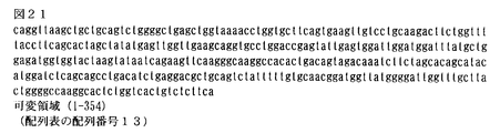
- FIG. 21 shows the base sequence of cDNA encoding the variable region of the heavy chain of the rat anti-FGFR2 antibody FR2-13 (SEQ ID NO: 13 in the sequence listing).
- FIG. 22 shows the amino acid sequence of the variable region of the heavy chain of the rat anti-FGFR2 antibody FR2-13 (SEQ ID NO: 14 in the Sequence Listing).
- FIG. 23 shows the nucleotide sequence of cDNA encoding the variable region of the heavy chain of the rat anti-FGFR2 antibody FR2-14 (SEQ ID NO: 15 of the Sequence Listing).
- FIG. 24 shows the amino acid sequence of the variable region of the heavy chain of the rat anti-FGFR2 antibody FR2-14 (SEQ ID NO: 16 in the Sequence Listing).
- FIG. 25 shows a rat light chain amplification primer (SEQ ID NO: 17 in the sequence listing).
- FIG. 26 shows a sequence primer for rat light chain (SEQ ID NO: 18 in the sequence listing).
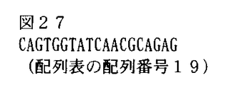
- FIG. 27 shows a sequence primer for the FR2-10 light chain (SEQ ID NO: 19 in the sequence listing).
- FIG. 28 shows the base sequence of cDNA encoding the variable region of the light chain of rat anti-FGFR2 antibody FR2-10 (SEQ ID NO: 20 in the sequence listing).
- FIG. 29 shows the amino acid sequence of the variable region of the light chain of rat anti-FGFR2 antibody FR2-10 (SEQ ID NO: 21 in the Sequence Listing).
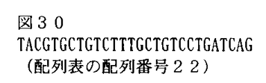
- FIG. 30 shows primers for amplifying rat FR2-13 and FR2-14 light chains (SEQ ID NO: 22 in the Sequence Listing).
- FIG. 31 shows the nucleotide sequence of the cDNA encoding the light chain variable region of the rat anti-FGFR2 antibody FR2-13 (SEQ ID NO: 23 in the Sequence Listing).
- FIG. 32 shows the amino acid sequence of the variable region of the light chain of rat anti-FGFR2 antibody FR2-13 (SEQ ID NO: 24 in the Sequence Listing).
- FIG. 33 shows the base sequence of cDNA encoding the variable region of the light chain of rat anti-FGFR2 antibody FR2-14 (SEQ ID NO: 25 in the sequence listing).
- FIG. 31 shows the nucleotide sequence of the cDNA encoding the light chain variable region of the rat anti-FGFR2 antibody FR2-13 (SEQ ID NO: 23 in the Sequence Listing).
- FIG. 32 shows the amino acid sequence of the variable region of the light chain of
- FIG. 34 shows the amino acid sequence of the variable region of the light chain of rat anti-FGFR2 antibody FR2-14 (SEQ ID NO: 26 in the Sequence Listing).
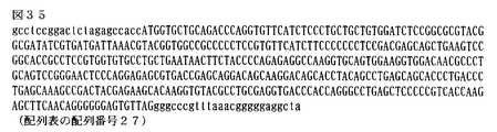
- FIG. 35 shows a DNA fragment (SEQ ID NO: 27 in the Sequence Listing) containing a DNA sequence encoding the human ⁇ chain secretion signal sequence and the amino acids of the human ⁇ chain constant region.
- FIG. 36 shows a primer F for a light chain expression vector (SEQ ID NO: 28 in the sequence listing).
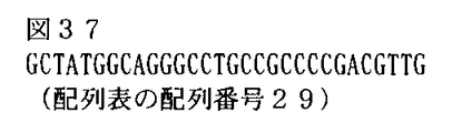
- FIG. 37 shows a primer R for a light chain expression vector (SEQ ID NO: 29 in the sequence listing).
- FIG. 38 shows a DNA fragment containing a human heavy chain signal sequence and a DNA sequence encoding the amino acid of the human IgG1 constant region (SEQ ID NO: 30 in the Sequence Listing).
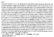
- FIG. 39 shows the base sequence of human chimerized FR2-10 (cFR2-10) light chain (SEQ ID NO: 31 in the sequence listing). Of these, nucleotide numbers 1 to 60 are signal sequences, and are usually not included in the base sequence of most mature cFR2-10 light chains.
- FIG. 40 shows the amino acid sequence of the human chimerized FR2-10 (cFR2-10) light chain (SEQ ID NO: 32 in the Sequence Listing).
- amino acid numbers 1 to 20 are signal sequences, and are usually not included in the amino acid sequences of most mature cFR2-10 light chains.
- FIG. 41 shows primer set F for human chimerized FR2-10 light chain (SEQ ID NO: 33 in the sequence listing).
- FIG. 42 shows primer set R for human chimerized FR2-10 light chain (SEQ ID NO: 34 in the sequence listing).
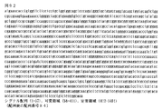
- FIG. 43 shows the base sequence of human chimerized FR2-10 (cFR2-10) heavy chain (SEQ ID NO: 35 in the Sequence Listing).
- nucleotide numbers 1 to 57 are signal sequences, and are usually not included in the base sequence of most mature cFR2-10 heavy chains.
- FIG. 41 shows primer set F for human chimerized FR2-10 light chain (SEQ ID NO: 33 in the sequence listing).
- FIG. 42 shows primer set R for human chimerized FR2-10 light chain (SEQ ID NO: 34 in the sequence listing).
- FIG. 43 shows the base sequence of human chim
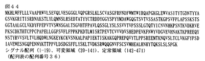
- FIG. 44 shows the amino acid sequence of human chimerized FR2-10 (cFR2-10) heavy chain (SEQ ID NO: 36 in the Sequence Listing). Among them, amino acid numbers 1 to 19 are signal sequences, and are usually not included in the amino acid sequences of most mature cFR2-10 heavy chains.
- FIG. 45 shows primer set F for human chimerized FR2-10 heavy chain (SEQ ID NO: 37 in the sequence listing).
- FIG. 46 shows primer set R for human chimerized FR2-10 heavy chain (SEQ ID NO: 38 in the Sequence Listing).
- FIG. 47 shows the nucleotide sequence of human chimerized FR2-13 (cFR2-13) light chain (SEQ ID NO: 39 in the Sequence Listing).
- nucleotide numbers 1 to 60 are signal sequences, and are usually not included in the base sequence of most mature cFR2-13 light chains.
- 48 shows human chimerized FR2-13 (amino acid sequence of cFR2-13 light chain (SEQ ID NO: 40 in the sequence listing), wherein amino acid numbers 1 to 20 are signal sequences, and usually amino acids of most mature cFR2-13 light chain) Not included in the array.
- FIG. 49 shows primer F for human chimerized FR2-13 light chain (SEQ ID NO: 41 in the sequence listing).
- FIG. 50 shows primer R for human chimerized FR2-13 light chain (SEQ ID NO: 42 in the sequence listing).
- FIG. 51 shows the base sequence of human chimerized FR2-13 (cFR2-13) heavy chain (SEQ ID NO: 43 in the Sequence Listing). Of these, nucleotide numbers 1 to 57 are signal sequences, and are usually not included in the base sequence of most mature cFR2-13 heavy chains.
- FIG. 52 shows the amino acid sequence of human chimerized FR2-13 (cFR2-13) heavy chain (SEQ ID NO: 44 in the Sequence Listing). Of these, amino acid numbers 1 to 19 are signal sequences, and are usually not included in the amino acid sequences of most mature cFR2-13 heavy chains.
- FIG. 53 shows primer F for human chimerized FR2-13 heavy chain (SEQ ID NO: 45 of the Sequence Listing).
- FIG. 54 shows a primer R for human chimerized FR2-13 heavy chain (SEQ ID NO: 46 in the sequence listing).
- FIG. 55 shows the nucleotide sequence of human chimerized FR2-14 (cFR2-14) light chain (SEQ ID NO: 47 in the sequence listing). Of these, nucleotide numbers 1 to 60 are signal sequences, and are usually not included in the base sequences of most mature cFR2-14 light chains.
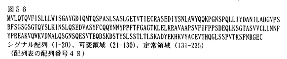
- FIG. 56 shows the amino acid sequence of human chimerized FR2-14 (cFR2-14) light chain (SEQ ID NO: 48 in the Sequence Listing). Among them, amino acid numbers 1 to 20 are signal sequences and are not usually included in the amino acid sequences of most mature cFR2-14 light chains.
- FIG. 55 shows the nucleotide sequence of human chimerized FR2-14 (cFR2-14) light chain (SEQ ID NO: 47 in the sequence listing). Of these, nucleotide numbers 1 to 60 are signal sequences, and are usually not included in the
- FIG. 57 shows a primer for human chimerized FR2-14 light chain (SEQ ID NO: 49 in the sequence listing).
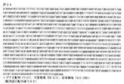
- FIG. 58 shows the base sequence of human chimerized FR2-14 (cFR2-14) heavy chain (SEQ ID NO: 50 in the Sequence Listing). Of these, nucleotide numbers 1 to 57 are signal sequences, and are usually not included in the base sequence of most mature cFR2-14 heavy chains.
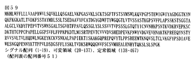
- FIG. 59 shows the amino acid sequence of human chimerized FR2-14 (cFR2-14) heavy chain (SEQ ID NO: 51 in the Sequence Listing). Of these, amino acid numbers 1 to 19 are signal sequences, and are usually not included in the amino acid sequences of most mature cFR2-14 heavy chains.
- FIG. 58 shows the base sequence of human chimerized FR2-14 (cFR2-14) heavy chain (SEQ ID NO: 50 in the Sequence Listing). Of these, nucleotide numbers 1 to 57 are signal sequences, and are usually not included in the base sequence
- FIG. 60 shows the amino acid sequence of the heavy chain CDR1 of the rat anti-FGFR2 antibody FR2-10 (SEQ ID NO: 52 of the Sequence Listing).
- FIG. 61 shows the amino acid sequence of heavy chain CDR2 of rat anti-FGFR2 antibody FR2-10 (SEQ ID NO: 53 of the Sequence Listing).
- FIG. 62 shows the amino acid sequence of heavy chain CDR3 of rat anti-FGFR2 antibody FR2-10 (SEQ ID NO: 54 of the Sequence Listing).
- FIG. 63 shows the amino acid sequence of the heavy chain CDR1 of the rat anti-FGFR2 antibody FR2-13 (SEQ ID NO: 55 of the Sequence Listing).
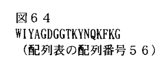
- FIG. 64 shows the amino acid sequence of heavy chain CDR2 of rat anti-FGFR2 antibody FR2-13 (SEQ ID NO: 56 of the Sequence Listing).
- FIG. 65 shows the amino acid sequence of heavy chain CDR3 of rat anti-FGFR2 antibody FR2-13 (SEQ ID NO: 57 of the Sequence Listing).
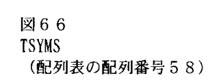
- FIG. 66 shows the amino acid sequence of heavy chain CDR1 of rat anti-FGFR2 antibody FR2-14 (SEQ ID NO: 58 of the Sequence Listing).
- FIG. 67 shows the amino acid sequence of the heavy chain CDR2 of the rat anti-FGFR2 antibody FR2-14 (SEQ ID NO: 59 of the Sequence Listing).
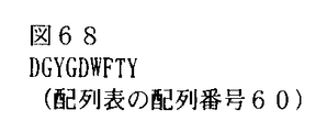
- FIG. 68 shows the amino acid sequence of heavy chain CDR3 of rat anti-FGFR2 antibody FR2-14 (SEQ ID NO: 60 of the Sequence Listing).
- FIG. 69 shows the amino acid sequence of the light chain CDR1 of the rat anti-FGFR2 antibody FR2-10 (SEQ ID NO: 61 of the Sequence Listing).
- FIG. 70 shows the amino acid sequence of the light chain CDR2 of the rat anti-FGFR2 antibody FR2-10 (SEQ ID NO: 62 of the Sequence Listing).
- FIG. 71 shows the amino acid sequence of the light chain CDR3 of the rat anti-FGFR2 antibody FR2-10 (SEQ ID NO: 63 of the Sequence Listing).
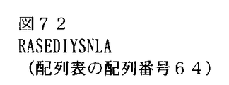
- FIG. 72 shows the amino acid sequence of the light chain CDR1 of the rat anti-FGFR2 antibody FR2-13 (SEQ ID NO: 64 of the Sequence Listing).
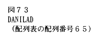
- FIG. 73 shows the amino acid sequence of the light chain CDR2 of the rat anti-FGFR2 antibody FR2-13 (SEQ ID NO: 65 of the Sequence Listing).
- FIG. 74 shows the amino acid sequence of the light chain CDR3 of the rat anti-FGFR2 antibody FR2-13 (SEQ ID NO: 66 of the Sequence Listing).
- FIG. 75 shows the amino acid sequence of the light chain CDR1 of the rat anti-FGFR2 antibody FR2-14 (SEQ ID NO: 67 of the Sequence Listing).
- FIG. 76 shows the amino acid sequence of the light chain CDR2 of the rat anti-FGFR2 antibody FR2-14 (SEQ ID NO: 68 of the Sequence Listing).
- FIG. 77 shows the amino acid sequence of the light chain CDR3 of the rat anti-FGFR2 antibody FR2-14 (SEQ ID NO: 69 of the Sequence Listing).
- FIG. 78 shows the amino acid sequence of human FGFR2IIIb (SEQ ID NO: 70 of the Sequence Listing).
- FIG. 79 shows the amino acid sequence of human FGFR2IIIc (SEQ ID NO: 71 in the sequence listing).
- FIG. 80 shows the nucleotide sequence of hFR2-14_L1 (SEQ ID NO: 72 in the sequence listing), of which nucleotide numbers 1 to 60 are signal sequences, and are usually not included in the nucleotide sequence of most mature hFR2-14_L1.
- FIG. 81 shows the amino acid sequence of hFR2-14_L1 (SEQ ID NO: 73 in the sequence listing), of which nucleotide numbers 1 to 20 are signal sequences, and are not usually included in the amino acid sequence of most mature hFR2-14_L1.
- FIG. 82 shows the base sequence of hFR2-14_H1 (SEQ ID NO: 74 in the sequence listing), of which nucleotide numbers 1 to 57 are signal sequences, and are usually not included in the base sequence of most mature hFR2-14_H1.
- FIG. 83 shows the amino acid sequence of hFR2-14_H1 (SEQ ID NO: 75 in the sequence listing), of which nucleotide numbers 1 to 19 are signal sequences, and are usually not included in the amino acid sequence of most mature hFR2-14_H1.
- FIG. 83 shows the amino acid sequence of hFR2-14_H1 (SEQ ID NO: 75 in the sequence listing), of which nucleotide numbers 1 to 19 are signal sequences, and are usually not included in the amino acid sequence of most mature hFR2-14_H1.
- FIG. 84 shows the base sequence of hFR2-14_H2 (SEQ ID NO: 76 in the sequence listing), of which nucleotide numbers 1 to 57 are signal sequences, and are usually not included in the base sequence of most mature hFR2-14_H2.
- FIG. 85 shows the amino acid sequence of hFR2-14_H2 (SEQ ID NO: 77 in the sequence listing), of which nucleotide numbers 1 to 19 are signal sequences, and are not usually included in the amino acid sequence of most mature hFR2-14_H2.
- FIG. 86 shows the nucleotide sequence of hFR2-14_H3 (SEQ ID NO: 78 in the sequence listing), of which nucleotide numbers 1 to 57 are signal sequences, and are usually not included in the nucleotide sequence of most mature hFR2-14_H3.
- FIG. 87 shows the amino acid sequence of hFR2-14_H3 (SEQ ID NO: 79 in the Sequence Listing), of which nucleotide numbers 1 to 19 are signal sequences, and are usually not included in the amino acid sequence of most mature hFR2-14_H3.
- FIG. 87 shows the amino acid sequence of hFR2-14_H3 (SEQ ID NO: 79 in the Sequence Listing), of which nucleotide numbers 1 to 19 are signal sequences, and are usually not included in the amino acid sequence of most mature hFR2-14_H3.
- FIG. 88 shows the nucleotide sequence of hFR2-14_H4 (SEQ ID NO: 80 in the sequence listing), of which nucleotide numbers 1 to 57 are signal sequences, and are usually not included in the nucleotide sequence of most mature hFR2-14_H4.
- FIG. 89 shows the amino acid sequence of hFR2-14_H4 (SEQ ID NO: 81 in the sequence listing), of which nucleotide numbers 1 to 19 are signal sequences, and are not usually included in the amino acid sequence of most mature hFR2-14_H4.
- FIG. 80 shows the nucleotide sequence of hFR2-14_H4 (SEQ ID NO: 80 in the sequence listing), of which nucleotide numbers 1 to 57 are signal sequences, and are usually not included in the nucleotide sequence of most mature hFR2-14_H4.
- FIG. 89 shows the amino acid sequence of hFR2-14_H4 (SEQ ID NO: 81 in the sequence listing), of which
- FIG. 90 shows the base sequence of hFR2-14_H5 (SEQ ID NO: 82 in the sequence listing), of which nucleotide numbers 1 to 57 are signal sequences, and are usually not included in the base sequence of most mature hFR2-14_H5.
- FIG. 91 shows the amino acid sequence of hFR2-14_H5 (SEQ ID NO: 83 in the Sequence Listing), of which nucleotide numbers 1 to 19 are signal sequences, and are usually not included in the amino acid sequence of most mature hFR2-14_H5.
- FIG. 82 shows the base sequence of hFR2-14_H5 (SEQ ID NO: 82 in the sequence listing), of which nucleotide numbers 1 to 57 are signal sequences, and are usually not included in the base sequence of most mature hFR2-14_H5.
- FIG. 91 shows the amino acid sequence of hFR2-14_H5 (SEQ ID NO: 83 in the Sequence Listing), of which nucleotide numbers 1 to 19 are signal sequence
- FIG. 92 shows the nucleotide sequence of hFR2-14_H6 (SEQ ID NO: 84 in the sequence listing), of which nucleotide numbers 1 to 57 are signal sequences and are mature and usually not included in the majority of hFR2-14_H6 nucleotide sequences.
- FIG. 93 shows the amino acid sequence of hFR2-14_H6 (SEQ ID NO: 85 of the sequence listing), of which nucleotide numbers 1 to 19 are signal sequences, and are usually not included in the amino acid sequence of most mature hFR2-14_H6.
- FIG. 93 shows the amino acid sequence of hFR2-14_H6 (SEQ ID NO: 85 of the sequence listing), of which nucleotide numbers 1 to 19 are signal sequences, and are usually not included in the amino acid sequence of most mature hFR2-14_H6.
- FIG. 94 shows the nucleotide sequence of hFR2-14_H7 (SEQ ID NO: 86 in the sequence listing), of which nucleotide numbers 1 to 57 are signal sequences, and are usually not included in the nucleotide sequence of most mature hFR2-14_H7.
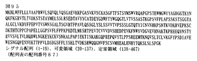
- FIG. 95 shows the amino acid sequence of hFR2-14_H7 (SEQ ID NO: 87 in the sequence listing), of which nucleotide numbers 1 to 19 are signal sequences, and are usually not included in the amino acid sequence of most mature hFR2-14_H7.
- FIG. 96 shows the nucleotide sequence of hFR2-14_H8 (SEQ ID NO: 88 in the sequence listing), of which nucleotide numbers 1 to 57 are signal sequences, and are usually not included in the nucleotide sequence of most mature hFR2-14_H8.
- FIG. 97 shows the amino acid sequence of hFR2-14_H8 (SEQ ID NO: 89 in the sequence listing), of which nucleotide numbers 1 to 19 are signal sequences, and are usually not included in the amino acid sequence of most mature hFR2-14_H8.
- FIG. 96 shows the nucleotide sequence of hFR2-14_H8 (SEQ ID NO: 88 in the sequence listing), of which nucleotide numbers 1 to 57 are signal sequences, and are usually not included in the nucleotide sequence of most mature hFR2-14_H8.
- FIG. 97 shows the amino acid sequence of hFR2-14_H8 (SEQ ID NO: 89 in
- FIG. 98 shows the base sequence of hFR2-14_H9 (SEQ ID NO: 90 in the sequence listing), of which nucleotide numbers 1 to 57 are signal sequences, and are usually not included in the base sequence of most mature hFR2-14_H9.
- FIG. 99 shows the amino acid sequence of hFR2-14_H9 (SEQ ID NO: 91 in the sequence listing), of which nucleotide numbers 1 to 19 are signal sequences, and are usually not included in the amino acid sequence of most mature hFR2-14_H9.
- FIG. 100 shows the base sequence of hFR2-14_H10 (SEQ ID NO: 92 in the sequence listing), of which nucleotide numbers 1 to 57 are signal sequences, and are usually not included in the base sequence of most mature hFR2-14_H10.
- FIG. 101 shows the amino acid sequence of hFR2-14_H10 (SEQ ID NO: 93 in the sequence listing), of which nucleotide numbers 1 to 19 are signal sequences, and are usually not included in the amino acid sequence of most mature hFR2-14_H10.
- FIG. 102 shows the nucleotide sequence of hFR2-14_H11 (SEQ ID NO: 94 in the sequence listing), of which nucleotide numbers 1 to 57 are signal sequences, and are usually not included in the nucleotide sequence of most mature hFR2-14_H11.
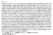
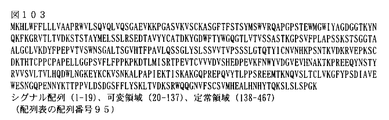
- FIG. 103 shows the amino acid sequence of hFR2-14_H11 (SEQ ID NO: 95 of the sequence listing), of which nucleotide numbers 1 to 19 are signal sequences, and are usually not included in the amino acid sequence of most mature hFR2-14_H11.
- FIG. 104 shows the nucleotide sequence of hFR2-14_H11 (SEQ ID NO: 94 in the sequence listing), of which nucleotide numbers 1 to 57 are signal sequences, and are usually not included in the nucleotide sequence of most mature hFR2-14_H11.
- FIG. 103 shows the amino acid sequence of hFR2-14_H11 (SEQ ID NO: 95 of the sequence listing), of
- FIG. 104 shows the nucleotide sequences of hFR2-14_H12 and hFR2-14_H19 (SEQ ID NO: 96 in the sequence listing), of which nucleotide numbers 1 to 57 are signal sequences. Usually, most of the mature hFR2-14_H12 and hFR2-14_H19 nucleotide sequences Is not included.
- FIG. 105 shows the amino acid sequences of hFR2-14_H12 and hFR2-14_H19 (SEQ ID NO: 97 in the Sequence Listing), of which nucleotide numbers 1 to 19 are signal sequences. Is not included.
- FIG. 105 shows the amino acid sequences of hFR2-14_H12 and hFR2-14_H19 (SEQ ID NO: 97 in the Sequence Listing), of which nucleotide numbers 1 to 19 are signal sequences. Is not included.
- FIG. 106 shows the base sequence of hFR2-14_H13 (SEQ ID NO: 98 in the sequence listing), of which nucleotide numbers 1 to 57 are signal sequences, and are usually not included in the base sequence of most mature hFR2-14_H13.
- FIG. 107 shows the amino acid sequence of hFR2-14_H13 (SEQ ID NO: 99 in the sequence listing), of which nucleotide numbers 1 to 19 are signal sequences, and are usually not included in the amino acid sequence of most mature hFR2-14_H13.
- FIG. 108 shows the base sequence of hFR2-14_H14 (SEQ ID NO: 100 in the sequence listing), of which nucleotide numbers 1 to 57 are signal sequences, and are usually not included in the base sequence of most mature hFR2-14_H14.
- FIG. 109 shows the amino acid sequence of hFR2-14_H14 (SEQ ID NO: 101 in the sequence listing), of which nucleotide numbers 1 to 19 are signal sequences, and are usually not included in the amino acid sequence of most mature hFR2-14_H14.
- FIG. 110 shows the base sequence of hFR2-14_H15 (SEQ ID NO: 102 in the sequence listing), of which nucleotide numbers 1 to 57 are signal sequences, and are usually not included in the base sequence of most mature hFR2-14_H15.
- FIG. 111 shows the amino acid sequence of hFR2-14_H15 (SEQ ID NO: 103 in the sequence listing), of which nucleotide numbers 1 to 19 are signal sequences, and are usually not included in the amino acid sequence of most mature hFR2-14_H15.
- FIG. 112 shows the base sequence of hFR2-14_H16 (SEQ ID NO: 104 in the sequence listing), of which nucleotide numbers 1 to 57 are signal sequences, and are usually not included in the base sequence of most mature hFR2-14_H16.
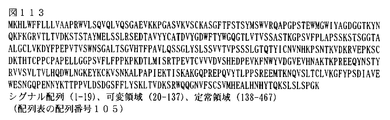
- FIG. 113 shows the amino acid sequence of hFR2-14_H16 (SEQ ID NO: 105 in the sequence listing), of which nucleotide numbers 1 to 19 are signal sequences, and are usually not included in the amino acid sequence of most mature hFR2-14_H16.
- FIG. 104 shows the base sequence of hFR2-14_H16 (SEQ ID NO: 104 in the sequence listing), of which nucleotide numbers 1 to 57 are signal sequences, and are usually not included in the base sequence of most mature hFR2-14_H16.
- FIG. 113 shows the amino acid sequence of hFR2-14_H16 (SEQ ID NO: 105 in the sequence listing), of which nucleotide numbers 1 to 19 are
- FIG. 114 shows the nucleotide sequence of hFR2-14_H17 (SEQ ID NO: 106 in the sequence listing), of which nucleotide numbers 1 to 57 are signal sequences, and are usually not included in the nucleotide sequence of most mature hFR2-14_H17.
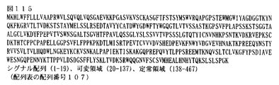
- FIG. 115 shows the amino acid sequence of hFR2-14_H17 (SEQ ID NO: 107 in the sequence listing), of which nucleotide numbers 1 to 19 are signal sequences, and are usually not included in the amino acid sequence of most mature hFR2-14_H17.
- FIG. 116 shows the nucleotide sequence of hFR2-14_H18 (SEQ ID NO: 108 in the sequence listing), of which nucleotide numbers 1 to 57 are signal sequences, and are usually not included in the nucleotide sequence of most mature hFR2-14_H18.
- FIG. 117 shows the amino acid sequence of hFR2-14_H18 (SEQ ID NO: 109 in the sequence listing), of which nucleotide numbers 1 to 19 are signal sequences, and are usually not included in the amino acid sequence of most mature hFR2-14_H18.
- FIG. 118 shows hFR2-14_H2 type heavy chain primer VH3A-F (SEQ ID NO: 110 in the Sequence Listing).
- FIG. 119 shows hFR2-14_H2 type heavy chain primer VH3A-R (SEQ ID NO: 111 in the Sequence Listing).
- FIG. 120 shows D2 amplification primer D23fw (SEQ ID NO: 112 in the sequence listing).
- FIG. 121 shows D2 amplification primer D23rv (SEQ ID NO: 113 in the sequence listing).
- FIG. 122 is a diagram in which the binding activity of each of four types of humanized anti-FGFR2 antibodies (hFR2-14_H1 to H4 / L1) and human chimerized anti-FGFR2 antibody (cFR2-14) to each human FGFR2 variant protein was measured by Biacore. Each antibody was expressed in 293F cells, purified and used for measurement.
- FIG. 123 is a diagram showing the binding activity of 15 types of humanized anti-FGFR2 antibodies (hFR2-14_H3 / L1 and hFR2-14_H5 to H18 / L1) to human FGFR2-IIIc variant protein measured by Biacore. Culture supernatants in which each antibody was expressed in 293F cells were used for measurement.
- FIG. 124 is a diagram in which the human FGFR2 selective binding properties of three types of humanized anti-FGFR2 antibodies (hFR2-14_H3 / L1, hFR2-14_H8 / L1 and hFR2-14_H12 / L1) were verified by the Cell-ELISA method.
- FIG. 125A shows thermograms of five types of humanized anti-FGFR2 antibodies.
- FIG. 125B is a diagram showing thermograms of five humanized anti-FGFR2 antibodies.
- FIG. 125C is a diagram showing Tm values of 10 types of humanized anti-FGFR2 antibodies.
- FIG. 126 shows hFR2-14_H1 / L1 antibody, hFR2-14_H2 / L1 antibody, hFR2-14_H3 / L1 antibody, hFR2-14_H4 / L1 antibody, hFR2-14_H5 / L1 antibody, hFR2-14_H8 / L1 antibody, hFR2-14_H9 / It is the figure which showed KD value with an antigen about the specimen before and behind degradation of L1 antibody, hFR2-14_H11 / L1 antibody, hFR2-14_H12 / L1 antibody, and hFR2-14_H19 / L1 antibody.
- FIG. 127A shows signals of humanized anti-FGFR2 antibodies (hFR2-14_H1 / L1, hFR2-14_H2 / L1, hFR2-14_H3 / L1, hFR2-14_H4 / L1) and human chimerized anti-FGFR2 antibody (cFR2-14) to human FGFR2IIIb. It is the figure which showed neutralizing activity by the Elk1 transreporter assay method.
- FIG. 127A shows signals of humanized anti-FGFR2 antibodies (hFR2-14_H1 / L1, hFR2-14_H2 / L1, hFR2-14_H3 / L1, hFR2-14_H4 / L1) and human chimerized anti-FGFR2 antibody (cFR2-14) to human FGFR2IIIb. It is the figure which showed neutralizing activity by the Elk1 transreporter assay method.
- 127B shows signals of humanized anti-FGFR2 antibodies (hFR2-14_H1 / L1, hFR2-14_H2 / L1, hFR2-14_H3 / L1, hFR2-14_H4 / L1) and human chimerized anti-FGFR2 antibody (cFR2-14) to human FGFR2IIIc. It is the figure which showed neutralizing activity by the Elk1 transreporter assay method.
- FIG. 128A shows the signal neutralization activity of humanized anti-FGFR2 antibodies (hFR2-14_H3 / L1, hFR2-14_H5 / L1, hFR2-14_H6 / L1, hFR2-14_H7 / L1, hFR2-14_H8 / L1) to human FGFR2IIIb. It is the figure shown by the transreporter assay method.
- FIG. 128B shows the signal neutralization activity of humanized anti-FGFR2 antibodies (hFR2-14_H9 / L1, hFR2-14_H10 / L1, hFR2-14_H11 / L1, hFR2-14_H12 / L1, hFR2-14_H13 / L1) to human FGFR2IIIb.
- FIG. 128C shows the signal neutralization activity of humanized anti-FGFR2 antibodies (hFR2-14_H14 / L1, hFR2-14_H15 / L1, hFR2-14_H16 / L1, hFR2-14_H17 / L1, hFR2-14_H18 / L1) to human FGFR2IIIb. It is the figure shown by the transreporter assay method.
- FIG. 129A shows signal neutralization activity of humanized anti-FGFR2 antibodies (hFR2-14_H12 / L1, hFR2-14_H19 / L1) to human FGFR2IIIb by Elk1 transreporter assay.
- FIG. 128C shows the signal neutralization activity of humanized anti-FGFR2 antibodies (hFR2-14_H14 / L1, hFR2-14_H15 / L1, hFR2-14_H16 / L1, hFR2-14_H17 / L1, hFR2-14_H18 / L1) to human FG
- FIG. 130A shows ADCC activity of humanized anti-FGFR2 antibodies (hFR2-14_H1 / L1, hFR2-14_H2 / L1). Human FGFR2IIIb-expressing 293T-lacZ cells were used as target cells and human PBMC as effector cells.
- FIG. 130B shows ADCC activity of humanized anti-FGFR2 antibodies (hFR2-14_H3 / L1, hFR2-14_H4 / L1) and human chimerized anti-FGFR2 antibody (cFR2-14).
- Human FGFR2IIIb-expressing 293T-lacZ cells were used as target cells and human PBMC as effector cells.
- 131 is a view showing ADCC activity of humanized anti-FGFR2 antibodies (hFR2-14_H3 / L1, hFR2-14_H8 / L1, and hFR2-14_H12 / L1.
- Human FGFR2 expressing NCI-H716 cells are target cells and human PBMCs. Were used as effector cells.
- FIG. 132 shows ADCC activity of humanized anti-FGFR2 antibodies (hFR2-14_H12 / L1, hFR2-14_H19 / L1).
- FIG. 133 shows ADCP activity of humanized anti-FGFR2 antibodies (hFR2-14_H12 / L1, hFR2-14_H19 / L1).
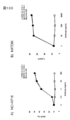
- FIG. 134A shows the in vivo antitumor activity of humanized anti-FGFR2 antibody hFR2-14_H1 / L1 in nude mice transplanted with human gastric cancer cell line SNU-16.
- FIG. 134B shows in vivo antitumor activity of humanized anti-FGFR2 antibody hFR2-14_H2 / L1 in nude mice transplanted with human gastric cancer cell line SNU-16.
- FIG. 134C shows in vivo antitumor activity of humanized anti-FGFR2 antibody hFR2-14_H3 / L1 in nude mice transplanted with human gastric cancer cell line SNU-16.
- FIG. 134D shows in vivo antitumor activity of humanized anti-FGFR2 antibody hFR2-14_H4 / L1 in nude mice transplanted with human gastric cancer cell line SNU-16.
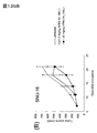
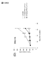
- FIG. 135A shows the in vivo antitumor activity of humanized anti-FGFR2 antibody hFR2-14_H5 / L1 in human gastric cancer cell line SNU-16 subcutaneous transplantation model.
- FIG. 135B shows in vivo antitumor activity of humanized anti-FGFR2 antibody hFR2-14_H8 / L1 in human gastric cancer cell line SNU-16 subcutaneous transplantation model.
- FIG. 135C shows in vivo antitumor activity of humanized anti-FGFR2 antibody hFR2-14_H9 / L1 in human gastric cancer cell line SNU-16 subcutaneous transplantation model.
- FIG. 135A shows the in vivo antitumor activity of humanized anti-FGFR2 antibody hFR2-14_H5 / L1 in human gastric cancer cell line SNU-16 subcutaneous transplantation model.
- FIG. 135B shows in vivo antitumor activity of humanized anti-FGFR2 antibody hFR2-14_H8 / L1
- FIG. 135D shows in vivo antitumor activity of humanized anti-FGFR2 antibody hFR2-14_H11 / L1 in human gastric cancer cell line SNU-16 subcutaneous transplantation model.
- FIG. 135E shows in vivo antitumor activity of humanized anti-FGFR2 antibody hFR2-14_H12 / L1 in human gastric cancer cell line SNU-16 subcutaneous transplantation model.
- FIG. 135F shows in vivo antitumor activity of humanized anti-FGFR2 antibody hFR2-14_H19 / L1 in human gastric cancer cell line SNU-16 subcutaneous transplantation model.
- FIG. 136 shows the in vivo antitumor activity of humanized anti-FGFR2 antibodies (hFR2-14_H12 / L1, hFR2-14_H19 / L1) in the human colon cancer cell line NCI-H716 tumor block model.
- (A) is the result of hFR2-14_H12 / L1
- (B) is the result of hFR2-14_H19 / L1.
- FIG. 137 shows the in vivo anti-tumor activity of humanized anti-FGFR2 antibodies (hFR2-14_H12 / L1, hFR2-14_H19 / L1) in a human colon cancer cell line NCI-H716-luc peritoneal dissemination model.
- FIG. 138 shows a ribbon model of the FGFR2D2 / H3L1 Fab complex. FGFR2D2 is displayed in the lower left, the H3L1 Fab H chain (dark gray) from the center to the upper right, and the L chain on the right side.
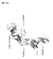
- FIG. 139 is a diagram showing the superposition of FGFR2 / FGF1 and FGFR2D2 / H3L1 Fab.
- FGFRD3 (IgD3) is displayed in the lower center, FGF1 (dark gray) above FGFRD3, FGFRD2 above FGF1, and H2L1 Fab H chain (dark gray) and L chain above FGFRD2, respectively.
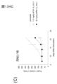
- FIG. 140 shows immunostaining of the 293 ⁇ cell block in which FGFR family molecules were forcibly expressed using rat antibody FR2-10.
- FIG. 141 shows immunostaining of A, D) SNU-16, B, E) KATOIII, and C, F) NCI-H716 cell blocks. A to C are results of using rat antibody FR2-10, and D to F are results of using commercially available antibodies.
- FIG. 140 shows immunostaining of the 293 ⁇ cell block in which FGFR family molecules were forcibly expressed using rat antibody FR2-10.
- FIG. 141 shows immunostaining of A, D) SNU-16, B, E) KATOIII, and C, F) NCI-H716 cell blocks.
- FIG. 143 is a graph showing the activity of inhibiting the binding of ligand FGF7 to the FGFR2 receptor by cFR2-10, hFR2-14_H12 / L1, and hFR2-14_H19 / L1 antibodies.
- the “gene” means a nucleotide containing a base sequence encoding a protein amino acid or a complementary strand thereof, for example, a nucleotide containing a base sequence encoding a protein amino acid or a complementary strand thereof.
- Polynucleotide, oligonucleotide, DNA, mRNA, cDNA, cRNA and the like are included in the meaning of “gene”.
- Such a gene is a single-stranded, double-stranded, or triple-stranded nucleotide, and an assembly of a DNA strand and an RNA strand, and ribonucleotide (RNA) and deoxyribonucleotide (DNA) are mixed on one nucleotide strand. Also included within the meaning of “gene” are double-stranded or triple-stranded nucleotides comprising such nucleotide chains.
- Examples of the “FGFR2 gene” of the present invention include DNA, mRNA, cDNA, cRNA and the like containing a base sequence encoding the amino acid sequence of the FGFR2 protein.
- nucleotide and “nucleic acid” are synonymous.
- DNA, RNA, probe, oligonucleotide, polynucleotide, primer and the like are also included in the meaning of “nucleotide”.
- nucleotide is a nucleotide composed of a single strand, a double strand, or three or more strands, and an assembly of a DNA strand and an RNA strand, and ribonucleotide (RNA) and deoxyribonucleotide (DNA) on a single nucleotide strand.
- RNA ribonucleotide
- DNA deoxyribonucleotide
- nucleotide also included within the meaning of “nucleotide” are those that are intermingled and aggregates of two or more strands containing such nucleotide strands.
- polypeptide “peptide” and “protein” are synonymous.
- antigen is sometimes used to mean “immunogen”.
- the “cell” includes various cells derived from individual animals, subculture cells, primary culture cells, cell lines, recombinant cells, microorganisms, and the like. In the present invention, antibodies that recognize FGFR2, FGFR2IIIb, FGFR2IIIc, FGFR3, FGFRs, etc.
- anti-FGFR2 antibody is designated as “anti-FGFR2 antibody”, “anti-FGFR2IIIb antibody”, “anti-FGFR2IIIc antibody”, “anti-FGFR3 antibody” and “anti-FGFRs antibody”, respectively. Or the like.
- Such antibodies include chimerized antibodies, humanized antibodies, human antibodies and the like.
- the “functional fragment of an antibody” in the present invention means an antibody fragment that exhibits at least a part of the function exhibited by the original antibody. Examples of the “functional fragment of the antibody” include, but are not limited to, Fab, F (ab ′) 2, scFv, Fab ′, single chain immunoglobulin and the like.
- Such functional fragments of antibodies are recombinant proteins produced in appropriate host cells using recombinant genes in addition to those obtained by treating full-length antibody protein molecules with enzymes such as papain and pepsin. May be.
- the “site” to which the antibody binds that is, the “site” recognized by the antibody means a partial peptide or a partial higher order structure on the antigen to which the antibody binds or recognizes. In the present invention, such a site is also referred to as an epitope or an antibody binding site.
- the site on the FGFR2 protein to which the anti-FGFR2 antibody of the present invention binds or recognizes include a partial peptide or a partial higher order structure on the FGFR2 protein.
- CDRs complementarity determining regions
- Complementarity-determining regions also called hypervariable domains, are sites in the variable regions of antibody heavy and light chains that have particularly high variability in primary structure. In the primary structure of the polypeptide chain, it is usually separated at three points.
- CDRH1, CDRH2, CDRH3 from the amino terminal side of the heavy chain amino acid sequence
- the complementarity determining region of the light chain is defined as the light chain amino acid.
- CDRL1, CDRL2, and CDRL3 are represented from the amino terminal side of the sequence.
- the “antibody variant” has an amino acid sequence in which amino acids are substituted, deleted, added and / or inserted (hereinafter collectively referred to as “mutation”) in the amino acid sequence of the original antibody. And a polypeptide that binds to the FGFR2 protein of the present invention.
- the number of variant amino acids in such antibody variants is 1 to 2, 1 to 3, 1 to 4, 1 to 5, 1 to 6, 1 to 7, 1 to 8, 1 to 9 1 to 10, 1 to 12, 1 to 15, 1 to 20, 1 to 25, 1 to 30, 1 to 40, or 1 to 50.
- Such antibody variants are also included in the “antibody” of the present invention.
- “several” in “1 to several” refers to 3 to 10.
- Examples of the activity / property exhibited by the antibody of the present invention include biological activity, physicochemical properties, and the like. Specifically, various biological activities, binding activity to antigens and epitopes, stability during production and storage Property, heat stability, etc. can be raised.
- “hybridize under stringent conditions” means hybridization at 65 ° C. in a solution containing 5 ⁇ SSC, and then in an aqueous solution containing 2 ⁇ SSC-0.1% SDS. 20 minutes at 65 ° C. in an aqueous solution containing 0.5 ⁇ SSC-0.1% SDS for 20 minutes at 65 ° C. and 65 ° C.
- cytotoxicity refers to causing a pathological change in a cell in some form, and is not limited to direct trauma, but also includes DNA breakage, base dimer formation, chromosome breakage, It means any structural or functional damage of cells, such as damage to cell division apparatus or reduction of various enzyme activities.
- cytotoxic activity means causing the above cytotoxicity.
- antibody-dependent cytotoxic activity refers to “antibody dependent cellular cytotoxicity (ADCC) activity”, and means an activity in which NK cells damage target cells such as tumor cells via antibodies.
- antibody-dependent cell-mediated phagocytic activity refers to “antibody dependent cell phagocytosis (ADCP) activity”, which is an activity in which monocytes and macrophage cells phagocytose target cells such as tumor cells via antibodies. means. Also referred to as “antibody-dependent phagocytic activity”.
- complement-dependent cytotoxic activity refers to “complement dependent cytotoxicity (CDC) activity” and means an activity in which complement damages a target cell such as a tumor cell via an antibody.
- cancer and “tumor” are used interchangeably.
- immunohistochemistry means a histological (histochemical) method for detecting an antigen in a tissue specimen, and is synonymous with “immunoantibody method”. “immunostaining”) is also used interchangeably.
- Antigen protein (2-1) Characteristics FGFRs are receptor proteins that bind to fibroblast growth factor (FGF).
- FGFRs are derived from vertebrates, preferably from mammals, and more preferably from humans.
- Human FGFs and FGFRs are classified into 22 FGFs (FGF1 to 14 and FGF16 to 23), and 4 FGFRs having a tyrosine kinase domain (FGFR1 to 4), respectively.
- FGFRs are composed of an extracellular region containing a ligand binding site consisting of two or three immunoglobulin-like domains (IgD1 to 3), a single transmembrane region, and an intracellular region containing a tyrosine kinase domain.
- FGFR1, FGFR2 and FGFR3 have two splicing variants called IIIb and IIIc, respectively. These isoforms differ by about 50 amino acid sequences in the second half of IgD3 and show different tissue distribution and ligand specificity.
- FGFRs have the following activities: (1) bind to FGF; (2) FGFRs dimerize by such binding; (3) such dimerization causes specific tyrosine residues of FGFRs to be phosphorylated. (4) The phosphorylation promotes recruitment of adapter proteins such as FGFR substrate 2 ⁇ (FRS2 ⁇ ); (5) Does FGF-stimulated signals or signals transmit to cells or tissues expressing the FGFRs? Or activate signaling.
- the FGFR2 protein has the following properties.
- (I) binds to FGF.
- the FGFR2IIIb protein normally binds to one or more selected from the group consisting of FGF1, FGF3, FGF7 (KGF), FGF10, FGF22, and FGF23, but may bind to other FGFs, and in the case of a mutant type May not bind even to FGF included in the group.
- the FGFR2IIIc protein normally binds to one or more selected from the group consisting of FGF1, FGF2, FGF4, FGF6, FGF9, FGF17, FGF18, FGF21, and FGF23.
- the FGFR2IIIb protein comprises the amino acid sequence described in any one of the following (a) to (d) (hereinafter referred to as “FGR2IIIb amino acid sequence”) or comprises the FGFR2IIIb amino acid sequence: Consisting of the amino acid sequence or consisting of the FGFR2IIIb amino acid sequence: (A) the amino acid sequence represented by SEQ ID NO: 70 (FIG.
- the FGFR2IIIc protein comprises the amino acid sequence described in any one of the following (a) to (d) (hereinafter referred to as “FGFR2IIIc amino acid sequence”) or is derived from an amino acid sequence comprising the FGFR2IIIc amino acid sequence. Or consists of the FGFR2IIIc amino acid sequence: (A) the amino acid sequence represented by SEQ ID NO: 71 (FIG.
- the FGFR2 protein of the present invention expresses FGFR2 of vertebrates, preferably mammals, more preferably rodents such as mice and rats and humans, and more preferably humans, rats or mice. Cells, tissues or cancer tissues, cells derived from such tissues, cultures of such cells and the like.
- Examples of normal tissues that highly express FGFR2 include brain, large intestine, thyroid gland, uterus, gallbladder, and skin. Cancers that highly express FGFR2 include those in which gene amplification is observed, such as gastric cancer and breast cancer, and those in which overexpression is observed, such as pancreatic cancer and ovarian cancer. Examples of cultured cell lines that highly express FGFR2IIIb include gastric cancer cell lines and breast cancer cell lines. Examples of cultured cell lines that highly express FGFR2IIIb include colon (cecal) cell carcinoma. Examples of cancer tissues expressing FGFR2IIIc include cervical cancer and non-small cell lung cancer. Among them, cervical cancer is highly expressed.
- the FGFR2 protein of the present invention may be either natural (non-recombinant) or recombinant.
- fusion with other peptides and proteins such as carriers and tags are also included in the meaning of the FGFR2 protein.
- the chemical modification including addition of a polymer such as PEG and / or biological modification including sugar chain modification is also included in the meaning of the FGFR2 protein.
- FGFR2 protein fragments are also included in the meaning of the FGFR2 protein of the present invention.
- those having the properties described in (i) and / or (ii) above are referred to as FGFR2 protein functional fragments.
- the FGFR2IIIb gene comprises the base sequence described in any one of the following (a) to (c) (hereinafter referred to as “FGFR2 gene sequence”) or is derived from the base sequence containing the FGFR2 gene sequence. Or consists of the FGFR2 gene sequence: (A) a base sequence encoding the amino acid sequence represented by SEQ ID NO: 70 (FIG. 78) in the sequence listing; (B) a polypeptide that hybridizes under stringent conditions with a nucleotide comprising a base sequence complementary to the base sequence encoding the amino acid sequence shown in SEQ ID NO: 70 (FIG.
- the polypeptide having an amino acid sequence encoded by the base sequence described in (b) or (c) may have other activity possessed by FGFR2 in addition to the FGF binding activity.
- the FGFR2IIIc gene comprises the base sequence described in any one of the following (a) to (c) (hereinafter referred to as “FGFR2 gene sequence”) or is derived from the base sequence containing the FGFR2 gene sequence. Or consists of the FGFR2 gene sequence: (A) a base sequence encoding the amino acid sequence represented by SEQ ID NO: 71 (FIG. 79) in the sequence listing; (B) a polypeptide that hybridizes under stringent conditions with a nucleotide comprising a nucleotide sequence complementary to the nucleotide sequence encoding the amino acid sequence shown in SEQ ID NO: 71 (FIG.
- the polypeptide having an amino acid sequence encoded by the base sequence described in (b) or (c) may have other activity possessed by FGFR2 in addition to the FGF binding activity.
- the expression of the FGFR2 gene and the measurement of the expression level may be performed using either the transcription product of the FGFR2 gene or the FGFR2 protein as an index.
- the former is performed by RT-PCR method, Northern blot hybridization method, etc. -Sorbent assay (hereinafter referred to as "ELISA"), immunoassay methods such as Western blotting and immunohistological staining, etc. (2-3) Preparation of antigen protein
- the FGFR2 protein of the present invention can be prepared by purification and isolation from animal tissues (including body fluids), cells derived from the tissues or cell cultures, genetic recombination, in vitro translation, chemical synthesis, and the like.
- Non-recombinant FGFR2 protein can be purified and isolated from cells expressing FGFR2, normal tissue, cancer tissue or cells derived therefrom. Examples of normal tissue, cancer tissue, and cancer cells expressing FGFR2 include those described in (2-2) (iv), but the origin of the non-recombinant FGFR2 protein is limited to them It is not a thing. Purification and isolation from such tissues, cells, cell cultures and the like can be performed by combining techniques such as fractionation and chromatography well known to those skilled in the art.
- a column for affinity chromatography can be prepared by preparing and packing an affinity gel cross-linked with an anti-FGFR2 monoclonal antibody. Add a crude or partially purified fraction containing FGFR2 protein to such a column, then remove non-specific adsorbate with sterile phosphate buffered saline (PBS), and then add elution buffer. Thus, the FGFR2 protein can be selectively recovered.
- the FGFR2 protein-containing solution can be subjected to buffer exchange and / or concentration with a concentration device such as gel filtration or Centriprep.
- the FGFR2 protein of the present invention can also be prepared as a recombinant form. That is, a gene encoding the amino acid sequence of the FGFR2 protein or FGFR2 protein fragment can be introduced into a host cell, and the FGFR2 protein can be recovered from the culture of such cells. For example, if a recombinant cell obtained by inserting the FGFR2 gene or a fragment thereof into an expression vector and then introducing the recombinant vector into a prokaryotic or eukaryotic host cell is incubated, the FGFR2 protein is expressed in the cell. be able to.
- FGFR2 protein As an expression form, known forms such as secretory expression, intracellular soluble expression, inclusion body method and the like can be used.
- the amino terminus (N terminus) and / or carboxy terminus (C terminus) is not only the same molecule as the natural type, but also a secretory signal, intracellular localization signal, affinity purification tag, and fusion protein with partner peptide It can also be expressed.
- Purification and isolation of FGFR2 protein from such recombinant cell culture can be carried out by appropriately performing operations such as fractionation and chromatography described in Purification and isolation of non-recombinant FGFR2 protein described in (2-3-1). It can be done by combining.
- the FGFR2 gene or a fragment thereof can be prepared by methods well known to those skilled in the art. For example, a polymerase chain reaction (hereinafter referred to as “PCR”: Saiki) using a cDNA library prepared from cells or tissues expressing FGFR2 as a template and using a set of primers capable of specifically amplifying the sequence. , RK, et al., Science (1988) 239, p.
- PCR polymerase chain reaction
- a primer that can reverse-transcribe the sequence using an mRNA fraction prepared from cells or tissues expressing FGFR2 as a template And reverse transcription PCR (hereinafter referred to as “RT-PCR”) using a set of primers capable of specifically amplifying the sequence, expression cloning using an immunoassay, cDNA using a partial amino acid sequence of the purified FGFR2 protein Cloning and the like can be mentioned.
- RT-PCR reverse transcription PCR
- the FGFR2 protein of the present invention can also be prepared by chemical synthesis.
- Examples of the chemical synthesis method include peptide solid phase synthesis methods such as Fmoc synthesis method and Boc synthesis method.
- the antibody of the present invention may be either a monoclonal antibody or a polyclonal antibody.
- Examples of the monoclonal antibody of the present invention include non-human animal-derived antibodies (non-human animal antibodies), human-derived antibodies (human antibodies), chimerized antibodies (also referred to as “chimeric antibodies”), humanized antibodies, and the like. it can.
- non-human animal antibodies include antibodies derived from vertebrates such as mammals and birds.
- the antibody derived from mammals include antibodies derived from rodents such as mouse antibodies and rat antibodies.
- avian-derived antibodies include chicken antibodies.
- the anti-human FGFR2 rat monoclonal antibody include FR2-10, FR2-13, FR2-14 and the like.
- the chimerized antibody include, but are not limited to, an antibody obtained by binding a variable region derived from a non-human animal antibody and a human antibody (human immunoglobulin) constant region.
- the chimerized antibody formed by combining a variable region derived from a non-human animal antibody and a human antibody constant region includes a heavy chain and light chain variable region derived from the aforementioned rat monoclonal antibody FR2-10, FR2-13 or FR2-14,
- a heavy chain and light chain variable region derived from the aforementioned rat monoclonal antibody FR2-10, FR2-13 or FR2-14 In addition, cFR2-10, cFR2-13, cFR2-14 and the like having human heavy chain and light chain constant regions can be exemplified.
- CDRs in the variable region of a non-human animal antibody are transplanted to a human antibody (variable region of human immunoglobulin), and in addition to the CDR, the sequence of the framework region of the non-human animal antibody is also partially human antibody
- the present invention is not limited to these, and those obtained by substituting one or more amino acids derived from any of these non-human animal antibodies with human amino acids.
- Examples of CDRs in the variable region of a non-human animal antibody include CDRH1 to CDRH3 in the heavy chain variable region derived from FR2-10, FR2-13 or FR2-14 described above, and CDRL1 to CDRL3 in the light chain variable region. be able to.
- the human antibody is not particularly limited as long as it is an antibody that recognizes the antigen of the present invention, but a human antibody that binds to the same site as the antibody having the CDR of the antibody of the present invention, the aforementioned FR2-10, Examples include human antibodies that bind to the same site on FR2-13 or FR2-14 and FGFR2.
- the antibody in the present invention may be an antibody composed of portions derived from a plurality of different antibodies as long as it has an activity of binding to FGFR2, for example, a heavy chain and / or between a plurality of different antibodies.
- Examples include those in which the light chain is exchanged, those in which the full length of the heavy chain and / or light chain is exchanged, those in which only the variable region or only the constant region is exchanged, and those in which all or part of the CDR is exchanged. .
- the heavy chain variable region and the light chain variable region of the chimerized antibody may be derived from different antibodies of the present invention.
- CDRH1 to CDRH3 and CDRL1 to CDRL3 in the variable regions of the heavy and light chains of the humanized antibody may be derived from two or more antibodies of the invention.
- CDRH1 to CDRH3 and CDRL1 to CDRL3 in the variable region of the heavy chain and light chain of a human antibody may be a combination of CDRs of two or more antibodies of the present invention.
- Such an antibody composed of portions derived from a plurality of different antibodies may have one or more activities described in (3-3) to (3-6).
- the isotype of the monoclonal antibody of the present invention is not particularly limited, and examples thereof include IgG such as IgG1, IgG2, IgG3, and IgG4, IgA such as IgM, IgA1, and IgA2, IgD, IgE, and the like. Can include IgG and IgM.
- the isotype and subclass of the monoclonal antibody can be determined by, for example, the Octellony method, ELISA method, Radio immunoassay (hereinafter referred to as “RIA”) method, etc.
- the antibody of the present invention recognizes the FGFR2 protein. In other words, the antibody of the present invention binds to the FGFR2 protein. Such antibodies are designated as “anti-FGFR2 antibodies”.
- the preferred antibody of the present invention specifically recognizes the FGFR2 protein. In other words, preferred antibodies of the present invention specifically bind to the FGFR2 protein.
- the more preferable antibody of the present invention specifically binds to the FGFR2IIIb protein and / or the FGFR2IIIc protein
- the more preferable antibody is the globulin-free domain (hereinafter referred to as “Ig-like”) possessed by the FGFR2IIIb protein and / or the FGFR2IIIc protein. Domain)).
- Ig-like domains include Ig-like domain 2, Ig-like domain 3, and the like.
- “specific recognition”, that is, “specific binding” means binding that is not non-specific adsorption.
- a dissociation constant hereinafter referred to as “KD” can be given.
- the preferred antibody of the present invention has a KD value of 1 ⁇ 10 5 for the FGFR2 protein.
- optimally 5 ⁇ 10 -10 2 ⁇ 10 below -10 Or 1x10 -10 It is as follows.
- the binding between the antigen and the antibody in the present invention can be measured or determined by ELISA method, RIA method, Surface Plasmon Resonance (hereinafter referred to as “SPR”) analysis method or the like.
- SPR Surface Plasmon Resonance
- BIAcore TM GE Healthcare
- ProteOn TM Manufactured by BioRad
- SPR-Navi TM Manufactured by BioNavis
- Spreeta TM Manufactured by Texas Instruments
- SPRi-PlexII TM Manufactured by Horiba
- Autolab SPR TM Made by Metrohm
- the binding between the antigen expressed on the cell surface and the antibody can be measured by a flow cytometry method, a Cell ELISA method or the like.
- (3-3) Antitumor activity of antibody The antibody of the present invention has anti-tumor activity, and preferably has anti-tumor activity in vivo. In the present invention, antitumor activity and anticancer activity are synonymous. In the present invention, the antitumor activity means an activity that suppresses the growth, malignancy, invasion, metastasis, tumor size increase or weight increase of tumor tissue and / or tumor cells. Antitumor activity can be evaluated according to a standard method.
- the in vivo antitumor activity can be evaluated for its effect on human tumors by using, for example, a non-human animal model (xenograft) transplanted with human cancer tissue or cancer cells.
- xenograft non-human animal model
- non-human animals used for xenograft include mice such as nude mice and rats.
- the antitumor activity can also be evaluated as a suppressive or inhibitory activity against cell proliferation of cancer cells.
- Cytotoxic activity of antibody The anti-FGFR2 antibody of the present invention may have antibody-dependent cytotoxicity (ADCC) activity and / or complement-dependent cytotoxicity (CDC) activity and / or antibody-dependent cell-mediated phagocytic activity (ADCP) activity. Good.
- ADCC antibody-dependent cytotoxicity
- CDC complement-dependent cytotoxicity
- ADCP antibody-dependent cell-mediated phagocytic activity
- a preferred antibody of the present invention has ADCC activity, and a more preferred antibody has ADCC activity against FGFR2-expressing cells.
- ADCC activity, CDC activity and ADCP activity can be measured by known methods.
- cells that express the target antigen (target cells) and effector cells that kill the target cells are used. Effector cells recognize Fc regions of antibodies bound to target cells via Fc ⁇ receptors. Signals transmitted from the Fc ⁇ receptor cause the effector cells to kill target cells.
- target cells cells that express the target antigen
- effector cells recognize Fc regions of antibodies bound to target cells via Fc ⁇ receptors. Signals transmitted from the Fc ⁇ receptor cause the effector cells to kill target cells.
- human NK cells are used as effector cells.
- Human NK cells can be prepared from human peripheral blood mononuclear cells (PMBC) by a known method.
- PBMC can be used as an effector cell as it is.
- target cells For the measurement of ADCP activity, cells expressing the target antigen (target cells) and effector cells such as monocytes and macrophages that phagocytose the target cells are used. These effector cells can be prepared by inducing differentiation of a monosite fraction isolated from human peripheral blood mononuclear cells (PMBC) into a macrophage by a known method.
- PMBC peripheral blood mononuclear cells
- Signal transduction by FGF stimulation via FGFR2 is not particularly limited, but includes, for example, FGFR2 autophosphorylation, FGFR substrate recruitment and its promotion, MAPK, PI3K, Akt, extracellular signal control through them Examples include activation of signal pathways such as kinase (ERK).
- Examples of the FGFR substrate include FGFR substrate 2 ⁇ (FRS2 ⁇ ).
- the test method for evaluating the activation of signal transduction and its suppression is not particularly limited, and can be arbitrarily selected from known methods. For example, a system for evaluating ERK signal transduction and the following The Elkl luciferase reporter assay can be exemplified.
- the preferred antibody of the present invention has neutralizing activity against FGFR2, more preferably neutralizing activity against FGFR2IIIb and / or FGFR2IIIc.
- the neutralizing activity means an activity that inhibits or suppresses FGFR2 activation by the FGFR2 ligand.
- an antibody that inhibits FGF-dependent FGFR2-mediated signals, signal transduction and the like can be determined to have such neutralizing activity. Examples of neutralization activity measurement are shown in 2) -3 and Example 11 of Example 2.
- (3-6) Activity of inhibiting binding between antibody receptor and ligand Preferred antibodies of the invention inhibit the binding of FGFR2 and its ligand, more preferably inhibit the binding of FGFR2IIIb and / or FGFRIIIc to FGF.
- Such receptor-ligand binding inhibition may be either competitive inhibition or non-competitive inhibition.
- ligands for FGFR2IIIb and FGFR2IIIc include FGF1, FGF3, FGF7, FGF10, FGF22 and FGF23, and FGF1, FGF2, FGF4, FGF6, FGF9, FGF17, FGF18, FGF21 and FGF23, respectively.
- Monoclonal antibody The present invention provides monoclonal antibodies. Monoclonal antibodies include rat antibodies, mouse antibodies, rabbit antibodies, chicken antibodies, monoclonal antibodies derived from non-human animals such as fish antibodies, chimeric antibodies, humanized antibodies, human antibodies, functional fragments thereof, modified versions thereof, etc. included.
- rat monoclonal antibody examples include FR2-10 antibody, FR2-13 antibody, FR2-14 antibody and the like.
- FR2-10 is an anti-human FGFR2 rat monoclonal antibody obtained by the method described in Example 1.
- the nucleotide sequence of the heavy chain variable region of FR2-10 is described in SEQ ID NO: 11 (FIG. 19) and the amino acid sequence is described in SEQ ID NO: 12 (FIG. 20).
- the nucleotide sequence of the light chain variable region of FR2-10 is shown in SEQ ID NO: 20 (FIG. 28), and the amino acid sequence is shown in SEQ ID NO: 21 (FIG. 29).
- the amino acid sequence of CDR2 of FR2-10 is SEQ ID NO: 52 (FIG. 60), the amino acid sequence of CDRH2 is SEQ ID NO: 53 (FIG. 61), the amino acid sequence of CDRH3 is SEQ ID NO: 54 (FIG. 62), and the amino acid sequence of CDRL1 Is shown in SEQ ID NO: 61 (FIG. 69), the amino acid sequence of CDRL2 is shown in SEQ ID NO: 62 (FIG. 70), and the amino acid sequence of CDRL3 is shown in SEQ ID NO: 63 (FIG. 71).
- FR2-13 is an anti-human FGFR2 rat monoclonal antibody obtained by the method described in Example 1.
- the nucleotide sequence of the heavy chain variable region of FR2-13 is described in SEQ ID NO: 13 (FIG. 21) and the amino acid sequence is described in SEQ ID NO: 14 (FIG. 22).
- the nucleotide sequence of the light chain variable region of FR2-13 is described in SEQ ID NO: 23 (FIG. 31) and the amino acid sequence is described in SEQ ID NO: 24 (FIG. 32).
- the amino acid sequence of CDR2 of FR2-13 is SEQ ID NO: 55 (FIG. 63), the amino acid sequence of CDRH2 is SEQ ID NO: 56 (FIG. 64), the amino acid sequence of CDRH3 is SEQ ID NO: 57 (FIG.
- FR2-14 is an anti-human FGFR2 rat monoclonal antibody obtained by the method described in Example 1.
- the nucleotide sequence of the heavy chain variable region of FR2-14 is described in SEQ ID NO: 15 (FIG. 23) and the amino acid sequence is described in SEQ ID NO: 16 (FIG. 24).
- the nucleotide sequence of the light chain variable region of FR2-14 is shown in SEQ ID NO: 25 (FIG.
- the amino acid sequence of CDR2 of FR2-14 is SEQ ID NO: 58 (FIG. 66), the amino acid sequence of CDRH2 is SEQ ID NO: 59 (FIG. 67), the amino acid sequence of CDRH3 is SEQ ID NO: 60 (FIG. 68), and the amino acid sequence of CDRL1 Is shown in SEQ ID NO: 67 (FIG. 75), the amino acid sequence of CDRL2 is shown in SEQ ID NO: 68 (FIG. 76), and the amino acid sequence of CDRL3 is shown in SEQ ID NO: 69 (FIG. 77).
- the antibody variants of the present invention preferably have reduced sensitivity to protein degradation or oxidation, improved biological activity, improved antigen binding ability, or imparted physicochemical or functional properties.
- an antibody having an amino acid sequence obtained by conservative amino acid substitution in the amino acid sequence of the antibody can be mentioned.
- Conservative amino acid substitutions are those that take place within a group of amino acids that are related to an amino acid side chain.
- the amino acid substitution in such an antibody variant is preferably performed within a range that does not reduce the antigen binding activity of the original antibody.
- Aspartic acid contained in a protein is easily converted to isoaspartic acid by isomerization when the amino acid linked to the C-terminal side has a small side chain, whereas in the case of asparagine, it is converted to aspartic acid by deamidation.
- aspartic acid, asparagine or amino acids adjacent to them in the protein can be substituted with other amino acids. It is preferable that the antibody variant in which such amino acid substitution is made retains the antigen-binding activity of the original antibody.
- a mouse antibody, a rat antibody, a chimerized antibody, a humanized antibody, a human antibody, and the like containing the CDR having an amino acid sequence in which a conservative amino acid mutation has been made in any of the amino acid sequences of CDRH3 and CDRRL1 to CDRL3 are also included in the present invention. Is done.
- an antibody containing CDRH3 having an amino acid sequence obtained by substituting an amino acid with another amino acid in the amino acid sequence of CDRH3 can be mentioned.
- a mutant in which the amino acid of FR2-14 heavy chain CDRH3 is substituted for example, a mutant in which the first amino acid aspartic acid (SEQ ID NO: 51, amino acid 118 in FIG. 59) is substituted with glutamic acid.
- the second amino acid glycine (SEQ ID NO: 51, amino acid 119 in FIG. 59) is substituted with tyrosine, alanine, tryptophan, valine, arginine, asparagine, methionine, leucine, lysine, isoleucine, histidine, phenylalanine or glutamic acid.
- Antibodies having CDRH1 to CDRH3 and CDRL1 to CDRL3 derived from a plurality of antibodies are also included in the antibody variants.
- Examples of such a variant include antibody variants derived from an antibody having only CDRH3, and CDRH1 and CDRH2 and CDRL1 to CDRL3 are derived from other antibodies.
- antibody variants are also included in the “antibody”.
- the constant region of the antibody of the present invention is not particularly limited, but a human antibody is preferably used as the antibody of the present invention for treating or preventing a human disease.
- Examples of the heavy chain constant region of a human antibody include C ⁇ 1, C ⁇ 2, C ⁇ 3, C ⁇ 4, C ⁇ , C ⁇ , C ⁇ 1, C ⁇ 2, and C ⁇ .
- Examples of the light chain constant region of a human antibody include C ⁇ and C ⁇ . (3-8) Chimeric antibody
- the anti-FGFR2 chimerized antibody of the present invention or a functional fragment thereof has antitumor activity, and preferably has antitumor activity in vivo.
- such a chimerized antibody or a functional fragment thereof preferably specifically binds to the FGFR2IIIb protein and / or FGFR2IIIc protein, and more preferably binds to an Ig-like domain of those proteins.
- such chimerized antibodies preferably have ADCC activity and / or ADCP activity.
- the chimeric antibody or functional fragment thereof of the present invention has neutralizing activity against FGFR2, preferably has neutralizing activity against FGFR2IIIb and / or FGFR2IIIc, and more preferably has neutralizing activity against FGFR2IIIb and FGFR2IIIc. . Furthermore, the chimeric antibody or functional fragment thereof of the present invention preferably inhibits the binding of FGFR2 and its ligand.
- the light chain base sequence and amino acid sequence and heavy chain base sequence and amino acid sequence of cFR2-10 exemplified as the rat-human chimerized antibody of the present invention are shown in SEQ ID NOs: 31, 32, 35 and 36 in the Sequence Listing (FIGS. 39 and 40). , 43 and 44), respectively.
- the light chain base sequence and amino acid sequence, and the heavy chain base sequence and amino acid sequence of cFR2-13 are shown in SEQ ID NOs: 39, 40, 43, and 44 (FIGS. 47, 48, 51, and 52) in the sequence listing, and cFR2-14.
- the light chain base sequence and amino acid sequence and the heavy chain base sequence and amino acid sequence are shown in SEQ ID NOs: 47, 48, 50 and 51 (FIGS. 55, 56, 58 and 59), respectively.
- Nucleotide numbers 1 to 60 in the base sequence of these light chains and amino acid numbers 1 to 20 in the amino acid sequences of these light chains are signal sequences and are usually in the base sequence and amino acid sequence of most mature light chains. Are not included.
- nucleotide numbers 1 to 57 in their heavy chain base sequences and amino acid numbers 1 to 19 in their heavy chain amino acid sequences are signal sequences, usually the base sequence of most mature heavy chains and Not included in each amino acid sequence.
- the functional fragment of an antibody means a fragment that retains at least a part of the function of the antibody.
- Such antibody functions generally include antigen binding activity, activity regulating antigen activity, antibody-dependent cytotoxicity (ADCC) activity, antibody-dependent cell-mediated food (ADCP) activity, and the like. .
- Examples of the function of the anti-FGFR2 antibody of the present invention include FGFR2 protein binding activity, ADCC activity, ADCP activity, neutralizing activity against FGFR2, in vivo antitumor activity, activity of inhibiting the binding of FGFR2 and its ligand, and the like. be able to.
- the functional fragment of the antibody is not particularly limited as long as it is a fragment of the antibody that retains at least a part of the activity of the antibody.
- Fab, F (ab ′) 2, Fv, heavy chain and light chain A single chain Fv (scFv), a diabody (diabodies), a linear antibody, and a multispecific antibody formed from an antibody fragment, in which Fv of the chain is linked with an appropriate linker, F (ab ′) 2 under reducing conditions Fab ′, which is a monovalent fragment of the variable region of the treated antibody, can be mentioned, but is not limited thereto.
- Molecules containing portions other than the fragments of the antibody of the present invention, such as scFv having a linker moiety, are also encompassed within the meaning of the functional fragment of the antibody of the present invention.
- a molecule in which one or more amino acid and / or carboxy terminal amino acids of an antibody protein are deleted and which retains at least a part of the functions of the antibody is also a functional fragment of an antibody. Included in the meaning. For example, it is known that the lysine residue at the carboxyl terminus of the heavy chain of an antibody produced in cultured mammalian cells is deleted (Journal of Chromatography A, 705: 129-134 (1995)), and also heavy. It is known that two amino acid residues of glycine and lysine at the chain carboxyl terminus are deleted and a proline residue located at the carboxyl terminus is newly amidated (Analytical Biochemistry, 360: 75-83 (2007)). ).
- modified antibody functional fragments are also encompassed by the antibody of the present invention or functional fragments thereof, or modified products thereof (described later).
- the antibody or functional fragment thereof of the present invention may be a multispecific antibody having specificity for at least two different antigens.
- the multispecific antibody is not limited to a bispecific antibody that binds to two different antigens, and an antibody having specificity for three or more different antigens also includes the “multispecific antibody” of the present invention. It is encompassed within the meaning of “antibody”.
- the multispecific antibody of the present invention may be a full-length antibody or a functional fragment thereof (eg, F (ab ′) 2 bispecific antibody).
- Bispecific antibodies can also be made by combining the heavy and light chains (HL pairs) of two types of antibodies. It can also be obtained by fusing two or more hybridomas producing monoclonal antibodies to produce bispecific antibody-producing fused cells (Millstein et al., Nature (1983) 305, p. 537). -539). Multispecific antibodies can be prepared by similar methods.
- One embodiment of the antibody of the present invention is a single chain antibody (hereinafter referred to as “scFv”).
- scFv is obtained by linking antibody heavy chain V region and light chain V region with a polypeptide linker (Pluckthun, The Pharmaceutical of Monoclonal Antibodies, 113, edited by Rosenburg and Moore, Springer Verlag, New York, New York). 269-315 (1994), Nature Biotechnology (2005), 23, p.
- BiscFv produced by joining two scFvs with a polypeptide linker can be used as a bispecific antibody.
- MultiscFv can be used as a multispecific antibody from three or more scFvs.
- the present invention includes single-chain immunoglobulins by linking the full-length sequences of antibody heavy and light chains using an appropriate linker (Lee, HS, et. Al.,).
- the antibody of the present invention may be an antibody having a single heavy chain variable region and no light chain sequence.
- Such an antibody is called a single domain antibody (sdAb) or nanobody, and has been reported to retain the antigen-binding ability (Muyldemans S. et. Al.). , Protein Eng. (1994) 7 (9), 1129-35, Hamers-Casterman C.
- the present invention provides a humanized antibody or functional fragment thereof.
- the anti-FGFR2 humanized antibody of the present invention or a functional fragment thereof has antitumor activity, and preferably has antitumor activity in vivo.
- such a humanized antibody or a functional fragment thereof preferably binds specifically to the FGFR2IIIb protein and / or FGFR2IIIc protein, and more preferably binds to an Ig-like domain of those proteins.
- such a humanized antibody or functional fragment thereof preferably has ADCC activity and / or ADCP activity.
- the humanized antibody of the present invention or a functional fragment thereof has neutralizing activity against FGFR2, preferably has neutralizing activity against FGFR2IIIb and / or FGFR2IIIc, and more preferably has neutralizing activity against FGFR2IIIb and FGFR2IIIc.
- the humanized antibody or functional fragment thereof of the present invention preferably inhibits the binding of FGFR2 and its ligand.
- suitable humanized antibodies of the present invention include rat FR2-10 antibody, rat FR2-13 antibody or rat FR2-14 antibody heavy chain CDRH1 to CDRH3 and light as described in A to C below.
- Examples of the anti-FGFR2 humanized antibody or functional fragment thereof of the present invention include, for example, CDRH1 consisting of the amino acid sequence shown in SEQ ID NO: 52 (FIG. 60) in the sequence listing, and amino acid shown in SEQ ID NO: 53 (FIG. 61) in the sequence listing. From a heavy chain having a variable region comprising CDRH2 comprising a sequence and CDRH3 comprising an amino acid sequence represented by SEQ ID NO: 54 (FIG.
- Humanized antibody having heavy chain CDRH1 to CDRH3 and light chain CDRRL1 to CDRRL3 of rat FR2-13 antibody Other anti-FGFR2 humanized antibodies or functional fragments thereof are, for example, CDRH1 consisting of the amino acid sequence shown in SEQ ID NO: 55 (FIG. 63) in the sequence listing, and SEQ ID NO: 56 (FIG. 64) in the sequence listing.
- CDRL1 comprising CDRL2 comprising the amino acid sequence shown in SEQ ID NO: 65 (FIG. 73) and CDRL3 comprising the amino acid sequence shown in SEQ ID NO: 66 (FIG. 74).
- Humanized antibody having heavy chain CDRH1 to CDRH3 and light chain CDRRL1 to CDRRL3 of rat FR2-14 antibody include, for example, CDRH1 consisting of the amino acid sequence shown in SEQ ID NO: 58 (FIG. 66) in the sequence listing, and SEQ ID NO: 59 (FIG. 67) in the sequence listing.
- CDRH1 consisting of the amino acid sequence shown in SEQ ID NO: 58 (FIG. 66) in the sequence listing
- SEQ ID NO: 59 FIG. 67
- the humanized antibody that recognizes the FGFR2 protein of the present invention or the FGFR2 protein binding activity of the antibody Fragment of that antibody or may be mentioned a variant thereof or the like.
- Suitable humanized antibodies of the present invention are not limited to those described in A to C above.
- More preferred humanized antibodies are humanized FR2-14 antibodies and variants thereof, examples of which include hFR2-14_H1 / L1 to hFR2-14_H19 / L1, but are not limited thereto, for example, , HFR2-14_H1 / L1 to hFR2-14_H19 / L1, the heavy chain comprising the heavy chain variable region of any one of the humanized antibodies, and hFR2-14_H1 / L1 to hFR2-14_H19 / L1 Antibodies comprising a light chain comprising the light chain variable region of the antibody are also included in the more preferred humanized antibody of the present invention.
- hFR2-14_H1 / L1 is the humanized antibody obtained in Example 8.
- the base sequence of the light chain of the antibody is nucleotide numbers 61 to 705 of SEQ ID NO: 72 (FIG. 80), the amino acid sequence is amino acid numbers 21 to 235 of SEQ ID NO: 73 (FIG. 81), and the base sequence of heavy chain is SEQ ID NO: 74 (FIG. 82) includes nucleotide numbers 58 to 1401, and the amino acid sequence includes amino acid numbers 20 to 467 of SEQ ID NO: 75 (FIG. 83).
- the antibody had a high Tm value, excellent conformational stability, high binding activity to FGFR2, FGFR2 ligand-dependent FGFR2 signal neutralization activity, ADCC activity, and in vivo antitumor activity (see Examples) .
- hFR2-14_H2 / L1 is a humanized antibody obtained in Example 8.
- the base sequence of the light chain of the antibody is nucleotide numbers 61 to 705 of SEQ ID NO: 72 (FIG. 80), the amino acid sequence is amino acid numbers 21 to 235 of SEQ ID NO: 73 (FIG. 81), and the base sequence of heavy chain is SEQ ID NO: 76 (FIG. 84) includes nucleotide numbers 58 to 1401, and the amino acid sequence includes amino acid numbers 20 to 467 of SEQ ID NO: 77 (FIG. 85).
- hFR2-14_H3 / L1 is the humanized antibody obtained in Example 8.
- the base sequence of the light chain of the antibody is nucleotide numbers 61 to 705 of SEQ ID NO: 72 (FIG. 80), the amino acid sequence is amino acid numbers 21 to 235 of SEQ ID NO: 73 (FIG. 81), and the base sequence of heavy chain is SEQ ID NO: 78 (FIG.
- hFR2-14_H4 / L1 is the humanized antibody obtained in Example 8.
- the base sequence of the light chain of the antibody is nucleotide numbers 61 to 705 of SEQ ID NO: 72 (FIG. 80), the amino acid sequence is amino acid numbers 21 to 235 of SEQ ID NO: 73 (FIG.
- the base sequence of heavy chain is SEQ ID NO: 80 (FIG. 88) includes nucleotide numbers 58 to 1401, and the amino acid sequence includes amino acid numbers 20 to 467 of SEQ ID NO: 81 (FIG. 89).
- the antibody had a high Tm value, excellent conformational stability, high binding activity to FGFR2, FGFR2 ligand-dependent FGFR2 signal neutralization activity, ADCC activity, and in vivo antitumor activity (see Examples) .
- hFR2-14_H5 / L1 is the humanized antibody obtained in Example 8.
- the base sequence of the light chain of the antibody is nucleotide numbers 61 to 705 of SEQ ID NO: 72 (FIG.
- the antibody has a high Tm value, excellent conformational stability, high binding activity to FGFR2, excellent thermal stability, FGFR2 ligand-dependent FGFR2 signal neutralization activity, and in vivo anti-tumor activity. High antigen binding activity was maintained even after exposure to various conditions (see Examples).
- hFR2-14_H6 / L1 is a humanized antibody obtained in Example 8.
- the base sequence of the light chain of the antibody is nucleotide numbers 61 to 705 of SEQ ID NO: 72 (FIG. 80), the amino acid sequence is amino acid numbers 21 to 235 of SEQ ID NO: 73 (FIG. 81), and the base sequence of heavy chain is SEQ ID NO: 84 (FIG. 92) includes nucleotide numbers 58 to 1401, and the amino acid sequence includes amino acid numbers 20 to 467 of SEQ ID NO: 85 (FIG. 93).
- the antibody had a high Tm value, excellent conformational stability, high binding activity to FGFR2, and FGFR2 ligand-dependent FGFR2 signal neutralization activity (see Examples).
- hFR2-14_H7 / L1 is a humanized antibody obtained in Example 8.
- the base sequence of the light chain of the antibody is nucleotide numbers 61 to 705 of SEQ ID NO: 72 (FIG. 80), the amino acid sequence is amino acid numbers 21 to 235 of SEQ ID NO: 73 (FIG. 81), and the base sequence of heavy chain is SEQ ID NO: 86 (FIG. 94) includes nucleotide numbers 58 to 1401, and the amino acid sequence includes amino acid numbers 20 to 467 of SEQ ID NO: 87 (FIG. 95).
- the antibody had a high Tm value, excellent conformational stability, high binding activity to FGFR2, and FGFR2 ligand-dependent FGFR2 signal neutralization activity (see Examples).
- hFR2-14_H8 / L1 is a humanized antibody obtained in Example 8.
- the base sequence of the light chain of the antibody is nucleotide numbers 61 to 705 of SEQ ID NO: 72 (FIG. 80), the amino acid sequence is amino acid numbers 21 to 235 of SEQ ID NO: 73 (FIG. 81), and the base sequence of heavy chain is SEQ ID NO: 88 (FIG. 96) includes nucleotide numbers 58 to 1401, and the amino acid sequence includes amino acid numbers 20 to 467 of SEQ ID NO: 89 (FIG. 97).
- hFR2-14_H9 / L1 is the humanized antibody obtained in Example 8.
- the base sequence of the light chain of the antibody is nucleotide numbers 61 to 705 of SEQ ID NO: 72 (FIG. 80), the amino acid sequence is amino acid numbers 21 to 235 of SEQ ID NO: 73 (FIG. 81), and the base sequence of heavy chain is SEQ ID NO: 90 (FIG.
- hFR2-14_H10 / L1 is the humanized antibody obtained in Example 8.
- the base sequence of the light chain of the antibody is nucleotide numbers 61 to 705 of SEQ ID NO: 72 (FIG. 80), the amino acid sequence is amino acid numbers 21 to 235 of SEQ ID NO: 73 (FIG.
- the base sequence of heavy chain is SEQ ID NO: 92 (FIG. 100) includes nucleotide numbers 58 to 1401, and the amino acid sequence includes amino acid numbers 20 to 467 of SEQ ID NO: 93 (FIG. 101).
- the antibody had a high Tm value, excellent conformational stability, high binding activity to FGFR2, and FGFR2 ligand-dependent FGFR2 signal neutralization activity (see Examples).
- hFR2-14_H11 / L1 is a humanized antibody obtained in Example 8.
- the base sequence of the light chain of the antibody is nucleotide numbers 61 to 705 of SEQ ID NO: 72 (FIG. 80), the amino acid sequence is amino acid numbers 21 to 235 of SEQ ID NO: 73 (FIG.
- the antibody has a high Tm value, excellent conformational stability, high binding activity to FGFR2, excellent thermal stability, FGFR2 ligand-dependent FGFR2 signal neutralization activity, ADCC activity, and in vivo antitumor activity Thus, high antigen binding activity was maintained even after exposure to harsh conditions (see Examples).
- hFR2-14_H12 / L1 is a humanized antibody obtained in Example 8.
- the base sequence of the light chain of the antibody is nucleotide numbers 61 to 705 of SEQ ID NO: 72 (FIG. 80), the amino acid sequence is amino acid numbers 21 to 235 of SEQ ID NO: 73 (FIG. 81), and the base sequence of heavy chain is SEQ ID NO: 96 (FIG. 104) includes nucleotide numbers 58 to 1401, and the amino acid sequence includes amino acid numbers 20 to 467 of SEQ ID NO: 97 (FIG. 105).
- the antibody has a high Tm value, excellent conformational stability, high binding activity to FGFR2, excellent thermal stability, FGFR2 ligand-dependent FGFR2 signal neutralization activity, ADCC activity, ADCP activity, and binding of FGFR2 ligand to FGFR2.
- hFR2-14_H13 / L1 is a humanized antibody obtained in Example 8.
- the base sequence of the light chain of the antibody is nucleotide numbers 61 to 705 of SEQ ID NO: 72 (FIG. 80), the amino acid sequence is amino acid numbers 21 to 235 of SEQ ID NO: 73 (FIG. 81), and the base sequence of heavy chain is SEQ ID NO: 98 (FIG. 106) includes nucleotide numbers 58 to 1401, and the amino acid sequence includes amino acid numbers 20 to 467 of SEQ ID NO: 99 (FIG. 107).
- hFR2-14_H14 / L1 is the humanized antibody obtained in Example 8.
- the base sequence of the light chain of the antibody is nucleotide numbers 61 to 705 of SEQ ID NO: 72 (FIG. 80), the amino acid sequence is amino acid numbers 21 to 235 of SEQ ID NO: 73 (FIG. 81), and the base sequence of heavy chain is SEQ ID NO: 100 (FIG. 108) includes nucleotide numbers 58 to 1401, and the amino acid sequence includes amino acid numbers 20 to 467 of SEQ ID NO: 101 (FIG. 109).
- hFR2-14_H15 / L1 is a humanized antibody obtained in Example 8.
- the base sequence of the light chain of the antibody is nucleotide numbers 61 to 705 of SEQ ID NO: 72 (FIG. 80), the amino acid sequence is amino acid numbers 21 to 235 of SEQ ID NO: 73 (FIG. 81), and the base sequence of heavy chain is SEQ ID NO: 102 (FIG. 110) includes nucleotide numbers 58 to 1401, and the amino acid sequence includes amino acid numbers 20 to 467 of SEQ ID NO: 103 (FIG. 111).
- hFR2-14_H16 / L1 is the humanized antibody obtained in Example 8.
- the base sequence of the light chain of the antibody is nucleotide numbers 61 to 705 of SEQ ID NO: 72 (FIG. 80), the amino acid sequence is amino acid numbers 21 to 235 of SEQ ID NO: 73 (FIG. 81), and the base sequence of heavy chain is SEQ ID NO: 104 (FIG. 112) includes nucleotide numbers 58 to 1401, and the amino acid sequence includes amino acid numbers 20 to 467 of SEQ ID NO: 105 (FIG. 113).
- hFR2-14_H17 / L1 is the humanized antibody obtained in Example 8.
- the base sequence of the light chain of the antibody is nucleotide numbers 61 to 705 of SEQ ID NO: 72 (FIG. 80), the amino acid sequence is amino acid numbers 21 to 235 of SEQ ID NO: 73 (FIG. 81), and the base sequence of heavy chain is SEQ ID NO: 106 (FIG. 114) includes nucleotide numbers 58 to 1401, and the amino acid sequence includes amino acid numbers 20 to 467 of SEQ ID NO: 107 (FIG. 115).
- hFR2-14_H18 / L1 is a humanized antibody obtained in Example 8.
- the base sequence of the light chain of the antibody is nucleotide numbers 61 to 705 of SEQ ID NO: 72 (FIG. 80), the amino acid sequence is amino acid numbers 21 to 235 of SEQ ID NO: 73 (FIG. 81), and the base sequence of heavy chain is SEQ ID NO: 108 (FIG. 116) includes nucleotide numbers 58 to 1401, and the amino acid sequence includes amino acid numbers 20 to 467 of SEQ ID NO: 109 (FIG. 117).
- hFR2-14_H19 / L1 is a humanized antibody obtained in Example 9 in which sugar chain modification is regulated.
- the light chain base sequence of the antibody is nucleotide numbers 61 to 705 of SEQ ID NO: 72 (FIG. 80), the amino acid sequence is amino acid numbers 21 to 235 of SEQ ID NO: 73 (FIG. 81), and the heavy chain base sequence is the sequence No. 96 (FIG.
- the antibody includes nucleotide numbers 58 to 1401, and the amino acid sequence includes SEQ ID NO: 97 (FIG. 105) amino acid numbers 20 to 467, respectively.
- the antibody has a high Tm value and excellent steric structure stability, high binding activity to FGFR2, excellent thermal stability, FGFR2 ligand-dependent FGFR2 signal neutralization activity, ADCC activity, ACDP activity, and binding of FGFR2 ligand to FGFR2. It has inhibitory activity as well as in vivo antitumor activity, and maintained high antigen binding activity even after exposure to harsh conditions (see Examples). Moreover, when these humanized FR2-14 antibodies were administered to mice, no weight loss or other significant toxic findings were observed.
- hFR2-14_H12 / L1 antibody and hFR2-14_H19 / L1 antibody were administered to cynomolgus monkeys at a dose of about 150 mg / kg and observed after 14 days.
- Significant clinical findings, hematologic changes, weight loss, and other significant toxicity No findings were found.
- the humanized antibody of the present invention has excellent safety as a pharmaceutical composition for treatment or prevention of diseases.
- the more preferred antibodies are hFR2-14_H5 / L1, hFR2-14_H11 / L1, hFR2-14_H8 / L1, hFR2- 14_H12 / L1 and hFR2-14_H19 / L1, and still more preferred antibodies are hFR2-14_H12 / L1 and hFR2-14_H19 / L1.
- rat FR2-10, FR-10FR and FR2-14 antibodies Any of rat FR2-10, FR-10FR and FR2-14 antibodies, chimeric cFR2-10, cFR2-13 and cFR2-14 antibodies of the present invention, and humanized hFR2-14_H1 / L1 to hFR2-14_H19 / L1 antibodies 80% or more, 82% or more, 84% or more, 86% or more, 88% or more, 90% or more, 92% or more, 94% or more, 96 Also included in the present invention are antibodies or functional fragments thereof that comprise a heavy chain or light chain comprising% or more, 98% or more, or 99% or more of the same amino acid sequence and that bind to FGFR2.
- Such sequence identity is preferably 94% or more, more preferably 96% or more, even more preferably 98% or more, and optimally 99% or more.
- These antibodies preferably have one or more of the activities described in (3-3) to (3-6).
- the identity or homology between two types of amino acid sequences is determined by Blast algorithm version 2.2.2 (Altschul, Stephen F., Thomas L. Madden, Alejandro A. Schaffer, Jinghui Zhang, Zheng Zhang, WebbMid, WebbMand J. Lipman (1997), “Gapped BLAST and PSI-BLAST: a new generation of protein database search programs”, Nucleic Acids Res. 25: 3389-3402).
- Blast algorithm is available on the Internet, for example, http: // blast. ncbi.
- nlm. nih. It can also be used by accessing gov /.
- An antibody or a functional fragment thereof comprising a heavy chain or a light chain containing an amino acid sequence obtained by deletion, addition or insertion and binding to FGFR2 is also encompassed in the present invention.
- Such amino acid mutations are preferably substitutions, and the number of amino acids to be mutated is preferably 1 to 5, more preferably 1 to 4, even more preferably 1 to 3, and even more preferably. Is one to two, and most preferably one.
- These antibodies preferably have one or more of the activities described in (3-3) to (3-6).
- rat FR2-10, FR-10FR and FR2-14 antibodies Any of rat FR2-10, FR-10FR and FR2-14 antibodies, chimeric cFR2-10, cFR2-13 and cFR2-14 antibodies of the present invention, and humanized hFR2-14_H1 / L1 to hFR2-14_H19 / L1 antibodies
- Antibodies or functional fragments thereof that comprise a heavy or light chain comprising a sequence and that bind to FGFR2 are also encompassed by the present invention.
- the present invention provides a human antibody or a functional fragment thereof.
- the human antibody of the present invention is not particularly limited as long as it is a human-derived antibody that binds to FGFR2, but has antitumor activity, and preferably has antitumor activity in vivo. .
- a humanized antibody or a functional fragment thereof preferably binds specifically to the FGFR2IIIb protein and / or FGFR2IIIc protein, and more preferably binds to an Ig-like domain of those proteins.
- such human antibodies or functional fragments thereof preferably have ADCC activity and / or ADCP activity.
- the human antibody or functional fragment thereof of the present invention has neutralizing activity against FGFR2, preferably has neutralizing activity against FGFR2IIIb and / or FGFR2IIIc, and more preferably has neutralizing activity against FGFR2IIIb and FGFR2IIIc. . Furthermore, the human antibody or functional fragment thereof of the present invention preferably inhibits the binding of FGFR2 and its ligand. (3-11) Antibody binding to epitope
- the “antibody that binds to the same site” as the antibody provided by the present invention is also included in the antibody of the present invention.
- An “antibody that binds to the same site” as an antibody means another antibody that binds to a site on an antigen molecule that the antibody recognizes.
- the second antibody binds to the partial peptide or partial conformation on the antigen molecule to which the first antibody binds, it can be determined that the first antibody and the second antibody bind to the same site.
- the second antibody prevents the binding between the first antibody and the antigen, the peptide sequence of the specific binding site or Even if the three-dimensional structure is not determined, it can be determined that the first antibody and the second antibody bind to the same site.
- the first antibody and the second antibody bind to the same site, and the first antibody has a characteristic effect on one embodiment of the antibody of the present invention such as antitumor activity, the second antibody has the same activity.
- the second anti-FGFR2 antibody binds to the site to which the first anti-FGFR2 antibody binds, it can be determined that the first antibody and the second antibody bind to the same site on the FGFR2 protein. Further, if it can be confirmed that the second anti-FGFR2 antibody competes with the binding of the first anti-FGFR2 antibody to the FGFR2 protein, an antibody that binds the first antibody and the second antibody to the same site on the FGFR2 protein. Can be determined. Antibodies that bind to a site on the FGFR2 protein recognized by the monoclonal antibodies FR2-10, FR2-13, or FR2-14 of the present invention are also included in the present invention.
- the binding site of the antibody can be determined by methods well known to those skilled in the art such as immunoassay. For example, a series of peptides are prepared by appropriately deleting the amino acid sequence of the antigen from the C-terminus or N-terminus, examining the reactivity of the antibody against them, determining a rough recognition site, and then synthesizing a shorter peptide. By examining the reactivity of antibodies to those peptides, the binding site can be determined. Antigen fragment peptides can be prepared using techniques such as gene recombination and peptide synthesis.
- the binding site of such antibody is determined by identifying amino acid residues on the antigen adjacent to the antibody using X-ray structural analysis.
- an amino acid residue on an antigen having an interaction distance with an antibody can be specified by binding an antibody or a fragment thereof and an antigen or a fragment thereof for crystallization, and conducting structural analysis.
- the interaction distance is 8 mm or less, preferably 6 mm or less, and more preferably 4 mm or less.
- One or more amino acid residues having an interaction distance with such an antibody may constitute an antigen-binding site (epitope) of the antibody.
- each amino acid may not be adjacent to each other on the primary sequence.
- the Fab fragment of the rat, chimeric or humanized FR2-14 antibody and the D2 fragment of human FGFR2IIIb (SEQ ID NO: 70: peptide consisting of amino acids 128 to 249 in FIG. 78) were combined, and 1.1 to 2.1 M ammonium sulfate-0
- the phase can be determined by performing a molecular replacement method using the three-dimensional structure coordinates (see Example 15).
- Rat, chimeric or humanized FR2-14 antibody recognizes a partial conformation on human FGFR2IIIb.
- the epitope of such antibody is the amino acid sequence of human FGFR2IIIb (SEQ ID NO: 70: FIG. 78) or the amino acid sequence of human FGFR2IIIc (SEQ ID NO: 71: FIG. 79), the 155th tyrosine (Tyr), the 157th threonine (Thr).
- such an antibody has the 155th tyrosine (Tyr) and 157th threonine (Thr) in the amino acid sequence of human FGFRIIIb (SEQ ID NO: 70: FIG. 78) or the amino acid sequence of human FGFR2IIIc (SEQ ID NO: 71: FIG. 79).
- the epitope site of this antibody is also present in the amino acid sequence of human FGFR2IIIc.
- An antibody that binds to such an epitope or has an interaction distance with such an amino acid residue, a functional fragment thereof, or a modified form thereof is also encompassed in the antibody, functional fragment or a modified form thereof of the present invention.
- (3-12) Modified antibody The present invention provides a modified antibody or a functional fragment thereof.
- the modified form of the antibody of the present invention or a functional fragment thereof means a product obtained by chemically or biologically modifying the antibody of the present invention or a functional fragment thereof. Chemical modifications include chemical modifications to the amino acid backbone, N-linked or O-linked carbohydrate chains, and the like.
- Biological modifications include post-translational modifications (eg, glycosylation on N- or O-links, N- or C-terminal processing, deamidation, aspartic acid isomerization, methionine oxidation) And those in which a methionine residue is added to the N-terminal by expression using a prokaryotic host cell.
- post-translational modifications eg, glycosylation on N- or O-links, N- or C-terminal processing, deamidation, aspartic acid isomerization, methionine oxidation
- those modified to allow detection or isolation of the antibody or antigen of the present invention for example, an enzyme label, a fluorescent label, and an affinity label are also included in the meaning of such a modified product.
- Such a modified product of the antibody of the present invention or a functional fragment thereof can be used to improve the stability and blood retention of the original antibody of the present invention or a functional fragment thereof, to reduce antigenicity, to detect or simply detect such an antibody or antigen.
- the chemical moiety contained in the chemically modified product include water-soluble polymers such as polyethylene glycol, ethylene glycol / propylene glycol copolymer, carboxymethyl cellulose, dextran, and polyvinyl alcohol.
- Biologically modified substances include those modified by enzyme treatment or cell treatment, fusions to which other peptides such as tags have been added by genetic recombination, and endogenous or exogenous sugar chain-modifying enzymes.
- the modified form of the antibody of the present invention includes an antibody in which the sugar chain modification is regulated. Such modifications may be made at any position in the antibody or functional fragment thereof, or at a desired position, and the same or two or more different modifications may be made at one or more positions.
- modified antibody fragment includes “modified antibody fragment” in its meaning.
- a modified antibody and a modified functional fragment thereof are sometimes simply referred to as “antibody” or “functional fragment of an antibody”.
- hFR2-14_H19 / L1 is a humanized antibody obtained in Example 9 in which sugar chain modification is regulated.
- the base sequence of the light chain of the antibody is nucleotide numbers 61 to 705 of SEQ ID NO: 72 (FIG. 80), the amino acid sequence is amino acid numbers 21 to 235 of SEQ ID NO: 73 (FIG. 81), and the base sequence of heavy chain is SEQ ID NO: 96 (FIG.
- Antibody production method (4-1) Method using hybridoma
- the anti-FGFR2 antibody of the present invention can be prepared by the method of Kohler and Milstein (Kohler and Milstein, Nature (1975) 256, p. 495-497, Kennet, R. ed., Monoclonal Antibody, p.
- an anti-FGFR2 antibody-producing cell is isolated from the spleen of an animal immunized with the FGFR2 protein or a soluble form thereof, and a hybridoma is established by fusing the cell and myeloma cell. Monoclonal antibodies can be obtained from such hybridoma cultures.
- (4-1-1) Preparation of antigen An antigen for producing an anti-FGFR2 antibody can be obtained according to a method for preparing a natural or recombinant FGFR2 protein described in other parts of the present invention.
- Antigens that can be prepared in this manner include FGFR2 protein or FGFR2 protein fragments comprising at least 6 consecutive partial amino acid sequences thereof, or derivatives obtained by adding an arbitrary amino acid sequence or carrier thereto (hereinafter referred to as FGFR2 protein fragments) And collectively referred to as “FGFR2 antigen”).
- a recombinant FGFR2 antigen can be prepared by introducing a gene containing a base sequence encoding the amino acid sequence of the FGFR2 antigen into a host cell and recovering the antigen from the cell culture.
- Such a recombinant antigen may be a fusion protein with another protein such as an Fc region of an immunoglobulin.
- a recombinant FGFR2 antigen also includes an FGFR2 antigen obtained in a cell-free system using a gene containing a base sequence encoding the amino acid sequence of the FGFR2 antigen in an in vitro translation system.
- Non-recombinant FGFR2 antigen should be purified and isolated from normal tissues, cancer tissues, cancer cells, cancer cell cultures, etc. expressing FGFR described in (2-2) (iv). Can do. (4-1-2) Production of anti-FGFR2 monoclonal antibody Monoclonal antibody Monoclonal antibody Monoclonal antibody Monoclonal antibody Production usually goes through the following steps.
- A preparing an antigen
- B preparing antibody-producing cells
- C preparing a myeloma cell
- D a step of fusing antibody-producing cells and myeloma
- E selecting a hybridoma group producing the target antibody
- F A step of obtaining a single cell clone (cloning). If necessary, (g) a step of culturing the hybridoma, a step of raising the animal transplanted with the hybridoma, etc., (h) a step of measuring and determining the biological activity of the monoclonal antibody are added.
- step (A) Purification of antigen According to the method for preparing the FGFR2 protein described in (2-3) above.
- B Step of preparing antibody-producing cells
- the antigen obtained in step (a) is mixed with Freund's complete or incomplete adjuvant, or an auxiliary agent such as potassium alum, and an experimental animal is immunized as an immunogen.
- an animal used in a known hybridoma production method can be used without any problem.
- mice for example, mouse, rat, goat, sheep, cow, horse and the like can be used.
- mice or rats as immunized animals.
- the mouse and rat strains actually used are not particularly limited.
- mice for example, A, AKR, BALB / c, BALB / cAnNCrj, BDP, BA, CE, C3H, 57BL, C57BL, C57L , DBA, FL, HTH, HT1, LP, NZB, NZW, RF, RIII, SJL, SWR, WB, 129, etc.
- mice and rats can be obtained, for example, from laboratory animal breeders such as Japan Marie and Japan Charles River.
- BALB / c strain is particularly preferable for mice
- Wistar and Low strains are particularly preferable for immunized animals.
- mice with reduced biological mechanisms for removing autoantibodies that is, autoimmune disease mice.
- the age at the time of immunization of these mice or rats is preferably 5 to 12 weeks old, more preferably 6 to 8 weeks old.
- HGPRT Hipoxanthine-guaninephosphoribosyltransferase
- HGPRT-deficient strains can be obtained from, for example, American Type Culture Collection (ATCC). These cell lines are prepared by adding 8-azaguanine to an appropriate medium such as 8-azaguanine medium [RPMI-1640 medium supplemented with glutamine, 2-mercaptoethanol, gentamicin, and fetal bovine serum (hereinafter referred to as “FBS”).
- 8-azaguanine medium RPMI-1640 medium supplemented with glutamine, 2-mercaptoethanol, gentamicin, and fetal bovine serum
- Iscove's Modified Dulbecco's Medium Iscove's Modified Dulbecco's Medium; hereinafter referred to as “IMDM”
- DMEM Dulbecco's Modified Eagle Medium
- a step of fusing antibody-producing cells and myeloma cells Fusion of antibody-producing cells and myeloma cells can be performed by a known method (Weir, DM, Handbook Experimental Immunology Vol. I.III., Blackwell Scientific Publications, Oxford, 1987, Kabford, 1987E, Kab. Mayer, MM, Experimental Immunochemistry, Charles C Thomas Publisher Spigfield, Illinois (1964), etc.) and can be performed under conditions that do not extremely reduce cell viability.
- a chemical method of mixing antibody-producing cells and myeloma cells in a high-concentration polymer solution such as polyethylene glycol, a physical method using electrical stimulation, or the like can be used.
- (E) a step of selecting a group of hybrodoma producing the target antibody
- the method for selecting a hybridoma obtained by cell fusion is not particularly limited, but is usually a HAT (hypoxanthine / aminopterin / thymidine) selection method (Kohler et al., Nature (1975) 256, p. 495; Milstein et al.,). Nature (1977) 266, p. 550) is used. This method is effective when hybridomas are obtained using myeloma cells of HGPRT-deficient strains that cannot survive with aminopterin.
- HAT hyperxanthine / aminopterin / thymidine
- Step of obtaining a single cell clone As a method for cloning a hybridoma, for example, a known method such as a methylcellulose method, a soft agarose method, a limiting dilution method or the like can be used (for example, Barbara, B.M. and Stanley, MS: Selected Methods in). Cellular Immunology, WH Freeman and Company, San Francisco (1980)), preferably the limiting dilution method.
- Hybridoma culture process breeding process of animal transplanted with hybridoma
- a monoclonal antibody can be produced by culturing the selected hybridoma.
- the desired hybridoma is cloned and then used for antibody production.
- Monoclonal antibodies produced by such hybridomas can be recovered from the hybridoma culture. Moreover, it can also be recovered as a recombinant antibody from a culture of cells into which the monoclonal antibody gene has been introduced.
- the hybridoma can be injected into the abdominal cavity of the same strain of mice (for example, the above-mentioned BALB / cAnNCrj) or Nu / Nu mouse, and the hybridoma can be proliferated to recover from the ascites.
- H Biological activity measurement / judgment steps of monoclonal antibodies
- Various biological tests can be selected and applied according to the purpose.
- An anti-FGFR2 antibody can be prepared by the hybridoma method described above by using a cell expressing a natural FGFR2 protein, a cell expressing a recombinant FGFR2 protein or a fragment thereof, and the like as an immunogen.
- Cells expressing natural FGFR2 protein include FGFR2 expressing cells, FGFR2 expressing tissues or cell lines derived from cancer, cells derived from cancer tissues in which switching from FGFR2IIIb to FGFR2IIIc is observed Stocks can be exemplified.
- Cancers that highly express FGFR2 include those in which gene amplification is observed, such as gastric cancer and breast cancer, and those in which overexpression is observed, such as pancreatic cancer and ovarian cancer.
- Examples of cultured cell lines that highly express FGFR2IIIb include gastric cancer cell lines and breast cancer cell lines.
- Examples of cultured cell lines that highly express FGFR2IIIc include colon (cecal) cell carcinoma.
- Examples of the cancer tissue in which switching from FGFR2IIIb to FGFR2IIIc is seen include prostate cancer, bladder cancer, breast cancer and the like.
- Examples of cancer tissues expressing FGFR2IIIc include cervical cancer and non-small cell lung cancer. Among them, cervical cancer is highly expressed.
- Examples of normal tissues that highly express FGFR2 include brain, large intestine, thyroid gland, uterus, gallbladder, and skin.
- Cells expressing FGFR2 are 1 ⁇ 10 5 To 1 ⁇ 10 9 Pieces, preferably 1 ⁇ 10 6 To 1 ⁇ 10 8 Pieces, more preferably 0.5 to 2 ⁇ 10 7 Pieces, more preferably 1 ⁇ 10 7
- the number of cells used for immunization can be changed according to the expression level of FGFR2 protein.
- the antibody of the present invention comprises a nucleotide (heavy chain nucleotide) containing a base sequence encoding its heavy chain amino acid sequence and a nucleotide (light chain nucleotide) containing a base sequence encoding its light chain amino acid sequence, or such heavy chain.
- It can be prepared by introducing a vector into which a chain nucleotide has been inserted and a vector into which a light chain nucleotide has been inserted into a host cell, culturing the cell, and then recovering such an antibody from the culture.
- a heavy chain nucleotide and a light chain nucleotide may be inserted into one vector.
- Prokaryotic cells or eukaryotic cells can be used as host cells. When eukaryotic cells are used as hosts, animal cells, plant cells, and eukaryotic microorganisms can be used. Examples of animal cells include mammal-derived cells, that is, monkey-derived COS cells (Gluzman, Y.
- cells modified so as to regulate the sugar chain modification of proteins can also be used as hosts.
- CHO cells modified to reduce or eliminate the sugar chain in which fucose is bound to N-acetylglucosamine at the sugar chain reducing end of N-glycoside-linked complex sugar chains that bind to the Fc region of an antibody When the antibody or the like is used for expression, it is possible to prepare a defucose antibody (also called a modified antibody) (WO00 / 61739, WO02 / 31140, etc.).
- eukaryotic microorganisms include yeast.
- prokaryotic cells include Escherichia coli and Bacillus subtilis.
- the signal peptide for secreting the antibody of the present invention is the same type, the same type and the same subtype as the antibody. It is not limited to the secretion signal of the antibody of the type, and the secretion signal of the antibody itself, but the secretion signal of other types or subtypes of antibodies, or of proteins from other eukaryotic species or prokaryotes Any secretory signal can be selected and used.
- the anti-FGFR2 human antibody is obtained by a method using a human antibody-producing mouse having a human genomic DNA fragment containing the heavy and light chain genes of a human antibody (Tomizuka, K. et al., Nature Genetics (1997) 16, p. 133). Kuroiwa, Y. et.al., Nuc.Acids Res. (1998) 26, p.3447-3448; Yoshida, H. et.al., Animal Cell Technology: Basic and Applied Aspects vol.10. p. 69-73 (Kitagawa, Y., Matsuda, T. and Iijima, S. eds.), Kluwer Academic Publishers, 1999.; Tomizuka, K.
- the human antibody-producing animal has a non-human mammal endogenous immunoglobulin heavy chain and light chain locus disrupted, and instead is transformed into a human via a yeast artificial chromosome (YAC) vector. Either a recombinant animal introduced with immunoglobulin heavy chain and light chain loci, or a recombinant animal created by crossing these animals may be used.
- YAC yeast artificial chromosome
- eukaryotic cells are transformed with cDNA encoding each of the heavy and light chains of such a human antibody, preferably a vector containing the cDNA, to produce a gene recombinant human monoclonal antibody.
- This antibody can also be obtained from the culture supernatant by culturing the transformed cells.
- eukaryotic cells preferably CHO cells
- mammalian cells such as lymphocytes and myeloma
- a method for obtaining a human antibody derived from phage display selected from a human antibody library (Wormstone, IM et al, Investigative Ophthalmology & Visual Science.
- phage display method (Nature Biotechnology (2005), 23, (9), p. 1105) in which a variable region of a human antibody is expressed on a phage surface as a single chain antibody (scFv) and a phage that binds to an antigen is selected. ⁇ 1116) can be used.
- the DNA sequence encoding the variable region of the human antibody that binds to the antigen can be determined. If the DNA sequence of scFv that binds to the antigen is clarified, an expression vector having the sequence is prepared, and introduced into a suitable host for expression to obtain a human antibody (WO92 / 01047, WO92 / 20791, WO 93/06213, WO 93/11236, WO 93/19172, WO 95/01438, WO 95/15388, Annu. Rev. Immunol (1994) 12, p. 433-455, Nature Biotechnology (2005) 23 (9), p. 1105-1116).
- the polypeptide linker that links the variable regions for example, any single chain peptide consisting of 12 to 19 residues is used.
- the scFv-encoding DNA is a DNA encoding the heavy chain or heavy chain variable region of the antibody, and a DNA encoding the light chain or light chain variable region.
- Amplification by PCR using a primer pair that defines both ends of the DNA portion that encodes as a template, and then the DNA that encodes the polypeptide linker portion, and both ends thereof are connected to the heavy chain and light chain, respectively. Obtained by combining and amplifying the primer pairs defined in 1.
- an expression vector containing the DNA and a host cell transformed with the expression vector can be prepared according to a conventional method, and by culturing the host cell, The scFv can be recovered from such culture according to the method.
- Other antibody functional fragments can also be obtained by obtaining a gene encoding the functional fragment according to the above-described method, introducing the gene into the cell, and recovering the functional fragment from the cell culture.
- the antibody of the present invention may be an antibody having increased affinity for an antigen by multimerization.
- the antibody that multiplies may be one type of antibody or a plurality of antibodies that recognize multiple epitopes of the same antigen.
- Examples of the method for increasing the number of antibodies include binding of an IgG CH3 domain and two scFvs, binding to streptavidin, introduction of a helix-turn-helix motif, and the like.
- the antibody of the present invention may be a mixture of a plurality of types of anti-FGFR2 antibodies having different amino acid sequences, that is, a polyclonal antibody.
- Examples of polyclonal antibodies include a mixture of a plurality of types of antibodies in which some or all of the CDRs are different.
- Such a polyclonal antibody can be obtained by co-culturing cells producing different antibodies and recovering from the culture (WO 2004/061104). It is also possible to mix separately prepared antibodies.
- the antiserum which is one embodiment of the polyclonal antibody can be prepared by immunizing an animal with a desired antigen and collecting the serum from the animal according to a standard method.
- an antibody conjugated with various molecules such as polyethylene glycol (PEG) can also be used.
- the antibody of the present invention may be one in which these antibody and another drug form a conjugate (Immunoconjugate).
- bonded with the radioactive substance and the compound which has a pharmacological action can be mentioned (Nature Biotechnology (2005) 23, p.1377-1146).
- Antibody purification The obtained antibody can be purified to homogeneity.
- Separation and purification of antibodies may be carried out using separation and purification methods used for ordinary proteins.
- antibodies can be separated and purified by selecting and combining chromatography columns, filters, ultrafiltration, salting out, dialysis, preparative polyacrylamide gel electrophoresis, isoelectric focusing, etc.
- Stratesies for Protein Purification and Charcterization: A Laboratoy Course Manual, Daniel R.Marshak et al.eds, Cold Spring Harbor Laboratory Press (1996); Antibodies:. A Laboratory Manual.Ed Harlow and David Lane, Cold Spring Harbor Laboratory (1988)) is Is limited to these No.
- Chromatography includes affinity chromatography, ion exchange chromatography, hydrophobic chromatography, gel filtration, reverse phase chromatography, adsorption chromatography and the like. These chromatography can be performed using liquid phase chromatography, such as HPLC and FPLC.
- the column used for affinity chromatography include a protein A column, a protein G column, and an antigen column.
- the protein A column include Hyper D (manufactured by Pole, POROS (manufactured by Applied Systems), Sepharose FF (manufactured by GE Healthcare)), and the like. It is also possible to purify an antibody using a carrier on which an antigen is immobilized, utilizing the binding property to the antigen.
- nucleotides encoding antibodies, recombinant vectors, recombinant cells The present invention includes nucleotides (referred to as “antibody genes”) encoding the antibodies of the present invention or functional fragments thereof or modified versions thereof, recombinant vectors into which the genes have been inserted, cells into which the genes or vectors have been introduced (“antibodies”
- antibodies cells that produce the antibody or functional fragment of the present invention or a modified form thereof (referred to as “antibody-producing cells”) are also provided.
- the antibody gene of the present invention preferably comprises the nucleotide sequence described in any one of the following (a) to (e) (hereinafter referred to as “antibody gene sequence”), Consisting of a base sequence comprising or consisting of an antibody gene sequence: (A) any one antibody of rat FR2-10, FR2-13 and FR2-14, chimeric cFR2-10, cFR2-13 and cFR2-14, and humanized hFR2-14_H1 / L1 to hFR2-14_H19 / L1 A combination of a base sequence encoding the heavy chain amino acid sequence and a base sequence encoding the light chain amino acid sequence; (B) any one antibody of rat FR2-10, FR2-13 and FR2-14, chimeric cFR2-10, cFR2-13 and cFR2-14, and humanized hFR2-14_H1 / L1 to hFR2-14_H19 / L1 A combination of a base sequence encoding a heavy chain amino acid
- the antibody gene of the present invention is not limited to those described in the above (a) to (e).
- the present invention includes a step of culturing the antibody gene-introduced cell of the present invention, and a step of recovering the antibody or a functional fragment thereof or a modified form thereof from the culture Alternatively, a method for producing a functional fragment or a modified product thereof is also provided. Antibodies or functional fragments thereof or modified forms thereof obtained by such production methods are also included in the present invention. 5.
- Pharmaceutical composition provides a pharmaceutical composition comprising an anti-FGFR2 antibody or a functional fragment thereof or a modified form thereof.
- the pharmaceutical composition of the present invention can be used for various diseases (such as FGFR2 or its ligand overexpression, FGFR2 mutation or gene amplification caused by FGFR2 signal abnormality or enhancement, or FGFR2 isoform switching).
- diseases such as FGFR2 or its ligand overexpression, FGFR2 mutation or gene amplification caused by FGFR2 signal abnormality or enhancement, or FGFR2 isoform switching.
- a disease related to FGFR2 particularly various cancers.
- causes of the induction or exacerbation of cancer that is the subject of treatment or prevention include single base substitution (SNP) in the intron of the FGFR2 gene, high expression of FGFR2, a missense mutation that constantly activates FGFR2, and the FGFR2 gene Amplification or overexpression of FGFR2IIIb to FGFR2IIIc can be exemplified.
- SNP single base substitution
- cancer types include lung cancer such as breast cancer, endometrial cancer, ovarian cancer, non-small cell lung cancer, gastric cancer, prostate cancer, kidney cancer, liver cancer, pancreatic cancer, colon cancer, esophageal cancer, bladder cancer, uterus.
- lung cancer such as breast cancer, endometrial cancer, ovarian cancer, non-small cell lung cancer, gastric cancer, prostate cancer, kidney cancer, liver cancer, pancreatic cancer, colon cancer, esophageal cancer, bladder cancer, uterus.
- Cervical cancer, hematological cancer, lymphoma, malignant melanoma and the like can be exemplified, and those cancers expressing FGFR2 protein can be preferably exemplified.
- the pharmaceutical composition of the present invention comprises a therapeutically or prophylactically effective amount of an anti-FGFR2 antibody or a functional fragment of the antibody and a pharmaceutically acceptable diluent, carrier, solubilizer, emulsifier, preservative and / or adjuvant. Can be contained.
- “Therapeutically effective amount” means an amount that exhibits a therapeutic or prophylactic effect for a specific disease, dosage form, and route of administration, and is synonymous with “pharmacologically effective amount”.
- the pharmaceutical composition of the present invention has pH, osmotic pressure, viscosity, transparency, color, isotonicity, sterility, stability, solubility, sustained release, absorbability, penetration of the composition or antibody contained therein.
- Substances for changing, maintaining, and maintaining properties, dosage forms, strength, properties, shapes, etc. (hereinafter referred to as “substances for formulation”) can be contained.
- the substance for the preparation is not particularly limited as long as it is a pharmacologically acceptable substance.
- non-toxicity or low toxicity is a property that a substance for preparation preferably comprises.
- the substance for formulation include, but are not limited to, the following: amino acids such as glycine, alanine, glutamine, asparagine, histidine, arginine or lysine, antibacterial agent, ascorbic acid , Antioxidants such as sodium sulfate or sodium bisulfite, phosphoric acid, citric acid, boric acid buffer, sodium hydrogen carbonate, buffer such as tris-hydrochloric acid (Tris-Hcl) solution, filler such as mannitol and glycine, ethylenediamine Chelating agents such as tetraacetic acid (EDTA), caffeine, polyvinylpyrrolidine, complexing agents such as ⁇ -cyclodextrin and hydroxypropyl- ⁇ -cyclodextrin, bulking agents such as glucose, mannose or dextrin, monosaccharides, disaccharides, Glucose, man
- the amount of these substances for preparations added is 0.001 to 1000 times, preferably 0.01 to 100 times, more preferably 0.001 times the weight of the anti-FGFR2 antibody or functional fragment thereof or a modified product thereof. 1 to 10 times.
- An immunoliposome containing an anti-FGFR2 antibody or a functional fragment thereof or a modified product thereof in a liposome, or a pharmaceutical composition containing an antibody modified product (US Pat. No. 6,214,388, etc.) formed by binding an antibody and a liposome is also present in the present invention. Included in the pharmaceutical composition of the invention.
- the excipient and carrier are usually liquid or solid, and are not particularly limited as long as they are water used for injection, physiological saline, artificial cerebrospinal fluid, and other substances used for oral or parenteral administration. .
- physiological saline include neutral ones and those containing serum albumin.
- buffer include Tris buffer prepared so that the final pH of the pharmaceutical composition is 7.0 to 8.5, acetate buffer prepared so as to be 4.0 to 5.5, and 5.
- Examples thereof include a citrate buffer prepared to be 0 to 8.0, a histidine buffer prepared to be 5.0 to 8.0, and the like.
- the pharmaceutical composition of the present invention is a solid, liquid, suspension or the like. Freeze-dried preparations can be mentioned.
- the administration route of the pharmaceutical composition of the present invention may be enteral administration, topical administration or parenteral administration, for example, intravenous administration, intraarterial administration, intramuscular administration, intradermal administration, subcutaneous administration, intraperitoneal administration Administration, transdermal administration, intraosseous administration, intraarticular administration and the like can be mentioned.
- the composition of such a pharmaceutical composition can be determined according to the administration method, the FGFR2 protein binding affinity of the antibody, and the like. The higher the affinity of the anti-FGFR2 antibody of the present invention or a functional fragment thereof or a modified product thereof for the FGFR2 protein (the lower the KD value), the more effective the drug can be exerted.
- the dose of the anti-FGFR2 antibody of the present invention is not limited as long as it is a pharmacologically effective amount, and the species of the individual, the type of disease, symptoms, sex, age, chronicity, FGFR2 protein binding affinity of the antibody or Although it can be appropriately determined depending on its biological activity and other factors, it is usually 0.01 to 1000 mg / kg, preferably 0.1 to 100 mg / kg once every 1 to 180 days, or 1 It can be administered twice or more times a day.
- the form of the pharmaceutical composition includes injections (including lyophilized preparations and infusions), suppositories, nasal absorption preparations, transdermal absorption preparations, sublingual preparations, capsules, tablets, ointments, granules, aerosols. Examples thereof include pills, pills, powders, suspensions, emulsions, eye drops, and implantable preparations.
- a pharmaceutical composition containing an anti-FGFR2 antibody or a functional fragment thereof or a modified form thereof as an active ingredient can be administered simultaneously with or separately from other drugs.
- a pharmaceutical composition containing an anti-FGFR2 antibody or a functional fragment of the antibody as an active ingredient is administered after the administration of another pharmaceutical, or another pharmaceutical is administered after the pharmaceutical composition is administered, or You may administer the said pharmaceutical composition and another pharmaceutical simultaneously.
- examples of other medicaments include various anticancer agents such as chemotherapeutic agents and radiotherapy. These are collectively referred to as “the combined use of the antibody of the present invention with another drug”, and a pharmaceutical composition containing an additional drug in addition to the antibody of the present invention, a functional fragment thereof or a modified form thereof is also included in the present invention.
- the present invention relates to a method for treating or preventing a disease associated with FGFR such as cancer, use of the antibody of the present invention for preparing a pharmaceutical composition for treating or preventing the disease, and the present invention for treating or preventing the disease.
- a therapeutic or prophylactic kit containing the antibody of the present invention is also included in the present invention. 6).
- Diagnostic composition Provided is a test or diagnostic composition (hereinafter collectively referred to as “diagnostic composition”) comprising the anti-FGFR2 antibody of the present invention or a functional fragment thereof or a modified form thereof.
- the diagnostic composition of the present invention is useful for testing or diagnosing FGFR2 expression, diseases associated with FGFR2, such as cancer.
- examination or diagnosis includes, for example, determination or measurement of morbidity risk, determination of the presence or absence of morbidity, measurement of the degree of progression or deterioration, measurement or determination of the effect of drug treatment with a pharmaceutical composition such as an anti-FGFR2 antibody.
- a pharmaceutical composition such as an anti-FGFR2 antibody.
- measurement or determination of the effect of treatment other than drug treatment measurement of recurrence risk, determination of the presence or absence of recurrence, and the like are included, but the test or diagnosis is not limited thereto.
- the diagnostic composition of the present invention is useful for identifying an individual to which the antibody of the present invention or a functional fragment thereof or a modified form thereof, a composition containing them, or a pharmaceutical composition containing them is administered.
- Such a diagnostic composition may contain a pH buffer, an osmotic pressure regulator, salts, a stabilizer, an antiseptic, a developer, a sensitizer, an aggregation inhibitor, and the like.
- the present invention relates to a method for testing or diagnosing a disease associated with FGFR2, such as cancer, use of the antibody of the present invention for preparing a diagnostic composition for the disease, use of the antibody of the present invention for testing or diagnosing the disease , Also provide.
- a test or diagnostic kit containing the antibody of the present invention is also included in the present invention.
- a sandwich ELISA is desirable as a test or diagnostic method including the antibody of the present invention, but a usual ELISA method, RIA method, ELISPOT (Enzyme-Linked ImmunoSpot) method, dot blot method, octalony method, CIE (Counterimmunoelectrophoresis) method, etc. Detection methods using antibodies can be used.
- Detection methods using antibodies can be used.
- labeling methods in addition to biotin, labeling methods that can be used for biochemical analysis such as HRP, alkaline phosphatase, FITC, and the like can be used.
- TMB (3,3 ′, 5,5′-tetramethylbenzidine), BCIP (5-bromo-4-chloro-3-indylphosphate), ⁇ -NPP ( ⁇ -nitrophenyl phosphate), OPD Chromogenic substrates such as (o-Phenylenediamine), ABTS (3-Ethylbenzothiazole-6-sulfonic acid), SuperSignal ELISA Pico Chemiluminescent Substrate (Thermo Fisher Scientific) TM In addition to a Fluorogenic Peroxidase Substrate (Thermo Fisher Scientific) fluorescent substrate, a chemiluminescent substrate can be used.
- test samples derived from living organisms include, but are not limited to, blood, joint fluid, ascites, lymph, cerebrospinal fluid, tissue homogenate supernatant, tissue section, and the like.
- the sandwich ELISA kit for testing or diagnosis containing the antibody of the present invention may contain FGFR2 protein standard solution, coloring reagent, dilution buffer, solid phase antibody, detection antibody, washing solution and the like.
- an absorption method, a fluorescence method, a luminescence method, an RI (Radioisotope) method or the like is suitably applied.
- an absorption plate reader, a fluorescence plate reader, a luminescence plate reader, An RI liquid scintillation counter or the like is preferably used.
- the present invention provides antibodies useful for immunohistochemistry (IHC) analysis, functional fragments thereof and modifications thereof, and compositions containing them. Such a composition is also included in the “diagnostic composition” of the present invention.
- Immunohistochemistry is not particularly limited as long as it is a technique in which a tissue section is reacted with an antibody (primary antibody) that binds to an antigen to detect the primary antibody bound to the antigen.
- the tissue section is preferably paraffinized.
- the paraffin-treated tissue section is deparaffinized and then subjected to antigen activation treatment and nonspecific reaction suppression treatment.
- the antigen activation treatment method include heat treatment, enzyme treatment with trypsin and the like, and heat treatment is preferred.
- heat treatment a temperature range of 90 to 110 ° C., a pH of 8 to 10 and a treatment time of 20 to 60 minutes are usually preferable.
- Tris-EDTA buffer for example, 10 mM Tris buffer containing 1 mM EDTA
- a method of inactivating an endogenous enzyme having a catalytic activity similar to or similar to the enzyme used for color development is usually used.
- endogenous peroxidase existing in the tissue in advance is converted to H. 2 O 2
- H. 2 O 2 It is preferable to inhibit by such as.
- H 2 O 2 As the solvent, water, methanol or the like can be used. 2 O 2 The concentration of is 0.1 to 3%, preferably 0.3 to 3%.
- H 2 O 2 Sodium azide can be added to the solution.
- a method of blocking with serum or casein can also be used as a nonspecific reaction suppression treatment.
- Serum and casein can treat tissues before the primary antibody reaction, but can also be included in a solvent that dilutes the primary antibody.
- the reaction conditions for the primary antibody are not particularly limited, but the temperature is 20 to 50 ° C, preferably 25 to 42 ° C, more preferably 37 ° C.
- the reaction time is 5 minutes to 1 day, preferably 10 minutes to 6 hours, more preferably 30 minutes to 2 hours.
- an antibody that can be visualized and binds to the primary antibody can be preferably used.
- an enzyme such as peroxidase or alkaline phosphatase is bound to the secondary antibody, or biotin is added to the secondary antibody and bound to streptavidin to which the enzyme is bound.
- a method of reacting a chromogenic substrate corresponding to this enzyme can be preferably used.
- the method for binding the enzyme to the secondary antibody include a method (polymer method) using a reagent in which a large number of the enzyme and the secondary antibody are bound to a dextrin polymer or an amino acid polymer.
- DAB in a method of reacting a biotinylated secondary antibody and peroxidase-labeled streptavidin (LSAB method), DAB or the like can be used as a chromogenic substrate.
- the operation related to immunohistochemistry can be performed by programming the reaction solution, reaction conditions, the number of washings, etc., and incorporating them into the immune device.
- the antibody, functional fragment thereof or modified form thereof contained in the diagnostic composition of the present invention is preferably an antibody that binds to FGFR2, that is, an antibody having FGFR2 selectivity, a functional fragment thereof or a modified form thereof, and more preferred. Is an antibody having human FGFR2IIIb selectivity, a functional fragment thereof, or a modified form thereof.
- the more suitable antibody, functional fragment thereof or modified form thereof has selectivity for both human FGFR2IIIb and human FGFR2IIIc.
- an antibody having human FGFR2IIIb selectivity an antibody comprising a heavy chain comprising CDRH1 to CDRH3 of the heavy chain of rat FR2-10 antibody, and a heavy chain comprising CDRL1 to CDRL3 of the light chain, rat FR2-10 Examples thereof include an antibody comprising a heavy chain variable region and a light chain variable region of an antibody, an antibody comprising a heavy chain and a light chain of a rat FR2-10 antibody, and the like.
- antibodies include, but are not limited to, rat FR2-10 antibody, chimeric cFR2-10 antibody, humanized FR2-10 antibody, and the like.
- An antibody having selectivity for both human FGFR2IIIb and human FGFR2IIIc comprises a heavy chain comprising CDRH1 to CDRH3 of the heavy chain of rat FR2-13 antibody and a heavy chain comprising CDRRL1 to CDRRL3 of the light chain
- An antibody comprising a heavy chain comprising CDRH1 to CDRH3 of a heavy chain of a rat FR2-14 antibody, and a heavy chain comprising CDRL1 to CDRL3 of a light chain, a heavy chain variable region and a light chain of a rat FR2-13 antibody
- the diagnostic composition is for detection or measurement of FGFR2, more preferably for detection or measurement of human FGFR2IIIb and / or human FGFR2IIIc, and even more preferably. It is for detection or measurement of human FGFR2IIIb, or human FGFR2IIIb and human FGFR2IIIc.
- the present invention provides a method for detecting or measuring human FGFR2IIIb in a test sample.
- Such measurement methods and diagnostic compositions are also included in the present invention for diagnosis or testing of human FGFR2 positive cancer, preferably human FGFR2IIIb and / or human FGFR2IIIc positive cancer.
- a method of identifying an individual to which a pharmaceutical composition of the present invention is administered is also encompassed by the present invention. In such an identification method, human FGFR2 is measured in the sample derived from the individual, and human FGFR2 is detected in the sample, or compared with the amount of human FGFR2 detected in the sample derived from a healthy individual. If many human FGFR2s are detected, the individual can be determined as positive.
- human FGFR2 is preferably human FGFR2IIIb and / or human FGFR2IIIc, more preferably human FGFR2IIIb, or human FGFR2IIIb and human FGFR2IIIc.
- the diagnostic composition of the present invention can be used.
- the individual suffers from or is at risk for cancer.
- the pharmaceutical composition of the present invention can be administered to an individual who has been determined to be positive in such an identification method. 7).
- reagent The antibody of the present invention or a functional fragment thereof or a modified form thereof is also useful as a reagent. Such reagents are used in the above-described examination or diagnostic, research and other applications. 8).
- the present invention provides a method for identifying a substance having FGFR2 neutralizing activity.
- a substance that binds to a site on an antigen to which the antibody of the present invention binds is identified.
- a test substance is contacted with human FGFR2IIIb protein or a fragment thereof, and then any one antibody of rat FR2-14 antibody, chimeric cFR2-14 antibody and humanized hFR2-14_H1 / L1 to hFR2-14_L19 / L1 binds
- the distance between the amino acid residue constituting the epitope on human FGFRIIIb and the substance is measured, and when each substance has an interaction distance, the substance can be determined to be positive.
- Such an identification method is also useful as a method for identifying a substance having antitumor activity.
- the antitumor activity of a substance determined to be positive in the method can also be measured.
- the test substance is preferably a peptide or an antibody, a functional fragment thereof or a modified form thereof.
- a peptide or antibody determined to be positive, a functional fragment thereof or a modified product thereof can also be prepared by genetic recombination, peptide synthesis, or in vitro translation, and a method for producing such a peptide or antibody can also be used.
- a method for producing such a peptide or antibody can also be used.
- pcDNA-DEST40-FGFR2IIIc was constructed. 1) -3-2 Construction of an Ig-like domain-deficient FGFR2IIIb expression vector Of the full length amino acid (1-822) of FGFR2IIIb, a deletion in the region 54-110 (hereinafter referred to as “IgD1 deletion”), a deletion in the region 154-246 (hereinafter referred to as “IgD2 deletion”) ) A vector expressing a deletion in the region of 263-358 (hereinafter referred to as “IgD3 deletion”) was constructed by PCR using pcDNA-DEST40-FGFR2IIIb as a template.
- OPD coloring solution 0.05 M trisodium citrate, 0.1 M disodium hydrogenphosphate, 12 water pH 4.5.
- OPD solution 0.05 M trisodium citrate, 0.1 M disodium hydrogenphosphate, 12 water pH 4.5.
- -Phenylenediamine dihydrochloride manufactured by Wako Pure Chemical Industries, Ltd.
- H 2 O 2 Were dissolved at 0.4 mg / ml and 0.6% (v / v), respectively, at 25 ⁇ l / well.
- the color development reaction was carried out with occasional agitation, and 25 ⁇ l / well of 1M HCl was added to stop the color development reaction, and then the absorbance at 490 nm was measured with a plate reader (ENVISION: PerkinElmer).
- EVISION PerkinElmer
- pcDNA-DEST40-FGFR2IIIb expression vector-introduced HEK293 cells Hybridomas producing culture supernatants showing absorbance were selected as positive for anti-FGFR2 antibody production.
- Example 1 After centrifuging the HEK293T cell suspension prepared in -5-1 and removing the supernatant, the hybridoma was applied to each of pcDNA-DEST40-FGFR2IIIb IgD1-deficient HEK293T cells and pcDNA-DEST40-introduced HEK293T cells. The culture supernatant was added and suspended, and allowed to stand at 4 ° C. for 1 hour. After washing twice with PBS containing 5% FBS, Anti-Rat IgG FITC conjugate (manufactured by SIGMA) diluted 320 times with PBS containing 5% FBS was added and suspended, and the mixture was allowed to stand at 4 ° C. for 1 hour.
- Anti-Rat IgG FITC conjugate manufactured by SIGMA
- a hybridoma that produces a sample in which the histogram of HEK293T cells introduced with pcDNA-DEST40-FGFR2IIIb IgD1 deficient is shifted to the strong fluorescence intensity side with respect to the fluorescence intensity histogram of HEK293T cells introduced with pcDNA-DEST40 as a control is an anti-FGFR2IIIb antibody-producing hybridoma. I got it.
- pFR-Luc (Stratagene # 219005) was cleaved with HindIII and the ends were blunted with T4 DNA polymerase, then cleaved with BamHI, and a 140 bp fragment containing 5 GAL4 binding elements and a TATA box was taken out.
- pGL4.12 [luc2CP] (Promega # E6661) was cleaved with EcoICRI and BglII and dephosphorylated, and ligated with the 140 bp fragment to prepare a pFR-Luc2CP vector.
- the concentration of the purified antibody As for the concentration of the purified antibody, the antibody bound to POROS G 20 ⁇ m Column PEEK, 4.6 mm ⁇ 50 mm, 0.83 ml (manufactured by Applied Biosystems) was eluted, and the absorbance (OD 280 nm) of the eluate was measured.
- POROS G 20 ⁇ m equilibrated with an equilibration buffer (30.6 mM sodium dihydrogen phosphate ⁇ 12 water, 19.5 mM monopotassium phosphate, 0.15 M NaCl, pH 7.0) was diluted with PBS.
- Antibody sample concentration (mg / ml) (peak area of antibody sample) / (peak area of standard product (human IgG1)) ⁇ concentration of standard product (mg / ml) ⁇ dilution ratio of sample.
- the endotoxin concentration contained in the obtained antibody was determined using Limulus ES-II single test Wako (including Wako Pure Chemical Industries 295-50030 control standard endotoxin) and toxinometer (Wako Pure Chemical Industries ET-301 or ET- 5000) and confirmed that it was 1 EU / mg or less, and used for the following experiments.
- Example 2 In vitro evaluation of rat anti-human FGFR2 antibodies (FR2-10, FR2-13, FR2-14) 2) -1 Examination of human FGFR2 selective binding property of acquired rat anti-FGFR2 antibodies (FR2-10, FR2-13, FR2-14) 2) -1-1 Construction of human FGFR1IIIc, human FGFR3IIIb, human FGFR3IIIc, and human FGFR4 expression vectors It encodes the human FGFR1IIIc variant protein (isoform 1: NP_075598), human FGFR3IIIb variant protein (isoform 3: NP_001156785), human FGFR3IIIc variant protein (isoform 1: NP_000133), and human FGFR4 protein (isoform 1: NP_002002) The cDNA was cloned into the pcDNA-DEST40 vector, and vectors pcDNA-DEST40-FGFR1IIIc, pcDNA-DEST40-FGFR3IIIb, pcDNA-
- Anti-Rat IgG FITC conjugate (manufactured by SIGMA) diluted 320 times with PBS containing 5% FBS was added and suspended, and the mixture was allowed to stand at 4 ° C. for 1 hour. After washing 3 times with 5% FBS-containing PBS, the suspension was resuspended in 5% FBS-containing PBS containing 2 ⁇ g / ml 7-aminoactinomycin D (Molecular Probes) and detected with a flow cytometer (FC500: Beckman Coulter). went. Data analysis was performed with Flowjo (manufactured by TreeStar).
- rat FR2-10 antibody was shown to selectively bind to human FGFR2IIIb
- rat FR2-13 and FR2-14 antibodies were shown to selectively bind to both human FGFR2IIIb and FGFR2IIIc. .
- each of the Ig-like domain-deficient FGFR2IIIb expression vector-introduced HEK293T cells and pcDNA-DEST40-introduced HEK293T cells was suspended by adding a hybridoma culture supernatant containing the FR2-10, FR2-13, or FR2-14 antibody, It left still at 4 degreeC for 1 hour. After washing twice with PBS containing 5% FBS, Anti-Rat IgG FITC conjugate (manufactured by SIGMA) diluted 320 times with PBS containing 5% FBS was added and suspended, and the mixture was allowed to stand at 4 ° C. for 1 hour.
- the suspension was resuspended in 5% FBS-containing PBS containing 2 ⁇ g / ml 7-aminoactinomycin D (Molecular Probes) and detected with a flow cytometer (FC500: Beckman Coulter). went. Data analysis was performed with Flowjo (manufactured by TreeStar). After excluding 7-aminoactinomycin D-positive dead cells at the gate, a histogram of FITC fluorescence intensity of live cells was created, and the mean fluorescence intensity (MFI) was calculated. As shown in FIG. 2, the rat FR2-10 antibody was shown to bind to Ig-like domain 3 of human FGFR2IIIb.
- rat FR2-13 and FR2-14 antibodies that also bind to human FGFR2IIIc were shown to bind to Ig-like domain 2 common to human FGFR2IIIb and FGFR2IIIc.
- 2) -3 Signal neutralization effect of acquired rat anti-FGFR2 antibodies (FR2-10, FR2-13, FR2-14)
- pcDNA-DEST40-FGFR2IIIb or pcDNA-DEST40-FGFR2IIIc constructed in Example 1) -3-1 and pFA2-Elk1 (manufactured by Stratagene) , PFR-Luc2CP, and pGL4.74 [hRluc / TK] (Promega) were introduced into 293 ⁇ cells by the method described in Example 1) -6-2, and the cells were incubated at 37 ° C., 5% CO 2 Incubated overnight.
- the cells were lysed with RIPA buffer (1% NP-40, 0.5% Sodium deoxycholate, 0.1% SDS in PBS) containing complete mini (Roche) and phosphatase inhibitor (Nakarai tesque). I let you.
- the lysate was centrifuged from the cell lysate and the protein concentration was determined using the BCA protein assay (PIERCE). Lysates were resuspended in buffer containing DTT and denatured at 99 ° C. for 5 minutes. Proteins (20 ⁇ g / lane) were separated by SDS-PAGE on a 5-20% gel. Proteins were blotted on a PVDF membrane (BioRad).
- the membrane was blocked with 5% skim milk (snow mark) -TBS-T (2 mM Tris, 50 mM NaCl, 0.1% Tween-20 (pH 7.4)) at room temperature for 1 hour, and then FGFR2, phosphorylated FGFR2 (p -FGFR2), FRS2, phosphorylated FRS2 (p-FRS2), ERK, phosphorylated ERK (p-ERK) antibodies were added and incubated overnight at 4 ° C. After washing, the membrane was incubated with horseradish peroxidase-conjugated anti-rabbit secondary antibody (Amersham). Immunoreactive bands were visualized on X-ray film using ECL plus substrate (GE Healthcare). As shown in FIG.
- Example 3 Determination of the nucleotide sequence of cDNA encoding the variable region of rat anti-human FGFR2 antibody (FR2-10, FR2-13, FR2-14) 3) -1 Identification of N-terminal amino acid sequences of the heavy and light chains of rat FR2-10, FR2-13, and FR2-14 In order to identify the N-terminal amino acid sequences of the heavy and light chains of rat FR2-10, FR2-13, and FR2-14 antibodies, rat FR2-10, FR2-13 purified in Example 1) -8, and Each FR2-14 antibody was separated by SDS-PAGE.
- the protein in the gel was transferred from the separated gel to a Sequi-Blot PVDF membrane (BIO-RAD), washed with a washing buffer (25 mM NaCl, 10 mM sodium borate buffer pH 8.0), and then stained (50% Methanol, 20% acetic acid, 0.05% Coomassie brilliant blue) was stained for 5 minutes and then decolorized with 90% methanol.
- Band portions corresponding to the respective heavy chains (lower mobility bands) and light chains (high mobility bands) visualized on the PVDF membrane were cut out, and Procise cLC protein sequencer Model 492cLC (Applied Biosystems) was used to identify each N-terminal amino acid sequence by the automated Edman method (see Edman et al. (1967) Eur. J. Biochem.
- the N-terminal amino acid sequence of the band corresponding to the heavy chain of FR2-10 is EVQLVESGGGLV (SEQ ID NO: 1 in FIG. 9), and the N-terminal amino acid sequence of the band corresponding to the light chain is DIQMTQSPSSLSA. (SEQ ID NO: 2 in the sequence listing: FIG. 10).
- the N-terminal amino acid sequence of the band corresponding to the heavy chain of FR2-13 is QVKLL (SEQ ID NO: 3 in the sequence listing: FIG. 11), and the N-terminal amino acid sequence of the band corresponding to the light chain is DIQMTQSPASLSASLGE (sequence listing). SEQ ID NO: 4 of FIG. 12).
- the N-terminal amino acid sequence of the band corresponding to the heavy chain of FR2-14 is QVKLL (SEQ ID NO: 5 in the sequence listing: FIG. 13), and the N-terminal amino acid sequence of the band corresponding to the light chain is DIQMTQSPASLSASLGE (sequence listing). SEQ ID NO: 6: FIG. 14).
- 3) -2 Preparation of mRNA from FR2-10, FR2-13, and FR2-14 producing hybridomas In order to amplify cDNA containing the variable region of FR2-10, FR2-13, and FR2-14, mRNA isolation kit (Roche applied science) was used from the hybridoma producing FR2-10, FR2-13, and FR2-14. MRNA was prepared from each cell.
- the UPM attached to the SMART RACE cDNA Amplification Kit (CLONTECH) was used, and RG1R1 was designed from the sequence of the constant region of the rat heavy chain (IgG2a and IgG1) in the database.
- the heavy chain of FR2-10, FR2-13, and FR2-14 by 5′-RACE PCR using the primer combination and the cDNA synthesized in Example 3) -3 (5′-RACE-Ready cDNA) as a template
- Each of the cDNAs containing the variable regions was amplified. PCR was performed using a touchdown PCR program according to the manual of SMART RACE cDNA Amplification Kit (CLONTECH) using KOD-Plus- (TOYOBO) as Polymerase.
- the cDNA containing the variable region of each heavy chain amplified by 5′-RACE PCR was purified using MinElute PCR Purification Kit (QIAGEN), and then the sequence analysis of the base sequence was performed.
- a sequence primer an oligonucleotide having a base sequence of 5′-GAGTTACTTTTGAGAGCAGTTCCAGGAG-3 ′ (RG1R1: SEQ ID NO: 7 in the sequence listing: FIG. 15) was used.
- sequence primers for the complementary strands of the respective cDNAs shown below were designed, and the sequence analysis of the base sequence was performed.
- Sequence primer for FR2-10 Sequence primer for FR2-13 Sequence primer for FR2-14 Sequence analysis was performed using a gene sequence analyzer (“ABI PRISM 3700 DNA Analyzer; Applied Biosystems” or “Applied Biosystems 3730xl Analyzer; Applied Biosystems”).
- the determined nucleotide sequence of cDNA encoding the variable region of the heavy chain of FR2-10, FR2-13, and FR2-14, and the amino acid sequence of the variable region are shown in SEQ ID NOs: 11, 13, and 15 in the sequence listing.
- SEQ ID NOs: 12, 14, and 16 (FIGS. 19, 21, and 23, and FIGS. 20, 22, and 24), respectively.
- the amino acid sequence of the variable region of the heavy chain of FR2-10, FR2-13, and FR2-14 determined from the base sequence was identical to the N-terminal amino acid sequence determined in Example 3) -1. 3) -5 Amplification and sequencing of cDNA containing the light chain variable region of FR2-10 by 5′-RACE PCR Since the isotype of the light chain of FR2-10 is kappa (Example 1) -7), UPM (Universal Primer A Mix: SMART RACE) was used as a primer for amplifying the variable region cDNA of the light chain gene by PCR.
- UPM Universal Primer A Mix: SMART RACE
- cDNA attached to cDNA Amplification Kit and 5′-TTCATGAGGCACACGACTGAGGCACCTC-3 ′ (RKR3: SEQ ID NO: 17 in the Sequence Listing: FIG. 25) were used.
- the UPM attached to the SMART RACE cDNA Amplification Kit (CLONTECH) was used, and RKR3 was designed from the sequence of the rat light chain constant region in the database.
- the cDNA containing the FR2-10 light chain variable region was amplified by 5'-RACE PCR using the primer combination and the cDNA synthesized in Example 3) -3 (5'-RACE-Ready cDNA) as a template. .
- PCR was performed using a touchdown PCR program according to the manual of SMART RACE cDNA Amplification Kit (CLONTECH) using KOD-Plus- (TOYOBO) as Polymerase.
- the cDNA containing the variable region of each light chain amplified by 5′-RACE PCR was purified using MinElute PCR Purification Kit (QIAGEN), and then the sequence analysis of the base sequence was performed.
- As a sequence primer an oligonucleotide having a base sequence of 5′-TCCAGTTGCTACACTGTTCCG-3 ′ (sqRK: SEQ ID NO: 18 in the sequence listing: FIG. 26) designed from the sequence of the rat light chain constant region in the database was used.
- sequence primers for the complementary strands of the respective cDNAs shown below were designed, and the sequence analysis of the base sequence was performed.
- Sequence primer for FR2-10 Sequence analysis and sequence reaction were performed as described above.
- the determined nucleotide sequence of the cDNA encoding the variable region of the light chain of FR2-10 and the amino acid sequence of the variable region are shown in SEQ ID NO: 20 and SEQ ID NO: 21 (FIGS. 28 and 29), respectively. Indicated.
- the amino acid sequence of the FR2-10 light chain variable region determined from the base sequence was identical to the N-terminal amino acid sequence determined in Example 3) -1.
- the UPM attached to the SMART RACE cDNA Amplification Kit (CLONTECH) was used, and RKR6 was designed from the sequence of the constant region of the rat light chain in the database.
- the variable region of the light chain gene of FR2-13 and FR2-14 by 5′-RACE PCR using this primer combination and the cDNA synthesized in Example 3) -3 (5′-RACE-Ready cDNA) as a template A cDNA containing was amplified.
- PCR was performed using a touchdown PCR program according to the manual of SMART RACE cDNA Amplification Kit (CLONTECH) using KOD-Plus- (TOYOBO) as Polymerase.
- CDNA containing each light chain variable region amplified by 5'-RACE PCR was purified using MinElute PCR Purification Kit (QIAGEN), then cloned using Zero Blunt TOPO PCR Cloning Kit (INVITROGEN) and cloned.
- the sequence analysis of the cDNA base sequence containing the variable region of the light chain was performed.
- As a sequence primer an oligonucleotide having a base sequence of 5'-TTCATGAGGCACACGACTGAGGACCCTCC-3 '(RKR3: SEQ ID NO: 17 in the sequence listing: FIG.
- Example 4 Production of human chimerized anti-FGFR2 antibodies (cFR2-10, cFR2-13, cFR2-14) 4) -1 Construction of chimerized and humanized light chain expression vector pCMA-LK An approximately 5.4 kb fragment obtained by digesting plasmid pcDNA3.3-TOPO / LacZ (INVITROGEN) with restriction enzymes XbaI and PmeI, a human ⁇ chain secretion signal shown in SEQ ID NO: 27 (FIG. 35) and A DNA fragment containing a DNA sequence encoding a human ⁇ chain constant region was ligated using an In-Fusion Advantage PCR cloning kit (CLONTECH) to prepare pcDNA3.3 / LK.
- CLONTECH In-Fusion Advantage PCR cloning kit
- PCR was carried out with the following primer set, and the obtained 3.8 kb fragment was phosphorylated and self-ligated to downstream of the CMV promoter to establish a signal sequence, cloning site, and human kappa chain.
- a chimerized and humanized light chain expression vector pCMA-LK with the normal region was constructed.
- Primer set 4 Construction of chimerized and humanized IgG1-type heavy chain expression vector pCMA-G1 A DNA fragment obtained by digesting pCMA-LK with XbaI and PmeI to remove the kappa chain secretion signal and the human kappa chain constant region, the human heavy chain signal sequence shown in SEQ ID NO: 30 (FIG.
- DNA fragment containing a DNA sequence encoding the amino acid of the region is ligated using an In-Fusion Advantage PCR cloning kit (CLONTECH) and has a signal sequence, a cloning site, and a human IgG1 heavy chain constant region downstream of the CMV promoter A chimerized and humanized IgG1-type heavy chain expression vector pCMA-G1 was constructed.
- CLONTECH In-Fusion Advantage PCR cloning kit
- the base sequence of human chimerized FR2-10 light chain and the amino acid sequence of the light chain are shown in SEQ ID NOs: 31 and 32 (FIGS. 39 and 40), respectively.
- Primer set for human chimerized FR2-10 light chain 4) -4 Construction of human chimerized FR2-10 heavy chain expression vector DNA containing cDNA encoding variable region of heavy chain using KOD-Plus- (TOYOBO) and the following primer set using cDNA encoding variable region of FR2-10 heavy chain obtained in Example 3) as a template Human chimerized FR2 by amplifying the fragment and inserting the chimerized and humanized IgG1-type heavy chain expression vector pCMA-G1 into the site cut with the restriction enzyme BlpI using In-Fusion Advantage PCR Cloning Kit (CLONTECH) A -10 heavy chain expression vector was constructed.
- the obtained expression vector was designated as “pCMA-G1 / cFR2-10”.
- the nucleotide sequence encoding the human chimerized FR2-10 heavy chain and the amino acid sequence of the heavy chain are shown in SEQ ID NOs: 35 and 36 (FIGS. 43 and 44), respectively.
- Primer set for human chimerized FR2-10 heavy chain 4) -5 Construction of human chimerized FR2-13 light chain expression vector DNA containing cDNA encoding variable region of light chain using KOD-Plus- (TOYOBO) and the following primer set with cDNA encoding variable region of FR2-13 light chain obtained in Example 3) as a template Human chimerized FR2 by amplifying the fragment and inserting the chimerized and humanized antibody light chain expression general-purpose vector pCMA-LK with the restriction enzyme BsiWI and using In-Fusion Advantage PCR cloning kit (CLONTECH) A -13 light chain expression vector was constructed. The obtained expression vector was designated as “pCMA-LK / cFR2-13”.
- the nucleotide sequence encoding the human chimerized FR2-13 light chain and the amino acid of the light chain are shown in SEQ ID NOs: 39 and 40 (FIGS. 47 and 48), respectively.
- Primer set for human chimerized FR2-13 light chain 4) -6 Construction of human chimerized FR2-13 heavy chain expression vector DNA containing cDNA encoding variable region of heavy chain using KOD-Plus- (TOYOBO) and the following primer set using cDNA encoding variable region of FR2-13 heavy chain obtained in Example 3) as a template Human chimerized FR2 by amplifying the fragment and inserting the chimerized and humanized IgG1-type heavy chain expression vector pCMA-G1 into the site cut with the restriction enzyme BlpI using In-Fusion Advantage PCR cloning kit (CLONTECH) A -13 heavy chain expression vector was constructed.
- the obtained expression vector was named "pCMA-G1 / cFR2-13".
- the base sequence encoding the human chimerized FR2-13 heavy chain and the amino acid sequence of the heavy chain are shown in SEQ ID NOs: 43 and 44 (FIGS. 51 and 52), respectively.
- Primer set for human chimerized FR2-13 heavy chain 4) -7 Construction of human chimerized FR2-14 light chain expression vector DNA containing the cDNA encoding the variable region of the FR2-14 light chain obtained in Example 3) as a template and the cDNA encoding the variable region of the light chain using KOD-Plus- (TOYOBO) and the following primer set The fragment was amplified, and inserted into the site where the chimeric and humanized antibody light chain expression general-purpose vector pCMA-LK was cleaved with the restriction enzyme BsiWI using an In-Fusion Advantage PCR cloning kit (CLONTECH) to produce human chimerized FR2- A 14 light chain expression vector was constructed.
- the obtained expression vector was designated as “pCMA-LK / cFR2-14”.
- the nucleotide sequence encoding the human chimerized FR2-14 light chain and the amino acid sequence of the light chain are shown in SEQ ID NOs: 47 and 48 (FIGS. 55 and 56), respectively.
- Primer set for human chimerized FR2-14 light chain 4) -8 Construction of human chimerized FR2-14 heavy chain expression vector DNA containing cDNA encoding variable region of heavy chain using KOD-Plus- (TOYOBO) and the following primer set using cDNA encoding variable region of FR2-14 heavy chain obtained in Example 3) as a template Human chimerized FR2 by amplifying the fragment and inserting the chimerized and humanized IgG1-type heavy chain expression vector pCMA-G1 into the site cut with the restriction enzyme BlpI using In-Fusion Advantage PCR cloning kit (CLONTECH) A -14 heavy chain expression vector was constructed.
- the obtained expression vector was named "pCMA-G1 / cFR2-14".
- the nucleotide sequence encoding the human chimerized FR2-14 heavy chain and the amino acid sequence of the heavy chain are shown in SEQ ID NOs: 50 and 51 (FIGS. 58 and 59), respectively.
- Human chimeric FR2-10 obtained by the combination of pCMA-G1 / cFR2-10 and pCMA-LK / cFR2-10 is referred to as “cFR2-10”, pCMA-G1 / cFR2-13 and pCMA-LK / cFR2-13.
- the human chimerized FR2-13 obtained by the combination of “cFR2-13” and the human chimerized FR2-14 obtained by the combination of pCMA-G1 / cFR2-14 and pCMA-LK / cFR2-14 were designated “cFR2-13”.
- 14 ". 4) -9-2 Purification of human chimerized anti-FGFR2 antibody The culture supernatant obtained in the above 4) -9-1 was purified by a two-step process of rProtein A affinity chromatography (under 4-6 ° C.) and ceramic hydroxylapatite (under room temperature). The buffer replacement step after purification of rProtein A affinity chromatography and after purification of ceramic hydroxylapatite was carried out at room temperature.
- the fraction was replaced with a 5 mM sodium phosphate / 50 mM MES / 20 mM NaCl / pH 6.5 buffer using a desalting column (manufactured by GE Healthcare Bioscience, HiTrap Desalting column: volume 5 ml ⁇ 2 linked). Further, the substituted antibody solution was applied to a ceramic hydroxyapatite column (Nippon Bio-Rad, Bio-Scale CHT2-1 Hydroxyapatite Column: volume 2 ml) equilibrated with a buffer of 5 mM NaPi / 50 mM MES / 20 mM NaCl / pH 6.5. Applied.
- pcDNA-DEST40-FGFR2IIIb or pcDNA-DEST40-FGFR2IIIc was introduced using Lipofectamine 2000 (manufactured by Invitrogen), and 100 ⁇ l each was dispensed into 96-well plate (Corning) and DMEM containing 10% FBS. 37 ° C, 5% CO in medium 2 Cultured overnight under the conditions of The obtained introduced cells were used for Cell-ELISA in an adherent state.
- cFR2-10, cFR2-13, and cFR2-14 antibodies were added at a final concentration of 2 ⁇ g / ml to pcDNA-DEST40-FGFR2IIIb or pcDNA-DEST40-FGFR2IIIc-introduced cells, respectively. Let stand for hours. After the cells in the wells were washed once with PBS containing 5% FBS, Anti-Human IgG-Peroxidase antibody produced ingoat (manufactured by SIGMA) diluted 500 times with PBS containing 5% FBS was added, and the cells were added at 4 ° C. for 1 Let stand for hours.
- OPD coloring solution OPD solution (0.05 M trisodium citrate, 0.1 M disodium hydrogenphosphate, 12 water pH 4.5).
- OPD solution 0.05 M trisodium citrate, 0.1 M disodium hydrogenphosphate, 12 water pH 4.5.
- -Phenylenediamine dihydrochloride manufactured by Wako Pure Chemical Industries, Ltd.
- H 2 O 2 Were dissolved at 0.4 mg / ml and 0.6% (v / v), respectively, at 100 ⁇ l / well.
- the color development reaction was carried out with occasional stirring, 100 ⁇ l / well of 1M HCl was added to stop the color development reaction, and then the absorbance at 490 nm was measured with a plate reader (ARVO: PerkinElmer).
- cFR2-10 antibody selectively bound to FGFR2IIIb, and cFR2-13 and cFR2-14 antibodies bound to both FGFR2IIIb and FGFR2IIIc.
- -2 Signal neutralization effect of human chimerized anti-FGFR2 antibodies (cFR2-10, cFR2-13, cFR2-14)
- cFR2-10, cFR2-13, cFR2-14, or human control antibody (hIgG) diluted with ADCC medium to a final concentration of 1 to 100 ng / ml was added at 50 ⁇ l / well, and 1 at 4 ° C. Let stand for hours.
- cFR2-10, cFR2-13, and cFR2-14 had ADCC activity against FGFR2IIIb-expressing cells.
- Example 6 In vivo anti-tumor activity of human chimerized anti-FGFR2 antibodies (cFR2-10, cFR2-13, cFR2-14) 5 ⁇ 10 6 cells of human stomach cancer strain SNU-16 (purchased from ATCC) was suspended in 50% Matrigel (purchased from Japan BD), and nude mice (CAnN.Cg-Foxnl) were suspended.
- FIG. 8-A The results of cFR2-10 antibody are shown in FIG. 8-A.
- the result of cFR2-13 antibody is shown in FIG. 8-B.
- the results of cFR2-14 antibody are shown in FIG. 8-C.
- Example 7 Design of humanized version (hFR2-14) of human chimerized anti-FGFR2 antibody (cFR2-14) 7) -1 Molecular modeling of the variable region of FR2-14 Molecular modeling of the variable region of cFR2-14 was performed by a method generally known as homology modeling (Methods in Enzymology, 203, 121-153, (1991)). Primary sequence of variable region of human immunoglobulin registered in Protein Data Bank (Nuc. Acid Res.
- CDRL1, CDRL2, CDRL3, CDRH1 and CDRH2 were assigned to clusters 11A, 7A, 10A, 10A, 10A, respectively.
- CDRH3 was classified as k (6)-using the H3 rule (FEBS letter 399, 1-8 (1996)).
- a representative conformation for each CDR was then incorporated into the framework model.
- an energy calculation was performed to eliminate adverse interatomic contacts. The above procedure was performed using a commercially available protein tertiary structure prediction program Prime and a conformational search program MacroModel (Schrodinger, LLC).
- the amino acid residues in the framework region for FV / IL-2′CL were aligned with the amino acid residues for cFR2-14 to identify the positions where different amino acids were used.
- the positions of these residues are analyzed using the three-dimensional model of cFR2-14 constructed above, and the donor residues to be grafted on the acceptor are described by Queen et al. (Proc. Natl. Acad. Sci. USA 86, 1000029-10033 (1989)).
- the humanized FR2-14 sequence was constructed as described in the examples below by transferring several selected donor residues into the acceptor antibody.
- a CDR-modified humanized FR2-14 sequence in which 1 to 3 amino acid residues in each CDR of cFR2-14 were substituted with other amino acid residues was also constructed as described in the following examples.
- hFR2-14_L1 type light chain Designed The door of FR2-14 light chain was named "hFR2-14_L1 type light chain".
- the nucleotide sequence encoding the hFR2-14_L1 type light chain is SEQ ID NO: 72 in the Sequence Listing, and the mature light chain from which the signal sequence has been excised is nucleotide numbers 61 to 705.
- the amino acid sequence of the hFR2-14_L1 type light chain is SEQ ID NO: 73 in the Sequence Listing, and the mature light chain from which the signal sequence has been excised consists of amino acid numbers 21 to 235. Further, the sequences of SEQ ID NOs: 72 and 73 are also described in FIGS. 80 and 81, respectively.
- FR2-14 heavy chain 7) -4-1 hFR2-14_H1 type heavy chain: Amino acid numbers 22 (lysine), 24 (leucine), 30 (leucine), 31 (valine), 39 (leucine), 43 (threonine), 56 (leucine) of the cFR2-14 heavy chain shown in SEQ ID NO: 51 of the Sequence Listing ), 57 (lysine), 59 (valine), 62 (proline), 63 (serine), 64 (threonine), 67 (isoleucine), 86 (lysine), 87 (alanine), 94 (phenylalanine), 95 (serine) ), 101 (aspartic acid), 106 (threonine), 110 (alanine), 114 (phenylalanine), glutamine, valine, valine, lysine, valine, alanine, valine, arginine, alanine, glutamine, glycine, leucine, methionine
- the nucleotide sequence encoding the hFR2-14_H1 type heavy chain is SEQ ID NO: 74 in the Sequence Listing, and the nucleotide sequence 58 to 1401 encodes the mature heavy chain from which the signal sequence has been excised.
- the amino acid sequence of the hFR2-14_H1 type heavy chain is SEQ ID NO: 75 in the Sequence Listing, and the mature heavy chain from which the signal sequence has been excised consists of amino acid numbers 20 to 467. Further, the sequences of SEQ ID NOs: 74 and 75 are also described in FIGS. 82 and 83, respectively.
- hFR2-14_H2-type heavy chain Leucine, methionine, arginine, valine, alanine, serine, threonine Glutamate was named arginine, threonine, and the accompanying designed humanized FR2-14 heavy chain replacing tyrosine as "hFR2-14_H2-type heavy chain".
- the nucleotide sequence encoding the hFR2-14_H2 type heavy chain is SEQ ID NO: 76 in the sequence listing, and the nucleotide sequence 58 to 1401 encodes the mature heavy chain from which the signal sequence has been excised.
- the amino acid sequence of the hFR2-14_H2 type heavy chain is SEQ ID NO: 77 in the Sequence Listing, and the mature heavy chain from which the signal sequence has been excised consists of amino acid numbers 20 to 467. Further, the sequences of SEQ ID NOs: 76 and 77 are also described in FIGS. 84 and 85, respectively.
- the humanized FR2-14 heavy chain thus prepared is referred to as “hFR -14_H3 was designated as type heavy chain ".
- the nucleotide sequence encoding the hFR2-14_H3 type heavy chain is SEQ ID NO: 78 in the sequence listing, and the nucleotide sequence 58 to 1401 encodes the mature heavy chain from which the signal sequence has been excised.
- the amino acid sequence of the hFR2-14_H3 type heavy chain is SEQ ID NO: 79 in the Sequence Listing, and the mature heavy chain from which the signal sequence has been excised consists of amino acid numbers 20 to 467. Further, the sequences of SEQ ID NOs: 78 and 79 are also described in FIGS. 86 and 87, respectively.
- the nucleotide sequence encoding the hFR2-14_H4 type heavy chain is SEQ ID NO: 80 in the sequence listing, and the nucleotide sequence 58 to 1401 encodes the mature heavy chain from which the signal sequence has been excised.
- the amino acid sequence of the hFR2-14_H4 type heavy chain is SEQ ID NO: 81 in the Sequence Listing, and the mature heavy chain from which the signal sequence has been excised consists of amino acid numbers 20 to 467.
- sequences of SEQ ID NOs: 80 and 81 are also described in FIGS. 88 and 89, respectively.
- FR2-14_H5-type heavy chain Humanized FR2-14 heavy chains were designated "hFR2-14_H5-type heavy chain".
- the nucleotide sequence encoding the hFR2-14_H5 type heavy chain is SEQ ID NO: 82 in the Sequence Listing, and the nucleotide sequence 58 to 1401 encodes the mature heavy chain from which the signal sequence has been excised.
- the amino acid sequence of the hFR2-14_H5 type heavy chain is SEQ ID NO: 83 in the sequence listing, and the mature heavy chain from which the signal sequence has been excised consists of amino acid numbers 20 to 467. Furthermore, the sequences of SEQ ID NOs: 82 and 83 are also described in FIGS. 90 and 91, respectively.
- the nucleotide sequence encoding the hFR2-14_H6 type heavy chain is SEQ ID NO: 84 of the Sequence Listing, and the mature heavy chain from which the signal sequence has been excised is nucleotide numbers 58 to 1401.
- the amino acid sequence of the hFR2-14_H6 type heavy chain is SEQ ID NO: 85 in the Sequence Listing, and the mature heavy chain from which the signal sequence has been excised consists of amino acid numbers 20 to 467. Furthermore, the sequences of SEQ ID NOs: 84 and 85 are also described in FIGS. 92 and 93, respectively.
- the nucleotide sequence encoding the hFR2-14_H7 type heavy chain is SEQ ID NO: 86 of the Sequence Listing, and the mature heavy chain from which the signal sequence has been excised is nucleotide numbers 58 to 1401.
- the amino acid sequence of the hFR2-14_H7 type heavy chain is SEQ ID NO: 87 in the Sequence Listing, and the mature heavy chain from which the signal sequence has been excised consists of amino acid numbers 20 to 467. Furthermore, the sequences of SEQ ID NOs: 86 and 87 are also described in FIGS. 94 and 95, respectively.
- the nucleotide sequence encoding the hFR2-14_H8 type heavy chain is SEQ ID NO: 88 in the Sequence Listing, and the nucleotide sequence 58 to 1401 encodes the mature heavy chain from which the signal sequence has been excised.
- the amino acid sequence of the hFR2-14_H8 type heavy chain is SEQ ID NO: 89 in the Sequence Listing, and the mature heavy chain from which the signal sequence has been excised consists of amino acid numbers 20 to 467. Furthermore, the sequences of SEQ ID NOs: 88 and 89 are also described in FIGS. 96 and 97, respectively.
- the nucleotide sequence encoding the hFR2-14_H9 type heavy chain is SEQ ID NO: 90 in the Sequence Listing, and the nucleotide sequence 58 to 1401 encodes the mature heavy chain from which the signal sequence has been excised.
- the amino acid sequence of the hFR2-14_H9 type heavy chain is SEQ ID NO: 91 in the Sequence Listing, and the mature heavy chain from which the signal sequence has been excised consists of amino acid numbers 20 to 467. Furthermore, the sequences of SEQ ID NOs: 90 and 91 are also described in FIGS. 98 and 99, respectively.
- the nucleotide sequence encoding the hFR2-14_H10 type heavy chain is SEQ ID NO: 92 of the Sequence Listing, and the nucleotide sequence 58 to 1401 encodes the mature heavy chain from which the signal sequence has been excised.
- the amino acid sequence of the hFR2-14_H10 type heavy chain is SEQ ID NO: 93 in the sequence listing, and the mature heavy chain from which the signal sequence has been excised consists of amino acid numbers 20 to 467. Further, the sequences of SEQ ID NOs: 92 and 93 are also described in FIGS. 100 and 101, respectively.
- the nucleotide sequence encoding the hFR2-14_H11 type heavy chain is SEQ ID NO: 94 of the Sequence Listing, and the mature heavy chain from which the signal sequence has been excised is nucleotide numbers 58 to 1401.
- the amino acid sequence of the hFR2-14_H11 type heavy chain is SEQ ID NO: 95 in the sequence listing, and the mature heavy chain from which the signal sequence has been excised consists of amino acid numbers 20 to 467. Further, the sequences of SEQ ID NOs: 94 and 95 are also described in FIGS. 102 and 103, respectively.
- the nucleotide sequence encoding the hFR2-14_H12 type heavy chain is SEQ ID NO: 96 in the Sequence Listing, and the nucleotide sequence 58 to 1401 encodes the mature heavy chain from which the signal sequence has been excised.
- the amino acid sequence of the hFR2-14_H12 type heavy chain is SEQ ID NO: 97 in the sequence listing, and the mature heavy chain from which the signal sequence has been excised consists of amino acid numbers 20 to 467.
- the sequences of SEQ ID NOs: 96 and 97 are also described in FIGS. 104 and 105, respectively.
- the nucleotide sequence encoding the hFR2-14_H13 type heavy chain is SEQ ID NO: 98 in the Sequence Listing, and the nucleotide sequence 58 to 1401 encodes the mature heavy chain from which the signal sequence has been excised.
- the amino acid sequence of the hFR2-14_H13 type heavy chain is SEQ ID NO: 99 in the sequence listing, and the mature heavy chain from which the signal sequence has been excised consists of amino acid numbers 20 to 467. Furthermore, the sequences of SEQ ID NO: 98 and 99 are also described in FIGS. 106 and 107, respectively.
- the nucleotide sequence encoding the hFR2-14_H14 type heavy chain is SEQ ID NO: 100 in the Sequence Listing, and the nucleotide sequence 58 to 1401 encodes the mature heavy chain from which the signal sequence has been excised.
- the amino acid sequence of the hFR2-14_H14 type heavy chain is SEQ ID NO: 101 in the Sequence Listing, and the mature heavy chain from which the signal sequence has been excised consists of amino acid numbers 20 to 467. Further, the sequences of SEQ ID NOs: 100 and 101 are also described in FIGS. 108 and 109, respectively.
- the nucleotide sequence encoding the hFR2-14_H15 type heavy chain is SEQ ID NO: 102 in the Sequence Listing, and the nucleotide sequence 58 to 1401 encodes the mature heavy chain from which the signal sequence has been excised.
- the amino acid sequence of the hFR2-14_H15 type heavy chain is SEQ ID NO: 103 in the Sequence Listing, and the mature heavy chain from which the signal sequence has been excised consists of amino acid numbers 20 to 467. Furthermore, the sequences of SEQ ID NOs: 102 and 103 are also described in FIGS. 110 and 111, respectively.
- the nucleotide sequence encoding the hFR2-14_H16 type heavy chain is SEQ ID NO: 104 in the sequence listing, and the nucleotide sequence 58 to 1401 encodes the mature heavy chain from which the signal sequence has been excised.
- the amino acid sequence of the hFR2-14_H16 type heavy chain is SEQ ID NO: 105 in the Sequence Listing, and the mature heavy chain from which the signal sequence has been excised consists of amino acid numbers 20 to 467. Further, the sequences of SEQ ID NOs: 104 and 105 are also described in FIGS. 112 and 113, respectively.
- the nucleotide sequence encoding the hFR2-14_H17 type heavy chain is SEQ ID NO: 106 in the Sequence Listing, and the mature heavy chain from which the signal sequence has been excised is nucleotide numbers 58 to 1401.
- the amino acid sequence of the hFR2-14_H17 type heavy chain is SEQ ID NO: 107 in the sequence listing, and the mature heavy chain from which the signal sequence has been excised consists of amino acid numbers 20 to 467. Furthermore, the sequences of SEQ ID NOs: 106 and 107 are also described in FIGS. 114 and 115, respectively.
- the nucleotide sequence encoding the hFR2-14_H18 type heavy chain is SEQ ID NO: 108 in the Sequence Listing, and the mature heavy chain from which the signal sequence has been excised is nucleotide numbers 58 to 1401.
- the amino acid sequence of the hFR2-14_H18 type heavy chain is SEQ ID NO: 109 in the Sequence Listing, and the mature heavy chain from which the signal sequence has been excised consists of amino acid numbers 20 to 467. Further, the sequences of SEQ ID NOs: 108 and 109 are also described in FIGS. 116 and 117, respectively.
- hFR2-14_L1 type light chain expression vector was constructed by inserting the vector (pCMA-LK) into the site cleaved with the restriction enzyme BsiWI.
- the obtained expression vector was named "pCMA-LK / hFR2-14_L1".
- hFR2-14_H1, hFR2-14_H3, and hFR2-14_H4 type heavy chain expression vectors were constructed.
- the obtained expression vectors were named “pCMA-G1 / hFR2-14_H1”, “pCMA-G1 / hFR2-14_H3”, and “pCMA-G1 / hFR2-14_H4”, respectively.
- hFR2-14_H2 type heavy chain expression vector Example 8 Using pCMA-G1 / hFR2-14_H1 constructed in 2-1 as a template, hFR2-14_H2 type heavy chain using the primer set shown below and QuikChange XL Site-Directed Mutagenesis Kit (Agilent Technologies) An expression vector was constructed. The obtained expression vector was designated as “pCMA-G1 / hFR2-14_H2”.
- the nucleotide sequence of hFR2-14_H2 type heavy chain is shown in SEQ ID NO: 76 in the sequence listing, and the amino acid sequence is shown in SEQ ID NO: 77.
- Primer set 8) -2-3 Construction of hFR2-14_H5 type heavy chain expression vector Example 8) Using the hFR2-14_H3 type heavy chain expression vector pCMA-G1 / hFR2-14_H3 prepared in 2-1 as a template, using the following primer set and KOD-Plus-Mutageness Kit (TOYOBO), mutation was performed. The hFR2-14_H5 type heavy chain expression vector was constructed by the introduction. The obtained expression vector was designated as “pCMA-G1 / hFR2-14_H5”.
- Example 8 Using the hFR2-14_H3 type heavy chain expression vector pCMA-G1 / hFR2-14_H3 prepared in 2-1 as a template, using the following primer set and KOD-Plus-Mutageness Kit (TOYOBO), mutation was performed. An hFR2-14_H6 type heavy chain expression vector was constructed by introduction. The obtained expression vector was designated as “pCMA-G1 / hFR2-14_H6”.
- Primer set: 8) -2-5 Construction of hFR2-14_H7 type heavy chain expression vector
- Example 8 Using the hFR2-14_H3 type heavy chain expression vector pCMA-G1 / hFR2-14_H3 prepared in 2-1 as a template, using the following primer set and KOD-Plus-Mutageness Kit (TOYOBO), mutation was performed. An hFR2-14_H7 type heavy chain expression vector was constructed by introduction. The obtained expression vector was designated as “pCMA-G1 / hFR2-14_H7”.
- Primer set: 8) -2-6 Construction of hFR2-14_H8 type heavy chain expression vector
- Example 8 Using the hFR2-14_H3 type heavy chain expression vector pCMA-G1 / hFR2-14_H3 prepared in 2-1 as a template, using the following primer set and KOD-Plus-Mutageness Kit (TOYOBO), mutation was performed. The hFR2-14_H8 type heavy chain expression vector was constructed by the introduction. The obtained expression vector was designated as “pCMA-G1 / hFR2-14_H8”.
- Primer set: 8) -2-8 Construction of hFR2-14_H10 type heavy chain expression vector
- Example 8 Using the hFR2-14_H3 type heavy chain expression vector pCMA-G1 / hFR2-14_H3 prepared in 2-1 as a template, using the following primer set and KOD-Plus-Mutageness Kit (TOYOBO), mutation was performed. The hFR2-14_H10 type heavy chain expression vector was constructed by the introduction. The obtained expression vector was designated as “pCMA-G1 / hFR2-14_H10”.
- Primer set: 8) -2-9 Construction of hFR2-14_H11 type heavy chain expression vector
- Example 8 Using the hFR2-14_H3 type heavy chain expression vector pCMA-G1 / hFR2-14_H3 prepared in 2-1 as a template, using the following primer set and KOD-Plus-Mutageness Kit (TOYOBO), mutation was performed. The hFR2-14_H11 type heavy chain expression vector was constructed by the introduction. The obtained expression vector was designated as “pCMA-G1 / hFR2-14_H11”.
- Primer set: 8) -2-10 Construction of hFR2-14_H12 type heavy chain expression vector
- Example 8 Using the hFR2-14_H3 type heavy chain expression vector pCMA-G1 / hFR2-14_H3 prepared in 2-1 as a template, using the following primer set and KOD-Plus-Mutageness Kit (TOYOBO), mutation was performed. An hFR2-14_hH12 type heavy chain expression vector was constructed by introduction. The obtained expression vector was designated as “pCMA-G1 / hFR2-14_H12”.
- Primer set: 8) -2-11 Construction of hFR2-14_hH13 type heavy chain expression vector
- Example 8 Using the hFR2-14_hH3 type heavy chain expression vector pCMA-G1 / hFR2-14_H3 prepared in 2-1 as a template, mutation was performed using the following primer set and KOD-Plus- Mutagenesis Kit (TOYOBO). An hFR2-14_hH13 type heavy chain expression vector was constructed by introduction. The obtained expression vector was designated as "pCMA-Gl / hFR2-14_H13".
- Primer set: 8) -2-12 Construction of hFR2-14_hH14 type heavy chain expression vector
- Example 8 Using the hFR2-14_hH3 type heavy chain expression vector pCMA-G1 / hFR2-14_H3 prepared in 2-1 as a template, mutation was performed using the following primer set and KOD-Plus- Mutagenesis Kit (TOYOBO). An hFR2-14_hH14 type heavy chain expression vector was constructed by introduction. The obtained expression vector was designated as “pCMA-G1 / hFR2-14_H14”.
- Primer set: 8) -2-14 Construction of hFR2-14_hH16 type heavy chain expression vector
- Example 8 Using the hFR2-14_hH3 type heavy chain expression vector pCMA-G1 / hFR2-14_H3 prepared in 2-1 as a template, mutation was performed using the following primer set and KOD-Plus- Mutagenesis Kit (TOYOBO). An hFR2-14_hH16 type heavy chain expression vector was constructed by introduction. The obtained expression vector was designated as “pCMA-G1 / hFR2-14_H16”.
- Primer set: 8) -2-15 Construction of hFR2-14_hH17 type heavy chain expression vector
- Example 8 Using the hFR2-14_hH3 type heavy chain expression vector pCMA-G1 / hFR2-14_H3 prepared in 2-1 as a template, mutation was performed using the following primer set and KOD-Plus- Mutagenesis Kit (TOYOBO). An hFR2-14_hH17 type heavy chain expression vector was constructed by introduction. The obtained expression vector was designated as “pCMA-G1 / hFR2-14_H17”.
- Primer set: 8) -2-16 Construction of hFR2-14_hH18 type heavy chain expression vector
- Example 8 Using the hFR2-14_hH3 type heavy chain expression vector pCMA-G1 / hFR2-14_H3 prepared in 2-1 as a template, mutation was performed using the following primer set and KOD-Plus- Mutagenesis Kit (TOYOBO). The hFR2-14_hH18 type heavy chain expression vector was constructed by the introduction. The obtained expression vector was designated as “pCMA-G1 / hFR2-14_H18”.
- 8) -3-2 Purification of humanized FR2-14 antibody The antibody was purified from the culture supernatant obtained in 8) -3-1 in two steps, rProtein A affinity chromatography (4 to 6 ° C.) and ceramic hydroxylapatite (room temperature). The buffer replacement step after purification of rProtein A affinity chromatography and after purification of ceramic hydroxylapatite was performed at 4 to 6 ° C. First, the culture supernatant was applied to MabSelectSuRe (GE Healthcare Bioscience, HiTrap column) equilibrated with PBS. After all of the culture solution entered the column, the column was washed with PBS having a column volume of 2 times or more.
- MabSelectSuRe GE Healthcare Bioscience, HiTrap column
- Example 9 Preparation of humanized FR2-14 antibody with modified glycosylation
- a heavy chain comprising amino acid numbers 20 to 467 of the amino acid sequence represented by SEQ ID NO: 97 (FIG. 105), and a light chain comprising amino acid numbers 21 to 235 of the amino acid sequence represented by SEQ ID NO: 73 (FIG. 81)
- the resulting humanized antibody was regulated by defucosylating the sugar chain modification that binds to the antibody protein according to a known method, and the obtained antibody was designated as hFR2-14 H19 / L1.
- the modified product was subjected to mass spectrometry, the peak of H chain containing fucose was below the detection limit.
- an antibody with modified sugar chain such as hFR2-14 H19 / L1 is also referred to as “antibody” or “modified antibody”.
- Example 10 Evaluation of physical properties of humanized anti-human FGFR2 antibody (hFR2-14) 10) -1 Measurement of antigen binding activity of humanized anti-human FGFR2 antibody (hFR2-14) by Biacore Dissociation constant measurement of antibody and antigen (rhFGFR2alpha (IIIb) Fc chimera or rhFGFR2alpha (IIIc) Fc chimera) was performed using Biacore 3000 (GE Healthcare Biosciences) and immobilized anti-human IgG (Fab) The capture was performed by capturing the antibody as a ligand (capture) and measuring the antigen as an analyte.
- Biacore 3000 GE Healthcare Biosciences
- Fab immobilized anti-human IgG
- Fab anti-human IgG
- CM5 BiAcore, Inc.
- HBS-EP + 10 mM HEPES pH 7.4, 0.15 M NaCl, 3 mM EDTA, 0.05% Surfactant P20
- an antibody solution of 1 ⁇ g / mL is added for 60 seconds at a flow rate of 10 ⁇ L / min for a purified antibody, and 60 seconds for a culture supernatant containing the antibody, and then an antigen dilution series
- the solution (0.3-500 nM) was added at a flow rate of 30 ⁇ l / min for 180 seconds, followed by monitoring the dissociation phase for 300 seconds.
- 10 mM Gly-HCl pH2.1 was added twice for 60 seconds at a flow rate of 10 ⁇ l / min.
- Example 10) -1-1 Evaluation of binding activity of four types of humanized anti-FGFR2 antibodies (hFR2-14_H1 / L1 to hFR2-14_H4 / L1) and human chimerized anti-FGFR2 antibody (cFR2-14) Expression of four types of humanized anti-FGFR2 antibodies (hFR2-14_H1 / L1 to hFR2-14_H4 / L1) and human chimerized anti-FGFR2 antibody (cFR2-14) by the methods of Example 8) -3 and Example 4) -9 -It refine
- the Biacore measurement results are shown in FIG.
- OPD coloring solution OPD solution (0.05 M trisodium citrate, 0.1 M disodium hydrogenphosphate.12 water, pH 4.5).
- OPD solution 0.05 M trisodium citrate, 0.1 M disodium hydrogenphosphate.12 water, pH 4.5.
- -Phenylenediamine dihydrochloride manufactured by Wako Pure Chemical Industries, Ltd.
- H 2 O 2 Were dissolved at 0.4 mg / ml and 0.6% (v / v), respectively, at 100 ⁇ l / well.
- the color development reaction was carried out with occasional stirring, 100 ⁇ l / well of 1M HCl was added to stop the color development reaction, and then the absorbance at 490 nm was measured with a plate reader (ARVO: PerkinElmer).
- hFR2-14_H3 / L1, hFR2-14_H8 / L1, and hFR2-14_H12 / L1 antibodies were shown to specifically bind to both human FGFR2IIIb and FGFR2IIIc. 10) -3 Measurement of thermal stability of humanized anti-human FGFR2 antibody (hFR2-14) using differential scanning calorimetry (DSC) Thermal stability was measured using differential scanning calorimetry (DSC). The sample was dissolved in HBSor buffer (prepared in 25 mM histidine (pH 6.0) 5% sorbitol) at a concentration of 0.5 mg / mL, and 400 ⁇ L each was used as a sample solution for DSC measurement.
- HBSor buffer prepared in 25 mM histidine (pH 6.0) 5% sorbitol
- the DSC measurement conditions were set as follows. That is, the initial temperature was 20 ° C., the final temperature was 100 ° C., the heating rate was 200 ° C./1 hour, the filter time was 2 seconds, and the feedback mode was Low. HBSor was used as a reference solution. As all DSC measuring apparatuses, VP-Capillary DSC Platform manufactured by GE Healthcare Biosciences, Inc., was used. Baseline correction was performed by subtracting the baseline (scan curve obtained by filling the sample cell with the reference solution) from the scan curve obtained from the sample solution. Next, concentration calibration was performed using the molar concentration calculated from the molecular weight of each sample. 125A and 125B show thermograms of various humanized FGFR2 antibodies.
- the Tm value of the hFR2-14_H1 / L1 antibody is 87.6 ° C., hFR2-14_H2 as shown in FIG. 125C.
- the Tm value of the / FR1 antibody is 87.2 ° C
- the Tm value of the hFR2-14_H3 / L1 antibody is 79.5 ° C
- the Tm value of the hFR2-14_H4 / L1 antibody is 81.6 ° C
- the Tm value of the hFR2-14_H5 / L1 antibody The value is 77.2 ° C.
- the Tm value of hFR2-14_H8 / L1 antibody is 81.0 ° C.
- the Tm value of hFR2-14_H9 / L1 antibody is 78.8 ° C.
- the Tm value of hFR2-14_H11 / L1 antibody is 80.3
- the Tm value of hFR2-14_H12 / L1 antibody was 82.2 ° C.
- the Tm value of hFR2-14_H19 / L1 antibody was 82.2 ° C.
- firefly luciferase activity (specific signal) and Renilla luciferase activity (signal for normalization) were measured using Dual-Glo Luciferase Assay System (Promega). In order to normalize the data for each well, the firefly / Renilla ratio was calculated. As shown in FIG. 127A, hFR2-14_H1 / L1, hFR2-14_H2 / L1, hFR2-14_H3 / L1, hFR2-14_H4 / L1 and cFR2-14 suppress ligand-dependent reporter activation by FGF7 in FGFR2IIIb-expressing cells. did. In addition, as shown in FIG.
- hFR2-14_H1 / L1, hFR2-14_H2 / L1, hFR2-14_H3 / L1, hFR2-14_H4 / L1 and cFR2-14 exhibit ligand-dependent reporter activity by FGF9 in FGFR2IIIc-expressing cells. Suppressed. As a result, it was revealed that these antibodies have an action of inhibiting the activation of FGFR2 by a ligand.
- firefly luciferase activity (specific signal) and Renilla luciferase activity (signal for normalization) were measured using Dual-Glo Luciferase Assay System (Promega). In order to normalize the data for each well, the firefly / Renilla ratio was calculated.
- firefly luciferase activity (specific signal) and Renilla luciferase activity (signal for normalization) were measured using Dual-Glo Luciferase Assay System (Promega). In order to normalize the data for each well, the firefly / Renilla ratio was calculated. As shown in FIG. 129A, hFR2-14_H12 / L1 and hFR2-14_H19 / L1 suppressed ligand-dependent reporter activation by FGF7 in FGFR2IIIb-expressing cells. Further, as shown in FIG.
- ADCC activity of humanized anti-human FGFR2 antibody 12) -1 Humanized anti-FGFR2 antibody (hFR2-14_H1 / L1, hFR2-14_H2 / L1, hFR2-14_H3 / L1, hFR2-14_H4 / L1) and human chimerized anti-FGFR2 antibody (cFR2-14) overexpressing cells ADCC activity against Example 5) 293T-lacZ cells expressing FGFR2IIIb prepared by the method of 3--2 were added to a 96-well U-bottom microplate at 50 ⁇ l / well.
- hFR2-14_H1 / L1, hFR2-14_H2 / L1, hFR2-14_H3 / L1, hFR2-14_H4 / L1, and cFR2-14 had ADCC activity against FGFR2IIIb-expressing cells.
- NCI-H716 cells prepared by the method of 2-1 were added to a 96-well U-bottom microplate at 50 ⁇ l / well. Then, hFR2-14_H3 / L1, hFR2-14_H8 / L1, hFR2-14_H12 / L1 or human IgG diluted with ADCC medium to a final concentration of 1 to 1000 ng / ml was added at 50 ⁇ l / well at 4 ° C. Let stand for 1 hour.
- the PBMC cells of Example 12) -2-2 13.4 ⁇ 10 6 / Ml was added, and after centrifugation at 1200 rpm ⁇ 5 minutes at room temperature, 37 ° C., 5% CO 2 Cultured overnight under the conditions of The following day, 17.5 ⁇ l / well of 10 ⁇ Lysis Solution included in the CytoTox96 Non-Radioactive Cytotoxicity Assay (Promega) kit was added to the wells containing only the target cells and the medium, and then stirred at 37 ° C., 5% CO 2. 2 The mixture was allowed to stand for 45 minutes under the following conditions.
- Cytotoxicity (%) (A ⁇ B ⁇ C) / (D ⁇ C) ⁇ 100
- B: Average value of spontaneous release from effector cells (correction from effector cell well to culture well) (n 3).
- C: Average value of spontaneous release from target cells (correction from target cell well to culture well) (n 3).
- hFR2-14_H3 / L1, hFR2-14_H8 / L1, and hFR2-14_H12 / L1 had ADCC activity against FGFR2-expressing cancer cell line NCI-H716 cells. 12) -3 ADCC activity of humanized anti-FGFR2 antibodies (hFR2-14_H12 / L1, hFR2-14_H19 / L1) against FGFR2-expressing cancer cell lines Example 12) KATOIII, NCI-H716 and SNU16 cells prepared by the method of 2-1 were added to a 96-well U-bottom microplate at 50 ⁇ l / well.
- Example 12 -2-2 effector cells 20 ⁇ 10 6 / Ml was added, and after centrifugation at 1200 rpm ⁇ 5 minutes at room temperature, 37 ° C., 5% CO 2 Cultured overnight under the conditions of The following day, 17.5 ⁇ l / well of 10 ⁇ Lysis Solution included in the CytoTox96 Non-Radioactive Cytotoxicity Assay (Promega) kit was added to the wells containing only the target cells and the medium, and then stirred at 37 ° C., 5% CO 2. 2 The mixture was allowed to stand for 45 minutes under the following conditions.
- hFR2-14_H12 / L1 and hFR2-14_H19 / L1 have ADCC activity against FGFR2-expressing cancer cell lines NCI-H716, SNU-16, and KATOIII. It was shown that hFR2-14_H19 / L1 is higher.
- Example 13 ADCP activity of humanized anti-human FGFR2 antibody (hFR2-14) 13) -1 ADCP activity of humanized anti-FGFR2 antibodies (hFR2-14_H12 / L1, hFR2-14_H19 / L1) against FGFR2-expressing cancer cell lines 13) -1-1 Preparation of target cells KATOIII and NCI-H716 cells were collected, washed 3 times with PBS, and the number of viable cells was counted by trypan blue dye exclusion test. 1 ⁇ 10 6 After sorting and centrifuging the cells, the cells were suspended in 200 ⁇ l of Diluent C attached to PKH26 Red Fluorescent Cell Linker Kit for General Cell Membrane Labeling (manufactured by Sigma).
- PKH26 Linker As a labeling solution, 1 mM PKH26 Linker was diluted to 10 ⁇ M with Diluent C, and immediately, an equal amount of PKH26 Linker solution to the cell suspension was mixed and allowed to stand at room temperature for 5 minutes. 5 ml of RPMI1640 medium containing 10% FBS (manufactured by Life Technology) was added and washed twice, then 5 ⁇ 10 5 Resuspended cells / ml were used as target cells. 13) -1-2 Preparation of PBMC cells 25 ml of healthy human blood was slowly layered on 20 ml Lymphosepar I (manufactured by Immunobiological Laboratories), and then centrifuged at 1500 rpm ⁇ 30 minutes at room temperature.
- a cell layer located between plasma and Lymphosepar I (manufactured by Immunobiological Laboratories) was collected with a dropper and suspended in 20 ml of 10% FBS-containing RPMI1640 medium (manufactured by Life Technology). After centrifugation at 1500 rpm for 5 minutes and removal of the supernatant, 20 ml of 10% FBS-containing RPMI1640 medium (manufactured by Life Technology) was added and washed twice. The number of viable cells was counted by trypan blue dye exclusion test and used as effector cells. 13) -1-3 Preparation of effector cells The PBMC cells prepared in Example 13) -1-2 were 5 ⁇ 10 5 using RoboSep buffer (manufactured by STEMCELL).
- RPMI 1640 medium containing 10% FBS (manufactured by Life Technology) was added once, and after washing once, RPMI 1640 medium containing 10% FBS containing 10 ng / ml GM-CSF (manufactured by PEPROTEC) and 10 ng / ml M-CSF (manufactured by PEPROTEC) (Life Technology) is added and 225cm for floating 2 It seed
- the RPMI 1640 medium (Life Technology) containing 10% FBS containing 10 ng / ml GM-CSF (PEPROTEC) and 10 ng / ml M-CSF (PEPROTEC) was replaced every 3-4 days. . 14 days later, the differentiation-induced macrophages were added with 0.05% Trypsin-EDTA (manufactured by Life Technology), reacted at 37 ° C. for 40 minutes, and then detached.
- RPMI1640 medium containing 10% FBS (manufactured by Life Technology) was added and washed twice, then 5 ⁇ 10 5 Resuspended in 10% FBS-containing RPMI 1640 medium (Life Technology) containing 10 ng / ml M-CSF (PEPROTEC) and 250 U / ml IFN- ⁇ (PEPROTEC) so as to be cells / ml. Turbid and used as effector cells. 13) -1-4 Evaluation of ADCP activity Example 13) 100 ⁇ l / well of target cells prepared by the method of 1-1 was added to a 96-well U-bottom microplate with an ultra-low adhesion surface (Corning).
- L1 or human IgG was added at 100 ⁇ l / well and allowed to stand at 4 ° C. for 30 minutes. After centrifugation at room temperature 1200 rpm ⁇ 5 minutes and removal of the supernatant, the suspension was suspended in 100 ⁇ l / well of 10% FBS-containing RPMI 1640 medium (manufactured by Life Technology).
- Example 13 5 ⁇ 10 prepared in 1-3 5 After addition of cells / ml 100 ⁇ l / well effector cells, 37 ° C., 5% CO 2 2 The mixture was allowed to stand for 3 hours under the following conditions. Centrifugation at 4 ° C. and 1200 rpm ⁇ 5 minutes, and removal of the supernatant was followed by washing with 200 ⁇ l / well of PBS containing 5% FBS. To the cells was added 45 ⁇ l / well 5% FBS-containing PBS, 5 ⁇ l / well APC human CD11b (manufactured by Becton Dickison) and allowed to stand at 4 ° C. for 15 minutes. Washed twice with 200 ⁇ l / well of 5% FBS-containing PBS.
- the suspension was suspended in 200 ⁇ l / well of 1% paraformaldehyde-containing PBS and left at 4 ° C. overnight. The measurement was performed on the next day by flow cytometry (FACS Canto II: Becton Dickison). Flowjo (manufactured by TreeStar) was used for data analysis. After developing with FSC (forward scattered light) / SSC (side scattered light), the number of cells positive for PE positive (A), APC and PE was calculated (B). Cells that were positive (B) for both APC and PE were those in which the target cells were phagocytosed by macrophages. The cell phagocytosis rate by ADCP activity was calculated by the following formula.
- Example 14 In vivo anti-tumor activity of humanized anti-FGFR2 antibody (hFR2-14) 14) -1
- In vivo anti-humanized anti-FGFR2 antibodies (hFR2-14_H1 / L1, hFR2-14_H2 / L1, hFR2-14_H3 / L1, hFR2-14_H4 / L1) in human gastric cancer cell line SNU-16 subcutaneous transplantation model Tumor activity 5 ⁇ 10 6 cells of human stomach cancer strain SNU-16 (purchased from ATCC) was suspended in 50% Matrigel (purchased from Japan BD), and nude mice (CAnN.Cg-Foxnl) were suspended.
- nu / CrlCrlj (purchased from Nihon Charles River).
- the major axis and minor axis of the transplanted tumor were measured twice a week using an electronic digital caliper (manufactured by Mitutoyo Corporation), and the tumor volume was calculated by the following formula.
- FIG. 134B The results of hFR2-14_H2 / L1 antibody are shown in FIG. 134B.
- the result of hFR2-14_H3 / L1 antibody is shown in FIG. 134C.
- the result of hFR2-14_H4 / L1 antibody is shown in FIG. 134D.
- the major axis and minor axis of the transplanted tumor were measured twice a week using an electronic digital caliper (manufactured by Mitutoyo Corporation), and the tumor volume was calculated by the following formula.
- Tumor volume (mm 3 ) 1/2 ⁇ minor axis (mm) ⁇ minor axis (mm) ⁇ major axis (mm)
- the result of hFR2-14_H5 / L1 is shown in FIG. 135A.
- the result of hFR2-14_H8 / L1 is shown in FIG. 135B.
- the result of hFR2-14_H9 / L1 is shown in FIG. 135C.
- the result of hFR2-14_H11 / L1 is shown in FIG. 135D.
- the result of hFR2-14_H12 / L1 is shown in FIG. 135E.
- the result of hFR2-14_H19 / L1 is shown in FIG. 135F.
- the result of hFR2-14_H12 / L1 is shown in FIG.
- Luciferase activity (p / s / cm 2 / Sr) was quantified using a Living Image (manufactured by Caliper Life Science).
- the tumor growth inhibitory effect of the humanized antibody is shown in FIG.
- the tumor growth inhibition rate with respect to the non-administered group with Luciferase activity as an index 37 days after transplantation was 98% in the hFR2-14_H12 / L1 administration group and 98% in the hFR2-14_H19 / L1 administration group.
- the life prolonging effect of the humanized antibody is shown in FIG.
- the survival rate after 93 days after transplantation was 0% in the non-administered group, 64% in the hFR2-14_H12 / L1 antibody-administered group, and 91% in the hFR2-14_H19 / L1 antibody-administered group.
- Example 15 The survival rate after 93 days after transplantation was 0% in the non-administered group, 64% in the hFR2-14_H12 / L1 antibody-administered group, and 91% in the hFR2-14_H19 / L1 antibody-administered group.
- PCR product was introduced into pET24b (+) (manufactured by Novagen) into which a His tag and HRV3c protease cleavage site had been introduced in advance using In-Fusion HD Cloning Kit (manufactured by Takara Bio Inc.) (hereinafter referred to as “pET24b ( +)-D2 ".
- pET24b ( +)-D2 In-Fusion HD Cloning Kit
- coli Origami2 (DE3) (manufactured by Novagen) is transformed with the expression plasmid pET24b (+)-D2, and 25 ⁇ g / ml kanamycin (manufactured by Wako) and 12 ⁇ g / ml tetracycline (manufactured by Wako) are added. Preformation was performed overnight at 37 ° C. and 200 rpm with 50 mL (made by FORMEDIADIM). 50 mL of the pre-cultured solution was added to 1 L of terrific medium supplemented with 25 ⁇ g / ml of kanamycin and 12 ⁇ g / ml of tetracycline. After culturing at 37 ° C.
- FGFR2D2 was expressed by adding 1 mM IPTG. Induction and culturing was carried out for 26 hours. The cells were collected by centrifugation at 4500 rpm for 30 minutes, suspended in a binding buffer (50 mM Tris-hydrochloric acid pH 8.0, 400 mM NaCl, 20 mM Imidazole) in which an inhibitor cocktail (Roche) was dissolved, and then ultrasonically disrupted on ice. It was. The supernatant was collected by centrifugation at 25000 rpm for 20 minutes, and applied to a HisTrap FF crude column (manufactured by GE Healthcare).
- a binding buffer 50 mM Tris-hydrochloric acid pH 8.0, 400 mM NaCl, 20 mM Imidazole
- elution buffer 50 mM Tris-HCl pH 8.0, 400 mM NaCl, 500 mM Imidazole
- fractions containing the target protein were collected.
- the collected sample was diluted with a buffer (50 mM Tris-hydrochloric acid pH 7.5, 0.1 mM EDTA) and then donated to HiTrap SP HP, and the buffer (50 mM Tris-hydrochloric acid pH 7.5, 1 M NaCl, 0.1 mM EDTA) was used.
- the fractions containing the target protein were collected.
- the obtained sample was donated to a gel filtration column (HiLoad 16/600 Superdex 75 pg: manufactured by GE Healthcare) equilibrated with a buffer (25 mM HEPES pH 7.5, 300 mM NaCl, 0.1 mM EDTA), and the target protein. Fractions containing were collected. The obtained FGFR2D2 was concentrated to 15 mg / mL with Amicon Ultra-4 (manufactured by Merck Millipore).
- Papain (Sigma) diluted to 1/100 with 5.5 ml of 20 mM phosphoric acid 10 mM EDTA (pH 7.0) and 5.5 ml of Cysteine (manufactured by Sigma) adjusted to 0.005 mM with 20 mM phosphoric acid 10 mM EDTA (pH 7.0). 0.19 ml) was added and reacted at 37 ° C. for 18 hours. 18 hours later, 2.53 ml of N-ethylmaleimide (manufactured by Tokyo Chemical Industry) dissolved in 120 mM with 20 mM phosphate 10 mM EDTA (pH 7.0) was added to stop the reaction.
- N-ethylmaleimide manufactured by Tokyo Chemical Industry
- the reaction solution was added to 5 ml of MabSelect SuRe (GE Healthcare) equilibrated with PBS, and 18 ml of the fraction corresponding to the Fab fragment was collected.
- Fraction corresponding to Fab fragment added to Superdex200 16/60 (GE Healthcare), concentrated with AmiconUltra15 MWCO 10K (Millipore) and equilibrated with 50 mM Tris-HCl, 20 mM NaCl (pH 7.5) 18 ml was recovered (hereinafter referred to as “H3L1 Fab fragment”).
- H3L1 Fab fragment and FGFR2D2 complex sample 0.5 mL of 15 mg / mL FGFR2D2 was mixed with 1 mL of H3L1 Fab fragment 12.5 mg / mL, and left overnight at 4 ° C., followed by binding buffer (50 mM Tris-HCl pH 8.0, 400 mM NaCl, 20 mM Imidazole). Added to equilibrated HisTrap FF crude. After washing with a binding buffer, elution was carried out with an elution buffer (50 mM Tris-hydrochloric acid pH 8.0, 400 mM NaCl, 500 mM imidazole).
- binding buffer 50 mM Tris-hydrochloric acid pH 8.0, 400 mM NaCl, 500 mM imidazole.
- the eluted sample was added to a gel filtration column (HiLoad 16/600 Superdex 200 pg: manufactured by GE Healthcare) equilibrated with a buffer (25 mM Tris-HCl pH 7.5, 50 mM NaCl), and 11 mL of the fraction corresponding to the complex was recovered. did. 15) -4 Crystallization and structural analysis of H3L1 Fab fragment and FGFR2D2 complex The obtained complex of H3L1 Fab and FGFR2D2 was concentrated to 22 mg / mL and used for crystallization. A vapor diffusion method was used for crystallization.
- a solution obtained by adding an equal amount of a precipitant solution (1.225 M ammonium sulfate, 0.15 M Tris-hydrochloric acid pH 8.5) to 0.5 to 0.7 ⁇ L of protein solution is placed in a sealed container containing 0.45 mL of the precipitant solution. Both solutions were stored so that they did not touch each other and allowed to stand at 20 ° C. After 3 days, a single crystal of 0.2 mm ⁇ 0.2 mm ⁇ 0.2 mm was obtained. The obtained crystals were immersed in a precipitant solution to which 30% (v / v) ethyleneglycol was added, and then frozen under a nitrogen stream at ⁇ 180 ° C.
- a precipitant solution (1.225 M ammonium sulfate, 0.15 M Tris-hydrochloric acid pH 8.5
- X-ray diffraction data was collected under a 95K nitrogen stream at BL5A of the Synchrotron Radiation Research Laboratory, High Energy Accelerator Research Organization.
- the diffraction intensity was quantified from the obtained diffraction image using software HKL2000 (manufactured by HKL) to determine the crystal structure factor.
- the molecular replacement method was performed to determine the phase. .
- CCP4 Collaborative Computational Project No. 4
- the crystal contained one complex in the asymmetric unit.
- the structure was refined using software refmac5 (CCP4), and the model was corrected using software coot. This operation was repeated to obtain a final R value of 21.8% and a free R value of 25.8% with a resolution of 2.3 mm.
- the model consists of one complex and includes L3 amino acid residues 1-215 of H3L1 Fab, H chain amino acid residues 1-221, FGFR2D2 amino acid residues 150-249, one sulfate ion, and 285 water molecules. .
- the H3L1 Fab L-chain C-terminal 1 residue, H-chain C-terminal 4 residues, FGFR2D2 His-tag and N-terminal 20 residues including protease cleavage sites were not modeled because the electron density was not clear.
- the amino acid residues of FGFR2D2 within 4 cm from the determined H3L1 Fab are as follows: Tyr155, Thr157, Lys176, Ala181, Gly182, Gly183, Asn184, Pro185, Met186, Thr188, Gln200, Glu201, Glu201, Glu201, Glu201, Glu201 , Val209, Arg210, Asn211, Gln212, His213, Trp214, Ile217.
- FIG. 139 shows a diagram in which the D2 region in the FGFR2D2 / H3L1 Fab complex structure is overlaid on the corresponding part of the FGFR2 / FGF1 complex structure (PDB code: 3OJ2). It was.
- Example 16 Immunostaining with rat anti-FGFR2 antibody FR2-10 16) -1 Preparation of immunostaining sample 16) -1-1 Preparation of cell lines expressing FGFR family molecules 293 ⁇ cells (described in Example 1) -6) in DMEM medium containing 10% FBS 6 ⁇ 10 6 6 Cell / 225cm 2 Prepared to be a flask (manufactured by Sumitomo Bakelite Co., Ltd.), 37 ° C, 5% CO 2 Incubated overnight.
- FGFR1IIIb, FGFR1IIIc, FGFR2IIIb, FGFR2IIIc, FGFR3IIIb, FGFR3IIIc, and FGFR4 constructed in Example 1) -3-1, 2) -1-1 and 10) -2-1, pcDNA-DEST40-FGFR1IIIb, pcDNA-DEST40-FGFR1IIIc, pcDNA-DEST40-FGFR2IIIb, pcDNA-DEST40-FGFR2IIIc, pcDNA-DEST40-FGFR3IIIb, pcDNA-DEST40-FGFR3IIIc, pcDNA-DEST40-FGFR4, pcDNA-DEST40-FGFR4, Introduced by use of a diagnostic company), 37 ° C., 5% CO 2 For 2 nights.
- the cell KATOIII was suspended in 100% Matrigel (manufactured by Nippon Becton Dickinson Co., Ltd.) and SCID mice (CB17 / lcr-Prkdc). scid / CrlCrlj (purchased from Nihon Charles River). Tumors were collected 30 days after transplantation and fixed with mild form (manufactured by Wako Pure Chemical Industries, Ltd.). 2.5 ⁇ 10 6 Cell NCI-H716 was suspended in 100% Matrigel (purchased from Japan BD), and nude mice (CAnN.Cg-Foxnl) were suspended. nu / CrlCrlj (purchased from Nihon Charles River).
- Tumors were collected 21 days after transplantation. 16) -2 Embedded in paraffin, sliced Paraffin embedding and slicing are common methods, and there are no particular restrictions on the instruments and equipment used.
- the cells prepared in Example 16) -1-1 and 16) -1-2 were collected in a 15 mL tube and centrifuged at 1500 rpm for 5 minutes to remove the supernatant.
- the cell pellet was overlaid with 3 mL of 20% neutral buffered formalin (manufactured by Wako Pure Chemical Industries, Ltd.) and allowed to stand for 30 minutes or more at room temperature for fixation. Thereafter, 5 mL of chloroform was added and immediately centrifuged at 1000 rpm for 10 minutes.
- the formalin layer was immediately removed, and then the cell pellet formed between the formalin layer and the chloroform layer was collected.
- the cell pellet was placed in a nylon mesh bag, placed in a tissue specimen cassette (Unicassette Standard, Sakura Finetech Japan Co., Ltd.), and the entire cassette was immersed in ethanol to wash away chloroform.
- Example 16 The xenograft tissue prepared in 1-3 was fixed with mild form (purchased from Wako Pure Chemical Industries), and then the cut surface was trimmed and placed in a cassette.
- the cell pellet and xenograft tissue were embedded in paraffin by a conventional method.
- Dehydration, degreasing, and paraffin infiltration were performed using an automatic fixed embedding apparatus (Tissue Tech VIP5 Junior: Sakura Finetech Japan Co., Ltd.).
- the cassette was taken out from the automatic fixed embedding device and transferred to a paraffin bath of a paraffin embedding block preparation device (Tissue Tech TEC Plus: manufactured by Sakura Finetech Japan Co., Ltd.).
- a small amount of melted paraffin was poured into the embedding dish set in the same apparatus, and the cell pellet or tissue in the cassette container taken out from the paraffin tank or nylon mesh was taken out and set in the paraffin of the embedding dish. .
- a cassette was placed as an embedding frame on the embedding plate, molten paraffin was poured from above, and the embedding plate including the embedding frame integrated with cells or tissues was placed on the cooling unit and cooled.
- the paraffin was solidified, it was taken out from the embedding dish and sliced as an embedding block. Slicing was performed by slicing the embedded block prepared above to a thickness of 3 ⁇ m with a microtome (IVS-410: manufactured by Sakura Finetech Japan Co., Ltd.). The sliced sections were pasted on an anti-peeling slide glass (platinum: manufactured by Matsunami Glass Industry Co., Ltd.).
- CC1 buffer (manufactured by Ventana Medical Systems) was used for 4 minutes at 95 ° C., changing the solution four times. Washed four times with a reaction buffer (Ventana Medical Systems).
- Rat anti-FGFR2 antibody FR2-10 and commercially available antibody Anti-Human K-sam Rabbit IgG Affinity Purity (manufactured by IBL) were diluted to 10 ⁇ g / mL and 210 ⁇ g / mL with Discovery-specific antibody diluent (manufactured by Ventana Medical Systems), respectively. The reaction was performed for 1 hour.
- DAB D (DAB map kit: manufactured by Ventana Medical Systems) was reacted for 4 minutes and then DAB H 2 O 2 (DAB map kit: manufactured by Ventana Medical Systems) was added, reacted for 8 minutes, and washed twice with reaction buffer.
- Copper-D (DAB map kit: manufactured by Ventana Medical Systems) was reacted for 4 minutes and washed 3 times with a reaction buffer.
- Hematoxylin nuclear staining reagent II (Ventana Medical Systems) was reacted for 4 minutes and washed twice with reaction buffer.
- Lithium carbonate reagent (Ventana Medical Systems) was reacted for 4 minutes and washed once with reaction buffer.
- the stained specimen was dehydrated with an ethanol series, clarified with a xylene series, and sealed with a cover glass together with a mounting medium. Observed with an optical microscope, the brown staining as a positive reaction product was evaluated.
- the rat anti-FGFR2 antibody FR2-10 showed very strong staining only in some cells of the cell block in which FGFR2IIIb was forcibly expressed. No positive staining was observed in other forced expression cells or empty vector-introduced cells. Therefore, it was determined that the rat anti-FGFR2 antibody FR2-10 can specifically stain FGFR2IIIb.
- the commercially available FGFR2 antibody is more cells against the block of SNU-16 cells (FIG.
- rat anti-FGFR2 antibody FR2-10 showed clear positive staining in many cells in SNU-16 cells (FIG. 141-A) and KATOIII cells (FIG. 141-B), but NCI-H716 cells ( Since no positive staining was observed in FIG. 141-C), it was revealed that NCI-H716 cells did not express FGFR2IIIb protein. Furthermore, as shown in FIG.
- rat anti-FGFR2 antibody FR2-10 shows clear positive staining in many cells in the xenograft tumors of SNU-16 cells (FIG. 142-A) and KATOIII cells (FIG. 142-B). On the other hand, no staining was observed in the NCI-H716 cell (FIG. 142-C) xenograft tumor.
- Example 17 Inhibitory activity of humanized anti-human FGFR2 antibody (hFR2-14) on ligand-receptor binding The binding between the FGF7 ligand and the antigen (rhFGFR2alpha (IIIb) with a His tag added to the C-terminus) was detected by ELISA.
- rhFGFR2alpha (IIIb) was diluted to 2 ⁇ g / ml with PBS, and 100 ⁇ l was added to 96 well Clear Polystyrene High Bind Stripwell Microplate (manufactured by Corning) and allowed to stand at 4 ° C. overnight. The next day, the solution was removed with an aspirator, washed 3 times with PBS containing 0.05% Tween-20 (BioRad), 200 ⁇ l of PBS containing 1% BSA (Sigma) was added, and left at room temperature for 1 hour. .
- FGF7 diluted with PBS containing 0.05% Tween-20 three times and diluted to 9 ng / ml with PBS containing 1% BSA, heparin diluted to 300 ⁇ g / ml (manufactured by Sigma), 0.3 Add 50 ⁇ l each of cFR2-10 prepared in Example 4) diluted to 30 ⁇ g / ml, hFR2-14_H12 / L1 prepared in Example 8), and hFR2-14_H19 / L1 antibody prepared in Example 9). And left at room temperature for 2 hours.
- the plate was washed 3 times with PBS containing 0.05% Tween-20, and 100 ⁇ l of a substrate solution (Substrate Reagent Pack, manufactured by R & D) mixed with equal amounts of Reagent A and Reagent B was added, protected from light at room temperature.
- the color reaction was carried out by standing for 20 minutes. After stopping the reaction by adding 50 ⁇ l of Stop solution (manufactured by R & D), absorbance at 450 nm and 570 nm was measured with a plate reader, and a value obtained by subtracting the measured value of 570 nm from the measured value of 450 nm was obtained.
- the cFR2-10, hFR2-14_H12 / L1, and hFR2-14_H19 / L1 antibodies all had an activity of inhibiting the binding of the ligand to the receptor.
- the use of the antibody provided by the present invention makes it possible to treat or prevent various cancers and to test or diagnose various cancers.
- SEQ ID NO: 1 N-terminal amino acid sequence of a band corresponding to the heavy chain of rat anti-FGFR2 antibody FR2-10 (FIG. 9).
- SEQ ID NO: 2 Rat anti-FGFR2 antibody N-terminal amino acid sequence of the band corresponding to the light chain of FR2-10 (FIG. 10).
- SEQ ID NO: 3 Rat anti-FGFR2 antibody N-terminal amino acid sequence of a band corresponding to the heavy chain of FR2-13 (FIG. 11).
- SEQ ID NO: 4 N-terminal amino acid sequence of the band corresponding to the light chain of rat anti-FGFR2 antibody FR2-13 (FIG. 12).
- SEQ ID NO: 5 Rat anti-FGFR2 antibody N-terminal amino acid sequence of the band corresponding to the FR2-14 heavy chain (FIG. 13).
- SEQ ID NO: 6 N-terminal amino acid sequence of a band corresponding to the light chain of rat anti-FGFR2 antibody FR2-14 (FIG. 14).
- SEQ ID NO: 7 Rat heavy chain amplification primer (FIG. 15).
- Sequence number 8 Sequence primer for FR2-10 heavy chain (FIG. 16).
- Sequence number 9 Sequence primer for FR2-13 heavy chain (FIG. 17).
- Sequence number 10 Sequence primer for FR2-14 heavy chain (FIG. 18).
- SEQ ID NO: 11 nucleotide sequence of cDNA encoding the variable region of the heavy chain of rat anti-FGFR2 antibody FR2-10 (FIG. 19).
- SEQ ID NO: 12 Amino acid sequence of the variable region of the heavy chain of rat anti-FGFR2 antibody FR2-10 (FIG. 20).
- SEQ ID NO: 13 base sequence of cDNA encoding the variable region of the heavy chain of rat anti-FGFR2 antibody FR2-13 (FIG. 21).
- SEQ ID NO: 14 amino acid sequence of the variable region of the heavy chain of rat anti-FGFR2 antibody FR2-13 (FIG. 22).
- SEQ ID NO: 15 base sequence of cDNA encoding the variable region of the heavy chain of rat anti-FGFR2 antibody FR2-14 (FIG. 23).
- SEQ ID NO: 16 amino acid sequence of the variable region of the heavy chain of rat anti-FGFR2 antibody FR2-14 (FIG. 24).
- SEQ ID NO: 17 primer for amplification of rat light chain (FIG. 25).
- SEQ ID NO: 18 Sequence primer for rat light chain (FIG. 26).
- SEQ ID NO: 19 Sequence primer for FR2-10 light chain (FIG. 27).
- SEQ ID NO: 20 nucleotide sequence of cDNA encoding the variable region of the light chain of rat anti-FGFR2 antibody FR2-10 (FIG. 28).
- SEQ ID NO: 21 amino acid sequence of the variable region of the light chain of rat anti-FGFR2 antibody FR2-10 (FIG. 29).
- SEQ ID NO: 22 Primer for amplification of rat FR2-13 and FR2-14 light chain (FIG. 30).
- SEQ ID NO: 23 nucleotide sequence of cDNA encoding the variable region of the light chain of rat anti-FGFR2 antibody FR2-13 (FIG. 31).
- SEQ ID NO: 24 amino acid sequence of the variable region of the light chain of rat anti-FGFR2 antibody FR2-13 (FIG. 32).
- SEQ ID NO: 25 nucleotide sequence of cDNA encoding the variable region of the light chain of rat anti-FGFR2 antibody FR2-14 (FIG. 33).
- SEQ ID NO: 26 amino acid sequence of the variable region of the light chain of rat anti-FGFR2 antibody FR2-14 (FIG. 34).
- SEQ ID NO: 27 DNA fragment containing a DNA sequence encoding the human ⁇ chain secretion signal sequence and the amino acid of the human ⁇ chain constant region (FIG. 35).
- SEQ ID NO: 28 Primer F for light chain expression vector (FIG. 36).
- SEQ ID NO: 29 Primer R for light chain expression vector (FIG. 37).
- SEQ ID NO: 30 DNA fragment comprising a DNA sequence encoding the amino acid sequence of the human heavy chain signal sequence and human IgG1 constant region (FIG. 38).
- SEQ ID NO: 31 Base sequence of human chimerized FR2-10 (cFR2-10) light chain (FIG. 39). Of these, amino acid numbers 1 to 60 are signal sequences, and are usually not included in the base sequence of most mature cFR2-10 light chains.
- SEQ ID NO: 32 amino acid sequence of human chimerized FR2-10 (cFR2-10) light chain (FIG. 40). Of these, amino acid numbers 1 to 20 are signal sequences, and are usually not included in the amino acid sequences of most mature cFR2-10 light chains.
- SEQ ID NO: 33 Primer set F for human chimerized FR2-10 light chain (FIG. 41).
- SEQ ID NO: 34 Primer set R for human chimerized FR2-10 light chain (FIG. 42).
- SEQ ID NO: 35 base sequence of human chimerized FR2-10 (cFR2-10) heavy chain (FIG. 43). Of these, amino acid numbers 1 to 57 are signal sequences, and are usually not included in the base sequence of most mature cFR2-10 heavy chains.
- SEQ ID NO: 36 Amino acid sequence of human chimerized FR2-10 (cFR2-10) heavy chain (FIG. 44).
- amino acid numbers 1 to 19 are signal sequences, and are usually not included in the amino acid sequences of most mature cFR2-10 heavy chains.
- SEQ ID NO: 37 Primer set F for human chimerized FR2-10 heavy chain (FIG. 45).
- SEQ ID NO: 38 Primer set R for human chimerized FR2-10 heavy chain (FIG. 46).
- SEQ ID NO: 39 nucleotide sequence of human chimerized FR2-13 (cFR2-13) light chain (FIG. 47).
- amino acid numbers 1 to 60 are signal sequences, and are usually not included in the base sequence of most mature cFR2-13 light chains.
- SEQ ID NO: 40 Human chimerized FR2-13 (amino acid sequence of cFR2-13 light chain (FIG. 48), wherein amino acid numbers 1 to 20 are signal sequences, usually the amino acid sequence of most mature cFR2-13 light chains Not included.
- SEQ ID NO: 41 Primer F for human chimerized FR2-13 light chain (FIG. 49).
- SEQ ID NO: 42 Primer R for human chimerized FR2-13 light chain (FIG. 50).
- SEQ ID NO: 43 base sequence of human chimerized FR2-13 (cFR2-13) heavy chain (FIG. 51). Of these, amino acid numbers 1 to 57 are signal sequences, and are usually not included in the base sequence of most mature cFR2-13 heavy chains.
- SEQ ID NO: 44 Amino acid sequence of human chimerized FR2-13 (cFR2-13) heavy chain (FIG. 52). Of these, amino acid numbers 1 to 19 are signal sequences, and are usually not included in the amino acid sequences of most mature cFR2-13 heavy chains.
- SEQ ID NO: 45 Primer F for human chimerized FR2-13 heavy chain (FIG. 53).
- SEQ ID NO: 46 Primer R for human chimerized FR2-13 heavy chain (FIG. 54).
- SEQ ID NO: 47 Base sequence of human chimerized FR2-14 (cFR2-14) light chain (FIG. 55).
- amino acid numbers 1 to 60 are signal sequences, and are usually not included in the base sequence of most mature cFR2-14 light chains.
- SEQ ID NO: 48 Amino acid sequence of human chimerized FR2-14 (cFR2-14) light chain (FIG. 56). Among them, amino acid numbers 1 to 20 are signal sequences and are not usually included in the amino acid sequences of most mature cFR2-14 light chains.
- SEQ ID NO: 49 Primer for human chimerized FR2-14 light chain (FIG. 57).
- SEQ ID NO: 50 base sequence of human chimerized FR2-14 (cFR2-14) heavy chain (FIG. 58).
- amino acid numbers 1 to 57 are signal sequences, and are usually not included in the base sequence of most mature cFR2-14 heavy chains.
- SEQ ID NO: 51 Amino acid sequence of human chimerized FR2-14 (cFR2-14) heavy chain (FIG. 59). Of these, amino acid numbers 1 to 19 are signal sequences, and are usually not included in the amino acid sequences of most mature cFR2-14 heavy chains.
- SEQ ID NO: 52 amino acid sequence of heavy chain CDR1 of rat anti-FGFR2 antibody FR2-10 (FIG. 60).
- SEQ ID NO: 53 amino acid sequence of heavy chain CDR2 of rat anti-FGFR2 antibody FR2-10 (FIG.
- SEQ ID NO: 54 amino acid sequence of heavy chain CDR3 of rat anti-FGFR2 antibody FR2-10 (FIG. 62)
- SEQ ID NO: 55 rat anti-FGFR2 antibody FR2-13 heavy chain CDR1
- SEQ ID NO: 56 amino acid sequence of heavy chain CDR2 of rat anti-FGFR2 antibody FR2-13
- SEQ ID NO: 57 amino acid sequence of heavy chain CDR3 of rat anti-FGFR2 antibody FR2-13
- SEQ ID NO: 58 amino acid sequence of heavy chain CDR1 of rat anti-FGFR2 antibody FR2-14 (FIG.
- SEQ ID NO: 59 amino acid sequence of heavy chain CDR2 of rat anti-FGFR2 antibody FR2-14 (FIG. 67)
- SEQ ID NO: 60 amino acid sequence of heavy chain CDR3 of rat anti-FGFR2 antibody FR2-14 (FIG. 68)
- SEQ ID NO: 61 amino acid sequence of light chain CDR1 of rat anti-FGFR2 antibody FR2-10 (FIG. 69)
- SEQ ID NO: 63 amino acid sequence of light chain CDR3 of rat anti-FGFR2 antibody FR2-10 ((FIG.
- SEQ ID NO: 64 amino acid sequence of light chain CDR1 of rat anti-FGFR2 antibody FR2-13 (FIG. 72)
- SEQ ID NO: 65 amino acid sequence of light chain CDR2 of rat anti-FGFR2 antibody FR2-13 (FIG. 73)
- SEQ ID NO: 66 amino acid sequence of light chain CDR3 of rat anti-FGFR2 antibody FR2-13 (FIG. 74)
- SEQ ID NO: 67: amino acid sequence of light chain CDR1 of rat anti-FGFR2 antibody FR2-14 (FIG. 75)
- SEQ ID NO: 69 amino acid sequence of light chain CDR3 of rat anti-FGFR2 antibody FR2-14 (FIG. 77)
- SEQ ID NO: 70 amino acid sequence of human FGFR2IIIb (FIG. 78)
- SEQ ID NO: 71 Amino acid sequence of human FGFR2IIIc (FIG. 79)
- SEQ ID NO: 72 base sequence of humanized FR2-14 light chain (hFR2-14_L1) (FIG. 80), of which nucleotide numbers 1 to 60 are signal sequences, and are usually included in the base sequence of most mature hFR2-14_L1 I can't.
- SEQ ID NO: 73 amino acid sequence of humanized FR2-14 light chain (hFR2-14_L1) (FIG. 81), of which amino acid numbers 1 to 20 are signal sequences, usually included in the amino acid sequence of most mature hFR2-14_L1 I can't.
- SEQ ID NO: 74 base sequence (FIG. 82) of humanized FR2-14 heavy chain (hFR2-14_H1), of which nucleotide numbers 1 to 57 are signal sequences, usually included in the base sequence of most mature hFR2-14_H1 I can't.
- SEQ ID NO: 75 Amino acid sequence of humanized FR2-14 heavy chain (hFR2-14_H1) (FIG.
- SEQ ID NO: 76 base sequence of humanized FR2-14 heavy chain (hFR2-14_H2) (FIG. 84), of which nucleotide numbers 1 to 57 are signal sequences, usually included in the base sequence of most mature hFR2-14_H2 I can't.
- SEQ ID NO: 77 Amino acid sequence of humanized FR2-14 heavy chain (hFR2-14_H2) (FIG. 85), wherein amino acid numbers 1 to 19 are signal sequences, usually included in the amino acid sequence of most mature hFR2-14_H2 I can't.
- SEQ ID NO: 78 Nucleotide sequence of humanized FR2-14 heavy chain (hFR2-14_H3) (FIG. 86), of which nucleotide numbers 1 to 57 are signal sequences, usually included in the base sequence of most mature hFR2-14_H3 I can't.
- SEQ ID NO: 79 Amino acid sequence of humanized FR2-14 heavy chain (hFR2-14_H3) (FIG. 87), wherein amino acid numbers 1 to 19 are signal sequences, and are usually included in the amino acid sequence of most mature hFR2-14_H3 I can't.
- SEQ ID NO: 80 base sequence of humanized FR2-14 heavy chain (hFR2-14_H4) (FIG.
- SEQ ID NO: 81 amino acid sequence of humanized FR2-14 heavy chain (hFR2-14_H4) (FIG. 89), of which amino acid numbers 1 to 19 are signal sequences, usually included in the amino acid sequence of most mature hFR2-14_H4 I can't.
- SEQ ID NO: 82 Nucleotide sequence of humanized FR2-14 heavy chain (hFR2-14_H5) (FIG. 90), of which nucleotide numbers 1 to 57 are signal sequences, usually included in the base sequence of most mature hFR2-14_H5 I can't.
- SEQ ID NO: 83 Amino acid sequence of humanized FR2-14 heavy chain (hFR2-14_H5) (FIG. 91), wherein amino acid numbers 1 to 19 are signal sequences, and are usually included in the amino acid sequence of most mature hFR2-14_H5 I can't.
- SEQ ID NO: 84 base sequence of humanized FR2-14 heavy chain (hFR2-14_H6) (FIG. 92), of which nucleotide numbers 1 to 57 are signal sequences, usually included in the base sequence of most mature hFR2-14_H6 I can't.
- amino acid numbers 1 to 19 are signal sequences, and are usually included in the amino acid sequence of most mature hFR2-14_H6 I can't.
- SEQ ID NO: 86 base sequence of humanized FR2-14 heavy chain (hFR2-14_H7) (FIG. 94), of which nucleotide numbers 1 to 57 are signal sequences, usually included in the base sequence of most mature hFR2-14_H7 I can't.
- SEQ ID NO: 88 base sequence of humanized FR2-14 heavy chain (hFR2-14_H8) (FIG. 96), of which nucleotide numbers 1 to 57 are signal sequences, usually included in the base sequence of most mature hFR2-14_H8 I can't.
- SEQ ID NO: 89 Amino acid sequence of humanized FR2-14 heavy chain (hFR2-14_H8) (FIG. 97), wherein amino acid numbers 1 to 19 are signal sequences, usually included in the amino acid sequence of most mature hFR2-14_H8 I can't.
- SEQ ID NO: 90 base sequence of humanized FR2-14 heavy chain (hFR2-14_H9) (FIG.
- SEQ ID NO: 91 Amino acid sequence of humanized FR2-14 heavy chain (hFR2-14_H9) (FIG. 99), wherein amino acid numbers 1 to 19 are signal sequences, usually included in the amino acid sequence of most mature hFR2-14_H9 I can't.
- SEQ ID NO: 92 base sequence of humanized FR2-14 heavy chain (hFR2-14_H10) (FIG. 100), of which nucleotide numbers 1 to 57 are signal sequences, usually included in the base sequence of most mature hFR2-14_H10 I can't.
- SEQ ID NO: 93 Amino acid sequence of humanized FR2-14 heavy chain (hFR2-14_H10) (FIG. 101), wherein amino acid numbers 1 to 19 are signal sequences, usually included in the amino acid sequence of most mature hFR2-14_H10 I can't.
- SEQ ID NO: 94 base sequence of humanized FR2-14 heavy chain (hFR2-14_H11) (FIG. 102), of which nucleotide numbers 1 to 57 are signal sequences, usually included in the base sequence of most mature hFR2-14_H11 I can't.
- SEQ ID NO: 95 Amino acid sequence of humanized FR2-14 heavy chain (hFR2-14_H11) (FIG.
- SEQ ID NO: 96 Nucleotide sequence of humanized FR2-14 heavy chain (hFR2-14_H12 and hFR2-14_H19) (FIG. 104), of which nucleotide numbers 1 to 57 are signal sequences, usually most mature hFR2-14_H12 and hFR2 It is not included in the base sequence of -14_H19.
- SEQ ID NO: 97 Amino acid sequence of humanized FR2-14 heavy chain (hFR2-14_H12 and hFR2-14_H19) (FIG.
- amino acid numbers 1 to 19 are signal sequences, usually most mature hFR2-14_H12 and hFR2 It is not included in the amino acid sequence of -14_H19.
- SEQ ID NO: 98 humanized FR2-14 heavy chain (hFR2-14_H13) base sequence (FIG. 106), of which nucleotide numbers 1 to 57 are signal sequences, usually included in most mature hFR2-14_H13 base sequences I can't.
- SEQ ID NO: 99 Amino acid sequence of humanized FR2-14 heavy chain (hFR2-14_H13) (FIG.
- amino acid numbers 1 to 19 are signal sequences, and are usually included in the amino acid sequence of most mature hFR2-14_H13 I can't.
- SEQ ID NO: 100 base sequence of humanized FR2-14 heavy chain (hFR2-14_H14) (FIG. 108), of which nucleotide numbers 1 to 57 are signal sequences, usually included in the base sequence of most mature hFR2-14_H14 I can't.
- SEQ ID NO: 102 base sequence (FIG. 110) of humanized FR2-14 heavy chain (hFR2-14_H15), of which nucleotide numbers 1 to 57 are signal sequences, usually included in the base sequence of most mature hFR2-14_H15 I can't.
- SEQ ID NO: 103 Amino acid sequence of humanized FR2-14 heavy chain (hFR2-14_H15) (FIG. 111), wherein amino acid numbers 1 to 19 are signal sequences, usually included in the amino acid sequence of most mature hFR2-14_H15 I can't.
- SEQ ID NO: 104 base sequence of humanized FR2-14 heavy chain (hFR2-14_H16) (FIG.
- SEQ ID NO: 112 amino acid sequence of humanized FR2-14 heavy chain (hFR2-14_H16) (FIG. 113), wherein amino acid numbers 1 to 19 are signal sequences, and are usually included in the amino acid sequence of most mature hFR2-14_H16 I can't.
- SEQ ID NO: 106 base sequence of humanized FR2-14 heavy chain (hFR2-14_H17) (FIG.
- SEQ ID NO: 107 amino acid sequence of humanized FR2-14 heavy chain (hFR2-14_H17) (FIG. 115), of which amino acid numbers 1 to 19 are signal sequences, usually included in the amino acid sequence of most mature hFR2-14_H17 I can't.
- SEQ ID NO: 108 base sequence of humanized FR2-14 heavy chain (hFR2-14_H18) (FIG. 116), of which nucleotide numbers 1 to 57 are signal sequences, usually included in the base sequence of most mature hFR2-14_H18 I can't.
- SEQ ID NO: 109 Amino acid sequence of humanized FR2-14 heavy chain (hFR2-14_H18) (FIG. 117), wherein amino acid numbers 1 to 19 are signal sequences, and are usually included in the amino acid sequence of most mature hFR2-14_H18 I can't.
- SEQ ID NO: 110 Primer VH3A-F for hFR2-14_H2 type heavy chain (FIG. 118).
- SEQ ID NO: 113: D2 amplification primer D23rv (FIG. 121)
Landscapes
- Health & Medical Sciences (AREA)
- Life Sciences & Earth Sciences (AREA)
- Chemical & Material Sciences (AREA)
- Immunology (AREA)
- Engineering & Computer Science (AREA)
- Molecular Biology (AREA)
- Biomedical Technology (AREA)
- Medicinal Chemistry (AREA)
- Hematology (AREA)
- Urology & Nephrology (AREA)
- General Health & Medical Sciences (AREA)
- Biochemistry (AREA)
- Organic Chemistry (AREA)
- Microbiology (AREA)
- Cell Biology (AREA)
- Biotechnology (AREA)
- Analytical Chemistry (AREA)
- Physics & Mathematics (AREA)
- General Physics & Mathematics (AREA)
- Pathology (AREA)
- Food Science & Technology (AREA)
- Biophysics (AREA)
- Genetics & Genomics (AREA)
- Proteomics, Peptides & Aminoacids (AREA)
- Bioinformatics & Cheminformatics (AREA)
- Public Health (AREA)
- Veterinary Medicine (AREA)
- Pharmacology & Pharmacy (AREA)
- Animal Behavior & Ethology (AREA)
- Toxicology (AREA)
- Tropical Medicine & Parasitology (AREA)
- Endocrinology (AREA)
- Mycology (AREA)
- Epidemiology (AREA)
- Nuclear Medicine, Radiotherapy & Molecular Imaging (AREA)
- General Chemical & Material Sciences (AREA)
- Chemical Kinetics & Catalysis (AREA)
- Peptides Or Proteins (AREA)
- Medicines Containing Antibodies Or Antigens For Use As Internal Diagnostic Agents (AREA)
- Oncology (AREA)
Abstract
Priority Applications (19)
| Application Number | Priority Date | Filing Date | Title |
|---|---|---|---|
| KR1020147028003A KR102115143B1 (ko) | 2012-04-09 | 2013-04-09 | 항 fgfr2 항체 |
| JP2014510224A JP6188681B2 (ja) | 2012-04-09 | 2013-04-09 | 抗fgfr2抗体 |
| NZ631445A NZ631445A (en) | 2012-04-09 | 2013-04-09 | Anti-fgfr2 antibody |
| ES13776053T ES2831309T3 (es) | 2012-04-09 | 2013-04-09 | Anticuerpo anti-FGFR2 |
| BR112014025037-5A BR112014025037A2 (pt) | 2012-04-09 | 2013-04-09 | anticorpo ou um fragmento funcional do mesmo, nucleotídeo, vetor recombinante, célula, métodos para a produção de um anticorpo ou fragmento funcional, para detecção ou ensaio de fgfr2 humano iiib, para a identificação de um indivíduo receptor de uma composição farmacêutica, e, para identificar e para produzir uma substância, forma modificada, composição, e, reagente |
| RU2014145001A RU2014145001A (ru) | 2012-04-09 | 2013-04-09 | Антитело против fgfr2 |
| MX2014012230A MX360851B (es) | 2012-04-09 | 2013-04-09 | Anticuerpo anti-fgfr2. |
| US14/391,435 US9714298B2 (en) | 2012-04-09 | 2013-04-09 | Anti-FGFR2 antibodies and methods of use thereof for treating cancer |
| SG11201406422TA SG11201406422TA (en) | 2012-04-09 | 2013-04-09 | Anti-fgfr2 antibody |
| HK15105448.0A HK1205180B (en) | 2012-04-09 | 2013-04-09 | Anti-fgfr2 antibody |
| BR122016008098-6A BR122016008098A2 (pt) | 2012-04-09 | 2013-04-09 | anticorpo ou fragmento funcional do mesmo, e, composição farmacêutica |
| AU2013247645A AU2013247645B2 (en) | 2012-04-09 | 2013-04-09 | Anti-FGFR2 antibody |
| EP13776053.4A EP2837685B1 (fr) | 2012-04-09 | 2013-04-09 | Anticorps anti-fgfr2 |
| CN201380030170.8A CN104334724B (zh) | 2012-04-09 | 2013-04-09 | 抗-fgfr2抗体 |
| CA2870126A CA2870126C (fr) | 2012-04-09 | 2013-04-09 | Anticorps anti-fgfr2 |
| IL235004A IL235004A0 (en) | 2012-04-09 | 2014-10-06 | Antibodies against fgfr2, preparations containing them and their uses |
| ZA2014/07316A ZA201407316B (en) | 2012-04-09 | 2014-10-08 | Anti-fgfr2 antibody |
| PH12014502269A PH12014502269A1 (en) | 2012-04-09 | 2014-10-08 | Anti-fgfr2 antibody |
| US14/791,698 US9481733B2 (en) | 2012-04-09 | 2015-07-06 | Anti-FGFR2 antibodies and pharmaceutical compositions thereof |
Applications Claiming Priority (2)
| Application Number | Priority Date | Filing Date | Title |
|---|---|---|---|
| JP2012-088299 | 2012-04-09 | ||
| JP2012088299 | 2012-04-09 |
Related Child Applications (2)
| Application Number | Title | Priority Date | Filing Date |
|---|---|---|---|
| US14/391,435 A-371-Of-International US9714298B2 (en) | 2012-04-09 | 2013-04-09 | Anti-FGFR2 antibodies and methods of use thereof for treating cancer |
| US14/791,698 Continuation US9481733B2 (en) | 2012-04-09 | 2015-07-06 | Anti-FGFR2 antibodies and pharmaceutical compositions thereof |
Publications (2)
| Publication Number | Publication Date |
|---|---|
| WO2013154206A1 true WO2013154206A1 (fr) | 2013-10-17 |
| WO2013154206A9 WO2013154206A9 (fr) | 2014-06-05 |
Family
ID=49327760
Family Applications (1)
| Application Number | Title | Priority Date | Filing Date |
|---|---|---|---|
| PCT/JP2013/061340 Ceased WO2013154206A1 (fr) | 2012-04-09 | 2013-04-09 | Anticorps anti-fgfr2 |
Country Status (19)
| Country | Link |
|---|---|
| US (2) | US9714298B2 (fr) |
| EP (1) | EP2837685B1 (fr) |
| JP (1) | JP6188681B2 (fr) |
| KR (1) | KR102115143B1 (fr) |
| CN (1) | CN104334724B (fr) |
| AU (1) | AU2013247645B2 (fr) |
| BR (2) | BR112014025037A2 (fr) |
| CA (1) | CA2870126C (fr) |
| CO (1) | CO7111294A2 (fr) |
| ES (1) | ES2831309T3 (fr) |
| IL (1) | IL235004A0 (fr) |
| MX (2) | MX360851B (fr) |
| NZ (1) | NZ631445A (fr) |
| PH (1) | PH12014502269A1 (fr) |
| RU (1) | RU2014145001A (fr) |
| SG (2) | SG11201406422TA (fr) |
| TW (1) | TWI669313B (fr) |
| WO (1) | WO2013154206A1 (fr) |
| ZA (1) | ZA201407316B (fr) |
Cited By (16)
| Publication number | Priority date | Publication date | Assignee | Title |
|---|---|---|---|---|
| WO2016163433A1 (fr) * | 2015-04-08 | 2016-10-13 | 第一三共株式会社 | Composition comprenant un anticorps anti-fgfr2 et un autre agent |
| WO2016171107A1 (fr) * | 2015-04-20 | 2016-10-27 | 第一三共株式会社 | Détection de fgfr2 |
| US20160317656A1 (en) * | 2013-10-08 | 2016-11-03 | Daiichi Sankyo Company, Limited | Combination of anti-fgfr2 antibody and other agent |
| WO2017100642A1 (fr) | 2015-12-11 | 2017-06-15 | Regeneron Pharmaceuticals, Inc. | Méthodes pour ralentir ou empêcher la croissance de tumeurs résistantes au blocage de l'egfr et/ou d'erbb3 |
| JP2018538274A (ja) * | 2015-11-23 | 2018-12-27 | ファイヴ プライム セラピューティクス インク | 癌治療におけるfgfr2阻害剤単独または免疫刺激剤との組み合わせ |
| US10172937B2 (en) | 2013-08-01 | 2019-01-08 | Five Prime Therapeutics, Inc. | Method of treatment of malignant solid tumors with afucosylated anti-FGFR2IIIb antibodies |
| WO2019065964A1 (fr) | 2017-09-29 | 2019-04-04 | 第一三共株式会社 | Conjugué anticorps-dérivé de pyrrolobenzodiazépine |
| JP2019065042A (ja) * | 2017-09-22 | 2019-04-25 | 旭化成ファーマ株式会社 | 安定性に優れるテリパラチド含有液状医薬組成物 |
| US10550198B2 (en) | 2015-09-24 | 2020-02-04 | Daiichi Sankyo Company, Limited | Anti-garp antibody |
| RU2729161C2 (ru) * | 2014-10-23 | 2020-08-04 | ЭнДжиЭм БАЙОФАРМАСЬЮТИКАЛЗ, ИНК. | Фармацевтические композиции, содержащие варианты пептидов, и способы их применения |
| WO2020196474A1 (fr) | 2019-03-25 | 2020-10-01 | 第一三共株式会社 | Conjugué anticorps-dérivé de pyrrolobenzodiazépine |
| WO2020196712A1 (fr) | 2019-03-27 | 2020-10-01 | 第一三共株式会社 | Combinaison d'un conjugué anticorps-dérivé de pyrrolobenzodiazépine et d'un inhibiteur de parp |
| WO2020196475A1 (fr) | 2019-03-25 | 2020-10-01 | 第一三共株式会社 | Conjugué anticorps anti-her2/dérivé de pyrrolobenzodiazépine |
| JPWO2021006212A1 (fr) * | 2019-07-08 | 2021-01-14 | ||
| US11091555B2 (en) | 2017-05-16 | 2021-08-17 | Five Prime Therapeutics, Inc. | Method of treating gastric cancer with anti-FGFR2-IIIb antibodies and modified FOLFOX6 chemotherapy |
| JP2024074805A (ja) * | 2015-03-06 | 2024-05-31 | ステルス バイオセラピューティクス インコーポレイテッド | 薬学的に適切なペプチドを調製するための方法 |
Families Citing this family (5)
| Publication number | Priority date | Publication date | Assignee | Title |
|---|---|---|---|---|
| CN116847876A (zh) * | 2020-12-29 | 2023-10-03 | 深圳福沃药业有限公司 | 抗fgfr2抗体及其用途 |
| WO2025130860A1 (fr) * | 2023-12-18 | 2025-06-26 | 山东先声生物制药有限公司 | Anticorps anti-fgfr2b et son utilisation |
| AR134836A1 (es) * | 2024-01-05 | 2026-04-15 | Beigene Ltd | Anticuerpos anti-fgfr2b, conjugados y métodos de uso |
| WO2025149667A1 (fr) | 2024-01-12 | 2025-07-17 | Pheon Therapeutics Ltd | Conjugués anticorps-médicament et leurs utilisations |
| TW202547875A (zh) * | 2024-05-09 | 2025-12-16 | 大陸商正大天晴藥業集團股份有限公司 | 靶向成纖維細胞生長因子受體2IIIb的抗體及其應用 |
Citations (20)
| Publication number | Priority date | Publication date | Assignee | Title |
|---|---|---|---|---|
| WO1990007861A1 (fr) | 1988-12-28 | 1990-07-26 | Protein Design Labs, Inc. | IMMUNOGLOBULINES CHIMERIQUES SPECIFIQUES CONTRE LA PROTEINE TAC p55 DU RECEPTEUR D'IL-2 |
| US4946778A (en) | 1987-09-21 | 1990-08-07 | Genex Corporation | Single polypeptide chain binding molecules |
| WO1992001047A1 (fr) | 1990-07-10 | 1992-01-23 | Cambridge Antibody Technology Limited | Procede de production de chainon de paires a liaison specifique |
| US5091513A (en) | 1987-05-21 | 1992-02-25 | Creative Biomolecules, Inc. | Biosynthetic antibody binding sites |
| WO1992020791A1 (fr) | 1990-07-10 | 1992-11-26 | Cambridge Antibody Technology Limited | Methode de production de chainons de paires de liaison specifique |
| WO1993006213A1 (fr) | 1991-09-23 | 1993-04-01 | Medical Research Council | Production d'anticorps chimeriques - une approche combinatoire |
| WO1993011236A1 (fr) | 1991-12-02 | 1993-06-10 | Medical Research Council | Production d'anticorps anti-auto-antigenes a partir de repertoires de segments d'anticorps affiches sur phage |
| WO1993019172A1 (fr) | 1992-03-24 | 1993-09-30 | Cambridge Antibody Technology Limited | Procedes de production d'elements de paires de liaison specifiques |
| US5260203A (en) | 1986-09-02 | 1993-11-09 | Enzon, Inc. | Single polypeptide chain binding molecules |
| WO1995001438A1 (fr) | 1993-06-30 | 1995-01-12 | Medical Research Council | Membres d'une paire de liaison specifique dont une fraction chimique est liee par covalence dans le site de liaison; production et selection de ces membres |
| WO1995015388A1 (fr) | 1993-12-03 | 1995-06-08 | Medical Research Council | Proteines et peptides de liaison recombines |
| WO1999054342A1 (fr) | 1998-04-20 | 1999-10-28 | Pablo Umana | Modification par glycosylation d'anticorps aux fins d'amelioration de la cytotoxicite cellulaire dependant des anticorps |
| WO2000061739A1 (fr) | 1999-04-09 | 2000-10-19 | Kyowa Hakko Kogyo Co., Ltd. | Methode de regulation de l'activite d'une molecule immunologiquement fonctionnelle |
| US6214388B1 (en) | 1994-11-09 | 2001-04-10 | The Regents Of The University Of California | Immunoliposomes that optimize internalization into target cells |
| WO2002031140A1 (fr) | 2000-10-06 | 2002-04-18 | Kyowa Hakko Kogyo Co., Ltd. | Cellules produisant des compositions d'anticorps |
| WO2004061104A2 (fr) | 2003-01-07 | 2004-07-22 | Symphogen A/S | Methode de production de proteines polyclonales de recombinaison |
| US6972323B1 (en) | 1997-04-01 | 2005-12-06 | Sankyo Company, Limited | Anti-Fas antibodies |
| WO2010111367A1 (fr) * | 2009-03-25 | 2010-09-30 | Genentech, Inc. | Anticorps anti-fgfr3 et procédés d'utilisation de ceux-ci |
| JP2010536384A (ja) * | 2007-08-23 | 2010-12-02 | アムジエン・インコーポレーテツド | プロタンパク質コンベルターゼスブチリシンケクシン9型(pcsk9)に対する抗原結合タンパク質 |
| JP2012508184A (ja) * | 2008-11-07 | 2012-04-05 | ギャラクシー バイオテック, エルエルシー | 線維芽増殖因子受容体2に対するモノクローナル抗体 |
Family Cites Families (5)
| Publication number | Priority date | Publication date | Assignee | Title |
|---|---|---|---|---|
| US20110059091A1 (en) | 2008-02-04 | 2011-03-10 | Xiao-Jia Chang | Inhibitors of oncogenic isoforms and uses thereof |
| US9260525B2 (en) | 2008-02-04 | 2016-02-16 | Xiao-Jia Chang | Antibody molecules to oncogenic isoforms of fibroblast growth factor receptor-2 and uses thereof |
| US8481688B2 (en) * | 2010-05-11 | 2013-07-09 | Aveo Pharmaceuticals, Inc. | Anti-FGFR2 antibodies |
| CN103201287B (zh) * | 2010-08-12 | 2016-04-13 | 艾托生物有限公司 | 成纤维细胞生长因子受体2的致癌异构体的抗体分子及其用途 |
| US9498532B2 (en) * | 2013-03-13 | 2016-11-22 | Novartis Ag | Antibody drug conjugates |
-
2013
- 2013-04-09 CN CN201380030170.8A patent/CN104334724B/zh active Active
- 2013-04-09 RU RU2014145001A patent/RU2014145001A/ru not_active Application Discontinuation
- 2013-04-09 EP EP13776053.4A patent/EP2837685B1/fr active Active
- 2013-04-09 NZ NZ631445A patent/NZ631445A/en not_active IP Right Cessation
- 2013-04-09 TW TW102112440A patent/TWI669313B/zh not_active IP Right Cessation
- 2013-04-09 ES ES13776053T patent/ES2831309T3/es active Active
- 2013-04-09 AU AU2013247645A patent/AU2013247645B2/en not_active Ceased
- 2013-04-09 SG SG11201406422TA patent/SG11201406422TA/en unknown
- 2013-04-09 MX MX2014012230A patent/MX360851B/es active IP Right Grant
- 2013-04-09 KR KR1020147028003A patent/KR102115143B1/ko not_active Expired - Fee Related
- 2013-04-09 WO PCT/JP2013/061340 patent/WO2013154206A1/fr not_active Ceased
- 2013-04-09 US US14/391,435 patent/US9714298B2/en active Active
- 2013-04-09 BR BR112014025037-5A patent/BR112014025037A2/pt not_active Application Discontinuation
- 2013-04-09 BR BR122016008098-6A patent/BR122016008098A2/pt not_active Application Discontinuation
- 2013-04-09 JP JP2014510224A patent/JP6188681B2/ja active Active
- 2013-04-09 SG SG10201608382QA patent/SG10201608382QA/en unknown
- 2013-04-09 CA CA2870126A patent/CA2870126C/fr active Active
-
2014
- 2014-10-06 IL IL235004A patent/IL235004A0/en unknown
- 2014-10-08 PH PH12014502269A patent/PH12014502269A1/en unknown
- 2014-10-08 ZA ZA2014/07316A patent/ZA201407316B/en unknown
- 2014-10-09 MX MX2018014178A patent/MX2018014178A/es unknown
- 2014-10-23 CO CO14234940A patent/CO7111294A2/es unknown
-
2015
- 2015-07-06 US US14/791,698 patent/US9481733B2/en active Active
Patent Citations (21)
| Publication number | Priority date | Publication date | Assignee | Title |
|---|---|---|---|---|
| US5455030A (en) | 1986-09-02 | 1995-10-03 | Enzon Labs, Inc. | Immunotheraphy using single chain polypeptide binding molecules |
| US5260203A (en) | 1986-09-02 | 1993-11-09 | Enzon, Inc. | Single polypeptide chain binding molecules |
| US5091513A (en) | 1987-05-21 | 1992-02-25 | Creative Biomolecules, Inc. | Biosynthetic antibody binding sites |
| US4946778A (en) | 1987-09-21 | 1990-08-07 | Genex Corporation | Single polypeptide chain binding molecules |
| WO1990007861A1 (fr) | 1988-12-28 | 1990-07-26 | Protein Design Labs, Inc. | IMMUNOGLOBULINES CHIMERIQUES SPECIFIQUES CONTRE LA PROTEINE TAC p55 DU RECEPTEUR D'IL-2 |
| WO1992001047A1 (fr) | 1990-07-10 | 1992-01-23 | Cambridge Antibody Technology Limited | Procede de production de chainon de paires a liaison specifique |
| WO1992020791A1 (fr) | 1990-07-10 | 1992-11-26 | Cambridge Antibody Technology Limited | Methode de production de chainons de paires de liaison specifique |
| WO1993006213A1 (fr) | 1991-09-23 | 1993-04-01 | Medical Research Council | Production d'anticorps chimeriques - une approche combinatoire |
| WO1993011236A1 (fr) | 1991-12-02 | 1993-06-10 | Medical Research Council | Production d'anticorps anti-auto-antigenes a partir de repertoires de segments d'anticorps affiches sur phage |
| WO1993019172A1 (fr) | 1992-03-24 | 1993-09-30 | Cambridge Antibody Technology Limited | Procedes de production d'elements de paires de liaison specifiques |
| WO1995001438A1 (fr) | 1993-06-30 | 1995-01-12 | Medical Research Council | Membres d'une paire de liaison specifique dont une fraction chimique est liee par covalence dans le site de liaison; production et selection de ces membres |
| WO1995015388A1 (fr) | 1993-12-03 | 1995-06-08 | Medical Research Council | Proteines et peptides de liaison recombines |
| US6214388B1 (en) | 1994-11-09 | 2001-04-10 | The Regents Of The University Of California | Immunoliposomes that optimize internalization into target cells |
| US6972323B1 (en) | 1997-04-01 | 2005-12-06 | Sankyo Company, Limited | Anti-Fas antibodies |
| WO1999054342A1 (fr) | 1998-04-20 | 1999-10-28 | Pablo Umana | Modification par glycosylation d'anticorps aux fins d'amelioration de la cytotoxicite cellulaire dependant des anticorps |
| WO2000061739A1 (fr) | 1999-04-09 | 2000-10-19 | Kyowa Hakko Kogyo Co., Ltd. | Methode de regulation de l'activite d'une molecule immunologiquement fonctionnelle |
| WO2002031140A1 (fr) | 2000-10-06 | 2002-04-18 | Kyowa Hakko Kogyo Co., Ltd. | Cellules produisant des compositions d'anticorps |
| WO2004061104A2 (fr) | 2003-01-07 | 2004-07-22 | Symphogen A/S | Methode de production de proteines polyclonales de recombinaison |
| JP2010536384A (ja) * | 2007-08-23 | 2010-12-02 | アムジエン・インコーポレーテツド | プロタンパク質コンベルターゼスブチリシンケクシン9型(pcsk9)に対する抗原結合タンパク質 |
| JP2012508184A (ja) * | 2008-11-07 | 2012-04-05 | ギャラクシー バイオテック, エルエルシー | 線維芽増殖因子受容体2に対するモノクローナル抗体 |
| WO2010111367A1 (fr) * | 2009-03-25 | 2010-09-30 | Genentech, Inc. | Anticorps anti-fgfr3 et procédés d'utilisation de ceux-ci |
Non-Patent Citations (56)
| Title |
|---|
| "Antibodies: A Laboratory Manual.", 1988, COLD SPRING HARBOR LABORATORY |
| "Monoclonal Antibodies", 1980, PLENUM PRESS, pages: 365 - 367 |
| "Strategies for Protein Purification and Characterization: A Laboratory Course Manual", 1996, COLD SPRING HARBOR LABORATORY PRESS |
| ANALYTICAL BIOCHEMISTRY, vol. 360, 2007, pages 75 - 83 |
| ANNU. REV. IMMUNOL, vol. 12, 1994, pages 433 - 455 |
| BAI, A. ET AL., CANCER RES., vol. 70, no. 19, 13 August 2010 (2010-08-13), pages 7630 - 7639 |
| BARBARA, B.M.; STANLEY, M.S.: "Selected Methods in Cellular Immunology", 1980, W.H. FREEMAN AND COMPANY |
| CARMEN, S., BRIEFINGS IN FUNCTIONAL GENOMICS AND PROTEOMICS, vol. 1, no. 2, 2002, pages 189 - 203 |
| CARSTENS, R.P. ET AL., ONCOGENE, vol. 15, no. 25, 18 December 1997 (1997-12-18), pages 3059 - 3065 |
| CHAFFER, C.L. ET AL., DIFFERENTIATION, vol. 75, no. 9, 14 August 2007 (2007-08-14), pages 831 - 842 |
| EASTON, D.F. ET AL., NATURE, vol. 447, no. 7148, 28 June 2007 (2007-06-28), pages 1087 - 1093 |
| EDMAN ET AL., EUR. J. BIOCHEM., vol. 1, 1967, pages 80 |
| ESWARAKUMAR, V.P. ET AL., J. CYTOKINE GROWTH FACTOR REV., vol. 16, no. 2, 1 February 2005 (2005-02-01), pages 139 - 149 |
| FEBS LETTER, vol. 399, 1996, pages 1 - 8 |
| GLUZMAN, Y., CELL, vol. 23, 1981, pages 175 - 182 |
| HAMERS-CASTERMAN C. ET AL., NATURE, vol. 363, no. 6428, 1993, pages 446 - 8 |
| HUNTER DJ ET AL., NAT. GENET., vol. 39, no. 7, 27 May 2007 (2007-05-27), pages 870 - 874 |
| HUSTON, J.S. ET AL., PROC. NATL. ACAD. SCI. U.S.A., vol. 85, 1988, pages 5879 - 5883 |
| JOURNAL OF CHROMATOGRAPHY A, vol. 705, 1995, pages 129 - 134 |
| KABAT, E. A.; MAYER, M. M.; CHARLES C THOMAS: "Experimental Immunochemistry", 1964, PUBLISHER SPIGFIELD |
| KATOH, Y.; KATOH, M., INT. J. MOL. MED., vol. 23, no. 3, March 2009 (2009-03-01), pages 307 - 311 |
| KOHLER ET AL., EUR. J. IMMNOL., vol. 6, 1977, pages 511 |
| KOHLER ET AL., NATURE, vol. 256, 1975, pages 495 |
| KOHLER; MILSTEIN, NATURE, vol. 256, 1975, pages 495 - 497 |
| KUROIWA, Y., NUC. ACIDS RES., vol. 26, 1998, pages 3447 - 3448 |
| LEE, H-S ET AL., MOLECULAR IMMUNOLOGY, vol. 36, 1999, pages 61 - 71 |
| METHODS IN ENZYMOLOGY, vol. 203, 1991, pages 121 - 153 |
| MILLSTEIN ET AL., NATURE, vol. 305, 1983, pages 537 - 539 |
| MILSTEIN ET AL., NATURE, vol. 266, 1977, pages 550 |
| MUYLDEMANS S. ET AL., PROTEIN ENG., vol. 7, no. 9, 1994, pages 1129 - 35 |
| NATURE BIOTECHNOLOGY, vol. 23, 2005, pages 1126 - 1136 |
| NATURE BIOTECHNOLOGY, vol. 23, 2005, pages 1137 - 1146 |
| NATURE BIOTECHNOLOGY, vol. 23, no. 9, 2005, pages 1105 - 1116 |
| NATURE, vol. 321, 1986, pages 522 - 525 |
| NUC. ACID RES., vol. 28, 2000, pages 235 - 242 |
| NUC. ACID RES., vol. 29, 2001, pages 205 - 206 |
| PLUCKTHUN: "The Pharmacology of Monoclonal Antibodies", vol. 113, 1994, SPRINGER VERLAG, pages: 269 - 315 |
| PROC. NATL. ACAD. SCI. USA, vol. 86, 1989, pages 10029 - 10033 |
| QUEEN ET AL., PROC. NATL. ACAD. SCI. USA, vol. 86, 1989, pages 10029 - 10033 |
| SAIKI, R.K. ET AL., SCIENCE, vol. 239, 1988, pages 487 - 489 |
| SAMBROOK, J.; FRITSCH, E.F.; MANIATIS, T.: "Molecular Cloning", 1989, COLD SPRING HARBOR LABORATORY PRESS |
| See also references of EP2837685A4 |
| SHIRRMANN, T. ET AL., MABS, vol. 2, no. 1, 2010, pages 1 - 4 |
| SIRIWARDENA, D., OPTHALMOLOGY, vol. 109, no. 3, 2002, pages 427 - 431 |
| THORNTON ET AL., J. MOL. BIOL., vol. 263, 1996, pages 800 - 815 |
| TOMIZUKA, K. ET AL., NATURE GENETICS, vol. 16, 1997, pages 133 - 143 |
| TOMIZUKA, K., PROC. NATL. ACAD. SCI. USA, vol. 97, 2000, pages 722 - 727 |
| TURNER, N.; GROSE, R., NAT. REV. CANCER, vol. 10, no. 2, February 2010 (2010-02-01), pages 116 - 129 |
| URLAUB, G.; CHASIN, L.A., PROC. NATL. ACAD. SCI. U.S.A., vol. 77, 1980, pages 4126 - 4220 |
| WALSH, NATURE, vol. 266, 1977, pages 495 |
| WEIR, D. M.: "Handbook of Experimental Immunology", vol. I. II. I, 1987, BLACKWELL SCIENTIFIC PUBLICATIONS |
| WEIR, D.M.: "Handbook of Experimental Immunology", vol. I. II. I, 1987, BLACKWELL SCIENTIFIC PUBLICATIONS |
| WORMSTONE, I. M., INVESTIGATIVE OPHTHALMOLOGY & VISUAL SCIENCE, vol. 43, no. 7, 2002, pages 2301 - 2308 |
| YOSHIDA, H.: "Animal Cell Technology: Basic and Applied Aspects", vol. 10, 1999, KLUWER ACADEMIC PUBLISHERS, pages: 69 - 73 |
| ZHAO, W.M. ET AL., CLIN. CANCER RES., vol. 16, 29 July 2010 (2010-07-29), pages 5750 - 5758 |
| ZHAO, W.M. ET AL.: "Monoclonal antibodies to fibroblast growth factor receptor 2 effectively inhibit growth of gastric tumor xenografts", CLINICAL CANCER RESEARCH, vol. 16, no. 23, 29 July 2010 (2010-07-29), pages 5750 - 5758, XP002673994 * |
Cited By (38)
| Publication number | Priority date | Publication date | Assignee | Title |
|---|---|---|---|---|
| US12465636B2 (en) | 2013-08-01 | 2025-11-11 | Five Prime Therapeutics, Inc. | Method of making afucosylated anti-FGFR2IIIB antibodies |
| US11235059B2 (en) | 2013-08-01 | 2022-02-01 | Five Prime Therapeutics, Inc. | Afucosylated anti-FGFR2IIIB antibodies |
| US10172937B2 (en) | 2013-08-01 | 2019-01-08 | Five Prime Therapeutics, Inc. | Method of treatment of malignant solid tumors with afucosylated anti-FGFR2IIIb antibodies |
| US9931401B2 (en) * | 2013-10-08 | 2018-04-03 | Daiichi Sankyo Company, Limited | Combination comprising anti-fibroblast growth factor receptor 2 (FGFR2) antibody and a tyrosine kinase inhibitor |
| US20160317656A1 (en) * | 2013-10-08 | 2016-11-03 | Daiichi Sankyo Company, Limited | Combination of anti-fgfr2 antibody and other agent |
| EP3056215A4 (fr) * | 2013-10-08 | 2017-03-15 | Daiichi Sankyo Company, Limited | Combinaison d'anticorps anti-fgfr2 et d'un autre agent |
| TWI664977B (zh) * | 2013-10-08 | 2019-07-11 | 第一三共股份有限公司 | 抗fgfr2抗體及其與其他藥劑之組合 |
| RU2729161C2 (ru) * | 2014-10-23 | 2020-08-04 | ЭнДжиЭм БАЙОФАРМАСЬЮТИКАЛЗ, ИНК. | Фармацевтические композиции, содержащие варианты пептидов, и способы их применения |
| JP7839213B2 (ja) | 2015-03-06 | 2026-04-01 | ステルス バイオセラピューティクス インコーポレイテッド | 薬学的に適切なペプチドを調製するための方法 |
| JP2024074805A (ja) * | 2015-03-06 | 2024-05-31 | ステルス バイオセラピューティクス インコーポレイテッド | 薬学的に適切なペプチドを調製するための方法 |
| WO2016163433A1 (fr) * | 2015-04-08 | 2016-10-13 | 第一三共株式会社 | Composition comprenant un anticorps anti-fgfr2 et un autre agent |
| JPWO2016163433A1 (ja) * | 2015-04-08 | 2018-02-22 | 第一三共株式会社 | 抗fgfr2抗体と他剤を含む組成物 |
| JPWO2016171107A1 (ja) * | 2015-04-20 | 2018-03-22 | 第一三共株式会社 | Fgfr2の検出 |
| WO2016171107A1 (fr) * | 2015-04-20 | 2016-10-27 | 第一三共株式会社 | Détection de fgfr2 |
| US10881734B2 (en) | 2015-04-20 | 2021-01-05 | Daiichi Sankyo Company, Limited | Monoclonal antibodies to human fibroblast growth factor receptor 2 (HFGFR2) and methods of use thereof |
| US12006372B2 (en) | 2015-09-24 | 2024-06-11 | Daiichi Sankyo Company, Limited | Anti-GARP antibody |
| US11046780B2 (en) | 2015-09-24 | 2021-06-29 | Daiichi Sankyo Company, Limited | Anti-garp antibody |
| US10550198B2 (en) | 2015-09-24 | 2020-02-04 | Daiichi Sankyo Company, Limited | Anti-garp antibody |
| JP7349787B2 (ja) | 2015-11-23 | 2023-09-25 | ファイヴ プライム セラピューティクス インク | 癌治療におけるfgfr2阻害剤単独または免疫刺激剤との組み合わせ |
| JP7450592B2 (ja) | 2015-11-23 | 2024-03-15 | ファイヴ プライム セラピューティクス インク | 癌治療におけるfgfr2阻害剤単独または免疫刺激剤との組み合わせ |
| JP2018538274A (ja) * | 2015-11-23 | 2018-12-27 | ファイヴ プライム セラピューティクス インク | 癌治療におけるfgfr2阻害剤単独または免疫刺激剤との組み合わせ |
| US11447553B2 (en) | 2015-11-23 | 2022-09-20 | Five Prime Therapeutics, Inc. | FGFR2 inhibitors alone or in combination with immune stimulating agents in cancer treatment |
| JP2022046480A (ja) * | 2015-11-23 | 2022-03-23 | ファイヴ プライム セラピューティクス インク | 癌治療におけるfgfr2阻害剤単独または免疫刺激剤との組み合わせ |
| WO2017100642A1 (fr) | 2015-12-11 | 2017-06-15 | Regeneron Pharmaceuticals, Inc. | Méthodes pour ralentir ou empêcher la croissance de tumeurs résistantes au blocage de l'egfr et/ou d'erbb3 |
| US12319738B2 (en) | 2017-05-16 | 2025-06-03 | Five Prime Therapeutics, Inc. | Anti-FGFR2 antibodies in combination with chemotherapy agents in gastric cancer |
| US11091555B2 (en) | 2017-05-16 | 2021-08-17 | Five Prime Therapeutics, Inc. | Method of treating gastric cancer with anti-FGFR2-IIIb antibodies and modified FOLFOX6 chemotherapy |
| JP2019116513A (ja) * | 2017-09-22 | 2019-07-18 | 旭化成ファーマ株式会社 | 安定性に優れるテリパラチド含有液状医薬組成物 |
| JP2020186270A (ja) * | 2017-09-22 | 2020-11-19 | 旭化成ファーマ株式会社 | 安定性に優れるテリパラチド含有液状医薬組成物 |
| JPWO2019059302A1 (ja) * | 2017-09-22 | 2019-11-14 | 旭化成ファーマ株式会社 | 安定性に優れるテリパラチド含有液状医薬組成物 |
| JP2019089863A (ja) * | 2017-09-22 | 2019-06-13 | 旭化成ファーマ株式会社 | 安定性に優れるテリパラチド含有液状医薬組成物 |
| JP2019081812A (ja) * | 2017-09-22 | 2019-05-30 | 旭化成ファーマ株式会社 | 安定性に優れるテリパラチド含有液状医薬組成物 |
| JP2019065042A (ja) * | 2017-09-22 | 2019-04-25 | 旭化成ファーマ株式会社 | 安定性に優れるテリパラチド含有液状医薬組成物 |
| WO2019065964A1 (fr) | 2017-09-29 | 2019-04-04 | 第一三共株式会社 | Conjugué anticorps-dérivé de pyrrolobenzodiazépine |
| WO2020196475A1 (fr) | 2019-03-25 | 2020-10-01 | 第一三共株式会社 | Conjugué anticorps anti-her2/dérivé de pyrrolobenzodiazépine |
| WO2020196474A1 (fr) | 2019-03-25 | 2020-10-01 | 第一三共株式会社 | Conjugué anticorps-dérivé de pyrrolobenzodiazépine |
| WO2020196712A1 (fr) | 2019-03-27 | 2020-10-01 | 第一三共株式会社 | Combinaison d'un conjugué anticorps-dérivé de pyrrolobenzodiazépine et d'un inhibiteur de parp |
| WO2021006212A1 (fr) * | 2019-07-08 | 2021-01-14 | テルモ株式会社 | Hybridome et son procédé de production, anticorps monoclonal et son procédé de production |
| JPWO2021006212A1 (fr) * | 2019-07-08 | 2021-01-14 |
Also Published As
Similar Documents
| Publication | Publication Date | Title |
|---|---|---|
| JP6188681B2 (ja) | 抗fgfr2抗体 | |
| JP6752321B2 (ja) | 抗fgfr2抗体と他剤の組合せ | |
| JP6267733B2 (ja) | 抗robo4抗体 | |
| WO2016171107A1 (fr) | Détection de fgfr2 | |
| JPWO2016163433A1 (ja) | 抗fgfr2抗体と他剤を含む組成物 | |
| JP2014141434A (ja) | 抗robo4抗体 | |
| HK1205180B (en) | Anti-fgfr2 antibody | |
| HK1227701A1 (en) | Combination of anti-fgfr2 antibody and other agent | |
| HK1227701B (en) | Combination of anti-fgfr2 antibody and other agent |
Legal Events
| Date | Code | Title | Description |
|---|---|---|---|
| 121 | Ep: the epo has been informed by wipo that ep was designated in this application |
Ref document number: 13776053 Country of ref document: EP Kind code of ref document: A1 |
|
| ENP | Entry into the national phase |
Ref document number: 2014510224 Country of ref document: JP Kind code of ref document: A |
|
| WWE | Wipo information: entry into national phase |
Ref document number: IDP00201406038 Country of ref document: ID |
|
| ENP | Entry into the national phase |
Ref document number: 20147028003 Country of ref document: KR Kind code of ref document: A |
|
| ENP | Entry into the national phase |
Ref document number: 2870126 Country of ref document: CA |
|
| NENP | Non-entry into the national phase |
Ref country code: DE |
|
| WWE | Wipo information: entry into national phase |
Ref document number: 14391435 Country of ref document: US Ref document number: 2013776053 Country of ref document: EP Ref document number: MX/A/2014/012230 Country of ref document: MX |
|
| WWE | Wipo information: entry into national phase |
Ref document number: 14234940 Country of ref document: CO |
|
| ENP | Entry into the national phase |
Ref document number: 2013247645 Country of ref document: AU Date of ref document: 20130409 Kind code of ref document: A |
|
| ENP | Entry into the national phase |
Ref document number: 2014145001 Country of ref document: RU Kind code of ref document: A |
|
| REG | Reference to national code |
Ref country code: BR Ref legal event code: B01A Ref document number: 112014025037 Country of ref document: BR |
|
| ENP | Entry into the national phase |
Ref document number: 112014025037 Country of ref document: BR Kind code of ref document: A2 Effective date: 20141007 |