WO2013190679A1 - Protéine de fusion, acide nucléique, vecteur, cellule, procédé de mesure de lab, et kit pouvant être utilisé pour mesurer lab - Google Patents
Protéine de fusion, acide nucléique, vecteur, cellule, procédé de mesure de lab, et kit pouvant être utilisé pour mesurer lab Download PDFInfo
- Publication number
- WO2013190679A1 WO2013190679A1 PCT/JP2012/065873 JP2012065873W WO2013190679A1 WO 2013190679 A1 WO2013190679 A1 WO 2013190679A1 JP 2012065873 W JP2012065873 W JP 2012065873W WO 2013190679 A1 WO2013190679 A1 WO 2013190679A1
- Authority
- WO
- WIPO (PCT)
- Prior art keywords
- antibody
- lox
- apob
- fusion protein
- lab
- Prior art date
- Legal status (The legal status is an assumption and is not a legal conclusion. Google has not performed a legal analysis and makes no representation as to the accuracy of the status listed.)
- Ceased
Links
Images





Classifications
-
- C—CHEMISTRY; METALLURGY
- C07—ORGANIC CHEMISTRY
- C07K—PEPTIDES
- C07K14/00—Peptides having more than 20 amino acids; Gastrins; Somatostatins; Melanotropins; Derivatives thereof
- C07K14/435—Peptides having more than 20 amino acids; Gastrins; Somatostatins; Melanotropins; Derivatives thereof from animals; from humans
- C07K14/775—Apolipopeptides
-
- C—CHEMISTRY; METALLURGY
- C07—ORGANIC CHEMISTRY
- C07K—PEPTIDES
- C07K16/00—Immunoglobulins [IG], e.g. monoclonal or polyclonal antibodies
- C07K16/18—Immunoglobulins [IG], e.g. monoclonal or polyclonal antibodies against material from animals or humans
- C07K16/28—Immunoglobulins [IG], e.g. monoclonal or polyclonal antibodies against material from animals or humans against receptors, cell surface antigens or cell surface determinants
-
- G—PHYSICS
- G01—MEASURING; TESTING
- G01N—INVESTIGATING OR ANALYSING MATERIALS BY DETERMINING THEIR CHEMICAL OR PHYSICAL PROPERTIES
- G01N33/00—Investigating or analysing materials by specific methods not covered by groups G01N1/00 - G01N31/00
- G01N33/48—Biological material, e.g. blood, urine; Haemocytometers
- G01N33/50—Chemical analysis of biological material, e.g. blood, urine; Testing involving biospecific ligand binding methods; Immunological testing
- G01N33/92—Chemical analysis of biological material, e.g. blood, urine; Testing involving biospecific ligand binding methods; Immunological testing involving lipids, e.g. cholesterol, lipoproteins, or their receptors
-
- C—CHEMISTRY; METALLURGY
- C07—ORGANIC CHEMISTRY
- C07K—PEPTIDES
- C07K2317/00—Immunoglobulins specific features
- C07K2317/60—Immunoglobulins specific features characterized by non-natural combinations of immunoglobulin fragments
- C07K2317/62—Immunoglobulins specific features characterized by non-natural combinations of immunoglobulin fragments comprising only variable region components
- C07K2317/622—Single chain antibody (scFv)
-
- C—CHEMISTRY; METALLURGY
- C07—ORGANIC CHEMISTRY
- C07K—PEPTIDES
- C07K2319/00—Fusion polypeptide
-
- G—PHYSICS
- G01—MEASURING; TESTING
- G01N—INVESTIGATING OR ANALYSING MATERIALS BY DETERMINING THEIR CHEMICAL OR PHYSICAL PROPERTIES
- G01N2333/00—Assays involving biological materials from specific organisms or of a specific nature
- G01N2333/435—Assays involving biological materials from specific organisms or of a specific nature from animals; from humans
- G01N2333/775—Apolipopeptides
Definitions
- the present invention relates to a fusion protein, nucleic acid, vector, cell, LAB (LOX-1LOligand containing ApoB) measurement method, and LAB measurement kit, and more specifically, specifically binds to LOX-1.
- Fusion protein in which full-length or partial fragment of ApoB is linked to protein, nucleic acid encoding the fusion protein, vector introduced with the nucleic acid, cell containing the vector, LAB measurement method using the fusion protein, and And a LAB measurement kit containing the fusion protein.
- LDL Low density lipoprotein
- ApoB apolipoprotein B
- oxidized LDL oxidized LDL
- LOX-1 Lectin-like oxidized low-density lipoprotein receptor-1, hereinafter referred to as “LOX-1”) has been found, and its functional analysis is rapidly performed.
- Non-Patent Document 1 LOX-1 is a single membrane-penetrating membrane protein, and its detailed structure has already been clarified and the gene has been cloned (Patent Document 1, Non-Patent Document 1). It is also known that soluble LOX-1 (soluble LOX-1, hereinafter sometimes referred to as “sLOX-1”) exists in blood.
- LOX index represented by the product of the concentration of LAB (LOX-1 ligand containing ApoB) typified by oxidized LDL and the sLOX-1 concentration
- CVD cardiovascular disorder
- LAB is an in vivo substance having an activity of binding to LOX-1 and contains ApoB in its molecule.
- a representative example of LAB is oxidized LDL.
- LAB can be measured by sandwich ELISA using immobilized LOX-1 and anti-ApoB antibody (Non-patent Documents 1 and 2).
- This sandwich ELISA system strongly recognizes artificially oxidized LDL, but hardly reacts to LDL that has not been oxidized.
- a lipid that binds to LOX-1 and is recognized by the anti-ApoB antibody, that is, LAB and it can be said that artificially oxidized LDL has a property very close to that of LAB.
- the present inventors have found a new problem in the process of repeatedly measuring blood LAB by the sandwich ELISA. That is, when measuring blood LAB by sandwich ELISA, a standard product (standard substance, standard protein) for drawing a calibration curve is required.
- the standard product conventionally used in the LAB measurement has been produced by artificially oxidizing LDL obtained from serum by ultracentrifugation in the presence of a copper ion catalyst (hereinafter referred to as “artificial oxidation LDL”). ").
- artificial oxidation LDL artificial oxidizing LDL obtained from serum by ultracentrifugation in the presence of a copper ion catalyst
- an object of the present invention is to provide a technique for easily and accurately measuring LAB with accuracy control.
- the inventors of the present invention have made extensive studies on measures for solving the above problems.
- a fusion protein in which a protein that specifically binds to LOX-1 such as an antibody against LOX-1 (anti-LOX-1 antibody) and ApoB are fused to LOX-1 and anti-ApoB antibody
- LOX-1 an antibody against LOX-1
- ApoB antibody anti-LOX-1 antibody
- the specific binding property was similar to that of artificially oxidized LDL.
- the present inventors have found that the fusion protein is excellent in stability and can be used as a new standard product that replaces the conventional artificial oxidized LDL.
- the present invention completed based on these new findings is as follows.
- One aspect of the present invention is a fusion protein in which the full length or partial fragment of ApoB is linked to a protein that specifically binds to LOX-1.
- This aspect relates to a fusion protein (also referred to as a chimeric protein), which consists of a full-length or partial fragment of ApoB linked to a protein that specifically binds to LOX-1.
- the fusion protein of this aspect has both specific binding to LOX-1 and specific binding to anti-ApoB antibody.
- artificial oxidized LDL and Equivalent functions are provided.
- the fusion protein of this aspect should be used as a new standard with superior stability in place of the conventional standard (artificial oxidized LDL) in LAB measurement by sandwich immunoassay using LOX-1 and anti-ApoB antibody, for example. Can do.
- mass production by recombinant DNA technology is possible, and a large amount of LAB standard products of the same lot can be provided.
- LAB refers to an in vivo substance having an activity of binding to LOX-1 and containing ApoB in the molecule.
- a representative example of LAB is oxidized LDL, but it is not limited thereto as long as it has binding activity. Details of LAB are described in, for example, Kakutani M., et al., Biochem Biophys Res Commun. 2001 Mar 23; 282 (1): 180-5.
- proteins that specifically bind to LOX-1 include antibodies to LOX-1 (anti-LOX-1 antibody), C-reactive protein (CRP), heat shock protein 70 (HSP70 ) And the like.
- the protein that specifically binds to LOX-1 is an antibody against LOX-1.
- the antibody against LOX-1 is a functional fragment of an antibody.
- the antibody against LOX-1 is a “functional fragment of the antibody” and has a low molecular weight. Therefore, it is easy to handle and production by recombinant DNA technology is easy.
- the functional fragment of the antibody is an Fv type antibody.
- the antibody against LOX-1 satisfies one or both of the following (a) and (b) for the variable region.
- the heavy chain variable region includes a heavy chain CDR1 comprising the amino acid sequence represented by SEQ ID NO: 1, a heavy chain CDR2 comprising the amino acid sequence represented by SEQ ID NO: 2, and an amino acid sequence represented by SEQ ID NO: 3. Having a heavy chain CDR3 comprising, (B) a light chain variable region comprising a light chain CDR1 comprising an amino acid sequence represented by SEQ ID NO: 4, a light chain CDR2 comprising an amino acid sequence represented by SEQ ID NO: 5, and an amino acid sequence represented by SEQ ID NO: 6 It has a light chain CDR3 containing.
- the partial fragment of ApoB includes at least one region selected from the group consisting of the region of amino acid numbers 28-97, the region of amino acid numbers 432-566, and the region of amino acid numbers 1049-1058 in human ApoB48. .
- Human ApoB has two isoforms ApoB48 and ApoB100.
- Human ApoB48 consists of 2179 amino acids.
- the fusion protein of this preferred aspect includes at least one region selected from the group consisting of the region of amino acid numbers 28-97, the region of amino acid numbers 432-566, and the region of amino acid numbers 1049-1058 in human ApoB48. It has an ApoB partial fragment. Since the fusion protein of this aspect has a partial fragment of ApoB, the overall molecular weight is small. Therefore, it is easy to handle and production by recombinant DNA technology is easy.
- the fusion protein of this aspect can surely exhibit the binding property (reactivity) with the anti-ApoB antibody.
- Another aspect of the present invention is a nucleic acid encoding the above fusion protein.
- Another aspect of the present invention is a vector into which the above nucleic acid has been introduced.
- Another aspect of the present invention is a cell containing the above vector.
- the fusion protein of the present invention can be produced by using the nucleic acid, vector, or cell of this aspect.
- Another aspect of the present invention is a method for measuring LAB using the specific binding property of LAB to LOX-1 and anti-ApoB antibody, wherein the fusion protein is used as a standard product of LAB, and the standard is used.
- This is a LAB measurement method for measuring LAB in a sample by comparison with a product.
- This aspect relates to the measurement method of LAB.
- the measurement method of LAB in this aspect utilizes the specific binding property of LAB to LOX-1 and anti-ApoB antibody.
- the fusion protein of the present invention is used as a standard product of LAB, The LAB in the sample is measured by comparing the above. That is, since the fusion protein of the present invention has both the binding ability to LOX-1 and the binding ability to the anti-ApoB antibody, it can be used as a substitute for artificial oxidation LDL. Furthermore, the fusion protein of the present invention is superior in stability compared to artificial oxidized LDL.
- the LAB measurement method of this aspect is easier to manage the accuracy than the conventional method using artificial oxidation LDL as a standard product, and can measure LAB reliably and easily.
- Examples of “method for measuring LAB using specific binding properties of oxidized LDL to LOX-1 and anti-ApoB antibody” include immunoassay (sandwich method, competitive method, etc.) using LOX-1 and anti-ApoB antibody ).
- one of LOX-1 and anti-ApoB antibody is immobilized on a support.
- Such a configuration enables, for example, an immunoassay by a solid phase method.
- the support include a microtiter plate and beads.
- Another aspect of the present invention is a kit for use in the above LAB measurement method, and a kit for measuring oxidized LDL containing the above fusion protein.
- This aspect relates to the LAB measurement kit and includes the fusion protein of the present invention.
- the fusion protein of the present invention can be used as a LAB standard product, it is not necessary to prepare a LAB standard product separately, which is convenient.
- the fusion protein as the standard product is excellent in stability, the accuracy control in LAB measurement is easy, and the measurement of LAB can be performed reliably and easily.
- it further comprises LOX-1 and / or anti-ApoB antibody.
- the fusion protein of the present invention can be used as a new standard product excellent in stability in place of conventional artificial LDL in LAB measurement by sandwich immunoassay using LOX-1 and anti-ApoB antibody. Moreover, mass production by recombinant DNA technology is possible, and a large amount of LAB standard products of the same lot can be provided.
- the fusion protein of the present invention can be produced by recombinant DNA technology by using the nucleic acid, vector or cell of the present invention.
- the LAB measurement method of the present invention is easy to manage the accuracy, and can measure LAB reliably and easily.
- the accuracy management of LAB measurement is easy, and LAB measurement can be performed reliably and easily.
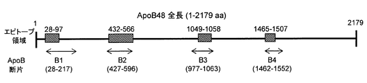
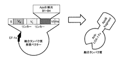
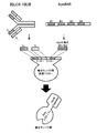
- FIG. 4 is an explanatory diagram showing the relationship between four epitopes in human ApoB48 and ApoB fragments B1 to B4 containing them. It is explanatory drawing which shows the structure of the expression vector and fusion protein which were constructed
- the fusion protein of the present invention is obtained by linking the full length or partial fragment of ApoB to a protein that specifically binds to LOX-1.
- the fusion protein of the present invention comprises a part of “protein that specifically binds to LOX-1” (hereinafter sometimes referred to as “LOX-1 binding protein part”) and “full length or partial fragment of ApoB. "(Hereinafter may be referred to as” ApoB part ").
- linking mode between the LOX-1 binding protein portion and the full length or partial fragment of ApoB linking via a peptide bond is typical.
- embodiments linked via peptide bonds will be described.
- Examples of linking modes other than peptide bonds include those in which a LOX-1 binding protein portion and the full length or partial fragment of ApoB are linked by a synthetic chemical technique.
- a typical example of a protein that specifically binds to LOX-1 is an antibody against LOX-1 (anti-LOX-1 antibody). That is, in a preferred embodiment, the protein that specifically binds to LOX-1 is an antibody against LOX-1.
- the “antibody against LOX-1” portion of the fusion protein of this embodiment may be referred to as “anti-LOX-1 antibody portion”.
- an antibody against LOX-1 (anti-LOX-1 antibody) and an anti-LOX-1 antibody portion will be described.
- antibody can usually be replaced with “immunoglobulin”.
- the antibody constituting the anti-LOX-1 antibody portion in the fusion protein of the present embodiment is not particularly limited as long as it is an antibody that binds to LOX-1.
- the origin of LOX-1 is not particularly limited, and all LOX-1 such as human, mouse, bovine and the like can be targeted.
- the origin of the anti-LOX-1 antibody is not particularly limited, and any antibody that binds to the desired LOX-1 can be used.
- mouse anti-LOX-1 monoclonal antibody # 10-1 specifically described in Examples and Reference [1] described later
- mouse anti-LOX-1 monoclonal antibody # 10-1 that specifically binds to human LOX-1 can be mentioned.
- the class (isotype) of the antibody constituting the anti-LOX-1 antibody portion is not particularly limited.
- any class such as IgG, IgM, IgA, IgD, and IgE may be used.
- the subclass of the antibody is not particularly limited, and for example, any subclass such as IgG1, IgG2, and IgG3 may be used as long as it is IgG.
- an antibody against LOX-1 includes both an antibody consisting of a full-length immunoglobulin and an “functional fragment of an antibody” containing an immunoglobulin variable region.
- the functional fragment of an antibody refers to a part of the antibody that retains at least one action of the antibody on the antigen.
- Examples of functional fragments of the antibody include V H , V L , Fv (scFv, synonymous with V H -V L ), Fab, F (ab ′) 2 , and the like.
- a conjugate of these functional fragments with the constant region can also be used as the functional fragment of the antibody in the present invention.
- the anti-LOX-1 antibody moiety is an Fv type antibody.
- An Fv type antibody (scFv, V H -V L ) is a functional fragment of an antibody in which a heavy chain variable region (V H ) and a light chain variable region (V L ) are linked and has a relatively small molecular weight. Easy to handle.
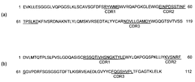
- FIG. 1 shows an example of the amino acid sequence and CDR of each variable region of the antibody constituting the “anti-LOX-1 antibody portion” in the fusion protein of this embodiment. That is, in one embodiment of the fusion protein of the present invention, the antibody against LOX-1 (anti-LOX-1 antibody portion) satisfies one or both of the following (a) and (b) for the variable region: .
- the heavy chain variable region includes a heavy chain CDR1 comprising the amino acid sequence represented by SEQ ID NO: 1, a heavy chain CDR2 comprising the amino acid sequence represented by SEQ ID NO: 2, and an amino acid sequence represented by SEQ ID NO: 3. Having a heavy chain CDR3 comprising, (B) a light chain variable region comprising a light chain CDR1 comprising an amino acid sequence represented by SEQ ID NO: 4, a light chain CDR2 comprising an amino acid sequence represented by SEQ ID NO: 5, and an amino acid sequence represented by SEQ ID NO: 6 It has a light chain CDR3 containing.
- the anti-LOX-1 antibody is employed as the “protein that specifically binds to LOX-1,” but the present invention is not limited to this.
- Other examples of “proteins that specifically bind to LOX-1” include C-reactive protein (CRP), HSP70, and the like. In the case of these proteins, as long as they specifically bind to LOX-1, it may be not only the full length but also a partial fragment, even if a mutation such as an amino acid substitution is introduced. Good.
- ApoB there is no particular limitation on the origin of ApoB, and all ApoB such as humans, mice and cows can be targeted.
- the ApoB portion in the fusion protein of the present invention is composed of either “full length of ApoB” or “partial fragment of ApoB”. Any fragment may be used as long as it is specifically recognized by the anti-ApoB antibody. However, in terms of ease of handling, a partial fragment of ApoB having a small molecular weight is preferably used.
- the ApoB partial fragment is not particularly limited as long as it is specifically recognized by the anti-ApoB antibody.
- a partial fragment containing a known epitope of ApoB can be used.
- the ApoB portion comprises at least one region selected from the group consisting of the region of amino acid numbers 28-97, the region of amino acid numbers 432-566, and the region of amino acid numbers 1049-1058 in human ApoB48. These regions have already been reported as epitopes of human ApoB48 (references [5] and [6]), and the present inventors have verified the functions as epitopes this time.
- the ApoB portion may include only one of these regions, or may include two or more.
- the amino acid sequence corresponding to the base sequence of the gene (cDNA) of human ApoB48 is shown in SEQ ID NOs: 7 and 8.
- amino acid sequence of the region corresponding to amino acid numbers 28-97 is SEQ ID NO: 9
- amino acid sequence of the region corresponding to amino acid numbers 432-566 is SEQ ID NO: 10
- amino acid sequence of the region corresponding to amino acid numbers 1049-1058 Are shown in SEQ ID NO: 11, respectively.
- ApoB in the present invention includes an ApoB isoform.
- ApoB isoforms of ApoB48 and ApoB100 are known for human ApoB.
- both human ApoB48 and human ApoB100 can be employed as the ApoB moiety.
- ApoB in the present invention includes naturally occurring ApoB (native ApoB), as well as mutant ApoB into which mutations due to amino acid substitutions, deletions, insertions, etc. have been introduced, but which can be regarded as functionally equivalent to native ApoB. It is. That is, both the full length and partial fragment of the mutant ApoB can constitute the “ApoB portion” in the fusion protein of the present invention.
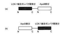
- the LOX-1 binding protein part (for example, anti-LOX-1 antibody part) and the ApoB part in the fusion protein of the present invention may be linked at the C-terminal of the LOX-1 binding protein part and the N-terminal of the ApoB part. And an embodiment in which the N-terminus of the LOX-1 binding protein portion and the C-terminus of the ApoB portion are joined (FIG. 2 (b)). Further, the LOX-1 binding protein portion and the ApoB portion may be indirectly linked via a sequence such as a spacer. Furthermore, the fusion protein of the present invention may contain proteins and peptides other than the LOX-1 binding protein portion and the ApoB portion.
- a sequence having a specific function or the like may be added to the N-terminus or C-terminus, or may be interposed between the LOX-1 binding protein portion and the ApoB portion. .
- the fusion protein of the present invention can be produced, for example, by expressing a nucleic acid encoding the fusion protein.
- DNA encoding a LOX-1 binding protein portion for example, an anti-LOX-1 antibody portion
- DNA encoding the full length or partial fragment of ApoB are obtained. These DNAs can be easily obtained by a technique such as PCR based on known base sequence information. It may be obtained by chemical synthesis.
- the DNA encoding the LOX-1 binding protein portion and the DNA encoding the full length or partial fragment of ApoB are linked so that the frames coincide with each other to produce a chimeric gene (chimeric nucleic acid).
- the chimeric gene is a gene encoding the fusion protein of the present invention.
- the obtained chimeric gene is incorporated into an appropriate vector to prepare a recombinant vector (fusion protein expression vector). Further, the recombinant vector is introduced into an appropriate host cell to produce a recombinant cell (fusion protein expression cell). Then, the recombinant cell can be cultured, and a desired fusion protein can be obtained from the culture.
- the vector into which the chimeric gene is incorporated is not particularly limited, and may be appropriately selected depending on the type of host cell introduced thereafter.
- the host cell into which the recombinant vector is introduced is not particularly limited as long as the introduced vector functions. Examples include animal cells (COS cells, CHO cells, etc.), yeasts, bacteria (E. coli etc.), plant cells, insect cells, and the like.
- fusion protein of the present invention has both binding ability to LOX-1 and binding ability to anti-ApoB antibody, affinity chromatography using a carrier on which LOX-1 or anti-ApoB antibody is immobilized can be used.
- the LAB measurement method of the present invention is a LAB measurement method that uses the specific binding property of LAB to LOX-1 and anti-ApoB antibody, and uses the above-mentioned fusion protein of the present invention as a standard product of LAB. Used to measure LAB in a sample by comparison with the standard product.
- the fusion protein of the present invention is used as a standard product of LAB. That is, since the fusion protein of the present invention has both the binding ability to LOX-1 and the binding ability to the anti-ApoB antibody, it can be used as a substitute for artificial oxidation LDL. Furthermore, since the fusion protein of the present invention is excellent in stability, accuracy control is easier than in the conventional method using artificial oxidized LDL as a standard product, and LAB can be measured reliably and easily.
- a typical example of “a method for measuring LAB using the specific binding property of oxidized LDL to LOX-1 and anti-ApoB antibody” is a sandwich immunoassay using LOX-1 and anti-ApoB antibody.
- LOX-1 is immobilized on a support such as a microtiter plate. Then, the measurement sample is brought into contact with the solid-phased LOX-1, and LAB in the sample is captured on LOX-1. Next, a labeled anti-ApoB antibody is bound to the captured LAB. Then, LAB in the sample can be measured using the bound anti-ApoB antibody as an index.
- the label examples include enzymes (enzyme immunoassay, EIA, ELISA), fluorescent substances (fluorescent immunoassay, FIA), radioactive substances (radioimmunoassay, RIA), and the like.
- the anti-ApoB antibody may be either labeled or unlabeled. When unlabeled, a labeled secondary antibody that binds to the anti-ApoB antibody may be further used.
- the anti-ApoB antibody may be monoclonal or polyclonal.
- LOX-1 and anti-ApoB antibody may be used in reverse. That is, an anti-ApoB antibody may be immobilized and LOX-1 may be labeled and used.
- Examples of the support on which LOX-1 or anti-ApoB antibody is immobilized include a microtiter plate, beads and the like.
- Another example of “a method for measuring LAB using the specific binding property of oxidized LDL to LOX-1 and anti-ApoB antibody” is an immunoassay based on a competitive method.
- Purified LOX-1 can be obtained by, for example, recombinant DNA technology (Patent Document 1, Non-Patent Document 1).
- Patent Document 1 a commercially available anti-ApoB antibody can be used as it is.
- the fusion protein of the present invention is used as a standard product of LAB, and LAB in a sample is measured by comparison with the standard product.
- serially diluted solutions of the fusion protein are prepared and used for the sandwich immunoassay and the like.
- a LAB standard curve with fusion protein equivalent values can be created.
- LAB in a sample such as serum can be obtained as a fusion protein equivalent (relative value) by applying the value obtained when a similar sandwich immunoassay or the like is performed to the standard curve.
- LAB in the sample can also be obtained as an absolute value.
- the measurement sample used in the present invention is not particularly limited, and examples thereof include serum and plasma collected from humans.
- the LAB measurement kit of the present invention is for use in the LAB measurement method of the present invention, and includes the above fusion protein. That is, according to the kit of the present invention, LAB can be easily measured using the fusion protein as a standard product. In preferred embodiments, it further comprises LOX-1 and / or anti-ApoB antibodies. With this configuration, the above-described sandwich immunoassay and the like can be easily performed.
- the kit may further contain a support (solid phase) such as a plate or a bead, a labeled secondary antibody, a chromogenic substrate, and the like.
- a support solid phase
- a support such as a plate or a bead, a labeled secondary antibody, a chromogenic substrate, and the like.
- kits configuration [Example of kit configuration] (A) LOX-1 (for solid phase) (B) Anti-ApoB antibody (labeled or unlabeled) (C) Fusion protein (LAB standard product) (D) Dilution buffer
- the method for measuring LAB of the present invention includes a method for measuring oxidized LDL.
- the LAB measurement kit of the present invention includes an oxidized LDL measurement kit.
- Mouse anti-LOX-1 monoclonal antibody The mouse anti-LOX-1 monoclonal antibody # 10-1 described in the reference [1] was used. Briefly, a recombinant protein corresponding to the amino acid 61-273 (61-273aa) portion of human LOX-1 (recombinant human LOX-1 protein; 61-273aa) (described in reference [2]) The spleen cells derived from Balb / c mice immunized were fused with the myeloma cell line P3U1. Positive clones were selected by ELISA to obtain clone # 10-1. The isotype of the antibody was determined using a mouse monoclonal antibody isotyping test kit (ABD) according to the attached instruction manual.
- ABS mouse monoclonal antibody isotyping test kit
- RNA was recovered from the # 10-1 antibody-producing hybridoma cloned in (1) above using 1 mL of TRIzol Reagent (Invitrogen). Synthesis of cDNA by reverse transcription reaction was performed from 1 ⁇ g of the recovered total RNA using Random hexamer (Invitrogen) and Super Script III Reverse Transcriptase (Invitrogen) according to the attached instruction manual.
- V H and V L regions were prepared by using 0.5 ⁇ g of synthesized cDNA as a template, 25 pmol of IgG Primer set (Novagen) forward primer and 50 ⁇ L of 10 ⁇ Ex-taq buffer, Amplification was carried out using a reaction solution prepared by adding 4.0 mM of 5 mM dNTPs, 1.5 U of Ex-taq polymerase (Takara Bio Inc.) and 34.8 ⁇ L of distilled water to adjust the total volume to 50 ⁇ L.
- the PCR cycle was initially performed at 94 ° C. for 3 minutes, followed by 35 cycles (94 ° C. for 1 minute, 55 ° C. for 1 minute, 72 ° C.
- the amplified DNA fragment was subcloned using TOPO TA Cloning Kit (Invitrogen), and the base sequences of VH gene and VL gene were determined using ABI PRISM Cycle sequencing kit (Applied Biosystems).
- Fv-type anti-LOX-1 antibody Using Fc-HF primer (SEQ ID NO: 17) and Fv-H-Linker-R primer (SEQ ID NO: 18) using cDNA prepared from # 10-1 antibody-producing hybridoma as a template PCR was performed using the above primer set to amplify the V H gene containing the # 10-1 antibody heavy chain secretion signal sequence.
- the Fv-H-Linker-R primer contains a part of the flexible linker sequence (Gly 4 -Ser) 3 at the 5 ′ end.
- PCR was performed using cDNA prepared from the # 10-1 antibody-producing hybridoma as a template, using a primer set of Linker-Fv-LF primer (SEQ ID NO: 19) and Fv-LR primer (SEQ ID NO: 20).
- the L gene was amplified. PCR conditions were as follows.
- the reaction solution was prepared by using 15 pmol of each primer set, 0.5 ng of cDNA, 5.0 ⁇ L of 10 ⁇ KOD plus buffer ver.2, 3.0 ⁇ L of 25 mM MgSO 4 , 5.0 ⁇ L of 2 mM dNTPs, KOD plus DNA polymerase 1.0 to U (Toyobo Co., Ltd.) and 32.0 ⁇ L of distilled water were added to adjust the total amount to 50.0 ⁇ L.
- the PCR cycle was initially performed at 94 ° C. for 2 minutes, followed by 30 cycles (94 ° C. for 15 seconds, 60 ° C. for 30 seconds, 68 ° C. for 30 seconds), and the final extension performed at 68 ° C. for 2 minutes. .
- each amplified fragment was ligated by overlap-extension PCR via a linker sequence inserted into a primer to prepare an Fv type antibody gene. That is, using the primer set of Fv-HF primer (SEQ ID NO: 17) and Fv-LR primer (SEQ ID NO: 20), the amplified V H gene and VL gene were linked by overlap-extension PCR.
- PCR conditions were as follows. The reaction solution used was 15 pmol of each primer set, 0.2 pmol of the amplified V H gene and VL gene, 5.0 ⁇ L of 10 ⁇ KOD plus buffer ver.
- the amplified Fv type antibody gene was subcloned into pcDNA6.2 / V5 / GW / D-TOPO vector (Invitrogen) using pcDNA Gateway Directional TOPO Expression kit (Invitrogen). After cloning, the base sequence was confirmed using ABI PRISM Cycle sequencing kit.
- the Fv antibody gene was excised using NotI and XbaI restriction enzyme sites present in the Multi cloning site of the pcDNA6.2 / V5 / GW / D-TOPO vector into which the Fv antibody gene was subcloned.
- Fv type antibody was expressed using FreeStyle 293 Expression system (Invitrogen). Purification of the Fv antibody was performed by TALON Metal Affinity Resins (Takara Bio Inc.).
- F1 primer set ApoB-1-F primer (SEQ ID NO: 21) and ApoB-1864-R (SEQ ID NO: 22), F2 primer set: ApoB-1802-F primer (SEQ ID NO: 23) and ApoB-4005-R Primer (SEQ ID NO: 24), F3 primer set: ApoB-3973-F primer (SEQ ID NO: 25) and ApoB-5207-R primer (SEQ ID NO: 26), F4 primer set: ApoB-5137-F primer (SEQ ID NO: 27) and ApoB-6537-R primer (SEQ ID NO: 28).
- PCR was performed using the liver cDNA library of Human MTC Panel II (Clontech) as a template and each primer set to obtain each gene fragment of F1 to F4.
- PCR conditions were as follows.
- the reaction solution was 0.5 ⁇ g of human liver cDNA, 25 pmol of each primer set, 5.0 ⁇ L of 10 ⁇ KOD plus buffer ver.2, 3.0 ⁇ L of 25 mM MgSO 4 , 5.0 ⁇ L of 2 mM dNTPs, and KOD plus DNA polymerase.
- 1.0 U and 32.0 ⁇ L of distilled water were added to adjust the total volume to 50.0 ⁇ L.
- the PCR cycle was initially performed at 94 ° C. for 2 minutes, followed by 35 cycles (94 ° C.
- each gene fragment was subcloned into pcDNA6.2 / V5 / GW / D-TOPO vector using pcDNA Gateway Directional TOPO Expression kit, and the nucleotide sequence was confirmed using ABI PRISM Cycle sequencing kit.
- B1 gene primer set B1-F primer (SEQ ID NO: 38) and B1-R primer (SEQ ID NO: 31), B2 gene primer set: B2-F primer (SEQ ID NO: 39) and B2-R primer (SEQ ID NO: 33) ), B3 gene primer set: B3-F primer (SEQ ID NO: 40) and B3-R primer (SEQ ID NO: 35), B4 gene primer set: B4-F primer (SEQ ID NO: 41) and B4-R primer (sequence) Number 37).
- PCR was performed using each primer set, and each gene encoding B1 to B4 was amplified (FIG. 3). PCR conditions were as follows.
- the reaction solution was 50 ng of ApoB48 full-length gene expression vector, 25 pmol of each primer set, 5.0 ⁇ L of 10 ⁇ Ex-taq buffer, 4.0 ⁇ L of 2.5 mM dNTPs mix, 1.0 U of Ex-taq DNA polymerase, distilled water 36.0 ⁇ L was added to adjust the total volume to 50.0 ⁇ L.
- the PCR cycle was initially performed at 94 ° C. for 2 minutes, followed by 35 cycles (94 ° C. for 15 seconds, 62 ° C. for 30 seconds, 72 ° C. for 30 seconds), and the final extension was performed at 72 ° C. for 7 minutes.
- the amplified gene fragments were combined in a frame so that the mouse antibody light chain secretion signal sequence was expressed on the N-terminal side and V5 and His tag were expressed on the C-terminal side, and pSecTag / FRT / V5-His-TOPO vector TA cloning in Invitrogen. After cloning, the base sequence was confirmed using ABI PRISM Cycle sequencing kit. Expression of ApoB protein fragments B1 to B4 was performed using the FreeStyle 293 Expression system. After 96 hours of culture, the supernatant and cell lysate were collected, and the culture supernatant was purified by TALON Metal Affinity Resins to obtain ApoB protein fragments B1 to B4.
- B1 to B4 are regions corresponding to the following amino acid numbers in the full length of human ApoB (SEQ ID NO: 8). B1: 28-217, B2: 427-596, B3: 977-1063, B4: 1462-1552.
- PCR cycle was initially performed at 94 ° C. for 2 minutes, followed by 30 cycles (94 ° C. for 15 seconds, 60 ° C. for 30 seconds, 68 ° C. for 45 seconds), and the final extension was performed at 68 ° C. for 2 minutes.
- PCR was performed using the ApoB48 full-length gene prepared in (5) above as a template, and using a primer set of the forward primer and reverse primer (4 pairs below) containing a part of the linker sequence at the 5 ′ end, and the ApoB48 fragment Each gene encoding B1-B4 was amplified.
- PCR conditions were as follows.
- the reaction solution was 50 ng of template, 15 pmol of each primer set, 5.0 ⁇ L of 10 ⁇ KOD plus buffer ver.2, 3.0 ⁇ L of 25 mM MgSO 4 , 5.0 ⁇ L of 2 mM dNTPs, 1.0 U of KOD plus DNA polymerase, The total amount was adjusted to 50.0 ⁇ L by adding 32.0 ⁇ L of distilled water.
- the PCR cycle was initially performed at 94 ° C. for 2 minutes, followed by 30 cycles (94 ° C. for 15 seconds, 60 ° C. for 30 seconds, 68 ° C. for 30 seconds), and the final extension was performed at 68 ° C. for 2 minutes.
- Fv-HF primer SEQ ID NO: 17
- B1-R primer SEQ ID NO: 31
- B2-R primer SEQ ID NO: 33
- B3-R primer SEQ ID NO: 35
- B4 B4 as a reverse primer
- Overlap-extension PCR was performed using the -R primer (SEQ ID NO: 37) and using the Fv type antibody gene and each ApoB fragment gene as a template.
- PCR conditions were as follows.
- the reaction solution was 0.2 pmol of Fv type antibody gene and each ApoB fragment gene, 15 pmol of primer set, 5.0 ⁇ L of 10 ⁇ KOD plus buffer ver.2, 3.0 ⁇ L of 25 mM MgSO 4 , 5 mM of 2 mM dNTPs.
- 0.0 ⁇ L, 1.0 U of KOD plus DNA polymerase and 32.0 ⁇ L of distilled water were added to adjust the total volume to 50.0 ⁇ L.
- the PCR cycle started at 94 ° C. for 2 minutes, followed by 30 cycles (94 ° C. for 15 seconds, 30 seconds at 60 ° C. and 90 seconds at 68 ° C.), and the final extension was performed at 68 ° C. for 2 minutes.
- fusion protein expression vector FIG. 4
- Fv-B1, Fv-B2, Fv-B3, Fv-B4 Four types of fusion proteins (Fv-B1, Fv-B2, Fv-B3, Fv-B4) were expressed using the FreeStyle 293 Expression system. Purification of each fusion protein was performed using TALON Metal Affinity Resins.
- ApoB fragments B1, B2
- fusion proteins Fv-B1, Fv-B2, Fv-B3, Fv-B4
- ApoB protein positive control, Sigma
- BSA negative Control
- HRP horseradish peroxidase
- HRP horseradish peroxidase
- ApoB fragment B1, B2
- fusion protein Fv-B1, Fv-B2, Fv-B3, Fv-B4
- HRP labeling Sheep anti-human ApoB polyclonal antibody (HRP-labeled sheep anti-human ApoB polyclonal antibody (Binding Site) was serially diluted 100-6.0 ⁇ 10 8 times with PBS containing 5% Immunoblock. Was added and incubated at room temperature for 1 hour.
- TMB solution 3,3'5,5'-tetramethylbenzidine-containing substrate solution
- antigens for anti-ApoB antibody culture supernatant and cell lysate of HEK293 cells expressing ApoB fragment, fusion protein (Fv-B1, Fv-B2, Fv-B3, Fv-B4), Fv type antibody, and BSA (negative) Control) was used.
- Each antigen is separated on a 10-20% gradient polyacrylamide gel (Wako Pure Chemical Industries, Ltd.) under non-reducing conditions and transferred to a PVDF membrane using an iBolt Dry Blotting system (Invitrogen). Went. Subsequently, the PVDF membrane used for studying the reactivity of the fusion protein was 100% Block Ace (DS Pharma), the PVDF membrane for studying anti-ApoB antibody reactivity was 5% skim milk (Morinaga Milk Industry), 0.1. Each was blocked with PBS containing 1% Tween 20 (Nacalai Tesque) (PBS-T) at room temperature for 1 hour.
- the PVDF membrane When an Fv-type anti-LOX-1 antibody or a fusion protein is used as the primary antibody, the PVDF membrane is immersed in a primary antibody solution diluted to 5 ⁇ g / mL with Can Get Signal Immunoreaction Enhancer Solution 1 (Toyobo Co., Ltd.) For 1 hour. Subsequently, it was immersed in a secondary antibody solution consisting of HRP-labeled mouse anti-V5 tag diluted 2000 times with Can Get Signal Immunoreaction Enhancer Solution 2 (Toyobo Co., Ltd.) and allowed to react at room temperature for 1 hour.
- a primary antibody solution diluted to 5 ⁇ g / mL with Can Get Signal Immunoreaction Enhancer Solution 1 (Toyobo Co., Ltd.) For 1 hour.
- a secondary antibody solution consisting of HRP-labeled mouse anti-V5 tag diluted 2000 times with Can Get Signal Immunoreaction Enhancer Solution 2 (Toyobo Co., Ltd.) and allowed to react at
- HRP-labeled sheep anti-human ApoB polyclonal antibody When an HRP-labeled sheep anti-human ApoB polyclonal antibody was used as the primary antibody, it was diluted 5000-fold with PBS-T containing 5% skim milk and allowed to react at room temperature for 1 hour.
- HRP-labeled mouse anti-V5 tag When the fusion protein expression confirmation antibody HRP-labeled mouse anti-V5 tag was used (positive control), it was diluted 2000-fold with PBS-T containing 5% skim milk and allowed to react at room temperature for 1 hour.
- the band was detected using the chemiluminescent substrate Immobilon Western Chemiluminescent HRP Substrate (Millipore) according to the attached instructions, and the signal was developed using LAS-4000 mini (GE Healthcare).
- Oxidative modification of LDL was performed by adding 7.5 ⁇ M CuSO 4 at a final concentration of LDL adjusted to 3 mg / mL and reacting at 37 ° C. for 16 hours, and then 10000 times LDL buffer (150 mM NaCl, 0.24 mM EDTA, pH 7. It was prepared by dialysis against 4) (artificial oxidation LDL). The degree of oxidation was determined by measuring the amount of thiobarbituric acid reaction product and the mobility of the agarose gel. Stored at 4 ° C. until use.
- fusion proteins As measurement samples (or standard products), four types of fusion proteins (Fv-B1, Fv-B2, Fv-B3, Fv-B4) diluted with HEPES buffer containing 2 mM EDTA and 5% BSA, artificially oxidized LDL ( The above (prepared in (8)) or LDL (prepared in (8) above) was used.
- HRP-labeled sheep anti-ApoB polyclonal antibody diluted 5000 times with HEPES buffer containing 2 mM EDTA and 1% BSA was added and incubated at room temperature for 1 hour. After the antibody reaction, the plate was washed 5 times with PBS, TMB solution was added to the plate and allowed to react at room temperature. The reaction was stopped with 2M sulfuric acid and the absorbance at 450 nm was measured.
- the isotype of mouse anti-LOX-1 antibody # 10-1 was IgG 1 , ⁇ .
- FIG. 1 (a) Identification of variable region amino acid sequence and CDR
- the # 10-1 antibody heavy chain variable region amino acid sequence and CDRs 1 to 3 are shown in FIG. 1 (a), and the light chain variable region amino acid sequence and CDRs 1 to 3 are shown. Each is shown in 1 (b).
- the cDNA base sequence and amino acid sequence of the heavy chain variable region of antibody # 10-1 are shown in SEQ ID NO: 13 and SEQ ID NO: 14, the amino acid sequence of heavy chain CDR1 is shown in SEQ ID NO: 1, and the amino acid sequence of heavy chain CDR2 is shown in SEQ ID NO: 2 shows the amino acid sequence of heavy chain CDR3 in SEQ ID NO: 3, respectively.
- the light chain variable region cDNA base sequence and amino acid sequence of antibody # 10-1 are shown in SEQ ID NO: 15 and SEQ ID NO: 16, the light chain CDR1 amino acid sequence in SEQ ID NO: 4, and the light chain CDR2 amino acid sequence in SEQ ID NO: 5 shows the amino acid sequence of the light chain CDR3 in SEQ ID NO: 6, respectively.
- FIG. 7 shows the result of ELISA in which BSA was immobilized.
- ⁇ indicates LOX-1
- ⁇ indicates a case where BSA is immobilized.
- Values represent the mean and SEM of three independent experiments. That is, the Fv-type anti-LOX-1 antibody showed reactivity in a dose-dependent manner with respect to recombinant human LOX-1 at an antibody concentration ranging from 46 ng / mL to 33 ⁇ g / mL.
- the Fv-type antibody can be used as a LOX-1 binding protein.
- ⁇ represents the B1 fragment
- ⁇ represents the B2 fragment
- ⁇ represents the full length ApoB (positive control)
- ⁇ represents BSA (negative control).
- Values represent the mean and SEM of three independent experiments.
- the sheep anti-ApoB polyclonal antibody has an antibody dilution ratio of 1.0 ⁇ 10 2 to 8.1 ⁇ 10 with respect to the B2 fragment.
- antibody dilution ratio of 1.0 ⁇ 10 2 to 9.0 ⁇ 10 2 times with respect to B1 fragment in the range of 1.0 ⁇ 10 2 to 7 of antibody dilution ratio for the full length ApoB of the positive control
- each showed a dose-dependent response.
- the negative control BSA showed no reactivity in the antibody dilution ratio range of 1.0 ⁇ 10 2 to 5.9 ⁇ 10 6 times.
- an ApoB protein fragment that can be used as an anti-ApoB antibody binding protein was produced.
- fusion protein of Fv-type anti-LOX-1 antibody and ApoB fragment (B1-B4)
- an artificially oxidized LDL standard product prepared from human plasma
- a fusion protein of an Fv-type anti-LOX-1 antibody and an ApoB fragment (B1-B4) was prepared (FIG. 5).
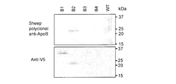
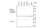
- the expression of the four types of fusion proteins prepared (Fv-B1, Fv-B2, Fv-B3, Fv-B4) was examined by Western blotting using an anti-V5 antibody.
- Fv-B1 was found to have a band around 63 kDa, Fv-B2 around 60 kDa, Fv-B3 around 66 kDa, and Fv-B5 around 60 kDa among the prepared fusion proteins (FIG. 11). ).
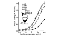
- ⁇ represents the case of Fv-B1
- ⁇ represents Fv-B2
- ⁇ represents Fv-B3
- ⁇ represents Fv-B4,
- ⁇ represents the case of Fv type anti-LOX-1 antibody (abbreviated as Fv type antibody).
- Values represent the mean and SEM of three independent experiments. That is, with Fv-B1 and Fv-B3, a dose response curve almost equivalent to that of the Fv-type anti-LOX-1 antibody was obtained in the range of the fusion protein addition amount of 45 ng / mL to 100 ⁇ g / mL.
- Fv-B2 and Fv-B4 showed reactivity in a dose-dependent manner with respect to LOX-1 at a fusion protein addition concentration ranging from 1.2 ⁇ g / mL to 100 ⁇ g / mL.
- the protein concentrations corresponding to the inflection points were 80.9 ⁇ g / mL for Fv-B2 and 20.6 ⁇ g / mL for Fv-B4, which were approximately compared to the Fv type antibody (2.91 ⁇ g / mL), respectively. 27 times higher, about 7 times higher.
- Fv-B1 and Fv-B3 showed the same reactivity as the Fv type antibody corresponding to the ELISA result, and the Fv-B2 and Fv-B4 had weaker reactivity than the Fv type antibody. (FIG. 13).
- the sheep anti-ApoB polyclonal antibody was strongly reactive to Fv-B2 and Fv-B3, weakly reactive to Fv-B1, and reactive to Fv-B4, Fv type antibody and BSA (A top of FIG. 11).
- the lower part of FIG. 11 is a positive control.
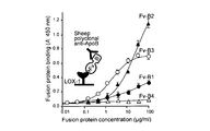
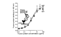
- ⁇ indicates Fv-B1
- Fv-B2 indicates Fv-B3
- ⁇ indicates Fv-B4
- ⁇ indicates the case where BSA (negative control) is immobilized.
- Values represent the mean and SEM of three independent experiments. That is, the sheep anti-ApoB polyclonal antibody is 1.0 ⁇ against Fv-B1 and Fv-B2 within a range of antibody dilution ratios of 1.0 ⁇ 10 2 to 7.3 ⁇ 10 4 times that of Fv-B3.
- Fv-B2 addition concentrations of 1.23 ⁇ g / mL to 100 ⁇ g / mL and Fv-B3 addition concentrations of 0.14 ⁇ g / mL to 33.3 ⁇ g / mL.
- Fv-B1 was detected in a dose-dependent manner at an addition concentration of 1.23 ⁇ g / mL to 100 ⁇ g / mL, but the maximum response was about 3 times lower at OD450 than Fv-B2.
- no reactivity was exhibited in the range of Fv-B4 addition concentration of 15 ng / mL to 100 ⁇ g / mL.
- the fusion protein bound to LOX-1 It could be specifically detected by the ApoB antibody, and could be used as a standard protein (standard product) of the LOX-1 ligand measurement system (LAB measurement) instead of artificially oxidized LDL.
- the protein concentration corresponding to the inflection point obtained by the 4-parameter logistic analysis is 38.6 ⁇ g / mL for artificial oxidation LDL lot 1, 17.7 ⁇ g / mL for lot 2, and for lot 3 It was 8.81 ⁇ g / mL and 8.34 ⁇ g / mL, and there was a difference of up to 4.6 times.
- the protein concentration corresponding to the inflection point obtained by the four-parameter logistic analysis between different lots is 2.17 ⁇ g / mL in lot 1, and lot 2 was 1.98 ⁇ g / mL, which was about 1.1 times the difference.
Landscapes
- Health & Medical Sciences (AREA)
- Life Sciences & Earth Sciences (AREA)
- Chemical & Material Sciences (AREA)
- Molecular Biology (AREA)
- Organic Chemistry (AREA)
- Immunology (AREA)
- General Health & Medical Sciences (AREA)
- Biophysics (AREA)
- Engineering & Computer Science (AREA)
- Medicinal Chemistry (AREA)
- Biochemistry (AREA)
- Genetics & Genomics (AREA)
- Urology & Nephrology (AREA)
- Hematology (AREA)
- Proteomics, Peptides & Aminoacids (AREA)
- Biomedical Technology (AREA)
- Food Science & Technology (AREA)
- Analytical Chemistry (AREA)
- Physics & Mathematics (AREA)
- Microbiology (AREA)
- General Physics & Mathematics (AREA)
- Pathology (AREA)
- Cell Biology (AREA)
- Biotechnology (AREA)
- Endocrinology (AREA)
- Toxicology (AREA)
- Zoology (AREA)
- Gastroenterology & Hepatology (AREA)
- Peptides Or Proteins (AREA)
Priority Applications (1)
| Application Number | Priority Date | Filing Date | Title |
|---|---|---|---|
| PCT/JP2012/065873 WO2013190679A1 (fr) | 2012-06-21 | 2012-06-21 | Protéine de fusion, acide nucléique, vecteur, cellule, procédé de mesure de lab, et kit pouvant être utilisé pour mesurer lab |
Applications Claiming Priority (1)
| Application Number | Priority Date | Filing Date | Title |
|---|---|---|---|
| PCT/JP2012/065873 WO2013190679A1 (fr) | 2012-06-21 | 2012-06-21 | Protéine de fusion, acide nucléique, vecteur, cellule, procédé de mesure de lab, et kit pouvant être utilisé pour mesurer lab |
Publications (1)
| Publication Number | Publication Date |
|---|---|
| WO2013190679A1 true WO2013190679A1 (fr) | 2013-12-27 |
Family
ID=49768308
Family Applications (1)
| Application Number | Title | Priority Date | Filing Date |
|---|---|---|---|
| PCT/JP2012/065873 Ceased WO2013190679A1 (fr) | 2012-06-21 | 2012-06-21 | Protéine de fusion, acide nucléique, vecteur, cellule, procédé de mesure de lab, et kit pouvant être utilisé pour mesurer lab |
Country Status (1)
| Country | Link |
|---|---|
| WO (1) | WO2013190679A1 (fr) |
Cited By (1)
| Publication number | Priority date | Publication date | Assignee | Title |
|---|---|---|---|---|
| WO2021177333A1 (fr) * | 2020-03-03 | 2021-09-10 | 国立大学法人信州大学 | Procédé de quantification d'adiponectine et réactif d'analyse à utiliser dans ledit procédé |
Citations (3)
| Publication number | Priority date | Publication date | Assignee | Title |
|---|---|---|---|---|
| JPH0998787A (ja) * | 1994-11-30 | 1997-04-15 | Nippon Chemiphar Co Ltd | 変性低密度リポ蛋白質受容体 |
| JP2011106847A (ja) * | 2009-11-13 | 2011-06-02 | Biomarker Science:Kk | 心血管障害発症リスクの評価方法及び評価用キット |
| JP2012100585A (ja) * | 2010-11-10 | 2012-05-31 | National Cerebral & Cardiovascular Center | 融合タンパク質、核酸、ベクター、細胞、labの測定方法、並びに、lab測定用キット |
-
2012
- 2012-06-21 WO PCT/JP2012/065873 patent/WO2013190679A1/fr not_active Ceased
Patent Citations (3)
| Publication number | Priority date | Publication date | Assignee | Title |
|---|---|---|---|---|
| JPH0998787A (ja) * | 1994-11-30 | 1997-04-15 | Nippon Chemiphar Co Ltd | 変性低密度リポ蛋白質受容体 |
| JP2011106847A (ja) * | 2009-11-13 | 2011-06-02 | Biomarker Science:Kk | 心血管障害発症リスクの評価方法及び評価用キット |
| JP2012100585A (ja) * | 2010-11-10 | 2012-05-31 | National Cerebral & Cardiovascular Center | 融合タンパク質、核酸、ベクター、細胞、labの測定方法、並びに、lab測定用キット |
Non-Patent Citations (3)
| Title |
|---|
| INOUE N ET AL.: "LOX index, a novel predictive biochemical marker for coronary heart disease and stroke", CLIN. CHEM., vol. 56, no. 4, April 2010 (2010-04-01), pages 550 - 558 * |
| IWAMOTO S ET AL.: "An Alternative Protein Standard to Measure Activity of LOX-1 Ligand Containing apoB (LAB) - Utilization of Anti- LOX-1 Single- Chain Antibody Fused to apoB Fragment", JOURNAL OF ATHEROSCLEROSIS AND THROMBOSIS, vol. 18, no. 9, 27 September 2011 (2011-09-27), pages 818 - 828 * |
| SHIN IWAMOTO ET AL.: "Sanka LDL Sokutei no Tameno Jinko Hyojun Tanpaku Sakusei", JOURNAL OF JAPANESE BIOCHEMICAL SOCIETY, vol. 82, no. 11, 25 November 2010 (2010-11-25), pages 3P-1146 * |
Cited By (3)
| Publication number | Priority date | Publication date | Assignee | Title |
|---|---|---|---|---|
| WO2021177333A1 (fr) * | 2020-03-03 | 2021-09-10 | 国立大学法人信州大学 | Procédé de quantification d'adiponectine et réactif d'analyse à utiliser dans ledit procédé |
| JPWO2021177333A1 (fr) * | 2020-03-03 | 2021-09-10 | ||
| JP7719507B2 (ja) | 2020-03-03 | 2025-08-06 | 達也 沢村 | アディポネクチンの定量方法及びそれに用いる分析用試薬 |
Similar Documents
| Publication | Publication Date | Title |
|---|---|---|
| JP5706669B2 (ja) | 融合タンパク質、核酸、ベクター、細胞、labの測定方法、並びに、lab測定用キット | |
| CN104067130B (zh) | 肾上腺髓质素测定与用于确定成熟肾上腺髓质素的方法 | |
| JP5941615B2 (ja) | ヒトcxcl1タンパク質の免疫学的測定方法 | |
| US11372001B2 (en) | Anti-human IgG4 monoclonal antibody and methods of making and using same | |
| KR20190116997A (ko) | 유전적 변이체를 인식하는 항체 | |
| EP3533459A1 (fr) | Anticorps anti-pla2-gib et leurs utilisations | |
| US20230384326A1 (en) | Test method and test kit for adult still's disease | |
| US12043669B2 (en) | IL-1 receptor accessory protein-inhibiting antibodies and antigen-binding fragments thereof | |
| WO2017116196A1 (fr) | Protéine picp recombinée, et procédé de préparation d'anticorps s'y liant spécifiquement | |
| WO2021237534A1 (fr) | Anticorps contre un nouveau coronavirus, combinaison d'anticorps, procédé de préparation et kit de détection le contenant | |
| JP7365050B2 (ja) | 融合タンパク質、及びそれを用いた高密度リポタンパク質の測定キット | |
| WO2025125234A1 (fr) | Anticorps de haute affinité se liant de manière spécifique au pepsinogène c | |
| US20210388108A1 (en) | Antibodies specific for glycosylated apoj and uses thereof | |
| JPH10226700A (ja) | Miaの検出のためのイムノアッセイ | |
| WO2024101357A1 (fr) | Anticorps visant la sous-unité bêta 2 du complexe protéique de coatomère | |
| Huang et al. | A high-affinity human/mouse cross-reactive monoclonal antibody, specific for VEGFR-2 linear and conformational epitopes | |
| JP5553603B2 (ja) | 新規肝癌マーカー | |
| US20210055300A1 (en) | Monoclonal antibody against apoa4, immunological measurement method, and kit for measurement | |
| CN118255883B (zh) | 针对人视锥蛋白样蛋白1的单克隆抗体、抗体对和试剂盒及其应用 | |
| CN116143929B (zh) | 抗重组人凝血因子VIIa-Fc融合蛋白的抗体及其应用 | |
| JP5448424B2 (ja) | ヒトIgGのFcを含有するタンパク質の測定試薬 | |
| JPWO2018079393A1 (ja) | ジスルフィド型hmgb1特異的抗体、ジスルフィド型hmgb1の測定方法および測定用キット、ならびに、還元型hmgb1、ジスルフィド型hmgb1、トロンビン分解hmgb1等の全てのhmgb1を定量することが可能な測定方法および測定用キット | |
| US20230331865A1 (en) | Antibodies for use in immunohistochemistry (ihc) protocols to diagnose cancer | |
| JP2018138520A (ja) | 抗ミッドカインモノクローナル抗体及びそれを用いた免疫学的測定キット | |
| JP2002356500A (ja) | 活性型肝細胞増殖因子アクティベーターに対する特異的抗体とその使用法 |
Legal Events
| Date | Code | Title | Description |
|---|---|---|---|
| 121 | Ep: the epo has been informed by wipo that ep was designated in this application |
Ref document number: 12879364 Country of ref document: EP Kind code of ref document: A1 |
|
| NENP | Non-entry into the national phase |
Ref country code: DE |
|
| 122 | Ep: pct application non-entry in european phase |
Ref document number: 12879364 Country of ref document: EP Kind code of ref document: A1 |
|
| NENP | Non-entry into the national phase |
Ref country code: JP |