WO2014123227A1 - ヒトnrg1タンパク質に対する抗体 - Google Patents
ヒトnrg1タンパク質に対する抗体 Download PDFInfo
- Publication number
- WO2014123227A1 WO2014123227A1 PCT/JP2014/052939 JP2014052939W WO2014123227A1 WO 2014123227 A1 WO2014123227 A1 WO 2014123227A1 JP 2014052939 W JP2014052939 W JP 2014052939W WO 2014123227 A1 WO2014123227 A1 WO 2014123227A1
- Authority
- WO
- WIPO (PCT)
- Prior art keywords
- amino acid
- acid sequence
- seq
- nrg1
- antibody
- Prior art date
- Legal status (The legal status is an assumption and is not a legal conclusion. Google has not performed a legal analysis and makes no representation as to the accuracy of the status listed.)
- Ceased
Links
Images









Classifications
-
- C—CHEMISTRY; METALLURGY
- C07—ORGANIC CHEMISTRY
- C07K—PEPTIDES
- C07K16/00—Immunoglobulins [IG], e.g. monoclonal or polyclonal antibodies
- C07K16/18—Immunoglobulins [IG], e.g. monoclonal or polyclonal antibodies against material from animals or humans
- C07K16/22—Immunoglobulins [IG], e.g. monoclonal or polyclonal antibodies against material from animals or humans against growth factors ; against growth regulators
-
- A—HUMAN NECESSITIES
- A61—MEDICAL OR VETERINARY SCIENCE; HYGIENE
- A61P—SPECIFIC THERAPEUTIC ACTIVITY OF CHEMICAL COMPOUNDS OR MEDICINAL PREPARATIONS
- A61P35/00—Antineoplastic agents
-
- A—HUMAN NECESSITIES
- A61—MEDICAL OR VETERINARY SCIENCE; HYGIENE
- A61K—PREPARATIONS FOR MEDICAL, DENTAL OR TOILETRY PURPOSES
- A61K39/00—Medicinal preparations containing antigens or antibodies
- A61K2039/505—Medicinal preparations containing antigens or antibodies comprising antibodies
-
- C—CHEMISTRY; METALLURGY
- C07—ORGANIC CHEMISTRY
- C07K—PEPTIDES
- C07K2317/00—Immunoglobulins specific features
- C07K2317/20—Immunoglobulins specific features characterized by taxonomic origin
- C07K2317/24—Immunoglobulins specific features characterized by taxonomic origin containing regions, domains or residues from different species, e.g. chimeric, humanized or veneered
-
- C—CHEMISTRY; METALLURGY
- C07—ORGANIC CHEMISTRY
- C07K—PEPTIDES
- C07K2317/00—Immunoglobulins specific features
- C07K2317/30—Immunoglobulins specific features characterized by aspects of specificity or valency
- C07K2317/33—Crossreactivity, e.g. for species or epitope, or lack of said crossreactivity
-
- C—CHEMISTRY; METALLURGY
- C07—ORGANIC CHEMISTRY
- C07K—PEPTIDES
- C07K2317/00—Immunoglobulins specific features
- C07K2317/30—Immunoglobulins specific features characterized by aspects of specificity or valency
- C07K2317/34—Identification of a linear epitope shorter than 20 amino acid residues or of a conformational epitope defined by amino acid residues
-
- C—CHEMISTRY; METALLURGY
- C07—ORGANIC CHEMISTRY
- C07K—PEPTIDES
- C07K2317/00—Immunoglobulins specific features
- C07K2317/70—Immunoglobulins specific features characterized by effect upon binding to a cell or to an antigen
- C07K2317/73—Inducing cell death, e.g. apoptosis, necrosis or inhibition of cell proliferation
-
- C—CHEMISTRY; METALLURGY
- C07—ORGANIC CHEMISTRY
- C07K—PEPTIDES
- C07K2317/00—Immunoglobulins specific features
- C07K2317/70—Immunoglobulins specific features characterized by effect upon binding to a cell or to an antigen
- C07K2317/76—Antagonist effect on antigen, e.g. neutralization or inhibition of binding
-
- C—CHEMISTRY; METALLURGY
- C12—BIOCHEMISTRY; BEER; SPIRITS; WINE; VINEGAR; MICROBIOLOGY; ENZYMOLOGY; MUTATION OR GENETIC ENGINEERING
- C12N—MICROORGANISMS OR ENZYMES; COMPOSITIONS THEREOF; PROPAGATING, PRESERVING, OR MAINTAINING MICROORGANISMS; MUTATION OR GENETIC ENGINEERING; CULTURE MEDIA
- C12N15/00—Mutation or genetic engineering; DNA or RNA concerning genetic engineering, vectors, e.g. plasmids, or their isolation, preparation or purification; Use of hosts therefor
- C12N15/09—Recombinant DNA-technology
-
- C—CHEMISTRY; METALLURGY
- C12—BIOCHEMISTRY; BEER; SPIRITS; WINE; VINEGAR; MICROBIOLOGY; ENZYMOLOGY; MUTATION OR GENETIC ENGINEERING
- C12N—MICROORGANISMS OR ENZYMES; COMPOSITIONS THEREOF; PROPAGATING, PRESERVING, OR MAINTAINING MICROORGANISMS; MUTATION OR GENETIC ENGINEERING; CULTURE MEDIA
- C12N15/00—Mutation or genetic engineering; DNA or RNA concerning genetic engineering, vectors, e.g. plasmids, or their isolation, preparation or purification; Use of hosts therefor
- C12N15/09—Recombinant DNA-technology
- C12N15/63—Introduction of foreign genetic material using vectors; Vectors; Use of hosts therefor; Regulation of expression
- C12N15/79—Vectors or expression systems specially adapted for eukaryotic hosts
- C12N15/85—Vectors or expression systems specially adapted for eukaryotic hosts for animal cells
Definitions
- the present invention relates to an antibody against human NRG1 protein, and more specifically, an antibody that specifically binds to human NRG1- ⁇ protein or human NRG1- ⁇ 1 protein, a DNA encoding the antibody, the antibody or a hybridoma containing the DNA,
- the present invention relates to a composition for treating or preventing cancer comprising the antibody as an active ingredient.
- NRG1 (neuregulin 1) is a member of the EGF family, and particularly soluble NRG1 binds to ErbB3 or ErbB4 and functions as a ligand for these receptors.
- ErbB3 and ErbB4 are members of the EGF (epidermal growth factor) receptor family.
- EGF epidermal growth factor
- soluble NRG1 binds to these receptors, the structure of the receptors is changed, resulting in homo- or heterodimerization. It is also known to do.
- the intracellular domains of these receptors are phosphorylated, and the phosphorylated intracellular domains bind to additional signal proteins and various signal transductions are activated.
- soluble NRG1 is derived from a transmembrane precursor, as with other factors belonging to the EGF family.
- pro-NRG1 transmembrane precursor
- plasma membrane the vicinity of the membrane in the extracellular portion is cleaved by a cleaving enzyme such as a metalloprotease (shell shape).
- cleaving enzyme such as a metalloprotease (shell shape).
- cleavage and release of pro-NRG1 is caused by a cell surface protease.
- PMA Phorbol-12-Milli
- PLC protein kinase C
- Non-patent Document 1 It is reported that it is activated by state-13-acetate (phorbol-12-myristate-13-acetate) (Non-patent Document 1). Further, as such proteases, TACE (tumor necrosis factor- ⁇ (TNF ⁇ ) converting enzyme, ADAM17) and ADAM19 (meltrin ⁇ ) have been reported so far (Non-patent Documents 2 and 3). Regarding the activated mechanism, involvement of PKC, Erk1 / 2, p38, etc. has been shown (Non-patent Document 4).
- NRG1 has been found to be involved in various life phenomena, and thus it does not specifically bind to each isoform, but an antibody against NRG1 has been developed (non- Patent Documents 5 to 8).
- the present invention has been made in view of the above-described problems of the prior art, and specifically recognizes an isoform of human NRG1 protein and can suppress signal transduction involving the isoform.
- the purpose is to provide.
- Another object of the present invention is to provide an antibody specific for an isoform of human NRG1 protein and having antitumor activity.
- the present inventors immunized mice with a total of 11 partial length proteins of human NRG1 protein isoforms 1 and 2 (human NRG1- ⁇ 1 protein and human NRG1- ⁇ protein), A total of 80 clones of monoclonal antibodies against NRG1 protein were obtained. Furthermore, among these monoclonal antibodies, four types of monoclonal antibodies (8a2, 8a4, 10bM3 and 10b2M3) were selected using as an index the strong reactivity against the NRG1 protein on the cell surface.
- 8a2 is an antibody that binds to both human NRG1- ⁇ protein and human NRG1- ⁇ 1 protein.
- the epitope was found to be N-terminal to the EGF domain, which is a common region of human NRG1- ⁇ protein and human NRG1- ⁇ 1 protein.
- 8a4 is an antibody that does not bind to human NRG1- ⁇ 1 protein, but specifically binds to human NRG1- ⁇ protein, and particularly an antibody having an epitope in the region of positions 221 to 234 in human NRG1- ⁇ protein. It became clear that there was.
- 10bM3 and 10b2M3 do not bind to human NRG1- ⁇ protein, but are antibodies that specifically bind to human NRG1- ⁇ 1 protein.
- the region at positions 213 to 239 in human NRG1- ⁇ 1 protein is defined as an epitope. It became clear that it is an antibody to.
- the present inventors succeeded in obtaining an antibody specific for human NRG1- ⁇ protein or human NRG1- ⁇ 1 protein.
- the present inventors determined the heavy and light chain variable regions and the CDR sequences for mouse monoclonal antibodies specific for these human NRG1 protein isoforms. Furthermore, based on the determined sequence, a chimeric antibody in which the constant region was substituted with one derived from human IgG was also produced. As a result of intensive studies on the chimeric antibodies thus obtained and the mouse monoclonal antibodies used as the basis of these antibodies, the region of positions 221 to 234 in human NRG1- ⁇ protein or human NRG1- ⁇ 1 protein It was shown that these antibodies can inhibit the cleavage of the protein, which is the origin of signal transduction involving human NRG1 protein, by specifically binding to the region of positions 213 to 239 in.
- the present invention provides the following ⁇ 1> to ⁇ 11>.
- ⁇ 1> An antibody that binds to a region at positions 221 to 234 in the human NRG1- ⁇ protein represented by SEQ ID NO: 1 or a region from positions 213 to 239 in the human NRG1- ⁇ 1 protein represented by SEQ ID NO: 2.
- ⁇ 2> The antibody according to ⁇ 1>, which has an activity of suppressing cleavage of the human NRG1- ⁇ protein represented by SEQ ID NO: 1 or the human NRG1- ⁇ 1 protein represented by SEQ ID NO: 2.
- ⁇ 3> having an activity of suppressing phosphorylation of ErbB3 protein in cancer cells in response to stimulation with the human NRG1- ⁇ protein represented by SEQ ID NO: 1 or the human NRG1- ⁇ 1 protein represented by SEQ ID NO: 2, ⁇ The antibody according to 1> or ⁇ 2>.
- ⁇ 4> The antibody according to any one of ⁇ 1> to ⁇ 3>, which has an activity of suppressing tumor growth in vivo.
- the antibody according to ⁇ 1> which has the characteristics described in any of the following (a) to (c) (a) The amino acid sequence according to SEQ ID NO: 3 to 5, or at least one of the amino acid sequences A light chain variable region comprising an amino acid sequence in which one or more amino acids are substituted, deleted, added and / or inserted; and the amino acid sequence set forth in SEQ ID NOs: 7 to 9 or at least one of the amino acid sequences A heavy chain variable region comprising an amino acid sequence in which one or more amino acids are substituted, deleted, added and / or inserted (b) The amino acid sequence of SEQ ID NOs: 11 to 13 or the amino acid sequence of the amino acid sequence A light chain variable region comprising an amino acid sequence in which one or more amino acids are substituted, deleted, added and / or inserted in at least any of the sequences; and SEQ ID NOs: 15 to 17 A heavy chain variable region comprising the amino acid sequence described above or an amino acid sequence in which one or more amino acids are substituted, deleted,
- the antibody according to ⁇ 1> having the characteristics described in any of the following (a) to (c) (a) The amino acid sequence according to SEQ ID NO: 6, or one or more amino acids in the amino acid sequence A light chain variable region comprising an amino acid sequence in which is substituted, deleted, added and / or inserted, and the amino acid sequence set forth in SEQ ID NO: 10 or one or more amino acids in the amino acid sequence are substituted, deleted or added And / or a heavy chain variable region containing the inserted amino acid sequence (b) The amino acid sequence of SEQ ID NO: 14 or one or more amino acids in the amino acid sequence are substituted, deleted, added, and / or Alternatively, the light chain variable region containing the inserted amino acid sequence is substituted with the amino acid sequence set forth in SEQ ID NO: 18 or one or more amino acids in the amino acid sequence.
- a heavy chain variable region comprising a deleted, added and / or inserted amino acid sequence
- the amino acid sequence of SEQ ID NO: 22 or one or more amino acids in the amino acid sequence are substituted or deleted
- a light chain variable region comprising an added and / or inserted amino acid sequence and the amino acid sequence set forth in SEQ ID NO: 26 or one or more amino acids in the amino acid sequence are substituted, deleted, added and / or inserted.
- a heavy chain variable region containing the amino acid sequence is
- Antibody having the characteristics described in any of the following (a) to (c):
- the amino acid sequence described in SEQ ID NOs: 3 to 5 or at least one of the amino acid sequences has one or more amino acids
- a light chain variable region comprising an amino acid sequence that is substituted, deleted, added and / or inserted, and at least one of the amino acid sequences of SEQ ID NOs: 7 to 9 or the amino acid sequence is substituted.
- B one or more of the amino acid sequences of SEQ ID NOs: 11 to 13 or at least one of the amino acid sequences.
- a light chain variable region comprising an amino acid sequence in which the amino acid is substituted, deleted, added and / or inserted, and the amino acids described in SEQ ID NOs: 15 to 17
- a heavy chain variable region comprising an amino acid sequence in which one or more amino acids are substituted, deleted, added and / or inserted in at least one of the sequence or the amino acid sequence
- SEQ ID NOs: 19 to 21 A light chain variable region comprising the amino acid sequence described in the above or an amino acid sequence in which one or more amino acids are substituted, deleted, added and / or inserted in at least one of the amino acid sequences; It retains the heavy chain variable region comprising the amino acid sequence described or the amino acid sequence in which at least one of the amino acid sequences is substituted, deleted, added and / or inserted.
- a light chain variable region comprising: the amino acid sequence set forth in SEQ ID NO: 18, or one or more amino acids in the amino acid sequence are substituted, deleted, added, and Or a heavy chain variable region containing the inserted amino acid sequence (c)
- the amino acid sequence of SEQ ID NO: 22 or one or more amino acids in the amino acid sequence are substituted, deleted, added and / or inserted
- a light chain variable region comprising the amino acid sequence described above and the amino acid sequence set forth in SEQ ID NO: 26 or an amino acid sequence in which one or more amino acids are substituted, deleted, added and / or inserted in the amino acid sequence Retains the heavy chain variable region.
- ⁇ 9> An antibody that binds to an epitope recognized by the antibody according to any one of ⁇ 1> to ⁇ 8>.
- ⁇ 10> A DNA encoding the antibody according to any one of ⁇ 1> to ⁇ 9>.
- ⁇ 11> A hybridoma that produces the antibody according to any one of ⁇ 1> to ⁇ 9> or includes the DNA according to ⁇ 10>.
- ⁇ 12> A composition for treating or preventing cancer, comprising the antibody according to any one of ⁇ 1> to ⁇ 9> as an active ingredient.
- an antibody that can specifically recognize an isoform of human NRG1 protein and suppress signal transduction involving the isoform.
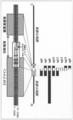
- results of comparison of structure and amino acid sequence between human NRG1- ⁇ protein and human NRG1- ⁇ 1 protein, and partial length proteins (ag4 to ag13 and agP) used as immunogens to produce the antibodies of the present invention Is a schematic diagram showing the positional relationship in human NRG1 protein.
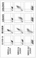
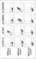
- a cell line (NRG1-a) that stably and highly expresses the obtained antibodies against human NRG1 protein (8a2, 8a4, 10bM3, and 10b2M3) and human NRG1- ⁇ protein or human NRG1- ⁇ 1 protein with an HA tag added to the N-terminus / St293T or NRG1-b / st293T) is a dot-plot diagram showing the results of analyzing the degree of binding by flow cytometry.
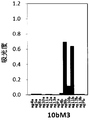
- 8 is a graph showing the results of analyzing the extent of binding of 8a2 to human NRG1- ⁇ partial length proteins (ag4a to ag13a and agPa) and human NRG1- ⁇ 1 partial length proteins (ag4b to ag13b and agPb) by ELISA. is there. 8 is a graph showing the results of analyzing the degree of binding between 8a4 and the partial length protein of human NRG1- ⁇ and the partial length protein of human NRG1- ⁇ 1 by ELISA. 10 is a graph showing the results of analyzing the degree of binding of 10bM3 to the partial length protein of human NRG1- ⁇ and the partial length protein of human NRG1- ⁇ 1 by ELISA.
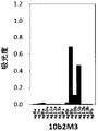
- 10 is a graph showing the results of analyzing the degree of binding of 10b2M3 to the partial length protein of human NRG1- ⁇ and the partial length protein of human NRG1- ⁇ 1 by ELISA.
- 10bM3 and 10b2M3, and cell lines that stably and highly express human NRG1- ⁇ protein, human NRG1- ⁇ 1 protein or human NRG1- ⁇ 2 protein with an N-terminal HA tag (NRG1-a / st293T, NRG1-b / It is a dot-plot diagram showing the results of analyzing the degree of binding with st293T or NRG1-b2 / st293T by flow cytometry.
- Antibody of the present invention (8a4, 10bM3 and 10b2M3) and factors belonging to EGF family (EGF, HB-EGF, TGFa (TGF ⁇ ), AREG, NRG1-a (NRG1- ⁇ ) and NRG1-b (NRG1- ⁇ 1)) It is a graph which shows the result of having analyzed the grade of the coupling
- the vertical axis represents the amount of human NRG1- ⁇ protein (average fluorescence intensity) remaining on the surface of cells (NRG1-a / st293T) after addition of PMA.
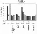
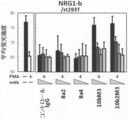
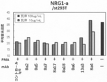
- the four bars in each group show the results when each antibody was added in the order of 80, 20, 5 and 1 ug / mL from the left (with respect to the notation of the graphs, FIGS. 10, 11, 19, 21). And 22). It is a graph which shows the result analyzed by flow cytometry about the inhibitory activity with respect to the cutting
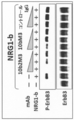
- MAb in each group indicates that the addition concentration of each antibody is 50, 10 and 5 ug / mL in order from the left (the notation in the figure is the same in FIG. 15). It is a photograph showing the result of Western blot analysis of the inhibitory activity against phosphorylation of ErbB3 induced by human NRG1- ⁇ 1 protein for the antibodies of the present invention (10bM3 and 10b2M3).
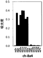
- FIG. 3 is a graph showing the results of analyzing the degree of binding between 8a4 and ch-8a4 and human NRG1- ⁇ protein by flow cytometry.
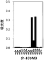
- the results of analyzing the extent of binding of ch-8a4 to the partial length proteins of human NRG1- ⁇ (ag4a to ag13a and agPa) and the partial length proteins of human NRG1- ⁇ 1 (ag4b to ag13b and agPb) are shown by ELISA. It is a graph. 2 is a graph showing the results of analyzing the degree of binding of ch-10bM3 with a partial length protein of human NRG1- ⁇ and a partial length protein of human NRG1- ⁇ 1 by ELISA.
- 2 is a graph showing the results of analyzing the degree of binding of ch-10b2M3 with a partial length protein of human NRG1- ⁇ and a partial length protein of human NRG1- ⁇ 1 by ELISA.
- 2 is a graph showing the results of analyzing the inhibitory activity against cleavage of human NRG1- ⁇ protein generated by PMA by flow cytometry for the chimeric antibodies (ch-8a4, ch-10bM3 and ch-10b2M3) of the present invention.
- 2 is a graph showing the results of analyzing the inhibitory activity against cleavage of human NRG1- ⁇ 1 protein caused by PMA by flow cytometry for the chimeric antibodies of the present invention (ch-8a4, ch-10bM3 and ch-10b2M3).
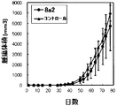
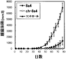
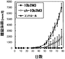
- the abscissa indicates the number of days that have elapsed when the day on which antibody administration has started is defined as day 0 (the notation of the abscissa is the same as in FIGS. 24 to 26).
- 2 is a graph showing changes in tumor volume over time in xenograft mice administered with the antibody of the present invention (8a4 or ch-8a4).
- 2 is a graph showing changes in tumor volume over time in xenograft mice administered with the antibody of the present invention (10b2M3 or ch-10b2M3).
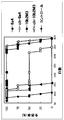
- 3 is a graph showing the change over time in the survival rate of xenograft mice administered with the antibody of the present invention (8a4, ch-8a4, 10b2M3 or ch-10b2M3).
- the present inventors succeeded in producing an antibody specific to human NRG1 protein isoform 1 or 2 (human NRG1- ⁇ 1 protein or human NRG1- ⁇ protein). Furthermore, while identifying the epitope of the obtained antibody, it also discovered that these antibodies have remarkable inhibitory activity with respect to the signal transduction which human NRG1 protein participates. Accordingly, the present invention provides the following antibodies that specifically bind to an isoform of human NRG1 protein.
- Antibody in the present invention includes all classes and subclasses of immunoglobulins. “Antibody” includes polyclonal antibodies and monoclonal antibodies, and also includes forms of functional fragments of antibodies. “Polyclonal antibodies” are antibody preparations comprising different antibodies directed against different epitopes. The “monoclonal antibody” means an antibody (including an antibody fragment) obtained from a substantially homogeneous antibody population. In contrast to polyclonal antibodies, monoclonal antibodies are those that recognize a single determinant on an antigen. The antibody of the present invention is preferably a monoclonal antibody. The antibodies of the present invention are antibodies that have been separated and / or recovered (ie, isolated) from components of the natural environment.
- NSG1 refers to neuregulin 1, HRG ⁇ (herregulin ⁇ ), HGL, HRGA, NDF (Neu differentiation factor), ARIA (acetylcholine receptor-inducing activator), GGF2 (glial growth factor 2), SMDF ( It is a protein also called a sensorimotor nerve-derived factor).
- Human-derived NRG1 protein isoform 2 (human NRG1- ⁇ protein) is typically a protein consisting of the amino acid sequence set forth in SEQ ID NO: 1 (a protein identified by RefSeq ID: NP — 039258, RefSeq ID: NM — 013964).
- Isoform 1 of human-derived NRG1 protein is typically a protein consisting of the amino acid sequence set forth in SEQ ID NO: 2 (RefSeq ID: NP_039250, a protein identified by RefSeq ID: NM — 013956)
- the protein encoded by the base sequence specified in (1) Therefore, the “region of positions 213 to 239 (or positions 232 to 239) in human NRG1- ⁇ 1 protein” typically represents the proline residue at position 213 (or the histidine residue at position 232 described in SEQ ID NO: 2).
- Group is a region consisting of an amino acid sequence of a glutamic acid residue at positions 239.
- the human NRG1 protein is ⁇ -type or ⁇ -type depending on whether the EGF domain has a C-terminal 10 amino acid residue length and the Juxta membrane region (Juxtamembrane domain, N-terminal from the transmembrane sequence on the C-terminal side of the EGF domain). It can be discriminated by the difference in the selection of exons that encode the region on the side. That is, the human NRG1- ⁇ protein is a protein encoded by a human NRG1 splicing variant containing exon 11, and the human NRG1- ⁇ protein is a protein encoded by a human NRG1 splicing variant containing exon 12a.
- the site to which the antibody of the present invention binds that is, “epitope” means an antigenic determinant (site on the antigen to which an antigen-binding domain in the antibody binds) present in the antigen (the aforementioned region).
- the epitope in the present invention may be a polypeptide composed of a plurality of amino acids that are continuous in the primary sequence of amino acids (linear epitope), and amino acids that are not adjacent in the primary sequence of amino acids are peptides or proteins. It may be a polypeptide (discontinuous epitope, structural epitope) formed by coming close by a three-dimensional structure such as folding. Such epitopes typically consist of at least 3, and most commonly at least 5 (eg, 8-10, 6-20) amino acids.
- “Signal transduction involving human NRG1 protein” that can be suppressed by binding of the antibody of the present invention to the specific region in human NRG1- ⁇ protein or human NRG1- ⁇ 1 protein includes the human NRG1 protein Including the series of biological reactions activated by the binding of the soluble form and the EGF receptor, as well as the cleavage (so-called shedding) and release of the human NRG1 protein that initiates this signaling is there.
- the antibody of the present invention includes cleavage of human NRG1- ⁇ protein or human NRG1- ⁇ 1 protein, release of soluble NRG1 protein generated by the cleavage, binding of soluble NRG1 protein to EGF receptor (ErbB3 or ErbB4), ErbB3 or ErbB4 structural change accompanying the binding, ErbB3 or ErbB4 homo- or heterodimerization induced by the structural change, human dimerization (human NRG1- ⁇ protein or human NRG1- ⁇ 1) Phosphorylation of ErbB3 or ErbB4 (in response to protein stimulation), activation of the MAPK pathway caused by the phosphorylation, activation of the PI3K-Akt pathway caused by the phosphorylation, and activation of these pathways Induced, cell proliferation, cellular Reduction, cell migration, only to have activity to suppress at least any one of the processes of infiltration such adhesion and cells of the cell.
- EGF receptor ErbB3 or ErbB4
- “Cleavage of human NRG1- ⁇ protein or human NRG1- ⁇ 1 protein” means human NRG1- ⁇ or human NRG1- ⁇ 1 protein by protease such as TACE, ADAM19 activated by PMA, PKC, Erk1 / 2 and p38, etc. This means cutting within the Jackson membrane region. Moreover, the activity which suppresses this cutting
- Phosphorylation of ErbB3 or ErbB4 in response to stimulation by human NRG1- ⁇ protein or human NRG1- ⁇ 1 protein refers to tyrosine in the intracellular domain of ErbB3 or ErbB4 in response to binding of soluble NRG1 protein to ErbB3 or ErbB4, etc.
- the target of suppression by the antibody of the present invention is preferably phosphorylation of ErbB3 protein, more preferably phosphorylation of ErbB3 protein in cancer cells.
- the activity which suppresses this phosphorylation can be evaluated by the method shown in Example 6 mentioned later, for example.
- “Suppression of cell proliferation” in the present invention means not only suppression of cell proliferation itself (cell division) but also suppression of cell proliferation by induction of cell death (apoptosis and the like).
- the target of suppression by the antibody of the present invention is preferably cancer cell growth, and more preferably cancer cell (tumor) growth in vivo.
- the activity of suppressing tumor growth in vivo can be evaluated, for example, by the method shown in Example 11 to be described later.
- a preferred embodiment of the antibody of the present invention is a cancer cell line in the method. 80 days after transplantation, when the tumor volume in the negative control group is 100%, the tumor volume is 30% or less (for example, 25% or less, 20% or less, 15% or less, 10% or less, 5% or less, 0% ).
- NRG1 As the type of cancer to be suppressed by the antibody of the present invention, the relationship between NRG1 and various types of cancer has been clarified as shown below, and is not particularly limited.
- ErbB2 For example, the expression of ErbB2, ErbB3 and ErbB4 has been investigated in lung cancer, and ErbB3 is overexpressed (Poller, DN et al. (1992) J. Pathol., 168, 275-280), and It has been shown that overexpression of ErbB3 correlates with poor prognosis (Yi, ES et al. (1997) Mod. Pathol., 10, 142-148). Furthermore, it has been reported that activation of ErbB by NRG1 leads to canceration (Al Mustafa, AE et al. (1999) Anticancer Res., 19, 481-486, Gallamudi, M. et al. ( 2004) Lung Cancer, 43, 135-143).
- NRG1 has been shown to activate ErbB2 / ErbB3 in an autocrine manner, leading to cell growth independent of other growth factors (Venkateswarlu, S. et al. (2002) Oncogene. , 21, 78-86).
- ErbB3 is up-regulated in gastric cancer (Sandas, EE et al. (1993) Int. J. Cancer, 54, 935-940), and NRG1-produced by mesenchymal cells. It has been suggested that ⁇ functions in a paracrine manner and contributes to canceration (Noguchi, H. et al. (1999) Gastroenteroloy, 117, 1119-1127). Furthermore, overexpression of ErbB4 has also been reported in gastric cancer, suggesting that NRG1- ⁇ functions via ErbB4.
- NRG1 expression is increased in 30% of patients who do not overexpress ErbB2, and NRG1 has been shown to be involved in canceration of mammary epithelial cells via ErbB2 / ErbB3.
- estrogen receptor negative breast cancer shows NRG1 expression with higher probability than estrogen positive breast cancer (Normanno, N. et al. (1995) Breast Cancer Res. Treat. 35, 293-297).
- many studies have been made on the relationship between NRG1 and ErbB and hormone requirement (Tang, CK et al. (1996) Cancer Res., 56, 3350-3358, Grunt, TW et al. (1995) Int.
- NRG1 may have a poor prognosis.
- Overexpression of NRG1 leads to breast cancer progression and metastasis via increased expression of MMP-9 and VEGF regardless of estrogen stimulation or ErbB2 overexpression (Atlas, E. et al. (2003) Mol. Cancer Res., 1,165-175).
- NRG1 is involved in prostate differentiation in a paracrine manner, and abnormal NRG1 / ErbB signaling pathway leads to early canceration (Grasso, AW et al. (1997) Oncogene, 15, 2705-2716, Lyne, JC et al. (1997) Cancer J. Sci. Am., 3, 21 -30).
- Another study shows that NRG1 and ErbB3 are overexpressed in many prostate cancers, and that NRG1- ⁇ functions in an autocrine fashion (Leung, HY et al. (1997). Br. J. Urol., 79, 212-216).
- Fab means a monovalent antigen-binding fragment of an immunoglobulin composed of one light chain and part of a heavy chain. It can be obtained by papain digestion of antibodies and by recombinant methods. “Fab ′” differs from Fab by the addition of a few residues at the carboxy terminus of the heavy chain CH1 domain, including one or more cysteines in the hinge region of the antibody. “F (ab ') 2” means a divalent antigen-binding fragment of an immunoglobulin that consists of both light chains and parts of both heavy chains.
- the antibodies of the present invention reduce the desired activity (antigen binding activity, activity of inhibiting signal transduction involving human NRG1- ⁇ protein or human NRG1- ⁇ 1 protein, and / or other biological properties) Rather, antibodies whose amino acid sequences are modified are included.
- Amino acid sequence variants of the antibodies of the invention can be made by introducing mutations into the DNA encoding the antibody chains of the invention or by peptide synthesis. Such modifications include, for example, residue substitutions, deletions, additions and / or insertions within the amino acid sequences of the antibodies of the invention.
- “having equivalent activity” means that the binding activity to the antigen or the activity of suppressing the signal transduction is equivalent to the target antibody (typically, 8a4, 10bM3 or 10b2M3 shown in Examples described later) (for example, 70% or more, preferably 80% or more, more preferably 90% or more).
- the antigen binding activity can be determined, for example, by analyzing the reactivity with the antigen by ELISA, or by producing cells that express the antigen, and the reactivity with the antibody sample using a flow cytometer, as shown in the Examples below. It can be evaluated by analyzing.
- the modification of the antibody of the present invention may be modification of a post-translational process of the antibody such as changing the number or position of glycosylation sites.
- the ADCC activity of the antibody can be improved.
- Antibody glycosylation is typically N-linked or O-linked.
- Antibody glycosylation is highly dependent on the host cell used to express the antibody.
- the glycosylation pattern can be modified by a known method such as introduction or deletion of a specific enzyme involved in sugar production (JP 2008-113663 A, US Pat. No. 5,047,335, US Pat. No. 5,510,261, U.S. Pat. No. 5,278,299, WO 99/54342).
- deamidation is suppressed by substituting an amino acid adjacent to the amino acid deamidated or deamidated with another amino acid for the purpose of increasing the stability of the antibody. May be.
- glutamic acid can be substituted with other amino acids to increase antibody stability.
- the present invention also provides the antibody thus stabilized.
- a typical example of the hybridoma method is the method of Kohler and Milstein (Kohler & Milstein, Nature, 256: 495 (1975)).
- the antibody-producing cells used in the cell fusion step in this method are spleen cells, lymph node cells, peripheral cells of animals immunized with the antigen (eg, mice, rats, hamsters, rabbits, monkeys, goats, chickens, camels). Such as blood leukocytes. It is also possible to use antibody-producing cells obtained by allowing an antigen to act in the medium on the above-mentioned cells or lymphocytes previously isolated from non-immunized animals. As the myeloma cells, various known cell lines can be used.
- the antibody-producing cells and myeloma cells may be of different animal species as long as they can be fused, but are preferably of the same animal species.
- the hybridoma is produced, for example, by cell fusion between a spleen cell obtained from a mouse immunized with an antigen and a mouse myeloma cell, and is then specific for human NRG1- ⁇ protein or human NRG1- ⁇ 1 protein by subsequent screening.
- a hybridoma producing a typical monoclonal antibody can be obtained.
- Monoclonal antibodies specific for human NRG1- ⁇ protein or human NRG1- ⁇ 1 protein can be obtained by culturing hybridomas or from ascites of mammals to which the hybridomas have been administered.
- the DNA encoding the heavy chain or the light chain may be separately incorporated into an expression vector to transform the host cell.
- a host cell may be transformed by incorporating it into a single expression vector (see International Publication No. 94/11523).
- the antibody of the present invention can be obtained in a substantially pure and uniform form by culturing the above host cell, separating and purifying it from the host cell or culture medium. For the separation and purification of the antibody, the methods used in the usual purification of polypeptides can be used.
- transgenic animal bovine, goat, sheep, pig, etc.
- a transgenic animal production technology a large amount of monoclonal antibody derived from the antibody gene is produced from the milk of the transgenic animal. It is also possible to obtain.
- Such a substance having antitumor properties is not particularly limited, and examples thereof include anticancer agents (irinotecan (CPT-11), irinotecan metabolite SN-38 (10-hydroxy-7-ethylcamptothecin), adriamycin, taxol, Alkylating agents such as 5-fluorouracil, nimustine and laministin, antimetabolites such as gemcitabine and hydroxycarbamide, plant alkaloids such as etoposide and vincristine, anticancer antibiotics such as mitomycin and bleomycin, platinum preparations such as cisplatin, sorafenib, Examples include molecular targeting agents such as erlotinib, methotrexate, cytosine arabinoside, 6-thioguanine, 6-mercaptopurine, cyclophosphamide, ifosfamide, busulfan, etc. In addition, radioisotopes are also included. It can be suitably used as
- the number of the compounds or molecules that bind to one antibody molecule of the present invention is not particularly limited in theory, but is usually 1 to 4 from the viewpoint of the stability and ease of production of a complex comprising an antibody and a compound.
- the number is 10, preferably 1 to 8.
- the antibodies of the present invention can suppress tumor growth in vivo and prolong the survival of cancer cell transplanted animals (xenograft mice), and therefore can be used for the treatment or prevention of cancer. it can.
- the present invention includes a composition for treating or preventing cancer comprising the antibody of the present invention as an active ingredient, and a step of administering a therapeutically or prophylactically effective amount of the antibody of the present invention to a human. It also provides a method for treating or preventing cancer.
- the cancer targeted by the antibody of the present invention is not particularly limited as described above, and various types of cancer can be targeted.
- the pharmaceutically acceptable carrier examples include mannitol, lactose, saccharose, human albumin and the like in the case of a lyophilized preparation.
- physiological saline, water for injection, phosphoric acid, etc. examples thereof include, but are not limited to, a salt buffer and aluminum hydroxide.
- the method for treating or preventing cancer of the present invention may include a step of evaluating the effectiveness of the antibody of the present invention in addition to the step of administering the antibody of the present invention. That is, Administering to a human a therapeutically or prophylactically effective amount of an antibody of the invention; Evaluating the effectiveness of the antibody of the present invention in the human after the administration,
- the present invention provides a method for treating or preventing cancer.
- the “evaluation of efficacy” of the antibody of the present invention is not particularly limited, and for example, if the tumor size after administration, cancer metastasis ability, or expression of various cancer markers is reduced compared to before administration, It can be determined that the antibody of the present invention is effective in treatment and the like.
- an index abnormalities associated with cancer such as weight loss, abdominal pain, back pain, decreased appetite, nausea, vomiting, general malaise, weakness, and jaundice. Can do.
- the antibody of the present invention can also be used in cancer treatment or the like by examining the degree of signal transduction involving the human NRG1 protein isoform in the tumor tissue.
- an antibody that binds to a region at positions 221 to 234 in human NRG1- ⁇ protein or a region at positions 213 to 239 (or positions 232 to 239) in human NRG1- ⁇ 1 protein has anticancer activity.
- a polypeptide consisting of 221 to 234 position in human NRG1- ⁇ protein or 213 to 239 position (preferably, 232 to 239 position) in human NRG1- ⁇ 1 protein or a partial peptide thereof as a cancer vaccine It is also possible to administer to mammals including humans (see, for example, JP 2007-277251 A and JP 2006-052216 A).
- NRG1- ⁇ protein is also referred to as NRG1-a, NRG1- ⁇ 1 protein as NRG1-b, and NRG1- ⁇ 2 protein as NRG1-b2.
- NRG1-a and NRG1-b2 Based on the cDNA sequences of human NRG1-a and NRG1-b (HRG- ⁇ : NM — 013964, HRG- ⁇ 1: NM — 013956), 5′UTR and 3 ′ which are common sequences The following primers were designed for UTR.
- NRG1_5'-1 5′-CTTGGACCAAACTCGCCTGCG-3 ′
- NRG1_3′-1 5′-ATAAAGTTTTACAGGTGAATCTATTGTG-3 ′
- 2nd PCR NRG1_5'-2 5′-GTAGAGCGCTCCGTCTCCGG-3 ′
- NRG1_3'-2 5′-GGTTTTATACAGCAATAGGGTCTTG-3 ′ (SEQ ID NO: 30).
- RNA extracted from human pancreatic cancer cells MIAPaCa-2 (ATCC: CRL-1420) and AsPC-1 (ATCC: CRL-1682), using SuperScriptIII cells direct cDNA Synthesis System (Invitrogen: 18080-200). And using this as a template, KOD Plus Ver. CDNA containing the full length of the protein coding region of NRG1 was amplified by nested PCR using 2 (Toyobo Co., Ltd .: KOD-211). The 1st PCR was [98 ° C. 20 seconds, 60 ° C. 20 seconds, 68 ° C. 130 seconds] for 35 cycles, and the 2nd PCR was [98 ° C. 15 seconds, 61 ° C. 20 seconds, 68 ° C.
- NRG1-a-pT7 The 2nd PCR amplification product was cloned into pT7Blue T-Vector (manufactured by Novagen: 69820), and the nucleotide sequence was confirmed. An auto sequencer (manufactured by Applied Biosystems) was used for confirmation of the base sequence. Since the cDNA cloned from the MIAPaCa-2 derived cDNA matched the sequence of human NRG1-a, it was named NRG1-a-pT7.
- the cDNA cloned from the AsPC-1-derived cDNA had a deletion of 24 bases in length 5 ′ from the transmembrane region compared to the sequence of human NRG1-b, and was consistent with the sequence of human NRG1-b2. And NRG1-b2-pT7.
- NRG1-b cDNA was prepared by PCR based on the NRG1-b2 cDNA. PCR was performed using NRG1-b2-pT7 as a template and the following primers and Pfu (Promega: M774A) [95 ° C. 50 seconds, 58 ° C. 30 seconds, 72 ° C. 10 minutes] under conditions of 25 cycles. went.
- NRG1-a-pT7, NRG1-b-pT7, or NRG1-b2-pT7 as a template, the ends of DNA amplified by two-step PCR with the following primers were cleaved with NotI and BamHI, It inserted into the NotI-BamHI site.
- pQCxmhIPG which is controlled by a CMV promoter and simultaneously expresses a target gene and a Puromycin-EGFP fusion protein by an IRES sequence, was used.
- pQCxmhIPG is a vector obtained by modifying the pQCXIP Retroviral Vector of “BD Retro-X Q Vectors” (Clontech: 631516) by the present inventors.
- the prepared vector was introduced into 293T cells as follows using a Pantropic Retrovirtual Expression System (Clontech: K1063-1). Collagen-coated 100 mm dish 80-90% confluent GP2-293 (Clontech: K1063-1) was prepared, and the expression vector constructed above using Lipofectamine 2000 (Invitrogen: 11668-019) (NRG1-a-pQCxmhIPG, NRG1-b-pQCxmhIPG or NRG1-b2-pQCxmhIPG) and pVSV-G (Clontech: K1063-1) were co-introduced in an amount of 11.2 ug.
- each clone is stained with an anti-HA tag antibody (MBL: M132-3) and a PE-labeled anti-mouse IgG antibody (Beckman Coulter: IM0855), and the average fluorescence intensity is measured by flow cytometry. Went by.
- NRG1-a-pT7 or NRG1-b-pT7 as a template, the end of the NRG1 partial length DNA amplified by PCR with the following primers was cleaved with NotI and BamHI, and put into the NotI-BamHI site of the animal cell secretion expression vector Inserted.
- This vector forcibly secretes and expresses the target protein in which the sequence (5′-ATGGGAGACAGACACACTCCTGCTCATGGGTACTGCTGCTCTGGGTTCTCGTGTCCACTGGGT-3 ′, SEQ ID NO: 36) encoding the secretion signal peptide of Ig ⁇ is incorporated upstream of the cloning site of pQCxmhIPG described above.
- the prepared vectors were named ag4a-pQCsxmhIPG, ag4b-pQCsxmhIPG and ag5a-pQCsxmhIPG.
- ag4a EGF_F_NotI 5′-AATA GCGGCCGC AAAAATGTGCGGAGAAGGAGAAAAC-3 ′ (SEQ ID NO: 37, underlined NotI recognition sequence)
- EGF-a_R_BamHI 5′-CG GGATCC AGTACATCTTGCTCCAGTG-3 ′
- ag4b EGF_F_NotI 5′-AATA GCGGCCGC AAAAATGTGCGGAGAAGGAGAAAAC-3 ′
- EGF-b_R_BamHI 5′-CG GGATCC TTGGCAGCGATCACCAGTAAACTCAT-3 ′ (SEQ ID NO:
- NRG1 partial length purified protein (preparation of animal cell-derived recombinant protein)
- the expression cell lines (ag4a / st293T, ag4b / st293T, and ag5a / st293T) established as described above were cultured in 1 L each of CD293 (Invitrogen). The culture supernatant was collected, and the recombinant protein was purified therefrom using a TALON Purification Kit (Clontech: K1253-1). The purified proteins (ag4a, ag4b and ag5a) were confirmed by SDS-PAGE and Western blot. Further, the protein concentration was determined using Protein Assay Kit II (BioRad: 500-0002JA).
- NRG1-a is aa1-aa242, NRG1-b is aa1-aa247), EGF domain and cleavage region (NRG1-a is aa181-aa242, NRG1- b is aa181-aa247), from the N-terminal of the EGF domain to the ⁇ or ⁇ type-specific sequence of the cleavage region (NRG1-a is aa181-aa234, NRG1-b is aa181-aa239), of the EGF domain and cleavage region ⁇ - or ⁇ -type specific sequence (NRG1-a is aa213-aa234, NRG1-b is aa213-aa239), or a cleavage region from EGF domain ⁇ - or ⁇ -type-specific sequence (NRG1-a is aa213
- NRG1-a-pT7 or NRG1-b-pT7 was amplification using the following primers.
- Ag8a, ag8b, ag10a, ag10b, ag11a and ag11b were cleaved with BamHI and SaII at the ends of the amplified NRG1 partial DNA and inserted into the BamHI-XhoI site of pET28a (Novagen: 69864-3).
- BL21 was transformed using these and named as ag8a / BL21, ag8b / BL21, ag10a / BL21, ag10b / BL21, ag11a / BL21 and ag11b / BL21, respectively.
- ag12a, ag12b, ag13a and ag13b were cleaved with BamHI and XhoI at the ends of the amplified NRG1 partial DNA and inserted into the BamHI-XhoI site of pGEX4T-1 (Amersham: 28-9545-49).
- BL21 was transformed using these and named as ag12a / BL21, ag12b / BL21, ag13a / BL21 and ag13b / BL21, respectively.
- NRG1 partial length purified protein (E. coli-derived recombinant protein)
- E. coli-derived recombinant protein Among the E. coli strains established as described above, ag8a / BL21, ag8b / BL21, ag10a / BL21, ag10b / BL21, ag11a / BL21 and ag11b / BL21 are: Each of the cells was cultured in 0.5 L of LB medium supplemented with kanamycin, and expression was induced with 1 mM IPTG. The collected pellet was crushed in PBS, and the insoluble fraction was solubilized with 6M urea / PBS, and then the recombinant protein was purified using TALON Purification Kit (Clontech; K1253-1).
- ag12a / BL21, ag12b / BL21, ag13a / BL21 and ag13b / BL21 were each cultured in 0.5 L of ampicillin-added LB medium, and expression was induced with 1 mM IPTG.
- the collected pellet was crushed in 1 mM DTT / PBS (KCl free), and the recombinant protein was purified from the soluble fraction using Glutathione Sepharose 4B (GE Healthcare: 17-0756-05).
- Purified proteins (ag8a, ag8b, ag10a, ag10b, ag11a, ag11b, ag12a, ag12b, ag13a and ag13b) were confirmed by SDS-PAGE and Western blotting.
- the protein concentration was determined using Protein Assay Kit II (manufactured by BioRad: 500-0002JA).
- NRG1 partial length purified protein synthetic peptide
- NRG1 cleavage region NRG1-a is aa223-aa242
- NRG1-b is aa223-aa247
- MBL contract service
- Antigen immunization Ag5a, ag7a, ag7b, ag8a, ag8b, ag10a, ag10b, ag13a, ag13b, agPa or agPb are mixed with the same amount of complete adjuvant (SIGMA: F5881) to give an emulsion.
- BALB / c mice manufactured by Nippon SLC Co., Ltd.
- lymphocyte cells were removed from the mice and fused with mouse myeloma cells P3U1 (P3-X63Ag8U1).
- Cell fusion Cell fusion was performed based on the following general method.
- the FBS in all the media used was inactivated by a treatment kept at 56 ° C. for 30 minutes.
- P3U1 was prepared by culturing in RPMI 1640-10% FBS (containing Penicillin-Streptomycin).
- the extracted mouse lymphocyte cells and P3U1 were mixed at a ratio of 10: 1 to 2: 1 and centrifuged.
- the precipitated cells were gently mixed while gradually adding 50% polyethylene glycol 4000 (Merck: 1.09727.0100), and then centrifuged.
- Precipitated fused cells were appropriately diluted with HAT medium (RPMI1640, HAT-supplement (Invitrogen: 11067-030), Penicillin-Streptomycin) containing 15% FBS, and seeded at 200 uL / well in a 96-well microplate. .
- HAT medium RPMI1640, HAT-supplement (Invitrogen: 11067-030), Penicillin-Streptomycin
- the fused cells were cultured in a CO 2 incubator (5% CO 2 , 37 ° C.), and when a colony was formed, the culture supernatant was sampled and screened as follows.
- Hybridomas producing anti-NRG1 antibodies were selected by enzyme immunoassay (ELISA).
- ELISA enzyme immunoassay
- the recombinant human NRG1 protein used as an immunogen was dispensed at 0.5 ug / mL and 50 uL / well into a 96-well ELISA plate (manufactured by nunc), and allowed to stand at room temperature for 2 hours or overnight at 4 ° C. Then, what was adsorbed was used.
- the plate was washed with 0.05% Tween 20-PBS, and then a color developing solution (5 mM sodium citrate, 0.8 mM 3.3′.5.5 ′ tetramethylbenzidine-2HCl, 10% N, N-dimethylformamide, 0.625% polyethylene glycol 4000, 5 mM citric acid monohydrate, 5 mM H 2 O 2) was added at 50 uL / well and allowed to stand for 20 minutes at room temperature, and coloring was stopped by adding 1 M phosphoric acid at 50 uL / well. Thereafter, the absorbance at 450 nm was measured using a plate reader (manufactured by Thermo Fisher Scientific).
- the hybridoma selected here was expanded and cultured in an HT medium containing 15% FBS (RPMI1640, HT-supplement (manufactured by Invitrogen: 21060-017), Penicillin-Streptomycin) and then cloned by limiting dilution. .
- Example 2 ⁇ Reactivity of Acquired Antibody to Cell Surface NRG1>
- those strongly reacting with the cell surface NRG1 were selected by a general method using flow cytometry.
- Each antibody under the same conditions (the same number of NRG1-a / st293T or NRG1-b / st293T (5 ⁇ 10 4), 293T (1 ⁇ 10 4), and each purified antibody (5 ug / mL) at the same concentration)
- the average fluorescence intensity of flow cytometry using a secondary antibody (1/100 dilution) at the same concentration (Beckman Coulter, Inc .: IM0855) was analyzed, and anti-HA tag antibody (MBL: M132-) was used as a positive control. 3) was used to confirm the expression of NRG1 on the cell surface, and the relative affinity was obtained by collecting data on the average fluorescence intensity dependent on the antibody concentration and analyzing the detectability at low concentrations. The results obtained are shown
- Example 3 ⁇ Epitope analysis of acquired antibody>
- the sequences recognized by 8a2, 8a4, 10bM3 and 10b2M3 were analyzed by evaluating their reactivity against recombinant NRG1 protein. That is, using a plurality of NRG1 partial length proteins, the reaction of each antibody against them was detected by the same enzyme immunoassay (ELISA) as described above.
- ELISA enzyme immunoassay
- FIG. 1 shows the correspondence between NRG1-a and NRG1-b and each NRG1 partial length protein.
- 8a4 reacts with agPa (positions 221 to 244 of human NRG1- ⁇ protein) and ag12a (positions 212 to 235 of human NRG1- ⁇ protein). Further, as shown in FIG. 2, since 8a4 does not react with human NRG1- ⁇ 1 protein, 8a4 is an ⁇ -type isoform-specific sequence of NRG1 protein, ie, positions 221 to 234 in human NRG1- ⁇ protein. It became clear to recognize the area.
- 10bM3 and 10b2M3 do not react with human NRG1- ⁇ protein, as in the results described in Example 2, so these antibodies are ⁇ -type isoforms of NRG1 protein. It was revealed that it recognizes a specific sequence, ie, the region between positions 213 and 239 in the human NRG1- ⁇ 1 protein.
- the NRG1 protein expressed in the cells is used for the evaluation in the analysis by flow cytometry, the NRG1 protein conformation subjected to the reaction with the antibody is more suitable for the analysis than the ELISA. Etc. are likely to be more correctly maintained. Therefore, considering the analysis results by flow cytometry (results shown in FIG. 7), since 10bM3 and 10b2M3 do not react with human NRG1- ⁇ 2 protein, these antibodies are specific to NRG1 protein- ⁇ 1 protein. The possibility of recognizing a simple sequence, that is, the region of positions 232 to 239 in the human NRG1- ⁇ 1 protein is also conceivable.
- 8a2 reacts with ag8a (positions 1 to 243 of human NRG1- ⁇ protein) and ag8b (positions 1 to 248 of human NRG1- ⁇ 1 protein), and ag10a (human NRG1- ⁇ protein). 173 to 243) and ag10b (positions 173 to 248 of human NRG1- ⁇ 1 protein), it was revealed that a common region on the N-terminal side of the EGF domain was recognized.
- the NRG1 protein is a protein belonging to the EGF family, but in regions other than the EGF domain, other proteins of the EGF family and the NRG1 protein are hardly similar.
- the EGF domain between the first to sixth cysteines of the EGF domain, the NRG1 protein has 45% homology with HB-EGF (heparin-binding EGF-like growth factor) and AREG (amphire Homology with GGF) is 35%, homology with TGF- ⁇ (transforming growth factor ⁇ ) is 32%, and homology with EGF is 27% (Holmes et al., Science, 1992). 256, see pages 1205-1210).
- the homology with the NRG1 protein in the EGF domain is low, but the reactivity of the 8a4, 10bM3, and 10b2M3 that recognize the periphery of the EGF domain to other EGF family factors was determined by ELISA as described above.
- EGF EGF
- HB-EGF R & D: 259-HE-050 / CF
- TGF-alpha R & D: 239-A-100
- AREG R & D 96-well ELISA for NRG1-a (manufactured by R & D: 296-HR-050 / CF) or NRG1-b (manufactured by R & D: 396-HB-050 / CF).
- the plate was dispensed at 0.5 ug / mL and 50 uL / well for immobilization, and the reaction of each antibody at 10 ug / mL and 1 ug / mL was detected.
- the reaction of each antibody at 10 ug / mL and 1 ug / mL was detected.
- none of 8a4, 10bM3, and 10b2M3 responded to other EGF family factors.
- the HA-NRG1-a / st293T, HA-NRG1-b / st293T or HA-NRG1-b2 / st293T was seeded in 20000 cells per well in a 96-well microplate and cultured at 37 ° C. for 6 hours. After confirming that the cells adhered to the bottom of the plate, the medium was replaced with serum-free DMEM medium and cultured for another 15 hours.
- the medium was replaced with a medium supplemented with 8a2, 8a4, 10bM3, 10b2M3 or a control antibody (manufactured by MBL: M075-3), and incubated at 37 ° C. for 60 minutes.
- the antibody concentration at this time is 80, 20, 5, 1, and 0 ug / mL, and the amount of medium per well is 60 uL.
- PMA was added to a final concentration of 200 nM by adding and mixing 30 uL of PMA-added medium adjusted to 600 nM per well. After culturing at 37 ° C. for 30 minutes, the cells were collected by pipetting. All samples were prepared in 3 wells. It has also been clarified that PMA (phorbol-12-myristate-13-acetate) induces shedding in NRG1 by activating protein kinase C (PKC).
- PKC protein kinase C
- NRG1 molecules remaining on the surface of these cells were analyzed by detecting the HA tag added to the N-terminus of NRG1 by flow cytometry.
- a biotinylated anti-HA tag antibody (MBL: M132-3) diluted to 2 ug / mL was used as the primary antibody, and PE-labeled streptavidin (Invitrogen: S866) diluted 1/100 was used as the secondary antibody. Done according to law.
- the obtained results are shown in FIGS.
- the vertical axis represents the average fluorescence intensity in flow cytometry.
- the value of each sample is the average of the results of measuring 3 wells.
- FIGS. 9 to 11 it was shown that 8a4 inhibited HA-NRG1-a / st293T and 10bM3 and 10b2M3 inhibited cleavage in a concentration-dependent manner against HA-NRG1-b / st293T. It was. That is, 8a4 that recognizes a region specific to each isoform (region of positions 221 to 234 in human NRG1- ⁇ protein or region of positions 213 to 239 (or positions 232 to 239) in human NRG1- ⁇ 1 protein), It was revealed that 10bM3 and 10b2M3 have cleavage inhibitory activity.
- 8a2, 8a5, 8a17, 8a18 and 8a7 are antibodies that bind to NRG1- ⁇ and NRG1- ⁇ 1. All the epitopes of 8a2, 8a5, 8a17 and 8a18 are N-terminal regions from the EGF domain. The epitope of 8a7 is a common region of NRG1- ⁇ and NRG1- ⁇ 1 in the EGF domain.
- 8a6, 13a3, and 8a4 are antibodies that specifically bind to NRG1- ⁇ .
- the epitopes of 8a6 and 13a3 are both C-terminal regions of EGF domains (regions having low homology between NRG1- ⁇ and NRG1- ⁇ 1).
- the epitope of 8a4 is the region at positions 221 to 234 in the human NRG1- ⁇ protein.
- 10bM3 is an antibody that specifically binds to NRG1- ⁇ 1, and its epitope is a region of positions 213 to 239 (or positions 232 to 239) in human NRG1- ⁇ 1 protein.
- anti-NRG1 antibodies were identified by the methods described in Examples 2 and 3. Further, the NRG1 cleavage inhibitory activity of these anti-NRG1 antibodies was evaluated using the same method as described above. That is, the HA-NRG1-a / st293T or HA-NRG1-b / st293T was seeded in 200000 cells per well in a 48-well microplate and cultured at 37 ° C. for 6 hours. After confirming that the cells adhered to the bottom of the plate, the medium was replaced with serum-free DMEM medium and cultured for another 15 hours.
- the medium was exchanged with the above-mentioned anti-NRG1 antibody or control antibody (MBL: M075-3) and incubated at 37 ° C. for 120 minutes.
- the antibody concentration at this time is 100 and 10 ug / mL, and the amount of medium per well is 250 uL.
- PMA was added to a final concentration of 100 nM by adding and mixing 250 uL of PMA-added medium adjusted to 200 nM per well. After culturing at 37 ° C. for 30 minutes, the cells were collected by pipetting.
- NRG1 molecules remaining on the surface of these cells were analyzed by detecting the HA tag added to the N-terminus of NRG1 by flow cytometry.
- a biotinylated anti-HA tag antibody (MBL: M132-3) diluted to 2 ug / mL was used as the primary antibody, and PE-labeled streptavidin (Invitrogen: S866) diluted 1/100 was used as the secondary antibody. Done according to law. The obtained results are shown in FIGS.
- the vertical axis represents the average fluorescence intensity in flow cytometry.
- Example 6 ⁇ Evaluation of NRG1 neutralizing activity of the obtained antibody> It was evaluated whether the obtained antibody could specifically suppress signal transduction involving human NRG1- ⁇ protein or human NRG1- ⁇ 1 protein. That is, whether or not the obtained antibody can suppress phosphorylation of ErbB3 protein in cancer cells caused in response to stimulation of these proteins was evaluated by the following method.
- MCF7 (ATCC: HTB-22) which is a cultured cell line of human breast cancer. Whether the NRG1 antibody has an activity to neutralize NRG1 was analyzed by Western blotting. MCF7 cultured in DMEM-10% FBS (containing Penicillin-Streptomycin) was seeded in a 6-well plate at 250,000 cells per well and cultured at 37 ° C. for 6 hours. After confirming that the cells adhered to the bottom of the plate, the cells were replaced with serum-free DMEM medium, and further cultured for 24 hours.
- DMEM-10% FBS containing Penicillin-Streptomycin
- NRG1-a and NRG1-b recombinant proteins manufactured by R & D: 296-HR / CF and 396-HB / CF
- anti-NRG1 antibody incubated at 37 ° C. for 60 minutes, 500 uL per well Added.
- the concentration of the recombinant protein is 100 ng / mL (NRG1-a) or 5 ng / mL (NRG1-b), and the antibody concentration is in four stages of 50, 10, 5 and 0 ug / mL.
- 8a4 inhibited phosphorylation of ErbB3 by NRG1-a and 10bM3 and 10b2M3 inhibited phosphorylation of ErbB3 by NRG1-b in a concentration-dependent manner, respectively. That is, it became clear that 8a4, 10bM3 and 10b2M3 have NRG1 neutralizing activity.
- Example 7 ⁇ Isolation of heavy and light chain variable region genes of 8a4, 10bM3 and 10b2M3 antibodies and identification of CDR>
- the heavy chain and light chain variable region genes were isolated by the method shown below, and the CDRs in these variable regions were identified.
- Hybridomas were cultured, and total RNA was extracted by a general method.
- cDNA was obtained by 5'-RACE method using GeneRacer kit (Invitrogen: L1502-01).
- GeneRacer 5 'Primer (5'-CGACTGGAGCACGAGGACACTGA-3' (SEQ ID NO: 54)) and CH1 (mouse IgG1 constant region 1)
- 3 'Primer (5'-AATTTTCTTTCCCCCTGG-3' (SEQ ID NO: 55) )
- PCR [94 ° C. for 30 seconds, 57 ° C. for 30 seconds, 72 ° C.
- the following PCR amplification primers were designed based on the gene sequence determined in Example 7, and the antibody variable region was amplified by PCR.
- the secretory signal sequence was converted into a sequence recommended by Lonza, and a restriction enzyme recognition sequence was added to the end of the amplified fragment (the heavy chain variable region was HindIII recognition sequence and XhoI recognition sequence, the light chain variable region was HindIII and BsiWI recognition sequence added).
Landscapes
- Chemical & Material Sciences (AREA)
- Health & Medical Sciences (AREA)
- Organic Chemistry (AREA)
- General Health & Medical Sciences (AREA)
- Medicinal Chemistry (AREA)
- Life Sciences & Earth Sciences (AREA)
- Biophysics (AREA)
- Immunology (AREA)
- Proteomics, Peptides & Aminoacids (AREA)
- Molecular Biology (AREA)
- Genetics & Genomics (AREA)
- Biochemistry (AREA)
- Chemical Kinetics & Catalysis (AREA)
- Veterinary Medicine (AREA)
- Public Health (AREA)
- Pharmacology & Pharmacy (AREA)
- General Chemical & Material Sciences (AREA)
- Animal Behavior & Ethology (AREA)
- Nuclear Medicine, Radiotherapy & Molecular Imaging (AREA)
- Peptides Or Proteins (AREA)
- Medicines Containing Antibodies Or Antigens For Use As Internal Diagnostic Agents (AREA)
- Preparation Of Compounds By Using Micro-Organisms (AREA)
- Micro-Organisms Or Cultivation Processes Thereof (AREA)
Priority Applications (4)
| Application Number | Priority Date | Filing Date | Title |
|---|---|---|---|
| EP14748891.0A EP2955226B1 (de) | 2013-02-08 | 2014-02-07 | Antikörper gegen humanes nrg1-protein |
| JP2014560823A JP6357113B2 (ja) | 2013-02-08 | 2014-02-07 | ヒトnrg1タンパク質に対する抗体 |
| US14/765,581 US9932396B2 (en) | 2013-02-08 | 2014-02-07 | Antibody against human NRG1 protein |
| US15/900,846 US10323087B2 (en) | 2013-02-08 | 2018-02-21 | Antibody against human NRG1 protein |
Applications Claiming Priority (2)
| Application Number | Priority Date | Filing Date | Title |
|---|---|---|---|
| JP2013023125 | 2013-02-08 | ||
| JP2013-023125 | 2013-02-08 |
Related Child Applications (2)
| Application Number | Title | Priority Date | Filing Date |
|---|---|---|---|
| US14/765,581 A-371-Of-International US9932396B2 (en) | 2013-02-08 | 2014-02-07 | Antibody against human NRG1 protein |
| US15/900,846 Division US10323087B2 (en) | 2013-02-08 | 2018-02-21 | Antibody against human NRG1 protein |
Publications (1)
| Publication Number | Publication Date |
|---|---|
| WO2014123227A1 true WO2014123227A1 (ja) | 2014-08-14 |
Family
ID=51299817
Family Applications (1)
| Application Number | Title | Priority Date | Filing Date |
|---|---|---|---|
| PCT/JP2014/052939 Ceased WO2014123227A1 (ja) | 2013-02-08 | 2014-02-07 | ヒトnrg1タンパク質に対する抗体 |
Country Status (4)
| Country | Link |
|---|---|
| US (2) | US9932396B2 (de) |
| EP (1) | EP2955226B1 (de) |
| JP (1) | JP6357113B2 (de) |
| WO (1) | WO2014123227A1 (de) |
Citations (22)
| Publication number | Priority date | Publication date | Assignee | Title |
|---|---|---|---|---|
| EP0125023A1 (de) | 1983-04-08 | 1984-11-14 | Genentech, Inc. | Rekombinante Immunoglobulin-Präparate, Verfahren zu ihrer Herstellung, DNA-Sequenzen, Expressionsvektoren und rekombinante Wirkzellen hierfür |
| EP0239400A2 (de) | 1986-03-27 | 1987-09-30 | Medical Research Council | Rekombinante Antikörper und Verfahren zu deren Herstellung |
| US4816397A (en) | 1983-03-25 | 1989-03-28 | Celltech, Limited | Multichain polypeptides or proteins and processes for their production |
| WO1990007861A1 (en) | 1988-12-28 | 1990-07-26 | Protein Design Labs, Inc. | CHIMERIC IMMUNOGLOBULINS SPECIFIC FOR p55 TAC PROTEIN OF THE IL-2 RECEPTOR |
| US5047335A (en) | 1988-12-21 | 1991-09-10 | The Regents Of The University Of Calif. | Process for controlling intracellular glycosylation of proteins |
| US5278299A (en) | 1991-03-18 | 1994-01-11 | Scripps Clinic And Research Foundation | Method and composition for synthesizing sialylated glycosyl compounds |
| WO1994011523A2 (en) | 1992-11-13 | 1994-05-26 | Idec Pharmaceuticals Corporation | Fully impaired consensus kozac sequences for mammalian expression |
| WO1996002576A1 (en) | 1994-07-13 | 1996-02-01 | Chugai Seiyaku Kabushiki Kaisha | Reconstituted human antibody against human interleukin-8 |
| US5510261A (en) | 1991-11-21 | 1996-04-23 | The Board Of Trustees Of The Leland Stanford Juniot University | Method of controlling the degradation of glycoprotein oligosaccharides produced by cultured Chinese hamster ovary cells |
| JPH08509612A (ja) | 1993-04-26 | 1996-10-15 | ジェンファーム インターナショナル インコーポレイテッド | 異種抗体を産生することができるトランスジェニック非ヒト動物 |
| JPH08280387A (ja) | 1994-06-30 | 1996-10-29 | Centro Immunologia Molecular | マウス抗体可変部ドメインの免疫原性を減弱させた修飾免疫グロブリンの取得方法およびそれらを含有する組成物 |
| JPH10146194A (ja) | 1990-01-12 | 1998-06-02 | Abjenics Inc | 異種抗体の生成 |
| US5807715A (en) | 1984-08-27 | 1998-09-15 | The Board Of Trustees Of The Leland Stanford Junior University | Methods and transformed mammalian lymphocyte cells for producing functional antigen-binding protein including chimeric immunoglobulin |
| JPH11505107A (ja) | 1995-04-28 | 1999-05-18 | アブジェニックス インク. | 免疫したゼノマウス(XenoMouse)に由来するヒト抗体 |
| JPH11206387A (ja) | 1991-08-28 | 1999-08-03 | Genpharm Internatl Inc | 異種免疫グロブリンを作る方法及びそのためのトランスジェニックマウス |
| JP2938569B2 (ja) | 1990-08-29 | 1999-08-23 | ジェンファーム インターナショナル,インコーポレイティド | 異種免疫グロブリンを作る方法及びトランスジェニックマウス |
| WO1999054342A1 (en) | 1998-04-20 | 1999-10-28 | Pablo Umana | Glycosylation engineering of antibodies for improving antibody-dependent cellular cytotoxicity |
| WO2002051870A2 (en) | 2000-12-22 | 2002-07-04 | GRAD, Carole Legal Representative of KAPLAN, Howard | Phage display libraries of human vh fragments |
| JP2006052216A (ja) | 2004-08-10 | 2006-02-23 | Oncotherapy Science Ltd | Cxadrl1またはgcud1タンパク質を発現する胃癌または結腸直腸癌の治療のためのペプチドワクチン |
| JP2007277251A (ja) | 2002-09-12 | 2007-10-25 | Oncotherapy Science Ltd | Kdrペプチド及びこれを含むワクチン |
| JP2008113663A (ja) | 2000-10-06 | 2008-05-22 | Kyowa Hakko Kogyo Co Ltd | 抗体組成物を生産する細胞 |
| WO2013025853A1 (en) * | 2011-08-17 | 2013-02-21 | Genentech, Inc. | Neuregulin antibodies and uses thereof |
Family Cites Families (2)
| Publication number | Priority date | Publication date | Assignee | Title |
|---|---|---|---|---|
| JP3738304B2 (ja) * | 2001-10-30 | 2006-01-25 | 独立行政法人理化学研究所 | ニューレグリンに対する抗体 |
| EP2425009A4 (de) * | 2009-04-29 | 2013-01-23 | Trellis Bioscience Llc | Verbesserte antikörper für immunreaktionen mit heregulin-gekoppeltem her3 |
-
2014
- 2014-02-07 US US14/765,581 patent/US9932396B2/en active Active
- 2014-02-07 WO PCT/JP2014/052939 patent/WO2014123227A1/ja not_active Ceased
- 2014-02-07 JP JP2014560823A patent/JP6357113B2/ja active Active
- 2014-02-07 EP EP14748891.0A patent/EP2955226B1/de active Active
-
2018
- 2018-02-21 US US15/900,846 patent/US10323087B2/en active Active
Patent Citations (24)
| Publication number | Priority date | Publication date | Assignee | Title |
|---|---|---|---|---|
| US4816397A (en) | 1983-03-25 | 1989-03-28 | Celltech, Limited | Multichain polypeptides or proteins and processes for their production |
| US4816567A (en) | 1983-04-08 | 1989-03-28 | Genentech, Inc. | Recombinant immunoglobin preparations |
| EP0125023A1 (de) | 1983-04-08 | 1984-11-14 | Genentech, Inc. | Rekombinante Immunoglobulin-Präparate, Verfahren zu ihrer Herstellung, DNA-Sequenzen, Expressionsvektoren und rekombinante Wirkzellen hierfür |
| US5807715A (en) | 1984-08-27 | 1998-09-15 | The Board Of Trustees Of The Leland Stanford Junior University | Methods and transformed mammalian lymphocyte cells for producing functional antigen-binding protein including chimeric immunoglobulin |
| EP0239400A2 (de) | 1986-03-27 | 1987-09-30 | Medical Research Council | Rekombinante Antikörper und Verfahren zu deren Herstellung |
| US5047335A (en) | 1988-12-21 | 1991-09-10 | The Regents Of The University Of Calif. | Process for controlling intracellular glycosylation of proteins |
| WO1990007861A1 (en) | 1988-12-28 | 1990-07-26 | Protein Design Labs, Inc. | CHIMERIC IMMUNOGLOBULINS SPECIFIC FOR p55 TAC PROTEIN OF THE IL-2 RECEPTOR |
| JPH10146194A (ja) | 1990-01-12 | 1998-06-02 | Abjenics Inc | 異種抗体の生成 |
| JPH10155492A (ja) | 1990-01-12 | 1998-06-16 | Abjenics Inc | 異種抗体の生成 |
| JP2938569B2 (ja) | 1990-08-29 | 1999-08-23 | ジェンファーム インターナショナル,インコーポレイティド | 異種免疫グロブリンを作る方法及びトランスジェニックマウス |
| US5278299A (en) | 1991-03-18 | 1994-01-11 | Scripps Clinic And Research Foundation | Method and composition for synthesizing sialylated glycosyl compounds |
| JPH11206387A (ja) | 1991-08-28 | 1999-08-03 | Genpharm Internatl Inc | 異種免疫グロブリンを作る方法及びそのためのトランスジェニックマウス |
| US5510261A (en) | 1991-11-21 | 1996-04-23 | The Board Of Trustees Of The Leland Stanford Juniot University | Method of controlling the degradation of glycoprotein oligosaccharides produced by cultured Chinese hamster ovary cells |
| WO1994011523A2 (en) | 1992-11-13 | 1994-05-26 | Idec Pharmaceuticals Corporation | Fully impaired consensus kozac sequences for mammalian expression |
| JPH08509612A (ja) | 1993-04-26 | 1996-10-15 | ジェンファーム インターナショナル インコーポレイテッド | 異種抗体を産生することができるトランスジェニック非ヒト動物 |
| JPH08280387A (ja) | 1994-06-30 | 1996-10-29 | Centro Immunologia Molecular | マウス抗体可変部ドメインの免疫原性を減弱させた修飾免疫グロブリンの取得方法およびそれらを含有する組成物 |
| WO1996002576A1 (en) | 1994-07-13 | 1996-02-01 | Chugai Seiyaku Kabushiki Kaisha | Reconstituted human antibody against human interleukin-8 |
| JPH11505107A (ja) | 1995-04-28 | 1999-05-18 | アブジェニックス インク. | 免疫したゼノマウス(XenoMouse)に由来するヒト抗体 |
| WO1999054342A1 (en) | 1998-04-20 | 1999-10-28 | Pablo Umana | Glycosylation engineering of antibodies for improving antibody-dependent cellular cytotoxicity |
| JP2008113663A (ja) | 2000-10-06 | 2008-05-22 | Kyowa Hakko Kogyo Co Ltd | 抗体組成物を生産する細胞 |
| WO2002051870A2 (en) | 2000-12-22 | 2002-07-04 | GRAD, Carole Legal Representative of KAPLAN, Howard | Phage display libraries of human vh fragments |
| JP2007277251A (ja) | 2002-09-12 | 2007-10-25 | Oncotherapy Science Ltd | Kdrペプチド及びこれを含むワクチン |
| JP2006052216A (ja) | 2004-08-10 | 2006-02-23 | Oncotherapy Science Ltd | Cxadrl1またはgcud1タンパク質を発現する胃癌または結腸直腸癌の治療のためのペプチドワクチン |
| WO2013025853A1 (en) * | 2011-08-17 | 2013-02-21 | Genentech, Inc. | Neuregulin antibodies and uses thereof |
Non-Patent Citations (65)
| Title |
|---|
| AGUILAR, Z. ET AL., ONCOGENE, vol. 18, 1999, pages 6050 - 6062 |
| AL MOUSTAFA, A. E. ET AL., ANTICANCER RES., vol. 19, 1999, pages 481 - 486 |
| ATLAS, E. ET AL., MOL. CANCER RES., vol. 1, 2003, pages 165 - 175 |
| C. DEAN: "Monoclonal Antibodies", 2000, OXFORD UNIVERSITY PRESS |
| CAPPUZZO, F. ET AL., BR. J. CANCER, vol. 93, 2005, pages 1334 - 1340 |
| DE ALAVA, E. ET AL., J. CLIN. ONCOL., vol. 25, 2007, pages 2656 - 2663 |
| ENGELMAN, J. A. ET AL., CLIN. CANCER RES., vol. 12, 2006, pages 4372 - 4376 |
| FLORES, A. I. ET AL., J. NEUROSCI., vol. 20, 2000, pages 7622 - 7630 |
| FLUGE, O. ET AL., INT. J. CANCER, vol. 87, 2000, pages 763 - 770 |
| GILBERTSON, R. J. ET AL., CANCER RES., vol. 58, 1998, pages 3932 - 3941 |
| GILMOUR, L. M. ET AL., CLIN. CANCER RES., vol. 8, 2002, pages 3933 - 3942 |
| GOLLAMUDI, M. ET AL., LUNGCANCER, vol. 43, 2004, pages 135 - 143 |
| GORDON-THOMSON, C. ET AL., MELANOMA RES., vol. 15, 2005, pages 21 - 28 |
| GRASSO, A. W. ET AL., ONCOGENE, vol. 15, 1997, pages 2705 - 2716 |
| GREGORY, C. W. ET AL., CLIN. CANCER RES., vol. 11, 2005, pages 1704 - 1712 |
| GRUNT, T. W. ET AL., INT. J. CANCER, vol. 63, 1995, pages 560 - 567 |
| HANSEN MR. ET AL., GLIA, vol. 53, no. 6, 2006, pages 593 - 600 |
| HARRIS, J. M. AND ZALIPSKY, S.: "Poly(ethyleneglycol), Chemistry and Biological Applications", 1997, ACS SYMPOSIUM SERIES |
| HERMANSON, G. T.: "Bioconjugate Techniques", 1996, ACADEMIC PRESS |
| HERRLICH, ANDREAS MARTIN ET AL.: "Heregulin, a necessary intermediate in osmotic stress- induced AQP5 expression.", FASEB JOURNAL, ANNUAL MEETING OF THE PROFESSIONAL RESEARCH SCIENTISTS ON EXPERIMENTAL BIOLOGY., vol. 16, no. 4, 2002, pages A56, XP008180453 * |
| HIJAZI MM. ET AL.: "Heregulin regulates the actin cytoskeleton and promotes invasive properties in breast cancer cell lines.", INT. J. ONCOL., vol. 17, no. 4, 2000, pages 629 - 641, XP008038554 * |
| HIJAZI, M. M. ET AL., INT. J. ONCOL., vol. 17, 2000, pages 629 - 641 |
| HINOHARA, K. ET AL., PROC. NAT. ACAD. SCI. USA, vol. 109, 2012, pages 6584 - 6589 |
| HOLMES ET AL., SCIENCE, vol. 256, 1992, pages 1205 - 1210 |
| HOLMES WE ET AL.: "Identification of heregulin, a specific activator of p185erbB2.", SCIENCE, vol. 256, 1992, pages 1205 - 1210, XP002002186 * |
| HSIEH SY ET AL., HEPATOLOGY, vol. 53, no. 2, 2011, pages 504 - 516 |
| HSIEH SY ET AL.: "Neuregulin/erythroblastic leukemia viral oncogene homolog 3 autocrine loop contributes to invasion and early recurrence of human hepatoma.", HEPATOLOGY, vol. 53, no. 2, 2011, pages 504 - 516, XP002636650 * |
| INTERN. REV. IMMUNOL, vol. 13, 1995, pages 65 - 93 |
| J. BIOL. CHEM., vol. 280, 2005, pages 24880 - 24887 |
| J. MOL. BIOL, vol. 222, 1991, pages 581 - 597 |
| KOHLER; MILSTEIN, NATURE, vol. 256, 1975, pages 495 |
| KOLB, A. ET AL., INT. J. CANCER, vol. 120, 2007, pages 514 - 523 |
| LEUNG, H. Y. ET AL., BR. J. UROL., vol. 79, 1997, pages 212 - 216 |
| LI, Q. ET AL., CANCER RES., vol. 64, 2004, pages 7078 - 7085 |
| LOEB ET AL., MOL. CEL. NEUROSCI., vol. 11, 1998, pages 77 - 91 |
| LUPU, R. ET AL., BREAST CANCER RES. TREAT., vol. 38, 1996, pages 57 - 66 |
| LYNE, J. C. ET AL., CANCER J. SCI. AM., vol. 3, 1997, pages 21 - 30 |
| MONTERO J. C. ET AL., BIOCHEM. J., vol. 363, 2002, pages 211 - 221 |
| MONTERO J. C. ET AL., MOL. CELL NEUROSCI., vol. 16, 2000, pages 631 - 648 |
| NATURE GENETICS, vol. 15, 1997, pages 146 - 156 |
| NATURE, vol. 362, 1993, pages 255 - 258 |
| NOGUCHI, H. ET AL., GASTROENTEROLOY, vol. 117, 1999, pages 1119 - 1127 |
| NORMANNO, N. ET AL., BREAST CANCER RES. TREAT, vol. 35, 1995, pages 293 - 297 |
| OSHEROF F PL. ET AL., GROWTH FACTORS, vol. 16, no. 3, 1999, pages 241 - 253 |
| P. J. DELVES: "Antibody Production: Essential Techniques", 1997, WILEY |
| PIETRAS, R. J. ET AL., ONCOGENE, vol. 10, 1995, pages 2435 - 2446 |
| PNAS, vol. 102, 2005, pages 8466 - 8471 |
| POLLER, D. N. ET AL., J. PATHOL., vol. 168, 1992, pages 275 - 280 |
| PROC. NATL. ACAD. SCI. USA, vol. 97, 2000, pages 722 - 727 |
| PROTEIN ENGINEERING, DESIGN & SELECTION, vol. 21, 2008, pages 345 - 351 |
| PROTEIN ENGINEERING, DESIGN & SELECTION, vol. 21, 2008, pages 485 - 493 |
| ROSENBAUM C. ET AL., EXPNEUROL., vol. 148, no. 2, 1997, pages 604 - 615 |
| SANIDAS, E. E. ET AL., INT. J. CANCER, vol. 54, 1993, pages 935 - 940 |
| See also references of EP2955226A4 * |
| SHIRAKABE K. ET AL., J. BIOL. CHEM., vol. 1, no. 276, 2001, pages 9352 - 9358 |
| SRINIVASAN R. ET AL.: "Expression of the c-erbB- 3/HER-3 and c-erbB-4/HER-4 growth factor receptors and their ligands, neuregulin-1 alpha, neuregulin-1 beta, and betacellulin, in normal endometrium and endometrial cancer.", CLINICAL CANCER RESEARCH, vol. 5, no. 10, 1999, pages 2877 - 2883, XP055042607 * |
| STOVE, C. ET AL., J. INVEST. DERMAT., vol. 121, 2003, pages 802 - 812 |
| TANG, C. K. ET AL., CANCERRES., vol. 56, 1996, pages 3350 - 3358 |
| VAN DER HORST, E. H. ET AL., INT. J. CANCER, vol. 113, 2005, pages 689 - 698 |
| VANDAMME A. M. ET AL., EUR. J. BIOCHEM., vol. 192, 1990, pages 767 - 775 |
| VENKATESWARLU, S. ET AL., ONCOGENE, vol. 21, 2002, pages 78 - 86 |
| VERONESE, F. AND HARRIS, J. M.: "Advanced Drug Delivery Review", vol. 54, 2002, article "Peptide and protein PEGylation" |
| WEN D. ET AL.: "Structural and functional aspects of the multiplicity of Neu differentiation factors.", MOL. CELL . BIOL., vol. 14, no. 3, 1994, pages 1909 - 1919, XP002064746 * |
| WESTPHAL, M. ET AL., J. NEUROONCOL., vol. 35, 1997, pages 335 - 346 |
| YI, E. S. ET AL., MOD. PATHOL., vol. 10, 1997, pages 142 - 148 |
Also Published As
| Publication number | Publication date |
|---|---|
| EP2955226B1 (de) | 2019-06-26 |
| JPWO2014123227A1 (ja) | 2017-02-02 |
| US20160046706A1 (en) | 2016-02-18 |
| US10323087B2 (en) | 2019-06-18 |
| EP2955226A1 (de) | 2015-12-16 |
| EP2955226A4 (de) | 2016-11-09 |
| JP6357113B2 (ja) | 2018-07-11 |
| US9932396B2 (en) | 2018-04-03 |
| US20180186868A1 (en) | 2018-07-05 |
Similar Documents
| Publication | Publication Date | Title |
|---|---|---|
| CN111094351B (zh) | 结合EGFR和cMET的抗体 | |
| US11370837B2 (en) | Anti-TIGIT antibody and use thereof | |
| JP5788384B2 (ja) | 形質転換増殖因子アルファに結合し、Ras遺伝子変異癌に対して増殖抑制活性を有する抗体 | |
| WO2020228806A1 (zh) | 针对密蛋白18a2的抗体及其应用 | |
| WO2010137654A1 (ja) | Egfファミリーリガンドのアンタゴニストを成分とする医薬組成物 | |
| WO2020026987A1 (ja) | 抗ror1モノクローナル抗体およびその機能的断片、遺伝子、薬剤デリバリー組成物、並びに、医薬組成物 | |
| US9346883B2 (en) | Antibodies against HER3 | |
| JP2022550121A (ja) | Lifに特異的な結合分子及びその使用 | |
| EP2990418B1 (de) | Funktionelle monoklonale antikörper gegen heparin-bindenden epidermalen wachstumsfaktor-ähnlichen wachstumsfaktor | |
| JP6357113B2 (ja) | ヒトnrg1タンパク質に対する抗体 | |
| JP5838427B2 (ja) | ヘパリン結合上皮増殖因子様増殖因子に対する機能性モノクローナル抗体 | |
| AU2013227987A1 (en) | Anti-hepcidin antibodies and methods of use |
Legal Events
| Date | Code | Title | Description |
|---|---|---|---|
| 121 | Ep: the epo has been informed by wipo that ep was designated in this application |
Ref document number: 14748891 Country of ref document: EP Kind code of ref document: A1 |
|
| ENP | Entry into the national phase |
Ref document number: 2014560823 Country of ref document: JP Kind code of ref document: A |
|
| NENP | Non-entry into the national phase |
Ref country code: DE |
|
| WWE | Wipo information: entry into national phase |
Ref document number: 2014748891 Country of ref document: EP |
|
| WWE | Wipo information: entry into national phase |
Ref document number: 14765581 Country of ref document: US |