WO2014136885A1 - 組織染色用染色剤、組織染色用染色剤の製造方法および組織染色用染色剤を含む組織染色用キット - Google Patents
組織染色用染色剤、組織染色用染色剤の製造方法および組織染色用染色剤を含む組織染色用キット Download PDFInfo
- Publication number
- WO2014136885A1 WO2014136885A1 PCT/JP2014/055798 JP2014055798W WO2014136885A1 WO 2014136885 A1 WO2014136885 A1 WO 2014136885A1 JP 2014055798 W JP2014055798 W JP 2014055798W WO 2014136885 A1 WO2014136885 A1 WO 2014136885A1
- Authority
- WO
- WIPO (PCT)
- Prior art keywords
- dye
- staining
- resin particles
- tissue
- compound
- Prior art date
- Legal status (The legal status is an assumption and is not a legal conclusion. Google has not performed a legal analysis and makes no representation as to the accuracy of the status listed.)
- Ceased
Links
Images


Classifications
-
- G—PHYSICS
- G01—MEASURING; TESTING
- G01N—INVESTIGATING OR ANALYSING MATERIALS BY DETERMINING THEIR CHEMICAL OR PHYSICAL PROPERTIES
- G01N1/00—Sampling; Preparing specimens for investigation
- G01N1/28—Preparing specimens for investigation including physical details of (bio-)chemical methods covered elsewhere, e.g. G01N33/50, C12Q
- G01N1/30—Staining; Impregnating ; Fixation; Dehydration; Multistep processes for preparing samples of tissue, cell or nucleic acid material and the like for analysis
-
- C—CHEMISTRY; METALLURGY
- C09—DYES; PAINTS; POLISHES; NATURAL RESINS; ADHESIVES; COMPOSITIONS NOT OTHERWISE PROVIDED FOR; APPLICATIONS OF MATERIALS NOT OTHERWISE PROVIDED FOR
- C09B—ORGANIC DYES OR CLOSELY-RELATED COMPOUNDS FOR PRODUCING DYES, e.g. PIGMENTS; MORDANTS; LAKES
- C09B67/00—Influencing the physical, e.g. the dyeing or printing properties of dyestuffs without chemical reactions, e.g. by treating with solvents grinding or grinding assistants, coating of pigments or dyes; Process features in the making of dyestuff preparations; Dyestuff preparations of a special physical nature, e.g. tablets, films
- C09B67/0001—Post-treatment of organic pigments or dyes
- C09B67/0004—Coated particulate pigments or dyes
- C09B67/0008—Coated particulate pigments or dyes with organic coatings
- C09B67/0013—Coated particulate pigments or dyes with organic coatings with polymeric coatings
-
- C—CHEMISTRY; METALLURGY
- C09—DYES; PAINTS; POLISHES; NATURAL RESINS; ADHESIVES; COMPOSITIONS NOT OTHERWISE PROVIDED FOR; APPLICATIONS OF MATERIALS NOT OTHERWISE PROVIDED FOR
- C09B—ORGANIC DYES OR CLOSELY-RELATED COMPOUNDS FOR PRODUCING DYES, e.g. PIGMENTS; MORDANTS; LAKES
- C09B67/00—Influencing the physical, e.g. the dyeing or printing properties of dyestuffs without chemical reactions, e.g. by treating with solvents grinding or grinding assistants, coating of pigments or dyes; Process features in the making of dyestuff preparations; Dyestuff preparations of a special physical nature, e.g. tablets, films
- C09B67/0071—Process features in the making of dyestuff preparations; Dehydrating agents; Dispersing agents; Dustfree compositions
- C09B67/0084—Dispersions of dyes
- C09B67/0085—Non common dispersing agents
- C09B67/009—Non common dispersing agents polymeric dispersing agent
-
- C—CHEMISTRY; METALLURGY
- C09—DYES; PAINTS; POLISHES; NATURAL RESINS; ADHESIVES; COMPOSITIONS NOT OTHERWISE PROVIDED FOR; APPLICATIONS OF MATERIALS NOT OTHERWISE PROVIDED FOR
- C09B—ORGANIC DYES OR CLOSELY-RELATED COMPOUNDS FOR PRODUCING DYES, e.g. PIGMENTS; MORDANTS; LAKES
- C09B67/00—Influencing the physical, e.g. the dyeing or printing properties of dyestuffs without chemical reactions, e.g. by treating with solvents grinding or grinding assistants, coating of pigments or dyes; Process features in the making of dyestuff preparations; Dyestuff preparations of a special physical nature, e.g. tablets, films
- C09B67/0097—Dye preparations of special physical nature; Tablets, films, extrusion, microcapsules, sheets, pads, bags with dyes
-
- C—CHEMISTRY; METALLURGY
- C09—DYES; PAINTS; POLISHES; NATURAL RESINS; ADHESIVES; COMPOSITIONS NOT OTHERWISE PROVIDED FOR; APPLICATIONS OF MATERIALS NOT OTHERWISE PROVIDED FOR
- C09B—ORGANIC DYES OR CLOSELY-RELATED COMPOUNDS FOR PRODUCING DYES, e.g. PIGMENTS; MORDANTS; LAKES
- C09B68/00—Organic pigments surface-modified by grafting, e.g. by establishing covalent or complex bonds, in order to improve the pigment properties, e.g. dispersibility or rheology
- C09B68/40—Organic pigments surface-modified by grafting, e.g. by establishing covalent or complex bonds, in order to improve the pigment properties, e.g. dispersibility or rheology characterised by the chemical nature of the attached groups
- C09B68/41—Polymers attached to the pigment surface
-
- G—PHYSICS
- G01—MEASURING; TESTING
- G01N—INVESTIGATING OR ANALYSING MATERIALS BY DETERMINING THEIR CHEMICAL OR PHYSICAL PROPERTIES
- G01N33/00—Investigating or analysing materials by specific methods not covered by groups G01N1/00 - G01N31/00
- G01N33/48—Biological material, e.g. blood, urine; Haemocytometers
- G01N33/50—Chemical analysis of biological material, e.g. blood, urine; Testing involving biospecific ligand binding methods; Immunological testing
- G01N33/52—Use of compounds or compositions for colorimetric, spectrophotometric or fluorometric investigation, e.g. use of reagent paper and including single- and multilayer analytical elements
-
- G—PHYSICS
- G01—MEASURING; TESTING
- G01N—INVESTIGATING OR ANALYSING MATERIALS BY DETERMINING THEIR CHEMICAL OR PHYSICAL PROPERTIES
- G01N1/00—Sampling; Preparing specimens for investigation
- G01N1/28—Preparing specimens for investigation including physical details of (bio-)chemical methods covered elsewhere, e.g. G01N33/50, C12Q
- G01N1/30—Staining; Impregnating ; Fixation; Dehydration; Multistep processes for preparing samples of tissue, cell or nucleic acid material and the like for analysis
- G01N2001/302—Stain compositions
Definitions
- the present invention relates to a tissue staining stain, a method for producing a tissue staining stain, and a tissue staining kit containing a tissue staining stain.
- Pathological diagnosis is performed as one of medical diagnoses.
- a pathologist diagnoses a disease from a piece of tissue collected from a human body and informs a clinician whether treatment or surgery is necessary.
- the medical doctor determines the drug treatment policy, and the surgical doctor determines whether or not to perform an operation.
- tissue specimens obtained by organ excision and needle biopsy are sliced to a thickness of several microns to create tissue specimens, and they are widely observed with an optical microscope to obtain various findings. It has been broken.
- tissue specimens are prepared by dehydrating and fixing paraffin blocks to fix the collected tissues, then slicing them to a thickness of several ⁇ m and removing the paraffin.
- immunostaining In pathological diagnosis, immunostaining called immunostaining is performed to confirm the expression of antigens and genes to be detected in tissue specimens, followed by immunological observations to diagnose functional abnormalities such as abnormal expression of genes and proteins. ing.
- DAB staining for example, a dye staining method using an enzyme (DAB staining or the like) is used (Patent Document 1).
- DAB staining the antigen to be observed is stained with the peroxidase-modified antibody by coloring, and the peroxidase substrate diaminobenzidine (DAB) is added to develop the color. It measures.
- staining with an enzyme such as DAB staining has a problem that it is difficult to estimate the actual amount of antigen or the like from the staining concentration because the staining concentration is greatly influenced by environmental conditions such as temperature and time.
- Non-Patent Document 1 a fluorescent labeling method using a fluorescent label is used instead of staining with an enzyme label. This method is characterized in that it has better quantitativeness than DAB staining.
- the amount of antigen is measured by staining and observing an antigen of interest using an antibody modified with a fluorescent dye.
- tissue specimen hardly absorbs and scatters light and is almost colorless and transparent. Therefore, prior to observation, the tissue specimen may be stained with a dye for morphological observation.
- Various dyeing techniques have been proposed. In particular, for tissue specimens, hematoxylin and eosin staining (HE staining) using two pigments of hematoxylin and eosin is standardly used as morphological observation staining for observing the morphology (Non-patent Document 1).
- Cell nucleus, lime, cartilage tissue, bacteria and mucus are stained blue-blue to light blue by hematoxylin staining, and cytoplasm, stroma, various fibers, erythrocytes and keratinocytes are stained red to dark red by eosin staining. .
- a pathologist makes a diagnosis based on morphological information and staining information such as changes in the size and shape of cell nuclei and changes in the pattern of tissue in a microscopic image of a stained tissue specimen. Yes.
- morphological observation staining include Papanicolaou staining (Pap staining) used for cytodiagnosis.
- water-based mounting agents and oil-based mounting media are known as mounting media for mounting pathological sections after staining. Since an aqueous encapsulant generally has a large refractive index difference from the specimen, there is a problem that it is difficult to make the tissue specimen transparent.
- oil-based encapsulants are used for permanent specimens because they have a small refractive index difference from tissue specimens, can make tissue specimens transparent, and have good morphological dyeing and color development. For this reason, oil-based encapsulants are preferably used for preparing tissue specimens.
- dye resin particles resin particles in which a fluorescent substance is fixed and used in the fluorescent dye labeling method
- the amount of adsorption of the dye resin particles can be evaluated by the number of bright spots derived from the dye resin particles.
- the present inventors When evaluating the number of bright spots derived from the dye resin particles, if the bright spot intensity varies, the determination accuracy of the fluorescent signal deteriorates. As a result of intensive studies on the cause of the variation in the bright spot, the present inventors have found that the variation in the particle diameter of the dye resin particles deteriorates the determination accuracy of the fluorescent signal.
- the original detection is performed.
- the problem that the fluorescent signal should not be detected occurs.
- the present invention has been made in view of the above problems, and includes a staining for tissue staining with improved determination accuracy of a fluorescence signal, a method for producing the staining for tissue staining, and a tissue staining kit including the same.
- the issue is to provide.
- the tissue staining stain reflecting one aspect of the present invention includes a thermosetting resin resin particle and a fluorescent dye fixed to the resin particle.
- the variation coefficient of the particle diameter of the dye resin particles is 15% or less.
- the method for producing a stain for tissue staining reflecting one aspect of the present invention provides a coefficient of variation of the dye resin particles by adding a surfactant to a reaction system when the resin particles are produced by a synthetic reaction. Including a step of 15% or less.
- tissue staining kit reflecting one aspect of the present invention includes any of the above-mentioned tissue staining stains as a constituent.
- the coefficient of variation of the particle diameter of the dye resin particles is 15% or less, the above-described problem of bright spot variation is solved.
- the fluorescent dye and the thermoplastic resin are ionically or covalently bonded to prevent the fluorescent dye from bleeding during fluorescence observation.
- FIG. 1 is a schematic diagram showing a state in which a section of a tissue specimen is immunostained with a stain for tissue staining containing the dye resin particles according to the present invention, and illustrates the binding state between the resin particles of the dye resin particles and the dye. It is a figure to do.
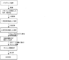
- FIG. 2 is a diagram showing a flow of producing the slice of FIG. 1 by performing staining, penetration and encapsulation.
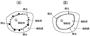
- (A) is a schematic view showing a state in which fluorescence observation is performed by immunostaining cells of a section of a tissue specimen using a conventional tissue staining stain.
- FIG. B is a schematic view showing a state in which fluorescence is observed by immunostaining cells of a section of a tissue specimen using the tissue staining dye according to the present invention.
- (A) is a photograph stained with a secondary antibody labeled with dye resin particles according to a comparative example.
- (B) is a photograph stained with a secondary antibody labeled with dye resin particles according to the example.
- tissue staining kit according to the present invention, the method for producing the tissue staining stain, and the tissue staining kit containing the tissue staining kit will be described with reference to FIGS.
- the tissue staining dye according to the present invention is a tissue staining dye containing, as a staining component, resin particles of thermosetting resin and dye resin particles having a fluorescent dye fixed to the resin particles.
- the coefficient of variation of the particle size of the resin particles is 15% or less.
- the fluorescent dye may be included in the resin particles.
- the substituent contributing to the ionic bond may be a negatively charged substituent for the fluorescent dye, and a positively charged substituent for the resin particles.
- the charge of the entire fluorescent dye and the charge of the resin particles may be the same as the charge of the substituent that contributes to the ionic bond.
- the fluorescent dye may have a molecular weight / charge of one molecule of the fluorescent dye and the number of substituents ⁇ 400.
- the fluorescent dye may have at least two negatively charged substituents per molecule of the fluorescent dye.
- the positively charged substituent may be an amino group, and the negatively charged substituent may be a sulfo group or a carboxyl group.
- At least one of the negatively charged substituents of the fluorescent dye may be a sulfo group.
- the fluorescent dye may be rhodamine, BODIPY, squarylium or an aromatic hydrocarbon dye molecule.
- the resin particles may be formed using melamine, and the fluorescent dye may be rhodamine or an aromatic hydrocarbon dye molecule.
- the resin particles and the fluorescent dye may be covalently bonded by any one of an amide bond, an ester bond, an ether bond, and a CN bond.
- the thermosetting resin may contain a structural unit formed from at least one monomer selected from the group consisting of melamine, urea, guanamine, phenol and xylene.
- the method for producing a staining agent for tissue staining includes a step of setting a coefficient of variation of the dye resin particles to 15% or less by adding a surfactant to a reaction system when the resin particles are produced by a synthetic reaction. including.
- the tissue staining kit according to the present invention contains any of the above-mentioned tissue staining stains as a constituent.
- the dye resin particles are resin particles and fluorescent dyes that are ion-bonded or covalently bonded.
- a dye resin particle is formed by ion-bonding an ammonium group in which a proton is added to an amino group of a resin constituting the resin particle and a sulfo group of the fluorescent dye ( (See the broken line in the left rectangle in FIG. 1).
- the amino group possessed by the monomer-derived portion constituting the resin particle and the sulfo group possessed by the fluorescent dye are covalently bonded to constitute the dye resin particle (FIG. 1). (See dashed line in right rectangle).
- Each bond between the resin particle and the fluorescent dye is performed between the inside and / or outer surface of the resin particle and the dye molecule.
- the example shown in FIG. 1 shows an example in which the dye is immobilized on the inner surface and / or the outer surface by ionic bond or covalent bond, respectively (surface immobilization is not shown).
- the bond between the resin particle and the fluorescent dye may include both an ionic bond and a covalent bond.
- the fluorescent dye encapsulated and / or surface-immobilized in the resin particles may be fixed to a resin particle by any one of the fluorescent dyes described later alone or by mixing a plurality of kinds. Good.
- two or more kinds of fluorescent dyes different from each other in the excitation wavelength and the emission wavelength may be included.
- the method for fixing the fluorescent dye to the resin particles is not particularly limited.
- a method of synthesizing dye resin particles by ion-bonding and / or covalently bonding fluorescent dye molecules to monomers and oligomers, which are raw materials for resin particles, and polymerizing them, and by adsorbing the dye to the produced resin particles Any method may be used for introducing the fluorescent dye into the resin particles, such as a method of introducing it.
- the average particle diameter of the dye resin particles is not particularly limited, but is usually 10 to 500 nm, and preferably 50 to 200 nm.
- one of the linkers such as streptavidin or biotin is added to the resin particles, and the other linker is added to the secondary antibody. Therefore, it may be configured to be used for immunostaining.
- thermosetting resin constituting the dye resin particles according to the present invention is not particularly limited as long as it includes a thermosetting resin capable of fixing the fluorescent dye by the above-described ionic bond and / or covalent bond.
- thermosetting resin used for ionic bonding with the fluorescent dye has at least part of hydrogen contained in its structural unit replaced by a charged substituent or a part of its chemical structure having a charge. It exists.
- thermosetting resins used for covalent bonding with fluorescent dyes have a site that can be covalently bonded to the dye in part of the chemical structure, regardless of the presence or absence of the above-mentioned substituent having a charge. It only needs to be covalently bonded to the dye.
- the “substituent or moiety having a charge” means a substituent or a chemical structure that is positively or negatively charged when dissolved in water or acidic or basic water.
- thermosetting resin a thermosetting resin containing a structural unit formed from at least one monomer selected from the group consisting of melamine, urea, guanamine, phenol, xylene, and derivatives thereof is preferably used. Can do.
- melamine, urea and guanamine are monomers having an amino group as a part of the structure as a positively charged substituent.
- Phenol is a monomer having a phenolic hydroxyl group in a part of the structure as a negatively charged substituent. Since xylene is a monomer having no charged substituents or moieties, it can be used for ionic bonding with a fluorescent dye by substituting some of the hydrogens it has with positive or negative charge substituents.
- thermosetting resins are obtained by a known polymerization method using a monomer containing a charged substituent or site, or a monomer of a thermosetting resin having a reactive group that forms a covalent bond with a functional group of a fluorescent dye. It can be produced by polymerization.
- the structure contains a substituent having a charge from the beginning, such as melamine, it is not necessary to introduce the substituent and it can be suitably used.
- thermosetting resin monomer examples include a sulfo group (—SO 3 ⁇ ) and a carboxyl group (—COO ⁇ ). Further, examples of the positively charged substituent that the thermosetting resin monomer has include an ammonium group (—NR 3 + ).
- R is hydrogen or an alkyl group.
- the introduction of a carboxyl group may be directly introduced by a Friedel-Crafts reaction, or an alkyl compound having a carboxyl group
- the monomer may be converted into a halide and boronic acid and combined by a Suzuki coupling reaction, or the alkyl compound having a carboxyl group and the monomer may be converted into a halide to be coupled with a Grignard reaction or the like. It may be introduced by binding in a reaction.
- the monomer may be directly sulfonated with fuming sulfuric acid or chlorosulfuric acid, or the alkyl compound having the sulfo group and the monomer are converted into halide and boronic acid and introduced by the Suzuki coupling reaction.
- an alkyl compound having a sulfo group and a monomer may be converted into a halide and combined and introduced by a coupling reaction such as a Grignard reaction.
- amino group it may be converted to amino group by nitration with fuming nitric acid and reduction reaction of nitro group, or the monomer may be converted to halogenated form and aminated by direct reaction with ammonia or Gabriel reaction.
- an alkyl compound having an amino group and a monomer may be converted into a halide and a boronic acid and then bonded by a Suzuki coupling reaction, or an alkyl compound having a carboxyl group and a monomer may be converted into a halide. They may be converted and combined and introduced by a coupling reaction such as a Grignard reaction.
- a protective group may be appropriately introduced, and after introduction into the monomer as a protected carboxyl group, sulfo group or amino group, deprotection may be performed to form a carboxyl group, sulfo group or amino group.
- thermosetting resin is a three-dimensional network structure, which is created by cross-linking polymers. For this reason, the fluorescent dye encapsulated in the resin particles of the thermosetting resin is unlikely to elute to the outside of the resin particles, and the effect of suppressing bright spot bleeding during fluorescence observation can be obtained.
- the thermosetting resin is not limited to a homopolymer, and may be a copolymer. Specifically, a copolymer obtained by copolymerizing a plurality of types of monomers or oligomers constituting the above polymelamine or the like, or a copolymer obtained by copolymerizing these monomers or oligomers and other types of monomers or oligomers. It may be.
- the fluorescent dye can be obtained or produced by a known method.
- the monomer or oligomer of the resin material and the fluorescent dye have opposite charges as shown in the left rectangle of FIG. Accordingly, the resin material and the dye molecules are ion-bonded and associated with each other before the resin material is thermally cured, and the fluorescent dye can be easily included in the resin particles.
- the fluorescent dye Since the resin particles and the fluorescent dye associate with each other, the molecules of the fluorescent dye are fixed to the resin particles, so that the elution of the dye from the resin particles hardly occurs.
- the fluorescent dye has two or more substituents.
- the fluorescent dye enters between the substituents of the adjacent resin and functions as a cross-linking agent that connects the two, and the bonding between the resin structural units is strengthened. Also, the resin particles and the fluorescent dye The bond between the dye and the resin is less likely to be eluted. Furthermore, since the molecules of the fluorescent dye are stabilized and the heat resistance of the fluorescent dye is further increased, when the resin particles are formed by polymerization reaction by thermosetting, the fluorescent dyes are less susceptible to the adverse effects of heat. From this, the fluorescent dye is difficult to elute.
- the fluorescent dye preferably has two or more reactive groups for the same reason as described above.
- Fluorescent dyes can be selected from, for example, each type of dye molecule such as rhodamine dye molecules, BODIPY (registered trademark, manufactured by Invitrogen), squarylium dye molecules, aromatic hydrocarbon dye molecules, and the like.
- fluorescent dyes such as aromatic ring dye molecules (aromatic hydrocarbon dye molecules) and rhodamine dye molecules are preferable because of their relatively high light resistance, and among them, perylene belonging to aromatic ring dye molecules. Pyrene and perylene diimide are preferable. Furthermore, rhodamine dyes and perylene diimide are excellent in quantum yield, light absorption, and the like, and are excellent in luminous efficiency. Therefore, resin particles encapsulating them have superior emission intensity compared to resin particles encapsulating other dyes.
- rhodamine dye molecules include 5-carboxy-rhodamine, 6-carboxy-rhodamine, 5,6-dicarboxy-rhodamine, rhodamine 6G, tetramethylrhodamine, X-rhodamine, Texas Red, Spectrum® Red, LD700® PERCHLORATE And derivatives thereof.
- BODIPY dye molecules include BODIPY FL, BODIPY TMR, BODIPY 493/503, BODIPY 530/550, BODIPY 558/568, BODIPY 564/570, BODIPY 576/589, BODIPY 581/591, 3061/3061 , BODIPY 650/665 (manufactured by Invitrogen), and derivatives thereof.
- squarylium dye molecule examples include SRfluor 680-Carboxylate, 1,3-Bis [4- (dimethylamino) -2-hydroxyphenyl] -2,4-dihydroxycyclobutenedidium dihydroxide, bis, 1,3-Bis, dimethylyl)) phenyl] -2,4-dihydroxycyclobutenedidium dihydroxide, bis, 2- (4- (Dithylamino) -2-hydroxy-4-hydroxy, 4- (dithyrimino) -2-hydryloxy-2-hydroxy-3-2-hydroxy-3 oxocyclobut-1-eno ate, 2- (4- (Dibutylamino) -2-hydroxyphenyl) -4- (4- (dibutylimino) -2-hydroxycyclohexa-2,5-diylenedene) -3-oxycyclobut-1-enolate, 2- (8-Hydroxy) -1,1,7,7-tetramethyl
- aromatic hydrocarbon dye molecule examples include N, N-Bis- (2,6-diisopropylphenyl) -1,6,7,12- (4-tert-butylphenoxy) -perylene-3,4,9 , 10-tetracarbonbonide diimide, N, N′-Bis (2,6-diisopropenylphenyl) -1,6,7,12-tetraphenylperylene-3, 4: 9,10-tetracarboximide, N, N′-Bis (2,6 -Diisopropylphenyl) perylene-3,4,9,10-bis (dicarbide), 16, N, N'-Bis (2,6-dimethylphenyl) perylene-3,4,9,10-tetraca rboxylic diimide, 4,4 '-[(8,16-Dihydro-8,16-dioxodibenzo [a, j] perylene-2,10-diyl
- the dye resin particles having a coefficient of variation of 15% or less, preferably less than 15%, according to the present invention are produced, for example, according to the following steps, and the dye resin particles are obtained by thermosetting one or more monomers or oligomers.
- a surfactant having an emulsifying action in the range of 10 to 60% by weight By adding a surfactant having an emulsifying action in the range of 10 to 60% by weight to the resin raw material, an arbitrary particle size can be obtained, for example, particles of 30 to 300 nm can be produced. Further, when the ratio of the surfactant is increased, even smaller particles can be produced and can be made 30 nm or less.
- the ratio of the surfactant is reduced, larger particles can be prepared, and particles of 300 nm or more can also be prepared. Further, the surfactant during the preparation of the particles can be arbitrarily changed from 0.1 to 3.0% by weight. Preferably it is 0.25 to 1.0% by weight. The ratio and addition amount of these surfactants are examples, and can be changed within an arbitrary range.
- the mixing step is a step of mixing the fluorescent dye, the surfactant, the proton supply agent, and one or more monomers or oligomers constituting the resin particles.
- the present inventors have found that the coefficient of variation of the particle size of the dye resin particles to be produced varies depending on the molar ratio of the surfactant and the monomer (monomer as the resin constituent unit) and the type of the surfactant. ing. For this reason, it is necessary to polymerize the resin by adjusting the type of the surfactant and the molar ratio with the monomer so that the variation coefficient of the dye resin particles is 15% or less.
- the type of surfactant all anionic, nonionic and cationic surfactants can be used.
- the magnitude of the influence of the charge of the surfactant on the coefficient of variation of the particle size of the resin particles has a relationship of nonionic system> anionic system> cationic system with respect to the cationic resin monomer. It is preferable to use a nonionic surfactant.
- the magnitude of the influence of the surfactant charge on the anionic resin monomer is nonionic> cationic> anionic, it is preferable to use a cationic or nonionic surfactant.
- the surfactant the following can be used, but is not limited thereto.
- the anionic surfactant include sodium dodecylbenzenesulfonate.
- nonionic surfactants include polyethylene glycol and polyoxyethylene alkyl ether.
- the cationic surfactant include dodecyltrimethylammonium bromide.
- Emulgen or Neoperex registered trademark, manufactured by Kao Corporation
- the active ingredient of Emulgen is polyoxyethylene alkyl ether
- the active ingredient of Neoperex is sodium dodecylbenzenesulfonate.
- the surfactant that is positioned plays a role of adjusting the amount of monomer inflow from the aqueous phase, and the particle diameters of the resin particles to be produced are uniform.
- the monomer in the water phase is surface active. It is easily attracted to the agent and easily supplied to the oil phase inside the micelle. Therefore, when a cationic monomer is used, an anionic or nonionic surfactant can be suitably used.
- thermosetting reaction temperature used in the polymerization step. If a cloud point lower than the thermosetting reaction temperature is selected, the surfactant loses hydration power with water and does not perform its function, and resin particles cannot be formed, resulting in a resin mass. It is.
- thermosetting resin for example, an acid can be used. It is known that the reaction of melamine resin, urea resin, xylene resin, and phenol resin is promoted by an acid catalyst.
- acid for example, formic acid, acetic acid, sulfuric acid, hydrochloric acid, nitric acid, paratoluenesulfonic acid, dodecylbenzenesulfonic acid, and the like are known.
- the reaction of the thermosetting resin proceeds only by heating, it proceeds at a lower temperature when a reaction accelerator is added, and therefore can be added within a range in which the reaction and performance can be controlled.
- the polymerization step is a step of thermosetting, that is, polymerizing monomers or oligomers to form dye resin particles.
- the reaction conditions (thermosetting temperature, polymerization time) in the polymerization step are determined from the composition of the monomer or oligomer to be polymerized and can be carried out in accordance with a known method. Here, it is necessary to set the reaction conditions (within the heat resistant temperature range of the fluorescent dye) so that the performance of the fluorescent dye does not deteriorate.
- the formation reaction of the melamine resin is performed at 70 ° C. to 200 ° C. Heating at 150 to 200 ° C. is preferable.
- the heat resistant temperatures of the fluorescent dyes are rhodamine dye molecules 200 ° C., BODIPY (registered trademark, manufactured by Invitrogen) 200 ° C., squarylium dye molecules 200 ° C., and aromatic hydrocarbon dye molecules 300 ° C. or more, and can withstand the formation reaction. It is necessary to use fluorescent dyes that can be obtained.
- the encapsulated fluorescent dye becomes difficult to elute from the dye resin particles.
- the produced dye resin particles are converted into dyes and resin particles at temperatures below the decomposition temperature or melting temperature of the resin used.
- heat treatment that is, crosslinking may be promoted by heat treatment to suppress bleeding of the fluorescent dye.
- the washing step is a step of removing impurities such as excess resin raw materials, fluorescent dyes and emulsifiers from the obtained dispersion of dye resin particles.
- the resin component is centrifuged from the reaction solution, and after removing the supernatant, washing is performed by adding ultrapure water, irradiating with ultrasonic waves, and dispersing again.
- a series of washing operations such as centrifugation, supernatant removal, and redispersion in ultrapure water is preferably repeated a plurality of times until no absorbance or fluorescence derived from the resin or pigment is observed in the supernatant.
- the addition step is a step of adding a linker or the like for immunostaining to the dye resin particles.
- the type of linker added to the dye resin particles may be a linker such as streptavidin-biotin, but is not limited thereto.
- streptavidin-biotin linker is performed, for example, as follows. First, a thiol group is added to streptavidin by a thiol group introduction reagent, while an amino group is introduced to the surface of the dye resin particle by an amino group introduction reagent, and then an active ester and a thiol group that react with the amino group In this method, dye resin particles and streptavidin are linked using a linker such as PEG having maleimide groups that react with each other at both ends.
- a linker such as PEG having maleimide groups that react with each other at both ends.
- amino group introduction reagents include aminopropyltrimethoxysilane.
- thiol group introduction reagent include N-succimidyl S acetylthioacetic acid, and the reagent and the addition itself can be performed by a known method.
- the particle diameter of the dye resin particles is determined by taking an electron micrograph of the produced dye resin particles using a scanning electron microscope (SEM), measuring the cross-sectional area of the dye resin particles, and measuring the measured value of the corresponding circle. It can be measured as the diameter (area circle equivalent diameter) when the area is taken.
- the average (average particle diameter) and coefficient of variation of the population of the dye resin particles are determined by measuring the particle diameter as described above for a sufficient number (for example, 300 particles) of the dye resin particles.
- the coefficient of variation is calculated by the formula: 100 ⁇ standard deviation of particle diameter / average particle diameter.
- tissue staining As a tissue staining method, a fluorescent staining method is used in which the dye resin particles are used as a fluorescent label for immunostaining to stain a biological material to be detected.
- a fluorescent label conjuggate
- a fluorescent label conjuggate
- a fluorescent label conjugate
- a fluorescent label conjugate
- a fluorescent label conjugate
- a fluorescent label conjugate
- a fluorescent label conjugate
- a fluorescent label in which a dye resin particle and a primary antibody are directly bonded via the linker
- the antigen is stained
- Secondary antibody method a method in which a fluorescent labeling body in which a dye resin particle and a secondary antibody are directly bound to each other is prepared, and a primary antibody bound to an antigen is stained (secondary antibody method), referring to FIG.
- a fluorescent labeling body in which dye resin particles and biotin are directly bound is prepared, and staining is performed using a primary antibody and an avidin- or streptavidin-modified secondary antibody bound to an antigen.
- a fluorescent labeling material in which avidin or streptavidin is directly bound to dye resin particles is prepared, and staining is performed using a primary antibody bound to an antigen and a secondary antibody modified with biotin (biotin- An avidin method or a sandwich method) can be used.
- Any primary antibody may be used for immunostaining and will vary depending on the subject to be immunostained. For example, when performing immunostaining using HER2 as an antigen, an anti-HER2 antibody is used.
- any secondary antibody may be used, and varies depending on the primary antibody. Examples include anti-mouse, anti-rabbit, anti-cow, anti-goat, anti-sheep, anti-dog and anti-chicken antibodies.
- Any existing method may be used for binding the dye resin particles to the antibody or biotin.
- amidation in the reaction of amine and carboxylic acid, sulfidation by reaction of maleimide and thiol, imination by reaction of aldehyde and amine, amination by reaction of epoxy and amine, and the like can be used.
- the above immunostaining is not limited to tissue staining, and can also be applied to cell staining.
- the biological substance to be detected is not limited as long as a substance that specifically binds to the biological substance exists.
- a combination of an antigen and an antibody is used as described above.
- a combination of a nucleic acid molecule (oligonucleotide, polynucleotide) and a nucleic acid molecule having a sequence capable of hybridizing thereto may be used.
- the fluorescence emitted from the fluorescent dye is observed by irradiating the excitation light having an appropriate wavelength according to the fluorescent dye of the dye resin particles used in the pathological section subjected to the tissue staining by the above process.
- a predetermined biomolecule present in the pathological section can be detected and used as information for determining whether or not an antibody drug (for example, Herceptin labeled with HER2) is necessary. be able to.
- an antibody drug for example, Herceptin labeled with HER2
- a predetermined wavelength may be selected from a laser light source provided in the fluorescence microscope as necessary.
- Fluorescence observation may be performed from a lens barrel of a fluorescence microscope, or may be performed by separately displaying an image photographed by a camera installed on the fluorescence microscope on a display means (a monitor or the like). Although it depends on the fluorescent dye, there are cases where it is possible to observe the fluorescence through taking an image with a camera, even if the fluorescence cannot be sufficiently observed by visual observation from the tube of the fluorescence microscope. You may use the filter which selectively passes a predetermined wavelength as needed.
- the luminance of the immunostaining part is a value obtained as an average value of the luminances of the parts stained with the fluorescent label for immunostaining.
- the bright spot of the immunostained portion means a spot that shines at a site stained with a fluorescent label for immunostaining, as shown in FIG. 3 (see FIGS. 3A and 3B).
- tissue staining kit includes the above-described tissue staining dye as a constituent.
- Other components may include reagents such as antibodies relating to immunostaining.
- FIGS. 1 When the coefficient of variation of the particle size of the dye resin particles is 15% or less, the result is that the size of the bright spot is uniform. As a result, the coefficient of variation exceeds 15%. The size is uniform (see FIGS. 3A and 3B or FIGS. 4A and 4B in comparison). For this reason, the fluorescence signals from all the bright spots fall within the dynamic range set in the fluorescence observation. As a result, the above-mentioned problems such as the fusion of adjacent bright spots by saturation are solved, and discrimination between bright spots is ensured. As a result, the determination accuracy of the fluorescent signal is improved.
- the fluorescent dye has a positively charged or negatively charged substituent
- the thermosetting resin constituting the resin of the dye resin particle has an oppositely charged substituent and is ionically bonded to the fluorescent dye.
- the fluorescent dye Since the fluorescent dye is encapsulated in the resin particles, the fluorescent dye is not unnecessarily exposed to light, thereby preventing degradation of the fluorescent dye and performance degradation as a fluorescent label.
- the substituent contributing to the ionic bond is such that the fluorescent dye is a negatively charged substituent and the resin particle is a positively charged substituent.
- the bleeding of the fluorescent dye can be particularly suppressed, and the brightness of the stained image can be ensured.
- the fluorescent dye has at least two negatively charged substituents per fluorescent dye molecule, so that it is sandwiched between a plurality of positively charged substituents of the resin as shown in the left rectangle of FIG. To be fixed.
- the fluorescent dye is fixed and functions like a crosslinking agent. As a result, it is possible to prevent bleeding of the fluorescent dye in the tissue-stained image as compared with the case of having one substituent in that it is more firmly fixed. In addition, brightness performance can be secured, and fusion of bright spots can be prevented.
- the positively charged substituent is an amino group
- the negatively charged substituent is a sulfo group or a carboxyl group, so that the strongly negatively charged sulfo group is compared to the amino group (—NH + —).
- the (SO 3 ⁇ ) electron pair can be coordinated to firmly bond the resin and the fluorescent dye.
- a fluorescent dye having a sulfo group can be firmly bonded to a thermosetting resin having an amino group such as melamine.
- the fluorescent dye is rhodamine, BODIPY, squarylium or an aromatic hydrocarbon dye molecule
- the dye molecule is firmly bonded together with the ionic bond of the substituent by the interaction of the fluorescent dye and the hydrophobic portion of the resin. And resin can be combined. Thereby, it is possible to further prevent bleeding of the fluorescent dye in the tissue stained image.
- thermosetting resin is formed using melamine, and the fluorescent dye is rhodamine or an aromatic hydrocarbon dye molecule, so that the melamine resin and the rhodamine or aromatic hydrocarbon dye molecule are each
- the dye molecule and the resin can be bound more firmly by the hydrophobic interaction between the benzene rings. Thereby, it is possible to further prevent bleeding of the fluorescent dye in the tissue-stained image after preparation of the tissue section (FIG. 2).
- rhodamine and aromatic hydrocarbons are preferably used from the viewpoint of luminance and light resistance.
- thermosetting resin and the fluorescent dye are covalently bonded by any one of an amide bond, an ester bond, an ether bond, and a CN bond. Since the fluorescent dye is encapsulated in the resin particles and firmly maintained during the penetration of the resin, the seepage of the resin particles outside the resin particles is suppressed.
- a coefficient of variation of 15% or less is included by adding a predetermined amount of a surfactant to a reaction system when the resin particles are produced by a synthetic reaction to make the coefficient of variation of the resin particles 15% or less.
- the dye resin particles can be produced, and a tissue staining dye having the above-described effects can be provided.
- a tissue staining kit having the above-described effects can be provided as long as it is a tissue staining kit containing the tissue staining stain as a constituent.
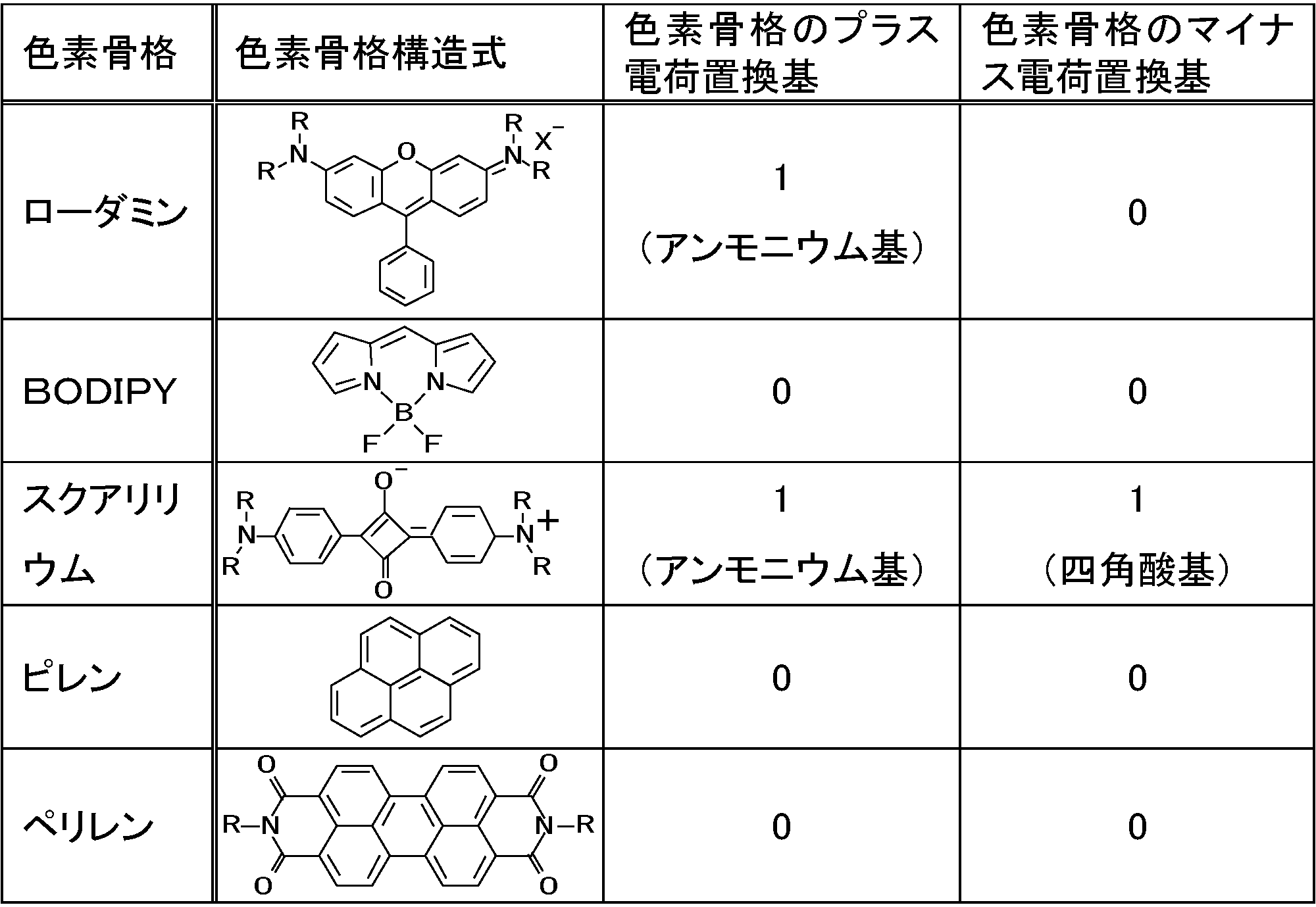
- Compounds 1-1 to 4-4 in Table 1-1 to Table 1-2 each have a skeleton of rhodamine, BODIPY, squarylium, pyrene, or perylene diimide (see Table 1-3), and have a substituent.
- a carboxylic acid a sulfonic acid, an ammonium group, a dye having a covalent bond site, or a dye having no such substituent.
- the compounds 1-1 to 1-8 have a carboxyl group or a sulfo group and have a negative charge as a whole molecule.
- Compounds 2-1 and 2-2 have an ammonium group and have a positive charge as a whole molecule.
- Compounds 3-1 and 3-2 have a covalent bond site.
- Compounds 4-1 to 4-4 do not have the above substituent.
- rhodamine contains one ammonium group in the skeleton. However, since the contribution of the substituent contained in the skeleton is small, it is not counted in the number of charges (substituents) of the dye. The same applies to squarylium.
- [Compound 1-1] Commercially available sulforhodamine 101 (Sigma Aldrich) was used.
- Compound 1-2 A fluorescent dye related to compound 1-2 having a structural formula close to that of AlEXA594 was prepared and used according to the published Japanese Patent Application Laid-Open No. 2010-18049.
- Compound 1-3 The compound 1-2 was NHS esterified, reacted with 2-Aminoethanesulfonic Acid (Tokyo Kasei Kogyo Co., Ltd.) in DFM at 90 ° C. for 1 hour, purified by column chromatography and used.
- [Compound 1-4] A commercially available Disodium-1,3,5,7,8-pentamethylpyrromethene-2,6-disulfonate-difluoroborate complex (Exiton) was used. [Compound 1-5] It was synthesized and used according to the method described in the literature (J. Phys. Chem. A 2005, 109, 5571). [Compound 1-6] Commercially available 8-Methoxypyrene-1,3,6-trisulfonic acid trisodium salt (Sigma Aldrich) was used. [Compounds 1-7, 1-8, 3-1, 3, 2, 4-4] It was prepared and used according to the previously reported Angew. Chem. Int. Ed. 2004, 43, 1528.
- [Compound 2-1] Commercially available rhodamine B (Wako Pure Chemical Industries, Ltd.) was used.
- [Compound 2-2] Commercially available sulforhodamine 101 acid chloride (Dojin Chemical Co., Ltd.) and ethylamine (Tokyo Kasei Kogyo Co., Ltd.) were reacted in DFM at 90 ° C. for 1 hour, used after purification by column chromatography.
- [Compound 4-1] Commercially available tetramethylrhodamine ethyl ester perchlorate (Sigma Aldrich) was used.
- the structure of the above compound is expressed as an inner salt, but for example, a form in which an anion such as a halide ion, methanesulfonic acid, trifluoromethanesulfonate or the like is attached as a counter ion may be used.
- Carboxylic acid and sulfonic acid are expressed in acid form, but may be a salt of an alkali metal or the like.
- the numbers of carboxylic acid, sulfonic acid and ammonium group in the molecule are shown, but this number does not include the number of acids and ammonium salts contained in the dye skeleton itself. For example, rhodamine contains one ammonium group in the dye skeleton, but this is not counted. Similarly, squarylium contains a square acid and ammonium group in the dye skeleton, but this is not counted.
- This solution was heated to 70 ° C. while stirring on a hot stirrer, and then 0.65 g of melamine resin raw material Nicalak MX-035 (manufactured by Nippon Carbide Industries Co., Ltd.) was added to this solution.
- the concentration of the resulting dye resin particles was adjusted to 3 nM using phosphate buffered saline (PBS) containing 2 mM ethylenediaminetetraacetic acid (EDTA).
- PBS phosphate buffered saline
- EDTA ethylenediaminetetraacetic acid
- SM (PEG) 12 Succinimidyl-[(N-maleoidopropionamid) -dodecaethyleneglycol] ester, manufactured by Thermo Scientific Co., Ltd.
- the mixture was reacted at 20 ° C. for 1 hour to obtain a mixed solution containing dye resin particles having a fluorescent dye with a maleimide at the end.
- the mixture was centrifuged at 10,000 G for 20 minutes, the supernatant was removed, PBS containing 2 mM EDTA was added to disperse the precipitate, and the mixture was centrifuged again. The above washing by the same procedure was performed three times.
- streptavidin manufactured by Wako Pure Chemical Industries, Ltd.
- streptavidin and N-succimidyl S-acetylthioacetic acid are used to add a thiol group to the streptavidin and perform gel filtration.
- SATA N-succinimidyl S-acetylthioacetate
- tissue staining Human breast tissue was immunostained using a tissue staining dye containing the dye resin particles.
- a buffer solution such as a PBS buffer solution containing 1% BSA was used as the stain for tissue staining.
- a tissue array slide manufactured by Cosmo Bio, product number CB-A712 was used as the stained section. The tissue array slide was deparaffinized, washed with water, and autoclaved in 10 mM citrate buffer (pH 6.0) for 15 minutes to activate the antigen.
- Anti-HER2 rabbit monoclonal antibody (4B5) diluted to 0.05 nM with PBS buffer containing 1% BSA was washed with the tissue array slide after antigen activation treatment with PBS buffer and reacted for 2 hours with tissue sections. I let you. After washing with PBS, it was reacted with a biotin-labeled anti-rabbit antibody diluted with 1% BSA-containing PBS buffer for 30 minutes. Furthermore, an immunohistochemically stained section was obtained by using the above-mentioned staining dye for tissue staining, that is, by reacting with the prepared dye resin particles having streptavidin for 2 hours and then washing. The obtained immunohistochemically stained section was immersed in a 4% neutral paraformaldehyde aqueous buffer solution for 10 minutes for fixation.
- the “elution amount of the fluorescent dye” in Tables 2 and 4 to 7 is a value obtained by immersing the dye resin particles in ethanol and evaluating the amount of the dye eluted in ethanol. Specifically, 0.1 mg of pigment resin particles are immersed in 1 mL of ethanol, subjected to ultrasonic waves for 10 minutes with a bath-type ultrasonic device (As One Co., Ltd.), centrifuged to remove the particles, and the supernatant liquid is removed. It is a value obtained by calculating the dye amount (pmol) in 1 mL from a calibration curve of the dye concentration and the peak value of the fluorometer measured in advance with a fluorometer F7000 (Hitachi). From this value, it is possible to indirectly estimate the dye bleeding of the evaluation 1: Her2 stained image.
- Example 2 (Compound 1-1-encapsulated polyurea particles) Production of pigment resin particles in the same manner as in Example 1 except that 0.80 g of urea resin raw material was used instead of 0.65 g of melamine resin raw material Nicarax MX-035 (Nippon Carbide Industries Co., Ltd.) in Example 1. Went. This urea resin has a positive charge because the skeleton itself contains many amino groups.
- the urea resin raw material was urea produced by an existing method and having a methylolation degree of 40 to 70%.
- Example 3 Compound 1-1-encapsulated polyxylene particles
- Example 1 instead of 0.65 g of the melamine resin raw material Nicarax MX-035 (manufactured by Nippon Carbide Industries Co., Ltd.), 0.80 g of xylene resin raw material Nicarak Y-50 (manufactured by Fudou), butyl isocyanate 0.8.
- Dye resin particles were produced in the same manner as in Example 1 except that 20 g and 0.20 g of Nicalac MX-035 (Nippon Carbide Industries Co., Ltd.) were used. Since this polyxylene resin contained many amino groups, the resin became a positive charge.
- Example 1 [Examples 4, 7, 10, 13, 16, 19, 24, 27, 30, 34] (Compound 1-2-3-2 encapsulated polymelamine particles)
- Compound 1-1 was converted to Compound 1-2 (Example 4), Compound 1-3 (Example 7), Compound 1-4 (Example 10), Compound 1-5 (Example 13).
- Compound 1-6 (Example 16), Compound 1-7 (Example 19), Compound 2-1 (Example 24), Compound 2-2 (Example 27), Compound 3-1 (Example 30)
- Dye resin particles were prepared in the same manner except that the compound 3-2 (Example 34) was changed.
- Example 2 [Examples 5, 8, 11, 14, 17, 20, 25, 28, 31, 35] (Compound 1-2-3-2 encapsulated polyurea particles)
- Compound 1-1 was changed to Compound 1-2 (Example 5), Compound 1-3 (Example 8), Compound 1-4 (Example 11), Compound 1-5 (Example) 14), Compound 1-6 (Example 17), Compound 1-7 (Example 20), Compound 2-1 (Example 25), Compound 2-2 (Example 28), Compound 3-1 (Example) Dye resin particles were prepared in the same manner except that 31) and compound 3-2 (Example 35) were changed.
- Examples 6, 9, 12, 15, 18, 21, 26, 29, 33, 37 (Compound 1-2-3-2 encapsulated polyxylene particles)
- compound 1-1 was converted to compound 1-2 (Example 6), compound 1-3 (Example 9), compound 1-4 (Example 12), compound 1-5 (Example 15).
- Compound 1-6 (Example 18), Compound 1-7 (Example 21), Compound 2-1 (Example 26), Compound 2-2 (Example 29), Compound 3-1 (Example 33)
- Dye resin particles were prepared in the same manner except that the compound was changed to compound 3-2 (Example 37).
- Example 22 (Compound 1-8-encapsulated polyphenol particles)
- the fluorescent dye was changed from Compound 1-1 to Compound 1-8, and instead of 0.65 g of melamine resin raw material Nicarac MX-035 (manufactured by Nippon Carbide Industries), phenol resin raw material Nikanol PR-1440M (Fudo Corporation) Without using heating and stirring (heating stirring at 70 ° C. for 50 minutes) after adding 1000 ⁇ L of 10% aqueous solution of dodecylbenzenesulfonic acid (manufactured by Kanto Chemical Co., Ltd.), using 0.80 g and 0.20 g of phenol.
- Dye resin particles were produced in the same manner as in Example 1 except that only heating and stirring at 90 ° C. for 20 minutes were performed, and then heating was performed at 125 ° C. for 5 minutes in an autoclave. In addition, since the resin particle contained phenol, it became a negative charge.
- Example 23 (Compound 1-8-encapsulated polyxylene particles)
- Compound 1-1 was changed to Compound 1-8, and instead of 0.65 g of melamine resin Nicarak MX-035 (Nihon Carbide Kogyo Co., Ltd.), xylene resin raw material Nikanol Y-50 (Fudo Co., Ltd.) 0.20 g, phenol resin raw material Nikanol PR-1440M (manufactured by Fudo) 0.20 g, 3- (4-Hydroxyphenil) propionic acid 0.20 g, dodecylbenzenesulfonic acid (manufactured by Kanto Chemical) 10% Without heating and stirring after adding 1000 ⁇ L of aqueous solution (heating and stirring at 70 ° C.
- Example 32 (Compound 3-1 and Compound 3-2 encapsulated polyphenol particles)
- dye resin particles were prepared in the same manner except that Compound 1-8 was changed to Compound 3-1 (Example 32) and Compound 3-2 (Example 36).
- melamine resin particles had a characteristic that the coefficient of variation of the particles was larger than that of the particles prepared in Example 1. Using the resulting dye resin particles, immunohistological staining and morphological staining were performed in the same manner as in Example 1.
- Comparative Example 4 (Compound 1-1 encapsulating polyurea particles)
- dye resin particles were produced in the same manner as in Example 1 except that 1.6 g of urea resin raw material was used instead of 1.5 g of melamine resin raw material Nicalac MX-035 (Nippon Carbide Industries Co., Ltd.). did.
- the urea resin raw material is urea produced by an existing method and having a methylolation degree of 40 to 70%.
- Comparative Example 5 (Compound 1-1 encapsulating polyxylene particles)
- Comparative Example 3 0.80 g of phenol resin raw material K-100 (manufactured by Fudou Co., Ltd.) and 0.20 g of 3,5-Dimethylaniline were used instead of 1.5 g of melamine resin raw material Nicarax MX-035 (manufactured by Nippon Carbide Industries Co., Ltd.) Except that, pigment resin particles were produced in the same manner as in Example 1. Since the present xylene resin contains a lot of amino groups, the resin has a positive charge.
- Comparative Example 25 (Compound 1-8-encapsulated polyxylene particles)
- Compound 1-1 was changed to Compound 1-8, and instead of 1.5 g of melamine resin raw material Nicarac MX-035 (Nihon Carbide Industries Co., Ltd.), phenol resin raw material Nikanol PR-1440 (Fudo Corporation) ) Using 0.80 g and 0.20 g of phenol, without adding 10 ⁇ L of 10% aqueous solution of dodecylbenzenesulfonic acid (manufactured by Kanto Chemical Co., Inc.) without performing heating and stirring (heating and stirring at 70 ° C.
- Comparative Examples 40, 43, 46, and 49 have a coefficient of variation of 15% or less, but include a dye containing a fluorescent dye (compounds 4-1 to 4-4) that has no charge with respect to the urea resin particles.
- a dye containing a fluorescent dye compounds 4-1 to 4-4 that has no charge with respect to the urea resin particles.
- Comparative Examples 41, 44, 47, and 50 the variation coefficient is 15% or less, but the fluorescent dye (compounds 4-1 to 4-4) having no charge is fixed to the polyurea resin particles. This is an example.
- Comparative Examples 42, 45, 48, and 51 the coefficient of variation is 15% or less, but fluorescent dyes (compounds 4-1 to 4-4) having no charge are fixed to the polyxylene resin particles. This is an example.
- Example 53 (Compound 1-1 encapsulating polyxylene particles)
- dye resin particles were prepared, streptavidin was prepared, and morphological dyeing was performed except that a negatively charged polyxylene resin was used instead of a positively charged polymelamine resin (Table 1). 6).
- Comparative Examples 52, 54, 58, 60, 62, and 64 have a coefficient of variation of 15% or less, but have the same negative ( ⁇ ) charge with respect to the negative ( ⁇ ) charged polyphenol resin particles.
- the coefficient of variation is 15% or less, but the same minus ( ⁇ ) with respect to particles of poly (xylene) resin having a minus ( ⁇ ) charge.
- dye resin particles in which a fluorescent dye having a charge (compounds 1-1 to 1-7) is encapsulated are manufactured, and immunohistochemical staining, morphological staining, and evaluation of a tissue image are performed.
- Example 38 (Compound 1-1 Encapsulating Polymelamine Particles)
- the time of centrifugal separation for washing the dye resin particles with pure water was reduced from 15 minutes to 10 minutes. Further, as in Example 1, centrifugation was performed 5 times, the supernatant was removed, and the redispersion in ultrapure water was performed, and then a new centrifugation was performed for 1 minute to remove the precipitate. Other than that was the same as Example 1.
- Example 39 to 48 (Compound 1-2 to 3-2 encapsulated polymelamine particles)
- Compound 1-1 was converted to Compound 1-2 (Example 39), Compound 1-3 (Example 40), Compound 1-4 (Example 41), Compound 1-5 (Example 42).
- Compound 1-6 (Example 43), Compound 1-7 (Example 44), Compound 2-1 (Example 45), Compound 2-2 (Example 46), Compound 3-1 (Example 47)
- Dye resin particles were prepared in the same manner except that the compound 3-2 (Example 48) was changed.
Landscapes
- Chemical & Material Sciences (AREA)
- Health & Medical Sciences (AREA)
- Life Sciences & Earth Sciences (AREA)
- Engineering & Computer Science (AREA)
- Immunology (AREA)
- Organic Chemistry (AREA)
- Biomedical Technology (AREA)
- Molecular Biology (AREA)
- Hematology (AREA)
- Chemical Kinetics & Catalysis (AREA)
- General Physics & Mathematics (AREA)
- General Health & Medical Sciences (AREA)
- Biochemistry (AREA)
- Analytical Chemistry (AREA)
- Pathology (AREA)
- Physics & Mathematics (AREA)
- Urology & Nephrology (AREA)
- Cell Biology (AREA)
- Microbiology (AREA)
- Biotechnology (AREA)
- Food Science & Technology (AREA)
- Medicinal Chemistry (AREA)
- Dispersion Chemistry (AREA)
- Investigating Or Analysing Biological Materials (AREA)
- Investigating, Analyzing Materials By Fluorescence Or Luminescence (AREA)
Abstract
Description
この色素樹脂粒子は、図1に示すように、樹脂粒子と蛍光色素とがイオン結合または共有結合したものである。前者の例は、樹脂粒子を構成する樹脂が有するアミノ基に対してプロトンが付加したアンモニウム基と、蛍光色素が有するスルホ基がイオン結合して、色素樹脂粒子を構成しているものである(図1の左側矩形内の破線参照)。一方、後者の例は、樹脂粒子を構成するモノマー由来の部分が有するアミノ基と、蛍光色素が有するスルホ基とが共有結合して、色素樹脂粒子を構成しているものである(図1の右側矩形内の破線参照)。
本発明に係る色素樹脂粒子を構成する熱硬化性樹脂は、蛍光色素を上述したイオン結合および/または共有結合により固定することができる熱硬化性樹脂を含むものであれば、特に限定されない。
本発明で用いることが可能な蛍光色素としては、既存のいかなるものを用いても構わない。ただし、熱硬化性樹脂の合成反応の加熱条件下であっても該加熱により悪影響を受けない蛍光色素を用いることが望ましい。蛍光色素は、公知の方法により入手または作製することができる。
本発明に係る変動係数15%以下、好ましくは15%未満の色素樹脂粒子は、例えば以下の各工程に沿って製造され、1種または2種以上のモノマーまたはオリゴマーを熱硬化させて色素樹脂粒子を製造する際に、界面活性剤が所定量存在した状態で重合反応を行うことにより製造することができる。樹脂原料に対し、10~60重量%の範囲で乳化作用を有する界面活性剤を加える事で、任意の粒子径を得る事ができ、例えば、30~300nmの粒子を作製できる。また、界面活性剤の割合を増やすと更に小さい粒子も作製可能で30nm以下とすることができる。あるいは、界面活性剤の割合を減らすと更に大きい粒子も作製可能で300nm以上の粒子も作製可能である。また、粒子作製時の界面活性剤は0.1から3.0重量%で任意に変更できる。好ましくは0.25から1.0重量%である。これら界面活性剤の割合と添加量は一例であり、任意の範囲で変更できる。
混合工程は、上記蛍光色素と、界面活性剤と、プロトン供給剤と、樹脂粒子を構成するモノマーまたはオリゴマーの1種または2種以上等とを混合する工程である。
本発明者らは、製造される色素樹脂粒子の粒径の変動係数が、界面活性剤とモノマー(樹脂構成単位としてのモノマー)とのモル比や、界面活性剤の種類により変化することを見出している。このため、色素樹脂粒子の変動係数が15%以下となるように、界面活性剤の種類やモノマーとのモル比を調節して樹脂を重合させる必要がある。
熱硬化性樹脂と蛍光色素とをイオン結合させる場合、プラス電荷となる熱硬化性樹脂の置換基や蛍光色素の置換基にH+を積極的に供給してプラス電荷とするプロトン供給剤を用いる事もできる。例えば、ギ酸や酢酸、パラトルエンスルホン酸等、これらに類するものが挙げられる。色素に付いている置換基がカルボン酸やスルホン酸等の酸の場合、これがプロトン供給剤として機能する事もできる。逆に熱硬化樹脂のマイナス電荷となる置換基や蛍光色素の置換基からH+を積極的に抜き取るプロトン受容剤を用いる事もできる。例えば、水酸化ナトリウム等の塩基の場合、これがプロトン受容剤として機能する。
熱硬化性樹脂の反応促進剤として、例えば酸を用いる事ができる。メラミン樹脂や尿素樹脂、キシレン樹脂、フェノール樹脂は、いずれも酸触媒により反応が促進される事が知られている。酸としては、例えば、ギ酸、酢酸、硫酸、塩酸、硝酸、パラトルエンスルホン酸、ドデシルベンゼンスルホン酸、等が知られている。熱硬化性樹脂の反応は加温のみでも進行するが、反応促進剤を加えるとより低温で進行するので、反応や性能を制御できる範囲で添加することができる。
重合工程は、モノマーまたはオリゴマーを熱硬化、即ち重合させて色素樹脂粒子を形成する工程である。重合工程の反応条件(熱硬化温度、重合時間)は、重合させるモノマーまたはオリゴマーの組成から決定され、公知の方法に即して行うことができる。ここで、蛍光色素の性能が低下しない反応条件(蛍光色素の耐熱温度範囲内)とする必要がある。
洗浄工程は、得られた色素樹脂粒子の分散液から、余剰の樹脂原料や蛍光色素、乳化剤等の不純物を除く工程である。例えば、反応液から樹脂成分を遠心分離し、上澄み除去後、超純水を加えて超音波照射して再度分散させることで洗浄を行う。遠心分離、上澄み除去、超純水への再分散の一連の洗浄操作は、上澄みに樹脂や色素に由来する吸光・蛍光が見られなくなるまで、複数回繰り返し行うことが好ましい。
付加工程は、色素樹脂粒子に免疫染色用のリンカー等を付加する工程である。色素樹脂粒子に付加するリンカーの種類は、ストレプトアビジン-ビオチン等のリンカーを用いることができるが、これに限定されない。
(樹脂粒子の粒径と変動係数)
色素樹脂粒子の粒径は、製造した色素樹脂粒子を、走査型電子顕微鏡(SEM)を用いて電子顕微鏡写真を撮影し、色素樹脂粒子の断面積を計測し、その計測値を相当する円の面積としたときの直径(面積円相当径)として測定することができる。色素樹脂粒子の集団の粒子径の平均(平均粒径)および変動係数は、十分な数(たとえば300個)の色素樹脂粒子について、上記のように粒子径を測定した後、平均粒径はその算術平均として算出され、変動係数は式:100×粒径の標準偏差/平均粒径により算出される。
組織染色の方法としては、上記色素樹脂粒子を免疫染色用の蛍光標識体として検出対象の生体物質を染色する蛍光染色法が用いられる。たとえば、特定の抗原に対して免疫染色を行う際には、上記リンカーを介して色素樹脂粒子と1次抗体を直接結合した蛍光標識体(コンジュゲート)を作製し、抗原を染色する方法(1次抗体法)、色素樹脂粒子と2次抗体とを直接結合した蛍光標識体を作製し、抗原に1次抗体を結合したものを染色する方法(2次抗体法)、図1を参照して上述したように、色素樹脂粒子とビオチンを直接結合した蛍光標識体を作製し、抗原に1次抗体、及び、アビジンあるいはストレプトアビジン修飾した2次抗体を結合したものを用いて染色する方法、同様に色素樹脂粒子にアビジンあるいはストレプトアビジンを直接結合した蛍光標識体を作製し、抗原に1次抗体、及び、ビオチン修飾した2次抗体を結合したものを用いて染色する方法(ビオチン-アビジン法またはサンドイッチ法)、等を用いることができる。
上記工程により組織染色が施された病理切片に用いられている色素樹脂粒子の蛍光色素に応じた適切な波長を有する励起光を照射することで、蛍光色素が発する蛍光を観察する。このような工程により、その病理切片に存在する所定の生体分子を検出することができ、抗体医薬(たとえばHER2を標識とするハ―セプチン)の適用の要否を判断するための情報として利用することができる。励起光の照射には、一般的な蛍光観察と同様の照射手段を用いればよく、例えば、蛍光顕微鏡が備えるレーザ光源から、必要に応じて所定の波長を選択すればよい。
本発明に係る組織染色用キットは、上述した組織染色用染色剤を構成品として含む。他の構成品は免疫染色に関する抗体等の試薬等を含んでいてよい。
(1)色素樹脂粒子の粒径の変動係数が15%以下の場合、輝点の大きさが揃う結果、変動係数が15%を超えるものと異なり、蛍光観察を行った場合の各輝点の大きさが一様となる(図3(A)と(B)または図4(A)と(B)を対比して参照)。このため、蛍光観察で設定するダイナミックレンジに全ての輝点からの蛍光シグナルが収まるようになる。この結果、隣接する輝点同士がサチレーションにより融合する等の上記問題が解消され、輝点同士の判別が担保される。この結果、蛍光シグナルの判定精度が改善される。
[色素]
以下の実施例、比較例に用いた色素を表1に示す。


市販のスルホローダミン101(シグマアルドリッチ社)を使用した。
[化合物1-2]
AlEXA594に近い構造式を有する化合物1-2に係る蛍光色素を、既報の特開2010-18049に従って作製して使用した。
[化合物1-3]
前記化合物1-2をNHSエステル化した後、2-Aminoethanesulfonic Acid(東京化成工業社)とDFM中で90℃、1時間反応し、カラムクロマトグラフィーで精製後、使用した。
市販のDisodium-1,3,5,7,8-pentamethylpyrromethene-2,6-disulfonate‐difluoroborate complex(Exiton社)を使用した。
[化合物1-5]
文献(J. Phys. Chem. A 2005, 109, 5571)に記載の方法に従い合成し、使用した。
[化合物1-6]
市販の8-Methoxypyrene-1,3,6-trisulfonic acid trisodium salt(シグマアルドリッチ社)を使用した。
[化合物1-7,1-8,3-1,3-2,4-4]
既報のAngew. Chem. Int. Ed. 2004, 43, 1528に従って作製し、使用した。
[化合物2-1]
市販のローダミンB(和光純薬工業社)を使用した。
[化合物2-2]
市販のスルホローダミン101アシッドクロライド(同仁化学社)とエチルアミン(東京化成工業社)をDFM中で90℃、1時間反応し、カラムクロマトグラフィーで精製後、使用した。
[化合物4-1]
市販のテトラメチルローダミンエチルエステル過塩素酸塩(シグマアルドリッチ社)を使用した。
[化合物4-2]
市販の1,3,5,7,8-pentamethyl-2,6-diethylpyrromethene-difluoroborate complex(Exiton社)を使用した。
[化合物4-3]
市販の1,3-ビス[4-(ジメチルアミノ)フェニル]-2,4-ジヒドロキシシクロブテンジイリウム 二水酸化物, ビス(分子内塩)(シグマアルドリッチ社)を使用した。
[実施例1](化合物1-1内包ポリメラミン粒子)
蛍光色素として化合物1-1であるSulfoRhodamine101(シグマアルドリッチ社製)14.4mgを水22mLに加えて溶解した。その後、この溶液に乳化重合用乳化剤のエマルゲン(登録商標)430(ポリオキシエチレンオレイルエーテル、花王社製)の5%水溶液を2mL加えた。この溶液をホットスターラー上で撹拌しながら70℃まで昇温させた後、この溶液にメラミン樹脂原料ニカラックMX-035(日本カーバイド工業社製)を0.65g加えた。
一方、ストレプトアビジン(和光純薬工業社製)とN-スクシミジル Sアセチルチオ酢酸(N-succinimidyl S-acetylthioacetate、略称:SATA)を用いて、ストレプトアビジンに対してチオール基の付加処理を行い、ゲル濾過を行って色素樹脂粒子に結合可能なストレプトアビジンを別途用意した。
上記色素樹脂粒子とストレプトアビジンを、2mMのEDTAを含有したPBS中で混合後、室温で1時間反応させて、両者を結合させる反応を行った。反応後、10mMメルカプトエタノールを添加して反応を停止させた。得られた溶液をφ=0.65μmの遠心フィルターで濃縮後、精製用ゲル濾過カラムを用いて未反応のストレプトアビジン等を除去し、ストレプトアビジンが結合した色素樹脂粒子を得た。
この色素樹脂粒子を含む組織染色用染色剤を用いて、ヒト乳房組織の免疫染色を行った。ここで組織染色用染色剤は、1%BSA含有PBS緩衝液等の緩衝液を用いた。染色切片は組織アレイスライド(コスモ・バイオ社製、品番CB-A712)を用いた。組織アレイスライドを脱パラフィン処理後、水に置換洗浄し、10mMクエン酸緩衝液(pH6.0)中で15分間オートクレーブ処理することで、抗原の賦活化処理を行った。抗原の賦活化処理後の組織アレイスライドを、PBS緩衝液を用いて洗浄後、1%BSA含有PBS緩衝液で0.05nMに稀釈した抗HER2ウサギモノクローナル抗体(4B5)を組織切片と2時間反応させた。PBSで洗浄後、1%BSA含有PBS緩衝液で稀釈したビオチン標識抗ウサギ抗体と、30分間反応させた。さらに、上記組織染色用染色剤を用いて、すなわち上記製造したストレプトアビジンを有する色素樹脂粒子と2時間反応させ、その後洗浄を行うことにより、免疫組織化学染色切片が得られた。得られた免疫組織化学染色切片を4%中性パラホルムアルデヒド水系緩衝液に10分間浸漬することにより、固定処理を行った。
上記固定処理をした各免疫組織化学染色切片に対してヘマトキシリン染色を行い、染色後の切片をエタノールに浸漬することにより脱水し、脱水切片をさらにキシレンに浸漬して透徹し、封入剤で封入して風乾させることにより、二重染色切片が得られた。ヘマトキシリン染色は、後述の評価において影響はなかった。また、形態染色を行わない場合はエタノール浸漬とキシレン浸漬による透徹のみ、あるいは形態染色としてエオシン染色を追加することもできた。
市販の蛍光顕微鏡を用いて色素内包樹脂粒子の蛍光組織画像を取得し、輝点計測への影響、組織像の色素にじみ、染色像の明るさについて評価した。
実施例1において、メラミン樹脂原料ニカラックMX-035(日本カーバイド工業社製)0.65gの代わりに、尿素樹脂原料0.80gを使用した以外は、実施例1と同様に色素樹脂粒子の製造等を行った。本尿素樹脂は、骨格自体にアミノ基を多く含有することで樹脂がプラス電荷となった。なお、尿素樹脂原料は既存の方法により作製した、メチロール化度が40~70%の尿素であった。
実施例1において、メラミン樹脂原料ニカラックMX-035(日本カーバイド工業社製)の使用量を0.65gの代わりに、キシレン樹脂原料ニカラックY-50(フドー社製)0.80g、ブチルイソシアネート0.20g、および、ニカラックMX-035(日本カーバイド工業社製)0.20gを使用した点以外は、実施例1と同様に色素樹脂粒子の製造等を行った。本ポリキシレン樹脂は、アミノ基を多く含有することで樹脂がプラス電荷となった。
実施例1において、化合物1-1を、化合物1-2(実施例4)、化合物1-3(実施例7)、化合物1-4(実施例10)、化合物1-5(実施例13)、化合物1-6(実施例16)、化合物1-7(実施例19)、化合物2-1(実施例24)、化合物2-2(実施例27)、化合物3-1(実施例30)、化合物3-2(実施例34)に変更した以外は同様にして色素樹脂粒子の作製等を行った。
実施例2において、化合物1-1を、化合物1-2に変更(実施例5)、化合物1-3(実施例8)、化合物1-4(実施例11)、化合物1-5(実施例14)、化合物1-6(実施例17)、化合物1-7(実施例20)、化合物2-1(実施例25)、化合物2-2(実施例28)、化合物3-1(実施例31)および化合物3-2(実施例35)に変更した以外は同様にして色素樹脂粒子の作製等を行った。
実施例3において、化合物1-1を、化合物1-2(実施例6)、化合物1-3(実施例9)、化合物1-4(実施例12)、化合物1-5(実施例15)、化合物1-6(実施例18)、化合物1-7(実施例21)、化合物2-1(実施例26)、化合物2-2(実施例29)、化合物3-1(実施例33)および化合物3-2(実施例37)に変更した以外は同様にして色素樹脂粒子の作製等を行った。
実施例1において、蛍光色素を化合物1-1から化合物1-8に変更し、メラミン樹脂原料ニカラックMX-035(日本カーバイド工業製)0.65gの代わりにフェノール樹脂原料ニカノールPR-1440M(フドー社製)0.80gとフェノール0.20gを用いた点、ドデシルベンゼンスルホン酸(関東化学社製)10%水溶液を1000μL添加した後の加熱撹拌(70℃で50分間加熱撹拌)を行わずに、90℃、20分間の加熱撹拌のみ行い、その後、オートクレーブにて125℃で5分間加熱した点以外は、実施例1と同様に色素樹脂粒子の製造等を行った。なお、樹脂粒子はフェノールを含むためマイナス電荷となった。
実施例1において、化合物1-1を化合物1-8に変更し、メラミン樹脂ニカラックMX-035(日本カーバイド工業社製)0.65gの代わりに、キシレン樹脂原料ニカノールY-50(フドー社製)0.20g、フェノール樹脂原料ニカノールPR-1440M(フドー社製)0.20g、3-(4-Hydroxyphenil)propionic Acid 0.20g、を用いた点、ドデシルベンゼンスルホン酸(関東化学社製)10%水溶液を1000μL添加した後の加熱撹拌(70℃で50分間加熱撹拌)を行わずに、90℃、20分間の加熱撹拌のみ行い、その後、オートクレーブにて125℃、5分間加熱した点以外は実施例1と同様にして色素樹脂粒子の作製等を行った。なお、本樹脂粒子は、フェニル基とカルボキシル基を有するため、マイナス電荷となった。
実施例22において、化合物1-8を、化合物3-1(実施例32)、化合物3-2(実施例36)に変更した以外実施例は同様に色素樹脂粒子の作製等を行った。
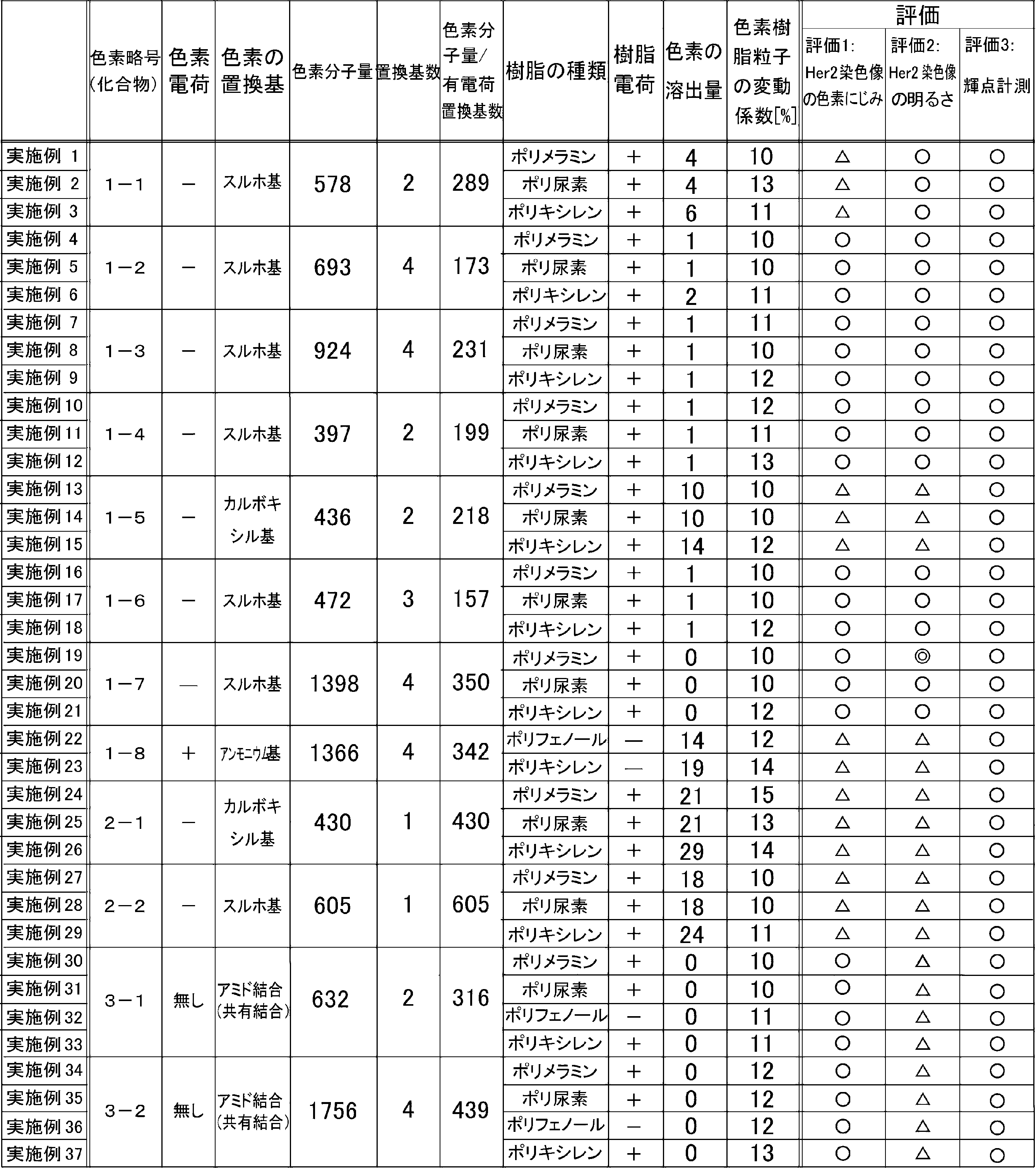
蛍光色素と樹脂の電荷が逆であり、且つ、色素樹脂粒子の変動係数が15%以下であることから、蛍光観察時の蛍光色素の滲みがほとんどなく、輝度や輝点計測の結果がよいものとなった(実施例1~37)。
[比較例1,2]
実施例1で製造した色素樹脂粒子の代わりに、市販のインビトロジェン社製の末端にアミノ基が付いたポリスチレン製の色素樹脂粒子(比較例1)、末端にアミノ基が付いた市販の半導体ナノ粒子「Q-dоt」(比較例2)を用いて実施例1と同様に免疫組織染色および形態染色を行った。
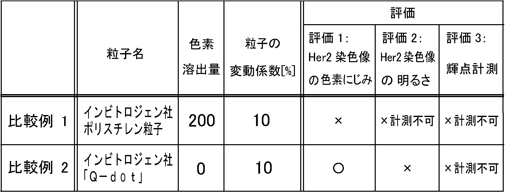
熱可塑性樹脂であるポリスチレン粒子に蛍光色素を固定した色素樹脂粒子であり、色素溶出が生じて組織像の評価3では色素が滲んで組織像が見えない結果となった(比較例1)。
[比較例3](化合物1-1内包ポリメラミン粒子)
蛍光色素として化合物1-1であるSulfоRhоdamine101(シグマアルドリッチ社製)2.5mgを水22.5mLに加え溶解した。その後、ホットスターラー上で70℃まで昇温させた後、メラミン樹脂ニカラックMX-035(日本カーバイド工業社製)1.5gを加え、5分間加熱撹拌した。ギ酸100μLを加え、70℃で20分間、加熱撹拌した後、室温放冷した。冷却後、反応混合物を遠心用チューブに入れて遠心分離機に20000Gで20分間、遠心分離し、上澄み除去後、超純水を加えて超音波照射して再分散させた。遠心分離、上澄み除去および超純水への再分散による洗浄を5回繰り返した。得られたメラミン樹脂粒子は実施例1で作成した粒子と比べて、粒子の変動係数が大きい特徴を有していた。得られた色素樹脂粒子を用いて、実施例1と同様に免疫組織染色および形態染色を行った。
比較例3において、メラミン樹脂原料ニカラックMX-035(日本カーバイド工業社製)1.5gの代わりに、尿素樹脂原料1.6gを使用した点以外は、実施例1と同様に色素樹脂粒子を製造した。なお、尿素樹脂原料は既存の方法により作製した、メチロール化度が40~70%の尿素である。
比較例3において、メラミン樹脂原料ニカラックMX-035(日本カーバイド工業社製)1.5gの代わりに、フェノール樹脂原料K-100(フドー社製)0.80gと3,5-Dimethylaniline0.20gを使用した以外は実施例1と同様に色素樹脂粒子の製造等を行った。本キシレン樹脂はアミノ基を多く含有する事で樹脂がプラス電荷となった。
比較例3において、化合物1-1を、化合物1-2(比較例6)、化合物1-3(比較例9)、化合物1-4(比較例12)、化合物1-5(比較例15)、化合物1-6(比較例18)、化合物1-7(比較例21)、化合物2-1(比較例26)、化合物2-2(比較例29)、化合物3-1(比較例32)、化合物3-2(比較例36)に変更した以外は同様にして色素樹脂粒子の作製等を行った。
比較例4において、化合物1-1を、化合物1-2(比較例7)、化合物1-3(比較例10)、化合物1-4(比較例13)、化合物1-5(比較例16)、化合物1-6(比較例19)、化合物1-7(比較例22)、化合物1-8(比較例27)、化合物2-1(比較例30)、化合物2-2(比較例33)、化合物3-1(比較例37)に変更した以外は同様にして色素樹脂粒子の作製等を行った。
比較例5において、化合物1-1を化合物1-2(比較例8)、化合物1-3(比較例11)、化合物1-4(比較例14)、化合物1-5(比較例17)、化合物1-6(比較例20)、化合物1-7(比較例23)、化合物2-1(比較例28)、化合物2-2(比較例31)、化合物3-1(比較例35)、化合物3-2(比較例39)に変更した以外は同様にして色素樹脂粒子の作製等を行った。
比較例3において、化合物1-1を化合物1-8に変更し、メラミン樹脂原料ニカラックMX-035(日本カーバイド工業社製)1.5gの代わりに、フェノール樹脂原料ニカノールPR-1440M(フドー社製)0.80gおよびフェノール0.20gを用いた点、ドデシルベンゼンスルホン酸(関東化学社製)10%水溶液を1000μL添加した後の加熱撹拌(70℃で50分間加熱撹拌)を行わずに、90℃、20分間の加熱撹拌のみ行い、その後、オートクレーブにて125℃で5分間加熱した点以外は比較例3と同様に色素樹脂粒子の製造等を行った。なお、樹脂粒子はフェノールを含み、分子全体としてマイナス電荷となった。
比較例3において、化合物1-1を化合物1-8に変更し、メラミン樹脂原料ニカラックMX-035(日本カーバイド工業社製)1.5gの代わりに、フェノール樹脂原料ニカノールPR-1440(フドー社製)0.80gおよびフェノール0.20gを用いた点、ドデシルベンゼンスルホン酸(関東化学社製)10%水溶液を1000μL添加した後の加熱撹拌(70℃で50分間加熱撹拌)を行わずに、90℃、20分間の加熱撹拌のみ行い、その後、オートクレーブにて125℃5分間加熱した点以外は、比較例3と同様に色素樹脂粒子の製造等を行った。なお、樹脂粒子はフェノールとカルボキシル基を含み、分子全体としてマイナス電荷となった。
比較例24において、化合物1-8を、化合物3-1(比較例34)、化合物3-2(比較例38)に変更した以外は同様に色素樹脂粒子の製造、形態染色等を行った。
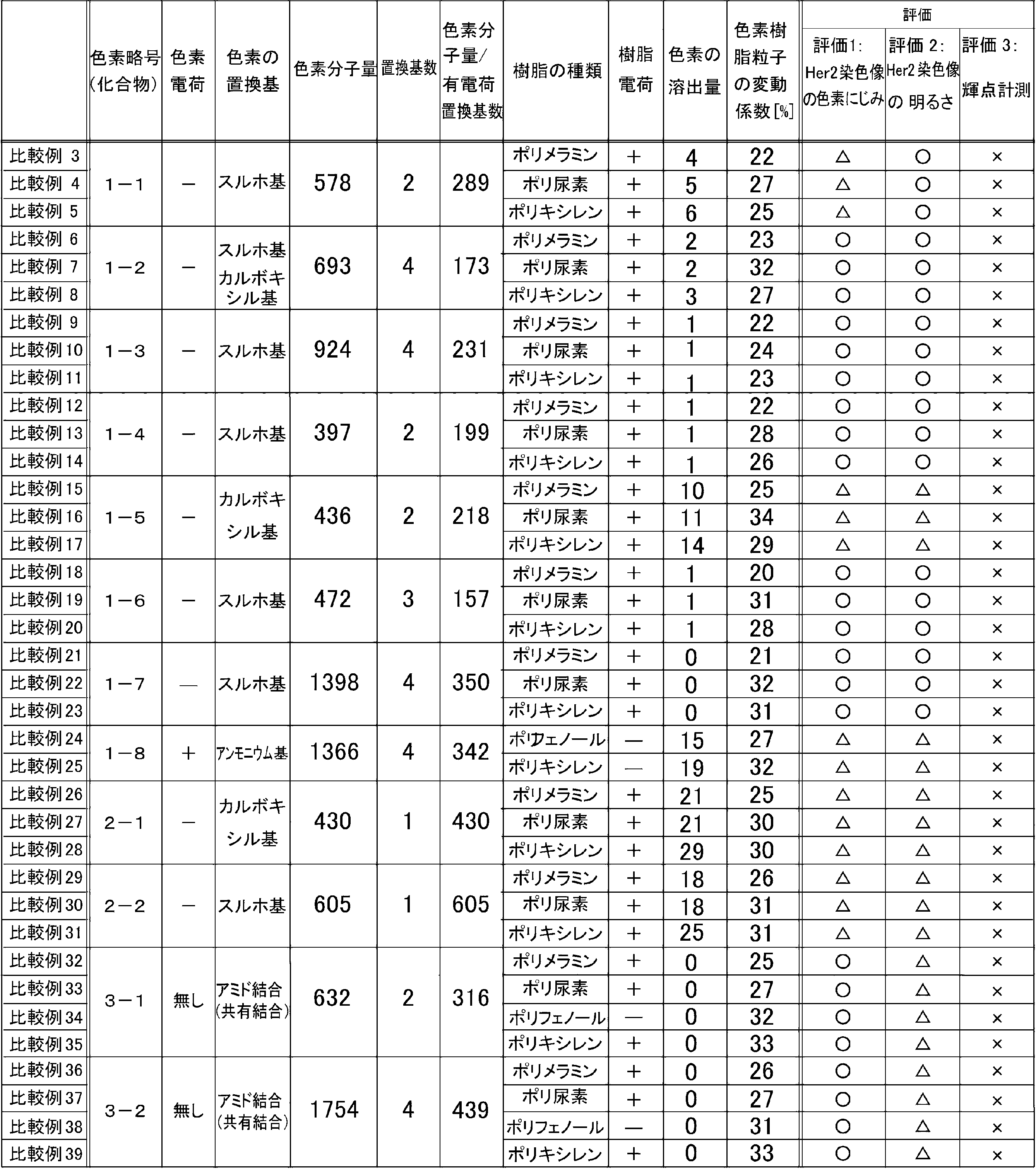
蛍光色素と樹脂の電荷が逆の電荷であっても、色素樹脂粒子の粒径サイズにバラツキがあり、変動係数15%を超える場合、輝点計測等が困難となった(比較例3~39)。
[比較例40,43,46,49](化合物4-1~4-4内包ポリメラミン粒子)
実施例1において、化合物1-1を、無電荷の化合物4-1(比較例40)、化合物4-2(比較例43)、化合物4-3(比較例46)、化合物4-4(比較例49)に変更した以外は同様にして色素樹脂粒子の作製等を行った。
実施例2において、化合物1-1を、無電荷の化合物4-1(比較例41)、化合物4-2(比較例44)、化合物4-3(比較例47)、化合物4-4(比較例50)に変更した以外は同様にして色素樹脂粒子の作製等を行った。
実施例3において、化合物1-1を、無電荷の化合物4-1(比較例42)、化合物4-2(比較例45)、化合物4-3(比較例48)、化合物4-4(比較例51)に変更した以外は同様にして色素樹脂粒子の作製等を行った。

蛍光色素が電荷を有しておらず樹脂とイオン結合しない上に、共有結合もしないことから色素溶出量が大きく、染色後の蛍光観察時の色素にじみが多くなるため、輝点計測等が困難となった(比較例40~51)。
[比較例52](化合物1-1内包ポリフェノール粒子)
実施例1において、使用したプラス電荷のポリメラミン樹脂の代わりに、蛍光色素(化合物1-1)の電荷と同じマイナス電荷を有するポリフェノール樹脂を用いた以外は同様に色素樹脂粒子の製造、ストレプトアビジンの調製および形態染色等を行った(表6参照)。
実施例1において、使用した樹脂をプラス電荷のポリメラミン樹脂の代わりにマイナス電荷のポリキシレン樹脂を用いた以外は同様に色素樹脂粒子の製造、ストレプトアビジンの調製および形態染色等を行った(表6参照)。
比較例52において、化合物1-1を、化合物1-2(比較例54)、化合物1-3(比較例56)、化合物1-4(比較例58)、化合物1-5(比較例60)、化合物1-6(比較例62)、化合物1-7(比較例64)に変更した以外は同様にして色素樹脂粒子の作製等を行った。
比較例53において、化合物1-1を、化合物1-2(比較例55)、化合物1-3(比較例57)、化合物1-4(比較例59)、化合物1-5(比較例61)、化合物1-6(比較例63)、化合物1-7(比較例65)に変更した以外は同様にして色素樹脂粒子の作製等を行った。

蛍光色素と樹脂の電荷が同一のマイナス電荷の場合、電気的に反発し合って、蛍光色素が樹脂にイオン結合で固定されず、また、共有結合もしていないので、染色後の蛍光観察において蛍光色素の滲みが生じて、輝度や輝点の計測が困難となった(比較例52~65)。
[比較例66](化合物1-8内包ポリメラミン粒子)
実施例1において、マイナス電荷の蛍光色素(化合物1-1)の代わりに、樹脂と同じプラス電荷の蛍光色素(化合物1-8)を用いた以外は同様に色素樹脂粒子の製造、組織染色等を行った(表7参照)。
実施例2において、化合物1-1の代わりに化合物1-8を用いた以外は同様に色素樹脂粒子の製造、組織染色等を行った(表7参照)。
実施例3において、化合物1-1の代わりに化合物1-8を用いた以外は同様に色素樹脂粒子の製造、組織染色等を行った(表7参照)。

蛍光色素と樹脂の電荷が同一のプラス電荷の場合、電気的に反発し合って、蛍光色素が樹脂にイオン結合で固定されず、染色後の蛍光観察において蛍光色素の滲みが生じて、輝度や輝点の計測が困難となった(比較例66~68)。
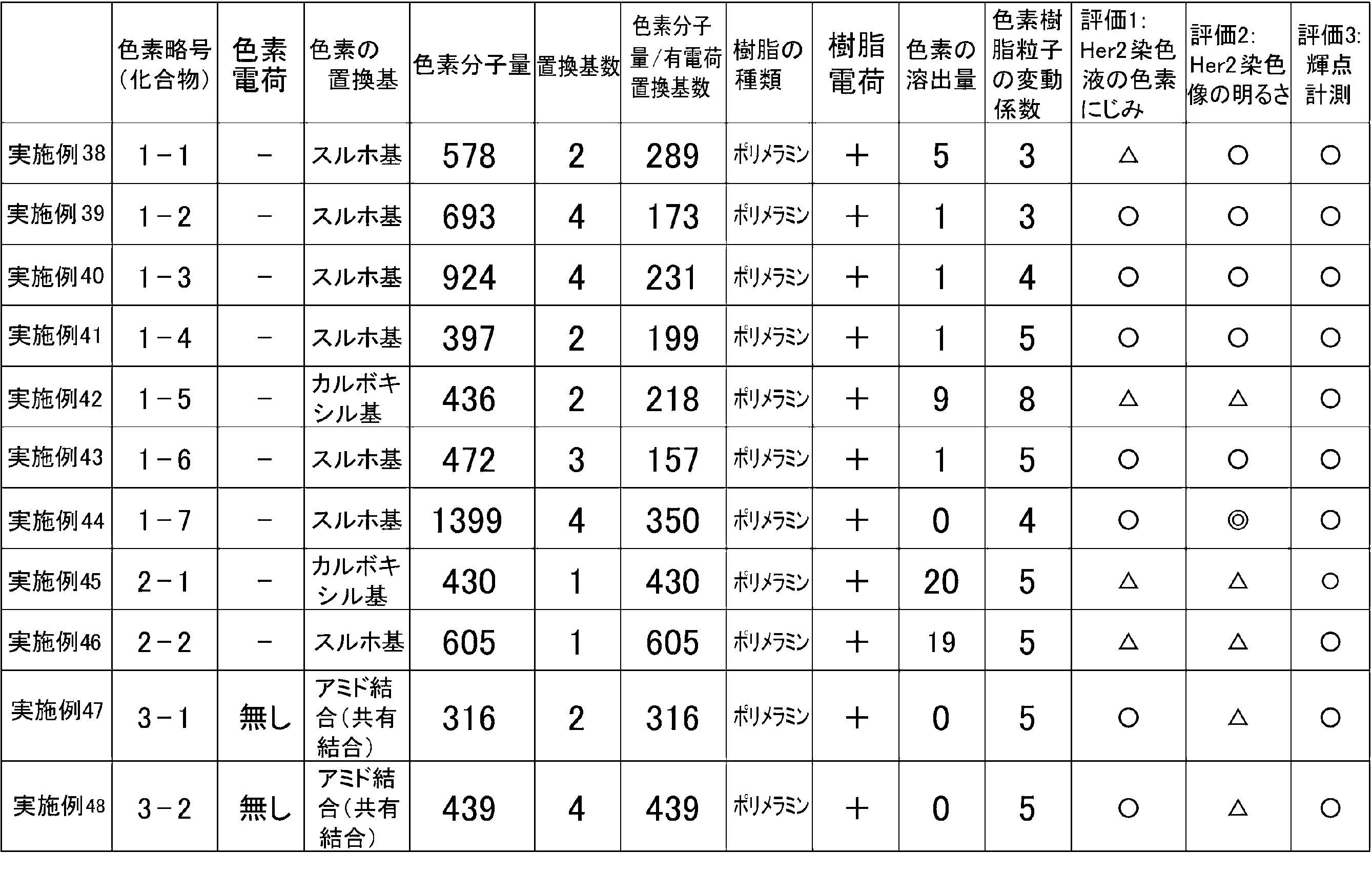
実施例1において、色素樹脂粒子の純水による洗浄の遠心分離の時間を15分から10分に短縮した。また、実施例1と同様に5回の遠心分離と上澄み除去と超純水への再分散の操作の後、新たに遠心分離を1分行ない、沈殿物を取り除いた。それ以外は実施例1と同様とした。
実施例38において、化合物1-1を、化合物1-2(実施例39)、化合物1-3(実施例40)、化合物1-4(実施例41)、化合物1-5(実施例42)、化合物1-6(実施例43)、化合物1-7(実施例44)、化合物2-1(実施例45)、化合物2-2(実施例46)、化合物3-1(実施例47)、化合物3-2(実施例48)に変更した以外は同様にして色素樹脂粒子の作製等を行った。
Claims (14)
- 熱硬化性樹脂の樹脂粒子および該樹脂粒子に固定された蛍光色素を有する色素樹脂粒子を染色成分として含有する組織染色用染色剤であって、
前記樹脂粒子が前記蛍光色素と逆の電荷の置換基を有して前記蛍光色素とイオン結合、または、共有結合しており、
前記色素樹脂粒子の粒径の変動係数が15%以下である、組織染色用染色剤。 - 前記蛍光色素が前記樹脂粒子に内包されている、請求項1に記載の組織染色用染色剤。
- 前記イオン結合に寄与している置換基が、前記蛍光色素はマイナス電荷の置換基であり、前記樹脂粒子はプラス電荷の置換基である、請求項1または2に記載の組織染色用染色剤。
- 前記蛍光色素全体の電荷および前記樹脂粒子全体の電荷が、前記イオン結合に寄与している置換基の電荷と同一の電荷である、請求項1~3いずれか1項に記載の組織染色用染色剤。
- 前記蛍光色素は、蛍光色素1分子の分子量/電荷を有する置換基数<400である、請求項1~4のいずれか1項に記載の組織染色用染色剤。
- 前記蛍光色素は、蛍光色素1分子あたりにマイナス電荷の置換基を少なくとも2つ有する、請求項3~5いずれか1項に記載の組織染色用染色剤。
- 前記プラス電荷の置換基がアミノ基であり、
前記マイナス電荷の置換基がスルホ基またはカルボキシル基である、請求項1~6のいずれか1項に記載の組織染色用染色剤。 - 前記蛍光色素のマイナス電荷の置換基の少なくとも1つがスルホ基である、請求項6または7に記載の組織染色用染色剤。
- 前記蛍光色素が、ローダミン、BODIPY、スクアリリウムまたは芳香族炭化水素系色素分子である、請求項1~8のいずれか1項に記載の組織染色用染色剤。
- 前記樹脂粒子がメラミンを用いて形成され、前記蛍光色素がローダミンまたは芳香族炭化水素系色素分子である、請求項9に記載の組織染色用染色剤。
- 前記樹脂粒子と前記蛍光色素とが、アミド結合、エステル結合、エーテル結合およびC-N結合のいずれかによって共有結合している、請求項1または2に記載の組織染色用染色剤。
- 前記熱硬化性樹脂が、メラミン、尿素、グアナミン、フェノール、キシレンおよびこれらの誘導体からなる群から選択された少なくとも1つのモノマーから形成される構成単位を含み、かつその構成単位に含まれる水素の少なくとも一部が電荷を有する置換基に置き換えられている、請求項1~9および11のいずれか1項に記載の組織染色用染色剤。
- 前記樹脂粒子を合成反応により製造する際の反応系に界面活性剤を添加することにより前記色素樹脂粒子の変動係数を15%以下とする工程を含む、組織染色用染色剤の製造方法。
- 請求項1~12の何れか1項に記載の組織染色用染色剤を構成品として含む、組織染色用キット。
Priority Applications (5)
| Application Number | Priority Date | Filing Date | Title |
|---|---|---|---|
| US14/773,148 US9903797B2 (en) | 2013-03-08 | 2014-03-06 | Staining agent for staining tissue, production method for staining agent for staining tissue and tissue staining kit including staining agent for staining tissue |
| JP2015504387A JP6311698B2 (ja) | 2013-03-08 | 2014-03-06 | 組織染色用染色剤、組織染色用染色剤の製造方法および組織染色用染色剤を含む組織染色用キット |
| EP14761085.1A EP2966445B1 (en) | 2013-03-08 | 2014-03-06 | Staining agent for staining tissue, production method for staining agent for staining tissue, and tissue staining kit including staining agent for staining tissue |
| US15/865,680 US10101249B2 (en) | 2013-03-08 | 2018-01-09 | Staining agent for staining tissue, production method for staining agent for staining tissue and tissue staining kit including staining agent for staining tissue |
| US16/150,866 US20190033181A1 (en) | 2013-03-08 | 2018-10-03 | Staining agent for staining tissue, production method for staining agent for staining tissue and tissue staining kit including staining agent for staining tissue |
Applications Claiming Priority (2)
| Application Number | Priority Date | Filing Date | Title |
|---|---|---|---|
| JP2013047257 | 2013-03-08 | ||
| JP2013-047257 | 2013-03-08 |
Related Child Applications (2)
| Application Number | Title | Priority Date | Filing Date |
|---|---|---|---|
| US14/773,148 A-371-Of-International US9903797B2 (en) | 2013-03-08 | 2014-03-06 | Staining agent for staining tissue, production method for staining agent for staining tissue and tissue staining kit including staining agent for staining tissue |
| US15/865,680 Division US10101249B2 (en) | 2013-03-08 | 2018-01-09 | Staining agent for staining tissue, production method for staining agent for staining tissue and tissue staining kit including staining agent for staining tissue |
Publications (1)
| Publication Number | Publication Date |
|---|---|
| WO2014136885A1 true WO2014136885A1 (ja) | 2014-09-12 |
Family
ID=51491393
Family Applications (1)
| Application Number | Title | Priority Date | Filing Date |
|---|---|---|---|
| PCT/JP2014/055798 Ceased WO2014136885A1 (ja) | 2013-03-08 | 2014-03-06 | 組織染色用染色剤、組織染色用染色剤の製造方法および組織染色用染色剤を含む組織染色用キット |
Country Status (4)
| Country | Link |
|---|---|
| US (2) | US9903797B2 (ja) |
| EP (1) | EP2966445B1 (ja) |
| JP (3) | JP6311698B2 (ja) |
| WO (1) | WO2014136885A1 (ja) |
Cited By (12)
| Publication number | Priority date | Publication date | Assignee | Title |
|---|---|---|---|---|
| JP2014174018A (ja) * | 2013-03-08 | 2014-09-22 | Konica Minolta Inc | 蛍光色素標識用樹脂粒子及びその製造方法並びに該粒子を含む組織免疫染色用キット |
| WO2016093268A1 (ja) * | 2014-12-12 | 2016-06-16 | コニカミノルタ株式会社 | 蛍光ナノ粒子用希釈液、これを用いた蛍光免疫染色用キット、蛍光免疫染色用溶液、および蛍光免疫染色法、遺伝子染色法 |
| WO2017104476A1 (ja) | 2015-12-18 | 2017-06-22 | コニカミノルタ株式会社 | 蛍光物質集積ナノ粒子およびそれを用いた標識剤 |
| EP3333574A4 (en) * | 2015-08-05 | 2018-06-20 | Konica Minolta, Inc. | Environment-responsive dye-accumulated nano particles, and method for analyzing intracellular environment |
| WO2018116465A1 (ja) * | 2016-12-22 | 2018-06-28 | 日立化成株式会社 | Her2陽性癌細胞の検出方法 |
| WO2018185943A1 (ja) | 2017-04-07 | 2018-10-11 | コニカミノルタ株式会社 | 蛍光プレミックス粒子、それを含有する蛍光染色液、およびそれらを用いた蛍光染色法 |
| WO2018185942A1 (ja) | 2017-04-07 | 2018-10-11 | コニカミノルタ株式会社 | タンパク質修飾蛍光体集積粒子の精製物を製造する方法、蛍光染色液の製造方法、タンパク質修飾蛍光体集積粒子の精製物、蛍光染色液およびタンパク質修飾蛍光体集積粒子精製用フィルター |
| EP3395906A4 (en) * | 2015-12-21 | 2019-01-02 | Konica Minolta, Inc. | Process for producing dye-containing heat-curable resin particles |
| WO2019235580A1 (ja) * | 2018-06-07 | 2019-12-12 | キヤノン株式会社 | 過酸化水素検出用染料、染料粒子複合体、および過酸化水素検出用構造体 |
| JP2020529866A (ja) * | 2017-07-26 | 2020-10-15 | シーティス エス.ピー.エー. | 分析および診断目的のために生体試料を固定化する方法 |
| WO2022131371A1 (ja) * | 2020-12-18 | 2022-06-23 | 凸版印刷株式会社 | ポリフェノール誘導体及び高分子材料 |
| JPWO2022234721A1 (ja) * | 2021-05-07 | 2022-11-10 |
Families Citing this family (3)
| Publication number | Priority date | Publication date | Assignee | Title |
|---|---|---|---|---|
| JP6620699B2 (ja) * | 2016-08-09 | 2019-12-18 | コニカミノルタ株式会社 | コアシェル型蛍光色素含有ナノ粒子およびその製造方法 |
| JP7467974B2 (ja) * | 2020-02-17 | 2024-04-16 | 富士フイルムビジネスイノベーション株式会社 | 樹脂粒子 |
| CN115505463B (zh) * | 2022-10-27 | 2024-06-04 | 浙江峻山生物科技有限公司 | 一种水性脱蜡剂及其制备方法与应用 |
Citations (6)
| Publication number | Priority date | Publication date | Assignee | Title |
|---|---|---|---|---|
| US4326008A (en) * | 1976-08-27 | 1982-04-20 | California Institute Of Technology | Protein specific fluorescent microspheres for labelling a protein |
| JP2007178315A (ja) * | 2005-12-28 | 2007-07-12 | Toyobo Co Ltd | 有形成分分類装置用粒子標準試薬 |
| JP2009014729A (ja) * | 2006-02-24 | 2009-01-22 | Furukawa Electric Co Ltd:The | フローサイトメトリーによる細胞の検出・分取システム、及び検出・分取方法 |
| JP2010018049A (ja) | 2008-07-08 | 2010-01-28 | Bridgestone Corp | タイヤ |
| JP2010134195A (ja) | 2008-12-04 | 2010-06-17 | Olympus Corp | 顕微鏡システム、標本観察方法およびプログラム |
| WO2012029752A1 (ja) * | 2010-08-31 | 2012-03-08 | コニカミノルタエムジー株式会社 | 生体物質検出方法 |
Family Cites Families (11)
| Publication number | Priority date | Publication date | Assignee | Title |
|---|---|---|---|---|
| US5326692B1 (en) * | 1992-05-13 | 1996-04-30 | Molecular Probes Inc | Fluorescent microparticles with controllable enhanced stokes shift |
| ATE239801T1 (de) | 1998-01-22 | 2003-05-15 | Luminex Corp | Mikropartikel mit multiplen fluoreszenz-signalen |
| JP4245415B2 (ja) * | 2002-11-25 | 2009-03-25 | 健二 山本 | 分子認識蛍光体の製造方法 |
| US20050123935A1 (en) * | 2003-12-09 | 2005-06-09 | Richard Haugland | Pyrenyloxysulfonic acid fluorescent agents |
| JP2007169440A (ja) * | 2005-12-21 | 2007-07-05 | Toyo Ink Mfg Co Ltd | 熱硬化性樹脂微粒子の製造方法 |
| JP5400297B2 (ja) * | 2005-12-27 | 2014-01-29 | 古河電気工業株式会社 | 蛍光ナノシリカ粒子、ナノ蛍光材料、それを用いたバイオチップ及びそのアッセイ法 |
| JP2008007688A (ja) * | 2006-06-30 | 2008-01-17 | Toyo Ink Mfg Co Ltd | 熱硬化性微粒子およびその製造方法 |
| JP5534290B2 (ja) * | 2008-06-12 | 2014-06-25 | 日立化成株式会社 | 液晶表示装置用スペーサ及びその製造方法、並びに液晶表示装置及びその製造方法 |
| EP3779433A3 (en) * | 2010-08-30 | 2021-05-19 | Konica Minolta, Inc. | Tissue staining method, tissue evaluation method and biosubstance detection method |
| CN102357004B (zh) * | 2011-09-03 | 2013-06-19 | 李万红 | 一种袋水机 |
| EP2755019B1 (en) * | 2011-09-09 | 2018-01-03 | Konica Minolta, Inc. | Biological substance detection method |
-
2014
- 2014-03-06 WO PCT/JP2014/055798 patent/WO2014136885A1/ja not_active Ceased
- 2014-03-06 EP EP14761085.1A patent/EP2966445B1/en not_active Not-in-force
- 2014-03-06 US US14/773,148 patent/US9903797B2/en not_active Expired - Fee Related
- 2014-03-06 JP JP2015504387A patent/JP6311698B2/ja active Active
-
2018
- 2018-01-09 US US15/865,680 patent/US10101249B2/en active Active
- 2018-03-22 JP JP2018054290A patent/JP6593482B2/ja active Active
-
2019
- 2019-09-26 JP JP2019175780A patent/JP7147724B2/ja active Active
Patent Citations (6)
| Publication number | Priority date | Publication date | Assignee | Title |
|---|---|---|---|---|
| US4326008A (en) * | 1976-08-27 | 1982-04-20 | California Institute Of Technology | Protein specific fluorescent microspheres for labelling a protein |
| JP2007178315A (ja) * | 2005-12-28 | 2007-07-12 | Toyobo Co Ltd | 有形成分分類装置用粒子標準試薬 |
| JP2009014729A (ja) * | 2006-02-24 | 2009-01-22 | Furukawa Electric Co Ltd:The | フローサイトメトリーによる細胞の検出・分取システム、及び検出・分取方法 |
| JP2010018049A (ja) | 2008-07-08 | 2010-01-28 | Bridgestone Corp | タイヤ |
| JP2010134195A (ja) | 2008-12-04 | 2010-06-17 | Olympus Corp | 顕微鏡システム、標本観察方法およびプログラム |
| WO2012029752A1 (ja) * | 2010-08-31 | 2012-03-08 | コニカミノルタエムジー株式会社 | 生体物質検出方法 |
Non-Patent Citations (4)
| Title |
|---|
| "Robbins Basic Pathology: With STUDENT CONSULT Online Access", 2012, SAUNDERS |
| ANGEW. CHEM. INT. ED, vol. 43, 2004, pages 1528 |
| J. PHYS. CHEM. A, vol. 109, 2005, pages 5571 |
| See also references of EP2966445A4 |
Cited By (21)
| Publication number | Priority date | Publication date | Assignee | Title |
|---|---|---|---|---|
| JP2014174018A (ja) * | 2013-03-08 | 2014-09-22 | Konica Minolta Inc | 蛍光色素標識用樹脂粒子及びその製造方法並びに該粒子を含む組織免疫染色用キット |
| JP2019194617A (ja) * | 2014-12-12 | 2019-11-07 | コニカミノルタ株式会社 | 蛍光ナノ粒子用希釈液、これを用いた蛍光免疫染色用キット、蛍光免疫染色用溶液、および蛍光免疫染色法、遺伝子染色法 |
| WO2016093268A1 (ja) * | 2014-12-12 | 2016-06-16 | コニカミノルタ株式会社 | 蛍光ナノ粒子用希釈液、これを用いた蛍光免疫染色用キット、蛍光免疫染色用溶液、および蛍光免疫染色法、遺伝子染色法 |
| JPWO2016093268A1 (ja) * | 2014-12-12 | 2017-09-21 | コニカミノルタ株式会社 | 蛍光ナノ粒子用希釈液、これを用いた蛍光免疫染色用キット、蛍光免疫染色用溶液、および蛍光免疫染色法、遺伝子染色法 |
| EP3333574A4 (en) * | 2015-08-05 | 2018-06-20 | Konica Minolta, Inc. | Environment-responsive dye-accumulated nano particles, and method for analyzing intracellular environment |
| WO2017104476A1 (ja) | 2015-12-18 | 2017-06-22 | コニカミノルタ株式会社 | 蛍光物質集積ナノ粒子およびそれを用いた標識剤 |
| JPWO2017104476A1 (ja) * | 2015-12-18 | 2018-10-04 | コニカミノルタ株式会社 | 蛍光物質集積ナノ粒子およびそれを用いた標識剤 |
| US10889720B2 (en) | 2015-12-18 | 2021-01-12 | Konica Minolta, Inc. | Phosphor integrated dots nanoparticles and labeling agent using same |
| US11279872B2 (en) | 2015-12-21 | 2022-03-22 | Konica Minolta, Inc. | Process for producing dye-containing thermosetting resin particles |
| EP3395906A4 (en) * | 2015-12-21 | 2019-01-02 | Konica Minolta, Inc. | Process for producing dye-containing heat-curable resin particles |
| WO2018116465A1 (ja) * | 2016-12-22 | 2018-06-28 | 日立化成株式会社 | Her2陽性癌細胞の検出方法 |
| WO2018185942A1 (ja) | 2017-04-07 | 2018-10-11 | コニカミノルタ株式会社 | タンパク質修飾蛍光体集積粒子の精製物を製造する方法、蛍光染色液の製造方法、タンパク質修飾蛍光体集積粒子の精製物、蛍光染色液およびタンパク質修飾蛍光体集積粒子精製用フィルター |
| JPWO2018185942A1 (ja) * | 2017-04-07 | 2020-02-13 | コニカミノルタ株式会社 | タンパク質修飾蛍光体集積粒子の精製物を製造する方法、蛍光染色液の製造方法、タンパク質修飾蛍光体集積粒子の精製物、蛍光染色液およびタンパク質修飾蛍光体集積粒子精製用フィルター |
| WO2018185943A1 (ja) | 2017-04-07 | 2018-10-11 | コニカミノルタ株式会社 | 蛍光プレミックス粒子、それを含有する蛍光染色液、およびそれらを用いた蛍光染色法 |
| JP2020529866A (ja) * | 2017-07-26 | 2020-10-15 | シーティス エス.ピー.エー. | 分析および診断目的のために生体試料を固定化する方法 |
| JP7289460B2 (ja) | 2017-07-26 | 2023-06-12 | シーティス エス.ピー.エー. | 分析および診断目的のために生体試料を固定化する方法 |
| US12504354B2 (en) | 2017-07-26 | 2025-12-23 | Tethis S.P.A. | Method for immobilising biological samples for analytical and diagnostic purposes |
| WO2019235580A1 (ja) * | 2018-06-07 | 2019-12-12 | キヤノン株式会社 | 過酸化水素検出用染料、染料粒子複合体、および過酸化水素検出用構造体 |
| WO2022131371A1 (ja) * | 2020-12-18 | 2022-06-23 | 凸版印刷株式会社 | ポリフェノール誘導体及び高分子材料 |
| JPWO2022234721A1 (ja) * | 2021-05-07 | 2022-11-10 | ||
| WO2022234721A1 (ja) * | 2021-05-07 | 2022-11-10 | コニカミノルタ株式会社 | 蛍光色素含有ナノ粒子及びその製造方法 |
Also Published As
| Publication number | Publication date |
|---|---|
| JPWO2014136885A1 (ja) | 2017-02-16 |
| JP6311698B2 (ja) | 2018-04-18 |
| EP2966445B1 (en) | 2018-06-20 |
| US9903797B2 (en) | 2018-02-27 |
| EP2966445A1 (en) | 2016-01-13 |
| JP7147724B2 (ja) | 2022-10-05 |
| JP2018136323A (ja) | 2018-08-30 |
| JP6593482B2 (ja) | 2019-10-23 |
| JP2020073879A (ja) | 2020-05-14 |
| US10101249B2 (en) | 2018-10-16 |
| EP2966445A4 (en) | 2016-10-26 |
| US20180128719A1 (en) | 2018-05-10 |
| US20160018300A1 (en) | 2016-01-21 |
Similar Documents
| Publication | Publication Date | Title |
|---|---|---|
| JP6593482B2 (ja) | 組織染色用染色剤、組織染色用染色剤の製造方法および組織染色用染色剤を含む組織染色用キット | |
| JP6112169B2 (ja) | 生体物質検出用の蛍光標識体 | |
| JP5974892B2 (ja) | 生体物質検出方法 | |
| JP6536706B2 (ja) | 蛍光標識用樹脂粒子 | |
| JP6107244B2 (ja) | 蛍光色素標識用樹脂粒子及びその製造方法並びに該粒子を含む組織免疫染色用キット | |
| JP6241239B2 (ja) | 蛍光色素内包ナノ粒子、蛍光色素内包ナノ粒子の製造方法、蛍光標識剤、及び蛍光免疫染色方法 | |
| JP6237194B2 (ja) | 染色方法 | |
| JP2015059806A (ja) | 蛍光標識用樹脂粒子およびその製造方法、並びに、これを含む免疫染色用蛍光標識剤 | |
| JP7798026B2 (ja) | 蛍光ナノ粒子の製造方法 | |
| US20190033181A1 (en) | Staining agent for staining tissue, production method for staining agent for staining tissue and tissue staining kit including staining agent for staining tissue | |
| JP6303313B2 (ja) | 水分散液、病理染色液および自動染色装置用の試薬ボトル | |
| JP6806138B2 (ja) | 病理染色用粒子の製造方法、病理染色用粒子および洗浄方法 |
Legal Events
| Date | Code | Title | Description |
|---|---|---|---|
| 121 | Ep: the epo has been informed by wipo that ep was designated in this application |
Ref document number: 14761085 Country of ref document: EP Kind code of ref document: A1 |
|
| ENP | Entry into the national phase |
Ref document number: 2015504387 Country of ref document: JP Kind code of ref document: A |
|
| WWE | Wipo information: entry into national phase |
Ref document number: 14773148 Country of ref document: US |
|
| NENP | Non-entry into the national phase |
Ref country code: DE |
|
| WWE | Wipo information: entry into national phase |
Ref document number: 2014761085 Country of ref document: EP |