WO2014192907A1 - Procédé de détection de micro-arn employé pour différencier des maladies entraînant un handicap des nerfs moteurs - Google Patents
Procédé de détection de micro-arn employé pour différencier des maladies entraînant un handicap des nerfs moteurs Download PDFInfo
- Publication number
- WO2014192907A1 WO2014192907A1 PCT/JP2014/064377 JP2014064377W WO2014192907A1 WO 2014192907 A1 WO2014192907 A1 WO 2014192907A1 JP 2014064377 W JP2014064377 W JP 2014064377W WO 2014192907 A1 WO2014192907 A1 WO 2014192907A1
- Authority
- WO
- WIPO (PCT)
- Prior art keywords
- hsa
- mir
- increase
- decrease
- mmn
- Prior art date
- Legal status (The legal status is an assumption and is not a legal conclusion. Google has not performed a legal analysis and makes no representation as to the accuracy of the status listed.)
- Ceased
Links
Images


Classifications
-
- C—CHEMISTRY; METALLURGY
- C12—BIOCHEMISTRY; BEER; SPIRITS; WINE; VINEGAR; MICROBIOLOGY; ENZYMOLOGY; MUTATION OR GENETIC ENGINEERING
- C12Q—MEASURING OR TESTING PROCESSES INVOLVING ENZYMES, NUCLEIC ACIDS OR MICROORGANISMS; COMPOSITIONS OR TEST PAPERS THEREFOR; PROCESSES OF PREPARING SUCH COMPOSITIONS; CONDITION-RESPONSIVE CONTROL IN MICROBIOLOGICAL OR ENZYMOLOGICAL PROCESSES
- C12Q1/00—Measuring or testing processes involving enzymes, nucleic acids or microorganisms; Compositions therefor; Processes of preparing such compositions
- C12Q1/68—Measuring or testing processes involving enzymes, nucleic acids or microorganisms; Compositions therefor; Processes of preparing such compositions involving nucleic acids
- C12Q1/6876—Nucleic acid products used in the analysis of nucleic acids, e.g. primers or probes
- C12Q1/6883—Nucleic acid products used in the analysis of nucleic acids, e.g. primers or probes for diseases caused by alterations of genetic material
-
- C—CHEMISTRY; METALLURGY
- C12—BIOCHEMISTRY; BEER; SPIRITS; WINE; VINEGAR; MICROBIOLOGY; ENZYMOLOGY; MUTATION OR GENETIC ENGINEERING
- C12Q—MEASURING OR TESTING PROCESSES INVOLVING ENZYMES, NUCLEIC ACIDS OR MICROORGANISMS; COMPOSITIONS OR TEST PAPERS THEREFOR; PROCESSES OF PREPARING SUCH COMPOSITIONS; CONDITION-RESPONSIVE CONTROL IN MICROBIOLOGICAL OR ENZYMOLOGICAL PROCESSES
- C12Q2600/00—Oligonucleotides characterized by their use
- C12Q2600/158—Expression markers
-
- C—CHEMISTRY; METALLURGY
- C12—BIOCHEMISTRY; BEER; SPIRITS; WINE; VINEGAR; MICROBIOLOGY; ENZYMOLOGY; MUTATION OR GENETIC ENGINEERING
- C12Q—MEASURING OR TESTING PROCESSES INVOLVING ENZYMES, NUCLEIC ACIDS OR MICROORGANISMS; COMPOSITIONS OR TEST PAPERS THEREFOR; PROCESSES OF PREPARING SUCH COMPOSITIONS; CONDITION-RESPONSIVE CONTROL IN MICROBIOLOGICAL OR ENZYMOLOGICAL PROCESSES
- C12Q2600/00—Oligonucleotides characterized by their use
- C12Q2600/178—Oligonucleotides characterized by their use miRNA, siRNA or ncRNA
Definitions
- the present invention relates to a detection method used for differentiating a specific disease. More specifically, amyotrophic lateral sclerosis, progressive muscular atrophy, pure exercise-type chronic inflammation, which is a disorder of motor nerve damage using a specific microRNA present in cerebrospinal fluid as an index It is invention regarding the detection method used in the case of differentiation of multiple demyelinating polyneuropathy or multifocal motor neuropathy.
- the present invention provides an invention relating to a standardization method when performing detection using an array for detecting microRNA in cerebrospinal fluid.
- Motor paralysis that can occur with motor nerve disorders is a state in which voluntary movements of the upper and lower limbs are affected, suggesting that there is a disorder somewhere from the motor center to the muscles. Therefore, when examining motor paralysis, central paralysis, anterior horn motoneurons, spinal horn motor neurons, and peripheral nerves to muscles, which are disorders of upper motor neurons that lead from the cerebral cortex to the inclusions, brainstem, spinal cord, and spinal cord horn neurons It is necessary to carry out accurate differentiation / diagnosis in consideration of local diagnosis of disorders of lower motor neurons, neuromuscular junctions, and muscular disorders. Although there are many diseases that cause motor paralysis that can occur with motor nerve disorders, it is essential for medical professionals who deal with neurological diseases to acquire the correct diagnosis process. .
- Non-Patent Documents 1 and 2 disclose techniques for supporting accurate discrimination / diagnosis of diseases that exhibit motor nerve disorders.
- ALS amyotrophic Lateral Sclerosis
- PMA progressive muscular atrophy
- mdCIDP motor dominant chron inflammatory demyelinating polyneuropathy
- MN multifocal motor neuropathy
- microRNA microRNA
- MiRNA is a short-chain RNA consisting of about 21 to 23 bases, and plays an important role in controlling gene expression by acting on the target mRNA in an inhibitory manner. It has been reported that the expression pattern of miRNA also changes in cancer, immune diseases, and neurodegenerative diseases, and reports using the change in miRNA as a disease marker in blood and the like are recognized.
- the present inventor has selected “miRNA detection array” as a group for finding miRNA that can be a target disease marker.
- the “miRNA detection array” is an exhaustive array of probe nucleic acids that can bind to miRNA in a specimen, mainly on the surface of a substrate-like material such as glass or hard polymer. This is a generic name for (arrayed). By contacting the miRNA in the sample, the miRNA is captured and the miRNA labeling signal in the pre-labeled sample is detected, so that the presence or absence of the miRNA in the sample or the signal intensity is detected. The corresponding abundance can be detected.
- the miRNA detection array it is possible to comprehensively identify two samples, for example, miRNA groups having different presence states between a sample of a patient suffering from a disease and a sample of a healthy person, with few experiments. That is, it is possible to describe, classify, and evaluate samples by analyzing a large number of samples with an array for miRNA detection, accumulating data, and comparing the information in parallel.
- MiRNA is a short non-coding RNA that is conserved in the process of evolution, and is known to control gene expression at the translation level.
- the mammalian genome encodes more than 1000 unique miRNAs, and it is estimated that at least 30% of the genes are regulated by these miRNA expression.
- a process called “standardization” is performed in which signal values obtained by bringing a sample into contact with the array are made relative.
- the amount of miRNA in cerebrospinal fluid is small compared to blood, etc., and the amount that can be collected is limited, and a known miRNA that serves as an endogenous control in cerebrospinal fluid is currently known. This is a problem. If we try to standardize miRNA detection arrays for cerebrospinal fluid miRNAs, we have to put miRNAs with poor quantification into the standardization population, which means that elements containing inaccurate information will be included in the standardization population. Means that it is a major obstacle to accurate statistical processing.
- Standardization method of the present invention In order to solve this problem, the present inventor reacts miRNA in a standard amount of cerebrospinal fluid with an array for miRNA detection, and normalizes only miRNAs that are recognized to have steady-state reactivity, preferably further quantitativeness. It was found that this problem can be solved by selecting an appropriate miRNA from those miRNA signals and standardizing signal data based on the miRNA signals.
- the present invention provides a miRNA standardization method using the miRNA detection array (hereinafter also referred to as the standardization method of the present invention), which comprises the following steps (A) to (C): Furthermore, a method for quantifying miRNA using the standardization method is provided.
- a miRNA group in which steady expression is observed is selected, (B) In the miRNA group, select one of the signal values of any microRNA whose signal value is in the upper 25% to 75% range, (C) Replacing the signal value of the selected miRNA with a specific number, obtaining numerical means used in an operation performed for the replacement, and using the numerical means as individual miRNAs constituting the miRNA group By taking these signal values, the miRNA group is normalized by correcting these signal values as relative signal intensities.
- miRNA can be quantified based on the standardized signal intensity.
- the amount of the cerebrospinal fluid specimen used for contacting the miRNA detection array in the standardization method of the present invention is not limited, but is preferably a standard amount.
- the number of miRNAs with steady-state reactivity and quantitativeness that should become the standardization population will increase, but the amount of cerebrospinal fluid that can be collected in the clinical field will be used. It deviates from the miRNA group that can be standardized, and far away from the population of the appropriate standardization in practice.
- the amount of the cerebrospinal fluid sample to be used in the standardization method of the present invention is preferably in the range of about 0.45 to 1.8 mL as the amount of the supernatant excluding the lymphocyte fraction. It is 0.9 mL in the present example and a normal clinical site.
- Cerebrospinal fluid specimens can be collected and secured by a generally known method. This is common in all aspects of the invention. That is, a lumbar puncture is performed in the specimen provider, and a supernatant is obtained by removing the lymphocyte component obtained by performing centrifugation to such an extent that the lymphocyte component can be separated from the collected cerebrospinal fluid. Can be used as a cerebrospinal fluid specimen.
- the cerebrospinal fluid specimen can be stored by freezing, preferably by freezing at ⁇ 80 ° C. or lower. If necessary, the cerebrospinal fluid specimen is thawed, RNA is extracted by a conventional method, and this can be used for detection of miRNA.
- the “signal” described in connection with the standardization method of the present invention is a signal that is manifested by binding of miRNA of a cerebrospinal fluid specimen to a nucleic acid probe on an array for miRNA detection, and in many cases Fluorescent labeling, dye labeling, etc. applied in advance to cerebrospinal fluid sample miRNA.
- the light emission pattern based on these signs is taken by a scanner or the like, and its presence or intensity is detected.
- a signal of the miRNA detection array is observed in a whole amount of cerebrospinal fluid, it is “steady expression”.
- the signal based on miRNA increases depending on the volume of cerebrospinal fluid at that time, it can be defined as “quantitative”.
- the miRNA selected as “miRNA constituting the standardization population” is “miRNA in which stationary expression is observed in the detection array”.
- An “miRNA having a quantitative property” is preferred. This will be described later.
- output information that serves as a detection index in other detection methods is also a “signal” in a broad sense.
- the “signal” related to the standardization method of the present invention and the broad “signal” related to other detection methods include both quantitative signals and qualitative signals.
- the miRNA group constituting the standardization population is selected in the above step (A).
- the miRNA group varies depending on the cerebrospinal fluid sample provider, particularly the disease affected, and the sensitivity of the miRNA detection array Varies depending on the amount of the cerebrospinal fluid sample, and also varies depending on the experimental technique such as miRNA extraction efficiency.
- miRNA array 3D GENE (registered trademark) / TORAY)” (Toray) was used as an array for miRNA detection.
- This miRNA detection array is an array in which sense strand oligonucleotides for detecting about 1700 human miRNAs selected from the database miRBase® Release 17 are immobilized spots on one high-performance DNA chip substrate 3D GENE (registered trademark), Cy (Cyanine) was used as a label applied to the detection target miRNA.
- 3D GENE registered trademark
- Cy Cy (Cyanine) was used as a label applied to the detection target miRNA.
- 337 solid miRNA groups were observed with respect to 0.9 mL as the amount of supernatant from the cerebrospinal fluid sample of a healthy person, excluding the lymphocyte fraction, and the use of a 0.45 mL cerebrospinal fluid sample
- 41 miRNA groups with quantitative expression were observed together with 41 constant expressions. Both of these miRNA groups are appropriate as a standard population. However, these are only examples.
- the “miRNA group constituting the standardization population” selected in the step (a) is ranked in order from the top of the signal value, that is, from the largest, to the individual miRNAs.
- This is a step of selecting a specific signal value as a basis for normalization from among them.
- the specific signal value that is the basis for this standardization one type can be selected from the signal values of any miRNA whose signal value is in the range of the top 25% to 75% of the miRNA group.
- the range of selectable signal values can be preferably the top 35% to 65%, more preferably the top 45% to 55%.
- this specific signal value range it is possible to set the intensity distribution of the signal values of the miRNA group above, that is, within the range of -25% to + 25% centering on the median value. It is preferable to further narrow down the median area. Specifically, it is preferably in the range of ⁇ 10% to + 10% around the median, and more preferably ⁇ 5% to + 5%.
- the miRNA group is strictly composed of “miRNA with quantitativeness”, but it can also be composed of “miRNA with steady expression” and is practical.
- miRNA that does not involve quantitative expression despite steady expression mainly appears due to a defect of a specific miRNA spot in the miRNA detection array or a defect or performance of a detection device. This depends on the yield of the array and detection device products. In reality, even if a certain amount of cerebrospinal fluid specimen is brought into contact with the miRNA detection array and a signal is obtained, and it is assumed that the miRNA of a spot that is recognized as having a constant expression is quantitative, there is a substantial disadvantage. Is not considered acceptable. On the other hand, in the field where miRNA signal intensity is measured to differentiate target diseases, etc., it is excessive and labor intensive to confirm signal quantification using different volumes of cerebrospinal fluid samples. The cost becomes scarce for many reasons.
- the step (C) is a step of standardizing the signal values of individual miRNAs constituting the miRNA group based on the specific signal values selected in the step (B).
- This correction is a process of replacing the specific signal value with a specific number, that is, a correction value.
- the “specific number” is not particularly limited and can be freely selected from real numbers, but is usually selected from any integer of 0 or more.
- the numerical means used in the operation for performing this numerical replacement is obtained and applied to the signal values of the individual miRNAs constituting the miRNA group, so that the signal values of the individual miRNAs are corrected as described above. Relative signal intensity centered on the value and desired standardization can be performed.
- a typical example of the above operation is multiplication, and the numerical means is use of a multiplier used in the multiplication.
- Detection method of identification index of the present invention Using the standardization method of the present invention, miRNA in cerebrospinal fluid can be detected as a differential index of a target disease (hereinafter also referred to as the differential index detection method of the present invention). In the examples described later, miRNA in cerebrospinal fluid was found as a differential index of a disease exhibiting a specific motor nerve disorder using the method for detecting a differential index of the present invention.
- the method for detecting a differential index of the present invention is a cerebrospinal fluid sample group derived from each of a healthy person and a target disease affected person, or a cerebrospinal fluid sample group derived from each of a plurality of target disease affected persons,
- the cerebrospinal fluid sample is contacted with the miRNA detection array to obtain miRNA signal values, and the signal values obtained by the standardization method of the present invention described above for each signal value are used to determine whether the healthy subject and the target disease
- the cerebrospinal fluid sample group derived from each affected person or the cerebrospinal fluid sample group derived from each affected person of a plurality of target diseases is compared, and miRNA serving as a differentiation index between these groups is selected.
- a method for detecting cerebrospinal fluid miRNA that serves as a differential indicator of a target disease is compared, and miRNA serving as a differentiation index between these groups.
- target diseases are not particularly limited, and are all diseases for which miRNA in cerebrospinal fluid may be used as a differentiation index.
- a group of diseases exhibiting motor nerve disorders which will be described later, are exemplified, and further malignant tumors, dementia and the like can be considered, but are not limited thereto.
- Means for detecting miRNA capable of desired differentiation used in the method for detecting a differentiation index of the present invention is not particularly limited, and a Welch method that can be used regardless of whether the population is equally dispersed, The Mann-Whitney method, etc. used when normality cannot be assumed in the comparison between the two groups, is preferably used, and miRNA distribution of cerebrospinal fluid samples between healthy subjects and affected individuals or affected by multiple target diseases
- a miRNA in a specific cerebrospinal fluid sample that is positively or negatively significantly different from the miRNA distribution of the cerebrospinal fluid sample between persons can be used as a disease marker.
- the threshold value (cut-off value) for each disease marker can be obtained specifically and practically according to the technical common sense of those skilled in the art.
- Support method of the present invention By using the standardization method of the present invention, a target disease differentiation support method (hereinafter also referred to as the support method of the present invention) based on the presence of a known target disease differentiation index is provided.
- the support method of the present invention is a method for supporting a detection method using an miRNA detection array for differentiation of a target disease based on miRNA in a cerebrospinal fluid sample as a target disease differentiation index.
- the miRNA amount used as the differentiation index in the test cerebrospinal fluid sample is obtained as a standardized signal intensity, and the signal intensity is used for the detection method, It is.
- the support for the differentiation of the target disease by the support method of the present invention is obtained by, for example, (a) obtaining the signal intensity as a standardized relative value by using the standardization method of the present invention for the cerebrospinal fluid specimen, A comparison between the signal intensity and the cutoff value of a specific miRNA in a cerebrospinal fluid sample that is a differential indicator of a target disease, which is also defined by the signal intensity obtained by the standardization method of the present invention, and (c) the cut Based on the magnitude of the signal intensity with respect to the off-value, the presence or absence of the target disease and the degree of the target cerebrospinal fluid specimen are differentiated.
- the support method of the present invention can also be suitably used in the detection method of the present invention described later.
- the standardization method of the present invention, the detection method of the identification index, and the support method described above can also be performed by executing the steps for performing the method in an apparatus equipped with software in which an algorithm is implemented. Can do.
- This step is the above steps (A) to (C) in the standardization method of the present invention, and the above steps (A) to (C) or, in addition thereto, the step (C) In the step of performing statistical test methods such as the above-mentioned Welch method and Mann-Whitney method on the standardized miRNA group obtained in (1) to detect miRNAs with significant differences as differentiation indicators of target diseases is there.
- the steps (a) to (c) are performed (provided that the step (a) is based on the presence of the steps (A) to (C)).
- These steps are algorithmized by procedures obvious to those skilled in the art and used as software.
- the device on which the software is installed stores an “arithmetic processing unit” for executing the software and data necessary for executing the software, and the data is stored along with the execution of the computer program.
- This is a device having a basic configuration of a computer having at least a “recording unit” that can be stored.
- the algorithm of the software is executed in the arithmetic processing unit in accordance with the processing of newly input data and the addition of data derived from the recording unit upon request, and the desired result in the method involving standardization. Obtainable.
- target diseases that are identified by detecting specific miRNAs in the cerebrospinal fluid based on the above-described method are amyotrophic lateral sclerosis (ALS), progressive muscles. Progressive muscular atrophy (PMA), pure motor chronic inflammatory demyelinating polyneuropathy (mdCIDP), and multifocal motor neuropathy (MMN). Although these four are all diseases that exhibit motor nerve disorders, ALS and PMA are fatal at present, but mdCIDP and MMN are decisive in that there is some therapeutic means. Is different.
- ALS amyotrophic lateral sclerosis
- PMA Progressive muscular atrophy
- mdCIDP pure motor chronic inflammatory demyelinating polyneuropathy
- MNN multifocal motor neuropathy
- ALS Amyotrophic Lateral Sclerosis
- ALS is a neurodegenerative disease in which the nerve cells (motor neurons) in the brain and spinal cord that are in charge of movement are selectively damaged and cannot move their bodies gradually. Both are disabled. Not only the extremities, but also muscle strength related to swallowing, conversation, and breathing is reduced, and daily life is significantly impaired. The prognosis is extremely poor and fatal. If there is no support such as a ventilator, the patient will die mainly due to respiratory failure 2-5 years after onset. Although the life can be extended by using a ventilator, the body is not moving at all. On the other hand, since intelligence and sensory functions are not impaired, it is painful for the patient and can be said to be an intractable disease.
- ALS patients are familial, that is, hereditary, and genetic mutations such as SOD1 and TDP-43 have been found.
- the majority of ALS is arcuate rather than hereditary, and its etiology hypothesis includes the excitatory amino acid excess theory represented by glutamic acid, autoimmunity theory, oxidative stress theory, endoplasmic reticulum (ER) stress theory, virus There are infection theories, trauma theories, etc., but nothing definitive yet.
- Glutamate release inhibitor “Riruzole” is known as the only therapeutic agent, but the life-prolonging effect is 2 to 3 months.
- As symptomatic therapies there are known moderate exercise, countermeasures centered on dietary forms for dysphagia, use of ventilators for respiratory failure, and the like. About 8,500 people in Japan have this disease.
- PMA Progressive muscular atrophy
- TMS magnetic stimulation Potential motor
- CIDP is a demyelinating peripheral neuropathy that causes motor and sensory disorders symmetrically in the upper and lower limbs and progresses over 2 months. CIDP is due to autoimmunity targeting myelin, and cellular immunity and humoral immunity are involved in the developmental mechanism. The prevalence is thought to be 2-3 per 100,000. Symptoms include limb movement and sensory impairment. Autonomic neuropathy is mild if any, and cranial neuropathy is rarely observed. Tendon reflexes decrease or disappear. Peripheral nerve conduction tests show findings suggesting demyelination such as conduction block, reduced conduction velocity, and prolonged distal latency. Sural nerve biopsy shows nodal demyelination and inner sheath edema. Perivascular mononuclear cell infiltration may be observed.
- the motor sensation type is the most common (60.5%), but the pure motor type CIDP (mdCIDP) verified in the examples described later is 8. 6%.
- this mdCIDP may be difficult to distinguish from motor neuropathy diseases such as MMN and ALS, particularly in the early stage of onset, and it is highly necessary to apply the detection method of the present invention.
- CIDP is diagnosed by denying other neuropathy (peripheral neuropathy) based on the clinical course and laboratory findings as described above.
- Multifocal motor neuropathy is an acquired chronic demyelinating peripheral nerve disease characterized by left-right asymmetric upper limb distal dominant muscle weakness and muscle atrophy without sensory impairment. Multifocal inflammatory demyelination is demonstrated by nerve biopsy of the lesion, but unlike CIDP, no abnormalities are found in sensory nerve conduction at the same site. Differentiation from ALS is important because muscle fiber bundle spasms, myxia, and muscle spasms are often observed in muscle atrophy. The fact that muscle weakness may also be observed in muscles without atrophy, and that there is no sign of upper motor neuron is a clinical differentiation point from ALS, but as an actual problem, accurate differentiation is quite difficult.
- MMN without CB motor nerve conduction block
- MMN without CB the effective rate of high-dose immunoglobulin therapy is not different from normal MMN (MMN with CB), and MMN diagnosis that relies only on CB is performed And you could miss a treatable patient.
- An antibody against GM1 ganglioside is known as the only biomarker of MMN.
- antibodies against GM1 ganglioside are found in about half of MMN, they are sometimes detected in ALS at a frequency as low as 5 to 10%, which is not a practical means of discrimination.
- MMN is a subtype of CIDP
- CIDP responds to corticosteroids, plasma purification, and high doses of immunoglobulin
- MMN only has high doses of immunoglobulin.
- it is likely to react with corticosteroids, and it may be worsened by corticosteroids.
- high-dose immunoglobulin therapy is a very expensive treatment.
- Detection method of the present invention has a group consisting of the following miRNAs in a cerebrospinal fluid sample at the time of differentiation of ALS, PMA, mdCIDP, or MMN, which is a disease exhibiting a motor nerve disorder (hereinafter also referred to as a motor neuropathy disease).
- a detection method hereinafter also referred to as the detection method of the present invention
- an increase or decrease in the amount of one or more types hereinafter also referred to as specific miRNAs
- the specific miRNA is as follows.
- Tables 1 to 5 below list the nucleic acid sequences of these miRNAs and the allocation of the sequence numbers shown in the sequence table.
- Tables 1-1 and 1-2 are miRNA groups in which a significant difference was observed between ALS and MMN / mdCIDP in the examples.
- Table 2 shows a significant difference between normal persons and ALS in the examples.
- Tables 3-1 and 3-2 are miRNA groups in which a significant difference was found between normal and MMN / mdCIDP in the examples, and Table 4 shows ALS in the examples.
- MiRNA groups in which significant differences were observed in MMN and MMN and Tables 5-1 and 5-2 are miRNA groups in which significant differences were observed in MMN and PMA in the Examples.
- ALS, PMA, mdCIDP, or MMN which is a disease that impairs motor nerves
- MMN which is a disease that impairs motor nerves
- microRNA detection signals from comprehensive differentiation to more intrusive differentiation using cerebrospinal fluid specimens.
- a standardization means for performing quantification in a cerebrospinal fluid specimen using a microRNA detection array a method for detecting cerebrospinal fluid microRNA that serves as a differentiation index when finding a disease marker using the standardization means, and the standardization means
- Provided is a method for assisting in the differentiation of a target disease using.
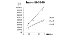
- the cerebrospinal fluid volume-dependent straight line of signal intensity in has-miR-3960 which is a miRNA with high quantification from 0.45 mL of cerebrospinal fluid volume as the supernatant volume excluding the lymphocyte fraction, is high It is drawing shown in 3 healthy persons.
- the signal intensity for each miRNA in 0.9 mL of cerebrospinal fluid was plotted as the amount of supernatant excluding the lymphocyte fraction.
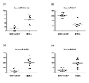
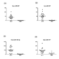
- MdCIDP and (1) hsa-miR-214, (2) hsa-miR-519d, (3) hsa-miR-575, and (4) hsa-miR-30c-2 * in cerebrospinal fluid samples It is drawing which showed distribution of signal intensity of MMN and a healthy person.
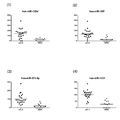
- MdCIDP and MMN for (1) hsa-miR-4323, (2) hsa-miR-4265, (3) hsa-miR-4269, and (4) hsa-miR-3605-3p in cerebrospinal fluid samples It is drawing which showed distribution of signal intensity of a healthy person.
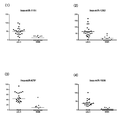
- MdCIDP for cerebrospinal fluid samples (1) hsa-miR-3682-3p, (2) hsa-miR-3918, (3) hsa-miR-3937, and (4) hsa-miR-3940-3p 2 is a signal intensity distribution of MMN and a healthy person.
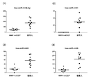
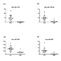
- MdCIDP and MMN for (1) hsa-miR-4446-3p, (2) hsa-miR-4461, (3) hsa-miR-4481 and (4) hsa-miR-4489 in cerebrospinal fluid samples It is drawing which showed distribution of signal intensity of a healthy person.
- MdCIDP and MMN for (1) hsa-miR-2392, (2) hsa-miR-3922-5p, (3) hsa-miR-4634, and (4) hsa-miR-4642 in cerebrospinal fluid samples It is drawing which showed distribution of signal intensity of a healthy person.
- About cerebrospinal fluid samples (1) hsa-miR-4669, (2) hsa-miR-4676-5p, (3) hsa-miR-4691-5p, and (4) hsa-miR-4713-5p , MdCIDP and MMN and a signal intensity distribution of a healthy person.
- hsa-miR-24 About (1) hsa-miR-24, (2) hsa-miR-296-5p, (3) hsa-miR-92a-2 *, and (4) hsa-miR-149 * in cerebrospinal fluid samples 5 is a diagram showing signal intensity distributions of LS, ALS, and mdCIDP or MMN.
- ALS (1) hsa-miR-296-3p, (2) hsa-miR-92b *, (3) hsa-miR-920, and (4) hsa-miR-939 in cerebrospinal fluid samples It is also a drawing showing the signal intensity distribution of mdCIDP or MMN.
- FIG. 6 is a diagram showing a signal intensity distribution of mdCIDP or MMN.
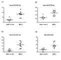
- ALS and mdCIDP for (1) hsa-miR-3917, (2) hsa-miR-3937, (3) hsa-miR-642b, and (4) hsa-miR-4417 in cerebrospinal fluid samples Or it is the figure which showed distribution of the signal strength of MMN.
- ALS and mdCIDP for (1) hsa-miR-4433, (2) hsa-miR-4442, (3) hsa-miR-4447, and (4) hsa-miR-4454 in cerebrospinal fluid samples Or it is the figure which showed distribution of the signal strength of MMN.
- FIG. 6 is a diagram showing a signal intensity distribution of mdCIDP or MMN.
- FIG. 6 is a diagram showing a signal intensity distribution of mdCIDP or MMN. ALS and (1) hsa-miR-4665-5p, (2) hsa-miR-1343, (3) hsa-miR-4688, and (4) hsa-miR-4689 in cerebrospinal fluid samples It is drawing which showed distribution of the signal intensity of mdCIDP or MMN.
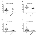
- About cerebrospinal fluid samples (1) hsa-miR-4695-5p, (2) hsa-miR-4697-5p, (3) hsa-miR-4706, and (4) hsa-miR-4723-5p 5 is a diagram showing signal intensity distributions of LS, ALS, and mdCIDP or MMN.
- (1) hsa-miR-4725-3p, (2) hsa-miR-4726-5p, (3) hsa-miR-4728-5p, and (4) hsa-miR-4730 in cerebrospinal fluid samples 5 is a diagram showing signal intensity distributions of LS, ALS, and mdCIDP or MMN.
- FIG. 3 is a diagram schematically illustrating a detection process using these together with sensitivity and specificity.
- FIG. 3 is a diagram schematically illustrating a detection process using these together with sensitivity and specificity.
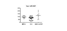
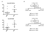
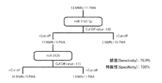
- FIG. (1) Drawing which picked up distribution of signal intensity of hsa-miR-4417 and hsa-miR-4697-5p which are suitable markers for differentiating ALS and mdCIDP or MMN in cerebrospinal fluid samples, and ( 2) A drawing showing the detection process using them together with sensitivity and specificity.
- ALS for cerebrospinal fluid samples (1) hsa-miR-718, (2) hsa-miR-2682 *, (3) hsa-miR-449c *, and (4) hsa-miR-3130-5p It is drawing which showed distribution of the signal intensity of MMN.
- FIG. 3 is a diagram showing signal intensity distributions of ALS and MMN.
- FIG. 4 is a schematic diagram together with (Specificity).
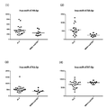
- MMN and PMA for (1) hsa-miR-99a, (2) hsa-miR-23b, (3) hsa-miR-184, and (4) hsa-miR-485-3p in cerebrospinal fluid samples It is drawing which showed distribution of signal intensity.
- hsa-miR-486-5p, (2) hsa-miR-432 *, (3) hsa-miR-675, and (4) hsa-miR-25 * in cerebrospinal fluid samples 2 is a graph showing the signal intensity distribution of PMA and PMA.
- MMN and PMA for (1) hsa-miR-711, (2) hsa-miR-3138, (3) hsa-miR-3162-5p, and (4) hsa-miR-3176 in cerebrospinal fluid samples It is drawing which showed distribution of signal intensity.
- hsa-miR-3187-3p Regarding (1) hsa-miR-3188, (3) hsa-miR-3197, and (4) hsa-miR-514b-5p in cerebrospinal fluid samples 2 is a graph showing the signal intensity distribution of PMA and PMA.
- MMN for cerebrospinal fluid samples (1) hsa-miR-3689a-3p, (2) hsa-miR-3180, (3) hsa-miR-3919, and (4) hsa-miR-3925-5p 2 is a graph showing the signal intensity distribution of PMA and PMA.
- MMN and PMA signals for (1) hsa-miR-3928, (2) hsa-miR-4425, (3) hsa-miR-4437, and (4) hsa-miR-4450 in cerebrospinal fluid samples It is drawing which showed distribution of intensity.
- MMN and PMA signals for (1) hsa-miR-4461, (2) hsa-miR-4465, (3) hsa-miR-4478, and (4) hsa-miR-2392 in cerebrospinal fluid samples It is drawing which showed distribution of intensity.
- MMN for (1) hsa-miR-4526, (2) hsa-miR-3619-3p, (3) hsa-miR-3940-5p, and (4) hsa-miR-4635 in cerebrospinal fluid samples 2 is a graph showing the signal intensity distribution of PMA and PMA.
- FIG. 3 is a graph showing signal intensity distributions of MMN and PMA.
- About cerebrospinal fluid samples (1) hsa-miR-4779, (2) hsa-miR-2467-5p, (3) hsa-miR-4787-3p, and (4) hsa-miR-4800-5p
- FIG. 3 is a graph showing signal intensity distributions of MMN and PMA.
- FIG. 3 is a diagram schematically illustrating a detection process using these together with sensitivity and specificity.
- a schematic diagram For each of hsa-miR-3162-5p and hsa-miR-3928, which are suitable markers for differentiating MMN and PMA in cerebrospinal fluid specimens, (1) a drawing picking up the signal intensity distribution, and (2) FIG. 3 is a diagram schematically illustrating a detection process using these together with sensitivity and specificity.
- miRNA is quantified in a CSF sample of a donor obtained by collecting and preparing cerebrospinal fluid according to the above-described conventional method, and the quantified value is lower or higher than the comparison target.
- the indicated signal that is, by detecting the increase or decrease in the amount of miRNA, a desired differentiation index can be obtained.
- the method for quantifying miRNA is not particularly limited, and for example, existing means based on the above-mentioned individual miRNA base sequences, and further developed means can be used.
- use of the “microRNA detection array” in which miRNAs are comprehensively used, which is used in the standardization method of the present invention described above, can be mentioned.
- RNA quantification methods based on gene amplification methods such as the RT-PCR method can be mentioned.
- a real-time RT-PCR method using an automated detection device is preferable from the viewpoint of mass processing and rapidity.
- the Northern blot method etc. are mentioned.
- the quantitative value based on the quantitative signal of each miRNA is relatively lower or higher than the comparison target (for example, a healthy person or a different motor neuropathy disease to be compared).
- the quantitative value can be treated as “low positive” or “high positive”.
- Specific threshold values can be set individually and specifically.
- a decrease in the amount of hsa-miR-744 or a decrease in the amount of hsa-miR-4505 in a cerebrospinal fluid sample can be detected as a differentiation index for ALS, mdCIDP, or MMN .
- the detection method of the present invention of this aspect is particularly useful when it is intended to grasp a general tendency of a motor neuropathy disease in a subject.
- the quantitative value of hsa-miR-744 or hsa-miR-4505 in the cerebrospinal fluid specimen decreases. Therefore, when it is observed that the quantitative value of hsa-miR-744 or hsa-miR-4505 in the cerebrospinal fluid sample of the subject is reduced from the standard for healthy subjects, the subject is ALS, mdCIDP, or Therefore, the possibility of suffering from MMN increases, and the direction of subsequent disease discrimination can be narrowed down.
- a detection method of a different aspect described later is provided.
- ALS differentiation indicators Upon differentiation from ALS healthy individuals, one or more selected from the changes in the amount of microRNA in the cerebrospinal fluid samples based on healthy individuals within the following “” are detected as ALS differentiation indicators. can do.
- the above-mentioned “decrease in the amount of hsa-miR-744” or “decrease in the amount of hsa-miR-4505” is also included as a differential indicator of this embodiment, but when this differential indicator is used, it is listed here. It is preferable to use a combination of other indices.
- hsa-miR-10a an increase in the amount of hsa-miR-10a, an increase in the amount of hsa-miR-516b, an increase in the amount of hsa-miR-122 *, and an increase in hsa-miR-4762-3p.
- Changes in the amount of one or more selected microRNAs can be used as a differentiation index of this embodiment.
- the detection method of this preferred embodiment is also advantageous in that a desired differentiation target can be detected with higher sensitivity and specificity.
- the above-mentioned miRNA values in the cerebrospinal fluid specimen show the characteristic increases and decreases listed respectively. Therefore, if the quantitative values of these miRNAs in the cerebrospinal fluid sample of the subject are found to show the listed increases and decreases in comparison with the standard for healthy subjects, the subject may be affected with ALS. Is found to be high.
- mdCIDP or MMN When differentiating mdCIDP or MMN from a healthy person, mdCIDP or an index of one or more selected from changes in the amount of microRNA in the cerebrospinal fluid sample based on the healthy person within the following “” It can be detected as a differentiation index of MMN.
- the above “decrease in the amount of hsa-miR-744” or “decrease in the amount of hsa-miR-4505” is also included as an indicator of this embodiment, but when this indicator is used in this embodiment, it is listed here. It is preferable to use a combination of other indices.
- Hsa-miR-214 decreased, hsa-miR-519d decreased, hsa-miR-575 decreased, hsa-miR-30c-2 * decreased, hsa-miR-135a * decreased, decrease in hsa-miR-185 * amount, decrease in hsa-miR-423-5p amount, decrease in hsa-miR-92b * amount, decrease in hsa-miR-551b * amount, decrease in hsa-miR-550a amount, hsa-miR-885-3p decrease, hsa-miR-936 decrease, hsa-miR-940 decrease, hsa-miR-1225-3p decrease, hsa-miR-1233 decrease, hsa -MiR-1181 decreased, hsa-miR-1268 increased, hsa-miR-1908 decreased, hsa-miR-2276
- an increase in the amount of hsa-miR-4417 can be used as a differentiation index of this embodiment.
- the detection method of this preferred embodiment is also advantageous in that a desired differentiation target can be detected with higher sensitivity and specificity.
- the above miRNA values in the cerebrospinal fluid specimens each show a characteristic increase or decrease listed.
- the subject is afflicted with mdCIDP or MMN It is recognized that the possibility is high. It should be noted that the discrimination between mdCIDP and MMN can be performed according to a method conventionally used by those skilled in the art.
- an increase in the amount of hsa-miR-642b from the group consisting of an increase in the amount of hsa-miR-642b, an increase in the amount of hsa-miR-3197, an increase in the amount of hsa-miR-4697-5p, and a decrease in the amount of hsa-miR-4417.
- One type or two or more types selected (however, the decrease in the amount of hsa-miR-4417 is a combination with one to three types selected from the increase or decrease in the amount of the other three microRNAs) It can be detected as a differentiation index.
- the detection method according to this aspect greatly assists in distinguishing between ALS that is difficult to distinguish between early stages of disease and mdCIDP or MMN.
- the above-mentioned miRNA quantitative values in cerebrospinal fluid samples based on “mdCIDP or MMN” show the characteristic increases and decreases listed respectively.
- the quantified value of these miRNAs in a subject's cerebrospinal fluid sample is observed to show the listed increase or decrease in comparison to the “mdCIDP or MMN” standard, the subject is identified as “mdCIDP or MMN”. Instead, it is recognized that there is a high possibility of suffering from “ALS”.
- the discrimination between mdCIDP and MMN can be performed according to a method conventionally performed by those skilled in the art, as in the above-described discrimination from healthy individuals of “mdCIDP or MMN”.
- the aspect disclosed here is the amount change toward “ALS” based on “mdCIDP or MMN”, this is only relative. That is, conversely, the reference can be set to “ALS” and equivalently replaced with the amount change toward “mdCIDP or MMN”.
- the “increase” of the quantitative value disclosed herein is “decrease”, “decrease” is “increase”, and “MDCIDP or MMN” is a differentiation index based on “ALS”. Is provided.
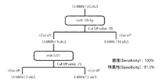
- More preferred embodiments include increased hsa-miR-139-3p, increased hsa-miR-574-5p, increased hsa-miR-1231, increased hsa-miR-2682 *, and hsa-miR-3130
- One or more changes in the amount of microRNA selected from the group consisting of an increase of ⁇ 5p can be detected as a differential indicator of ALS of this embodiment.
- any one selected from the group consisting of a combination of an increase in hsa-miR-2682 * and an increase in hsa-miR-3130-5p is extremely suitable for detection as an ALS differentiation index. is there.
- the detection methods of these preferred embodiments are advantageous in that a desired differential target can be detected with higher sensitivity and higher specificity.
- the detection method of this embodiment is a great help for differentiating between ALS and MMN, which is particularly difficult to identify each other in the early stages of disease.
- the above-mentioned miRNA quantitative values in the cerebrospinal fluid specimens based on “MMN” each show a characteristic increase listed.
- a quantitative value of these miRNAs in a subject's cerebrospinal fluid sample is shown to show the listed increase in comparison to the “MMN” standard, the subject is not “MMN” but “ It is recognized that there is a high probability of suffering from “ALS”.
- the aspect disclosed here is the amount change toward “ALS” based on “MMN”, this is only relative. That is, conversely, the reference can be set to “ALS” and equivalently replaced with the amount change toward “MMN”.
- the “increase” of the quantitative value disclosed herein is “decrease”, and a discrimination index of “MMN” based on “ALS” is provided.
- More preferred embodiments include an increase in hsa-miR-1275, an increase in hsa-miR-711, an increase in hsa-miR-3138, an increase in hsa-miR-3162-5p, an increase in hsa-miR-3928, and One type or two or more types of changes in the amount of microRNA selected from the group consisting of an increase in hsa-miR-1914 * can be detected as a differential indicator of PMA of this embodiment.
- the detection method of the present embodiment is a great help for differentiating between MMN and PMA, which is particularly difficult to identify each other in the early stages of disease.
- the above-mentioned miRNA quantitative values in the cerebrospinal fluid specimens based on “MMN” each show a characteristic increase listed.
- a quantitative value of these miRNAs in a subject's cerebrospinal fluid sample is shown to show the listed increase in comparison to the “MMN” standard, the subject is not “MMN” but “ It is recognized that the possibility of having “PMA” is high.
- the aspect disclosed here is the amount change toward “PMA” based on “MMN”, this is only relative. That is, conversely, in “PMA”, the reference can be equivalently replaced with a change in quantity toward “MMN”.
- the “increase” of the quantitative value disclosed herein is “decrease”, and a differentiation index of “MMN” based on “PMA” is provided.
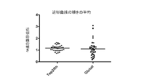
- the miRNA in the cerebrospinal fluid which originally has a small amount of miRNA, is corrected by the Global Normalization method (a correction method that sets the median when the signal intensities are arranged in order) to 25, the median is selected. There is a possibility that even if the miRNA is normal, the one whose signal intensity varies considerably will be selected.
- the Global Normalization method a correction method that sets the median when the signal intensities are arranged in order
- the present inventor found that when comparing the signal intensities of miRNA groups of individual samples under the assumption that “the signal values of miRNA groups in the comprehensive analysis of normal cerebrospinal fluid are not significantly different among individuals” as a whole.
- the fluorescence intensity recognized in the top 20 miRNA is replaced with a specific number as a signal value, and the multiplier used in the multiplication for performing this replacement is calculated with respect to the fluorescence intensity recognized in other miRNAs.
- the above 41 miRNA groups were normalized.
- FIG. 3 shows a signal for each miRNA of 0.9 mL of cerebrospinal fluid as a supernatant amount excluding the lymphocyte fraction between the conventional method (Global normalization method) and the normalization method of the present invention (Top20th normalization method). It is drawing which showed distribution of the inclination between two normal cerebrospinal fluid samples which plotted the intensity
- the signal used this time is “Cy (Cyanine) bound to the test miRNA” as described above.
- the fluorescence intensity of the fluorescent dye is the signal value of this example.
- RNA array 3D GENE (registered trademark) / TORAY)” (Toray) is used as an array for miRNA detection, and all the miRNAs mounted on the array are used as a population.
- miRNA in cerebrospinal fluid that can be used as a marker for three categories of diseases (ALS, MMN, and mdCIDP) was selected.
- sorting method the Welch method based on the null assumption was used, and the significance level was sorted at 5%.
- the breakdown of cerebrospinal fluid samples used for selecting each category is “Healthy person: 10 samples, ALS: 23 samples, mdCIDP: 5 samples, MMN: 5 samples (mdCIDP / MMN: 10 samples)”. . However, in selecting an index for distinguishing ALS from a healthy person, since cerebrospinal fluid in which blood was mixed was excluded, “healthy person: 9 specimens, ALS: 19 specimens”.
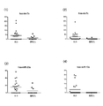
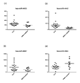
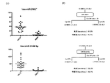
- FIG. 4 shows healthy subjects for (1) hsa-miR-744 and (2) hsa-miR-4505 in cerebrospinal fluid samples.
- 2 is a diagram showing signal intensity distributions of ALS, ALS, and mdCIDP and MMN.
- the vertical axis of the graph is the signal intensity
- the horizontal line in the graph is the average value of each group.
- the signal of the disease group was significantly decreased between the healthy subject and ALS, and between the healthy subject and mdCIDP / MMN.
- hsa-miR-744 and hsa-miR-4505 are particularly useful when it is intended to grasp the general tendency of motor neuropathy disorders in subjects.
- Hsa-miR-744 and hsa-miR-4505 can be used in combination with other markers that can distinguish these diseases, which will be described later. Is preferred.
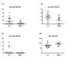
- FIG. 5 shows the signal intensity of a marker that provides a differentiation index between ALS and healthy individuals. Distribution is shown.
- FIG. 5-1 shows (1) hsa-let-7a, (2) hsa-let-7c, (3) hsa-miR-23a, and (4) hsa-miR- About 10a, the distribution of the signal intensity of ALS and a healthy person is shown.
- the signal intensity of hsa-let-7a was significantly increased in ALS in healthy individuals.
- the signal intensity of hsa-let-7c was significantly increased in ALS in healthy individuals.
- the signal intensity of hsa-miR-23a was significantly increased in ALS compared with healthy individuals.
- the signal intensity of hsa-miR-10a was significantly increased in ALS in healthy individuals.
- these miRNAs present in the cerebrospinal fluid are markers that provide a differentiation index between healthy individuals and ALS.
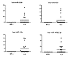
- FIG. 5-2 shows (1) hsa-miR-200a, (2) hsa-miR-524-5p, (3) hsa-miR-516b, and (4) hsa- About miR-552, the distribution of signal intensity between ALS and healthy individuals is shown.
- the signal intensity of hsa-miR-200a was significantly increased in ALS compared with healthy individuals.
- the signal intensity of hsa-miR-524-5p was significantly reduced in ALS in healthy individuals.
- the signal intensity of hsa-miR-516b was significantly increased in ALS compared with healthy individuals.
- the signal intensity of hsa-miR-552 was significantly increased in ALS in healthy individuals.
- these miRNAs present in the cerebrospinal fluid are markers that provide a differentiation index between healthy individuals and ALS.
- Fig. 5-3 shows (1) hsa-miR-573, (2) hsa-miR-644, (3) hsa-miR-24-2 *, and (4) hsa in cerebrospinal fluid samples.
- -MiR-122 * shows the distribution of signal intensity between ALS and healthy individuals.
- the signal intensity of hsa-miR-573 was significantly increased in ALS in healthy individuals.
- the signal intensity of hsa-miR-644 was significantly increased in ALS in healthy individuals.
- the signal intensity of hsa-miR-24-2 * was significantly increased in ALS compared with healthy individuals.
- the signal intensity of hsa-miR-122 * was significantly increased in ALS compared with healthy individuals.
- these miRNAs present in the cerebrospinal fluid are markers that provide a differentiation index between healthy individuals and ALS.
- Fig. 5-4 shows (1) hsa-miR-145 *, (2) hsa-miR-361-3p, (3) hsa-miR-1291, and (4) hsa in cerebrospinal fluid samples.
- -miR-1260 shows the distribution of signal intensity between ALS and healthy individuals. (1) The signal intensity of hsa-miR-145 * was significantly increased in ALS compared with healthy individuals. (2) The signal intensity of hsa-miR-361-3p was significantly increased in ALS compared with healthy individuals. (3) The signal intensity of hsa-miR-1291 was significantly increased in ALS in healthy individuals. (4) The signal intensity of hsa-miR-1260 was significantly reduced in ALS in healthy individuals.
- these miRNAs present in the cerebrospinal fluid are markers that provide a differentiation index between healthy individuals and ALS.
- Fig. 5-5 shows (1) hsa-miR-1273, (2) hsa-miR-3161, (3) hsa-miR-4310, and (4) hsa-miR- About 4313, the distribution of the signal intensity of ALS and a healthy person is shown.
- the signal intensity of hsa-miR-1273 was significantly reduced in ALS in healthy individuals.
- the signal intensity of hsa-miR-3161 was significantly increased in ALS compared with healthy individuals.
- the signal intensity of hsa-miR-4310 was significantly increased in ALS in healthy individuals.
- the signal intensity of hsa-miR-4313 was significantly reduced in ALS in healthy individuals.
- these miRNAs present in the cerebrospinal fluid are markers that provide a differentiation index between healthy individuals and ALS.
- Figure 5-6 shows (1) hsa-miR-4479, (2) hsa-miR-4493, (3) hsa-miR-4524, and (4) hsa-miR- 3688-5p shows the distribution of signal intensity between ALS and healthy individuals.
- the signal intensity of hsa-miR-4479 was significantly increased in ALS in healthy individuals.
- the signal intensity of hsa-miR-4493 was significantly reduced in ALS in healthy individuals.
- the signal intensity of hsa-miR-4524 was significantly increased in ALS in healthy individuals.
- the signal intensity of hsa-miR-3688-5p was significantly increased in ALS compared with healthy individuals.
- these miRNAs present in the cerebrospinal fluid are markers that provide a differentiation index between healthy individuals and ALS.
- FIG. 5-7 shows (1) hsa-miR-4633-5p, (2) hsa-miR-4708-5p, (3) hsa-miR-3545-3p, and ( 4) About hsa-miR-4741, distribution of signal intensity of ALS and healthy persons is shown.
- the signal intensity of hsa-miR-4633-5p was significantly increased in ALS compared with healthy individuals.
- the signal intensity of hsa-miR-4708-5p was significantly increased in ALS compared with healthy individuals.
- the signal intensity of hsa-miR-3545-3p was significantly increased in ALS compared with healthy individuals.
- the signal intensity of hsa-miR-4741 was significantly reduced in ALS compared with healthy individuals.
- these miRNAs present in the cerebrospinal fluid are markers that provide a differentiation index between healthy individuals and ALS.
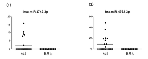
- Fig. 5-8 shows the distribution of signal intensity between ALS and healthy individuals for (1) hsa-miR-4742-3p and (2) hsa-miR-4762-3p in cerebrospinal fluid samples. ing. (1) The signal intensity of hsa-miR-4742-3p was significantly increased in ALS compared with healthy individuals. (2) The signal intensity of hsa-miR-4762-3p was significantly increased in ALS compared with healthy individuals.
- these miRNAs present in the cerebrospinal fluid are markers that provide a differentiation index between healthy individuals and ALS.
- FIG. 6 shows the signal intensity distribution of these four types of markers.
- FIG. 7 is a diagram schematically illustrating a detection process using these four kinds of markers in a cerebrospinal fluid specimen together with its sensitivity (Sensitivity) and specificity (Specificity). As a result, in these markers, when any one of the above four types was detected as a positive signal, it was judged as positive, and thus an excellent discrimination result with a sensitivity of 89.5% and a specificity of 100%. Was recognized
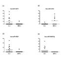
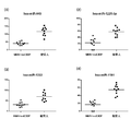
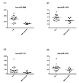
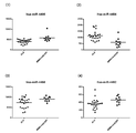
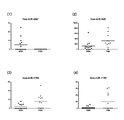
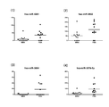
- FIG. 8 shows a marker that provides a differentiation index between mdCIDP or MMN and a healthy person The signal intensity distribution is shown.
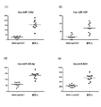
- FIG. 8-1 shows (1) hsa-miR-214, (2) hsa-miR-519d, (3) hsa-miR-575, and (4) hsa-miR- For 30c-2 *, the distribution of signal intensity of mdCIDP and MMN and healthy persons is shown.
- the signal intensity of hsa-miR-214 was significantly reduced in mdCIDP / MMN for healthy individuals.
- the signal intensity of hsa-miR-519d was significantly decreased in mdCIDP / MMN for healthy individuals.
- the signal intensity of hsa-miR-575 was significantly reduced in mdCIDP / MMN for healthy individuals.
- the signal intensity of hsa-miR-30c-2 * was significantly decreased in mdCIDP / MMN with respect to healthy individuals.
- these miRNAs present in the cerebrospinal fluid are markers that provide a differentiation index between healthy people and CIDP or MMN.
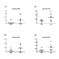
- FIG. 8-2 shows (1) hsa-miR-135a *, (2) hsa-miR-185 *, (3) hsa-miR-423-5p, and (4) in the cerebrospinal fluid specimen.
- hsa-miR-92b * the signal intensity distribution of mdCIDP and MMN and a healthy person is shown.
- the signal intensity of hsa-miR-135a * was significantly decreased in mdCIDP / MMN in healthy subjects.
- the signal intensity of hsa-miR-185 * was significantly decreased in mdCIDP / MMN for healthy individuals.
- hsa-miR-423-5p showed a significant decrease in signal intensity in mdCIDP / MMN compared to healthy individuals.
- the signal intensity of hsa-miR-92b * was significantly decreased in mdCIDP / MMN for healthy individuals.
- these miRNAs present in the cerebrospinal fluid are markers that provide a differentiation index between healthy people and mdCIDP or MMN.
- FIG. 8-3 shows (1) hsa-miR-551b *, (2) hsa-miR-550a, (3) hsa-miR-885-3p, and (4) hsa in the cerebrospinal fluid sample.
- -miR-936 the signal intensity distributions of mdCIDP and MMN and healthy individuals are shown.
- the signal intensity of hsa-miR-551b * was significantly decreased in mdCIDP / MMN in healthy subjects.
- the signal intensity of hsa-miR-550a was significantly decreased in mdCIDP / MMN for healthy individuals.
- these miRNAs present in the cerebrospinal fluid are markers that provide a differentiation index between healthy people and CIDP or MMN.
- FIG. 8-4 shows (1) hsa-miR-940, (2) hsa-miR-1225-3p, (3) hsa-miR-1233, and (4) hsa- About miR-1181, the distribution of signal intensity of mdCIDP and MMN and healthy persons is shown.
- the signal intensity of hsa-miR-940 was significantly reduced in mdCIDP / MMN for healthy individuals.
- the signal intensity of hsa-miR-1225-3p was significantly reduced in mdCIDP / MMN in healthy subjects.
- the signal intensity of hsa-miR-1233 was significantly reduced in mdCIDP / MMN for healthy individuals.
- the signal intensity of hsa-miR-1181 was significantly decreased in mdCIDP / MMN for healthy individuals.
- these miRNAs present in the cerebrospinal fluid are markers that provide a differentiation index between healthy people and mdCIDP or MMN.
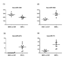
- Fig. 8-5 shows (1) hsa-miR-1268, (2) hsa-miR-1908, (3) hsa-miR-2276, and (4) hsa-miR- About 711, the distribution of the signal intensity of mdCIDP and MMN and a healthy person is shown.
- the signal intensity of hsa-miR-1268 was significantly increased in mdCIDP / MMN for healthy individuals.
- the signal intensity of hsa-miR-1908 was significantly reduced in mdCIDP / MMN for healthy individuals.
- the signal intensity of hsa-miR-2276 was significantly reduced in mdCIDP / MMN for healthy individuals.
- the signal intensity of hsa-miR-711 was significantly decreased in mdCIDP / MMN for healthy individuals.
- these miRNAs present in the cerebrospinal fluid are markers that provide a differentiation index between healthy people and mdCIDP or MMN.
- Figure 8-6 shows (1) hsa-miR-718, (2) hsa-miR-1193, (3) hsa-miR-3184, and (4) hsa-miR- About 3197, the distribution of the signal intensity of mdCIDP and MMN and a healthy person is shown.
- the signal intensity of hsa-miR-718 was significantly reduced in mdCIDP / MMN for healthy individuals.
- the signal intensity of hsa-miR-1193 was significantly reduced in mdCIDP / MMN for healthy individuals.
- the signal intensity of hsa-miR-3184 was significantly decreased in mdCIDP / MMN for healthy individuals.
- the signal intensity of hsa-miR-3197 was significantly reduced in mdCIDP / MMN for healthy individuals.
- these miRNAs present in the cerebrospinal fluid are markers that provide a differentiation index between healthy people and mdCIDP or MMN.
- Fig. 8-7 shows (1) hsa-miR-4323, (2) hsa-miR-4265, (3) hsa-miR-4269, and (4) hsa-miR- About 3605-3p, the distribution of the signal intensity of mdCIDP and MMN and a healthy person is shown.
- the signal intensity of hsa-miR-4323 was significantly reduced in mdCIDP / MMN for healthy individuals.
- the signal intensity of hsa-miR-4265 was significantly reduced in mdCIDP / MMN for healthy individuals.
- these miRNAs present in the cerebrospinal fluid are markers that provide a differentiation index between healthy people and mdCIDP or MMN.
- FIGS. 8-8 show (1) hsa-miR-3648, (2) hsa-miR-3663-5p, (3) hsa-miR-3667-3p, and (4) in the cerebrospinal fluid specimen.
- hsa-miR-3679-5p the signal intensity distribution of mdCIDP and MMN and a healthy person is shown.
- the signal intensity of hsa-miR-3648 was significantly reduced in mdCIDP / MMN for healthy individuals.
- the signal intensity of hsa-miR-3663-5p was significantly reduced in mdCIDP / MMN compared to healthy individuals.
- these miRNAs present in the cerebrospinal fluid are markers that provide a differentiation index between healthy people and mdCIDP or MMN.
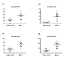
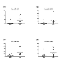
- FIGS. 8-9 show (1) hsa-miR-3682-3p, (2) hsa-miR-3918, (3) hsa-miR-3937, and (4) hsa- About miR-3940-3p, the distribution of the signal intensity of mdCIDP and MMN and a healthy person is shown.
- the signal intensity of hsa-miR-3682-3p was significantly decreased in mdCIDP / MMN compared to healthy individuals.
- the signal intensity of hsa-miR-3918 was significantly reduced in mdCIDP / MMN for healthy individuals.
- these miRNAs present in the cerebrospinal fluid are markers that provide a differentiation index between healthy people and mdCIDP or MMN.
- FIGS. 8-10 show (1) hsa-miR-3944-3p, (2) hsa-miR-4417, (3) hsa-miR-4430, and (4) hsa- About miR-4440, the distribution of the signal intensity of mdCIDP and MMN and healthy persons is shown.
- the signal intensity of hsa-miR-3944-3p was significantly reduced in mdCIDP / MMN in healthy subjects.
- the signal intensity of hsa-miR-4417 was significantly increased in mdCIDP / MMN for healthy individuals.
- the signal intensity of hsa-miR-4430 was significantly reduced in mdCIDP / MMN for healthy individuals.
- the signal intensity of hsa-miR-4440 was significantly reduced in mdCIDP / MMN for healthy individuals.
- these miRNAs present in the cerebrospinal fluid are markers that provide a differentiation index between healthy people and mdCIDP or MMN.
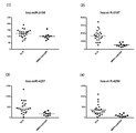
- FIGS. 8-11 show (1) hsa-miR-4446-3p, (2) hsa-miR-4461, (3) hsa-miR-4481, and (4) hsa- About miR-4489, the distribution of signal intensity of mdCIDP and MMN and healthy persons is shown.
- the signal intensity of hsa-miR-4446-3p was significantly reduced in mdCIDP / MMN compared to healthy individuals.
- the signal intensity of hsa-miR-4461 was significantly reduced in mdCIDP / MMN for healthy individuals.
- the signal intensity of hsa-miR-4481 was significantly decreased in mdCIDP / MMN for healthy individuals.
- the signal intensity of hsa-miR-4489 was significantly reduced in mdCIDP / MMN for healthy individuals.
- these miRNAs present in the cerebrospinal fluid are markers that provide a differentiation index between healthy people and mdCIDP or MMN.
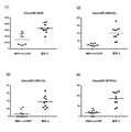
- FIGS. 8-12 show (1) hsa-miR-2392, (2) hsa-miR-3922-5p, (3) hsa-miR-4634, and (4) hsa- About miR-4642, the distribution of the signal intensity of mdCIDP and MMN and healthy persons is shown.
- the signal intensity of hsa-miR-2392 was significantly reduced in mdCIDP / MMN for healthy individuals.
- the signal intensity of hsa-miR-3922-5p was significantly reduced in mdCIDP / MMN compared to healthy individuals.
- these miRNAs present in the cerebrospinal fluid are markers that provide a differentiation index between healthy people and mdCIDP or MMN.
- FIGS. 8-13 show (1) hsa-miR-4669, (2) hsa-miR-4676-5p, (3) hsa-miR-4691-5p, and (4) in the cerebrospinal fluid specimen.
- hsa-miR-4713-5p the signal intensity distribution of mdCIDP and MMN and a healthy person is shown.
- the signal intensity of hsa-miR-4669 was significantly reduced in mdCIDP / MMN for healthy individuals.
- the signal intensity of hsa-miR-4676-5p was significantly reduced in mdCIDP / MMN compared to healthy individuals.
- these miRNAs present in the cerebrospinal fluid are markers that provide a differentiation index between healthy people and mdCIDP or MMN.
- FIGS. 8-14 show (1) hsa-miR-4722-3p, (2) hsa-miR-4725-3p, (3) hsa-miR-4727-3p, and ( 4) About hsa-miR-4736, the distribution of signal intensity of mdCIDP and MMN and healthy persons is shown.
- the signal intensity of hsa-miR-4722-3p was significantly decreased in mdCIDP / MMN compared to healthy individuals.
- the signal intensity of hsa-miR-4725-3p was significantly reduced in mdCIDP / MMN for healthy subjects.
- these miRNAs present in the cerebrospinal fluid are markers that provide a differentiation index between healthy people and mdCIDP or MMN.
- Figure 8-15 shows the distribution of signal intensity of mdCIDP and MMN and healthy individuals for (1) hsa-miR-371b-3p and (2) hsa-miR-2467-3p in cerebrospinal fluid samples Is shown.
- the signal intensity of hsa-miR-371b-3p was significantly decreased in mdCIDP / MMN with respect to healthy subjects.
- the signal intensity of hsa-miR-2467-3p was significantly reduced in mdCIDP / MMN compared to healthy individuals.
- these miRNAs present in the cerebrospinal fluid are markers that provide a differentiation index between healthy people and mdCIDP or MMN.
- FIG. 9 is a suitable marker for distinguishing healthy person from mdCIDP and MMN in cerebrospinal fluid specimen.
- -miR-4717 (1) Drawing which picked up signal intensity distribution, (2) ROC curve (receiver operating characteristic curve), and (3) Detection process using the manufacturer
- FIG. 2 is a diagram schematically showing sensitivity and specificity. FIG. As a result, the marker was found to have excellent discrimination results with a sensitivity of 80.0% and a specificity of 90.0%.
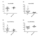
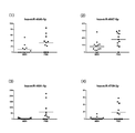
- FIG. 10 shows ALS and mdCIDP or MMN alternatives.
- 2 shows the distribution of signal intensity of markers that provide a differential indicator.
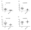
- FIG. 10-1 shows (1) hsa-miR-24, (2) hsa-miR-296-5p, and (3) hsa-miR-92a-2 in the cerebrospinal fluid sample.
- * And (4) hsa-miR-149 * show the signal intensity distribution of ALS and mdCIDP or MMN.
- the signal intensity of ALS is expressed by an increase / decrease based on the signal intensity of mdCIDP or MMN (the same applies to the description of the series of FIG. 10 below).
- (1) The signal intensity of hsa-miR-24 was significantly increased in ALS.
- the signal intensity of hsa-miR-296-5p was significantly increased in ALS.
- the signal intensity of hsa-miR-92a-2 * was significantly reduced in ALS.
- the signal intensity of hsa-miR-149 * was significantly increased in ALS.
- these miRNAs present in the cerebrospinal fluid are markers that give an alternative differentiation index of ALS and mdCIDP or MMN.
- FIG. 10-2 shows (1) hsa-miR-296-3p, (2) hsa-miR-92b *, (3) hsa-miR-920, and (4) hsa in the cerebrospinal fluid specimen.
- -miR-939 the signal intensity distribution of ALS and mdCIDP or MMN is shown.
- the signal intensity of hsa-miR-296-3p was significantly increased in ALS.
- the signal intensity of hsa-miR-92b * was significantly increased in ALS.
- the signal intensity of hsa-miR-920 was significantly increased in ALS.
- the signal intensity of hsa-miR-939 was significantly reduced in ALS.
- these miRNAs present in the cerebrospinal fluid are markers that give an alternative differentiation index of ALS and mdCIDP or MMN.
- FIG. 10-3 shows (1) hsa-miR-1225-5p, (2) hsa-miR-1228 *, (3) hsa-miR-1202, and (4) hsa in the cerebrospinal fluid sample.
- -miR-1207-5p the signal intensity distribution of ALS and mdCIDP or MMN is shown.
- the signal intensity of hsa-miR-1225-5p was significantly reduced in ALS.
- the signal intensity of hsa-miR-1228 * was significantly increased in ALS.
- the signal intensity of hsa-miR-1202 was significantly increased in ALS.
- the signal intensity of hsa-miR-1207-5p was significantly increased in ALS.
- these miRNAs present in the cerebrospinal fluid are markers that give an alternative differentiation index of ALS and mdCIDP or MMN.
- FIG. 10-4 shows (1) hsa-miR-1908, (2) hsa-miR-1909, (3) hsa-miR-3131, and (4) hsa-miR- 3141 shows the distribution of signal intensity of ALS and mdCIDP or MMN.
- the signal intensity of hsa-miR-1908 was significantly increased in ALS.
- the signal intensity of hsa-miR-1909 was significantly increased in ALS.
- the signal intensity of hsa-miR-3131 was significantly increased in ALS.
- the signal intensity of hsa-miR-3141 was significantly increased in ALS.
- these miRNAs present in the cerebrospinal fluid are markers that give an alternative differentiation index of ALS and mdCIDP or MMN.
- FIG. 10-5 shows (1) hsa-miR-1260b, (2) hsa-miR-3178, (3) hsa-miR-3180-3p, and (4) hsa- For miR-3185, the signal intensity distribution of ALS and mdCIDP or MMN is shown.
- the signal intensity of hsa-miR-1260b was significantly reduced in ALS.
- the signal intensity of hsa-miR-3178 was significantly increased in ALS.
- the signal intensity of hsa-miR-3180-3p was significantly increased in ALS.
- the signal intensity of hsa-miR-3185 was significantly increased in ALS.
- these miRNAs present in the cerebrospinal fluid are markers that give an alternative differentiation index of ALS and mdCIDP or MMN.
- FIG. 10-6 shows (1) hsa-miR-3195, (2) hsa-miR-3197, (3) hsa-miR-4257, and (4) hsa-miR- 4259 shows the distribution of signal intensity of ALS and mdCIDP or MMN.
- the signal intensity of hsa-miR-3195 was significantly increased in ALS.
- the signal intensity of hsa-miR-3197 was significantly increased in ALS.
- the signal intensity of hsa-miR-4257 was significantly increased in ALS.
- the signal intensity of hsa-miR-4259 was significantly increased in ALS.
- these miRNAs present in the cerebrospinal fluid are markers that give an alternative differentiation index of ALS and mdCIDP or MMN.
- Fig. 10-7 shows (1) hsa-miR-4327, (2) hsa-miR-4270, (3) hsa-miR-4281, and (4) hsa-miR- 3621 shows the distribution of the signal intensity of ALS and mdCIDP or MMN.
- the signal intensity of hsa-miR-4327 was significantly increased in ALS.
- the signal intensity of hsa-miR-4270 increased significantly in ALS.
- the signal intensity of hsa-miR-4281 increased significantly in ALS.
- the signal intensity of hsa-miR-3621 was significantly reduced in ALS.
- these miRNAs present in the cerebrospinal fluid are markers that give an alternative differentiation index of ALS and mdCIDP or MMN.
- FIG. 10-8 shows (1) hsa-miR-3622a-5p, (2) hsa-miR-3648, (3) hsa-miR-3656, and (4) hsa- About miR-3663-3p, the signal intensity distribution of ALS and mdCIDP or MMN is shown.
- the signal intensity of hsa-miR-3622a-5p was significantly increased in ALS.
- the signal intensity of hsa-miR-3648 was significantly increased in ALS.
- the signal intensity of hsa-miR-3656 was significantly reduced in ALS.
- the signal intensity of hsa-miR-3663-3p was significantly increased in ALS.
- these miRNAs present in the cerebrospinal fluid are markers that give an alternative differentiation index of ALS and mdCIDP or MMN.
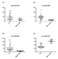
- FIGS. 10-9 show (1) hsa-miR-3917, (2) hsa-miR-3937, (3) hsa-miR-642b, and (4) hsa-miR- For 4417, the distribution of the signal intensity of ALS and mdCIDP or MMN is shown.
- the signal intensity of hsa-miR-3917 was significantly increased in ALS.
- the signal intensity of hsa-miR-3937 was significantly increased in ALS.
- the signal intensity of hsa-miR-642b was significantly increased in ALS.
- the signal intensity of hsa-miR-4417 was significantly reduced in ALS.
- these miRNAs present in the cerebrospinal fluid are markers that give an alternative differentiation index of ALS and mdCIDP or MMN.
- FIGS. 10-10 show (1) hsa-miR-4433, (2) hsa-miR-4442, (3) hsa-miR-4447, and (4) hsa-miR- For 4454, the distribution of the signal intensity of ALS and mdCIDP or MMN is shown.
- the signal intensity of hsa-miR-4433 was significantly increased in ALS.
- the signal intensity of hsa-miR-4442 was significantly increased in ALS.
- the signal intensity of hsa-miR-4447 was significantly increased in ALS.
- the signal intensity of hsa-miR-4454 was significantly reduced in ALS.
- these miRNAs present in the cerebrospinal fluid are markers that give an alternative differentiation index of ALS and mdCIDP or MMN.
- FIGS. 10-11 show (1) hsa-miR-4466, (2) hsa-miR-4484, (3) hsa-miR-4488, and (4) hsa-miR- in the cerebrospinal fluid specimen.
- the signal intensity distribution of ALS and mdCIDP or MMN is shown.
- the signal intensity of hsa-miR-4466 was significantly reduced in ALS.
- the signal intensity of hsa-miR-4484 was significantly increased in ALS.
- the signal intensity of hsa-miR-4488 was significantly reduced in ALS.
- the signal intensity of hsa-miR-4492 was significantly reduced in ALS.
- these miRNAs present in the cerebrospinal fluid are markers that give an alternative differentiation index of ALS and mdCIDP or MMN.
- FIGS. 10-12 show (1) hsa-miR-4508, (2) hsa-miR-4530, (3) hsa-miR-3619-3p, and (4) hsa- About miR-3940-5p, the distribution of the signal intensity of ALS and mdCIDP or MMN is shown.
- the signal intensity of hsa-miR-4508 was significantly reduced in ALS.
- the signal intensity of hsa-miR-4530 was significantly increased in ALS.
- the signal intensity of hsa-miR-3619-3p was significantly increased in ALS.
- the signal intensity of hsa-miR-3940-5p was significantly reduced in ALS.
- these miRNAs present in the cerebrospinal fluid are markers that give an alternative differentiation index of ALS and mdCIDP or MMN.
- FIGS. 10-13 show (1) hsa-miR-3960, (2) hsa-miR-4634, (3) hsa-miR-4640-5p, and (4) hsa- For miR-4655-5p, the signal intensity distribution of ALS and mdCIDP or MMN is shown.
- the signal intensity of hsa-miR-3960 was significantly reduced in ALS.
- the signal intensity of hsa-miR-4634 was significantly reduced in ALS.
- the signal intensity of hsa-miR-4640-5p was significantly increased in ALS.
- the signal intensity of hsa-miR-4655-5p was significantly increased in ALS.
- these miRNAs present in the cerebrospinal fluid are markers that give an alternative differentiation index of ALS and mdCIDP or MMN.
- FIGS. 10-14 show (1) hsa-miR-4665-5p, (2) hsa-miR-1343, (3) hsa-miR-4688, and (4) hsa- For miR-4689, the signal intensity distribution of ALS and mdCIDP or MMN is shown.
- the signal intensity of hsa-miR-4665-5p was significantly increased in ALS.
- the signal intensity of hsa-miR-1343 was significantly increased in ALS.
- the signal intensity of hsa-miR-4688 was significantly increased in ALS.
- the signal intensity of hsa-miR-4689 was significantly increased in ALS.
- these miRNAs present in the cerebrospinal fluid are markers that give an alternative differentiation index of ALS and mdCIDP or MMN.
- FIGS. 10-15 show (1) hsa-miR-4695-5p, (2) hsa-miR-4697-5p, (3) hsa-miR-4706, and (4) in the cerebrospinal fluid specimen.
- About hsa-miR-4723-5p the signal intensity distribution of ALS and mdCIDP or MMN is shown.
- the signal intensity of hsa-miR-4695-5p was significantly increased in ALS.
- the signal intensity of hsa-miR-4697-5p was significantly increased in ALS.
- the signal intensity of hsa-miR-4706 was significantly increased in ALS.
- the signal intensity of hsa-miR-4723-5p was significantly increased in ALS.
- these miRNAs present in the cerebrospinal fluid are markers that give an alternative differentiation index of ALS and mdCIDP or MMN.
- FIGS. 10-16 show (1) hsa-miR-4725-3p, (2) hsa-miR-4726-5p, (3) hsa-miR-4728-5p, and ( 4) About hsa-miR-4730, the distribution of signal intensity of ALS and mdCIDP or MMN is shown.
- the signal intensity of hsa-miR-4725-3p was significantly increased in ALS.
- the signal intensity of hsa-miR-4726-5p was significantly increased in ALS.
- the signal intensity of hsa-miR-4728-5p was significantly increased in ALS.
- the signal intensity of hsa-miR-4730 was significantly increased in ALS.
- these miRNAs present in the cerebrospinal fluid are markers that give an alternative differentiation index of ALS and mdCIDP or MMN.
- FIGS. 10-17 show (1) hsa-miR-4749-5p, (2) hsa-miR-4758-5p, (3) hsa-miR-4763-3p, and ( 4) About hsa-miR-4787-5p, the signal intensity distribution of ALS and mdCIDP or MMN is shown.
- the signal intensity of hsa-miR-4749-5p was significantly increased in ALS.
- the signal intensity of hsa-miR-4758-5p was significantly increased in ALS.
- the signal intensity of hsa-miR-4763-3p was significantly increased in ALS.
- the signal intensity of hsa-miR-4787-5p was significantly reduced in ALS.
- these miRNAs present in the cerebrospinal fluid are markers that give an alternative differentiation index of ALS and mdCIDP or MMN.
- FIG. 11 shows the distribution of signal intensity of healthy persons, ALS, and mdCIDP and MMN for hsa-miR-4467 in the cerebrospinal fluid specimen.
- the signal intensity of hsa-miR-4467 is significant in ALS. It was confirmed that it had decreased.
- miRNA present in these cerebrospinal fluid is a marker that gives an alternative differential index of ALS and mdCIDP or MMN.
- an AUC area Under the curve: the area under the ROC curve was 0.95 or more.
- the miRNA indices selected by this were an increase in the amount of hsa-miR-642b, an increase in the amount of hsa-miR-3197, an increase in the amount of hsa-miR-4697-5p, and a decrease in the amount of hsa-miR-4417.
- the decrease in the amount of hsa-miR-4417 is used as a combination with 1 to 3 kinds of differential indicators selected from the increase and decrease of the other 3 kinds of microRNA amounts.
- FIG. 12 is a drawing showing the signal intensity distribution of ALS in a cerebrospinal fluid sample and these four markers suitable for distinguishing mdCIDP or MMN
- FIG. 13 shows the ALS in the cerebrospinal fluid sample.
- ROC curves receiveriverreceiveoperating characteristic curve
- FIGS. 14 to 16 show particularly preferable discrimination modes.
- FIG. 14 shows (1) a signal intensity distribution pick-up of hsa-miR-4417 and hsa-miR-642b, which are suitable markers for distinguishing ALS and mdCIDP or MMN in cerebrospinal fluid samples, and (2) It is the figure which schematized the detection process using these with sensitivity (Sensitivity) and specificity (Specificity). In this embodiment, very good discrimination results with a sensitivity of 100% and a specificity of 95.7% were recognized.
- FIG. 15 shows (1) a signal intensity distribution pick-up of hsa-miR-4417 and hsa-miR-3197, which are suitable markers for distinguishing ALS and mdCIDP or MMN in cerebrospinal fluid samples, and (2) It is the figure which schematized the detection process using these with sensitivity (Sensitivity) and specificity (Specificity). In this embodiment, very good discrimination results with a sensitivity of 91.3% and a specificity of 90.0% were recognized.
- FIG. 16 is a drawing in which (1) signal intensity distribution is picked up for hsa-miR-4417 and hsa-miR-4697-5p, which are suitable markers for differentiating ALS and mdCIDP or MMN in cerebrospinal fluid samples. (2) It is the figure which schematized the detection process using these with sensitivity (Sensitivity) and specificity (Specificity). In this embodiment, very good discrimination results with a sensitivity of 91.3% and a specificity of 90.0% were recognized.
- miRNA in cerebrospinal fluid that can be used as a marker related to ALS, MMN, and PMA is used for the detection method of the differential index of the present invention having the same contents as the previous process of establishing a disease marker. And sorted. The Welch method based on the null assumption was used as the selection method. The breakdown of cerebrospinal fluid samples used for selecting each category is “Healthy person: 10 samples, sALS: 23 samples, MMN: 13 samples, PMA11 sample”.
- FIG. 17 (FIGS. 17-1 to 17-8) provides a differential indicator of ALS (arcogenic, sALS) and MMN The distribution of marker signal intensity is shown.
- the sorting method the Welch method based on the null assumption was used, and the significance level was sorted at 0.001%.
- FIG. 5-1 shows (1) hsa-miR-214, (2) hsa-miR-518c *, (3) hsa-miR-574-3p, and (4) hsa in the cerebrospinal fluid specimen.
- -miR-642a the signal intensity distribution of ALS and MMN is shown.
- the signal intensity of hsa-miR-214 was significantly increased in ALS relative to MMN.
- the signal intensity of hsa-miR-518c * was significantly increased in ALS relative to MMN.
- the signal intensity of hsa-miR-574-3p was significantly increased in ALS relative to MMN.
- the signal intensity of hsa-miR-642a was significantly increased in ALS compared to MMN.
- these miRNAs present in the cerebrospinal fluid are markers that provide a differentiation index between ALS and MMN.
- FIG. 17-2 shows (1) hsa-miR-657, (2) hsa-miR-25 *, (3) hsa-miR-139-3p, and (4) hsa in the cerebrospinal fluid specimen.
- -miR-187 * the signal intensity distributions of ALS and MMN are shown.
- the signal intensity of hsa-miR-657 was significantly increased in ALS relative to MMN.
- the signal intensity of hsa-miR-25 * was significantly increased in ALS compared to MMN.
- the signal intensity of hsa-miR-139-3p was significantly increased in ALS relative to MMN.
- the signal intensity of hsa-miR-187 * was significantly increased in ALS compared to MMN.
- these miRNAs present in the cerebrospinal fluid are markers that provide a differentiation index between ALS and MMN.
- FIG. 17-3 shows (1) hsa-miR-135a *, (2) hsa-miR-185 *, (3) hsa-miR-574-5p, and (4) in the cerebrospinal fluid specimen.
- hsa-miR-1231 the signal intensity distribution of ALS and MMN is shown.
- the signal intensity of hsa-miR-135a * was significantly increased in ALS relative to MMN.
- the signal intensity of hsa-miR-185 * was significantly increased in ALS relative to MMN.
- the signal intensity of hsa-miR-574-5p was significantly increased in ALS relative to MMN.
- the signal intensity of hsa-miR-1231 was significantly increased in ALS compared to MMN.
- these miRNAs present in the cerebrospinal fluid are markers that provide a differentiation index between ALS and MMN.
- FIG. 17-4 shows (1) hsa-miR-1181, (2) hsa-miR-1292, (3) hsa-miR-675 *, and (4) hsa-miR in the cerebrospinal fluid specimen.
- the signal intensity distribution of ALS and MMN is shown.
- the signal intensity of hsa-miR-1181 was significantly increased in ALS relative to MMN.
- the signal intensity of hsa-miR-1292 was significantly increased in ALS relative to MMN.
- the signal intensity of hsa-miR-675 * was significantly increased in ALS relative to MMN.
- the signal intensity of hsa-miR-1539 was significantly increased in ALS relative to MMN.
- these miRNAs present in the cerebrospinal fluid are markers that provide a differentiation index between ALS and MMN.
- Fig. 17-5 shows (1) hsa-miR-718, (2) hsa-miR-2682 *, (3) hsa-miR-449c *, and (4) hsa-
- the distribution of signal intensity of ALS and MMN is shown for miR-3130-5p.
- (1) The signal intensity of hsa-miR-718 was significantly increased in ALS compared to MMN.
- the signal intensity of hsa-miR-2682 * was significantly increased in ALS compared to MMN.
- the signal intensity of hsa-miR-449c * was significantly increased in ALS relative to MMN.
- the signal intensity of hsa-miR-3130-5p was significantly increased in ALS relative to MMN.
- these miRNAs present in the cerebrospinal fluid are markers that provide a differentiation index between ALS and MMN.
- FIG. 17-6 shows (1) hsa-miR-3138, (2) hsa-miR-3156-5p, (3) hsa-miR-3197, and (4) hsa-
- the distribution of signal intensity of ALS and MMN is shown for miR-4297.
- the signal intensity of hsa-miR-3138 was significantly increased in ALS compared to MMN.
- the signal intensity of hsa-miR-3156-5p was significantly increased in ALS relative to MMN.
- the signal intensity of hsa-miR-3197 was significantly increased in ALS compared to MMN.
- the signal intensity of hsa-miR-4297 was significantly increased in ALS compared to MMN.
- these miRNAs present in the cerebrospinal fluid are markers that provide a differentiation index between ALS and MMN.
- FIG. 17-7 shows (1) hsa-miR-4276, (2) hsa-miR-3667-3p, (3) hsa-miR-3937, and (4) hsa-
- the distribution of signal intensity of ALS and MMN is shown for miR-4437.
- the signal intensity of hsa-miR-4276 was significantly increased in ALS relative to MMN.
- the signal intensity of hsa-miR-3667-3p was significantly increased in ALS relative to MMN.
- the signal intensity of hsa-miR-3937 was significantly increased in ALS relative to MMN.
- the signal intensity of hsa-miR-4437 was significantly increased in ALS compared to MMN.
- these miRNAs present in the cerebrospinal fluid are markers that provide a differentiation index between ALS and MMN.
- FIGS. 17-8 show (1) hsa-miR-4446-3p, (2) hsa-miR-4481, (3) hsa-miR-4639-3p, and (4) in the cerebrospinal fluid specimen.
- hsa-miR-4701-3p the signal intensity distribution of ALS and MMN is shown.
- the signal intensity of hsa-miR-4446-3p was significantly increased in ALS relative to MMN.
- the signal intensity of hsa-miR-4481 was significantly increased in ALS compared to MMN.
- the signal intensity of hsa-miR-4639-3p was significantly increased in ALS relative to MMN.
- the signal intensity of hsa-miR-4701-3p was significantly increased in ALS relative to MMN.
- these miRNAs present in the cerebrospinal fluid are markers that provide a differentiation index between ALS and MMN.
- FIG. 17-9 shows signal intensity distributions of ALS and MMN for (1) hsa-miR-4726-3p and (2) hsa-miR-4754 in cerebrospinal fluid samples.
- the signal intensity of hsa-miR-4726-3p was significantly increased in ALS relative to MMN.
- the signal intensity of hsa-miR-4754 was significantly increased in ALS compared to MMN.
- these miRNAs present in the cerebrospinal fluid are markers that provide a differentiation index between ALS and MMN.
- [C] -1 (2) Suitable marker for providing an ALS / MMN differentiation index
- a high AUC> 0.95 Sensitivity and high specificity can be expected, including increased hsa-miR-139-3p, increased hsa-miR-574-5p, increased hsa-miR-1231, increased hsa-miR-2682 *, and increase in hsa-miR-3130-5p.
- MMN and ALS can be differentiated with high sensitivity and high specificity.
- ALS and MMN could be distinguished with a sensitivity of 100% and a specificity of 91%.
- the increase in hsa-miR-2682 * and the increase in hsa-miR-3130-5p are illustrated together with the sensitivity (Sensitivity) and specificity (Specificity). .
- ALS and MMN could be distinguished with a sensitivity of 92.3% and a specificity of 100%.
- FIG. 21 shows the distribution of signal intensity of a marker that provides a differential indicator of MMN and PMA. Show.
- the sorting method the Welch method based on the null assumption was used, and the significance level was sorted at 5%.
- FIG. 21-1 shows (1) hsa-miR-99a, (2) hsa-miR-23b, (3) hsa-miR-184, and (4) hsa-miR- For 485-3p, the signal intensity distribution of MMN and PMA is shown.
- the signal intensity of hsa-miR-99a was significantly reduced in PMA relative to MMN.
- the signal intensity of hsa-miR-23b was significantly reduced in PMA compared to MMN.
- the signal intensity of hsa-miR-184 was significantly increased in PMA with respect to MMN.
- the signal intensity of hsa-miR-485-3p was significantly reduced in PMA relative to MMN.
- these miRNAs present in the cerebrospinal fluid are markers that provide a differentiation index between ALS and MMN.
- FIG. 21-2 shows (1) hsa-miR-486-5p, (2) hsa-miR-432 *, (3) hsa-miR-675, and (4) hsa in the cerebrospinal fluid specimen.
- -miR-25 * the signal intensity distribution of MMN and PMA is shown.
- the signal intensity of hsa-miR-486-5p was significantly decreased in PMA with respect to MMN.
- the signal intensity of hsa-miR-432 * was significantly decreased in PMA with respect to MMN.
- the signal intensity of hsa-miR-675 was significantly increased in PMA compared to MMN.
- the signal intensity of hsa-miR-25 * was significantly increased in PMA relative to MMN.
- these miRNAs present in the cerebrospinal fluid are markers that provide a differentiation index between ALS and MMN.
- FIG. 21-3 shows (1) hsa-miR-26b *, (2) hsa-miR-920, (3) hsa-miR-1296, and (4) hsa-miR in cerebrospinal fluid samples. It is drawing which showed distribution of the signal intensity of MMN and PMA about -1182.
- the signal intensity of hsa-miR-26b * was significantly decreased in PMA with respect to MMN.
- the signal intensity of hsa-miR-920 was significantly increased in PMA with respect to MMN.
- the signal intensity of hsa-miR-1296 was significantly increased in PMA with respect to MMN.
- the signal intensity of hsa-miR-1182 was significantly increased in PMA relative to MMN.
- these miRNAs present in the cerebrospinal fluid are markers that provide a differentiation index between ALS and MMN.
- FIG. 21-4 shows (1) hsa-miR-1250, (2) hsa-miR-1275, (3) hsa-miR-1292, and (4) hsa-miR- About 1914, the distribution of the signal intensity of MMN and PMA is shown.
- the signal intensity of hsa-miR-1250 was significantly increased in PMA with respect to MMN.
- the signal intensity of hsa-miR-1275 was significantly increased in PMA with respect to MMN.
- the signal intensity of hsa-miR-1292 was significantly increased in PMA with respect to MMN.
- the signal intensity of hsa-miR-1914 was significantly increased in PMA relative to MMN.
- these miRNAs present in the cerebrospinal fluid are markers that provide a differentiation index between ALS and MMN.
- FIG. 21-5 shows (1) hsa-miR-711, (2) hsa-miR-3138, (3) hsa-miR-3162-5p, and (4) hsa- For miR-3176, the signal intensity distributions of MMN and PMA are shown.
- the signal intensity of hsa-miR-711 was significantly increased in PMA with respect to MMN.
- the signal intensity of hsa-miR-3138 was significantly increased in PMA relative to MMN.
- the signal intensity of hsa-miR-3162-5p was significantly increased in PMA relative to MMN.
- the signal intensity of hsa-miR-3176 was significantly increased in PMA relative to MMN.
- these miRNAs present in the cerebrospinal fluid are markers that provide a differentiation index between ALS and MMN.
- FIG. 21-6 shows (1) hsa-miR-3187-3p, (2) hsa-miR-3188, (3) hsa-miR-3197, and (4) hsa- For miR-514b-5p, the signal intensity distribution of MMN and PMA is shown.
- the signal intensity of hsa-miR-3187-3p was significantly increased in PMA with respect to MMN.
- the signal intensity of hsa-miR-3188 was significantly increased in PMA with respect to MMN.
- the signal intensity of hsa-miR-3197 was significantly increased in PMA with respect to MMN.
- the signal intensity of hsa-miR-514b-5p was significantly increased in PMA with respect to MMN.
- these miRNAs present in the cerebrospinal fluid are markers that provide a differentiation index between ALS and MMN.
- FIG. 21-7 shows (1) hsa-miR-4261, (2) hsa-miR-3652, (3) hsa-miR-3654, and (4) hsa-miR- For 3679-5p, the signal intensity distribution of MMN and PMA is shown.
- the signal intensity of hsa-miR-4261 was significantly increased in PMA relative to MMN.
- the signal intensity of hsa-miR-3652 was significantly increased in PMA with respect to MMN.
- the signal intensity of hsa-miR-3654 was significantly increased in PMA compared to MMN.
- the signal intensity of hsa-miR-3679-5p was significantly increased in PMA with respect to MMN.
- these miRNAs present in the cerebrospinal fluid are markers that provide a differentiation index between ALS and MMN.
- FIG. 21-8 shows (1) hsa-miR-3689a-3p, (2) hsa-miR-3180, (3) hsa-miR-3919, and (4) hsa- For miR-3925-5p, the signal intensity distribution of MMN and PMA is shown.
- the signal intensity of hsa-miR-3689a-3p was significantly increased in PMA with respect to MMN.
- the signal intensity of hsa-miR-3180 was significantly reduced in PMA compared to MMN.
- the signal intensity of hsa-miR-3919 was significantly increased in PMA compared to MMN.
- the signal intensity of hsa-miR-3925-5p was significantly increased in PMA with respect to MMN.
- these miRNAs present in the cerebrospinal fluid are markers that provide a differentiation index between ALS and MMN.
- FIG. 21-9 shows (1) hsa-miR-3928, (2) hsa-miR-4425, (3) hsa-miR-4437, and (4) hsa-miR- About 4450, the distribution of the signal intensity of MMN and PMA is shown.
- the signal intensity of hsa-miR-3928 was significantly increased in PMA with respect to MMN.
- the signal intensity of hsa-miR-4425 was significantly increased in PMA with respect to MMN.
- the signal intensity of hsa-miR-4437 was significantly increased in PMA with respect to MMN.
- the signal intensity of hsa-miR-4450 was significantly increased in PMA with respect to MMN.
- these miRNAs present in the cerebrospinal fluid are markers that provide a differentiation index between ALS and MMN.
- FIG. 21-10 shows (1) hsa-miR-4461, (2) hsa-miR-4465, (3) hsa-miR-4478, and (4) hsa-miR- For 2392, the signal intensity distribution of MMN and PMA is shown.
- the signal intensity of hsa-miR-4461 was significantly increased in PMA with respect to MMN.
- the signal intensity of hsa-miR-4465 was significantly increased in PMA with respect to MMN.
- the signal intensity of hsa-miR-4478 was significantly increased in PMA relative to MMN.
- the signal intensity of hsa-miR-2392 was significantly increased in PMA compared to MMN.
- these miRNAs present in the cerebrospinal fluid are markers that provide a differentiation index between ALS and MMN.
- FIGS. 21-11 show (1) hsa-miR-4526, (2) hsa-miR-3619-3p, (3) hsa-miR-3940-5p, and (4) in the cerebrospinal fluid specimen.
- hsa-miR-4635 the signal intensity distribution of MMN and PMA is shown.
- the signal intensity of hsa-miR-4526 was significantly increased in PMA relative to MMN.
- the signal intensity of hsa-miR-3619-3p was significantly increased in PMA with respect to MMN.
- the signal intensity of hsa-miR-3940-5p was significantly reduced in PMA relative to MMN.
- the signal intensity of hsa-miR-4635 was significantly increased in PMA relative to MMN.
- these miRNAs present in the cerebrospinal fluid are markers that provide a differentiation index between ALS and MMN.
- Fig. 21-12 shows (1) hsa-miR-4646-5p, (2) hsa-miR-4667-5p, (3) hsa-miR-4691-5p, and ( 4) The signal intensity distribution of MMN and PMA is shown for hsa-miR-4709-3p.
- the signal intensity of hsa-miR-4646-5p was significantly increased in PMA with respect to MMN.
- the signal intensity of hsa-miR-4667-5p was significantly increased in PMA with respect to MMN.
- the signal intensity of hsa-miR-4691-5p was significantly increased in PMA with respect to MMN.
- the signal intensity of hsa-miR-4709-3p was significantly increased in PMA relative to MMN.
- these miRNAs present in the cerebrospinal fluid are markers that provide a differentiation index between ALS and MMN.
- FIGS. 21-13 show (1) hsa-miR-4740-5p, (2) hsa-miR-4751, (3) hsa-miR-4753-5p, and (4) in the cerebrospinal fluid specimen.
- hsa-miR-4769-5p the signal intensity distribution of MMN and PMA is shown.
- the signal intensity of hsa-miR-4740-5p was significantly increased in PMA with respect to MMN.
- the signal intensity of hsa-miR-4751 was significantly increased in PMA compared to MMN.
- the signal intensity of hsa-miR-4753-5p was significantly increased in PMA with respect to MMN.
- hsa-miR-4769-5p showed a significant decrease in signal intensity in PMA versus MMN.
- these miRNAs present in the cerebrospinal fluid are markers that provide a differentiation index between ALS and MMN.
- FIGS. 21-14 show (1) hsa-miR-4779, (2) hsa-miR-2467-5p, (3) hsa-miR-4787-3p, and (4) in the cerebrospinal fluid specimen.
- hsa-miR-4800-5p the signal intensity distribution of MMN and PMA is shown.
- the signal intensity of hsa-miR-4779 was significantly increased in PMA relative to MMN.
- the signal intensity of hsa-miR-2467-5p was significantly increased in PMA relative to MMN.
- the signal intensity of hsa-miR-4787-3p was significantly increased in PMA relative to MMN.
- the signal intensity of hsa-miR-4800-5p was significantly increased in PMA with respect to MMN.
- these miRNAs present in the cerebrospinal fluid are markers that provide a differentiation index between ALS and MMN.
- [C] -2 (2) Suitable marker for providing a differentiation index between MMN and PMA
- a marker for providing a differentiation index for MMN and PMA high sensitivity at a significance level of 1% Are expected to increase hsa-miR-1275, increase hsa-miR-711, increase hsa-miR-3138, increase hsa-miR-3162-5p, increase hsa-miR-3928, Is mentioned.
- an increase in hsa-miR-3162-5p and an increase in hsa-miR-3928 can be mentioned.
- an increase in hsa-miR-1914 * can be cited as an indicator of the balance of AUC> 0.85 at a significance level of 1-5%.
- FIG. 22 shows (1) the signal intensity distribution and (2) these for each of these preferred two types, namely hsa-miR-3162-5p and hsa-miR-3928.
- the detection process using is shown in a diagram together with sensitivity and specificity.
- MMN and PMA could be distinguished with a sensitivity of 84.6% and specificity of 90.9%.
- FIG. 23 the increase of hsa-miR-3162-5p and hsa-miR-3928 is illustrated with a detection process using a combination of these together with sensitivity and specificity. In this combination, MMN and PMA could be distinguished with a sensitivity of 76.9% and a specificity of 100%.
Landscapes
- Chemical & Material Sciences (AREA)
- Life Sciences & Earth Sciences (AREA)
- Proteomics, Peptides & Aminoacids (AREA)
- Health & Medical Sciences (AREA)
- Organic Chemistry (AREA)
- Wood Science & Technology (AREA)
- Analytical Chemistry (AREA)
- Zoology (AREA)
- Genetics & Genomics (AREA)
- Engineering & Computer Science (AREA)
- Pathology (AREA)
- Immunology (AREA)
- Microbiology (AREA)
- Molecular Biology (AREA)
- Biotechnology (AREA)
- Biophysics (AREA)
- Physics & Mathematics (AREA)
- Biochemistry (AREA)
- Bioinformatics & Cheminformatics (AREA)
- General Engineering & Computer Science (AREA)
- General Health & Medical Sciences (AREA)
- Measuring Or Testing Involving Enzymes Or Micro-Organisms (AREA)
Abstract
Le problème à résoudre par la présente invention est de découvrir un marqueur de maladie permettant de différencier des troubles entraînant un handicap des nerfs moteurs, en particulier la sclérose latérale amyotrophique (SLA), l'atrophie musculaire progressive (AMP), la polyneuropathie démyélinisante inflammatoire chronique pure (mdCIDP), ou la neuropathie motrice multifoyer (MMM), et de proposer un moyen de distinguer facilement ces maladies, qu'il est difficile de différencier de façon fiable à leurs stades précoces. Les inventeurs ont découvert que la fluctuation de 218 types spécifiques de micro-ARN sur une population de micro-ARN de liquide cérébrospinal est un excellent indicateur permettant de différencier les quatre troubles susmentionnés, ce qui a mené à la présente invention. Les inventeurs ont également découvert un indicateur permettant de différencier des troubles spécifiques à partir de micro-ARN du liquide cérébrospinal, puis ils ont développé un procédé de standardisation lors de l'utilisation un substrat de détection de micro-ARN pour effectuer une différenciation au moyen de l'indicateur. L'invention concerne donc un procédé de différenciation de différentes maladies utilisant le micro-ARN du liquide cérébrospinal.
Applications Claiming Priority (2)
| Application Number | Priority Date | Filing Date | Title |
|---|---|---|---|
| JP2013113912 | 2013-05-30 | ||
| JP2013-113912 | 2013-05-30 |
Publications (1)
| Publication Number | Publication Date |
|---|---|
| WO2014192907A1 true WO2014192907A1 (fr) | 2014-12-04 |
Family
ID=51988929
Family Applications (1)
| Application Number | Title | Priority Date | Filing Date |
|---|---|---|---|
| PCT/JP2014/064377 Ceased WO2014192907A1 (fr) | 2013-05-30 | 2014-05-30 | Procédé de détection de micro-arn employé pour différencier des maladies entraînant un handicap des nerfs moteurs |
Country Status (1)
| Country | Link |
|---|---|
| WO (1) | WO2014192907A1 (fr) |
Cited By (14)
| Publication number | Priority date | Publication date | Assignee | Title |
|---|---|---|---|---|
| CN105349643A (zh) * | 2015-11-05 | 2016-02-24 | 中国航天员科研训练中心 | 预测睡眠剥夺后5-羟色胺变化的方法与microRNAs标志物 |
| JP2016158525A (ja) * | 2015-02-27 | 2016-09-05 | 国立大学法人北海道大学 | がんの悪性度、予後及び/又は抗がん剤治療の有効性を判定するためのバイオマーカー、抗がん剤を選択するためのコンパニオン診断薬並びに抗がん剤 |
| CN106939354A (zh) * | 2017-05-11 | 2017-07-11 | 中国人民解放军军事医学科学院放射与辐射医学研究所 | miRNA‑4530作为肺癌诊断标志物的应用 |
| CN109266751A (zh) * | 2018-12-03 | 2019-01-25 | 中国科学院苏州纳米技术与纳米仿生研究所 | 用于鼻咽癌诊断的生物标志物组合以及应用 |
| WO2019116412A1 (fr) * | 2017-12-15 | 2019-06-20 | Istituto Superiore Di Sanita' | Procédé de diagnostic in vitro de la sclérose latérale amyotrophique |
| US10487324B2 (en) | 2015-02-25 | 2019-11-26 | Washington University | Methods to detect motor neuron disease comprising micro-RNAs |
| CN110511994A (zh) * | 2019-09-09 | 2019-11-29 | 中南大学湘雅二医院 | miRNA-4769-3p及其同源物的应用 |
| WO2020073933A1 (fr) * | 2018-10-11 | 2020-04-16 | 伽蓝(集团)股份有限公司 | Procédé de criblage, par une cible de photovieillissement de la peau, d'une matière active améliorant le photovieillissement de la peau, et matière active améliorant le photovieillissement de la peau |
| JPWO2019004436A1 (ja) * | 2017-06-29 | 2020-05-07 | 東レ株式会社 | 肺がんの検出のためのキット、デバイス及び方法 |
| JP2020141681A (ja) * | 2014-06-13 | 2020-09-10 | 東レ株式会社 | 乳がんの検出キット又はデバイス及び検出方法 |
| KR20220155145A (ko) * | 2021-05-14 | 2022-11-22 | 한양대학교 산학협력단 | miRNA를 포함하는 신경퇴행성 질환의 예후 예측용 바이오마커 및 이의 용도 |
| US11519927B2 (en) | 2014-06-18 | 2022-12-06 | Toray Industries, Inc. | Lung cancer detection kit or device, and detection method |
| US12084718B2 (en) | 2019-06-17 | 2024-09-10 | Biorchestra Co., Ltd. | Method for diagnosis of Alzheimer's disease using microrna |
| US12595514B2 (en) | 2019-08-02 | 2026-04-07 | Kabushiki Kaisha Toshiba | Analytical method and kit |
-
2014
- 2014-05-30 WO PCT/JP2014/064377 patent/WO2014192907A1/fr not_active Ceased
Non-Patent Citations (3)
| Title |
|---|
| AKIRA MACHIDA ET AL.: "Kin Ishukusei Sokusaku Kokasho ni Okeru Zuieki microRNA Profiling Kaiseki", SOCIETAS NEUROLOGICA JAPONICA GAKUJUTSU TAIKAI PROGRAM SHOROKUSHU, vol. 54 TH, 22 May 2013 (2013-05-22), pages 422 * |
| BOLSTAD B.M. ET AL.: "A comparison of normalization methods for high density oligonucleotide array data based on variance and bias.", BIOINFORMATICS, vol. 19, no. 2, 2003, pages 185 - 193, XP008041261, DOI: doi:10.1093/bioinformatics/19.2.185 * |
| SCHMIDT M.T. ET AL.: "Impact of DNA microarray data transformation on gene expression analysis - comparison of two normalization methods.", ACTA BIOCHIM. POL., vol. 58, no. 4, 2011, pages 573 - 580 * |
Cited By (27)
| Publication number | Priority date | Publication date | Assignee | Title |
|---|---|---|---|---|
| JP7058399B2 (ja) | 2014-06-13 | 2022-04-22 | 東レ株式会社 | 乳がんの検出キット又はデバイス及び検出方法 |
| JP2020141681A (ja) * | 2014-06-13 | 2020-09-10 | 東レ株式会社 | 乳がんの検出キット又はデバイス及び検出方法 |
| US11519927B2 (en) | 2014-06-18 | 2022-12-06 | Toray Industries, Inc. | Lung cancer detection kit or device, and detection method |
| US12117462B2 (en) | 2014-06-18 | 2024-10-15 | Toray Industries, Inc. | Lung cancer detection kit or device, and detection method |
| US12405287B2 (en) | 2014-06-18 | 2025-09-02 | Toray Industries, Inc. | Lung cancer detection kit or device, and detection method |
| US10487324B2 (en) | 2015-02-25 | 2019-11-26 | Washington University | Methods to detect motor neuron disease comprising micro-RNAs |
| US11225663B2 (en) | 2015-02-25 | 2022-01-18 | Washington University | Methods to detect motor neuron disease comprising micro-RNAs |
| JP2016158525A (ja) * | 2015-02-27 | 2016-09-05 | 国立大学法人北海道大学 | がんの悪性度、予後及び/又は抗がん剤治療の有効性を判定するためのバイオマーカー、抗がん剤を選択するためのコンパニオン診断薬並びに抗がん剤 |
| CN105349643A (zh) * | 2015-11-05 | 2016-02-24 | 中国航天员科研训练中心 | 预测睡眠剥夺后5-羟色胺变化的方法与microRNAs标志物 |
| CN105349643B (zh) * | 2015-11-05 | 2018-09-07 | 中国航天员科研训练中心 | 预测睡眠剥夺后5-羟色胺变化的方法与microRNAs标志物 |
| CN106939354A (zh) * | 2017-05-11 | 2017-07-11 | 中国人民解放军军事医学科学院放射与辐射医学研究所 | miRNA‑4530作为肺癌诊断标志物的应用 |
| CN106939354B (zh) * | 2017-05-11 | 2020-05-15 | 中国人民解放军军事医学科学院放射与辐射医学研究所 | miRNA-4530作为肺癌诊断标志物的应用 |
| US12460266B2 (en) | 2017-06-29 | 2025-11-04 | Toray Industries, Inc. | Kit, device, and method for detecting lung cancer |
| JPWO2019004436A1 (ja) * | 2017-06-29 | 2020-05-07 | 東レ株式会社 | 肺がんの検出のためのキット、デバイス及び方法 |
| JP7306633B2 (ja) | 2017-06-29 | 2023-07-11 | 東レ株式会社 | 肺がんの検出のためのキット、デバイス及び方法 |
| US11634778B2 (en) | 2017-06-29 | 2023-04-25 | Toray Industries, Inc. | Kit, device, and method for detecting lung cancer |
| US12043872B2 (en) | 2017-06-29 | 2024-07-23 | Toray Industries, Inc. | Kit, device, and method for detecting lung cancer |
| WO2019116412A1 (fr) * | 2017-12-15 | 2019-06-20 | Istituto Superiore Di Sanita' | Procédé de diagnostic in vitro de la sclérose latérale amyotrophique |
| WO2020073933A1 (fr) * | 2018-10-11 | 2020-04-16 | 伽蓝(集团)股份有限公司 | Procédé de criblage, par une cible de photovieillissement de la peau, d'une matière active améliorant le photovieillissement de la peau, et matière active améliorant le photovieillissement de la peau |
| CN109266751B (zh) * | 2018-12-03 | 2021-11-23 | 中国科学院苏州纳米技术与纳米仿生研究所 | 用于鼻咽癌诊断的生物标志物组合以及应用 |
| CN109266751A (zh) * | 2018-12-03 | 2019-01-25 | 中国科学院苏州纳米技术与纳米仿生研究所 | 用于鼻咽癌诊断的生物标志物组合以及应用 |
| US12084718B2 (en) | 2019-06-17 | 2024-09-10 | Biorchestra Co., Ltd. | Method for diagnosis of Alzheimer's disease using microrna |
| US12595514B2 (en) | 2019-08-02 | 2026-04-07 | Kabushiki Kaisha Toshiba | Analytical method and kit |
| CN110511994B (zh) * | 2019-09-09 | 2023-05-26 | 中南大学湘雅二医院 | miRNA-4769-3p及其同源物的应用 |
| CN110511994A (zh) * | 2019-09-09 | 2019-11-29 | 中南大学湘雅二医院 | miRNA-4769-3p及其同源物的应用 |
| KR102563723B1 (ko) | 2021-05-14 | 2023-08-03 | 한양대학교 산학협력단 | miRNA를 포함하는 신경퇴행성 질환의 예후 예측용 바이오마커 및 이의 용도 |
| KR20220155145A (ko) * | 2021-05-14 | 2022-11-22 | 한양대학교 산학협력단 | miRNA를 포함하는 신경퇴행성 질환의 예후 예측용 바이오마커 및 이의 용도 |
Similar Documents
| Publication | Publication Date | Title |
|---|---|---|
| WO2014192907A1 (fr) | Procédé de détection de micro-arn employé pour différencier des maladies entraînant un handicap des nerfs moteurs | |
| AU2019391602B2 (en) | Methods of diagnosing a disease state | |
| US10017821B2 (en) | Biomarkers for diagnosing ischemia | |
| Khodadadian et al. | Candidate biomarkers for Parkinson's disease | |
| EP3294903B1 (fr) | Procédé de diagnostic in vitro de synucléinopathies utilisant des transcriptions du gène de l'alpha-synucléine | |
| AU2020395776A1 (en) | Methods of detection and analysis of nucleic acid in neural-derived exosomes | |
| KR20160080165A (ko) | 퇴행성 뇌질환 진단용 조성물 및 이를 이용한 퇴행성 뇌질환의 진단 방법 | |
| CN110272990A (zh) | 外泌体microRNA作为抑郁症标志物及其应用 | |
| JP5348595B2 (ja) | 線維筋痛症の検査法 | |
| EP3449009B1 (fr) | Micro-arn biomarqueurs dans le sang destiné au diagnostic de la maladie d'alzheimer | |
| Xing et al. | Insights into spinal muscular atrophy from molecular biomarkers | |
| US20120245050A1 (en) | Biomarker for predicting therapeutic efficacy of allergen immunotherapy | |
| JP2014132863A (ja) | 神経変性疾患バイオマーカー | |
| JP6281831B2 (ja) | 神経変性疾患の制御因子 | |
| CN112779331B (zh) | 一种血管痴呆血清外泌体microRNAs标志物及其应用 | |
| EP4370712A1 (fr) | Procédés de détection du syndrome de sjögren à l'aide d'exosomes salivaires | |
| JP7394441B2 (ja) | 脳腫瘍を検査する方法 | |
| CN113151450A (zh) | 一种用于散发性肌萎缩性侧索硬化症和帕金森病早期诊断的生物标志物及其应用 | |
| CN114736961B (zh) | 基于转录因子识别老年期抑郁症的诊断试剂、应用及系统 | |
| CN112111570A (zh) | 一种与缺血性卒中辅助诊断相关的血清miRNA标志物及其应用 | |
| Kerrebijn | Elucidating cell types associated with case-control differences in cortical thickness in neuropsychiatric disorders | |
| US20190086428A1 (en) | Alzheimer's disease early diagnosis and/or prognosis in circulating immune cells based on heparan sulfates and/or of heparan sulfate sulfotransferases | |
| JP2012105614A (ja) | 鼻粘膜検体内部標準遺伝子 | |
| CN121154819A (zh) | 基于hsa-miR-4433b-3p抑制剂的脑衰老相关认知功能障碍防治产品及应用 | |
| WO2014182726A2 (fr) | Marqueurs génétiques utilisables en vue du traitement de la dégénérescence maculaire |
Legal Events
| Date | Code | Title | Description |
|---|---|---|---|
| 121 | Ep: the epo has been informed by wipo that ep was designated in this application |
Ref document number: 14803888 Country of ref document: EP Kind code of ref document: A1 |
|
| NENP | Non-entry into the national phase |
Ref country code: DE |
|
| 122 | Ep: pct application non-entry in european phase |
Ref document number: 14803888 Country of ref document: EP Kind code of ref document: A1 |
|
| NENP | Non-entry into the national phase |
Ref country code: JP |