WO2014204001A1 - Procédé utilisant la détection de cellules activées anormalement pour tester des tumeurs malignes et appareil thérapeutique d'aphérèse de cellules activées anormalement - Google Patents
Procédé utilisant la détection de cellules activées anormalement pour tester des tumeurs malignes et appareil thérapeutique d'aphérèse de cellules activées anormalement Download PDFInfo
- Publication number
- WO2014204001A1 WO2014204001A1 PCT/JP2014/066480 JP2014066480W WO2014204001A1 WO 2014204001 A1 WO2014204001 A1 WO 2014204001A1 JP 2014066480 W JP2014066480 W JP 2014066480W WO 2014204001 A1 WO2014204001 A1 WO 2014204001A1
- Authority
- WO
- WIPO (PCT)
- Prior art keywords
- cells
- cell
- abnormally activated
- blood
- abnormally
- Prior art date
- Legal status (The legal status is an assumption and is not a legal conclusion. Google has not performed a legal analysis and makes no representation as to the accuracy of the status listed.)
- Ceased
Links
Images


Classifications
-
- A—HUMAN NECESSITIES
- A61—MEDICAL OR VETERINARY SCIENCE; HYGIENE
- A61M—DEVICES FOR INTRODUCING MEDIA INTO, OR ONTO, THE BODY; DEVICES FOR TRANSDUCING BODY MEDIA OR FOR TAKING MEDIA FROM THE BODY; DEVICES FOR PRODUCING OR ENDING SLEEP OR STUPOR
- A61M1/00—Suction or pumping devices for medical purposes; Devices for carrying-off, for treatment of, or for carrying-over, body-liquids; Drainage systems
- A61M1/34—Filtering material out of the blood by passing it through a membrane, i.e. hemofiltration or diafiltration
- A61M1/3496—Plasmapheresis; Leucopheresis; Lymphopheresis
-
- A—HUMAN NECESSITIES
- A61—MEDICAL OR VETERINARY SCIENCE; HYGIENE
- A61M—DEVICES FOR INTRODUCING MEDIA INTO, OR ONTO, THE BODY; DEVICES FOR TRANSDUCING BODY MEDIA OR FOR TAKING MEDIA FROM THE BODY; DEVICES FOR PRODUCING OR ENDING SLEEP OR STUPOR
- A61M1/00—Suction or pumping devices for medical purposes; Devices for carrying-off, for treatment of, or for carrying-over, body-liquids; Drainage systems
- A61M1/36—Other treatment of blood in a by-pass of the natural circulatory system, e.g. temperature adaptation, irradiation ; Extra-corporeal blood circuits
- A61M1/3681—Other treatment of blood in a by-pass of the natural circulatory system, e.g. temperature adaptation, irradiation ; Extra-corporeal blood circuits by irradiation
-
- A—HUMAN NECESSITIES
- A61—MEDICAL OR VETERINARY SCIENCE; HYGIENE
- A61M—DEVICES FOR INTRODUCING MEDIA INTO, OR ONTO, THE BODY; DEVICES FOR TRANSDUCING BODY MEDIA OR FOR TAKING MEDIA FROM THE BODY; DEVICES FOR PRODUCING OR ENDING SLEEP OR STUPOR
- A61M1/00—Suction or pumping devices for medical purposes; Devices for carrying-off, for treatment of, or for carrying-over, body-liquids; Drainage systems
- A61M1/36—Other treatment of blood in a by-pass of the natural circulatory system, e.g. temperature adaptation, irradiation ; Extra-corporeal blood circuits
- A61M1/3693—Other treatment of blood in a by-pass of the natural circulatory system, e.g. temperature adaptation, irradiation ; Extra-corporeal blood circuits using separation based on different densities of components, e.g. centrifuging
- A61M1/3696—Other treatment of blood in a by-pass of the natural circulatory system, e.g. temperature adaptation, irradiation ; Extra-corporeal blood circuits using separation based on different densities of components, e.g. centrifuging with means for adding or withdrawing liquid substances during the centrifugation, e.g. continuous centrifugation
-
- A—HUMAN NECESSITIES
- A61—MEDICAL OR VETERINARY SCIENCE; HYGIENE
- A61M—DEVICES FOR INTRODUCING MEDIA INTO, OR ONTO, THE BODY; DEVICES FOR TRANSDUCING BODY MEDIA OR FOR TAKING MEDIA FROM THE BODY; DEVICES FOR PRODUCING OR ENDING SLEEP OR STUPOR
- A61M2202/00—Special media to be introduced, removed or treated
- A61M2202/04—Liquids
- A61M2202/0413—Blood
- A61M2202/0439—White blood cells; Leucocytes
Definitions
- the present invention relates to a method for examining a malignant tumor by detecting abnormally activated cells, and an abnormally activated cell-removal perfusion return treatment apparatus. More specifically, the present invention relates to an abnormally activated cell-removal reflux return treatment apparatus for suppressing or treating the onset of leukemia by removing abnormally activated cells.
- T-cell leukemia / lymphoma is an intractable leukemia / lymphoma.
- lymphoma is an intractable leukemia / lymphoma.
- HTLV-I Human T-cell leukemia virus type I
- the annual incidence of ATLL per 1,000 HTLV-I carriers is 1.0 to 1.5 in men and 0.5 to 0.7 in women
- the lifetime incidence in HTLV-I carriers over 30 years of age is , 4-7% for men and 2% for women.
- HTLV-I Infects CD4-positive T cells, and cell-to-cell infection, and clones that escape from the immune system in a long incubation period accumulate genetic abnormalities, chromosomal abnormalities, epigenetic abnormalities, etc. It is thought to develop by causing cells to become tumors. 3-5% of HTLV-I carriers develop ATLL 40-60 years after HTLV-I infection. Infection routes are mainly breast milk infection, blood transfusion, and sexual intercourse.
- ATLL was diagnosed in 1991 based on prognostic factor analysis and clinical pathological features, depending on the presence and extent of leukoplasia, organ invasion, hyper-LDH, hypercalcemia, and 4 types of disease: acute, lymphoma, chronic, and smoldering
- a classification has been proposed and the median survival according to recent reports is 11 months for acute, 20 months for lymphoma, 24 months for chronic, and 3 years or more for smoldering.
- Treatment outcomes with recent chemotherapy and allogeneic hematopoietic stem cell transplantation (allo-HSCT) have improved, but the prognosis is still poor compared to other leukemias.
- the survival rate of patients with acute and lymphoma ATLL falls to less than 50% within one year from the start of observation.
- the number of patients is slightly higher in men, and the median age of onset in Japan is 67 years, with onset rarely before 40 years.
- Conventional methods for diagnosing such intractable ATLL include identification of HTLV-I-infected tumor cells in the patient's peripheral blood, as well as lymphadenopathy, hepatosplenomegaly, hypercalcemia, skin lesions, and opportunistic infections. Based on clinical symptoms specific to ATLL, such as many mergers.
- Patent Document 1 In the collection of data for estimating the prognosis of ATLL, there is disclosed a method for measuring the methylation status of CpG islands in the promoter region of specific genes of peripheral blood mononuclear cells (PBMC) ( Patent Document 1). Creates a methylation profile of CpG islands in the promoter region of the gene, calculates CIMP (CpG island methylator phenotype) as a prognostic factor, and develops / progresses ATLL calculated from methylated genes, number of methylated genes, and CIMP value HRLV-I carrier onset or indolent (slow progression) smoldering and chronic ATLL by displaying risk stratification by risk score and / or methylation status of specific genes
- the present invention relates to a method for creating data for estimating ATLL prognosis that makes it possible to predict the progression to aggressive type (high malignant type) acute type and lymphoma type ATLL.
- High-grade acute type and lymphoma type ATLL and chronic type ATLL with poor prognosis are indications of chemotherapy, but are resistant to standard treatment of non-Hodgkin's lymphoma (CHOP therapy).
- -Strong chemotherapy is repeated at short treatment intervals in combination with CSF (granulocyte colony stimulating factor).
- graft-versus-host disease a complication often seen in allogeneic transplants, in which donor-derived lymphocytes attempt to attack and eliminate the patient's organs as if they were not their own There is also a problem. Further, although development of new therapeutic methods such as antibody drugs is expected in the future, development of effective therapeutic methods with an excellent prognosis is desired.
- Protoporphyrin IX is cultured in histiocytic lymphoma via the heme metabolic pathway by administration of 5-aminolevulinic acid (5-ALA), a physiological precursor of a photosensitizer porphyrin-related compound. It has been reported that cells accumulate in large quantities and generate strong fluorescence. Using this property, it has been studied that leukemia cells that emit strong PpIX fluorescence can be distinguished from healthy cells by administration of 5-ALA.
- 5-ALA 5-aminolevulinic acid
- Non-Patent Document 1 5-ALA-dependent photodynamic therapy (PDT: Photo) -Dynamic (Therapy) reported that reactive oxygen species (ROS) such as singlet oxygen generated by light irradiation of PpIX accumulated in target tumor cells can specifically induce cell death of tumor cells (Non-Patent Document 1).
- ROS reactive oxygen species
- Water-insoluble indirect bilirubin can be converted into water-soluble direct bilirubin by irradiation with light of a predetermined wavelength. Irradiation can be converted to water-soluble direct bilirubin to reduce the concentration of water-insoluble indirect bilirubin.
- An object of the present invention is to provide a method for examining a malignant tumor. Then, it is an object of the present invention to provide a method for removing abnormally activated cells from a blood sample, an abnormally activated cell-removed perfusion return treatment apparatus, and a tumor treatment method using the abnormally activated cell-removal perfusion return treatment apparatus. .
- ATL adult T-cell leukemia / lymphoma
- allo-HSCT allogeneic hematopoietic stem cell transplantation
- new antibody drugs is progressing, but in the case of this disease, which is characterized by high incidence in the elderly, problems with the supply of transplanted stem cells and patients
- problems with the supply of transplanted stem cells and patients there is a problem that many treatment-resistant patients are not rarely excluded from the application of treatment methods with a large burden.
- An object of the present invention is to provide an abnormally activated cell-removing perfusion return treatment apparatus that is less invasive, less burdensome on the patient, and can specifically remove leukemia cells with high efficiency.
- the present inventors have made extensive studies by focusing on the fact that PpIX is accumulated in abnormally activated cells such as leukemia cells in blood so that the abnormally activated cells can be identified.
- the inventors have completed an invention relating to an abnormally activated cell-removal perfusion return device that specifically removes abnormally activated cells such as leukemia cells in blood.
- the abnormally activated cell-removal perfusion return treatment apparatus of the present invention includes a blood collection line having a puncture needle for blood collection at one end, and a centrifuge that separates a white blood cell fraction from blood fed by the blood collection line
- a cell sorter that removes abnormally activated cells from the leukocyte fraction separated by the centrifuge, and light that irradiates light of a predetermined wavelength to a normal blood cell fraction from which abnormally activated cells have been removed by the cell sorter Refluxing blood that returns the circulating blood that consists of blood components other than the leukocyte fraction separated by the centrifuge and the normal white blood cell fraction for circulating blood that has been irradiated by the light irradiator. Line.
- 5-ALA is administered to the blood collected via the blood collection line to the patient 2 to 48 hours before, preferably 2 to 12 hours before. It is characterized by accumulating PpIX in abnormally activated cells including tumor cells and irradiating blood containing abnormally activated cells with light having a wavelength of 350 to 490 nm or 600 to 700 nm with a light irradiator. Is.
- this invention consists of the following. 1. A method for examining a malignant tumor, comprising analyzing a distribution pattern of abnormally activated cells and tumor marker positive cells for a collected blood sample. 2. 2. The method for examining a malignant tumor according to item 1, wherein the abnormally activated cells are PpIX positive cells. 3.
- the malignant tumor is a hematopoietic tumor, carcinoma or sarcoma
- the tumor marker is a hematopoietic tumor
- immunology consisting of TSLC1, CD3, CD13, CD19, CD20, CD10, CD13, CD7, CD56 and HLA-D Any of the above markers, and in the case of carcinoma or sarcoma, any one selected from cytokeratin 19-9 (CA19-9), CEA, NSE, PSA, TPA, AFP, SCC, PTH, TSH 3.
- the test method according to item 4 above wherein the donor of the collected blood sample is a carrier of HTLV-I or an ATLL patient, and the test for malignant tumor is a test for predicting the risk of developing ATLL and / or confirming progress. 6).
- the examination method according to item 5 above wherein the confirmation of progress of ATLL is confirmation of progress of ATLL to any one of (A) smoldering type, (B) chronic type, and (C) acute type. 7).
- the collected blood sample is irradiated with light having a wavelength of 350 to 490 nm and / or 600 to 700 nm, PpIX positive cells present in the sample are detected, and cell death is induced by the light irradiation of the PpIX positive cells.
- a method for removing abnormally activated cells from a specimen 8).
- the method for removing abnormally activated cells according to item 7 or 8 above, wherein the abnormally activated cells are any of malignant tumor cells, chronic inflammatory cells, and immune abnormal cells.
- the abnormally activated cells are malignant tumor cells, and the collected blood sample is examined using the examination method described in any one of 1 to 6 above, and the cells classified as malignant tumor cells are collected. 11.
- the abnormally activated cell-removal reflux return treatment apparatus comprising the following a) to e) and comprising a mechanism for carrying out the method for removing an abnormally activated cell according to any one of items 7 to 13: a) a blood collection line having a puncture needle for blood collection at one end; b) a centrifuge for separating the red blood cell fraction and the white blood cell fraction from the blood fed by the blood collection line; c) a cell sorter for removing the abnormally activated cells from the leukocyte fraction separated by the centrifuge; d) a light irradiator for irradiating the normal leukocyte fraction from which the abnormally activated cells have been removed by the cell sorter of c) with light having a wavelength of 350 to 490 nm and / or 600 to 700 nm; e) Recirculation that recirculates the recirculated blood composed of blood components other than the leukocyte fraction separated by the centrifuge and the normal white blood cell fraction for recirculation
- Blood return line. 15. The abnormally activated cell-removal recirculating blood return treatment device according to 14 above, wherein an artificial dialysis device is further connected to the recirculation blood return line of e). 16.
- the abnormally activated cells are removed from the blood sample using the abnormally activated cell-removal reflux return treatment apparatus according to the above 14 or 15, and the blood sample after the abnormally activated cells are removed is returned to the living body.
- a method of treating a hematopoietic tumor characterized by the above.
- malignant tumor cells can be detected effectively.
- the risk of tumor development / development can be ascertained.
- abnormally activated cell-removal reflux return treatment apparatus of the present invention not only abnormally activated cells such as leukemia cells are removed by a cell sorter, but also malignant tumor cells remaining by irradiation of light with a light irradiator, chronic inflammatory
- abnormally activated cells such as immune abnormal cells such as self-antigen-responsive cells in cells and autoimmune diseases
- the risk of abnormally activated cells remaining can be reduced to a minimum.
- abnormally activated cell-removal perfusion treatment apparatus of the present invention for example, only abnormally activated cells are removed from blood collected from a patient, such as component transfusion, and normal blood cell components, platelets, and plasma are returned to the patient again For example, it can contribute to leukemia treatment.
- abnormally activated cells-removal circulating blood recirculation treatment apparatus of the present invention in the separation using a centrifuge and a cell sorter, in principle, abnormally activated cells are almost completely separated by setting the cutoff value of the fluorescence intensity from PpIX. Can be removed. Even if abnormally activated cells mixed in the normal cell fraction coexist, 98.7% of abnormally activated cells can be removed by inducing cell death by further light irradiation. It is possible to remove most abnormally activated cells efficiently.
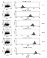
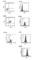
- Example 1 (1) Healthy individuals, (2) Low risk HTLV-I carriers, (3) Medium risk HTLV-I carriers, (4) High risk HTLV-I carriers, (5) Smoldering ATLL patients, (6) It is a figure which shows the result of having analyzed the cell distribution pattern of PpIX fluorescence intensity and TSLC1 expression intensity which is a leukemia marker about the peripheral blood derived from a chronic ATLL patient and (7) acute type ATLL patient by flow cytometry.
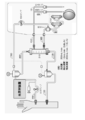
- Example 2 It is a figure which shows the concept of the abnormally activated cell removal reflux return treatment apparatus of this invention.
- Example 1 It is a figure which shows the concept at the time of connecting a dialysis apparatus to the abnormally activated cell removal reflux return treatment apparatus of this invention.
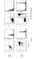
- Example 1 Peripheral blood mononuclear in patients with TLOm1 (ATLL leukemia cell line) or chronic ATLL treated with photodynamic therapy by irradiating with Na-Li lamp for 48 hours in the presence of 1 mM 5-ALA It is a figure which shows the result of having investigated the cell death pattern by flow cytometry of the sphere (PBMC) by double staining of PI (Propidium iodide) staining and Annexin-V-FITC staining. (Example 3) It is a figure which shows the result when the peripheral blood of a chronic type
- PBMC Peripheral blood mononuclear cells isolated from the blood of chronic ATLL patients were cultured for 48 hours in the presence of 1 mM 5-ALA, and then photodynamic therapy was performed by irradiating light with a Na-Li lamp. The flow cytometry analysis result of specific cell death induction is shown.
- PBMC peripheral blood mononuclear cells
- ATLL leukemia cells were specifically dead (implemented)
- Example 4 Specific cell death in the case of photodynamic therapy by adding 1 mM 5-ALA to peripheral blood of chronic ATLL patients and culturing for 48 hours followed by irradiation with Na-Li lamp The flow cytometry analysis result of induction is shown.
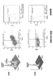
- Example 6 It is a figure which shows the result of having analyzed the cell distribution pattern which concerns on expression of a PpIX positive cell and a tumor marker about various cell culture malignant tumor cells by flow cytometry. (Example 6) It is a figure which shows the result of having analyzed the cell distribution pattern which concerns on expression of a PpIX positive cell and a tumor marker about various cell culture cancer cells by flow cytometry. (Example 6)
- the malignant tumor test of the present invention can be performed by the following steps 1) to 3). 1) a step of detecting PpIX positive cells in the collected blood sample; 2) a step of detecting tumor marker positive cells in the collected blood sample; 3) Analyzing the distribution pattern of PpIX positive cells and tumor marker positive cells obtained in 1) and 2), respectively.
- PpIX protoporphyrin IX
- physiological precursors of porphyrin-related compounds include 5-ALA (5-aminolevulinic acid), which is an amino acid that is a precursor substance for heme synthesis, but is not particularly limited thereto. Any physiological precursor that generates strong fluorescence with a photosensitive substance may be used.
- the “PpIX positive cell” refers to a cell in which PpIX is accumulated by excessive administration of a physiological precursor of a porphyrin-related compound such as 5-ALA from the outside. It is known that PpIX selectively accumulates in immunologically abnormal cells such as malignant tumor cells, chronic inflammatory cells and autoantigen-responsive cells in autoimmune diseases, and in particular, selectively accumulates at high concentrations in malignant tumor cells.
- PpIX is irradiated with excitation light having a peak at a wavelength of 350 to 490 nm, preferably a wavelength of 400 to 490 nm or a wavelength of 600 to 700 nm, preferably a wavelength of 610 to 640 nm, and a wavelength of 600 to 700 nm, preferably a wavelength of 620 to 650 nm.
- it has the property of exhibiting red fluorescence having a peak at a wavelength of 635 nm, and has been clinically applied to photodynamic diagnosis (PDD) of tumor tissues in a wide range.
- PPD photodynamic diagnosis
- FIG. 1 is a graph showing the measurement results of the time course of accumulation of PpIX positive cells after administration of 1 mM 5-ALA to TLOm1 (ATLL leukemia cell line). It can be seen that the PpIX concentration reached the upper limit 48 hours after administration.
- Fig. 2 shows PpIX fluorescence of peripheral blood mononuclear cells (PBMC), TLOm1 (ATLL leukemia cell line) and ED-40515 (ATLL leukemia cell line) of healthy subjects after 48 hours of culture after administration of 1 mM 5-ALA. It is a figure which shows the result of having analyzed intensity
- PBMC peripheral blood mononuclear cells
- TLOm1 ATLL leukemia cell line
- ED-40515 ATLL leukemia cell line
- FIG. 3 shows contact activated by stimulation with peripheral blood mononuclear cells (PBMC) and CD3 / CD28 immuno-beads (Dynabeads (R) human T-cell activator CD3 / CD28 (Invitrogen)) from patients with contact dermatitis.
- PBMC peripheral blood mononuclear cells
- TLOm1 ATLL leukemia cell line
- ED-40515 ATLL leukemia cell line
- peripheral blood mononuclear cells PBMC
- PBMC peripheral blood mononuclear cell
- abnormally activated cells refers to pathological cells, and examples thereof include immunologically abnormal cells such as malignant tumor cells, chronic inflammatory cells, and autoantigen-responsive cells in autoimmune diseases.
- cells that emit strong PpIX fluorescence in the presence of 5-ALA by addition or administration of 5-ALA are also referred to as “abnormally activated cells”.
- the “malignant tumor” may be any of “hematopoietic tumor”, “carcinoma (carcinoma), sarcoma”, and is not particularly limited, but preferably “hematopoietic tumor” is Can be mentioned.
- Examples of “hematopoietic tumors” include leukemia and lymphoma.
- Leukemia is roughly classified into acute leukemia and chronic leukemia, and is divided into myeloid leukemia that develops from myeloid cells and lymphocytic leukemia that develops from lymphoid cells. Specifically, acute myeloid leukemia (Acute Myeloid Leukemia: AML), acute lymphoblastic leukemia (Acute Lymphoblastic Leukemia: ALL), chronic myelogenous leukemia (Leukemia: CML), chronic lymphocytic leukemia (Chronic Lymphoblastic Leukemia: CLL) Etc. ATLL is also listed as another type of leukemia. In this specification, it is ATLL that is the object of examination and treatment. As shown in the background art section, ATLL is caused by HTLV-I.
- a sample collected for examination or treatment is a blood sample.
- the collected blood sample may be irradiated with light as it is, but is preferably processed by the following steps. 1) The red blood cell fraction and the white blood cell fraction are separated by centrifugation of the collected blood sample. 2) The separated leukocyte fraction is irradiated with light having a wavelength of 350 to 490 nm or 600 to 700 nm. 3) After the light irradiation, the accumulated PpIX positive cells are detected.
- leukocytes exhibiting abnormal proliferation ability including hematopoietic tumors such as leukemia cells are also referred to as “abnormally activated leukocytes”.
- the tumor marker may be a tumor marker specified for a tumor, and is not particularly limited.
- a tumor marker specified for a tumor, and is not particularly limited.
- PBMC peripheral blood mononuclear cells
- TSLC1 a cell surface marker
- CD3, CD13, CD19, CD20, CD10, CD13, CD7, CD56, HLA -Immunological markers such as DR an example of a cell surface marker whose expression is specifically increased in ATLL leukemia cells is TSLC1.
- Tumor markers for carcinoma include per se known cytokeratin 19-9 (CA19-9), CEA, NSE, PSA, EMA, AFP, SCC, PTH, TSH, but are particularly limited is not.
- test method combining “detection of PpIX positive cells” and “detection of tumor marker positive cells” of the present invention, cells that have become malignant tumors can be detected more reliably than when they are performed alone.
- FIG. 4 is a diagram showing the results of analyzing the cell distribution pattern by flow cytometry from the PpIX measurement value and the measurement value of TSLC1, which is a tumor marker, for peripheral blood derived from ATLL patients.
- TSLC1 a tumor marker
- FIG. 4 is a diagram showing the results of analyzing the cell distribution pattern by flow cytometry from the PpIX measurement value and the measurement value of TSLC1, which is a tumor marker, for peripheral blood derived from ATLL patients.
- the TSLC1 (-) / PpIX (-) pattern in which cells accumulate on the lower left side, whereas in the HTLV-I carrier, (2) TSLC1 that is almost the same as that of healthy individuals (4) From low risk HTLV-I carriers with (-) / PpIX (-) pattern, (3) Medium risk HTLV-I carriers with increasing fraction of TSLC1 (+) / PpIX (-), (4)
- HTLV-I carriers Especially in high-risk HTLV-I carriers, it means that there are many cell groups showing TSLC1 (+) / PpIX (+) leukemia cell-specific profiles. Is high. Thus, in the case of a carrier, it is possible to predict the onset of ATLL by confirming the cell distribution pattern (flow cytometry profile) in the blood sample, and to start preemptive treatment according to the progress of the disease state at an early stage Can do.
- the ATLL smoldering type, chronic type, or acute type also gradually changes as shown in the patterns (5) to (7) in Fig. 4, and the increase in TSLC1 (+) / PpIX (-) cells indicates that TSLC1 ( +) / PpIX (+) migration to leukemia cells.
- Low-grade ATLL such as smoldering type and chronic type is also acutely changed to high-grade ATLL by monitoring the transition from TSLC1 (+) / PpIX (-) cells to TSLC1 (+) / PpIX (+) leukemia cells Conversion (progress) can be detected at an early stage.
- the risk and progression of malignant tumor onset / progress can be predicted or grasped using, for example, the following as an index.
- the risk / progression index of malignant tumor and the index of the progression are RF (RF: Risk Factor)
- the RF value can be derived as follows.
- the flow cytometry profile shown in FIG. 4 is considered as follows.
- RF Risk Factor
- Healthy people 0.01 ⁇ RF ⁇ 1.00
- Low risk HTLV-I carrier 0.01 ⁇ RF ⁇ 2.50
- Medium risk HTLV-I carrier 2.00 ⁇ RF ⁇ 6.00
- High risk HTLV-I carrier 3.50 ⁇ RF ⁇ 20.00
- Smoldering ATLL 4.00 ⁇ RF ⁇ 30.00
- Chronic ATLL 5.00 ⁇ RF ⁇ 80.00
- Acute ATLL 20.00 ⁇ RF ⁇ 100.00
- the present invention also extends to a method for removing malignant tumor cells.
- the “malignant tumor” in the method for removing malignant tumor cells may be any hematopoietic tumor, carcinoma or sarcoma, but is particularly preferably a hematopoietic tumor.
- the collected specimen is irradiated with light having a wavelength of 350 to 490 nm or 600 to 700 nm, PpIX positive cells are detected, and cell death is induced by the light irradiation to remove malignant tumor cells. it can.
- PpIX positive cells it is preferably a specimen containing a physiological precursor of a porphyrin-related compound, specifically 5-ALA.
- a sample containing 5-ALA may be administered in advance before the sample is collected, or may be administered to the sample after the sample is collected, but it is preferable to administer the sample before collecting the sample. is there.
- 5-ALA has proven to be highly safe when used for oral administration and intravenous injection.
- By administering 5-ALA to a patient in advance accumulation of PpIX in abnormally activated cells such as leukemia cells can be caused. Since it takes some time for PpIX to accumulate in abnormally activated cells such as leukemia cells, it is desirable to administer 5-ALA at least 2 days before the test.
- the present invention also extends to an abnormally activated cell removal perfusion return treatment device for removing abnormally activated cells including hematopoietic tumors from a collected blood sample.
- the abnormally activated cell removal perfusion return treatment device of the present invention is a device for removing abnormally activated cells in blood, and in particular, it accumulates PpIX in malignant tumor cells, chronic inflammatory cells and immune abnormal cells. It selectively accumulates, and in particular, abnormally activated cells such as malignant tumor cells such as leukemia cells or leukemia progenitor cells can be identified and effectively removed.
- the abnormally activated cell-removal reflux return treatment apparatus of the present invention has the following configuration. a) a blood collection line having a puncture needle for blood collection at one end; b) a centrifuge for separating the red blood cell fraction and the white blood cell fraction from the blood fed by the blood collection line; c) a cell sorter for removing the abnormally activated cells from the leukocyte fraction separated by the centrifuge; d) a light irradiator for irradiating the normal white blood cell fraction from which the abnormally activated cells have been removed by the cell sorter of c) with a predetermined wavelength; f) A recirculation that recirculates the recirculated blood composed of blood components other than the leukocyte fraction separated by the centrifuge and the normal white blood cell fraction for recirculation that has been irradiated with light by the light irradiator. Blood return line. Further, g) an artificial dialysis apparatus may be connected (see FIG. 6).
- the abnormally activated cell-removal perfusion return treatment apparatus of the present invention will be described in more detail.
- the device of the present invention is a device for removing abnormally activated cells such as tumor cells, chronic inflammatory cells or immune abnormal cells in blood, and in particular, distinguishing abnormally activated cells from normal cells by accumulating PpIX. As possible, it is an abnormally activated cell-removal reflux return treatment device that effectively removes.
- Abnormally activated cells relate to tumor cells and include leukemia cells found in hematopoietic tumors as well as tumor cells dropped into the blood from carcinomas (epitheliomas), sarcomas, and the like.
- the abnormally activated cells are particularly preferably abnormally activated leukocytes such as leukemia cells.
- the abnormally activated cell-removal reflux return treatment apparatus of the present invention is an abnormally activated cell-removal reflux return treatment apparatus that can effectively identify abnormally activated leukocytes or leukemia progenitor cells, and effectively remove them.
- 5-ALA a physiological precursor of a photosensitizer porphyrin-related compound
- PpIX a physiological precursor of a photosensitizer porphyrin-related compound
- Abnormally activated cells can be identified because strong fluorescence is generated by 700 nm excitation light irradiation.
- the 5-ALA that is a physiological precursor of a porphyrin-related compound as a photosensitive substance is not particularly limited as long as it is a physiological precursor that generates strong fluorescence with a photosensitive substance.
- 5-ALA can cause PpIX accumulation in abnormally activated cells by pre-administration to patients. Since it takes some time for PpIX to accumulate in the abnormally activated cells, 5-ALA is preferably used 2 to 48 hours, preferably 2 to 12 hours before using the abnormally activated cell-removal reflux treatment apparatus. It is desirable to administer.
- An abnormally activated cell removal perfusion return treatment device as shown in FIG. 5, has a blood collection with a puncture needle (not shown) for blood collection at one end.
- Line 10 a centrifuge 20 for separating a leukocyte fraction from a blood sample supplied by the blood collection line 10, and abnormal activity such as leukemia cells against the leukocyte fraction separated by the centrifuge 20
- the cell sorter 30 for removing activated cells
- the light irradiation device 40 for irradiating the normal leukocyte fraction from which abnormally activated cells such as leukemia cells have been removed by the cell sorter 30, and the centrifuge 20
- Blood components other than the leukocyte fraction, blood circulation components such as erythrocyte fraction and platelet fraction, and blood circulation solution that is irradiated with light from the light irradiator 40 and plasma other than the leukocyte fraction Components, red blood cell fraction and platelets It is composed of a circulation return line 50 that returns blood components
- abnormally activated cells can be detected by PpIX-specific fluorescence emitted when irradiated with excitation light having a wavelength of 488 nm, and the detected PpIX (+) leukocytes can be removed with a cell sorter.
- the blood collection line 10 may be a blood collection line used in general component blood donation.
- a blood collection puncture needle is attached to one end of the blood collection line 10, and the other end can be connected to the centrifuge 20.
- the centrifuge 20 can also be a continuous centrifuge used for component blood donation.
- the leukocyte fraction is separated by centrifugation with the centrifuge 20, and the plasma component, the red blood cell fraction and the platelet fraction are used as the recirculation blood for recirculation. It can be delivered to the first circulation return line 51 of the line 50.
- the white blood cell fraction separated from the blood by the centrifuge 20 is fed to the cell sorter 30 via the first connection line 61.
- the cell sorter 30 is normally used as a cell analysis / separation device, in which individual cells of fine particles are dispersed in a fluid, and the fluid is finely flowed to excite each particle cell with laser light of a specific wavelength. , Optically analyze the specific fluorescence generated, perform flow cytometry, charge droplets containing cells with specific optical properties after droplet formation, and charge with a charged deflector It is an apparatus for performing cell sorting for separating cells contained in a droplet.
- the cell sorter 30 of the present embodiment detects abnormally activated cells such as leukemia cells that emit strong fluorescence due to a large amount of PpIX accumulation in the leukocyte fraction fed from the centrifuge 20, and abnormal activity such as leukemia cells. Abnormally activated cells such as leukemia cells are removed by sorting the activated cells.
- the leukocyte fraction separated in the centrifuge 20 is mixed with a buffer solution such as sheath liquid, and leukocytes are diluted to an extent that can be individually identified, abnormalities such as leukemia cells in the cell sorter 30 After removal of the activated cells, removal of excess components contained in the sheath liquid and appropriate concentration are performed. That is, although not shown, the cell sorter 30 is connected to a concentrator using a centrifuge or the like, or a concentration adjuster using physiological saline, etc. A normal leukocyte fluid for reflux return from which activated cells have been removed is generated. The normal white blood cell fraction for recirculation is returned to the light irradiator 40 via the second connection line 62.
- abnormally activated cells such as almost 100% leukemia cells can be removed by setting the cutoff value of the fluorescence intensity from PpIX.
- the abnormally activated cells removed by the cell sorter 30 may be discarded as they are or used for inspection.
- the number of abnormally activated cells such as removed leukemia cells can be used for early examination of leukemia and disease state monitoring.
- 63 is a discharge line through which abnormally activated cells such as leukemia cells removed by the cell sorter 30 are discharged.
- the normal white blood cell fraction for recirculation returning blood fed via the second connecting line 62 is irradiated with light of a predetermined wavelength by the light irradiator 40, and this light is used for the normal white blood cell fraction for recirculation recirculation. It is supposed to cause cell death in the leukemia cells that remain.
- the light irradiator 40 is constituted by a feeding tube 41 that feeds a normal white blood cell fraction for recirculation and a light source 42 that radiates light toward the feeding tube 41.
- the feed pipe 41 is made of a transparent or semi-transparent pipe, and can irradiate light to the normal white blood cell fraction fed through the pipe.
- the light source 42 is a metal halide lamp (Na-Li lamp) in which sodium and lithium are enclosed.
- the light source 42 may be an LED lamp capable of irradiating a wavelength of 350 to 490 nm or 600 to 700 nm or another light source including laser light.
- a pump for feeding may be provided in the middle of the second connecting line 62, A pump for feeding may be provided.
- a cooling device that cools the heat generated by light irradiation may be provided in the feed pipe 41.
- the length of the feeding tube 41 and the feeding speed of the normal white blood cell fraction for reflux return are adjusted so that the irradiation time is 10 minutes or longer.
- one light source 42 is used, but a plurality of light sources may be used.
- the light irradiator 40 irradiates a normal white blood cell fraction for recirculation with a light having a wavelength of 350 to 490 nm or 600 to 700 nm by a Na-Li lamp.
- PpIX is excited by light irradiation to abnormally activated cells such as leukemia cells that have not been removed by the cell sorter 30 despite the accumulation of PpIX, and singlet oxygen is generated from oxygen with this excitation.
- cell death is induced by the strong cell destruction action by the singlet oxygen.
- a dialysis apparatus may be connected as shown in FIG.
- PBMC nuclear cells
- TLOm1 and ED-40515 ATLL leukemia cell line
- PpIX accumulation in cells 48 hours after administration of 1 mM 5-ALA was analyzed by flow cytometry.
- accumulation of PpIX was confirmed specifically for activated T cells and tumors (FIGS. 2 and 3).
- Example 2 Confirmation of flow cytometry profile of mononuclear cells
- flow cytometry was performed on peripheral blood mononuclear cells (PBMC) isolated from the peripheral blood of healthy subjects, HTLV-I carriers or ATLL patients. ⁇ The profile was confirmed. 1 mM 5-ALA was added to the culture broth containing each peripheral blood mononuclear cell (PBMC) and cultured for 48 hours. PpIX accumulation and TSLC1 expression were confirmed in these cells, and cell distribution by flow cytometry Pattern analysis was performed.
- PBMC peripheral blood mononuclear cells
- HTLV-I carriers are at about 5% risk of developing ATLL in their lifetime, but for HTLV-I carriers, knowing the risk of developing ATLL is very important for the quality of life (QOL) of HTLV-I carriers is important. Based on the method of the present invention, an appropriate treatment method is selected immediately before the onset or at the beginning of the onset by recognizing the pattern of the medium risk and high risk HTLV-I carriers, which is the risk pattern of (3) and (4) be able to.
- the RF value was derived as follows.
- the flow cytometry profile shown in FIG. 4 is considered as follows.
- Example 3 In this example, 1 mM 5-ALA was added to the culture solution of TLOm1 (ATLL leukemia cell line) or peripheral blood mononuclear cells (PBMC) of chronic ATLL patients and cultured for 48 hours, followed by a Na-Li lamp for 10 minutes. Irradiation (29 mW / cm 2 ). Thereafter, the cell death pattern by flow cytometry was examined by double staining of PI (Propidium iodide) staining and Annexin-V-FITC staining. As a control, the same analysis was performed on cells not added with 5-ALA. As a result, in TLOm1, cells were cultured for 48 hours in the presence of 1 mM 5-ALA.
- TLOm1 ATLL leukemia cell line
- PBMC peripheral blood mononuclear cells
- AnnexinV (+) / PI ( -) Apoptosis
- AnnexinV (+) / PI (+) necrosis
- PBMC Peripheral blood mononuclear cells
- PBMC Peripheral blood mononuclear cells
- Example 4 was performed in order to confirm what kind of cells the fraction causing cell death and the fraction remaining alive consist of.
- Example 4 In this example, 1 mM 5-ALA was administered to peripheral blood mononuclear cells (PBMC) of chronic ATLL patients using the apparatus of Example 1 and cultured for 48 hours, followed by a 10-minute Na-Li lamp (29 mW). / cm 2 ) Irradiation. Subsequently, triple staining with Annexin-V-FITC, PI, and TSLC1-Alexa647 was performed, and detailed analysis of the cell death fraction by flow cytometry was performed. As a result, the fraction of cells in which ATLL leukemia cells of TSLC1 (+) / AnnexinV (+) caused cell death (upper right) reached 98.7% of the fraction of all ATLL leukemia cells (TSLC1 (+)).
- PBMC peripheral blood mononuclear cells
- Photodynamic therapy by irradiating light with Na-Li lamp after adding 1 mM 5-ALA to peripheral blood mononuclear cells (PBMC) of chronic ATLL patients for 48 hours
- PBMC peripheral blood mononuclear cells
- abnormally activated cells such as leukemia cells that have not been removed by the cell sorter 30 are killed by the light irradiator 40, so that abnormally activated cells such as leukemia cells remain in the normal leukocyte fraction for recirculation. Only healthy white blood cells can be returned to the patient without fear.
- Abnormally activated cells such as leukemia cells that caused cell death can be removed using the discoloration function of the patient's liver and spleen even if they are returned to the patient together with the normal white blood cell fraction for recirculation.
- abnormally activated cell components such as leukemia cells that have been killed by a filter or the like may be removed to prevent oncolysis syndrome.
- the normal white blood cell fraction for reflux return sent from the light irradiator 40 is sent to the second reflux return line 52 of the reflux return line 50 connected at one end to the light irradiator 40.
- the first circulating blood return line 51 and the second circulating blood return line 52 are connected by a Y-shaped connector (not shown), thereby comprising normal blood components other than the leukocyte fraction separated by the centrifuge 20.
- the recirculating blood and the normal white blood cell fraction for recirculating blood irradiated with light by the light irradiator 40 are mixed.
- a third recirculation return line 53 provided with a blood puncture needle (not shown) at one end is connected, and the recirculation composed of blood components other than the leukocyte fraction A mixture of the returned blood and the normal white blood cell fraction for recirculation is returned to the patient.
- abnormally activated cells such as leukemia cells are removed by the cell sorter 30, but also leukemia cells and the like by irradiation with light from the light irradiator 40.
- the possibility of abnormally activated cells remaining can be eliminated.
- the specific abnormally activated cell removal reflux return treatment apparatus of the present invention is used not only as a treatment for removing abnormally activated cells such as leukemia cells, but also for counting the number of abnormally activated cells such as leukemia cells removed by the cell sorter 30, Since it can be used for pathological analysis, it can also be used for early examination of leukemia and pathological monitoring as described above.
- the heterospecific normal activated leukocyte removal reflux return treatment device of the present invention is used for transplantation of patient immunity against other organ transplantation in addition to ATLL lymphoid leukemia, myeloproliferative diseases including myeloid leukemia, etc.
- aggressive activated lymphocyte clones in organ rejection or attack of patient organs by donor lymphocytes due to acute GVHD (graft versus host disease) or chronic GVHD after hematopoietic stem cell transplantation or bone marrow transplantation It can be used for removal.
- it may be widely used for various diseases associated with abnormal growth of activation-specific clones such as various autoimmune diseases or allergic diseases.
- it can be used for suppression of metastasis by separation / removal of epithelial carcinoma cells and sarcoma cells in blood undergoing hematogenous metastasis.
- Example 5 In this example, 1 mM 5-ALA was administered to peripheral blood of chronic ATLL patients and cultured for 48 hours, and then a photodynamic therapy was performed by irradiating light with a Na-Li lamp. Flow cytometric analysis was performed for specific cell death induction. As a result of analyzing the cell distribution pattern of the chronic ATLL leukemia fraction (TSLC1 (+)) and the normal mononuclear cell fraction (TSLC1 (-)) with PpIX fluorescence intensity and TSLC1 expression intensity as a leukemia marker by flow cytometry, This treatment removed 98.83% ATLL leukemia cells by cell death.
- TSLC1 (+) chronic ATLL leukemia fraction
- TSLC1 (-) normal mononuclear cell fraction
- PBMC peripheral blood mononuclear cells
- Example 6 In this example, 1 mM 5-ALA was administered to various established malignant tumor cultured cells and cultured for 48 hours, and tumor markers and PpIX accumulation were confirmed for these cells.
- hematopoietic tumor-derived cells are Ramos cells (Burkitt lymphoma (malignant lymphoma)), K562 cells (chronic myeloid leukemia) and BALL1 cells (acute B lymphocytic leukemia).
- the expression of TSLC1 as a tumor marker was also confirmed (FIG. 10).
- Jurkat cells T-lymphocytic leukemia
- FL18 cystic lymphoma (malignant lymphoma)
- A549 cells lung cancer
- SCCKN cells thyroidgue cancer
- LK79 cells lung cancer
- PpIX accumulation by administration of 1 mM 5-ALA positive for PpIX positive cells and tumor marker (CK19-9)
- PpIX (+) / CK19-9 (+) the distribution pattern of epithelial cancer cells
- normal cells PpIX ( ⁇ ) / CK19-9 ( ⁇ )
- FIG. 12 the distribution of PpIX positive cells and tumor marker positive cells was analyzed for hematopoietic tumors and solid tumors, confirming a cell distribution different from normal cells. It was thought that tumor cells could be removed by kinetic therapy.
- malignant tumor cells can be detected effectively.
- the risk of tumor onset / progress can be ascertained.
- the risk of ATLL onset / progress can be predicted for the HTLV-I carrier.
- ATLL is categorized as (A) smoldering type, (B) chronic type, (C) acute type, and (D) lymphoma type.
- ATLL is classified into high-risk HTLV-I carriers. Since it is possible to monitor in detail the pathology of low-grade ATLL patients who are expected to be identified and progressed, more appropriate preemptive treatment can be performed before onset / progress.
- abnormally activated cells such as leukemia cells are removed from blood collected from a patient, for example, component transfusion, and normal blood cell components, platelets, and plasma are removed again. Can be returned to the patient and contribute to leukemia treatment.
- leukemia cells can be substantially removed in principle by setting the cutoff value of the fluorescence intensity from PpIX in the separation using a centrifuge and a cell sorter. it can. Even if leukemia cells mixed in the normal cell fraction coexist, 98.7% of leukemia cells can be removed by inducing cell death by further light irradiation. It is possible to remove leukemia cells.
- the abnormally activated cell-removal perfusion return treatment apparatus of the present invention is a lymphatic leukemia other than ATLL, myeloproliferative diseases including myeloid leukemia, etc., as well as rejection of patient immunity against transplanted organs of patient immunity against allogeneic organ transplantation
- it can be used to remove aggressive lymphocyte clones in patients' organs attacked by donor lymphocytes due to acute GVHD (graft-versus-host disease) or chronic GVHD that occurs after hematopoietic stem cell transplantation or bone marrow transplantation It is thought that.
- GVHD graft-versus-host disease
- chronic GVHD that occurs after hematopoietic stem cell transplantation or bone marrow transplantation It is thought that.
- it may be widely used for various diseases such as various autoimmune diseases or lymphoproliferative diseases accompanying abnormal growth of specific clones such as allergic diseases.
- it can be used for suppression of metastasis by separation / removal of epithelial carcinoma cells
Landscapes
- Health & Medical Sciences (AREA)
- Heart & Thoracic Surgery (AREA)
- Vascular Medicine (AREA)
- Life Sciences & Earth Sciences (AREA)
- General Health & Medical Sciences (AREA)
- Anesthesiology (AREA)
- Biomedical Technology (AREA)
- Hematology (AREA)
- Veterinary Medicine (AREA)
- Animal Behavior & Ethology (AREA)
- Engineering & Computer Science (AREA)
- Public Health (AREA)
- Cardiology (AREA)
- Investigating Or Analysing Biological Materials (AREA)
- Measuring Or Testing Involving Enzymes Or Micro-Organisms (AREA)
- External Artificial Organs (AREA)
- Investigating, Analyzing Materials By Fluorescence Or Luminescence (AREA)
- Apparatus Associated With Microorganisms And Enzymes (AREA)
Abstract
Cette invention concerne un appareil thérapeutique d'aphérèse de cellules activées anormalement permettant d'inhiber l'apparition de la leucémie ou de traiter la leucémie en éliminant du sang des cellules activées anormalement, spécifiquement des leucocytes activés anormalement ou des cellules progénitrices leucémiques. Ledit appareil thérapeutique d'aphérèse de cellules activées anormalement qui élimine du sang des cellules activées anormalement telles que des cellules leucémiques, lesdites cellules ayant été rendues identifiables par une accumulation à concentration élevée de la protoporphyrine IX, comprend les éléments suivants : un tube de récupération du sang dont une extrémité est pourvue d'une aiguille pour récupérer le sang; un séparateur centrifuge qui sépare du sang une fraction leucocytaire expulsée vers le tube de récupération du sang; un trieur de cellules qui retire des cellules activées anormalement de la fraction leucocytaire séparée; un dispositif d'exposition à la lumière qui expose à une lumière d'une longueur d'onde prévue la fraction leucocytaire normale résultant de l'élimination des cellules activées anormalement précitées; un conduit de retour qui renvoie au patient la fraction leucocytaire normale et un fluide de retour comprenant les composants sanguins autres que la fraction leucocytaire.
Priority Applications (2)
| Application Number | Priority Date | Filing Date | Title |
|---|---|---|---|
| JP2015522993A JP6352912B2 (ja) | 2013-06-21 | 2014-06-20 | 異常活性化細胞検出による悪性腫瘍の検査方法および異常活性化細胞除去環流返血治療装置 |
| CN201480035534.6A CN105683752B (zh) | 2013-06-21 | 2014-06-20 | 基于异常活化细胞检测的恶性肿瘤的检查方法及去除异常活化细胞血液循环回输治疗装置 |
Applications Claiming Priority (2)
| Application Number | Priority Date | Filing Date | Title |
|---|---|---|---|
| JP2013-130758 | 2013-06-21 | ||
| JP2013130758 | 2013-06-21 |
Publications (1)
| Publication Number | Publication Date |
|---|---|
| WO2014204001A1 true WO2014204001A1 (fr) | 2014-12-24 |
Family
ID=52104726
Family Applications (1)
| Application Number | Title | Priority Date | Filing Date |
|---|---|---|---|
| PCT/JP2014/066480 Ceased WO2014204001A1 (fr) | 2013-06-21 | 2014-06-20 | Procédé utilisant la détection de cellules activées anormalement pour tester des tumeurs malignes et appareil thérapeutique d'aphérèse de cellules activées anormalement |
Country Status (3)
| Country | Link |
|---|---|
| JP (2) | JP6352912B2 (fr) |
| CN (1) | CN105683752B (fr) |
| WO (1) | WO2014204001A1 (fr) |
Cited By (4)
| Publication number | Priority date | Publication date | Assignee | Title |
|---|---|---|---|---|
| JP2017513952A (ja) * | 2014-04-24 | 2017-06-01 | オスロ ウニヴェルスィテーツシケヒュース ホーエフOslo Universitetssykehus Hf | ポルフィリン前駆体を用いる体外循環式光化学療法技術の改変 |
| WO2018159833A1 (fr) * | 2017-03-02 | 2018-09-07 | 株式会社ニコン | Procédé de distinction de cellules, procédé d'inspection de cancer, dispositif de mesure, dispositif d'inspection de cancer, et programme d'inspection |
| EP3932447A4 (fr) * | 2019-02-25 | 2022-11-23 | Otsuka Electronics Co., Ltd. | Dispositif de thérapie photodynamique et cartouche de dispositif de thérapie photodynamique |
| EP4454673A1 (fr) * | 2023-04-27 | 2024-10-30 | Fenwal, Inc. | Système de traitement de cellules de chevet doté d'un système de reperfusion |
Families Citing this family (4)
| Publication number | Priority date | Publication date | Assignee | Title |
|---|---|---|---|---|
| JP3151923B2 (ja) | 1992-04-21 | 2001-04-03 | ブラザー工業株式会社 | ジグザグミシンのボタンホール縫い装置 |
| CN106085962B (zh) * | 2016-06-20 | 2022-03-22 | 上海交通大学 | 一种用于检测与分离循环黑色素瘤细胞的装置 |
| CN111855541A (zh) * | 2019-04-26 | 2020-10-30 | 朱德新 | 循环肿瘤细胞检测试剂、试剂盒和检测方法 |
| WO2021246761A1 (fr) * | 2020-06-01 | 2021-12-09 | 서울바이오시스주식회사 | Module de stérilisation et système de stérilisation le comprenant |
Citations (7)
| Publication number | Priority date | Publication date | Assignee | Title |
|---|---|---|---|---|
| JPH053917A (ja) * | 1991-06-28 | 1993-01-14 | Toshiro Tachibana | 血液処理装置 |
| JP2005013266A (ja) * | 2003-06-23 | 2005-01-20 | Kazuhiko Ikeuchi | 顔面ホルダ |
| JP2005074234A (ja) * | 2003-09-03 | 2005-03-24 | Therakos Inc | 生物学的流体を成分に連続的に分離する装置 |
| JP3124006U (ja) * | 2006-05-23 | 2006-08-03 | ワン、シャンユ | 血液透析機 |
| JP2008511829A (ja) * | 2004-08-27 | 2008-04-17 | ベックマン コールター,インコーポレーテッド | 細胞シグナル伝達経路のサイトメトリー解析のための全血製剤 |
| JP2009045111A (ja) * | 2007-08-14 | 2009-03-05 | Renaissance Energy Investment:Kk | フォトフェレシス処理用カラム、フォトフェレシス処理システム、フォトフェレシス処理方法 |
| JP2010505845A (ja) * | 2006-10-04 | 2010-02-25 | ジヤンセン・フアーマシユーチカ・ナームローゼ・フエンノートシヤツプ | 不活性化された人工抗原提示細胞の調製および細胞治療におけるそれらの使用 |
Family Cites Families (9)
| Publication number | Priority date | Publication date | Assignee | Title |
|---|---|---|---|---|
| US6190877B1 (en) * | 1999-12-27 | 2001-02-20 | Edwin L. Adair | Method of cancer screening primarily utilizing non-invasive cell collection and fluorescence detection techniques |
| JP2001249126A (ja) * | 2000-03-06 | 2001-09-14 | Bml Inc | 免疫状態の確認方法 |
| JP2005132766A (ja) * | 2003-10-30 | 2005-05-26 | Cosmo Oil Co Ltd | 光動力学的癌治療薬 |
| CN101669031A (zh) * | 2007-02-27 | 2010-03-10 | 森托科隆股份公司 | 使用与细胞外标记物结合的试剂组多重检测肿瘤细胞 |
| CN101126758A (zh) * | 2007-09-06 | 2008-02-20 | 江苏省肿瘤医院 | 流式细胞术同步检测肿瘤中多种细胞蛋白表达的方法 |
| GB0724279D0 (en) * | 2007-12-12 | 2008-01-23 | Photocure Asa | Use |
| CN101319201A (zh) * | 2008-04-18 | 2008-12-10 | 中国科学院昆明动物研究所 | 一种诱导恶性肿瘤细胞凋亡的方法和用途 |
| JP5991970B2 (ja) * | 2010-07-09 | 2016-09-14 | フォトキュア エイエスエイ | 光力学的治療または光力学的診断で使用する乾燥組成物および当該乾燥組成物を含む装置 |
| CN103096884B (zh) * | 2010-09-14 | 2017-11-03 | 学校法人东京农业大学 | 癌温热疗法的作用增强剂 |
-
2014
- 2014-06-20 WO PCT/JP2014/066480 patent/WO2014204001A1/fr not_active Ceased
- 2014-06-20 JP JP2015522993A patent/JP6352912B2/ja not_active Expired - Fee Related
- 2014-06-20 CN CN201480035534.6A patent/CN105683752B/zh not_active Expired - Fee Related
-
2018
- 2018-06-07 JP JP2018109331A patent/JP6703736B2/ja active Active
Patent Citations (7)
| Publication number | Priority date | Publication date | Assignee | Title |
|---|---|---|---|---|
| JPH053917A (ja) * | 1991-06-28 | 1993-01-14 | Toshiro Tachibana | 血液処理装置 |
| JP2005013266A (ja) * | 2003-06-23 | 2005-01-20 | Kazuhiko Ikeuchi | 顔面ホルダ |
| JP2005074234A (ja) * | 2003-09-03 | 2005-03-24 | Therakos Inc | 生物学的流体を成分に連続的に分離する装置 |
| JP2008511829A (ja) * | 2004-08-27 | 2008-04-17 | ベックマン コールター,インコーポレーテッド | 細胞シグナル伝達経路のサイトメトリー解析のための全血製剤 |
| JP3124006U (ja) * | 2006-05-23 | 2006-08-03 | ワン、シャンユ | 血液透析機 |
| JP2010505845A (ja) * | 2006-10-04 | 2010-02-25 | ジヤンセン・フアーマシユーチカ・ナームローゼ・フエンノートシヤツプ | 不活性化された人工抗原提示細胞の調製および細胞治療におけるそれらの使用 |
| JP2009045111A (ja) * | 2007-08-14 | 2009-03-05 | Renaissance Energy Investment:Kk | フォトフェレシス処理用カラム、フォトフェレシス処理システム、フォトフェレシス処理方法 |
Cited By (8)
| Publication number | Priority date | Publication date | Assignee | Title |
|---|---|---|---|---|
| JP2017513952A (ja) * | 2014-04-24 | 2017-06-01 | オスロ ウニヴェルスィテーツシケヒュース ホーエフOslo Universitetssykehus Hf | ポルフィリン前駆体を用いる体外循環式光化学療法技術の改変 |
| WO2018159833A1 (fr) * | 2017-03-02 | 2018-09-07 | 株式会社ニコン | Procédé de distinction de cellules, procédé d'inspection de cancer, dispositif de mesure, dispositif d'inspection de cancer, et programme d'inspection |
| JPWO2018159833A1 (ja) * | 2017-03-02 | 2020-01-16 | 株式会社ニコン | 細胞の判別方法、がんの検査方法、計測装置、がんの検査装置および検査プログラム |
| EP3932447A4 (fr) * | 2019-02-25 | 2022-11-23 | Otsuka Electronics Co., Ltd. | Dispositif de thérapie photodynamique et cartouche de dispositif de thérapie photodynamique |
| TWI812839B (zh) * | 2019-02-25 | 2023-08-21 | 日商大塚電子股份有限公司 | 光動力治療裝置及光動力治療裝置用卡匣盒 |
| US12023429B2 (en) | 2019-02-25 | 2024-07-02 | Otsuka Electronics Co., Ltd. | Photodynamic therapy device and photodynamic therapy device cartridge |
| US12433987B2 (en) | 2019-02-25 | 2025-10-07 | Otsuka Electronics Co., Ltd. | Photodynamic therapy device |
| EP4454673A1 (fr) * | 2023-04-27 | 2024-10-30 | Fenwal, Inc. | Système de traitement de cellules de chevet doté d'un système de reperfusion |
Also Published As
| Publication number | Publication date |
|---|---|
| JP6703736B2 (ja) | 2020-06-03 |
| CN105683752B (zh) | 2019-03-29 |
| JP6352912B2 (ja) | 2018-07-04 |
| CN105683752A (zh) | 2016-06-15 |
| JP2018153195A (ja) | 2018-10-04 |
| JPWO2014204001A1 (ja) | 2017-02-23 |
Similar Documents
| Publication | Publication Date | Title |
|---|---|---|
| JP6352912B2 (ja) | 異常活性化細胞検出による悪性腫瘍の検査方法および異常活性化細胞除去環流返血治療装置 | |
| Yoshida et al. | Surgical margin and recurrence after resection of hepatocellular carcinoma in patients with cirrhosis. Further evaluation of limited hepatic resection | |
| Yan et al. | Efficacy of dendritic cell-cytokine-induced killer immunotherapy plus intensity-modulated radiation therapy in treating elderly patients with esophageal carcinoma | |
| KR20210062599A (ko) | 혈액암 치료용 마이크로웨이브 치료 장치 | |
| Sando et al. | 5-aminolevulinic acid-mediated photodynamic therapy can target aggressive adult T cell leukemia/lymphoma resistant to conventional chemotherapy | |
| US20140329322A1 (en) | Method of differentiating glioblastoma cells | |
| Toshkova et al. | Bioinfluence with Infrared Thermal and Electromagnetic Fields as a Therapeutic Approach of Hamsters with Experimental Graffi Myeloid Tumor | |
| RU2392668C1 (ru) | Способ индукции антипролиферативного, цитотоксического эффекта в опухолевых клетках линейных и свежевыделенных культур | |
| Toshkova et al. | Beneficial Effects of Drossinakis Bio-influence (With Infrared Thermal and Electromagnetic Fields) on the Development of Experimental Graffi Myeloid Tumors in Hamsters. Hematological Studies | |
| Takaba et al. | Role of Bone Marrow‐Derived Monocytes/Macrophages in the Repair of Mucosal Damage Caused by Irradiation and/or Anticancer Drugs in Colitis Model | |
| Adil | FOUNDATIONS IN HEMATOLOGY AND ONCOLOGY: A MEDICAL STUDENT’S GUIDE: ISBN: 9798312125009 | |
| Saat et al. | An overview diagnosis and treatment methods in cases of transmissible venereal tumor in female dogs | |
| RU2738301C2 (ru) | Способ оценки противоопухолевой эффективности фотодинамической терапии | |
| RU2505817C1 (ru) | Способ оценки радиочувствительности рака верхних дыхательных путей | |
| Gao et al. | Transfusion of stored autologous blood in patients with low-grade pseudomyxoma peritonei: A retrospective analysis of its safety and outcome | |
| KOCSIS | CHAPTER FIFTEEN SUSTAINABLE DETECTION OF CIRCULATING MELANOMA CELLS WITHIN PATIENT BLOOD SAMPLES USING TWO-PHASE PHOTOACOUSTIC FLOW CYTOMETRY | |
| Croci | lnvestigating, imaging and targeting the glioma microenvironment | |
| Zahra et al. | Primary Malt Thyroid Lymphoma: a case report. | |
| Prachi | Splenic marginal zone lymphoma: A case report and review of the literature | |
| CN116223344A (zh) | 针对急性t淋巴细胞白血病的生物标志物及其产品和用途 | |
| Awad | Assessment of Complete Blood Count Among Patients Receiving Chemotherapy at Tumor Therapy and Cancer Research Center in Shendi Town | |
| Gao et al. | Xiaoyun Gao1, Liduo Kou1, Hang Guan1, Hua Tian1, Junhui Jia1, Yu Bai1, Wei Bai1, Yanhui Di1, Ruiqing Ma2 and Xinhua Wang1 | |
| Corley et al. | Glucagon-like peptide-1 receptor signaling deficiency exacerbates hematopoietic stem cell graft rejection in mice | |
| Murzakayeva et al. | CLINICAL AND MORPHOLOGICAL MANIFESTATIONS OF BREAST TUMORS IN CATS | |
| Pape et al. | 374P Immunohistochemical evaluation of oral lichen planus: A prospective clinical study |
Legal Events
| Date | Code | Title | Description |
|---|---|---|---|
| 121 | Ep: the epo has been informed by wipo that ep was designated in this application |
Ref document number: 14813629 Country of ref document: EP Kind code of ref document: A1 |
|
| DPE1 | Request for preliminary examination filed after expiration of 19th month from priority date (pct application filed from 20040101) | ||
| ENP | Entry into the national phase |
Ref document number: 2015522993 Country of ref document: JP Kind code of ref document: A |
|
| NENP | Non-entry into the national phase |
Ref country code: DE |
|
| 122 | Ep: pct application non-entry in european phase |
Ref document number: 14813629 Country of ref document: EP Kind code of ref document: A1 |