WO2015020234A1 - ドパミン神経細胞の製造方法 - Google Patents
ドパミン神経細胞の製造方法 Download PDFInfo
- Publication number
- WO2015020234A1 WO2015020234A1 PCT/JP2014/071352 JP2014071352W WO2015020234A1 WO 2015020234 A1 WO2015020234 A1 WO 2015020234A1 JP 2014071352 W JP2014071352 W JP 2014071352W WO 2015020234 A1 WO2015020234 A1 WO 2015020234A1
- Authority
- WO
- WIPO (PCT)
- Prior art keywords
- cells
- cell
- medium
- differentiation
- dopamine
- Prior art date
- Legal status (The legal status is an assumption and is not a legal conclusion. Google has not performed a legal analysis and makes no representation as to the accuracy of the status listed.)
- Ceased
Links
Images






Classifications
-
- C—CHEMISTRY; METALLURGY
- C12—BIOCHEMISTRY; BEER; SPIRITS; WINE; VINEGAR; MICROBIOLOGY; ENZYMOLOGY; MUTATION OR GENETIC ENGINEERING
- C12N—MICROORGANISMS OR ENZYMES; COMPOSITIONS THEREOF; PROPAGATING, PRESERVING, OR MAINTAINING MICROORGANISMS; MUTATION OR GENETIC ENGINEERING; CULTURE MEDIA
- C12N5/00—Undifferentiated human, animal or plant cells, e.g. cell lines; Tissues; Cultivation or maintenance thereof; Culture media therefor
- C12N5/06—Animal cells or tissues; Human cells or tissues
- C12N5/0602—Vertebrate cells
- C12N5/0618—Cells of the nervous system
- C12N5/0619—Neurons
-
- A—HUMAN NECESSITIES
- A61—MEDICAL OR VETERINARY SCIENCE; HYGIENE
- A61K—PREPARATIONS FOR MEDICAL, DENTAL OR TOILETRY PURPOSES
- A61K35/00—Medicinal preparations containing materials or reaction products thereof with undetermined constitution
- A61K35/12—Materials from mammals; Compositions comprising non-specified tissues or cells; Compositions comprising non-embryonic stem cells; Genetically modified cells
- A61K35/30—Nerves; Brain; Eyes; Corneal cells; Cerebrospinal fluid; Neuronal stem cells; Neuronal precursor cells; Glial cells; Oligodendrocytes; Schwann cells; Astroglia; Astrocytes; Choroid plexus; Spinal cord tissue
-
- A—HUMAN NECESSITIES
- A61—MEDICAL OR VETERINARY SCIENCE; HYGIENE
- A61P—SPECIFIC THERAPEUTIC ACTIVITY OF CHEMICAL COMPOUNDS OR MEDICINAL PREPARATIONS
- A61P25/00—Drugs for disorders of the nervous system
-
- A—HUMAN NECESSITIES
- A61—MEDICAL OR VETERINARY SCIENCE; HYGIENE
- A61P—SPECIFIC THERAPEUTIC ACTIVITY OF CHEMICAL COMPOUNDS OR MEDICINAL PREPARATIONS
- A61P25/00—Drugs for disorders of the nervous system
- A61P25/08—Antiepileptics; Anticonvulsants
-
- A—HUMAN NECESSITIES
- A61—MEDICAL OR VETERINARY SCIENCE; HYGIENE
- A61P—SPECIFIC THERAPEUTIC ACTIVITY OF CHEMICAL COMPOUNDS OR MEDICINAL PREPARATIONS
- A61P25/00—Drugs for disorders of the nervous system
- A61P25/14—Drugs for disorders of the nervous system for treating abnormal movements, e.g. chorea, dyskinesia
-
- A—HUMAN NECESSITIES
- A61—MEDICAL OR VETERINARY SCIENCE; HYGIENE
- A61P—SPECIFIC THERAPEUTIC ACTIVITY OF CHEMICAL COMPOUNDS OR MEDICINAL PREPARATIONS
- A61P25/00—Drugs for disorders of the nervous system
- A61P25/14—Drugs for disorders of the nervous system for treating abnormal movements, e.g. chorea, dyskinesia
- A61P25/16—Anti-Parkinson drugs
-
- A—HUMAN NECESSITIES
- A61—MEDICAL OR VETERINARY SCIENCE; HYGIENE
- A61P—SPECIFIC THERAPEUTIC ACTIVITY OF CHEMICAL COMPOUNDS OR MEDICINAL PREPARATIONS
- A61P25/00—Drugs for disorders of the nervous system
- A61P25/28—Drugs for disorders of the nervous system for treating neurodegenerative disorders of the central nervous system, e.g. nootropic agents, cognition enhancers, drugs for treating Alzheimer's disease or other forms of dementia
-
- C—CHEMISTRY; METALLURGY
- C12—BIOCHEMISTRY; BEER; SPIRITS; WINE; VINEGAR; MICROBIOLOGY; ENZYMOLOGY; MUTATION OR GENETIC ENGINEERING
- C12N—MICROORGANISMS OR ENZYMES; COMPOSITIONS THEREOF; PROPAGATING, PRESERVING, OR MAINTAINING MICROORGANISMS; MUTATION OR GENETIC ENGINEERING; CULTURE MEDIA
- C12N2500/00—Specific components of cell culture medium
- C12N2500/30—Organic components
- C12N2500/38—Vitamins
-
- C—CHEMISTRY; METALLURGY
- C12—BIOCHEMISTRY; BEER; SPIRITS; WINE; VINEGAR; MICROBIOLOGY; ENZYMOLOGY; MUTATION OR GENETIC ENGINEERING
- C12N—MICROORGANISMS OR ENZYMES; COMPOSITIONS THEREOF; PROPAGATING, PRESERVING, OR MAINTAINING MICROORGANISMS; MUTATION OR GENETIC ENGINEERING; CULTURE MEDIA
- C12N2501/00—Active agents used in cell culture processes, e.g. differentation
- C12N2501/01—Modulators of cAMP or cGMP, e.g. non-hydrolysable analogs, phosphodiesterase inhibitors, cholera toxin
-
- C—CHEMISTRY; METALLURGY
- C12—BIOCHEMISTRY; BEER; SPIRITS; WINE; VINEGAR; MICROBIOLOGY; ENZYMOLOGY; MUTATION OR GENETIC ENGINEERING
- C12N—MICROORGANISMS OR ENZYMES; COMPOSITIONS THEREOF; PROPAGATING, PRESERVING, OR MAINTAINING MICROORGANISMS; MUTATION OR GENETIC ENGINEERING; CULTURE MEDIA
- C12N2501/00—Active agents used in cell culture processes, e.g. differentation
- C12N2501/10—Growth factors
- C12N2501/16—Activin; Inhibin; Mullerian inhibiting substance
-
- C—CHEMISTRY; METALLURGY
- C12—BIOCHEMISTRY; BEER; SPIRITS; WINE; VINEGAR; MICROBIOLOGY; ENZYMOLOGY; MUTATION OR GENETIC ENGINEERING
- C12N—MICROORGANISMS OR ENZYMES; COMPOSITIONS THEREOF; PROPAGATING, PRESERVING, OR MAINTAINING MICROORGANISMS; MUTATION OR GENETIC ENGINEERING; CULTURE MEDIA
- C12N2501/00—Active agents used in cell culture processes, e.g. differentation
- C12N2501/70—Enzymes
- C12N2501/72—Transferases [EC 2.]
- C12N2501/727—Kinases (EC 2.7.)
Definitions
- the present invention relates to a method for producing dopamine neurons. Furthermore, the present invention also provides a medicament containing dopamine neurons obtained by the method, and reagents and kits for use in the method.
- Dopamine (3,4-dihydroxyphenylethylamine) is an in vivo molecule having various actions and functions mainly as a neurotransmitter in the central nervous system.
- dopamine is biosynthesized in cells by the action of two enzymes, tyrosine hydroxylase and dopa decarboxylase, starting from the amino acid tyrosine.
- Dopamine neurons are neurons that synthesize and release dopamine as a neurotransmitter, and are present mainly in the midbrain in the brain and partly in the hypothalamus. Dopamine neurons existing in the midbrain play an important role in the control of movement and emotion, and when the midbrain dopamine neurons degenerate and drop off, for example, severe neurodegenerative diseases such as Parkinson's disease can be caused.
- One of the most promising therapeutic approaches for treating such neurodegenerative diseases is cell transplantation therapy in which dopamine neurons are transplanted into a subject.
- dopamine neurons include stem cells such as embryonic stem cells (sometimes referred to herein as ES cells) and induced pluripotent stem cells (sometimes referred to herein as iPS cells).
- stem cells such as embryonic stem cells (sometimes referred to herein as ES cells) and induced pluripotent stem cells (sometimes referred to herein as iPS cells).
- ES cells embryonic stem cells
- iPS cells induced pluripotent stem cells
- Non-Patent Document 1 a method of differentiating stem cells into bottom plate cells using two types of SMAD (Small Mothers against Decaplegic) signaling inhibitors
- Patent Document 2 a method of inducing midbrain dopamine neurons by inducing bottom plate cells and further culturing the bottom plate cells with neurotrophic factors
- Patent Document 5 a method of inducing bottom plate cells efficiently by causing a Nodal inhibitor to act has been reported one after another.
- dopamine neurons obtained by these methods are still low, and the cells are not functionally sufficient such as inability to reproduce responsiveness to oxidative stress and drug stimulation in vitro. Therefore, there is still a need to develop a method for efficiently obtaining high-quality dopamine neurons.
- the present inventors have cultivated neural progenitor cells in a medium containing (i) a cAMP analog and (ii) a MEK inhibitor. It has been found that cells can be produced. Furthermore, it was confirmed that the obtained dopamine neurons were high-quality dopamine neurons, such as having the same phenotype and function as dopamine neurons in vivo, and the present invention was completed.
- the present invention provides the following.
- a method for producing dopamine neurons characterized in that the bottom plate cells are subjected to the following step (1): (1) culturing in a medium containing (i) cAMP analog and (ii) MEK inhibitor; [2] The production method of the above-mentioned [1], wherein the medium further contains ascorbic acid or a salt thereof; [3] The production method according to [2], wherein the medium further contains an activator receptor-like kinase-4,7 activator; [4] The production method of [1], wherein the cAMP analog is dibutyryl cAMP.
- the MEK inhibitor is (i) N-[(2R) -2,3-dihydroxypropoxy] -3,4-difluoro-2-[(2-fluoro-4-iodo-phenyl) amino] benzamide (PD0325901).
- dopamine neurons produced according to the present invention are similar to the dopamine neurons in vivo, such as responsiveness to oxidative stress and drug stimulation not observed in dopamine neurons produced by conventional methods. Has phenotype and function. Therefore, the dopamine neurons produced according to the present invention achieve a high engraftment rate in cell transplantation therapy for treating diseases caused by decreased production (release) of dopamine, such as neurodegenerative diseases such as Parkinson's disease.
- FIG. 1 shows the results of inducing bottom plate cells using Neuro / B27 supplemented with various combinations of differentiation-inducing factors and examining the expression variation of the bottom plate cell marker by quantitative RT-PCR on the 13th day of culture.
- [-] Indicates that each differentiation-inducing factor is not added, and [All] uses all five types of LDN193189 (LDN), SB431542 (SB), Sonic Hedgehog (SHH), purmorphamine (Pur), and CHIR99021 (CHIR). Shows the conditions.
- LDN193189 LDN
- SB431542 SB
- SHH Sonic Hedgehog
- Pur purmorphamine
- CHIR99021 CHIR99021
- FIG. 2 shows the results of inducing bottom plate cells and immunofluorescent staining using anti-FOXA2 antibody and anti-LMX1A antibody on day 13 of culture.
- the cell line 253G1 was used. Red indicates nuclei of FOXA2-positive cells, green indicates nuclei of LMX1A-positive cells, and blue (DAPI staining) indicates cell nuclei.
- FIG. 1 shows the results of inducing bottom plate cells and immunofluorescent staining using anti-FOXA2 antibody and anti-LMX1A antibody on day 13 of culture.
- the cell line 253G1 was used. Red indicates nuclei of FOXA2-positive cells, green indicates nuclei of LMX1A-positive cells, and blue (DAPI staining) indicates cell nuclei.
- FIG. 3 shows that from 13 days after the induction of the bottom plate cells, ascorbic acid (AA), dibutyryl cAMP (dbcAMP) and PD0325901 (PD) were added in various combinations and cultured, and NURR1 expression fluctuation was observed on the 45th day.
- AA ascorbic acid
- dbcAMP dibutyryl cAMP
- PD0325901 PD
- the result investigated by quantitative RT-PCR is shown.
- the cell lines used were 253G1 and 201B7.
- the value on the Y axis indicates the value obtained by correcting the copy number of each gene with the copy number of GAPDH, and the error bar indicates the standard deviation.
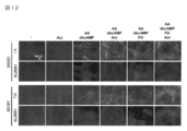
- FIG. 4 shows the result of differentiation induction performed in the same manner as in FIG. 3 and immunofluorescence staining using anti-TH antibody and anti-NURR1 antibody on the 45th day of culture.
- the cell lines used were 253G1 and 201B7. Green indicates the cell body of TH positive cells, and red indicates the nucleus of NURR1 positive cells.
- FIG. 5 shows that from the 13th day after the induction of the bottom plate cells, ascorbic acid (AA), dibutyryl cAMP (dbcAMP) and PD0325901 (PD) were added and cultured, and the 45th day (253G1 strain) or the 52nd day (201B7). The results of immunofluorescent staining using anti-TH antibody, anti-NURR1 antibody and anti-FOXA2 antibody are shown.
- the cell lines used were 253G1 and 201B7.
- Green indicates the cell body of TH positive cells, red indicates the nuclei of NURR1-positive cells, magenta indicates the nuclei of FOXA2-positive cells, and blue (DAPI staining) indicates the cell nuclei.
- FIG. 6 shows a diagram obtained by observing the results obtained using the 253G1 strain of FIG. 5 at a high magnification. Green indicates the cell body of TH positive cells, red indicates the nuclei of NURR1 positive cells, and blue (DAPI staining) indicates the cell nuclei.
- FIG. 7 shows the results of differentiation induction performed in the same manner as in FIG. The cell lines are 253G1 and 201B7, and two independent experimental results are shown.
- FIG. 8 shows the culturing by adding various MEK inhibitors (including FGFR inhibitors) in addition to ascorbic acid (AA) and dibutyryl cAMP (dbcAMP) from the 13th day after induction of the bottom plate cells.
- MEK inhibitors including FGFR inhibitors
- AA ascorbic acid
- dbcAMP dibutyryl cAMP
- FIG. 9 shows a schematic diagram of a method for inducing differentiation of human iPS cells into bottom plate cells and dopamine neurons.
- FIG. 10 shows the results of quantitative RT-PCR examining changes in expression of various differentiation markers over time when differentiation was induced according to the method of FIG. [All] is performed according to FIG. 9, [-] excludes CHIR99021, LDN193189, SB431542, SHH, and purmorphamine, [-SHH] excludes SHH only, and [-Pur] excludes purmorphamine only. Shows that differentiation was induced.
- the cell lines used were 253G1 and 201B7.
- the value on the Y axis indicates the value obtained by correcting the copy number of each gene with the copy number of GAPDH, and the error bar indicates the standard deviation.
- FIG. 11 shows that in Step 3 shown in FIG. 9, ascorbic acid (AA), dibutyryl cAMP (dbcAMP), PD0325901 (PD) and activin A (Act) were added in various combinations to induce differentiation, and culture was performed for 26 days.
- variation of the dopamine neuron marker by quantitative RT-PCR in eyes is shown. [-] Indicates the case where each differentiation-inducing factor is not added.
- the cell lines used were 253G1 and 201B7.
- FIG. 12 shows the result of performing differentiation induction in the same manner as in FIG. 11 and performing immunofluorescent staining using anti-TH antibody and anti-NURR1 antibody on the 26th day of culture.
- the cell lines used were 253G1 and 201B7. Green indicates the cell body of TH positive cells, and red indicates the nucleus of NURR1 positive cells.
- FIG. 13 shows step 3 shown in FIG.
- AA ascorbic acid
- dbcAMP dibutyryl cAMP
- PD0325901 PD
- ACT activin A
- FIG. 14 shows the result of thawing cryopreserved cells in the same manner as FIG. 13 and adding AA, dbcAMP, PD and ACT, culturing for 2 weeks, and then performing immunofluorescence staining using anti-TH antibody and anti-NURR1 antibody Indicates. Green indicates the cell body of TH positive cells, and red indicates the nucleus of NURR1 positive cells. Blue (DAPI staining) indicates cell nuclei.
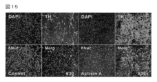
- FIG. 15 shows the addition of ascorbic acid, dbcAMP, PD0325901 (Control group; 4 panels on the left side) or ascorbic acid, dbcAMP, PD0325901 and activin A (Activin A group; 4 panels on the right side) in Step 3 shown in FIG. Then, differentiation was induced, and cells were collected on the 26th day of culture and transplanted into mouse striatum. The results of tissue staining performed 4 weeks after transplantation are shown. Green indicates the cell body of TH positive cells, and red indicates the nucleus of hNuc positive cells. Blue (DAPI staining) indicates cell nuclei.
- FIG. 16 shows the result of transplantation in the same manner as in FIG. 15 and tissue staining 8 weeks after transplantation.
- Green indicates the cell body of TH positive cells, and red indicates the nucleus of hNuc positive cells. Blue (DAPI staining) indicates cell nuclei.
- FIG. 17 shows the result of transplanting in the same manner as in FIG. 15 and performing tissue staining 12 weeks after transplantation. Green indicates the cell body of TH positive cells, and red indicates the nucleus of hNuc positive cells. Blue (DAPI staining) indicates cell nuclei.
- dopamine neuron means a neuron having an ability to produce dopamine (3,4-dihydroxyphenylethylamine).
- Dopamine neurons need not always produce dopamine, as long as they have the ability to produce dopamine. Further, the amount of dopamine produced is not particularly limited.
- dopamine neurons existing in the living body in particular, dopamine neurons existing in the midbrain of the substantia nigra and ventral tegmental area are tyrosine hydroxylase (TH), FOXA2 (forkhead box A2) in vitro.
- TH tyrosine hydroxylase
- FOXA2 forkhead box A2
- the dopamine neurons present in the midbrain can also be characterized in vitro by the expression of specific cell markers such as TH, FOXA2, LMX1A (LIM homebox transcription factor 1 alpha), NURR1 gene / protein.
- a “dopamine neuron” obtained by subjecting a base plate cell to the production method of the present invention is a dopamine neuron existing in the midbrain (ie, midbrain dopamine neuron).
- the “neural progenitor cell” refers to a cell capable of generating a dopamine neuron after differentiation. Specifically, for example, it is characterized by an expression marker such as a bottom plate cell or an intermediate filament protein Nestin. Examples of such cells are neuroectodermal cells, and the most preferred are bottom plate cells.
- floor plate cell means a morphologically specialized organizer cell located in the ventral midline of the neural tube from the spinal cord to the diencephalon.
- bottom plate cells especially those located in the ventral midbrain can be characterized in vitro by the expression of certain cell markers such as FOXA2 and LMX1A gene / protein.
- stem cell refers to a cell that can be cultured in vitro and can differentiate into a plurality of cells constituting a living body.
- ES cells embryonic primordial germ cell-derived pluripotent stem cells (EG cells: Proc Natl Acad Sci US A. 1998, 95: 13726-31), testis-derived pluripotent stem cells (GS cells: Nature, 2008, 456: 344-9), somatic cell-derived induced pluripotent stem cells (iPS cells), human pluripotent somatic stem cells (neural stem cells), preferably iPS cells and ES cells, more preferably iPS cells.
- EG cells embryonic primordial germ cell-derived pluripotent stem cells
- GS cells Nature, 2008, 456: 344-9
- somatic cell-derived induced pluripotent stem cells iPS cells
- human pluripotent somatic stem cells preferably iPS cells and ES cells, more preferably iPS cells.
- ES cells ES cells derived from any warm-blooded animal, preferably a mammal, can be used.
- mammals include mice, rats, guinea pigs, hamsters, rabbits, cats, dogs, sheep, pigs, cows, horses, goats, monkeys, and humans.
- Preferable examples of ES cells include human-derived ES cells.
- Specific examples of ES cells include ES cells such as mammals established by culturing early embryos before implantation, ES established by culturing early embryos produced by nuclear transfer of somatic cell nuclei. ES cells in which the genes on the chromosomes of these cells and these ES cells have been modified using genetic engineering techniques.
- Each ES cell can be prepared according to a method commonly practiced in the art or a known literature.
- Mouse ES cells were obtained in 1981 from Evans et al. (1981, Nature 292: 154-6) and Martin et al. (Martin GR. Et al., 1981, Proc Natl Acad Sci 78: 7634-8). And can be purchased from, for example, Dainippon Sumitomo Pharma Co., Ltd. (Osaka, Japan).
- Human ES cells were established in 1998 by Thomson et al. (Science, 1998, 282: 1145-7) and are available at the WiCell Research Institute (WiCell Research Institute, website: http: // www. available from Wisell.org/, Madison, Wisconsin, USA, National Institute of Health, Kyoto University, etc., for example, Cellaritis (website: http://www.cellaritis.com). /, Sweden).
- an iPS cell an iPS cell derived from any warm-blooded animal, preferably a mammal can be used.
- the mammal include mouse, rat, guinea pig, hamster, rabbit, cat, dog, sheep, pig, cow, horse, goat, monkey and human.
- Preferred examples of iPS cells include iPS cells derived from humans.
- Specific examples of iPS cells include cells obtained by introducing a plurality of genes into somatic cells such as skin cells, which have acquired pluripotency similar to ES cells.
- Oct3 / 4 gene, Klf4 gene, c- Examples include iPS cells obtained by introducing Myc gene and Sox2 gene, iPS cells obtained by introducing Oct3 / 4 gene, Klf4 gene and Sox2 gene (Nat Biotechnol 2008; 26: 101-106).
- a method in which a transgene is further reduced (Nature. 2008 Jul 31; 454 (7204): 646-50), a method using a low molecular weight compound (Cell Stem Cell. 2009 Jan 9; 4 (1): 16 -9, Cell Stem Cell. 2009 Nov 6; 5 (5): 491-503), a method using a transcription factor protein instead of a gene (Cell Stem Cell.
- iPS cell lines include the 253G1 line (iPS cell line prepared by expressing OCT4 / SOX2 / KLF4 in skin fibroblasts of a 36-year-old female), 201B7 line (skin fiber of a 36-year-old female).
- IPS cell line prepared by expressing OCT4 / SOX2 / KLF4 / c-MYC in blast cells
- 1503-iPS (297A1) (OCT4 / SOX2 / KLF4 / c-MYC in skin fibroblasts of a 73-year-old woman) IPS cell line prepared by expression
- 1392-iPS iPS cell line prepared by expressing OCT4 / SOX2 / KLF4 / c-MYC in 56-year-old male skin fibroblasts
- NHDF- iPS (297L1) i produced by expressing OCT4 / SOX2 / KLF4 / c-MYC in newborn male skin fibroblasts S cell lines
- the present invention provides a method for producing a dopamine neuron characterized by subjecting neural progenitor cells to the following step (1) (hereinafter also referred to as the production method of the present invention): (1) The step of culturing in a medium containing (i) cAMP (cyclic adenosine monophosphate) analog and (ii) MEK inhibitor is provided.
- the method for obtaining neural progenitor cells used in the production method of the present invention is not particularly limited, and can be directly collected from the target animal embryo, but from the viewpoint of obtaining a large amount of neural progenitor cells, stem cells Is preferably used as a starting material.
- the method for producing neural progenitor cells from stem cells is not particularly limited, and a method for culturing pluripotent stem cells in the presence of a low molecular weight BMP inhibitor, or a differentiation induction method by co-culture with stromal cells (SDIA method) A method known per se can be used.
- the neural progenitor cells used in the production method of the present invention may be any cells as long as they can generate dopamine neurons after differentiation, but it is most preferable to use a bottom plate cell as a starting cell. Therefore, a method for differentiating stem cells into bottom plate cells will be specifically described below.
- This differentiation method includes a step of culturing stem cells in a medium containing a bottom plate cell differentiation inducing factor.
- stem cells are usually cultured on an incubator.
- the incubator used here, for example, flask, tissue culture flask, dish, petri dish, tissue culture dish, multi-dish, microplate, microwell plate, multiplate, multiwell plate, chamber slide, petri dish, A tube, a tray, a culture bag, and a roller bottle are mentioned.
- Preferred are a dish, a petri dish, a tissue culture dish, a multi-dish, a microplate, a microwell plate, a multiplate, and a multiwell plate.
- the incubator is preferably provided with a coating suitable for maintaining and culturing stem cells. Specifically, it is preferable to use an incubator coated with feeder cells or extracellular matrix components.
- a feeder cell for example, a fibroblast (a mouse
- the feeder cells are preferably inactivated by a method known per se, for example, irradiation with radiation (such as gamma rays) or treatment with an anticancer agent (such as mitomycin C).
- Extracellular matrix components include fibrous proteins such as gelatin, collagen and elastin, glucosaminoglycans and proteoglycans such as hyaluronic acid and chondroitin sulfate, cell adhesion proteins such as fibronectin, vitronectin and laminin, or basement membranes such as matrigel Ingredients and the like.
- the bottom plate cell differentiation inducing factor used in this differentiation method is not particularly limited as long as it is a substance that induces differentiation into bottom plate cells, and any substance known as a bottom plate cell differentiation inducing factor can be used.
- the substance includes a low molecular compound, a peptide, a protein and the like.
- bottom plate cell differentiation inducing factors examples include BMP inhibitors such as Noggin, LDN-193189 (4- (6- (4- (piperazin-1-yl) phenyl) pyrazolo [1,5-a] pyrimidine-3 -Yl) quinoline hydrochloride), dorsomorphin (6- [4- (2-piperidin-1-ylethoxy) phenyl] -3-pyridin-4-ylpyrazolo [1,5-a] pyrimidine), etc .; TGF ⁇ family inhibitors, For example, SB431542 (4- [4- (1,3-benzodioxol-5-yl) -5- (2-pyridinyl) -1H-imidazol-2-yl] -benzamide), A-83-01 ( 3- (6-methylpyridin-2-yl) -1-phenylthiocarbamoyl-4-quinolin-4-ylpyrazole) and the like; GSK3 ⁇ inhibitor, For example, CH
- the inventors performed primary culture for 3 to 5 days in a medium containing LDN193189, SB431542, purmorphamine and CHIR99021 (optionally containing SHH), and then in a medium containing LDN193189 and CHIR99021. It was found that stem cells can be induced to differentiate into bottom plate cells more efficiently by secondary culture for 5 to 8 days. Therefore, by using LDN193189, SB431542, purmorphamine and CHIR99021 in combination as the bottom plate cell differentiation inducing factor, stem cells can be efficiently induced to differentiate into bottom plate cells without adding protein components such as SHH to the medium. Therefore, it becomes possible to produce bottom plate cells, and thus dopamine neurons, at a lower cost than in the past.
- the concentration of the bottom plate cell differentiation inducing factor in the medium in this differentiation method is appropriately set depending on the type of factor used.
- the concentration when LDN193189, purmorphamine, CHIR99021 is used as the bottom plate cell differentiation inducing factor is: Each is usually 0.05 to 10 ⁇ M, preferably 0.1 to 5 ⁇ M.
- the concentration when SB431542 is used as a bottom plate cell differentiation inducing factor is usually 1 to 20 ⁇ M, preferably 5 to 15 ⁇ M.
- the concentration when SHH is used as the bottom plate cell differentiation inducing factor is usually 10 to 500 ng / ml, preferably 100 to 300 ng / ml.
- the medium used in this differentiation method is not particularly limited as long as it contains the above-mentioned bottom plate cell differentiation-inducing factor.
- the bottom plate is a medium used for culturing stem cells (hereinafter sometimes referred to as a basal medium).
- a cell differentiation inducer is added.
- the above-mentioned basal medium is Neurobasal medium, Neurobasal-A medium, Neural Progenitor Basal medium, NS-A medium, BME medium, BGJb medium, CMRL 1066 medium, Glasgow MEM medium, Improved MEM ZincOption medium, IMDM medium, IMDM medium, IMDM medium, IMDM medium, IMDM medium, IMDM medium, IMDM medium, IMDM medium, IMDM medium, IMDM medium, IMDM medium, IMDM medium, IMDM medium, IMDM medium, IMDM medium, IMDM medium, IMDM medium Any medium that can be used for culturing animal cells, such as MEM medium, ⁇ MEM medium, DMEM medium, DMEM / F12 medium, Ham medium, RPMI 1640 medium, Fischer's medium, and mixed medium thereof, is not particularly limited.
- basal media can be purchased from Invitrogen, SIGMA, Wako Pure Chemical Industries, Dainippon Sumitomo Pharmaceutical Co., Ltd., etc., and the medium composition is the same regardless of the manufacturer as long as it has the same name or the same brand name. It is.
- a DMEM / F12 medium, a Neurobasal medium, and a mixed medium thereof are preferably used as a basal medium in that differentiation induction into bottom plate cells can be performed more efficiently.
- the medium used in this differentiation method may be a serum-containing medium or a serum-free medium.
- the serum-free medium means a basal medium that does not contain unconditioned or unpurified serum, and there is no medium in which purified blood-derived components or animal tissue-derived components (for example, growth factors) are mixed. It shall correspond to serum medium.
- the medium used in this differentiation method is a serum-containing medium, mammalian serum such as fetal bovine serum can be used as the serum.
- the concentration of the serum in the medium is usually 0.01 to 20% by weight, preferably 0.1 to 10% by weight.
- the medium used in this differentiation method may also contain a serum replacement.
- serum substitutes include albumin (eg, lipid-rich albumin), transferrin, fatty acid, collagen precursor, trace elements (eg, zinc, selenium), B-27 supplement, N2 supplement, knockout sealum replacement, 2-mercapto Ethanol, 3 ′ thiol glycerol, or the equivalent thereof.
- concentrations in these media are the same as those in the serum.
- N2 supplements and B-27 supplements (Brewer GJ et al., J. Neurosci. Res. (1993) 35, 567) can be suitably added to the medium as serum substitutes.
- the concentration of N2 supplement in the medium is preferably 0.1 to 10% by weight, more preferably 0.5 to 2% by weight, and the concentration of B-27 supplement is 0.1 to 10% by weight. Is more preferable, and more preferably 1 to 5% by weight.
- Knockout sealum replacements can be purchased from Invitrogen. Other serum substitutes can be purchased from Invitrogen, SIGMA, Wako Pure Chemical Industries, Dainippon Sumitomo Pharma Co., Ltd., etc. It is the same regardless.
- the medium used in this differentiation method also includes lipids, amino acids (eg, non-essential amino acids), vitamins, growth factors, cytokines, antioxidants, 2-mercaptoethanol, pyruvate, buffers, inorganic salts, antibiotics (eg, Penicillin or streptomycin) or an antibacterial agent (for example, amphotericin B) may be contained.
- concentrations in these media are the same as those in the serum.
- Other culture conditions such as culture temperature and CO 2 concentration can be set as appropriate.
- the culture temperature is not particularly limited and is, for example, about 30 to 40 ° C., preferably about 37 ° C.
- the CO 2 concentration is, for example, about 1 to 10%, preferably about 5%.
- bottom plate cell markers proteins and genes expressed specifically in the bottom plate cells (in the present specification, these proteins and genes may be referred to as bottom plate cell markers). Can be done by evaluating The evaluation of the expression fluctuation of the bottom plate cell marker can be performed by, for example, a protein expression evaluation method using an antigen-antibody reaction, a gene expression evaluation method using quantitative RT-PCR, or the like. Examples of the bottom plate cell marker that differentiates into the midbrain include FOXA2 and LMX1A gene / protein.
- Neural progenitor cells such as bottom plate cells obtained by the above differentiation method can be further differentiated into dopamine neurons by a step of culturing in a medium containing (i) cAMP analog and (ii) MEK inhibitor.
- a neurotrophic factor such as factor (GDNF)
- GDNF glial cell line-derived neurotrophic
- the cAMP analog used in the production method of the present invention is not particularly limited as long as it is a compound having a structure similar to cAMP capable of increasing the intracellular cAMP concentration upon contact with a cell.
- Examples of the cAMP analog include 8-bromo cAMP, dibutyryl cAMP, N6-benzoyl cAMP, 8-thiomethyl cAMP and the like. These can be purchased from Sigma, Merck Bioscience, Wako Pure Chemical Industries, etc. If they have the same name or the same product name, they refer to the same substance, and the structure and physical properties are the same regardless of the manufacturer. . Even if it is not available as a commercial product, those skilled in the art can prepare it according to known literature.
- the cAMP analog used in the production method of the present invention is preferably dibutyryl cAMP.
- the concentration of the cAMP analog in the medium is appropriately set depending on the type of cAMP analog to be used, and the concentration when dibutyryl cAMP is used as the cAMP analog is usually 0.01 to 5 mM, preferably 0.1 to 1 mM.
- the MEK inhibitor used in the production method of the present invention refers to a substance having a MAP kinase kinase (MITogen activated protein kinase / ERK Kinase; MEK) inhibitory activity, and upstream of the MEK signal transduction pathway as long as the action of MEK is inhibited.
- Inhibitors for factors eg, FGF receptor inhibitors are also included in the MEK inhibitors of the present invention.
- Examples of the MEK inhibitor include PD0325901 (N-[(2R) -2,3-dihydroxypropoxy] -3,4-difluoro-2-[(2-fluoro-4-iodophenyl) amino] -benzamide), PD184352 (2- (2-chloro-4-iodophenylamino) -N-cyclopropylmethoxy-3,4-difluorobenzamide), SU5402 (3- [4-methyl-2- (2-oxo-1,2) -Dihydro-indole-3-ylidenemethyl) -1H-pyrrol-3-yl] -propanoic acid), PD 173074 (N- [2-[[4- (diethylamino) butyl] amino-6- (3,5-dimethoxyphenyl) ) Pyrido [2,3-d] pyrimidin-7-yl] -N ′-(1,1-dimethyl) urea).
- the concentration of the MEK inhibitor in the medium is appropriately set depending on the type of MEK inhibitor to be used.
- the concentration when using PD0325901 or PD184352 as the MEK inhibitor is usually 0.1 to 10 ⁇ M, preferably 1 to 5 ⁇ M. It is.
- the concentration when SU5402 is used as the MEK inhibitor is usually 0.1 to 20 ⁇ M, preferably 5 to 15 ⁇ M.
- the cAMP analog and the MEK inhibitor may be added to the medium at the same time, and as long as differentiation from neural progenitor cells to dopamine neurons can be induced, a time difference is provided separately. It may be added to the medium. It is convenient and preferable that the cAMP analog and the MEK inhibitor are added simultaneously to the medium.
- the medium used in the production method of the present invention is the above 1-1. It is prepared by adding a cAMP analog and MEK inhibitor to the basal medium exemplified in the differentiation method (which may contain various additives, serum or serum substitute as exemplified above if desired).
- the medium used in the production method of the present invention is prepared using a different basal medium, even if it is prepared using the same basal medium as the basal medium used in the above-mentioned method for producing bottom plate cells. However, it is preferably prepared using the same type of basal medium.
- ascorbic acid or a salt thereof By adding ascorbic acid or a salt thereof to the medium used in the production method of the present invention in addition to the cAMP analog and MEK inhibitor, high-quality dopamine neurons can be produced more efficiently.
- ascorbate that can be used in the production method of the present invention include, but are not limited to, sodium ascorbate, potassium ascorbate, and calcium ascorbate.
- the concentration when ascorbic acid or a salt thereof is added to the medium is usually 0.01 to 10 mM, preferably 0.05 to 1 mM.
- Ascorbic acid or a salt thereof may be added to the medium at the same time as the cAMP analog and the MEK inhibitor, and as long as differentiation from neural progenitor cells to dopamine neurons can be induced, a separate time difference is provided in the medium. It may be added. Ascorbic acid or a salt thereof is conveniently and preferably added to the medium simultaneously with the cAMP analog and the MEK inhibitor.
- activin receptor-like kinase-4,7 activator is added to the medium to further culture neural progenitor cells.
- the activator of activin receptor-like kinase-4,7 is not particularly limited as long as activin receptor-like kinase-4 and / or activin receptor-like kinase-7 is activated.
- Activin is preferred.
- Activin is a 24 kD peptide cell growth and differentiation factor belonging to the TGF ⁇ (transforming growth factor ⁇ ) family, and two ⁇ subunits form a dimer via SS bonds (Ling, N., et al., (1986) Nature 321, 779-782; Vale, W., et al., (1986) Nature 321, 776-779).
- Activins A, B, C, D and AB are known as activins, but any of activins A, B, C, D and AB can be used in the production method of the present invention. When activin is used in the production method of the present invention, activin A is particularly preferably used as activin.
- activin derived from any mammal such as human and mouse can be used.
- activin is used in the production method of the present invention, it is preferable to use activin derived from the same animal species as the neural progenitor cell to be used.
- Activin is preferably used. These activins are commercially available.
- the concentration of activin in the medium in the production method of the present invention is appropriately set depending on the type of activin used.
- the concentration when activin A is used as activin is usually 0.1 to 200 ng / ml, preferably 5 to 150 ng / ml, particularly preferably 10 to 100 ng / ml.
- Activin may be added to the medium at the same time as the cAMP analog and the MEK inhibitor, or may be added to the medium separately with a time difference as long as differentiation from neural progenitor cells to dopamine neurons can be induced. Good. Activin is conveniently and preferably added to the medium simultaneously with the cAMP analog and MEK inhibitor.
- the production method of the present invention is CO 2 aerated with 1 to 10% (preferably 5%) carbon dioxide at a culture temperature suitable for culturing neural progenitor cells (usually about 30 to 40 ° C., preferably about 37 ° C.). It is carried out by culturing in an incubator.
- confirmation that neural progenitor cells have differentiated into dopamine neurons is confirmed by proteins or genes expressed specifically in dopamine neurons (in the present specification, the proteins and genes are referred to as dopamine neurons markers). This may be done by evaluating expression fluctuations.
- the evaluation of the expression fluctuation of the dopamine neuron marker can be performed by, for example, a protein expression evaluation method using an antigen-antibody reaction, a gene expression evaluation method using quantitative RT-PCR, or the like.
- Examples of the dopamine neuron marker present in the midbrain include tyrosine hydroxylase (TH), FOXA2, LMX1A, and NURR1 gene / protein.
- confirmation that the dopamine neurons obtained by the production method of the present invention have the same functions as dopamine neurons in vivo is based on the evaluation of responsiveness to dopamine release, oxidative stress and drug stimulation. It can be carried out.
- said 1-1 If the manufacturing method of this invention is used, said 1-1. It is also possible to efficiently produce high quality dopamine neurons using as starting materials bottom plate cells or neural progenitor cells other than the bottom plate cells obtained by this differentiation method.
- high-quality dopamine neurons can be produced in large quantities by efficiently inducing differentiation of neural progenitor cells into dopamine neurons. Since this dopamine neuron has the same phenotype and function as dopamine neurons in vivo, it is used to treat diseases caused by decreased production (release) of dopamine, such as neurodegenerative diseases such as Parkinson's disease. When used as a medicine in cell transplantation therapy, a high engraftment rate can be achieved. It is also useful as a tool for developing therapeutic agents for the disease.
- the cells obtained in the course of the production method of the present invention and the dopamine neurons of the present invention can be cryopreserved and thawed.
- Cell freezing and thawing methods are known in the art, and are not particularly limited as long as they do not affect cell differentiation ability, viability, dopamine production ability, and the like.
- the cells are detached from the culture dish with a cell dispersion (eg, Accutase (registered trademark) Innovative Cell Technologies), the cell dispersion is removed, and the cryopreservation solution (E.g., suspended in Cell Banker 2 (LSI stipulatece Corporation)) and stored at -80 ° C.
- a cell dispersion eg, Accutase (registered trademark) Innovative Cell Technologies
- the cryopreservation solution E.g., suspended in Cell Banker 2 (LSI stipulatece Corporation)
- Examples of the thawing method include a method of thawing in a 37 ° C. constant temperature bath, centrifuging a cryopreservation solution, suspending it in a medium, and the like.
- a method of thawing in a 37 ° C. constant temperature bath centrifuging a cryopreservation solution, suspending it in a medium, and the like.
- the present invention provides a medicament comprising the dopamine neuron produced by the production method of the present invention described above (sometimes abbreviated as the medicament of the present invention).
- the dopamine neuron cell is not particularly limited as long as it is a cell obtained by the production method of the present invention described above.
- dopamine neurons are used as they are or as cell masses such as pellets concentrated by filtration.
- the medicament can be stored frozen by adding a protective agent such as DMSO (dimethyl sulfoxide).
- the medicine can be subjected to treatment under conditions such as heat treatment, radiation treatment, etc., such that the protein of the pathogen is denatured while leaving the function as a dopamine neuron. Good.
- treatment such as heat treatment, radiation treatment, etc.
- the protein of the pathogen is denatured while leaving the function as a dopamine neuron.
- the medicine can be subjected to treatment under conditions such as heat treatment, radiation treatment, etc., such that the protein of the pathogen is denatured while leaving the function as a dopamine neuron. Good.
- a treatment such as a method of killing (suicide gene therapy).
- the medicament of the present invention is safe and has low toxicity, and can be administered to mammals (eg, humans, mice, rats, guinea pigs, pigs, monkeys).
- mammals eg, humans, mice, rats, guinea pigs, pigs, monkeys.
- the administration form (transplantation method) of the medicament of the present invention to humans include, for example, Nature Neuroscience, 2, 1137 (1999) or N Engl J Med. 344: 710-9 (2001).
- the medicament of the present invention is administered (transplanted) to a dopamine-deficient region of the brain.
- the dosage (transplantation amount) and the number of administrations (transplantation number) of the medicament of the present invention can be appropriately determined depending on the age, weight, symptoms, etc. of the patient to be administered.
- the medicament containing dopamine neurons of the present invention can be efficiently engrafted in a patient by administration (transplantation) itself, and as a result, efficient production (release) of dopamine in the patient becomes possible. . Therefore, the medicament of the present invention is useful for the treatment of diseases caused by decreased production (release) of dopamine, for example, neurodegenerative diseases such as Parkinson's disease, Huntington's chorea, Alzheimer's disease, epilepsy and schizophrenia.
- neurodegenerative diseases such as Parkinson's disease, Huntington's chorea, Alzheimer's disease, epilepsy and schizophrenia.
- the dopamine neurons of the present invention have the same phenotype and function as dopamine neurons in vivo, they are useful for screening pharmaceutical compounds, preferably compounds for treating neurodegenerative diseases. For example, whether or not the test compound is useful as a medicament by measuring the morphological or functional change of the test compound alone or in combination with other drugs in addition to the dopamine neuron of the present invention. Can be evaluated. An example of a functional change can be performed by measuring the amount of dopamine produced or released from the cell.
- the dopamine neuron is preferably a cell exhibiting a phenotype similar to the disease to be treated, and particularly preferably a dopamine neuron produced by inducing differentiation of a stem cell prepared from a somatic cell derived from the disease.
- the test compound include peptides, proteins, antibodies, non-peptide compounds, synthetic compounds, fermentation products, cell extracts, plant extracts, animal tissue extracts, plasma and the like.
- the test compound may form a salt.
- a salt with a physiologically acceptable acid eg, inorganic acid, organic acid
- base eg, alkali metal salt, alkaline earth metal salt, aluminum salt
- the salt examples include a salt with an inorganic acid (for example, hydrochloric acid, phosphoric acid, hydrobromic acid, sulfuric acid), or an organic acid (for example, acetic acid, formic acid, propionic acid, fumaric acid, maleic acid, succinic acid, tartaric acid).
- sodium salt potassium salt, calcium salt, magnesium salt, barium salt, aluminum salt can be used.
- the medicine obtained by using the above screening can be formulated according to a known method using a physiologically acceptable additive.
- the dosage form of the preparation thus obtained include oral preparations such as tablets, capsules, elixirs, and microcapsules with sugar coating as required; and parenteral preparations such as injections.
- the content of the active ingredient (the compound selected by the above screening method) in these preparations is, for example, 0.1 to 90% by weight.
- the additive examples include binders such as gelatin, corn starch, tragacanth, and gum arabic; excipients such as crystalline cellulose; swelling agents such as corn starch, gelatin, and alginic acid; lubricants such as magnesium stearate; sucrose Sweeteners such as lactose and saccharin; flavoring agents such as peppermint, red mono oil and cherry; oils and fats, water for injection, vegetable oils (eg sesame oil, coconut oil, soybean oil), buffers (eg phosphate buffer, acetic acid) Liquid carriers such as sodium buffer; solubilizers (eg, ethanol, propylene glycol, polyethylene glycol); nonionic surfactants (eg, polysorbate 80 TM , HCO-50); solubilizers (eg, benzoic acid) Benzyl, benzyl alcohol); soothing agent (eg, benzal chloride) Chloride, procaine hydrochloride); stabilizers (e.g., human serum album
- water for injection examples include isotonic solutions including physiological saline; glucose, D-sorbitol, D-mannitol, sodium chloride and the like.
- medicine preferably therapeutic agent for neurodegenerative diseases obtained by the above screening is safe and low toxic, for example, mammals (eg, humans, mice, rats, rabbits, sheep, pigs, cows, horses, cats, Canine, monkey, chimpanzee) orally or parenterally.
- the dose and frequency of administration of the drug are appropriately set depending on the action, target disease, administration subject, administration route, and the like.
- the dopamine neurons of the present invention can also be used for toxicity evaluation of compounds. For example, by adding a test compound alone or in combination with another drug to the dopamine neuron of the present invention and measuring a change in the morphology or function of the cell, it is determined whether or not the test compound is toxic. Can be evaluated. An example of a functional change can be performed by measuring the amount of dopamine produced or released from the cell.
- the test compound include peptides, proteins, antibodies, non-peptide compounds, synthetic compounds, fermentation products, cell extracts, plant extracts, animal tissue extracts, and plasma.
- the test compound may form a salt as described in the above screening.
- the dopamine neurons obtained by the production method of the present invention can be further used for verification of drug discovery targets and analysis of disease mechanisms.
- the present invention also provides reagents and kits for producing dopamine neurons from neural progenitor cells comprising (i) a cAMP analog and (ii) a MEK inhibitor.
- the reagents and kits may further contain (1) ascorbic acid or a salt thereof and / or (2) an activator receptor-like kinase-4,7 activator.
- Examples of the cAMP analog, MEK inhibitor, and activin receptor-like kinase-4,7 activator include those that can be used in the production method of the present invention.
- feeder cells mouse fibroblasts (MEFs, Kitayama Labes Co., Ltd.) that had been proliferated and inactivated by mitomycin C (Wako Pure Chemical Industries) treatment were seeded on gelatin-coated plates. .
- a medium for primate ES cells Reprocell
- 4 ng / ml bFGF basic fibroblast growth factor
- Penicillin-streptomycin Wako Pure Chemical Industries
- the iPS cells are detached from the plate in a cell mass state using a cell detachment solution for primate ES cells (manufactured by Reprocell), and then the detached iPS cells are seeded on new feeder cells. It went by.
- iPS cells maintained in a cell mass state are treated with a primate ES cell detachment solution for 10 seconds, and lightly pipetted to give MEFs to some extent. Removed. Subsequently, the cells were washed with PBS, treated with Accutase (Innovative Cell Technologies) at 37 ° C. for 5 minutes, and dissociated until they became single cells. Subsequently, iPS cells dispersed in the medium were seeded in a 96-well plate at a density of 1.5 to 2 ⁇ 10 4 per well and cultured at 37 ° C. under 5% CO 2 for 1 day (pre-culture). .
- iPS cells maintained in a method that does not use feeder cells were treated with PBS supplemented with 0.5 mM EDTA at 37 ° C. for 10 minutes, Dissociate until single cells. Subsequently, iPS cells dispersed in the medium were seeded in a 96-well plate at a density of 1.5 to 2 ⁇ 10 4 per well and cultured at 37 ° C. under 5% CO 2 for 1 day (pre-culture). . As a culture solution at the time of seeding, Essential 8 supplemented with 10 ⁇ M Y27632 (Wako Pure Chemical Industries) was used. The 96-well plate was prepared by adding Matrigel (BD) diluted 1/30 to 1/40 with DMEM / F12 (Life Technologies) and coating at 37 ° C. overnight.
- BD Matrigel
- DMEM / F12 Life Technologies
- the medium was replaced with a differentiation-inducing medium containing 0.5 ⁇ M LDN193189 and 1 ⁇ M CHIR99021, and cultured at 37 ° C. under 5% CO 2 for 5 to 8 days (10 to 13 days in total).
- a differentiation induction medium Neurobasal (Life Technologies, hereinafter referred to as Neuro / B27) (2) B27 (Life Technologies) and 2 mM GlutaMaxI (Life Technologies) (hereinafter referred to as Neuro / B27) DMEM / F12 (hereinafter referred to as N2B27) containing% N2 (Wako Pure Chemical Industries) and 2% B27 (Life Technologies) was used. During these culturing periods, the medium was changed every 3 to 4 days.
- FOXA2 and LMX1A showed high expression. It was found that bottom plate cells can be induced. In addition, even when SHH was excluded from the above five types (-SHH in the figure), the expression of FOXA2 and LMX1A increased to some extent, so even when SHH was not used, that is, when only a compound was added as a differentiation-inducing factor, It was thought that cells could be induced. In addition to the above five types, even when FGF8, which is widely used as a factor for inducing the neuroectodermal differentiated from human ES / iPS cells, in the direction of the midbrain region, FOXA2 and LMX1A are hardly expressed. Since it did not change (All + FGF8 in FIG. 1), it was considered that FGF8 is not essential in this system.
- the primary antibody is reacted with an anti-FOXA2 antibody (sc-6544, Santa Cruz) and an anti-LMX1A antibody (AB10533, Millipore), and further, as a secondary antibody, an Alexa488-labeled secondary antibody (according to the primary antibody immunized animal) ( Invitrogen) and Alexa568-labeled secondary antibody were sequentially reacted, and then observed with a fluorescence microscope.
- an Alexa488-labeled secondary antibody accordinging to the primary antibody immunized animal
- Alexa568-labeled secondary antibody were sequentially reacted, and then observed with a fluorescence microscope.
- FIG. When either (a) or (b) was used as the differentiation-inducing medium, it was observed that most cells expressed both FOXA2 and LMX1A proteins.
- the bottom plate was efficiently obtained by culturing for 5 days in a differentiation induction medium supplemented with LDN193189, SB431542, SHH, purmorphamine and CHIR99021, and then culturing in a differentiation induction medium supplemented with LDN193189 and CHIR99021. It became clear that cells could be induced.
- Example 1 Induction of differentiation from baseplate cells to dopamine nerves Baseplate cells were induced in the same manner as described in Reference Examples 1 to 3, and on the 13th day of culture, (A) 0.1 mM ascorbic acid (SIGMA), 0 Neuro / B27 supplemented with 3 factors of 5 mM dibutyryl cAMP (SIGMA, hereinafter referred to as dbcAMP) and 3 ⁇ M PD0325901, or (B) Neuro / B27 supplemented with 2 factors of 0.1 mM ascorbic acid and 0.5 mM dbcAMP And cultured at 37 ° C. under 5% CO 2 for 30 days or more. During the culture period, the medium was changed every 3 to 4 days.
- SIGMA 0.1 mM ascorbic acid
- dbcAMP dibutyryl cAMP
- PD0325901 3 ⁇ M PD0325901
- B Neuro / B27 supplemented with 2 factors of 0.1 mM ascor
- TH, NURR1 and FOXA2 proteins which are midbrain dopamine neuron markers, was examined by immunofluorescence staining using anti-TH antibody, anti-NURR1 antibody and anti-FOXA2 antibody.
- Base plate cells were induced by the same method as described in Reference Examples 1 to 3, and from day 13 of culture, (A) Neuro / B27 to which three factors of (A) ascorbic acid, dbcAMP and PD0325901 were added, or (B) ascorbic acid Further, differentiation was promoted by exchanging with Neuro / B27 supplemented with 2 factors of dbcAMP, and 4% PFA was added on the 45th to 52nd days of culture, and fixation was performed at room temperature for 30 minutes.
- FIG. 6 shows a stained image when observed at a high magnification. The above results were obtained in substantially the same way with the 253G1 and 201B7 strains. From these facts, by inducing differentiation of bottom plate cells using Neuro / B27 of (A) above, the mesencephalic dopamine is several tens of times more efficient than when cAMP analog and MEK inhibitor are not added. It was found that nerve cells can be induced.
- Example 2 Examination of various MEK inhibitors (including FGFR inhibitors) In place of PD0325901 in Neuro / B27 of (A), PD184352 (Axon MedChem), SU5402 (FGF receptor (FGFR) inhibitor, Wako Pure Chemical) or PD173074 Whether or not midbrain dopamine neurons can be induced by a medium containing any of (FGFR inhibitor, Axon MedChem) was examined.
- FGFR inhibitors including FGFR inhibitors
- Base plate cells were induced by the same method as described in Reference Examples 1 to 3, and from the 13th day of culture, (1) three factors of ascorbic acid, dbcAMP and PD0325901, (2) two factors of ascorbic acid and dbcAMP, ( 3) Ascorbic acid, dbcAMP and 3 ⁇ M PD184352, (4) Ascorbic acid, dbcAMP and 10 ⁇ M SU5402, or (5) Neuro / B27 supplemented with ascorbic acid, dbcAMP and 0.1 ⁇ M PD1733074 at 37 ° C., 5% CO 2 and cultured for 30 days or more. During the culture period, the medium was changed every 3 to 4 days.
- step 1 the cells were cultured for 5 days in Neuro / B27 supplemented with 0.5 ⁇ M LDN193189, 10 ⁇ M SB431542, 0.5 ⁇ M purmorphamine, 200 ng / ml SHH and 1 ⁇ M CHIR99021.
- step 2 the cells were cultured for 8 days in Neuro / B27 supplemented with 0.5 ⁇ M LDN193189 and 1 ⁇ M CHIR99021 (13 days in total).
- step 3 the cells were cultured for 32 days in Neuro / B27 supplemented with 0.1 mM ascorbic acid, 0.5 mM dbcAM and 3 ⁇ M PD0325901 (45 days in total). After culturing, changes in expression of various differentiation markers over time were measured using the same method as in Reference Example 3. The result of expression analysis is shown in FIG.
- the cells were cultured under conditions in which a differentiation-inducing factor was not added (-, in the figure), conditions excluding only SHH (-SHH in the figure) or conditions excluding only purmorphamine (-Pur in the figure), From step 2 onward, the group induced to differentiate under the conditions shown in FIG. 9 was also examined.
- Example 3 Efficiency improvement of differentiation induction from bottom plate cell to dopamine neuron
- step 3 a factor that promotes differentiation to dopamine neuron was searched. As a result, it was found that when activin A was added in step 3, the differentiation efficiency into dopamine neurons increased.
- FIG. 11 shows the results obtained by collecting the cultured cells and examining the variation in expression of TH and NURR1 in the same manner as in Reference Example 3.
- TH and NURR1 proteins which are dopamine neuron markers, was examined by immunofluorescence staining using anti-TH antibody and anti-NURR1 antibody.
- Baseplate cells are induced, Neuro / B27 supplemented with one or more of 0.1 mM ascorbic acid, 0.5 mM dbcAMP, 3 ⁇ M PD0325901, 20 ng / ml activin A on day 12 of culture, or differentiation inducer as a control It was replaced with Neuro / B27 not added and cultured for another 14 days (26 days in total). After incubation, 4% PFA was added and fixation was performed at room temperature for 30 minutes.
- the cells were suspended in Cell Banker 2 (Toji Field) at a concentration of about 2 ⁇ 10 6 cells / ml / tube and stored frozen at ⁇ 80 ° C.
- the cryopreserved cells were immersed in a 37 ° C. constant temperature bath, thawed, centrifuged, and then seeded at a density of 2 ⁇ 10 4 cells per well in a 96-well plate, followed by 2 weeks at 37 ° C. and 5% CO 2. Cultured.
- the cells cultured for 2 weeks after thawing were collected, and the results of examining expression changes of dopamine neuronal markers TH, NURR1, FOXA2, and LMX1A by the same method as in Reference Example 3 are shown in FIG.
- each differentiation marker showed the highest expression.
- the expression level of the differentiation marker was low, indicating that the differentiation factor is essential after thawing.
- the presence or absence of Y27632 (10 ⁇ M) was examined, but almost no difference was observed in the expression level of the marker.
- TH and NURR1 proteins were examined by immunofluorescence staining using anti-TH antibody and anti-NURR1 antibody.
- 4% PFA was added to the cells cultured for 2 weeks after thawing and fixed at room temperature for 30 minutes, reacted with anti-TH antibody and anti-NURR1 antibody as primary antibodies, and Alexa488 labeled according to the immunized animal of the primary antibody After sequentially reacting with the secondary antibody and Alexa568-labeled secondary antibody, they were observed with a fluorescence microscope. The results are shown in FIG.
- the cells on differentiation induction day 26 can be cryopreserved. Further, after thawing, by adding ascorbic acid, dbcAMP, activin A and PD0325901, and culturing, the TH and NURR1 proteins are expressed. It was revealed that brain dopamine nerve can be efficiently induced.
- the cell concentration was adjusted to 1 ⁇ 10 5 cells / ⁇ l with Neuro / B27 medium and kept on ice until transplantation. .
- the transplantation experiment into the mouse striatum was performed as follows. Twelve 8-week-old male NOD SCID mice (Charles River Japan) were divided into Control group and Activin A group (6 mice each). For transplantation into the Control group, cells cultured with addition of ascorbic acid, dbcAMP, and PD0325901 were used. For transplantation into the activin A group, cells cultured with addition of ascorbic acid, dbcAMP, PD0325901, and activin A were used.
- mice Under the anesthesia of pentobarbital (50 mg / kg, ip), the mouse was shaved in the head surgery area and was applied with isodine. Several minutes later, the isodine was wiped off with a member soaked with 0.1% Osban aqueous solution to disinfect the skin, and the brain was fixed to a stereotaxic apparatus (David Coff, David Kopf, USA). The scalp was incised from the midline, the periosteum was removed and the mice were exposed to Bregma and Lambda.
- AP +0.5 mm
- ML +1.8 mm
- the mouse brain in stereocoordinates drill a hole in the skull using an electric drill ( ⁇ 0.5 mm), and use a Hamilton syringe Was inserted at a depth of DV: ⁇ 3.5 mm from the surface of the skull, and cells (1 ⁇ 10 5 cells / ⁇ l, 2 ⁇ l) were transplanted for 10 minutes. After transplantation, the head incision was sutured and recovered. Two animals were sampled from each group 4 weeks, 8 weeks and 12 weeks after transplantation.
- mice were decapitated after isoflurane aspiration anesthesia, the head was fixed with 4% paraformaldehyde for 24 hours, and the skull was peeled off, followed by dehydration with 30% saccharose solution for 24 hours. Thereafter, the brain was frozen and embedded, and a frozen section (40 ⁇ m) was prepared using a cryostat (Leica CM3050S). Tissue staining of dopamine neurons was performed using anti-TH antibody and anti-human specific Nuclei antibody (MAB1281, Millipore) (hNuc). Four weeks after transplantation, TH / hNuc-positive cells were slightly observed (less than 5%) from the mouse striatum in the Control group (FIG.
- both TH / hNuc were positive in the activin A group. Many cells were observed (over 40%, right in FIG. 15).
- both TH / hNuc positive cells increased in the brain section of the Control group compared to 4 weeks later, whereas in the activin A group, both TH / hNuc positive cells increased further compared to 4 weeks and were more than 50%.
- FIG. 16 At 12 weeks after transplantation, TH / hNuc positive cells were further increased after 8 weeks in the brain section of the Control group, but less than 10%. In the activin A group, the TH / hNuc positive cells were not significantly increased compared to 8 weeks (FIG. 17).
- dopamine neurons produced according to the present invention are similar to the dopamine neurons in vivo, such as responsiveness to oxidative stress and drug stimulation not observed in dopamine neurons produced by conventional methods. Has phenotype and function. Therefore, the dopamine neurons produced according to the present invention achieve a high engraftment rate in cell transplantation therapy for treating diseases caused by decreased production (release) of dopamine, such as neurodegenerative diseases such as Parkinson's disease.
Landscapes
- Health & Medical Sciences (AREA)
- Engineering & Computer Science (AREA)
- Biomedical Technology (AREA)
- Life Sciences & Earth Sciences (AREA)
- Neurology (AREA)
- Chemical & Material Sciences (AREA)
- Bioinformatics & Cheminformatics (AREA)
- Neurosurgery (AREA)
- Organic Chemistry (AREA)
- General Health & Medical Sciences (AREA)
- Medicinal Chemistry (AREA)
- Pharmacology & Pharmacy (AREA)
- Animal Behavior & Ethology (AREA)
- Public Health (AREA)
- Veterinary Medicine (AREA)
- Biotechnology (AREA)
- Zoology (AREA)
- Cell Biology (AREA)
- Nuclear Medicine, Radiotherapy & Molecular Imaging (AREA)
- General Chemical & Material Sciences (AREA)
- Chemical Kinetics & Catalysis (AREA)
- Wood Science & Technology (AREA)
- Genetics & Genomics (AREA)
- Developmental Biology & Embryology (AREA)
- Microbiology (AREA)
- Biochemistry (AREA)
- General Engineering & Computer Science (AREA)
- Epidemiology (AREA)
- Immunology (AREA)
- Ophthalmology & Optometry (AREA)
- Virology (AREA)
- Psychology (AREA)
- Pain & Pain Management (AREA)
- Psychiatry (AREA)
- Hospice & Palliative Care (AREA)
- Micro-Organisms Or Cultivation Processes Thereof (AREA)
- Natural Medicines & Medicinal Plants (AREA)
- Alternative & Traditional Medicine (AREA)
- Botany (AREA)
- Medical Informatics (AREA)
Abstract
Description
最近、底板細胞(floor plate cells)から中脳のドパミン神経細胞が産生されることが報告された。また、中脳ドパミン神経細胞を得るために2種類のSMAD(Small Mothers Against Decapentaplegic)シグナリング阻害剤を用いて幹細胞を底板細胞に分化させる方法(特許文献1、非特許文献1)や、ヒト幹細胞から底板細胞を誘導し、さらに神経栄養因子とともに底板細胞を培養することにより中脳ドパミン神経細胞を誘導する方法(特許文献2、非特許文献2~4)、ヒトES細胞にGSK3beta阻害剤およびアクチビン/Nodal阻害剤を作用させることにより効率的に底板細胞を誘導する方法(非特許文献5)が相次いで報告されている。
[1]底板細胞を、以下の工程(1)に付すことを特徴とする、ドパミン神経細胞の製造方法:
(1)(i)cAMPアナログおよび(ii)MEK阻害剤を含む培地で培養する工程;
[2]培地が、さらにアスコルビン酸またはその塩を含む培地である、上記[1]記載の製造方法;
[3]培地が、さらにアクチビン受容体様キナーゼ−4,7の活性化剤を含む培地である、[2]記載の製造方法;
[4]cAMPアナログがジブチリルcAMPである、[1]記載の製造方法。
[5]MEK阻害剤が
(i)N−[(2R)−2,3−ジヒドロキシプロポキシ]−3,4−ジフルオロ−2−[(2−フルオロ−4−ヨード−フェニル)アミノ]ベンズアミド(PD0325901)、
(ii)2−(2−クロロ−4−ヨードフェニルアミノ)−N−シクロプロピルメトキシ−3,4−ジフルオロベンズアミド(PD184352)、または
(iii)2−[(1,2−ジヒドロ−2−オキソ−3H−インドール−3−イリデン)メチル]−4−メチル−1H−ピロール−3−プロパン酸(SU5402)
である、[1]記載の製造方法;
[6]アクチビン受容体様キナーゼ−4,7の活性化剤がアクチビンである、[3]記載の製造方法;
[7]cAMPアナログがジブチリルcAMPであり、
MEK阻害剤が
(i)N−[(2R)−2,3−ジヒドロキシプロポキシ]−3,4−ジフルオロ−2−[(2−フルオロ−4−ヨード−フェニル)アミノ]ベンズアミド(PD0325901)、
(ii)2−(2−クロロ−4−ヨードフェニルアミノ)−N−シクロプロピルメトキシ−3,4−ジフルオロベンズアミド(PD184352)、または
(iii)2−[(1,2−ジヒドロ−2−オキソ−3H−インドール−3−イリデン)メチル]−4−メチル−1H−ピロール−3−プロパン酸(SU5402)
であり、
アクチビン受容体様キナーゼ−4,7の活性化剤がアクチビンである、[3]記載の製造方法;
[8][1]記載の製造方法で得られたドパミン神経細胞を含む、医薬;
[9](i)cAMPアナログおよび(ii)MEK阻害剤を含む、底板細胞からドパミン神経細胞を製造するための試薬;
[10](i)cAMPアナログおよび(ii)MEK阻害剤を含む、底板細胞からドパミン神経細胞を製造するためのキット;
[11]底板細胞からドパミン神経細胞を製造するための(i)cAMPアナログおよび(ii)MEK阻害剤の使用。
図2は底板細胞を誘導し、培養13日目に抗FOXA2抗体および抗LMX1A抗体を用いた免疫蛍光染色を実施した結果を示す。細胞株は253G1株を用いた。赤色はFOXA2陽性細胞の核、緑色はLMX1A陽性細胞の核、青色(DAPI染色)は細胞核を示す。
図3は底板細胞を誘導後13日目から、アスコルビン酸(AA)、ジブチリルcAMP(dbcAMP)およびPD0325901(PD)を様々な組み合わせで添加して培養を行い、45日目にNURR1の発現変動を定量RT−PCRで調べた結果を示す。細胞株は253G1株と201B7株を用いた。Y軸の値は各遺伝子のコピー数をGAPDHのコピー数で補正した値を示し、エラーバーは標準偏差を示す。
図4は図3と同様に分化誘導を行い、培養45日目に抗TH抗体および抗NURR1抗体を用いた免疫蛍光染色を実施した結果を示す。細胞株は253G1株と201B7株を用いた。緑色はTH陽性細胞の細胞体を、赤色はNURR1陽性細胞の核を示す。
図5は底板細胞を誘導後13日目から、アスコルビン酸(AA)、ジブチリルcAMP(dbcAMP)およびPD0325901(PD)を添加して培養を行い、45日目(253G1株)または52日目(201B7株)に抗TH抗体、抗NURR1抗体および抗FOXA2抗体を用いた免疫蛍光染色を実施した結果を示す。細胞株は253G1株と201B7株を用いた。緑色はTH陽性細胞の細胞体を、赤色はNURR1陽性細胞の核を、マゼンタ色はFOXA2陽性細胞の核を、青色(DAPI染色)は細胞核を示す。
図6は図5の253G1株を用いて得られた結果について高倍率で観察した図を示す。緑色はTH陽性細胞の細胞体を、赤色はNURR1陽性細胞の核を、青色(DAPI染色)は細胞核を示す。
図7は図5と同様に分化誘導を行い、培養50日目に高KCl刺激によるドパミンの放出能を評価した結果を示す。細胞株は253G1株と201B7株を用い、独立した2つの実験結果を示す。CtrlはHBSSで、KCLは55mM KClを含むHBSSで刺激した条件を示す。また、Y軸の値はコントロール群のドパミン放出量を1としたときの相対値を示し、エラーバーは標準偏差を示す。
図8は底板細胞を誘導後13日目から、アスコルビン酸(AA)およびジブチリルcAMP(dbcAMP)に加え、各種MEK阻害剤(FGFR阻害剤を含む)を添加して培養を行い、45日目にNURR1の発現変動を定量RT−PCRで調べた結果を示す。細胞株は253G1株と201B7株を用いた。Y軸の値は各遺伝子のコピー数をGAPDHのコピー数で補正した値を示し、エラーバーは標準偏差を示す。
図9はヒトiPS細胞からの底板細胞およびドパミン神経細胞への分化誘導法の模式図を示す。
図10は図9の方法に従って分化誘導を行った際の、各種分化マーカーの経時的な発現変動を定量RT−PCRで調べた結果を示す。〔All〕は図9に従って行い、〔−〕はCHIR99021、LDN193189、SB431542、SHHおよびプルモルファミンを除いて、〔−SHH〕はSHHのみを除いて、〔−Pur〕はプルモルファミンのみを除いて分化誘導を行ったことを示す。細胞株は253G1株と201B7株を用いた。Y軸の値は各遺伝子のコピー数をGAPDHのコピー数で補正した値を示し、エラーバーは標準偏差を示す。
図11は図9で示した工程3において、アスコルビン酸(AA)、ジブチリルcAMP(dbcAMP)、PD0325901(PD)およびアクチビンA(Act)を様々な組み合わせで添加して分化誘導を行い、培養26日目にドパミン神経細胞マーカーの発現変動を定量RT−PCRで調べた結果を示す。〔−〕は各分化誘導因子を添加しない場合を示す。細胞株は253G1株と201B7株を用いた。Y軸の値は各遺伝子のコピー数をGAPDHのコピー数で補正した値を示し、エラーバーは標準偏差を示す。
図12は図11と同様に分化誘導を行い、培養26日目に抗TH抗体および抗NURR1抗体を用いた免疫蛍光染色を実施した結果を示す。細胞株は253G1株と201B7株を用いた。緑色はTH陽性細胞の細胞体を、赤色はNURR1陽性細胞の核を示す。
図13は図9で示した工程3において、アスコルビン酸(AA)、ジブチリルcAMP(dbcAMP)、PD0325901(PD)およびアクチビンA(ACT)を添加して分化誘導を行い、培養26日目に細胞を凍結保存した。この細胞を解凍し、AA、dbcAMP、PDおよびACTを様々な組み合わせで添加して2週間培養した後、ドパミン神経マーカーの発現変動を定量RT−PCRで調べた結果を示す。〔−〕は各分化誘導因子を添加しない場合を示す。細胞株は253G1株と201B7株を用いた。Y軸の値は各遺伝子のコピー数をGAPDHのコピー数で補正した値を示し、エラーバーは標準偏差を示す。
図14は図13と同様に凍結保存した細胞を解凍し、AA、dbcAMP、PDおよびACTを添加して2週間培養した後、抗TH抗体および抗NURR1抗体を用いた免疫蛍光染色を実施した結果を示す。緑色はTH陽性細胞の細胞体を、赤色はNURR1陽性細胞の核を示す。青色(DAPI染色)は細胞核を示す。
図15は図9で示した工程3において、アスコルビン酸、dbcAMP、PD0325901(Control群;左側4枚のパネル)またはアスコルビン酸、dbcAMP、PD0325901およびアクチビンA(ActivinA群;右側4枚のパネル)を添加して分化誘導を行い、培養26日目に細胞を回収してマウス線条体に移植した。移植4週後、組織染色を実施した結果を示す。緑色はTH陽性細胞の細胞体を、赤色はhNuc陽性細胞の核を示す。青色(DAPI染色)は細胞核を示す。
図16は図15と同様に移植を行い、移植8週後に組織染色を実施した結果を示す。緑色はTH陽性細胞の細胞体を、赤色はhNuc陽性細胞の核を示す。青色(DAPI染色)は細胞核を示す。
図17は図15と同様に移植を行い、移植12週後に組織染色を実施した結果を示す。緑色はTH陽性細胞の細胞体を、赤色はhNuc陽性細胞の核を示す。青色(DAPI染色)は細胞核を示す。
生体内に存在するドパミン神経細胞のなかでも特に黒質緻密部や腹側被蓋野などの中脳に存在するドパミン神経細胞は、インビトロでは、チロシンヒドロキシラーゼ(TH)、FOXA2(forkhead box A2)、NURR1(Nuclear Receptor−related 1)遺伝子/タンパク質などの特定の細胞マーカーの発現により特徴付けることができる。また、上記中脳に存在するドパミン神経細胞は、インビトロでは、TH、FOXA2、LMX1A(LIM homeobox transcription factor 1 alpha)、NURR1遺伝子/タンパク質などの特定の細胞マーカーの発現でも特徴付けることができる。
底板細胞を本発明の製造方法に供して得られる「ドパミン神経細胞」は、中脳に存在するドパミン神経細胞(すなわち、中脳ドパミン神経細胞)である。
ES細胞の具体例として、着床以前の初期胚を培養することによって樹立した哺乳動物などのES細胞、体細胞の核を核移植することによって作製された初期胚を培養することによって樹立したES細胞、およびこれらのES細胞の染色体上の遺伝子を遺伝子工学の手法を用いて改変したES細胞が挙げられる。
各ES細胞は当該分野で通常実施されている方法や、公知文献に従って調製することができる。
マウスのES細胞は、1981年にエバンスら(Evans et al.,1981,Nature 292:154−6)や、マーチンら(Martin GR.et al.,1981,Proc Natl Acad Sci 78:7634−8)によって樹立されており、例えば大日本住友製薬株式会社(大阪、日本)などから購入可能である。
ヒトのES細胞は、1998年にトムソンら(Thomson et al.,Science,1998,282:1145−7)によって樹立されており、WiCell研究施設(WiCell Research Institute、ウェブサイト:http://www.wicell.org/、マジソン、ウイスコンシン州、米国)、米国国立衛生研究所(National Institute of Health)、京都大学などから入手可能であり、例えばCellartis社(ウェブサイト:http://www.cellartis.com/、スウェーデン)などから購入可能である。
iPS細胞の具体例として、皮膚細胞などの体細胞に複数の遺伝子を導入して得られる、ES細胞同様の多分化能を獲得した細胞が挙げられ、例えばOct3/4遺伝子、Klf4遺伝子、c−Myc遺伝子およびSox2遺伝子を導入することによって得られるiPS細胞、Oct3/4遺伝子、Klf4遺伝子およびSox2遺伝子を導入することによって得られるiPS細胞(Nat Biotechnol 2008;26:101−106)が挙げられる。他にも、導入遺伝子をさらに減らした方法(Nature.2008 Jul 31;454(7204):646−50)、低分子化合物を利用した方法(Cell Stem Cell.2009 Jan 9;4(1):16−9、Cell Stem Cell.2009 Nov 6;5(5):491−503)、遺伝子の代わりに転写因子タンパク質を利用した方法(Cell Stem Cell.2009 May 8;4(5):381−4)などが挙げられる。
作製されたiPS細胞は、その作出方法によらずいずれも本発明に用いられうる。
ヒトiPS細胞株としては、具体的には、253G1株(36歳女性の皮膚線維芽細胞にOCT4/SOX2/KLF4を発現させて作製されたiPS細胞株)、201B7株(36歳女性の皮膚線維芽細胞にOCT4/SOX2/KLF4/c−MYCを発現させて作製されたiPS細胞株)、1503−iPS(297A1)(73歳女性の皮膚線維芽細胞にOCT4/SOX2/KLF4/c−MYCを発現させて作製されたiPS細胞株)、1392−iPS(297F1)(56歳男性の皮膚線維芽細胞にOCT4/SOX2/KLF4/c−MYCを発現させて作製されたiPS細胞株)、NHDF−iPS(297L1)(新生児男性の皮膚線維芽細胞にOCT4/SOX2/KLF4/c−MYCを発現させて作製されたiPS細胞株)などが挙げられる。
本発明は、神経前駆細胞を、以下の工程(1)に付すことを特徴とする、ドパミン神経細胞の製造方法(以下、本発明の製造方法と称することもある):(1)(i)cAMP(cyclic adenosine monophosphate)アナログおよび(ii)MEK阻害剤を含む培地で培養する工程、を提供する。
本分化方法は、底板細胞分化誘導因子を含む培地で幹細胞を培養する工程を含む。
従って、上記底板細胞分化誘導因子としてLDN193189、SB431542、プルモルファミンおよびCHIR99021を組み合わせて用いることにより、SHHなどのタンパク成分を培地に添加しなくとも、効率的に幹細胞を底板細胞へと分化誘導することができ、従来よりも安価に底板細胞、ひいてはドパミン神経細胞を製造することが可能となる。
上記基礎培地は、Neurobasal培地、Neurobasal−A培地、Neural Progenitor Basal培地、NS−A培地、BME培地、BGJb培地、CMRL 1066培地、Glasgow MEM培地、Improved MEM ZincOption培地、IMDM培地、Medium 199培地、Eagle MEM培地、αMEM培地、DMEM培地、DMEM/F12培地、ハム培地、RPMI 1640培地、Fischer’s培地、およびこれらの混合培地など、動物細胞の培養に用いることのできる培地であれば特に限定されない。これらの基礎培地は、Invitrogen社、SIGMA社、和光純薬工業社、大日本住友製薬社などから購入可能であり、同一名称あるいは同一商品名の培地であれば培地の組成は製造元によらず同等である。底板細胞への分化誘導がより効率的に行えるという点で、DMEM/F12培地、Neurobasal培地、およびこれらの混合培地が基礎培地として好適に用いられる。
本分化方法で用いられる培地は、血清含有培地であっても無血清培地であってもよい。ここで、無血清培地とは、無調整または未精製の血清を含まない基礎培地を意味し、精製された血液由来成分や動物組織由来成分(例えば、成長因子)が混入している培地は無血清培地に該当するものとする。本分化方法で用いられる培地が血清含有培地である場合、該血清としてはウシ胎児血清(Fetal Bovine Serum)などの哺乳動物の血清が使用できる。該血清の培地中の濃度は通常0.01~20重量%、好ましくは0.1~10重量%である。
本分化方法では、血清代替物としてN2サプリメントやB−27サプリメント(Brewer G.J.et al.,J.Neurosci.Res.(1993)35,567)が好適に培地中に添加され得る。
その場合、培地中のN2サプリメントの濃度は、0.1~10重量%が好ましく、より好ましくは0.5~2重量%であり、B−27サプリメントの濃度は、0.1~10重量%が好ましく、より好ましくは1~5重量%である。
ノックアウトシーラムリプレースメントはInvitrogen社から購入可能である。その他の血清代替物については、Invitrogen社、SIGMA社、和光純薬工業社、大日本住友製薬社などから購入可能であり、同一名称あるいは同一商品名の試薬あるいは添加物であれば組成は製造元によらず同等である。
培養温度、CO2濃度などの他の培養条件は適宜設定できる。培養温度は、特に限定されるものではないが、例えば約30~40℃、好ましくは約37℃である。また、CO2濃度は、例えば約1~10%、好ましくは約5%である。
上記分化方法により得られた底板細胞などの神経前駆細胞は、(i)cAMPアナログおよび(ii)MEK阻害剤を含む培地で培養する工程により、ドパミン神経細胞へとさらに分化させることができる。神経前駆細胞として底板細胞を使用する場合、本発明の製造方法によれば、底板細胞からドパミン神経細胞への分化誘導時に通常使用される脳由来神経栄養因子(BDNF)やグリア細胞株由来神経栄養因子(GDNF)などの神経栄養因子の培地への添加は必須ではない。
上記cAMPアナログの例としては、8−ブロモcAMP、ジブチリルcAMP、N6−ベンゾイルcAMP、8−チオメチルcAMPなどが挙げられる。これらは、Sigma社、Merck Bioscience社、和光純薬工業社などから購入可能であり、同一名称あるいは同一の商品名であれば、同一の物質を指し、構造ならびに物性は製造元によらず同等である。また、市販品として入手できない場合であっても、当業者であれば既知文献に従って調製することもできる。
本発明の製造方法において使用されるcAMPアナログとしては、ジブチリルcAMPが好ましい。
cAMPアナログの培地中の濃度は、用いるcAMPアナログの種類によって適宜設定されるが、cAMPアナログとしてジブチリルcAMPを用いる場合の濃度は、通常0.01~5mM、好ましくは0.1~1mMである。
上記MEK阻害剤の例としては、PD0325901(N−[(2R)−2,3−ジヒドロキシプロポキシ]−3,4−ジフルオロ−2−[(2−フルオロ−4−イオドフェニル)アミノ]−ベンズアミド)、PD184352(2−(2−クロロ−4−イオドフェニルアミノ)−N−シクロプロピルメトキシ−3,4−ジフルオロベンズアミド)、SU5402(3−[4−メチル−2−(2−オキソ−1,2−ジヒドロ−インドール−3−イリデンメチル)−1H−ピロール−3−イル]−プロパン酸)、PD173074(N−[2−[[4−(ジエチルアミノ)ブチル]アミノ−6−(3,5−ジメトキシフェニル)ピリド[2,3−d]ピリミジン−7−イル]−N′−(1,1−ジメチル)尿素)などが挙げられる。これらは、Axon Medchem BV社、和光純薬工業社、Enzo Life Sciences,Inc.社、Merck Bioscience社、Tocris bioscience社、Stemgent社、Sigma社などから購入可能であり、同一名称あるいは同一の商品名であれば、同一の物質を指し、構造ならびに物性は製造元によらず同等である。また、市販品として入手できない場合であっても、当業者であれば既知文献に従って調製することもできる。
また、MEKのmRNAに対するアンチセンスオリゴヌクレオチドやsiRNAなどもMEK阻害剤として使用することができる。これらはいずれも商業的に入手可能であるか既報に従って合成することができる。
本発明の製造方法において使用されるMEK阻害剤としては、PD0325901、PD184352またはSU5402が好ましい。
MEK阻害剤の培地中の濃度は、用いるMEK阻害剤の種類によって適宜設定されるが、MEK阻害剤としてPD0325901またはPD184352を使用する場合の濃度は、通常0.1~10μM、好ましくは1~5μMである。MEK阻害剤としてSU5402を使用する場合の濃度は、通常0.1~20μM、好ましくは5~15μMである。
本発明の製造方法で用いられる培地は、前記の底板細胞の製造方法にて用いた基礎培地と同種の基礎培地を用いて作製されたものであっても、異種の基礎培地を用いて作製されたものであってもよいが、同種の基礎培地を用いて作製されたものであることが好ましい。
本発明の製造方法で使用され得るアスコルビン酸塩の例としては、アスコルビン酸ナトリウム、アスコルビン酸カリウム、アスコルビン酸カルシウムなどが挙げられるが、これらに限定されない。
アスコルビン酸またはその塩が培地に添加される場合の濃度は、通常0.01~10mM、好ましくは0.05~1mMである。
アスコルビン酸またはその塩は、cAMPアナログおよびMEK阻害剤と同時に培地に添加されてもよく、また、神経前駆細胞からドパミン神経細胞への分化を誘導し得る限り、別個に時間差を設けて培地中に添加されてもよい。アスコルビン酸またはその塩は、cAMPアナログおよびMEK阻害剤と同時に培地中に添加されることが簡便であり、また好ましい。
本発明の製造方法における培地中のアクチビンの濃度は、用いるアクチビンの種類によって適宜設定されるが、アクチビンとしてアクチビンAを使用する場合の濃度は、通常0.1~200ng/ml、好ましくは5~150ng/ml、特に好ましくは10~100ng/mlである。
アクチビンは、cAMPアナログおよびMEK阻害剤と同時に培地に添加されてもよく、また、神経前駆細胞からドパミン神経細胞への分化を誘導し得る限り、別個に時間差を設けて培地中に添加されてもよい。アクチビンは、cAMPアナログおよびMEK阻害剤と同時に培地中に添加されることが簡便であり、また好ましい。
また、本発明の製造方法により得られたドパミン神経細胞が、インビボにおけるドパミン神経細胞と同質の機能を有することの確認は、ドパミンの放出、酸化ストレスや薬剤刺激への応答性を評価することによって行うことができる。
本発明は、上記した本発明の製造方法により製造されたドパミン神経細胞を含む医薬(本明細書中、本発明の医薬と略記する場合がある)を提供する。
ここでドパミン神経細胞は、上記した本発明の製造方法により得られた細胞であれば特に限定されない。
該医薬において、ドパミン神経細胞はそのまま、もしくはフィルター濾過などにより濃縮したペレットなどの細胞塊などとして用いられる。さらに、該医薬は、DMSO(ジメチルスルホキシド)などの保護剤を加え、凍結保存することもできる。該医薬は、医薬として、より安全に利用するために、加熱処理、放射線処理など、ドパミン神経細胞としての機能を残しつつ、病原体のタンパク質が変性する程度の条件下での処理に付してもよい。また、ドパミン神経細胞が必要量以上に増殖することを防止するために、上記処理と組み合わせて、マイトマイシンC前処理などによる増殖の抑制や、哺乳類が自然には持っていない代謝酵素の遺伝子を当該細胞に導入して、その後、必要に応じて未活性型の薬を投与し、哺乳類が自然には持っていない代謝酵素の遺伝子を導入した細胞の中だけでその薬を毒物に変化させて細胞を死滅させる方法(自殺遺伝子療法)などの処理に付してもよい。
本発明の医薬のヒトへの投与形態(移植方法)としては、例えば、Nature Neuroscience,2,1137(1999)もしくはN Engl J Med.;344:710−9(2001)に記載されるような手法が挙げられる。好ましくは、本発明の医薬は、脳のドパミン欠乏領域へと投与(移植)される。
本発明の医薬において、患者本人の細胞あるいは組織適合型が許容範囲のドナーの細胞を用いて作製されたドパミン神経細胞を用いることが好ましいが、年齢や体質などの理由から充分な細胞が得られない場合には、ポリエチレングリコールやシリコンのようなカプセル、多孔性の容器などに包埋して拒絶反応を回避した状態で移植することも可能である。また、本発明の医薬の投与量(移植量)および投与回数(移植回数)は、投与される患者の年齢、体重、症状などによって適宜決定することができる。
本発明のドパミン神経細胞は、生体内のドパミン神経細胞と同様の表現形質及び機能を有するため、医薬化合物、好ましくは神経変性疾患治療用化合物のスクリーニングに有用である。例えば、試験化合物を単独でまたは他の薬剤と組み合わせて、本発明のドパミン神経細胞に加え、当該細胞の形態または機能的な変化を測定することにより、当該試験化合物が医薬として有用であるか否かを評価することができる。機能的な変化の例としては、当該細胞から産生または放出されるドパミン量を計測することにより行うことができる。ここで、ドパミン神経細胞は、治療対象となる疾患と同様の表現型を呈する細胞が好ましく、特に好ましくは、疾患由来の体細胞から作製された幹細胞を分化誘導することにより製造されたドパミン神経細胞である。
試験化合物としては、例えば、ペプチド、タンパク質、抗体、非ペプチド性化合物、合成化合物、発酵生産物、細胞抽出液、植物抽出液、動物組織抽出液、血漿などが挙げられる。ここで試験化合物は塩を形成していてもよい。該塩としては、生理学的に許容される酸(例、無機酸、有機酸)や塩基(例、アルカリ金属塩、アルカリ土類金属塩、アルミニウム塩)などとの塩が用いられ、この様な塩としては、例えば、無機酸(例えば、塩酸、リン酸、臭化水素酸、硫酸)との塩、または有機酸(例えば、酢酸、ギ酸、プロピオン酸、フマル酸、マレイン酸、コハク酸、酒石酸、クエン酸、リンゴ酸、蓚酸、安息香酸、メタンスルホン酸、ベンゼンスルホン酸)との塩、ナトリウム塩、カリウム塩、カルシウム塩、マグネシウム塩、バリウム塩、アルミニウム塩が用いられ得る。
このようにして得られる製剤の剤形としては、例えば、必要に応じて糖衣を施した錠剤、カプセル剤、エリキシル剤、マイクロカプセル剤などの経口剤;注射剤などの非経口剤が挙げられる。これら製剤における有効成分(上記スクリーニング方法により選択された化合物)の含量は例えば、0.1~90重量%である。
前記添加剤としては、例えば、ゼラチン、コーンスターチ、トラガント、アラビアゴムなどの結合剤;結晶性セルロースなどの賦形剤;コーンスターチ、ゼラチン、アルギン酸などの膨化剤;ステアリン酸マグネシウムなどの潤滑剤;ショ糖、乳糖、サッカリンなどの甘味剤;ペパーミント、アカモノ油、チェリーなどの香味剤;油脂、注射用水、植物油(例えば、ゴマ油、ヤシ油、大豆油)、緩衝剤(例えば、リン酸塩緩衝液、酢酸ナトリウム緩衝液)などの液状担体;溶解補助剤(例えば、エタノール、プロピレングリコール、ポリエチレングリコール);非イオン性界面活性剤(例えば、ポリソルベート80TM、HCO−50);溶解補助剤(例えば、安息香酸ベンジル、ベンジルアルコール);無痛化剤(例えば、塩化ベンザルコニウム、塩酸プロカイン);安定剤(例えば、ヒト血清アルブミン、ポリエチレングリコール);保存剤(例えば、ベンジルアルコール、フェノール);酸化防止剤が挙げられる。
前記注射用水としては、例えば、生理食塩水;ブドウ糖、D−ソルビトール、D−マンニトール、塩化ナトリウムなどを含む等張液があげられる。
上記スクリーニングによって得られる医薬(好ましくは、神経変性疾患治療薬)は安全で低毒性であるので、例えば、哺乳動物(例えば、ヒト、マウス、ラット、ウサギ、ヒツジ、ブタ、ウシ、ウマ、ネコ、イヌ、サル、チンパンジー)に対して経口的または非経口的に投与することができる。
該医薬の投与量および投与回数は、その作用、対象疾患、投与対象、投与ルートなどにより適宜設定される。
試験化合物としては、例えばペプチド、タンパク質、抗体、非ペプチド性化合物、合成化合物、発酵生産物、細胞抽出液、植物抽出液、動物組織抽出液、血漿が挙げられる。ここで試験化合物は、上記スクリーニングに記載したような塩を形成していてもよい。
上記試薬およびキットは、さらに(1)アスコルビン酸またはその塩および/または(2)アクチビン受容体様キナーゼ−4,7の活性化剤を含んでいてもよい。
上記cAMPアナログ、MEK阻害剤およびアクチビン受容体様キナーゼ−4,7の活性化剤としては、本発明の製造方法に使用可能であるものが挙げられる。
未分化ヒトiPS細胞の維持培養
ヒトiPS細胞としては253G1株(Nature Biotechnology 2008;26:101−106)または201B7株(Cell.2007;131:861−872)を使用した。
未分化状態のiPS細胞(253G1株または201B7株)の維持培養は、(i)フィーダー細胞を使用する方法、または(ii)使用しない方法の2通りで行った。
フィーダー細胞としてマイトマイシンC(和光純薬)処理により増殖不活性化したマウス線維芽細胞(MEFs、北山ラベス社)をゼラチンコートしたプレート上に播種したものを使用した。本方法では、培地として4ng/ml bFGF(basic fibroblast growth factor)(PeproTech社)と0.5×Penicillin−streptomycin(和光純薬)を添加した霊長類ES細胞用培地(リプロセル社)を用い、37℃、5%CO2下で培養を行った。培地交換は毎日行い、6~7日ごとに継代を行った。上記継代は、霊長類ES細胞用細胞剥離液(リプロセル社製)を用いて細胞塊の状態でiPS細胞をプレートから剥離させた後、剥離させたiPS細胞を新しいフィーダー細胞上に播種することにより行った。
フィーダー細胞を使用しない方法では、ビトロネクチン(Life Technologies)をコートしたプレートを用いた。本方法では、培地として0.5×Penicillin−streptomycin(和光純薬)を添加したEssential 8(Life Technologies)用い、37℃、5%CO2下で培養を行った。培地交換は毎日行い、6~7日ごとに継代を行った。上記継代は、0.5mM EDTAを添加したPBSを用いて細胞塊の状態でiPS細胞をプレートから剥離させた後、剥離させたiPS細胞を、ビトロネクチンをコートした新しいプレート上に播種することにより行った。
ヒトiPS細胞の前培養
底板細胞への分化誘導の前培養として、上記参考例1に記載の(i)または(ii)の方法で維持培養した未分化ヒトiPS細胞を96穴プレートに播種した。
まず細胞塊の状態で維持していたiPS細胞を霊長類ES細胞用細胞剥離液で10秒間処理し、軽くピペッティングしてMEFsをある程度除去した。続いて、PBSで洗浄し、Accutase(Innovative Cell Technologies)を用いて37℃で5分間処理し、単一細胞になるまで解離させた。続いて、培地に分散させたiPS細胞を96穴プレートに1穴あたり1.5~2×10⁴個の密度で播種し、37℃、5%CO2下で1日間培養(前培養)した。播種時の培養液としては、10μMのY27632((R)−(+)−trans−4−(1−アミノエチル)−N−(4−ピリジル)シクロヘキサンカルボキサミド)(和光純薬)を添加した霊長類ES細胞用培地を用いた。
まず細胞塊の状態で維持していたiPS細胞を0.5mM EDTAを添加したPBSを用いて37℃で10分間処理し、単一細胞になるまで解離させた。続いて、培地に分散させたiPS細胞を96穴プレートに1穴あたり1.5~2×10⁴個の密度で播種し、37℃、5%CO2下で1日間培養(前培養)した。播種時の培養液としては、10μMのY27632(和光純薬)を添加したEssential 8を使用した。上記96穴プレートとしては、Matrigel(BD社)をDMEM/F12(Life Technologies社)で1/30~1/40希釈したものを添加して、37℃で一晩コーティングしたものを用いた。
ヒトiPS細胞から底板細胞への分化誘導
ヒトiPS細胞から底板細胞への分化誘導は次の方法で行った。
上記参考例2に記載した前培養後の培地を、底板細胞への分化誘導因子(LDN193189(0.5μM、Axon MedChem社)、SB431542(10μM、和光純薬)、プルモルファミン(0.5μM、Merck社)、SHH(200ng/ml、R&D systems社)およびCHIR99021(1μM、Axon MedChem社))を含む分化誘導培地に交換し(培養0日目)、37℃、5%CO2下で5日間培養した。その後、培地を、0.5μM LDN193189および1μM CHIR99021を含む分化誘導培地に交換し、37℃、5%CO2下で5~8日間培養した(計10~13日間)。ここで、上記分化誘導培地としては、(a)2%B27(Life Technologies社)と2mM GlutaMaxI(Life Technologies社)を含むNeurobasal(Life Technologies、以下、Neuro/B27と記載)、または(b)1%N2(和光純薬)と2%B27(Life Technologies社)を含むDMEM/F12(以下、N2B27と記載)を用いた。なお、これらの培養期間中、培地交換は3~4日毎に行った。
底板細胞からドパミン神経への分化誘導
参考例1ないし3に記載した方法と同様の方法で底板細胞を誘導し、培養13日目に培地を(A)0.1mM アスコルビン酸(SIGMA社)、0.5mM ジブチリルcAMP(SIGMA社、以下、dbcAMPと記載)および3μM PD0325901の3因子を添加したNeuro/B27、または(B)0.1mM アスコルビン酸および0.5mM dbcAMPの2因子を添加したNeuro/B27に交換し、37℃、5%CO2下で30日以上培養した。なお、上記培養期間中、3~4日毎に培地交換を行った。
ドパミン神経細胞分化誘導後の中脳ドパミン神経細胞マーカー遺伝子およびマーカータンパク質の発現解析
培養45日目に細胞を回収し、中脳ドパミン神経細胞の分化、成熟化および機能維持に極めて重要な転写因子として知られるNURR1の発現変動を調べた。その結果を図3に示す。アスコルビン酸およびdbcAMPの2因子を添加して分化誘導することでNURR1の発現は上昇したが、この2因子に加えPD0325901を添加することによって発現がさらに大きく上昇した。
次に、中脳ドパミン神経細胞マーカーであるTH、NURR1およびFOXA2タンパク質の発現を、抗TH抗体、抗NURR1抗体および抗FOXA2抗体を用いた免疫蛍光染色で調べた。参考例1ないし3に記載した方法と同様の方法で底板細胞を誘導し、培養13日目から(A)アスコルビン酸、dbcAMPおよびPD0325901の3因子を添加したNeuro/B27、または(B)アスコルビン酸およびdbcAMPの2因子を添加したNeuro/B27に交換して分化誘導を進め、培養45~52日目に4%PFAを添加して室温で30分間の固定を行った。1次抗体として抗TH抗体(AB152、Millipore社)、抗NURR1抗体(PP−N1404−00、Perseus Proteomics社)および抗FOXA2抗体(sc−6544、Santa Cruz社)と反応させ、さらに2次抗体として1次抗体の免疫動物に合わせたAlexa488標識2次抗体、Alexa568標識2次抗体およびAlexa647標識2次抗体と順次反応させた後、蛍光顕微鏡で観察した。結果を図4、図5および図6に示す。いずれの条件においてもTHタンパク質を発現する細胞が同じように観察されたが、PD0325901を添加(実施例1、(A))することによってNURR1タンパク質の染色が明らかに強陽性となり(図4)、また、TH、NURR1およびFOXA2タンパク質を同時に発現する細胞が多数観察された(図5)。図6は、高倍率で観察したときの染色像を示す。以上の結果は、253G1および201B7株でほぼ同様に得られた。
これらのことから、底板細胞を前記(A)のNeuro/B27を用いて分化誘導することにより、cAMPアナログおよびMEK阻害剤を加えない場合に比して、数十倍も効率的に中脳ドパミン神経細胞を誘導できることが判明した。
dbcAMPおよびPD0325901を添加した培地で誘導したドパミン神経細胞の機能評価
参考例1ないし3に記載した方法と同様の方法で底板細胞を誘導し、培養13日目から(A)アスコルビン酸、dbcAMPおよびPD0325901の3因子を添加したNeuro/B27、または(B)アスコルビン酸およびdbcAMPの2因子を添加したNeuro/B27に交換して分化誘導を進め、培養50日目に高KCl刺激によるドパミンの放出能を評価した。
上記評価は次のように行った。分化誘導した細胞の培地をNeuro/B27に交換し、一晩培養した。翌日HBSS(Life Technologies、カルシウムおよびマグネシウム含有)中で37℃、5%CO2下で1時間インキュベートを行い、HBSS(コントロール)または55mM KClを添加したHBSSに交換して、37℃、5%CO2下で15~30分インキュベートした。インキュベート後、上清を回収してフィルターろ過(UFC30HVNB、Millipore社)した後、0.01N HClおよび100μM EDTAを添加し、得られた試料は分析まで−80℃で保存した。分析には微量生体試料分析システムおよびHTEC500(エイコム社)、電気化学検出器EPC−500(エイコム社)を使用し、エイコム社のマニュアル(エイコム情報 No.25)に従って、試料中に含まれるドパミン量の測定を行った。結果を図7に示す。前記(A)のNeuro/B27を用いて分化誘導することにより、高KCl刺激によるドパミン放出量の増加を明確に検出することができるようになった。前記(B)のNeuro/B27を用いて分化誘導した場合には、ロットによってドパミン放出が認められない場合があったが、前記(A)のNeuro/B27を用いて分化誘導した場合には、高KCl刺激によるドパミン放出量の増加は、異なるロットでも再現よく検出できた。このことから、PD0325901によって分化系が安定化したことが示唆された。また、253G1および201B7株で若干の応答の違いが認められたが、PD0325901を添加した場合では、高KCl刺激によるドパミン放出量の増加が安定して検出され、異なる細胞株間において高KCl刺激によるドパミン放出量の増加が確認された。
各種MEK阻害剤(FGFR阻害剤を含む)の検討
前記(A)のNeuro/B27におけるPD0325901に代えて、PD184352(Axon MedChem社)、SU5402(FGFレセプター(FGFR)阻害剤、和光純薬)またはPD173074(FGFR阻害剤、Axon MedChem社)のいずれかを含む培地により中脳ドパミン神経細胞を誘導できるか否かについて検討を行った。
以上の結果から、NURR1の発現上昇はMEK(または上流のFGFR)阻害作用に起因していることが示唆された。
分化誘導過程における各分化マーカーの発現変動
上記参考例、実施例および試験例の結果を元に、図9に示した3つの工程からなるドパミン神経分化系を設定し、未分化iPS細胞からの分化誘導過程における各種分化マーカーの発現を調べた。
底板細胞からドパミン神経細胞への分化誘導の効率化
PD0325901に加え、工程3においてドパミン神経細胞への分化を促進させる因子を探索した。その結果、アクチビンAを工程3において添加した場合に、ドパミン神経細胞への分化効率が上昇することが見出された。
分化誘導26日目における凍結保存
参考例1ないし3に記載した方法と同様の方法で中脳底板細胞を誘導し、培養12日目に0.1 mM アスコルビン酸、0.5 mM dbcAMP、3 μM PD0325901、20 ng/ml アクチビンA(R&D社)を添加したNeuro/B27に交換し、さらに14日間培養した(計26日間)。培養後、細胞をPBSで洗浄し、Accutase(Innovative Cell Technologies)を用いて37℃、20−30分間処理して分散させた。遠心洗浄後、約2x 10⁶ cells/ml/tubeの濃度で細胞をセルバンカー2(十慈フィールド)に懸濁し、−80℃で凍結保存した。
凍結保存した細胞を37℃の恒温槽に浸して解凍し、遠心洗浄を行った後、96穴プレートに1穴あたり2x10⁴個の密度で播種し、37℃、5%CO2下で2週間培養した。上記96穴プレートとしては、MatrigelをDMEM/F12(Life Technologies社)で1/30−1/40希釈したもの、またはLaminin(Trevigen社)をDMEM/F12で10 μg/mlの濃度に希釈したものを添加して、37℃で一晩コーティングしたものを用いた。培養液としては、0.1 mM Ascorbic acid、0.5 mM dbcAMP、3 μM PD0325901、20 ng/ml アクチビンA(R&D社)のうちの一種類以上を添加したNeuro/B27、もしくはコントロールとしてアスコルビン酸、dbcAMP、 PD0325901、およびアクチビンAを添加していないNeuro/B27を用いた。
解凍後2週間培養した細胞を回収し、ドパミン神経マーカーであるTH、NURR1、FOXA2およびLMX1Aの発現変動を参考例3と同様の方法で調べた結果を図13に示す。アスコルビン酸、dbcAMP、アクチビンAおよびPD0325901を同時に添加することで、各分化マーカーは最も高い発現を示した。これらの因子を添加しない場合(図中、−)では、分化マーカーの発現レベルは低かったことから、解凍後は分化因子が必須であることがわかった。またこのときY27632(10 μM)の有無について検討を行ったが、マーカーの発現レベルにほとんど差は認められなかった。
次に、THおよびNURR1タンパク質の発現を、抗TH抗体および抗NURR1抗体を用いた免疫蛍光染色で調べた。解凍後2週間培養した細胞に4%PFAを添加して室温で30分間固定した後、1次抗体として抗TH抗体および抗NURR1抗体と反応させ、さらに1次抗体の免疫動物に合わせたAlexa488標識2次抗体およびAlexa568標識2次抗体と順次反応させた後、蛍光顕微鏡で観察した。結果を図14に示す。
移植実験
参考例1ないし3に記載した方法と同様の方法で中脳底板細胞を誘導し、培養12日目に0.1 mM アスコルビン酸、0.5 mM dbcAMP、3 μM PD0325901または0.1 mMアスコルビン酸、0.5mM dbcAMP、3 μM PD0325901、20 ng/ml アクチビンAを添加したNeuro/B27に交換して、さらに14日間培養した(計26日間)。培養後、細胞をPBSで洗浄しTrypLE Express (Life Technologies)で37℃、10分間処理して分散させ、Neuro/B27培地で細胞濃度を1x105 cells/μlに調整し、移植まで氷上で保持した。
マウス線条体への移植実験は以下のように行った。12匹8週齢の雄性NOD SCIDマウス(日本チャールズリバー)をControl群とアクチビンA群(各群6匹)に分けた。Control群への移植はアスコルビン酸、dbcAMP、PD0325901を添加して培養した細胞を用い、アクチビンA群への移植はアスコルビン酸、dbcAMP、PD0325901、アクチビンAを添加して培養した細胞を用いた。マウスはペントバルビタール(50mg/kg、ip)麻酔下で頭部術野の毛を剃り、イソジンを塗布した。数分後に0.1%オスバン水溶液を浸したメンバンにてイソジンを拭取って皮膚を消毒し、脳定位固定装置(デビット・コフ、米国David Kopf社)に固定した。頭皮を正中から切開して、骨膜を剥離し、マウスのBregmaおよびLambdaを暴露した。線条体の座標(AP: +0.5 mm;ML: +1.8mm)は「The mouse brain in stereotaxic coordinates」を参照し、電動ドリル(φ0.5mm)を用いて頭蓋骨に穴を開け、ハミルトンシリンジを頭蓋骨表面から深さDV:−3.5 mmに挿入し、細胞(1x10⁵ cells/μl、2μl)を10分間かけて移植した。移植後、頭部の切り口を縫合し回復させた。移植して4週、8週および12週後に各群から2匹ずつサンプリングを行った。マウスをイソフルラン吸引麻酔後断頭放血し、頭部を4%パラホルムアルデヒドで24時間固定し、頭蓋骨を剥いた後30%のサッカロース液にて24時間脱水を行った。その後、脳を凍結包埋し、クリオスタット(Leica CM3050S)を用いて凍結切片(40 μm)を作製した。ドパミン神経の組織染色は抗TH抗体と抗ヒト特異的Nuclei抗体(MAB1281、Millipore社)(hNuc)を用いて行った。
移植4週後、Control群のマウス線条体からTH/hNuc両陽性細胞がわずかに(5%以下)観察された程度であったが(図15左)、アクチビンA群ではTH/hNuc両陽性細胞が多数観察された(40%以上、図15右)。移植8週後、Control群の脳切片ではTH/hNuc両陽性細胞は4週後に比べ増えたが、アクチビンA群ではTH/hNuc両陽性細胞は4週に比べさらに増加し、50%以上であった(図16)。移植12週後、Control群の脳切片ではTH/hNuc両陽性細胞は8週後よりさらに増加したが、10%未満であった。アクチビンA群ではTH/hNuc両陽性細胞は8週に比べ、顕著な増加は認められなかった(図17)。
Claims (11)
- 底板細胞を、以下の工程(1)に付すことを特徴とする、ドパミン神経細胞の製造方法:
(1)(i)cAMPアナログおよび(ii)MEK阻害剤を含む培地で培養する工程。 - 培地が、さらにアスコルビン酸またはその塩を含む培地である、請求項1記載の製造方法。
- 培地が、さらにアクチビン受容体様キナーゼ−4,7の活性化剤を含む培地である、請求項2記載の製造方法。
- cAMPアナログがジブチリルcAMPである、請求項1記載の製造方法。
- MEK阻害剤が
(i)N−[(2R)−2,3−ジヒドロキシプロポキシ]−3,4−ジフルオロ−2−[(2−フルオロ−4−ヨード−フェニル)アミノ]ベンズアミド、
(ii)2−(2−クロロ−4−ヨードフェニルアミノ)−N−シクロプロピルメトキシ−3,4−ジフルオロベンズアミド、または
(iii)2−[(1,2−ジヒドロ−2−オキソ−3H−インドール−3−イリデン)メチル]−4−メチル−1H−ピロール−3−プロパン酸
である、請求項1記載の製造方法。 - アクチビン受容体様キナーゼ−4,7の活性化剤がアクチビンである、請求項3記載の製造方法。
- cAMPアナログがジブチリルcAMPであり、
MEK阻害剤が
(i)N−[(2R)−2,3−ジヒドロキシプロポキシ]−3,4−ジフルオロ−2−[(2−フルオロ−4−ヨード−フェニル)アミノ]ベンズアミド、
(ii)2−(2−クロロ−4−ヨードフェニルアミノ)−N−シクロプロピルメトキシ−3,4−ジフルオロベンズアミド、または
(iii)2−[(1,2−ジヒドロ−2−オキソ−3H−インドール−3−イリデン)メチル]−4−メチル−1H−ピロール−3−プロパン酸
であり、
アクチビン受容体様キナーゼ−4,7の活性化剤がアクチビンである、請求項3記載の製造方法。 - 請求項1記載の製造方法で得られたドパミン神経細胞を含む、医薬。
- (i)cAMPアナログおよび(ii)MEK阻害剤を含む、底板細胞からドパミン神経細胞を製造するための試薬。
- (i)cAMPアナログおよび(ii)MEK阻害剤を含む、底板細胞からドパミン神経細胞を製造するためのキット。
- 底板細胞からドパミン神経細胞を製造するための(i)cAMPアナログおよび(ii)MEK阻害剤の使用。
Priority Applications (10)
| Application Number | Priority Date | Filing Date | Title |
|---|---|---|---|
| CA2920287A CA2920287C (en) | 2013-08-06 | 2014-08-06 | Method for producing dopaminergic neurons |
| NZ716812A NZ716812A (en) | 2013-08-06 | 2014-08-06 | Method for producing dopaminergic neurons |
| ES14834874T ES2712878T3 (es) | 2013-08-06 | 2014-08-06 | Método para producir neuronas dopaminérgicas |
| SG11201600777WA SG11201600777WA (en) | 2013-08-06 | 2014-08-06 | Method for producing dopaminergic neurons |
| EP14834874.1A EP3031908B1 (en) | 2013-08-06 | 2014-08-06 | Method for producing dopaminergic neurons |
| JP2015530994A JP6419073B2 (ja) | 2013-08-06 | 2014-08-06 | ドパミン神経細胞の製造方法 |
| AU2014303330A AU2014303330B2 (en) | 2013-08-06 | 2014-08-06 | Method for producing dopaminergic neurons |
| US14/910,018 US10017734B2 (en) | 2013-08-06 | 2014-08-06 | Method for producing dopaminergic neurons |
| CN201480044581.7A CN105658788B (zh) | 2013-08-06 | 2014-08-06 | 多巴胺能神经元的制备方法 |
| KR1020167005862A KR102215373B1 (ko) | 2013-08-06 | 2014-08-06 | 도파민 뉴런의 제조 방법 |
Applications Claiming Priority (2)
| Application Number | Priority Date | Filing Date | Title |
|---|---|---|---|
| JP2013-163062 | 2013-08-06 | ||
| JP2013163062 | 2013-08-06 |
Publications (1)
| Publication Number | Publication Date |
|---|---|
| WO2015020234A1 true WO2015020234A1 (ja) | 2015-02-12 |
Family
ID=52461553
Family Applications (1)
| Application Number | Title | Priority Date | Filing Date |
|---|---|---|---|
| PCT/JP2014/071352 Ceased WO2015020234A1 (ja) | 2013-08-06 | 2014-08-06 | ドパミン神経細胞の製造方法 |
Country Status (11)
| Country | Link |
|---|---|
| US (1) | US10017734B2 (ja) |
| EP (1) | EP3031908B1 (ja) |
| JP (1) | JP6419073B2 (ja) |
| KR (1) | KR102215373B1 (ja) |
| CN (1) | CN105658788B (ja) |
| AU (1) | AU2014303330B2 (ja) |
| CA (1) | CA2920287C (ja) |
| ES (1) | ES2712878T3 (ja) |
| NZ (1) | NZ716812A (ja) |
| SG (1) | SG11201600777WA (ja) |
| WO (1) | WO2015020234A1 (ja) |
Cited By (12)
| Publication number | Priority date | Publication date | Assignee | Title |
|---|---|---|---|---|
| KR20180014743A (ko) * | 2015-06-01 | 2018-02-09 | 메모리얼 슬로안-케터링 캔서 센터 | 중간뇌 도파민(mda) 뉴런의 시험관내 분화 방법 |
| KR101833412B1 (ko) * | 2017-07-24 | 2018-02-28 | 연세대학교 산학협력단 | 신경줄기세포로부터 도파민 신경세포로의 분화 유도용 조성물 및 이를 이용하여 신경줄기세포로부터 도파민 신경세포로 분화시키는 방법 |
| KR101862548B1 (ko) * | 2017-07-24 | 2018-05-30 | 연세대학교 산학협력단 | 신경줄기세포로부터 도파민 신경세포로의 분화 유도용 조성물 및 이를 이용하여 신경줄기세포로부터 도파민 신경세포로 분화시키는 방법 |
| WO2018193949A1 (ja) | 2017-04-19 | 2018-10-25 | 国立大学法人名古屋大学 | ドパミン神経細胞の調製方法 |
| US10179132B2 (en) | 2015-05-26 | 2019-01-15 | Industry-Academic Cooperation Foundation, Yonsei University | Composition for inducing differentiation of multipotent neural stem cells into dopaminergic neurons and method for inducing differentiation of multipotent neural stem cells into dopaminergic neurons by using the same |
| KR20190071930A (ko) * | 2017-12-15 | 2019-06-25 | 한양대학교 산학협력단 | 신경줄기세포 또는 신경전구세포에서 도파민 신경세포로의 분화방법 |
| JP2019106895A (ja) * | 2017-12-15 | 2019-07-04 | インダストリー‐ユニバーシティー コーぺレーション ファンデーション ハンヤン ユニバーシティ | 神経幹細胞または神経前駆細胞からドーパミン神経細胞への分化方法 |
| US10485802B2 (en) | 2016-11-25 | 2019-11-26 | Genuv Inc. | Composition for inducing differentiation and protection of neural stem cells and method for inducing neuro-regeneration using the same composition |
| WO2020204149A1 (ja) | 2019-03-29 | 2020-10-08 | 公立大学法人横浜市立大学 | スクリーニング方法および毒性評価法 |
| JP2021519103A (ja) * | 2018-04-04 | 2021-08-10 | ジョージアン フゥオデ バイオエンジニアリング カンパニー リミテッド | 機能性大脳皮質細胞への分化を誘導するための方法 |
| JP2023510823A (ja) * | 2020-01-13 | 2023-03-15 | コアステム カンパニー リミテッド | ヒト多能性幹細胞から調製された3dオルガノイドをチョッピングして大量の細胞を獲得する分化方法 |
| JP2023534625A (ja) * | 2020-06-26 | 2023-08-10 | ミネルバ バイオテクノロジーズ コーポレーション | 多能性幹細胞からドーパミン作動性ニューロンを導出する方法 |
Families Citing this family (21)
| Publication number | Priority date | Publication date | Assignee | Title |
|---|---|---|---|---|
| US12209253B2 (en) * | 2016-08-29 | 2025-01-28 | EMULATE, Inc. | Development of spinal cord on a microfluidic chip |
| US20190018000A1 (en) | 2016-01-12 | 2019-01-17 | Cedars-Sinai Medical Center | A method of non destructive monitoring of biological processes in microfluidic tissue culture systems |
| EP3411470A4 (en) | 2016-02-01 | 2019-10-09 | Emulate, Inc. | SYSTEMS AND METHOD FOR BREEDING DARM CELLS IN MICROFLUIDIC DEVICES |
| KR101870240B1 (ko) * | 2016-04-12 | 2018-06-22 | 서울대학교산학협력단 | 지방줄기세포에서 신경줄기세포, 신경세포 및 가바성 신경세포로의 분화 유도 방법 |
| KR102625865B1 (ko) | 2016-08-16 | 2024-01-16 | 후지필름 셀룰러 다이내믹스, 인코포레이티드 | 만능 세포를 분화시키는 방법 |
| US11913022B2 (en) | 2017-01-25 | 2024-02-27 | Cedars-Sinai Medical Center | In vitro induction of mammary-like differentiation from human pluripotent stem cells |
| US11767513B2 (en) | 2017-03-14 | 2023-09-26 | Cedars-Sinai Medical Center | Neuromuscular junction |
| WO2018176001A2 (en) | 2017-03-24 | 2018-09-27 | Cedars-Sinai Medical Center | Methods and compositions for production of fallopian tube epithelium |
| EP4700387A2 (en) | 2018-03-23 | 2026-02-25 | Cedars-Sinai Medical Center | Methods of use of islet cells |
| WO2019195798A1 (en) | 2018-04-06 | 2019-10-10 | Cedars-Sinai Medical Center | Human pluripotent stem cell derived neurodegenerative disease models on a microfluidic chip |
| WO2019195800A1 (en) | 2018-04-06 | 2019-10-10 | Cedars-Sinai Medical Center | Novel differentiation technique to generate dopaminergic neurons from induced pluripotent stem cells |
| EP3787613B1 (en) | 2018-04-30 | 2026-03-11 | Cedars-Sinai Medical Center | Pkc activation for use in treating early onset sporadic parkinson's disease |
| US11130937B2 (en) * | 2018-05-24 | 2021-09-28 | The Board Of Regents Of The University Of Texas System | Compositions and methods for long-term in vitro culture of the syphilis spirochete |
| KR20230044539A (ko) * | 2020-08-17 | 2023-04-04 | 인스티튜트 오브 주올로지, 차이니즈 아카데미 오브 사이언스 | 신경 세포를 위한 확장 배양 배지 및 배양 방법 |
| AU2022247250A1 (en) | 2021-03-30 | 2023-11-09 | Trailhead Biosystems Inc. | Methods and compositions for generating oligodendrocyte progenitor cells |
| WO2022216911A1 (en) | 2021-04-07 | 2022-10-13 | FUJIFILM Cellular Dynamics, Inc. | Dopaminergic precursor cells and methods of use |
| CN115572712A (zh) * | 2021-06-21 | 2023-01-06 | 香港再生医学有限公司 | 促使iPSC分化为中枢神经元细胞的方法及培养基和系统 |
| US20230027059A1 (en) * | 2021-07-19 | 2023-01-26 | Trallhead Biosystems Inc. | Methods and compositions for generating human midbrain neural progenitor cells |
| CN115521903B (zh) * | 2022-09-30 | 2024-05-17 | 中国科学院生态环境研究中心 | 体外诱导人多能干细胞分化为多巴胺能神经元的方法 |
| CN118530944A (zh) * | 2023-02-21 | 2024-08-23 | 士泽生物医药(苏州)有限公司 | 一种诱导干细胞分化为多巴胺能神经前体细胞和/或多巴胺神经元的试剂盒及其应用 |
| WO2026006245A1 (en) | 2024-06-25 | 2026-01-02 | Bluerock Therapeutics Lp | Three-dimensional culturing of pluripotent stem cells for improved differentiability |
Citations (4)
| Publication number | Priority date | Publication date | Assignee | Title |
|---|---|---|---|---|
| WO2004081172A2 (en) * | 2003-03-12 | 2004-09-23 | Reliance Life Sciences Pvt. Ltd. | Derivation of terminally differentiated dopaminergic neurons from human embryonic stem cells |
| US20120094381A1 (en) | 2009-02-17 | 2012-04-19 | Stuart Chambers | Methods of neural conversion of human embryonic stem cells |
| WO2013067362A1 (en) | 2011-11-04 | 2013-05-10 | Memorial Sloan-Kettering Cancer Center | Midbrain dopamine (da) neurons for engraftment |
| JP2013163062A (ja) | 2013-05-23 | 2013-08-22 | Sanyo Product Co Ltd | 遊技機 |
Family Cites Families (10)
| Publication number | Priority date | Publication date | Assignee | Title |
|---|---|---|---|---|
| US7250294B2 (en) | 2000-05-17 | 2007-07-31 | Geron Corporation | Screening small molecule drugs using neural cells differentiated from human embryonic stem cells |
| US20040214324A1 (en) | 2002-12-09 | 2004-10-28 | Ole Isacson | Dopaminergic neurons differentiated from embryonic cells for treating neurodegenerative diseases |
| DE102004028899B4 (de) * | 2004-06-09 | 2009-09-03 | Technische Universität Dresden | Verwendung einer Kombination zur präventiven und/oder therapeutischen Behandlung von bakteriell bedingten Infektionserkrankungen oder der Sepsis |
| EP1791949A2 (en) | 2004-09-02 | 2007-06-06 | Neuro Therapeutics AB | Methods and materials relating to enhanced production of dopamine neurons |
| KR100952200B1 (ko) * | 2007-10-31 | 2010-04-09 | 한양대학교 산학협력단 | 신경전구세포 확장방법 및 확장된 신경전구세포 집단을사용한 도파민 뉴런 생산방법 |
| WO2010108008A2 (en) * | 2009-03-18 | 2010-09-23 | University Of Georgia Research Foundation | Bsc cell differentiation and use in therapy |
| WO2011130675A2 (en) | 2010-04-16 | 2011-10-20 | The Mclean Hospital Corporation | Dopaminergic neurons differentiated from pluripotent stem cells and uses of thereof |
| KR101106020B1 (ko) | 2010-06-14 | 2012-01-17 | 차의과학대학교 산학협력단 | 인간 신경전구세포의 도파민 뉴우런으로의 분화방법 및 분화용 배지 |
| WO2013187416A1 (ja) | 2012-06-12 | 2013-12-19 | 学校法人 慶應義塾 | 神経系分化に適したiPS細胞の増幅方法、及び神経幹細胞の誘導方法 |
| KR20210018540A (ko) * | 2013-02-18 | 2021-02-17 | 유니버시티 헬스 네트워크 | 다능성 줄기세포로부터 간세포 및 담관세포의 생성 방법 |
-
2014
- 2014-08-06 EP EP14834874.1A patent/EP3031908B1/en active Active
- 2014-08-06 KR KR1020167005862A patent/KR102215373B1/ko active Active
- 2014-08-06 SG SG11201600777WA patent/SG11201600777WA/en unknown
- 2014-08-06 WO PCT/JP2014/071352 patent/WO2015020234A1/ja not_active Ceased
- 2014-08-06 CN CN201480044581.7A patent/CN105658788B/zh active Active
- 2014-08-06 JP JP2015530994A patent/JP6419073B2/ja active Active
- 2014-08-06 CA CA2920287A patent/CA2920287C/en active Active
- 2014-08-06 AU AU2014303330A patent/AU2014303330B2/en not_active Ceased
- 2014-08-06 US US14/910,018 patent/US10017734B2/en active Active
- 2014-08-06 NZ NZ716812A patent/NZ716812A/en not_active IP Right Cessation
- 2014-08-06 ES ES14834874T patent/ES2712878T3/es active Active
Patent Citations (4)
| Publication number | Priority date | Publication date | Assignee | Title |
|---|---|---|---|---|
| WO2004081172A2 (en) * | 2003-03-12 | 2004-09-23 | Reliance Life Sciences Pvt. Ltd. | Derivation of terminally differentiated dopaminergic neurons from human embryonic stem cells |
| US20120094381A1 (en) | 2009-02-17 | 2012-04-19 | Stuart Chambers | Methods of neural conversion of human embryonic stem cells |
| WO2013067362A1 (en) | 2011-11-04 | 2013-05-10 | Memorial Sloan-Kettering Cancer Center | Midbrain dopamine (da) neurons for engraftment |
| JP2013163062A (ja) | 2013-05-23 | 2013-08-22 | Sanyo Product Co Ltd | 遊技機 |
Non-Patent Citations (29)
| Title |
|---|
| BREWER G.J. ET AL., J. NEUROSCI. RES., vol. 35, 1993, pages 567 |
| CELL STEM CELL, vol. 4, no. 1, 9 January 2009 (2009-01-09), pages 16 - 9 |
| CELL STEM CELL., vol. 4, no. 5, 8 May 2009 (2009-05-08), pages 381 - 4 |
| CELL STEM CELL., vol. 5, no. 5, 6 November 2009 (2009-11-06), pages 491 - 503 |
| CELL., vol. 131, 2007, pages 861 - 872 |
| DENHAM ET AL., STEM CELLS, vol. 30, 2012, pages 2400 - 2411 |
| DENHAM M. ET AL.: "Glycogen synthase kinase 3beta and activin/nodal inhibition in human embryonic stem cells induces a pre-neuroepithelial state that is required for specification to a floor plate cell lineage", STEM CELLS, vol. 30, no. 11, November 2012 (2012-11-01), pages 2400 - 2411, XP055314359 * |
| EG CELL: PROC NATL ACAD SCI U S A., vol. 95, 1998, pages 13726 - 31 |
| EVANS ET AL., NATURE, vol. 292, 1981, pages 154 - 6 |
| FASANO ET AL., CELL STEM CELL, vol. 6, 2010, pages 336 - 347 |
| GS CELL: NATURE, vol. 456, 2008, pages 344 - 9 |
| HWANG D.Y. ET AL.: "Human ES and iPS cells as cell sources for the treatment of Parkinson's disease: current state and problems", J. CELL . BIOCHEM., vol. 109, no. 2, pages 292 - 301, XP055314361 * |
| JAEGER I. ET AL.: "Temporally controlled modulation of FGF/ERK signaling directs midbrain dopaminergic neural progenitor fate in mouse and human pluripotent stem cells", DEVELOPMENT, vol. 138, no. 20, October 2011 (2011-10-01), pages 4363 - 4374, XP055314360 * |
| KIRKEBY ET AL., CELL REPORTS, vol. 1, 2012, pages 703 - 714 |
| KRIKS ET AL., NATURE, vol. 480, 2011, pages 547 - 553 |
| KRIKS S. ET AL.: "Dopamine neurons derived from human ES cells efficiently engraft in animal models of Parkinson's disease", NATURE, vol. 480, no. 7378, pages 547 - 551, XP055271235 * |
| KUPERSHMIDT L. ET AL.: "The neuroprotective effect of Activin A and B: implication for neurodegenerative diseases", J. NEUROCHEM., vol. 103, no. 3, November 2007 (2007-11-01), pages 962 - 971, XP055139439 * |
| LING, N. ET AL., NATURE, vol. 321, 1986, pages 779 - 782 |
| MARTIN GR ET AL., PROC NATL ACAD SCI, vol. 78, 1981, pages 7634 - 8 |
| N ENGL J MED., vol. 344, 2001, pages 710 - 9 |
| NAT BIOTECHNOL, vol. 26, 2008, pages 101 - 106 |
| NATURE BIOTECHNOLOGY, vol. 26, 2008, pages 101 - 106 |
| NATURE NEUROSCIENCE, vol. 2, 1999, pages 1137 |
| NATURE, vol. 454, no. 7204, 31 July 2008 (2008-07-31), pages 646 - 50 |
| See also references of EP3031908A4 |
| SUZUKI K. ET AL.: "Activin A induces neuronal differentiation and survival via ALK4 in a SMAD-independent manner in a subpopulation of human neuroblastomas", BIOCHEM. BIOPHYS. RES. COMMUN., vol. 394, no. 3, 9 April 2010 (2010-04-09), pages 639 - 645, XP055314358 * |
| THOMSON ET AL., SCIENCE, vol. 282, 1998, pages 1145 - 7 |
| VALE, W. ET AL., NATURE, vol. 321, 1986, pages 776 - 779 |
| XI ET AL., STEM CELLS, vol. 30, 2012, pages 1655 - 1663 |
Cited By (26)
| Publication number | Priority date | Publication date | Assignee | Title |
|---|---|---|---|---|
| US10179132B2 (en) | 2015-05-26 | 2019-01-15 | Industry-Academic Cooperation Foundation, Yonsei University | Composition for inducing differentiation of multipotent neural stem cells into dopaminergic neurons and method for inducing differentiation of multipotent neural stem cells into dopaminergic neurons by using the same |
| KR102679619B1 (ko) | 2015-06-01 | 2024-07-01 | 메모리얼 슬로안 케터링 캔서 센터 | 중간뇌 도파민(mda) 뉴런의 시험관내 분화 방법 |
| KR20220020416A (ko) * | 2015-06-01 | 2022-02-18 | 메모리얼 슬로안 케터링 캔서 센터 | 중간뇌 도파민(mda) 뉴런의 시험관내 분화 방법 |
| KR102358878B1 (ko) | 2015-06-01 | 2022-02-08 | 메모리얼 슬로안 케터링 캔서 센터 | 중간뇌 도파민(mda) 뉴런의 시험관내 분화 방법 |
| KR20180014743A (ko) * | 2015-06-01 | 2018-02-09 | 메모리얼 슬로안-케터링 캔서 센터 | 중간뇌 도파민(mda) 뉴런의 시험관내 분화 방법 |
| KR102519796B1 (ko) | 2016-11-25 | 2023-04-11 | 주식회사 지뉴브 | 신경줄기세포의 분화 촉진 및 보호용 조성물 및 이를 이용하여 신경재생을 유도하는 방법 |
| US10485802B2 (en) | 2016-11-25 | 2019-11-26 | Genuv Inc. | Composition for inducing differentiation and protection of neural stem cells and method for inducing neuro-regeneration using the same composition |
| US11701360B2 (en) | 2016-11-25 | 2023-07-18 | Genuv Inc. | Composition for inducing differentiation and protection of neural stem cells and method for inducing neuro-regeneration using the same composition |
| KR20210095748A (ko) * | 2016-11-25 | 2021-08-02 | 주식회사 지뉴브 | 신경줄기세포의 분화 촉진 및 보호용 조성물 및 이를 이용하여 신경재생을 유도하는 방법 |
| US11147816B2 (en) | 2016-11-25 | 2021-10-19 | Genuv Inc. | Composition for inducing differentiation and protection of neural stem cells and method for inducing neuro-regeneration using the same composition |
| WO2018193949A1 (ja) | 2017-04-19 | 2018-10-25 | 国立大学法人名古屋大学 | ドパミン神経細胞の調製方法 |
| KR101862548B1 (ko) * | 2017-07-24 | 2018-05-30 | 연세대학교 산학협력단 | 신경줄기세포로부터 도파민 신경세포로의 분화 유도용 조성물 및 이를 이용하여 신경줄기세포로부터 도파민 신경세포로 분화시키는 방법 |
| KR101833412B1 (ko) * | 2017-07-24 | 2018-02-28 | 연세대학교 산학협력단 | 신경줄기세포로부터 도파민 신경세포로의 분화 유도용 조성물 및 이를 이용하여 신경줄기세포로부터 도파민 신경세포로 분화시키는 방법 |
| JP2019106895A (ja) * | 2017-12-15 | 2019-07-04 | インダストリー‐ユニバーシティー コーぺレーション ファンデーション ハンヤン ユニバーシティ | 神経幹細胞または神経前駆細胞からドーパミン神経細胞への分化方法 |
| JP2022107819A (ja) * | 2017-12-15 | 2022-07-22 | インダストリー‐ユニバーシティー コーぺレーション ファンデーション ハンヤン ユニバーシティ | 神経幹細胞または神経前駆細胞からドーパミン神経細胞への分化方法 |
| KR102206657B1 (ko) * | 2017-12-15 | 2021-01-22 | 한양대학교 산학협력단 | 신경줄기세포 또는 신경전구세포에서 도파민 신경세포로의 분화방법 |
| JP7435942B2 (ja) | 2017-12-15 | 2024-02-21 | インダストリー‐ユニバーシティー コーぺレーション ファンデーション ハンヤン ユニバーシティ | 神経幹細胞または神経前駆細胞からドーパミン神経細胞への分化方法 |
| KR20190071930A (ko) * | 2017-12-15 | 2019-06-25 | 한양대학교 산학협력단 | 신경줄기세포 또는 신경전구세포에서 도파민 신경세포로의 분화방법 |
| JP2021519103A (ja) * | 2018-04-04 | 2021-08-10 | ジョージアン フゥオデ バイオエンジニアリング カンパニー リミテッド | 機能性大脳皮質細胞への分化を誘導するための方法 |
| JP7223448B2 (ja) | 2018-04-04 | 2023-02-16 | ジョージアン フゥオデ バイオエンジニアリング カンパニー リミテッド | 機能性大脳皮質細胞への分化を誘導するための方法 |
| JP2023027243A (ja) * | 2018-04-04 | 2023-03-01 | ジョージアン フゥオデ バイオエンジニアリング カンパニー リミテッド | 機能性大脳皮質細胞への分化を誘導するための方法 |
| WO2020204149A1 (ja) | 2019-03-29 | 2020-10-08 | 公立大学法人横浜市立大学 | スクリーニング方法および毒性評価法 |
| JP2023510823A (ja) * | 2020-01-13 | 2023-03-15 | コアステム カンパニー リミテッド | ヒト多能性幹細胞から調製された3dオルガノイドをチョッピングして大量の細胞を獲得する分化方法 |
| JP7587584B2 (ja) | 2020-01-13 | 2024-11-20 | コアステムケモン インコーポレイテッド | ヒト多能性幹細胞から調製された3dオルガノイドをチョッピングして大量の細胞を獲得する分化方法 |
| JP2023534625A (ja) * | 2020-06-26 | 2023-08-10 | ミネルバ バイオテクノロジーズ コーポレーション | 多能性幹細胞からドーパミン作動性ニューロンを導出する方法 |
| EP4171592A4 (en) * | 2020-06-26 | 2024-07-31 | Minerva Biotechnologies Corporation | Methods for deriving dopaminergic neurons from pluripotent stem cells |
Also Published As
| Publication number | Publication date |
|---|---|
| CA2920287C (en) | 2022-03-08 |
| NZ716812A (en) | 2022-04-29 |
| AU2014303330A1 (en) | 2016-03-10 |
| AU2014303330B2 (en) | 2020-08-27 |
| ES2712878T3 (es) | 2019-05-16 |
| KR102215373B1 (ko) | 2021-02-10 |
| EP3031908A1 (en) | 2016-06-15 |
| EP3031908B1 (en) | 2018-12-05 |
| SG11201600777WA (en) | 2016-03-30 |
| KR20160040286A (ko) | 2016-04-12 |
| EP3031908A4 (en) | 2016-12-21 |
| CA2920287A1 (en) | 2015-02-12 |
| JPWO2015020234A1 (ja) | 2017-03-02 |
| US10017734B2 (en) | 2018-07-10 |
| JP6419073B2 (ja) | 2018-11-07 |
| CN105658788B (zh) | 2020-07-10 |
| US20160177260A1 (en) | 2016-06-23 |
| CN105658788A (zh) | 2016-06-08 |
Similar Documents
| Publication | Publication Date | Title |
|---|---|---|
| JP6419073B2 (ja) | ドパミン神経細胞の製造方法 | |
| US20230365928A1 (en) | Cortical interneurons and other neuronal cells produced by the directed differentiation of pluripotent and multipotent cells | |
| JP7236114B2 (ja) | 体細胞を製造する方法、体細胞、及び組成物 | |
| US20230233617A1 (en) | Methods for differentiating stem cells into dopaminergic progenitor cells | |
| CN110662832A (zh) | 由间介中胚层细胞分化诱导肾祖细胞的方法和由多能干细胞分化诱导肾祖细胞的方法 | |
| WO2011081222A1 (ja) | 膵ホルモン産生細胞の製造法 | |
| JP2014532440A (ja) | 移植用中脳ドーパミン(da)ニューロン | |
| JP2019535263A (ja) | 幹細胞由来シュワン細胞を使用する薬物発見のための方法 | |
| JP7758347B2 (ja) | 心筋細胞の精製方法 | |
| US20250250538A1 (en) | Methods for making and using differentiated neural cells | |
| JP2023169391A (ja) | 細胞凝集体、細胞凝集体の混合物及びそれらの製造方法 | |
| WO2012115120A1 (ja) | 5-ht神経細胞の製造方法 | |
| US20230392115A1 (en) | Methods for making and using differentiated neural cells | |
| JP7090881B2 (ja) | 細胞内カルシウム動態評価系 | |
| US20250263671A1 (en) | Neuron-innervated assembloids and methods of making the same | |
| JP7785351B2 (ja) | 心筋細胞の精製方法 | |
| WO2019167398A1 (ja) | 薬剤評価方法 | |
| CN121420058A (zh) | 增强神经祖细胞的神经元分化 |
Legal Events
| Date | Code | Title | Description |
|---|---|---|---|
| 121 | Ep: the epo has been informed by wipo that ep was designated in this application |
Ref document number: 14834874 Country of ref document: EP Kind code of ref document: A1 |
|
| ENP | Entry into the national phase |
Ref document number: 2015530994 Country of ref document: JP Kind code of ref document: A |
|
| ENP | Entry into the national phase |
Ref document number: 2920287 Country of ref document: CA |
|
| WWE | Wipo information: entry into national phase |
Ref document number: 14910018 Country of ref document: US |
|
| WWE | Wipo information: entry into national phase |
Ref document number: 2014834874 Country of ref document: EP |
|
| NENP | Non-entry into the national phase |
Ref country code: DE |
|
| ENP | Entry into the national phase |
Ref document number: 20167005862 Country of ref document: KR Kind code of ref document: A |
|
| ENP | Entry into the national phase |
Ref document number: 2014303330 Country of ref document: AU Date of ref document: 20140806 Kind code of ref document: A |
|
| WWW | Wipo information: withdrawn in national office |
Ref document number: 716812 Country of ref document: NZ |