WO2016076102A1 - 軟骨細胞の無血清培養方法,及び無血清培地 - Google Patents
軟骨細胞の無血清培養方法,及び無血清培地 Download PDFInfo
- Publication number
- WO2016076102A1 WO2016076102A1 PCT/JP2015/080139 JP2015080139W WO2016076102A1 WO 2016076102 A1 WO2016076102 A1 WO 2016076102A1 JP 2015080139 W JP2015080139 W JP 2015080139W WO 2016076102 A1 WO2016076102 A1 WO 2016076102A1
- Authority
- WO
- WIPO (PCT)
- Prior art keywords
- serum
- chondrocytes
- culture
- medium
- sag
- Prior art date
- Legal status (The legal status is an assumption and is not a legal conclusion. Google has not performed a legal analysis and makes no representation as to the accuracy of the status listed.)
- Ceased
Links
Images







Classifications
-
- C—CHEMISTRY; METALLURGY
- C12—BIOCHEMISTRY; BEER; SPIRITS; WINE; VINEGAR; MICROBIOLOGY; ENZYMOLOGY; MUTATION OR GENETIC ENGINEERING
- C12N—MICROORGANISMS OR ENZYMES; COMPOSITIONS THEREOF; PROPAGATING, PRESERVING, OR MAINTAINING MICROORGANISMS; MUTATION OR GENETIC ENGINEERING; CULTURE MEDIA
- C12N5/00—Undifferentiated human, animal or plant cells, e.g. cell lines; Tissues; Cultivation or maintenance thereof; Culture media therefor
- C12N5/06—Animal cells or tissues; Human cells or tissues
- C12N5/0602—Vertebrate cells
- C12N5/0652—Cells of skeletal and connective tissues; Mesenchyme
- C12N5/0655—Chondrocytes; Cartilage
-
- C—CHEMISTRY; METALLURGY
- C12—BIOCHEMISTRY; BEER; SPIRITS; WINE; VINEGAR; MICROBIOLOGY; ENZYMOLOGY; MUTATION OR GENETIC ENGINEERING
- C12N—MICROORGANISMS OR ENZYMES; COMPOSITIONS THEREOF; PROPAGATING, PRESERVING, OR MAINTAINING MICROORGANISMS; MUTATION OR GENETIC ENGINEERING; CULTURE MEDIA
- C12N1/00—Microorganisms; Compositions thereof; Processes of propagating, maintaining or preserving microorganisms or compositions thereof; Processes of preparing or isolating a composition containing a microorganism; Culture media therefor
-
- C—CHEMISTRY; METALLURGY
- C12—BIOCHEMISTRY; BEER; SPIRITS; WINE; VINEGAR; MICROBIOLOGY; ENZYMOLOGY; MUTATION OR GENETIC ENGINEERING
- C12N—MICROORGANISMS OR ENZYMES; COMPOSITIONS THEREOF; PROPAGATING, PRESERVING, OR MAINTAINING MICROORGANISMS; MUTATION OR GENETIC ENGINEERING; CULTURE MEDIA
- C12N2500/00—Specific components of cell culture medium
- C12N2500/05—Inorganic components
- C12N2500/10—Metals; Metal chelators
- C12N2500/20—Transition metals
- C12N2500/24—Iron; Fe chelators; Transferrin
- C12N2500/25—Insulin-transferrin; Insulin-transferrin-selenium
-
- C—CHEMISTRY; METALLURGY
- C12—BIOCHEMISTRY; BEER; SPIRITS; WINE; VINEGAR; MICROBIOLOGY; ENZYMOLOGY; MUTATION OR GENETIC ENGINEERING
- C12N—MICROORGANISMS OR ENZYMES; COMPOSITIONS THEREOF; PROPAGATING, PRESERVING, OR MAINTAINING MICROORGANISMS; MUTATION OR GENETIC ENGINEERING; CULTURE MEDIA
- C12N2500/00—Specific components of cell culture medium
- C12N2500/30—Organic components
- C12N2500/32—Amino acids
- C12N2500/33—Amino acids other than alpha-amino carboxylic acids, e.g. beta-amino acids, taurine
-
- C—CHEMISTRY; METALLURGY
- C12—BIOCHEMISTRY; BEER; SPIRITS; WINE; VINEGAR; MICROBIOLOGY; ENZYMOLOGY; MUTATION OR GENETIC ENGINEERING
- C12N—MICROORGANISMS OR ENZYMES; COMPOSITIONS THEREOF; PROPAGATING, PRESERVING, OR MAINTAINING MICROORGANISMS; MUTATION OR GENETIC ENGINEERING; CULTURE MEDIA
- C12N2500/00—Specific components of cell culture medium
- C12N2500/30—Organic components
- C12N2500/36—Lipids
-
- C—CHEMISTRY; METALLURGY
- C12—BIOCHEMISTRY; BEER; SPIRITS; WINE; VINEGAR; MICROBIOLOGY; ENZYMOLOGY; MUTATION OR GENETIC ENGINEERING
- C12N—MICROORGANISMS OR ENZYMES; COMPOSITIONS THEREOF; PROPAGATING, PRESERVING, OR MAINTAINING MICROORGANISMS; MUTATION OR GENETIC ENGINEERING; CULTURE MEDIA
- C12N2500/00—Specific components of cell culture medium
- C12N2500/30—Organic components
- C12N2500/38—Vitamins
-
- C—CHEMISTRY; METALLURGY
- C12—BIOCHEMISTRY; BEER; SPIRITS; WINE; VINEGAR; MICROBIOLOGY; ENZYMOLOGY; MUTATION OR GENETIC ENGINEERING
- C12N—MICROORGANISMS OR ENZYMES; COMPOSITIONS THEREOF; PROPAGATING, PRESERVING, OR MAINTAINING MICROORGANISMS; MUTATION OR GENETIC ENGINEERING; CULTURE MEDIA
- C12N2500/00—Specific components of cell culture medium
- C12N2500/90—Serum-free medium, which may still contain naturally-sourced components
-
- C—CHEMISTRY; METALLURGY
- C12—BIOCHEMISTRY; BEER; SPIRITS; WINE; VINEGAR; MICROBIOLOGY; ENZYMOLOGY; MUTATION OR GENETIC ENGINEERING
- C12N—MICROORGANISMS OR ENZYMES; COMPOSITIONS THEREOF; PROPAGATING, PRESERVING, OR MAINTAINING MICROORGANISMS; MUTATION OR GENETIC ENGINEERING; CULTURE MEDIA
- C12N2501/00—Active agents used in cell culture processes, e.g. differentation
- C12N2501/10—Growth factors
- C12N2501/105—Insulin-like growth factors [IGF]
-
- C—CHEMISTRY; METALLURGY
- C12—BIOCHEMISTRY; BEER; SPIRITS; WINE; VINEGAR; MICROBIOLOGY; ENZYMOLOGY; MUTATION OR GENETIC ENGINEERING
- C12N—MICROORGANISMS OR ENZYMES; COMPOSITIONS THEREOF; PROPAGATING, PRESERVING, OR MAINTAINING MICROORGANISMS; MUTATION OR GENETIC ENGINEERING; CULTURE MEDIA
- C12N2501/00—Active agents used in cell culture processes, e.g. differentation
- C12N2501/10—Growth factors
- C12N2501/115—Basic fibroblast growth factor (bFGF, FGF-2)
-
- C—CHEMISTRY; METALLURGY
- C12—BIOCHEMISTRY; BEER; SPIRITS; WINE; VINEGAR; MICROBIOLOGY; ENZYMOLOGY; MUTATION OR GENETIC ENGINEERING
- C12N—MICROORGANISMS OR ENZYMES; COMPOSITIONS THEREOF; PROPAGATING, PRESERVING, OR MAINTAINING MICROORGANISMS; MUTATION OR GENETIC ENGINEERING; CULTURE MEDIA
- C12N2501/00—Active agents used in cell culture processes, e.g. differentation
- C12N2501/30—Hormones
- C12N2501/33—Insulin
-
- C—CHEMISTRY; METALLURGY
- C12—BIOCHEMISTRY; BEER; SPIRITS; WINE; VINEGAR; MICROBIOLOGY; ENZYMOLOGY; MUTATION OR GENETIC ENGINEERING
- C12N—MICROORGANISMS OR ENZYMES; COMPOSITIONS THEREOF; PROPAGATING, PRESERVING, OR MAINTAINING MICROORGANISMS; MUTATION OR GENETIC ENGINEERING; CULTURE MEDIA
- C12N2501/00—Active agents used in cell culture processes, e.g. differentation
- C12N2501/30—Hormones
- C12N2501/38—Hormones with nuclear receptors
- C12N2501/39—Steroid hormones
-
- C—CHEMISTRY; METALLURGY
- C12—BIOCHEMISTRY; BEER; SPIRITS; WINE; VINEGAR; MICROBIOLOGY; ENZYMOLOGY; MUTATION OR GENETIC ENGINEERING
- C12N—MICROORGANISMS OR ENZYMES; COMPOSITIONS THEREOF; PROPAGATING, PRESERVING, OR MAINTAINING MICROORGANISMS; MUTATION OR GENETIC ENGINEERING; CULTURE MEDIA
- C12N2501/00—Active agents used in cell culture processes, e.g. differentation
- C12N2501/70—Enzymes
- C12N2501/73—Hydrolases (EC 3.)
- C12N2501/734—Proteases (EC 3.4.)
-
- C—CHEMISTRY; METALLURGY
- C12—BIOCHEMISTRY; BEER; SPIRITS; WINE; VINEGAR; MICROBIOLOGY; ENZYMOLOGY; MUTATION OR GENETIC ENGINEERING
- C12N—MICROORGANISMS OR ENZYMES; COMPOSITIONS THEREOF; PROPAGATING, PRESERVING, OR MAINTAINING MICROORGANISMS; MUTATION OR GENETIC ENGINEERING; CULTURE MEDIA
- C12N2501/00—Active agents used in cell culture processes, e.g. differentation
- C12N2501/999—Small molecules not provided for elsewhere
Definitions
- the present invention relates to a method for culturing chondrocytes in which cells are treated with a proteolytic enzyme before serum-free culture, and a serum-free medium suitable for the method.
- JP 2003-125787 A discloses a serum-free medium used for chondrocyte differentiation culture and a method for culturing chondrocytes using the serum-free medium.
- Japanese Patent No. 3638614 discloses a chondrocyte culture medium and a method for culturing chondrocytes using the medium. Also, this document discloses ITS.
- JP 2002-529071 discloses a serum-free medium for chondrocyte-like cells.
- JP 2009-540826 primary human chondrocytes are isolated from articular cartilage biopsy, enzymatically digested with protease (Streptomyces griseus) for 1 hour, and then enzymatically digested with collagenase.
- a method for culturing chondrocytes is disclosed.
- collagenase is used to detach chondrocytes from cartilage. When collagenase acts excessively, it damages cells. For this reason, if serum-free culture is performed with collagenase attached to cells, chondrocytes cannot be cultured well, and the obtained chondrocytes may not be suitable for transplantation or the like.
- JP-T-2015-522590 it is described that cultogenin is a small molecule that helps treat osteoarthritis.
- JP 2003-125787 A Japanese Patent No. 3638614 Japanese translation of PCT publication No. 2002-529071 Special table 2009-540826 gazette Special table 2015-522590
- an object of the present invention is to provide a method for serum-free culture of chondrocytes that can be effectively cultured and obtain good transplantation results, and a serum-free medium that can be used in the method. .
- the first aspect of the present invention relates to a serum-free medium for culturing chondrocytes.
- This medium contains either or both cultogenin or smoothened agonist SAG, ITS, FGF2, and hydrocortisone.
- This medium preferably contains both cultogenin and SAG.
- This medium preferably further contains IGF (insulin-like growth factor).
- IGF insulin-like growth factor
- This medium preferably further contains chondrocytes to be cultured.
- a preferred embodiment of this medium is a medium kit comprising this medium and an ascorbic acid agent and a fatty acid agent, wherein the ascorbic acid agent comprises ascorbic acid, a salt of ascorbic acid or a solvate of ascorbic acid, and the fatty acid agent Preferably contains a fatty acid, a salt of the fatty acid, or a solvate of the fatty acid.
- the second aspect of the present invention relates to a serum-free culture method for chondrocytes.
- This method An enzyme treatment step of treating tissue containing human chondrocytes with collagenase; A culture step of culturing the tissue in a serum-free medium;
- Serum-free medium is either cultogenin or SAG or both, Including ITS, FGF2, and hydrocortisone,
- a method for serum-free culture of chondrocytes The culture process Ascorbic acid, ascorbic acid salt or ascorbic acid solvate is added to the serum-free medium during the period from the 3rd day to the 28th day from the start of the culture, Adding a fatty acid, a salt of a fatty acid, or a solvate of a fatty acid to the serum-free period from the 3rd day to the 28th day from the start of the culture, Is the method.
- This method preferably further includes an inhibitor treatment step of treating the tissue with a collagenase inhibitor after the enzyme treatment step.
- a serum-free culture method for chondrocytes and a serum-free medium that can be used for the method, which can be effectively cultured and can obtain a good transplantation result.
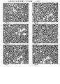
- FIG. 1 is a photograph replacing a drawing showing cultured chondrocytes from the 0th day to the 16th day of culture in Example 1.
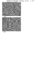
- FIG. 2 is a photograph replacing a drawing showing cultured chondrocytes from the 19th day to the 41st day in culture in Example 1.
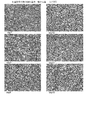
- FIG. 3 is a photograph replacing a drawing showing cultured chondrocytes from the first day to the fifth day of culture in Comparative Example 1.
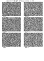
- FIG. 4 is a photograph replacing a drawing showing cultured chondrocytes from the first day to the thirteenth day of culture in Example 2 (subculture).
- FIG. 5 is a photograph replacing a drawing showing cultured chondrocytes from the 18th day to the 28th day in culture in Example 2.
- FIG. 1 is a photograph replacing a drawing showing cultured chondrocytes from the 0th day to the 16th day of culture in Example 1.
- FIG. 2 is a photograph replacing a drawing showing cultured chondrocytes from the 19th day to
- FIG. 6 is a photograph replacing a drawing showing cultured chondrocytes from the 29th day to the 52nd day.
- FIG. 7 is a photograph replacing a drawing showing cultured chondrocytes from the first day to the thirteenth day of culture in Comparative Example 2.
- cartilage was cultured in the same manner as in Example 2 except that a DMEM medium containing no SAG was used.
- FIG. 8 is a photograph replacing a drawing showing a state in which human auricular cartilage cultured in a serum-free medium was transplanted to the back of a nude mouse and cartilage was formed after 6 months.
- FIG. 9 is a photograph replacing a drawing showing a specimen obtained by extracting cartilage formed on the back of a nude mouse.
- FIG. 10 shows the results of transplanting human auricular cartilage cultured in a serum-free medium (SAG + ITS) to the back of a nude mouse and staining the formed cartilage section with toluidine blue, Alicia blue or EVG (Elastica Vangelon). It is the photograph replaced with drawing.
- FIG. 11 replaces the drawing showing the results of transplanting human auricular cartilage cultured in serum-free medium (Kartogenin + ITS) on the back of nude mice and staining the formed cartilage sections with toluidine blue, alician blue or EVG It is a photograph.
- FIG. 12 is a graph showing the relationship between the SAG concentration and the proliferation of cultured chondrocytes.
- FIG. 13 is a graph showing the relationship between the SAG concentration and the growth of cultured chondrocytes when ITS and SAG are added to the medium.
- FIG. 14 is another graph showing the relationship between the SAG concentration and the growth of cultured chondrocytes when ITS and SAG are added to the medium.
- the present invention relates to a serum-free culture method for chondrocytes.
- This method includes an enzyme treatment step and a culture step.
- This method may include an inhibitor treatment step.
- the enzyme treatment step is a step of treating a tissue containing human chondrocytes with a proteolytic enzyme. Specifically, it is a step of bringing a tissue containing human chondrocytes into contact with a proteolytic enzyme and decomposing the protein contained in the tissue containing human chondrocytes with a proteolytic enzyme. Since the treatment with a proteolytic enzyme itself is already known, this step may be performed based on a known method.
- human chondrocytes are auricular cartilage, nasal septal cartilage, costal cartilage, articular cartilage, intervertebral cartilage, tracheal cartilage, chondrocytes collected from the epiglottis, and human chondrocytes cultured from human genes.
- Methods for collecting human chondrocytes are known. That is, human chondrocytes may be surgically collected from a patient. In the present invention, human chondrocytes may be obtained by appropriately using cells that can differentiate into chondrocytes.
- Examples of cells that can be differentiated into chondrocytes may be human or warm-blooded bone marrow-derived cells, articular cartilage-derived cells, skin-derived cells, embryonic cells, and fetal-derived cells.
- Human mesenchymal stem cells eg, dedifferentiated normal human chondrocytes.
- human cartilage tissue and aggregated chondrocytes are treated with a proteolytic enzyme in order to make single cells.
- This process is a digestion process in which a part of the protein contained in human cartilage tissue or aggregated cells is degraded by a proteolytic enzyme.
- proteolytic enzymes are trypsin or collagenase.
- Collagenase is a known compound as disclosed in, for example, Japanese Patent No. 4764006, Japanese Patent No. 5322637, and Japanese Translation of PCT International Publication No. 2009-540826.
- the collagenase is preferably a collagenase that does not contain animal-derived components.
- a collagenase that does not contain animal-derived components may be purchased.
- This solution may appropriately contain known antibiotics such as penicillin and streptomycin.
- a solution containing a proteolytic enzyme may be prepared, and human chondrocytes may be immersed in this solution. In this way, a part of the protein contained in human chondrocytes can be degraded with a proteolytic enzyme.
- a proteolytic enzyme such as collagenase acts on human chondrocytes to such an extent that human chondrocytes are not completely degraded.
- the proteolytic enzyme is contained in the solution in an amount of 0.01% by weight to 1% by weight (or 0.1% by weight to 0.5% by weight). And it is preferable to adjust immersion time so that it may be 3 hours or more and 24 hours or less, for example.
- the inhibitor treatment step is a step of treating the tissue with a protease inhibitor after the enzyme treatment step.
- This step is a step of bringing a tissue treated with a proteolytic enzyme into contact with an inhibitor of the proteolytic enzyme and performing a treatment for inhibiting the function of the proteolytic enzyme.
- inhibitors are trypsin inhibitors or collagenase inhibitors.
- trypsin inhibitors and collagenase inhibitors are soybean derived protease inhibitors.
- Other examples of collagenase inhibitors are retinoic acid disclosed in Japanese Patent No. 5044071 and 8-hydroxyquinoline derivatives disclosed in Japanese Patent Application Laid-Open No. 5-97674.
- Retinol acid may be prepared in the form of a salt or solvate.
- This inhibitor treatment step can also be performed in the same manner as the previous enzyme treatment step.
- a solution containing a protease inhibitor may be prepared, and human chondrocytes may be immersed in this solution. In this way, the function of the proteolytic enzyme contained in human chondrocytes can be inhibited.
- the present invention can effectively culture good chondrocytes.
- the present invention intentionally treats the collected chondrocytes with collagenase or trypsin to decompose unsuitable components after culturing or transplanting the chondrocytes.
- trypsin continues to function, soft cells are damaged and chondrocyte culture does not proceed. Therefore, the present invention inhibits the function of proteolytic enzymes such as trypsin in the inhibitor treatment step.
- a protease-like component is present in serum, it is not necessary to add a protease inhibitor to the serum-containing medium. On the other hand, since there are no enzyme inhibitor-like components in serum-free medium, an enzyme inhibitor-like component is added to inhibit the function of proteolytic enzymes. In the case of using collagenase, a collagenase inhibitor may be used. However, since the action of collagenase is not strong, it is not necessary to use a collagenase inhibitor.
- the culture step is a step of culturing the tissue in a serum-free medium after the inhibitor treatment step.
- Serum-free culture of chondrocytes is already known as described above. Therefore, a well-known culture method may be adopted as appropriate in the present invention.
- the serum-free medium preferably contains a smoothed agonist.
- a smoothened or smoothed receptor is a protein that activates sonic hedgehog (Shh), for example, as introduced in Japanese Patent No. 5270362 and Japanese Patent No. 5424923.
- the smoothened agonist is preferably SAG or SAG1.1.
- SAG is known by CAS number 364590-63-6, and its chemical name is N-methyl-N ′-(3-pyridinylbenzyl) -N ′-(3-chlorobenzo [b] thiophene-2-carbonyl) It is a compound that is 1,4-diaminocyclohexane (N-Methyl-N ′-(3-pyridinylbenzoyl) -N ′-(3-chlorobenzo [b] thiophene-2-carbonyl) -1,4-diaminocyclohexane).
- SAG is a protein that activates sonic hedgehog (Shh), as disclosed in the pamphlet of International Publication No.
- SAG1.1 has the chemical name N-methyl-N ′-(3- (4-benzonitrile) -4-methoxybenzyl) -N- (3-chlorobenzo [b] thiophene-2-carbonyl) -1, 4-diaminocyclohexane (N-Methyl-N- (3- (4-benzonitrile) -4-methoxybenzoyl) -N ′-(3-chlorobenzo [b] thiophene-2-carbonyl) -1,4-diaminocyclohexane)) is there.
- SAG see Sinha S, Chen JK. Nat Chem Biol. 2006 Jan; 2 (1): 29-30.
- the method for culturing chondrocytes may be appropriately performed based on normal experience and knowledge. What is necessary is just to use what added the reagent suitably to the basic culture medium as a serum-free culture medium.
- the amount of the reagent may be used by appropriately modifying a known amount.
- the present invention also provides a serum-free medium.
- This serum-free medium preferably contains a smoothed agonist (eg, SAG).
- SAG smoothed agonist
- This medium is preferably used for culturing chondrocytes. For this reason, this serum-free medium is a serum-free medium containing chondrocytes during culture.
- the medium of the present invention preferably contains kartogenin.
- the name of cultogenin is 2-[(biphenyl-4-yl) carbamoyl] benzene acid (2-[(Biphenyl-4-yl) carbamoyl] benzoic acid) and is commercially available.
- the medium of the present invention preferably contains either or both of cultogenin and SAG, and may contain only cultogenin or only SAG, or may contain both cultogenin and SAG. There may be.
- the serum-free medium of the present invention necessary elements may be added as appropriate to the basic medium.
- basal media are Eagle basal media and DMEM.
- the reagent include steroids such as SAG (or SAG1.1), FGF2 (beads), IGF (insulin-like growth factor), insulin, HC (hydrocortisone), PDGF (plateletlegrowth factor), ACTH (adrenal cortex).
- Stimulating hormone LIF (leukemia inhibitory factor), TGF ⁇ , BMP, steroid, fatty acid, soybean trypsin inhibitor, ascorbic acid, hyaluronic acid, proline, dexamethasone, insulin, transferrin, selenite.
- a salt form such as 2-phosphate may be added.
- Japanese Patent No. 5228187 discloses a chondrocyte culture medium composition containing glucuronic acid. Also in the present invention, for example, the medium additives described in this publication may be appropriately used.
- Insulin is preferably added as ITS.
- ITS is a reagent containing insulin, transferrin, and sodium selenite, and its compositional examples are insulin 5 ⁇ g / ml, transferrin 5 ⁇ g / ml, and sodium selenite 5 ng / ml. Since ITS is commercially available, commercially available products may be used as appropriate. ITS is preferably animal-free without animal-derived components. In the present invention, it is preferable to use a raw material which basically does not contain animal-derived components.
- SAG cultogenin
- FGF2 insulin-like growth factor
- IGF insulin-like growth factor
- steroids such as insulin, hydrocortisone, PDGF, ACTH (adrenocorticotropic hormone), LIF (leukemia inhibitory factor), TGF ⁇ , BMP, steroid, and ITS, for example, What is necessary is just to add to a culture medium so that it may become 0.1 ng / mL or more and 20 microgram / mL or less (or 0.2 ng / mL or more and 10 microgram / mL or less), respectively. These may be added by appropriately adjusting according to the degree of purification and the required amount.
- the serum-free medium is added to the basic medium with SAG (for example, 0.05 to 5 ⁇ M, preferably 0.1 to 2 ⁇ M, preferably 0.2 to 1 ⁇ M), FGF2 (for example, 1 ng / mL to 1 ⁇ g / mL or less), IGF (for example, 1 ng / mL to 50 ⁇ g / mL or less), and HC (for example, 1 ng / ML to ⁇ g / mL or less) added are preferred.
- SAG for example, 0.05 to 5 ⁇ M, preferably 0.1 to 2 ⁇ M, preferably 0.2 to 1 ⁇ M
- FGF2 for example, 1 ng / mL to 1 ⁇ g / mL or less
- IGF for example, 1 ng / mL to 50 ⁇ g / mL or less
- HC for example, 1 ng / ML to ⁇ g / mL or less
- the above cells are cultured and grown under normal culture conditions (for example, 37 ° C., 5% CO 2 ).
- the amount of cells can range from about 10% to 100% confluent, and can be cultured in a high-density, multi-layered state exceeding 100% confluent.
- a commercially available nitrogen gas can be mixed to reduce the oxygen partial pressure, and a low oxygen incubator can be used.
- nitrogen gas can be blown into an appropriate space to reduce the oxygen partial pressure. May be cultured.
- the culture process performed in the examples is as follows. That is, the above SAG was added to the medium. Ascorbic acid and fatty acids are added to the medium as appropriate. As a result of repeated experiments, chondrocytes did not grow well when ascorbic acid or fatty acids were included from the beginning of the culture. On the other hand, chondrocytes markedly increased when ascorbic acid or fatty acid was added after a predetermined period of time from the start of culture. Therefore, it is preferable to add ascorbic acid, a salt of ascorbic acid or a solvate of ascorbic acid to the serum-free medium during the period from the 3rd day to the 28th day after the start of the culture.
- the ascorbic acid may be added during the period from the 5th day to the 25th day from the start of the culture, from the 10th day to the 22nd day, or from the 14th day to the 21st day. It may be a period. Moreover, the period which shortened or extended these periods suitably may be sufficient. Moreover, it is preferable to add a fatty acid, a fatty acid salt, or a fatty acid solvate to the serum-free medium during the period from the 3rd day to the 28th day from the start of the culture.
- the addition time of fatty acids, etc. may be from the period from the 5th day to the 2nd month from the start of the culture, from the period from the 10th day to the 40th day, or from the 19th day. It may be the 25th day.
- Chondrocytes or cartilage produced by the method of the present invention may be used in, for example, joint diseases (eg, fractures in the orthopedic region, re-fractures, bone deformities / degenerative spondylosis, osteosarcoma, myeloma, dysplasia, scoliosis, etc.
- joint diseases eg, fractures in the orthopedic region, re-fractures, bone deformities / degenerative spondylosis, osteosarcoma, myeloma, dysplasia, scoliosis, etc.
- Non-metabolic bone diseases bone defects, osteoporosis, osteomalacia, rickets, fibro-osteitis, renal osteodystrophy, osteophetotic disease, ankylosing myelitis, etc .; osteoarthritis , Cartilage diseases such as rheumatoid arthritis), etc., and can be used for cartilage transplantation performed as a surgical technique for repairing cartilage.
- the cartilage transplantation method can be performed using conventional means. Therefore, the chondrocytes or cartilage produced by the method of the present invention can be used as a safe and low-toxic preventive / therapeutic agent for the above joint diseases.
- this culture system it is possible to search for genes involved in cartilage differentiation and drug search.
- Chondrocytes cultured as described above form an aggregate of cells. Therefore, for example, after culturing, a culture of cartilage tissue can be effectively recovered by inserting a rod from the lateral direction into the medium and pulling the rod upward. Normally, it is necessary to peel the cartilage tissue from the container in order to recover the cultured cells. For this reason, in a normal method, a proteolytic enzyme such as collagenase or trypsin is allowed to act to detach the chondrocytes from the container. For this reason, only dissociated cartilage tissues obtained by culturing were obtained. In the culturing method of the present invention, the aggregate formed by stacking chondrocytes is in a state where it is easily detached from the container. it can.
- a DMEM medium supplemented with FGF 100 ng / mL, SAG 0.5 ⁇ M, HC 400 ng / mL, IGF 5 ng / mL and insulin 5 ⁇ g / mL was used as the medium.
- the previously obtained cultured cells were seeded at a density of 1000 cells / cm 2 .
- the culture was performed in an environment of 37 ° C. and 5% CO 2 .
- FIG. 1 is a photograph replacing a drawing showing cultured chondrocytes from day 0 to day 16 of culture in Example 1. It can be seen that the cartilage tissue gradually increased.
- FIG. 2 is a photograph replacing a drawing showing cultured chondrocytes from the 19th day to the 41st day in culture in Example 1.
- Ascorbic acid was added to the medium.
- Fatty acids were added from 1: 1000 to 1: 500 on the third week of culture. Adding fatty acid increased the number of cells and increased the viscosity of the medium.
- the fatty acids and cholesterol added during the culture are as follows.
- the above addition amount may be increased or decreased within a range of ⁇ 200%, preferably ⁇ 100%.
- the range of 1:50 to 1: 2000 may be sufficient as the range of a fatty acid.
- trypsin was used instead of collagenase, and the collected cells were immersed in a soybean-derived protease inhibitor that is a trypsin inhibitor.
- FIG. 3 is a photograph replacing a drawing showing cultured chondrocytes from the first day to the fifth day of culture in Comparative Example 1.
- cartilage was cultured in the same manner as in Example 1 except that a DMEM medium containing no SAG was used. As shown in FIG. 3, it was shown that chondrocytes could not be effectively cultured in a state not containing SAG.
- Ascorbic acid and fatty acid addition timing adjustment Ascorbic acid was added at the start of culture, 1 hour, 3 hours, 12 hours, 1 day, 2 days, 3 days, 3 days, 5 days, 1 week, and 10 days after the start of culture. 2 weeks, 20 days, 3 weeks, 25 days, 4 weeks, 30 days, 5 weeks, 40 days, 6 weeks, 45 days, 7 weeks, 50 days, 8 weeks, 60 days, Nine weeks later, 70 days later started. Ascorbic acid was added periodically (for example, every 12 hours, every day, every two days). The addition of fatty acid was adjusted in the same manner as the addition of ascorbic acid. As a result, it was found that it is preferable to add ascorbic acid after about 3 days rather than adding ascorbic acid immediately after the start of culture. The addition of fatty acid was similar to that of ascorbic acid, but it was found that the case where fatty acid was added after addition of ascorbic acid was more desirable.
- a DMEM medium supplemented with FGF2 at 100 ng / mL, SAG 0.5 ⁇ M, HC 400 ng / mL, IGF 5 ng / mL and insulin 5 ⁇ g / mL was used as a subculture medium.
- the cultured cells were seeded at a density of 10,000 cells / cm 2 .
- the culture was performed in an environment of 37 ° C. and 5% CO 2 .
- FIG. 4 is a photograph replacing a drawing showing cultured chondrocytes from the first day to the thirteenth day of culture in Example 2 (subculture). About 10 days after the start of the culture, the medium was found to be viscous, and when the medium was removed with an aspirator, it was observed that a thin thread was pulled. From the 13th day, the viscosity of the medium increased.
- FIG. 5 is a photograph replacing a drawing showing cultured chondrocytes from the 18th day to the 28th day in culture in Example 2.
- Ascorbic acid was added to the medium.
- Fatty acids were added from 1: 1000 to 1: 500 on the third week of culture. Adding fatty acid increased the number of cells and increased the viscosity of the medium.
- FIG. 6 is a photograph replacing a drawing showing cultured chondrocytes from the 29th day to the 52nd day. When the medium was removed with an aspirator, a thick thread was drawn. As shown in the figure, it was shown that chondrocytes can be effectively cultured by culturing chondrocytes using a serum-free medium.
- the fatty acids and cholesterol added during the culture are as follows.
- the above addition amount may be increased or decreased within a range of ⁇ 200%, preferably ⁇ 100%.
- the range of 1:50 to 1: 2000 may be sufficient as the range of a fatty acid.
- FIG. 7 is a photograph replacing a drawing showing cultured chondrocytes from the first day to the thirteenth day of culture in Comparative Example 2.
- cartilage was cultured in the same manner as in Example 2 except that a DMEM medium containing no SAG was used. As shown in FIG. 7, it was shown that chondrocytes could not be effectively cultured in a state not containing SAG.
- chondrocytes cultured in the serum-free medium of the present invention are cells suitable for transplantation, they were transplanted into nude mice and examined whether cartilage was formed in vivo.
- chondrocytes derived from human auricular cartilage were prepared according to the procedure described in Example 1 and cultured in the serum-free medium of the present invention.
- the cultured chondrocytes were immersed in collagen cotton, and then transplanted with the cotton into two places on the back of nude mice.
- Transplantation and nude mouse management were performed according to known methods. Six months after transplantation, the back of a nude mouse was incised, and it was confirmed whether or not the cells were fixed at the transplant site and cartilage was formed.
- FIG. 8 is a photograph showing a situation in which the back of a nude mouse was incised 6 months after transplantation.
- Large white cartilage (A-2, B-2) was confirmed at the site (left and right back) where the cultured chondrocytes were transplanted. Therefore, it was considered that the cultured cells settled and proliferated at the transplant site to form cartilage.
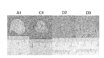
- FIG. 9 shows the cartilage formed on the back of a nude mouse.
- A1 shows cartilage formed in a nude mouse transplanted with chondrocytes cultured under the following culture conditions (A) (SAG + ITS). ITS used the animal free thing.
- C1 indicates cartilage formed in a nude mouse transplanted with chondrocytes which were cultured under the following culture conditions (C) (Kartogenin + ITG) and the medium was liable to separate and had the lowest viscosity.
- the chondrocytes cultured under the culture condition (A) formed larger cartilage than the chondrocytes cultured under the culture condition (C).
- cartilage was not formed (D2 and D3).
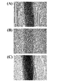
- FIG. 10 shows the staining result of A1 cartilage.
- FIG. 10 (A) shows the result of toluidine blue staining, and the cartilage was stained purple.
- FIG. 10 (B) shows the results of Alician blue staining, and the cartilage was stained blue. Therefore, it was confirmed that the cartilage of A1 is positive for both stainings and has characteristics as cartilage.
- FIG. 10C shows the result of EVG staining, and the cartilage was stained brown. Therefore, it was proved to be elastic cartilage and identified as auricular cartilage.
- FIG. 11 shows the staining result of C1 cartilage.
- FIG. 11A shows the results of toluidine blue staining, and the cartilage was stained purple.
- FIG. 11 (B) shows the results of Alician blue staining, where the cartilage was stained blue. Therefore, it was confirmed that the C1 cartilage is positive for both stainings and has characteristics as a cartilage.
- FIG. 11C shows the result of EVG staining, and the cartilage was stained brown. Therefore, it was proved to be elastic cartilage and identified as auricular cartilage.
- the chondrocytes cultured in the serum-free medium of the present invention are cells suitable for transplantation, and when transplanted into nude mice, cartilage can be formed at the transplant site.
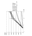
- the SAG concentration in the serum-free medium was changed to 0.05 uM, 0.01 uM, or 0.5 uM, and the influence on the proliferation of cultured chondrocytes was examined. Specifically, chondrocytes prepared by the same procedure as in Example 1 were seeded at a density of 10,000 cells / well, and the number of cells at 4, 7, 14, and 21 after seeding was counted. The results are shown in FIG. At any SAG concentration, there was almost no difference in cell growth until the 14th day of culture, and no significant difference as expected was observed on the 21st day of culture.
- ITS Insulin-Transferrin-Selenium
- the cultured chondrocytes are cultured by changing the SAG concentration of the serum-free medium supplemented with ITS to 0.01 uM, 0.05 uM, 0.1 uM or 0.5 uM, and 4, 7, 14 after seeding. The number of cells in the 21st eye was counted. The results are shown in FIG.
- the effect on the proliferation of chondrocytes was lowest in the treatment group (ITS-SAG-) to which neither ITS nor SAG was added.
- ITS-SAG- In the treatment group to which ITS was added alone (ITS + SAG-), a slight growth effect was observed.
- the effect was small compared with the treatment section to which both ITS and SAG were added. Therefore, the effect of ITS on the proliferation of chondrocytes is not so strong, and it can be said that a remarkable effect is obtained by combining with SAG.
- the proliferative effect of chondrocytes was also observed in the treatment group (ITS + Kartogenin 1 uM) combining ITS and Kartogenin. However, the effect was small compared to the combination of ITG and SAG.
- the serum-free medium and chondrocyte culture method of the present invention can be used to produce cultured cartilage. Therefore, it can be used in the field of the present invention and the pharmaceutical industry.
Landscapes
- Health & Medical Sciences (AREA)
- Engineering & Computer Science (AREA)
- Life Sciences & Earth Sciences (AREA)
- Chemical & Material Sciences (AREA)
- Biomedical Technology (AREA)
- Wood Science & Technology (AREA)
- Biotechnology (AREA)
- Organic Chemistry (AREA)
- Bioinformatics & Cheminformatics (AREA)
- Genetics & Genomics (AREA)
- Zoology (AREA)
- Rheumatology (AREA)
- General Engineering & Computer Science (AREA)
- Microbiology (AREA)
- Biochemistry (AREA)
- General Health & Medical Sciences (AREA)
- Cell Biology (AREA)
- Virology (AREA)
- Tropical Medicine & Parasitology (AREA)
- Medicinal Chemistry (AREA)
- Micro-Organisms Or Cultivation Processes Thereof (AREA)
- Peptides Or Proteins (AREA)
Abstract
Description
ヒト軟骨細胞を含む組織を,コラゲナーゼで処理する酵素処理工程と,
組織を無血清培地で培養する培養工程と,
を,この順で含み,
無血清培地は,カルトゲニン又はSAGのいずれか又は両方と,
ITS,FGF2,及びヒドロコルチゾンを含む,
軟骨細胞の無血清培養方法であって,
培養工程は,
培養開始から3日目から28日目の期間にアスコルビン酸,アスコルビン酸の塩又はアスコルビン酸の溶媒和物を無血清培地に添加し,
培養開始から3日目から28日目の期間に脂肪酸,脂肪酸の塩,又は脂肪酸の溶媒和物を無血清に添加する工程を含む,
方法である。
上記のようにして培養された軟骨細胞は,細胞が積み重なってできた集合体を形成している。そのため,例えば,培養後,培地に横方向から棒を挿入し,その棒を上方へ引き上げると,軟骨組織の培養物を効果的に回収することができる。通常であれば,培養細胞を回収するために,容器から軟骨組織をはがす必要がある。このため,通常の方法では,コラゲナーゼやトリプシンといったタンパク質分解酵素を作用させて容器から軟骨細胞を剥離していた。このため,培養して得られる軟骨組織は,ばらばらのものしか得られなかった。本発明の培養方法では,軟骨細胞が積み重なってできた集合体が,容器から剥がれやすい状態となっているため,上記の回収方法を用いるだけで,例えばシート状や塊状の軟骨組織を容易に回収できる。
初代ヒト軟骨細胞を関節軟骨生検から単離し,その試料を細分した。その後,0.1%から0.3%コラゲナーゼ溶液に浸漬し,37℃にて4から5時間酵素的消化を行なった。得られた細胞を5分間,1,200rpmで遠心分離して回収した。
濃度 mg/mL
アラキドン酸 2.0
リノール酸 10.0
リノレン酸 10.0
ミリスチン酸 10.0
オレイン酸 10.0
パルミチン酸 10.0
パルミトオレイン酸 10.0
ステアリン酸 10.0
コレステロール 220.0
DLα酢酸トコフェロール(ビタミンE) 70.0
図3は,比較例1における培養1日目から5日目までの培養軟骨細胞を示す図面に替わる写真である。この例では,SAGを含まないDMEM培地を用いた以外は実施例1と同様にして軟骨を培養した。図3に示されるとおり,SAGを含まない状態では,軟骨細胞を効果的に培養できないことが示された。
アスコルビン酸の添加を培養開始時,培養開始1時間後,3時間後,12時間後,1日後,2日後,3日後,5日後,1週間後,10日後,2週間後,20日後,3週間後,25日後,4週間後,30日後,5週間後,40日後,6週間後,45日後,7週間後,50日後,8週間後,60日後,9週間後,70日後のそれぞれから開始した。アスコルビン酸の添加は,定期的(例えば,12時間毎,1日毎,2日毎)とした。
脂肪酸の添加も,アスコルビン酸の添加と同様に調整した。
その結果,培養開始からすぐにアスコルビン酸を添加するよりも,3日程度経過した後からアスコルビン酸を添加することが望ましいことがわかった。
脂肪酸の添加も,アスコルビン酸の添加と同様であったが,アスコルビン酸の添加を行った後に,脂肪酸を添加したケースの方が望ましいことがわかった。
濃度 mg/ML
アラキドン酸 2.0
リノール酸 10.0
リノレン酸 10.0
ミリスチン酸 10.0
オレイン酸 10.0
パルミチン酸 10.0
パルミトオレイン酸 10.0
ステアリン酸 10.0
コレステロール 220.0
DLα酢酸トコフェロール(ビタミンE) 70.0
図7は,比較例2における培養1日目から13日目までの培養軟骨細胞を示す図面に替わる写真である。この例では,SAGを含まないDMEM培地を用いた以外は実施例2と同様にして軟骨を培養した。図7に示されるとおり,SAGを含まない状態では,軟骨細胞を効果的に培養できないことが示された。
まず,ヒト耳介軟骨由来の軟骨細胞を,実施例1で述べた手順に従って準備し,本発明の無血清培地で培養した。培養後の軟骨細胞をコラーゲンの綿に浸漬させ,次いで,この綿と共にヌードマウスの背部の2か所に移植した。移植手術およびヌードマウスの管理は,公知の方法に従って行った。移植の6ヶ月後,ヌードマウスの背部を切開し,移植部位に細胞が定着し,軟骨が形成されているか否かを確認した。
DME(H):F―12=1:1
SAG 0.1uM
FGF 50ng/mL
HC 400ng/mL
ITS ×1
培養8日目からアスコルビン酸 50ug/mL,脂肪酸 1:500を添加
DME(H):F―12=1:1
Kartogenin 1uM
FGF 50ng/mL
HC 400ng/mL
ITS ×1
培養8日目からアスコルビン酸 50ug/mL,脂肪酸 1:500を添加
また,図10(C)は,EVG染色の結果を示しており,軟骨は褐色に染色された。従って,弾性軟骨であることが証明され,耳介軟骨であることが同定された。
また,図11(C)は,EVG染色の結果を示しており,軟骨は褐色に染色された。従って,弾性軟骨であることが証明され,耳介軟骨であることが同定された。
いずれのSAG濃度においても,培養14日目まで細胞の増殖にほとんど差は見られず,培養21日目においても,想定したほどの顕著な差は見られなかった。
また,SAGではなく,カルトゲニン(Kartogenin)を添加した場合にも,軟骨細胞の増殖に効果的であることが明らかとなった。
また,ITSとKartogeninを組み合わせた処理区(ITS+ Kartogenin 1uM)でも,軟骨細胞の増殖効果が見られることが分かった。ただし,ITGとSAGの組み合わせた場合に比べると,その効果は小さかった。
Claims (7)
- カルトゲニン又はSAGのいずれか又は両方と,
ITS,FGF2,及びヒドロコルチゾンを含む軟骨細胞培養用の無血清培地。 - 請求項1に記載の培地であって,カルトゲニン及びSAGの両方を含む培地。
- 請求項1に記載の培地であって,IGF(インスリン様増殖因子)を更に含む培地。
- 請求項1に記載の培地であって,培養対象である軟骨細胞を更に含む培地。
- 請求項1に記載の培地と,アスコルビン酸剤及び脂肪酸剤を含む培地キットであって,
前記アスコルビン酸剤は,アスコルビン酸,アスコルビン酸の塩又はアスコルビン酸の溶媒和物を含み,
前記脂肪酸剤は,脂肪酸,脂肪酸の塩,又は脂肪酸の溶媒和物を含む,
培地キット。 - ヒト軟骨細胞を含む組織を,コラゲナーゼで処理する酵素処理工程と,
前記組織を無血清培地で培養する培養工程と,
を,この順で含み,
前記無血清培地は,カルトゲニン又はSAGのいずれか又は両方と,
ITS,FGF2,及びヒドロコルチゾンを含む,
軟骨細胞の無血清培養方法であって,
前記培養工程は,
培養開始から3日目から28日目の期間にアスコルビン酸,アスコルビン酸の塩又はアスコルビン酸の溶媒和物を前記無血清培地に添加し,
培養開始から3日目から28日目の期間に脂肪酸,脂肪酸の塩,又は脂肪酸の溶媒和物を前記無血清に添加する工程を含む,
方法。 - 請求項6の方法であって,前記酵素処理工程の後に,前記組織をコラゲナーゼ阻害剤で処理する阻害剤処理工程を更に含む,方法。
Priority Applications (7)
| Application Number | Priority Date | Filing Date | Title |
|---|---|---|---|
| CA2977354A CA2977354C (en) | 2014-11-14 | 2015-10-26 | Method for serum-free culture of chondrocytes and serum-free culture medium |
| CN201580024032.8A CN106459912B (zh) | 2014-11-14 | 2015-10-26 | 软骨细胞的无血清培养方法、及无血清培养基 |
| EP15859245.1A EP3219792B1 (en) | 2014-11-14 | 2015-10-26 | Method for serum-free culture of cartilage cells and serum-free culture medium |
| JP2016558958A JP6533234B2 (ja) | 2014-11-14 | 2015-10-26 | 軟骨細胞の無血清培養方法,及び無血清培地 |
| AU2015346672A AU2015346672B2 (en) | 2014-11-14 | 2015-10-26 | Method for serum-free culture of cartilage cells and serum-free culture medium |
| EP19171369.2A EP3536778B1 (en) | 2014-11-14 | 2015-10-26 | Method for serum-free culture of chondrocytes and serum-free culture medium |
| US15/310,442 US11427808B2 (en) | 2014-11-14 | 2015-10-26 | Method for serum-free culture of chondrocytes and serum-free culture medium |
Applications Claiming Priority (2)
| Application Number | Priority Date | Filing Date | Title |
|---|---|---|---|
| JP2014231489 | 2014-11-14 | ||
| JP2014-231489 | 2014-11-14 |
Publications (1)
| Publication Number | Publication Date |
|---|---|
| WO2016076102A1 true WO2016076102A1 (ja) | 2016-05-19 |
Family
ID=55954194
Family Applications (1)
| Application Number | Title | Priority Date | Filing Date |
|---|---|---|---|
| PCT/JP2015/080139 Ceased WO2016076102A1 (ja) | 2014-11-14 | 2015-10-26 | 軟骨細胞の無血清培養方法,及び無血清培地 |
Country Status (7)
| Country | Link |
|---|---|
| US (1) | US11427808B2 (ja) |
| EP (2) | EP3536778B1 (ja) |
| JP (3) | JP6533234B2 (ja) |
| CN (2) | CN112980780A (ja) |
| AU (1) | AU2015346672B2 (ja) |
| CA (1) | CA2977354C (ja) |
| WO (1) | WO2016076102A1 (ja) |
Cited By (3)
| Publication number | Priority date | Publication date | Assignee | Title |
|---|---|---|---|---|
| JP2021126094A (ja) * | 2020-02-17 | 2021-09-02 | 義昭 工藤 | コンドロイチン硫酸型プロテオグリカン及びヒアルロン酸の製造方法 |
| WO2022158152A1 (ja) * | 2021-01-20 | 2022-07-28 | 株式会社リジェネシスサイエンス | 成熟軟骨細胞および成熟軟骨細胞含有組成物の製造方法 |
| JPWO2023190271A1 (ja) * | 2022-03-28 | 2023-10-05 |
Families Citing this family (3)
| Publication number | Priority date | Publication date | Assignee | Title |
|---|---|---|---|---|
| CN107674859B (zh) * | 2017-08-28 | 2021-01-05 | 浙江大学 | 一种利用小分子组合物诱导小鼠成纤维细胞成软骨的方法 |
| CN110172444B (zh) * | 2019-06-28 | 2023-05-23 | 领航干细胞再生医学工程有限公司 | 一种人软骨干细胞的制备方法 |
| CN117230002A (zh) * | 2022-06-06 | 2023-12-15 | 友康生物科技(北京)股份有限公司 | 一种软骨细胞培养基 |
Citations (5)
| Publication number | Priority date | Publication date | Assignee | Title |
|---|---|---|---|---|
| JP2000515023A (ja) * | 1996-07-25 | 2000-11-14 | ジェンザイム・コーポレーション | 軟骨細胞培地組成および培養方法 |
| JP2002529071A (ja) * | 1998-11-09 | 2002-09-10 | コンソルツィオ・ペール・ラ・ジェスティオーネ・デル・チェントロ・ディ・ビオテクノロジー・アヴァンツァテ | 軟骨細胞様細胞のための無血清培地 |
| JP2008136396A (ja) * | 2006-11-30 | 2008-06-19 | Yokohama City Univ | 軟骨細胞の培養方法 |
| JP2009540826A (ja) * | 2006-06-20 | 2009-11-26 | ジェンザイム・コーポレーション | 軟骨細胞増幅のための無血清培地およびその使用 |
| WO2014084085A1 (ja) * | 2012-11-28 | 2014-06-05 | 国立大学法人名古屋大学 | 聴覚障害又は小脳性運動失調症の予防・治療剤 |
Family Cites Families (14)
| Publication number | Priority date | Publication date | Assignee | Title |
|---|---|---|---|---|
| JPS5424923B2 (ja) | 1971-09-21 | 1979-08-24 | ||
| FR2200935A5 (ja) | 1972-09-27 | 1974-04-19 | Hydromatik Gmbh | |
| JPS5322637B2 (ja) | 1973-07-27 | 1978-07-10 | ||
| JPS5955043A (ja) | 1982-09-24 | 1984-03-29 | Mitsubishi Metal Corp | 電子装置の構造部材用ろう付け複合材の製造法 |
| JPH0597674A (ja) | 1991-10-01 | 1993-04-20 | Nikko Kyodo Co Ltd | コラーゲナーゼ阻害剤 |
| FR2779058B1 (fr) | 1998-05-29 | 2003-02-21 | Dior Christian Parfums | Utilisation d'au moins une saponine ou un sapogenol cosmetiquement acceptable, comme agent cosmetique destine a augmenter la quantite de collagene iv dans la jonction dermo-epidermique |
| JP2003125787A (ja) | 2001-06-26 | 2003-05-07 | Takeda Chem Ind Ltd | 軟骨分化培養法 |
| EP1418185A1 (en) | 2002-11-11 | 2004-05-12 | Aventis Pharma Deutschland GmbH | Use of EDG2 receptor in an animal model of heart failure |
| JP5270362B2 (ja) | 2005-11-10 | 2013-08-21 | ジェナボン バイオファーマシューティカルズ エルエルシー | 幹細胞のmntf分化および成長 |
| CN101370511B (zh) * | 2005-11-23 | 2015-07-01 | 阿塞勒隆制药公司 | Activin-ActRIIa拮抗剂及其促进骨骼生长的应用 |
| WO2013065763A1 (ja) * | 2011-10-31 | 2013-05-10 | 独立行政法人理化学研究所 | 幹細胞の培養方法 |
| US9421245B2 (en) | 2012-06-25 | 2016-08-23 | The Brigham And Women's Hospital, Inc. | Targeted therapeutics |
| JP2014176353A (ja) * | 2013-03-15 | 2014-09-25 | Kazuo Todokoro | ヒト造血幹細胞を増幅させるための組成物及び方法 |
| CN104120105A (zh) | 2013-04-26 | 2014-10-29 | 杭州龙禧生物医药科技有限公司 | 一种用于培养老龄人群软骨细胞的无血清培养基 |
-
2015
- 2015-10-26 EP EP19171369.2A patent/EP3536778B1/en active Active
- 2015-10-26 CN CN202110494081.5A patent/CN112980780A/zh active Pending
- 2015-10-26 AU AU2015346672A patent/AU2015346672B2/en not_active Ceased
- 2015-10-26 CA CA2977354A patent/CA2977354C/en active Active
- 2015-10-26 US US15/310,442 patent/US11427808B2/en active Active
- 2015-10-26 WO PCT/JP2015/080139 patent/WO2016076102A1/ja not_active Ceased
- 2015-10-26 JP JP2016558958A patent/JP6533234B2/ja active Active
- 2015-10-26 CN CN201580024032.8A patent/CN106459912B/zh active Active
- 2015-10-26 EP EP15859245.1A patent/EP3219792B1/en active Active
-
2019
- 2019-05-23 JP JP2019096659A patent/JP6753981B2/ja active Active
-
2020
- 2020-08-20 JP JP2020139666A patent/JP7008360B2/ja active Active
Patent Citations (5)
| Publication number | Priority date | Publication date | Assignee | Title |
|---|---|---|---|---|
| JP2000515023A (ja) * | 1996-07-25 | 2000-11-14 | ジェンザイム・コーポレーション | 軟骨細胞培地組成および培養方法 |
| JP2002529071A (ja) * | 1998-11-09 | 2002-09-10 | コンソルツィオ・ペール・ラ・ジェスティオーネ・デル・チェントロ・ディ・ビオテクノロジー・アヴァンツァテ | 軟骨細胞様細胞のための無血清培地 |
| JP2009540826A (ja) * | 2006-06-20 | 2009-11-26 | ジェンザイム・コーポレーション | 軟骨細胞増幅のための無血清培地およびその使用 |
| JP2008136396A (ja) * | 2006-11-30 | 2008-06-19 | Yokohama City Univ | 軟骨細胞の培養方法 |
| WO2014084085A1 (ja) * | 2012-11-28 | 2014-06-05 | 国立大学法人名古屋大学 | 聴覚障害又は小脳性運動失調症の予防・治療剤 |
Non-Patent Citations (4)
| Title |
|---|
| CHEN, J. K. ET AL.: "Small molecule modulation of Smoothened activity", PROCEEDINGS OF THE NATIONAL ACADEMY OF SCIENCES OF THE UNITED STATES OF AMERICA, vol. 99, no. 22, 21 October 2002 (2002-10-21), pages 14071 - 14076, XP002251705 * |
| JOHNSON, K. ET AL.: "A stem cell -based approach to cartilage repair", SCIENCE, vol. 336, no. 6082, 5 April 2012 (2012-04-05), pages 717 - 721, XP055317824 * |
| LONG, F. ET AL.: "Genetic manipulation of hedgehog signaling in the endochondral skeleton reveals a direct role in the regulation of chondrocyte proliferation", DEVELOPMENT, vol. 128, no. 24, 15 December 2001 (2001-12-15), pages 5099 - 5108, XP055317821 * |
| See also references of EP3219792A4 * |
Cited By (9)
| Publication number | Priority date | Publication date | Assignee | Title |
|---|---|---|---|---|
| JP2021126094A (ja) * | 2020-02-17 | 2021-09-02 | 義昭 工藤 | コンドロイチン硫酸型プロテオグリカン及びヒアルロン酸の製造方法 |
| JP7113436B2 (ja) | 2020-02-17 | 2022-08-05 | 義昭 工藤 | コンドロイチン硫酸型プロテオグリカン及びヒアルロン酸の製造方法 |
| WO2022158152A1 (ja) * | 2021-01-20 | 2022-07-28 | 株式会社リジェネシスサイエンス | 成熟軟骨細胞および成熟軟骨細胞含有組成物の製造方法 |
| JP2022111482A (ja) * | 2021-01-20 | 2022-08-01 | 株式会社リジェネシスサイエンス | 成熟軟骨細胞の製造方法 |
| JP7690737B2 (ja) | 2021-01-20 | 2025-06-11 | 株式会社リジェネシスサイエンス | 成熟軟骨細胞の製造方法 |
| JP2025124675A (ja) * | 2021-01-20 | 2025-08-26 | 株式会社リジェネシスサイエンス | 成熟軟骨細胞の製造方法 |
| JPWO2023190271A1 (ja) * | 2022-03-28 | 2023-10-05 | ||
| WO2023190271A1 (ja) * | 2022-03-28 | 2023-10-05 | 株式会社リジェネシスサイエンス | miR-140を含む医薬組成物及びその製造方法 |
| JP7773257B2 (ja) | 2022-03-28 | 2025-11-19 | 合同会社HiRoko Science | miR-140を含む医薬組成物及びその製造方法 |
Also Published As
| Publication number | Publication date |
|---|---|
| JP2019150057A (ja) | 2019-09-12 |
| JP6753981B2 (ja) | 2020-09-09 |
| CN106459912B (zh) | 2021-05-25 |
| US20170306294A1 (en) | 2017-10-26 |
| AU2015346672B2 (en) | 2017-09-21 |
| EP3219792A4 (en) | 2018-04-11 |
| JP6533234B2 (ja) | 2019-06-19 |
| AU2015346672A1 (en) | 2016-11-17 |
| CA2977354C (en) | 2021-09-21 |
| US11427808B2 (en) | 2022-08-30 |
| CN106459912A (zh) | 2017-02-22 |
| CN112980780A (zh) | 2021-06-18 |
| EP3536778A1 (en) | 2019-09-11 |
| JPWO2016076102A1 (ja) | 2017-10-05 |
| EP3219792B1 (en) | 2019-05-01 |
| JP2020191899A (ja) | 2020-12-03 |
| CA2977354A1 (en) | 2016-05-19 |
| EP3536778B1 (en) | 2021-05-19 |
| JP7008360B2 (ja) | 2022-02-10 |
| EP3219792A1 (en) | 2017-09-20 |
Similar Documents
| Publication | Publication Date | Title |
|---|---|---|
| JP7008360B2 (ja) | 軟骨細胞の無血清培養方法,及び無血清培地 | |
| CN1871343B (zh) | 从脂肪组织制备干细胞的方法和系统 | |
| CN103068969A (zh) | 化学成分确定的无血清细胞培养基 | |
| JPWO2018164228A1 (ja) | Ror1陽性の間葉系幹細胞を含有する、線維症を伴う疾患の予防又は処置のための医薬組成物、及びその調製方法、並びにror1陽性の間葉系幹細胞を用いる線維症を伴う疾患の予防又は処置方法 | |
| Smeringaiova et al. | Ex vivo expansion and characterization of human corneal endothelium for transplantation: a review | |
| CN114480273A (zh) | 用于获得间充质干细胞及其外泌体的培养基及其制备方法 | |
| Shen et al. | Enhancing the potential of aged human articular chondrocytes for high-quality cartilage regeneration | |
| Xie et al. | Generation of hyaline-like cartilage tissue from human mesenchymal stromal cells within the self-generated extracellular matrix | |
| CN101014699B (zh) | 去分化型软骨细胞向软骨细胞的再分化用培养基 | |
| CN101657536B (zh) | 软骨细胞制备方法 | |
| KR100394430B1 (ko) | 인간 혈청을 포함하는 인간 세포 배양용 배지 및 이를이용한 인간 세포의 배양 방법 | |
| JP2022545376A (ja) | 硝子軟骨組織のin vitroでの産生のための方法 | |
| WO2017170925A1 (ja) | Egfr及びmic-abからなる群より選択される少なくとも1種の細胞表面マーカーを高発現する間葉系幹細胞及びその調製方法、並びに上記間葉系幹細胞を含む医薬組成物及びその調製方法 | |
| US10415014B2 (en) | Method for the ex vivo cultivation of oral mucosal epithelial progenitor cells and oral mucosal epithelial cells | |
| US11471562B2 (en) | Cartilage material having minimal hypertrophy and robust integration capacity, and uses therefor | |
| RU2730864C1 (ru) | Способ производства культивируемых клеток и лекарственного препарата для лечения спинномозговых травм | |
| HK1230656A1 (en) | Method for serum-free culture of cartilage cells and serum-free culture medium | |
| HK1230656B (zh) | 软骨细胞的无血清培养方法、及无血清培养基 | |
| JP7758411B1 (ja) | 間葉系幹細胞培養物の製造方法 | |
| Ho et al. | Corneal Endothelium Regeneration: Future Prospects | |
| US20230159898A1 (en) | Stem cell culturing method for promoting initial yield of stem cells | |
| CN103961352B (zh) | Sb505124在制备治疗软骨损伤的药物中的应用 | |
| WO2025206959A1 (en) | Cell and tissue culture methods and related compositions and uses | |
| JP2005261344A (ja) | 幹細胞/前駆細胞の新規取得/維持方法 |
Legal Events
| Date | Code | Title | Description |
|---|---|---|---|
| 121 | Ep: the epo has been informed by wipo that ep was designated in this application |
Ref document number: 15859245 Country of ref document: EP Kind code of ref document: A1 |
|
| ENP | Entry into the national phase |
Ref document number: 2016558958 Country of ref document: JP Kind code of ref document: A |
|
| REEP | Request for entry into the european phase |
Ref document number: 2015859245 Country of ref document: EP |
|
| WWE | Wipo information: entry into national phase |
Ref document number: 2015859245 Country of ref document: EP |
|
| WWE | Wipo information: entry into national phase |
Ref document number: 15310442 Country of ref document: US |
|
| ENP | Entry into the national phase |
Ref document number: 2015346672 Country of ref document: AU Date of ref document: 20151026 Kind code of ref document: A |
|
| NENP | Non-entry into the national phase |
Ref country code: DE |
|
| ENP | Entry into the national phase |
Ref document number: 2977354 Country of ref document: CA |