WO2016143227A1 - モノクローナル抗体の検出のためのサンプル調製用キット - Google Patents
モノクローナル抗体の検出のためのサンプル調製用キット Download PDFInfo
- Publication number
- WO2016143227A1 WO2016143227A1 PCT/JP2015/085459 JP2015085459W WO2016143227A1 WO 2016143227 A1 WO2016143227 A1 WO 2016143227A1 JP 2015085459 W JP2015085459 W JP 2015085459W WO 2016143227 A1 WO2016143227 A1 WO 2016143227A1
- Authority
- WO
- WIPO (PCT)
- Prior art keywords
- protease
- kit
- nanoparticles
- monoclonal antibody
- peptide
- Prior art date
- Legal status (The legal status is an assumption and is not a legal conclusion. Google has not performed a legal analysis and makes no representation as to the accuracy of the status listed.)
- Ceased
Links
Images


Classifications
-
- C—CHEMISTRY; METALLURGY
- C12—BIOCHEMISTRY; BEER; SPIRITS; WINE; VINEGAR; MICROBIOLOGY; ENZYMOLOGY; MUTATION OR GENETIC ENGINEERING
- C12N—MICROORGANISMS OR ENZYMES; COMPOSITIONS THEREOF; PROPAGATING, PRESERVING, OR MAINTAINING MICROORGANISMS; MUTATION OR GENETIC ENGINEERING; CULTURE MEDIA
- C12N11/00—Carrier-bound or immobilised enzymes; Carrier-bound or immobilised microbial cells; Preparation thereof
- C12N11/02—Enzymes or microbial cells immobilised on or in an organic carrier
-
- C—CHEMISTRY; METALLURGY
- C12—BIOCHEMISTRY; BEER; SPIRITS; WINE; VINEGAR; MICROBIOLOGY; ENZYMOLOGY; MUTATION OR GENETIC ENGINEERING
- C12Q—MEASURING OR TESTING PROCESSES INVOLVING ENZYMES, NUCLEIC ACIDS OR MICROORGANISMS; COMPOSITIONS OR TEST PAPERS THEREFOR; PROCESSES OF PREPARING SUCH COMPOSITIONS; CONDITION-RESPONSIVE CONTROL IN MICROBIOLOGICAL OR ENZYMOLOGICAL PROCESSES
- C12Q1/00—Measuring or testing processes involving enzymes, nucleic acids or microorganisms; Compositions therefor; Processes of preparing such compositions
- C12Q1/34—Measuring or testing processes involving enzymes, nucleic acids or microorganisms; Compositions therefor; Processes of preparing such compositions involving hydrolase
- C12Q1/37—Measuring or testing processes involving enzymes, nucleic acids or microorganisms; Compositions therefor; Processes of preparing such compositions involving hydrolase involving peptidase or proteinase
-
- G—PHYSICS
- G01—MEASURING; TESTING
- G01N—INVESTIGATING OR ANALYSING MATERIALS BY DETERMINING THEIR CHEMICAL OR PHYSICAL PROPERTIES
- G01N1/00—Sampling; Preparing specimens for investigation
- G01N1/02—Devices for withdrawing samples
- G01N1/10—Devices for withdrawing samples in the liquid or fluent state
-
- G—PHYSICS
- G01—MEASURING; TESTING
- G01N—INVESTIGATING OR ANALYSING MATERIALS BY DETERMINING THEIR CHEMICAL OR PHYSICAL PROPERTIES
- G01N27/00—Investigating or analysing materials by the use of electric, electrochemical, or magnetic means
- G01N27/62—Investigating or analysing materials by the use of electric, electrochemical, or magnetic means by investigating the ionisation of gases, e.g. aerosols; by investigating electric discharges, e.g. emission of cathode
- G01N27/622—Ion mobility spectrometry
- G01N27/623—Ion mobility spectrometry combined with mass spectrometry
-
- G—PHYSICS
- G01—MEASURING; TESTING
- G01N—INVESTIGATING OR ANALYSING MATERIALS BY DETERMINING THEIR CHEMICAL OR PHYSICAL PROPERTIES
- G01N30/00—Investigating or analysing materials by separation into components using adsorption, absorption or similar phenomena or using ion-exchange, e.g. chromatography or field flow fractionation
- G01N30/02—Column chromatography
- G01N30/04—Preparation or injection of sample to be analysed
- G01N30/06—Preparation
-
- G—PHYSICS
- G01—MEASURING; TESTING
- G01N—INVESTIGATING OR ANALYSING MATERIALS BY DETERMINING THEIR CHEMICAL OR PHYSICAL PROPERTIES
- G01N30/00—Investigating or analysing materials by separation into components using adsorption, absorption or similar phenomena or using ion-exchange, e.g. chromatography or field flow fractionation
- G01N30/02—Column chromatography
- G01N30/62—Detectors specially adapted therefor
- G01N30/72—Mass spectrometers
- G01N30/7233—Mass spectrometers interfaced to liquid or supercritical fluid chromatograph
-
- G—PHYSICS
- G01—MEASURING; TESTING
- G01N—INVESTIGATING OR ANALYSING MATERIALS BY DETERMINING THEIR CHEMICAL OR PHYSICAL PROPERTIES
- G01N33/00—Investigating or analysing materials by specific methods not covered by groups G01N1/00 - G01N31/00
- G01N33/48—Biological material, e.g. blood, urine; Haemocytometers
- G01N33/50—Chemical analysis of biological material, e.g. blood, urine; Testing involving biospecific ligand binding methods; Immunological testing
- G01N33/53—Immunoassay; Biospecific binding assay; Materials therefor
- G01N33/577—Immunoassay; Biospecific binding assay; Materials therefor involving monoclonal antibodies binding reaction mechanisms characterised by the use of monoclonal antibodies
-
- G—PHYSICS
- G01—MEASURING; TESTING
- G01N—INVESTIGATING OR ANALYSING MATERIALS BY DETERMINING THEIR CHEMICAL OR PHYSICAL PROPERTIES
- G01N33/00—Investigating or analysing materials by specific methods not covered by groups G01N1/00 - G01N31/00
- G01N33/48—Biological material, e.g. blood, urine; Haemocytometers
- G01N33/50—Chemical analysis of biological material, e.g. blood, urine; Testing involving biospecific ligand binding methods; Immunological testing
- G01N33/68—Chemical analysis of biological material, e.g. blood, urine; Testing involving biospecific ligand binding methods; Immunological testing involving proteins, peptides or amino acids
- G01N33/6803—General methods of protein analysis not limited to specific proteins or families of proteins
- G01N33/6848—Methods of protein analysis involving mass spectrometry
-
- C—CHEMISTRY; METALLURGY
- C12—BIOCHEMISTRY; BEER; SPIRITS; WINE; VINEGAR; MICROBIOLOGY; ENZYMOLOGY; MUTATION OR GENETIC ENGINEERING
- C12Q—MEASURING OR TESTING PROCESSES INVOLVING ENZYMES, NUCLEIC ACIDS OR MICROORGANISMS; COMPOSITIONS OR TEST PAPERS THEREFOR; PROCESSES OF PREPARING SUCH COMPOSITIONS; CONDITION-RESPONSIVE CONTROL IN MICROBIOLOGICAL OR ENZYMOLOGICAL PROCESSES
- C12Q1/00—Measuring or testing processes involving enzymes, nucleic acids or microorganisms; Compositions therefor; Processes of preparing such compositions
-
- G—PHYSICS
- G01—MEASURING; TESTING
- G01N—INVESTIGATING OR ANALYSING MATERIALS BY DETERMINING THEIR CHEMICAL OR PHYSICAL PROPERTIES
- G01N30/00—Investigating or analysing materials by separation into components using adsorption, absorption or similar phenomena or using ion-exchange, e.g. chromatography or field flow fractionation
- G01N30/02—Column chromatography
- G01N30/04—Preparation or injection of sample to be analysed
- G01N30/06—Preparation
- G01N2030/065—Preparation using different phases to separate parts of sample
-
- G—PHYSICS
- G01—MEASURING; TESTING
- G01N—INVESTIGATING OR ANALYSING MATERIALS BY DETERMINING THEIR CHEMICAL OR PHYSICAL PROPERTIES
- G01N30/00—Investigating or analysing materials by separation into components using adsorption, absorption or similar phenomena or using ion-exchange, e.g. chromatography or field flow fractionation
- G01N30/02—Column chromatography
- G01N30/04—Preparation or injection of sample to be analysed
- G01N30/06—Preparation
- G01N2030/067—Preparation by reaction, e.g. derivatising the sample
-
- G—PHYSICS
- G01—MEASURING; TESTING
- G01N—INVESTIGATING OR ANALYSING MATERIALS BY DETERMINING THEIR CHEMICAL OR PHYSICAL PROPERTIES
- G01N30/00—Investigating or analysing materials by separation into components using adsorption, absorption or similar phenomena or using ion-exchange, e.g. chromatography or field flow fractionation
- G01N30/02—Column chromatography
- G01N30/88—Integrated analysis systems specially adapted therefor, not covered by a single one of the groups G01N30/04 - G01N30/86
- G01N2030/8809—Integrated analysis systems specially adapted therefor, not covered by a single one of the groups G01N30/04 - G01N30/86 analysis specially adapted for the sample
- G01N2030/8813—Integrated analysis systems specially adapted therefor, not covered by a single one of the groups G01N30/04 - G01N30/86 analysis specially adapted for the sample biological materials
- G01N2030/8831—Integrated analysis systems specially adapted therefor, not covered by a single one of the groups G01N30/04 - G01N30/86 analysis specially adapted for the sample biological materials involving peptides or proteins
-
- G—PHYSICS
- G01—MEASURING; TESTING
- G01N—INVESTIGATING OR ANALYSING MATERIALS BY DETERMINING THEIR CHEMICAL OR PHYSICAL PROPERTIES
- G01N2333/00—Assays involving biological materials from specific organisms or of a specific nature
- G01N2333/90—Enzymes; Proenzymes
- G01N2333/901—Antibodies with enzymatic activity; e.g. abzymes
Definitions
- the present invention relates to a sample preparation kit for detecting a monoclonal antibody by mass spectrometry. More specifically, the present invention allows selective digestion of a peptide fragment containing a specific sequence of a monoclonal antibody to enable more efficient analysis. It is related with the kit for doing.
- antibody drugs targeting pathogenic proteins as target antigens are attracting attention as molecular target drugs. Since antibodies are inherently present in the body, they are expected to have low side effects, and can be administered at a high concentration in order to enhance the molecular target effect. Furthermore, antibody drugs are suitable for cocktail therapy using a plurality of drugs, and there have been reports of cases where dramatic success can be obtained. As an approach from the drug delivery area, next-generation antibody drugs (antibody drug conjugates) in which a low molecular weight anticancer drug is bound to an antibody have also appeared. In addition, antibodies with enhanced medicinal effects by glycosylation and so-called immune checkpoint antibodies that bring about anti-cancer effects by activating immune cells are emerging, and the need for antibody drugs will increase in the future. Expected.
- ELISA Enzyme-Linked ImmunoSorbent Assay
- the measurement target is not directly measured, so abnormal values may appear, and it is necessary to create antibodies for each measurement target, which is time consuming and expensive, and multiple analyzes
- antibody drugs cross-reaction with endogenous antibodies also occurs, making accurate measurement difficult.
- the neutralizing antibody and the antigen are bound, the antigen recognition site is blocked, and it is often impossible to measure by ELISA.
- the ELISA analysis conditions adopted in the animal test phase often cannot be applied to large animals or humans as they are due to cross-species problems.
- the drug development stage and human clinical trials must be compared under different measurement conditions.
- the matrix components that inhibit the detection are different. Therefore, in order to perform pharmacokinetics based on the ELISA, it is necessary to prepare a plurality of antibodies. This entails enormous risks, such as enormous costs and dropouts in late development.
- the quantification and structural analysis of proteins using mass spectrometry has dramatically expanded its application range with the development of mass spectrometry technology and data analysis server software.
- the absolute quantification technique using mass spectrometry has increased recognition as a method that does not depend on specific antibodies.
- a device called a laser microdissection which cuts out a single cell, can be used universally. For example, it becomes possible to collect only cancer cells and directly observe the expression variation analysis of the pathogenic protein in the cells by mass spectrometry. This is an extremely technological innovation, especially in the pathology and clinical settings, and the development of standardization of analytical techniques is desired.
- Patent Document 1 in order to detect an antibody in a sample, trypsin digestion is performed after digesting the antibody with pepsin together with degradation of the non-immunoglobulin protein to produce an F (ab ′) 2 fragment. It is disclosed. Further, Patent Document 2 is to select only peptides that have been appropriately separated at the liquid chromatography stage prior to mass spectrometry as targets for quantification. Non-Patent Document 1 is to concentrate a peptide to be measured using an anti-peptide antibody.
- Non-Patent Document 2 reports that a protein having a small molecular weight can be selectively digested with trypsin by using mesoporous silica in which trypsin is fixed in the pores.
- Non-Patent Document 3 reports an example in which trypsin is immobilized on a nylon membrane to make protein trypsin digestion highly efficient. In any of these methods, the protease is immobilized in the pores of the porous body, and the protease on the solid phase surface reacts with the substrate protein in the liquid phase.
- Non-Patent Document 4 proposes a high-throughput method for discriminating between monoclonal antibodies and endogenous antibodies in a sample.
- Protein recovery from paraffin-embedded pathological sections is sold as a kit from Agilent, AMR, Qiagen and others.
- quantification of lesion proteins by mass spectrometry and clinical correlation have been published after collection by cancer cell laser microdissection from pathological sections, deparaffinization, protein extraction and digestion.
- the target protein is cleaved in a position-selective manner, efficiently producing peptide fragments specific to that protein, and producing other peptide fragments. It is required to reduce the amount. Therefore, in the case of antibodies, it is necessary to regioselectively digest the Fab domain, particularly the variable region of the Fab domain, while suppressing digestion of the Fc domain.
- the inventors of the present invention have been able to achieve regioselective protease digestion of monoclonal antibodies by immobilizing both the substrate antibody and the protease enzyme on a solid phase (N. Iwamoto et al., Selective detection of complementarity determining regions of monoclonal antibody by limiting protease access to the substrate: nano-surface and molecular-orientation limited proteolysis, Analyst, 2014, 139, 576) (Non-Patent Document 5). This method was obtained by subjecting a porous body in which a monoclonal antibody to be measured was immobilized in pores to nanoparticles in which protease was immobilized in a liquid and performing selective protease digestion of the monoclonal antibody.
- LC-MS liquid chromatography mass spectrometry
- the above method is an epoch-making method for selective protease digestion of monoclonal antibodies using a solid phase-solid phase reaction, but there is still room for further study for practical use.
- An object of the present invention is to provide a sample preparation kit for providing an extremely versatile analytical technique that is not affected by antibody diversity, interspecies differences, matrix, and the like.
- the present inventors have determined the conditions for preparing peptides to be detected by mass spectrometry in order to enable highly accurate and simple analysis without depending on the skill of the measurer.
- Various studies were conducted, and it was studied to provide a kit that satisfies the optimum condition, and the present invention was completed.
- the present invention includes the following inventions.
- a sample preparation kit for detecting a monoclonal antibody by high performance liquid chromatography mass spectrometry A porous body for immobilizing the monoclonal antibody to be measured; Nanoparticles with immobilized protease, A reaction vessel for selectively digesting monoclonal antibodies by contacting the porous body and the nanoparticles; A buffer solution that is introduced into the reaction container together with the nanoparticles and the porous body, and causes a digestion reaction with the protease; A filtration membrane for removing the porous body and the nanoparticles after the digestion reaction and extracting the product of the digestion reaction together with the buffer; A kit as described above.
- LC-MS liquid chromatography mass spectrometry
- the filtration membrane hardly permeates the peptide produced by the digestion reaction with the buffer solution and the protease under a condition where no pressure or centrifugal force is applied, and the buffer solution under a condition where the pressure or centrifugal force is applied.
- the kit according to (1) which is capable of permeating the peptide.
- the filtration membrane is a membrane made of polyvinylidene fluoride (PVDF).
- PVDF polyvinylidene fluoride
- LC-MS liquid chromatograph mass spectrometry
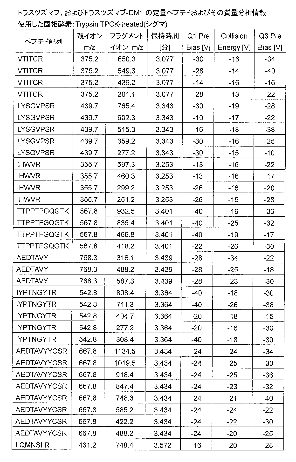
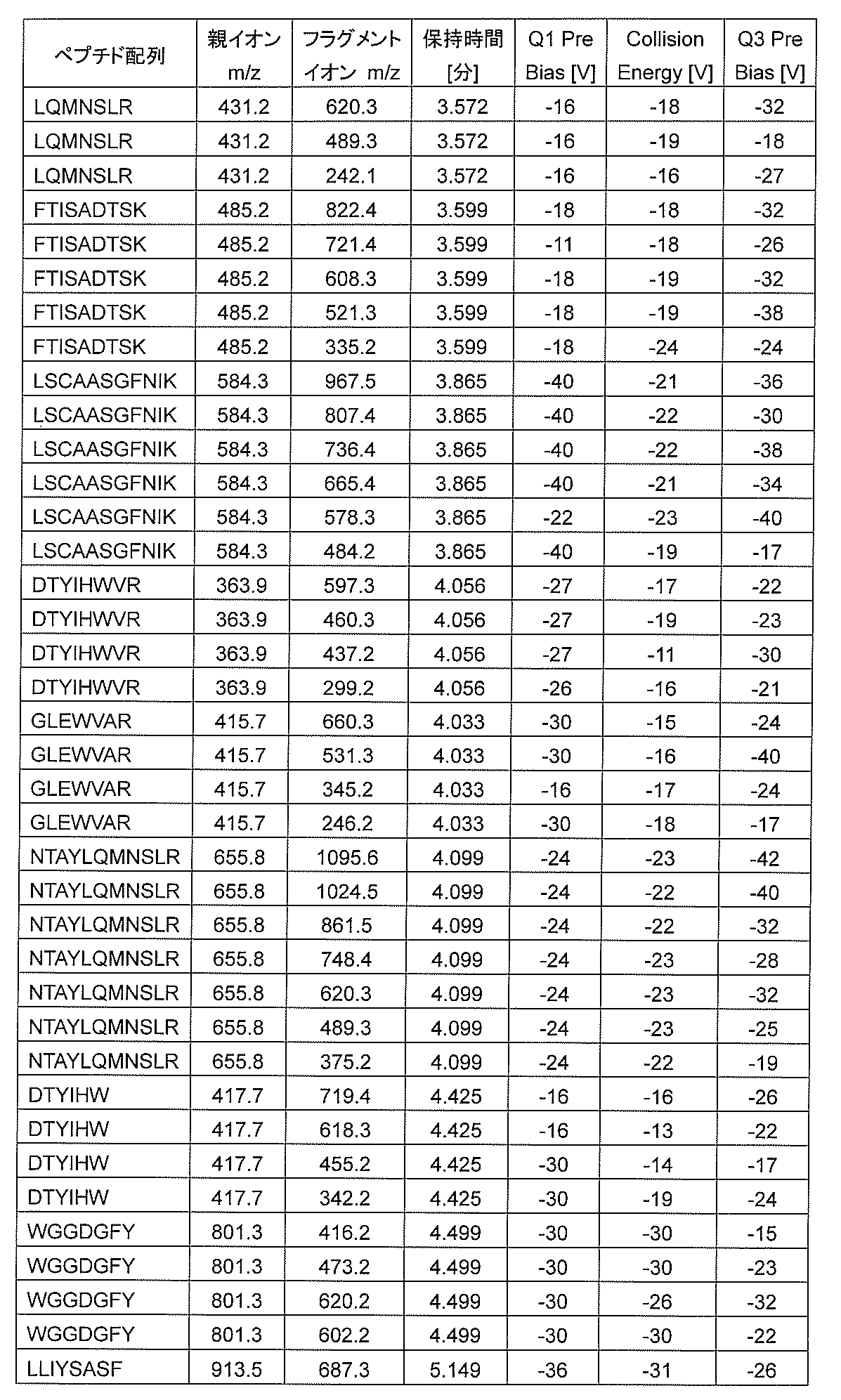
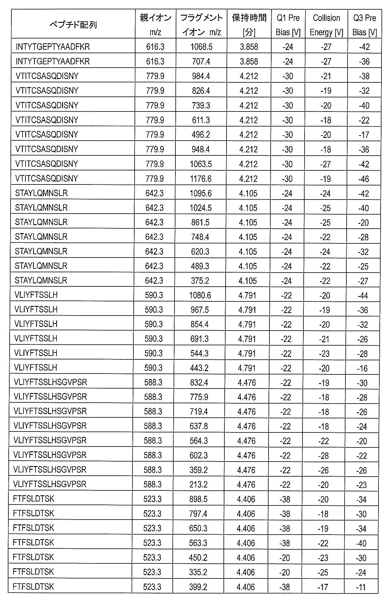
- kit according to any one of (1) to (5) further comprising one or more internal standard peptides including an amino acid sequence specific to the monoclonal antibody to be measured.
- the measurement target is trastuzumab, trastuzumab-DM1, bevacizumab, or rituximab
- the internal standard peptide is a peptide having any one or more amino acid sequences of SEQ ID NOs: 1 to 47.
- a peptide obtained by subjecting a porous body in which a monoclonal antibody to be measured is immobilized in pores to nanoparticles in which protease is immobilized in a liquid to perform selective protease digestion of the monoclonal antibody A computer-readable record in which data for performing the mass spectrometry is recorded for use in a method for detecting the monoclonal antibody by analyzing fragments by high performance liquid chromatography mass spectrometry (LC-MS)
- LC-MS liquid chromatography mass spectrometry
- the recording medium wherein the data includes at least parent ion, fragment ion, expected retention time, voltage data in each of the triple quadrupoles for one or more peptides obtained by protease digestion of the monoclonal
- a method package for detection of monoclonal antibodies by high performance liquid chromatography mass spectrometry comprising the recording medium according to (8) and instructions for use of the recording medium.
- the data is for a peptide having any one or more amino acid sequences of SEQ ID NOS: 1 to 47.
- the kit of the present invention is used for pretreatment of a liquid chromatograph mass spectrometer (particularly a triple quadrupole type).
- mass spectrometers that can be used to carry out the present invention include LCMS-8030, LCMS-8040, LCMS-8050, and LCMS-8080 (all manufactured by Shimadzu Corporation).
- LCMS-IT-TOF and LCMS-Q-TOF manufactured by Shimadzu Corporation
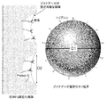
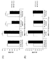
- the principle of the method of digesting an antibody using the kit of the present invention is shown.
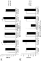
- the result of examination of the filter membrane material used for detection of a trastuzumab digestion fragment is shown.
- polytetrafluoroethylene (PTFE) and polyvinylidene fluoride (PVDF) were compared.
- PTFE polytetrafluoroethylene
- PVDF polyvinylidene fluoride
- B Trastuzumab 33.3 ⁇ g / ml.
- the result of examination of the filter membrane material used for detection of a trastuzumab digestion fragment is shown.
- polytetrafluoroethylene and polyvinylidene fluoride were compared.
- the present invention is a sample preparation kit for detecting a monoclonal antibody by high performance liquid chromatography mass spectrometry (LC-MS), A porous body for immobilizing the monoclonal antibody to be measured; Nanoparticles with immobilized protease, A reaction vessel for selectively digesting monoclonal antibodies by contacting the porous body and the nanoparticles; A buffer solution that is introduced into the reaction container together with the nanoparticles and the porous body, and causes a digestion reaction with the protease; A filtration membrane for removing the porous body and the nanoparticles after the digestion reaction and extracting the product of the digestion reaction together with the buffer; The kit.
- LC-MS liquid chromatography mass spectrometry
- FIG. 1 shows the principle of a method for digesting a monoclonal antibody using the kit of the present invention.
- the present invention will be specifically described in association with the above method.
- ⁇ Outline of mass spectrometry> a quantitative technique using mass spectrometry is mainly performed by a hybrid mass spectrometer called a triple quadrupole. Specifically, ionized biomolecules first pass through a portion called octopole to reduce the ionic molecule vibration radius. Next, ions having a specific mass number are selected by resonating in the first quadrupole, and other ions are excluded. This step is also called single ion monitoring (SIM).
- SIM single ion monitoring
- CID collision-induced dissociation
- MRM multiple reaction monitoring
- Quantification of biological samples using mass spectrometry has the greatest advantage of being able to quantify the structure-specific ions of biomolecules as an indicator, and this can be connected to a high-performance liquid chromatograph to enable continuous analysis. be able to.
- this is the only technology that has good advantages.
- an antibody In order to detect an antibody by mass spectrometry, it is necessary to first extract the antibody from a biological sample such as blood or tissue and dissolve it in an appropriate solvent. In addition, since an antibody has a large molecule for analysis as it is, it is decomposed into a peptide by protease and then separated by liquid chromatography, followed by mass spectrometry.
- the molecular weight of peptides suitable for analysis is about 1000 to 3000 Da.
- the matrix effect is a phenomenon in which the ionization efficiency of a target substance is reduced due to the presence of an ionization-inhibiting substance in the same droplet or the presence of various ions at the same time. Since the energy given to ionization is equal, if the ionization target substance increases, energy inevitably disperses and the amount of ions decreases.
- the present invention intends to reduce the population of the analysis target while maintaining the specificity of the measurement target.
- the measurement object for preparing a sample for detection by LC-MS using the kit of the present invention is a monoclonal antibody.
- a monoclonal antibody is a biopolymer having a structure in which two heavy chains (molecular weight 50 kDa) and two light chains (molecular weight 25 kDa) are connected by a disulfide bond.
- the Fab domain and Fc domain are connected via a hinge, and the heavy and light chains are composed of a constant region and a variable region, respectively.
- the constant region has a structure that maintains the characteristic Y-shape of the antibody (framework structure), and has an amino acid sequence that is common to most antibodies from the same species.
- each variable region has three sites each having a specific sequence called a complementarity-determining region (CDR).
- CDR complementarity-determining region
- the higher-order structure of the antibody is further characterized by a very flexible hinge and a variable region with respect to a stationary region having a rigid structure. It is known that there is a site to which a specific protein called Protein A or Protein G binds at the C-terminus of the heavy chain.
- Monoclonal antibodies that can be measured include, but are not limited to, human antibodies such as panitumumab, ofatumumab, golimumab, and ipilimumab; tocilizumab, trastuzumab, trastuzumab-DM1, bevacizumab, omalizumab, mepolizumab, torizumab And humanized antibodies such as ocrelizumab, mogamulizumab and eculizumab; chimeric antibodies such as rituximab, cetuximab, infliximab and basiliximab.
- the molecular diameter of the monoclonal antibody is about 14.5 nm.
- a complex added with further functions while maintaining the specificity of the monoclonal antibody such as an Fc fusion protein, an antibody-drug complex (for example, gemtuzumab ozogamicin, trastuzumab-emtansine, etc.) is also a measurement target in the method of the present invention. It should be included in the monoclonal antibody.
- the binding of the complex may be dissociated and the antibody alone may be subjected to LC-MS, or the complex may be used in the form of a complex for LC-MS.
- a person skilled in the art can set the optimum conditions for the method of the present invention in accordance with the measurement object based on the description of the present specification.
- the kit of the present invention is used for the identification and quantification of antibodies by subjecting the Fab domain of a monoclonal antibody to site-selective protease digestion and mass spectrometry of the obtained peptide fragment.
- the porous body included in the kit of the present invention is not particularly limited as long as it has a large number of pores, and activated carbon, porous membranes, porous resin beads, metal particles, and the like can be used. . Among these, those capable of binding an antibody site-specifically are particularly preferable.
- FIG. 1 shows a hemispherical pore, but the shape of the pore is not particularly limited. Moreover, what formed the pore which penetrates a porous body like a porous film can also be used.
- the size of the pores in the porous body is not particularly limited, and considers the molecular diameter of the antibody so that when the antibody is immobilized, the site to be selectively digested is located near the surface of the pore. Is preferably determined.
- the average pore diameter D2 of the porous body is appropriately set in a range of about 10 nm to 200 nm and smaller than the average particle diameter D1 of the nanoparticles.
- the average pore diameter D2 of the porous body is, for example, preferably about 20 nm to 200 nm, and more preferably about 30 nm to 150 nm.
- the pore diameter of the porous body is preferably 30 nm to 150 nm, more preferably 40 nm to 120 nm, and more preferably 50 nm to 100 nm. Particularly preferred is about 100 nm.
- the monoclonal antibody to be measured is immobilized in the pores of the porous body.
- the antibody is likely to denature, the fluctuation of the molecule is perturbed, and the probability of being attacked by the protease is improved.
- the protease is immobilized on the nanoparticles, so that it becomes an environment that is sterically stable and difficult to cause self-digestion. Therefore, according to the method of the present invention, in addition to enabling regioselective protease digestion, high activity of the protease can be maintained.
- those in which a linker molecule that interacts site-specifically with an antibody is immobilized in the pores of the porous body are preferably used.
- the interaction between the antibody and the linker molecule include chemical bond, hydrogen bond, ionic bond, complex formation, hydrophobic interaction, van der Waals interaction, electrostatic interaction, and stereoselective interaction.
- the linker molecule Protein A, Protein ⁇ G, or the like that binds site-specifically to the Fc domain of the antibody is preferably used.
- the Fc domain of the antibody is immobilized in the pores, and the Fab domain is located near the surface layer of the pores. In this way, by controlling the orientation of the antibody in the pore, position selective digestion of the Fab domain by a protease becomes possible.
- the size of the linker molecule is selected so that the selective cleavage site of the antibody is located near the surface layer of the pore.
- the molecular size of the state in which the linker molecule is bound to the antibody is preferably about 0.5 to 1.5 times, more preferably about 0.6 to 1.2 times the pore diameter of the porous body. It is more preferably about 7 to 1.1 times, and particularly preferably about 0.8 to 1 times.
- the linker molecule is not fixed to the porous body and the antibody is directly bonded to the pore, it is preferable that the molecular diameter of the antibody and the pore diameter of the porous body satisfy the above relationship.
- the porous material that can be suitably used in the present invention is not particularly limited, and examples thereof include Protein® G Ultralink resin (manufactured by Pierce), Toyopearl, TSKgel (manufactured by TOSOH), and the like.
- Protein® G® Ultralink resin it has been found that 95% of the Protein® G bound to the resin is in the pores.
- the method for immobilizing the antibody in the pores of the porous body is not particularly limited, and an appropriate method can be adopted depending on the characteristics of the antibody and the porous body or the linker molecule.
- an antibody is immobilized on a porous body in which protein A or protein G is immobilized in the pores, a mixture of the porous body suspension and the antibody-containing solution is mixed into the pores. It is possible to easily immobilize the antibody.
- the amount ratio of the porous body and the antibody can be appropriately set according to the purpose. For example, when performing quantitative analysis of an antibody, it is desired that almost the entire amount of the antibody in the sample is immobilized on the porous body. Therefore, it is preferable to set the quantity ratio so that the amount of the porous material is excessive with respect to the estimated content of the antibody in the sample.
- Nanoparticles contained in the kit of the present invention are used for the purpose of controlling protease access to the antibody immobilized in the pores of the porous body by immobilizing the protease on the surface thereof. Therefore, it is assumed that the average particle diameter D1 of the nanoparticles is larger than the average pore diameter D2 of the porous body so that the nanoparticles do not enter deeply into the pores of the porous body (FIG. 1).
- the shape of the nanoparticles is not particularly limited, but spherical nanoparticles are preferable from the viewpoint of uniform protease access to the pores of the porous body.
- the nanoparticles are preferably highly dispersible and have a uniform average particle size.
- the average particle diameter D1 of the nanoparticles is in the range of 50 nm to 500 nm, preferably 1.2 times or more than the average pore diameter D2 of the porous body, more preferably 1.5 times or more, 1.8 times or more, For example, about twice is particularly preferable.
- the average particle diameter D1 of the nanoparticles is preferably 100 nm or more, and more preferably 150 nm or more.
- the average particle diameter of the nanoparticles is preferably 120 nm or more, more preferably 150 nm or more, and particularly preferably 170 nm or more.
- the upper limit of the average particle diameter D1 of the nanoparticles is preferably 500 nm or less, and more preferably 300 nm or less from the viewpoint of increasing the digestion efficiency by the protease.
- the material of the nanoparticles is not particularly limited as long as the protease can be immobilized on the surface, and a metal, a resin, or the like is appropriately used. Moreover, what coated the metal surface with resin, what coated the resin surface with the metal, etc. can also be used.
- the type of nanoparticles is preferably magnetic nanoparticles that can be dispersed or suspended in an aqueous medium and can be easily recovered from the dispersion or suspension by magnetic separation or magnetic precipitation separation.
- magnetic nanoparticles whose surfaces are coated with an organic polymer are more preferable in that aggregation is unlikely to occur.
- the base material of the magnetic nanoparticles include ferromagnetic alloys such as iron oxide (magnetite (Fe 3 O 4 ), maghemite ( ⁇ -Fe 2 O 3 )), and ferrite (Fe / M) 3 O 4 .
- M means a metal ion that can be used together with iron ions to form a magnetic metal oxide, typically Co 2+ , Ni 2+ , Mn 2+. Mg 2+ , Cu 2+ , Ni 2+ and the like are used.
- the organic polymer that coats the magnetic nanoparticles include polyglycidyl methacrylate (polyGMA), a copolymer of GMA and styrene, polymethyl methacrylate (PMMA), and polymethyl acrylate (PMA).
- polyGMA polyglycidyl methacrylate
- PMMA polymethyl methacrylate
- PMA polymethyl acrylate
- Specific examples of magnetic nanobeads coated with an organic polymer include FG beads, SG beads, Adembeads, and nanomag.
- FG beads manufactured by Tamagawa Seiki Co., Ltd. (polymer magnetic nanoparticles having a particle diameter of about 200 nm in which ferrite particles are coated with polyglycidyl methacrylate (polyGMA)) are preferably used.
- the nanoparticles are preferably modified with a spacer molecule capable of binding to a protease in order to suppress nonspecific protein adsorption and to selectively immobilize the protease.
- a spacer molecule capable of binding to a protease in order to suppress nonspecific protein adsorption and to selectively immobilize the protease.
- the spacer is preferably one that can bind to the protease and does not inactivate the protease. From the viewpoint of controlling the access range of the protease immobilized on the nanoparticle surface, the spacer preferably has a small molecular diameter.
- the spacer molecular diameter is preferably 5 nm or less, more preferably 3 nm or less, and even more preferably 2 nm or less. Further, the molecular weight of the spacer is preferably 2000 or less, more preferably 1500 or less, and further preferably 1000 or less.
- the spacer molecule capable of immobilizing protease with the above molecular diameter is preferably a non-protein, and has an amino group, carboxyl group, ester group, epoxy group, tosyl group, hydroxyl group, thiol group, aldehyde group, maleimide group, succinimide group at the terminal.
- Molecules having functional groups such as azide group, biotin, avidin, chelate and the like are preferable.
- spacer molecules having an activated ester group are preferred for immobilizing trypsin.
- spacer arm portions other than the above functional groups are polyethylene glycol and derivatives thereof, polypropylene glycol and derivatives thereof, polyacrylamide and derivatives thereof, polyethyleneimine and derivatives thereof, poly (ethylene oxide) and derivatives thereof, poly Hydrophilic molecules such as (ethylene terephthalic acid) and its derivatives are used.
- nanoparticles surface-modified with spacer molecules are also commercially available and may be used.
- nanoparticles modified with spacer molecules having an ester group (active ester group) activated with N-hydroxysuccinimide are commercially available under the trade name “FG beads NHS” (Tamakawa Seiki Co., Ltd.).
- the particle size of FG beads NHS is about 200 nm ⁇ 20 nm, and it is very homogeneous as nanoparticles.
- the kit of the present invention preferably contains a nanoparticle in which a protease is immobilized.
- a nanoparticle in which a protease is immobilized.
- the nanoparticles and protease are provided separately and immobilized prior to use.
- a protease cleaves an antibody immobilized in a pore of a porous body at a specific amino acid sequence site to generate a peptide fragment.
- Protease can be included in the kit alone or immobilized on the surface of the nanoparticles.
- the type of protease immobilized on the nanoparticles may be appropriately selected according to the type of protein to be quantified or identified by mass spectrometry, and is not limited.
- trypsin trypsin, chymotrypsin, lysyl endopeptidase , V8 protease, AspN protease (Asp-N), ArgC protease (Arg-C), papain, pepsin, dipeptidyl peptidase and the like.
- Two or more proteases can be used in combination.
- trypsin is particularly preferably used in the present invention. Trypsin has high substrate specificity, and since Lys or Arg is present at the C-terminus of the peptide after cleavage, the charge amount and charge localization of the peptide can be made uniform, so that a sample for mass spectrometry can be prepared. Is particularly suitable. Trypsin has a small molecular diameter (about 3.8 nm) and an active site is present inside the molecule. Therefore, the region where the active site can access the antibody is limited, and the position selectivity of protease digestion can be enhanced.
- protease When a peptide fragment of an antibody after protease digestion is subjected to mass spectrometry as a measurement data, it is preferable to use a protease with low self-digestion and high selectivity of the cleaved sequence.
- a commercially available protease it is preferable to use a mass spectrometry grade or sequencing (sequence) grade protease.
- native trypsin derived from living organisms is known to have low cleavage site specificity because it contains high self-digesting activity or trypsin that exhibits chymotrypsin-like activity. Therefore, as a mass spectrometry grade, a product obtained by reducing methylation of a lysine residue of trypsin to increase resistance to autolysis is commercially available.
- proteases examples include Trypsin® Gold (manufactured by Promega) and Trypsin® TPCK-treated (manufactured by Sigma® Aldrich).
- the method for immobilizing the protease on the surface of the nanoparticle is not particularly limited, and an appropriate method can be adopted depending on the characteristics of the protease and the nanoparticle (or the spacer molecule that modifies the nanoparticle surface).
- the protease can be immobilized on the nanoparticle surface by mixing a suspension of nanoparticles and a solution containing protease.
- the amine coupling method of nanoparticles and protease via the functional group of the spacer molecule is preferred.
- the surface-modified carboxyl group of the nanoparticles can be esterified with N-hydroxysuccinimide (NHS) to form an activated ester group, and the protease amino group can be bound thereto.
- NHS N-hydroxysuccinimide
- 1-ethyl-3- (3-dimethylaminopropyl) carbodiimide (EDAC), N, N'-dicyclohexylcarbodiimide (DCC), bis (2,6-diisopropylphenyl) carbodiimide (DIPC), etc. Can be carried out in the presence of a condensing agent.
- the amino group of the surface of the nanoparticles is modified with a protease amino acid using a crosslinking agent such as glutaraldehyde, bifunctional succinimide, bis (sulfosuccinimidyl) suberate (BS3), sulfonyl chloride, maleimide, pyridyl disulfide.
- a crosslinking agent such as glutaraldehyde, bifunctional succinimide, bis (sulfosuccinimidyl) suberate (BS3), sulfonyl chloride, maleimide, pyridyl disulfide.
- the coupling method of nanoparticles and protease via the functional group of the spacer molecule can be performed by a simple operation of adding a protease solution to a suspension of nanoparticles and mixing and stirring under certain conditions.
- the active part that is not bound to the protease on the nanoparticle surface after the protease is immobilized on the nanoparticle surface.
- the unbound spacer molecule binds to impurities in the sample and adversely affects protease digestion or is produced by protease digestion.
- the peptide fragments may be immobilized on the nanoparticles. Such imperfections are suppressed by blocking unbound spacer molecules after immobilizing the protease.
- chemical modification is preferred.
- an activated ester group can be inactivated by forming an amide bond by reaction with a primary amine.
- ⁇ Protease digestion> By contacting the porous body on which the antibody is immobilized and the nanoparticles on which the protease is immobilized on the surface in a liquid, the antibody is digested with the protease and a peptide fragment is produced.
- liquid means that the substrate (solid phase) and the enzyme (solid phase) come into contact with each other in the liquid phase, and an aqueous medium suitable for the protease digestion reaction is intended.
- the conditions for protease digestion in the present invention are not particularly limited, and conditions similar to those for general protease digestion can be appropriately employed. For example, it is preferable to incubate at a temperature of about 37 ° C. for about 1 to 20 hours in a buffer solution adjusted to near the optimum pH of the protease.
- the mixing ratio of the porous body on which the antibody is immobilized and the nanoparticle on which the protease is immobilized is not particularly limited, and may be set so that the amount of the protease corresponds to the amount of the antibody.
- the amount of the protease is increased as compared with general protease digestion.
- antibody: protease about 30: 1 to 3: 1 is preferable, about 15: 1 to 4: 1 is more preferable, and about 10: 1 to 5: 1 is more preferable.
- protease digestion is carried out with the antibody immobilized on the porous body. Since the peptide fragment produced by protease digestion exists in the liquid phase, the target peptide fragment can be obtained in a position-selective manner without performing antibody elution or denaturation operation. According to the method of the present invention, peptide fragments can be recovered in a position-selective manner with a simpler operation than the conventional method.
- the C-terminal side of the antibody is immobilized on a Protein® G resin having a pore diameter of 100 nm, and the variable region of the antibody is always oriented to the solution side.
- protease is immobilized on the surface of the nanoparticle having a particle diameter of 200 nm.
- Protease digestion is not particularly limited, but can be performed under tapping rotation accompanied by periodic tapping with stirring by gentle rotation.
- Slow rotation refers to a rotational speed of, for example, about 3 to 10 rpm
- “tapping” refers to an instantaneous operation such as playing or shocking (frequency: for example, 1 to 5 times per minute) , Preferably 2 to 4 times). Accordingly, the porous body on which the antibody is immobilized and the nanoparticle on which the protease is immobilized are effectively brought into contact with each other while maintaining the dispersed state, and the protease digestion reaction efficiency can be increased.
- the pore size of the filtration membrane to be used is selected within the range in which the porous body and nanoparticles cannot pass through and the digested peptide can pass through.
- a filtration membrane made of polyvinylidene fluoride (PVDF) Low-binding hydrophilic PVDF, pore size 0.2 ⁇ m, manufactured by Millipore
- PTFE polytetrafluoroethylene
- the porous body and the nanoparticles can be easily removed by filtering using a product manufactured by KK If the filtration is centrifugal filtration, rapid and simple filtration is possible.
- the filter membrane is not limited, but a filter membrane in the form of a double-structured filter tube that enables centrifugal filtration can be suitably used.
- a filter membrane in the form of a double-structured filter tube that enables centrifugal filtration
- it is preferable to use a 0.2 ⁇ m filtration membrane spin filter such as Millipore Ultra Free PVDF, 0.2 ⁇ m or Millipore UFC30LG00 Ultra Free-C3LCR, 0.2 ⁇ m.
- 0.2 ⁇ m filtration membrane plates such as Millipore multi-screen PVDF, Barex, and 0.2 ⁇ m can be preferably used.
- the tube can be used as a reaction vessel for protease digestion reaction.
- protease digestion can be carried out by putting a medium (including a buffer solution) for the reaction between the nanoparticles on which the protease is immobilized and the porous body on which the antibody is immobilized in a tube.
- the antibody in the sample can be immobilized on the porous body in a tube, and after washing operation, nanoparticles having immobilized protease can be added.
- a person skilled in the art can appropriately modify a series of operation procedures such as protease digestion reaction, removal of porous bodies and nanoparticles using the kit of the present invention.
- LC-MS ⁇ Liquid chromatograph mass spectrometry
- the sample before being subjected to mass spectrometry is separated and concentrated by liquid chromatography (LC).
- LC liquid chromatography
- the eluate from LC may be directly ionized and subjected to mass spectrometry.
- Analysis can also be performed by LC / MS / MS or LC / MSn, which combines LC and tandem mass spectrometry.
- the eluate from LC may be collected once and then subjected to mass spectrometry.
- the LC column is not particularly limited, and a hydrophobic column such as C30, C18, C8, or C4 generally used for peptide analysis, a carrier for hydrophilic affinity chromatography, or the like can be appropriately selected and used. .
- mass spectrometry can determine an amino acid sequence, it can be determined whether or not a peptide fragment is a peptide fragment derived from a specific protein such as an antibody. Further, the concentration of the peptide fragment in the sample can be determined based on the peak intensity. In the present invention, the type of peptide fragment contained in the sample is reduced because the antibody is treated with protease in a site-selective manner. Therefore, analysis conditions can be easily set by mass spectrometry or the like. In the analysis, if necessary, the sample may be used for the analysis after treatment such as desalting, solubilization, extraction, concentration, and drying.
- the ionization method in mass spectrometry is not particularly limited. Electron ionization (EI) method, chemical ionization (CI) method, field desorption (FD) method, fast atom collision (FAB) method, matrix-assisted laser desorption ionization (MALDI) Method, electrospray ionization (ESI) method and the like can be employed.
- the analysis method of the ionized sample is not particularly limited. Magnetic field deflection type, quadrupole (Q) type, ion trap (IT) type, time of flight (TOF) type, Fourier transform ion cyclotron resonance (FT-ICR) type Etc. can be appropriately determined according to the ionization method.
- MS / MS analysis or multistage mass spectrometry of MS3 or higher can be performed using a triple quadrupole mass spectrometer or the like.
- An apparatus particularly suitable for use in the method of the present invention is not particularly limited.
- LCMS-8030, LCMS-8040, LCMS-8050, and LCMS-8080 (all of which are Shimadzu Corporation), LCMS-IT-TOF LCMS-Q-TOF (Shimadzu Corporation).
- peptide fragments obtained by subjecting an antibody to position-specific protease digestion are used, so that the hit rate and data accuracy by database search can be improved.
- An antibody can also be identified by specifying the amino acid sequence of a peptide fragment by multistage mass spectrometry or the like. If a peptide fragment having an amino acid sequence specific to an antibody, for example, an amino acid sequence containing an amino acid in the CDR2 region can be detected, the target antibody can be identified and quantified.
- the peptide to be detected preferably has about 5 to 30 amino acid residues, more preferably about 7 to 25. If the number of amino acid residues is excessively small, it is difficult to distinguish from contaminants and peptide fragments derived from other parts of the same protein, which may cause false detection. On the other hand, if the number of amino acid residues is excessively large, detection may be difficult or the quantitative property may be lowered due to reasons such as difficulty in ionization.
- the amount of antibody can be calculated based on the peak area and peak intensity of the detected peptide fragment ions (in the case of multistage MS, fragment ions obtained by cleavage of the parent ion).
- a peptide fragment in a sample can be obtained by associating a calibration curve (calibration curve) obtained in advance with a peak area or by associating a peak area derived from an internal standard added to the sample with a peak area derived from the sample.
- the amount and concentration of antibody are calculated based on the peptide fragment concentration.
- the present invention is a sample preparation kit for use in the above-described method for detecting a monoclonal antibody by high performance liquid chromatography mass spectrometry (LC-MS), A porous body for immobilizing the monoclonal antibody to be measured; Nanoparticles with immobilized protease, A reaction vessel for selectively digesting monoclonal antibodies by contacting the porous body and the nanoparticles; A buffer solution that is introduced into the reaction container together with the nanoparticles and the porous body, and causes a digestion reaction with the protease; A filtration membrane for removing the porous body and the nanoparticles after the digestion reaction and extracting the product of the digestion reaction together with the buffer; The kit.
- LC-MS liquid chromatography mass spectrometry
- Measures by mass spectrometry can be performed with very high accuracy, while appropriate sample preparation and setting of appropriate analysis conditions are very important.
- the present invention provides a sample preparation kit that can be used to carry out the above-described method so that accurate test results can be obtained more easily in a clinical setting.
- the porous body and nanoparticles contained in the kit of the present invention are as described above.
- the reaction vessel may be any vessel that can contact the monoclonal antibody immobilized on the porous body and the protease immobilized on the nanoparticles in the liquid phase, and is not particularly limited. However, considering that it is for preparing a sample to be detected by mass spectrometry, it is preferable to use a microtube or a plate. In consideration of reaction steps such as mixing by vortex or rotator for the reaction, filtration for separation of the peptide after the reaction and the porous material and nanoparticles, a person skilled in the art assumes an appropriate reaction vessel. be able to.
- the buffer contained in the kit of the present invention is introduced into the reaction vessel together with the nanoparticles and the porous body, and is used for digestion reaction with the protease, and reaction conditions suitable for protease digestion. Is to provide.
- the reaction conditions can be appropriately determined depending on the selected protease and the like, and the composition of the buffer solution can also be appropriately determined.
- the kit of the present invention also includes a filtration membrane for removing the porous body and the nanoparticles after the protease digestion reaction and extracting the product of the digestion reaction together with the buffer solution.
- a filtration membrane for removing the porous body and the nanoparticles after the protease digestion reaction and extracting the product of the digestion reaction together with the buffer solution.
- the filtration membrane in the kit of the present invention functions as a “bottom of the reaction vessel” and hardly permeates the peptide produced by digestion of the buffer solution and the protease under the condition where no pressure or centrifugal force is applied, and is used for centrifugation, etc.
- the centrifugation conditions to be added because the filtration membrane can permeate the buffer solution and the peptide are not limited, but for example, a range of 3,000 to 10,000 g is preferable.
- filtration membrane examples include a filtration membrane made of polyvinylidene fluoride (PVDF) (Low-binding hydrophilic PVDF, pore size 0.2 ⁇ m, manufactured by Millipore).
- PVDF polyvinylidene fluoride
- a filtration membrane is made of PVDF
- a housing material is polyacrylonitrile resin, for example, Barex (registered trademark) (made by Mitsui Chemicals Fine Co., Ltd.) can do.
- the kit of the present invention can also include instructions describing the method of using the kit and / or mass spectrometric conditions for detection of monoclonal antibodies.
- the kit of the present invention can also contain one or more internal standard peptides.
- the internal standard peptide provides more reliable analysis results by analyzing under the same conditions simultaneously with the sample or separately.
- the internal standard peptide includes a specific amino acid sequence of the monoclonal antibody to be measured, and is a peptide generated by digestion with a protease included in the kit of the present invention.
- the internal standard peptide can be selected from peptides having any one or more amino acid sequences of SEQ ID NOs: 1 to 47.
- the internal standard peptide is a peptide having an amino acid sequence represented by SEQ ID NOs: 1 to 7 Or more.
- the internal standard peptide can be one or more of the peptides having the amino acid sequences shown in SEQ ID NOs: 8-12.
- the internal standard peptide can be one or more of the peptides having the amino acid sequences shown in SEQ ID NOs: 13 to 19.
- the monoclonal antibody to be measured is trastuzumab or trastuzumab-DM1 and the protease is Trypsin TPCK-treated (manufactured by Sigma), in addition to the peptide having the amino acid sequence shown by SEQ ID NOs: 1 to 7, It can be one or more of the peptides having the amino acid sequences shown in SEQ ID NOs: 20-28, 46 and 47.
- the monoclonal antibody to be measured is bevacizumab and the protease is TrypsintreatedTPCK-treated (manufactured by Sigma), in addition to the peptide having the amino acid sequence shown in SEQ ID NOs: 8-12, the internal standard peptide is SEQ ID NO: 29- One or more of the peptides having the amino acid sequence represented by 38 can be used.
- the monoclonal antibody to be measured is rituximab and the protease is Trypsin TPCK-treated (manufactured by Sigma), in addition to the peptides having the amino acid sequences shown in SEQ ID NOs: 13 to 19, the internal standard peptide is SEQ ID NO: 39 to One or more of the peptides having the amino acid sequence represented by 45 can be used.
- the internal standard peptide is a peptide containing an amino acid sequence specific to the monoclonal antibody to be measured, more specifically an amino acid sequence of the Fa domain, more preferably an amino acid sequence containing an amino acid derived from the CDR2 region of the heavy chain or light chain It is preferable to use a peptide consisting of
- peptide fragment preparation for monoclonal antibody identification and quantification can be performed more easily, and automation with an apparatus can be easily achieved.
- trypsin and the like can retain activity even when immobilized on the surface of the nanoparticle. Therefore, if protease is provided as a component of the kit in a state of being immobilized on the surface of the nanoparticle, the operation of peptide fragment preparation Can be further simplified.
- kit of the present invention is provided in the following configuration, for example.
- a reagent kit for analyzing one sample at a time has the following configuration, for example. 0.2 ⁇ m filtration membrane spin filter (Millipore Ultra Free PVDF, 0.2 ⁇ m) Protein G resin slurry with a pore size of 100 nm (stored at 4 ° C) Trypsin beads immobilized on nanoparticles with a particle size of 200 nm (stored at -20 ° C) Low-adsorption tubes for plasma dilution Solution collection microtubes Buffer for plasma dilution (PBS + 0.1% n-octyl- ⁇ -D-thioglycoside or equivalent surfactant such as n-octyl- ⁇ -D-glycoside) Bead washing buffer (PBS) Protease reaction buffer (25 mM Tris-HCl, pH 8.0 + protease reaction-promoting additive) 10% formic acid aqueous solution instruction manual
- a reagent kit for multi-sample processing that enables simultaneous analysis of 96 samples has the following configuration, for example.
- 0.2 ⁇ m filtration membrane plate (Millipore multiscreen PVDF, Barex, 0.2 ⁇ m) Protein G resin slurry with a pore size of 100 nm (stored at 4 ° C) Trypsin and protease beads immobilized on nanoparticles with a particle size of 200 nm (stored at -20 ° C)
- Protease bead plate Plasma dilution buffer (PBS + 0.1% n-octyl- ⁇ -D-thioglycoside or equivalent surfactant such as n-octyl- ⁇ -D-glycoside) Bead washing buffer (PBS)
- Protease reaction buffer 25 mM Tris-HCl, pH 8.0 + protease reaction
- an internal standard peptide is included in the kit of the present invention. This is to increase the quantification accuracy of the biopharmaceutical peptide, and it is desirable to prepare a plurality of peptides for each biopharmaceutical and sell them separately.
- the internal standard peptides may include those labeled with stable isotope amino acids. In that case, the mass spectrometric quantification conditions differ from those of peptides that do not contain isotopes. preferable.
- the present invention is also obtained by subjecting a porous body in which a monoclonal antibody to be measured is immobilized in pores to nanoparticles in which trypsin is immobilized in a liquid to perform selective trypsin digestion of the monoclonal antibody.
- a method package is provided for use in the method of detecting the monoclonal antibody by analyzing the obtained peptide fragment by high performance liquid chromatography mass spectrometry (LC-MS).
- LC-MS liquid chromatography mass spectrometry
- the “method package” refers to a package that includes the analysis conditions of liquid chromatograph mass spectrometry for a specific measurement target in a readable form and can be circulated alone. By importing the data included in the method package into LC-MS, it is possible to analyze under the optimal measurement conditions obtained after detailed examination.
- the present applicant has provided method packages for analysis by LC-MS in order to allow users to perform mass spectrometry of agricultural chemicals and veterinary drugs more easily. Therefore, the present invention also provides a method package for identifying and quantifying monoclonal antibodies in a sample by LC-MS.
- the present invention performs selective protease digestion of a monoclonal antibody by bringing a porous body in which the monoclonal antibody to be measured is immobilized in the pores and nanoparticles in which the protease is immobilized into contact with each other in a liquid.
- LC-MS high performance liquid chromatography mass spectrometry
- the data includes, but is not limited to, for example, a peptide obtained by protease digestion of the monoclonal antibody, such as a peptide having an amino acid sequence comprising an amino acid of a Fab domain, more preferably a CDR region.
- each of the triple quadrupole contains data of the voltage at, provides the recording medium.
- the expected holding time, voltage data, and the like described above are numerical values that vary depending on the equipment used, measurement conditions, etc., and are preferably provided according to the equipment. Further, as will be understood by those skilled in the art, it is preferable to provide the fluctuation range of the numerical value that varies depending on the conditions.
- the recording medium may be in any form and is not particularly limited.
- a disk or memory capable of recording information magnetically and optically can be mentioned.
- the above method package can include, for example, the following information and software functions.
- optimized ⁇ been parent ion m / z values and optimized fragment ion m / z values, optimized Q1 pre bias voltage-Optimized Q2 collision energy voltage-Optimized Q3 pre Bias voltage value • Expected ion retention time and mass analysis time • Quantitative value conversion formula • Analysis result report output function *: Measures each condition item, the highest ion intensity, and the most reproducible m / z Adopt a value and make it the optimal value.
- the present invention also provides a method package for detection of monoclonal antibodies by high performance liquid chromatography / mass spectrometry (LC-MS), which includes the above recording medium and instructions for using the recording medium.
- LC-MS high performance liquid chromatography / mass spectrometry
- Examples of the above recording medium or method package include those containing only information limited to a specific monoclonal antibody. Therefore, when the monoclonal antibody is, for example, trastuzumab, trastuzumab-DM1, bevacizumab, or rituximab, it is possible to provide a recording medium or method package that describes analysis conditions suitable for them.
- the data included in the recording medium may be related to analysis conditions for a peptide having any one or more amino acid sequences of SEQ ID NOs: 1 to 47, for example.
- the method package may describe data common to a plurality of mass spectrometers, or may describe various data suitable for analysis by a specific mass spectrometer.
- the recording medium or method package can be provided together with the kit of the present invention described above or separately from the kit.
- Example 1 Sample preparation kit Prepare a kit with the following configuration for the analysis of a single sample.
- PBS buffer PBS + 0.1% n-octyl- ⁇ -D-thioglucoside, Dojindo
- Enzyme reaction buffer 25 mM Tris-HCl, pH 8.0
- Enzyme stop solution 10% formic acid
- Filter tube Li-binding hydrophilic PVDF, pore size 0.2 ⁇ m, Millipore
- Low adsorption tube (Richell Micro Resico Tube 92017)
- LCMS vial, insert Shiadzu GLC GLC4010-VP target vial VP, C4010-630P Target PP Polyspring
- Porous material Porous material (Pierce 53126 Protein G UltraLink Resin, 40 ⁇ l dispensing)
- Nanoparticles trypsin-immobilized FG beads (trypsin 40 ⁇ g))
- 9 9
- ⁇ Protocol (described in the instruction manual (9))> 1. Take 20 ⁇ l of blood sample into a low adsorption tube (5) and dilute buffer (1) with 180 ⁇ l. 2. Centrifuge the porous body (7) in a tabletop centrifuge and discard the supernatant. Add 100 ⁇ l of buffer (1), stir gently, centrifuge, and discard the supernatant. Repeat this operation three times. Thereafter, 40 ⁇ l of buffer solution (1) is added and suspended, and then transferred to the filter tube (4). This operation may be performed on the filter tube (4). 3. Transfer the blood diluted sample from step 1 to the filter tube (4). 4. Stir at room temperature for 1 hour while tapping lightly with a tapping rotary mixer (Nisshin Rika). 5.
- Example 3 Examination of filtration membrane 2
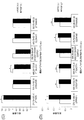
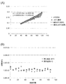
- An experiment similar to Example 2 was verified using bevacizumab. The results are shown in FIG.
- Example 2 As a result, as in Example 2, there was no difference in the case of a high concentration peptide, but when the peptide concentration was low, a significant difference occurred in the recovery rate, and PVDF was found to be suitable as a filtration membrane. did.
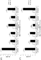
- Example 4 Examination of filtration membrane 3
- Two membrane materials (PTFE, PVDF) and three types of housings were compared to verify the peptide recovery rate of filtration membrane plates for use in multi-sample measurements.
- PTFE membrane / Solvinert housing (Merck) 2) PVDF membrane / Acryl clear housing (Merck), 3) PVDF membrane / Barex white housing (Merck) were used for comparison.
- PTFE was dominant on the high concentration side of the peptide, but PVDF was superior on the low concentration side.
- 5-20 ⁇ g / ml is the required peptide concentration range, so it was judged effective to select PFDV Barex.
- saturation with a high concentration of peptide when Barex resin is used aggregation of protein components can be considered.
- the blood sample assumed clinically includes a large amount of antibody and there is a washing step with a surfactant, so this saturation phenomenon is considered to be eliminated.
- the acrylic resin was excluded from the selection because of its large peptide loss due to physical adsorption.
- Example 5 Stability evaluation of protease activity
- enzyme activity was measured. Since the nanoparticles inhibit the substrate absorbance, several modifications were made to the conventional method.
- TPCK Trypsin Gold, Mass Spec Grade (Promega) (hereinafter referred to as “Gold”) and Trypsin TPCK Treated from bovine pancreas, Product Number T1426 (Sigma Aldrich) (hereinafter referred to as “TPCK”)
- the two types of trypsin and “FG-Gold” and “FG-TPCK” in which “Gold” and “TPCK” were immobilized on the nanoparticles using the kit described in Example 1 were used for various conditions. Under the enzyme reaction, enzyme stability was examined. "Gold” is resistant to self-digestion by applying a reductive dimethylation reaction in addition to chymotrypsin inactivation treatment (TPCK treatment), and has a broad activity independent of temperature and pH. It is a mass spectrometry grade protease that maintains On the other hand, “TPCK” is a protease in which chymotrypsin remains, although chymotrypsin is inactivated.
- ⁇ Protocol> Dissolve the protease substrate in DMSO to a final concentration of 10 mM to make a stock solution. Mix as follows. ⁇ Substrate 10 ⁇ l (100 nmol) ⁇ Reaction buffer (50 mM Tris-HCl, 150 mM NaCl, pH 8.0) 200 ⁇ l ⁇ Immobilized enzyme suspension (0.5 mg / ml) 2 ⁇ l
- protease digestion reaction was performed for 1.5 hours under vortexing.
- 50 ml of 2N-HCl was added to stop the reaction completely.
- Filter through a multi-screen filter plate (Millipore multi-screen PVDF, Barex, 0.2 ⁇ m), remove the nanoparticles, dispense into an optical bottom plate (Cosmo Bio Co., Ltd.), and microplate reader (TECAN Infinite M200Pro, Tecan
- the nanoparticles with immobilized trypsin were stored at 4 ° C. and ⁇ 20 ° C. and monitored for about 3 months to evaluate their enzyme activity. As shown in FIG. 5, the storage stability does not depend on the temperature, and it was found that the storage temperature should be 4 ° C. for providing as a kit.
- Example 6 Impact assessment by scale-up
- Example 7 Impact assessment by scale-up
- kit of the present invention makes it easy to identify and quantify monoclonal antibodies in a sample and reduce the error in the procedure.
- the target ion species since the target ion species is determined, it is not necessary for the user to set mass analysis conditions.
- the concentration quantification technique by mass spectrometry of antibody drugs has the potential to replace the current ELISA method, and the present invention can contribute to the advancement into the clinical pharmacology field and the market expansion.
Landscapes
- Life Sciences & Earth Sciences (AREA)
- Health & Medical Sciences (AREA)
- Chemical & Material Sciences (AREA)
- Immunology (AREA)
- Engineering & Computer Science (AREA)
- Physics & Mathematics (AREA)
- Biochemistry (AREA)
- General Health & Medical Sciences (AREA)
- Molecular Biology (AREA)
- Analytical Chemistry (AREA)
- Pathology (AREA)
- General Physics & Mathematics (AREA)
- Biomedical Technology (AREA)
- Urology & Nephrology (AREA)
- Hematology (AREA)
- Bioinformatics & Cheminformatics (AREA)
- Microbiology (AREA)
- Biotechnology (AREA)
- Organic Chemistry (AREA)
- Medicinal Chemistry (AREA)
- Cell Biology (AREA)
- Food Science & Technology (AREA)
- Bioinformatics & Computational Biology (AREA)
- Proteomics, Peptides & Aminoacids (AREA)
- Spectroscopy & Molecular Physics (AREA)
- Wood Science & Technology (AREA)
- Zoology (AREA)
- Chemical Kinetics & Catalysis (AREA)
- Biophysics (AREA)
- Hydrology & Water Resources (AREA)
- Genetics & Genomics (AREA)
- General Engineering & Computer Science (AREA)
- Electrochemistry (AREA)
- Other Investigation Or Analysis Of Materials By Electrical Means (AREA)
- Peptides Or Proteins (AREA)
Abstract
Description
測定対象のモノクローナル抗体を固定化するための多孔質体と、
プロテアーゼを固定化したナノ粒子と、
前記多孔質体と前記ナノ粒子とを接触させてモノクローナル抗体を選択的に消化するための反応容器と、
前記ナノ粒子及び多孔質体と共に前記反応容器内に導入され、前記プロテアーゼによる消化反応をさせるための緩衝液と、
前記消化反応後に前記多孔質体および前記ナノ粒子を除去して、前記消化反応の生成物を前記緩衝液と共に抽出するためのろ過膜と、
を含む、上記キット。
(2)前記ろ過膜が、圧力又は遠心力を加えない条件下では前記緩衝液及び前記プロテアーゼによる消化反応によって生成するペプチドをほとんど透過せず、圧力又は遠心力を加えた条件下では前記緩衝液及び前記ペプチドを透過することができるものである、(1)記載のキット。
(3)前記ろ過膜がポリフッ化ビニリデン(PVDF)製の膜である、(1)または(2)記載のキット。
(4)前記キットが多検体を同時に処理するためのキットであり、前記ろ過膜のハウジング素材がポリアクリロニトリル樹脂である、(1)~(3)のいずれか記載のキット。
(5)(1)~(4)のいずれか記載のキットに、更にモノクローナル抗体の検出のための質量分析条件を記載した説明書を含む、高速液体クロマトグラフ質量分析(LC-MS)によるモノクローナル抗体検出のためのキット。
(6)測定対象となるモノクローナル抗体に特異的なアミノ酸配列を含む1以上の内部標準ペプチドを更に含む、(1)~(5)のいずれか記載のキット。
(7)測定対象がトラスツズマブ、トラスツズマブ-DM1、ベバシズマブ、またはリツキシマブであって、内部標準ペプチドが配列番号1~47のいずれか1以上のアミノ酸配列を有するペプチドである、(6)記載のキット。
(8)測定対象のモノクローナル抗体を細孔内に固定化した多孔質体と、プロテアーゼを固定化したナノ粒子とを液体中で接触させてモノクローナル抗体の選択的プロテアーゼ消化を行い、得られたペプチド断片を高速液体クロマトグラフ質量分析(LC-MS)によって分析することによって前記モノクローナル抗体の検出を行う方法に使用するための、前記質量分析を実行させるためのデータが記録されたコンピュータ読み取り可能な記録媒体であって、前記データが、前記モノクローナル抗体のプロテアーゼ消化によって得られるペプチドの1以上に対して、少なくとも親イオン、フラグメントイオン、予想保持時間、三連四重極のそれぞれにおける電圧のデータを含む、上記記録媒体。
(9)(8)記載の記録媒体と、前記記録媒体の使用説明書とを含む、高速液体クロマトグラフ質量分析(LC-MS)によるモノクローナル抗体の検出のためのメソッドパッケージ。
(10)前記モノクローナル抗体が、トラスツズマブ、トラスツズマブ-DM1、ベバシズマブ、リツキシマブの1種以上である、(8)記載の記録媒体または(9)記載のメソッドパッケージ。
(11)前記データが、配列番号1~47のいずれか1以上のアミノ酸配列を有するペプチドに対するものである、(8)記載の記録媒体または(9)記載のメソッドパッケージ。
測定対象のモノクローナル抗体を固定化するための多孔質体と、
プロテアーゼを固定化したナノ粒子と、
前記多孔質体と前記ナノ粒子とを接触させてモノクローナル抗体を選択的に消化するための反応容器と、
前記ナノ粒子及び多孔質体と共に前記反応容器内に導入され、前記プロテアーゼによる消化反応をさせるための緩衝液と、
前記消化反応後に前記多孔質体および前記ナノ粒子を除去して、前記消化反応の生成物を前記緩衝液と共に抽出するためのろ過膜と、
を含む、上記キットに関する。
質量分析を用いた定量技術は、近年では主に三連四重極と呼ばれるハイブリッド型質量分析装置によって行われる。具体的には、イオン化された生体分子はまず、オクトポールと呼ばれる部分を通過することで、そのイオン分子振動半径を小さくする。次に第1四重極の中で、特定の質量数を持つイオンを共振させることで選択し、他のイオンを排除する。このステップはシングルイオンモニタリング(single ion monitoring, SIM)とも呼ばれる。
本発明のキットを用いてLC-MSによる検出のためのサンプルを調製する測定対象はモノクローナル抗体である。モノクローナル抗体は、2本の重鎖(分子量50 kDa)と2本の軽鎖(分子量25 kDa)がジスルフィド結合でつながった構造を有する生体高分子である。FabドメインとFcドメインがヒンジを介してつながっており、また重鎖及び軽鎖はそれぞれ定常領域と可変領域から成る。定常領域は、抗体の特徴的なY字型の形を保つ構造(フレームワーク構造)を有し、同一種由来の抗体のほとんどで共通するアミノ酸配列を有している。一方、可変領域には、相補性決定領域(complementarity-determining region, CDR)と呼ばれる特異的な配列を持つ部位が各3つずつ存在する。このCDR(CDR1、CDR2、CDR3)領域が規定する立体構造が抗原との特異的結合に関わっており、それによって抗体-抗原複合体が形成される。
本発明のキットに含まれる多孔質体は、多数の細孔を有するものであれば、その材料は特に限定されず、活性炭、多孔質膜、多孔質樹脂ビーズ、金属粒子等を用いることができる。これらの中でも、抗体を部位特異的に結合可能なものが特に好ましい。
抗体を多孔質体の細孔内に固定化する方法は特に限定されず、抗体と多孔質体あるいはリンカー分子の特性等に応じて適宜の方法を採用できる。例えば、細孔内にprotein Aやprotein Gが固定化された多孔質体に抗体を固定化する場合は、多孔質体の懸濁液と抗体を含む溶液とを混合することにより、細孔内に抗体を容易に固定化できる。
本発明のキットに含まれるナノ粒子は、その表面にプロテアーゼを固定化して、多孔質体の細孔内に固定化された抗体へのプロテアーゼのアクセスを制御する目的で用いられる。そのため、ナノ粒子は、多孔質体の細孔の奥深くまで入り込まないように、その平均粒径D1が、多孔質体の平均細孔径D2よりも大きいものとする(図1)。
本発明に係る方法は、プロテアーゼが、多孔質体の細孔内に固定化された抗体を特定のアミノ酸配列部位で切断してペプチド断片を生じさせるものである。
プロテアーゼをナノ粒子の表面に固定化する方法は特に限定されず、プロテアーゼとナノ粒子(あるいはナノ粒子表面を修飾するスペーサ分子)の特性等に応じて適宜の方法を採用でき、例えば、プロテアーゼをスペーサ修飾されたナノ粒子表面に固定化する場合は、ナノ粒子の懸濁液とプロテアーゼを含む溶液とを混合することにより、ナノ粒子表面にプロテアーゼを固定化できる。上記のスペーサ分子の官能基を介したナノ粒子とプロテアーゼのアミンカップリング法が好ましい。例えば、ナノ粒子に表面修飾したカルボキシル基をN-ヒドロキシスクシンイミド(NHS)でエステル化して活性化エステル基とし、これに、プロテアーゼのアミノ基を結合させることができる。このカップリング反応には、1-エチル-3-(3-ジメチルアミノプロピル)カルボジイミド(EDAC)、N,N'-ジシクロヘキシルカルボジイミド(DCC)、ビス(2,6-ジイソプロピルフェニル)カルボジイミド(DIPC)等のカルボジイミドを縮合剤の存在下に行うことができる。また、ナノ粒子に表面修飾したアミノ基に、グルタルアルデヒド、2官能性スクシンイミド、ビス(スルホスクシンイミジル)スベレート(BS3)、スルホニルクロリド、マレイミド、ピリジルジスルフィド等の架橋剤を用いてプロテアーゼのアミノ基を結合させてもよい。
抗体が固定化された多孔質体と、プロテアーゼが表面に固定化されたナノ粒子とを液体中で接触させることにより、抗体がプロテアーゼ消化され、ペプチド断片が産生される。ここで、「液体」とは、基質(固相)及び酵素(固相)が液相中で接触することを意味するものであり、またプロテアーゼ消化反応に適した水性媒体を意図する。
プロテアーゼ消化によって得られた目的のペプチド断片を質量分析に供するためには、多孔質体及びナノ粒子を除去することが必要である。これは、プロテアーゼ消化後のサンプルに対してろ過、遠心分離、磁気分離、透析等の操作を行うことで達成できる。
上記で得られたペプチド断片を含む試料を、LC-MSにより分析することで、抗体の同定や定量を行い得る。
本発明は、上記した高速液体クロマトグラフ質量分析(LC-MS)によってモノクローナル抗体を検出する方法において使用するためのサンプル調製用キットであって、
測定対象のモノクローナル抗体を固定化するための多孔質体と、
プロテアーゼを固定化したナノ粒子と、
前記多孔質体と前記ナノ粒子とを接触させてモノクローナル抗体を選択的に消化するための反応容器と、
前記ナノ粒子及び多孔質体と共に前記反応容器内に導入され、前記プロテアーゼによる消化反応をさせるための緩衝液と、
前記消化反応後に前記多孔質体および前記ナノ粒子を除去して、前記消化反応の生成物を前記緩衝液と共に抽出するためのろ過膜と、
を含む、上記キットに関する。
一サンプルずつ分析するための試薬キットは、例えば以下の構成のものである。
0.2μmのろ過膜スピンフィルター(ミリポア ウルトラフリーPVDF、0.2μm)
細孔径100 nmのProtein G樹脂スラリー(4℃保存)
粒子径200 nmのナノ粒子に固定化されたトリプシンビーズ(-20℃保存)
血漿希釈用低吸着チューブ
溶液回収マイクロチューブ
血漿希釈用バッファー(PBS + 0.1% n-オクチル-β-D-チオグリコシドもしくは相当する界面活性剤、例えばn-オクチル-β-D-グリコシド)
ビーズ洗浄用バッファー(PBS)
プロテアーゼ反応用バッファー(25 mM Tris-HCl, pH 8.0 + プロテアーゼ反応促進添加剤)
10%ギ酸水溶液
使用説明書
例えば96サンプル同時分析を可能とする多検体処理用の試薬キットは、例えば以下の構成のものである。
0.2μmのろ過膜プレート(ミリポア マルチスクリーンPVDF、Barex、0.2 μm)
細孔径100 nmのProtein G樹脂スラリー(4℃保存)
粒子径200 nmのナノ粒子に固定化されたトリプシンおよびプロテアーゼビーズ(-20℃保存)
血漿希釈用低吸着プレート
溶液回収用プレート
溶液廃棄用プレートリザーバー
プロテアーゼビーズ用プレート
血漿希釈用バッファー(PBS + 0.1% n-オクチル-β-D-チオグリコシドもしくは相当する界面活性剤、例えばn-オクチル-β-D-グリコシド)
ビーズ洗浄用バッファー(PBS)
プロテアーゼ反応用バッファー(25 mM Tris-HCl, pH 8.0 + プロテアーゼ反応促進添加剤)
10%ギ酸水溶液
プレートカバーシール
オートサンプラー用DMSO耐性、ニードルピアシブルプレートカバーシール
使用説明書
本発明のキットに場合によって含めるものとして、例えば内部標準ペプチドがある。これは、バイオ医薬品定量用ペプチドの定量精度を高めるためのものであって、バイオ医薬ごとに複数のペプチドを用意し、別売することが望ましい。内部標準ペプチドには、安定同位体アミノ酸でラベルしたものを含めても良く、その場合、同位体を含まないペプチドと比較すると質量分析定量条件が異なるため、内部標準用定量条件を同封することが好ましい。
内部標準ペプチド(例えばトラスツズマブ用として、配列番号1~7のペプチドのいずれか1以上)
試薬品質保証データ(質量分析データ、および原子純度測定結果)
定量条件ファイルパッケージ
使用説明書
本発明はまた、測定対象のモノクローナル抗体を細孔内に固定化した多孔質体と、トリプシンを固定化したナノ粒子とを液体中で接触させてモノクローナル抗体の選択的トリプシン消化を行い、得られたペプチド断片を高速液体クロマトグラフ質量分析(LC-MS)によって分析することによって前記モノクローナル抗体の検出を行う方法に使用するための、メソッドパッケージを提供する。本明細書において、「メソッドパッケージ」とは、特定の測定対象に対する液体クロマトグラフ質量分析の分析条件を読取り可能な形態で含み、単独で流通可能なものをいう。メソッドパッケージに含まれるデータをLC-MSにインポートすることで、詳細な検討の末に得られた最適測定条件で分析することが可能となる。
・最適化※されたペアレントイオンm/z値
・最適化されたフラグメントイオンm/z値
・最適化されたQ1 pre bias電圧値
・最適化されたQ2 collision energy電圧値
・最適化されたQ3 pre bias電圧値
・目的イオンの予想保持時間および質量分析時間
・定量値換算式
・解析結果レポート出力機能
※:各条件項目を実測し、最もイオン強度の高いもの、および最も再現性のあるm/z値を採択し、これを最適値とする。
単一サンプルの分析のために、以下の構成のキットを準備する。
(1) PBS緩衝液(PBS + 0.1% n-オクチル-β-D-チオグルコシド, Dojindo)
(2) 酵素反応緩衝液(25 mM Tris-HCl, pH8.0)
(3) 酵素反応停止液(10% ギ酸)
(4) フィルターチューブ(Low-binding hydrophilic PVDF、孔径0.2μm、ミリポア社)
(5) 低吸着チューブ(リッチェル マイクロレシコチューブ 92017)
(6) LCMSバイアル、インサート(島津GLC GLC4010-VPターゲットバイアルVP、C4010-630P Target PP Polyspring)
(7) 多孔質体(Pierce 53126 Protein G UltraLink Resin、40 μl分注)
(8) ナノ粒子(トリプシン固定化FGビーズ(トリプシン 40 μg))
(9) 使用説明書
1. 血液サンプル20 μlを低吸着チューブ(5)にとり、緩衝液(1)を180 μlで希釈する。
2. 多孔質体(7)を卓上遠心機にて遠心し、上清を捨てる。緩衝液(1)を100 μl加え、軽く攪拌後遠心し、上清を捨てる。この操作を3回行う。その後40 μlの緩衝液(1)を加え懸濁した後、フィルターチューブ(4)へ移す。この操作はフィルターチューブ(4)上で行ってもよい。
3. ステップ1の血液希釈サンプルを、フィルターチューブ(4)へ移す。
4. タッピングロータリーミキサー(日伸理化)で軽くタッピングしながら、室温で1時間攪拌する。
5. 遠心ろ過(10,000g x 1 分)し、溶液を分離する。緩衝液(1)を200 μl加え遠心ろ過し、ろ液を廃棄する。この操作を3回行う。
6. PBS緩衝液(1)を200 μl加え、軽く攪拌後遠心ろ過し、ろ液を捨てる。この操作を1回行う。
7. ステップ6の多孔質体に、緩衝液(2)を200μl加える。
8. ナノ粒子(8)を氷上で溶解する。手早く超音波洗浄機もしくはボルテックスミキサーで均一に分散し、ステップ7へ加える。
9. フィルターチューブに溶液回収用チューブをとりつけ、ふた側にパラフィルムなどを用いシールをする。タッピングロータリーミキサーで軽くタッピングしながら、37℃で6時間攪拌して、タンパク質分解を行う。
10. 反応混合液を遠心ろ過(10,000g x 1 分)し、樹脂を除去する。ろ液を回収する。
11. ステップ10に、酵素反応停止液(3)を15 μl加える。
12. LC-MSバイアルセット(6)に移し、底の気泡を除去する。
13. LC-MSのオートサンプラーにセットし、分析を行う。
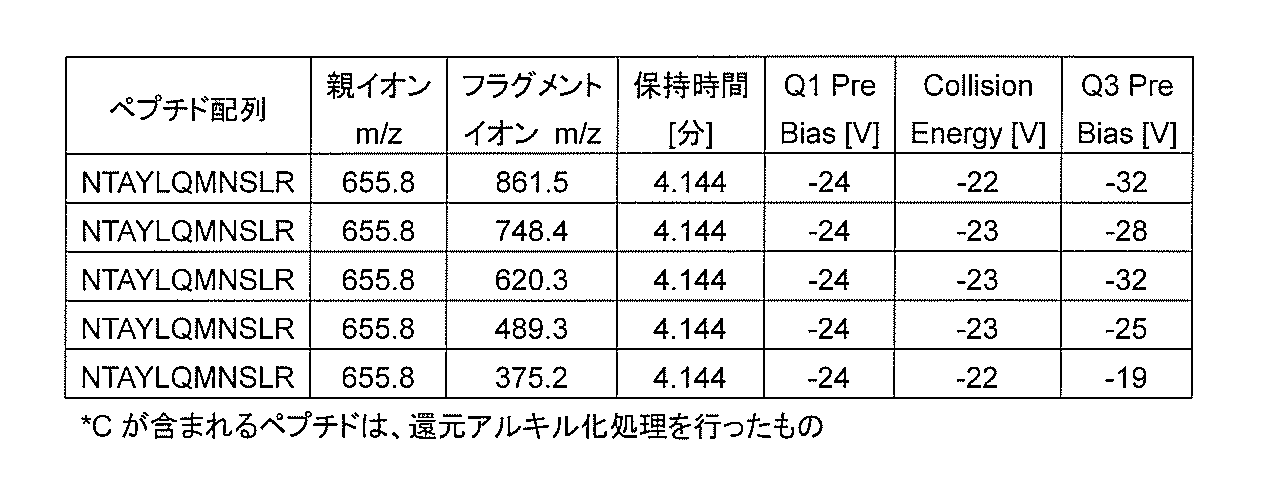
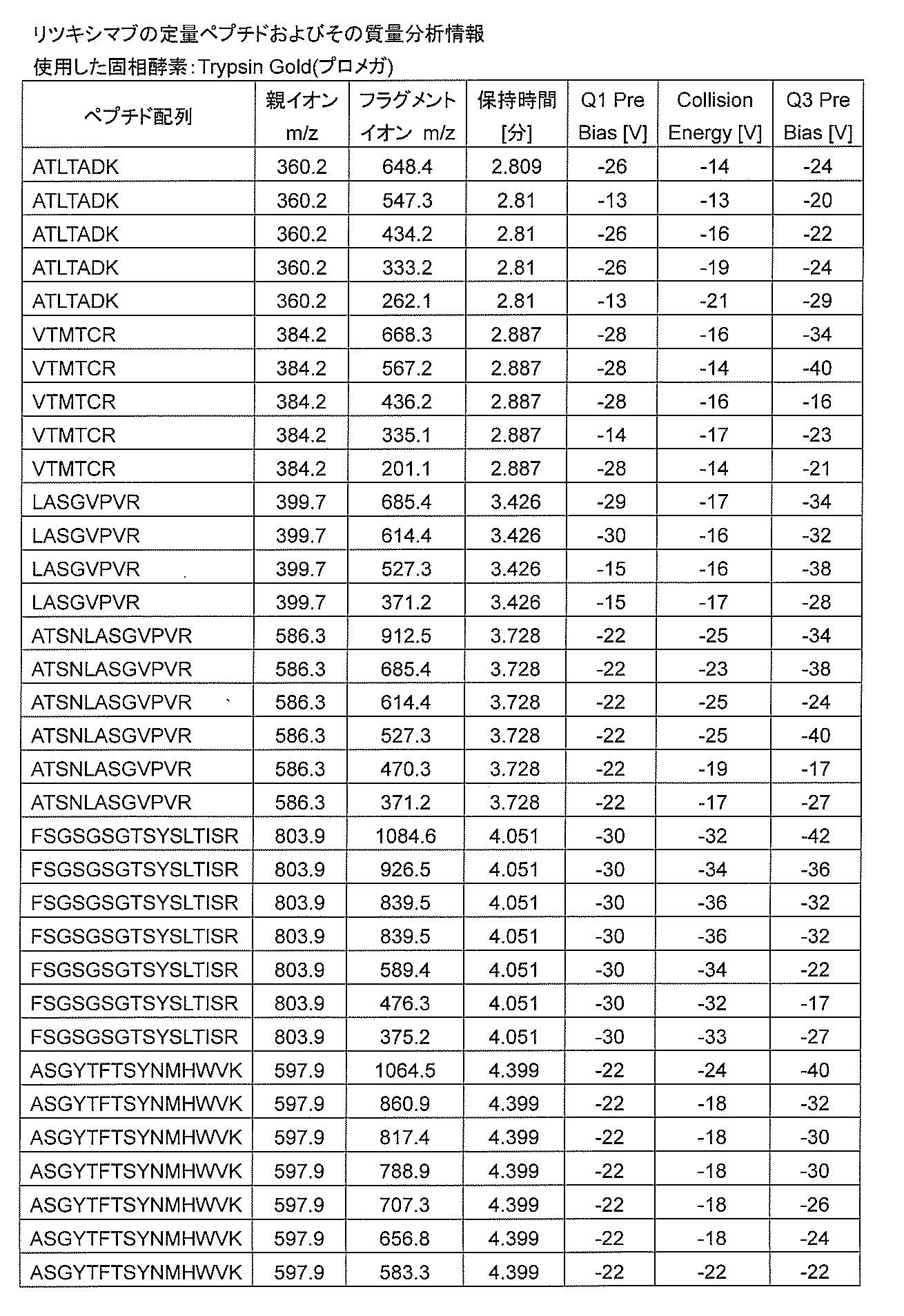
14. LC-MS分析条件リストは、別紙添付する(表1~表6)。


ろ過に使用する膜素材によるペプチド回収率の相違を検証するため、ポリテトラフルオロエチレン(polytetrafluoroethylene、PTFE)およびポリフッ化ビニリデン(polyvinylidene difluoride、PVDF)の2つの膜素材を比較した。
実施例2と同様の実験を、ベバシズマブを用いて検証した。結果を図3に示す。
多検体の測定に用いるためのろ過膜プレートのペプチド回収率を検証するため、2つの膜素材(PTFE、PVDF)およびハウジング3種を比較した。マルチスクリーンプレートとして、1) PTFE membrane/Solvinert housing(メルク社)、2) PVDF membrane/Acryl clear housing(メルク社)、3) PVDF membrane/Barex white housing(メルク社)を用いて比較した。
ナノ粒子に固定化するプロテアーゼ(トリプシン)の活性の安定性をモニターするために、酵素活性測定を行った。ナノ粒子が基質吸光度を阻害するので、従来法に対し、いくつかの改変を加えた。
プロテアーゼ基質を終濃度10 mMとなるよう、DMSOに溶解し、ストック溶液とする。
下記のように混合する。
・基質 10μl(100 nmol)
・反応緩衝液(50 mM Tris-HCl, 150 mM NaCl, pH8.0) 200μl
・固定化酵素懸濁液(0.5 mg/ml) 2μl
プロテアーゼ固定化ナノ粒子の大量製造に対し、プロテアーゼ活性に変化が生じるか否かを検証した。測定方法は、実施例5と同様の改変に加え、反応緩衝液を50 mM Tris-HCl、pH8.0に変更した。
ナノ粒子の大量製造に伴い、洗浄工程が不十分となる可能性が考えられる。洗浄工程の効率化を図る目的で、固定化後の洗浄液に界面活性剤を使用し、その効果を検証した。
マスキングなし(detergent -)FGビーズトリプシン:5.784E-03
マスキングあり(detergent +)FGビーズトリプシン:5.902E-03
となり、界面活性剤洗浄による酵素失活はないと判断できた。
Claims (11)
- 高速液体クロマトグラフ質量分析(LC-MS)によってモノクローナル抗体を検出するためのサンプル調製用キットであって、
測定対象のモノクローナル抗体を固定化するための多孔質体と、
プロテアーゼを固定化したナノ粒子と、
前記多孔質体と前記ナノ粒子とを接触させてモノクローナル抗体を選択的に消化するための反応容器と、
前記ナノ粒子及び多孔質体と共に前記反応容器内に導入され、前記プロテアーゼによる消化反応をさせるための緩衝液と、
前記消化反応後に前記多孔質体および前記ナノ粒子を除去して、前記消化反応の生成物を前記緩衝液と共に抽出するためのろ過膜と、
を含む、上記キット。 - 前記ろ過膜が、圧力又は遠心力を加えない条件下では前記緩衝液及び前記プロテアーゼによる消化反応によって生成するペプチドをほとんど透過せず、圧力又は遠心力を加えた条件下では前記緩衝液及び前記ペプチドを透過することができるものである、請求項1記載のキット。
- 前記ろ過膜がポリフッ化ビニリデン(PVDF)製の膜である、請求項1または2記載のキット。
- 前記キットが多検体を同時に処理するためのキットであり、前記ろ過膜のハウジング素材がポリアクリロニトリル樹脂である、請求項1~3のいずれか1項記載のキット。
- 請求項1~4のいずれか1項記載のキットに、更にモノクローナル抗体の検出のための質量分析条件を記載した説明書を含む、高速液体クロマトグラフ質量分析(LC-MS)によるモノクローナル抗体検出のためのキット。
- 測定対象となるモノクローナル抗体に特異的なアミノ酸配列を含む1以上の内部標準ペプチドを更に含む、請求項1~5のいずれか1項記載のキット。
- 測定対象がトラスツズマブ、トラスツズマブ-DM1、ベバシズマブ、またはリツキシマブであって、内部標準ペプチドが配列番号1~47のいずれか1以上のアミノ酸配列を有するペプチドである、請求項6記載のキット。
- 測定対象のモノクローナル抗体を細孔内に固定化した多孔質体と、プロテアーゼを固定化したナノ粒子とを液体中で接触させてモノクローナル抗体の選択的プロテアーゼ消化を行い、得られたペプチド断片を高速液体クロマトグラフ質量分析(LC-MS)によって分析することによって前記モノクローナル抗体の検出を行う方法に使用するための、前記質量分析を実行させるためのデータが記録されたコンピュータ読み取り可能な記録媒体であって、前記データが、前記モノクローナル抗体のプロテアーゼ消化によって得られるペプチドの1以上に対して、少なくとも親イオン、フラグメントイオン、予想保持時間、三連四重極のそれぞれにおける電圧のデータを含む、上記記録媒体。
- 請求項8記載の記録媒体と、前記記録媒体の使用説明書とを含む、高速液体クロマトグラフ質量分析(LC-MS)によるモノクローナル抗体の検出のためのメソッドパッケージ。
- 前記モノクローナル抗体が、トラスツズマブ、トラスツズマブ-DM1、ベバシズマブ、リツキシマブの1種以上である、請求項8記載の記録媒体または請求項9記載のメソッドパッケージ。
- 前記データが、配列番号1~47のいずれか1以上のアミノ酸配列を有するペプチドに対するものである、請求項8記載の記録媒体または請求項9記載のメソッドパッケージ。
Priority Applications (9)
| Application Number | Priority Date | Filing Date | Title |
|---|---|---|---|
| JP2017504580A JP6515995B2 (ja) | 2015-03-10 | 2015-12-18 | モノクローナル抗体の検出のためのサンプル調製用キット |
| EP15884711.1A EP3270152A4 (en) | 2015-03-10 | 2015-12-18 | KIT FOR SAMPLE PREPARATION FOR THE DETECTION OF MONOCLONAL ANTIBODIES |
| KR1020177027613A KR20170120178A (ko) | 2015-03-10 | 2015-12-18 | 모노클로날 항체의 검출을 위한 샘플 조제용 키트 |
| US15/556,813 US20180052172A1 (en) | 2015-03-10 | 2015-12-18 | Kit for preparing sample for detecting monoclonal antibody |
| BR112017017975A BR112017017975A2 (pt) | 2015-03-10 | 2015-12-18 | kit para preparação de amostra para detecção de anticorpo monoclonal |
| CN201580077602.XA CN107407665A (zh) | 2015-03-10 | 2015-12-18 | 用于检测单克隆抗体的样品调制用试剂盒 |
| CN202210803075.8A CN115932065A (zh) | 2015-03-10 | 2015-12-18 | 用于检测单克隆抗体的样品调制用试剂盒 |
| US17/455,089 US12216126B2 (en) | 2015-03-10 | 2021-11-16 | Kit for preparing sample for detecting monoclonal antibody |
| US18/975,813 US20250102515A1 (en) | 2015-03-10 | 2024-12-10 | Kit for preparing sample for detecting monoclonal antibody |
Applications Claiming Priority (2)
| Application Number | Priority Date | Filing Date | Title |
|---|---|---|---|
| JP2015047729 | 2015-03-10 | ||
| JP2015-047729 | 2015-03-10 |
Related Child Applications (2)
| Application Number | Title | Priority Date | Filing Date |
|---|---|---|---|
| US15/556,813 A-371-Of-International US20180052172A1 (en) | 2015-03-10 | 2015-12-18 | Kit for preparing sample for detecting monoclonal antibody |
| US17/455,089 Continuation US12216126B2 (en) | 2015-03-10 | 2021-11-16 | Kit for preparing sample for detecting monoclonal antibody |
Publications (1)
| Publication Number | Publication Date |
|---|---|
| WO2016143227A1 true WO2016143227A1 (ja) | 2016-09-15 |
Family
ID=56879312
Family Applications (1)
| Application Number | Title | Priority Date | Filing Date |
|---|---|---|---|
| PCT/JP2015/085459 Ceased WO2016143227A1 (ja) | 2015-03-10 | 2015-12-18 | モノクローナル抗体の検出のためのサンプル調製用キット |
Country Status (7)
| Country | Link |
|---|---|
| US (3) | US20180052172A1 (ja) |
| EP (1) | EP3270152A4 (ja) |
| JP (1) | JP6515995B2 (ja) |
| KR (1) | KR20170120178A (ja) |
| CN (2) | CN115932065A (ja) |
| BR (1) | BR112017017975A2 (ja) |
| WO (1) | WO2016143227A1 (ja) |
Cited By (7)
| Publication number | Priority date | Publication date | Assignee | Title |
|---|---|---|---|---|
| WO2019130536A1 (ja) | 2017-12-28 | 2019-07-04 | 株式会社島津製作所 | モノクローナル抗体の簡素化された定量方法 |
| WO2019155576A1 (ja) * | 2018-02-08 | 2019-08-15 | 株式会社島津製作所 | モノクローナル抗体の検出結果を向上する方法 |
| JP2022526671A (ja) * | 2019-04-11 | 2022-05-25 | エンクリアー セラピーズ, インク. | 脳脊髄液の改善の方法ならびにそのためのデバイスおよびシステム |
| CN114878289A (zh) * | 2022-07-12 | 2022-08-09 | 北京大学 | 一种耐低温低盐的蓝藻藻胆体样品的制备方法 |
| WO2022270126A1 (ja) | 2021-06-23 | 2022-12-29 | 株式会社島津製作所 | 抗体を分析する方法 |
| US12188942B2 (en) | 2017-12-28 | 2025-01-07 | Shimadzu Corporation | Method for improving result monoclonal antibody detection |
| US12329930B2 (en) | 2020-09-29 | 2025-06-17 | Enclear Therapies, Inc. | Subarachnoid fluid management method and system |
Families Citing this family (4)
| Publication number | Priority date | Publication date | Assignee | Title |
|---|---|---|---|---|
| EP3270154B1 (en) * | 2015-03-09 | 2020-03-04 | Shimadzu Corporation | Method for detecting a monoclonal antibody using mass spectrometry |
| JP7215467B2 (ja) * | 2019-08-19 | 2023-01-31 | 株式会社島津製作所 | ホルマリン固定パラフィン包埋組織試料からの抗体の分離、検出及び/又は分析方法 |
| CN115552215A (zh) * | 2020-05-08 | 2022-12-30 | 沃特世科技公司 | 用于热辅助酶消化的方法 |
| CN112326816B (zh) * | 2020-10-19 | 2021-07-20 | 广东莱恩医药研究院有限公司 | 一种定量检测血清中地舒单抗的液质联用方法 |
Family Cites Families (9)
| Publication number | Priority date | Publication date | Assignee | Title |
|---|---|---|---|---|
| US4244694A (en) * | 1978-03-31 | 1981-01-13 | Union Carbide Corporation | Reactor/separator device for use in automated solid phase immunoassay |
| JPS6321559A (ja) * | 1986-07-16 | 1988-01-29 | Matsushita Electric Ind Co Ltd | ハプテンの酵素免疫測定法 |
| US6803237B2 (en) * | 2000-01-25 | 2004-10-12 | Woods Hole Oceanographic Institution | Sequential processing reaction vessel for chemical fractionation and analysis |
| EP1495323A2 (en) * | 2002-04-05 | 2005-01-12 | University Of Georgia Research Foundation, Inc. | Method for cleaving and deglycosylating antibodies to promote ligand binding |
| RU2461390C2 (ru) * | 2005-01-28 | 2012-09-20 | Галенбио, Инк. | Иммунологически активные составы |
| JP2010515020A (ja) * | 2006-12-21 | 2010-05-06 | ノバルティス アーゲー | 抗体定量法 |
| WO2009029231A2 (en) * | 2007-08-24 | 2009-03-05 | Millipore Corporation | Method of detecting filter extractables in biopharmaceutical products by liquid chromatography-mass spectrometry |
| JP2012197258A (ja) | 2011-03-23 | 2012-10-18 | Tohoku Univ | 個別化治療診断のためのマーカータンパク質絶対量の定量方法 |
| CN105518447B (zh) | 2013-09-09 | 2018-01-16 | 株式会社岛津制作所 | 肽片段的制备方法及该方法中使用的肽片段制备用试剂盒、以及分析方法 |
-
2015
- 2015-12-18 WO PCT/JP2015/085459 patent/WO2016143227A1/ja not_active Ceased
- 2015-12-18 EP EP15884711.1A patent/EP3270152A4/en not_active Withdrawn
- 2015-12-18 CN CN202210803075.8A patent/CN115932065A/zh active Pending
- 2015-12-18 KR KR1020177027613A patent/KR20170120178A/ko not_active Ceased
- 2015-12-18 JP JP2017504580A patent/JP6515995B2/ja active Active
- 2015-12-18 BR BR112017017975A patent/BR112017017975A2/pt not_active Application Discontinuation
- 2015-12-18 CN CN201580077602.XA patent/CN107407665A/zh active Pending
- 2015-12-18 US US15/556,813 patent/US20180052172A1/en not_active Abandoned
-
2021
- 2021-11-16 US US17/455,089 patent/US12216126B2/en active Active
-
2024
- 2024-12-10 US US18/975,813 patent/US20250102515A1/en active Pending
Non-Patent Citations (3)
| Title |
|---|
| IWAMOTO N: "Selective detection of complementarity-determining regions of monoclonal antibody by limiting protease access to the substrate: nano-surface and molecular- orientation limited proteolysis", ANALYST, vol. 139, no. 3, pages 576 - 580, XP055308690 * |
| MERCK MILLIPORE BUNSEKIYO FILTER CATALOG 2014, 2014, pages 17, 21, XP009504967 * |
| See also references of EP3270152A4 * |
Cited By (13)
| Publication number | Priority date | Publication date | Assignee | Title |
|---|---|---|---|---|
| US12188942B2 (en) | 2017-12-28 | 2025-01-07 | Shimadzu Corporation | Method for improving result monoclonal antibody detection |
| JPWO2019130536A1 (ja) * | 2017-12-28 | 2020-10-22 | 株式会社島津製作所 | モノクローナル抗体の簡素化された定量方法 |
| US11209392B2 (en) | 2017-12-28 | 2021-12-28 | Shimadzu Corporation | Simplified monoclonal antibody quantification method |
| WO2019130536A1 (ja) | 2017-12-28 | 2019-07-04 | 株式会社島津製作所 | モノクローナル抗体の簡素化された定量方法 |
| WO2019155576A1 (ja) * | 2018-02-08 | 2019-08-15 | 株式会社島津製作所 | モノクローナル抗体の検出結果を向上する方法 |
| JPWO2019155576A1 (ja) * | 2018-02-08 | 2021-01-07 | 株式会社島津製作所 | モノクローナル抗体の検出結果を向上する方法 |
| JP7056675B2 (ja) | 2018-02-08 | 2022-04-19 | 株式会社島津製作所 | モノクローナル抗体の検出結果を向上する方法 |
| JP2022526671A (ja) * | 2019-04-11 | 2022-05-25 | エンクリアー セラピーズ, インク. | 脳脊髄液の改善の方法ならびにそのためのデバイスおよびシステム |
| US12329930B2 (en) | 2020-09-29 | 2025-06-17 | Enclear Therapies, Inc. | Subarachnoid fluid management method and system |
| US12337140B2 (en) | 2020-09-29 | 2025-06-24 | Enclear Therapies, Inc. | Subarachnoid fluid management method and system with varying rates |
| WO2022270126A1 (ja) | 2021-06-23 | 2022-12-29 | 株式会社島津製作所 | 抗体を分析する方法 |
| CN114878289B (zh) * | 2022-07-12 | 2022-09-27 | 北京大学 | 一种耐低温低盐的蓝藻藻胆体样品的制备方法 |
| CN114878289A (zh) * | 2022-07-12 | 2022-08-09 | 北京大学 | 一种耐低温低盐的蓝藻藻胆体样品的制备方法 |
Also Published As
| Publication number | Publication date |
|---|---|
| EP3270152A1 (en) | 2018-01-17 |
| EP3270152A4 (en) | 2019-05-15 |
| BR112017017975A2 (pt) | 2018-04-10 |
| US12216126B2 (en) | 2025-02-04 |
| CN107407665A (zh) | 2017-11-28 |
| CN115932065A (zh) | 2023-04-07 |
| JPWO2016143227A1 (ja) | 2017-12-28 |
| US20180052172A1 (en) | 2018-02-22 |
| US20220137062A1 (en) | 2022-05-05 |
| KR20170120178A (ko) | 2017-10-30 |
| JP6515995B2 (ja) | 2019-05-22 |
| US20250102515A1 (en) | 2025-03-27 |
Similar Documents
| Publication | Publication Date | Title |
|---|---|---|
| US20250102515A1 (en) | Kit for preparing sample for detecting monoclonal antibody | |
| EP3270154B1 (en) | Method for detecting a monoclonal antibody using mass spectrometry | |
| EP3306310B1 (en) | Method for quantifying monoclonal antibody | |
| US20200011876A1 (en) | Method for Simultaneous Quantification of Monoclonal Antibodies | |
| WO2018235228A1 (ja) | 抗原または抗抗体が結合したモノクローナル抗体の定量方法 | |
| EP3269821A1 (en) | Method for obtaining peptide fragment from antibody by means of protease decomposition reaction with restricted reaction field | |
| JP7786458B2 (ja) | 抗体を分析する方法 |
Legal Events
| Date | Code | Title | Description |
|---|---|---|---|
| 121 | Ep: the epo has been informed by wipo that ep was designated in this application |
Ref document number: 15884711 Country of ref document: EP Kind code of ref document: A1 |
|
| REG | Reference to national code |
Ref country code: BR Ref legal event code: B01A Ref document number: 112017017975 Country of ref document: BR |
|
| WWE | Wipo information: entry into national phase |
Ref document number: 15556813 Country of ref document: US |
|
| ENP | Entry into the national phase |
Ref document number: 2017504580 Country of ref document: JP Kind code of ref document: A |
|
| NENP | Non-entry into the national phase |
Ref country code: DE |
|
| ENP | Entry into the national phase |
Ref document number: 20177027613 Country of ref document: KR Kind code of ref document: A |
|
| REEP | Request for entry into the european phase |
Ref document number: 2015884711 Country of ref document: EP |
|
| ENP | Entry into the national phase |
Ref document number: 112017017975 Country of ref document: BR Kind code of ref document: A2 Effective date: 20170822 |