WO2016143808A1 - Procédé de production de sphéroïdes et milieu de culture utilisé dans le procédé - Google Patents
Procédé de production de sphéroïdes et milieu de culture utilisé dans le procédé Download PDFInfo
- Publication number
- WO2016143808A1 WO2016143808A1 PCT/JP2016/057307 JP2016057307W WO2016143808A1 WO 2016143808 A1 WO2016143808 A1 WO 2016143808A1 JP 2016057307 W JP2016057307 W JP 2016057307W WO 2016143808 A1 WO2016143808 A1 WO 2016143808A1
- Authority
- WO
- WIPO (PCT)
- Prior art keywords
- glucose
- medium
- culture
- cells
- spheroids
- Prior art date
- Legal status (The legal status is an assumption and is not a legal conclusion. Google has not performed a legal analysis and makes no representation as to the accuracy of the status listed.)
- Ceased
Links
Images


Definitions
- the present invention relates to a culture method suitable for producing spheroids (functional cell aggregates).
- the present invention also relates to a medium suitable for the culture method.
- the spheroid culture method that allows cells to grow three-dimensionally and take a three-dimensional form such as a sphere or egg shape has a physiological gradient of nutrients, oxygen, or carbon dioxide and other metabolites and waste products. It is thought that it occurs in spheroids, and it is possible to cause a cell state closer to cells existing in the living body environment, and for functional expression such as drug screening such as drug efficacy evaluation, tumor evaluation, production of cell products, etc. Use is rapidly expanding as an effective method.
- Non-patent Document 1 As methods for forming spheroids, the hanging drop culture method (Non-patent Document 1), the culture in gel (Non-patent Document 2), the culture using a low adhesion container (Non-patent Document 3), and nanoimprinting.
- Various methods have been proposed, such as culture using a special dish in which a scaffold is produced by precision processing technology (Non-patent Document 4), addition of a growth factor or Matrigel (Non-patent Document 5), and the like.
- the hanging drop culturing method is a method of culturing by spotting a cell suspension in a droplet of a culture solution using surface tension (Non-patent Document 1).
- Non-patent Document 1 a method of culturing by spotting a cell suspension in a droplet of a culture solution using surface tension.
- Spheroid culture using a low-adhesion dish is a culture method that forms spheroids by preventing cells from adhering to the dish surface, but the spheroids once formed tend to gradually fuse together, and there is a problem with stability. Yes (Non-Patent Document 6).
- a culture method using a U-Shape dish or the like can avoid the fusion of spheroids and can form a single spheroid. And the method of using the special container of U bottom and V bottom is devised, and is marketed. Although this method forms spheroids of uniform size, the cells tend to be sparse, and the shape is not spherical or elliptical and tends to be bowl-shaped. In order to obtain dense spheroids, growth factors and the like are added to the medium, but the properties as spheroids tend to be insufficient.
- Non-patent Document 1 In the spheroid culture method by rotational culture, a special apparatus is required for rotational culture, and the size and shape of the spheroid are not uniform, which affects the reproducibility of the spheroid experiment (Non-patent Document 1).
- Non-patent Document 4 As a spheroid culture method in a special container with fine special processing, cell adhesion on the bottom of the dish is unstable by arranging a cell adhesion surface with a small area and a cell non-adhesion surface with a large area on the bottom of the dish.
- a method of forming spheroids by forming a spheroid by suppressing the cell adhesion by finely processing a nano-sized lattice structure on the bottom of the dish has been proposed and marketed. By using these methods, spheroids having a uniform shape are formed.
- specially processed dishes are extremely expensive, and even when matrigel or recommended special additives that have not been disclosed are added, the formation of dense spheroids that mimic the state in vivo is not possible. Tend to be enough. Further, in the microscopic observation, there is a disadvantage that processing of the bottom surface of these special dishes may be an obstacle (Non-patent Document 4).
- the spheroids rapidly collapse with the passage of the number of days of culture, so that the timing at which good quality spheroids can be used is very short and difficult to use.
- the cells are sparse and it is difficult to form good quality spheroids.
- cell aggregates form a three-dimensional structure, so that a state closer to the living body appears and a specific function of the cell can be exhibited.
- drug discovery screening such as drug efficacy evaluation, tumor evaluation, and evaluation of functional expression such as production of cell products
- cell clusters composed of cells that exhibit specific functions similar to cells in vivo are stable. It is desirable to be manufactured. At that time, it is desired to produce a large amount of high-quality spheroids by a method as simple as possible.
- Patent Document 2 when cultured cancer cells are used, cells that take 2-NBDLG are recognized in spheroids including a group of cells exhibiting abnormal nuclear states of any size. That is, it has been reported that 2-NBDLG is taken up when spheroids are formed in cultured cancer cells as well as cells derived from living organisms. However, it has been difficult to form such spheroids in large quantities with good reproducibility even by the addition of matrigel or the like in the conventional method.
- the present inventors have cultured in a medium added with L-glucose instead of D-glucose (hereinafter sometimes referred to as L-glucose medium) as glucose.
- L-glucose medium a medium added with L-glucose instead of D-glucose (hereinafter sometimes referred to as L-glucose medium) as glucose.
- spheroids begin to form after a certain number of days (for example, 3 to 10 days depending on the cells) after replacement with L-glucose medium, and then dense spheroids are formed one after another with good reproducibility.
- the headline and the present invention were completed.
- the present invention includes the following. (1) A method for producing a spheroid comprising culturing cells using a glucose-containing medium, wherein the glucose is substantially composed of L-glucose.
- the step (b) is a step of culturing cells while gradually decreasing the D-glucose concentration in the medium and gradually increasing the L-glucose concentration. .
- step (10) The method according to (8) or (9) above, wherein the ratio of L-glucose in the glucose-containing medium in step (c) is 90% or more of the total glucose composed of D-glucose and L-glucose.
- (11) The method according to any one of (8) to (10) above, wherein the culture in the step (c) is 3 days or more.
- (12) The method according to any one of (8) to (11) above, wherein the cells are cancer cell-derived cells.
- the medium in the step (c) is a medium to which no growth factor and / or adhesion factor is added.
- the present invention provides a method by which spheroids with dense cells can be produced with good reproducibility. Moreover, the culture medium for producing such a spheroid is provided by this invention.
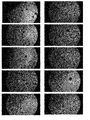
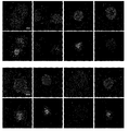
- FIG. 3 is a diagram showing the formation process of MIN6 cell spheroids observed in Example 1.
- 13 is a photograph (a phase contrast image taken with an x4 objective lens) on the 13th day after the start of L-glucose culture. The figure shows 10 typical fields of view cultured on the same plastic dish, and it was observed that many spheroids were formed. The scale bar is 200 ⁇ m.
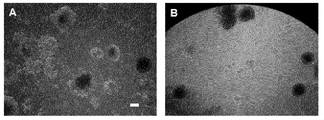
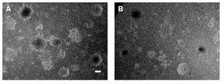
- It is a photograph which shows the MIN6 cell spheroid formed on the glass bottom dish performed in Example 2.
- FIG. 11 is a photograph (a phase contrast image taken with an x4 objective lens) on the 11th day after the start of L-glucose culture. It shows two fields of view on the same dish.
- the scale bar is 200 ⁇ m. In the photograph of the 14th day after the start of the L-glucose culture carried out in Example 2, it is observed that spheroids are stably formed. The scale bar is 200 ⁇ m. 2 is a photograph taken on Example 21 on the 21st day after the start of L-glucose culture. It shows two fields of view on the same dish. It can be seen that good quality spheroids with necrotic areas in the center continue to form. The scale bar is 200 ⁇ m. FIG.
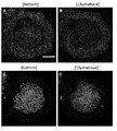
- FIG. 6 shows the result of applying a 2-NBDLG / 2-TRLG mixed solution to MIN6 spheroids cultured in an L-glucose medium for 14 days in a glass bottom dish in Example 2.
- FIG. The photograph shows the field of view of eight representatives on the same dish.
- the detection result in the green channel which is the fluorescence wavelength of 2-NBDLG
- the red channel which is the fluorescence wavelength of 2-TRLG.
- the results are shown in the lower part.
- the central part of the spheroid causes necrosis and incorporates 2-TRLG that is impermeable to the cell membrane, and the presence of cells that incorporate 2-NBDLG is observed around it.
- the scale bar is 100 ⁇ m.
- FIGS. 6A and 6B are phase contrast micrographs of x4 and x20, respectively, on the 11th day after the start of L-glucose culture, and it is observed that spheroids are also formed on the cover slip.
- the scale bar is 200 ⁇ m.
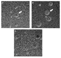
- 2 is a photograph showing the state of 2-NBDLG and 2-TRLG uptake in the MIN6 cell spheroid formed on the coverslip in Example 3.
- FIG. (A) and (C) are images before administration of 2-NBDLG and 2-TRLG
- (B) and (D) are images after 16 minutes from the start of washing after the administration is completed.
- the upper row shows the detection result in the green channel that is the fluorescence wavelength of 2-NBDLG
- the lower row shows the detection result in the red channel that is the fluorescence wavelength of 2-TRLG.
- the scale bar is 100 ⁇ m.
- a and C show fluorescence images of a green channel for detecting 2-NBDLG uptake and a red channel for detecting 2-TRLG uptake, respectively, taken on the surface in contact with the cover glass.
- the central part of the spheroid incorporates 2-TRLG, which is impermeable to cell membranes, causing necrosis and increasing cell membrane permeability.
- B and D are tomographic photographs in a section 15 microns above from the surface in contact with the cover glass.
- 2-NBDLG the central part of the spheroid formed using a conventional D-glucose-containing medium
- cells that take up 2-NBDLG are only found in a part of the spheroid, but according to the culture method using the L-glucose medium of the present invention.
- 2-NBDLG uptake is observed in many cells in the peripheral region of the central core.
- the scale bar is 100 ⁇ m.
- B reaches the 15th day of culture and shows a state in which the spheroids are disintegrated.
- C shows that MIN6 cell spheroids formed on a cover glass using a normal D-glucose-containing medium are completely disintegrated on the 14th day of culture. It is the result of having observed a mode that the HT-29 human colon adenocarcinoma cell illustrated in Example 5 formed a spheroid.
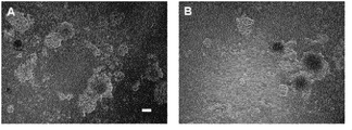
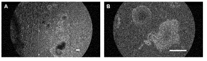
- A is a photomicrograph on the third day after the start of L-glucose culture (phase contrast image, using x4 objective lens) (total six days after the start of culture). Spheroid formation showing a marked necrotic area was observed in the center.
- B is a photograph on the sixth day after the start of culture using only the D-glucose medium without using the L-glucose medium. Compared with culture using L-glucose medium (A), the growth is faster, but no necrotic part is observed in the center, and a three-dimensional cell cluster (Sparsely-packed three-dimensional cell-cluster) is formed. ing.
- C is a photomicrograph of the fifth day after the start of L-glucose culture (total eight days after the start of culture). The size exceeds 200 ⁇ m, and a three-dimensional cell cluster (Tightly-packed three dimensional cell cluster) in which cells are densely assembled is formed.
- the scale bar is 100 ⁇ m.
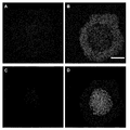
- HT29 cells in which spheroids were formed by the culture method shown in Example 5 were transferred onto cover slips on the 10th day after the start of L-glucose culture (13 days after the start of culture), and were fixed for 1 day. That is, the photographs show the uptake of 2-NBDLG and 2-TRLG on the 11th day after the start of L-glucose culture (the 14th day after the start of culture).
- (A) and (C) show images before administration of 2-NBDLG and 2-TRLG
- (B) and (D) show images 6 minutes after the end of administration.
- the upper row shows the detection result in the green channel that is the fluorescence wavelength of 2-NBDLG
- the lower row shows the detection result in the red channel that is the fluorescence wavelength of 2-TRLG.
- the scale bar is 100 ⁇ m.
- the L-glucose medium is a medium in which L-glucose is added as glucose while D-glucose is not actively added, that is, a glucose-containing medium, and the glucose is substantially composed of L-glucose.
- a glucose-containing medium a medium in which L-glucose is added as glucose while D-glucose is not actively added, that is, a glucose-containing medium, and the glucose is substantially composed of L-glucose.
- the D-glucose concentration is about 100 mg / L.
- the L-glucose medium of the present invention is a glucose-containing medium, and the glucose is substantially composed of L-glucose, preferably 90% or more, more preferably 95% or more, more preferably 98% of glucose. Above, most preferably, refers to a medium in which 99% or more is L-glucose.
- the glucose concentration in the L-glucose medium used in the present invention is not particularly limited. For example, the glucose concentration when D-glucose is used as glucose in normal culture can be used. Although not limited thereto, for example, 0.5 mM to 50 mM, preferably 1.0 mM to 25 mM, more preferably 5 mM to 25 mM.
- L-glucose medium a medium used for normal cell culture can be used as long as the glucose is substantially composed of L-glucose.
- the medium include, but are not limited to, a medium that can be used for animal cell culture.
- it is DMEM culture medium. These media are commercially available.
- the medium used in the present invention may be a serum-containing medium or a serum-free medium.
- a serum-free medium means a medium that does not contain unconditioned or unpurified serum, and a medium that contains purified blood-derived components or animal tissue-derived components (for example, growth factors) corresponds to a serum-free medium. It shall be.
- the medium used in the present invention is a serum-containing medium, mammalian serum such as fetal bovine serum can be used.
- the medium is preferably a serum-free medium, more preferably a serum-free medium that does not contain chemically undefined components.
- the medium used in the present invention may also contain a serum replacement.
- Serum substitutes include, for example, albumin, transferrin, fatty acid, collagen precursor, trace elements (eg, zinc, selenium), B-27 supplement, N2 supplement, knockout serum replacement (KSR), 2-mercaptoethanol, monothio Glycerol, or an equivalent thereof. These serum replacements are commercially available.
- the medium used in the present invention also contains other additives such as lipids, amino acids (eg, non-essential amino acids), vitamins, growth factors, cytokines, antioxidants, 2-mercaptoethanol, pyruvic acid, buffers, inorganics. It may contain salts, antibiotics (for example, penicillin or streptomycin) or antibacterial agents (for example, amphotericin B).
- the medium used in the present invention may also contain other additives that contribute to the formation of spheroids, such as growth factors and adhesion factors, and may contain an intercellular matrix.
- growth factors include NGF, FGF, EGF, TGF, BDNF, VEGF, G-CSF, PDGF, EPO, and the like.
- adhesion factor include integrin, cadherin, laminin, immunoglobulin superfamily and the like.
- the intercellular matrix include collagen, fibronectin, netrin, proteoglycans and the like.
- Corning Matrigel registered trademark
- Scivax NanoCulture Medium Supplement can be used.
- the medium used in the present invention is particularly preferably a serum-free medium that contains only chemically defined raw materials and is a medium that does not contain growth factors, adhesion factors, or intercellular matrix.
- a DMEM medium in which the added glucose is substantially composed of L-glucose can be mentioned.
- spheroids are formed by removing D-glucose from the medium and culturing cultured cells in an L-glucose medium instead.
- a D-glucose medium In changing from a D-glucose medium to an L-glucose medium, it is important to start the replacement with the L-glucose medium from an appropriate number of culture days depending on the cells.
- the method of replacement with the L-glucose medium can be appropriately changed depending on the cell. For example, the method of replacing the D-glucose medium with the L-glucose medium, the ratio of L-glucose in the total glucose stepwise You can give a way to raise it.
- the concentration of D-glucose is reduced stepwise and The concentration can be easily increased.
- the half amount exchange is not limited to this, but can be performed, for example, on the 6th and 8th days after the start of the D-glucose culture. On the 10th day, the entire culture medium is discarded, and D-glucose is removed as much as possible by washing with L-glucose medium to finally make 100% L-glucose medium. After reaching the 100% L-glucose medium, but not limited to this, for example, half the amount is changed every 6 hours over one or two days. Subsequent medium changes are performed every few days when the number of cells is small, and every day when the number of cells is large.
- the cells are cultured for a certain number of days or more after being replaced with a medium supplemented with 100% L-glucose (added with 0% D-glucose) as glucose. “Cultivation over a certain number of days” is appropriately selected depending on the cells to be used. For example, the number of days can be increased by 3-10. Generally, for example, 3 days or more, preferably 10 days or more, more preferably Is cultured for 11 days or longer, more preferably 13 days or longer. Thereby, after that, spheroids dense between cells continue to be formed one after another with good reproducibility. A large amount of spheroids can be supplied for at least several days or more, preferably for at least one week or more, more preferably for at least two weeks or more. For example, spheroids can also be supplied in the third week.
- the cells to which the method of the present invention can be applied are not particularly limited, and can be used for any cells as long as they have the ability to form spheroids.
- cells derived from cancer as used in the examples of the present invention cells collected from a living body, such as, but not limited to, hepatocytes, kidney cells, or pancreatic cells, cells of various glandular tissues, etc.
- cells that can form spheroids include, but are not limited to, various stem cells including ES cells and iPS cells.
- spheroids with dense cells can be formed.
- the phenomenon that adjacent spheroids are often connected one after another in the horizontal direction which is often observed when a normal culture method is used, is less likely to occur.
- the spheroid produced using the present invention is a spheroid having a dense cell-to-cell size and having a diameter of 100 ⁇ m or more, preferably 150 ⁇ m or more, more preferably 200 ⁇ m or more.
- the three-dimensional structure is more stably maintained, and a high-quality spheroid can be obtained at any time over a long period of time.
- the spheroid culture using the method of the present invention is a three-dimensional or three-dimensional support such as a culture vessel having a bottom of U-Shape or V-Shape, or a three-dimensional process such as a lattice structure on the bottom.
- Good quality spheroids can be formed without using a given culture vessel or the like.
- spheroids can also be produced using the method of the present invention using a culture vessel containing such a support, and this is also included in the present invention.
- the method of the present invention is characterized in that spheroids can be produced without the addition of a special device or container, a growth factor, an adhesion factor, an intercellular matrix or the like. Furthermore, according to the method of the present invention, spheroids can be formed and cultured using a normal culture vessel, and in this state, observation with a microscope is easy.
- the present invention it is not necessary to use special equipment, special containers or expensive additives for culturing the formed spheroids, and general containers such as plastic dishes and cover slips can be used, Moreover, since the culture medium to be used can be carried out on the basis of a general DMEM culture medium and no special technique or skilled technique is required in the culture operation, anyone has the advantage of easily producing spheroids.
- the method of the present invention may be used in combination with any one or more of spheroid production techniques such as addition of special devices, containers, growth factors, adhesion factors, intercellular matrix, etc., or all of them. It is possible to expect to produce a large amount of spheroids with high efficiency and good quality by such a combination. Such combinations can be appropriately selected by those skilled in the art based on the teachings of the present invention, and are also part of the present invention.
- spheroids when culturing in a glass bottom dish having a diameter of 4 cm using the method of the present invention, several tens or more, preferably about 90 or more spheroids are formed.
- This makes it possible to stably and continuously produce a large amount of spheroids required for a small amount of analysis in one dish.
- This is a great advantage in terms of operability and subsequent cell recovery compared to spheroid formation using a multiplate such as a three-dimensional culture platform.
- the additive when an additive such as a growth factor is used for spheroid formation, the additive may interfere with subsequent observation with a microscope or optical analysis. Furthermore, there is a concern that the additive itself may have biochemical and physiological effects on the cells themselves.
- the above-described additives are not required for the formation of spheroids, so that there are no such harmful effects, and spheroids suitable for experiments can be provided.
- spheroids produced using MIN6 cells which are mouse insulinomas, according to the method of the present invention are characterized in that they have a property of favorably taking 2-NBDLG that is selectively taken up into cancer cells.
- 2-NBDLG a property of favorably taking 2-NBDLG that is selectively taken up into cancer cells.
- approximately 70% of the spheroids produced using the present invention are good quality spheroids that incorporate 2-NBDLG into cells.
- Such spheroids can be supplied in large quantities from the 11th day to at least the 3rd week after the medium becomes a 100% L-glucose medium.
- D-glucose two isomers that are mirror images of each other, namely D-glucose and L-glucose, can be considered, but only D-glucose exists in nature.
- the organism selectively takes this D-glucose into the cell and metabolizes it as a basic energy source.
- normal cells cannot use L-glucose which is not found in nature.
- the inventor has introduced a molecule 2- [N- (7-nitrobenzo-2-oxa-1,3-diazol-4-yl) amino] -2-deoxy- in which a fluorescent group NBD is introduced at the second position of D-glucose.
- D-glucose (2-NBDG) is incorporated into mammalian cells via glucose transporters in a concentration, time and temperature-dependent manner with kinetics similar to radiolabeled glucose derivatives and is its control molecule Molecule 2- [N- (7-nitrobenz-2-oxa-1,3-diazol-4-yl) amino] -2-deoxy-L-glucose with a fluorescent group NBD introduced at the second position of L-glucose ( 2-NBDLG) was not selectively taken up through the glucose transporter.
- the present inventor has also shown that when 2-NBDLG is applied to spheroid-formed cancer cells (mouse insulinoma MIN6), the cancer cells selectively take 2-NBDLG into the cells (Patent Document 2). .
- the present inventor has also reported that 2-TRLG having a fluorescent group TexasTRed introduced at the second position of L-glucose in addition to 2-NBDLG can be used to detect necrotic cells (Patent Document 2).
- the present inventors apply 2-NBDLG to live cancer tissue or cancer cells excised from humans, the cancer cells selectively take in 2-NBDLG and emit fluorescence, thereby producing non-cancer cells. (Japanese Patent Application Nos. 2014-015735 and 2014-193424).
- 2-NBDLG was characterized in that the cells showed a nuclear state of any size and were taken up by a sufficiently dense spheroid between cells.
- Such spheroids cannot be produced in large quantities with high reproducibility even if special culture containers for preparing spheroids, Matrigel, special media, or the like are added, and it is difficult to produce 2-NBDLG in one culture dish by various means. As a result, it was sufficient if there were 2-3 spheroids that significantly incorporated
- MIN6 is a mouse insulinoma cell
- the method of the present invention can be applied to other cells.
- the materials, reagents, and conditions to be used can be appropriately selected and used according to known information about each cell.
- the cell suspension of mouse insulinoma cell MIN6 may be adjusted to 10 ⁇ 10 4 cells / ml using a medium containing D-glucose, and seeded on a dish or cover slip according to subsequent observation and analysis. it can.
- the number of cells to be seeded is, for example, 30-60 ⁇ 10 4 cells / dish for a commercially available 90 mm diameter dish (Greiner TC Petri dish 664160), and 10 ⁇ 10 4 for a WillCo-Glass bottom dish (WillCo Wells 170 micron diameter 40 mm). 4 pieces / dish is appropriate.
- a glass slip of 6 mm ⁇ 2 mm it is appropriate to seed 600 to 1000 cells.
- the D-glucose medium is replaced with an L-glucose medium at a certain time after the start of culture.
- Such medium replacement can be performed by methods known to those skilled in the art. For example, it can be carried out by removing the D-glucose medium used for cell culture or collecting the cells from the medium and adding an L-glucose medium.
- the replacement of the medium can also be performed as follows. First, the medium is changed so that the D-glucose concentration of the medium is 25% (6.25 mM) of the initial concentration. For this purpose, the D-glucose concentration can be reduced to 25% by continuously exchanging half of the medium twice.
- an L-glucose-containing medium (D-glucose-free medium) can be used as a medium used for medium exchange.
- the day 2 to day 10 after the start of culture is suitable for setting the D-glucose concentration to 25% of the initial concentration.
- the medium is exchanged by half to make the D-glucose concentration 12.5% (3.125 mM).
- the entire amount of the medium is discarded and washed twice with an L-glucose-containing medium, and then the L-glucose medium is added, so that the D-glucose concentration added to the medium can be reduced to 0%.
- L-glucose medium Although the state varies depending on the cells, for example, the following occurs by using L-glucose medium. Among the cells that have continued to grow during culture, floating dead cells become noticeable after D-glucose concentration of 12.5% (3.125 mM), and after D-glucose concentration reaches 0%. The cell suspension is prominent, and the number of cells that have been increasing until then decreases once. In order to remove metabolites from the dead cells floating, half of the medium is exchanged with a medium containing L-glucose every 6 hours from the time when the concentration of D-glucose added to the medium becomes 0%. Thereby, it is possible to minimize the influence of the metabolite.
- the medium exchange every 6 hours is preferably performed at least 4 times or more until floating cells become inconspicuous.
- the present invention it is possible to stably form a large amount of high-quality cancer cell spheroids that reproduce in vitro the uptake of a fluorescent L-glucose derivative in human cancer cells.
- a fluorescent L-glucose derivative 2-NBDLG there is a large difference in the uptake of the fluorescent L-glucose derivative 2-NBDLG between the spheroids produced by the present invention using cancer cells and the spheroids produced by the existing three-dimensional platform. Since the fluorescent L-glucose derivative 2-NBDLG is strongly taken into tissues ex vivo from cancerous lesions in the tissues, the spheroids according to the present invention are more effective than the spheroids prepared by the conventional method. It is thought that it has a feature close to in vivo.
- HT-29 human colon adenocarcinoma cells were obtained from ATCC (American Type Culture Collection) (cell number: HTB-38).
- D-glucose medium DMEM-high glucose medium (SIGMA: D5648, D-glucose concentration 25 mM) was dissolved in distilled water, 3.7 g of NaHCO 3 and 5 ⁇ l of 2-mercaptoethanol were added. After adjusting the pH to 7.3 and adjusting the total volume to 1000 ml, sterilized by filtration, FBS (HyClone: SH30070) was added to 10%, penicillin / streptomycin was added, and a medium containing D-glucose (hereinafter, “ D-glucose medium ”.
- SIGMA D5648, D-glucose concentration 25 mM
- L-glucose medium After adjusting the pH to 7.3 and making the total volume 1000 ml, sterilized by filtration, then added 20 ml of L-glutamine (SIGMA: G7513) and further FBS (HyClone: SH30070) to 10%, and penicillin / streptomycin To obtain a medium containing L-glucose (hereinafter referred to as “L-glucose medium”).
- Example 1 Formation of Spheroids on Plastic Dish MIN6 cells (mouse insulinoma-derived cells) were suspended at 30 ⁇ 10 4 cells / ml in D-glucose medium, which is a medium used in general cell culture. 1 ml of the cell suspension was added to a plastic dish (Greiner, TC petri dish for cell culture 100 ⁇ 20 mm), and a medium was further added to make the total medium volume 10 ml. The culture was started at 37 ° C. in a 5% CO 2 incubator (0 day after the start of culture: 100% D-glucose) and left to stand until the third day after the start of the culture.
- D-glucose medium which is a medium used in general cell culture. 1 ml of the cell suspension was added to a plastic dish (Greiner, TC petri dish for cell culture 100 ⁇ 20 mm), and a medium was further added to make the total medium volume 10 ml. The culture was started at 37 ° C. in a 5% CO 2 incubator
- the culture was further continued, and half of the medium was exchanged using L-glucose medium on the 8th day of culture.
- the ratio of D-glucose was 12.5% (87.5% (21.9 mM) of L-glucose (21.9 mM). 3.1 mM) (culture day 8: 12.5% D-glucose, 87.5% L-glucose). From this stage, many of the cell masses that had been proliferating until now have been observed to shrink.
- Example 2 Observation of uptake of L-glucose derivative 2-NBDLG after formation of adhesive spheroids in glass bottom dish MIN6 cells (mouse insulinoma-derived cells) in D-glucose medium at 10 ⁇ 10 4 cells / ml 1 ml of cell suspension is added to a culture dish WillCo-Glass bottom dish (WillCo Wells: 170 micron diameter 40 mm), and D-glucose medium is added to make a total medium volume of 3 ml at 5% CO 2 at 37 ° C. The culture was started (0 day after the start of culture: 100% D-glucose) and left to stand until the third day after the start of the culture.
- MIN6 cells mouse insulinoma-derived cells
- the L-glucose derivatives 2-NBDLG and 2-TRLG were taken up by a confocal laser microscope using an excitation wavelength of 488 nm and a fluorescence wavelength of 500 GH Observation was at ⁇ 580 nm and red channel 580-740 nm. It was confirmed that 19 out of 24 observed spheroids well incorporated 2-NBDLG. The observation results are shown in FIG.
- Example 3 Observation of incorporation of L-glucose derivative 2-NBDLG / 2-TRLG mixed solution into spheroids formed on cover slip 6.5 mm x 2 mm cut cover slip (MATSUNAMI MICROCOVER GLASS No. 0 13 x 22 mm) was placed in a 35 mm dish (IWAKI Non-treated Dish 1000-035), and 10 ⁇ l of MIN6 cells suspended at 10 ⁇ 10 4 cells / ml in D-glucose medium were seeded on a coverslip. 3 ml of D-glucose medium was added to the dish, culture was started at 37 ° C.
- Table 1 shows the ratio of spheroids indicating 2-NBDLG uptake in Examples 2 and 3 above. In Example 2, over 70%, while in Example 3, over half of the spheroids indicate 2-NBDLG uptake.
- Example 4 Stability of spheroids formed on cover slips
- MIN6 cells were cultured in L-glucose medium to form MIN6 cell spheroids, and their transition was observed.
- the results are shown in FIG. It can be seen that the spheroids produced by the method of the present invention form a typical heterogeneous cell cluster in which proliferating cells in the peripheral part and necrotic cells in the central part are formed. Moreover, it turns out that the spheroid produced by the method of the present invention maintains a stable spheroid for a long period of time.
- FIG. 10A On the 11th day after the start of culture, when a sufficiently large spheroid was formed, the spheroid was transferred to a glass bottom dish using a micropipette and used for observation. The result is shown in FIG. 10A. On the 14th day of culture, the cells were seeded on a coverslip using a micropipette and allowed to settle for 1 day, and then observed on the 15th day of culture. The results are shown in FIG. 10B.
- necrotic cells appear in the eyeball-like region of the central core of the spheroids formed by the L-glucose culture according to the present invention, and the diameter of the entire spheroids is maintained at 200 ⁇ m or more. Furthermore, the shape does not collapse from 11 to 21 days after the start of L-glucose culture. However, the size of the spheroids produced by the nanoimprinting dish is smaller than that of the spheroid of the present invention, and spheroids that start to collapse as early as the 11th day of culture appear, and the form cannot be maintained on the 15th day. In addition, the spheroids formed in the D-glucose medium on the coverslip are not maintained at all on the 14th day. The time at which the spheroids formed on the D-glucose medium break down varies depending on the individual spheroid, but once an ideal spheroid is formed, it is not uncommon to lose its shape within a few hours to one day.
- Example 5 Example in which HT-29 human colon adenocarcinoma cells were cultured in L-glucose medium to form spheroids Human colon adenocarcinoma was added to D-glucose medium, which is a medium used in general cell culture.
- Cell HT-29 was suspended at a rate of 10 ⁇ 10 4 cells / ml, 3 ml of this cell suspension was added to a filter cap flask for floating cells (Sumitomo Bakelite MS-2325R, 250 ml), and a medium was further added. The total medium volume was 20 ml. The culture was started at 37 ° C.
- FIG. 11A On the 3rd day after the start of L-glucose culture (6th day after the start of culture), spheroid formation showing a necrotic region was observed in the center (FIG. 11A).
- HT-29 human colon adenocarcinoma cells were cultured using the same method, but not using L-glucose medium and using only D-glucose-containing medium, three-dimensional cell clusters (Sparsely-packed) 3 dimensional cell cluster), and good quality cancer cell spheroids were not formed (FIG. 11B, 6th day after the start of culture). Spheroid formation with a diameter exceeding 200 ⁇ m was also observed on the 5th day after the start of the culture in the L-glucose medium (the 8th day after the start of the culture) (FIG. 11C).
- Example 6 Observation of incorporation of L-glucose derivative 2-NBDLG / 2-TRLG mixed solution of HT-29 human colon adenocarcinoma cells cultured in L-glucose medium HT-29 formed by the culture method of Example 5 In order to observe the cell spheroids with a confocal microscope, the cells were transferred to cover slips on the 10th day after the start of the L-glucose culture (the 13th day after the start of the culture) and fixed.
- high quality spheroids can be obtained using a normal culture vessel or cover slip by a simple method of removing D-glucose from the culture medium and replacing it with L-glucose instead. It can be formed in a large amount with good reproducibility.
- Spheroids produced according to the present invention are used in the medical field such as cancer research and drug screening, the field of stem cell biology such as the formation of embryo-like state, the field of neurobiology such as neurosphere formation, and the field of various other cell research. Available.
Landscapes
- Micro-Organisms Or Cultivation Processes Thereof (AREA)
Abstract
La présente invention vise à procurer un procédé grâce auquel des sphéroïdes de bonne qualité peuvent être produits en grandes quantités par un procédé simple. Un autre objectif de la présente invention consiste à procurer un procédé grâce auquel des sphéroïdes peuvent être facilement produits sans utiliser de facteur de croissance ou de facteur d'adhésion, de matrice intracellulaire ou de système de culture spécial. La présente invention décrit un procédé de production de sphéroïdes, caractérisé en ce que des cellules sont cultivées à l'aide d'un milieu de culture contenant du glucose dans lequel la glucose comprend essentiellement du l-glucose. L'invention décrit également un procédé de production de sphéroïdes dans un milieu de culture exempt de facteur de croissance, de facteur d'adhésion et de matrice intercellulaire, à l'aide d'un système de culture qui ne fait pas appel à un support tridimensionnel.
Priority Applications (1)
| Application Number | Priority Date | Filing Date | Title |
|---|---|---|---|
| JP2017505367A JP6721868B2 (ja) | 2015-03-09 | 2016-03-09 | スフェロイドの作製方法及び該方法に用いる培地 |
Applications Claiming Priority (2)
| Application Number | Priority Date | Filing Date | Title |
|---|---|---|---|
| JP2015-046420 | 2015-03-09 | ||
| JP2015046420 | 2015-03-09 |
Publications (1)
| Publication Number | Publication Date |
|---|---|
| WO2016143808A1 true WO2016143808A1 (fr) | 2016-09-15 |
Family
ID=56879498
Family Applications (1)
| Application Number | Title | Priority Date | Filing Date |
|---|---|---|---|
| PCT/JP2016/057307 Ceased WO2016143808A1 (fr) | 2015-03-09 | 2016-03-09 | Procédé de production de sphéroïdes et milieu de culture utilisé dans le procédé |
Country Status (2)
| Country | Link |
|---|---|
| JP (1) | JP6721868B2 (fr) |
| WO (1) | WO2016143808A1 (fr) |
Cited By (2)
| Publication number | Priority date | Publication date | Assignee | Title |
|---|---|---|---|---|
| JP2019088200A (ja) * | 2017-11-10 | 2019-06-13 | オリンパス株式会社 | 細胞集塊の突起形成能評価方法 |
| WO2024166912A1 (fr) * | 2023-02-06 | 2024-08-15 | オルバイオ株式会社 | Animal modèle de cancer et son procédé de production |
Families Citing this family (1)
| Publication number | Priority date | Publication date | Assignee | Title |
|---|---|---|---|---|
| JP6888779B2 (ja) | 2016-03-31 | 2021-06-16 | 国立大学法人弘前大学 | 多面画像取得システム、観察装置、観察方法、スクリーニング方法、および被写体の立体再構成方法 |
Citations (2)
| Publication number | Priority date | Publication date | Assignee | Title |
|---|---|---|---|---|
| JPH09501411A (ja) * | 1993-06-11 | 1997-02-10 | アイ ビチャー,ハイム | 抗−新生物組成物及びその使用方法 |
| WO2012133688A1 (fr) * | 2011-03-31 | 2012-10-04 | 国立大学法人弘前大学 | Procédé pour détecter une cellule cancéreuse en utilisant un dérivé de l-glucose marqué par fluorescence, et agent d'imagerie de cellules cancéreuses comprenant un dérivé de l-glucose marqué par fluorescence |
-
2016
- 2016-03-09 JP JP2017505367A patent/JP6721868B2/ja active Active
- 2016-03-09 WO PCT/JP2016/057307 patent/WO2016143808A1/fr not_active Ceased
Patent Citations (2)
| Publication number | Priority date | Publication date | Assignee | Title |
|---|---|---|---|---|
| JPH09501411A (ja) * | 1993-06-11 | 1997-02-10 | アイ ビチャー,ハイム | 抗−新生物組成物及びその使用方法 |
| WO2012133688A1 (fr) * | 2011-03-31 | 2012-10-04 | 国立大学法人弘前大学 | Procédé pour détecter une cellule cancéreuse en utilisant un dérivé de l-glucose marqué par fluorescence, et agent d'imagerie de cellules cancéreuses comprenant un dérivé de l-glucose marqué par fluorescence |
Non-Patent Citations (4)
| Title |
|---|
| ACKER H ET AL.: "Influence of glucose on metabolism and growth of rat glioma cells(C6) in multicellular spheroid culture.", INT J CANCER, vol. 52, no. 2, 9 September 1992 (1992-09-09), pages 279 - 285, XP055309245 * |
| HIDEKI TANIGUCHI: "Sanjigen Baiyoho o Mochiita Sui beta Saibo Spheroid Tairyo Soshutsu Gijutsu no Kaihatsu", REGENERATIVE MEDICINE, vol. 10, 2011, pages 127, S-13 - 1 * |
| SUTHERLAND R M ET AL.: "Oxygenation and differentiation in multicellular spheroids of human colon carcinoma.", CANCER RES, vol. 46, no. 10, October 1986 (1986-10-01), pages 5320 - 5329, XP055309250 * |
| ZIOLKOWSKA KARINA ET AL.: "Development of a three-dimensional microfluidic system for long- term tumor spheroid culture", SENS ACTUATORS B CHEM, vol. 173, October 2012 (2012-10-01), pages 908 - 913, XP055309251 * |
Cited By (3)
| Publication number | Priority date | Publication date | Assignee | Title |
|---|---|---|---|---|
| JP2019088200A (ja) * | 2017-11-10 | 2019-06-13 | オリンパス株式会社 | 細胞集塊の突起形成能評価方法 |
| JP7044518B2 (ja) | 2017-11-10 | 2022-03-30 | オリンパス株式会社 | 細胞集塊の突起形成能評価方法 |
| WO2024166912A1 (fr) * | 2023-02-06 | 2024-08-15 | オルバイオ株式会社 | Animal modèle de cancer et son procédé de production |
Also Published As
| Publication number | Publication date |
|---|---|
| JPWO2016143808A1 (ja) | 2018-02-15 |
| JP6721868B2 (ja) | 2020-07-15 |
Similar Documents
| Publication | Publication Date | Title |
|---|---|---|
| US20160244721A1 (en) | Method for purification of retinal pigment epithelial cells | |
| US20220395537A1 (en) | Methods of stem cell culture for obtaining products, and implementations thereof | |
| CN112280734A (zh) | 一种外泌体的制备方法及含有外泌体的干细胞增殖试剂 | |
| JP2010538681A (ja) | 人または動物胚から間葉系幹細胞を抽出及びその分泌物を抽出する方法 | |
| EP3250678A1 (fr) | Procédés pour générer des constructions de tissu épithélial gastro-intestinal | |
| CN101384705A (zh) | 用于肝祖细胞扩增或分化的细胞外基质组分 | |
| JP2022048342A (ja) | ヒト褐色脂肪由来幹細胞およびその使用 | |
| CN111961640B (zh) | 一种尿源性肾干细胞三维分化模型的构建方法及培养体系 | |
| JP6721868B2 (ja) | スフェロイドの作製方法及び該方法に用いる培地 | |
| JP7153951B2 (ja) | 高齢細胞集団由来の高い分化能を有するヒト間葉系幹細胞の濃縮および増幅 | |
| WO2023278285A1 (fr) | Plaques empilables pour la culture de modèles de tissus | |
| US20070077654A1 (en) | Platelets from stem cells | |
| WO2024251234A1 (fr) | Milieu de culture sans sérum pour cellules souches mésenchymateuses et son utilisation | |
| CN115558633B (zh) | 一种利用微基质胶球快速培养类器官的方法 | |
| JPWO2008091013A1 (ja) | 軟骨細胞調製方法 | |
| CN112852709B (zh) | 小鼠肺类器官培养方法 | |
| CN115161282A (zh) | 一种小鼠脑微血管内皮细胞与周细胞联合提取培养方法 | |
| AU2018102120A4 (en) | Preparation and expansion methods for human pluripotent stem cell-derived human retinal pigment epithelial cells | |
| RU2631005C1 (ru) | Способ культивирования клеток слюнной железы человека | |
| JP7844055B2 (ja) | 多層細胞構造物及びその製造方法 | |
| CA2982959C (fr) | Cellules cd133+ et procede de developpement | |
| Tasto et al. | Towards a Continuous Production of Human Mesenchymal Stromal Cells in a Chemically Defined Medium: Opportunities and Challenges for a Robust and Scalable Expansion Process | |
| Xue | Retinal Organoids On-a-Chip: Study of Stem Cell Derived Retinal Organoids Long-Term Development and Maintenance | |
| US20150087063A1 (en) | Method of obtaining high purity stem cells from tissue | |
| US20240010985A1 (en) | Dissolvable gelatin-based microcarriers generated through droplet microfluidics for expansion and culture of mesenchymal stromal cell |
Legal Events
| Date | Code | Title | Description |
|---|---|---|---|
| 121 | Ep: the epo has been informed by wipo that ep was designated in this application |
Ref document number: 16761775 Country of ref document: EP Kind code of ref document: A1 |
|
| ENP | Entry into the national phase |
Ref document number: 2017505367 Country of ref document: JP Kind code of ref document: A |
|
| NENP | Non-entry into the national phase |
Ref country code: DE |
|
| 122 | Ep: pct application non-entry in european phase |
Ref document number: 16761775 Country of ref document: EP Kind code of ref document: A1 |