WO2016167364A1 - Procédé d'analyse - Google Patents
Procédé d'analyse Download PDFInfo
- Publication number
- WO2016167364A1 WO2016167364A1 PCT/JP2016/062176 JP2016062176W WO2016167364A1 WO 2016167364 A1 WO2016167364 A1 WO 2016167364A1 JP 2016062176 W JP2016062176 W JP 2016062176W WO 2016167364 A1 WO2016167364 A1 WO 2016167364A1
- Authority
- WO
- WIPO (PCT)
- Prior art keywords
- carrier
- sample
- analysis
- sample solution
- solvent
- Prior art date
- Legal status (The legal status is an assumption and is not a legal conclusion. Google has not performed a legal analysis and makes no representation as to the accuracy of the status listed.)
- Ceased
Links
Images






Classifications
-
- G—PHYSICS
- G01—MEASURING; TESTING
- G01N—INVESTIGATING OR ANALYSING MATERIALS BY DETERMINING THEIR CHEMICAL OR PHYSICAL PROPERTIES
- G01N1/00—Sampling; Preparing specimens for investigation
- G01N1/02—Devices for withdrawing samples
- G01N1/04—Devices for withdrawing samples in the solid state, e.g. by cutting
-
- G—PHYSICS
- G01—MEASURING; TESTING
- G01N—INVESTIGATING OR ANALYSING MATERIALS BY DETERMINING THEIR CHEMICAL OR PHYSICAL PROPERTIES
- G01N27/00—Investigating or analysing materials by the use of electric, electrochemical, or magnetic means
- G01N27/62—Investigating or analysing materials by the use of electric, electrochemical, or magnetic means by investigating the ionisation of gases, e.g. aerosols; by investigating electric discharges, e.g. emission of cathode
- G01N27/622—Ion mobility spectrometry
- G01N27/623—Ion mobility spectrometry combined with mass spectrometry
Definitions
- One embodiment of the present invention relates to a method for qualitatively or quantitatively analyzing a component contained in a sample.
- Specific embodiments of the present invention relate to a method for quickly and conveniently analyzing stratum corneum components.
- HPLC high resolution liquid chromatography
- NMR nuclear magnetic resonance
- mass a method for qualitatively or quantitatively analyzing samples for various analytes such as small amounts of organic compounds, peptides, proteins, and synthetic polymers.
- Analytical methods and the like are known, but these analytical methods require pretreatment of the analyte, and cannot be said to be simple methods.
- the skin condition changes from moment to moment depending on various factors such as the season, the physical condition or age of mammals (for example, humans), or before and after applying cosmetics to the skin.
- a simple analytical method is required. It is important to apply cosmetics or pharmaceuticals for the purpose of improving the skin condition or retaining ingredients (eg, moisture, protein) and instantaneously measure whether the effect has been achieved.
- ingredients eg, moisture, protein
- NMF natural moisturizing factor
- components of NMF include amino acids, minerals, PCA (pyrrolidone carboxylic acid), lactate, urea and the like. Therefore, a technique for quickly and simply analyzing these components in a certain skin condition qualitatively or quantitatively is desired.
- Patent Document 1 discloses a method for analyzing a skin substance, in which a sample collected from the skin is quantified using a pyrolysis mass spectrum method or a thermal desorption mass spectrum method to determine a specific substance present on the skin surface or inside the skin. ing. At this time, an adhesive tape is used when a sample is collected from the skin. However, there is a problem that the distribution state of components contained in the stratum corneum cannot be accurately quantified.
- mass spectrometry is an effective means for analyzing each component contained in a sample collected from the skin.
- Mass spectrometry is an analysis method for obtaining a mass spectrum by ionizing a sample and dividing the generated ions into m / z values by the action of an electric field or a magnetic field. In this case, it is important to select an optimum ionization method according to the state of the sample.
- Various methods are known as ionization methods in mass spectrometry, but recently, DESI (Desorption Electrospray Ionization) and DART (Direct Analysis in Real Time) have attracted attention (Patent Documents 2 and 3).
- DESI is a method of desorbing ions by attaching an ionized solvent to a sample.
- DART a method is known in which protons generated by penning ionization by colliding an atom or molecule in an electronically excited state with water in the atmosphere are added to a sample and ionized.
- a paper spray ionization method is known as a method for easily preparing a sample for mass spectrometry. This is because the sample is placed on the filter paper and infiltrated, so that the sample can be handled easily, and can be applied to either a solid sample or a liquid sample. Moreover, it has the advantage that the filter paper used can be utilized also as a separation field (patent document 4). However, it is necessary to apply a high voltage for ionization, which is dangerous for sample measurement. As described above, the DESI, DART, and paper spray ionization methods require a facility for ionizing a sample, and thus have a complicated configuration.
- the inlet ionization method is known, and voltage, heat, and gas are not necessary for ionization of the sample, and there is little danger due to (high) voltage, high heat, and gas.
- the apparatus can be reduced in size, and has the advantage that initial investment and running cost are low (Non-Patent Document 2 and Non-Patent Document 3).
- Non-Patent Document 2 and Non-Patent Document 3 since it is necessary to dissolve the sample in a liquid in advance, it is difficult to analyze the solid sample and components on the surface thereof.
- Non-Patent Document 2 since an injection tool such as a pipette is used when injecting a sample dissolved in a liquid into the apparatus, when analyzing other samples However, it is necessary to replace the injection tool, which is troublesome and is not a simple method.
- an injection tool such as a pipette
- the apparatus becomes complicated, and even if the apparatus can be simplified, it cannot be easily analyzed in a short time.
- the present invention enables an analysis method and an analysis method that can easily analyze components contained in a sample, particularly components contained in a stratum corneum, and their distribution state in a short time in view of the above-described problems of the prior art.
- An object is to provide an analyzer.
- the present inventors have found a novel analysis method having the advantages of the paper spray ionization method and the inlet ionization method described above. That is, the inventors impregnate a sample solution containing a sample dissolved in a solvent into a carrier (for example, filter paper), and the sample solution is in a state where the carrier and the suction port of the analysis unit of the mass spectrometer are not in contact with each other. Was successfully sucked from the suction port and mass spectrometry was performed within a few seconds without the need for voltage, heat, gas, etc., and the present invention was completed.
- a carrier for example, filter paper
- the present invention is as follows. [1] A method for qualitatively or quantitatively analyzing a component contained in a sample, Supplying the sample solution to a carrier formed with a micro space group that guides the sample solution to at least one end from a liquid receiving portion that receives the sample solution containing the sample dissolved in the solvent; Sucking the sample solution that has reached one end of the carrier from the suction port in a state where the carrier and the suction port for sucking the sample solution of the analysis unit of the mass spectrometer are not in contact with each other; The analysis method comprising a step of performing mass analysis of the component.
- a method for qualitatively or quantitatively analyzing a component contained in a sample Applying the sample to a carrier in which a minute space group is formed from the application part for applying the sample to at least one end; Supplying a solvent to the application part; The sample solution that has reached one end of the carrier in a state where the carrier is not in contact with the suction port that sucks the sample solution containing the sample dissolved in the solvent in the analysis unit of the mass spectrometer.
- a step of sucking from The analysis method comprising a step of performing mass analysis of the component.
- a method for qualitatively or quantitatively analyzing a component contained in a sample Transferring the sample to a carrier in which a micro space group is formed from the portion where the sample is transferred to at least one end; Supplying a solvent to the portion where the sample has been transferred; The sample solution that has reached one end of the carrier in a state where the carrier is not in contact with the suction port that sucks the sample solution containing the sample dissolved in the solvent in the analysis unit of the mass spectrometer.
- a step of sucking from The analysis method comprising a step of performing mass analysis of the component.
- a method for qualitatively or quantitatively analyzing a component contained in the stratum corneum A step of sticking a sheet body having an adhesive layer on the surface so that the adhesive layer side becomes skin, and peeling off the stratum corneum by peeling off the sheet body from the skin; Sandwiching the stratum corneum attached to the sheet body between the sheet body and a carrier in which a micro space group is formed; Supplying a solvent to the carrier; The sample solution that has reached one end of the carrier in a state where the carrier is not in contact with the suction port for sucking the sample solution containing the stratum corneum dissolved in the solvent in the analysis unit of the mass spectrometer. Sucking from the mouth; The analysis method comprising a step of performing mass analysis of the component.
- a method for qualitative or quantitative analysis of components contained in the stratum corneum A step in which a microscopic space group is formed and a carrier having an adhesive layer on the surface is attached so that the adhesive layer side becomes skin, and the stratum corneum is peeled off by peeling the carrier from the skin; Supplying a solvent to the carrier; The sample solution that has reached one end of the carrier is sucked from the suction port while the carrier is not in contact with the suction port for sucking the sample solution containing the stratum corneum dissolved in the solvent of the mass spectrometer. And a process of The analysis method comprising a step of performing mass analysis of the component.
- An analyzer for qualitatively or quantitatively analyzing components contained in a sample A carrier in which a microspace group is formed; A holding part for holding the carrier; A supply unit for supplying a solvent or a sample solution to the carrier; Having a suction port, and an analysis unit for qualitatively or analyzing the component,
- the carrier is held by the holding unit in a state where the carrier and the suction port are not in contact, and the sample solution containing the sample dissolved in the solvent supplied from the supply unit reaches one end of the carrier, A mass spectrometer characterized in that the sample solution that has reached is sucked from the suction port.
- the present invention it is possible to easily and accurately quantify the distribution state and composition of components contained in a sample, specifically, components contained in the stratum corneum (eg, NMF, lipid, peptide, protein, etc.) in a short time. Is possible.
- the stratum corneum eg, NMF, lipid, peptide, protein, etc.
- FIG. 1 It is a figure which shows the typical example of the mass spectrometer of this invention.
- A It is the figure which looked at the periphery of the one end 110 and the suction port 410 of the support
- B A sectional view in the aa ′ direction is shown.
- a carrier 100 ' Provided with a guide mechanism 10a, and
- a carrier provided with a guide mechanism (step) on the edge side of the carrier 100 ′′ is shown.
- FIG. 1 A sectional view in the direction of cc ′ is shown.
- A It is a figure which shows an example which mounted two sheet
- C A sectional view in the dd ′ direction is shown.
- A An example in which one sheet body 500 ′ is placed on the carrier 100 and (B) a guide mechanism 10 g is formed on the edge side of the sheet body is shown.
- EmDI-MS extraction micro-droplet introduction mass spectrometry
- the present invention relates to a method for qualitative or quantitative analysis of a component contained in a sample (for example, a component contained in a stratum corneum using the EmDI-MS.
- a component contained in a sample for example, a component contained in a stratum corneum using the EmDI-MS.
- EmDI-MS has the characteristics of paper spray ionization method and inlet ionization method that have been proven to be effective.
- EmDI-MS the components of the solid sample and the liquid sample stacked (mounted) on the carrier are extracted with a few ⁇ l of solvent supplied at a time.
- the sample solution containing the extracted component is instantaneously introduced from one end of the carrier into the analysis unit of the mass spectrometer, and the mass analysis is completed within a few seconds.
- the EmDI-MS of the present invention combines the advantages of paper spray ionization and inlet ionization, such as requiring high voltage, laser, heat or gas to ionize the analyte.
- the high-speed analysis, easy handling of the sample, and the fact that it can be separated can be cited.
- a method for qualitatively or quantitatively analyzing a component contained in a sample wherein a microspace group that guides the sample solution to at least one end from a liquid receiving part that receives the sample solution containing the sample dissolved in a solvent.
- the step of supplying the sample solution to the formed carrier, and the suction of the sample solution reaching the one end of the carrier in a state where the carrier is not in contact with the suction port for sucking the sample solution of the mass spectrometer An analysis method and the like characterized by including a step of sucking from the mouth and a step of performing mass analysis of the component are provided.
- the present invention is essentially an analytical method using a mass spectrometer, but does not require voltage, heat and gas to ionize the analyte within 2 to 3 seconds (possibly less than 1 second). Qualitative or quantitative analysis can be completed. More specifically, the analysis method of the present invention is characterized by combining the advantages of the conventional paper spray ionization method and the inlet ionization method. Typically, as shown in FIGS. 1 and 2, the analysis method of the present invention is a microspace group that guides the sample solution to at least one end from the liquid receiver 120 that receives the sample solution 310 containing the sample dissolved in the solvent.
- the suction port 410 for sucking the sample solution of the analysis unit 400 of the mass spectrometer 1 is not in contact with the carrier 100, A step (2) of sucking the sample solution reaching the one end 110 of the carrier 100 from the suction port 410 and a step (3) of obtaining a mass spectrum in the analysis unit 400 are included.
- the sample is directly applied to the carrier 100, and then the solvent 320 is supplied and the target component in the carrier 100 is changed to the solvent 320.
- the sample solution may be dissolved.
- the sample is transferred to the carrier 100, the solvent 320 is supplied to the portion where the sample is transferred, and the target component is dissolved in the solvent 320 in the carrier 100 to obtain a sample solution. Good.
- a sample solution obtained by supplying a few ⁇ L of solvent to the sample on the carrier may be used.
- the sheet body 600 having the adhesive layer on the surface is used, and the adhesive layer side is the skin.
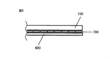
- the horny layer 700 is peeled off by peeling off the sheet body 600 from the skin, and the horny layer 700 attached to the sheet body 600 is sandwiched between the sheet body 600 and the carrier 100 in which a micro space group is formed.
- the solvent 320 may be supplied to dissolve the components contained in the stratum corneum 700 in the solvent 320 to obtain a sample solution.
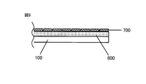
- the carrier 100 having the adhesive layer 800 is attached so that the adhesive layer 800 side is the skin, and the carrier is peeled off from the skin.
- the solvent 320 may be supplied to the carrier 100, and the stratum corneum 700 may be dissolved in the solvent 320 to form a sample solution.
- step (2) the sample solution 310 supplied to the carrier 100 is taken into the analysis unit 400 from the suction port 410.
- step (3) the analysis unit 400 measures the mass of the components contained in the sample solution sucked by the suction force generated due to the vacuum pressure of the analysis unit 400 in step (2).
- the solvent 320 is supplied to the sample on the carrier 100 to obtain the sample solution from which the target component has been extracted.
- the target component can be qualitatively or quantitatively analyzed by instantaneously taking the sample solution into the analysis unit 400 under vacuum.
- the pretreatment operation is reduced, the measurement time is instantaneous, and the amount of the target component that can be taken into the analyzer at a time can be greatly increased, so that the absolute sensitivity can be increased.
- the vaporization volume of the sucked droplet is calculated from the vacuum pressure and temperature inside the analysis unit 400, a droplet of about 6 ⁇ L of water has a volume of about 12 L. Actually, it has been confirmed that the influence on the pressure is negligible and the measurement returns to the original vacuum pressure instantaneously, so that the measurement is not affected.
- water with a low molecular weight has the largest vaporization volume
- the analytical method of the present invention has demonstrated that measurement is possible when the solvent is water, and therefore it is possible to use any other solvent. I can say that.
- the fact that the solvent species can be changed means that a rough fraction can be obtained from the sample placed on the carrier by changing the polarity of the solvent (the target component can be analyzed sequentially). In this respect, similar to the conventional paper spray ionization method, there is an advantage that it can be rapidly analyzed even in the case of blood having a lot of interfering components.
- the sample that can be measured by the analysis method of the present invention is not limited, and may be either a solid sample, a semi-solid sample, or a liquid sample, and a component present on any object surface.
- a sample solution 310 in which a sample is dissolved in a solvent may be used for analysis.
- an appropriate solvent 320 can be supplied to a liquid or (semi) solid sample previously applied to the carrier 100 and used as a sample solution.
- an appropriate solvent 320 may be supplied to the sample transferred to the carrier 100 and used as the sample.
- a horny layer 700 peeled from the skin using a carrier 100 having an adhesive layer 800 can be used as a sample, and a suitable solvent 320 is used for the horny layer 700. It can also be supplied and used as a sample solution.
- the stratum corneum 700 peeled from the stratum corneum using a sheet body (adhesive sheet body 600) having a pressure-sensitive adhesive layer on the surface can be used as a sample, and an appropriate solvent 320 can be supplied to obtain a sample solution.
- any sample can be used, but a carrier for sandwiching the sheet material on which the sample is applied, impregnated with the sample solution, or adhered to the peeled horny layer is required.
- the solvent used in sample preparation is not limited as long as it dissolves the sample and extracts components contained in the sample, but is not limited to water, organic solvents (for example, ethanol, methanol, acetonitrile, hexane), or those It may be a mixture of Acids, bases, trace amounts of ion-pairing agents and salts may be added.
- the amount supplied may be one drop or an amount corresponding to about 2 to 20 ⁇ L.
- the carrier when the sample is a sample solution containing a target component, the carrier The sample solution 310 can be directly supplied to 100 and the sample solution can be sucked from the suction port 410 of the analysis unit 400 and analyzed.
- the amount of the sample solution in this case conforms to the above supply amount.
- any of the above-described solvents may be dropped onto the carrier 100 as preliminary infiltration.
- any of the above-described solvents may be dropped onto the carrier 100 before and / or after the sample is applied or transferred.
- any of the above-described solvents may be dropped onto the carrier 100 before or after the stratum corneum 700 is attached.
- Carrier The analysis method of the present invention is characterized by using a carrier 100 for stacking (mounting) a sample.
- the suction port 410 of the analysis unit 400 in the mass spectrometer 1 of the present invention is not in contact with the carrier 100 supplied with the sample solution 310. Mass spectrometry is performed.
- the carrier 100 that can be used is a microscopic material for guiding the sample solution from at least one end of the carrier 100 to the suction port 410 of the analysis unit 400 from the liquid receiving unit 120 that receives the supplied sample solution 310 or the solvent 320. It is preferable that a space group is formed.
- liquid receiver means any area on the carrier 100 that receives the supplied sample solution 310 or solvent 320, and the liquid receiver 120 and other areas. Is not intended to be clearly distinguished. If the sample is placed on the carrier 100, it means a portion (or structure) for supplying the solvent 320 for dissolving the components contained in the sample, and if the sample is a liquid sample, It means a part (or structure) for supplying the sample solution 310.
- the liquid receiving unit may be any region on the carrier 100 that temporarily receives the sample solution 310 or the solvent 320 supplied from the supply unit 300, but the carrier 100 includes the guide mechanism 10. If it is, it is preferable that the region is in contact with or near the guide mechanism. In one embodiment, when the sample is dissolved or extracted in the carrier 100, the liquid receiving unit 120 may be near one end (not shown) on the carrier 100 far from the suction port 410.
- a blank space for forming the liquid receiving part on the carrier is required.
- a margin corresponds to, for example, S 1 and S 2 in FIG. 2, but the margin portion (S 2 ) on the front end side is not necessarily provided, and the holding portion 200 is connected to the one end 110 of the carrier. It may overlap.
- the margin part (S 1 ) along the side surface of the holding part 200 may be on both sides of the holding part 200 or may be on one side.
- the carrier 100 used in the present invention is preferably formed with a micro space group.
- the “microspace group” means an object having a structure that causes capillary action, and the impregnated sample solution passes through the microspace group and at least one end 110 of the carrier 100.
- the carrier that can be used in the present invention is not particularly limited as long as it is a carrier having the above-described liquid receiving part and a micro space group.
- the shape of the carrier 100 used in the analysis method of the present invention is a shape suitable for allowing the sample solution impregnated in the carrier 100 to be sucked into the suction port 410 of the analysis unit 400 at at least one end 110 of the carrier 100. It is preferable.
- the shape of the carrier 100 is preferably close to the suction port 410 but not pointed.
- the angle of one end of the carrier is preferably not less than 90 °.
- Examples of the shape of the carrier 100 include a square, a rectangle, and a trapezoid.
- a filter paper when used as an example, as the filter paper, for example, a commercially available qualitative filter paper (manufactured by GE Health Care Japan; circular filter paper: maximum 500 mm diameter, square filter paper: maximum 600 mm) should be used. Can do.
- a small piece of filter paper obtained by cutting the filter paper so as to have an appropriate shape and size in accordance with the state (shape, size, etc.) of the suction port 410.
- the shape and size of the filter paper pieces are not limited, but are typically rectangular (for example, short side 2.5 mm ⁇ long side 10 mm), trapezoid (upper base 2.5 mm ⁇ lower base 10 mm) Also good.
- Such a small piece of filter paper may be cut out from the filter paper after the sample is laminated on the filter paper before cutting.
- carrier 100 is not less than 90 degrees as above-mentioned. Such an angle is preferably 90 ° or more and less than 180 °, more preferably 90 ° or more and less than 150 °, and even more preferably 90 °.
- the carrier 100 used in the present invention is preferably formed with the liquid receiving portion 120 and the micro space group. Further, the carrier solution 100 contains the sample solution 310 or the solvent 320 and the analysis portion 400.
- a guide mechanism for assisting suction from the suction port 410 may be provided.
- the “guide mechanism” is a term commonly used by those skilled in the art.
- the sample solution 310 or the solvent 320 is sucked from the suction port 410 of the analysis unit 400.
- the material and the size are not limited.
- a side edge 10 of a holding portion 200 for example, a clip
- the holding unit 200 used since it is not necessary to apply a high voltage to ionize the sample, the holding unit 200 used may be insulative.
- the filter paper piece is preferably fixed so that a blank space remains on the filter paper piece without being entirely covered with the clip.
- the blank portion on the side of the clip corresponds to the liquid receiver 120 that supplies the sample solution 310 or the solvent 320.
- the width of the margin is not limited, but may be about 0.5 to 2 mm.
- the clip side surface 10 serves as a guide mechanism so that the sample solution 310 or the solvent 320 flows while being impregnated toward one end 110 of the filter paper, that is, the suction port 410 of the analysis unit 400.
- the blank portion on the side close to the suction port 410 does not necessarily have to be provided as described above. It is sufficient that the size is sufficient to form a fine liquid droplet (liquid reservoir).
- the area of such a blank portion is not limited, but is preferably about 0.001 to 10 mm 2 .
- the side edge of the holding part 200 may be used as the guide mechanism 10, or the groove provided on the carrier is used as the guide mechanism (10a, 10b, 10c, 10d, 10d ′). It may be used.
- the shape of the provided groove has an inclined surface and a curved surface as shown in the sectional view, but the depth and position of the groove are not limited. Therefore, as long as the object of the present invention is achieved, the shape, depth, and position of the groove on the carrier used as the guide mechanism are not limited.
- the guide mechanisms (10b, 10c, 10d, 10d '). 5 by providing a step on one side surface of the carrier 100 ′′, this step can be used as the guide mechanism 10e (FIG. 5A).
- the cross section of ′′ is as shown in FIG.
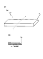
- the guide mechanism can also be installed by placing the sheet body 500 on the surface of the carrier 100 without providing a groove in the carrier 100.
- the guide mechanism 10 f can be installed between the two sheet bodies 500. it can.
- FIG. 7 by placing one sheet body 500 ′ on the carrier 100, a step is formed on the carrier 100 using the side edges of the sheet body 500 ′ as in the case of FIG. It can be formed and used as the guide mechanism 10g.
- usable sheet body 500 is not particularly limited, and may be any material, and may have a property of not absorbing the solvent.
- the sheet placed on the carrier has an effect of suppressing evaporation of the solvent, and has an effect of increasing extraction efficiency when components are extracted on the carrier.
- corneal layer means a layer in the outermost layer of the skin, in which flat stratum corneum cells keratinized by epidermal keratinocytes overlap.
- the cell-cell space is filled with stratum corneum intercellular lipids, and the stratum corneum has about 15 layers of stratum corneum cells and has an average thickness of about 10 to 20 ⁇ m.
- the stratum corneum has a different layer thickness depending on the site, and is relatively thin on the cheeks, while the horny layer or the like reaches about 50 layers of stratum corneum cells.
- the layer thickness measurable by the method of the present invention is not particularly limited, but about 5 to 15 layers are analyzed.
- a tape stripping method is effective.
- the sheet body (such as the pressure-sensitive adhesive sheet body 600) having an adhesive layer on the surface that can be used for peeling the stratum corneum is not particularly limited, but D-Square (manufactured by CuDerm), Cellotape (registered trademark) (Nichiban) And PPS tape (manufactured by Nichiban Co., Ltd.).
- the stratum corneum 700 attached to the pressure-sensitive adhesive sheet body 600 is sandwiched between the pressure-sensitive adhesive sheet body 600 and the carrier 100 in which a minute space group is formed ( FIG. 8).
- a solvent is supplied to the carrier sandwiching the stratum corneum, and the solvent sample solution in which the components of the stratum corneum are dissolved can be used.
- the carrier 100 having the adhesive layer 800 on the surface and formed with a minute space group can be obtained by supplying a solvent to the carrier (FIG. 9).
- Mass Spectrometer typically, a sample solution 310 containing a sample dissolved in a solvent is impregnated in the carrier 100, and the sample is in a state where the carrier 100 and the suction port 410 of the analysis unit 400 are not in contact with each other. The solution 310 is sucked from the suction port 410, and mass spectrometry is performed within a few seconds without requiring voltage, heat, gas, or the like (see FIG. 1).
- an analyzer for qualitative or quantitative analysis of components contained in a sample A carrier 100 in which a minute space group is formed; A holding unit 200 for holding the carrier; A supply unit 300 for supplying the solvent 320 or the sample solution 310 to the carrier; An analysis part 400 having a suction port 410 and qualitatively or analyzing the component;
- the carrier 100 is held by the holding unit 200 in a state where the carrier 100 and the suction port 410 are not in contact with each other, and a sample solution 310 containing a sample dissolved in the solvent supplied from the supply unit 300 is provided at one end 110. It is possible to provide the mass spectrometer 1 that reaches and sucks the reached sample solution from the suction port 410.
- the “holding part” (200) used in the present invention means a part or means for holding the carrier 100 in front of the suction port 410 of the analysis part 400 (see FIGS. 1 and 2). Since the holding unit 200 does not require a high voltage to ionize the sample, it may be made of an insulating material and is not limited as long as it has a structure sufficient to hold the carrier. Although it is not limited to the holding
- the “supply section” (300) used in the present invention is a space for storing the sample solution or solvent and means for discharging the sample solution or the like in order to supply the sample solution 310 or the solvent 320 to the carrier 100.
- Examples thereof include pipettes and syringes.
- the supply unit 300 is not particularly limited, and may have an independent structure such as use of a pipette, or may be provided in the holding unit 200 or may be fixed to the carrier 100. Good.
- the “analyzer” (400) used in the present invention is a portion where the components in the sample solution are subjected to analysis in addition to the suction port 410 for sucking the sample solution, depending on the properties of the components.
- the measurement signal generated is detected.
- Such an analysis unit 400 is not limited, but includes a hybrid tandem mass spectrometer (for example, LTQ-Orbtrap (registered trademark) (Thermo Fisher Scientific, Inc., San Jose, Calif.)), 6430qTOF (Agilent Technologies, Inc.).
- ion mobility-TOF mass spectrometer eg, Synapt G2 SI (Nippon Waters KK)
- qTOF type mass spectrometer eg, 6530qTOF (Agilent Technologies)
- Triple quadrupole mass spectrometer eg 6430QQQ (Agilent Technologies)
- small single quadrupole mass spectrometer eg QDa (Nippon Waters KK)
- Q-Orbitrap mass spectrometer QExactive (Thermo) Fisher Scientific)
- the sample solution is sucked from the one end 110 of the carrier to the suction port 410 in a state where the one end 110 of the carrier 100 and the suction port 410 are not in contact with each other.
- the one end 110 of the carrier The distance and height from the suction port 410 are adjusted.
- the distance between the one end 110 of the carrier and the suction port 410 is not limited, but is preferably 0.1 to 0.5 mm.
- the vacuum pressure in the analysis unit 400 is not limited, but is preferably 0.1 hPa or less.
- the analysis of a sample using the mass spectrometer 1 of the present invention is typically as follows.
- the carrier 100 is fixed to the holding unit 200, and the one end 110 of the carrier is adjusted and fixed so that the interval is 0.1 to 0.5 mm so as not to contact the suction port 410 of the analysis unit 400.
- the prepared sample solution 310 is supplied from the supply unit 300 to the liquid receiving unit 120 on the carrier 100.
- the supplied sample solution is stored at one end 110 of the carrier while impregnating the carrier 100, and is quickly sucked into the analysis unit 400 from the suction port 410 of the analysis unit 400.
- the aspirated sample solution is measured by the analysis unit 400, and a measurement signal can be obtained (see Examples 1 to 4).
- Lysine hydrochloride was obtained from Ajinomoto Co., Inc. (Tokyo, Japan). Glycine (d2) was purchased from Sigma-Aldrich.
- Filter paper 42 (55 mm diameter) was obtained from Whatman (GE Healthcare Life Sciences, Inc., UK). As described below, it was used without treatment, but was cut into small pieces of appropriate size in the analysis. For analyzing stratum corneum components, see Promotool, Inc. Adhesive skin stripping tape (Skinchecker®) available from (Tokyo, Japan) was used.
- Example 1 The standard mixture consisting of 17 amino acids used in Example 1 was treated as 1 mM (nmol / ⁇ L) in 0.1 M hydrochloric acid using Agilent Technologies, Inc. (Part No. 5061-3330; L-aspartic acid, glycine, L-alanine, L-valine, L-leucine, L-isoleucine, L-serine, L-threonine, L-tyrosine, obtained from (Palo alto, CA) L-proline, L-arginine, L-histidine, L-glutamic acid, L-cystine, L-phenylalanine, L-lysine, and L-methionine).
- Agilent Technologies, Inc. Part No. 5061-3330; L-aspartic acid, glycine, L-alanine, L-valine, L-leucine, L-isoleucine, L-serine, L-threonine, L-tyrosine, obtained from
- Reagent grade angiotensin II and insulin used in Example 2 were obtained from Sigma-Aldrich.
- cosmetics Ipsa Metabolizer Moist White W1
- Shiseido Co. , Ltd. Tokyo, Japan.
- the suction force into the mass spectrometer is fixed at a height sufficient to suck the micro droplet formed at one end of the filter paper piece, and the distance between the entrance capillary and the filter paper piece is about 0.5 mm.
- the distance is optimized according to the composition of the extraction solvent and can be adjusted as appropriate.It is preferable to match the center line of the entrance capillary with the center line of the filter paper piece. As long as they are formed and successfully aspirated into the mass spectrometer, the axes do not need to coincide.
- the volume of the extraction solvent to be supplied is typically 5 to 20 ⁇ L, but may be changed according to the size of the filter paper piece. Generally, when the size of the filter paper piece is increased, it is necessary to increase the volume of the extraction solvent to be supplied accordingly. This is because a “dome” is formed on the filter paper piece by an excess solvent and a micro droplet (liquid reservoir) is formed at one end of the filter paper piece, but this is not a necessary condition.
- mass spectrometry data was acquired using LTQ-Orbtrap (registered trademark) (Thermo Fisher Scientific, Inc., San Jose, Calif.), which is a hybrid tandem mass spectrometer.
- LTQ-Orbtrap registered trademark
- a nanospray flex ion source registered trademark
- a connection cable used to supply a high voltage typically ⁇ 2 to 5 kV
- the ground potential of the sample region was fixed by connecting the main conductive surface / non-conductive surface using a conductive cable with an alligator clip.
- the entrance capillary temperature was set to 350 ° C. in each experiment. In order to avoid unintentional voltage application to the outlet, the capillary voltage was set to 0 kV.
- Most of the mass spectrometry was performed using a linear quadrupole ion trap mass spectrometer LTQ®. Instrument-specific settings were as follows: microscan number: 1, maximum injection time: 10 ms, scan speed: normal or high, AGC ion target setting (full scan): 3.00e + 4.
- Xcalibur 2.1 software (Thermo Fisher Scientific, CA) was used to control the mass spectrometer and to perform data analysis.
- Example 1 Verification of the analysis method of the present invention The verification of the analysis method of the present invention was carried out using a standard mixture of 17 amino acids from the viewpoint of (a) reproducibility, (b) linearity, and (c) sensitivity. went.
- the execution time of each sample was set to 30 seconds, but the actual analysis time was within several seconds.
- the same procedure was repeated for three filter paper pieces to assess reproducibility.
- the averaged mass spectrum was obtained from TICC (total ion current chromatogram) of each data, and was compared qualitatively from the apparent peak intensity ratio (height of the y-axis) (FIG. 10).
- the linearity of the EmDI-MS method of the present invention was evaluated by an internal standard method using a mixture of 17 amino acid standards and glycine (d2).
- a mixture solution (described later) was laminated to each of the four filter paper pieces.
- the amount of 17 amino acids was 1, 5, 10 and 50 ng per filter paper piece (2.5 mm ⁇ 10 mm) (hereinafter referred to as “Std1”, “Std2”, “Std3” and “Std4”, respectively).
- the amount of glycine (d2) was fixed (10 ng) in order to function as an internal standard.
- Std1, Std2, and Std3 were prepared by laminating three standard solution dispensing solutions on three filter paper pieces.
- a mother liquor (1 mM amino acid each) containing 10 mM standard solution was layered.
- solutions containing 0.01 mM and 0.1 M amino acids were used, respectively (both included a 10 mM internal standard).
- These were prepared by serial dilution of the mother liquor with methanol-purified water (1: 1).
- a dispense of mother liquor spiked with 1 mM internal standard was layered on another piece of filter paper.
- the dispensing solution was laminated 10 times on the filter paper pieces by repeating the lamination-drying cycle. The drying process was performed at room temperature under a gentle air flow.
- the Std1-Std4 filter paper pieces were subjected to EmDI-MS analysis in the same manner as described above. From the average mass spectrum of the Std1 to Std4 calibration standards, the peak height between each amino acid and the internal standard was calculated and compared. Next, the amount of amino acid laminated on the filter paper piece was plotted on the x-axis to create a calibration curve.
- the linearity in the analysis method of the present invention was evaluated from R 2 (determination coefficient) and intercept of a calibration line generated by the least square method (FIG. 11).
- Sensitivity was estimated from the signal-to-noise (SN) ratio of the amino acid signal in the mass spectrum of Std1 (FIG. 12).
- the lower detection limit (LLOD) was defined as the estimated concentration of SN> 3.
- Example 2 Verification of analysis method using standard sample Peptides were used to verify the feasibility of the analysis method of the present invention.
- angiotensin II and insulin were used as peptides and dissolved in purified water containing 1 mM 0.1% formic acid and 2% ACN, respectively.
- 10 ⁇ L of each solution was layered on separate filter paper pieces (2.5 mm ⁇ 10 mm rectangle) and the filter paper pieces were dried at room temperature. Each filter paper piece is laminated with 10 nmol of peptide.
- the EmDI-MS analysis of the present invention was performed according to the procedure of Example 1, but in this example, the extraction solvent was changed to ACN-purified water (1: 1) containing 0.1% formic acid.
- the extraction solvent was applied twice to the filter paper strip to increase the peptide extraction efficiency by “pre-wetting” and to facilitate the absorption of the microdroplets into the mass spectrometer.
- pre-wetting 5 ⁇ L of ACN-purified water (1: 1) containing 0.1 formic acid was applied.
- the solvent (10 ⁇ L) was applied immediately after the pre-wetting process (after about 10 seconds) for extraction and microdroplet formation.
- the analysis results (mass spectrum) of angiotensin II and insulin are shown in FIGS. 13 and 14, respectively. As is apparent from these mass spectra, the unique mass spectra of angiotensin II and insulin were observed, so that it was demonstrated that the analysis method of the present invention can also analyze a sample using a peptide.
- Example 3 Verification of analytical method using cosmetics
- a part (1 ⁇ L) of a cosmetic product prototype cosmetic liquid, no pretreatment
- a small piece of filter paper 2.5 mm ⁇ 10 mm rectangle
- the EmDI-MS analysis of the present invention was performed according to the procedure of Example 1. The results are shown in FIG.
- the cosmetic contains a large number of components, but mainly the synthetic polymer, surfactant, and active ingredient can be clearly separated, and even when a semi-solid is used, the analysis method of the present invention. The effectiveness of was demonstrated.
- Example 4 Verification of analysis method using human skin (corneal layer) Using human skin, components contained in the stratum corneum were analyzed by the EmDI-MS method.
- the horny layer was collected from the inner forearm of a healthy male volunteer (48 years old) by means of the adhesive tape stripping method using an adhesive tape device (Skinchecker®) as a sample.
- Skinchecker® an adhesive tape device
- a typical procedure is shown below. (1) Gently wash the inner forearm with soap. (2) Wait 30 minutes for equilibration. (3) The stratum corneum (10 layers) is collected by tape stripping using Skinchecher (registered trademark). (4) Place the collected stratum corneum on the filter paper and gently apply pressure to the adhesive area to fix it.
- the filter paper to which the stratum corneum is bonded is cut into small pieces (2.5 mm ⁇ 10 mm rectangle).
- a crocodile clip metal, plastic, etc.
- One EmDI-MS experiment is completed within a few seconds.
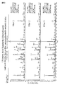
- FIG. 16 shows the result of analysis using the stratum corneum.
- mass spectra of amino acids, minerals, PCA (pyrrolidone carboxylic acid), lactate, urea, etc. appear as components of NMF (natural moisturizing factor) contained in the stratum corneum, and human skin is sampled. Even in this case, the effectiveness of the analysis method of the present invention was proved.
- FIG. 1 shows an analyzer for carrying out the above-described method of the present invention.
- the analysis apparatus includes an analysis unit 410, a holding unit 200 that holds a carrier 100 in which a micro space group is formed, and a supply unit 300 that supplies a sample to the carrier 100 as main components.
- the analysis unit 400 includes a vacuum container as an ion source, a vacuum pump that forms a vacuum in the container, a high voltage source that applies a high voltage in the container, and an ionized sample that communicates with the vacuum container as an ion source.
- An analysis unit to be separated, a detection unit that sensitizes and detects ions selected by the analysis unit, and the like can be included.
- These components are housed in a housing 402.
- a suction port 410 constituting a sample introduction unit is provided on one side surface of the housing 402.
- the carrier 100 is held by the holding unit 200 at a position where the sample solution can be sucked from the one end 110 of the carrier without the one end 110 of the carrier contacting the suction port 410 in the vicinity of the suction port 410.
- the carrier 100 is, for example, a filter paper, a fiber, a resin, a cloth, or a thin layer chromatography plate (a thin layer plate in which an adsorbent such as silica gel, alumina, or polyamide resin is fixed in a thin film on a glass or aluminum plate). Can do.
- the holding unit 200 may be fixed at a predetermined position in front of the suction port 410 by using a fixing unit such as a stand (not shown) or a frame (not shown).
- the supply unit 300 can be configured by a pipette, a syringe, or the like.
- the supply unit 300 is disposed at a position where the solvent 320 or the sample solution 310 can be supplied to the carrier 100.
- the supply unit 300 may be fixed at a predetermined position above the carrier 100 using fixing means such as a stand (not shown) or a frame (not shown).
Landscapes
- Physics & Mathematics (AREA)
- Chemical & Material Sciences (AREA)
- Health & Medical Sciences (AREA)
- Life Sciences & Earth Sciences (AREA)
- Spectroscopy & Molecular Physics (AREA)
- Analytical Chemistry (AREA)
- Biochemistry (AREA)
- General Health & Medical Sciences (AREA)
- General Physics & Mathematics (AREA)
- Immunology (AREA)
- Pathology (AREA)
- Chemical Kinetics & Catalysis (AREA)
- Electrochemistry (AREA)
- Molecular Biology (AREA)
- Other Investigation Or Analysis Of Materials By Electrical Means (AREA)
- Sampling And Sample Adjustment (AREA)
- Investigating Or Analysing Biological Materials (AREA)
Abstract
La présente invention concerne un procédé pour analyser qualitativement ou quantitativement un composé contenu dans un échantillon, le procédé comprenant : une étape dans laquelle une solution d'échantillon (310) contenant un échantillon dissous dans un solvant est fournie à un support (100) dans lequel sont formés un groupe de minuscules espaces pour guider la solution d'échantillon entre une section de réception de solution (120) qui reçoit la solution d'échantillon et au moins une extrémité ; une étape dans laquelle la solution d'échantillon qui est arrivé à ladite extrémité du support est aspirée depuis un orifice d'aspiration (410) pour aspirer la solution d'échantillon dans un dispositif de spectrométrie de masse (400), dans un état où l'orifice d'aspiration et le support ne sont pas reliés ; et une étape dans laquelle une spectrométrie de masse du composé est effectuée.
Applications Claiming Priority (2)
| Application Number | Priority Date | Filing Date | Title |
|---|---|---|---|
| JP2015-083770 | 2015-04-15 | ||
| JP2015083770 | 2015-04-15 |
Publications (1)
| Publication Number | Publication Date |
|---|---|
| WO2016167364A1 true WO2016167364A1 (fr) | 2016-10-20 |
Family
ID=57127258
Family Applications (1)
| Application Number | Title | Priority Date | Filing Date |
|---|---|---|---|
| PCT/JP2016/062176 Ceased WO2016167364A1 (fr) | 2015-04-15 | 2016-04-15 | Procédé d'analyse |
Country Status (3)
| Country | Link |
|---|---|
| JP (1) | JP6100946B2 (fr) |
| TW (1) | TW201702592A (fr) |
| WO (1) | WO2016167364A1 (fr) |
Cited By (2)
| Publication number | Priority date | Publication date | Assignee | Title |
|---|---|---|---|---|
| WO2020021784A1 (fr) * | 2018-07-25 | 2020-01-30 | 株式会社島津製作所 | Procédé de collecte de composants biologiques et procédé d'analyse de composants biologiques |
| CN111684274A (zh) * | 2018-02-09 | 2020-09-18 | 浜松光子学株式会社 | 试样支撑体、电离法以及质量分析方法 |
Families Citing this family (1)
| Publication number | Priority date | Publication date | Assignee | Title |
|---|---|---|---|---|
| GB202001571D0 (en) * | 2020-02-05 | 2020-03-18 | Univ Manchester | Identification of biomarkers and uses thereof |
Citations (4)
| Publication number | Priority date | Publication date | Assignee | Title |
|---|---|---|---|---|
| JP2004125576A (ja) * | 2002-10-02 | 2004-04-22 | Hitachi Ltd | 危険物探知装置及び危険物探知方法 |
| WO2012056729A1 (fr) * | 2010-10-29 | 2012-05-03 | アトナープ株式会社 | Appareil d'échantillonnage |
| JP2012525687A (ja) * | 2009-04-30 | 2012-10-22 | パーデュー・リサーチ・ファウンデーション | 濡れた多孔質材料を用いるイオン生成 |
| JP2014512538A (ja) * | 2011-04-19 | 2014-05-22 | ポーレックス コーポレイション | 焼結多孔性プラスチックを含む試料貯蔵および送達のためのカード |
-
2016
- 2016-04-15 WO PCT/JP2016/062176 patent/WO2016167364A1/fr not_active Ceased
- 2016-04-15 TW TW105111925A patent/TW201702592A/zh unknown
- 2016-04-15 JP JP2016082108A patent/JP6100946B2/ja not_active Expired - Fee Related
Patent Citations (4)
| Publication number | Priority date | Publication date | Assignee | Title |
|---|---|---|---|---|
| JP2004125576A (ja) * | 2002-10-02 | 2004-04-22 | Hitachi Ltd | 危険物探知装置及び危険物探知方法 |
| JP2012525687A (ja) * | 2009-04-30 | 2012-10-22 | パーデュー・リサーチ・ファウンデーション | 濡れた多孔質材料を用いるイオン生成 |
| WO2012056729A1 (fr) * | 2010-10-29 | 2012-05-03 | アトナープ株式会社 | Appareil d'échantillonnage |
| JP2014512538A (ja) * | 2011-04-19 | 2014-05-22 | ポーレックス コーポレイション | 焼結多孔性プラスチックを含む試料貯蔵および送達のためのカード |
Cited By (3)
| Publication number | Priority date | Publication date | Assignee | Title |
|---|---|---|---|---|
| CN111684274A (zh) * | 2018-02-09 | 2020-09-18 | 浜松光子学株式会社 | 试样支撑体、电离法以及质量分析方法 |
| CN111684274B (zh) * | 2018-02-09 | 2023-09-05 | 浜松光子学株式会社 | 试样支撑体、电离法以及质量分析方法 |
| WO2020021784A1 (fr) * | 2018-07-25 | 2020-01-30 | 株式会社島津製作所 | Procédé de collecte de composants biologiques et procédé d'analyse de composants biologiques |
Also Published As
| Publication number | Publication date |
|---|---|
| JP2016206188A (ja) | 2016-12-08 |
| TW201702592A (zh) | 2017-01-16 |
| JP6100946B2 (ja) | 2017-03-22 |
Similar Documents
| Publication | Publication Date | Title |
|---|---|---|
| US11830716B2 (en) | Mass spectrometry analysis of microorganisms in samples | |
| CN105606691B (zh) | 分析样品中的蛋白质或肽的方法 | |
| US6175112B1 (en) | On-line liquid sample deposition interface for matrix assisted laser desorption ionization-time of flight (MALDI-TOF) mass spectroscopy | |
| Manicke et al. | Assessment of paper spray ionization for quantitation of pharmaceuticals in blood spots | |
| US5595636A (en) | Method for mass spectrometric analysis of samples from electrophoresis plates | |
| JP6764778B2 (ja) | キラルアミノ酸の分離方法 | |
| TW200842359A (en) | A method of mass spectrometry to combine electrospray ionization with laser-induced acoustic desorption | |
| CA2988172A1 (fr) | Sonde d'extraction de molecules d'interet a partir d'un echantillon | |
| US20150318160A1 (en) | Probe for extraction of molecules of interest from a sample | |
| JP7659012B2 (ja) | 吸着材料を使用して試料から抽出された分析物を分析するためのシステムおよび方法 | |
| JP6100946B2 (ja) | 分析方法 | |
| US6825463B2 (en) | On-line and off-line deposition of liquid samples for matrix assisted laser desorption ionization-time of flight (MALDI-TOF) mass spectroscopy | |
| US6674070B2 (en) | On-line and off-line deposition of liquid samples for matrix assisted laser desorption ionization-time of flight (MALDI-TOF) mass spectroscopy | |
| CN104134606A (zh) | 一种微萃取探针电喷雾离子源及其制备方法和应用 | |
| KR20170021398A (ko) | 말디톱 질량분석기에 이용가능한 시료 플레이트 및 상기 시료 플레이트의 제조방법 | |
| Rahman et al. | Direct analysis of aqueous solutions and untreated biological samples using nanoelectrospray ionization mass spectrometry with pipette tip in series with high-ohmic resistor as ion source | |
| JP2007108060A (ja) | 肌質の評価方法 | |
| JP2002502543A (ja) | マトリクス支援レーザ離脱イオン化−飛翔時間(maldi−tof)質量分光用のオンライン液体試料析出インターフェース | |
| US20090166529A1 (en) | Method for preparing specimen for mass spectrometry | |
| CN114078687A (zh) | 一种毛细管纸喷雾离子源装置及离子生成方法 | |
| TWI452601B (zh) | 薄層層析電噴灑游離裝置及質譜系統 | |
| CN202549785U (zh) | 一种电喷雾离子化装置 | |
| CN102610482A (zh) | 一种电喷雾离子化方法和实现该方法的装置 | |
| CN113237981B (zh) | 用于活体微透析质谱联用实时在线分析技术的新型接口 | |
| JP4048869B2 (ja) | 磁場による試料濃縮方法を利用した分析方法及び試料濃縮装置 |
Legal Events
| Date | Code | Title | Description |
|---|---|---|---|
| 121 | Ep: the epo has been informed by wipo that ep was designated in this application |
Ref document number: 16780155 Country of ref document: EP Kind code of ref document: A1 |
|
| NENP | Non-entry into the national phase |
Ref country code: DE |
|
| 122 | Ep: pct application non-entry in european phase |
Ref document number: 16780155 Country of ref document: EP Kind code of ref document: A1 |